purification and characterization of trehalase from seeds of chickpea (cicer arietinum l.)
TRANSCRIPT
1
Purification and characterization of trehalase from seeds of chickpea (Cicer arietinum L.)
Maimona KORD, Elhusseiny YOUSSEF, Hanaa AHMED, Ebtesam QAID
Department of Botany, Faculty of Science, University of Cairo, Giza, 12613, Egypt
Corresponding author:[email protected]
Abstract
In the present study, trehalase was purified and characterized from the seeds of
Cicer arietinum L. (cv. Giza 1). Crude extract was prepared and purified for
electrophoretical homogeneity using ammonium sulphate, chromatography on DEAE-
cellulose, CM-Sepharose and Sephadex G-200. The final specific activity was 7 unit/mg
protein, with 232 fold purification. The purified enzyme exhibited pH optimum
at 5.5. The optimum temperature was 60°C. The determined Km value was 3.64 mM
trehalose. The enzyme activity was stimulated by 20 mM of Mn2+
, Ni2+
or Co2+
, while it
was inhibited by 20 mM of Na+, K
+, Li
+, Ca
2+, Zn
2+ , Cu
2+ or Fe
3+. Zn
2+ proved to be
a non- competitive inhibitor, while mannitol and validamycin A proved to be competitive
inhibitors. The inhibition constants (Ki) of Zn2+
, mannitol and validamycin A were 7 mM,
9 mM and 4 nM, respectively. The molecular mass of the native enzyme was 223 kDa by
gel filtration. SDS-PAGE indicated that, the enzyme consisted of six identical subunits
with a molecular mass of 38 kDa.
Keywords: Cicer arietinum,trehalase,trehalose, purification,molecular mass,validamycin A
2
1. Introduction
Trehalose is a non-reducing disaccharide, formed of two α, D glucose molecules linked by
an α, α-1,1 glycosidic linkage. It is widely distributed through the biological world. This
sugar plays multiple physiological roles in stabilizing and protecting proteins and
membranes against environmental stresses. It also serves as a source of energy and
a source of carbon, and a sensing and regulator compound (Elbein et al., 2003; Fernandez
et al., 2010). Trehalase (EC 3.2.1.28) is a glycosyl hydrolase that hydrolyzes trehalose. It is
detected in many prokaryotic and eukaryotic cells including, bacteria (Carroll et al., 2007),
fungi (Murata et al.,2001), higher plants (Frison et al., 2007), as well as insects (Kamei
et al., 2011 ) and mammals (Kamiya et al., 2004 ). It is the only known pathway of
utilization of trehalose (Silva et al., 2004; Reguera et al., 2011). This enzyme plays an
important role in trehalose metabolism, as it is either directly involved in the assimilation
of exogenous trehalose, or it controls the level of this osmolyte in the cell. In many
organisms, changes in trehalase activity are closely linked to alteration in physiological
conditions or development, indicating that this enzyme plays an important role in such
biological functions as homeostasis and developmental events.
Trehalase has been purified and characterized from various organisms such as
Saccharomyces cerevisiae (Alizadeh and Klionsky, 1996), Lentinula edodes (Murata et al.,
2001), Acidobacterium capsulatum (Inagaki et al., 2001), Medicago sativa (Wolska-
Mitaszko et al., 2005), as well as root nodules of Phaseolus vulgaris (Gracía et al., 2005)
and soybean (Müller et al., 1992; Aeschbacher et al., 1999). Recently, it has also been
purified from the seeds of Triticum aestivum (Kord et al., 2012).
3
So far the purification and characterization of the trehalase enzyme from seeds have not
been properly studied.
The present study deals with the extraction, purification and characterization of trehalase
from the seeds of Cicer arietinum (cv. Giza 1).
2. Materials and Methods
2.1 Plant material
In this study, purification of trehalase was carried out using seeds of chickpea (Cicer
arietinum L.) cv. Giza 1. The seeds were purchased from the Agricultural Research Center
Giza, Egypt.
2.2 Preparation of a crude extract
300 g of chickpea seeds were surface sterilized by immersing in 20% sodium hypochloride
(v/v) for 20 min and rinsed with sterile distilled water. The sterile seeds were homogenized
in a prechilled mortar with 900 ml extraction buffer composed of 100 mM cold sodium
citrate buffer pH 5.5, 1 mM phenylmethanesulfonyl fluoride (PMSF), 1 mM EDTA, 10 M
2- mercaptoethanol, 10% glycerol (v/v) and insoluble polyvinylpyrrolidone (PVP) (10 mg/
g fresh weight). The homogenate was centrifuged at 10 000g for 30 min at 4°C.
2.3 Assay of trehalase
Trehalase activity was measured by estimating the glucose produced by hydrolysis of
trehalose with the glucose oxidase-peroxidase kit (Spainreact) according to the method
described by Bergmeyer and Bernt (1974). The reaction mixture contained: 100 mM
trehalose, 50 mM sodium citrate buffer (pH 5.5) and 0.25 ml crude extract in a final volume
of 1.5 ml. After incubation at 55°C for 30 min, the reaction mixture was boiled for
4
three min and then centrifuged at 5000 g for 10 min. From the supernatant, 10 l was taken
and mixed with 1 ml of glucose oxidase-peroxidase kit. The mixture was then incubated at
37C for 15 min. The absorbance of the sample was measured at 470 nm. Enzyme and
substrate blanks were subtracted. The activity of trehalase enzyme is calculated according
to the following equation:
ΔA sample × standard conc. × 0.0555 / A470 standard × 2
Where: ΔA= difference between optical density of sample before and after addition of
substrate. Concentration of standard =100 mg d/l. Conversion factor 0.0555 mol/ml.
Dividing by 2 = Hydrolysis of 1 mole of trehalose produce 2 moles of glucose. One unit
(nkat) of trehalase activity is defined as the amount of enzyme that hydrolyzes nmol
trehalose per second at pH 5.5.
2.4 Determination of soluble protein
Soluble protein was routinely determined spectrophotometrically at 280 nm as described by
Warburg and Christian (1942). In the presence of interfering compounds, soluble protein
was determined according to Lowry et al. (1951).
2.5 Purification of trehalase
Step 1. Solid ammonium sulphate was added to the crude extract of chickpea seeds to
reach the final saturation of 80%. The precipitated proteins were collected by
centrifugation at 10 000 g for 15 min at 4°C. The precipitant was dissolved in the least
amount of 100 mM sodium citrate buffer pH 5.5, 1 mM EDTA, 10 M 2-mercaptoethanol
and 10% glycerol (v/v). After dialysis and centrifugation, trehalase activity and protein
content were determined.
5
Step 2. DEAE cellulose column (35 × 1.5 cm) was equilibrated with 100 mM sodium
citrate buffer (pH 5.5). The dialyzed fraction from ammonium sulfate was applied to the
column. The proteins were eluted with discontinuous gradient of 0.0, 0.05, 0.1, 0.2, 0.3 and
0.5 M KCl. Three ml fractions were collected at a flow rate 0.5 ml/ min. In each fraction,
trehalase activity as well as the protein content were determined.
Step 3. The enzymatically active fractions of step 2 were pooled and applied to CM -
Sepharose Cl-6B column (20 × 2 cm). Preparation, equilibration and elution of proteins
were again performed as in step 2. Three ml fractions were collected at a flow rate of 1ml/
min.
Step 4. The enzymatically active fractions of step 3 were pooled, lyophilized and applied to
Sephadex G-200 (30 × 1.5 cm) column. The proteins were eluted with the same buffer
used in column equilibration contained 0.2 M KCl. Three ml was collected at a flow rate
of 1.5 ml/ min. Trehalase activity and protein content were determined for each fraction.
All the purification steps were carried out at 4C.
2.6 Characterization of trehalase
2.6.1 Effect of pH
The effect of pH was assayed using 100 mM sodium citrate buffer for pH 3.5 – 5.5;
100 mM sodium phosphate buffer for pH from 6.0 – 7.5 and Tris / HCl buffer for pH from
8.0 – 10.
6
2.6.2 Optimum temperature
The assay of optimum temperature was carried out using 100 mM sodium citrate buffer at
pH 5.5 and at temperatures from 20 – 70C. For consecutive tests, the temperature was
increased by 5C.
2.6.3 Thermal stability
Test samples were preincubated for 30 min at temperature from 40 to 70C. Next, trehalose
was added and the routine assay was followed up as previously mentioned.
2.6.4 Influence of chemical compounds
The effect of 5, 10 and 20 mM solutions of chloride salts of Na+, K
+, Mn
2+, Mg
2+, Ca
2+,
Ni2+
, Co2+
and Fe3+
, as well as sulphate of Li+, Zn
2+ and Cu
2+ were studied. The effect of
10 and 20 mM solutions of mannitol, succinate, borate, malate and 2, 5 nM validamycin A,
as well as 2, 10 mM EDTA and ATP were also studied.
2.6.5 Substrate specificity
The action of the enzyme on 100 mM solution of the following sugars: trehalose, sucrose,
cellobiose, lactose and maltose were studied. The results expressed as relative percentage of
the activity referred to trehalose reaction.
2.6.6 Km determination
The dependence between the reaction velocity and the substrate concentration Michaelis
constant (km) and maximum velocity (Vmax) were determined for trehalose applying the
Lineweaver-Burk’s equation . The reaction mixture contained in 1.5 ml: 50 mM sodium
citrate buffer pH 5.5, 1 nkat of purified enzyme and different concentrations of trehalose
ranging from 2 to 10 mM at 55C.
7
2.6.7 Determination of the inhibition constant
The inhibition constant (ki) for ZnSO4 (5, 10, 20 mM); mannitol (5, 10, 20 mM) and
validamycin A (2, 5, 7 nM) were determined. The reaction mixture contained in 1.5 ml:
50 mM sodium citrate buffer pH 5.5 and 1.163 unit of purified enzyme.
2.7 Determination of molecular mass
The molecular mass of the purified enzyme was estimated by gel fractionation on Sephadex
G-200 (30 × 1.5 cm) which was equilibrated with 100 mM sodium citrate buffer (pH 5.5).
The following proteins were run separately through the same conditions: cytochrome c (13
kDa), carbonic anhydrase (29 kDa), ovalbumin (44 kDa), serum albumin (66 kDa), alcohol
dehydrogenase (150 kDa), catalase (250 kDa) (Serva Co.). The void volume (Vo) was
determined by dextran blue (2000 kDa).
The molecular mass of the denatured form of purified trehalase was estimated with SDS-
polyacrylamide gel electrophoresis (SDS- PAGE) according to Walker (1994). 10% gel
(w/v) was prepared, the run was performed at 100 volts. The following proteins were used
as molecular mass standards: β-galactosidase (116 kDa), bovine serum albumin (66 kDa),
ovalalbumin (45 kDa), lactate dehydrogenase (35 kDa), REase Bsp 981 (25 kDa),
β -lactoglobulin (18.4 kDa) and lysozyme (14.4 kDa) (Fermentas Co).
All data of trehalase characterization are the means of three replicates. Standard errors had
been calculated and represented in figures.
8
3. Results and discussion
3.1 Purification of trehalase
Although trehalase has been purified from different bacteria, yeast and other fungi,
mammals and insects, few reports describe the purification of this enzyme from seeds.
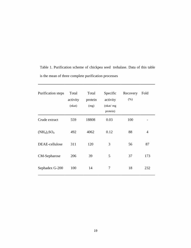
In this study we purified trehalase from seeds of chickpea. The purification protocol is
given in Table 1. The specific activity of the crude extract of trehalase was 0.03 nkat/mg
protein.
In step 1. The specific activity increased to 0.12 nkat/mg protein which represented four
fold purification with 88% recovery after ammonium sulphate precipitation. In this
context, the specific activity of crude extract of trehalase isolated from different plants
expressed in nkat/mg protein was as follows: 0.1 for T. aestivum seeds (Kord et al., 2012),
0.4 for P. vulgaris nodules (Gracía et al.,2005), 0.8 for soybean nodules (Müller et
al.,1992).
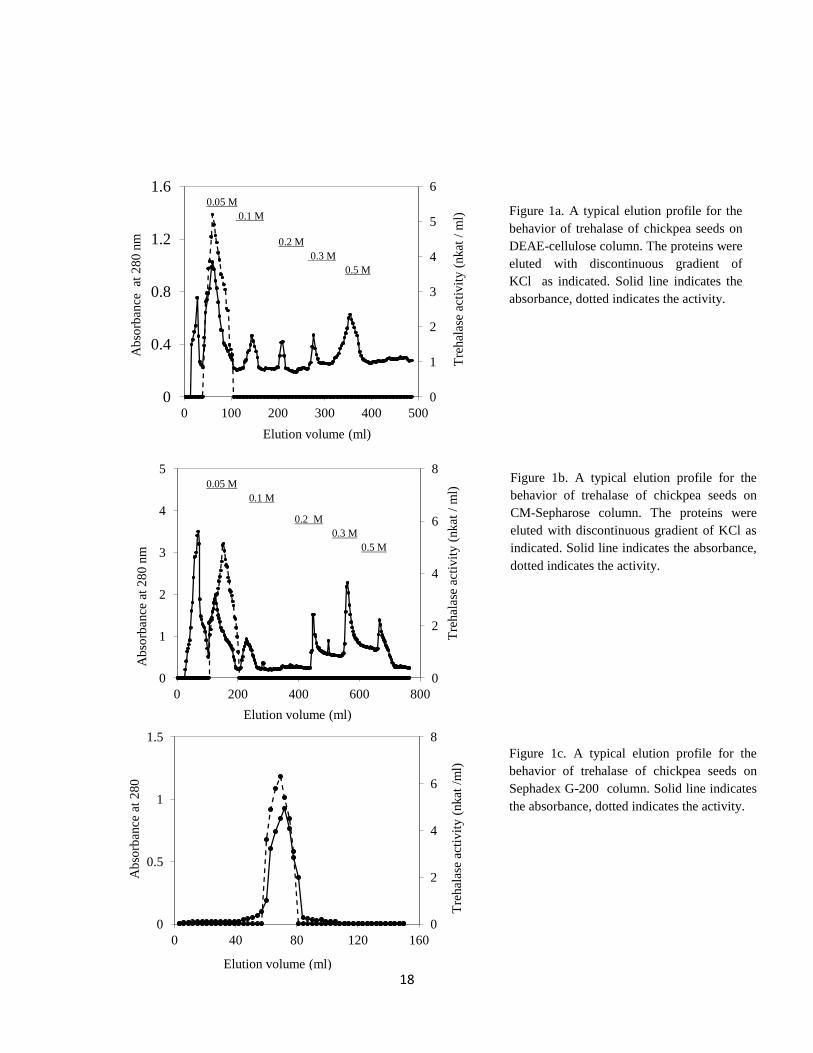
In step 2. The protein fractions exhibiting trehalase activity were eluted from DEAE-
cellulose column as a single peak with 0.05 M KCl (Figure 1a).This indicates that the
protein of trehalase exhibits low density of negative charges as the column contained
positively charged beads.
In step 3. The protein fractions which exhibited trehalase activity was eluted with 0.05 M
KCl from CM- Sepharose column (Figure 1b). The fold purification was 173 fold with 37%
recovery.
In step 4. The specific activity of chickpea seed trehalase was 7 nkat/mg protein, which
represented 232 fold purification (Figure 1c & Table 1). Generally, loss in the activity and
hence in the specific activity were mostly due to the exclusion of some fractions which
9
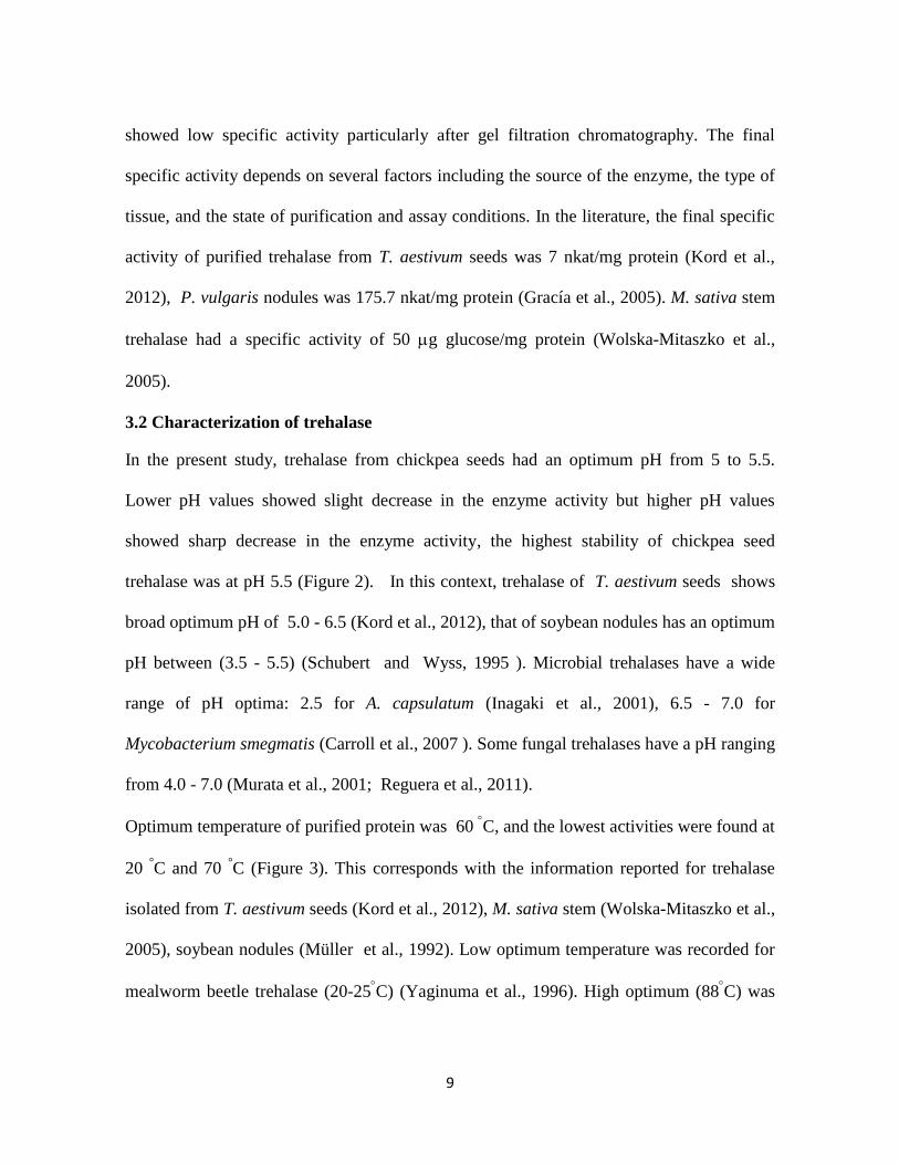
showed low specific activity particularly after gel filtration chromatography. The final
specific activity depends on several factors including the source of the enzyme, the type of
tissue, and the state of purification and assay conditions. In the literature, the final specific
activity of purified trehalase from T. aestivum seeds was 7 nkat/mg protein (Kord et al.,
2012), P. vulgaris nodules was 175.7 nkat/mg protein (Gracía et al., 2005). M. sativa stem
trehalase had a specific activity of 50 g glucose/mg protein (Wolska-Mitaszko et al.,
2005).
3.2 Characterization of trehalase
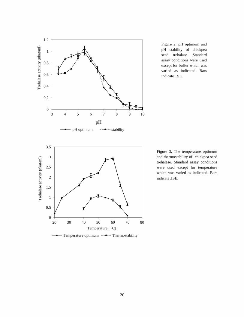
In the present study, trehalase from chickpea seeds had an optimum pH from 5 to 5.5.
Lower pH values showed slight decrease in the enzyme activity but higher pH values
showed sharp decrease in the enzyme activity, the highest stability of chickpea seed
trehalase was at pH 5.5 (Figure 2). In this context, trehalase of T. aestivum seeds shows
broad optimum pH of 5.0 - 6.5 (Kord et al., 2012), that of soybean nodules has an optimum
pH between (3.5 - 5.5) (Schubert and Wyss, 1995 ). Microbial trehalases have a wide
range of pH optima: 2.5 for A. capsulatum (Inagaki et al., 2001), 6.5 - 7.0 for
Mycobacterium smegmatis (Carroll et al., 2007 ). Some fungal trehalases have a pH ranging
from 4.0 - 7.0 (Murata et al., 2001; Reguera et al., 2011).
Optimum temperature of purified protein was 60 C, and the lowest activities were found at
20 °C and 70
°C (Figure 3). This corresponds with the information reported for trehalase
isolated from T. aestivum seeds (Kord et al., 2012), M. sativa stem (Wolska-Mitaszko et al.,
2005), soybean nodules (Müller et al., 1992). Low optimum temperature was recorded for
mealworm beetle trehalase (20-25C) (Yaginuma et al., 1996). High optimum (88
C) was
10
reported for trehalase isolated from Rhodothermus marinus (Jorge et al., 2007). In this
investigation, the highest thermostability of chickpea seed trehalase was reported between
45C to 60
C (Figure 3). The same behavior was reported for trehalase isolated from P.
vulgaris nodules (Gracía et al., 2005) and soybean nodules (Müller et al., 1992).
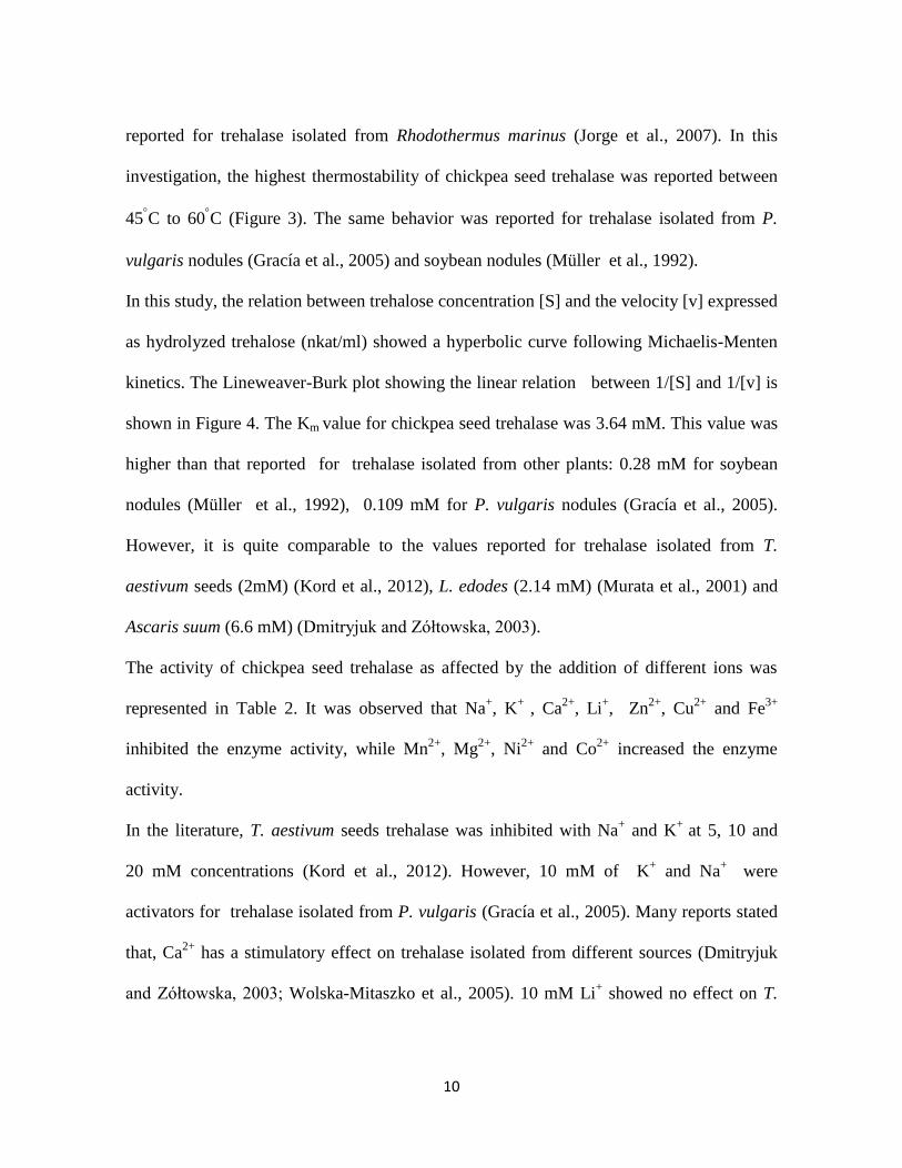
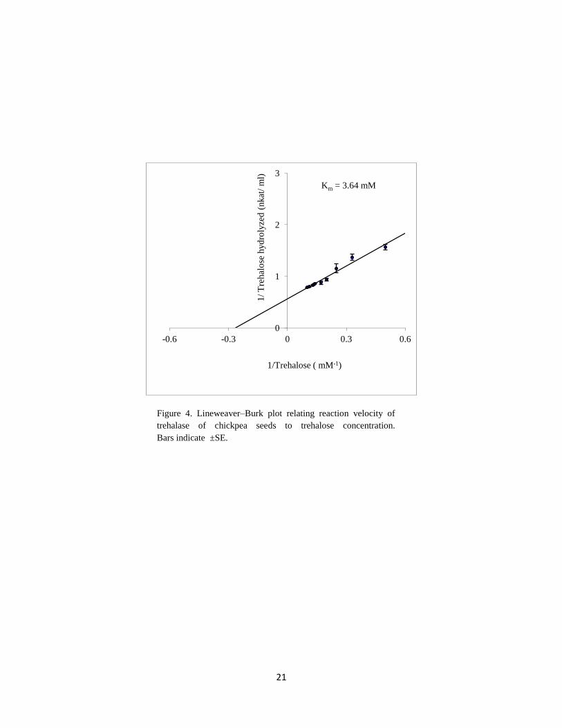
In this study, the relation between trehalose concentration [S] and the velocity [v] expressed
as hydrolyzed trehalose (nkat/ml) showed a hyperbolic curve following Michaelis-Menten
kinetics. The Lineweaver-Burk plot showing the linear relation between 1/[S] and 1/[v] is
shown in Figure 4. The Km value for chickpea seed trehalase was 3.64 mM. This value was
higher than that reported for trehalase isolated from other plants: 0.28 mM for soybean
nodules (Müller et al., 1992), 0.109 mM for P. vulgaris nodules (Gracía et al., 2005).
However, it is quite comparable to the values reported for trehalase isolated from T.
aestivum seeds (2mM) (Kord et al., 2012), L. edodes (2.14 mM) (Murata et al., 2001) and
Ascaris suum (6.6 mM) (Dmitryjuk and Zółtowska, 2003).
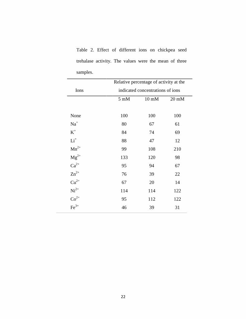
The activity of chickpea seed trehalase as affected by the addition of different ions was
represented in Table 2. It was observed that Na+, K
+ , Ca
2+, Li
+, Zn
2+, Cu
2+ and Fe
3+
inhibited the enzyme activity, while Mn2+
, Mg2+
, Ni2+
and Co2+
increased the enzyme
activity.
In the literature, T. aestivum seeds trehalase was inhibited with Na+ and K
+ at 5, 10 and
20 mM concentrations (Kord et al., 2012). However, 10 mM of K+ and Na
+ were
activators for trehalase isolated from P. vulgaris (Gracía et al., 2005). Many reports stated
that, Ca2+
has a stimulatory effect on trehalase isolated from different sources (Dmitryjuk
and Zółtowska, 2003; Wolska-Mitaszko et al., 2005). 10 mM Li+ showed no effect on T.
11
aestivum seed trehalase (Kord et al., 2012), however, it was stimulator for P. vulgaris
nodule trehalase (Gracía et al., 2005). Zn2+
, Cu2+
and Fe3+
had an inhibitiory effect on T.
aestivum seed trehalase (Kord et al., 2012). T. aestivum seed trehalase was stimulated by
5 mM Ni2+
, however by increasing the concentration, this behavior was reversed. Inagaki
et al. (2001) reported that, 1mM of Ni 2+
inhibited trehalase from A. capsulatum. 5 mM of
Co2+
acted as a stimulator for T. aestivum seed trehalase (Kord et al., 2012). 10 mM of
Mg2+
increased the activity of trehalase from soybean (Müller et al., 1992) and A. suum
(Dmitryjuk and Zółtowska, 2003).
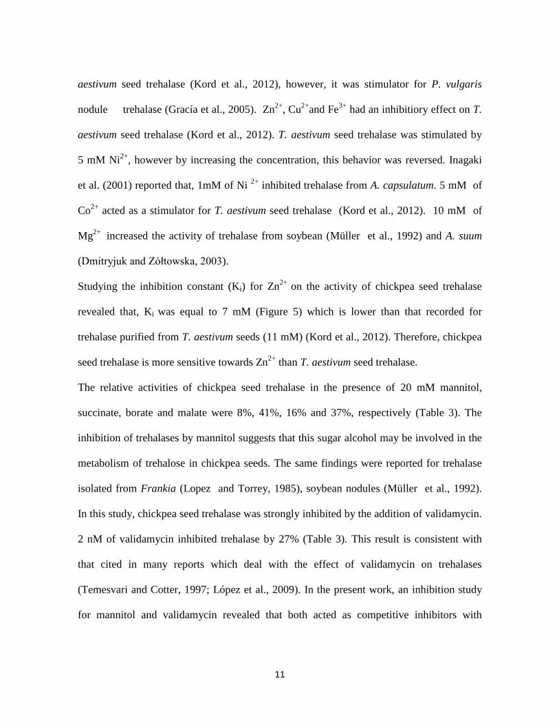
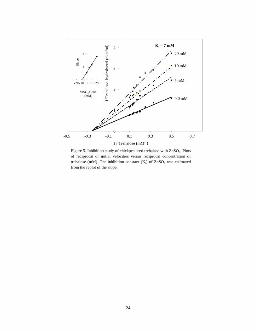
Studying the inhibition constant (Ki) for Zn2+
on the activity of chickpea seed trehalase
revealed that, Ki was equal to 7 mM (Figure 5) which is lower than that recorded for
trehalase purified from T. aestivum seeds (11 mM) (Kord et al., 2012). Therefore, chickpea
seed trehalase is more sensitive towards Zn2+
than T. aestivum seed trehalase.
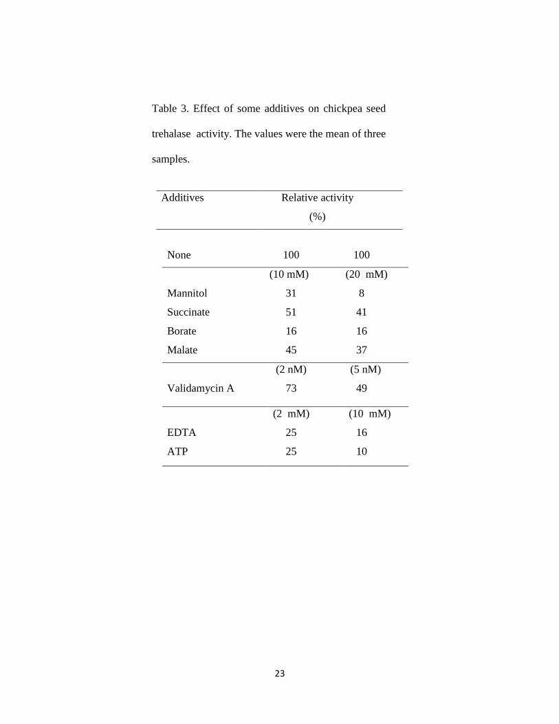
The relative activities of chickpea seed trehalase in the presence of 20 mM mannitol,
succinate, borate and malate were 8%, 41%, 16% and 37%, respectively (Table 3). The
inhibition of trehalases by mannitol suggests that this sugar alcohol may be involved in the
metabolism of trehalose in chickpea seeds. The same findings were reported for trehalase
isolated from Frankia (Lopez and Torrey, 1985), soybean nodules (Müller et al., 1992).
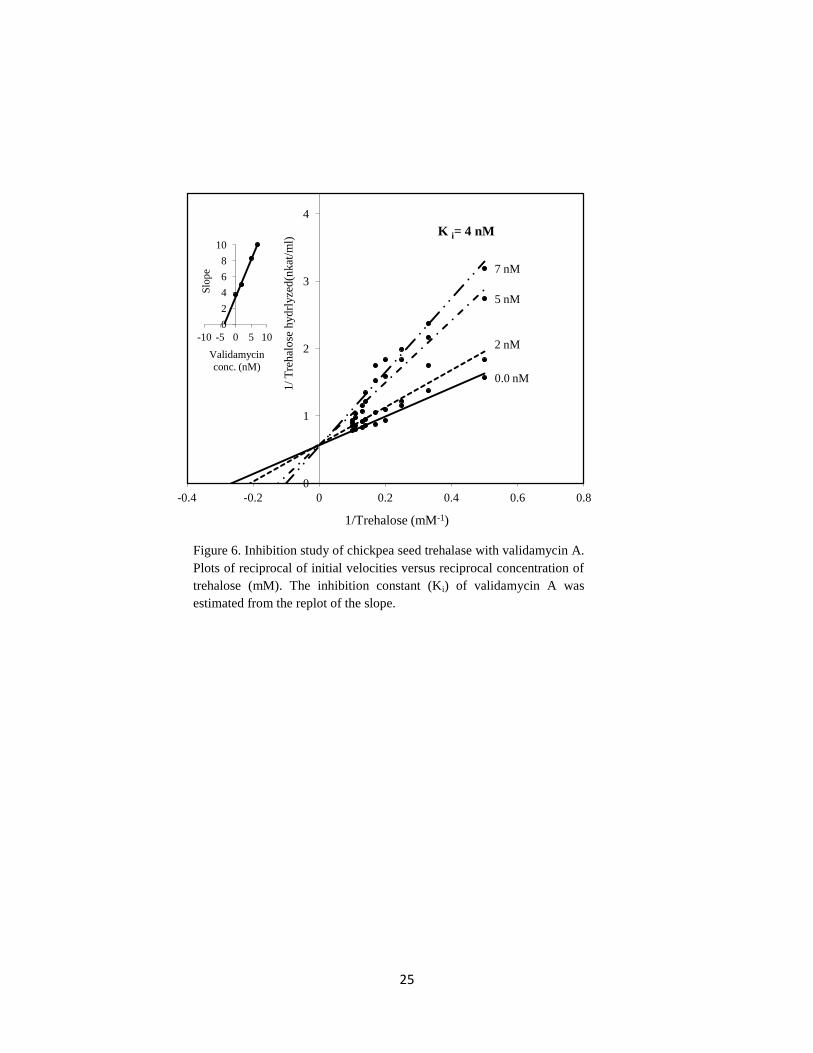
In this study, chickpea seed trehalase was strongly inhibited by the addition of validamycin.
2 nM of validamycin inhibited trehalase by 27% (Table 3). This result is consistent with
that cited in many reports which deal with the effect of validamycin on trehalases
(Temesvari and Cotter, 1997; López et al., 2009). In the present work, an inhibition study
for mannitol and validamycin revealed that both acted as competitive inhibitors with
12
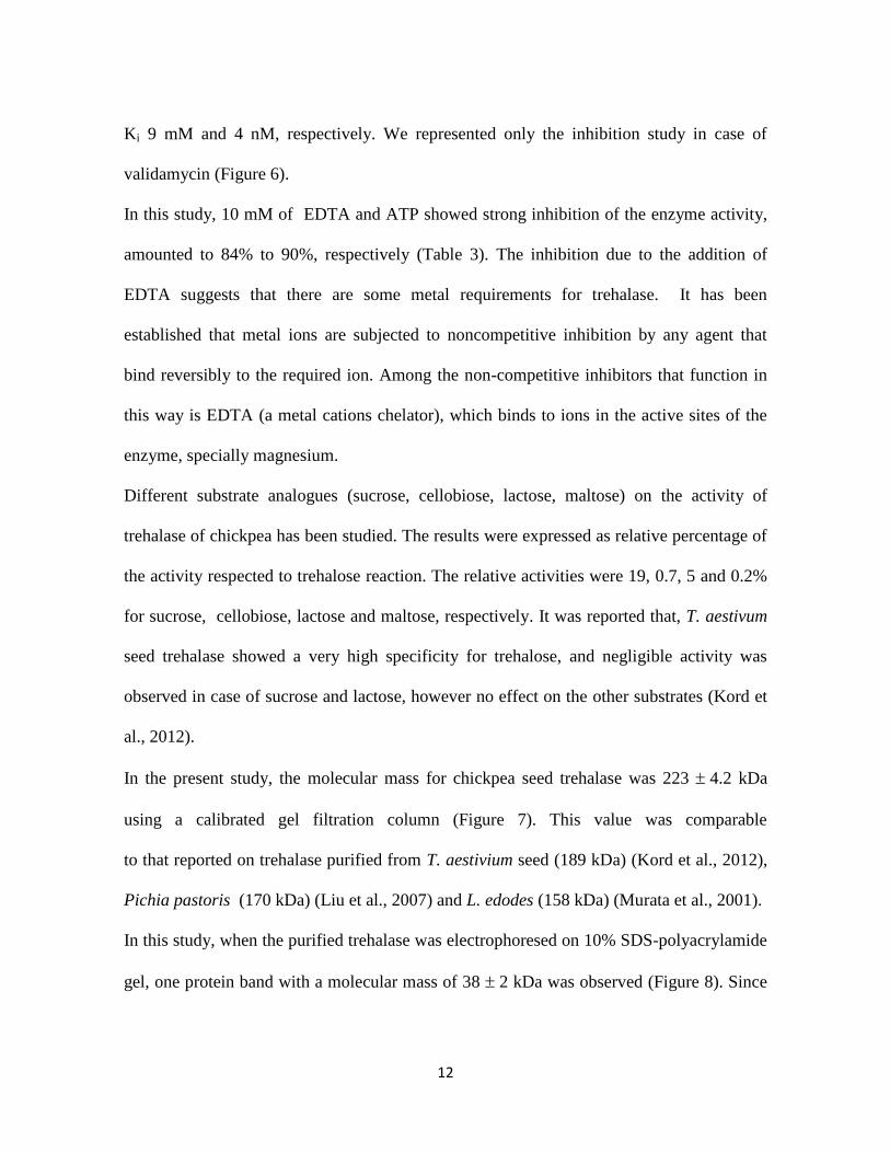
Ki 9 mM and 4 nM, respectively. We represented only the inhibition study in case of
validamycin (Figure 6).
In this study, 10 mM of EDTA and ATP showed strong inhibition of the enzyme activity,
amounted to 84% to 90%, respectively (Table 3). The inhibition due to the addition of
EDTA suggests that there are some metal requirements for trehalase. It has been
established that metal ions are subjected to noncompetitive inhibition by any agent that
bind reversibly to the required ion. Among the non-competitive inhibitors that function in
this way is EDTA (a metal cations chelator), which binds to ions in the active sites of the
enzyme, specially magnesium.
Different substrate analogues (sucrose, cellobiose, lactose, maltose) on the activity of
trehalase of chickpea has been studied. The results were expressed as relative percentage of
the activity respected to trehalose reaction. The relative activities were 19, 0.7, 5 and 0.2%
for sucrose, cellobiose, lactose and maltose, respectively. It was reported that, T. aestivum
seed trehalase showed a very high specificity for trehalose, and negligible activity was
observed in case of sucrose and lactose, however no effect on the other substrates (Kord et
al., 2012).
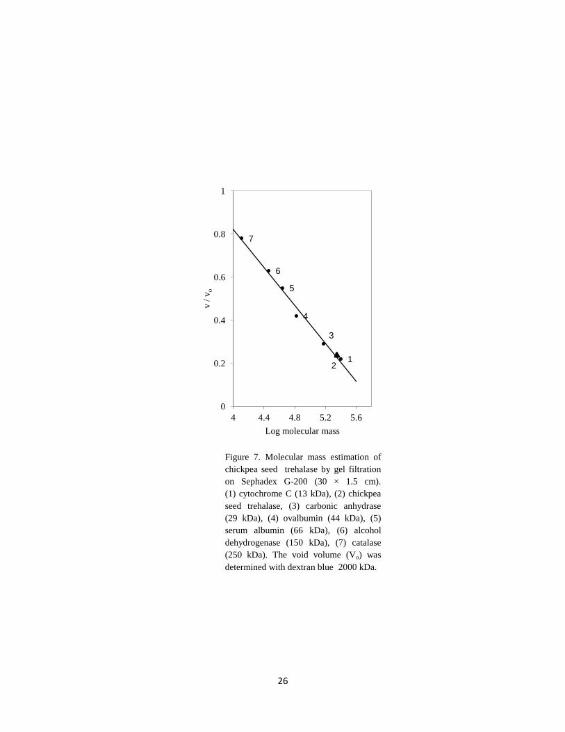
In the present study, the molecular mass for chickpea seed trehalase was 223 4.2 kDa
using a calibrated gel filtration column (Figure 7). This value was comparable
to that reported on trehalase purified from T. aestivium seed (189 kDa) (Kord et al., 2012),
Pichia pastoris (170 kDa) (Liu et al., 2007) and L. edodes (158 kDa) (Murata et al., 2001).
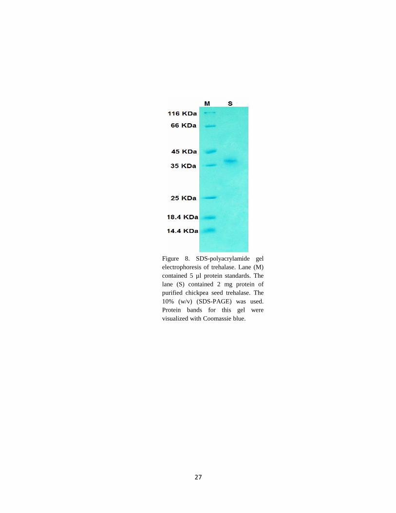
In this study, when the purified trehalase was electrophoresed on 10% SDS-polyacrylamide
gel, one protein band with a molecular mass of 38 2 kDa was observed (Figure 8). Since
13
the native molecular mass determined by gel filtration was 223 4.2 kDa, this indicated
that chickpea seed trehalase was a hexamer of six identical subunits. The available data in
the literature pointed out that trehalase is a monomer with a molecular mass ranged from
45 - 75 kDa (Inagaki et al., 2001; Jorge et al., 2007). However, some literature reported that
it is a dimer with a molecular mass of 38 kDa for A. suum (Dmitryjuk and Zółtowska,
2003) or trimer with a molecular mass of 63 kDa for T. aestivium (Kord et al., 2012).
Carroll et al. (2007) reported that trehalase isolated from M. smegmatis consisted of
multimer of about 20 or more subunits each with a molecular mass of 71 kDa.
4. Conclusion
Trehalase enzyme of chickpea seeds is purified till homogeneity with final specific activity
of 7 unit/mg protein and a molecular mass of 223 kDa. It consists of six identical subunits,
38 kDa each. The purified enzyme is relatively heat stable with optimum pH 5.5, and
optimum temperature 60C. It has km value of 3.64 mM trehalose. Validamycin A proved to
be a competitive inhibitor, with Ki = 4 nM. EDTA shows strong inhibition; suggesting that
it is a metalloenzyme.
Acknowledgement
Our sincere thanks to Dr. Gabr M and Dr. Maksoud SA, Professors of plant physiology,
Department of Botany, Faculty of Science, University of Cairo for advices and valuable
help during this study. The financial support was donated from the Faculty of Science,
University of Cairo.
14
References
Aeschbacher RA, Müller J, Boller T, Wiemken A (1999). Purification of the
trehalase GMTRE1 from soybean nodules and cloning of its cDNA. GMTRE1 is expressed
at a low level in multiple tissues. Plant Physiol 119: 489-495.
Alizadeh P, Klionsky DJ (1996). Purification and biochemical characterization of
the ATH1 gene product, vacuolar acid trehalase, from Saccharomyces cerevisiae. FEBS
Lett 391: 273-278.
Bergmeyer HU, Bernt EM (1974). Methods of enzymatic analysis, New York,
USA: Academic Press, pp. 1205-121.
Carroll JD, Pastuszak I, Edavana VK, Pan YT, Elbein AD (2007). A novel
trehalase from Mycobacterium smegmatis purification, properties, requirements. FEBS J
274: 1701-1714.
Dmitryjuk M, Zółtowska K (2003). Purification and characterization of acid trehalase from
muscle of Ascaris suum (Nematoda). Comp Biochem Phys B 136: 61-69.
Elbein AD, Pan YT, Pastuszak I, Carroll D ( 2003). New insights on trehalose: a
multifunctional molecule. Glycobiology 13: 17R-27R.
Fernandez O, Be´ thencourt L, Quero A, Sangwan RS, Cle´ment C (2010). Trehalose
and plant stress responses: friend or foe?. Trends Plant Sci 15: 409-417.
Frison M, Parrou JL, Guillaumot D, Masquelier D, Francois J, Chaumont F,
Batoko, H. (2007). The Arabidopsis thaliana is a plasma membrane-bound enzyme with
extracellular activity. FEBS Lett 581: 4010-4016.
15
Gracía NAT, Tribarne C, López M, Herrera-Cervera JA, Liuch C (2005).
Physiological implications of trehalase from Phaseolus vulgaris root nodules: partial
purification and characterization. Plant Physiol Bioch 43: 355-361.
Inagaki K, Ueno N, Tamura T, Tanaka H (2001). Purification and characterization
of an acid trehalase from Acidobacterium capsulatum. J Biosci Bioeng 91: 141-146.
Jorge CD, Sampaio MM, Hreggvidsson GO, Kristjá nson JK, Santos H (2007).
A highly thermostable trehalase from the thermophilic bacterium Rhodothermus marinus.
Extremophiles 11 :115-122.
Kamei Y, Hasegawa Y, Niimi T, Yamashita O, Yaginuma T (2011). Trehalase-2
protein contributes to trehalase activity enhanced by diapauses hormone in developing
ovaries of the silkworm, Bombyx mori. J Insect Physiol 57: 608-613.
Kamiya T, Hirata K, Matsumoto S, Arai C, Yoshizane C, Kyono F, Ariyasu T,
Hanaya T, Arai S, Okura T, et al. (2004). Targeted disruption of the trehalase gene:
determination of the digestion and absorption of trehalose in trehalase- deficient mice. Nutr
Res 24: 185-196.
Kord MAE, Youssef EA, Ahmed HE, Qaid EA (2012). Purification and some
properties of trehalase from Triticum aestivum seeds. In: Proceedings of the 2nd
International conference of physiological, microbiological and ecological plant sciences.
University of Mina. Egypt.
Liu Y, Wang Z, Yin Y, Cao Y, Zhao H, Xia Y (2007). Expression, purification,
and characterization of recombinant Metarhizium anisopliae acid trehalase in Pichia
pastoris. Protein Expres Purif 54:66-72.
16
López M, Gracía NAT, Liuch C (2009). Validamycin A improves the response of
Medicago truncatula plants to salt stress by inducing trehalose accumulation in the root
nodules. J Plant Physiol 166: 1218-1222.
Lopez MF, Torrey JG (1985). Purification and properties of trehalase in Frankia
ArI3. Arch Microbiol 143: 209-215.
Lowry OH, Rosebrough NJ, Farr AL, Randall, RJ (1951). Protein measurement
with the folin phenol reagent. J Biol Chem 193: 265-275.
Müller J, Staehelin C, Mellor RB, Boller T, Wiemken A (1992). Partial purification
and characterization of trehalase from soybean nodules. J Plant Physiol 140: 8-13.
Murata M, Nagai M, Takao M, Suzuki A., Sakai T, Terashita T (2001). Purification
and characterization of an extracellular acid trehalase from Lentinula edodes. Mycoscience
42: 479-482.
Reguera M, Peleg Z, Blumwald E (2011). Targeting metabolic pathways for genetic
engineering abiotic stress-tolerance in crops. Biochim Biophys Acta. In press.
Schubert A, Wyss P (1995). Trehalase activity in mycorrhizal and non-mycorrhizal
roots of leek and soybean. Mycorrhiza 5: 401-404.
Silva MCP, Terra WR, Ferreira C (2004). The role of carboxyl, guanidine and
imidazole groups in catalysis by a midgut trehalase purified from an insect larvae. Insect
Biochem Molec 34:1089-1099.
Temesvari LA, Cotter DA (1997). Trehalase of Dictyostelium discoideum:
Inhibition by amino-containing analogs of trehalose and affinity purification. Biochimie 79:
229-239.
Walker JM (1994). Basic protein and peptide protocols. Ann Bot-London 81: 449-454.
17
Warburg O, Christian W (1942). Isolation and crystallization of enolase. Biochem Z
310: 384-421.
Wolska-Mitaszko B, Molestak E, Malek W (2005). Properties of trehalase from
different organs of alfalfa, Medicago sativa. Acta Physiol Plant 27: 53-60.
Yaginuma T, Mizuno T, Mizuno C, Ikeda M, Wada T, Hattori K, Yamashita O,
Happ, GM (1996). Trehalase in the spermatophore from the bean-shaped accessory gland of
the male mealworm beetle, Tenebrio molitor: Purification, kinetic properties and
localization of the enzyme. J Comp Physiol 166: 1-10.
18
0.05 M
0.1 M
0.05 M
0.1 M
0.2 M
0.3 M
0.5 M
0
1
2
3
4
5
6
0
0.4
0.8
1.2
1.6
0 100 200 300 400 500
Tre
hal
ase
acti
vit
y (
nkat
/ m
l)
Ab
sorb
ance
at
28
0 n
m
Elution volume (ml)
Figure 1a. A typical elution profile for the
behavior of trehalase of chickpea seeds on
DEAE-cellulose column. The proteins were
eluted with discontinuous gradient of
KCl as indicated. Solid line indicates the
absorbance, dotted indicates the activity.
Figure 1b. A typical elution profile for the
behavior of trehalase of chickpea seeds on
CM-Sepharose column. The proteins were
eluted with discontinuous gradient of KCl as
indicated. Solid line indicates the absorbance,
dotted indicates the activity.
Figure 1c. A typical elution profile for the
behavior of trehalase of chickpea seeds on
Sephadex G-200 column. Solid line indicates
the absorbance, dotted indicates the activity.
0
2
4
6
8
0
1
2
3
4
5
0 200 400 600 800
Tre
hal
ase
acti
vit
y (
nkat
/ m
l)
Ab
sorb
ance
at
28
0 n
m
Elution volume (ml)
0
2
4
6
8
0
0.5
1
1.5
0 40 80 120 160
Ab
sorb
ance
at
28
0
Tre
hal
ase
acti
vit
y (
nkat
/m
l)
Elution volume (ml)
0.2 M
0.3 M
0.5 M
19
Table 1. Purification scheme of chickpea seed trehalase. Data of this table
is the mean of three complete purification processes
Fold
Recovery
(%)
Specific
activity
(nkat/ mg
protein)
Total
protein
(mg)
Total
activity
(nkat)
Purification steps
- 100 0.03 18808 559 Crude extract
4 88 0.12 4062 492 (NH4)2SO4
87 56 3 120 311 DEAE-cellulose
173 37 5 39 206 CM-Sepharose
232 18 7 14 100 Sephadex G-200
20
0
0.2
0.4
0.6
0.8
1
1.2
3 4 5 6 7 8 9 10
Tre
hal
ase
acti
vit
y (
nkat
/ml)
pH
pH optimum stability
0
0.5
1
1.5
2
2.5
3
3.5
20 30 40 50 60 70 80
Tre
hal
ase
acti
vit
y (
nkat
/ml)
Temperature [ oC]
Temperature optimum Thermostability
Figure 3. The temperature optimum
and thermostability of chickpea seed
trehalase. Standard assay conditions
were used except for temperature
which was varied as indicated. Bars
indicate SE.
Figure 2. pH optimum and
pH stability of chickpea
seed trehalase. Standard
assay conditions were used
except for buffer which was
varied as indicated. Bars
indicate ±SE.
21
0
1
2
3
-0.6 -0.3 0 0.3 0.6
1/
Tre
hal
ose
hyd
roly
zed
(nkat
/ m
l)
1/Trehalose ( mM-1)
Km = 3.64 mM
Figure 4. Lineweaver–Burk plot relating reaction velocity of
trehalase of chickpea seeds to trehalose concentration.
Bars indicate ±SE.
22
Table 2. Effect of different ions on chickpea seed
trehalase activity. The values were the mean of three
samples.
Relative percentage of activity at the
indicated concentrations of ions
Ions
20 mM
10 mM 5 mM
100 100 100 None
61 67 80 Na+
69 74 84 K+
12 47 88 Li+
210 108 99 Mn2+
98 120 133 Mg2+
67 94 95 Ca2+
22 39 76 Zn2+
14 20 67 Cu2+
122 114 114 Ni2+
122 112 95 Co2+
31 39 46 Fe3+
23
Relative activity
(%)
Additives
100 100 None
(20 mM) (10 mM)
8 31 Mannitol
41 51 Succinate
16 16 Borate
37 45 Malate
(5 nM)
49
(2 nM)
73
Validamycin A
(10 mM) (2 mM)
16 25 EDTA
10 25 ATP
Table 3. Effect of some additives on chickpea seed
trehalase activity. The values were the mean of three
samples.
24
0.0 mM
5 mM
10 mM
20 mM
0
1
2
3
4
-0.5 -0.3 -0.1 0.1 0.3 0.5 0.7
1/T
rehal
ose
hyd
roly
zed
(nkat
/ml)
1 / Trehalose (mM-1)
Figure 5. Inhibition study of chickpea seed trehalase with ZnSO4. Plots
of reciprocal of initial velocities versus reciprocal concentration of
trehalose (mM). The inhibition constant (Ki) of ZnSO4 was estimated
from the replot of the slope.
Ki = 7 mM
1211212mmm
mmmmmmM
m mmMmMm
0
1
2
-20 -10 0 10 20
Slo
pe
ZnSO4 Conc.
(mM)
25
0.0 nM
7 nM
5 nM
2 nM
0
1
2
3
4
-0.4 -0.2 0 0.2 0.4 0.6 0.8
1/
Tre
hal
ose
hyd
rlyze
d(n
kat
/ml)
1/Trehalose (mM-1)
K i= 4 nM
0
2
4
6
8
10
-10 -5 0 5 10
Slo
pe
Validamycin
conc. (nM)
Figure 6. Inhibition study of chickpea seed trehalase with validamycin A.
Plots of reciprocal of initial velocities versus reciprocal concentration of
trehalose (mM). The inhibition constant (Ki) of validamycin A was
estimated from the replot of the slope.
26
7
6
5
4
3
2 1
0
0.2
0.4
0.6
0.8
1
4 4.4 4.8 5.2 5.6
v /
vo
Log molecular mass
Figure 7. Molecular mass estimation of
chickpea seed trehalase by gel filtration
on Sephadex G-200 (30 × 1.5 cm).
(1) cytochrome C (13 kDa), (2) chickpea
seed trehalase, (3) carbonic anhydrase
(29 kDa), (4) ovalbumin (44 kDa), (5)
serum albumin (66 kDa), (6) alcohol
dehydrogenase (150 kDa), (7) catalase
(250 kDa). The void volume (Vo) was
determined with dextran blue 2000 kDa.
27
Figure 8. SDS-polyacrylamide gel
electrophoresis of trehalase. Lane (M)
contained 5 µl protein standards. The
lane (S) contained 2 mg protein of
purified chickpea seed trehalase. The
10% (w/v) (SDS-PAGE) was used.
Protein bands for this gel were
visualized with Coomassie blue.