proteomic screen defines the polo-box domain interactome and identifies rock2 as a plk1 substrate
TRANSCRIPT
Proteomic screen defines the Polo-box domaininteractome and identifies Rock2 as a Plk1substrate
Drew M Lowery1,4, Karl R Clauser2,4,Majbrit Hjerrild1,3,4, Dan Lim1,Jes Alexander1, Kazuhiro Kishi1,Shao-En Ong2, Steen Gammeltoft3,Steven A Carr2,* and Michael B Yaffe1,2,*1Departments of Biology and Biological Engineering, Center for CancerResearch, Massachusetts Institute of Technology, Cambridge, MA, USA,2Broad Institute of MIT and Harvard, Cambridge, MA, USA and3Department of Clinical Biochemistry, Glostrup Hospital, Glostrup,Denmark
Polo-like kinase-1 (Plk1) phosphorylates a number of mito-
tic substrates, but the diversity of Plk1-dependent processes
suggests the existence of additional targets. Plk1 contains a
specialized phosphoserine–threonine binding domain, the
Polo-box domain (PBD), postulated to target the kinase to
its substrates. Using the specialized PBD of Plk1 as an
affinity capture agent, we performed a screen to define the
mitotic Plk1-PBD interactome by mass spectrometry. We
identified 622 proteins that showed phosphorylation-
dependent mitosis-specific interactions, including proteins
involved in well-established Plk1-regulated processes, and
in processes not previously linked to Plk1 such as trans-
lational control, RNA processing, and vesicle transport.
Many proteins identified in our screen play important roles
in cytokinesis, where, in mammalian cells, the detailed
mechanistic role of Plk1 remains poorly defined. We go
on to characterize the mitosis-specific interaction of
the Plk1-PBD with the cytokinesis effector kinase Rho-
associated coiled–coil domain-containing protein kinase 2
(Rock2), demonstrate that Rock2 is a Plk1 substrate, and
show that Rock2 colocalizes with Plk1 during cytokinesis.
Finally, we show that Plk1 and RhoA function together to
maximally enhance Rock2 kinase activity in vitro and with-
in cells, and implicate Plk1 as a central regulator of multiple
pathways that synergistically converge to regulate actomyo-
sin ring contraction during cleavage furrow ingression.
The EMBO Journal (2007) 26, 2262–2273. doi:10.1038/
sj.emboj.7601683; Published online 19 April 2007
Subject Categories: signal transduction
Keywords: phosphopeptide-binding domain; Polo-box
domain; Polo-like kinase; proteomics; signal transduction
Introduction
In eukaryotic cells, Polo-like kinase-1 (Plk1) and related
orthologues perform a wide variety of essential functions
during mitosis (Barr et al, 2004; Glover, 2005; van de Weerdt
and Medema, 2006). Levels of Plk1 peak in late G2 and early
M, accompanied by dramatic changes in subcellular localiza-
tion as cells transit through various mitotic stages (Golsteyn
et al, 1994). During interphase and early prophase, Plk1
resides primarily at the centrosome, where it facilitates
centrosome maturation, separation, and microtubule nuclea-
tion during late prophase and prometaphase (Lane and Nigg,
1996; Rapley et al, 2005; De Luca et al, 2006). By metaphase,
a fraction of Plk1 has relocalized to the kinetochores, where
it seems to be involved in regulating aspects of spindle
checkpoint function and the metaphase–anaphase transition
(Ahonen et al, 2005; Goto et al, 2006). During anaphase, Plk1
is concentrated in the spindle midzone, where it probably
facilitates microtubule sliding, while following chromosome
segregation, Plk1 remains in the central spindle and midbody,
where it participates in ingression of the cleavage furrow
(Neef et al, 2003; Liu et al, 2004). Particularly prominent
cytokinetic phenotypes are observed in budding and fission
yeast, where mutations in the respective Plk1 orthologues,
Cdc5 and Plo1, result in incomplete assembly of actomyosin
and septin ring structures along with delayed and improper
deposition of septal material (Lee et al, 2005; Yoshida et al,
2006).
Although these and related mutational studies have
provided many insights into Cdc5/Plo1 function in simple
model organisms, the diversity of Plk1 functions in higher
eukaryotes makes it difficult to comprehensively identify
Plk1-regulated pathways or define the bulk of the Plk1
interactome using standard molecular genetics techniques.
Separation of function alleles are hard to identify owing to the
presence of a single common binding pocket and substrate
phosphorylation cleft shared by most, if not all substrates
(Cheng et al, 2003; Elia et al, 2003b). Full genetic disruption
of the Drosophila Plk1 orthologue, polo, causes embryonic
lethality (Donaldson et al, 2001), whereas full depletion of
the Xenopus Plk1 orthologue, Plx1, prevents mitotic entry
(Qian et al, 2001).
More recently, RNA interference has been used to examine
the effect of Plk1 depletion in human cell lines, revealing a
marked dependence of phenotype on the particular genetic
background of the cell. In tumor-derived cell lines, depletion
of Plk1 causes apoptosis along with mitotic catastrophe,
making the delineation of specific Plk1 functions difficult
(Spankuch-Schmitt et al, 2002; Liu et al, 2006). In other
immortalized cell lines, depletion of Plk1 causes a delay in
mitotic entry with subsequent arrest at prometaphase, pre-
venting analysis of later phenotypes without sensitization
strategies to avoid activation of various mitotic checkpoints.
If both the DNA damage and spindle checkpoints areReceived: 27 August 2006; accepted: 14 March 2007; publishedonline: 19 April 2007
*Corresponding authors. SA Carr, Broad Institute of MIT and Harvard,7 Cambridge Center, Cambridge MA 02142, USA.E-mail: [email protected] or MB Yaffe, Departments of Biology andBiological Engineering, Center for Cancer Research, MassachusettsInstitute of Technology, Building E18-580, 77 Massachusetts Avenue,Cambridge, MA 02139, USA. Tel.: þ 1 617 452 2103;Fax: þ 1 617 452 4978; E-mail: [email protected] authors contributed equally to this work
The EMBO Journal (2007) 26, 2262–2273 | & 2007 European Molecular Biology Organization | All Rights Reserved 0261-4189/07
www.embojournal.org
The EMBO Journal VOL 26 | NO 9 | 2007 &2007 European Molecular Biology Organization
EMBO
THE
EMBOJOURNAL
THE
EMBOJOURNAL
2262
bypassed, Plk1-depleted cells can complete an apparently
normal mitosis; however, chromosome segregation fails
(van Vugt et al, 2004a, b). In non-transformed cell lines,
depletion of Plk1 causes only very minor cell-cycle defects,
although co-depletion of p53 mimics the cell death pheno-
types seen in tumor-derived cell lines (Liu et al, 2006).
The various Plk1-depletion phenotypes are complicated by
varying degrees of Plk1 knockdown, as a 90% knockdown of
Plk1 in HeLa cells can allow normal mitotic processes
whereas an B100% depletion completely blocks cell-cycle
progression (Liu et al, 2005, 2006).
In an effort to more comprehensively elucidate the sub-
strates and interacting proteins of Plk1, we pursued a bio-
chemical/proteomic approach. All Plks have a similar protein
architecture including an N-terminal Ser/Thr kinase domain
and a conserved C-terminal Polo-box domain (PBD)
(Figure 1A). The PBD of Plks was previously identified in
our lab as a pSer/pThr-binding module that specifically
recognizes Ser-[pSer/pThr]-Pro/X motifs on peptides with
low micromolar affinity (Elia et al, 2003a). Phospho-depen-
dent ligand recognition by the PBD is necessary for the
targeting of Plk1 to specific substrates (i.e. processive phos-
phorylation), as well as for the dynamic re-localization of
Plk1 to specific subcellular structures during mitosis where
other Plk1 targets reside (i.e. distributive phosphorylation)
(Lowery et al, 2005). We therefore performed affinity purifi-
cation and mass spectrometry studies to identify Plk1 PBD-
interacting proteins from U2OS cells at different stages of
the cell cycle.
Our strategy was to define all putative interacting partners,
and then select one particular sub-network to investigate in
more detail at the molecular level. We identified approxi-
mately 600 proteins that are members of phosphorylation-
dependent Plk1 PBD interacting complexes. These proteins
are known to be involved in a wide variety of mitotic
processes, including processes not previously thought to be
regulated by Plk1. We chose to focus on the roles of Plk1 in
regulating cytokinesis. The requirement for Plk1 and its
orthologues for proper initiation and completion of cytokin-
esis has been well established in unicellular organisms
(Ohkura et al, 1995; Song and Lee, 2001) and Drosophila
(Carmena et al, 1998), and several previously described
PBD-associated proteins are known to play roles in this
process in mammalian cells (Neef et al, 2003; Zhou et al,
2003; Liu et al, 2004), although exactly how Plk1 fits into the
complete cytokinesis network is unclear. One of the major
upstream regulators of cytokinesis is RhoA, which, together
with its downstream targets, controls the formation and
constriction of the actomyosin ring at the cleavage furrow
(Glotzer, 2005). The functions of Plk1 and Rho GTPases may
be linked, as cytokinesis-specific GEFs for Rho were recently
identified as Plk targets both in mammalian cells (Niiya et al,
2006) and by our recent work in budding yeast (Yoshida et al,
2006).
We now report that Plk1 functions in multiple parallel
overlapping pathways with RhoA, and directly interacts
with a subset of critical effectors of RhoA. We demonstrate
that Plk1 and RhoA synergize to maximally activate the
cytokinetic protein kinase Rock2 in vitro and within cells,
and that Plk1 and Rock2 interact directly in vivo in a
phosphorylation-dependent mitosis-specific manner, with
maximal colocalization at the midbody during cytokinesis.
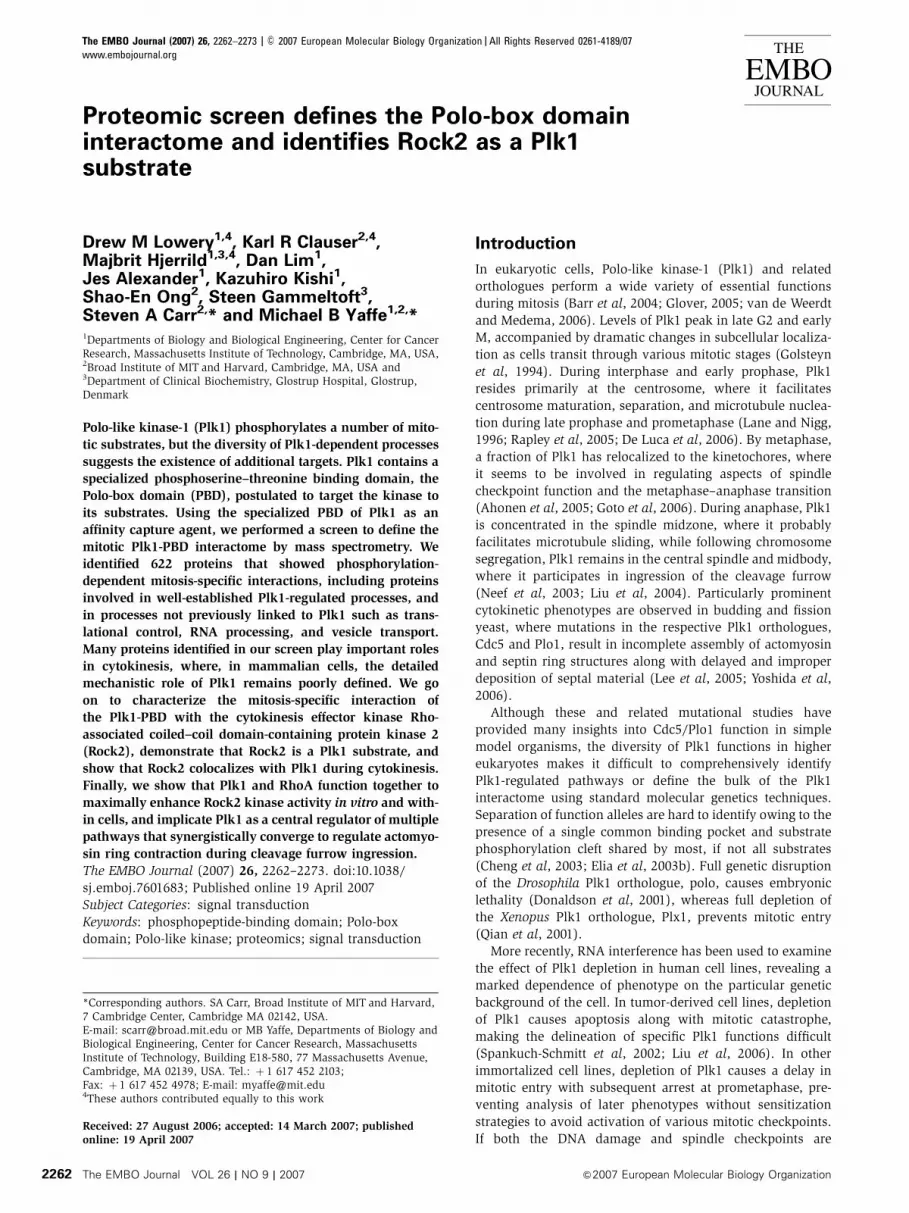
Figure 1 The PBD of Plk1 preferentially binds ligands in mitosis. (A) Domain structure of Plk1. Residues His-538 and Lys-540 are required forphosphopeptide binding by the PBD. (B) Experimental strategy for identifying cell-cycle-dependent PBD ligands. (C) Purity and equivalence ofrecombinant wild-type (WT) and mutant (MUT) PBDs used for interaction screening. Samples were analyzed by SDS–PAGE and stained withCoomassie blue. (D) U2OS cells were synchronized at the G1/S transition by a double thymidine block or in M-phase by nocodazole treatment,and DNA content analyzed by flow cytometry. (E) WT and MUT PBD were used to pull down interaction partners from double thymidineblocked or the nocodazole-arrested U2OS cell lysates. Bound proteins were separated by SDS–PAGE and visualized by SYPRO Ruby staining.Lines on the right of gel indicate where gel was cut before subsequent mass spectrometry analysis with each labeled section correlated withSupplementary Table S1.
Plk1 PBD mitotic interactomeDM Lowery et al
&2007 European Molecular Biology Organization The EMBO Journal VOL 26 | NO 9 | 2007 2263
Results and discussion
The Plk1 PBD shows mitosis-specific interactions with
many proteins involved in diverse aspects of cell
division
To identify potential targets of Plk1, we examined the ability
of the isolated PBD to bind to proteins in a cell cycle-
dependent manner. Recombinant Plk1 PBD was expressed
in bacteria and purified to homogeneity (Figure 1C). We also
purified a His-538 Ala/Lys-540 Met double mutant form of
the PBD, which does not bind to phosphorylated ligands (Elia
et al, 2003b) and therefore serves as an optimal negative
control to reveal sticky nonspecific interactions or phospho-
independent protein–PBD interactions that might arise from
high abundance. The wild-type and mutant PBD proteins
were crosslinked to Sepharose CN-4B beads and used as
an affinity matrix. Human osteosarcoma U2OS cells were
arrested at the G1/S transition by a double thymidine block
or arrested in mitosis by treatment with nocodazole. Cell-
cycle synchronization was verified by FACS (Figure 1D).
Lysates from these two cell populations were prepared and
equal amounts of total protein were applied to columns
containing either the wild-type or mutant PBD column.
After extensive washing with a near neutral-pH medium-
salt buffer that is not expected to disrupt complexes, PBD-
interacting proteins were eluted off the columns by competi-
tion with an optimal PBD-binding phospho-peptide (YMQS-
pT-PK) (Elia et al, 2003a). The recovered proteins were then
separated by SDS–PAGE and visualized by SYPRO Ruby
staining (Figure 1B, top).
Both the wild-type and mutant PBD bound very weakly,
and with similar affinity, to a variety of proteins in the G1/S
cell lysate. In marked contrast, the wild-type PBD, but not the
mutant PBD, showed very strong binding to a large number
of proteins in the mitotic cell lysates (Figure 1E). The darkest
band at 25 kDa is the PBD itself, indicating that there is
some leeching of the PBD from the column. These observa-
tions suggest that the Plk1 PBD can specifically interact with
many mitotic proteins in a phospho-dependent manner.
Furthermore, because Plk1 has been reported to interact
with microtubules (Feng et al, 1999), we used nocodazole
treatment to obtain mitotically arrested cells, as this drug
depolymerizes microtubules and should therefore minimize
potential indirect interactions of the PBD with other micro-
tubule-interacting proteins. The affinity-based purification
assay shown in Figure 1E could either isolate mitotic proteins
that bound directly to the PBD, or proteins that were not
themselves direct PBD interactors but instead were compo-
nents of larger PBD-associated complexes. The specificity of
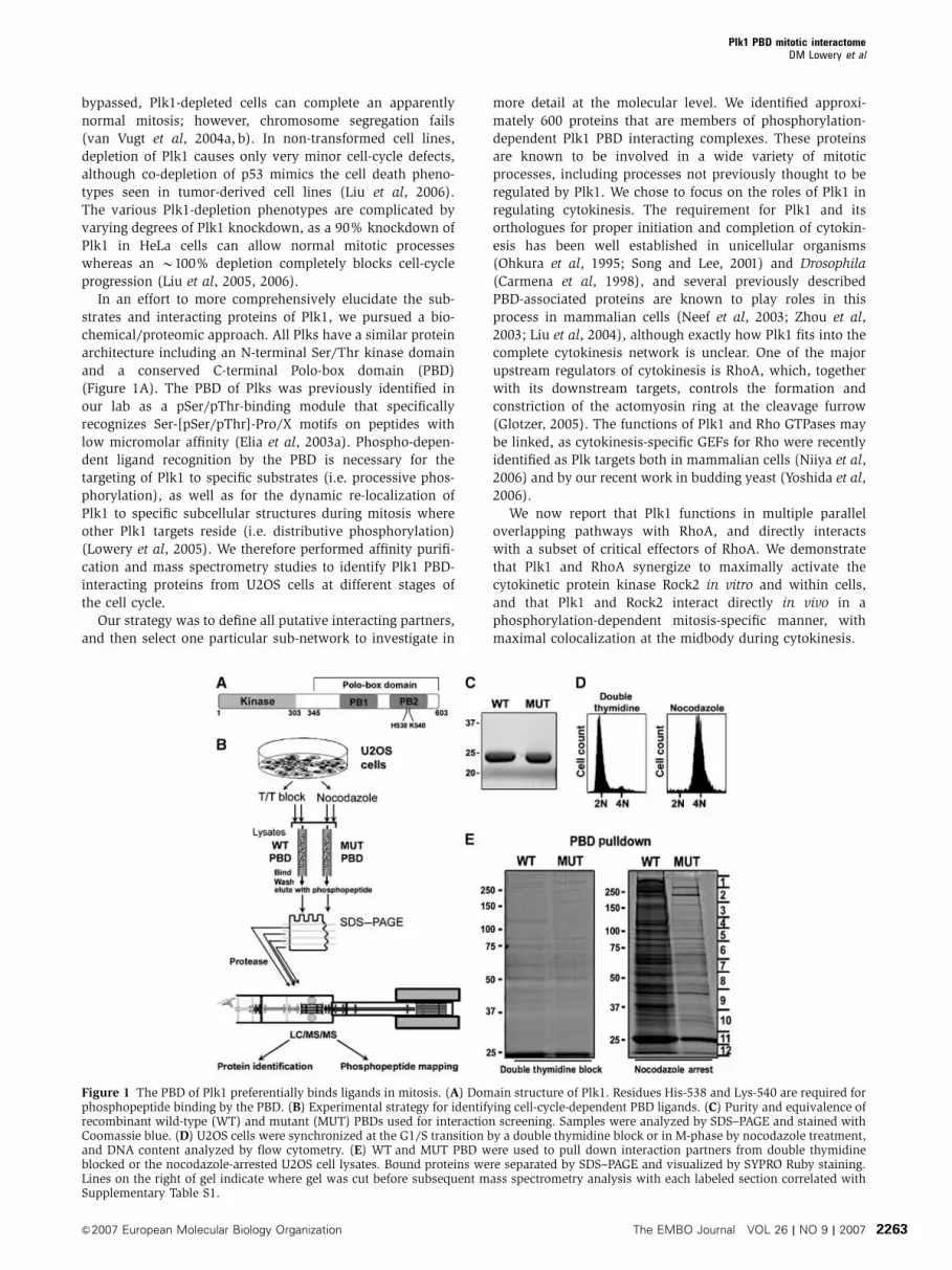
PBD binding was therefore investigated by Far-Western blot-
ting. Many of the mitotic proteins captured with the wild-type
PBD were capable of direct interaction. In contrast, none of
the proteins that were retained by the mutant PBD showed a
strong detectable interaction with the wild-type PBD in this
assay (Figure 2A).
To examine the extent to which the binding of mitotic
ligands to the PBD is dependent upon phosphorylation,
nocodazole-arrested cell lysates were dephosphorylated with
l-phosphatase before the PBD pull-down. As shown in
Figure 2B, both the number of recovered ligands and intensity
of ligand binding were greatly reduced after phosphatase
treatment of the mitotic cell lysates. Next, to investigate if
any of the Plk1 PBD-interacting proteins were also potential
Plk1 substrates, the affinity-purified proteins were incubated
with a constitutively active mutant of full-length Plk1, Plk1-
T210D, in the presence of [g-32P]ATP. As shown in Figure 2C,
incubation with Plk1-T210D resulted in the direct phosphoryla-
tion of many of these PBD-interacting proteins. Taken together,
the data in Figures 1 and 2 strongly support a model where the
PBD facilitates the interaction of Plk1 with a wide range of
mitotic targets that have undergone prior priming phosphory-
lation during mitosis, and indicates that a subset of the inter-
acting proteins are themselves Plk1 substrates.
Liquid chromatography tandem mass spectrometry (LC/
MS/MS) was used to identify the mitotic PBD-interacting
proteins. Each lane of the gel from the nocodazole-arrested
Figure 2 Mitotic proteins bind to the PBD in a phospho-dependent manner and are substrates of Plk1. (A) Direct binding of PBD-interactingproteins. Proteins interacting with the wild-type (WT) and mutant (MUT) PBD from nocodazole-arrested U2OS cell lysates (Figure 1B) weretransferred to PVDF membrane and analyzed for direct binding by Far-Western analysis using the WT PBD as a probe. (B) Phosphorylation-dependent PBD interactions. Nocodazole-arrested U2OS cell lysates were incubated with (þ ) or without (�) l-protein phosphatase beforeWT-PBD pull-down. The bound proteins were separated by SDS–PAGE and visualized by SYPRO Ruby staining. (C) Plk1 phosphorylation ofPBD-interacting proteins. Wild-type PBD was used to pull down interacting proteins from nocodazole-arrested U2OS cell lysates. The proteinswere incubated with or without active Plk1 and [g-32P]ATP, separated by SDS–PAGE, and visualized by autoradiography.
Plk1 PBD mitotic interactomeDM Lowery et al
The EMBO Journal VOL 26 | NO 9 | 2007 &2007 European Molecular Biology Organization2264
cell lysates in Figure 1E was excised, cut into 12 pieces as
indicated (Figure 1E), and subjected to in-gel digestion with
trypsin. The extracted peptide mixtures were then separated
using reverse phase HPLC, which was coupled to an LTQ-FT
hybrid ion trap Fourier transform mass spectrometer for
peptide identification (Figure 1B, bottom), and the corre-
sponding proteins identified by database searching. For
each protein, the relative ratio of wild-type/mutant PBD-
bound abundances was determined using the sum of the
extracted ion current measured for each sequenced peptide
precursor ion in the intervening MS scans of the LC/MS/MS
chromatogram. Proteins were then categorized as being
wild-type specific (peptide ions present only in the wild-
type PBD eluents), wild-type enriched (peptide ions present
at 420-fold intensity in the wild-type PBD eluents compared
to the mutant PBD eluents), and nonspecific (peptide ions
present at p20-fold intensity in the wild-type versus mutant
PBD eluents). In total, we identified 622 distinct proteins that
were at least 20-fold more abundant in the wild-type PBD
pull-down compared to the mutant H538A/K540 M PBD pull-
down and were considered to be potential PBD interaction
partners (Supplementary Table S1). All proteins were charac-
terized according to GO categories defining their molecular
function (GO Consortium, 2001) (Figure 3A).
We selected a small random subset of proteins identified in
the mass spectrometry-based screen for further validation
based on the availability of antibodies: Lamin A, Cofilin,
MCMs, the protein kinase CK2 alpha, myosin phosphatase
targeting subunit 1 (MYPT), and Rock2. Pull-downs from
mitotic lysates followed by immunoblotting confirmed that
all these proteins preferentially interacted with wild-type PBD
compared to the mutant PBD (Supplementary Figure S1A).
The cell-cycle-dependent interaction of the selected proteins
with the PBD was investigated by performing in vitro
pull-down assays with lysates from double thymidine
blocked (G1) and nocodazole-treated (M) cells. As shown
in Supplementary Figure S1B, interactions between the PBD
and all of these proteins were mitotic specific. To investigate
the requirement for phosphorylation, mitotic lysates were
treated with l-protein phosphatase before PBD pull-downs
and immunoblotting. In each case, interaction of these pro-
teins with the PBD was either eliminated or substantially
reduced following phosphatase treatment (Supplementary
Figure S1C). Thus, all six of the interactions tested were
mitotic phosphorylation-dependent interactions.
The complete set of proteins identified in our PBD inter-
actome included proteins previously demonstrated to asso-
ciate with Plk1 such as MCMs (Tsvetkov and Stern, 2005),
septins (Song and Lee, 2001), anillin (shown to be a Plkl
substrate in vitro; Straight et al, 2005), and members of
the 20S proteasome complex (Feng et al, 2001). Some
other known endogenous Plk1-interacting proteins, such as
Cdc25C, were not identified in this screen, likely as a result of
our nocodazole treatment/spindle checkpoint arrest strategy,
which appeared to enrich for late mitotic targets. Plk1 targets
such as Cdc25C and Wee1 are thought to play a role in entry
into mitosis, and thus might not bind to Plk1 once mitosis is
underway. Additional Plk1 interactors, such as Bub3, are only
expected to be engaged after the spindle checkpoint is
extinguished.
Most of the PBD-interacting proteins identified in this study
have not been previously reported. Many of those proteins
participate in a broad range of cellular functions that show
distinct changes during mitosis, transcription, translation, spli-
cing, and metabolism (Figure 3A). Intriguingly, although protein
synthesis is necessary for mitotic entry and progression, the
overall rate of protein synthesis in mitotic cells has been
reported to be markedly decreased to 25–30% of the rate seen
in interphase cells (Tarnowka and Baglioni, 1979). At the same
time, the synthesis of a number of proteins including c-myc is
enhanced during mitosis (Kim et al, 2003). Transcription and
mRNA splicing has also been shown to be inhibited during
mitosis (Shin and Manley, 2002). The metabolic state of mitotic
cells also undergoes significant alterations in order to accom-
modate disruption and distribution of membrane compartments
and components (Warren, 1993). Recently, a transcriptional
coactivator protein, Ndd1, has been discovered to be a direct
substrate of Cdc5, the yeast Plk1 orthologue. Cdc5 was recruited
to specific promoters where it phosphorylated Ndd1 to activate
transcription of cell-cycle-regulated genes involved in mitotic
progression (Darieva et al, 2006). Plk1 has also been shown to
regulate the nuclear translocation of the transcription factor
HSF1 (Kim et al, 2005). Thus, we anticipate further elucidation
of the role of Plk1 in these processes.
Bioinformatic analysis reveals that Plk1 PBD ligands are
enriched in Ser-[Ser/Thr]-Pro motifs and are potential
Plk1 substrates
In order to help distinguish between indirect and direct
interactors, each of the PBD-interacting proteins identified
by mass spectrometry was evaluated for the presence of an
optimal PBD recognition motif. Isolated phosphopeptides
bind to the Plk1 PBD through the optimal consensus motif
S-[pS/pT]-[P/X], where the Ser residue preceding the pSer/
pThr makes three hydrogen-bonding interactions with the
PBD (one through a bound water molecule) and contributes
significantly to ligand affinity (Elia et al, 2003b). As seen in
Figure 3B, PBD recognition motifs were identified in 47.3% of
the wild-type-specific PBD-interacting proteins and 41.3% of
the wild-type-enriched interactions. In contrast, only 16.2%
of the nonspecific PBD-interacting proteins contained this
motif. To determine whether these values were statistically
significant, we generated 2000 ‘mock’ protein data sets by
randomly selecting either 622 (the total number of combined
wild-type PBD-specific or -enriched interactions; Figure 3B,
column 1, red boxes) or 277 proteins (the total number of
nonspecific interactions; Figure 3B, column 1, gray box) from
the current human NCBI RefSeq collection, and examined
these data sets of randomly selected proteins for the percen-
tage of proteins containing S-[S/T]-P motifs. As shown in
Supplementary Figure S2A and B, the distribution of the
number of protein in each of the random data sets containing
S-[S/T]-P motifs was roughly normally distributed, with the
same average value of 31.4%. The percentage of PBD-specific
or -enriched interacting proteins containing S-[S/T]-P motifs
that we identified in our mass spectrometry-based screen
(average value 45.3%) was over 7 s.d. above the mean from
that expected for a similar sized collection of random proteins
(Supplementary Figure S2B). Likewise, the percentage of PBD
nonspecific interacting proteins containing S-[S/T]-P motifs
was over 5 s.d. below that expected (Supplementary Figure
S2A). Thus, our non-biased method for identifying PBD
interactors greatly enriched for proteins that contained the
optimal motifs necessary for PBD binding, suggesting that the
Plk1 PBD mitotic interactomeDM Lowery et al
&2007 European Molecular Biology Organization The EMBO Journal VOL 26 | NO 9 | 2007 2265
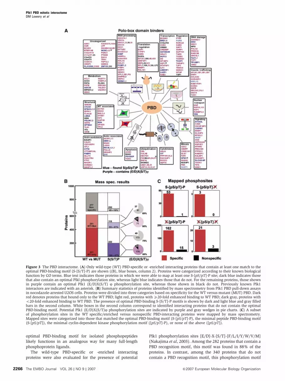
optimal PBD-binding motif for isolated phosphopeptides
likely functions in an analogous way for many full-length
phosphoprotein ligands.
The wild-type PBD-specific or -enriched interacting
proteins were also evaluated for the presence of potential
Plk1 phosphorylation sites [E/D]-X-[S/T]-[F/L/I/Y/W/V/M]
(Nakajima et al, 2003). Among the 282 proteins that contain a
PBD recognition motif, this motif was found in 88% of the
proteins. In contrast, among the 340 proteins that do not
contain a PBD recognition motif, this phosphorylation motif
Figure 3 The PBD interactome. (A) Only wild-type (WT) PBD-specific or -enriched interacting proteins that contain at least one match to theoptimal PBD-binding motif (S-[S/T]-P) are shown ((B), blue boxes, column 2). Proteins were categorized according to their known biologicalfunction by GO terms. Blue text indicates those proteins in which we were able to map at least one S-[pS/pT]-P site; dark blue indicates thosethat also contain an optimal Plkl phosphorylation site, whereas light blue indicates those that do not. For the remaining proteins, those shownin purple contain an optimal Plk1 (E/D)X(S/T) j phosphorylation site, whereas those shown in black do not. Previously known Plk1interactors are indicated with an asterisk. (B) Summary statistics of proteins identified by mass spectrometry from Plk1 PBD pull-down assaysin nocodazole-arrested U2OS cells. Proteins were divided into three categories based on specificity for the WT versus mutant (MUT) PBD. Darkred denotes proteins that bound only to the WT PBD; light red, proteins with X20-fold enhanced binding to WT PBD; dark gray, proteins witho20-fold enhanced binding to WT PBD. The presence of optimal PBD-binding S-[S/T]-P motifs is shown by dark and light blue and gray filledbars in the second column. White boxes in the second column correspond to identified interacting proteins that do not contain the optimalPBD-binding motif. Potential Plk1 (E/D)X(S/T)j phosphorylation sites are indicated by purple and gray wedges in pie charts. (C) A subsetof phosphorylation sites in the WT specific/enriched versus nonspecific PBD-interacting proteins were mapped by mass spectrometry.Mapped sites were categorized into those that matched the optimal PBD-binding motif (S-[pS/pT]-P), the minimal peptide PBD-binding motif(S-[pS/pT]), the minimal cyclin-dependent kinase phosphorylation motif ([pS/pT]-P), or none of the above ([pS/pT]).
Plk1 PBD mitotic interactomeDM Lowery et al
The EMBO Journal VOL 26 | NO 9 | 2007 &2007 European Molecular Biology Organization2266
was present in 75% of the proteins (Figure 3B). We performed
a similar statistical analysis for putative Plk1 phosphorylation
sites in random protein data sets containing either 282 pro-
teins (the number of wild-type PBD-specific or -enriched
interactions containing S-[S/T]-P motifs; Figure 3B, blue
boxes, column 2) or 340 proteins (the number of wild-type
PBD-specific or -enriched interactions not containing S-[S/T]-
P motifs; Figure 3B, white boxes, column 2). This revealed a
similar mean value of 74.4% (Supplementary Figure S2C and
D). The co-occurrence of a Plk1 phosphorylation motif in the
S-[S/T]-P motif-containing PBD-specific or -enriched proteins
found in our mass spectrometry-based screen was nearly
5 s.d. greater than that expected by chance alone in a
randomly selected set of proteins (Supplementary Figure
S2D). In contrast, for the PBD-specific or -enriched proteins
found in our mass spectrometry-based screen that lacked
S-[S/T]-P motifs, the occurrence of a Plk1 phosphorylation
motif was the same as that expected by random chance
(Supplementary Figure S2C). We interpret these findings as
evidence that Plk1 interactors capable of binding directly to
the PBD are also likely to be Plk1 substrates, whereas proteins
in complexes with direct PBD interactors may be less likely to
be Plk1 substrates than the PBD interactors themselves.
The mass spectrometry analysis of the PBD-interacting
proteins also resulted in the mapping of 247 phosphophory-
lation sites (Figure 3C and Supplementary Table S2),
although this was not the primary intent of our study. The
majority of mapped phosphorylation sites were derived from
the gel band tryptic digests of our PBD pull-downs with a few
from a TiO2 IMAC using a tryptic digest of an aliquot of
the PBD pull-downs. Within the tryptic peptides, many of
the phosphorylation motifs are present in a sequence context
that is not readily amenable to binding and elution from a
reversed-phase column and ionization/fragmentation by MS/
MS (i.e. too short, long, hydrophobic, hydrophilic, or contain
too many nearby basic residues). Therefore, we expect to
observe only a portion of phosphopeptides actually present.
In 282 proteins that were identified as wild-type PBD-specific
or -enriched interacting proteins (Figure 3B, column 2, blue
boxes), we were able to map 49 phosphorylation sites in 43
proteins that exactly matched the Ser-(pSer/pThr)-Pro PBD-
recognition motif (Supplementary Table S2). An additional
four mapped sites contained the motif Ser-pSer, which would
also be expected to bind strongly to the PBD (Elia et al,
2003b). Among the entire 899 proteins identified in our study,
we were able to map an additional 168 phosphorylation sites
matching the minimal consensus [pS/pT]P motif for CDKs.
Thus, 217/247 or 90% of our mapped sites match the mini-
mal CDK motif, consistent with mitotic arrest.
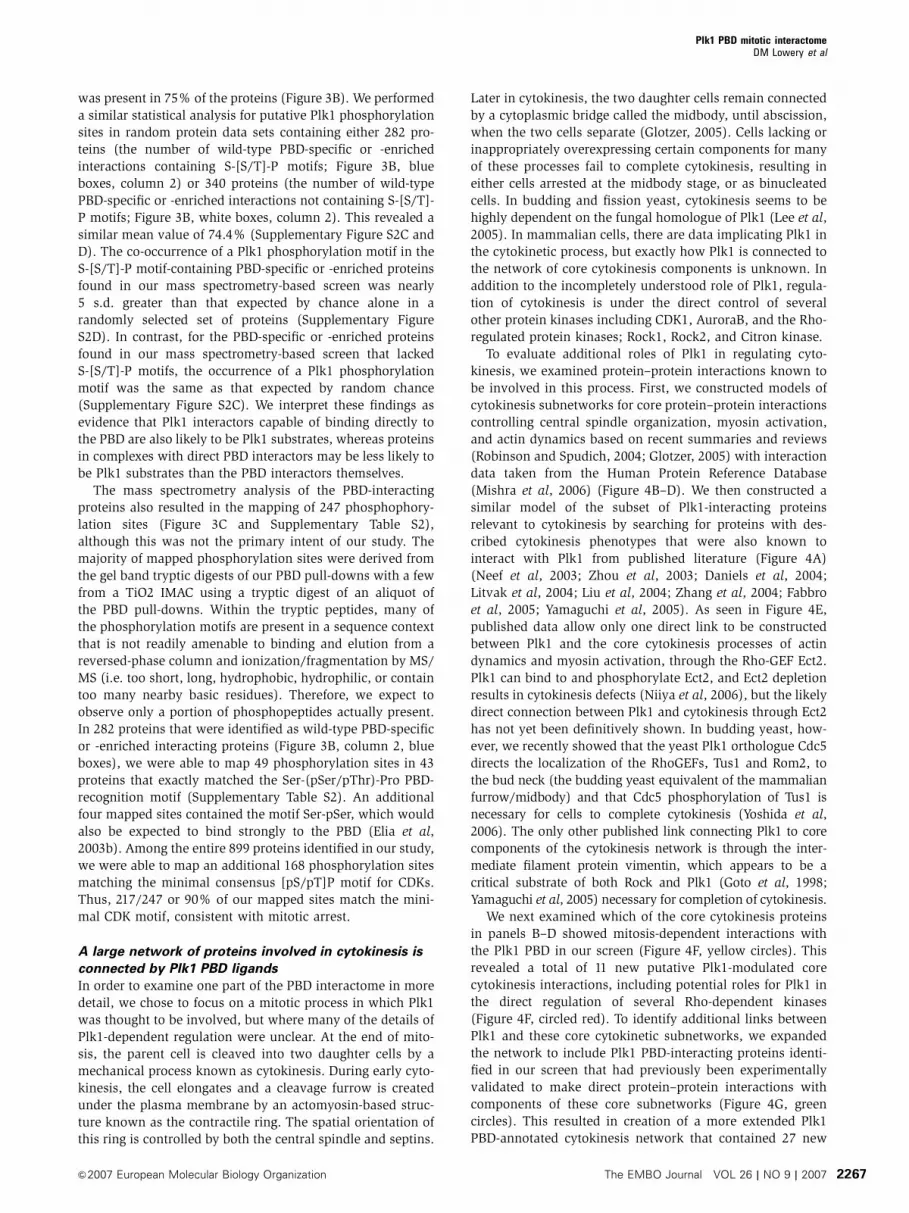
A large network of proteins involved in cytokinesis is
connected by Plk1 PBD ligands
In order to examine one part of the PBD interactome in more
detail, we chose to focus on a mitotic process in which Plk1
was thought to be involved, but where many of the details of
Plk1-dependent regulation were unclear. At the end of mito-
sis, the parent cell is cleaved into two daughter cells by a
mechanical process known as cytokinesis. During early cyto-
kinesis, the cell elongates and a cleavage furrow is created
under the plasma membrane by an actomyosin-based struc-
ture known as the contractile ring. The spatial orientation of
this ring is controlled by both the central spindle and septins.
Later in cytokinesis, the two daughter cells remain connected
by a cytoplasmic bridge called the midbody, until abscission,
when the two cells separate (Glotzer, 2005). Cells lacking or
inappropriately overexpressing certain components for many
of these processes fail to complete cytokinesis, resulting in
either cells arrested at the midbody stage, or as binucleated
cells. In budding and fission yeast, cytokinesis seems to be
highly dependent on the fungal homologue of Plk1 (Lee et al,
2005). In mammalian cells, there are data implicating Plk1 in
the cytokinetic process, but exactly how Plk1 is connected to
the network of core cytokinesis components is unknown. In
addition to the incompletely understood role of Plk1, regula-
tion of cytokinesis is under the direct control of several
other protein kinases including CDK1, AuroraB, and the Rho-
regulated protein kinases; Rock1, Rock2, and Citron kinase.
To evaluate additional roles of Plk1 in regulating cyto-
kinesis, we examined protein–protein interactions known to
be involved in this process. First, we constructed models of
cytokinesis subnetworks for core protein–protein interactions
controlling central spindle organization, myosin activation,
and actin dynamics based on recent summaries and reviews
(Robinson and Spudich, 2004; Glotzer, 2005) with interaction
data taken from the Human Protein Reference Database
(Mishra et al, 2006) (Figure 4B–D). We then constructed a
similar model of the subset of Plk1-interacting proteins
relevant to cytokinesis by searching for proteins with des-
cribed cytokinesis phenotypes that were also known to
interact with Plk1 from published literature (Figure 4A)
(Neef et al, 2003; Zhou et al, 2003; Daniels et al, 2004;
Litvak et al, 2004; Liu et al, 2004; Zhang et al, 2004; Fabbro
et al, 2005; Yamaguchi et al, 2005). As seen in Figure 4E,
published data allow only one direct link to be constructed
between Plk1 and the core cytokinesis processes of actin
dynamics and myosin activation, through the Rho-GEF Ect2.
Plk1 can bind to and phosphorylate Ect2, and Ect2 depletion
results in cytokinesis defects (Niiya et al, 2006), but the likely
direct connection between Plk1 and cytokinesis through Ect2
has not yet been definitively shown. In budding yeast, how-
ever, we recently showed that the yeast Plk1 orthologue Cdc5
directs the localization of the RhoGEFs, Tus1 and Rom2, to
the bud neck (the budding yeast equivalent of the mammalian
furrow/midbody) and that Cdc5 phosphorylation of Tus1 is
necessary for cells to complete cytokinesis (Yoshida et al,
2006). The only other published link connecting Plk1 to core
components of the cytokinesis network is through the inter-
mediate filament protein vimentin, which appears to be a
critical substrate of both Rock and Plk1 (Goto et al, 1998;
Yamaguchi et al, 2005) necessary for completion of cytokinesis.
We next examined which of the core cytokinesis proteins
in panels B–D showed mitosis-dependent interactions with
the Plk1 PBD in our screen (Figure 4F, yellow circles). This
revealed a total of 11 new putative Plk1-modulated core
cytokinesis interactions, including potential roles for Plk1 in
the direct regulation of several Rho-dependent kinases
(Figure 4F, circled red). To identify additional links between
Plk1 and these core cytokinetic subnetworks, we expanded
the network to include Plk1 PBD-interacting proteins identi-
fied in our screen that had previously been experimentally
validated to make direct protein–protein interactions with
components of these core subnetworks (Figure 4G, green
circles). This resulted in creation of a more extended Plk1
PBD-annotated cytokinesis network that contained 27 new
Plk1 PBD mitotic interactomeDM Lowery et al
&2007 European Molecular Biology Organization The EMBO Journal VOL 26 | NO 9 | 2007 2267
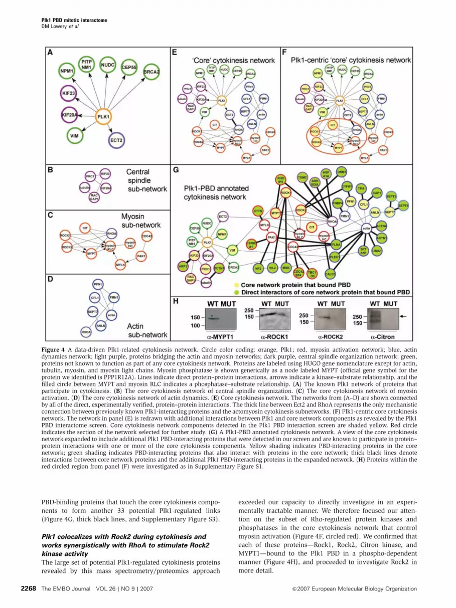
PBD-binding proteins that touch the core cytokinesis compo-
nents to form another 33 potential Plk1-regulated links
(Figure 4G, thick black lines, and Supplementary Figure S3).
Plk1 colocalizes with Rock2 during cytokinesis and
works synergistically with RhoA to stimulate Rock2
kinase activity
The large set of potential Plk1-regulated cytokinesis proteins
revealed by this mass spectrometry/proteomics approach
exceeded our capacity to directly investigate in an experi-
mentally tractable manner. We therefore focused our atten-
tion on the subset of Rho-regulated protein kinases and
phosphatases in the core cytokinesis network that control
myosin activation (Figure 4F, circled red). We confirmed that
each of these proteins—Rock1, Rock2, Citron kinase, and
MYPT1—bound to the Plk1 PBD in a phospho-dependent
manner (Figure 4H), and proceeded to investigate Rock2 in
more detail.
Figure 4 A data-driven Plk1-related cytokinesis network. Circle color coding: orange, Plk1; red, myosin activation network; blue, actindynamics network; light purple, proteins bridging the actin and myosin networks; dark purple, central spindle organization network; green,proteins not known to function as part of any core cytokinesis network. Proteins are labeled using HUGO gene nomenclature except for actin,tubulin, myosin, and myosin light chains. Myosin phosphatase is shown generically as a node labeled MYPT (official gene symbol for theprotein we identified is PPP1R12A). Lines indicate direct protein–protein interactions, arrows indicate a kinase–substrate relationship, and thefilled circle between MYPT and myosin RLC indicates a phosphatase–substrate relationship. (A) The known Plk1 network of proteins thatparticipate in cytokinesis. (B) The core cytokinesis network of central spindle organization. (C) The core cytokinesis network of myosinactivation. (D) The core cytokinesis network of actin dynamics. (E) Core cytokinesis network. The networks from (A–D) are shown connectedby all of the direct, experimentally verified, protein–protein interactions. The thick line between Ect2 and RhoA represents the only mechanisticconnection between previously known Plk1-interacting proteins and the actomyosin cytokinesis subnetworks. (F) Plk1-centric core cytokinesisnetwork. The network in panel (E) is redrawn with additional interactions between Plk1 and core network components as revealed by the Plk1PBD interactome screen. Core cytokinesis network components detected in the Plk1 PBD interaction screen are shaded yellow. Red circleindicates the section of the network selected for further study. (G) A Plk1-PBD annotated cytokinesis network. A view of the core cytokinesisnetwork expanded to include additional Plk1 PBD-interacting proteins that were detected in our screen and are known to participate in protein–protein interactions with one or more of the core cytokinesis components. Yellow shading indicates PBD-interacting proteins in the corenetwork; green shading indicates PBD-interacting proteins that also interact with proteins in the core network; thick black lines denoteinteractions between core network proteins and the additional Plk1 PBD-interacting proteins in the expanded network. (H) Proteins within thered circled region from panel (F) were investigated as in Supplementary Figure S1.
Plk1 PBD mitotic interactomeDM Lowery et al
The EMBO Journal VOL 26 | NO 9 | 2007 &2007 European Molecular Biology Organization2268
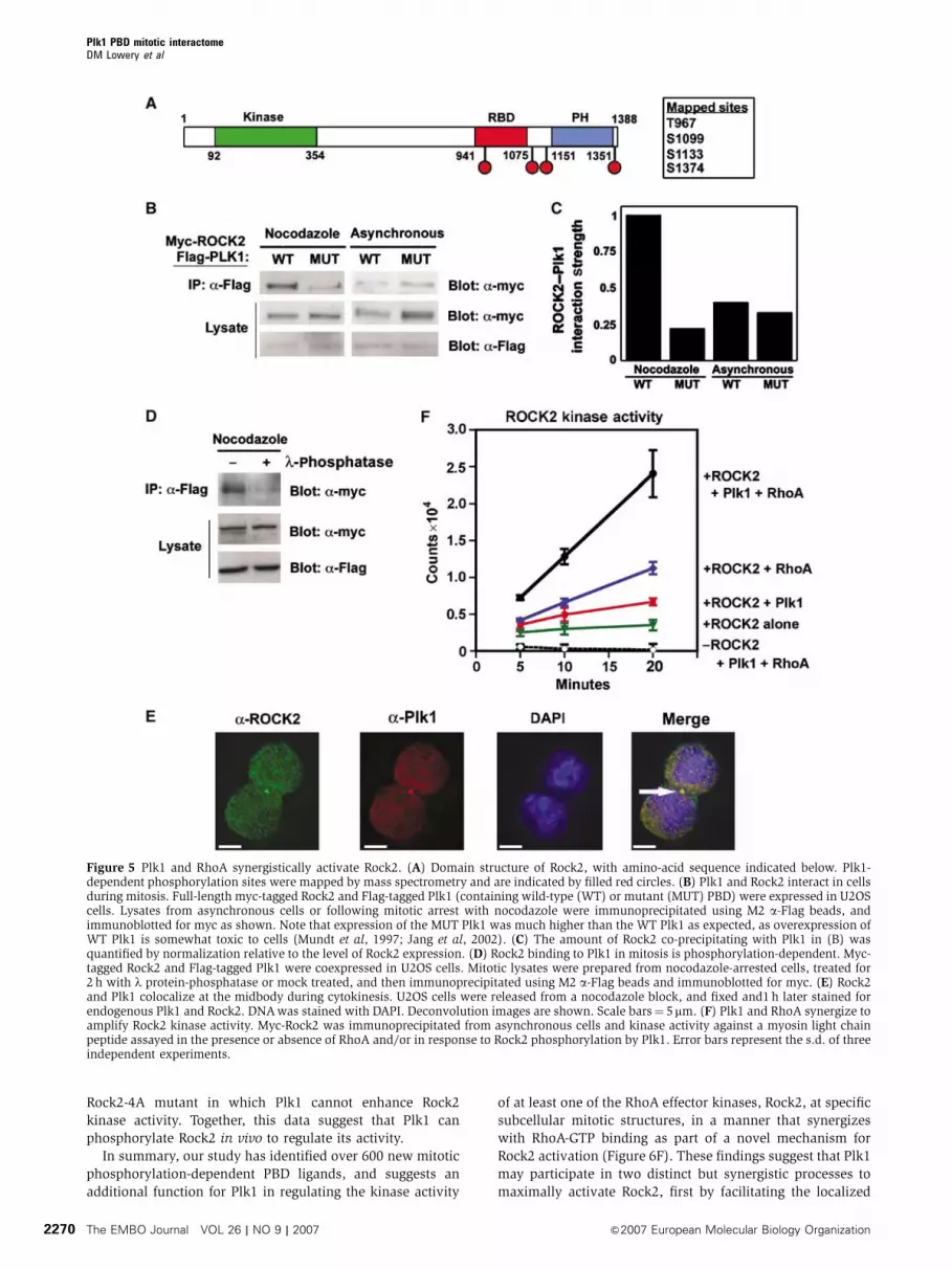
Rock2 contains an N-terminal kinase domain and a
C-terminal Rho-binding domain and PH domain (Figure 5A),
and phosphorylates myosin regulatory light chain and
myosin phosphatase targeting subunit in a manner that is
enhanced upon RhoA binding (Matsumura, 2005). To demon-
strate an interaction between full-length Rock2 and Plk1
within cells, myc-tagged Rock2 and Flag-tagged Plk1 contain-
ing either a wild-type or mutant PBD were coexpressed
in U2OS cells. Lysates were collected from asynchronous
and mitotically arrested cells. Immunoprecipitation of the
lysates using anti-Flag M2 beads revealed a strong mitosis-
dependent interaction of full-length Plk1 containing a wild-
type PBD, but not a mutant PBD, with Rock2 (Figure 5B
and C). Furthermore, this Plk1–Rock2 interaction was lost
when the lysates were treated with l-phosphatase, confirm-
ing that the interaction between the full-length proteins
was phospho-dependent (Figure 5D). We also observed
significant colocalization of the endogenous proteins during
cytokinesis, most prominently at the midbody (Figure 5E),
consistent with previously published localization data for
each of these proteins individually (Kosako et al, 2000;
Neef et al, 2003).
To investigate whether Rock2 could serve as a substrate for
Plk1, full-length bovine Rock2 (Rock2-FL) as well as N-
terminal kinase domain- (Rock2-CAT) and C-terminal RBD
and PH domain-containing fragments (Rock2-RBD/PH) of
Rock2 were phosphorylated by the Plk1 kinase domain
in vitro in the presence of [g-32P]ATP (Supplementary
Figure S4). Radioactivity was incorporated into all three
fragments of Rock2. When in vitro-phosphorylated full-length
Rock2 was analyzed by mass spectrometry, phosphorylated
peptides from only the C-terminal part of the molecule were
detected, corresponding to residues Thr-967 (DApTI),
Ser-1099 (EEpSQ), a monophosphorylated species phos-
phorylated on one of three consecutive serines from Ser-
1132 to Ser-1134 (Figures 5A and Supplementary Figures S4
and S5), and Ser-1374 (NQpSI). Three of these mapped
sites conform to the general Plk1 kinase consensus motif
[E/D]X[S/T] assuming that the third site is actually Ser-1133
(DSpSSI), whereas the fourth site at Ser-1374 contains an N in
the �2 position. This pSer-2 Asn has recently been reported
in several mapped phosphorylation sites targeted by Cdc5
(Brar et al, 2006), and fits with our unpublished data on the
in vitro Plk1 phosphorylation consensus motif (J Alexander
and MB Yaffe, unpublished data). In addition, Ser-1374
phosphorylation was reported in a high-throughput phos-
pho-proteomics screen (Beausoleil et al, 2006), indicating
that it is phosphorylated in vivo. All four of these motifs
are conserved in bovine, mouse, rat, and human Rock2. The
most likely candidate Plk1 N-terminal site in Rock2 on the
basis of consensus motif matching is Thr-489, which is
contained in a very acidic stretch of sequence, and peptides
containing this site were not recovered in either a phosphory-
lated or nonphosphorylated form.
To assess the functional significance of Rock2 phosphory-
lation by Plk1, myc-tagged bovine Rock2 was immunopreci-
pitated from 293Tcells and its kinase activity assayed using a
substrate peptide from its physiological substrate, myosin
regulatory light chain (Figure 5F). Following phosphoryla-
tion, the myosin regulatory light chain peptide substrate was
recovered from solution by spotting onto P81 paper and the
incorporated radioactivity measured by scintillation count-
ing. The addition of GTP-loaded RhoA increased the basal
activity of Rock2 by B3-fold, similar to the two-fold increase
reported previously (Ishizaki et al, 1996). Intriguingly, phos-
phorylation of Rock2 by Plk1 resulted in a similar two-fold
elevation of Rock2 kinase activity even in the absence of
RhoA-GTP, whereas Plk1 phosphorylation in combination
with RhoA resulted in an even more dramatic six-fold
enhancement. Control reactions containing RhoA-GTP, Plk1,
and myosin regulatory light chain substrate peptide did not
result in detectable peptide phosphorylation in the absence of
Rock2, nor did additional control reactions containing RhoA-
GTP, Plk1 and Rock2 but no peptide substrate. Therefore, the
data in Figure 5B–F, taken together, suggest that both Plk1
phosphorylation of Rock2 and RhoA-GTP binding may act
synergistically to maximize the kinase activity of Rock2.
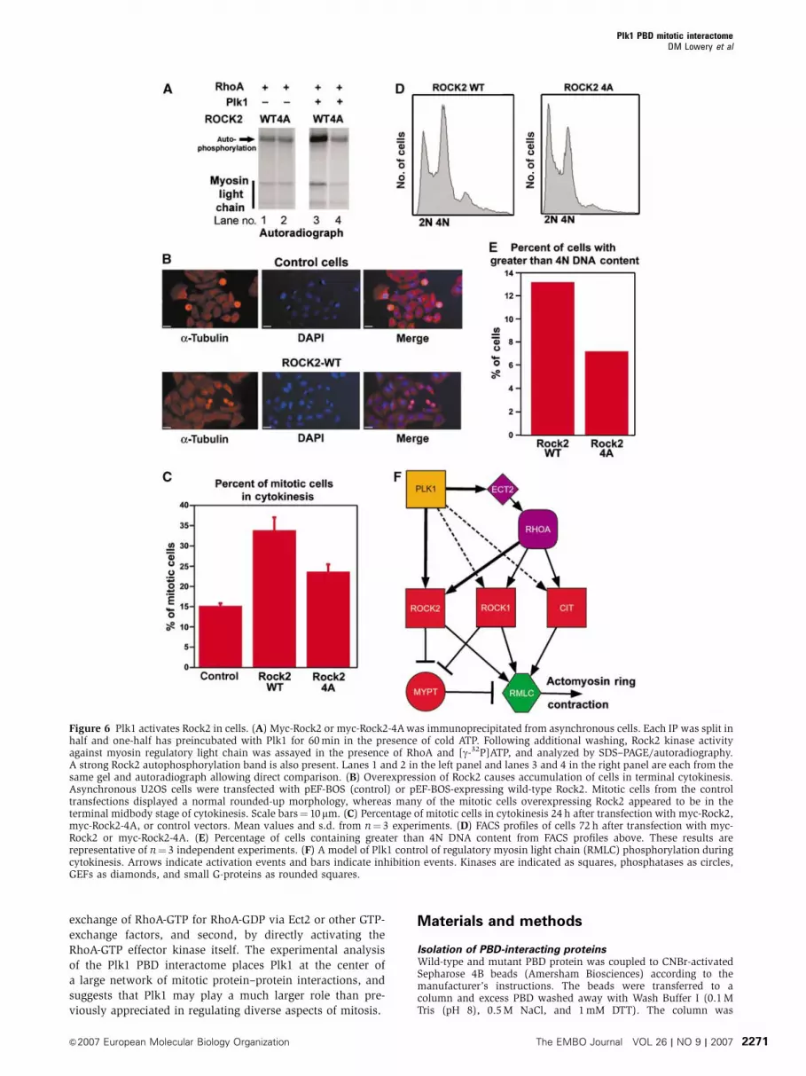
To further verify that Plk1 phosphorylation directly
activated Rock2, we constructed a mutant version of Rock2
lacking all four Plk1 phosphorylation sites (Rock2-4A). Both
wild-type Rock2 and Rock2-4A had similar basal RhoA-
activated kinase activity in vitro towards myosin regulatory
light chain in the absence of Plk1 (Figure 6A, lanes 1 and 2).
Following pre-incubation of the wild-type and 4A mutant of
Rock2 with Plk1, only the wild-type Rock2, but not the
Rock2-4A mutant, showed enhanced protein kinase activity
(Figure 6A, lanes 3 and 4).
In order to investigate the potential relevance of Plk1
phosphorylation of Rock2 within cells, it was necessary to
devise an assay in which Rock2 activity resulted in a specific
cellular phenotype. Because the pathways controlling acto-
myosin ring contraction during cytokinesis are highly redun-
dant, with at least three kinases other than Rock2 able to
phosphorylate myosin regulatory light chain (Matsumura,
2005), simple knockdowns or knockouts of several of these
kinases result in very mild or absent phenotypes. For exam-
ple, elimination of Rock2 alone, or even both Rock2 and
Rock1 by siRNA causes only a slight increase in the popula-
tion of multinucleated cells (Yokoyama et al, 2005; data not
shown). We therefore took advantage of the fact that comple-
tion of cytokinesis requires that the activity of RhoA and
Rock2 be shut off in order to allow disassembly of the
actomyosin ring (Emoto et al, 2005). Overexpression of
Rock2 prevented this disassembly and resulted in delay or
failure of cells at the midbody stage to complete cytokinesis
(Figure 6B).
We used siRNA to deplete endogenous Rock2, while simul-
taneously overexpressing either the wild-type or 4A mutant
forms of Rock2, and examined an asynchronous population
of Rock2-overexpressing cells for failure of cytokinesis exit.
As seen in Figure 6C, by 24 h after transfection, over one-third
of the cells that overexpressed wild-type Rock2 and were in
mitosis appeared to be in cytokinesis, compared to B15% of
the control cells. In contrast, overexpression of the Rock2-4A
mutant that cannot undergo Plk activation reduced the
percentage of mitotic cells that were in cytokinesis by about
50% compared to the wild-type Rock2. By 72 h following
transfection, overexpression of wild-type Rock2 resulted in a
marked increase in the population of cells with X4N DNA
content (Figure 6D and E), consistent with the emergence
of binucleated cells capable of undergoing additional rounds
of DNA replication, similar to what is seen in cells with
persistently active RhoA (Wolf et al, 2006). This phenotype
was significantly diminished in the cells overexpressing the
Plk1 PBD mitotic interactomeDM Lowery et al
&2007 European Molecular Biology Organization The EMBO Journal VOL 26 | NO 9 | 2007 2269
Rock2-4A mutant in which Plk1 cannot enhance Rock2
kinase activity. Together, this data suggest that Plk1 can
phosphorylate Rock2 in vivo to regulate its activity.
In summary, our study has identified over 600 new mitotic
phosphorylation-dependent PBD ligands, and suggests an
additional function for Plk1 in regulating the kinase activity
of at least one of the RhoA effector kinases, Rock2, at specific
subcellular mitotic structures, in a manner that synergizes
with RhoA-GTP binding as part of a novel mechanism for
Rock2 activation (Figure 6F). These findings suggest that Plk1
may participate in two distinct but synergistic processes to
maximally activate Rock2, first by facilitating the localized
Figure 5 Plk1 and RhoA synergistically activate Rock2. (A) Domain structure of Rock2, with amino-acid sequence indicated below. Plk1-dependent phosphorylation sites were mapped by mass spectrometry and are indicated by filled red circles. (B) Plk1 and Rock2 interact in cellsduring mitosis. Full-length myc-tagged Rock2 and Flag-tagged Plk1 (containing wild-type (WT) or mutant (MUT) PBD) were expressed in U2OScells. Lysates from asynchronous cells or following mitotic arrest with nocodazole were immunoprecipitated using M2 a-Flag beads, andimmunoblotted for myc as shown. Note that expression of the MUT Plk1 was much higher than the WT Plk1 as expected, as overexpression ofWT Plk1 is somewhat toxic to cells (Mundt et al, 1997; Jang et al, 2002). (C) The amount of Rock2 co-precipitating with Plk1 in (B) wasquantified by normalization relative to the level of Rock2 expression. (D) Rock2 binding to Plk1 in mitosis is phosphorylation-dependent. Myc-tagged Rock2 and Flag-tagged Plk1 were coexpressed in U2OS cells. Mitotic lysates were prepared from nocodazole-arrested cells, treated for2 h with l protein-phosphatase or mock treated, and then immunoprecipitated using M2 a-Flag beads and immunoblotted for myc. (E) Rock2and Plk1 colocalize at the midbody during cytokinesis. U2OS cells were released from a nocodazole block, and fixed and1 h later stained forendogenous Plk1 and Rock2. DNA was stained with DAPI. Deconvolution images are shown. Scale bars¼ 5mm. (F) Plk1 and RhoA synergize toamplify Rock2 kinase activity. Myc-Rock2 was immunoprecipitated from asynchronous cells and kinase activity against a myosin light chainpeptide assayed in the presence or absence of RhoA and/or in response to Rock2 phosphorylation by Plk1. Error bars represent the s.d. of threeindependent experiments.
Plk1 PBD mitotic interactomeDM Lowery et al
The EMBO Journal VOL 26 | NO 9 | 2007 &2007 European Molecular Biology Organization2270
exchange of RhoA-GTP for RhoA-GDP via Ect2 or other GTP-
exchange factors, and second, by directly activating the
RhoA-GTP effector kinase itself. The experimental analysis
of the Plk1 PBD interactome places Plk1 at the center of
a large network of mitotic protein–protein interactions, and
suggests that Plk1 may play a much larger role than pre-
viously appreciated in regulating diverse aspects of mitosis.
Materials and methods
Isolation of PBD-interacting proteinsWild-type and mutant PBD protein was coupled to CNBr-activatedSepharose 4B beads (Amersham Biosciences) according to themanufacturer’s instructions. The beads were transferred to acolumn and excess PBD washed away with Wash Buffer I (0.1 MTris (pH 8), 0.5 M NaCl, and 1 mM DTT). The column was
Figure 6 Plk1 activates Rock2 in cells. (A) Myc-Rock2 or myc-Rock2-4A was immunoprecipitated from asynchronous cells. Each IP was split inhalf and one-half has preincubated with Plk1 for 60 min in the presence of cold ATP. Following additional washing, Rock2 kinase activityagainst myosin regulatory light chain was assayed in the presence of RhoA and [g-32P]ATP, and analyzed by SDS–PAGE/autoradiography.A strong Rock2 autophosphorylation band is also present. Lanes 1 and 2 in the left panel and lanes 3 and 4 in the right panel are each from thesame gel and autoradiograph allowing direct comparison. (B) Overexpression of Rock2 causes accumulation of cells in terminal cytokinesis.Asynchronous U2OS cells were transfected with pEF-BOS (control) or pEF-BOS-expressing wild-type Rock2. Mitotic cells from the controltransfections displayed a normal rounded-up morphology, whereas many of the mitotic cells overexpressing Rock2 appeared to be in theterminal midbody stage of cytokinesis. Scale bars¼ 10mm. (C) Percentage of mitotic cells in cytokinesis 24 h after transfection with myc-Rock2,myc-Rock2-4A, or control vectors. Mean values and s.d. from n¼ 3 experiments. (D) FACS profiles of cells 72 h after transfection with myc-Rock2 or myc-Rock2-4A. (E) Percentage of cells containing greater than 4N DNA content from FACS profiles above. These results arerepresentative of n¼ 3 independent experiments. (F) A model of Plk1 control of regulatory myosin light chain (RMLC) phosphorylation duringcytokinesis. Arrows indicate activation events and bars indicate inhibition events. Kinases are indicated as squares, phosphatases as circles,GEFs as diamonds, and small G-proteins as rounded squares.
Plk1 PBD mitotic interactomeDM Lowery et al
&2007 European Molecular Biology Organization The EMBO Journal VOL 26 | NO 9 | 2007 2271
equilibrated with Wash Buffer II (50 mM Tris (pH 8), 0.2 M NaCl,2 mM DTT, and 10 mM NaF), and the beads then incubated withU2OS cell lysate overnight. Unbound proteins were washed awaywith 10 column volumes Wash Buffer II. PBD interacting proteinswere eluted by incubating the beads with 2 ml of 1 mM phospho-peptide (YMQS-pT-PK) in Wash Buffer II for 1 h at 41C. The beadswere washed with additional 2 ml of the phosphopeptide-containingbuffer.
Mass spectrometryConcentrated eluates from the wild-type and mutant PBD columnswere boiled in reducing sample buffer containing b-mercaptoetha-nol, separated by SDS–PAGE, and visualized by SYPRO Rubystaining (Bio-Rad). Lanes from the gel were excised, cut into 12fields as shown in Figure 1E and digested overnight at 371C with anexcess of sequencing grade trypsin. Peptides were extracted fromthe gel with 50% acetonitrile/0.1% formic acid and concentrated ina Speed-Vac. Tryptic digests were analyzed with an automated nanoLC/MS/MS system, using a 1100 series autosampler and nano pump(Agilent Technologies, Wilmington, DE) coupled to an LTQ-FThybrid ion trap Fourier transform mass spectrometer (ThermoElectron, San Jose, CA) equipped with a nanoflow ionization source(James A. Hill Instrument Services, Arlington, MA). Peptides wereeluted from a (75mm� 10 cm) PicoFrit (New Objective, Woburn,MA) column packed with (5mm� 200 A) Magic C-18AQ reversed-phase beads (Michrom Bioresources Inc., Auburn, CA) using a70 min acetonitrile/0.1% formic acid gradient at a flow rate of250 nl/min to yield B25 s peak widths. Data-dependent LC/MS/MSspectra were acquired in B3 s cycles; each cycle was of thefollowing form: one full FT MS scan followed by eight MS/MS scansin the ion trap on the most abundant precursor ions excludingcharge 1 and unknown charge, subject to accurate mass, dynamicexclusion for a period of 45 s. Some of the phosphorylation siteswere established with additional LC/MS/MS experiments per-formed on aliquots of the gel band digests with the instrumentoperated in a targeted mode where MS/MS of 5–17 precursor m/z’swere repetitively taken in 3–9 s cycles throughout the acetonitrilegradient. These precursor masses were selected because theycorrespond to the phosphorylated form of expected tryptic peptides
containing S[ST]P motifs derived from proteins confidentlyidentified in aliquots of sample used for the data-dependentexperiments. For Rock2 phosphopeptide mapping, both trypsinand chymotrypsin were used to maximize coverage.
Rock2 kinase assaysMyc-tagged Rock2 was preincubated in kinase buffer (50 mM Tris(pH 8), 200 mM NaCl, 2 mM DTT, 5 mM NaF, 200mM ATP, and500mM MgCl2) containing 1 mM GTP with or without 5mg RhoAand/or 7 ng/ml Plk1 kinase domain for 30 min at 301C. Reactionswere then supplemented with 20mCi [g-32P]ATP and 50mMsubstrate peptide (Myosin Light Chain Kinase Substrate; AKRPQ-RATSNVFS; Sigma-Aldrich), and aliquots of the reaction spottedonto squares of Whatman P81 paper after an additional 0, 5, 10, or20 min. The P81 paper was washed with 0.5% phosphoric acid fivetimes, and incorporation of 32P into the substrate peptide wasassessed by scintillation counting. For comparison of wild-type and4A mutant ROCK2, full-length myosin light chain (Sigma) was usedas a substrate, and the radiolabeled phosphate incorporation wasassessed by autoradiography following SDS–PAGE.
Additional experimental details are provided in Supplementarydata.
Supplementary dataSupplementary data are available at The EMBO Journal Online(http://www.embojournal.org).
Acknowledgements
We thank K Kaibuchi for providing Rock2 constructs and the MITCCR Microscopy and Imaging Core Facility. MH is supported by theDanish Medical Research Council. DML was supported by a HowardHughes Medical Institute Predoctoral Fellowship and is supportedby a David Koch Graduate Fellowship. KK is supported by ManpeiSuzuki Diabetes Foundation. DL is supported by a Janes CoffinChilds Memorial Fellowship. This work was supported by NIH grantP50-CA112962 to SAC, and GM60594 and a Burrough’s WellcomeCareer Development Award to MBY.
References
Ahonen LJ, Kallio MJ, Daum JR, Bolton M, Manke IA, Yaffe MB,Stukenberg PT, Gorbsky GJ (2005) Polo-like kinase 1 creates thetension-sensing 3F3/2 phosphoepitope and modulates the asso-ciation of spindle-checkpoint proteins at kinetochores. Curr Biol15: 1078–1089
Barr FA, Sillje HH, Nigg EA (2004) Polo-like kinases and theorchestration of cell division. Nat Rev Mol Cell Biol 5: 429–440
Beausoleil SA, Villen J, Gerber SA, Rush J, Gygi SP (2006) A proba-bility-based approach for high-throughput protein phosphoryla-tion analysis and site localization. Nat Biotechnol 24: 1285–1292
Brar GA, Kiburz BM, Zhang Y, Kim JE, White F, Amon A (2006)Rec8 phosphorylation and recombination promote the step-wiseloss of cohesins in meiosis. Nature 441: 532–536
Carmena M, Riparbelli MG, Minestrini G, Tavares AM, Adams R,Callaini G, Glover DM (1998) Drosophila polo kinase is requiredfor cytokinesis. J Cell Biol 143: 659–671
Cheng KY, Lowe ED, Sinclair J, Nigg EA, Johnson LN (2003) Thecrystal structure of the human polo-like kinase-1 polo boxdomain and its phospho-peptide complex. EMBO J 22: 5757–5768
Daniels MJ, Wang Y, Lee M, Venkitaraman AR (2004) Abnormalcytokinesis in cells deficient in the breast cancer susceptibilityprotein BRCA2. Science 306: 876–879
Darieva Z, Bulmer R, Pic-Taylor A, Doris KS, Geymonat M,Sedgwick SG, Morgan BA, Sharrocks AD (2006) Polo kinasecontrols cell-cycle-dependent transcription by targeting a coacti-vator protein. Nature 444: 494–498
De Luca M, Lavia P, Guarguaglini G (2006) A functional interplaybetween Aurora-A, Plk1 and TPX2 at spindle poles: Plk1 controlscentrosomal localization of Aurora-A and TPX2 spindle associa-tion. Cell Cycle 5: 296–303
Donaldson MM, Tavares AA, Ohkura H, Deak P, Glover DM (2001)Metaphase arrest with centromere separation in polo mutants ofDrosophila. J Cell Biol 153: 663–676
Elia AE, Cantley LC, Yaffe MB (2003a) Proteomic screen finds pSer/pThr-binding domain localizing Plk1 to mitotic substrates.Science 299: 1228–1231
Elia AE, Rellos P, Haire LF, Chao JW, Ivins FJ, Hoepker K,Mohammad D, Cantley LC, Smerdon SJ, Yaffe MB (2003b) Themolecular basis for phosphodependent substrate targeting andregulation of plks by the polo-box domain. Cell 115: 83–95
Emoto K, Inadome H, Kanaho Y, Narumiya S, Umeda M (2005)Local change in phospholipid composition at the cleavagefurrow is essential for completion of cytokinesis. J Biol Chem280: 37901–37907
Fabbro M, Zhou BB, Takahashi M, Sarcevic B, Lal P, Graham ME,Gabrielli BG, Robinson PJ, Nigg EA, Ono Y, Khanna KK (2005)Cdk1/Erk2- and Plk1-dependent phosphorylation of a centro-some protein, Cep55, is required for its recruitment to midbodyand cytokinesis. Dev Cell 9: 477–488
Feng Y, Hodge DR, Palmieri G, Chase DL, Longo DL, Ferris DK(1999) Association of polo-like kinase with alpha-, beta- andgamma-tubulins in a stable complex. Biochem J 339 (Part 2):435–442
Feng Y, Longo DL, Ferris DK (2001) Polo-like kinase interacts withproteasomes and regulates their activity. Cell Growth Differ 12:29–37
Glotzer M (2005) The molecular requirements for cytokinesis.Science 307: 1735–1739
Glover DM (2005) Polo kinase and progression through M phase inDrosophila: a perspective from the spindle poles. Oncogene 24:230–237
Golsteyn RM, Schultz SJ, Bartek J, Ziemiecki A, Ried T, Nigg EA(1994) Cell cycle analysis and chromosomal localization ofhuman Plk1, a putative homologue of the mitotic kinasesDrosophila polo and Saccharomyces cerevisiae Cdc5. J Cell Sci107 (Part 6): 1509–1517
Plk1 PBD mitotic interactomeDM Lowery et al
The EMBO Journal VOL 26 | NO 9 | 2007 &2007 European Molecular Biology Organization2272

GO Consortium (2001) Creating the gene ontology resource: designand implementation. Genome Res 11: 1425–1433
Goto H, Kiyono T, Tomono Y, Kawajiri A, Urano T, Furukawa K,Nigg EA, Inagaki M (2006) Complex formation of Plk1 andINCENP required for metaphase–anaphase transition. Nat CellBiol 8: 180–187
Goto H, Kosako H, Tanabe K, Yanagida M, Sakurai M, Amano M,Kaibuchi K, Inagaki M (1998) Phosphorylation of vimentin byRho-associated kinase at a unique amino-terminal site that isspecifically phosphorylated during cytokinesis. J Biol Chem 273:11728–11736
Ishizaki T, Maekawa M, Fujisawa K, Okawa K, Iwamatsu A, FujitaA, Watanabe N, Saito Y, Kakizuka A, Morii N, Narumiya S (1996)The small GTP-binding protein Rho binds to and activates a160 kDa Ser/Thr protein kinase homologous to myotonic dystro-phy kinase. EMBO J 15: 1885–1893
Jang YJ, Ma S, Terada Y, Erikson RL (2002) Phosphorylation ofthreonine 210 and the role of serine 137 in the regulation ofmammalian polo-like kinase. J Biol Chem 277: 44115–44120
Kim JH, Paek KY, Choi K, Kim TD, Hahm B, Kim KT, Jang SK (2003)Heterogeneous nuclear ribonucleoprotein C modulates transla-tion of c-myc mRNA in a cell cycle phase-dependent manner.Mol Cell Biol 23: 708–720
Kim SA, Yoon JH, Lee SH, Ahn SG (2005) Polo-like kinase 1phosphorylates heat shock transcription factor 1 and mediatesits nuclear translocation during heat stress. J Biol Chem 280:12653–12657
Kosako H, Yoshida T, Matsumura F, Ishizaki T, Narumiya S, InagakiM (2000) Rho-kinase/ROCK is involved in cytokinesis throughthe phosphorylation of myosin light chain and not ezrin/radixin/moesin proteins at the cleavage furrow. Oncogene 19: 6059–6064
Lane HA, Nigg EA (1996) Antibody microinjection reveals an essen-tial role for human polo-like kinase 1 (Plk1) in the functionalmaturation of mitotic centrosomes. J Cell Biol 135: 1701–1713
Lee KS, Park JE, Asano S, Park CJ (2005) Yeast polo-like kinases:functionally conserved multitask mitotic regulators. Oncogene 24:217–229
Litvak V, Argov R, Dahan N, Ramachandran S, Amarilio R,Shainskaya A, Lev S (2004) Mitotic phosphorylation of the peri-pheral Golgi protein Nir2 by Cdk1 provides a docking mechanismfor Plk1 and affects cytokinesis completion. Mol Cell 14: 319–330
Liu X, Lei M, Erikson RL (2006) Normal cells, but not cancer cells,survive severe Plk1 depletion. Mol Cell Biol 26: 2093–2108
Liu X, Lin CY, Lei M, Yan S, Zhou T, Erikson RL (2005) CCTchaperonin complex is required for the biogenesis of functionalPlk1. Mol Cell Biol 25: 4993–5010
Liu X, Zhou T, Kuriyama R, Erikson RL (2004) Molecular inter-actions of Polo-like-kinase 1 with the mitotic kinesin-like proteinCHO1/MKLP-1. J Cell Sci 117: 3233–3246
Lowery DM, Lim D, Yaffe MB (2005) Structure and function ofPolo-like kinases. Oncogene 24: 248–259
Matsumura F (2005) Regulation of myosin II during cytokinesis inhigher eukaryotes. Trends Cell Biol 15: 371–377
Mishra GR, Suresh M, Kumaran K, Kannabiran N, Suresh S, Bala P,Shivakumar K, Anuradha N, Reddy R, Raghavan TM, Menon S,Hanumanthu G, Gupta M, Upendran S, Gupta S, Mahesh M,Jacob B, Mathew P, Chatterjee P, Arun KS, Sharma S, ChandrikaKN, Deshpande N, Palvankar K, Raghavnath R, Krishnakanth R,Karathia H, Rekha B, Nayak R, Vishnupriya G, Kumar HG, NaginiM, Kumar GS, Jose R, Deepthi P, Mohan SS, Gandhi TK, HarshaHC, Deshpande KS, Sarker M, Prasad TS, Pandey A (2006)Human protein reference database—2006 update. Nucleic AcidsRes 34: D411–D414
Mundt KE, Golsteyn RM, Lane HA, Nigg EA (1997) On the regula-tion and function of human polo-like kinase 1 (PLK1): effects ofoverexpression on cell cycle progression. Biochem Biophys ResCommun 239: 377–385
Nakajima H, Toyoshima-Morimoto F, Taniguchi E, Nishida E (2003)Identification of a consensus motif for Plk (Polo-like kinase)phosphorylation reveals Myt1 as a Plk1 substrate. J Biol Chem278: 25277–25280
Neef R, Preisinger C, Sutcliffe J, Kopajtich R, Nigg EA, Mayer TU,Barr FA (2003) Phosphorylation of mitotic kinesin-like protein 2by polo-like kinase 1 is required for cytokinesis. J Cell Biol 162:863–876
Niiya F, Tatsumoto T, Lee KS, Miki T (2006) Phosphorylation of thecytokinesis regulator ECT2 at G2/M phase stimulates associationof the mitotic kinase Plk1 and accumulation of GTP-bound RhoA.Oncogene 25: 827–837
Ohkura H, Hagan IM, Glover DM (1995) The conservedSchizosaccharomyces pombe kinase plo1, required to form abipolar spindle, the actin ring, and septum, can drive septumformation in G1 and G2 cells. Genes Dev 9: 1059–1073
Qian YW, Erikson E, Taieb FE, Maller JL (2001) The polo-like kinasePlx1 is required for activation of the phosphatase Cdc25C andcyclin B-Cdc2 in Xenopus oocytes. Mol Biol Cell 12: 1791–1799
Rapley J, Baxter JE, Blot J, Wattam SL, Casenghi M, Meraldi P, NiggEA, Fry AM (2005) Coordinate regulation of the mother centriolecomponent nlp by nek2 and plk1 protein kinases. Mol Cell Biol25: 1309–1324
Robinson DN, Spudich JA (2004) Mechanics and regulation ofcytokinesis. Curr Opin Cell Biol 16: 182–188
Shin C, Manley JL (2002) The SR protein SRp38 represses splicing inM phase cells. Cell 111: 407–417
Song S, Lee KS (2001) A novel function of Saccharomyces cerevisiaeCDC5 in cytokinesis. J Cell Biol 152: 451–469
Spankuch-Schmitt B, Wolf G, Solbach C, Loibl S, Knecht R,Stegmuller M, von Minckwitz G, Kaufmann M, Strebhardt K(2002) Downregulation of human polo-like kinase activity byantisense oligonucleotides induces growth inhibition in cancercells. Oncogene 21: 3162–3171
Straight AF, Field CM, Mitchison TJ (2005) Anillin binds nonmusclemyosin II and regulates the contractile ring. Mol Biol Cell 16:193–201
Tarnowka MA, Baglioni C (1979) Regulation of protein synthesis inmitotic HeLa cells. J Cell Physiol 99: 359–367
Tsvetkov L, Stern DF (2005) Interaction of chromatin-associatedPlk1 and Mcm7. J Biol Chem 280: 11943–11947
van de Weerdt BC, Medema RH (2006) Polo-like kinases: a team incontrol of the division. Cell Cycle 5: 853–864
van Vugt MA, Bras A, Medema RH (2004a) Polo-like kinase-1controls recovery from a G2 DNA damage-induced arrest inmammalian cells. Mol Cell 15: 799–811
van Vugt MA, van de Weerdt BC, Vader G, Janssen H, Calafat J,Klompmaker R, Wolthuis RM, Medema RH (2004b) Polo-likekinase-1 is required for bipolar spindle formation but is dispen-sable for anaphase promoting complex/Cdc20 activation andinitiation of cytokinesis. J Biol Chem 279: 36841–36854
Warren G (1993) Membrane partitioning during cell division. AnnuRev Biochem 62: 323–348
Wolf A, Keil R, Gotzl O, Mun A, Schwarze K, Lederer M,Huttelmaier S, Hatzfeld M (2006) The armadillo proteinp0071 regulates Rho signalling during cytokinesis. Nat Cell Biol8: 1432–1440
Yamaguchi T, Goto H, Yokoyama T, Sillje H, Hanisch A, UldschmidA, Takai Y, Oguri T, Nigg EA, Inagaki M (2005) Phosphorylationby Cdk1 induces Plk1-mediated vimentin phosphorylation duringmitosis. J Cell Biol 171: 431–436
Yokoyama T, Goto H, Izawa I, Mizutani H, Inagaki M (2005) Aurora-B and Rho-kinase/ROCK, the two cleavage furrow kinases, in-dependently regulate the progression of cytokinesis: possibleexistence of a novel cleavage furrow kinase phosphorylatesezrin/radixin/moesin (ERM). Genes Cells 10: 127–137
Yoshida S, Kono K, Lowery DM, Bartolini S, Yaffe MB, Ohya Y,Pellman D (2006) Polo-like kinase Cdc5 controls the local activa-tion of Rho1 to promote cytokinesis. Science 313: 108–111
Zhang H, Shi X, Paddon H, Hampong M, Dai W, Pelech S (2004)B23/Nucleophosmin serine 4 phosphorylation mediates mitoticfunctions of Polo-like kinase 1. J Biol Chem 279: 35726–35734
Zhou T, Aumais JP, Liu X, Yu-Lee LY, Erikson RL (2003) A rolefor Plk1 phosphorylation of NudC in cytokinesis. Dev Cell 5:127–138
Plk1 PBD mitotic interactomeDM Lowery et al
&2007 European Molecular Biology Organization The EMBO Journal VOL 26 | NO 9 | 2007 2273