prolactin alters the mechanisms of b cell tolerance induction
TRANSCRIPT
Prolactin Alters the Mechanisms of B Cell Tolerance Induction
Subhrajit Saha*,†, Juana Gonzalez*,†, Gabriel Rosenfeld*,†, Harold Keiser*, and ElenaPeeva*,†
*Department of Medicine, Albert Einstein College of Medicine, Bronx, New York, USA†Department of Microbiology and Immunology, Albert Einstein College of Medicine, Bronx, NewYork, USA
AbstractObjective—Autoimmune diseases predominantly affect women suggesting that female sexhormones may play a role in pathogenesis. We have previously shown that persistent mild-moderate elevations in serum prolactin levels induce a break in self-tolerance in mice with aBALB/c genetic background. In this study we evaluated the effects of hyperprolactinemia onmechanisms of B cell tolerance induction.
Methods—Effects of prolactin on splenic B cell subsets were studied in female Balb/c mice.BCR-mediated apoptosis and proliferation of transitional B cells were analyzed by flow-cytometry. Expression of apoptotic genes was examined by microarrays and real-time PCR.Kappa/lambda light chain-coexpressing B cells were assessed by flowcytometry andimmunohistochemistry. Activation status of T3 B cells was evaluated by BCR-induced calciuminflux studies.
Results—BCR-mediated apoptosis of the T1 B cell subset, a major checkpoint for negativeselection of autoreactive specificities, was decreased in prolactin-treated mice. Microarray studiesindicated that this event may be mediated by the prolactin-induced upregulation of the anti-apoptotic gene INF-γRII and downregulation of the pro-apoptotic gene Trp63. Prolactin treatmentalso altered the amount of receptor editing as indicated by the increased number of transitional Bcells co-expressing kappa/lambda light chains. Additionally, hyperprolactinemia modified thelevel of B cell anergy by increasing the degree of BCR-induced calcium influx in the T3 B cells.
Conclusion—Persistently elevated serum prolactin levels interfere with B cell toleranceinduction by impairing BCR-mediated clonal deletion, deregulating receptor editing, anddecreasing the threshold for activation of anergic B cells, thereby promoting autoreactivity.
KeywordsSystemic Lupus Erythematosus; B cells; Tolerance/Suppression/Anergy; Autoantibodies
The strong predominance of females in autoimmune diseases suggests that female sexhormones may play a role in disease susceptibility. There is some clinical evidence in thisregard for prolactin. Increased serum levels of this hormone have been reported in patientswith systemic lupus erythematosis (SLE) (1), scleroderma (2) and multiple sclerosis (3), andhave been correlated with disease activity in a subset of SLE (4) and scleroderma patients(5); high serum prolactin levels during breast feeding have also been linked to flares ofrheumatoid arthritis (6). Further evidence for a role of prolactin in autoimmunity has been
Address correspondence to: Dr. Elena Peeva, Rheumatology Division, Albert Einstein College of Medicine, 1300 Morris ParkAvenue, F717, Bronx, New York 10461. [email protected]: The authors have declared that no conflicts of interest exist.
NIH Public AccessAuthor ManuscriptArthritis Rheum. Author manuscript; available in PMC 2009 August 27.
Published in final edited form as:Arthritis Rheum. 2009 June ; 60(6): 1743–1752. doi:10.1002/art.24500.
NIH
-PA Author Manuscript
NIH
-PA Author Manuscript
NIH
-PA Author Manuscript
obtained from experimental studies in mice (7-10). For example, we have shown thattreatment of ovariectomized non-lupus prone BALB/c mice bearing the appropriatetransgenic marker with a dose of prolactin causing mild to moderate elevation of serumlevels induced the development of a lupus-like disease (11). To understand the basis forprolactin-induced autoimmunity, we have evaluated the effects of the hormone on theinduction of B cell tolerance. We demonstrate here that prolactin impairs the three crucialmechanisms for B cell tolerance induction: BCR-mediated deletion, receptor editing, andanergy.
Materials and MethodsAnimals
Eight-10 week-old female BALB/c mice, purchased from Taconic Farms (Germantown,NY), were ovariectomized and subjected to the treatments described below. Mice werehoused in the barrier animal facility at Albert Einstein College of Medicine, Bronx, NY inaccordance with current guidelines.
Anti-mouse CD40L antibodyAnti-mouse CD40L antibody was prepared from culture-supernatants of the MR1 Armenianhamster B cell-hybridoma (ATCC cell line CRL-2580, Manassas, VA, USA). Antibodyconcentration was tested by ELISA (BD Pharmingen, San Jose, CA, USA) and prepared at1mg/ml.
TreatmentsMice were injected subcutaneously with 0.1ml normal saline or 0.1mg ovine prolactin(Sigma-Aldrich, St. Louis, Missouri, USA) every day for 4 weeks. This prolactin treatmentsleads to twofold increase in serum prolactin concentration: serum prolactin levels of 68.3 ±20.75 nanograms/ml (ng/ml) in prolactin-treated mice vs. 30.3 ± 19.7 ng/ml in placebo-treated mice (11). Anti-CD40L-antibody (250mg) was administered intraperitoneally 3times/week for 4 weeks.
Flow-cytometrySplenocytes isolated from sacrificed animals were subjected to RBC-depletion with ACK-lysis buffer. Single-cell suspensions were stained with PerCp-Cy5.5, PE-Cy7, PE, APC,FITC and biotin-conjugated antibodies to CD19 (BD Pharmingen, clone 1d3, BDBiosciences, San Jose, California), CD93 (eBioscience clone AA4.1, eBioscience, SanDiego, California), CD23 (Caltag, Carlsbad, California), IgM (BD Pharmingen, cloneR6-60.2), CD21 (BD Pharmingen, clone 7g6), CD22 (Chemicon International, Temecula,CA), CD40 (BD Pharmingen), BAFF-R (R&D Systems, McKinely Place N.E Minneapolis)kappa (κ) (BD Pharmingen, clone 187.1) and lambda (λ) (BD Pharmingen, clone r26-46)light chains at 4°C for 30 minutes. Cells were washed and stained with pacific blue-conjugated streptavidin (SA) (Invitrogen, Carlsbad, California), and then fixed with 2%paraformaldehype. After cell- permeabilization with 0.3% saponin, intracellular staining wasperformed for P-Syk (BD Pharmingen) and SHP-1 (Santa Cruz Biotechnology, Santa Cruz,CA).
CD19 and CD93-staining was used to differentiate the transitional (CD19+CD93+) frommature (CD19+CD93-) B cells. IgM and CD23-staining allowed for identification of thetransitional T1 (CD19+CD93+IgM+CD23-), T2 (CD19+CD93+IgM+CD23+) and T3(CD19+CD93+IgMlowCD23+) B cell subsets (12). CD21 and CD23-staining allowed foridentification of the mature marginal zone (CD19+CD93-CD21++CD23-) and follicular(CD19+CD93-CD21-CD23++) B cells (13). Samples were sorted by MoFlow cell-sorter
Saha et al. Page 2
Arthritis Rheum. Author manuscript; available in PMC 2009 August 27.
NIH
-PA Author Manuscript
NIH
-PA Author Manuscript
NIH
-PA Author Manuscript
(Dako-Cytomation, Dako Colorado Inc., Fort Collins, Colorado, USA) (purity 95% orabove) or acquired with the LSRII flow-cytometer (BD Biosciences). Sorted transitional Bcells were subjected to RNA isolation for real-time PCR analysis of RAG expression (seebelow) whereas sorted λ+B cells were used for immunofluorescent-cytology studies orgeneration of hybridomas as described bellow. The data acquired by LSRII flow-cytometerwas analyzed with FlowJo v.7.1 software (Treestar Inc, Ashlaand, OR, USA).
Proliferation assaySplenocytes from placebo and prolactin-treated mice were labeled with CFSE(carboxyfluorescein-diacetate-succidimyl ester, Molecular Probes, Eugene, OR). Cells werere-suspended in phenol-free RPMI-1640 medium supplemented with charcoal stripped 10%FCS. B lymphocyte-proliferation was induced by culturing the cells with anti-IgM (10μg/ml) for 3 days. Cells were stained for plasma-membrane markers to identify B cell subsetsas depicted above. Samples were analyzed for proliferating B cells subtypes by flow-cytometry, as previously described (14).
Apoptosis assayRBC-depleted splenocytes were incubated at 37°C in phenol-free RPMI-mediumsupplemented with 5% fetal calf serum in the presence and absence of 10μg/mL anti-IgMantibody (Jackson ImmunoResearch West Grove, PA, USA). After 10-14 hours, splenocyteswere washed and stained for 30minutes at 4°C with fluorochrome-labeled antibodies toidentify the T1 and T2 B cells as described above. Cells were washed again and stained withFITC-conjugated antibody to AnnexinV (Pharmingen), and, just prior to acquisition,samples were stained with impermeable dye Topro-3-labeled with APC (Invitrogen) in orderto distinguish the necrotic from apoptotic (AnnexinV+ Topro3-) cells. Samples wereacquired by an LSRII flow-cytometer (BD Biosciences) and data was analyzed with FlowJov7.1 (Treestar).
B cell purificationSplenic B cells were isolated by negative selection using biotinylated anti-mouse CD43antibody (BD PharMingen) and streptavidin conjugated Dynabeads according to themanufacturer's protocol (Dynal Invitrogen bead separations, Invtrogen Corporation). Thepurity of the isolated B cells was 95% or more as determined by flow-cytometry.
RNA isolationPurified B cells were lysed using RLT buffer from RNeasy Mini-kit (Qiagen, Valencia, CA)and 1% betamercaptoethanol mix. Qiagen's protocol for the RNeasy Mini Kit with on-column DNA digestion was employed to isolate RNA from the lysates. The RNA sampleswere stored at -80°C until further use.
Microarray analysisThe gene expression in B cells was determined by Affymetrix genechips (genechip #45102)(Affymetrix, Inc., Santa Clara, CA). RNA was isolated from 5 placebo and 5 prolactin-treated mice by Trizol (Invitrogen). First-strand cDNA was synthesized from 30-50μg ofRNA by using the first-strand cDNA-synthesis kit from Invitrogen as per the manufacturer'sprotocol. The reaction product was subjected to second-strand cDNA synthesis with thesecond-strand cDNA-synthesis kit (Invitrogen). Clean-up of double-stranded cDNA wasdone following the GeneChip sample-clean-up module #900371 (Affymetrix Inc). RNAtranscript labeling was performed by ENZO BioArray™ Highyield™ RNA transcript-labeling kit (ENZO Lifesciences, Farmingdale, NY). Labeled RNA transcripts, cleanedaccording to the GeneChip sample clean-up module, were quantified by a
Saha et al. Page 3
Arthritis Rheum. Author manuscript; available in PMC 2009 August 27.
NIH
-PA Author Manuscript
NIH
-PA Author Manuscript
NIH
-PA Author Manuscript
spectrophotometer. Fragmentation of cRNA was done as per the Affimetrix protocol.Fragmented cRNA was sent to the Microarray facility at our institution for hybridization andscanning. The microarray data was analyzed by Array-assist software (Stratagene, La Jolla,CA).
Real-time PCRThe gene sequences were obtained from the Ensembl mouse genome database(http://www.ensembl.org/Mus_musculus/index.html). The primers were designed usingPrimer3 software (http://frodo.wi.mit.edu/cgi-bin/primer3/primer3_www.cgi). Any primerpair generated with Primer3 was checked for gene specificity using the nucleotide-nucleotide BLAST database (http://130.14.29.110/BLAST/). The primer pair spanning the5-6 intron for beta actin, which served as housekeeping gene, was the following: left primer5′TGTACCCAGGCATTGCTGAC3′ and right primer5′ACAGTGAGGCCAGGATGGAG3′. The RAG-1 primers were the following: left primer5′CGGAACTCCTCTCCACCAAG3′ and right primer 5′ACCCGATTCATTTCCCTCAC 3′the RAG-2 primers were the following: left primer 5′TGCATGGATTTGGAAGAACG 3′and right primer 5′GGGGTTTCTTTTGGGAGTTTG3′. For the IFN-γRII, the followingprimers were used: left primer 5′CCTGCTTCACCCTGTTCCTC3′ and right primer 5′CCGTCCTTGTCCAAGACCTC3′, whereas for Trp63 the left primer was 5′CCACCATCTATCAGATTGAGCA3′ and right primer was 5′GAGATGAGGAGGTGAGGAGAAG3′. cDNA was synthesized using the SuperScript™First-Strand Synthesis System from Invitrogen.
Real-time PCR was done in a Light Cycler real-time PCR-machine (Bio Rad Laboratories,Hercules, CA) using Absolute QPCR SYBER Green Mix (ABgene, Rochester, USA). Theconditions followed the standard ABgene protocol for the kit except for the annealing andextension step, where a temperature of 55°C for RAG1 and RAG2, 57°C for IFN-γRII, and54°C for Trp63 were used for 30 seconds followed by 30 seconds at 72°C. A melting curvewas generated at the end of the PCR and different samples containing the same primer pairshowed matching amplicon melting temperatures.
Analysis of calcium mobilizationFor measurements of free-intracellular-calcium concentration, splenocytes were loaded withIndo-1AM (Invitrogen), stained for CD19, CD93, CD23 and anti-IgM antibody (all from BDPharmingen) and resuspended at 4×10⁶ cells/ml in IMDM-medium (Hyclone, Logan, UT).Analysis was initiated with flow-cytometry and after establishing the baseline calciumconcentration, cells were stimulated with 20μg/ml anti-IgM F(ab')2 (Southern Biotech).Calcium concentration was measured over time with LSR flow-cytometer and calciuminflux was analyzed with FlowJo software (Tree Star).
B cell hybridomasHybridomas were generated from sorted λ+B cells obtained from mice treated with prolactinfor 4 weeks and NSO-fusion partner at a 2:1 ratio, as described previously (15). Hybridoma-producing wells were screened for κ and λ-expression by ELISA using plates coated withanti-κ and anti-λ antibody (BD Biosciences). Positive clones were detected by alkalinephosphatase-conjugated anti-κ and anti-λ antibodies (Southern Biotech). Cell lines co-expressing κ and λ light chains were cloned on soft agarose, expended, and then tested byELISA for reactivity to single and double-stranded (ds)DNA, as previously described (16).
Saha et al. Page 4
Arthritis Rheum. Author manuscript; available in PMC 2009 August 27.
NIH
-PA Author Manuscript
NIH
-PA Author Manuscript
NIH
-PA Author Manuscript
PCR analysisκ+λ+ double positive clones identified by ELISA of their supernatants were evaluated for Jκrearrangement by PCR. DNA-amplification was carried out in two rounds of PCR using aPCR SPRINT Thermo-Cycler (Thermo Fisher Scientific Inc., Waltham, MA). The PCRconditions and primers used for the first and second round PCR for Vκ, Jκ1-5, universal Vλand universal Jλ were performed as previously described (17,18).
The studies described above have been reviewed and approved by the Animal Institute atAlbert Einstein College of Medicine.
Statistical Analysis—Standard statistical tests (mean value, standard deviation, two-tailed Student's t test) were performed for data analysis. p-values less than 0.05 wereconsidered significant.
ResultsEffects of prolactin on antigen-mediated B cell deletion
Prolactin-mediated alterations of transitional B cell subsets—Mice treated withplacebo, prolactin or prolactin+anti-CD40L antibody had similar absolute numbers ofsplenocytes (97.2×10⁶±6×10⁶, 100.4×10⁶±1.26×10⁶, 102×106±2.7×10⁶ respectively) and Bcells (38.8×10⁶±3×10⁶, 39.6×10⁶±2.5×10⁶, 40.2×10⁶±1.5×10⁶ respectively). Allexperimental groups displayed similar percentages of mature and transitional B cells. Theabsolute numbers of total transitional B cells did not differ among the mice treated withprolactin (6.7×10⁶±0.7×10⁶), placebo (6.6×10⁶±0.5×10⁶), and prolactin+anti-CD40Lantibody (6.7×10⁶±0.4+106). However, placebo-treated mice have more T1 than T2 B cells;the lower number of T2 cells reflects the negative selection of autoreactive specificities thatoccurs at the T1/T2 junction (19-21). Prolactin-treated mice show an increased percentage ofT2 B cells and a decreased percentage of T1 B cells, resulting in a T1/T2 ratio of less than 1.Prolactin-mediated alteration of the transitional B cell subsets was reversed by treatmentwith anti-CD40L-antibody; the mice that received simultaneous treatment with prolactin andanti-CD40L-antibody displayed T1/T2 ratios similar to that of placebo-treated miceindicating that CD40-CD40L interactions are necessary for prolactin-induced alterations ofthe transitional B cell subsets (Figure 1). The absolute numbers of T1 B cells were higher inplacebo (1.2×10⁶±0.9×10⁶) and prolactin+anti-CD40L antibody-treated mice(1.1×10⁶±0.4×10⁶) than in mice treated with prolactin (0.6×10⁶±0.1+106) (p=0.0002 and0.0001, respectively). The absolute number of T2 B cells was larger in prolactin-treatedmice (1.3×10⁶±0.2×10⁶) than in placebo (0.8×10⁶±0.06×10⁶) and prolactin+anti-CD40Lantibody-treated mice (0.6×10⁶± 0.04×10⁶), (p=0.0005 and 0.0003, respectively). Since T2is a cycling subset, we wanted to evaluate whether the increased number of T2 B cells inprolactin-treated mice is induced by prolactin-mediated proliferation of that subset. Asdetermined by CFSE assay, T2 subsets from placebo and prolactin-treated mice showedsimilar percentages of proliferating cells upon stimulation with anti-IgM antibody(29.81±10.74 and 37.37±7.31, respectively) (p=0.1). These data indicated that prolactin-induced expansion of T2 B cells is not caused by prolactin-mediated increase in T2 cycling.
In contrast to the transitional T2 subset that gives rise to mature B cells, the T3 subsetconsists of anergic B cells (see below) and does not directly contribute to the mature subsets(12,22). The size of T3 B cell compartment was similar in all experimental groups (Figure1).
There were no significant differences in the percentages and absolute numbers of marginalzone and follicular B cells among the three experimental groups (data not shown).
Saha et al. Page 5
Arthritis Rheum. Author manuscript; available in PMC 2009 August 27.
NIH
-PA Author Manuscript
NIH
-PA Author Manuscript
NIH
-PA Author Manuscript
Effects of prolactin on BCR-mediated apoptosis—To study the impact of prolactinon the negative selection of B cells, we evaluated the effect of the hormone on BCR-mediated apoptosis of transitional B cells. Upon stimulation with anti-IgM antibody as asurrogate antigen, T1 B cells from placebo-treated mice showed a higher degree of apoptosisthan T1 B cells from prolactin-treated mice (p=0.008). T1 B cells from mice treated withprolactin+anti-CD40L antibody displayed a degree of apoptosis similar to that of T1 B cellsfrom placebo-treated mice (p=0.003) (Figure 2A). No significant difference in anti-IgMantibody-induced apoptosis of T2 B cells was found among the placebo, prolactin, andprolactin+anti-CD40L-treated mice (Figure 2B). These findings indicated that persistentelevation of serum prolactin level inhibits BCR-mediated apoptosis of T1 B cells, and thatthis effect of the hormone is dependent upon the CD40-CD40L pathway.
Prolactin modulates the expression of apoptosis-related genes—To understandthe molecular basis for the effects of prolactin, microarray analysis of purified B cells fromplacebo and prolactin-treated mice was performed using Affymetrix genechips. 160apoptosis-related genes were affected by prolactin; Mcl1, Birc1, NF-κB2, IFN-γRII, Trp63,Trp73, E2f1, and chk2 were chosen for further evaluation by real-time PCR. mRNAexpression of IFN-γRII, a potent anti-apoptotic molecule, was up-regulated 2.73-fold byprolactin, whereas Trp63, a member of p53 family of pro-apoptotic molecules, was down-regulated 3.7-fold. Although the changes by real-time PCR were always in the samedirection as the changes identified by the microarrays, the expression differences by real-time PCR did not reach statistical significance for the remaining apoptosis-related genesmentioned above.
Prolactin upregulates CD40 expression—Hyperactivity of CD40-CD40L interactionsmay contribute to autoantibody production (23). Treatment with prolactin upregulates theexpression of CD40 on transitional T1 B cells (p=0.05) (Table 1). This upregulation ofCD40 may be a contributory factor to the increased survival of T1 B cells in prolactin-treated mice since CD40-engagement can rescue transitional B cells from antigen-mediatedapoptosis (24). CD40-engagement is also linked to overexpression of the antiapoptoticmolecule Bcl-2 (25) which we have previously shown to be upregulated by prolactin (11).
Prolactin upregulates the expression of BAFF-R—BAFF is a B cell-activatingfactor belonging to the TNF family. BAFF-BAFF-R interaction plays a critical role in B celldevelopment at the transition between T1 and T2 B cells, and functions as a survival factorfor T2 B cells (26). T2 B cells from prolactin-treated mice express more BAFF-R than T2 Bcells from placebo-treated mice (p=0.006) (Table 1).
Effects of prolactin on receptor editingProlactin alters the number of κ+λ+ dual positive B cells—Co-expression of duallight chains on a B cell reflects an ongoing process of receptor editing (27,28). Byflowcytometric analysis we found that prolactin-treated mice displayed higher numbers ofκ+λ+B cells with T1 and T2 phenotype than placebo-treated mice (p=0.003 and 0.004,respectively) (Fig 3A and B). In addition, immuncytochemical enumeration of κ+λ+ B cellsshowed that compared to placebo-treated mice, the mice treated with prolactin had elevatednumbers of B cells co-expressing κ and λ light chains (Fig 3C), indicating that prolactinincreases the number of B cells that escape allelic exclusion. The elevated number of duallight chain-expressing transitional B cells in prolactin-treated mice was also accompanied by2.17 and 2.67-fold higher expression of RAG-1 and RAG-2 mRNA, as determined by real-time PCR.
Saha et al. Page 6
Arthritis Rheum. Author manuscript; available in PMC 2009 August 27.
NIH
-PA Author Manuscript
NIH
-PA Author Manuscript
NIH
-PA Author Manuscript
During the maturation process from transitional to mature stage, prolactin-treated miceexhibited significantly lesser drop in λ-expressing B cells than placebo-treated mice (Figure3D). Although we observed a drop in the percentage of λ-expressing B cells duringmaturation from T1 to follicular stage in both placebo and prolactin-treated mice, the declinewas statistically significant only for the placebo-treated mice (p=0.0001 vs p=0.08).
B cell hybridomas—Three fusions of B cells from prolactin-treated mice produced 29κ+λ+ hybridomas which were then subjected to soft-agarose cloning. The clones wereexpanded and tested for DNA-reactivity by ELISA. Nine clones were DNA-reactive. Whenthe supernatant protein concentration was normalized to 5μg/ml, two clones showedreactivity to ssDNA and one clone showed reactivity to dsDNA. In addition, 14 κ+λ+ cloneswere chosen for PCR analysis of the κ light chain usage. Nine clones were VκJκ2, 2 wereVκJκ4 and 3 were VκJκ5.
Effect of prolactin on anergy inductionProlactin decreases the activation threshold of anergic T3 B cells—T3transitional B cell subset (CD19+CD93+IgMlowCD23+) consists of anergic cells (12,22). Toevaluate the effect of prolactin on anergy induction, we studied the impact of the hormoneon the size of T3 B cell subset and found that treatment with prolactin did not affect thenumber of T3 B cells (Fig 1). However, treatment with prolactin decreased the threshold forBCR-mediated activation of T3 B cells. Stimulation with anti-IgM antibody induced ahigher degree of calcium influx in T3 B cells from prolactin-treated mice than in T3 B cellsfrom placebo-treated mice (p=0.008) (Figure 4). To study the mechanisms by whichprolactin affects BCR-mediated calcium influx in T3 B cells, we examined CD22/SHP-1expression and Syk-phosphorylation upon BCR-engagement. T3 B cells from prolactin-treated mice expressed more phosphorylated syk that the T3 cells from placebo-treated mice(p= 0.01). No differences in the expression of BCR-inhibitory molecules CD22/SHP-1 wasfound between the T3 cells from placebo and prolactin-treated mice.
DiscussionProlactin is a peptide hormone with lactogenic and immunomodulatory functions. Severalclinical observations have suggested that prolactin stimulates a number of autoimmunediseases such as lupus (4), scleroderma (5), and multiple sclerosis (3). In addition, numerousmurine studies have provided insight into modulation of autoimmunity by prolactin. Datafrom lupus-prone mice have indicated that prolactin affects the onset of disease and diseaseactivity (29). We have shown that mild-moderately increased serum prolactin levels inducean autoimmune state in BALB/c mice expressing a transgene for the heavy chain of apathogenic anti-DNA antibody (11). In these mice, hyperprolactinemia increases the numberof autoreactive B cells with the follicular phenotype and leads to their activation withsubsequent anti-DNA antibody production, and IgG deposition in the kidneys. Thesefindings indicate that modestly increased serum prolactin levels break B cell tolerance, butthey do not indicate how this is accomplished. In this report, we demonstrate that the samelevel of hyperprolactinemia interferes with the three known mechanisms for B cell toleranceinduction: BCR-mediated deletion, receptor editing and anergy
Autoreactive B cells are constantly generated in the bone marrow and periphery (30). Highaffinity autoreactive immature B cells are negatively selected and purged from the normal Bcell repertoire by clonal deletion (31). Clonal deletion, the main mechanism of centraltolerance, also occurs in the periphery during the transitional stage of B cell development inthe spleen. A significant number of immature B cells that have survived the negativeselection in the bone marrow undergo deletion at the T1/T2 junction (32,33), so that the T1
Saha et al. Page 7
Arthritis Rheum. Author manuscript; available in PMC 2009 August 27.
NIH
-PA Author Manuscript
NIH
-PA Author Manuscript
NIH
-PA Author Manuscript
B cell subset is normally larger than the T2 subset. The T1/T2 interphase thus is a majorcheckpoint for negative selection of autorecative specificities (19-21). B cell selection at thispoint is mediated by BCR-signaling, with strong signals inducing deletion of autoreactive Bcells through apoptosis (34).
Treatment with prolactin alters the B cell-maturation pattern in the spleen and leads to aninverted T1/T2 B cell ratio (11). Prolactin decreases the degree of BCR-mediated apoptosisof T1 B cells. Prolactin thereby overrides the negative selection that occurs at this stage of Bcell development, leading to the survival of autoreactive specificities normally destined fordeletion and allowing their maturation into T2 B cells. This effect of prolactin is dependenton the CD40-CD40L costimulatory pathway. Prolactin upregulates the expression of CD40on T1 B cells, and CD40 engagement is known to increase the expression of anti-apoptoticmolecule Bcl-2 (25), which we have shown to be overexpressed by prolactin treatment (11).Prolactin also upregulates the transcription of anti-apoptotic factors INFγRII and Trp3 in Bcells, but it is not known whether this contributes to the impact of the hormone on BCR-mediated apoptosis.
T2 B cells from prolactin-treated mice show a trend toward increased resistance to BCR-induced apoptosis, which may be linked to the prolactin-induced overexpression of BAFF-R. Both BAFF-BAFF-R and the CD40-CD40L-costimulatory pathway provide stimulatoryand survival signals to B cells (23,26) so that the prolactin-induced small increases in theexpression of BAFF-R and CD40 might be responsible for the accelerated and skewedmaturation of transitional B cells into mature B cells. This may explain our previous findingof the increased numbers of autoreactive follicular B cells in prolactin-treated BALB/c mice(11).
B cells can escape clonal deletion by co-expression of more than one light chains on theirsurface. Dual light chain expressing B cells have been observed in a variety of lymphoidneoplasmas and in transgenic mouse models, but also in normal humans and mice (35,36).Receptor editing, an attempt to rescue autoreactive B cells from deletion by replacing self-reactive Ig light or heavy chains with non-autoreactive ones (28), plays a role in thegeneration of dual isotype-expressing B cells (27). This is of special importance inautoimmunity since the coexpression of two BCRs may allow autoreactive B cells to escapeclonal deletion (36-38). Studies in PC and anti-DNA models have demonstrated that alteredreceptor editing may result in autoreactivity (39,40). We found that prolactin-treated micehave higher numbers of κ+λ+ transitional B cells than placebo-treated mice. Analysis of thelight chain expression of the hybridomas generated from κ+λ+ B cells from prolactin-treatedmice showed that these clones predominantly use Jk2, Jk4, and Jk5 genes. The fact that noJk1 clones were detected along with the increased number of Jk4 and Jk5 clones indicatesthat prolactin accelerates the process of receptor editing. This is supported by the finding ofincreased expression of RAG-1 and RAG-2 in transitional B cells of prolactin-treated mice.Furthermore, prolactin-induced dysregulation of receptor editing leads to the generation ofautoreactivity, as shown here by the finding of DNA-reactive clones among the hybridomasgenerated from dual receptor-expressing B cells.
Autoreactive B cells with lower affinity are not subjected to deletion or receptor editing, butmay be rendered anergic, retaining their antigen binding capacity but not responding to theirspecific antigen under optimal conditions of stimulation (41). The continuous presence ofautoantigens is required for the anergic cells to remain in the anergic state (42); removal ofthe autoantigen changes the anergic B cells into naïve B cells that can be activated andinduced to secrete autoantibodies (43). Thus, anergic B cells pose a potential threat becausethey can become autoreactive. Given the fact that almost 30-50% of newly produced B cellsare destined to become anergic (12,43), both the frequency of the anergic B cells as well as
Saha et al. Page 8
Arthritis Rheum. Author manuscript; available in PMC 2009 August 27.
NIH
-PA Author Manuscript
NIH
-PA Author Manuscript
NIH
-PA Author Manuscript
the level of anergy are very important variables in the induction and maintenance of B celltolerance.
It has been shown that a substantial number of anergic B cells exist even under physiologicconditions and comprise the majority of the transitional T3 B cell subset (12). Unlike T2 Bcells, T3 B cells do not give rise to mature B cells (12,22). T3 B cells do not mobilize Ca++,proliferate, upregulate activation markers or mount an immune response upon BCRengagement (12), and they preferentially use Ig JH3 segment that has been linked toautoreactivity (22). Prolactin did not affect the size of the T3 B cell subset, but did decreasethe threshold for T3 activation. B cell overactivity is a well-known feature of SLE, and it hasbeen shown that lupus B cells exhibit aberrant early signal transduction events includingincreased anti-IgM-mediated free intracytoplasmic Ca++ responses (44,45).
These findings demonstrate for the first time that a hormone affects all three knownmechanisms of B cell tolerance induction: BCR-mediated deletion, receptor editing, andanergy. It remains unclear whether these effects of prolactin are unique. Murine studies haveshown that persistently increased levels of estrogen can break B cell tolerance and induce alupus-like syndrome in mice with a BALB/c genetic background (46) by impairing thenegative selection of autoreactive B cells (47). However, the effects of estrogen on othermechanisms for B cell tolerance induction have not yet been explored.
Like prolactin, estrogen exerts immunostimulatory effects and induces autoantibodyproduction (46). Additive or synergistic effects of the two hormones on the immune systemlikely contribute to gender-distinct autoimmune responses, but it may be difficult todifferentiate individual hormone-specific immunomodulatory effects because estrogen andprolactin affect each other's serum concentration - estrogen stimulates prolactin secretionand increased prolactin levels suppress secretion of estrogen (48). Some previous studies inmice have suggested that estrogen's effects on autoimmunity are mediated, at least in part,by prolactin (16,49). However, the observation that estrogen induces autoreactive B cells ofthe marginal zone phenotype (50) whereas prolactin induces autoreactive B cells of thefollicular phenotype (11), indicates that the effects of estrogen and prolactin onautoimmunity are hormone specific. Since the studies reported here utilized ovariectomizedmice, it is clear that the effects of prolactin on B cell tolerance we describe are independentof those of estrogen; to our knowledge there have been no similar studies of the effects ofestrogen on the mechanisms of B cell tolerance induction. In view of the strong gender biasin autoimmune disease, targeted manipulation of specific pathways of immune functionaffected by prolactin and/or estrogen may provide new and more effective forms oftreatment.
AcknowledgmentsGrant Support: This work was supported by Grant AI 057924 from the National Institutes of Health (to E.P.).
The authors would like to thank George Tsokos and Moncef Zouali for critical reading of the manuscript, MartinPepeljugoski for technical support during the preparation of the manuscript for publication, Yi Bio for proceduralhelp with the microarrays, Susan Buhl for her help with the generation of the hybridomas, and Milagros Mejia forthe maintenance of the mouse colony and administration of the treatments.
References1. Walker S, McMurray R, Houri J, Allen S, Keisler D, Sharp G, et al. Effects of prolactin in
stimulating disease activity in systemic lupus erythematosus. Ann N Y Acad Sci. 1998; 840:762–72.[PubMed: 9629303]
2. Kucharz E, Jarczyk R, Jonderko G, Rubisz-Brezeziska J, L BW. High serum level of prolactin inpatients with systemic sclerosis. Clin Rheumatol. 1996; 15:314. [PubMed: 8793271]
Saha et al. Page 9
Arthritis Rheum. Author manuscript; available in PMC 2009 August 27.
NIH
-PA Author Manuscript
NIH
-PA Author Manuscript
NIH
-PA Author Manuscript
3. Azar S, Yamout B. Prolactin secretion is increased in patients with multiple sclerosis. Endocr Res.1999 May; 25(2):207–14. [PubMed: 10382682]
4. Jara L, Gomez-Sanchez C, Silveira LH, Martinez-Osuna P, Vasey FB, Espinoza LR.Hyperprolactinemia in systemic lupus erythematosus: association with disease activity. Am J MedSci. 1992; 303:222–6. [PubMed: 1562038]
5. Mirone L, Barini A, Barini A. Androgen and prolactin (Prl) levels in systemic sclerosis (SSc):relationship to disease severity. Ann N Y Acad Sci. 2006; 1069:257–62. [PubMed: 16855152]
6. Olsen N, Kovacs W. Hormones, pregnancy, and rheumatoid arthritis. Gend Specif Med. 2002:28–37.
7. Mattsson R, Mattsson A, Hansson I, Holmdahl R, Rook G, Whyte A. Increased levels of prolactinduring, but not after, the immunisation with rat collagen II enhances the course of arthritis in DBA/1mice. Autoimmunity. 1992; 11:163–70. [PubMed: 1571479]
8. Berczi I, Nagy E. A possible role of prolactin in adjuvant arthritis. Arthritis Rheum. 1982; 25:591–4.[PubMed: 7082406]
9. Dijkstra C, van der Voort E, De Groot C, Huitinga I, Uitdehaag B, Polman C, et al. Therapeuticeffect of the D2-dopamine agonist bromocriptine on acute and relapsing experimental allergicencephalomyelitis. Psychoneuroendocrinology. 1994; 19:135–42. [PubMed: 8190833]
10. Mc Murray R. Prolactin in murine systemic lupus erythematosus. Lupus. 2001; 10:742–7.[PubMed: 11721701]
11. Peeva E, Michael D, Cleary J, Rice J, Chen X, Diamond B. Prolactin modulates the naive B cellrepertoire. J Clin Invest. 2003; 111:275–83. [PubMed: 12531884]
12. Merrell KT, Benschop RJ, Gauld SB, Aviszus K, Decote-Ricardo D, Wysocki LJ, et al.Identification of anergic B cells within a wild-type repertoire. Immunity. 2006; 25:953–62.[PubMed: 17174121]
13. Peeva E, Venkatesh J, Diamond B. Tamoxifen blocks estrogen-induced B cell maturation but notsurvival. J Immunol. 2005; 175:1415–23. [PubMed: 16034077]
14. Lyons A, Parish C. Determination of lymphocyte division by flow cytometry. J Immunol Methods.1994; 171:131–7. [PubMed: 8176234]
15. de StGroth S, Scheidegger D. Production of monoclonal antibodies: strategy and tactics. JImmunol Methods. 1980; 35:1–21. [PubMed: 7009747]
16. Peeva E, Grimaldi C, Spatz L, Diamond B. Bromocriptine restores tolerance in estrogen-treatedmice. J Clin Invest. 2000; 106:1373–9. [PubMed: 11104790]
17. Ehlich A, Martin V, Muller W, Rajewsky K. Analysis of the B-cell progenitor compartment at thelevel of single cells. Curr Biol. 1994; 4:573–583. [PubMed: 7953531]
18. Yamagami T, ten Boekel E, Schaniel C, Andersson J, Rolink A, Melchers F. Four of five RAG-expressing JCκ-/- small pre-BII cells have no L chain gene rearrangements: detection by high-efficiency single cell PCR. Immunity. 1999; 11:309–316. [PubMed: 10514009]
19. Loder F, Mutschler B, Ray R, Paige C, Sideras P, Torres R, et al. B cell development in the spleentakes place in discrete steps and is determined by the quality of B cell receptor-derived signals. JExp Med. 1999; 190:75–89. [PubMed: 10429672]
20. Su T, Rawlings D. Transitional B lymphocyte subsets operate as distinct checkpoints in murinesplenic B cell development. J Immunol. 2002; 168:2101–10. [PubMed: 11859095]
21. Petro J, Gerstein R, Lowe J, Carter R, Shinners N, Khan W. Transitional type 1 and 2 Blymphocyte subsets are differentially responsive to antigen receptor signaling. J Biol Chem. 2002;277:48009–19. [PubMed: 12356763]
22. Teague B, Pan Y, Mudd P, Nakken B, Zhang Q, Szodoray P, et al. Cutting edge: Transitional T3 Bcells do not give rise to mature B cells, have undergone selection, and are reduced in murine lupus.J Immunol. 2007; 178:7511–5. [PubMed: 17548583]
23. Koshy M, Berger D, Crow M. Increased expression of CD40 ligand on systemic lupuserythematosus lymphocytes. J Clin Invest. 1996; 98:826–37. [PubMed: 8698875]
24. Sater R, Sandel P, Monroe J. B cell receptor-induced apoptosis in primary transitional murine Bcells: signaling requirements and modulation by T cell help. Int Immunol. 1998; 10:1673–82.[PubMed: 9846696]
Saha et al. Page 10
Arthritis Rheum. Author manuscript; available in PMC 2009 August 27.
NIH
-PA Author Manuscript
NIH
-PA Author Manuscript
NIH
-PA Author Manuscript
25. Buckley A. Prolactin, lymphocyte growth and survival factor. Lupus. 2001; 10:684–690. [PubMed:11721694]
26. Batten M, Groom J, Cachero TG, Qian F, Schneider P, Tschopp J, et al. BAFF mediates survival ofperipheral immature B lymphocytes. J Exp Med. 2000; 192:1453–66. [PubMed: 11085747]
27. Rezanka L, Kenny J, Longo D. Dual isotype expressing B cells [kappa(+)/lambda(+)] arise duringthe ontogeny of B cells in the bone marrow of normal nontransgenic mice. Cell Immunol. 2005;238:38–48. [PubMed: 16458869]
28. Nemazee D. Receptor editing in B cells. Adv Immunol. 2000; 74:89–126. [PubMed: 10605605]29. Peeva E, Venkatesh J, Michael D, Diamond B. Prolactin as a modulator of B cell function;
implications for SLE. Biomed Pharmacother. 2004; 58:310–19. [PubMed: 15194167]30. Wardemann H, Yurasov S, Schaefer A, Young J, Meffre E, Nussenzweig M. Predominant
autoantibody production by early human B cell precursors. Science. 2003; 301:1374–7. [PubMed:12920303]
31. Nemazee DA, Bürki K. Clonal deletion of B lymphocytes in a transgenic mouse bearing anti-MHCclass I antibody genes. Nature. 1989; 337:562–566. [PubMed: 2783762]
32. Chung JB, Sater RA, Fields ML, Erikson J, Monroe JG. CD23 defines two distinct subsets ofimmature B cells which differ in their responses to T cell help signals. Int Immunol. 2002; 14:157–66. [PubMed: 11809735]
33. Carsetti R, Kohler G, Lamers M. Transitional B cells are the target of negative selection in the Bcell compartment. J Exp Med. 1995; 181:2129–2140. [PubMed: 7760002]
34. Niiro H, Clark EA. Regulation of B-cell fate by antigen-receptor signals. Nat Rev Immunol. 2002;2:945–56. [PubMed: 12461567]
35. Rice JS, Newman J, Wang C, Michael DJ, Diamond B. Receptor editing in peripheral B celltolerance. Proc Natl Acad Sci U S A. 2005; 102:1608–13. [PubMed: 15659547]
36. Radic MZ, Erikson J, Litwin S, Weigert M. B lymphocytes may escape tolerance by revising theirantigen receptors. J Exp Med. 1993; 177:1165–73. [PubMed: 8459210]
37. Gay D, Saunders T, Camper S, Weigert M. Receptor editing: an approach by autoreactive B cellsto escape tolerance. J Exp Med. 1993; 177:999–1008. [PubMed: 8459227]
38. Tiegs SL, Russell DM, Neamazee D. Receptor editing in self-reactive bone marrow B cells. J ExpMed. 1993; 177:1009–20. [PubMed: 8459201]
39. Li Y, Li H, Ni D, Weigert M. Anti-DNA B cells in MRL/lpr mice show altered differentiation andediting pattern. J Exp Med. 2002; 196:1543–52. [PubMed: 12486097]
40. Kenny JJ, Rezanka LJ, Lustig A, Fischer RT, Yoder J, Marshall S, et al. Autoreactive B cellsescape clonal deletion by expressing multiple antigen receptors. J Immunol. 2000; 164:4111–9.[PubMed: 10754305]
41. Nossal GJ, Pike BL. Clonal anergy: persistence in tolerant mice of antigen-binding B lymphocytesincapable of responding to antigen or mitogen. Proc Natl Acad Sci U S A. 1980; 77:1602–6.[PubMed: 6966401]
42. Gauld SB, Benschop RJ, Merrell KT, Cambier J. Maintenance of B cell anergy requires constantantigen receptor occupancy and signaling. Nat Immunol. 2005; 6:1160–7. [PubMed: 16200069]
43. Melchers F. Anergic B cells caught in the act. Immunity. 2006; 25:864–7. [PubMed: 17174929]44. Liossis S, Kovacs B, Dennis G, Kammer G, Tsokos G. B cells from patients with systemic lupus
erythematosus display abnormal antigen receptor-mediated early signal transduction events. J ClinInvest. 1996; 98:2549–57. [PubMed: 8958217]
45. Liossis SN, Sfikakis PP, Tsokos GC. Immune cell signaling aberrations in human lupus. ImmunolRes. 1998; 18:27–39. [PubMed: 9724847]
46. Bynoe M, Grimaldi C, Diamond B. Estrogen up-regulates Bcl-2 and blocks tolerance induction ofnaive B cells. Proc Natl Acad Sci U S A. 2000; 97:2703–8. [PubMed: 10694576]
47. Grimaldi CM, Jeganathan V, Diamond B. Hormonal regulation of B cell development: 17 beta-estradiol impairs negative selection of high-affinity DNA-reactive B cells at more than onedevelopmental checkpoint. J Immunol. 2006; 176:2703–10. [PubMed: 16493025]
48. Wilson, J.; Foster, D. Textbook of endocrinology. 8th. Philadelphia, PA: WB Saunders; 1992.
Saha et al. Page 11
Arthritis Rheum. Author manuscript; available in PMC 2009 August 27.
NIH
-PA Author Manuscript
NIH
-PA Author Manuscript
NIH
-PA Author Manuscript
49. Elbourne K, Keisler D, McMurray RW. Differential effects of estrogen and prolactin onautoimmune disease in the NZB/NZW F1 mouse model of systemic lupus erythematosus. Lupus.1998; 7:420–7. [PubMed: 9736327]
50. Grimaldi C, Michael D, Diamond B. Cutting edge: expansion and activation of a population ofautoreactive marginal zone B cells in a model of estrogen-induced lupus. J Immunol. 2001;167:1886–90. [PubMed: 11489967]
Abbreviations used in this paper
SLE systemic lupus erythematosus
trp63 transformation related protein 63
BAFF B cell-activating factor of the TNF family
Saha et al. Page 12
Arthritis Rheum. Author manuscript; available in PMC 2009 August 27.
NIH
-PA Author Manuscript
NIH
-PA Author Manuscript
NIH
-PA Author Manuscript
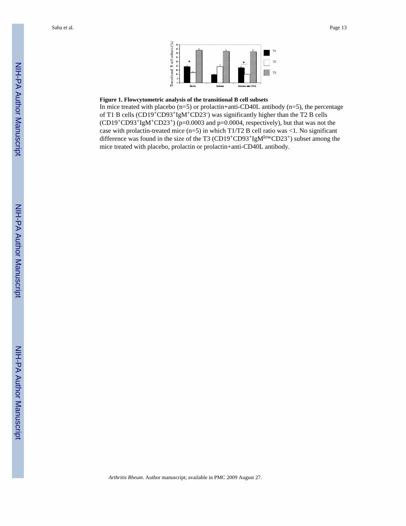
Figure 1. Flowcytometric analysis of the transitional B cell subsetsIn mice treated with placebo (n=5) or prolactin+anti-CD40L antibody (n=5), the percentageof T1 B cells (CD19+CD93+IgM+CD23-) was significantly higher than the T2 B cells(CD19+CD93+IgM+CD23+) (p=0.0003 and p=0.0004, respectively), but that was not thecase with prolactin-treated mice (n=5) in which T1/T2 B cell ratio was <1. No significantdifference was found in the size of the T3 (CD19+CD93+IgMlowCD23+) subset among themice treated with placebo, prolactin or prolactin+anti-CD40L antibody.
Saha et al. Page 13
Arthritis Rheum. Author manuscript; available in PMC 2009 August 27.
NIH
-PA Author Manuscript
NIH
-PA Author Manuscript
NIH
-PA Author Manuscript
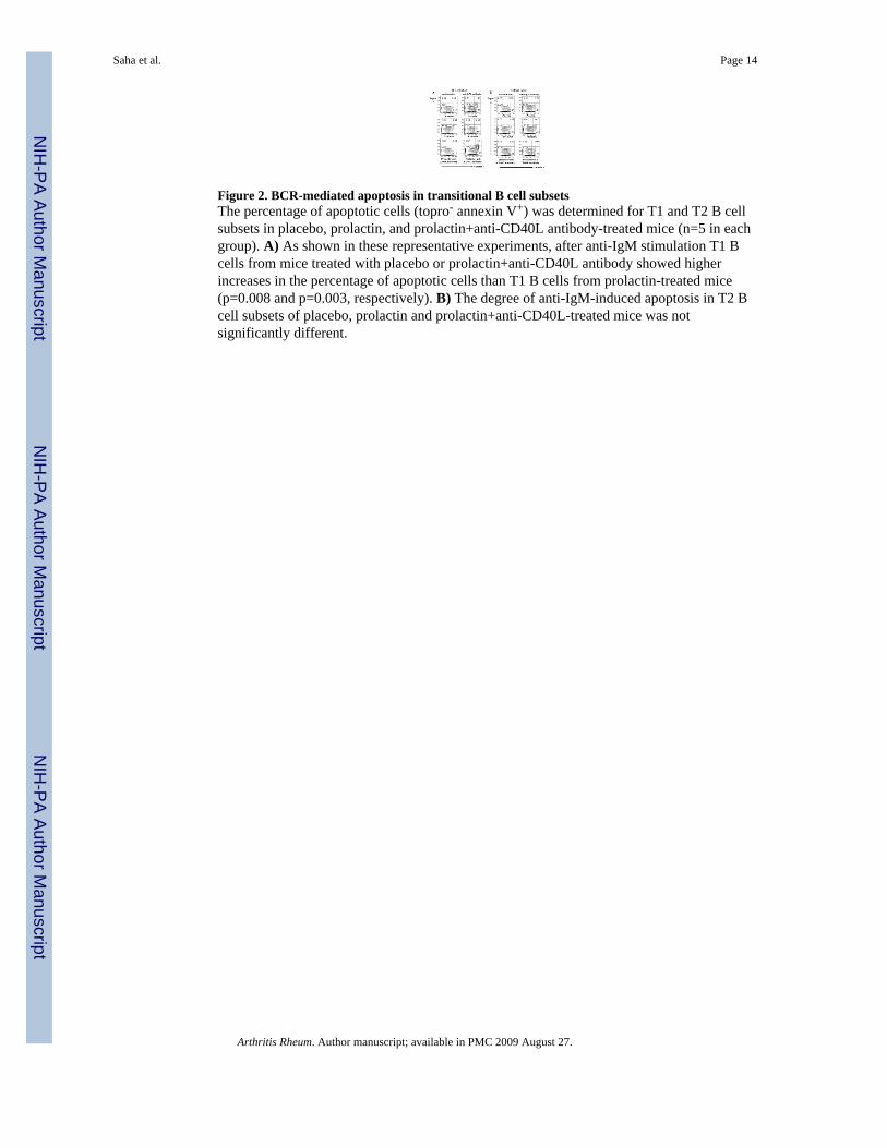
Figure 2. BCR-mediated apoptosis in transitional B cell subsetsThe percentage of apoptotic cells (topro- annexin V+) was determined for T1 and T2 B cellsubsets in placebo, prolactin, and prolactin+anti-CD40L antibody-treated mice (n=5 in eachgroup). A) As shown in these representative experiments, after anti-IgM stimulation T1 Bcells from mice treated with placebo or prolactin+anti-CD40L antibody showed higherincreases in the percentage of apoptotic cells than T1 B cells from prolactin-treated mice(p=0.008 and p=0.003, respectively). B) The degree of anti-IgM-induced apoptosis in T2 Bcell subsets of placebo, prolactin and prolactin+anti-CD40L-treated mice was notsignificantly different.
Saha et al. Page 14
Arthritis Rheum. Author manuscript; available in PMC 2009 August 27.
NIH
-PA Author Manuscript
NIH
-PA Author Manuscript
NIH
-PA Author Manuscript
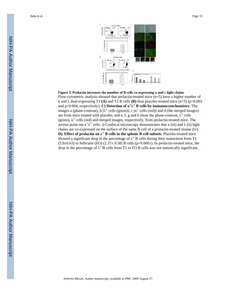
Figure 3. Prolactin increases the number of B cells co-expressing κ and λ light chainsFlow-cytometric analysis showed that prolactin-treated mice (n=5) have a higher number ofκ and λ dual-expressing T1 (A) and T2 B cells (B) than placebo-treated mice (n=5) (p=0.003and p=0.004, respectively). C) Detection of κ+λ+ B cells by immunocytochemistry. Theimages a (phase-contrast), b (λ+ cells (green)), c (κ+ cells (red)) and d (the merged images)are from mice treated with placebo, and e, f, g and h show the phase-contrast, λ+ cells(green), κ+ cells (red) and merged images, respectively, from prolactin-treated mice. Thearrows point out κ+λ+ cells. i) Confocal microscopy demonstrates that κ (iii) and λ (ii) lightchains are co-expressed on the surface of the same B cell of a prolactin-treated mouse (iv).D). Effect of prolactin on λ+ B cells in the splenic B cell subsets. Placebo-treated miceshowed a significant drop in the percentage of λ+ B cells during their maturation from T1(5.9±0.63) to follicular (FO) (2.37± 0.58) B cells (p=0.0001). In prolactin-treated mice, thedrop in the percentage of λ+ B cells from T1 to FO B cells was not statistically significant..
Saha et al. Page 15
Arthritis Rheum. Author manuscript; available in PMC 2009 August 27.
NIH
-PA Author Manuscript
NIH
-PA Author Manuscript
NIH
-PA Author Manuscript
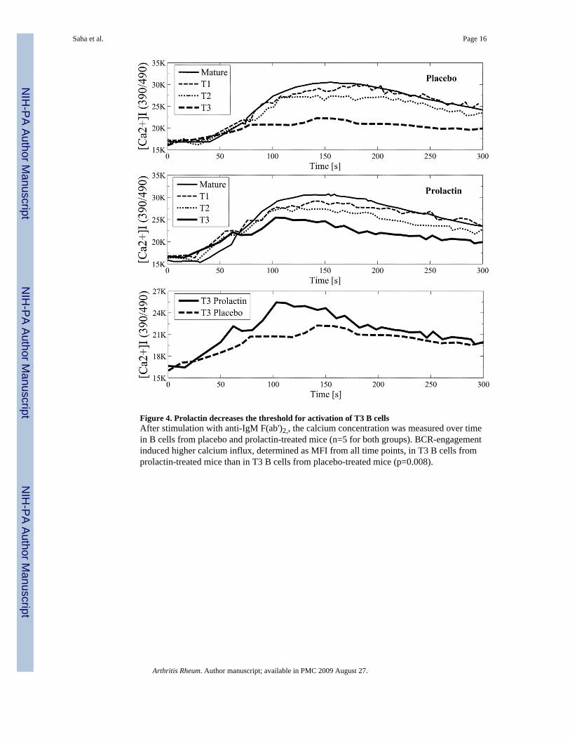
Figure 4. Prolactin decreases the threshold for activation of T3 B cellsAfter stimulation with anti-IgM F(ab')2,, the calcium concentration was measured over timein B cells from placebo and prolactin-treated mice (n=5 for both groups). BCR-engagementinduced higher calcium influx, determined as MFI from all time points, in T3 B cells fromprolactin-treated mice than in T3 B cells from placebo-treated mice (p=0.008).
Saha et al. Page 16
Arthritis Rheum. Author manuscript; available in PMC 2009 August 27.
NIH
-PA Author Manuscript
NIH
-PA Author Manuscript
NIH
-PA Author Manuscript

NIH
-PA Author Manuscript
NIH
-PA Author Manuscript
NIH
-PA Author Manuscript
Saha et al. Page 17
Table 1Expression of CD40 and BAFF-R *
MFI Placebo Prolactin P value
CD40 expression on T1 B cells 527±98 740±78 p=0.05
BAFF-R expression on T2 B cells 419.33±41.96 555.96±17.73 p=0.006
*Values are the mean ± SEM results from 4 mice per group for the CD40 experiment and 5 mice per group for the BAFF-R expression experiment.
Arthritis Rheum. Author manuscript; available in PMC 2009 August 27.