proinflammatory versus anti-inflammatory response in sepsis patients: looking at the cytokines
TRANSCRIPT

MEETING ABSTRACTS Open Access
Sepsis 2014Paris, France. 3-5 December 2014
Published: 3 December 2014
These abstracts are available online at http://ccforum.com/supplements/18/S2
POSTER PRESENTATIONSP1TLR-independent activation of NK cells during systemic inflammation.O Rasid*, J-M CavaillonCytokines and Inflammation, Institut Pasteur, Paris, FranceCritical Care 2014, 18(Suppl 2):P1; doi:10.1186/cc14004
Introduction: During the course of systemic inflammation, most of theimmune cell types get activated to a certain degree as part of, orcontributing to, the cascade of physiopathological events. Whether for somecells, classically phagocytes of the innate immune system, it is clear thatdirect sensing of pathogen-associated molecular patterns leads to activationinitiating systemic inflammation, the picture is not so clear for natural killer(NK) cells. While NK cells have been shown to express toll-like receptors(TLR), the role of these receptors on NKs during systemic inflammation hasnot been directly addressed.Methods: To directly assess the role of TLR expression on NK cells weused an adoptive transfer model in which NKs purified from the spleensof WT, TLR4KO and TLR2/4DKO mice were transferred intravenously toRAG2-/-gc-/- (devoid of T, B and NK cells). Five days after reconstitution themice were challenged intraperitoneally with conventional or TLR-gradelipopolysaccharide (LPS). Immune cell activation and production of IFNgby NK cells was determined after 6 hours by FACS analysis.Results: We observed no differences in reconstitution of the recipientmice with NK cells from different backgrounds suggesting no differencein trafficking and survival of the transferred cells. At 6 hours after LPSchallenge, WT, TLR4KO or TLR2/4DKO NK cells recovered from the spleenand lungs of RAG2-/-gc-/- mice showed comparable levels of CD69activation marker expression. Intracellular labeling for IFNg in NK cells alsorevealed no significant differences.Conclusion: Whether there is a role for direct TLR signaling on NK cellsremains the objective of further investigations; however, our data showthat in the course of a systemic inflammatory process, like endotoxinemia,the expression of TLR2 and TLR4 by NK cells makes no difference in termsof their activation and secretion of IFNg
P2Role of 6-hour, 12-hour, and 24-hour lactate clearance in mortality ofsevere sepsis and septic shock patients.V Herwanto1*, KC Lie2, S Suwarto2, CM Rumende31Department of Internal Medicine, University of Indonesia, Jakarta, Indonesia;2Division of Tropical Medicine and Infectious Diseases, Department ofInternal Medicine, University of Indonesia, Jakarta, Indonesia; 3Division ofRespirology and Critical Care, Department of Internal Medicine, University ofIndonesia, Jakarta, IndonesiaCritical Care 2014, 18(Suppl 2):P2; doi:10.1186/cc14005
Introduction: Lactate is one of biomarkers used for risk stratification,resuscitation target, and death prediction in sepsis [1,2]. Interpretation oflactate clearance was proven more superior than single measurement to
evaluate resuscitation adequacy and to determine prognosis [3,4]. This study
aimed to find out whether mean differences of 6-hour, 12-hour, and 24-hour
lactate clearance were observed between nonsurvivors and survivors of
acute phase mortality in severe sepsis and septic shock patients.Methods: The study design was prospective cohort. Subjects were
collected by consecutive sampling from the emergency department,
hospital ward, and ICU at Cipto Mangunkusumo Hospital, Jakarta. Lactate
levels were measured at 6, 12, and 24 hours, and subjects were
subsequently followed to evaluate 3-day mortality. To determine their
association with mortality, we used mean difference analysis of those
three lactate clearance periods between nonsurvivors and survivors. In
addition, to determine the cutoff value, we used receiver operator curve
analysis.Results: Eighty-one subjects were included in this study. Eighty of
81 were followed until 12 hours, and 72 out of 80 were followed until
24 hours. Twenty-five subjects (31%) did not survive within 3 days of
hospitalization. Only 24-hour lactate clearance had significant median
difference (-17.0% in nonsurvivor vs. 15.2% in survivor group; P = 0.034).
The best cutoff value for 24-hour lactate clearance was -6.0% (AUC 0.744,
sensitivity 62.5% and specificity 87.5%, positive predictive value 58.8%
and negative predictive value 89.1%, relative risk 5.39). From multivariate
analysis, 24-hour lactate clearance was proven to be an independent
predictor of mortality.Conclusion: Median of 24-hour lactate clearance was significantly lower
in nonsurvivors of severe sepsis and septic shock patients. Its cutoff value
was -6.0%.Acknowledgements: Gratitude to Dr Imam Subekti, head of Internal
Medicine Department, Faculty of Medicine University of Indonesia, and
Dr Aida Lydia, head of Internal Medicine study program, Faculty of
Medicine University of Indonesia for their guidance and useful critiques of
this research work. Thanks also to Dr Kuntjoro Harimurti, Dr Esthika
Dewiasty, and Ms Utami for their advice and assistance in doing
methodology and statistic of this studyReferences1. Okorie ON, Dellinger P: Lactate: biomarker and potential therapeutic
target. Crit Care Clin 2011, 27:299-326.2. Schuetz P, Haubitz S, Mueller B: Do sepsis biomarkers in the emergency
room allow transition from bundled sepsis care to personalized patient
care? Curr Opin Crit Care 2012, 18:341-349.3. Nguyen HB, Loomba M, Yang JJ, Jacobsen G, Shah K, Otero RM: Early
lactate clearance is associated with improved outcome in severe sepis
and septic shock. Crit Care Med 2004, 32:1637-1642.4. Nichol A, Bailey M, Egi M, Pettila V, French C, Stachowski E: Dynamic
lactate indices as predictors of outcome in critically ill patients. Crit Care
2011, 15:R242.
Critical Care 2014, Volume 18 Suppl 2http://ccforum.com/supplements/18/S2
© 2014 various authors, licensee BioMed Central Ltd. All articles published in this supplement are distributed under the terms of theCreative Commons Attribution License (http://creativecommons.org/licenses/by/4.0), which permits unrestricted use, distribution, andreproduction in any medium, provided the original work is properly cited.

P3High frequency of myeloid-derived suppressor cells in sepsis patients,with the granulocytic subtype dominating in Gram-positive cases.H Janols1,2*, C Bergenfelz2, R Allaoui2, A-M Larsson3, L Rydén4, S Björnsson5,S Janciauskiene6, M Wullt1, A Bredberg7, K Leandersson21Department of Infectious Diseases, Skane University Hospital, LundUniversity, Malmo, Sweden; 2Center for Molecular Pathology, SkaneUniversity Hospital, Lund University, Malmo, Sweden; 3Department ofLaboratory Medicine, TCR, MV, Lund University, Lund, Sweden; 4Departmentof Surgery, Lund University Hospital, SUS, Lund, Sweden; 5CytometryLaboratory and Department of Laboratory Medicine, Skane UniversityHospital, Lund University, Malmo, Sweden; 6Department of Pulmonology,Hannover Medical School, Hannover, Germany; 7Department of MedicalMicrobiology, Skane University Hospital, Lund University, Malmo, SwedenCritical Care 2014, 18(Suppl 2):P3; doi:10.1186/cc14006
Introduction: Myeloid-derived suppressor cells (MDSCs) constitute aheterogeneous population of immature myeloid cells that potentlysuppress immune responses. They were originally identified in cancerpatients and have since been reported to occur also in chronicinflammation, autoimmunity and even bacterial infections. Human MDSCsare commonly divided into monocytic (Mo-MDSCs) and granulocytic(PMN-MDSCs) subtypes. To what extent the bona fide cancer MDSCs arerepresentative of the proposed MDSCs found in other diseases is not wellknown. PMN-MDSCs have previously been found to be enriched amonglow-density granulocytes (LDGs) in density gradient centrifuged blood.Methods: In this study we analyzed potential MDSCs in sepsis patientswith different causative microorganisms, using total peripheral blood ascompared to density gradient centrifuged blood.Results: We found a high frequency of typical CD14+HLADRlow Mo-MDSCs in all sepsis patients, whereas the typical PMN-MDSCs as well as aprominent CD14low PMN-MDSC-like population appeared preferentially inGram-positive cases (Figures 1 to 3). The CD14low PMN-MDSC variant wasdemonstrated to suppress T-cell proliferation in vitro via a ROS-dependent
mechanism, to display an increased IL-10:TNFa ratio, and to present withsigns of immaturity: blast morphology and low cytokine levels (Figures 4and 5).Conclusion: We conclude that a spectrum of cells with MDSC featuresare enriched in sepsis, and that microbial origin of sepsis contributes tothe substantial interindividual patient variation in MDSC pattern.
P4Selective decontamination using antibiotics in ICU patients:counterfactual protection versus contextual hazard toward bacteremiaincidences.JC HurleyMelbourne Medical School, University of Melbourne and Ballarat HealthServices, Ballarat, AustraliaCritical Care 2014, 18(Suppl 2):P4; doi:10.1186/cc14007
Introduction: Among methods for preventing pneumonia and possiblyalso bacteremia in ICU patients, selective digestive decontamination (SDD;topical with or without protocolized parenteral antibiotic) appears mosteffective within randomized concurrent controlled trials (RCCTs) [1].However, whether parenteral antibiotic is required, and whether SDDactually increases pneumonia incidences in SDD RCCTs versus thebroader ICU pneumonia evidence base, remain unresolved [2,3]. Thepurpose of this analysis is to test for counterfactual and contextual effectsof the topical and parenteral SDD components on the bacteremiaincidence versus the broader evidence base related to the patient groupat risk of VAP.Methods: Bacteremia incidence proportion data were extracted fromcomponent (control and intervention) groups from studies investigatingantibiotic (SDD) or nonantibiotic methods of VAP prevention. Both thecounterfactual and the contextual effects of SDD factorized as topical orprotocolized parenteral exposures were estimated using random-effectsmeta-analysis of study and group level data. Studies without anyprevention methods under study constituted the reference category(benchmark groups).Results: As a counterfactual within RCCTs, SDD when given as combinedtopical and parenteral antibiotic appears to halve the bacteremiaincidence (odds ratio (OR) 0.59; 0.48 to 0.73; n = 18 studies). As acontextual however, the mean bacteremia incidence among 27 controlgroups (17.1%; 13.1 to 22.1%) and 12 intervention groups receivingtopical antibiotic alone (16.2%; 9.1 to 27.3%) from SDD RCCTs is doublethat of 36 benchmark groups (8.3; 7.0 to 10.8%) and 19 control groupsfrom studies of nonantibiotic methods (7.7%; 5.2 to 11.1). The upwarddispersion in bacteremia incidence among component groups from SDDRCCTs away from this benchmark is striking with all but two of the 27control groups and all but two of 12 SDD intervention groups that didnot receive PPAP being above this benchmark.Conclusion: The major contextual hazard of SDD toward bacteremiaamong ICU patients is inapparent within individual studies. The apparentprotection in SDD RCCTs is spurious as the SDD counterfactual isconflated by the strong contextual effect with partial mitigation by SDD
Figure 1(abstract P3)
Figure 2(abstract P3)
Critical Care 2014, Volume 18 Suppl 2http://ccforum.com/supplements/18/S2
Page 2 of 53

Figure 3(abstract P3)
Figure 4(abstract P3)
Figure 5(abstract P3)
Critical Care 2014, Volume 18 Suppl 2http://ccforum.com/supplements/18/S2
Page 3 of 53

protocolized parenteral antibiotic. Not only is the safety of SDD within theICU environment unclear, but this SDD contextual effect may conflate theapparent SDD counterfactual effect on the incidence of bacteremia, aswith VAP.References1. Hurley JC: Prophylaxis with enteral antibiotics in ventilated patients:
selective decontamination or selective cross-infection? Antimicrob AgentsChemother 1995, 39:941-947.
2. Hurley JC: Ventilator associated pneumonia prevention methods usingtopical antibiotics: herd protection or herd peril? Chest 2014 in press.
3. Hurley JC: The perfidious effect of topical placebo: Calibration ofStaphylococcus aureus ventilator-associated pneumonia incidencewithin selective digestive decontamination studies versus the broaderevidence base. Antimicrob Agents Chemother 2013, 57:4524-4531.
P5Thalidomide exerts protective immunomodulatory action duringKlebsiella pneumoniae B5055-induced acute lung infection inBALB/c mice.V Kumar*, S ChhibberDepartment of Microbiology, Panjab University, Chandigarh, IndiaCritical Care 2014, 18(Suppl 2):P5; doi:10.1186/cc14008
Introduction: Thalidomide (a-naphthylimidoglutarimide), a psychoactivedrug that readily crosses the blood-brain barrier, has been shown toexhibit anti-inflammatory, anti-angiogenic, immunomodulatory propertiesthrough a mechanism that is not fully established. Keeping theseproperties in mind, we have tried to find out the anti-inflammatory andimmunomodulatory properties of thalidomide in mouse model of acuteinflammation by introducing Klebsiella pneumoniae B5055 in BALB/c micevia the intranasal route.Methods: Acute lung infection (ALI) or pneumonia in BALB/c mice wasinduced via instillation of selected dose (104 CFU/ml) of bacteria (that is,K. pneumoniae B5055) intranasally. Mice were observed for 7 days andlungs were isolated on designated days for studying difference inbacterial load and other proinflammatory mediators using standardbiochemical methods and ELISA.Results: The intranasal instillation of bacteria in this mouse model ofacute pneumonia-induced inflammation led to significant increase inneutrophil infiltration into the lungs. This was further accompanied by anincreased production of proinflammatory cytokines (that is, TNFa andIL-1a) and other mediators of inflammation (that is, malondialdehyde(MDA), myeloperoxidase (MPO) and nitric oxide (NO)) in the lung tissue.The animals, which received thalidomide alone orally or in combinationwith augmentin, 30 minutes prior to bacterial instillation into the lungs
via intranasal route, showed significant (P ≤ 0.05) decrease in neutrophilinflux into the lungs. A significant (P ≤ 0.05) decrease in the productionof proinflammatory cytokines (that is, TNFa and IL-1a) and otherbiochemical mediators of acute inflammation (that is, MDA, MPO, andNO) was also observed in this group. But the augmentin treatment alonedid not decrease these proinflammatory mediators significantly (P ≥0.05)as compared to the control group.Conclusion: We therefore conclude that thalidomide ameliorates lunginflammation induced by K. pneumoniae B5055 without significantly (P ≤ 0.05)decreasing the bacterial load in the lung tissue whereas augmentin takes careof bacterial proliferation. Hence, it can be used as an adjunct therapy alongwith antibiotics as an anti-inflammatory or an immunomodulatory agent incase of acute lung infection or pneumonia.
P6Impact of purulent complications and sepsis on cardiovascular systemin patients with type 2 diabetes.E Shalaeva*, B Babadjanov, U Pulatov, N Dadabayeva, A ShalaevaRepublican Center of Purulent Surgery and Complications of Diabetes,Tashkent Medical Academy, Tashkent, UzbekistanCritical Care 2014, 18(Suppl 2):P6; doi:10.1186/cc14009
Introduction: Purulent complications in patients with type 2 diabetes areusually severe, often complicated by sepsis and require emergencysurgery. Noncardiac surgery is associated with a 7 to 11% complicationrate and mortality of 0.8 to 1.5% [1], up to 42% are cardiac reasons [2].After surgery, 2% of patients suffer major cardiac complications [3], and8% show evidence of significant myocardial injury [2]. The aim of thisstudy was to identify the impact of purulent complications and sepsis oncardiovascular system in patients with type 2 diabetes.Methods: We analyzed 112 consecutive patients (54 men and 58 women)aged 57.2 ± 8.4 years with purulent-necrotic complications (gangrene,phlegmon, and abscess) of type 2 diabetes and sepsis in 2013. We comparedlaboratory and instrumental data (blood tests, ECG, echocardiography andothers), which were previously obtained in the same patients receivinginpatient treatment before sepsis (2011 to 2012).Results: Gangrene of lower extremities in 59 (52.7%) prevailed amongpurulent complications. After the development of sepsis we detected in allpatients significantly increased heart rate, respiratory rate per minute,leukocytosis, anemia, worse glucose metabolism and renal function (Table 1).Congestive heart failure became more severe. This was confirmed bydecrease of left ventricle ejection fraction (55.2 ± 5.1% before sepsis vs.49.3 ± 4.1% after) and increase brain natriuretic peptide (291.4 ± 34.5 ng/mlvs. 395.2 ± 28.1 ng/ml, P < 0.001). Prior sepsis in 66 (58.9%) of patients witharterial hypertension was observed, after in 88 (78.6%). After admission to
Table 1(abstract P6) Hemodynamic parameters and blood tests in patients with purulent complications of type 2diabetes and sepsis
Parameter Before sepsis (n = 112) After sepsis (n = 112) P value
Heart rate (beats/minute) 78.4 ± 15.2 112.5 ± 18.9 < 0.001
Respiratory rate (breaths/minute) 18.0 ± 2.0 29.5 ± 5.5 < 0.001
Systolic BP (mmHg) 155.7 ± 35.4 154.2 ± 58.5 n.s
Diastolic BP (mmHg) 90.4 ± 10.3 91.9 ± 8.6 n.s
Left ventricle ejection fraction (%) 55.2 ± 5.1 49.3 ± 4.1 0.033
Fasting plasma glucose (mmol/l) 8.4 ± 2.5 15.4 ± 4.8 < 0.001
Two-hour plasma glucose (mmol/l) 10.2 ± 2,8 19.9 ± 3.3 < 0.001
HbA1c (%) 8.4 ± 0.5 12.1 ± 0.5 < 0.001
Hemoglobin (g/l) 121.5 ± 12.5 105.4 ± 11.7 0.04
White count (103) 6.7 ± 1.2 14.4 ± 2.1 < 0.001
Fibrinogen (mg%) 411.6 ± 103.6 715.4 ± 215.5 < 0.001
Blood urea (mmol/l) 6.1 ± 2.9 8.8 ± 2.5 0.011
Blood creatinine (mmol/l) 88.4 ± 18.5 105.6 ± 17.3 0.02
Brain natriuretic peptide (ng/ml) 291.4 ± 34.5 395.2 ± 28.1 < 0.001
BP, blood pressure; HbA1c, glycosylated hemoglobin A1c
Critical Care 2014, Volume 18 Suppl 2http://ccforum.com/supplements/18/S2
Page 4 of 53

the centre, patients had no signs of septic shock. In 13 (11.6%) patients, theperioperative period was complicated by acute myocardial infarction, whichwas accompanied by a fall in blood pressure. We detected an increase of thefunctional class of stable angina, congestive heart failure, 4.2 times increasedincidence of unstable angina, 2.6 times ventricular and four timessupraventricular extra systole after septic complications (Table 2).Conclusion: After the development of purulent complications and sepsisin patients with type 2 diabetes, we observed increased incidence ofarterial hypertension, arrhythmias, worsened severity of coronary arterydisease and congestive heart failure. Perioperative risk of acutemyocardial infarction amounted to 11.6%.References1. Haynes AB, Weiser TG, et al: A surgical safety checklist to reduce
morbidity and mortality in a global population. N Engl J Med 2009,
360:491-499.2. Devereaux PJ, Chan MT, et al: Association between post-operative
troponin levels and 30-day mortality among patients undergoingnoncardiac surgery. JAMA 2012, 307:2295-2304.
3. Devereaux PJ, Goldman L, et al: Perioperative cardiac events in patients
undergoing noncardiac surgery: a review of the magnitude of theproblem, the pathophysiology of the events and methods to estimate
and communicate risk. CMAJ 2005, 173:627-634.
P7Severity of sepsis in patients with acute purulent destructivepulmonary disease depending on the presence of type 2 diabetes:impact on the forecast.A Babobekov*, B Babadjanov, S Atakov, E ShalaevaRepublican Center of Purulent Surgery and Complications of Diabetes,Tashkent Medical Academy, Tashkent, UzbekistanCritical Care 2014, 18(Suppl 2):P7; doi:10.1186/cc14010
Introduction: Lung abscesses and gangrene are the most severe clinicalmanifestation and outcome among acute purulent destructive pulmonarydisease (APDPD). Mortality ranges from 10 to 35%, and in the presence ofdiabetes increases up to 30 to 90% [1]. The main reason for this is thegeneralization of infection (sepsis), leading to the development of multipleorgan failure [2,3]. The aim of this study was to identify the severity of sepsisin patients with APDPD depending on the presence of type 2 diabetes, andthe impact on the forecast.Methods: During the period 2012 to 2013, we examined 408 patients aged48.5 ± 12.5 years (258 men/150 women) who underwent surgical treatmentfor APDPD. The patients were divided into two groups: 144 patients withtype 2 diabetes, and controls (n = 246). We carried out computedtomography, ECG, echocardiography, laboratory biochemical testing, andbacteriological analysis of pathologic material and blood samples.Results: Patients with type 2 diabetes had much more complications andcases of severe sepsis and septic shock (Table 1). Bacteriological analysisof the pathologic material showed Gram-positive bacteria in 35 to 45%,anaerobic association in 55 to 65%, pathological fungi in 50 to 60%. Thepatients with type 2 diabetes had much more time from the onset of thefirst symptoms of lung disease prior to admission (12.5 ± 3.5 vs. 7.5 ± 2.5days, P = 0.002), and the duration of inpatient treatment was significantlylonger (13.8 ± 5.5 vs. 7.1 ± 3.4 days, P = 0.001). Only 53 (36.8%) of patientswith type 2 diabetes and 68 (29.5%) without it had bacteriological positiveblood culture. The analysis of the distribution of pathogens in groups ispresented in Figure 1. Patients with diabetes had more Candida spp.
Table 2(abstract P6) Cardiovascular comorbidity inpatients with type 2 diabetes before and after purulent-necrotic complications and sepsis
Parameter Beforesepsis(n = 112)
Aftersepsis(n = 112)
Insulin dependence 42 (37.5) 112 (100)
Normal blood pressure (110 to 139mmHg)
46 (41.1) 11 (9.8)
Arterial hypertension 66 (58.9) 88 (78.6)
First degree (140 to 159 mmHg) 33 (29.5) 21 (8.9)
Second degree (160 to 179 mmHg) 21 (18.6) 43 (38.4)
Third degree (>180 mmHg) 12 (10.7) 24 (21.4)
Arterial hypotension (<90 mmHg) - 13 (11.6)
CAD, stable angina 108 (94.6) 82 (73.2)
FC I 18 (16.1) -
FC II 29 (25.9) 18 (16.1)
FC III 52 (46.4) 45 (40.2)
FC IV 9 (8.0) 19 (17.0)
CAD, unstable angina 4 (3.6) 17 (15.2)
Acute myocardial infarction - 13 (11.6)
Postinfarction cardiosclerosis 7 (6.3) 7 (6.3)
Atrial fibrillation 7 (6.3) 7 (6.3)
Supraventricular arrhythmia 3 (2.7) 12 (10.7)
Ventricular arrhythmia 14 (12.5) 36 (32.1)
Congestive heart failure 112 (100) 112 (100)
FC II (NYHA) 76 (67.8) 26 (23.2)
FC III (NYHA) 36 (32.1) 65 (58)
FC IV (NYHA) - 21 (18.7)
Abscesses of the lower extremity - 22 (19.6)
Phlegmon of the lower extremity - 31 (27.7)
Gangrene of lower extremity - 59 (52.7)
Data presented as n (%). CAD, coronary artery disease; FC, functional class;NYHA, New York Heart Association
Table 1(abstract P7) Clinical symptoms and severity ofsepsis in patients with acute purulent destructivepulmonary disease depending on the presence of type 2diabetes
Data Type 2diabetes(n = 144)
Control(n = 264)
Acute lung abscess 59 (40.9) 122 (46.2)
Necrotizing pneumonia 47 (32.6) 98 (37.1)
Lung gangrene 38 (26.4) 44 (16.7)
Empyema 88 (61.1) 81 (30.7)
Pyopneumothorax 16 (11.1) 9 (3.4)
Mediastinitis 34 (23.6) 16 (6.1)
Body temperature >38°C/<36°C 98 (68.1)/21(14.6)
261 (98.9)/3 (1.1)
Respiratory rate >20/minute 144 (100) 264 (100)
Heart rate >90 beats/minute 138 (95.8) 242 (91.7)
PaCO2 <32 mmHg 144 (100) 264 (100)
Leukocytes >12,000/<4,000 cells/mm3
111 (77.1)/13(9.1)
202 (76.5)/11(4.2)
Renal failure, oliguria 42 (29.2) 34 (12.9)
Increase liver enzymes 34 (23.6) 45 (17.1)
Systolic blood pressure <90 mmHg 33 (22.9) 51 (19.3)
Sepsis 101 (70.1) 223 (84.5)
Severe sepsis 25 (17.4) 29 (11.0)
Septic shock 18 (12.5) 12 (4.5)
Data presented as n (%)
Critical Care 2014, Volume 18 Suppl 2http://ccforum.com/supplements/18/S2
Page 5 of 53

(Figure 1). Figures 2, Figure 3 and Figure 4 present the X-ray dynamics of a42-year-old man with lung abscess. Clinical recovery in patients with type2 diabetes was significantly worthy compared to controls (45 (31.2%) vs.153 (57.9%)), mortality rate 48 (33.3%) versus 39 (14.7%), respectively.Conclusion: In patients with acute purulent destructive pulmonarydisease and type 2 diabetes, severe sepsis and septic shock more oftenprevailed, inpatient mortality rate was 2.27 times higher, compared topatients with normal glucose metabolism.References1. Defraigne JO, et al: Cavernostomy: an old but effective technique in the
treatment of pulmonary abscess. Rev Med Liege 2007, 52:498-501.2. Refaely J, Weissberg D: Gangrene of the lung: treatment in two stages.
Ann Thorac Surg 1997, 64:970-973.3. Rice TW, Ginsberg PJ, Todd TR: Tube drainage of lung abscesses. Ann
Thorac Surg 1987, 44:356-359.
P8Risk factors and incidence of mediastinitis in patients with lungabscess and sepsis.E Shalaeva*, B Babadjanov, B Janabaev, A ShalaevaRepublican Center of Purulent Surgery and Complications of Diabetes,Tashkent Medical Academy, Tashkent, UzbekistanCritical Care 2014, 18(Suppl 2):P8; doi:10.1186/cc14011
Introduction: Mediastinitis is a life-threatening condition, which isaccompanied by high rates of mortality in cases of delayed diagnosis andinadequate treatment. The aim of the study was to identify the riskfactors and incidence of mediastinitis in patients with lung abscess andsepsis.Methods: In 2013, 218 consecutive patients (83 women and 135 men)with lung abscess and sepsis aged 45.8 ± 13.2 years were operated. Theyhad a full range of laboratory and instrumental examinations, includingechocardiography and computed tomography.Results: Aerobic-anaerobic association in sputum was revealed in allpatients with lung abscess and sepsis, Candida spp. in 34 (15.6%). Bloodculture was positive only in 59 (27%) patients, which had not previouslyreceived antibacterial therapy (polymicrobial flora including Staphylococcusand Streptococcus specimen). Empyema was diagnosed in 123 patients(56.4%), 31 (14.2%) of them were complicated by mediastinitis. Themain clinical symptoms of mediastinitis were hyperthermia (100%),dysphagia (83.8%), dyspnea (80.6%), chest pain (61.3%), orthopnea
Figure 1(abstract P7)
Figure 2(abstract P7) Man, 42 years old, with right lung abscess afteradmission to the centre
Figure 3(abstract P7) The same patient on the fourth day afterdrainage of the abscess
Figure 4(abstract P7) The same patient before discharge, 14 daysin dynamics
Critical Care 2014, Volume 18 Suppl 2http://ccforum.com/supplements/18/S2
Page 6 of 53

(61.3%), and tachycardia (87.1%). The computer tomography revealedan increase in mediastinum size with accumulation of fluids and fluid inthe pleural cavities (100%), free gas in the mediastinum (45.1%),enlarged mediastinal lymph nodes (45.1%), and fluid in the pericardiumcavity (48.4%). To analyze the risk factors, we include 31 patients withlung abscess and sepsis complicated by mediastinitis in the first group,and 187 patients without mediastenitis in the second group. Groupswere similar in age (46.1 ± 8.2 years vs. 45.8 ± 13.2 years). A total77.4% of patients with mediastinitis were women who suffered fromtype 2 diabetes (HbA1c = 9.7 ± 1.4%), congestive heart failure andanemia. Significant differences in the groups according to the data oflaboratory and instrumental studies are presented in Table 1.Conclusion: In total, 14.2% of patients presented with lung abscess andsepsis complicated by mediastinitis, more commonly in women with diabetesmellitus, obesity, anemia and reduced ejection fraction of the left ventricle.
P9Impact of KDO in biological activity of Re-LPS.I Prokhorenko*, S Zubova, D Kabanov, S GrachevInstitute of Basic Biological Problems, Pushchino, Moscow Region, RussiaCritical Care 2014, 18(Suppl 2):P9; doi:10.1186/cc14012
Introduction: The minimal biological active structure of endotoxins(lipopolysaccharides (LPS)) is Re-LPS (KDO2-lipid A), which consist of lipidA and two (or three) molecules of 3-deoxy-D-manno-2-octulosonic acid(KDO) [1,2]. Biological activity of endotoxins is defined in general by thenumber and distribution of acyl residues on the lipid A backbone [3].Recently it has been reported that KDO-treated RAW 264.7 cells exhibiteda gene expression pattern similar to that in LPS-treated cells. Theseauthors revealed that free KDO participated in crosstalk between Toll-likereceptors (TLR) and G protein-coupled receptors and so that regulatedactivators and repressors of immune signaling [4]. LPS-dependentTLR4-triggered activation of target cells leads to specific changes in thelevels of surface receptors and induces synthesis of proinflammatorycytokines [5]. However, the dependence of these processes on thestructural composition of LPS is not well understood. To extend ourknowledge in this field, the effects of free KDO as well as KDO ascovalently linked to lipid A constituent of Re-LPS on expression of TLR4,CD11b and CD14 receptors and TNFa synthesis in whole human bloodhave been investigated.Methods: Human blood was incubated with Re-LPS from Escherichia coliJM103 or Salmonella enterica sv Typhimurium SL1181 (100 ng/ml) or withlipids A from E. coli F583 or S. enterica sv Minnesota R595 (80 ng/ml) orwith ammonium salt of KDO (20 ng/ml) at 37°C in 5% CO2-humidifiedatmosphere for 2 or 6 hours to determine receptor expression or TNFarelease, respectively. Receptor expression was monitored by EPICSXL-MCL flow cytometer using Alexa Fluor 488 anti-TLR4 (HTA125),anti-CD11b (ICRF44) and anti-CD14 (HCD14) antibodies. Human TNF-aELISA Kit II was exploited to TNFa determination.Results: Re-LPS E. coli or Re-LPS S. enterica differentially affected receptorexpression in comparison to their respective lipids A. Free KDO in theequimolar concentration as it exists in KDO2-lipid A (Re-LPS) did notinfluence the level of CD14 but downregulated the expression of TLR4 andCD11b (Figure 1). Tenfold increased KDO concentration did not affectfurther the receptor expression. The addition of KDO2 to lipid A E. coli - that
is, applying KDO as covalently linked constituent of Re-LPS - led toupregulation of CD14 and TLR4 but downregulated CD11b expression. Theexpression of TLR4 was most pronounced upregulated by Re-LPS S. entericabut in the case of CD14 and CD11b this Re-LPS had an opposite effect incomparison to E. coli endotoxins (table in Figure 1). Lipid A S. enterica was aless potent TNFa inductor than that from E. coli (Figure 2). This may beexplained by the differences in lipid A composition determining lipid Aaffinity to target receptor(s). LPS E. coli, as had been shown early, causedMyD88-dependent fast NF-�B degradation (rapid TNFa response) whereasLPS S. enterica induced MyD88-independent signaling (delayed TNFaresponse) [5]. In our study, free KDO did not stimulate TNFa release. KDO2as a constituent of Re-LPS S. enterica increased significantly the TNFa-inducing activity of lipid A S. enterica but this effect was not so distinguishedbetween Re-LPS E. coli and lipid A E. coli (Figure 2).Conclusion: Free KDO in the used concentration was inactive inregulation of TLR4, CD11b and CD14 expression and did not induce TNFarelease but its impact in biological activity was detected when KDO wasapplied as constituent of Re-LPS. This may be explained by the effect ofKDO on the spatial conformation of Re-LPS.Acknowledgements: The work was supported by Grant 16.N08.12.1014established by the Russian Ministry of Education and ScienceReferences1. Olsthoorn M, Peterson B, Schlecht S, Haverkamp J, Bock K, Thomas-Oates J,
Holst O: Identification of a novel core type in Salmonellalipopolysaccharide. J Biol Chem 1998, 273:3817-3829.
2. Fregolino E, Fugazza G, Galano E, Gargiulo V, Landini P, Lanzetta R,Lindner B, Pagani L, Parrilli M, Holst O, et al: Complete lipooligosaccharidestructure of the clinical isolate Acinetobacter baumannii, strain SMAL.Eur J Org Chem 2010, 7:1345-1352.
3. Rietschel E, Kirikae T, Schade U, Mamat U, Schmidt G, Loppnow H, Ulmer A,Zahringer U, Seydel U, Di Padova F, et al: Bacterial endotoxin: molecular
Table 1(abstract P8) Risk factors for mediastinitis in patients with lung abscess and sepsis
With mediastinitis (n = 31) Without mediastinitis (n = 187) P value
Gender, male/female, n (%) 7 (22.6)/24 (77.4) 128 (68.5)/59 (31.5) 0.001
Type 2 diabetes mellitus, n (%) 26 (83.9) 42 (22.5) 0.001
Body mass index 32.3 ± 5.3 27.8 ± 6.1 0.031
Hemoglobin (g/l) 87.9 ± 9.2 128.4 ± 18.4 < 0.001
Fibrinogen (mg%) 800 ± 200 533 ± 166 < 0.001
End-diastolic volume of the left ventricle (ml) 139 ± 27 108 ± 28 0.024
End-systolic volume of the left ventricle (ml) 66.1 ± 8.2 46.8 ± 10.4 < 0.001
Left ventricular ejection fraction (%) 52.4 ± 2.2 57.2 ± 3.4 0.002
Figure 1(abstract P9) Expression of TLR4, CD11b and CD14 onmonocytes after incubation of whole blood with Re-LPS, lipid A orKDO. Presented are the results of six independent experiments.Alteration in receptor expression was calculated according to thecontrol level that had been expressed as 100%. *Changes in receptorexpression were calculated as %MnIX [KDO2-lipid A] - %MnIX [lipid A]
Critical Care 2014, Volume 18 Suppl 2http://ccforum.com/supplements/18/S2
Page 7 of 53

relationships of structure to activity and function. FASEB J 1994,8:217-225.
4. Krishnan J, Choi S: Systems biological approaches reveal non-additiveresponses and multiple crosstalk mechanisms between TLR and GPCRsignaling. Genomics Inform 2012, 10:153-166.
5. Zughaier S, Zimmer S, Datta A, Carlson R, Stephens D: Differentialinduction of Toll-like receptor 4-MyD88-dependent and -independentsignaling pathways by endotoxins. Infect Immun 2005, 73:2940-2950.
P10Two novel formulae are superior to procalcitonin for prediction ofsepsis in trauma patients.H-p Liang*, H Jin, Y Xiao, Z LiuState Key Laboratory of Trauma, Burns and Combined Injury, ResearchInstitute of Surgery, The Third Military Medical University, Chongqing,People’s Republic of ChinaCritical Care 2014, 18(Suppl 2):P10; doi:10.1186/cc14013
Introduction: The purpose of this study was to verify the predictive valueof two novel formulae and compare them with that of procalcitonin (PCT)for predicting sepsis in trauma patients.Methods: We performed a retrospective study of trauma patients treated atDaping Hospital in Chongqing, China and Affiliated Hospital of ZunyiMedical College between 2010 and 2013. Patients ≥16 years old, admittedto hospital after injury within 24 hours and with length of hospital stay ≥48hours were included. Predictive ability of two formulae based on LD50 valuesof the Injury Severity Score (ISS) and New Injury Severity Score (NISS) wereverified: ISS/LD50ISS+SIRS score and NISS/LD50NISS+SIRS score, and thenwere compared with the most common used biomarker PCT. LD50 values fordifferent age groups and genders were obtained in our former study. Thestatistical performance of the two formulae and PCT to predict sepsis wasevaluated using receiver operating characteristic (ROC) curve analysis.Results: Two hundred and twenty-one trauma patients’ data were enrolledin the study, including 44 females and 177 males. The average age of thepatients was 44.77 ± 15.01 years. The performance of the ISS/LD50ISS+SIRSscore and the NISS/LD50NISS+SIRS score was equivalent (area under the ROCcurve (AUC) = 0.816 vs. 0.819, P >0.05) and both performed better than PCT(AUC = 0.592, P < 0.05) in predicting posttraumatic sepsis. For the ISS/LD50ISS+SIRS score, the cutoff value was 2.38, with a positive predictivevalue of 78.08%, a negative predictive value of 81.33%, a sensitivity of89.06%, a specificity of 65.59%, a positive likelihood ratio of 2.59, a negativelikelihood ratio of 0.17, a Youden index of 0.5465, an odds ratio of 15.52,and an accuracy of 79.19%. For the NISS/LD50NISS+SIRS score, the cutoffvalue was 2.4677, with a positive predictive value of 79.70%, a negativepredictive value of 75.00%, a sensitivity of 82.81%, a specificity of 70.97%, apositive likelihood ratio of 2.85, a negative likelihood ratio of 0.24, a Youdenindex of 0.5378, an odds ratio of 11.78, and an accuracy of 77.83%.
Conclusion: The two novel formulae ISS/LD50ISS+SIRS score and NISS/LD50NISS+SIRS score performed well and were both better than PCT inpredicting sepsis post trauma. The value of the two formulae can beeasily calculated in real time and can identify the high-risk patientssusceptible to sepsis. This method may become an effective way to guidethe early assessment and treatment in trauma patients.
P11Inhibitory effects of evodiamine on zymosan-induced inflammation:inactivation of NF-�B by inhibiting I�Ba phosphorylation.X Fan*, J-y Zhu, Z Liu, J Yan, Q Ma, H-p LiangState Key Laboratory of Trauma, Burns and Combined Injury, ResearchInstitute of Surgery, The Third Military Medical University, Chongqing,People’s Republic of ChinaCritical Care 2014, 18(Suppl 2):P11; doi:10.1186/cc14014
Introduction: Inflammation is a host defense reaction against pathogenicinfection. In this process, inflammatory cytokines contribute to combatagainst infection, but excess cytokines will lead to tissue damage.Nonsteroidal anti-inflammatory drugs and corticosteroids are commonlyused for regulating these inflammatory mediators and treatment ofinflammatory disorders. But these drugs are not sufficient for clinicalpractice due to their adverse effects, so new anti-inflammatory drugs arestill needed. Evodiamine (EVO), an important alkaloidal componentextracted from the fruit of Evodiae fructus, possesses the property ofanalgesia, antiemesis, and vascular dilatation. Its anti-inflammatory effectand underlying mechanism were investigated using a zymosan-inducedinflammatory model.Methods: In vitro, RAW264.7 cells and primary peritoneal macrophageswere treated with different doses of EVO (25, 50 or 100 μM) for 1 hourprior to incubation with zymosan (100 μg/ml), and the effects of EVO onprotein and mRNA levels of proinflammatory cytokines were determinedby ELISA and qRT-PCR, respectively. In vivo, peritonitis was induced inC57BL/6 mice by zymosan (500 mg/kg) injection, and the effects of EVO(10 mg/kg) on plasma cytokine levels and organ injury were evaluated.Activation of the NF-�B signal pathway was investigated by ELISA-basedTrans-AM transcription factor NF-�B p65 kit, immunocytochemistry andwestern blotting.Results: EVO effectively suppressed the expression of IL-1b, IL-6 as wellas TNFa in both protein and mRNA level in vitro. It can also attenuatezymosan-induced DNA-binding activity of NF-�B, which was achievedthrough the inhibitory effects on the phosphorylation of inhibitory kappaB a (I�Ba) and p65 nuclear translocation, but there was little associationwith mitogen-activated protein kinase activation. In vivo, treatment withEVO could ameliorate inflammatory cell infiltration and vascular ectasiainduced by zymosan in both lung and intestine tissues. EVO canmarkedly decrease the level of TNFa and IL-6 in plasma, and effectivelydownregulate expression of IL-6, TNFa and myeloperoxidase in both lungand intestine. Moreover, cell apoptosis in organs was also attenuated bytreatment of EVO. The underlying mechanism that a decrease in thephosphorylation of I�Ba and the subsequent transcription activity ofNF-�B was also confirmed.Conclusion: Taken together, our data demonstrate that EVO displays anti-inflammatory actions in vitro and in vivo by suppressing the phosphorylationof I�Ba and inactivating NF-�B, which suggests that EVO is a potentialtherapeutic agent against inflammatory disorder.
P12Imaging in severe sepsis and septic shock: is early radiologicalidentification of occult sources of infection needed?.A Creamer*, J KeepEmergency Department, Kings College Hospital, London, UKCritical Care 2014, 18(Suppl 2):P12; doi:10.1186/cc14015
Introduction: The importance of imaging in establishing the focus ofinfection is recognised in current guidelines for the management of severesepsis [1], with decisions regarding timing and modality of imaging left tothe physicians’ clinical judgement. In the emergency department (ED),clinical assessment combined with bedside investigations of chest X-ray
Figure 2(abstract P9) Production of TNFa after incubation of wholeblood with lipid A or Re-LPS
Critical Care 2014, Volume 18 Suppl 2http://ccforum.com/supplements/18/S2
Page 8 of 53
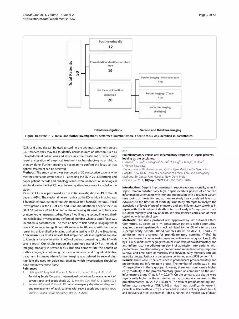
(CXR) and urine dip can be used to confirm the two most common sources[2]. However, they may fail to identify occult sources of infection, such asintraabdominal collections and abscesses, the treatment of which mayrequire alteration of empirical treatment or be refractory to antibiotictherapy alone. Further imaging is necessary to confirm the focus so thatoptimal treatment can be achieved.Methods: The study cohort was composed of 50 consecutive patients whomet the criteria for severe sepsis [1] attending the ED in 2013. Electronic andpaper patient records and radiology results were analysed. All radiologicalstudies done in the first 72 hours following attendance were included in thestudy.Results: CXR was performed as the initial investigation in 49 of the 50patients (98%). The median time from arrival at the ED to initial imaging was1 hour:00 minutes (range 0 hours:04 minutes to 4 hours:25 minutes). Initialinvestigations in the ED of CXR and urine dip identified a septic focus in30 of 50 patients (60%). Fourteen of the remaining 20 went on to have oneor more further imaging studies. Figure 1 outlines the second-line and third-line radiological investigations performed (number where a septic focus wasidentified in parentheses). The median time to first positive imaging was 0hours: 50 minutes (range 0 hours:04 minutes to 40 hours), with the sourceremaining unidentified by imaging and urine testing in 15 of the 50 patients.Conclusion: Our results indicate that simple bedside investigations are ableto identify a focus of infection in 60% of patients presenting to the ED withsevere sepsis. Our results support the continued use of CXR as the initialimaging modality in severe sepsis, but also demonstrate the benefit offurther imaging in confirming the focus of infection and to guide definitivetreatment. Instances where further imaging was delayed by several dayshighlight the need for guidelines detailing which investigations should bedone and in what time frame.References1. Dellinger RP, Levy MM, Rhodes A, Annane D, Gerlach H, Opal SM, et al:
Surviving Sepsis Campaign: international guidelines for management of
severe sepsis and septic shock, 2012. Intensive Care Med 2013, 39:165-228.2. Perman SM, Goyal M, Gaieski DF: Initial emergency department diagnosis
and management of adult patients with severe sepsis and septic shock.
Scand J Trauma Resusc Emergency Med 2012, 20:41.
P13Proinflammatory versus anti-inflammatory response in sepsis patients:looking at the cytokines.D Anand1*, S Ray2, S Bhargava1, S Das1, A Garg2, S Taneja2, D Dhar2,L Mohan Srivastava11Department of Biochemistry and Critical Care Medicine, Sir Ganga RamHospital, New Delhi, India; 2Department of Critical Care and EmergencyMedicine, Sir Ganga Ram Hospital, New Delhi, IndiaCritical Care 2014, 18(Suppl 2):P13; doi:10.1186/cc14016
Introduction: Despite improvements in supportive care, mortality rates insepsis remain substantially high. Sepsis exhibits phases of enhancedinflammation, alternating with immune suppression with a resultant varianttime point of mortality; yet no human study has correlated levels ofcytokines to the timeline of mortality. Our study attempts to analyze theassociation of levels of proinflammatory and anti-inflammatory cytokines insepsis with the timeline of death in terms of early (<5 days) versus late(>5 days) mortality, and day of death. We also assessed correlation of thesecytokines with length of stay.Methods: The study protocol was approved by Institutional EthicsCommittee. Subjects were 74 consecutive patients with community-acquired severe sepsis/septic shock admitted to the ICU of a tertiary caresuperspeciality hospital. Blood samples drawn on days 1, 3 and 7 ofadmission were analysed for proinflammatory cytokine (TNFa) bychemiluminescent immunometric assay and anti-inflammatory cytokine (IL-10)by ELISA. Subjects were segregated on basis of: ratio of proinflammatory andanti-inflammatory mediators on day 1 of admission into patients withpredominant proinflammatory or predominant anti-inflammatory response.Survival and time point of mortality into survivor, early mortality and latemortality groups. Statistical analyses were performed using SPSS version 17.Results: There were 37 patients each in predominant proinflammatory andpredominant anti-inflammatory groups. The number of deaths was 11 and17 respectively in these groups. However, there was significantly higherearly mortality in the proinflammatory group as compared to the anti-inflammatory group (7 vs. 1, P = 0.0247). On the contrary, late deaths weresignificantly higher in the anti-inflammatory group as compared to theproinflammatory (16 vs. 4 P = 0.0017). The ratio of proinflammatory/anti-inflammatory cytokines (TNF/IL-10) on day 1 was significantly lower inpatients of late death (n = 20) as compared to patients of early death (n = 8)and survivors (n = 46) as shown in Table 1. Further, the median day of death
Figure 1(abstract P12) Initial and further investigations performed (number where a septic focus was identified in parentheses)
Critical Care 2014, Volume 18 Suppl 2http://ccforum.com/supplements/18/S2
Page 9 of 53

was significantly delayed in patients in the anti-inflammatory as comparedto the proinflammatory group (20 vs. 5, P < 0.001). Length of hospital stayamongst survivors was significantly longer in the anti-inflammatory ascompared to the proinflammatory group (23 vs. 10 P < 0.001).Conclusion: Our preliminary data suggest that in sepsis, the ratio ofproinflammatory/anti-inflammatory cytokines on day 1 is significantlyassociated with time point of mortality; hence, this ratio can be used toparticularize management. Further studies are in progress to substantiatethe role of proinflammatory and anti-inflammatory cytokines in thissubset of patients. Moreover, since predominant anti-inflammatoryresponse was associated with later death, role of immunomodulators insepsis needs to be explored.
P14Understanding heterogeneity in the host response to Staphylococcusaureus infection for prognostic biomarker discovery.JB Dinoso1*, J Gutierrez2, DF Choy1, S Kummerfeld3, A Baruch2, HF Chambers4,CM Rosenberger11ITGR Diagnostic Discovery, Genentech Inc., South San Francisco, CA, USA;2Pharmacodynamic Biomarkers, Genentech Inc., South San Francisco, CA, USA;3Bioinformatics, Genentech Inc., South San Francisco, CA, USA; 4Division ofInfectious Disease, University of California San Francisco, San Francisco, CA, USACritical Care 2014, 18(Suppl 2):P14; doi:10.1186/cc14017
Introduction: Invasive Staphylococcus aureus infections remain an unmetmedical need with the issues of drug resistance (MRSA) and mortality.Understanding clinical trial data in the development of antibiotics toS. aureus is complicated by heterogeneous clinical outcomes (that is,length of hospitalization, mortality, treatment response), which makesinterpretation of drug efficacy challenging. Identification of prognosticbiomarkers of different biological processes that associate with clinicaloutcomes would aid in clinical development of novel therapies forS. aureus infections.Methods: In an effort to discover biomarkers that differentiate patientpopulations based on clinical outcomes following S. aureus infection, weretrospectively analyzed published gene expression datasets of S. aureusinfection and sepsis.Results: We identified a leukocyte gene expression signature that positivelycorrelated with S. aureus disease severity [1]. This severity signature wasenriched for genes associated with neutrophils but was not solely explainedby increased percentage of neutrophils. This set of genes was also associatedwith severity in sepsis, with higher expression in patients with septic shockcompared with sepsis [2] and in nonsurvivors compared with survivors ofseptic shock [3]. Our in vitro studies revealed that the severity signature mayreflect an increase in circulating immature neutrophils or band cells whichhas been previously reported in the context of bacterial stimuli and sepsis[4,5]. This line of evidence is consistent with a recent report by Guerin andcolleagues that demonstrated that quantification of immature neutrophils byflow cytometry was prognostic for sepsis mortality [6].Conclusion: To extend the insight gained from our retrospective analysisand in vitro studies, we are conducting a longitudinal, non-interventionalclinical study of patients with S. aureus bacteremia. The goal of the study isto associate molecular metrics (gene expression, plasma protein levels,immune cell subsets) with clinical outcomes (length of hospitalization,mortality, treatment response, readmission for recalcitrant infection). Ourpreliminary data show an association between grade of sepsis or infectionlocalization and increased immature neutrophils as well as monocytesubsets that can promote inflammation or immune exhaustion. Ongoingexperiments are designed to understand the impact of these cellularphenotypes on disease progression and to identify robust protein or RNAbiomarkers that are prognostic for clinical outcomes.
References1. Banchereau R, Jordan-Villegas A: Host immune transcriptional profiles
reflect the variability in clinical disease manifestations in patients with
Staphylococcus aureus infections. PLoS ONE 2012, 7:e34390.2. Wong HR, Cvijanovich N: Genomic expression profiling across the
pediatric systemic inflammatory response syndrome, sepsis, and septic
shock spectrum. Crit Care Med 2009, 37:1558-1566.3. Wynn JL, Cvijanovich NZ: The influence of developmental age on the
early transcriptomic response of children with septic shock. Mol Med
2011, 17:1146-1156.4. Taneja R, Sharma AP: Immature circulating neutrophils in sepsis have
impaired phagocytosis and calcium signaling. Shock 2008, 30:618-622.5. Pillay J, Ramakers P: Functional heterogeneity and differential priming of
circulating neutrophils in human experimental endotoxemia. J Leukoc
Biol 2010, 88:211-220.6. Guerin E, Orabona M: Circulating immature granulocytes with T-cell killing
functions predict sepsis deterioration. Crit Care Med 2014, 42:2007-2018.
P15miR-20a-5p mediates hypoxia-induced autophagy by targetingATG16L1 in acute kidney injury.I-K Wang1,2*, C-Y Li2,31Graduate Institute of Clinical Medical Science, China Medical University,
Taichung, Taiwan; 2Division of Nephrology, China Medical University Hospital,
Taichung, Taiwan; 3Department of Anesthesiology, China Medical University
Hospital, Taichung, TaiwanCritical Care 2014, 18(Suppl 2):P15; doi:10.1186/cc14018
Introduction: Autophagy could be induced under stress conditions,including starvation, infection, and hypoxia. The microRNA (miRNA)network may be critical in the regulation of autophagy. Upregulation ofautophagy may be a protective response for cell survival in ischemickidney injury. The aim of this study was to evaluate whether miRNAregulates autophagy in ischemic kidney injury and renal proximal tubularcells under hypoxic conditions.Methods: Ischemic kidney injury was performed by clamping bilateralrenal pedicles for 60 minutes in male mice. Human kidney proximaltubular (HK2) cells are incubated in a hypoxic chamber with 0.3% O2.Bioinformatics analyses were used to select the candidate miRNA. Gain-of-function and loss-of-function methods were employed to evaluate theeffects of miRNA on autophagy. Chromatin immunoprecipitation analysesand promoter luciferase reporter assays were used to evaluate theinteraction of transcriptional factors with miRNA.Results: Increase of LC3 and ATG16L1, autophagy-related proteins, and downexpression of miR-20a-5p were detected in kidneys after ischemic injury andin HK2 cells under hypoxic conditions. The 3’-untranslated region luciferasereporter assays indicated that miR-20a-5p targeted ATG16L1 messenger RNA.Overexpression of miR-20a-5p reduced the expression of LC3-II and ATG16L1in HK2 cells under hypoxic conditions, whereas antagomiR-20a reversed theinhibition. Using RNAi against hypoxia-inducible factor-1a (HIF-1a) in HK2cells, we confirmed the inhibitory binding of HIF-1a to miR-20a-5p.Conclusion: The signaling axis of HIF-1a, miR-20a-5p, and ATG16L1 inautophagic process might be a critical adapting mechanism for ischemickidney injury.
Table 1(abstract P13) TNF/IL-10 ratio in study groups at different time points
Early death (≤ 5 days) (n = 8) Late death (>5 days) (n = 20) Survivors (n = 46) P value
Day 1 1.81 (1.00 to 3.44) 0.50 (0.31 to 0.90) 1.22 (0.43 to 3.91) 0.020*
Day 3 1.12 (0.50 to 3.91) 1.01(0.20 to 2.21) 2.5 (0.90 to 3.91) 0.158
Day 7 - 1.25 (0.59 to 2.38) 1.79 (0.75 to 3.90) 0.256
Data presented as median (IQR). Kruskal-Wallis test was performed for significance between three groups. *P < 0.05 considered significant
Critical Care 2014, Volume 18 Suppl 2http://ccforum.com/supplements/18/S2
Page 10 of 53

P16Evaluating the sensitivity and specificity of a severe sepsis tool utilizedat a community hospital in Miami, FL.J Hirigoyen4 Tower Medical-Surgical Unit, Baptist Hospital of Miami, Miami, FL, USACritical Care 2014, 18(Suppl 2):P16; doi:10.1186/cc14019
Introduction: Since the initial development of the Surviving SepsisCampaign guidelines outlining the management of severe sepsis, therehas been an absolute discount on the management of septic patients inmedical surgical units. In efforts to improve severe sepsis, a communityhospital in Miami adopted a severe sepsis screening tool (SSST) to rapidlyidentify severe septic patients in medical surgical units. A pilot study wasconducted to evaluate the sensitivity and specificity of the SSST.Methods: A descriptive retrospective study. There were two phases. Phase 1evaluated the percentage of patients with sepsis criteria utilizing the SSST.Patients admitted to 4 Tower during 2013 presenting with a diagnosis ofsepsis syndrome and admitted to 4 Tower presenting without sepsissyndrome were reviewed. Phase 2 evaluated the sensitivity and specificity ofSSST from August 2013 to January 2014. Total number of patients admittedto 4 Tower: of those patients, total number with discharge diagnosis ofsepsis, total number who screened positive >1 time during hospital stay,and total number who screened negative during hospital stay; there werefive missing cases. The receiver operating curve (Figure 1) and therespective area under the curve were calculated. Utilizing a 2 × 2 design, thesensitivity and specificity of the tool was calculated.Results: Phase 1: a total of 220 patients records were reviewed, afrequency distribution was utilized (Table 1), demonstrating that the SSSTidentified those patients with sepsis criteria 76 % (n = 167) of the time.Phase 2: a total of 1,555 patients were included during phase 2. A 2 × 2design (Table 2) was utilized: 78 patients were identified as true positiveand 1,233 patients were identified as true negative. The study yielded asensitivity of 41.49% and a specificity of 90.53%. The positive predictivevalue of the tool was estimated at 37.68%, negative predictive value wasestimated at 91.81% and disease prevalence was 12.13%. Area under thereceiver operating curve (Table 3) was 0.66.Conclusion: A two-phase retrospective chart review study demonstratedthat the SSST utilized at a community hospital in Miami had a sensitivityvalue of 41.49% and a specificity value of 90.53% when evaluating medical
surgical patients. These results indicate the tool is accurate in detectingpatients that are not septic; however, it is not reliable in identifying patientswho are truly septic. Further studies need to be conducted to validate thesensitivity and specificity of the SSST; changes will be recommended in aneffort to improve sensitivity.Acknowledgements: Thanks to Eve Butler and Andrea Prentiss fromBaptist Research Department, 4 Tower team. Special thanks to MelanieSantos, Luz Lorenzo, Disney Granado, Katiuska Diaz, Sandra Benitez,Magdely Perez, Viviana Castillo and Ofelia Cabrera.
P17Surviving Sepsis Campaign 2012 3-hour bundle in the emergencydepartment: compliance and impact of pathway of care before andafter implementation.J Masse1*, A Filali2, O Nigeon1, N Van Grunderbeeck2, P Gosselin3, L Tronchon2,J Mallat2, D Thevenin21Emergency Department, Hospital Center of Lens, France; 2Intensive CareUnit, Hospital Center of Lens, France; 3Emergency Department, LilleUniversity Hospital, Lille, FranceCritical Care 2014, 18(Suppl 2):P17; doi:10.1186/cc14020
Introduction: Compliance with the Surviving Sepsis Campaign 2012 (SSC)bundle in the emergency department (ED) is a key point to improveoutcome of severe sepsis and septic shock [1,2]. Before and aftereducation of ED staff, we registered compliance and timing of lactatedosing, blood culture sampling, empiric antibiotic therapy (ATB) and fluidresuscitation, the 3-hour (H3) bundle. Survival and compliance accordingto the initial pathway of care were also studied.Methods: A monocentric study before and after education of ED staffabout SSC bundles (courses, posters, pocket guides). We looked atcompliance of the H3 bundle items in a retrospective and a prospectivecohort, timing of realisation, day 28 survival, overall severity (SAPS2, SOFAand RISCC scores), impact of prehospital medical management, and initialpathway of care. Statistical analysis was performed with Fisher exact testand Mann-Whitney test. Multivariate analysis of factors associated withsurvival was made through logistic regression.Results: Eighty-nine patients were included in the prospective cohort, 65in the retrospective cohort. Patterns of patients in the retrospective andprospective cohort were respectively: sex ratio M/F 29/39 and 39/47 (NS);
Figure 1(abstract P16) ROC curve
Critical Care 2014, Volume 18 Suppl 2http://ccforum.com/supplements/18/S2
Page 11 of 53

median age 63.29/61.38 (NS); SAPS2 44/40 (P = 0.019); SOFA 4/3 (P =0.005); RISCC 9/12.5 (P = 0.002). Compliance with the H3 bundle itemsbefore and after intervention was: lactate 72.1% vs. 81.4% (NS); bloodcultures 61.8% vs. 67.4% (NS); ATB 29.3% vs. 52.3% (P = 0.005); fluids52.9% vs. 59.3% (NS). Median delays before and after implementationwere (in minutes): lactate 56 vs. 40 (P = 0.024); blood cultures 68 vs. 75(NS); ATB 229 vs. 160 (NS); fluids 100 vs. 74 (NS). Survival was superiorafter intervention 67.6 vs. 81.4% (P = 0.049), and associated with a lowSAPS2 score in multivariate analysis. Admission through a prehospitalmedical team was associated with a stronger H3 ATB compliance beforeintervention (P = 0.032). Within the ED, initial orientation to the acutecare unit was associated with a better H3 ATB compliance compared tostandard care before and after staff education (P = 0.001; P = 0.003), andwith better overall compliance (P = 0.004; P = 0.026).Conclusion: Compliance with the SSC H3 bundle was increased but stillneeds to be improved. There is an impact of the initial pathway of careon compliance of the bundle, and on timing of ATB injection. Differencesin healthworker/patient ratio in the units of care could explain thesedisparities [3]. Improvement could be obtained through optimizing earlyscreening, correct initial guidance, or with dedicated teams.Acknowledgements: The authors would like to thank the members ofthe Unit of Clinical Research of the Hospital Center of Lens for their greathelp in data recoveryReferences1. Dellinger RP, Levy MM, Rhodes A, Annane D, Gerlach H, Opal SM,
Sevransky JE, Sprung CL, Douglas IS, Jaeschke R, Osborn TM, Nunnally ME,Townsend SR, Reinhart K, Kleinpell RM, Angus DC, Deutschman CS,Machado FR, Rubenfeld GD, Webb S, Beale RJ, Vincent JL, Moreno R,Surviving Sepsis Campaign Guidelines Committee including The PediatricSubgroup: Surviving Sepsis Campaign: international guidelines formanagement of severe sepsis and septic shock, 2012. Intensive Care Med2013, 39:165-228.
2. van Zanten AR, Brinkman S, Arbous MS, Abu-Hanna A, Levy MM, deKeizer NF, Netherlands Patient Safety Agency Sepsis Expert Group:Guideline bundles adherence and mortality in severe sepsis and septicshock. Crit Care Med 2014, 42:1890-1898.
3. Shin TG, Jo IJ, Choi DJ, Kang MJ, Jeon K, Suh GY, Sim MS, Lim SY, Song KJ,Jeong YK: The adverse effect of emergency department crowding on
compliance with the resuscitation bundle in the management of severesepsis and septic shock. Crit Care 2013, 17:R224.
P18Benefit of achieving lactate clearance versus central venous oxygensaturation target as microcirculation end point resuscitation in severesepsis and septic shock.R Sinto*, S Suwarto, KC Lie, D Widodo, HT PohanDivision of Tropical and Infectious Disease, Department of Internal Medicine,Faculty of Medicine Universitas Indonesia, Jakarta, IndonesiaCritical Care 2014, 18(Suppl 2):P18; doi:10.1186/cc14021
Introduction: In severe sepsis and septic shock patients, lactate clearance>10% and central venous oxygen saturation (ScvO2) >70% are acceptedparameters of tissue oxygenation adequacy. There is controversy of whichparameters better associate with early mortality, and thus should beimplemented as the microcirculation end point resuscitation [1-3]. Thisstudy was aimed to address the association of achieving either one ortwo targets of microcirculatory end point resuscitation and early mortalityin severe sepsis and septic shock patients.Methods: A retrospective cohort study was conducted in severe sepsisand septic shock patients (aged 18 years and older) hospitalized in theICU, Cipto Mangunkusumo Hospital, Indonesia. Patients’ early outcomeswere observed during first 120 hours of hospitalization. Cox’s regressionanalysis was used to analyse risk of early mortality in subject groupsachieving lactate clearance target only, ScvO2 target only, both targets,and not achieving any target in 6 hours after onset of resuscitation.Results: Subjects consisted of 268 patients. Early mortality developed in70 subjects. Fifty-four subjects achieved lactate clearance target only, 16achieved ScvO2 target only, 138 achieved both targets, 60 did notachieve any target. Subjects who achieved both targets had a significantlowest early mortality risk (P = 0.104 compared with subjects achievedlactate clearance target only and P = 0.000 compared with remainingsubject groups) (Figure 1). In subgroup analysis of subjects who achievedlactate clearance or ScvO2 target only, failure to achieve lactate clearancetarget associated with higher early mortality risk (hazard ratio 5.92; 95%CI 2.18 to 16.01).Conclusion: Achieving both lactate clearance and ScvO2 targets in6 hours after onset of resuscitation associates with lowest early mortalityrisk in severe sepsis and septic shock patients. Patients who achievelactate clearance target only have a significant lower early mortality riskcompared with those who achieve ScvO2 target only.References1. Jones AE, Shapiro NI, Trzeciak S, Arnold RC, Claremont HA, Kline JA: Lactate
clearance vs. central venous oxygen saturation as goals of early sepsistherapy: a randomized clinical trial. JAMA 2010, 303:739-746.
2. Jones AE: Point: should lactate clearance be substituted for centralvenous oxygen saturation as goals of early severe sepsis and septicshock therapy? Yes. Chest 2011, 140:1406-1408.
3. Rivers EP, Elkin R, Cannon CM: Counterpoint: should lactate clearance besubstituted for central venous oxygen saturation as goals of early severesepsis and septic shock therapy? No. Chest 2011, 140:1408-1413.
Table 1(abstract P16) Frequency distribution
Statistics Diagnosis on admission Sepsis tool identifies sepsis
Valid 220 220
Missing 0 0
Frequency table Frequency Percent Valid percentage Cumulative percentage
Diagnosis on admission
Valid 220 100.0 100.0 100.0
Sepsis tool identifies sepsis
Not valid 53 24.1 24.1 24.1
Valid 167 75.9 75.9 100.0
Total 220 100.0 100.0
Table 2(abstract P16) The 2 × 2 design
Sepsis present Sepsis absent
Positive 78 true positive 129 false positive
Negative 110 false negative 1,233 true negative
Table 3(abstract P16) Area under the curve
Test result variable(s):screen
Area
0.660
Critical Care 2014, Volume 18 Suppl 2http://ccforum.com/supplements/18/S2
Page 12 of 53

P19The PRESEP score: an early warning scoring system to identify septicpatients in the emergency care setting.O Bayer1*, CS Hartog1,2, D Schwarzkopf2, C Stumme1, A Stacke1, F Bloos1,2,C Hohenstein3, B Kabisch1, C Weinmann1, J Winning1, Y Sakr1, JK Reinhart1,21Department of Anaesthesiology and Intensive Care Medicine, JenaUniversity Hospital, Jena, Germany; 2Center for Sepsis Control & Care, JenaUniversity Hospital, Jena, Germany; 3Emergency Department, Jena UniversityHospital, Jena, GermanyCritical Care 2014, 18(Suppl 2):P19; doi:10.1186/cc14022
Introduction: Many patients present with sepsis through emergencyservices [1]. Their outcome could be improved if sepsis could be detectedalready in the prehospital setting. This study aims to develop andevaluate a score to detect prehospital early sepsis.Methods: A retrospective study of 375 patients admitted to JenaUniversity Hospital emergency department (ED) through emergencymedical services (EMS). Sepsis was present in the ED in 93 (24.8%)patients, of which 60 (16.0%) had severe sepsis and 12 (3.2%) had septicshock. The following predictors for sepsis based on consensus criteriawere extracted from the EMS protocol: body temperature (T), heart rate(HR), respiratory rate (RR), oxygen saturation (SaO2), Glasgow Coma Scale,blood glucose and systolic blood pressure (BP). Sepsis predictors weredetermined based on inspection of loess graphs. Backward modelselection was performed to select risk factors for the final model. ThePRESEP score was calculated as the sum of simplified regression weights.Its predictive validity was compared to the modified Early Warning Score(MEWS) [2], the Robson screening tool [3] and the BAS 90-30-90 [4].Results: Backward model selection identified T, HR, RR, SaO2 and BP forinclusion in the PRESEP score. Its AUC was 0.93 (CI 0.89 to 0.96). Thecutoff based on maximum Youden’s Index was ≥4 (sensitivity 0.85,specificity 0.86, PPV 0.66, NPV 0.95). The PRESEP score had a larger AUCthan the MEWS (0.93 vs. 0.77, P < 0.001) and surpassed MEWS and BAS90-60-90 concerning sensitivity (0.74, 0.62), specificity (0.75, 0.83), PPV(0.45, 0.51) and NPV (0.91, 0.89). The Robson screening tool had a higher
sensitivity and NPV (0.95, 0.97) was better, but its specificity and PPVlower (0.43, 0.43).Conclusion: The PRESEP score can be easily applied in the emergencysetting and could be a valuable tool to identify septic patients in thecase of suspected infection.Acknowledgements: OB was supported, in part, by an unrestricted grantof the Thuringian Ministry of Cultural Affairs (LandesprogrammProExzellenz; PE 108-2), the Foundation of Technology, Innovation, andResearch Thuringia (STIFT), and the German Sepsis Society. JKR and CSHreceive CSCC research grants. DS is funded in full by the Center forSepsis Control and Care (CSCC). The CSCC is funded by the GermanFederal Ministry of Ministry of Education and Research (BMBF), Germany,FKZ: 01EO1002.References1. Seymour CW, Rea TD, Kahn JM, Walkey AJ, Yealy DM, Angus DC: Severe
sepsis in pre-hospital emergency care: analysis of incidence, care, andoutcome. Am J Respir Crit Care Med 2012, 186:1264-1271.
2. Subbe CP, Kruger M, Rutherford P, Gemmel L: Validation of a modifiedEarly Warning Score in medical admissions. QJM 2001, 94:521-526.
3. Robson W, Nutbeam T, Daniels R: Sepsis: a need for prehospitalintervention? Emerg Med J 2009, 26:535-538.
4. Ljungstrom L: A challenge to doctors of infectious disease: make themanagement of patients with acute severe bacterial infection as goodas the management of acute coronary syndromes. [http://www.mediahuset.se/Infektionslakaren/il405/en_utmaning.htm].
P20Simultaneous targeting of interleukin-1 and interleukin-18 is requiredfor protection against inflammatory and septic shock.T Vanden Berghe1,2*, D Demon1,2,3, P Bogaert1,2, B Vandendriessche1,2,A Goethals1,2, B Depuydt1,2, M Vuylsteke4, R Roelandt1,2,E Van Wonterghem1,2, J Vandenbroecke1,2, SM Choi1,2, E Meyer3,S Krautwald5, W Declercq1,2, N Takahashi1,2, A Cauwels1,2, P Vandenabeele1,21Inflammation Research Center, VIB, Ghent, Belgium; 2Department ofBiomedical Molecular Biology, Ghent University, Ghent, Belgium;3Department of Plant Systems Biology, VIB, Ghent, Belgium; 4Lab
Figure 1(abstract P18) Survival analysis of groups based on target achievement
Critical Care 2014, Volume 18 Suppl 2http://ccforum.com/supplements/18/S2
Page 13 of 53

Aquaculture & Artemia Reference Center, Faculty Bioscience Engineering,Ghent University, Belgium; 5Department of Nephrology and Hypertension,University Medical Center Schleswig-Holstein UKSH, Kiel, GermanyCritical Care 2014, 18(Suppl 2):P20; doi:10.1186/cc14023
Introduction: Sepsis is one of the leading causes of death around theworld. The failure of clinical trials to treat sepsis demonstrates thatthe molecular mechanisms are multiple and still insufficiently understood.The objective is to clarify the long disputed hierarchical contribution ofseveral central inflammatory mediators, namely IL-1b, IL-18, CASP7, CASP1and CASP11, in septic shock, and to explore their therapeutic potential.Methods: LPS-induced and TNF-induced lethal shock, as well as cecal ligationand puncture (CLP), were performed in genetically or pharmacologicallytargeted mice. Body temperature and survival were monitored closely, andplasma was analyzed for several markers of cellular disintegration andinflammation.Results: Interestingly, deficiency of both IL-1b and IL-18 additivelyprevented LPS-induced mortality. The detrimental role of IL-1b and IL-18was confirmed in mice subjected to a lethal dose of TNF, or to a lethal CLPprocedure. Although their upstream activator, CASP1, and its amplifier,CASP11, are considered potential therapeutic targets because of their crucialinvolvement in endotoxin-induced toxicity, CASP11 or CASP1/11 deficientmice were not, or hardly, protected against a lethal TNF or CLP challenge. Inline with our results obtained in genetically deficient mice, only thecombined neutralization of IL-1 and IL-18, using the IL-1 receptor antagonistAnakinra and anti-IL-18 antibodies, conferred complete protection againstendotoxin-induced lethality.Conclusion: Our data point towards the therapeutic potential ofneutralizing IL-1 and IL-18 simultaneously in sepsis, rather than inhibitingthe upstream inflammatory caspases.Acknowledgements: DD and PB contributed equally to this work. AC andPV share senior authorship. The authors are grateful to R Flavell (HowardHughes Medical Institute, Chevy Chase, USA) and A Zychlinsky (Max PlanckInstitute, Berlin, Germany) for respectively the CASP1 knockout mice and theIL-1b/IL-18 double knockout mice. They thank B Lambrecht for fruitfuldiscussions. TVB holds a postdoctoral fellowship from the FWO, PB is paidby VIB. Research in the Vandenabeele group is funded by European grants(FP6 ApopTrain, MRTN-CT-035624; FP7 EC RTD Integrated Project, Apo-Sys,FP7-200767; Euregional PACT II), Belgian grants (Interuniversity AttractionPoles, IAP 7/32), Flemish grants (Research Foundation Flanders, FWOG.0875.11, FWO G.0973.11 and FWO G.0A45.12N, FWO G.0787.13N,Methusalem), Ghent University grants (MRP, GROUP-ID consortium), a grantfrom the Foundation against Cancer, F94 and grants from Flanders Institutefor Biotechnology (VIB). PV holds a Methusalem grant (BOF09/01M00709)from the Flemish Government. The authors declare no conflict of interest.
P21Sepsis electronic surveillance and clinical outcomes: impact overmortality of a sepsis early detection electronic rule implemented in theemergency department.P Martin-Rico1*, A Valdivia-Perez2, MD Marco-Lattur1, J Chorda-Ribelles1,N Lozano-Cortell1, P Olcina-Lloret1, R Andres-Navarro1, E Mateo-Sanchis1,M Jordan-Lluch1, O Esparcia-Rodriguez3, J Magraner-Egea3, J Lacalle-Martinez4,A Barcelo-Lopez51Internal Medicine (Infectious D. Unit), Hospital de Denia, Denia Alicante,Spain; 2Preventive Medicine, Hospital de Denia, Denia Alicante, Spain;3Microbiology Department, Hospital de Denia, Denia Alicante, Spain;4IT Department, Hospital de Denia, Denia Alicante, Spain; 5EmergencyDepartment, Hospital de Denia, Denia Alicante, SpainCritical Care 2014, 18(Suppl 2):P21; doi:10.1186/cc14024
Introduction: Severe sepsis and septic shock (SS/SS) have a highmortality. Therapeutic guidelines can improve mortality, but earlyrecognition and timely implementation of these requires a proactiveattitude that can be electronically supported.Methods: From May 2013 our hospital implemented a Sepsis Code(SC) based on an early detection electronic rule developed by ourmultiprofessional sepsis team: clinicians and IT engineers (EMR CernerMillennium platform) and a standardized order set plus systematic follow-upby our sepsis team. We performed a before-after study to assess the impactover mortality of this strategy. Time-series analysis of sepsis admissions and
mortality from January 2011 to December 2013, before and after SCimplementation. (Analysis by STATA.) All urgent admissions recorded in theminimum basic data set in patients over 14 years from 1 January 2011 to 31December 2013 were included. Inclusion criteria: patients with ICD-9 sepsis-associated codes in the principal diagnosis or patients with infection-associated codes in the principal diagnosis together with sepsis-associatedcodes in secondary diagnosis. Medical records were manually reviewed byclinicians to confirm SS/SS diagnosis. Temporal series analysis (Poissonregression). First analysis: sepsis admissions in relation to total urgentadmissions. Second analysis: deaths due to SS/SS related to admissions inthis group. In both cases we compared results before SC activation(28 months) and after that (first 2 transitional months and 6 consolidatedmonths). The multivariate adjustment in both analyses included year, monthof the year, and months with activated rule. Graphic analysis estimatedpredictions for the last 8 months based on the previous 28 months,comparing both observed and predicted sepsis and deaths.Results: A total of 24,118 urgent admissions were included, 5,657 in thepostalert period. Mean monthly admissions: 652 (SD 47) (570 to 740). In total,408 and 178 SS/SS were identified in the prealert and postalert period,respectively. After SC implementation we observed no significant changes insepsis admission risk but a clear downward trend in sepsis mortality: in thefirst 2 transitional months we did not observe major changes, while in thelast 6 months the risk of death does fall 36% reaching statistical significance(IRR 0.64 (95% CI 0.43 to 0.97, P = 0.036)) (Table 1 Figures 1 and Figure 2).Both antibiotic door-to-needle time and adequacy significantly improved insepsis cases where the alert was triggered.Conclusion: Implementation of a SC triggered by an electronic detectionalert, compared to the prealert period, decreased mortality risk by 36% (IRR0.64 (95% CI 0.43 to 0.97, P = 0.036)) with the rule fully implemented.
P22Assessing the value of a real-time electronic screening algorithm forearly detection of severe sepsis in the emergency department.P Martin-Rico1*, A Valdivia-Perez2, JM Lacalle-Martinez3, J Chorda-Ribelles1,MD Marco-Lattur1, N Lozano-Cortell1, M Jordan-Lluch1, E Mateo-Sanchis1,R Andres-Navarro1, P Olcina-Lloret1, JD Garcia-Pedro1, O Esparcias-Rodriguez4,J Magraner-Egea4, A Barcelo-Lopez51Internal Medicine (Infectious D. Unit), Hospital de Denia, Denia Alicante,Spain; 2Preventive Medicine, Hospital de Denia, Denia Alicante, Spain;3IT Department, Hospital de Denia, Denia Alicante, Spain; 4MicrobiologyDepartment, Hospital de Denia, Denia Alicante, Spain; 5EmergencyDepartment, Hospital de Denia, Denia Alicante, SpainCritical Care 2014, 18(Suppl 2):P22; doi:10.1186/cc14025
Introduction: In severe sepsis/septic shock (SS/SS), early recognition andtimely implementation of treatment is critical for survival, and this could beelectronically supported. We assess the value of an electronic automaticalgorithm based on EMR data as a screening tool for early detection of sepsis.Methods: Our multiprofessional sepsis team (clinicians and IT engineers)developed an electronic algorithm using data from our EMR (CernerMillennium platform) aimed at the automatic, real-time recognition of twoor more systemic inflammatory response syndrome components + one ormore organ failure parameter (according to sepsis definition) in everypatient attended in the ER. The firing of this sepsis rule issues an alert to theresponsible clinician to confirm an infectious etiology and opens anelectronic standardized order set according to sepsis bundles. The alertdatabase (from its start in May 2013 to December 2013) was cross-matchedwith the minimum basic data set for urgent admissions (>14 years) duringthis same period. We selected, based on an ad hoc syntaxes, thoseadmissions due to sepsis. Medical records were manually reviewed forconfirmation of SS/SS. We assessed sensibility, specificity, negative andpositive predictive value of the electronic rule, considering the confirmedclinical diagnosis at discharge as the gold standard.Results: In total, 37,323 patients were seen in the ER, 5,657 emergencyadmissions took place and 178 were due to SS/SS. Alert fired in 1,190 (3.2%)total emergencies and in 754 emergency admissions (13.3%). Data analysisafter alert implementation identifies a global sensitivity of 80%, whichimproved after the first 2 months of transition. In the last 6 months(consolidated period) it was between 85 and 90%. Global specificity 89%,NPV of 99% and PPV of 19% for a global prevalence at admission of 3.2cases/100 (Table 1). The mean door-to-alert time was 167 minutes (SD 193).
Critical Care 2014, Volume 18 Suppl 2http://ccforum.com/supplements/18/S2
Page 14 of 53

Median door to needle (antibiotic) time in the alert-sepsis group was 259minutes vs. 417 minutes in the group where alert did not fire. In the alert +order set group, antibiotic treatment was adequate in 70% vs. 35% in thenonalert sepsis group (Fisher’s exact = 0.001). Overall mortality among alert-fired admissions not due to sepsis was 15% (RR for death during admission:5.33, 95% CI: 3.73 to 7.59; P < 0.0001).Conclusion: This pioneering sepsis algorithm identified 80% of SS/SS. Thealert prompted adequate antibiotic treatment and shortened door to needletime from 7 to 4 hours. Furthermore, in patients without sepsis the algorithmidentifies a poor-prognosis subset (RR for death 4.75 (95% CI: 3.72 to 6.07).
P23Histidine-rich glycoprotein prevents septic lethality throughneutrophil regulation.M Nishibori1*, H Wake1, S Mori2, K Liu1, Y Morioka1, K Teshigawara1,M Sakaguchi3, K Kuroda4, H Takahashi5, A Ohtsuka6, T Yoshino7, H Morimatsu41Department of Pharmacology, Okayama University Graduate School ofMedicine, Dentistry, and Pharmaceutical Sciences, Okayama, Japan;2Department of Pharmacology, Faculty of Pharmaceutical Science, ShujitsuUniversity, Okayama, Japan; 3Department of Molecular Biology, Okayama
University Graduate School of Medicine, Dentistry, and PharmaceuticalSciences, Okayama, Japan; 4Department of Anesthesiology, OkayamaUniversity Graduate School of Medicine, Dentistry, and PharmaceuticalSciences, Okayama, Japan; 5Department of Pharmacology, Faculty ofMedicine, Kinki University, Osakasayama, Japan; 6Department of HumanMorphology, Okayama University Graduate School of Medicine, Dentistry,and Pharmaceutical Sciences, Okayama, Japan; 7Department of Pathology,Okayama University Graduate School of Medicine, Dentistry, andPharmaceutical Sciences, Okayama, JapanCritical Care 2014, 18(Suppl 2):P23; doi:10.1186/cc14026
Introduction: Although there have been many clinical trials for treatmentof sepsis, no drugs are available at present. Sepsis is thought to becomplex disorders; activation/lesion of vascular endothelial cells,accelerated coagulation and platelet aggregation, leukocyte activation orparalysis, and hypercytokinemia. To treat the pathological status of sepsis,it must be necessary to control these plural disorders simultaneously orsequentially. In the present study, we identified and characterized a plasmaprotein histidine-rich glycoprotein (HRG) as a factor that decreasesdramatically in septic condition and maintains neutrophil’s shape andfunctional quiescence. We clarified the involvement of HRG in septicpathogenesis and propose a novel therapy for sepsis based on that.Methods: Sepsis was induced in mice by cecal ligation and puncture. Themice were treated with HRG purified from human plasma after confirmingthe marked decrease in plasma HRG. We evaluated the beneficial effectsof HRG administration on survival rate, lung inflammation, and the stateof circulating neutrophils using in vivo imaging. Purified neutrophils fromhuman blood were treated with HRG and analyzed with respect toneutrophil shape, adhesiveness to vascular endothelial cells, passagethrough microcapillaries, production of reactive oxygen species, andcytoskeleton rearrangement with relevant signal transduction.Results: Supplementary treatment of septic mice with exogenous HRG forthe decrease in plasma HRG improved the survival of mice remarkably. HRGtreatment reduced the number of neutrophils in the lung, on which plateletaggregation and fibrin deposit were observed. HRG also inhibited theexpression of mRNAs of IL-6, TNFa, iNOS, and PAI-1 in the lung. In contrast,knockdown of HRG by siRNA exacerbated lethality. Purified human HRGreversibly induced morphological changes in human neutrophils in vitro;induction of spherical shape with reduced microvilli and adhesiveness tovascular endothelial cells. HRG maintained the passage of neutrophilsthrough microcapillaries and abolished the production of reactive oxygenspecies whereas HRG had no effect on the expression of adhesionmolecules including CD11b and CD62L.Conclusion: We show that plasma protein HRG is a crucial factor thatkeeps the circulating neutrophils quiescent and prevents unnecessaryactivation in bloodstream using a cecal ligation and puncture model in
Table 1(abstract P21) Risk of death in sepsis admissions
Variable Category IRR 95% CI P value
Month January 2.11 0.80 to 5.55 0.130
February 2.55 1.04 to 6.24 0.040
March 2.45 1.00 to 6.01 0.051
April 1.0 (reference)
May 2.24 0.86 to 5.88 0.100
June 1.45 0.53 to 4.01 0.470
July 2.15 0.82 to 5.66 0.121
August 2.62 1.00 to 6.85 0.050
September 3.74 1.47 to 9.53 0.006
October 2.31 0.89 to 6.02 0.085
November 2.69 1.09 to 6.65 0.032
December 1.89 0.71 to 5.01 0.203
Alert Previous 1.0 (reference)
Transition 1.26 0.64 to 2.47 0.506
Implemented 0.64 0.43 to 0.97 0.036
Figure 1(abstract P21) Emergency admissions for sepsis
Critical Care 2014, Volume 18 Suppl 2http://ccforum.com/supplements/18/S2
Page 15 of 53

mice and human neutrophils. The attachment of neutrophils on thevascular wall under reduced concentration of HRG was accompanied bymicrothrombus formation in the lung, leading to ARDS. Supplementarytherapy with HRG may thus provide a novel strategy for the treatment ofseptic patients through neutrophil regulation.Acknowledgements: This work was supported by grants from theScientific Research from the Ministry of Health, Labor and Welfare of Japan(WA2F2547) and from the Japan Society for the Promotion of Science (No.2567046405).
P24Usefulness of intravenous immunoglobulin administration tosepsis-induced coagulopathy in ICU patients.N Matsumoto*, H Ishikura, Y Nakamura, J Tanaka, M Mizunuma, Y Kawano,S Morimoto, H Kaneyama, A MuraiDepartment of Emergency and Critical Care Medicine, Faculty of Medicine,Fukuoka University, Fukuoka, JapanCritical Care 2014, 18(Suppl 2):P24; doi:10.1186/cc14027
Introduction: Intravenous immunoglobulin (IVIG) has been used as adjuvanttherapy for severe sepsis patients expecting the anti-inflammatory effect.However, the effects of IVIG have been controversial. The majority of illpatients with SIRS had coagulation abnormalities. In addition, inflammationand coagulation play pivotal roles in the pathogenesis of sepsis. Moreover,the evidence of extensive cross-talk between these two systems has beenincreasing. The aim of this study is to investigate the effects of IVIG treatmentfor inflammation and hemostatic abnormality in sepsis patients.
Methods: This prospective single-center observational study wasconducted in our ICU between January and July 2013. We enrolled41 patients (≥18 years, admitted to the ICU diagnosed for sepsis, andmore than 7 days of ICU stay) and divided them into two groups: IVIG-treated group (IVIG group) and non-IVIG-treated group (non-IVIG group).After that, we compared inflammatory molecule markers (WBC, CRP,procalcitonin (PCT), and interleukin-6 (IL-6)), coagulation/fibrinolysismarkers (platelet counts, PT-INR, APTT, D-dimer, TAT, PIC, soluble fibrin(SF), and plasminogen activator inhibitor-1 (PAI-1)), and Japanese Associationfor Acute Medicine Disseminated Intravascular Coagulation (JAAM DIC) scoreand positive rate at the admission period (day 1) and days 4 to 7 betweentwo groups. Moreover, the 28-day mortality rate was investigated.Results: Nineteen patients were treated with IVIG (IVIG group) and 22patients were without (non-IVIG group). In the IVIG group, PCT and IL-6 weresignificantly higher than that in the non-IVIG group at day 1 (P < 0.01, and P <0.01). In this group, CRP, PCT, and IL-6 were significantly decreased at days 4to 7 rather than that at day 1 (P < 0.01, P < 0.01, P < 0.01, respectively).Moreover, the JAAM DIC score was decreased at days 4 to 7 than that at day 1significantly (P < 0.05) in this group. We also confirmed that PT-INR, APTT, TAT,SF, and PAI-1 were significantly improved between day 1 and days 4 to 7. Onthe other hand, in the non-IVIG group there was significantly decreased IL-6and TAT only, but not CRP, PCT, PT-INR, APTT, SF, PAI-1 and JAAM DIC scorebetween day 1 and days 4 to 7. For the 28-day mortality rate, the IVIG groupwas lower than that of the non-IVIG group (IVIG group, 5.3%; non-IVIGgroup, 18.2%).Conclusion: Our study demonstrated that IVIG treatment significantlyimproved the inflammatory and hemostatic abnormalities in sepsispatients.
Figure 2(abstract P21) Mortality in emergency admissions for sepsis
Table 1(abstract P22) Prevalence of SS/SS upon admission and sensitivity, specificity, and predictive values of thesepsis rule
Month Prevalence Sensitivity Specificity PPV NPV
May 3.1 (1.8 to 4.5) 72.7 (51.8 to 93.6) 88.3 (85.8 to 90.7) 16.7 (8.7 to 24.6) 99.0 (98.1 to 99.9)
June 3.0 (1.6 to 4.3) 55.0 (30.7 to 79.3) 89.2 (86.7 to 91.6) 13.4 (5.4 to 21.4) 98.5 (97.4 to 99.6)
July 3.0 (1.7 to 4.3) 86.4 (69.8 to 100) 89.8 (87.5 to 92.1) 20.7 (11.8 to 29.5) 99.5 (89.9 to 100)
August 1.8 (0.8 to 2.8) 78.6 (53.5 to 100) 87.8 (85.4 to 90.2) 10.7 (4.2 to 17.1) 99.6 (99.0 to 100)
September 2.2 (1.0 to 3.4) 86.7 (66.1 to 100) 88.0 (85.4 to 90.5) 14.0 (6.4 to 21.6) 99.7 (99.1 to 100)
October 4.3 (2.7 to 5.9) 82.8 (67.3 to 98.2) 90.6 (88.3 to 92.9) 28.2 (18.1 to 38.4) 99.2 (98.3 to 100)
November 5.4 (3.5 to 7.2) 84.9 (71.1 to 98.6) 88.9 (86.2 to 91.5) 30.1 (20.3 to 40.0) 99.1 (98.1 to 100)
December 2.9 (1.6 to 4.2) 90.5 (75.5 to 100) 88.4 (85.9 to 90.8) 19.0 (10.8 to 27.2) 99.7 (99.1 to 100)
Total 3.2 (2.7 to 3.6) 80.1 (73.9 to 86.3) 88.8 (88.0 to 89.7) 19.0 (16.1 to 21.8) 99.3 (99.0 to 99.5)
Results of each period are indicated in percentages with confidence intervals at 95%
Critical Care 2014, Volume 18 Suppl 2http://ccforum.com/supplements/18/S2
Page 16 of 53
P25Impact of fluid management during the three ICU days after admissionin patients with ARDS.A Murai*, H Ishikura, N Matsumoto, Y Nakamura, D Ohta, K Muranishi,Y Izutani, T NishidaDepartment of Emergency and Critical Care Medicine, Faculty of Medicine,Fukuoka University, Fukuoka, JapanCritical Care 2014, 18(Suppl 2):P25; doi:10.1186/cc14028
Introduction: The optimal evaluation of fluid resuscitation in patientswith acute respiratory distress syndrome (ARDS) remains to be clearlyelucidated. The purpose of this study was to clarify the influence of fluidoverload in patients with ARDS.Methods: Two hundred and seven ARDS patients admitted to the ICUs of23 tertiary referral hospitals in Japan were enrolled in this study. Thesepatients were divided into survivor (survival group, n = 137) ornonsurvivor (nonsurvival group, n = 70) groups according to the 28-daymortality. All patients received mechanical ventilation and also underwenttranspulmonary thermodilution monitoring. The data for analysis werecollected for three consecutive days from time of admission.Results: On the second hospital day, the extravascular lung water index ofthe nonsurvival group was significantly higher than that of the survivalgroup (18.6 ± 9.4 ml/kg vs. 15.4 ± 6.3 ml/kg, P = 0.03). Moreover, regardingthe first 3-day cumulative fluid balance, the nonsurvival group wassignificantly higher than survival group (5.1 ± 4.3 l vs. 3.5 ± 0.4 l, P = 0.015).We suspected that these results might be related to the cardiovascular and/or renal function. We therefore excluded any patients with a score of threeor more regarding the cardiovascular and/or renal criteria aboutthe Sequential Organ Failure Assessment score. Thereafter, we confirmedthe results to be similar for the first 3-day cumulative fluid balance betweenthe two groups (3.8 ± 1.6 l vs. 2.2 ± 4.0 l, P = 0.0339). A stepwise logisticregression analysis identified the 3-day cumulative fluid balance to be anindependent risk factor for the 28-day mortality (adjusted odds ratio: 1.0001,95% CI: 1.000017 to 1.000217, P = 0.0252).Conclusion: Our findings suggest that excessive fluid resuscitation maytherefore have a negative impact on the 28-day mortality for patientswith ARDS.Acknowledgements: In this study, we retrospectively analyzed thedatabase of a multicenter observational study (The PiCCO PulmonaryEdema Study)
P26Candida albicans versus Mycobacterium tuberculosis: infection-specifichuman immune responses.JPLF Saraiva1,2*, R König1,21Network Modelling, Hans-Knöll-Institut, Jena, Germany; 2Centre for SepsisControl and Care, Jena University Hospital, Jena, GermanyCritical Care 2014, 18(Suppl 2):P26; doi:10.1186/cc14029
Introduction: Systemic infection of the human host can arise from severalpathogenic bacteria as well as from fungal species, and it is of high clinicalrelevance to trace back the nature of infection from the host response.Amongst these systemic infections are sepsis inducers Candida albicans andMycobacterium tuberculosis, responsible for a great amount of worldwidedeaths [1]. Sepsis, however, does not necessarily originate from infection.Traumas and non-infection-based injuries can also trigger an unboundinflammatory response from the human host most commonly known assystemic inflammatory response syndrome. Both cases, infection and non-infection, nevertheless display similar clinical symptoms with significantlybetter recovery times of patients included in the latter. Despite the clinicalsimilarities, studies have suggested that distinct infection-induced hostresponses from different pathogens occur, namely at the signalling pathwaylevel [2].Methods: Studies have been performed focused on common geneexpression profiles between several pathogens [3]. However, understandingthe difference in immune responses between these two pathogens, but notexclusively, might lead to better diagnostic tools and treatment decision-making. Relying on systems biology concepts and bioinformatics tools, weuse gene expression data to distinguish C. albicans and M. tuberculosis
infections in the human host; for example, cellular regulation andcommunication between host immune cells and pathogens [4,5].Results: We have, in a first analysis, focused on the cytokine-cytokinesignalling pathway due to its role in inflammation response towardsinfections. Genes belonging to the IL-2 cytokine family are only expressedwhen facing infection by C. albicans in comparison to M. tuberculosissuggesting a tendency towards B-cell proliferation and production ofantibodies. However, during infection caused by M. tuberculosis, nosignificant changes in gene expression occur in this pathway that indicates aspecific immune response for this pathogen. Further analysis of additionalsignalling pathways might highlight other human infection-specific immuneresponses in regards to the pathogens considered.Conclusion: Next we will focus on developing a mathematical modelcapable of simulating such immune responses and possibly identifyinggenes and pathways which might clarify how these inflammationresponses can be targeted, countered and moderated [6].References1. Angus , et al: Epidemiology of severe sepsis in the United States: analysis of
incidence, outcome, and associated costs of care. Crit Care Med 2001, 29:1303.2. Smeekens SP, et al: Functional genomics identifies type I interferon
pathway as central for host defense against Candida albicans.Nat Commun 2013, 4:1342.
3. Jenner RG, Young RA: Insights into host responses against pathogensfrom transcriptional profiling. Nat Rev Microbiol 2005, 3:281-294.
4. Väremo L, Nielsen J, Nookaew I: Enriching the gene set analysis ofgenome-wide data by incorporating directionality of gene expressionand combining statistical hypotheses and methods. Nucleic Acids Res2013, 41:4378-4391.
5. Luo W, Brouwer C: Pathview: an R/Bioconductor package for pathway-based data integration and visualization. Bioinformatics 2013,29:1830-1831.
6. Mogensen TH: Pathogen recognition and inflammatory signaling ininnate immune defenses. Clin Microbiol Rev 2009, 22:240-273.
P27Vasoactive intestinal peptide inhibits the production ofSalmonella-induced inflammatory cytokines by human monocytes.B Askar*, P Barrow, N FosterAnimal Infection and Immunity, School of Veterinary Medicine and Science,University of Nottingham, UKCritical Care 2014, 18(Suppl 2):P27; doi:10.1186/cc14030
Introduction: Systemic salmonella infection is a frequent cause of Gram-negative sepsis. Bacterial lipopolysaccharide in blood triggers immuneresponse by monocytes, which results in overwhelming production ofproinflammatory cytokines and pathology in peripheral organs such as theliver, heart and lungs. The mortality rate due to sepsis remains high evenafter chemotherapeutic clearance of pathogens, due to sustainedproduction of inflammatory mediators. Therefore, anti-inflammatory therapyas an adjunct to antibiotics could reduce the mortality from sepsis.Methods: To date, several studies have evaluated the role of vasoactiveintestinal peptide (VIP) as a therapeutic agent in sepsis both in vivo and invitro since it possesses several desirable biological properties. The peptide,acting via VIP cell receptors, mediates effects by altering production andsecretion of inflammatory mediators such as cytokines. VIP has beenshown to decrease production of proinflammatory cytokines, amelioratehistopathological changes and inhibit mortality in mice rendered septic byLPS administration. Nothing is known about the effect of VIP onproduction of proinflammatory cytokines in human monocytes infected byvirulent Salmonella.The aim of the current study, therefore, was to investigate the effect ofvasoactive intestinal peptide on the production of inflammatory cytokinesin human monocytes exposed to S. Typhimurium 4/74.Results: Our finding demonstrates that freshly isolated human monocytesproduce proinflammatory cytokines such as TNFa, IL-6, IL-1b and also anti-inflammatory cytokines such as IL-4 and IL-10 following bacterial challenge.Conclusion: However, co-culture of infected monocytes with VIP (10-7 M)significantly reduced (P < 0.05) production of TNFa, IL-6, IL-1b but significantlyincreased (P < 0.05) concomitant production of anti-inflammatory IL-10.
Critical Care 2014, Volume 18 Suppl 2http://ccforum.com/supplements/18/S2
Page 17 of 53

P28Mortality reduction in patients with severe sepsis and septic shockthrough a comprehensive sepsis initiative.K Nguyen1*, L Cook2, EP Greenlee31Performance Improvement Department, El Camino Hospital, Mountain View,CA, USA; 2Emergency Department, El Camino Hospital, Mountain View, CA,USA; 3Clinical Effectiveness, El Camino Hospital, Mountain View, CA, USACritical Care 2014, 18(Suppl 2):P28; doi:10.1186/cc14031
Introduction: The Integrated Nurse Leadership Program (INLP), a regionalquality improvement initiative to reduce deaths from sepsis, began in 2008.Nine participating hospitals were provided networking opportunities andtraining on: care of the septic patient, program and leadership development,and data collection and analysis. El Camino Hospital (ECH) applied theseconcepts during implementation of the early goal-directed therapy bundleas recommended by the Surviving Sepsis Campaign and the Institute forHealthcare Improvement. This paper describes the implementation processand presents data analysis from the 22-month project, focusing on severesepsis and septic shock mortality.Methods: A multidisciplinary team approach guided the sepsis initiative.Lean methodologies such as: root cause analysis, process mapping, datacollection and direct observation were applied. Frequent meetings withexecutive personnel, frontline staff and physician leaders occurred toevaluate current practices. Policies and procedures were created, includingstandardized screening tools and order sets for early identification andmanagement. Training materials (modules, lectures, handouts andsimulations) were developed. Extensive training occurred at all levels, withexperts presenting to large groups of clinicians. Ongoing data analysisincluded screening tool and bundle element compliance as well as ICD-9-based mortality among patients with severe sepsis and septic shock(excluding patients <18 years old, pregnant, or designated ‘Do Not
Resuscitate’ within 24 hours of presentation). Length of stay (LOS) datawere compared with data from a large healthcare collaborate qualityalliance (Premier) using US Medicare Severity Diagnosis-Related Groupsepsis codes (MSDRG 870, 871, 872).Results: Prior to implementation of the program (April 2009) throughApril 2014, ECH achieved a relative reduction of 68% in the mortality rateamong those with severe sepsis and septic shock (P = 0.03). This equatesto 1,456 lives saved (Figure 1) [1-3]. During the initiative ECH maintaineda lower than expected average LOS compared to other hospitals withinthe Premier database (Figure 2).Conclusion: The significant and sustained decrease in mortality amongthose with severe sepsis and septic shock was achieved through thestructure and support of multisite collaboration with the INLP and robustinternal operations [1,3]. The focus is now on sustainability; including keyelements pertaining to accountability, affordability, compassionate careand systematic excellence [4].References1. Agency for Research Healthcare and Quality: Nine-hospital collaborative uses
patient screening criteria, fast-track diagnosis, and treatment protocols toreduce sepsis mortality by approximately 50 percent. 2014 [http://www.innovations.ahrq.gov/content.aspx?id = 3424], Retrieved June 30.
2. Colliver V: Sepsis: Bay Area hospitals sharply cut death rates. SF Gate2014 [http://www.sfgate.com/bayarea/article/Sepsis-Bay-Area-hospitals-sharply-cut-death-rates-2374402.php], Retrieved June 30.
3. Nguyen K: How a great team can save 900 lives. Center for the HealthProfessions 2014 [http://www.futurehealth.ucsf.edu/Public/Publications-and-Resources/Content.aspx?topic=Kelly_Nguyen_MS,_RN:_How_a Great TeamCan Save 900 Lives], Retrieved June 30.
4. Mullins JM: The performance imperative. The Public Manager 2010,Summer: 60-65.
Figure 1(abstract P28) Mortality among patients with severe sepsis and septic shock; excluding patients <18 years of age, pregnant femalesand patients designated ‘Do Not Resuscitate’ within 24 hours of presentation
Critical Care 2014, Volume 18 Suppl 2http://ccforum.com/supplements/18/S2
Page 18 of 53

P29Presence of bacterial infection and duration of antibiotic therapy inpatients with standardized sepsis detection in the emergencydepartment.TC Minderhoud1*, C Spruyt1, M Lomax1, S Huisman1, SCE Schuit2, M-D Levin11Department of Internal Medicine, Albert Schweitzer Ziekenhuis, Dordrecht,the Netherlands; 2Department of Internal Medicine and EmergencyMedicine, Erasmus University Medical Center, Rotterdam, the NetherlandsCritical Care 2014, 18(Suppl 2):P29; doi:10.1186/cc14032
Introduction: In 2002 the international Surviving Sepsis Campaign wasinitiated. Following this, Dutch authorities introduced a nationwide safetycampaign, encouraging screening for sepsis and advocating early treatment.In 2010, the Albert Schweitzer hospital, a large community hospital,introduced a screening program for sepsis in the emergency department(ED) [1]. The goal of this study was to evaluate the bacterial outcome inpatients targeted with this campaign.Methods: All patients 18 years and older visiting the ED were screenedusing the criteria of systemic inflammatory response syndrome (SIRS).Patients with more than two SIRS criteria and a clinical suspicion forinfection were eligible for prompt antibiotic administration, after a shortassessment by an ED physician. Patient data were collected prospectively,but a retrospective analysis was conducted using a cohort of patientspresenting in the ED in the first 6 months of 2011. The definitions for sepsisseverity were derived from the guidelines of the Surviving Sepsis Campaignin 2008 and criteria to define bacterial infection were derived from an articleby Limper and colleagues [2].Results: A total of 269 patients were included in the study. Review ofinfectious outcomes showed no evidence of bacterial disease in 79 (29.4%)patients. Of these patients, 70.9% were in the lowest category of sepsis (SIRSand clinical suspicion of infectious disease). Patients in the lowest categorywere less likely to suffer from bacterial infection than patients (P = 0.046, seeTable 1). In the patients without objective bacterial infection, the median
duration of antibiotic treatment was 7 days (IQR 4 to 10). Overall mortalitywas 7.8 %, which is low compared to current literature regarding (severe)sepsis [3,4], but comparable to literature addressing SIRS and fever in the EDsettings [5-8].Conclusion: Much effort has been put into promoting early antibiotictreatment for (bacterial) sepsis. However, overtreatment has hardly beenaddressed and no optimal screening strategy has been identified.Evaluation of our screening protocol using SIRS criteria showed that almost30% of patients did not suffer from bacterial infection but did receiveantibiotic treatment for a median duration of 7 days. Future investigationsshould address the possible negative effects of overtreatment.References1. Rivers E, Nguyen B, Havstad S, Early goal-Directed Therapy Collaborative
Group, et al: Early goal-directed therapy in the treatment of severesepsis and septic shock. N Engl J Med 2001, 345:1368-1377.
2. Limper M, Eeftinck Schattenkerk D, de Kruif MD, van Wissen M, Brandjes DP,Duits AJ, van Gorp EC: One year epidemiology of fever at the emergencydepartment. Neth J Med 2011, 69:124-128.
3. Angus DC, Linde-Zwirble WT, Lidicker J: Epidemiology of severe sepsis inthe United States: analysis of incidence, outcome, and associated costsof care. Crit Care Med 2001, 29:1303-1310.
4. Pittet D, Rangel-Fausto MS, Li N: Systemic inflammatory responsesyndrome, sepsis, severe sepsis and septic shock: incidence, morbiditiesand outcomes in surgical ICU patients. Int Care Med 1995, 21:302-309.
5. Comstedt P, Storgaard M, Lassen AT: The systemic inflammatory responsesyndrome (SIRS) in acutely hospitalised medical patients: a cohort study.Scand J Trauma Resusc Emerg Med 2009, 17:67.
6. Shapiro N, Howell MD, Bates DW: The association of sepsis syndrome andorgan dysfunction with mortality in emergency department patientswith suspected infection. Ann Emerg Med 2006, 48:583-590.
7. Heffner AC, Horton JM, Marchick MR, Jones AE: Etiology of illness inpatients with severe sepsis admitted to the hospital from theemergency department. Clin Infect Dis 2010, 50:814-820.
Figure 2(abstract P28) Average length of stay for patients with sepsis codes (MSDRG 870, 871, 872), comparing El Camino Hospital toexpected LOS from the Premier database. Red line, Premier database expected LOS for sepsis patients; blue line, El Camino Hospital LOS for sepsis patients.
Table 1(abstract P29)
No bacterial infection (n = 79) Bacterial infection proven/probable (n = 190) P value
Culture-proven infection - 97 (51.1%) -
Positive blood cultures - 51 (26.8%) -
Probable bacterial infection - 93 (48.9 %) -
Sepsis category
Sepsis 56 (70,9%) 110 (57.9%) 0.046a,b
Severe sepsis 20 (26,3%) 57 (30.0%) 0.549a
Sepsis-induced hypotension 2 (2,6%) 18 (9.5%) 0.053a,b
Septic shock 1 (1,3%) 5 (2.6%) 0.506a
Antibiotic treatment duration (days)c 7 (4 to 10) 10 (7 to 14) 0.000b,d
aChi-square test. bTest reached statistical significance. cData provided as median (IQR). dMann-Whitney U test
Critical Care 2014, Volume 18 Suppl 2http://ccforum.com/supplements/18/S2
Page 19 of 53

8. Rangel-Fausto MS, Pittet D, Costigan M: The natural history of thesystemic inflammatory response syndrome (SIRS). A prospective study.JAMA 1995, 273:117-123.
P30Clinical audit system in implementing Surviving Sepsis Campaignguidelines in patients with peritonitis.RC Valiveru1*, NK Maroju1, K Srinivasan1, A Cherian21Department of Surgery, Jawaharlal Institute of Postgraduate MedicalEducation and Research, Dhanvantari Nagar, Pondicherry, India; 2Departmentof Anesthesiology and Critical Care, Jawaharlal Institute of PostgraduateMedical Education and Research, Dhanvantari Nagar, Pondicherry, IndiaCritical Care 2014, 18(Suppl 2):P30; doi:10.1186/cc14033
Introduction: Sepsis is the predominant cause of morbidity and mortality inpatients with peritonitis [1-6]. The Surviving Sepsis Campaign (SSC) is aninternational effort in reducing mortality based on evidence-based guidelines[7-13]. This study aims to assess the impact of audit-based feedback in aPlan-Do-Study-Act (PDSA) format on improving implementation of the SSCguidelines in patients with generalised peritonitis at our centre.Methods: This prospective observational study was conducted in four auditcycles in PDSA format. Multidepartmental inputs were taken to suggestmodifications in practice. A questionnaire-based analysis of reasons fornoncompliance was done to find out the opinions and reasons fornoncompliance. Morbidity, mortality, the ICU and hospital stay among thesepatients were also analysed.Results: The baseline compliance with i.v. bolus administration, CVP-guided fluids and inotrope supports when indicated were 100%. Over thecourse of the three audit cycles, statistically significant improvement incompliance was noted for antibiotic administration within 3 hours ofpresentation (46% to 90%) (Table 1 Figure 1), obtaining blood culturesbefore antibiotics (13.8% to 72.5%) (Table 1 Figure 2) and serum lactatemeasurement (0% to 78.2%) (Figure 3). Overall bundle compliance improvedfrom 9.2% to 64.7% (Table 2 Figure 4) by the end of Audit III. The mortalityrate decreased from 32.3% to 20% (Table 2 Figure 5).Conclusion: This study demonstrates that audit-based feedback is adependable means of improving compliance with SSC guidelines. It bringsabout improvement by educating users, by modifying their behaviourthrough feedback and also enhances process improvement by identifyingand correcting systemic deficiencies in the organisation.References1. Afridi SP, Malik F, Ur-Rahman S, Shamim S, Samo KA: Spectrum of
perforation peritonitis in Pakistan: 300 cases Eastern experience. World JEmerg Surg 2008, 3:31.
2. Chakma SM, Singh RL, Parmekar MV, Singh KHG, Kapa B, Sharatchandra KH,Longkumer AT, Rudrappa S: Spectrum of perforation peritonitis. J ClinDiagn Res 2013, 7:2518-2520.
3. Jimenez MF, Marshall JC: International Sepsis Forum: source control inthe management of sepsis. Intensive Care Med 2001, 27(Suppl 1):S49-S62.
4. Bali RS, Verma S, Agarwal PN, Singh R, Talwar N: Perforation peritonitis andthe developing world. ISRN Surg 2014, 2014:105492.
5. Søreide K, Thorsen K, Søreide JA: Strategies to improve the outcome ofemergency surgery for perforated peptic ulcer. Br J Surg 2014, 101:e51-e64.
6. Noguiera C, Silva AS, Santos JN, Silva AG, Ferreira J, Matos E, Vilaça H:Perforated peptic ulcer: main factors of morbidity and mortality. World JSurg 2003, 27:782-787.
7. Dellinger RP, Levy MM, Rhodes A, Annane D, Gerlach H, Opal SM, Sevransky JE,Sprung CL, Douglas IS, Jaeschke R, Osborn TM, Nunnally ME, Townsend SR,Reinhart K, Kleinpell RM, Angus DC, Deutschman CS, Machado FR,Rubenfeld GD, Webb S, Beale RJ, Vincent JL, Moreno R, Surviving sepsisCampaign Guidelines Committee including The Pediatric Subgroup: Surviving
Sepsis Campaign: international guidelines for management of severesepsis and septic shock, 2012. Intensive Care Med 2013, 39:165-228.
8. Marshall JC, Dellinger RP, Levy M: The Surviving Sepsis Campaign: ahistory and a perspective. Surg Infect 2010, 11:275-281.
9. Castellanos-Ortega A, Suberviola B, García-Astudillo LA, Holanda MS, Ortiz F,Llorca J, Delgado-Rodríguez M: Impact of the Surviving Sepsis Campaignprotocols on hospital length of stay and mortality in septic shock patients:results of a three-year follow-up quasi-experimental study. Crit Care Med2010, 38:1036-1043.
10. Zambon M, Ceola M, Almeida-de-Castro R, Gullo A, Vincent JL:Implementation of the Surviving Sepsis Campaign guidelines for severesepsis and septic shock: we could go faster. J Crit Care 2008, 23:455-460.
11. Kang MJ, Shin TG, Jo IJ, Jeon K, Suh GY, Sim MS, Lim SY, Song KJ, Jeong YK:Factors influencing compliance with early resuscitation bundle in themanagement of severe sepsis and septic shock. Shock 2012, 38:474-479.
12. Wang Z, Xiong Y, Schorr C, Dellinger RP: Impact of sepsis bundle strategyon outcomes of patients suffering from severe sepsis and septic shockin china. J Emerg Med 2013, 44:735-741.
13. Li Z-Q, Xi X-M, Luo X, Li J, Jiang L: Implementing surviving sepsiscampaign bundles in China: a prospective cohort study. Chin Med J(Engl) 2013, 126:1819-1825.
P31Introduction of bundle of care and effect on surgical site infections inpatients taken for elective surgical procedures.A Waheed1*, S Bhat2, M Parvez31Department of Anesthesia, District Hospital Doda (Deemed MedicalCollege), Jammu and Kashmir, India; 2Department of Gynecology, DistrictHospital Doda (Deemed Medical College), Jammu and Kashmir, India;3Department of Surgery, District Hospital Doda (Deemed Medical College),Jammu and Kashmir, IndiaCritical Care 2014, 18(Suppl 2):P31; doi:10.1186/cc14034
Introduction: Surgical site infection (SSI) previously termed postoperativewound infection is defined as that infection presenting up to 30 days after asurgical procedure if no prosthetic is placed and up to 1 year if a prostheticis implanted in the patient. Several recommendations have been published(skin preparation, surgical antibiotic prophylaxis, control of the ORenvironment and improvements in the surgical technique) to improve thepatient safety and quality of care in the OR. Use of the bundle of care toimprove patient outcomes is becoming more widespread; however, theiruse is more common internationally than in India.Methods: A surveillance study for SSI after routine surgical procedures wasconducted from September 2012 until August 2014. A bundle of careconsisting of five elements covering the surgical process was introduced inSeptember 2013. The elements of the bundle were perioperative antibioticprophylaxis, hair removal before surgery, perioperative normothermia,perioperative euglycemia and operating room discipline. Normothermia wasdefined as a temperature between 36.0 and 38.0°C. Euglycemia was definedas blood glucose <180 mg/dl. Antibiotic prophylaxis was given 15 minutesbefore the incision. Hair removal whenever needed was done with clippers.Theatre discipline was counted for following points: permanent wearing ofscrub suits, surgical cap and mask covering the nose and mouth by allpersons in the OR during the surgical procedure. The incidence of SSI wasstudied as a primary outcome. Morbidity/mortality for next 3 months wasstudied as the secondary outcome.Results: Implementing the bundle of care led to a decline in infectionrate from 15% before the intervention to 11.4% after the introduction ofbundle of care, a fall of 27%, which is significant (P < 0.001). No significantdifference in 3-month mortality was found. The compliance to this bundle ofcare also steadily increased from 10% in September 2013 to 55% in August
Table 1(abstract P30) Compliance with obtaining blood cultures before antibiotics and antibiotic administrationwithin 3 hours
Number of patients Pre audit (n = 65) Audit I (n = 55) Audit II (n = 50) Audit III (n = 51)
Blood cultures obtained before antibiotics 9 (13.6%) 18 (32.7%)a 30 (60%)b 37 (72.54%)b
Antibiotics given within 3 hours 30 (46.1%) 30 (67.2%)c 40 (80%)d 46 (90.1%)b
aP = 0.016. bP < 0.0001. cP = 0.463. dP = 0.0002
Critical Care 2014, Volume 18 Suppl 2http://ccforum.com/supplements/18/S2
Page 20 of 53

Figure 1(abstract P30) Compliance with antibiotic administration within 3 hours of presentation
Figure 2(abstract P30) Compliance with obtaining blood cultures before antibiotic administration
Figure 3(abstract P30) Compliance with measurement of serum lactate
Critical Care 2014, Volume 18 Suppl 2http://ccforum.com/supplements/18/S2
Page 21 of 53

Table 2(abstract P30) Total bundle compliance
Total number of bundle components performed Pre audit (n = 65) Audit I (n = 55) Audit II (n = 50) Audit III (n = 51)
6 0 0 0 33a (64.7)
5 6 (9.2%) 13 (23.6%) 27 (54%) 3 (5.8%)
4 26 (40%) 29 (52.7%) 16 (32%) 11 (21.5%)
3 33 (50.7%) 13 (23.6%) 7 (14%) 4 (7.8%)
2 0 0 0 0
1 0 0 0 0aSerum lactate was available in the hospital only during audit cycle III
Figure 4(abstract P30) Overall bundle compliance
Figure 5(abstract P30)
Table 3(abstract P30) Overall mortality
Pre audit (n = 65) Audit I (n = 55) Audit II (n = 50) Audit III (n = 51)
Overall mortality in percentage 21 (32.3%) 16 (29%) 12 (24%) 11 (20%)
P value (compared with the initial cycle) 0.843 0.407 0.143
Critical Care 2014, Volume 18 Suppl 2http://ccforum.com/supplements/18/S2
Page 22 of 53

2014. For 542 surgical procedures during the study period, 62 SSI (11.4%)occurred as compared to 102 cases for 680 in the control period (15%). Theadjusted odds ratio of the SSI rate was 0.7319 and was found to be 27%lower post intervention.Conclusion: The implementation of the bundle was associated withimproved compliance over time and a significant reduction of the SSIrate. This makes the bundle an important tool to improve patientsafety.
P32More effective use of polymyxin-B hemoperfusion for nonoperationcases.M Yahata*, Y Sakamoto, S Inoue, T Iwamura, R Fujita, H Koami, T Miike,H Imahase, S Narumi, A Goto, M OhtaEmergency and Critical Care Medicine, Saga University, Saga City, JapanCritical Care 2014, 18(Suppl 2):P32; doi:10.1186/cc14035
Introduction: Direct hemoperfusion with polymyxin-B has been reported toimprove hemodynamics in postsurgical patients. In 2012, the JapaneseGuidelines for the Management of Sepsis were published and mention theefficacy of polymyxin-B direct hemoperfusion. But how to use and the targetpatients are varied by facilities. We investigated the effective use ofpolymyxin-B direct hemoperfusion in nonsurgical patients.Methods: We analyzed retrospectively all septic shock patients who weretreated with polymyxin-B hemoperfusion between January 2008 andDecember 2012.We checked their mean arterial pressure (MAP), andvasopressor requirement every 30 minutes until stopping treatment.Results: There were 32 patients under treatment and 11 patients did notneed surgical treatment. Even in the nonsurgical group, hemodynamicstates and vasopressor requirement was improved after polymyxin-Bhemoperfusion started. And the effects were continued over 120 minutes. Asecond plolymyxin-B hemoperfusion treatment underwent in nine patients.In second treatment, MAP increased in the nonsurgical group greater thanin the postsurgical group.Conclusion: Polymyxyn-B direct hemoperfusion improves hemodynamicstatus even in nonsurgical patients. A second polymyxin-B directhemoperfusion is effective especially in nonsurgical septic shock patients.And if its hemodynamic effect was significantly, long-time treatmentshould be considered.
P33Sepsis modulates the human hematopoietic stem cell compartment inperipheral blood and bone marrow.T Skirecki1,2*, U Zielińska-Borkowska2, J Kawiak1, G Hoser11Laboratory of Flow Cytometry, The Center of Postgraduate MedicalEducation, Warsaw, Poland; 2Department of Anesthesiology and IntensiveCare, The Center of Postgraduate Medical Education, Warsaw, PolandCritical Care 2014, 18(Suppl 2):P33; doi:10.1186/cc14036
Introduction: Efficient fight with infection requires robust production ofimmunocompetent cells. This response is called emergency hematopoiesisand depends on the proliferation of progenitor cells and awakeningdormant hematopoietic stem cells (HSCs) into cycling. As during sepsisan altered immune system is often observed, it seems important toreveal the impact of this syndrome on HSCs. Recent discoveries haveshown that HSCs circulate in the peripheral blood and may boost localimmune response via paracrine mechanisms and differentiation intomyeloid cells. Altogether, these rationales led us to investigate thecirculating HSCs in septic patients and in the bone marrow (BM) of septic‘humanized mice’ transplanted earlier with human HSCs.Methods: Samples of peripheral blood were collected from 23 patients withsepsis (on days 1 and 3) and 20 healthy volunteers. The following antigenswere analyzed by flow cytometry: CD34, CD38, Ki-67, CD133, Lin and CD45.In order to investigate HSCs in their microenvironment, a model of cecumligation and puncture (CLP) was performed on the NOD.Cg-Prkdc/scidIL2rgmice that were transplanted with human cord blood CD34+ cells 8 weeksearlier. BM cells were analyzed 24 hours after CLP by colony-forming unitassay with medium supporting growth of human cells.
Results: Septic patients had a significantly increased (threefold, P < 0.01)number of CD34+CD38- HSCs on the third day of the disease. Also, theCD133+ HSC number was increased in septic patients, while CD34+CD45+Lin- progenitors were detected at much lower level than in controls.Interestingly, Ki-67+CD34+Lin- cells were fourfold higher in septic patients.Patients with higher number of CD133+ HSCs had significantly lowerlikelihood of 60-day survival (P < 0.05). Analysis of human HSCs from BMof septic mice revealed significantly compromised hematopoietic coloniesoutput (248 vs. 125 in sham animals). CLP caused also expansion ofCD34+CD38- HSCs in BM and absolute increase of Ki-67+CD34+Lin- cells(1.5-fold).Conclusion: In this work we have observed significant changes incirculating HSCs during sepsis. During the disease, dormant HSCs enter thecell cycle (measured by Ki-67 expression) and are mobilized to theperipheral blood. However, the progenitor cells disappear from circulation.Novel use of humanized mice confirmed expansion of early human HSCs inBM during the sepsis model. Despite expansion of the HSC pool, theamount of functional progenitors in BM is decreased in this model. Wesuggest that HSCs play a significant role in the course of sepsis and maybecome a new prognostic and therapeutic target.Acknowledgements: Supported by ‘Innovative Methods of Stem CellApplications in Medicine’, EU Innovative Economy Operational Program,POIG 01.02-00-109/09.
P34Physiological changes after fluid bolus therapy in sepsis: a systematicreview of the contemporary literature.NJ Glassford1,2*, GM Eastwood1,3, R Bellomo1,21Department of Intensive Care, Austin Hospital, Heidelberg, VIC, Australia;2ANZICS Research Centre, School of Public Health and PreventativeMedicine, Monash University, Melbourne, VIC, Australia; 3School of Nursingand Midwifery, Faculty of Health, Deakin University, Burwood, VIC, AustraliaCritical Care 2014, 18(Suppl 2):P34; doi:10.1186/cc14037
Introduction: Fluid bolus therapy (FBT) is a ubiquitous intervention inintensive care. However, the physiological effects in the critically ill arepoorly understood. Therefore, we systematically reviewed the contemporaryliterature to determine the current practice and effect of FBT in themanagement of severe sepsis and septic shock.Methods: We interrogated the MEDLINE, CENTRAL and EMBASE electronicreference databases using a combination of terms to define a set of recordsof studies of fluid administration in patients with severe sepsis or septicshock. To achieve contemporary relevance, results were limited to English-language studies in adults between 2010 and 2013.Results: We identified 22 prospective observational studies, fourretrospective observational studies, two quasi-experimental studies, and fiverandomised controlled trials (RCTs), 41 boluses in total. No RCT comparedFBT with alternative interventions. The median fluid bolus was 500 ml(range: 100 to 1,000 ml) administered over 30 minutes (range: 10 to60 minutes) and 0.9% sodium chloride solution was the most commonlyadministered. Although 17 studies describe the temporal course ofphysiological changes after FBT in 31 patient groups, only three studiesdescribe the physiological changes at 60 minutes, and only one studybeyond this point (Figure 1). No studies related the physiological changesafter FBT with clinically relevant outcomes.Conclusion: There is a need for obtaining randomised controlled evidencefor the physiological effects of FBT in patients with severe sepsis and septicshock beyond the period immediately following its administration.
P35Forty percent of hospitalizations after severe sepsis are potentiallypreventable.HC Prescott1*, KM Langa1,2,3, TJ Iwashyna1,2,31University of Michigan, Department of Medicine and Institute of HealthcarePolicy and Innovation, Ann Arbor, MI, USA; 2VA Center for Clinical
Critical Care 2014, Volume 18 Suppl 2http://ccforum.com/supplements/18/S2
Page 23 of 53

Management and Research, Ann Arbor, MI, USA; 3Institute of Social Research,Ann Arbor, MI, USACritical Care 2014, 18(Suppl 2):P35; doi:10.1186/cc14038
Introduction: Patients are frequently rehospitalized in the 90 days aftersevere sepsis. The rate of readmission exceeds patients’ baseline rate ofhospitalization, and also exceeds the rate after matched nonsepsishospitalizations [1]. We sought to determine the most common readmissiondiagnoses after severe sepsis, the extent to which readmissions may bepreventable, and whether the pattern of readmission diagnoses differs fromthat of nonsepsis hospitalizations.Methods: We studied participants in the US Health and Retirement Studywith linked Medicare claims (1998 to 2010) [2]. Using validated methods[3,4], we identified severe sepsis and nonsepsis hospitalizations, thenmeasured 90-day readmissions in each cohort. Using Healthcare Cost &Utilization Project’s Clinical Classification Software [5], we determined the10 most common readmission diagnoses after severe sepsis. We measuredrates of ‘potentially preventable’ readmissions using published definitions[6]. We compared rates of all-cause, potentially preventable, and cause-specific hospitalizations between survivors of severe sepsis and nonsepsishospitalizations using chi-squared tests.Results: We identified 3,703 severe sepsis and 44,840 nonsepsishospitalizations, of which 3,036 (82.0%) and 43,539 (93.1%) survived todischarge, respectively. In the next 90 days, 43.6% of severe sepsissurvivors were rehospitalized, compared to 34.8% of nonsepsis survivors,P < 0.001. The top readmission diagnoses following severe sepsis (Table 1)included several recognized potentially preventable diagnoses: heartfailure, pneumonia, exacerbation of chronic obstructive pulmonary disease(COPD), and urinary infection. Also common were readmissions for sepsis,acute renal failure, and aspiration pneumonitis, diagnoses that could
plausibly be prevented or treated early to prevent hospitalization. Patternsof readmission differed in severe sepsis survivors; rates of readmission forsepsis, renal failure, respiratory failure, device complication, and aspirationpneumonitis were higher and accounted for a greater proportion of thetotal readmissions. Potentially preventable hospitalizations - infection(sepsis, pneumonia, urinary tract, and skin or soft tissue), heart failure,COPD exacerbation, acute renal failure, and aspiration pneumonitis -accounted for 40.5% of all readmissions after severe sepsis (compared to25.8% following nonsepsis admission, P < 0.001), and 19.6% of severesepsis survivors experienced a readmission for one of these diagnoses(compared to 9.5% following a nonsepsis admission, P < 0.001).Conclusion: Forty percent of hospitalizations after severe sepsis occur fordiagnoses that may be preventable. A few disease categories account fora relatively large proportion of the hospitalizations after severe sepsis,suggesting the feasibility of tailoring postdischarge interventions topatient’s personalized risk for these common postsepsis diagnoses.Acknowledgements: The authors declare financial support from the USNational Institutes of Health and the US Department of Veteran’s Affairs.There are no potential conflicts of interest. The views expressed in thisarticle are those of the authors and do not necessarily reflect the position orpolicy of the Department of Veterans Affairs or the US governmentReferences1. Prescott HC, Langa KM, Liu V, Escobar GJ, Iwashyna TJ: Increased 1-year
healthcare use in survivors of severe sepsis. Am J Respir Crit Care Med2014, 190:62-69.
2. Sonnega A, Faul JD, Ofstedal MB, Langa KM, Phillips JWR, Weir DR: Cohortprofile: the Health and Retirement Study (HRS). Int J Epidemiol 2014,43:576-585.
Figure 1(abstract P34) Haemodynamic changes following FBT at 1, 2, 3 and 4 hours
Table 1(abstract P35) Top ten hospitalization diagnoses in the 90 days following severe sepsis
Rank Diagnosis category Proportion of all 90-day admissions (%) Survivors with 90-day admission (%)
1 Congestive heart failure, nonhypertensive 10.4 5.7*
2 Septicemia 9.5* 6.5*
3 Pneumonia 5.4 3.5*
4 Rehabilitation care 5.1 3.2
5 Acute and unspecified renal failure 4.6* 3.2*
6 Respiratory failure 4.1* 2.5*
7 Complication of device, implant, or graft 3.5* 2.3*
8 COPD and bronchiectasis 3.1 1.8
9 Urinary tract infection 3.1 1.8*
10 Aspiration pneumonitis 2.8* 1.8*
*Value greater than that of nonsepsis survivors, P ≤ 0.001 for each comparison
Critical Care 2014, Volume 18 Suppl 2http://ccforum.com/supplements/18/S2
Page 24 of 53

3. Angus DC, Linde-Zwirble WT, Lidicker J, Clermont G, Carcillo J, Pinsky MR:Epidemiology of severe sepsis in the United States: analysis of incidence,outcome, and associated costs of care. Crit Care Med 2001, 29:1303-1310.
4. Iwashyna TJ, Odden A, Rohde J, Bonham C, Kuhn L, Malani P, Chen L,Flanders S: Identifying patients with severe sepsis using administrativeclaims: patient-level validation of the angus implementation of theinternational consensus conference definition of severe sepsis. Med Care2014, 52:e39-e43.
5. Elixhauser A, Steiner C, Palmer L: Clinical Classifications Software (CCS),2013. US Agency for Healthcare Research and Quality [http://www.hcup-us.ahrq.gov/toolssoftware/ccs/ccs.jsp], (Accessed 29 June 2014).
6. Culler SD, Parchman ML, Przybylski M: Factors related to potentiallypreventable hospitalizations among the elderly. Med Care 1998, 36:804-817.
P36Aryl hydrocarbon receptor activation increases survival inpolymicrobial sepsis.A de Freitas*, P Barbim Donate, F Vargas e Silva Castanheira, V Borges,D Carvalho Nascimento, J Talbot, JC Alves-Filho, F de Queiróz CunhaDepartment of Pharmacology, Faculty of Medicine of Ribeirão Preto,University of São Paulo, Ribeirão Preto, BrazilCritical Care 2014, 18(Suppl 2):P36; doi:10.1186/cc14039
Introduction: Sepsis is a systemic inflammatory response resulting from theinability of the host to restrict the infection locally. Studies realized in ourlaboratory demonstrated that the high mortality observed in severe sepsis(S-CLP) correlates with the failure of the neutrophil migration to infectiousfocus and infection dissemination. However, animals subjected to a modelof local infection (NS-CLP) present an efficient neutrophil recruitment thatconstrains the spreading of infection. In this context, we demonstrated adirect action of IL-17 mediating the neutrophil recruitment [1]. Recently, itwas demonstrated that aryl hydrocarbon receptor (AhR) activation has a rolein immune response. The AhR is expressed by Th17 cells and also by innateimmune system cells and is important for their effectors functions, includingIL-17 and IL-22 production [2]. However, the function of the AhR in sepsisremains uncertain. Herein, we investigate the role of AhR in polymicrobialsepsis induced by cecal ligation and puncture (CLP).Methods: C57BL/6 mice were subjected to NS-CLP or S-CLP sepsis. Theprotein expressions of AhR, CYP1A1 (indicator of AhR activation) and AhRR(AhR repressor) were determined in the spleen, liver and lung by westernblot, 18 hours after sepsis. AhR and CYP1A1 were also evaluated, 6 hoursafter surgery, by RT-PCR in peripheral blood mononuclear cells (PBMC). Micewere pretreated subcutaneously with 30 μg/kg FICZ, high-affinity AhRagonist, 12 hours and 1 hour before the induction of moderate sepsis(M-CLP). Intraperitoneal neutrophil migration, bacteremia, kidney functionand AhR activation were determined 6 hours after surgery. The survival ratewas assessed every 12 hours up to 120 hours.Results: We observed reduced expression of AhR, CYP1A1 and increasedAhRR expression in all analyzed organs of the S-CLP group compared withthe NS-CLP group. Moreover, AhR and CYP1A1 were not detected in PBMCafter S-CLP. Our results also demonstrated that activation of Ahr, throughFICZ pretreatment, increased the neutrophil recruitment to the peritonealcavity of mice subjected to M-CLP. Consequently, these animals presentedreduced bacteremia and kidney injury, resulting in increased survival rate.Conclusion: These data suggest that during severe infection the AhRexpression and activity is reduced and can contribute to the mortalityobserved in this process.Acknowledgements: Financial support by #2012/04076-9 and 2013/08216-2 from FAPESPReferences1. Freitas A, Alves-Filho JC, Victoni T, Secher T, Lemos HP, Sônego F,
Cunha FQ, Ryffel B: IL-17 receptor signaling is required to controlpolymicrobial sepsis. J Immunol 2009, 182:7846-7854.
2. Esser C, Rannug A, Stockinger B: The aryl hydrocarbon receptor inimmunity. Trends Immunol 2009, 30:447-454.
P37Epidemiology, management and clinical outcomes of ICU-acquiredenterococcal bacteraemias.DSY Ong1,2,3*, K Safdari2, PMC Klein Klouwenberg1,2,3, C Spitoni1,4, JF Frencken2,3,E Witteveen5, J Horn5, MJM Bonten1,3, OL Cremer21Department of Medical Microbiology, University Medical Center Utrecht, theNetherlands; 2Department of Intensive Care Medicine, University MedicalCenter Utrecht, the Netherlands; 3Julius Center for Health Sciences andPrimary Care, University Medical Center Utrecht, the Netherlands;4Department of Mathematics, Utrecht University, the Netherlands;5Department of Intensive Care, Academic Medical Center, University ofAmsterdam, the NetherlandsCritical Care 2014, 18(Suppl 2):P37; doi:10.1186/cc14040
Introduction: Enterococcal bacteraemias are associated with highmortality rates, ranging from 23 to 48% in critically ill patients. However, itremains uncertain whether these events are causes or merely markers ofdisease severity and mortality. Our aim was to describe the epidemiology,management and clinical outcomes of ICU-acquired enterococcalbacteraemia in comparison to other frequently isolated pathogens.Furthermore, we aimed to estimate the population attributable fraction ofICU mortality caused by enterococcal bacteraemia.Methods: Between January 2011 and March 2013 we included consecutivepatients with an ICU length of stay of at least 3 days in two tertiary carecentres in the Netherlands. ICU-acquired bacteraemia was defined as a firstpositive blood culture occurring at least 3 days after ICU admission.Enterococcal bacteraemias were compared to other frequently isolatedpathogens with respect to patient characteristics, their management andoutcomes. We used competing risk survival regression, a multistate modeland cumulative incidence functions to estimate the population attributablefraction of ICU mortality due to enterococcal bacteraemia.Results: Out of a total of 3,108 patients, 222 (7.1%) patients were responsiblefor 272 events of ICU-acquired bacteraemia, of which 76 were due toenterococci, 124 to coagulase-negative staphylococci and 40 to Gram-negative bacteria. Patients with enterococcal bacteraemia were more severelyill compared to patients with bacteraemia caused by other pathogens. Incomparison to patients with coagulase-negative staphylococci, those withenterococci were more frequently managed with renal replacement therapyand also had more intravascular or orthopaedic hardware. Although crudeICU mortality was higher in patients with bacteraemias due to enterococcicompared to coagulase-negative staphylococci, this association disappearedafter adjustment for confounders (subdistribution hazard ratio 1.04, 95%confidence interval 0.65 to 1.68). The population attributable fraction of ICUmortality due to enterococcal bacteraemia was 4.7%.Conclusion: Bacteraemias with enterococci occur in more severely illpatients, but their virulence seems comparable to that of coagulase-negative staphylococci. Furthermore, the population attributable fractionof ICU mortality due to enterococcal bacteraemia is low. Therefore,enterococcal bacteraemias are more likely to be markers than causes ofincreased disease severity and mortality.Acknowledgements: This research was performed within the framework ofCTMM, the Center for Translational Molecular Medicine (http://www.ctmm.nl), project MARS (grant 04I-201).
P38Systemic inflammatory response in the pediatric emergencydepartment: a common phenomenon that does not predictsevere illness.CPZ Foo1*, G Sangha1,2, J Seabrook2,3, J Foster1,21Department of Paediatrics, Western University, London, Canada; 2Children’sHealth Research Institute, London Health Sciences Centre, London, Canada;3Division of Food and Nutritional Sciences, Brescia University College,London, CanadaCritical Care 2014, 18(Suppl 2):P38; doi:10.1186/cc14041
Introduction: Systemic inflammatory response syndrome (SIRS) wasdefined in pediatrics primarily to aid in research subject identification. Thereis a paucity of data on the clinical utility of SIRS in the pediatric population.We aimed to determine the incidence of SIRS in children presenting to thepediatric emergency department (PED) of a mid-sized Canadian tertiary care
Critical Care 2014, Volume 18 Suppl 2http://ccforum.com/supplements/18/S2
Page 25 of 53

center and, secondarily, to examine the sensitivity and specificity of SIRS forpredicting infection, the range of clinical entities presenting with SIRS, andoutcomes of children with SIRS.Methods: We conducted a prospective cohort study of all children frombirth to 18 years presenting to the PED on 16 days distributed evenly over1 year. Charts of all patients presenting to the PED on study days werereviewed to determine the presence of SIRS and sepsis. Three pediatriciansadjudicated all cases of presumed infection. All patients were followed for1 week to determine outcomes.Results: The incidence of SIRS was 14.3% (n = 202 of 1,416). The rate ofdocumented or presumed infection in the entire population was 37.1% (n =525), and in SIRS patients was 81.2% (n = 164). Therefore, 11.6% of thepopulation (n = 164) met criteria for sepsis, one patient had severe sepsis,and there were no cases of septic shock. Sensitivity and specificity of SIRSfor predicting infection was 31.2% (95% CI: 27.3 to 35.4%) and 95.7% (95%CI: 94.2 to 97.0%) respectively. No difference in sensitivity or specificity wasseen when cases were separated by age. Patients with SIRS had a higher riskof requiring hospital admission with a relative risk of 2.8 (95% CI: 2.0 to 4.1,P < 0.0001). Although children with SIRS stayed in hospital statisticallylonger (P < 0.001), the median length of stay for both groups was ≤1 day.However, 28.7% (n = 47) of patients meeting definition of sepsis hadnonsevere infection types (for example, acute otitis media, viral upperrespiratory tract infection); 72.3% of patients with SIRS and 75% of patientsmeeting sepsis criteria were discharged home from the PED without returnvisits.Conclusion: While SIRS has a high specificity for infection, its poorsensitivity suggests a risk of missing infection if used as a screening tool.Although patients with SIRS have an increased risk of admission, the currentdefinition of sepsis may result in over-inclusion of patients with nonseveredisease.Acknowledgements: JF received a New Investigator grant, Children’sHealth Foundation, LHSC. CF received a Resident research grant,Department of Paediatrics, Western University.
P39Early bacterial spreading and inflammatory profile in a pneumosepsismodel.M de Carvalho Gonçalves*, V Horewicz, J AssreuyDepartment of Pharmacology, Universidade Federal de Santa Catarina,Florianopolis, BrazilCritical Care 2014, 18(Suppl 2):P39; doi:10.1186/cc14042
Introduction: Pneumonia is the major cause of sepsis, responsible foralmost one-half of all sources of infection [1]. Sepsis and septic shock lead toorgan failure and death. Spreading of microorganisms and their toxinsthrough the blood could contribute to the organ dysfunction. The heart,liver and kidney are examples of organs damaged during the systemicinfection and organ failure predicts poor prognosis in patients with sepsis[2]. However the correlation, if any, between bacterial spreading and organinjury is unclear. Thus, the aim of this study was to study the bacterialsystemic spreading from a localized infection along with time and the targetorgan inflammatory profile using proinflammatory cytokines as surrogatemarkers.Methods: Pneumosepsis was induced by Klebsiella pneumoniae asdescribed in [3]. The number of viable bacteria inoculated was 109 colony-forming units to achieve a mortality rate of ~50% by 48 hours. Blood, lung,heart, liver, spleen, kidney and brain were aseptically harvested,homogenized and plated on Müeller-Hinton agar dishes, for 18 hours at37°C. Plasma and the tissues homogenates were assayed for interleukin-6(IL-6) and interleukin-1b (IL-1b) using commercially available enzyme-linked immunosorbent assay kits according to the manufacturer ’srecommendations (PeproTech Inc., Rocky Hills, NJ, USA).Results: Lung inoculation with K. pneumoniae evolved to systemicspreading of bacteria to all organs, mostly the liver and kidney. Surprisingly,bacteria were found as early as 30 minutes in vital organs such as the brainand heart. The infection in the organs rose steadily up to 24 to 48 hours.Significant increases in IL-6 and IL-1b were found in the plasma, 24 hoursafter infection. However, cytokine levels in the organs were as high asfivefold the plasma levels.Conclusion: Our data show that the early bacterial dissemination may beimportant for the onset of organ inflammation. Assuming that the higher
organ cytokine level is a marker of an ongoing inflammation, this mayexplain organ dysfunction during sepsis. Thus, our study suggests thatsystemic (meaning blood) parameters may not reflect the severity ofinflammation/dysfunction in target organs, and that might be thedeterminant to sepsis outcome.Acknowledgements: Brazilian financial support from CNPq, CAPES,FAPESC and FINEPReferences1. Angus DC, Linde-Zwirble WT, Lidicker J, Clermont G, Carcillo J, Pinsky MR:
Epidemiology of severe sepsis in the United States: analysis ofincidence, outcome, and associated costs of care. Crit Care Med 2001,29:1303-1310.
2. Martin GS, Mannino DM, Eaton S, Moss M: The epidemiology of sepsis inthe United States from 1979 through 2000. N Engl J Med 2003,348:1546-1554.
3. Sordi R, Menezes-de-Lima O, Della-Justina AM, Rezende E, Assreuy J:Pneumonia induced sepsis in mice: temporal study of inflammatory andcardiovascular parameters. Int J Exp Pathol 2013, 94:144-155.
P40Effects of splenectomy and GTS-21, a selective a7 nicotinicacetylcholine receptor agonist, on the development of septic ileusin mice.S Nullens1*, JG De Man1, PA Pelckmans1,2, BY De Winter11Laboratory for Experimental Medicine and Pediatrics, Division ofGastroenterology and Hepatology, University of Antwerp, Belgium;2Department of Gastroenterology and Hepatology, Antwerp UniversityHospital, Antwerp, BelgiumCritical Care 2014, 18(Suppl 2):P40; doi:10.1186/cc14043
Introduction: Sepsis remains a leading cause of mortality in our ICUs. Ileus,defined as the inhibition of the propulsive motility in the gastrointestinal (GI)tract, together with mucosal barrier dysfunction will maintain sepsis by thetranslocation of intestinal bacteria. Preliminary data in our group showedthat the administration of GTS-21, a selective alpha7 nicotinic acetylcholinereceptor (a7nAChR) agonist, ameliorates inflammation and GI motilitydisturbances during sepsis [1]. Activation of the a7nAChR is the final step inthe vagal anti-inflammatory pathway, a spleen-dependent and macrophage-dependent pathway that dampens inflammation. We aimed to study theeffects of splenectomy (SPLX) combined with GTS-21 on GI motility and onlocal colonic and systemic inflammation, as the role of the spleen in thevagal anti-inflammatory pathway is currently under debate.Methods: Septic ileus was induced in Swiss-OF 1 mice by cecal ligation andpuncture (CLP). One week prior to CLP, mice underwent SPLX or shamsurgical incision (sham). GTS-21 (8 mg/kg) or vehicle was administeredintraperitoneally 1 hour before CLP, and once every 24 hours after theprocedure. Mice were sacrificed 48 hours following CLP. Motility wasassessed by measurement of GI transit of beads, with calculation ofpercentage of gastric emptying (%GE) and geometric center (GC). Serumsamples were analyzed with cytometric bead array (CBA) for theproinflammatory cytokines IL-6 and TNFa. Supernatants of centrifugedhomogenized colonic samples were assessed with CBA for IL-6 and TNFaResults: Administration of GTS-21 resulted in a significant improvementin GI motility in septic mice, as was demonstrated by an increase in the%GE and GC (Table 1). GTS-21 significantly decreased the serumconcentration of IL-6, but not TNFa, in septic mice treated with GTS-21compared to vehicle-treated mice. Prior SPLX protected mice fromdeveloping ileus following CLP. SPLX + vehicle mice demonstrated asignificantly lower serum concentration of IL-6 and TNFa compared tosham + vehicle, and colonic TNFa levels declined significantly followingSPLX. Administering GTS-21 in SPLX mice did not result in an additionalbenefit on GI motility, nor on systemic or local colonic inflammation(Table 1).Conclusion: Splenectomy protects mice from developing ileus followingCLP, an animal model of polymicrobial sepsis, as does the preventiveadministration of the selective a7nAChR agonist GTS-21. Splenectomyresulted in a reduction of systemic TNFa and IL-6 levels, indicating that thespleen is a major source of proinflammatory cytokines. GTS-21 amelioratesCLP-induced septic ileus, but did not lead to a complimentary benefit inaddition to splenectomy.
Critical Care 2014, Volume 18 Suppl 2http://ccforum.com/supplements/18/S2
Page 26 of 53

Reference1. Nullens S, et al: GTS-21, a selective alpha7 nicotinic acetylcholine
receptor agonist, ameliorates septic ileus in a mouse model of cecalligation and puncture [abstract]. Presented at the FirstNeurogastroenterology and Motility Meeting 2014. NeurogastroenterolMotil 2014 in press.
P41Biobanking in the emergency department: implementation of theMayo Clinic Emergency Department Sepsis Biorepository.CM Clements*, JR Anderson, J Uhl, MI Rudis, FR CockerillDepartment of Emergency Medicine, Mayo Clinic, Rochester, MN, USACritical Care 2014, 18(Suppl 2):P41; doi:10.1186/cc14044
Introduction: Biomarker discovery research has not focused on theemergency department (ED) due to perceived lack of access to ED patientsfor study, difficult patient identification, and preemption by time-criticalclinical needs. The aim of this study was to use an automated process toaccrue, within the ED, a biobank of patients presenting at risk for severesepsis.Methods: Our group has previously derived an algorithm for identificationof at-risk patients of which about 45% will be severely septic [1]. Here, weutilize that algorithm to identify a prospective cohort to be included in ourED sepsis biobank. Patients are excluded if they are pregnant, <18 yearsold, a vulnerable adult, or imprisoned. An electronic notification systemidentifies patients and automatically pages phlebotomy and laboratoryprocessing personnel directly. Blood draws occur immediately after patientidentification and at 6 and 24 hours into hospitalization. We use an IRB-approved delayed consent model that minimizes the time betweenidentification and blood draw. Plasma extracts and cellular isolates areprocessed and stored immediately. Clinical data are extracted for relevantpatient outcomes, severe sepsis, and indicators of critical illness.Results: In 1 year of accrual, the alert algorithm identified 1,773 eligiblepatients for screening. Blood was drawn from 873 patients based onphlebotomy availability and laboratory processing staffing. A total of 642patients completed blood draws and consented for inclusion, with thebalance of patients having been discharged, transferred, or deceased withno identifiable legally authorized representative for consent. The median
time from alert to first blood draw was 25 minutes (range 5 to 60 minutes).In an observation period, during which 227 consecutive patients had blooddrawn, only 12 patients or families (5.2%) declined consent after the initialblood draw.Conclusion: We have successfully developed and implemented a biobankin the ED that functions within practice limitations and has high patientaccrual. Automation is key for efficient implementation in the ED. Usingautomated design improvements, we obtain blood samples from sepsispatients presenting to the ED faster than previous studies. Prompt biobankcollection within the ED allows research into mechanisms of sepsis earlierin a patient’s hospital course than has heretofore been possible andenables biomarker discovery for testing development directly relevant toemergency providers.Reference1. Attwood RJ, Smith VD, Russi CS, Hess EP, Escalante P, Baudoin MR, Frazee EN,
Rudis MI: An automated electronic notification system as a screening toolfor severe sepsis and septic shock. Ann Emerg Med 2010, 56:S64-S65.
P42Defining fever: likelihood of infection diagnosis as a function of bodytemperature in the emergency department.E Small*, CM ClementsDepartment of Emergency Medicine, Mayo Clinic, Rochester, MN, USACritical Care 2014, 18(Suppl 2):P42; doi:10.1186/cc14045
Introduction: Fever predicts an infection cause of SIRS/sepsis morespecifically than other commonly used hemodynamic criteria. Traditionally, abody temperature of 38.0°C has been used to define fever, but this cutoff isbased on limited research. Furthermore, it has been proposed that feverresponses are blunted in older adults, limiting the utility of fever in infection.This study determines the likelihood that a body temperature will predictdiagnosis of infection in the emergency department (ED).Methods: This was a retrospective cohort analysis of adult patients (>18years old) presenting to a large academic emergency department fromSeptember 2010 to December 2012. Patient age, emergency physiciandiagnosis, final disposition, and initial body temperature were examinedfor each patient. Likelihood of a diagnosis of infection was calculated forall temperature ranges. Sensitivity and specificity of fever thresholds for a
Table 1(abstract P40) %GE, GC, serum cytokine levels (pg/ml) and colonic cytokine levels (pg/g colon) in mice thatunderwent splenectomy or sham surgery, followed by the CLP procedure and administration of vehicle or 8 mg/kgGTS-21
GI transit study (n = 11) %GE GC
Nonseptic animals 88.96 ± 8.13 4.86 ± 0.45
Sham + vehicle + CLP 53.27 ± 10.32 2.53 ± 0.42
Sham + GTS-21 8 mg/kg + CLP 83.62 ± 5.91* 4.26 ± 0.35*
SPLX + vehicle + CLP 79.49 ± 8.98* 3.86 ± 0.44*
SPLX + GTS-21 8 mg/kg + CLP 77.64 ± 11.81* 3.55 ± 0.52
Cytokine level in serum (n = 8 to 11) IL-6 TNFa
Nonseptic animals 2.57 ± 0.48 4.95 ± 0.29
Sham + vehicle + CLP 243.56 ± 54.89 63.72 ± 21.25
Sham + GTS-21 8 mg/kg + CLP 90.26 ± 31.31* 54.23 ± 25.14
SPLX + vehicle + CLP 74.31 ± 17.45* 11.52 ± 1.99#
SPLX + GTS-21 8 mg/kg + CLP 64.98 ± 7.15* 12.81 ± 3.91#
Cytokine levels in colonic tissue (n = 5 to 11) IL-6 TNFa
Nonseptic animals 20.43 ± 4.87 41.30 ± 4.47
Sham + vehicle + CLP 1281.85 ± 932.10 137.15 ± 61.36
Sham + GTS-21 8 mg/kg + CLP 134.88 ± 36.15 46.46 ± 5.03*
SPLX + vehicle + CLP 214.22 ± 75.88 29.97 ± 8.60*
SPLX + GTS-21 8 mg/kg + CLP 487.49 ± 343.87 46.03 ± 17.22*
Data presented as mean ± SEM. Two-way ANOVA or one-way ANOVA as appropriate with SNK post hoc testing; *P ≤ 0.05 versus sham + vehicle, #P ≤ 0.05significant effect of splenectomy (SPLX)
Critical Care 2014, Volume 18 Suppl 2http://ccforum.com/supplements/18/S2
Page 27 of 53

diagnosis of infection were calculated and receiver operating characteristic
(ROC) curves were generated. Confidence intervals were determined using
Newcombe-Wilson hybrid scoring with continuity correction.Results: We identified and analyzed records from 121,587 patients,
including 37,933 persons >65 years old. Overall, 15.9% of patients received a
diagnosis of infection in the ED with those >65 years old having a higher
rate of infection at 18.3%. Likelihood of infection varied by temperature in a
nonlinear relationship (see Figure 1). In adults of all ages, temperature
>38.0°C had a specificity >99% for a diagnosis of infection in the ED
although sensitivity was relatively poor. Specificity decreases only slightly
with lower fever cutoffs (37.2 to 37.9°C) while sensitivity is increased to a
greater degree (see Table 1).
Conclusion: The likelihood of a diagnosis of infection increases at the
extremes of body temperature and is higher overall among older adults
(≥65 years old). In our population, elevated temperature was at least as
sensitive for a diagnosis of infection in older people as in younger adults.
The use of body temperature for predicting diagnosis of infection has an
overall low sensitivity but high specificity. Changing the current practical
definition of fever from ≥38.0°C to ≥37.5°C significantly increases sensitivity
of predicting infection without greatly impacting the specificity. Only 2.3%
of all patients had a temperature between 37.5 and 37.9°C, and these
patients were four times more likely to have a diagnosis of infection than
those with temperature of 36.0 to 36.9°C.
P43Ghrelin: an anti-inflammatory theurapeutic agent in septic rats.GA Uluçay1*, E Özkök2, H Yorulmaz3, İ Aydın4, Ş Tamer11Department of Physiology, Faculty of Medicine, University of Istanbul,Turkey; 2Institute of Experimental Medicine Research, Department ofNeuroscience, University of Istanbul, Turkey; 3Faculty of Health Science,University of Halic, Turkey; 4Department of Histology, Faculty of Medicine,University of Istanbul, Istanbul, TurkeyCritical Care 2014, 18(Suppl 2):P43; doi:10.1186/cc14046
Introduction: Sepsis is a life-threatening systemic inflammatory syndrome(SIRS), which affects many organ systems, that leads to hemodynamicchanges in the presence of suspected or proven infection, advancing toorgan dysfunction and failure [1-3]. In recent studies, 377 out of 100,000cases of sepsis were observed while $14,600,000,000 has been determinedas the average annual hospital costs. Although there are many studies, themolecular mechanism is not yet clearly elucidated [2,3]. Lipopolysaccharide(LPS) is the lipid molecule which the outer membrane of Gram-negativebacteria used in designing the experimental sepsis model [4]. Ghrelin wasdiscovered in 1999 as a specific ligand for growth hormone secretagoguereceptor and then pleiotropic effects such as anti-inflammatory, antioxidant,and so forth were found. Ghrelin was released in many tissues and organs inwhich there also was its receptor. The liver is an organ which has ghrelinreceptors and was affected by sepsis primary [5,6].Methods: In our study, male Wistar albino rats of average body mass 200 to250 g were separated into four groups including: Control (n = 10), LPS (E. coli055:B5, 5 mg/kg, n = 10), ghrelin (10 nmol/kg i.v., n = 10), LPS + ghrelin LPS
Figure 1(abstract P42) Likelihood of infection diagnosis as a function of initial body temperature
Table 1(abstract P42) Sensitivity and specificity of body temperature for a diagnosis of infection
18 to 64 years of age ≥65 years of age
Sensitivity (95% CI) Specificity (95% CI) Sensitivity (95% CI) Specificity (95% CI)
≥37.0 33.5% (32.6 to 34.3) 81.9% (81.6 to 82.2) 33.3% (32.2 to 34.4) 85.7% (85.3 to 86.1)
≥37.1 25.8% (25.0 to 26.6) 89.4% (89.1 to 89.6) 27.9% (26.8 to 29.0) 91.2% (90.8 to 91.5)
≥37.2 20.4% (19.7 to 21.2) 93.4% (93.2 to 93.5) 23.4% (22.4 to 24.4) 94.0% (93.7 to 94.3)
≥37.3 17.1% (16.4 to 17.8) 95.6% (95.5 to 95.8) 20.3% (19.3 to 21.2) 95.7% (95.5 to 95.9)
≥37.4 14.6% (14.0 to 15.3) 96.9% (96.8 to 97.0) 17.8% (16.9 to 18.7) 96.6% (96.4 to 96.8)
≥37.5 12.6% (12.0 to 13.2) 97.8% (97.7 to 97.9) 15.9% (15.0 to 16.7) 97.4% (97.2 to 97.6)
≥38.0 6.48% (6.06 to 6.93) 99.3% (99.2 to 99.3) 8.77% (8.12 to 9.47) 99.0% (98.9 to 99.1)
Critical Care 2014, Volume 18 Suppl 2http://ccforum.com/supplements/18/S2
Page 28 of 53

(5 mg/kg, ghrelin 10 nmol/kg i.v., n = 10). Rats were decapitated 24 hoursafter first injection. We aimed in this study to investigate effects of ghrelin insepsis which is created by LPS with sepsis descriptive parameters such asbody temperature and leukocyte count with proinflammatory cytokine TNFaand anti-inflammatory cytokines IL-10 by ELISA, and hematoxylin and eosinstain for observed morphological changes.Results: We detected the increase of leukocyte number, hypothermia,proinflammatory and anti-inflammatory cytokines such as TNFa and IL-10 asdeveloping inflammatory reactions, resulting in hemodynamic andmetabolic changes in rats treated with LPS. Also in groups with ghrelintreatment, ghrelin affects leukocyte numbers, body temperature,proinflammatory and anti-inflammatory cytokine levels and histologicalchanges in controls and the LPS group (Figures 1 to 4). As a result ofhistologic examination, the curative effect of ghrelin partially on liver tissuedamage is observed (Figure 5).
Conclusion: We think the results of ghrelin may be affected depending onthe dose and duration. Also partial healing effects of ghrelin in our resultson this topic at the molecular level will contribute to other studies.References1. Angus DC, van der Poll T: Severe sepsis and septic shock. N Engl J Med
2013, 369:2063.2. Levy MM, Fink MP, Marshall JC, Abraham E, Angus D, Cook D, Cohen J,
Opal SM, Vincent JL, Ramsay G, SCCM/ESICM/ACCP/ATS/SIS: 2001 SCCM/
ESICM/ACCP/ATS/SIS International Sepsis Definitions Conference. Crit
Care Med 2003, 31:1250-1256.3. Schorr CA, Zanotti S, Dellinger RP: Severe sepsis and septic shock:
management and performance improvement. Virulence 2014, 1:190-199.
Figure 1(abstract P43)
Figure 2(abstract P43)
Figure 3(abstract P43)
Figure 4(abstract P43)
Critical Care 2014, Volume 18 Suppl 2http://ccforum.com/supplements/18/S2
Page 29 of 53

4. Ramachandran G: Gram-positive and gram-negative bacterial toxins insepsis: a brief review. Virulence 2014, 5:213-218.
5. Kojima M, Hosoda H, Date Y, Nakazato M, Matsuo H, Kangawa K: Ghrelin is agrowth-hormone-releasing acylated peptide from stomach. Nature 1999,402:656-660.
6. Chang L, Zhao J, Yang J, Zhang Z, Du J, Tang C: Therapeutic effects ofghrelin on endotoxic shock in rats. Eur J Pharmacol 2003, 473:171-176.
P44Restrictive parenteral fluid therapy in infants and children presentingwith acute severe viral pneumonia in the PICU: a single-centerexperience.S Kandasamy*, N Vijayakumar, T Sankaralingam, V Varadarajan,N KrishnamoorthyDivision of Pediatric Intensive Care, Department of Pediatrics, MehtaChildren’s Hospital Pvt Ltd, Chennai, IndiaCritical Care 2014, 18(Suppl 2):P44; doi:10.1186/cc14047
Introduction: Acute viral (RSV and non-RSV) pneumonia is one of the mostcommon causes of lower respiratory tract infection in infants and children.Although only 2 to 3% of infants with bronchiolitis require hospitalizedmanagement based on published literature, in recent years we see more
children requiring hospitalization in high-dependency units. Most of thesechildren require intravenous fluid therapy and some form of oxygensupplementation. As there is no standardized evidence-based parenteralfluid therapy protocol specific for acute severe viral pneumonia available, wedecided to retrospectively analyze the result of our restrictive fluid therapyprotocol.Methods: All children less than 5 years age admitted to our PICU from June2013 to December 2013 with etiological evaluation confirmed acute severeviral (RSV and non-RSV) pneumonia who received a minimum 48 hours ofparenteral fluid therapy were enrolled and the data retrieved from the casesheet. The data analyzed were duration of PICU stay, hospital stay, oxygentherapy requirement, duration of oxygen therapy and their relationship tofluid balance on restrictive fluid regimen.Results: A total of 32 children met the criteria for inclusion in the studyperiod, 23 boys (71.9%) and nine girls (28.1%). The median age of childrenwas 9.5 months (IQR 3.3 to 27.3). Of 32 children, 4/32 (12.5%) requiredinvasive mechanical ventilation; 5/32 (15.6%) required NP-CPAP; 12/32(37.5%) were on HHFNC; and nasal prong oxygen supplementation in 26(81.3%) children. The median duration of ICU stay was 68.9 hours (IQR 39.2to 80.2), that of hospital stay was 116.5 (IQR 92 to 179.2). Fluid balance atthe end of 72 hours of ICU stay did not significantly differ the need forventilation (P = 0.45) or the duration of ventilation (P = 0.60). Fluid balancein the first 72 hours correlated positively with change in serum sodium
Figure 5(abstract P43)
Critical Care 2014, Volume 18 Suppl 2http://ccforum.com/supplements/18/S2
Page 30 of 53

levels indicating a fall in sodium levels with more positive fluid balance(Spearman coefficient of 0.452 (P = 0.16)). There was no significantcorrelation between fluid balance and duration of PICU/hospital stay (P =0.58/0.75). eGFR calculated using modified Schwartz formula did notcorrelate with treatment parameters like duration of ICU/hospital stay,mechanical ventilation, and CPAP.Conclusion: Positive fluid balance may not influence the duration of PICUstay, hospital stay, or oxygen therapy in children with severe viralpneumonia when initiated on restrictive (70% of Holliday Segarcalculation) parenteral fluid therapy. eGFR at admission did not influencethe fluid balance when they were uniformly initiated on restrictiveparenteral fluids. Further studies with calculated sample size may help toconfirm the observations.
P45Blood sugar variation during the first 48 hours of hospitalization forpatients with sepsis was associated with in-hospital mortality.K-F Chen*, C-C WuChang Gung University, Taoyuan, Taiwan and Chang Gung MemorialHospital at Keelung and Linkou, TaiwanCritical Care 2014, 18(Suppl 2):P45; doi:10.1186/cc14048
Introduction: Blood sugar control for patients with sepsis remainscontroversial. We aimed to test the hypothesis that the variation of bloodsugar level is associated with patient outcome in this study.
Methods: A retrospective cohort study on nontraumatic adult patientswho visited the ED of a tertiary hospital in 2010 and had a clinicaldiagnosis of severe sepsis was conducted. Patients with two sets of bloodculture ordered by emergency physicians and at least two blood sugartests results available during the first 48 hours of hospitalization wereincluded. The coefficients of variation (CoV, the ratio of the standarddeviation to the mean) of the blood sugar level were analyzed withmultivariate logistic regression models to test the association between in-hospital mortality.Results: Of the 1,537 patients included, most were older than 70 years ofage (median; 71, IQR: 59 to 80), male (54%), without a diagnosis of severesepsis (63%) and had a previous diagnosis of diabetes (84%). The initialblood sugar levels of patients with and without previously diagnoseddiabetes were 259 ± 9.9 and 154 ± 5.7, respectively (mean ± SEM). TheCoV of the consecutively monitored blood sugar level during the first 48hours of admission for patients with and without previously diagnoseddiabetes were 29.4 ± 0.5% and 21.0 ± 0.5%, respectively. Patients withCoV lower than 10% and higher than 30% tended to have highermortality rate, compared to patients with 10 to 30% CoV level (11% vs.12% and 7%, respectively, Figure 1). In the multivariate logistic regressionmodel adjusting for age, initial blood sugar level, severity of sepsis,previous diagnosis of diabetes and diagnosis of severe sepsis, the higherCoV level (>30%) was found to be associated with 60% increased odds ofin-hospital mortality (aOR: 1.61 ± 0.34); while the previous diagnosis ofdiabetes was found to be associated with 45% lower odds of in-hospitalmortality (aOR: 0.55 ± 0.13, Table 1).
Figure 1(abstract P45) Lowess smoother plot for mortality versus coefficient of variation
Table 1(abstract P45)
Odds ratio Standard error P value 95% CI
Sugar CoV <10% 1.51 0.44 0.15 0.86 2.67
Sugar CoV >30% 1.61 0.34 0.02 1.06 2.43
Initial sugar level <100 0.98 0.01 0.00 0.97 0.99
Initial sugar level ≥ 100 and <500 1.00 0.00 0.67 1.00 1.00
Initial sugar level higher ≥ 500 1.00 0.00 0.54 1.00 1.00
Severe sepsis 2.43 0.48 0.00 1.65 3.56
Previous diagnosis of diabetes 0.55 0.13 0.01 0.34 0.88
Severity of sepsis 1.14 0.04 0.00 1.07 1.21
Age 1.01 0.01 0.49 0.99 1.02
Critical Care 2014, Volume 18 Suppl 2http://ccforum.com/supplements/18/S2
Page 31 of 53

Conclusion: In this retrospectively cohort study, we found that increasedblood sugar variation was associated with worse patient outcome.However, further study is merited to test the possible causal relationshipbetween variation of blood sugar level and patient outcome.
P46Association of initial intracellular signalling pathway and cytokine levelwith early mortality in severe sepsis patients.KC Lie1*, D Widodo1, S Lardo2, Eppy3, R Sinto11Division of Tropical and Infectious Diseases, Department of InternalMedicine, Faculty of Medicine, Universitas Indonesia, Jakarta, Indonesia;2Department of Internal Medicine, Gatot Subroto Indonesia Central ArmyHospital, Jakarta, Indonesia; 3Department of Internal Medicine, PersahabatanHospital, Jakarta, IndonesiaCritical Care 2014, 18(Suppl 2):P46; doi:10.1186/cc14049
Introduction: Sepsis occurs as a result of systemic inflammatory process.The release of bacterial components to the systemic circulation leads toactivation of inhibitor kappa B kinase (IKK)-b and nuclear factor kappa B(NF-�B) through phosphorylation and ubiquitination. Excessiveinflammatory process with maladaptive host’s immune response leads toorgan dysfunctions and death [1,2]. This study was designed toinvestigate association of the initial level of intracellular signallingpathway and cytokine involved in sepsis pathophysiology (that is, IKK-b,NF-�B, tumour necrosis factor (TNF)a) and 72-hour (early) mortality insevere sepsis patients.Methods: A prospective cohort study was conducted in severe sepsispatients (aged 18 years and older) admitted to the Emergency Unit ofCipto Mangunkusumo Hospital, Persahabatan Hospital, and Gatot SubrotoIndonesia Central Army Hospital, Jakarta, Indonesia. All blood samples forintracellular signalling pathway (that is, IKK-b, total NF-�B, phospho NF-�B) and cytokine (that is, TNFa) were collected and measured using theELISA method during first 24 hours of inclusion. Patients’ outcome wasobserved during first 72 hours after inclusion. Mann-Whitney test was
used to analyse the median difference of IKK-b, total NF-�B, phospho NF-�B level in two groups based on 72-hour survival. The t test was used toanalyse the mean difference of TNFa levels in both groups.Results: Subjects consisted of 90 patients. Early mortality developed in27 subjects. Baseline characteristics of survival and death subjects are shownin Table 1. The initial intracellular signalling pathway and cytokineparameters level are shown in Table 2. There was a significant highermedian first 24 hours IKK-b and total NF-�B level in survival subjectscompared with death subjects (P = 0.025 for median IKK-b and P = 0.036 formedian total NF-�B). There was no significant difference of initial phosphoNF-�B and TNFa level in survival and death subjects.Conclusion: There is a significant lower initial IKK-b and total NF-�B levelin severe sepsis patients surviving on 72-hour observation. There is atendency of lower initial phospho NF-�B and TNFa level in severe sepsispatients surviving on 72-hour observation.References1. Coldewey SM, Rogazzo M, Collino M, Patel NSA, Thiemermann C: Inhibition
of IkB kinase reduces the multiple organ dysfunction caused by sepsisin the mouse. Dis Mode Mech 2013, 6:1031-1042.
2. Liu SF, Malik AB: NFkB activation as a pathological mechanism of septicshock and inflammation. Am J Physiol Lung Cell Mol Physiol 2006, 290:L622-L645.
P47Sepsis and neutropenia in hematological malignancies.Y Labiad1*, L Farnault2, B Loriod1, R Costello1,2, C Nguyen11Technological Advances for Genomics and Clinics U1090, INSERM, Marseille,France; 2Service d’hématologie de La Conception, Assistance publique -Hôpitaux de Marseille, FranceCritical Care 2014, 18(Suppl 2):P47; doi:10.1186/cc14050
Introduction: The pre-existing inflammatory state influences the behaviorof infection, a first peak of inflammation may be the risk factor duringinfection and predispose to a severe form of the disease [1,2]. Thishypothesis is frequently accepted but has never been tested. Patients withhematological malignancies treated by chemotherapy in case of autologousstem cell transplantation (ASCT) are exposed to a neutropenic state: anabnormal low number of neutrophils. In the neutropenic patient, sepsis ismore frequent than in the non-neutropenic patient. The severity of infectiondepends on many factors, particularly the pre-existing inflammatory state,the genetic factors, and the duration and the depth of neutropenia. In theliterature, the factors involved in inflammation and infection are oftenconfused. On the other hand, no predictive transcriptomic signature ofsepsis was found. Our objective is to identify predictive genes that influencethe outcomes of the infection in neutropenic patients.Methods: To test our hypothesis, high-throughput transcriptomic analysisand bioinformatics will be combined, laying on the modulation of geneexpression of the peripheral blood mononuclear cells of patients withhematological malignancies treated by chemotherapy in case of ASCT.Results: A preliminary study on a reduced number of patients has allowedus to assess the feasibility of the project. After a statistical analysis, 615genes are differently expressed between patients who developed sepsisand those who have not developed with a false discovery rate of 5%.These 615 genes are potentially predictive of sepsis 2 days before thedevelopment of the infection. Furthermore, 28 genes are differentiallyexpressed between patients who developed sepsis and those who have
Table 1(abstract P46) Baseline characteristics of thesubjects
Characteristic Survival subjects(n = 63)
Death subjects(n = 27)
Mean age (years) 52.98 ± 15.38 50.19 ± 13.46
Sex
Male 23 (36.5) 12 (44.4)
Female 40 (63.5) 15 (55.6)
Sepsis severity
Severe sepsis 42 (66.7) 13 (48.1)
Septic shock 21 (33.3) 14 (51.9)
Mean APACHE II score 13.03 ± 5.27 14.37 ± 6.55
Comorbidity
Without comorbidity 4 (6.3) 2 (7.4)
With comorbidity(s) 59 (93.7) 25 (92.6)
Source of infection
Respiratory tract 45 (71.4) 24 (88.9)
Intraabdominal 4 (6.3) 1 (3.7)
Skin and soft tissue 5 (7.9) 1 (3.7)
Urinary tract 2 (3.1) 1 (3.7)
≥ 2 sources 7 (11.3) 0
Mean procalcitonin (pg/ml)
21.47 ± 44.30 30.26 ± 17.29
Mean baseline lactate(mmol/l)
3.10 ± 1.77 4.89 ± 2.57
Data presented as n (%) or mean ± standard deviation. APACHE, Acutephysiology and chronic health evaluation
Table 2(abstract P46) Initial intracellular signallingpathway and cytokine level based on 72-hour survival
Parameter Survived subjects(n = 63)
Death subjects(n = 27)
P value
IKK-ba 0.10 (0.03) 0.12 (0,03) 0.025
Total NF-�Ba 0.08 (0.08) 0.12 (0.08) 0.036
Phospho NF-�Ba 0.10 (0.38) 0.33 (0.38) 0.579
TNFab 9.10 (7.80) 10.91 (11.74) 0.393aMedian (interquartile range) in optical density, Mann-Whitney test. bMean(standard deviation) in pg/ml, t test
Critical Care 2014, Volume 18 Suppl 2http://ccforum.com/supplements/18/S2
Page 32 of 53

not developed with a false discovery rate of 5%, and these 28 genes arepotentially predictive of sepsis 7 days before the development of theinfection.Conclusion: The ongoing transcriptomic study with a larger effect will allowus to test our hypothesis and confirm the results of our preliminary study.The microarray results should be strengthened with quantitative PCR assays,than validated with protein assays. Finally, we will transfer the experimentaldata to clinical practices.
P48Histopathological changes in septic acute kidney injury in critically illchildren: an observational analytical study of postmortem renalbiopsies.R Rameshkumar1*, S Mahadevan1, RN Ganesh2, P Narayanan1, V Bhat11Department of Pediatrics, Jawaharlal Institute of Postgraduate MedicalEducation & Research, Puducherry, India; 2Department of Pathology,Jawaharlal Institute of Postgraduate Medical Education & Research,Puducherry, IndiaCritical Care 2014, 18(Suppl 2):P48; doi:10.1186/cc14051
Introduction: Critically ill children often manifest acute kidney injury (AKI)as part of their disease process with incidence up to 82% and it isindependently associated with poor outcome [1]. Sepsis is the mostcommon contributing factor and little is known about the pathogenesis ofseptic AKI. There are no consistent histopathological changes in human orexperimental septic AKI [2]. Hence, understanding of the structuralchanges associated with its occurrence is therefore important in themanagement of AKI in critically ill children.Methods: Children aged less than 12 years who died with septic AKI werescreened for percutaneous USG-guided kidney biopsy after obtainingwritten informed consent from July 2012 to June 2014. Three cores ofkidney tissue were taken for histopathological evaluation by light,immunofluorescence and electron microscopic examination. Sepsis and AKIwas defined using international pediatric sepsis consensus conference andpRIFLE criteria respectively. Events related to death, laboratory parametersand microbiological details 24 hours preceding death were recorded.Results: A total of 40 kidney biopsies were done during the study period.Median (IQR) age was 12 (2 to 36) months and PRISM-III was 14 (12 to 18).The most common change was normal histology in 37.5% (n = 15)followed by tubular change alone in 22% (n = 10), glomerular changealone in 5% (n = 2) and blood vessel change in 2.5% (n = 1) of specimens.Twelve specimens were showing a combination of changes (tubular +glomerular + interstitium = 5; tubular + glomerular + blood vessels = 3;tubular + glomerular = 2; tubular + interstitium = 1; tubular + glomerular +interstitium + blood vessels = 1). All tubular changes were consistent withacute tubular necrosis (ATN) and changes involved from 5 to 15% ofcortex. Thrombotic microangiopathy was diagnosed in 12.5% (n = 5) ofspecimens. There were no significant difference between ATN versus non-ATN groups with respect to baseline characteristics and events related tomortality.Conclusion: The most common change in septic AKI in critically illchildren is normal histology followed by ATN. This is consistent withpublished literature [2].References1. Fortenberry JD, Paden ML, Goldstein SL: Acute kidney injury in children: an
update on diagnosis and treatment. Pediatr Clin North Am 2013, 60:669-688.2. Langenberg C, Bagshaw SM, May CN, Bellomo R: The histopathology of
septic acute kidney injury: a systematic review. Crit Care 2008, 12:R38.
P49Impact before and after introduction of a multifaceted qualityimprovement intervention on device-related infections in a pediatricICU in India: a single-centre experience.R Rameshkumar1*, A Jagadeesh1, M Kedarnath1, S Mahadevan1, P Narayanan1,KN Harikrishnan1, S Sistla21Department of Pediatrics, Jawaharlal Institute of Postgraduate MedicalEducation & Research, Puducherry, India; 2Department of Microbiology,
Jawaharlal Institute of Postgraduate Medical Education & Research,Puducherry, IndiaCritical Care 2014, 18(Suppl 2):P49; doi:10.1186/cc14052
Introduction: Healthcare-associated infections (HAI) are a significantproblem in the pediatric intensive care unit (PICU). Apart from contributingto mortality, they also increase the coast of PICU care [1]. Surveillance andprevention of HAI by multifaceted quality improvement interventionamong critically ill children remain part of the standard of care [2].Methods: The study was conducted in the 19-bed PICU of a tertiary carereferral academic institute. Data regarding ventilator-associatedpneumonia (VAP) and central line associated bloodstream infection(CLABSI) using the CDC definition were collected prospectively from July2013 to June 2014. Multifaceted quality improvement interventionconsisting of infection control nurse and physician and hand hygieneeducation module and wearing a gown and mask during the care ofcritically ill children was introduced from January 2014. Incidence of VAPand CLABSI was compared before (July to December 2013) and after(January to June 2014) introduction of the intervention.Results: Before the intervention period, the incidence of VAP was 28.5per 1,000 ventilation-days and CLABSI was 13.7 per 1,000 catheter-days.After the intervention period the incidence of VAP was 13.3 per 1,000ventilation-days and CLABSI was 8.3 per 1,000 catheter-days. Theproportion of patients ventilated for more than 48 hours who had VAPwas significantly less after intervention as compared to before theintervention period (14.2%, n = 25/176 vs. 25.2%, n = 29/155; P = 0.012,odds ratio (OR), 95% CI 0.49, 0.28 to 0.86). Both groups were similar withrespect to age, sex ratio, severity (PRISM-III), device utilization rate andgrade of infection. No significant difference occurred in overall PICUmortality before and after intervention (28.2% vs. 28.9%).Conclusion: Multifaceted quality improvement intervention results insignificant reduction of the healthcare-associated infection rate althoughit was higher than reported from developed countries [1-3].References1. Muszynski JA, Sartori J, Steele L, Frost R, Wang W, Khan N, et al:
Multidisciplinary quality improvement initiative to reduce ventilator-associated tracheobronchitis in the PICU. Pediatr Crit Care Med 2013,14:533-538.
2. Esteban E, Ferrer R, Urrea M, Suarez D, Rozas L, Balaguer M, et al: Theimpact of a quality improvement intervention to reduce nosocomialinfections in a PICU. Pediatr Crit Care Med 2013, 14:525-532.
3. Flodgren G, Conterno LO, Mayhew A, Omar O, Pereira CR, Shepperd S:Interventions to improve professional adherence to guidelines forprevention of device-related infections. Cochrane Database Syst Rev 2013,3:CD006559.
P50Diagnostic and prognostic evaluation of soluble CD14 subtype forsepsis in critically ill patients: a preliminary study.A Foca*, C Peronace, V Marano, GS Barreca, AG Lamberti, N Marascio,A Giancotti, R Puccio, MC LibertoDepartment of Health Sciences, Institute of Microbiology, University ‘MagnaGraecia’ of Catanzaro, ItalyCritical Care 2014, 18(Suppl 2):P50; doi:10.1186/cc14053
Introduction: Sepsis, a leading cause of death in critical care patients, is theresult of complex interactions between the infecting microorganisms andthe host responses that influence clinical outcomes [1]. Reliable biochemicalmarkers that enable early diagnosis are still needed. A new marker,presepsin (soluble CD14 subtype, sCD14-ST) is a circulating fragment of13 kDa of soluble CD14 that originates from the cleavage of the CD14expressed on the surface membranes of monocytes/macrophages. sCD14-SThas been shown to increase significantly in patients with sepsis [2]. In ourstudy we compare diagnostic performance of presepsin, C-reactive protein(CRP) and procalcitonin (PCT).Methods: Critical patients with suspected sepsis admitted to the Unit ofIntensive Care of the University Hospital of Catanzaro (Italy) wererecruited into this study; healthy volunteers were also included ascontrols. Plasma samples in EDTA from each patient were collected atmultiple time points; samples were tested for CRP, PCT and presepsin.
Critical Care 2014, Volume 18 Suppl 2http://ccforum.com/supplements/18/S2
Page 33 of 53

Blood cultures were also evaluated and processed by a BacT/Alert 3Dsystem (bioMérieux, Italy); CRP was measured by immunonephelometry(Siemens Healthcare Diagnostic, Italy) and PCT was assayed by anenzyme-linked fluorescent assay (VIDAS BRAHMS PCT, bioMérieux, Italy);presepsin levels were measured by rapid automated PATHFASTimmunoanalyzer (kindly provided by GEPA SRL, Italy), based onchemiluminescent enzyme immunoassay. A statistical analysis was carriedout by Mann-Whitney test.Results: Presepsin and PCT levels were significantly higher in culture-positive subjects versus negative controls; such difference was found evenat the admission time. The presepsin values in worsening/dead patientsexhibited a significantly higher level at admission time. On the contrary, inthe same group of patients, PCT exhibited a decrease of its level. In poorprognosis patients CRP showed a quite irregular kinetic, although in such agroup the admission value was higher than the same marker in livesubjects.Conclusion: In this preliminary study, presepsin and PCT levels exhibitedsubstantial higher values in culture-positive patients. The kinetic curves ofpresepsin, obtained from both survival and worsening/dead subjects,revealed the optimal performance of this biomarker, particularly in severelyill patients, as also shown in other studies. During sepsis, increase ofpresepsin levels may be a more reliable marker, indicating an unfavorableoutcome [3,4]. Furthermore, high presepsin levels could alert clinicians notto suspend antibiotic treatments even after clinical symptoms haveimproved and PCT levels have returned to normal.References1. Masson S, Caironi P, Spanuth E, Thomae R, Panigada M, Sangiorgi G,
Fumagalli R, Mauri T, Isgrò S, Fanizza C, et al: Presepsin (soluble CD14subtype) and procalcitonin levels for mortality prediction in sepsis: datafrom the Albumin Italian Outcome Sepsis trial. Crit Care 2014, 18:R6.
2. Yaegashi Y, Shirakawa K, Sato N, Suzuki Y, Kojika M, Imai S, Takahashi G,Miyata M, Furusako S, Endo S: Evaluation of a newly identified solubleCD14 subtype as a marker for sepsis. J Infect Chemother 2005, 11:234-238.
3. Ulla M, Pizzolato E, Lucchiari M, Loiacono M, Soardo F, Forno D, Morello F,Lupia E, Moiraghi C, Mengozzi G, et al: Diagnostic and prognostic value ofpresepsin in the management of sepsis in the emergency department: amulticenter prospective study. Crit Care 2013, 17:R168.
4. Endo S, Suzuki Y, Takahashi G, Shozushima T, Ishikura H, Murai A, Nishida T,Irie Y, Miura M, Iguchi H, et al: Presepsin as a powerful monitoring toolfor the prognosis and treatment of sepsis: a multicenter prospectivestudy. J Infect Chemother 2014, 20:30-34.
P51Sphingosine-1 phosphate promotes thymic atrophy during sepsisprogression.L Kuchler1*, LK Sha1, AK Giegerich1, T Knape2, A Weigert1, B Brüne1,A von Knethen11Faculty of Medicine, Institute of Biochemistry I - Pathobiochemistry, Goethe-University Frankfurt, Germany; 2Project Group Translational Medicine &Pharmacology TMP, Fraunhofer Institute for Molecular Biology and AppliedEcology IME, Frankfurt, GermanyCritical Care 2014, 18(Suppl 2):P51; doi:10.1186/cc14054
Introduction: T-cell depletion is a marker of the hypo-inflammatory phaseof sepsis [1,2]. Systemic T-cell loss could be caused i.a. by thymus involutionduring sepsis, thus contributing to immune paralysis [3]. Usually, thymicT-cell egress is mediated by a sphingosine-1-phosphate (S1P) gradient. Thatis, T cells leave the thymus towards an increased S1P-level in the periphery(blood/lymph) [4]. A disruption of this S1P gradient blocks T-cell emigration.S1P is produced by sphingosine kinases 1 and 2 (SPHK1 and SPHK2), whichare ubiquitously expressed [5]. Apoptotic cells are known to release S1P [6].We suggest that apoptotic events in the thymus during sepsis increase S1Plevels, thereby disrupting the gradient and preventing egress of T cells fromthe thymus.Methods: We used a murine polymicrobial sepsis model to analyze thymusinvolution. Sepsis is induced in wildtype mice, SPHK1-/-, or SPHK2-/- mice bycecal ligation and puncture (CLP). Thymus involution was determined byanalyzing the T-cell amount of single-positive mature (CD4+CD8- vs. CD4-CD8+), double-positive late immature (CD4+/CD8+), and double-negative earlyimmature cells by FACS analysis. T cells from SPHK1-/- or SPHK2-/- mice,
which produce less S1P, and wildtype mice treated with a SPHK1 inhibitorwere used to characterize whether T cells in these mice have a higher rate ofemigration compared to control mice. Thymic and serum S1P levels werequantified by LC-MS/MS. Apoptosis was determined by annexin V FACSstaining. SPHK mRNA levels were determined by qPCR.Results: The thymus of septic mice showed more CD3-positive cells but adecreased number of CD4/CD8 double-positive T cells, pointing to a thymicretention of single-positive mature T cells. In line with our assumption, CLPcauses apoptosis of thymocytes and increases SPHK1 mRNA expression.Concomitantly S1P levels are increased in the thymus and consequentlydecreased in serum following CLP. The knockout of the S1P producingsphingosine-kinases SPHK1 or SPHK2 and the pharmacological inhibition ofSPHK1 restores T-cell egress as indicated by an increase of double-positiveimmature T cells compared to wildtype mice.Conclusion: Our data suggest that inhibition of SPHK1-mediated S1Pgeneration during sepsis restores thymic T-cell egress, which might improveseptic outcome. Therefore, understanding mechanisms of thymus involutionduring sepsis may help to reconstitute thymic function, finally improvingimmune reactions in the hypoinflammatory phase of sepsis.References1. Soller M, Tautenhahn A, Brüne B, Zacharowski K, John S, Link H, von
Knethen A: Peroxisome proliferator-activated receptor gammacontributes to T lymphocyte apoptosis during sepsis. J Leuk Biol 2006,79:235-243.
2. Schmidt MV, Paulus P, Kuhn A-M, Weigert A, Morbitzer V, Zacharowski K,Kempf VAJ, Brüne B, von Knethen A: Peroxisome proliferator-activatedreceptor gamma-induced T cell apoptosis reduces survival duringpolymicrobial sepsis. Am J Respir Crit Care Med 2011, 184:64-74.
3. Verinaud V, de Souza Souto P, Niéto Brito V: Thymic atrophy in infectiousdiseases. Braz J Morphol Sci 2004, 21:111-116.
4. Matloubian M, Lo CG, Cinamon G, Lesneski MJ, Xu Y, Brinkmann V,Allende ML, Proia RL, Cyster JG: Lymphocyte egress from thymus andperipheral lymphoid organs is dependent on S1P receptor 1. Nature2004, 427:355-360.
5. Spiegel S, Milstien S: Sphingosine-1-phosphate: an enigmatic signallinglipid. Nat Rev Mol Cell Biol 2003, 4:397-407.
6. Weigert A, Johann AM, von Knethen A, Schmidt H, Geisslinger G, Brüne B:Apoptotic cells promote macrophage survival by releasing theantiapoptotic mediator sphingosine-1-phosphate. Blood 2006,108:1635-1642.
P52Activation of the peroxisome proliferator activated receptor gcounteracts sepsis-induced T-cell cytotoxicity towards alloantigenictarget cells.A von Knethen1*, LK Sha1, T Knape2, L Kuchler1, AK Giegerich1, M Schulz1,IA Hauser3, B Brüne11Institute of Biochemistry I - Pathobiochemistry, Faculty of Medicine, Goethe-University Frankfurt, Germany; 2Fraunhofer Institute for Molecular Biologyand Applied Ecology IME, Project Group Translational Medicine &Pharmacology TMP, Frankfurt am Main, Germany; 3Department ofNephrology, Medical Clinic III, Frankfurt am Main, GermanyCritical Care 2014, 18(Suppl 2):P52; doi:10.1186/cc14055
Introduction: Sepsis originates from an uncontrolled inflammatoryresponse. Despite intensive research, sepsis remains a major cause of deathin ICUs. Therefore, new therapeutic approaches are mandatory. Taking intoaccount that during sepsis progression cytotoxic T cells (CTL) are activatedin an autoimmune fashion contributing to multiorgan damage, it remainsunclear whether CTL are activated towards alloantigenic cells as well. This isespecially important for patients receiving an immune suppressive therapyto permit organ transplantation and thus known to be at high risk fordeveloping sepsis. Therefore, we analyzed whether sepsis activates CTLtowards alloantigenic target cells and whether this can be inhibited byPPARg activation, known to block T-helper cell responses.Methods: To characterize whether sepsis activates CTL and whether thiscan be inhibited by PPARg activation, we used an ex vivo cytotoxicity assayto analyze CD8+ T-cell-dependent cytotoxicity. Responder CD8+ T cellswere isolated from C57Bl/6N PPARg wildtype (PPARgfl/fl) and T-cell specificknockout (Tc-PPARg-/-) mice (haplotype H2Kb) following cecal ligation and
Critical Care 2014, Volume 18 Suppl 2http://ccforum.com/supplements/18/S2
Page 34 of 53

puncture (CLP) versus sham treatment. P815 mastocytoma cells, a cell lineoriginally derived from DBA/2 mice (haplotype H2Kd), were used asalloantigenic target cells. Pharmacological inhibition and/or activation ofPPARg in vivo and ex vivo was performed to clarify the impact of PPARg inblocking CTL-dependent cytotoxicity. In vivo, PPARg activity in wildtypemice was pharmacologically inhibited by the irreversible antagonistGW9662 or induced by the thiazolidinedione rosiglitazone. Systemicapplication of both compounds was performed intraperitoneally. A classicsplenocyte-driven stimulation protocol to activate CTL was carried out ascontrol.Results: CTL isolated from septic mice showed enhanced cytotoxicitytowards alloantigenic P815 target cells. Enhanced cytotoxicity was effectivelyreduced by both PPARg activation in vivo and ex vivo. In line, in CTL isolatedfrom T-cell-specific PPARg knockout (Tc-PPARg-/-) mice PPARg activation wasineffective, strengthening a PPARg-dependent mechanism. At the molecularlevel in vivo and ex vivo activation of PPARg reduced Fas and granzymeB expression in activated CTL, which might explain reduced cytotoxicity.Conclusion: Our study therefore suggests PPARg activation in vivo toattenuate CTL-dependent alloantigenic cytotoxicity to possibly inhibitacute organ rejection.
P53Using process mapping to identify barriers to effective management ofsepsis in a cancer hospital: lessons for successful implementation of awhole hospital pathway.K Thursky1*, G Haeusler1, B Teh1, D Comodo2, N Dean2, C Brown2, J Willis2,K Morris2, S Lingaratnam3, M Slavin1, L Worth11Department of Infectious Diseases, Peter MacCallum Cancer Centre,Melbourne, Australia; 2Nursing, Peter MacCallum Cancer Centre, Melbourne,Australia; 3Pharmacy Department, Peter MacCallum Cancer Centre,Melbourne, AustraliaCritical Care 2014, 18(Suppl 2):P53; doi:10.1186/cc14056
Introduction: Infection and sepsis are common problems in cancermanagement affecting up to 45% of patients. However, internationalguidelines focus on the management of neutropenic fever, and fail toaddress the recognition and resuscitation of patients who meet sepsiscriteria. Peter MacCallum Hospital is a 100 inpatient-bed tertiary cancerhospital with hematology, medical oncology, cancer surgery and radiationoncology, as well as a medical and chemotherapy day unit, apheresis, andlarge ambulatory service but no emergency department. Records showedthat up to 25% of all in-hospital Medical Emergency Team (MET) calls wereattributable to sepsis with in-hospital mortality rates of up to 25%. Weaimed to identify barriers to effective management of inpatient sepsis atPeter MacCallum Cancer Centre and to implement a hospital-wide sepsispathway.Methods: A sepsis working party was formed with the antimicrobialstewardship team, clinicians, and senior ambulatory and inpatient nurses.Each member undertook direct observation and focus group interviews inan allocated clinical area. Three key areas were examined: issues relatingthe identification of sepsis, issues relating to clinical review of the patient,and issues relating to timely administration of first dose of antibiotic.Inpatient and outpatient issues were graphically represented in a processmap (Figure 1).Results: Process mapping revealed significant gaps in knowledge inmedical and nursing staff and structural barriers to rapid resuscitation ofpatients. There were significant knowledge gaps in the awareness of sepsisdiagnostic criteria, the role of lactate, effective fluid resuscitation, and theneed for early clinical review and referral to the ICU. Examples of structuralbarriers to effective resuscitation included lack of availability of nursecannulators, availability of antibiotics, rapid intravenous fluid infusers andsufficient after-hours medical and ICU liaison support. Knowledge andstructural barriers were systematically addressed during the implementationof the clinical sepsis pathway. The sepsis pathway was designed as amedical record form to be used across all clinical areas, and supports nurseinitiation. Following pathway implementation in March 2013 there havebeen substantial improvements in the number of patients who have had alactate taken, received appropriate fluid resuscitation and time to first doseof antibiotics. Administrative data shown in Figure 2 demonstrate increasedascertainment of cases, and a fall in all-cause sepsis mortality after thepathway was commenced.
Conclusion: Identifying knowledge gaps and structural barriers usingprocess mapping led to the successful design and implementation of asepsis program. The figures show that despite increased rates of codedsepsis cases, mortality and ICU admission rates have not increased. Mortality
Figure 1(abstract P53) Portion of process map identifying barriersto timely administration of antibiotics in the ambulatory setting.RTx, radiotherapy department.
Critical Care 2014, Volume 18 Suppl 2http://ccforum.com/supplements/18/S2
Page 35 of 53

of patients coded for sepsis March to October 2012 (13/56 (23.2%))compared with those for March to October 2013 (10/121 (8.3%)).
P54Dopamine mediates vagal modulation of the immune system byelectroacupuncture.R Torres Rosas1,2*, P Morcillo2, L Ulloa2,31Medical Research Unit on Immunochemistry, National Medical Center SigloXXI, Mexico City, Mexico; 2Laboratory of Anti-inflammatory Signaling,Department of Surgery, Rutgers University New Jersey Medical School,Newark, NJ, USA; 3Center of Immunology and Inflammation, RutgersUniversity New Jersey Medical School, Newark, NJ, USACritical Care 2014, 18(Suppl 2):P54; doi:10.1186/cc14057
Introduction: Previous anti-inflammatory strategies against sepsis, a leadingcause of death in hospitals, had limited efficacy in clinical trials, in partbecause they targeted single cytokines and the experimental models failedto mimic clinical settings [1-3]. Neuronal networks represent physiologicalmechanisms, selected by evolution to control inflammation, that can beexploited for the treatment of inflammatory and infectious disorders [3].Methods: Animal procedures were approved by the Institutional AnimalCare & Use Committee of the New Jersey Medical School of RutgersUniversity. All animal experiments were performed in 6-week-old to 8-week-old (~25 ± 5 g) male mice without any exclusion criteria. Experimentalsepsis: endotoxemia and CLP were performed as we previously described.LPS was dissolved in sterile pyrogen-free PBS and sonicated for 30 minutesimmediately before use. Mice received a LD50 dose of LPS (6 mg/kg bodyweight i.p.). LPS was added to the whole blood to a final concentration of250 ng/ml for the in vitro procedures. Selective neurectomies and electricalstimulations: all selective neurectomies and electrical stimulations wereperformed in mice anesthetized with ketamine and xylazine. The electrical
stimulation in electroacupuncture and direct nerve stimulation (sciatic andvagus nerves) was performed with a continuous-mode stimulation for15 minutes with a electrical potential difference of 4 V, an electric current of40 mA, a pulse width of 50 μs and a frequency of 10 Hz using anelectrostimulator.Results: Here, we report that sciatic nerve activation with electro-acupuncture controls systemic inflammation and rescues mice frompolymicrobial peritonitis. Electroacupuncture at the sciatic nerve controlssystemic inflammation by inducing vagal activation of aromatic L-aminoacid decarboxylase, leading to the production of dopamine in the adrenalmedulla. Experimental models with adrenolectomized mice mimic clinicaladrenal insufficiency [4], increase the susceptibility to sepsis and prevent theanti-inflammatory effects of electroacupuncture. Dopamine inhibits cytokineproduction via dopamine type 1 (D1) receptors. D1 receptor agonistssuppress systemic inflammation and rescue mice with adrenal insufficiencyfrom polymicrobial peritonitis. Our results suggest a new anti-inflammatorymechanism mediated by the sciatic and vagus nerves that modulates theproduction of catecholamines in the adrenal glands.Conclusion: From a pharmacological perspective, the effects of selectivedopamine agonists mimic the anti-inflammatory effects of electroacupunctureand can provide therapeutic advantages to control inflammation in infectiousand inflammatory disorders. Preliminary results in human clinical trialsindicate that electroacupuncture attenuates the postsurgical inflammatoryresponse decreasing the serum levels of inflammatory cytokines.References1. Angus DC, van der Poll T: Severe sepsis and septic shock. N Engl J Med
2013, 369:840-851.2. Ulloa L, Tracey KJ: The ‘cytokine profile’: a code for sepsis. Trends Mol Med
2005, 11:56-63.3. Ulloa L: The vagus nerve and the nicotinic anti-inflammatory pathway.
Nat Rev Drug Discov 2005, 4:673-684.4. Annane D: Adrenal insufficiency in sepsis. Curr Pharm Des 2008, 14:1882-1886.
Figure 2(abstract P53) Increased rate of sepsis at Peter MacCallum Cancer Centre from March 2013, coinciding with improved identification ofpatients with sepsis.
Critical Care 2014, Volume 18 Suppl 2http://ccforum.com/supplements/18/S2
Page 36 of 53

P55Immunomodulation and infection: identification of small moleculeTLR3 blockers to combat deleterious inflammation in pneumonia.F Sônego1*, B Pouzet1, F Pecceu2, L Leotard1, B Martin1, P-E Rouby1,L Goullieux1, C Ravinet-Trillou1, J-M Bras3, C Serradeil-Le Gal11Sanofi TSU ID, Toulouse, France; 2SCP LGCR, Toulouse, France; 3Biologics,Toulouse, FranceCritical Care 2014, 18(Suppl 2):P55; doi:10.1186/cc14058
Introduction: Pneumonia is a common cause of death worldwide and theleading cause of sepsis and shock in the ICU. Controlling excessive immunestimulation with deleterious consequences for the host organs is a majorstrategy in severe infections. In addition to viral dsRNA, the immune Toll-likereceptor 3 (TLR3) senses RNA released from injured tissues and necrotic cellspromoting excessive inflammatory cytokine release in pulmonary epithelial,endothelial and immune cells. Thus, as a potent regulator of the lungimmune response, TLR3 represents a specific target to control damaginginflammation in severe lung infections in combination with antimicrobials.Here, we aimed to characterize potent small molecules displaying in vitroand in vivo TLR3 blocker activities and develop several preclinical models toinvestigate the role of TLR3 in pneumonia.Results: We identified compounds with anti-TLR3 activity (IC50 = 50 nM)through random screening and structure-activity relationship analysis bytesting small molecule libraries on recombinant hTLR3-HEK293 cells.Selected compounds interacted with mouse and human TLR3, weredevoid of effect on TNFa-induced activation at 1 µM on HEK293 cells andexhibited good early-ADME properties. Hit molecules also counteractedPolyAU (PAU)/Poly(I:C) (PIC)-induced cytokine release (IL6, IL8, IP-10) onhuman bronchial epithelial cells (BEAS-2B) expressing native TLR3receptors. Interestingly, sustained stimulation of BEAS-2B by PAU/PIC (0.1to 10 µM) to mimic viral activation induced overexpression of TLR3mRNA, which was dose-dependently inhibited by these compounds, asevidenced by RT-qPCR. In vivo, we confirmed the anti-TLR3 activity (3 to30 mg/kg i.p.) against PAU/PIC (100 µg/mouse i.v.) but not flagellin-induced plasma cytokine release in CD1 mice. To study these TLR3inhibitors under pathological situations, we developed models ofStreptoccocus pneumoniae (ATCC6303) or Pseudomonas aeruginosa (PAO-1)-induced pneumonia post influenza A virus (IAV PR/8/34 H1N1)infection. Mice previously challenged with intranasal IAV showedenhanced susceptibility to lung bacterial infections with a dramaticmortality rate, whereas separately these pathogens are not lethal. Inaddition, IAV/S. pneumoniae co-infected mice showed increased lung andblood bacterial loads with severe inflammation signs evidenced byelevated systemic IL-6 and KC levels and important lung tissue damages.Currently, TLR3 blockers are ongoing evaluation in these models.Conclusion: Immunomodulation and personalized treatments arebecoming relevant approaches to manage severe infections. Here, wediscovered the first small molecule probes targeting mouse and humanTLR3. These compounds efficiently control TLR3-triggered proinflammatoryresponse in vitro and in vivo and shape TLR3 overexpression in humanlung epithelial cells. They represent valuable pharmacological tools tostudy the contribution of TLR3 in pneumonia and to propose newadjuvant immunotherapies.Acknowledgements: The research leading to these results has receivedfunding from the People Programme (Marie Curie Actions) of theEuropean Union’s Seventh Framework Programme FP7/2007-2013/ underREA grant agreement number 316682
P56Similarity of inflammatory response in epileptic seizures and sepsis:does the sensitivity to sepsis in epileptic patients increase?.G Üzüm1*, K Akgün-Dar2, A Kandil2, N Bahçekapılı1, I Albeniz21Department of Physiology, Medical Faculty of Istanbul, Istanbul University,Istanbul, Turkey; 2Department of Biology, Faculty of Science, IstanbulUniversity, Istanbul, TurkeyCritical Care 2014, 18(Suppl 2):P56; doi:10.1186/cc14059
Introduction: It is known that the systemic response during sepsis is causedby proinflammatory mediators such as interleukin-1 (IL-1), interleukin-6 (IL-6)and tumor necrosis factor alpha (TNFa). The inflammatory response in sepsis
causes disorders in the brain in addition to multiorgan dysfunctions includingthe kidneys and liver [1]. Inducible nitric oxide synthase (iNOS) is induced inhepatocytes by sepsis and mediates hepatic injury [2]. Matrix metallo-proteinases (MMPs) play an important role in the formation of sepsis andmediate inflammatory response and tissue damage [3]. On the other hand,there are some cases in which systemic inflammatory response occurswithout the presence of infection such as epilepsy. Cytokines are well-knowninflammatory mediators in the brain, and they increase following seizures. Wepreviously demonstrated that pentyleneterazol (PTZ)-induced generalizedepileptic seizures significantly increased inflammatory markers (TNFa, IL-1b,IL-6) in the brain and S100B in serum [4]. In this preliminary study, we aimedto investigate the MMP2, MMP9, NOS, and myeloperoxidase activity in theliver and kidney and levels of serum proinflammatory cytokines followingPTZ-induced generalized clonic-tonic seizures.Methods: Adult Sprague-Dawley rats were divided into two groups asControl and PTZ groups. The Control group was given saline and the PTZgroup was given 80 mg/kg PTZ i.p. Two hours after seizures, the ratswere decapitated and a cardiac blood sample was drawn, and liver andkidneys were removed. Proinflammatory markers (IL-1bN, TNF-a, IL-6)were investigated in serum by ELISA. eNOS, iNOS, MMP2, and MMP9levels were analyzed immunohistochemically in the liver and kidney.Results: Proinflammatory markers significantly increased in the serum of ratsafter PTZ-induced seizures (Table 1). iNOS reaction was markedly increasedwhile eNOS reactions were decreased (Figure 1) in the liver of rats after PTZ-induced seizures. MMP2 in the central vein of the liver and connective tissueareas of liver and kidney tissues in the PTZ group were markedly increased(Figure 2). MMP9 immune reaction in the PTZ group slightly increased in thekidney and liver (Figure 3). MPO reactions, which are an indicator ofinflammatory activity, were markedly increased in both tissues (Figure 4).Conclusion: The first findings show that long-term generalized clonic-tonicseizures markedly increase markers that mediate inflammation (iNOS,especially MMP2, MMP9, MPO) in the liver and kidney such as sepsis. Inaddition, proinflammatory markers (TNFa, IL-1b, IL-6) were foundsignificantly high in serum. Thus, it is concluded that it will be worthwhile todetermine whether epileptic seizures cause sensitivity to sepsis.References1. Chen Q, Yu W, Shi J, Shen J, Gao T, Zhang J, Xi F, Li J, Li N: J Inflamm 2014, 11:18.2. Zhang B, Crankshaw W, Nesemeier R, Patel J, Nweze I, Lakshmanan J,
Harbrecht BG: J Surg Res 2014 in press.3. Al-Azri AR, Gibson RJ, Bowen JM, Stringer AM, Keefe DM, Logan RM: J Oral
Pathol Med 2014 in press.4. Bahçekapılı N, Akgün-Dar K, Albeniz I, Kapucu A, Kandil A, Yağız O, Uzüm G:
Int J Neurosci 2014, 124:762-770.
P57Early phases of sepsis: effects of simvastatin on mitochondrial enzymeactivities in kidney tissue in rats.E Özkök1*, H Yorulmaz2, G Ateş3, İ Aydın4, AŞ Tamer31Department of Neuroscience, The Institute for Experimental Medicine,Istanbul University, Istanbul, Turkey; 2School of Health of Science, HalicUniversity, Istanbul, Turkey; 3Department of Physiology, Istanbul MedicalFaculty, Istanbul University, Istanbul, Turkey; 4Department of Histology andEmbryology, Istanbul Medical Faculty, Istanbul University, Istanbul, TurkeyCritical Care 2014, 18(Suppl 2):P57; doi:10.1186/cc14060
Introduction: Acute kidney injury (AKI) is a frequent and seriouscomplication of sepsis. Moreover, there is strong evidence that AKI inpatients with severe sepsis is associated with a higher mortality rate. The
Table 1(abstract P56) Proinflammatory cytokines in controlversus experimental groups
pg/ml Control PTZ
IL-1b 0.117 ± 0.042 0.775 ± 0.064
TNFa 0.062 ± 0.010 0.783 ± 0.044
IL-6 0.160 ± 0.012 0.0654 ± 0.026
*P < 0.001, compared to control one-way ANOVA and Benferroni for post hocwere used. n = 7
Critical Care 2014, Volume 18 Suppl 2http://ccforum.com/supplements/18/S2
Page 37 of 53

Figure 1(abstract P56) Immunohistochemical detection of iNOS and eNOS staining (arrows) in liver sections in control and experimental groups(bar: 50 μm)
Figure 2(abstract P56) Immunohistochemical detection of MMP2 staining (arrows) in liver (A) and kidney (B) sections in control andexperimental groups. PT, proximal tubule; GL, glomerulus (bar: 50 μm)
Critical Care 2014, Volume 18 Suppl 2http://ccforum.com/supplements/18/S2
Page 38 of 53

Figure 3(abstract P56) Immunohistochemical detection of MMP9 staining (arrows) in liver (A) and kidney (B) sections in control andexperimental groups. PT, proximal tubule; GL, glomerulus (bar: 50 μm).
Figure 4(abstract P56) Immunohistochemical detection of MPO staining (arrows) in liver (A) and kidney (B) sections in control andexperimental groups. PT, proximal tubule; GL, glomerulus (bar: 50 μm)
Critical Care 2014, Volume 18 Suppl 2http://ccforum.com/supplements/18/S2
Page 39 of 53

devastating effects of Gram-negative sepsis are largely based on the effectsof lipopolysaccharide (LPS), also known as endotoxin. Mitochondrialdysfunction has been suggested to contribute to the development of organdysfunction and failure in sepsis. The mitochondrial electron transport chainconsists of four complexes (CI to CIV) and its function can be assessed withdifferent approaches. Statins, such as simvastatin and atorvastatin, arehypocholesterolemic drugs that possess pleiotropic effects, includingantioxidant and anti-inflammatory properties, that are either dependent onor independent of 3-hydroxy-3-methyl-glutaryl-CoA reductase (HMG-CoAreductase) inhibition. In addition, in vitro and in vivo studies havedemonstrated that simvastatin has an anti-inflammatory effect in patientswith predialytic chronic kidney disease, and may play an important role incounteracting the mechanisms involved in pathogenesis of inflammation.We aimed to investigate the effects of prior simvastatin on mitochondrialenzyme activities in kidney tissue of the early phase of sepsis.Methods: We used male adult Wistar albino rats weighing 200 to 250 g inthe experiments. The rats were divided into four groups, each composed ofeight rats: control group, LPS group, Simvastatin group, Simvastatin + LPSgroup. Lipopolysaccharide (LPS) from Escherichia coli O127:B8 (Sigma, St.Louis, MO, USA) was injected intraperitoneally at a daily dose of 20 mg/kg,Simvastatin (20 mg/kg) was given p.o. via oral gavage for 5 days. In theSimvastatin + LPS-treated group, LPS was given 1.5 hours after the fifth doseof simvastatin. Mitochondrial electron transport chain enzymes citratesynthase, NADH-cytochrome c reductase (complex I + III), and NADHdehydrogenase (complex I) were measured kinetically in spectrophotometerfrom kidney tissue homogenate. The kidney tissue samples were fixed in10% buffered formalin and embedded in paraffin for hematoxylin and eosin(H&E) staining. Data were expressed as mean ± standard deviation (SD) andanalyzed during analysis of variance.Conclusion: There were no changes in activities of citrate synthase, complexI, complex I + III in tissue homogenate (P >0.05) (Figures 1 to Figure 3). In theLPS + Simvastatin group, we found decreased activity of complex I comparedwith those of the LPS and Simvastatin groups (P = 0.05, P = 0.07, respectively).As a result of the light microscopic examination with H&E stained sections, weobserved tubule lumens widened and partially damaged in the epithelium. Inthe Simvastatin group was seen partially widened tubular structure and evenin some areas tubular structure was found the same as control sections.Moreover, the damage in tubular width and proximal epithelium wasobserved to continue (Figure 4).
References1. Oppert M, Engel C, Brunkhorst FM, et al: Acute renal failure in patients
with severe sepsis and septic shock - a significant independent risk
factor for mortality: results from the German Prevalence Study. Nephrol
Dial Transplant 2008, 23:904-909.2. Cohen J: The immunopathogenesis of sepsis. Nature 2002, 420:885-891.3. Crouser ED, Julian MW, Blaho DV, Pfeiffer DR: Endotoxin-induced
mitochondrial damage correlates with impaired respiratory activity. Crit
Care Med 2002, 30:276-284.
Figure 1(abstract P57) Complex I activity (µmol/minute/g tissue)
Figure 2(abstract P57) Complex I + III activity (µmol/minute/g tissue)
Figure 3(abstract P57) Citrate synthase activity (µmol/minute/gtissue)
Critical Care 2014, Volume 18 Suppl 2http://ccforum.com/supplements/18/S2
Page 40 of 53

4. Jeger V, Djafarzadeh S, Jakob SM, Takala J: Mitochondrial function insepsis. Eur J Clin Invest 2013, 43:532-542.
P58Characterization of a murine model of septic cachexia.D Restagno1*, J-M Bonnet1, K Mouchain2, P Brune2, C Paquet1, T Delale3,V Louzier11VetAgro sup, campus vétérinaire, Marcy L’Etoile, France; 2Bertin Pharma, CEASaclay, Gig sur Yvette, France; 3Alizé Pharma, Espace Européen, Ecully, FranceCritical Care 2014, 18(Suppl 2):P58; doi:10.1186/cc14061
Introduction: Muscular loss is a characteristic phenomenon induced by amassive inflammation as occurring in sepsis. Indeed, the early phase ofsepsis is responsible for the expression of proinflammatory cytokines,namely TNFa and IL-6, known to be effectors of cachexia. Althoughcachexia is a morbidity factor in human, there is to date no animal modelof septic cachexia. The goal of this study is to create and characterize amurine model of septic cachexia and evaluate endogenous ghrelinvariations. Among the factors involved in both sepsis and muscularprotection, current research highlights a potential role for ghrelin inmuscle protection but there are conflicting data regarding its variations
Figure 4(abstract P57) Hematoxylin and eosin staining in all experimental groups
Critical Care 2014, Volume 18 Suppl 2http://ccforum.com/supplements/18/S2
Page 41 of 53

during experimental sepsis. In this study we evaluate its two circulatingforms (AG, acylated ghrelin and UAG, unacylated ghrelin).Methods: Sixty male C57Bl6 mice (20 to 25 g, 6 to 8 weeks) were usedfor these experiments. Sepsis is induced by a mild CLP (survival ≃ 80%).After anesthesia, cecum is isolated, ligated at 1/3, and punctured twice(21G needle). A laparotomy is performed without ligation or puncture.Mice weight is followed every day, blood collections and hind limbmuscles dissection are realized at 2 hours, 6 hours, 1, 2, 3, 5 and 13 daysafter CLP. Plasmatic cytokines (TNFa, IL-6) and ghrelin levels areevaluated via the Luminex technique and ELISA assays respectively andmuscles are weighed.Results: After surgery, weight loss in CLP mice significantly decreases fromD1 to D11 with a peak at D3 (20% vs. 10% for the Sham). On D12, septicmice reach their original weight, which becomes identical to the Sham onD13. Despite a similar weight, CLP mice muscles are lighter. Circulating TNFaand IL-6 levels increase 2 hours after CLP and remain higher than for theSham operated mice. Ghrelin concentrations, regardless of its form, increaseafter the surgery: AG rises on D1 and D2 while UAG increases earlier, from2 to 24 hours.Conclusion: We finalize a model of muscular and weight loss due to septicconditions. Wasting syndrome and Inflammation are two of the threeconditions for cachexia. The next step is to determine proteolysis in ourmodel to confirm that we performed a murine model of septic cachexia. Therole of increased circulating levels of ghrelin in this model remains to beelucidated.
P59Mice survival in a two-hit model of sepsis depends on intratrachealP. aeruginosa bacterial load.D Restagno1*, J-M Bonnet1, A Kodjo1, F Venet2, C Paquet1, L Freyburger1,V Louzier11VetAgro sup, campus vétérinaire, Marcy l’Etoile, France; 2GroupementHospitalier Edouard Heriot, Laboratoire d’immunologie cellulaire, Lyon,FranceCritical Care 2014, 18(Suppl 2):P59; doi:10.1186/cc14062
Introduction: Sepsis is a systemic reaction in the presence of an infectioncharacterized by an early systemic inflammatory response syndromefollowed by a compensatory anti-inflammatory response syndrome. The firstside of this typical immune response is responsible for widespread damageorgan whereas the second part leads to an immunoparalysis increasingpatients’ susceptibility to secondary infections. The goal of this study is tofinalize a murine model of sepsis to understand the physiopathology ofsepsis and eventually test new therapeutic approaches.Methods: Male C57Bl6 mice (20 to 25 g, 6 to 8 weeks) were used. Sepsis isinduced by a mild CLP (survival ≃ 80%). Cecum is isolated, ligated (1/3), andpunctured twice (21G needle). A laparotomy is performed without ligationor puncture. Blood is collected 2 hours, 6 hours, 1, 2, 3, 5 and 13 days afterthe CLP. Plasmatic cytokines (TNFa, IL-6, IL-10) are evaluated via theLuminex technique. Five days after CLP, splenocytes are collected and usedfor immunological assays. Mice are intratracheally instilled with P. aeruginosa5 days after CLP (5 × 10⁶, 2 × 10⁷ and 108 CFU) to evaluate survival.Results: Circulating TNFa, IL-6 and IL-10 levels increase 2 hours after CLPand remain high until the end of the experiment, as compared to Shamoperated mice. Five days after CLP, total T cells (T-CD3+) population andproliferation significantly decrease as compared to the nonseptic conditionwhereas the T-reg cell (T-CD4+/CD25+) population significantly increaseswhereas it remains low in the Sham operated animals. TNFa induction byLPS in cultured splenocytes increases both for CLP and Sham operated micebut remains lower in septic mice. Survival following a secondary infectiondecreases while the quantity of instilled P. aeruginosa increases. No mortalityis observed for 5 × 10⁶ CFU, 50% of CLP die with 2 × 10⁷ CFU and 100% ofmortality is observed for both CLP and Sham with 108 CFU. Two days afterinstillation, P. aeruginosa is detected in the lungs of CLP and Sham but inthe spleen and liver only for CLP mice.Conclusion: This study demonstrates that immunosuppression followingCLP increases mice susceptibility to secondary P. aeruginosa intratrachealinfection. However, this susceptibility depends on the bacterial load.
P60Tyrosine metabolism disorder and the potential capability of anaerobicmicrobiota to decrease the value of aromatic metabolites in criticallyill patients.N Beloborodova*, V Moroz, A Osipov, A Bedova, Y Sarshor, A Vlasenko,A OleninNegovsky Research Institute of General Reanimatology, Russian Academy ofMedical Science, Moscow, RussiaCritical Care 2014, 18(Suppl 2):P60; doi:10.1186/cc14063
Introduction: Metabolism of tyrosine can be switched in conditions ofhemodynamic instability and tissue hypoperfusion. The violation of theoxygen-dependent metabolism of tyrosine must be accompanied by theactivation of alternative pathways (Figure 1). Previously, we found a high levelof content in the blood of aromatic intermediates p-hydroxyphenyllactic acid(p-HPLA) and p-hydroxyphenylacetic acid (p-HPAA) in patients with sepsis[1,2]. We assume that the anaerobic microbiota can take an important partin biodegradation of excess alternative metabolites of aromatic aminoacids [3,4].Methods: Serum samples were collected from critically ill patients (n =65) with surgical diseases (n = 32), brain injury (n = 22), and lungdiseases (n = 11). Patients were included in the study on the day ofadmission to the ICU. The median of age was 62 (IR 42 to 77) years, theAPACHE II score was 17 (IR 11 to 29). The level of p-HPLA and p-HPAAwere measured in serum using GC-FID. Anaerobic bacteria (Figure 2) werecultured in Shedler media, and the level of aromatic metabolites weremeasured before and after 48 hours of incubation using GC-MS. Datawere compared by Mann-Whitney U test, P < 0.05 considered significant(IBM SPSS Statistics 22).Results: In surviving patients (n = 24) the total level of p-HPLA and p-HPAA (4.47; 3.24 to 8.35 µM) was less (P < 0,001) than in patients whodied (n = 41) (13.67; 5.78 to 52.26 µM). The severity of organ dysfunctionon a SOFA scale correlates (rs = 0.7, P < 0.001) with the total level of thep-HPLA and p-HPAA. Also the total level of aromatic compoundscorrelates with lactate (rs = 0.6; P < 0.001), BE (rs = -0.5, P < 0.001) andperfusion blood pressure (rs = -0.5, P < 0.001). ROC analysis revealed thatp-HPLA has the largest area under the curve (0.78; CI 0.67 to 0.90, P <0.001). In experimental studies, anaerobic bacteria significantly reducedthe level of p-HPAA and p-HPLA (Figure 2).Conclusion: High level of p-HPLA and p-HPAA correlate with severity andmortality of patients. Hypoxia can be one of the leading mechanisms oftyrosine metabolism disorders in critically ill patients. Bacteroides spp. areable to consume p-HPLA and p-HPAA and consequently may be involvedin the elimination of these intermediates from the human body mutuallywith endogenous mechanisms of detoxification.Acknowledgements: This work was supported by the RussianFoundation for Basic Research (project number 1304-01758/13)References1. Khodakova A, Beloborodova N: Microbial metabolites in the blood of
patients with sepsis. Crit Care 2007, 11(Suppl 4):P5.2. Beloborodova NV, Khodakova AS, Olenin AJ: Are phenylcarboxylic acids
really markers in severe sepsis? Crit Care 2009, 13:41.3. Beloborodova NV, Khodakova AS, Bairamov IT, Olenin AYu: Microbial origin
of phenylcarboxylic acids in the human body. Biochemistry (Mosc) 2009,74:1350-1355.
4. Beloborodova NV: Integration of metabolism in man and his microbiomein critical conditions. General Reanimatol 2012, 8:42-54.
P61RADICAL study: rapid diagnosis of suspected bloodstream infectionsfrom direct blood testing using PCR/ESI-MS.D Brealey1*, N Libert2, J Pugin3, M O’Dwyer4, K Zacharowski5,M Mikaszewska-Sokolewicz6, MP Maureau7, DJ Ecker7, R Sampath7, M Singer1,J-L Vincent81University College Hospital, London, UK; 2Military Hospital du Val-de-Grâce,Paris, France; 3Hôpitaux Universitaires de Genève, Geneva, Switzerland; 4TheRoyal London Hospital Barts, London, UK; 5Universitätsklinikum Frankfurt,Germany; 6Child of Christ Hospital, Warsaw, Poland; 7Ibis Biosciences, Abbott,
Critical Care 2014, Volume 18 Suppl 2http://ccforum.com/supplements/18/S2
Page 42 of 53

Carlsbad, CA, USA; 8Erasme University Hospital, Université Libre de Bruxelles,
Belgium, BelgiumCritical Care 2014, 18(Suppl 2):P61; doi:10.1186/cc14064
Introduction: Survival from sepsis and bloodstream infections (BSI) often
depends upon rapid identification of the infecting pathogen and
expeditious antimicrobial therapy. We report findings from the final
analysis of RADICAL, a multicenter observational study that compared
results from direct blood specimen testing using PCR/ESI-MS to standard
microbiology in critically ill patients.
Methods: Eight ICUs in six European countries participated. Patients withsuspected infection plus ≥2 new onset SIRS criteria were enrolled andhad an extra blood specimen taken for PCR/ESI-MS direct analysis. Resultswere compared to standard of care microbiology. All patients werefollowed up to 28 days or until inpatient death or discharge. A panel ofthree independent physicians reviewed a summary of each case todetermine the potential impact upon patient management if results hadbeen available for decision-making.Results: A total of 609 direct blood specimens from 543 patients meetinginclusion criteria were tested. Patient demographics and organ failurecriteria observed were consistent with previously published sepsis studies.
Figure 1(abstract P60)
Critical Care 2014, Volume 18 Suppl 2http://ccforum.com/supplements/18/S2
Page 43 of 53

Culture/PCR comparisons were as follows: +/+ 54; +/- 13; -/ + 169; and -/-393, respectively, for a sensitivity of 81%, specificity of 69%, PPV of 24%and NPV of 97%. The distribution of the organisms in the culture-/PCR+group was similar to the culture+/PCR+ group and was reproducible inreplicate testing with an independent blood draw. Analysis of additionalsample types (lower respiratory tract or sterile fluids and tissues)corroborated direct blood findings in 58% of cases where multiplespecimens from the same patients were analyzed. The independentexpert panel would have considered a different course of care in 57% ofthe cases when PCR/ESI-MS was positive, 41% of which would haveresulted in altered, instituted, or ceased antibiotic therapy, earlier.Conclusion: These results suggest that PCR/ESI-MS accurately detects theinfecting pathogen in critically ill patients with BSI, and that its use mightoften lead to a different treatment. The test’s ability to rule in or rule outinfection approximately within 8 hours of the blood draw has addedpotential clinical and economic benefit, as it might minimize unnecessaryuse of antibiotics.Acknowledgements: RADICAL Study Team
P62Subgroup analysis of the lipid infusion and patient outcomes in sepsistrial (LIPOS) reveals benefit in a subgroup not treated with stressreplacement doses of corticosteroids.TS Parker1,2*, DM Levine1,2, BR Gordon1,3, SD Saal1,31The Rogosin Institute, New York, NY, USA; 2Department of Biochemistry,New York Presbyterian Hospital and Weill Cornell Medical College, New York,NY, USA; 3Department of Medicine, New York Presbyterian Hospital andWeill Cornell Medical College, New York, NY, USACritical Care 2014, 18(Suppl 2):P62; doi:10.1186/cc14065
Introduction: Lipidose (formerly GR270773) is a protein-freephospholipid emulsion intended for the treatment of hospitalizedpatients with suspected or confirmed Gram-negative severe sepsis.Lipidose contains phosphatidylcholine, triglyceride and sodium cholateformulated to optimize delivery of the phospholipid component to thesurface of high-density lipoprotein (HDL) and other lipoproteins, therebyenhancing the capacity of the patient’s circulating lipoprotein pool tobind and neutralize microbial toxins. When Lipidose is infused intoblood, the cholic acid is adsorbed onto serum albumin and thephospholipid selectively associates with lipoproteins. Bound andneutralized toxins are removed from the circulation by the liver andexcreted along with the cholic acid into the bile. The LIPOS trialenrolled 1,400+ patients at 235 study centers in 31 countries to accessLipidose treatment at two dose levels. The LIPOS headline datapresented only a small mortality benefit for the lower dose and nobenefit from the higher dose [1]. A subgroup analysis was carried out totest the hypothesis of benefit in the subgroup with adequate liverfunction, using serum albumin levels as a measure of liver function, andadequate pre-existing HDL or total lipoprotein to accept phospholipidas predicted by the mechanism of action.Methods: Albumin, cholesterol and HDL were measured in stored serumsamples. The response to treatment and interactions with baselinecovariates specified in LIPOS were tested after exclusion of subjects inthe lowest biomarker quartiles (AlbTC25 and AlbHDL25).Results: Subjects above the lowest quartile of albumin cleared Lipidosesignificantly faster than those in the lowest quartile (P < 0.003). Interactionsbetween treatment and planned use of intravenous stress replacementdoses of corticosteroids (IVCST) were found in the AlbTC25 and AlbHDL25subgroups (P < 0.05). Exclusion of these subjects revealed strongrelationships between treatment benefit and cholesterol or HDL that wereused to select optimal biomarker thresholds. Requiring albumin ≥1.5 g/dland either cholesterol ≥1 mM or HDL ≥0.5 mM selected 59% and 36%,respectively, of the LIPOS population. Treatment with Lipidose reducedmortality in these subgroups by 6.6% (P < 0.025) or 10.8% (P < 0.005)respectively. The treatment benefits persisted for at least 1 year.Conclusion: A strong negative interaction with IVCST may have masked asignificant treatment benefit in LIPOS. This interaction may be related tothe ability of bile acids to slow clearance and raise concentrations ofcorticosteroids [2,3]. Biomarkers can be used to select subjects with earlysevere septic shock responsive to treatment with Lipidose.Acknowledgements: TSP, DML, BRG and SDS are listed as inventors onpatents filed by and/or assigned to Sepsicure, L.L.CReferences1. Dellinger RP, Tomayko JF, Angus DC, Opal S, Cupo MA, McDermott S,
Ducher A, Calandra T, Cohen J: Efficacy and safety of a phospholipidemulsion (GR270773) in Gram-negative severe sepsis: results of a phaseII multicenter, randomized, placebo-controlled, dose-finding clinical trial.Crit Care Med 2009, 37:2929-2938.
2. Boonen E, Vervenne H, Meersseman P, Andrew R, Mortier L, Declercq PE,Vanwijngaerden YM, Spriet I, Wouters PJ, Vander Perre S, et al: Reducedcortisol metabolism during critical illness. N Engl J Med 2013,368:1477-1488.
3. Vanwijngaerden YM, Wauters J, Langouche L, Vander Perre S, Liddle C,Coulter S, Vanderborght S, Roskams T, Wilmer A, Van den Berghe G, et al:Critical illness evokes elevated circulating bile acids related to alteredhepatic transporter and nuclear receptor expression. Hepatology 2011,54:1741-1752.
P63Presence of infection in patients with presumed sepsis at the time ofICU admission.PMC Klein Klouwenberg1,2,3*, OL Cremer1, LA van Vught4, DSY Ong1,2,3,JF Frencken1,3, MJ Schultz5, MJ Bonten2,3, T van der Poll41Department of Intensive Care Medicine, University Medical Center Utrecht, theNetherlands; 2Department of Medical Microbiology, University Medical CenterUtrecht, the Netherlands; 3Julius Center for Health Sciences and Primary Care,University Medical Center Utrecht, the Netherlands; 4Center for Experimental andMolecular Medicine and Division of Infectious Diseases, Academic Medical
Figure 2(abstract P60)
Critical Care 2014, Volume 18 Suppl 2http://ccforum.com/supplements/18/S2
Page 44 of 53

Center, University of Amsterdam, the Netherlands; 5Department of Intensive CareMedicine, Academic Medical Center, University of Amsterdam, the NetherlandsCritical Care 2014, 18(Suppl 2):P63; doi:10.1186/cc14066
Introduction: A clinical suspicion of infection is mandatory for diagnosingsepsis in patients with a systemic inflammatory response syndrome. Yetthe accuracy of categorizing critically ill patients presenting to the ICU asbeing infected or not is unknown. We therefore assessed the likelihood ofinfection in patients who were treated for sepsis upon admission to theICU, and quantified the association between plausibility of infection andmortality.Methods: We studied a cohort of critically ill patients admitted withclinically suspected sepsis to two tertiary ICUs in the Netherlands betweenJanuary 2011 and December 2013. The likelihood of infection wascategorized as none, possible, probable or definite by post hoc assessment.We used multivariable competing risks survival analyses to determine theassociation of the plausibility of infection with mortality.Results: Among 2,579 patients treated for sepsis, 13% had a post hocinfection likelihood of ‘none’, and an additional 30% of only ‘possible’. Thesepercentages were largely similar for different primary suspected sites ofinfection. In crude analyses, the likelihood of infection had no impact on ICUmortality, but was associated with increased length of stay and complications.In multivariable analysis, however, patients with an unlikely infection had ahigher mortality rate compared to patients with a definite infection(subdistribution hazard ratio 1.23; 95% confidence interval 1.03 to 1.49).Conclusion: This study is the first prospective analysis to show that theclinical diagnosis of sepsis upon ICU admission corresponds poorly with thepresence of infection on post hoc assessment. A higher likelihood ofinfection does not adversely influence outcome in this population.Acknowledgements: This research was performed within the frameworkof CTMM, the Center for Translational Molecular Medicine (http://www.ctmm.nl), project MARS (grant 04I-201).
P64Aetiology of community-acquired pneumonia in the ICU setting and itseffect on mortality, length of mechanical ventilation and length of ICUstay: a 1-year retrospective review.D Ryan*, R Connolly, J Fennell, G FitzpatrickThe Adelaide and Meath Hospital, incorporating the National Children’sHospital (AMNCH), Tallaght, Dublin, IrelandCritical Care 2014, 18(Suppl 2):P64; doi:10.1186/cc14067
Introduction: Community-acquired pneumonia (CAP) is the most commoninfectious reason for admission to ICUs and has mortality of up to 37% [1].Highest mortality rates are in Gram-negative infections with lower rates inStreptococcus pneumoniae and viral infections [2]. Microbiology is difficultto establish with most prospective studies identifying agents in only 50%of cases [3]. We analysed microbial aetiology of CAP in ICU over 1 year andassessed its effects on inpatient mortality, length of mechanical ventilationand length of ICU stay.Methods: We retrospectively reviewed admissions to AMNCH ICU betweenFebruary 2013 and February 2014 catalogued as having pneumonia fromchest radiograph and clinical findings on the internal audit system (n = 91).After chart review, 28 were excluded as hospital-acquired pneumonias, 12due to insufficient information and 21 due to primary diagnosis other thanpneumonia. Thirty patients were selected on the basis that CAP was thelikely reason for ICU admission.Results: Pathogens were detected in 73% of patients by culture, antigendetection or molecular methods: S. pneumoniae (n = 6; 20%), influenza (n =4; 13%), viral with superimposed bacterial infection (n = 4; 13%), virusesother than influenza (n = 3; 10%), Gram-negative bacilli (n = 2; 7%),Legionella spp. (n = 2; 7%) and Haemophilus spp. (n = 1; 3%). No organismwas identified in 27%. Gram-negative infection had highest mortality(100%), average length of mechanical ventilation (57 days) and average ICUstay (64 days). The mean age of affected patients was 68.2. Mortality waslower in influenza/other viral infections (75/50%), patients spent less timemechanically ventilated (mean 6.6/13.3 days) and less time in ICU (mean4/16.3 days), reflecting the younger mean age of patients (53.2/51.4). Ofpatients infected with S. pneumoniae, Legionella spp. or Haemophilus spp.alone, all survived to discharge. However, of note, when superimposed onviral infection, these pathogens carried a higher mortality (50%). Infection
with pneumococcus alone occurred in younger patients (mean age 50.6)and was associated with a shorter ventilation period (mean 2.66 days) andICU stay (mean 6 days).Conclusion: Microbial aetiology was identified in a high proportion ofpatients (72%) admitted to ICU with CAP, reflecting timely collection ofappropriate specimens. Infection with Gram-negative organisms had thehighest mortality, length of mechanical ventilation and length of ICU stay,while the pathogens usually seen in CAP were associated with morefavourable outcomes.References1. Fine MJ, Smith MA, Carson CA, Mutha SS, Sankey SS, Weissfeld LA,
Kapoor WN: Prognosis and outcomes of patients with community-acquired pneumonia. A meta-analysis. JAMA 1996, 275:134.
2. Kothe H, Bauer T, Marre R, Suttorp N, Welte T, Dalhoff K, CompetenceNetwork for Community-Acquired Pneumonia study group: Outcome ofcommunity-acquired pneumonia: influence of age, residence status andantimicrobial treatment. Eur Respir J 2008, 32:139.
3. Miyashita N, Shimizu H, Ouchi K, et al: Assessment of the usefulness ofsputum Gram stain and culture for diagnosis of community-acquiredpneumonia requiring hospitalization. Med Sci Monit 2008, 14:CR171-CR176.
P65In vivo study of endothelial barrier-related GTPase expression in thekidney and liver during the acute phase of nonlethal sepsis.BC Zychar1*, MA Cenezede2, AMA Liberatore1, NOS Câmara2, IH Koh11Department of Surgery, UNIFESP/EPM, São Paulo, Brazil; 2Laboratory ofCellular Immunology Experimental, UNIFESP/EPM, São Paulo, BrazilCritical Care 2014, 18(Suppl 2):P65; doi:10.1186/cc14068
Introduction: Maintenance of the integrity of the endothelial barrier iscrucial in pathological inflammatory/infectious conditions [1]. Theendothelial barrier dysfunction leading to increased vascular permeabilityand leukocyte transmigration in the systemic inflammatory state has beenintrinsically related to the multiple organ dysfunction syndrome. The RhoGTPases family regulates the organization of the actin cytoskeleton, andplays a fundamental role in maintaining homeostasis and function of theendothelial barrier [2]. The study of the mechanisms of intracellular signalingrelated to the integrity of the endothelial barrier may help in understandingthe hemodynamic changes during systemic infection. Thus, this study aimedto correlate the pattern of genic expression of GTPases during the initialperiods of sepsis in order to understand the kinetic of these molecularmechanisms in organs often affected in sepsis.Methods: Wistar rats weighting 200 to 250 g were submitted to: nonlethalsepsis (2 ml Escherichia coli 107 UFC/ml i.v. inoculation [3,4], n = 5, S7group); minor trauma (cervical incision with catheter implantation injugular, injection of 2 ml saline, n = 5, SHAM group); without anyprocedure (n = 5, NAIVE group). After 2 and 6 hours, the liver and kidneywere collected to determine RhoA, Rac1 and Cdc42 gene expression byquantitative real-time PCR.Results: Low expression of RhoA was observed in both organs (Figures 1Aand Figure 2A). Rac1 and/or Cdc42 showed higher expression in both organsin animals submitted to systemic infection (S7 group) compared to theSHAM and NAÏVE groups in both periods (Figures 1B, C and Figure 2B, C).Although both organs showed a similar pattern, the kidney showed astatistically significant increase of Cdc42 and Rac1 as compared to the liver,showing that GTPase expression might differ, possibly due to endothelialheterogeneity of each organ to perform its specific function. These resultssuggest that the infectious process leads to a higher gene expression of Rac1and/or Cdc42 in relation to a minor surgical trauma inflammation. The RhoAand Rac1 gene expression were inversely correlated, as reported in theliterature [5].Conclusion: Rac1 and Cdc42 possibly act in favor of preserving theintegrity of the endothelial barrier by their attributed stabilization andprotection of microvessel endothelial cells, by gathering the cell junctionsduring the infectious state. Studies are being conducted to betterunderstand the mechanisms of intracellular signaling of the endothelialbarrier in different organs of animals submitted to varying degrees ofsystemic infections.Acknowledgements: FAPESP.
Critical Care 2014, Volume 18 Suppl 2http://ccforum.com/supplements/18/S2
Page 45 of 53

References1. Nourshargh S, Hordijk PL, Sixt M: Breaching multiple barriers: leukocyte
motility through venular walls and the interstitium. Nat Rev 2010,11:366-378.
2. Ramiro-Marcos B, Weber-Garcia D, Millán J: TNF-induced endothelial barrierdisruption: beyond actin and Rho. Thromb Haemost 2014, 112:1-15.
3. Koh IH, Liberatore AM, Menchaca-Diaz JL, Ruiz-Silva M, Vilela-Oliveira L,Watanabe AY, Salomao R, Fagundes-Neto U, Silva RM: Bacterial
translocation, microcirculation injury and sepsis. Endocr Metab Immune
Disord Drug Targets 2006, 6:143-150.4. Koh IH, Menchaca-Diaz JL, Koh TH, Souza RL, Shu CM, Rogerio VE,
Liberatore AMA: Microcirculatory evaluation in sepsis: a difficult task.
Shock 2010, 34(7, Suppl 1):27-33.5. Etienne-Manneville S, Hall A: Rho GTPases in cell biology. Nature 2002,
420:629-635.
Figure 1(abstract P65) Gene expression of GTPases RhoA (A), Rac1(B) and Cdc42 (C) in the liver of rats. Animals were subjected tononlethal sepsis 2 ml Escherichia coli 107 UFC/ml i.v. inoculation(S7 group), minor trauma 2 ml saline i.v. inoculation (SHAM group), oranimals without inoculation were used as control group (NAÏVE group).After 2 or 6 hours the kidney was isolated. Total RNA was extracted andtranscribed to cDNA. Gene expression was quantified by real-time PCR.Graph bars show the relative concentration of each cDNA comparedwith the NAIVE group and the housekeeping gene HPRT. Results aremean ± SEM (n = 5 animals/group) and analyzed in triplicate. *P < 0.05compared to NAÏVE group
Figure 2(abstract P65) Gene expression of GTPases RhoA (A), Rac1(B) and Cdc42 (C) in the kidney of rats. Animals were subjected tononlethal sepsis 2 ml Escherichia coli 107 UFC/ml i.v. inoculation (S7group), minor trauma 2 ml saline i.v. inoculation (SHAM group), oranimals without inoculation were used as control group (NAÏVE group).After 2 or 6 hours the kidney was isolated. Total RNA was extracted andtranscribed to cDNA. Gene expression was quantified by real-time PCR.Graph bars show the relative concentration of each cDNA comparedwith the NAIVE group and the housekeeping gene HPRT. Results aremean ± SEM (n = 5 animals/group) and analyzed in triplicate. *P < 0.05compared to NAÏVE group
Critical Care 2014, Volume 18 Suppl 2http://ccforum.com/supplements/18/S2
Page 46 of 53

P66Direct bacterial identification from blood culture by matrix-assistedlaser desorption-ionization time of flight mass spectrometer using asimplified protocol.A García-Tapia*, I Guerrero, T Trujillo, F Galán, P Marín, C Fernández,M Rodriguez-IglesiasClinical Microbiology Laboratory, Puerta del Mar University Hospital, Cadiz,SpainCritical Care 2014, 18(Suppl 2):P66; doi:10.1186/cc14069
Introduction: Throughout the world, the number of patients at risk forbloodstream infections (BSIs) continues to rise. BSIs are associated withhigh rates of morbidity and mortality, and they markedly increase thecosts of hospital care. Prompt identification of the causative agent(s) andrapid initiation of appropriate antimicrobial therapy are critical forreducing mortality, especially in patients with septic shock. Matrix-assistedlaser desorption-ionization time of flight (MALDI-TOF) equipment isincreasingly used in the microbiological laboratory. The goal of the presentinvestigation was to apply mass spectrometry directly from positive bloodcultures in order to detect and identify bacterial strains using a simplifiedhomemade protocol.Methods: One-hundred and sixty-six positive blood cultures (Bactec FX;Becton Dickinson) were analyzed. Gram and conventional plates were usedand the identification was done by Wider System (Soria Melguizo). In theMALDI-TOF protocol, positive blood cultures were processed as follow: 5 mlsample of positive broth was extracted and centrifuged at 1,000 rpm for10 minutes in order to remove blood cells. The supernatant was collectedand centrifuged at 4,000 rpm for 15 minutes, then the supernatant wasremoved and the pellet was placed directly onto a MALDI-TOF target plateand dried at room temperature, subsequently adding 1 μl HCCAMatrixsolution (10 mg/ml cyano-4-hydroxycinnamic acid) and drying. Mass spectrawere acquired with a Microflex LT mass spectrometer using FlexControl 3.3software (Bruker Daltonics GmbH).Results: One-hundred and forty-nine (89.7%) blood cultures were correctlydetected. Thirty-three bacterial species were identified as being Escherichiacoli (47), the most frequent, followed by Pseudomonas aeruginosa (14),Enterobacter spp. (11), Klebsiella spp. (9), Staphylococcus aureus (9),coagulase-negative staphylococci (9), Enterococcus spp. (8), Bacteroides spp.(7) and streptococci (6). Bacteria fastidious as Burkholderia, Raoultella, Delftia,Listeria, Acinetobacter or Stenotrophomonas were correctly identified. Onlyone strain of Streptococcus pyogenes and 14 strains of coagulase-negativestaphylococci cannot be identified. In hemolyzed samples, it was notpossible to identify two strains of S. aureus.Conclusion: Using MALDI-TOF MS for bacteria identification directly fromblood cultures represents an advance in the treatment of septic patients.Compared with conventional culture-based methods, this approach canimprove species-level identification of bloodstream isolates in terms oftime, accuracy, and costs. We conclude that this technique is simple, fastand reliable for direct bacterial detection from positive blood cultures.
P67Approaches combining mice and Drosophila melanogaster models todecipher human sepsis.R Ramy*, S Garnier, L Pradel, L Röder, B Loriod, A Defaye, L Perrin, P Rihet,C NguyenAix-Marseille Université, Inserm, UMRS 1090, TAGC, Parc scientifique deLuminy, Marseille, FranceCritical Care 2014, 18(Suppl 2):P67; doi:10.1186/cc14070
Introduction: Sepsis is a complex and heterogeneous syndrome in whichinflammatory and infection mechanisms are implicated. Thus, it is difficult todifferentiate those mechanisms in mammals where inflammation is a highlycomplex biological process. In our laboratory, besides studying sepsis inhuman blood biological samples, we choose to investigate a murine modelin ordered to describe a global transcriptome overview of critical eventsoccurring in the blood, brain and lung during an induced non-infectioninflammation. We decided to complete our view with another animal modelwhere the inflammatory process is less complex and mainly achievedthrough innate immunity Drosophila melanogaster.
Methods: Female C57BL6/J mice received an intravenous oleic acid (OA)injection to induce a controlled inflammation. Lung, brain and peripheralblood mononuclear cell (PBMC) expression patterns were analyzed using anAgilent 60K cDNA mouse microarray. The enrichment of canonical pathwaysrevealed marked changes in pathways involving the immune andinflammatory responses. The inflammatory process in D. melanogastershares common mechanisms in innate immunity with mammals (Tollpathway/TLR). We decided to set up a model, called the double-hit model,where drosophila females aged from 7 to 10 days have been injured with aneedle (inflammatory hit) before being infected with a needle that has beendipped in a bacterial solution of Pseudomonas entomophila (infectious hit).The next step will be to investigate the transcriptome in order to highlightthe differences and have a global view of biological mechanisms involved;and also look for genetic factors involved in this experiment. The DGRP(Drosophila Genomic Reference Panel) project is a panel of 200 inbred flylines that have been fully sequenced.Results: Strikingly, in mice models, all significant pathways identified in thebrain were also significant in the lung. The inflammatory responsesoscillated between proinflammatory and anti-inflammatory response in boththe lung and brain, the time course, however, being different in the twoorgans. In PBMC, we observed a significant response delay after OA injectionand the pathways identified differed from those identified in the lung andbrain. Our second objective will be to use this model of the expression toset up a tool to analyze precisely the effect of an infection in a second hitduring the sterile inflammation activation/repression time courses. In D.melanogaster, we recorded their survival in comparison with flies that onlyreceived the infectious hit and observed a difference of ~40%. Flies thathave received a non-infectious double-hit showed a 10% decrease insurvival rate. We will perform the double-hit experiment with all the lines ofthe DGRP and then through genome-wide association study. We expectedto identify single nucleotide polymorphisms that are associated withresistance or susceptibility to the double-hit.Conclusion: In mice, we assessed gene expression profiles in mouse lung,brain and PBMC associated with a lung inflammation during a 24-hourtime course. Overall, our microarray analysis provides a global and detailedoverview of critical transcriptional events occurring in the blood, brain andlung. The analysis of gene functional annotation revealed several majorfeatures. First, many genes were upregulated or downregulated over thetime in the lung, brain, and blood. Second, the analysis revealed pathwaysclearly related to inflammation mainly in the lung and brain. The stronginflammation observed in the brain, and a limited inflammation inthe blood, indicates that blood gene expression profiles poorly reflectthose observed in the lung and brain. Third, all the pathways identified inthe brain were also identified in the lung, although a few common geneswere characterized; it should also be stressed that there was a delayresponse in the brain.
P68Role of nonpneumoniae mycoplasma in the pathogenesis ofventilator-associated pneumonia: an in vitro assessment.TJ Nolan*, AC Morris, A Rossi, T WalshCentre for Inflammation Research, Queens Medical Research Centre, LittleFrance, Edinburgh, UKCritical Care 2014, 18(Suppl 2):P68; doi:10.1186/cc14071
Introduction: Mycoplasma organisms are the smallest bacteria capable ofself-replication [1] and include species capable of causing disease (forexample, Mycoplasma pneumoniae, Mycoplasma genitalium) as well as thosethat are generally thought to exist synergistically with their human host (forexample, Mycoplasma salivarium). The Edinburgh critical care group (Prof TW/ACM) has recently identified a high prevalence of M. salivarium in thebronchoalveolar lavage washings from patients with confirmed andsuspected ventilator-associated pneumonia (VAP) (Figure 1) [2]. The aim ofthis study was to examine the effect of M. salivarium on human immune cellsin vitro. Specifically, we measured cytokine production and phagocytosisactivity in response to M. salivarium exposure.Methods: Whole human blood was obtained from healthy donorvolunteers and cell types were isolated using diffusion gradients andmagnetic labeling as appropriate. Monocytes and macrophages wereincubated with M. salivarium for 24 hours before a subsequent LPS stimulus.
Critical Care 2014, Volume 18 Suppl 2http://ccforum.com/supplements/18/S2
Page 47 of 53

Macrophage phagocytosis assays were conducted after exposure times of 60minutes and 24 hours to M. salivarium. Cytokines were measured usingELISA and human cytokine bead array kits.Results: There was a statistically significant decrease in phagocytosisbetween control cells and the macrophages exposed to both a low titerof M. salivarium (P value 0.018) and a medium titer of M. salivarium
(P value 0.011) after 24 hours of exposure (Figure 2). There was astatistically significant decrease in phagocytosis activity between themacrophages exposed to the medium titer of M. salivarium for 24 hoursversus 60 minutes (P value 0.013). Exposure of macrophages tomycoplasma resulted in decreased release of TNFa after a subsequentLPS stimulus (Figure 3). To our knowledge, this is the first timeextracellular traps have been induced in macrophages in response toM. salivarium (Figure 4).Conclusion: Although further research is needed, it is interesting that thepresence of M. salivarium caused an anti-inflammatory effect as well asimpaired antigen presentation secondary to impaired phagocytosis. Thiscould be consistent with the better outcome in mechanically ventilatedpatients that did not have M. salivarium bacteria detected in theirbronchoalveolar lavage washings. Extracellular traps contribute to microbialcontainment by forming a physical barrier composed of chromatin andcytoplasmic proteins to enhance antimicrobial synergy while minimizingdamage to host tissues [3]. It is interesting that M. salivarium inducedextracellular traps.Acknowledgements: Thanks to supervisors ACM and Prof AR for theirsupport and expertise. Thanks also to all the staff in the Centre forInflammation Research for their help throughout the year and for being sowilling to help when help was neededReferences1. Razin S, Yogev D, Naot Y: Molecular biology and pathogenicity of
mycoplasmas. Microbiol Mol Biol Rev 1998, 62:1094-1156.2. Wilkinson TS, et al: Ventilator-associated pneumonia is characterized by
excessive release of neutrophil proteases in the lung. Chest 2012,142:1425-1432.
3. Papayannopoulos V, Zychlinsky A: NETs: a new strategy for using oldweapons. Trends Immunol 2009, 30:513-521.
P69Probiotic pretreatment improves survival and prevents gut mucosalbarrier dysfunction in sepsis.KL Calisto*, ACAP Camacho, FC Mittestainer, MCS Mendes, AC Santos,MJA SaadDepartment of Internal Medicine, Faculty of Medical Sciences, UNICAMPCampinas, BrazilCritical Care 2014, 18(Suppl 2):P69; doi:10.1186/cc14072
Introduction: The gut is the largest immune organ and plays a central rolein the promotion of systemic inflammatory responses [1]. Perturbations ofintestinal epithelial homeostasis during sepsis include increasedproinflammatory cytokine production, increased intestinal permeability andapoptosis [2-6]. Healthy gut is essential to promote host health and preventorgan dysfunction in sepsis. Probiotics seem to keep gut homeostasisthrough different pathways, such as the modulation of microbial activity,energy regulation, anti-inflammatory cytokine production, gene expression
Figure 1(abstract P68) Combined mycoplasma detection by PCR inthe prospective and clinical groups
Figure 2(abstract P68) Twenty-four-hour exposure of macrophagesto Mycoplasma salivarium.
Figure 3(abstract P68) Mycoplasma salivarium (30,000) for 24 hoursprior to 100 ng/ml LPS exposure
Figure 4(abstract P68)
Critical Care 2014, Volume 18 Suppl 2http://ccforum.com/supplements/18/S2
Page 48 of 53

and cell differentiation [7]. Probiotics have been shown an effectivetreatment in various clinical conditions, although the potential benefits ofprobiotic treatment in sepsis remain largely undefined. The aim of thepresent study was to investigate the effect of probiotic treatment on gutdysfunction and inflammatory signaling in septic rats.Methods: Sepsis was induced by cecal ligation and puncture (CLP) inWistar male rats (8 weeks old). They were pretreated with probiotics orvehicle once a day during 7 days before CLP. The chosen probioticmixture contained 10 × 107 CFU Bifidobacterium longum, 10 × 106 CFULactobacillus bulgaricus, 10 × 106 CFU Lactobacillus acidophilus. Colonictissue and serum samples were collected 24 hours after CLP for ELISAand protein expression analysis by western blotting.Results: Our data demonstrate that probiotic pretreatment improvedsurvival of septic rats (Figure 1) and this effect is accompanied by amarked decrease of IL-1b and TNFa (Figure 2A,B). Sepsis leads to severeintestinal epithelial damage with a decrease in claudin 2 and occludinprotein expression (Figure 3A,B); probiotic pretreatment reversed thesealterations in parallel with an increase in Hsp72 and Hsp25 activation(Figure 4A,B). In intestinal epithelial cells, the inducible Hsp have beenshown to preserve tight junction and barrier function. The maintenanceof epithelial barrier integrity induced by probiotic pretreatment, inparallel with an activation of cytoprotective pathway, may culminate inthe restoration of the intestinal epithelial function.Conclusion: Our results show that probiotics pretreatment fulfills a dualfunction at the intestinal mucosa: in addition to preventing intestinalpermeability disruption, it also attenuates proinflammatory cytokinerelease, diminishing the exacerbate host’s reaction to infection andoffering a novel prophylactic strategy to sepsis.References1. Clark JA, Coopersmith CM: Intestinal crosstalk: a new paradigm for
understanding the gut as the ‘motor’ of critical illness. Shock 2007,28:384-393.
2. Dominguez JA, Coopersmith CM: Can we protect the gut in criticalillness? The role of growth factors and other novel approaches. Crit CareClin 2010, 26:549-565.
3. Hassoun HT, Kone BC, Mercer DW, Moody FG, Weisbrodt NW, Moore FA:Post-injury multiple organ failure: the role of the gut. Shock 2001,15:1-10.
4. Yu P, Martin CM: Increased gut permeability and bacterial translocationin Pseudomonas pneumonia-induced sepsis. Crit Care Med 2000,28:2573-2577.
5. Neal MD, Leaphart C, Levy R, Prince J, Billiar TR, Watkins S, Li J, Cetin S,Ford H, Schreiber A, et al: Enterocyte TLR4 mediates phagocytosis andtranslocation of bacteria across the intestinal barrier. J Immunol 2006,176:3070-3079.
6. Clark JA, Gan H, Samocha AJ, Fox AC, Buchman TG, Coopersmith CM:Enterocyte-specific epidermal growth factor prevents barrier dysfunctionand improves mortality in murine peritonitis. Am J Physiol GastrointestLiver Physiol 2009, 297:G471-G479.
7. Sartor RB: Probiotic therapy of intestinal inflammation and infections.Curr Opin Gastroenterol 2005, 21:44-50.
P70Positive fluid balance and prognostic factors of ICU mortality inpatients admitted with septic shock.W Koonrangsesomboon1*, B Khwannimit21Department of Internal Medicine, Faculty of Medicine, Prince of SongklaUniversity, Hat Yai, Songkhla, Thailand; 2Division of Critical Care Medicine,Department of Internal Medicine, Faculty of Medicine, Prince of SongklaUniversity, Hat Yai, Songkhla, ThailandCritical Care 2014, 18(Suppl 2):P70; doi:10.1186/cc14073
Introduction: The amount of fluid during resuscitation of septic shock isimportant. Too little fluid may result in tissue hypoperfusion; however, toomuch fluid may result in volume accumulation. Several recent studies havedemonstrated that a positive fluid balance in critical illness is associatedwith deteriorating outcomes. However, some studies have shown oppositeresults. The objective of this study was to determine whether initial fluidbalance in septic shock patients is correlated with ICU mortality.Methods: This is a retrospective study of septic shock patients admitted toa mixed medical-coronary care unit of Songklanagarind hospital from 2005to 2011. Multivariate logistic regression analysis was used to identifypredictors of mortality.Results: A total of 1,048 patients admitted to ICU for septic shock wasdivided into two groups: in-ICU survivors (n = 555 (53%)) and nonsurvivors(n = 493 (47%)). Median survival time was 10 days (95% CI: 8 to 12 days).The respiratory tract was the most common site of infection (47.6%).Community-acquired infections accounted for 59.6%. Survivors were olderthan nonsurvivors (62 vs. 56 years, P = 0.016). Nonsurvivors were moreseverely ill and had shorter ICU stays (2 vs. 5 days, P < 0.001). Nonsurvivorsreceived albumin and steroid more than survivors. Median cumulative fluidat 24, 48 and 72 hours of septic shock onset were 4.2, 7.7 and 10.5 lrespectively. Nonsurvivors had significantly larger median cumulative fluidintake at 24 hours (4.6 vs. 3.9 l, P < 0.001), at 48 hours (8.2 vs. 7.1 l, P <0.001) and at 72 hours (11.4 vs. 9.9 l, P < 0.001). Nonsurvivors also had
Figure 1(abstract P69)
Figure 2(abstract P69)
Critical Care 2014, Volume 18 Suppl 2http://ccforum.com/supplements/18/S2
Page 49 of 53

significantly larger fluid balance (5.4 vs. 4.4 l, P < 0.001) and mean fluidbalance (2.8 vs. 1.6 l, P < 0.001) within 72 hours. In multivariate logisticregression analysis, factors significantly associated with ICU mortality weremean fluid balance, APACHE II score, SOFA score, length of ICU stay, ARDS,steroid use, parenteral nutrition use and source of infection.Conclusion: A more positive cumulatively fluid balance over 3 days isassociated with ICU mortality in septic shock. Multivariate analysis found notonly nonmodifiable factors such as severity score, source of infection, lengthof ICU stay and ARDS, but also modifiable factors such as parenteralnutrition use, steroid use and mean fluid balance were significantlyassociated with mortality.
P71Alpha lipoic acid attenuates oxidative stress-induced damagemacromolecules in the brain of rats with sepsis-associatedencephalopathy.LG Danielski*, M Michels, D Florentino, A Viera, A Lauriano, Fabrícia PetronilhoLaboratório de Fisiopatologia Clínica e Experimental (LAFICEXP), Programade Pós, Graduação em Ciências da Saúde, Universidade do Sul de SantaCatarina (UNISUL), Tubarão, SC, BrazilCritical Care 2014, 18(Suppl 2):P71; doi:10.1186/cc14074
Introduction: Pathophysiological mechanisms of sepsis-associatedencephalopathy involve oxidative stress. This imbalance between the pro-oxidant and antioxidant causes damage to macromolecules such as lipidsand proteins, thus the employment of antioxidants becomes an attractiveproposition. Alpha lipoic acid (LA), a potent antioxidant, is able to cross
the blood-brain barrier, and is an important cofactor in enzymatic andcellular energy metabolism. We aimed to determine the use of AL inoxidative damage and neutrophil infiltration in rat brain 12 and 24 hoursafter induction of sepsis model by cecal ligation and puncture (CLP).Methods: Male Wistar rats (250 to 350 g) were subjected to CLP model,with sham control. Groups were divided into sham + saline, sham + AL,CLP + saline and CLP + AL (200 mg/kg orally with single administrationafter CLP), n = 10. At 12 and 24 hours, rats were euthanized, thehippocampus, striatum, cerebellum, cortex and prefrontal cortex removed,lipid peroxidation assessed by TBARS, damage to proteins by proteincarbonylation, myeloperoxidase activity (MPO) and the formation ofnitrite and nitrate. Data were analyzed by ANOVA with post hoc Tukeytest and log-rank test with P < 0.05.Results: In 12 hours compared with the CLP group, the CLP + AL groupshowed a reduction in lipid peroxidation in the striatum, in the proteincarbonylation in the cortex and hippocampus, in the MPO activity in thestriatum and hippocampus, and decreased formation of nitrite and nitratein the hippocampus and cortex.Conclusion: While differences were not observed in 24 hours, TBARSfound protein carbonylation in a reduction of damage to the CLP + ALgroup over the cerebellum, MPO in the striatum, hippocampus andprefrontal, and hippocampus, cerebellum, and prefrontal to nitrite/nitrate.AL may be an important therapeutic target in reducing neurologiccomplications in animal models of sepsis.Acknowledgements: Financial support from Conselho Nacional deDesenvolvimento Científico e Tecnológico (CNPq), Universidade do Sul deSanta Catarina (UNISUL)
Figure 3(abstract P69)
Figure 4(abstract P69)
Critical Care 2014, Volume 18 Suppl 2http://ccforum.com/supplements/18/S2
Page 50 of 53

P72Effects of mesenchymal stromal cells on human umbilical veinendothelial cells in in vitro sepsis models.K Lund1*, J Peltzer1, F Montespan1, N Oru2, E Vicaut2, J Duranteau3,J-J Lataillade11Unité de Thérapie Cellulaire et Réparation Tissulaire, Institut de RechercheBiomédicale des Armées/Centre de Transfusion Sanguine des Armées,Clamart, France; 2Laboratoire d’étude de la microcirculation, Université Paris -Diderot, Paris, France; 3Service d’Anesthésie-Réanimation Chirurgicale, CentreHospitalier Universitaire de Bicêtre, Le Kremlin-Bicêtre, FranceCritical Care 2014, 18(Suppl 2):P72; doi:10.1186/cc14075
Introduction: Septic shock is a medical emergency that, despite themedical advances that have been made, still remains a major cause ofhospital deaths. Cell therapy is an innovative field of research that couldprovide a therapy for sepsis. Mesenchymal stromal cells (MSC) arepromising in cell therapy and more importantly for sepsis because oftheir immunosuppressive capabilities [1]. MSC have been shown byseveral groups to have a positive effect against sepsis in vivo [2-4]. It hasbeen predicted that the MSC interact with macrophage to release IL-10that in turns reduces inflammation [4]. Other groups have focused on theuse of stimulated MSC to ameliorate their immunosuppressive capabilities[5]. The main stimulation of MSC has been the use of inflammatorystimulants like IFNg. Our work focuses on the identification of effectiveMSC donors, whether primed with IFNg or naïve, and the development ofin vitro models that will predict how an MSC donor will act in vivo. Wealso want to eliminate the use of cells completely and use their secretedmicrovesicles as a therapy. The hypothesis is that the in vitro models willeliminate a noneffective MSC donor and allow us to identify the MSCdonor that will have the greatest effect.Methods: We developed two in vitro models that are similar to whathappens in vivo with WBC as they circulate in a septic patient. The first testis the adherence of WBC to a layer of HUVECs in the presence of MSC ormicrovesicles. The second is a permeability test to determine MSC abilityto block the permeability of a HUVEC layer.Results: Our preliminary results have shown that we are able to identify,using our two in vitro models, which MSC donor would be an effectiveMSC for cell therapy.Conclusion: MSC and their paracrine factors have to the potential to bean effective therapy for sepsis, but one needs to identify an effectivedonor before use in cell therapy.References1. Meirelles L, Fontes AM, Covas DT, et al: Mechanisms involved in the
therapeutic properties of mesenchymal stem cells. Cytokine Growth FactorRev 2009, 20:419-427.
2. Gonzalez-Rey E, Anderson P, Gonzalez MA, et al: Human adult stem cellsderived from adipose tissue protect against experimental colitis andsepsis. Gut 2009, 58:929-939.
3. Hall SR, Tsoyi K, Ith B, et al: Mesenchymal stromal cells improve survivalduring sepsis in the absence of heme oxygenase-1: the importance ofneutrophils. Stem Cells 2012, 31:397-407.
4. Nemeth K, Leelahavanichkul A, Yuen PS, et al: Bone marrow stromal cellsattenuate sepsis via prostaglandin E(2)-dependent reprogramming ofhost macrophages to increase their interleukin-10 production. Nat Med2009, 15:42-49.
5. Polchert D, Sobinsky J, Douglas G, et al: IFN-gamma activation ofmesenchymal stem cells for treatment and prevention of graft versushost disease. Eur J Immunol 2008, 38:1745-1755.
P73Endotoxin Activity Assay levels correlate with the microbiologicalresults of Gram-negative organisms in septic patients.M Tsunoda*, M Kang, M Saito, N Saito, M Namiki, T Harada, M Takeda,A YaguchiDepartment of Critical Care and Emergency Medicine, Tokyo Women’sMedical University, Tokyo, JapanCritical Care 2014, 18(Suppl 2):P73; doi:10.1186/cc14076
Introduction: The Endotoxin Activity Assay (EAA™; Spectral Diagnostics Inc.,Toronto, Canada) is a useful diagnostic test for sepsis due to Gram-negative
infection and is based on the reaction of neutrophils to endotoxincomplexed with an anti-endotoxin antibody. However, the relationsbetween values of EAA and microbiological data have not been elucidated.Our hypothesis is that EAA values correlate to the results of microbiologicalcultures and also severity.Methods: From July 2008 to July 2013, all adult patients with suspectedsepsis admitted to our medico-surgical ICU in whom EAA was measuredwere included in this study. Data collected included age, gender, ICUmortality, white blood cell (WBC) count, C-reactive protein (CRP),procalcitonin (PCT), EAA levels, SOFA score and results of microbiologicalculture. Patients with no microbiological data were excluded. Data wereanalyzed by Kruskal-Wallis test, Mann-Whitney U test and multivariatelogistic regression. P < 0.05 was considered significant.Results: Of 569 patients (353 men and 216 women; mean age 66.0 ±17.4 years), 283 patients had Gram-negative infection and 286 patientshad no Gram-negative infection. Of 283 patients with Gram-negativeinfection, 65 patients had Gram-negative organisms in blood. EAA levelswere significantly different between patients with Gram-negative blood,in other infectious sites and no Gram-negative infection (0.45 ± 0.21 vs.0.39 ± 0.17 vs. 0.36 ± 0.15, P = 0.03). The odds ratio (95% confidenceinterval (CI)) of EAA levels for Gram-negative infection and Gram-negativebacteremia were 3.89 (1.44 to 10.4) (P = 0.007) and 3.36 (2.16 to 40.6)(P = 0.003), respectively. The odds ratio and CI of age and SOFA score forICU mortality were 1.03 (1.01 to 1.04) (P = 0.0003) and 1.33 (1.26 to 1.41)(P < 0.0001), respectively, while gender, WBC, CRP, PCT and EAA levelshad no relations with ICU mortality. SOFA score was significantly higherin patients with Gram-negative infection than in patients with no Gram-negative infection (8.0 ± 4.6 vs. 6.7 ± 4.2, P = 0.0003).Conclusion: EAA levels related to the detections of Gram-negativeorganisms in cultures. Thus, a high EAA level may show the existence ofGram-negative organisms in patients’ sites. EAA levels had relations withSOFA score but no relations with ICU mortality.
P74Early diagnosis of sepsis due to Gram-negative infection with theEndotoxin Activity Assay.M Kang*, M Tsunoda, M Saito, N Saito, M Namiki, M Takeda, T Harada,A YaguchiDepartment of Critical Care and Emergency Medicine, Tokyo Women’sMedical University, Tokyo, JapanCritical Care 2014, 18(Suppl 2):P74; doi:10.1186/cc14077
Introduction: The Endotoxin Activity Assay (EAA™; Spectral Diagnostics Inc.,Toronto, Canada) is a rapid in vitro diagnostic test of the neutrophil reactionto endotoxin and reflects the endotoxemia. A higher value of EAA (>0.60)has been shown to correlate with developing severe sepsis and a highmortality in other previous studies. We hypothesize that a value of EAAmore than 0.55, not >0.60, may be useful to earlier diagnose sepsis due toGram-negative organisms and to assess the severity.Methods: The present study is a single-center retrospective observationalanalysis of adult septic patients in whom EAA was performed from July 2008to July 2013. Patients were divided into two groups: (1) EAA >0.55 and (2)EAA < 0.54. Age, sex, days of ICU stay, ICU mortality, body temperature,WBC, CRP, procalcitonin (PCT), SOFA score, and microbiological data werecompared between two groups. Values are expressed as mean ± SD. Datawere analyzed by chi-square test and Mann-Whitney U test. P < 0.05 wasconsidered significant. The sensitivity, specificity, positive predictive value(PPV), negative predictive value (NPV) and odds ratio were also evaluated.Results: Five hundred and ninety-four patients (377 men and 217 women;mean age 65.0 ± 17.5 years) were studied. There were (1) 104 patients withEAA >0.55 and (2) 490 patients with EAA < 0.54. ICU mortality (39.4 vs. 26.6%, P = 0.01), PCT (17.2 ± 36.0 vs. 11.8 ± vs. 30.6 ng/ml, P = 0.04), SOFA score(8.1 ± 4.9 vs. 6.2 ± 4.6, P = 0.04) and positive Gram-negative organisms incultures (57.7 vs. 45.5 %, P = 0.02) were significantly higher in group (1) thangroup (2). Age, sex, body temperature, WBC and CRP were not significantlydifferent between two groups. Using detections of Gram-negativeorganisms in cultures, the sensitivity, the specificity, the PPV and the NPVwere 22.1, 85.2, 57.7 and 54.6%, respectively. The odds ratio was 1.64.Conclusion: ICU mortality and severity were higher in patients with EAA>0.55 than in patients with EAA < 0.54. There is a possibility that an EAA
Critical Care 2014, Volume 18 Suppl 2http://ccforum.com/supplements/18/S2
Page 51 of 53

value more than 0.55 is a meaningful value for early diagnosis of sepsisdue to Gram-negative infection.
P75Effect of estimated glomerular filtration rate and fluid balance onclinical course and outcomes of children admitted with severe dengue.N Vijayakumar1,2*, S Kandasamy2, T Sangaralingam1, VV Varadarajan2,N Krishnamoorthi1,31Department of Pediatrics, Mehta Children’s Hospital, Chennai, India;2Department of Pediatric Intensive Care, Mehta Children’s Hospital, Chennai,India; 3Department of Academics and Research, Mehta Children’s Hospital,Chennai, IndiaCritical Care 2014, 18(Suppl 2):P75; doi:10.1186/cc14078
Introduction: Dengue fever is one of the most important seasonalepidemics in Asia Case fatality rates vary from 1 to 5% [1]. Mortality fromsevere dengue may range from 26% in DHF to as high as 47% in DSS [2,3].The pathogenesis of shock in dengue fever (DF) is centered on increasedcapillary permeability in the critical phase leading to hypovolemia and shockin severe dengue. There have been multiple studies that compare fluidregimens in the management of dengue [4,5]. These studies do not assessthe child’s renal function and ability to handle the fluid loads. GFR <60 ml/minute indicates a significant decrease in the renal functioning and thereare no pediatric studies that examine their association with in-hospital stayand outcomes in children with severe dengue. With this introduction weformulated this study protocol to examine that association. The objectiveswere to measure the estimated glomerular filtration rate (eGFR) at admissionand fluid balance in the first 36 hours of ICU stay and assess their effect ondisease course and outcomes in severe dengue.Methods: This was designed as a retrospective descriptive study in atertiary-level pediatric ICU in South India. Case records of all childrenfulfilling the WHO case definition of severe dengue were included, thosewho received intravenous fluid for less than 12 hours were excluded.Primary parameters measured included fluid balance in the first 36 hoursmeasured every 12 hours, durations of oxygen requirement, mechanicalventilation, ICU stay and total hospital stay. Outcomes measured were deathand survival.Results: Twenty-six children were enrolled, 14 boys and 12 girls. Themedian duration of ICU stay was 60 hours, and that of hospital stay 109hours. eGFR was less than 60 ml/minute in six patients (83.3% expired and16.7% survived). eGFR, measured by modified Schwartz’s formula, at thetime of admission correlated inversely with requirement of oxygen therapyand mechanical ventilation (P < 0.05) and fluid balance in the first 36 hours.Positive fluid balance (FO > 15%) in the first 36 hours was significantlyhigher in children who expired (P = 0.011). eGFR <90 ml/minute atadmission had 100% sensitivity and 79% specificity to predict the possibleoccurrence of fluid overload >15% (area under curve = 0.882).Conclusion: Fluid balance in the first 36 hours had a significant positivecorrelation with mortality and negative correlation with eGFR. Children withadmission eGFR <90 ml/minute may require restrictive fluid therapy toimprove survival.References1. Dengue Hemorrhagic Fever: Diagnosis, Treatment, Prevention and
Control. Geneva: World Health Organization (WHO) and SpecialProgramme for Research and Training in Tropical Diseases (TDR), 3 2009.
2. Agarwal R, Kapoor S, Nagar R, et al: A clinical study of patients withdengue hemorrhagic fever during the epidemic of 1996 at Lucknow,India. Southeast Asian J Trop Med Public Health 1999, 30:735-740.
3. Ranjit S, Kissoon N, Jayakumar I: Aggressive management of dengueshock syndrome may decrease mortality rate: a suggested protocol.Pediatr Crit Care Med 2005, 6:412-419.
4. Dung NM, Day NP, Tam DT, et al: Fluid replacement in dengue shocksyndrome: a randomized, double-blind comparison of four intravenous-fluid regimens. Clin Infect Dis 1999, 29:787-794.
5. Ngo NT, Cao XT, Kneen R, et al: Acute management of dengue shocksyndrome: a randomized double-blind comparison of 4 intravenous fluidregimens in the first hour. Clin Infect Dis 2001, 32:204-213.
P76Audit of antibiotic prophylaxis at a district general hospital.K McGrath*, J King-Robson, A Hayes, F Dashti, S PatelDepartment of Microbiology, Kingston Hospital NHS Trust, Kingston-upon-Thames, UKCritical Care 2014, 18(Suppl 2):P76; doi:10.1186/cc14079
Introduction: This audit evaluates the adherence to the prophylacticsurgical antibiotic policy at Kingston Hospital. With a greater understandingof the current use of prophylactic surgical antibiotics comes the ability toimprove patient care while minimising the development of antibioticresistance. Surgical site infections are a major source of hospital-acquiredinfections, causing significant morbidity and mortality. In appropriate cases,surgical antibiotic prophylaxis is essential in preventing such infections;however, this comes with increased risks of antibiotic resistance andantibiotic-associated diarrhoea. Consequently, this institution has extensiveguidelines as to the cases in which antibiotic prophylaxis is required andwhat antibiotics should be administered. This audit examines the adherenceto these guidelines. We audited against our local antibiotic prescribingpolicy, named the Blue Book guidelines.Methods: Notes were audited retrospectively for 80 patients undergoingsurgery between 19 August and 18 December 2013. These were auditedagainst local antibiotic guidelines (Blue Book guidelines) and analysed usingdescriptive statistics.Results: Only 57% of operations were compliant with Blue Book antibioticprophylaxis guidelines. For operations where administration of antibioticswas appropriate, 24% of patients received the incorrect choice, 19% weregiven the incorrect dose, 28% were given antibiotics for an inappropriatetime relative to the procedure, and 38% received antibiotics for the incorrectduration. For operations not requiring prophylaxis, 48% of patientsincorrectly received antibiotics.Conclusion: Compliance to local guidelines for prophylactic antibiotics isextremely poor. This not only risks increased morbidity and mortalityfrom surgical site infections but also risks the development of antibioticresistance and Clostridium difficile colitis.We suggest enhancement of the antibiotic guidelines, and education ofthose using them. This will improve the provision of antibiotic prophylaxis inthe Trust, and ultimately improve patient care.
P77Presepsis biomarker: high-density lipoprotein.S Al-ZaidawiSurgery Department, Burn Unit, Al-Sadr Teaching Hospital, Maysan, IraqCritical Care 2014, 18(Suppl 2):P77; doi:10.1186/cc14080
Introduction: Delay in diagnosis and initiation of antibiotic treatment hasbeen shown to increase mortality. Biomarkers can play an important role indiagnosis and prognosis of sepsis. We aimed to evaluate the correlationbetween septicemia and high-density lipoprotein (HDL) level in burnedpatients.Methods: A prospective study conducted at Al-Sadr teaching hospital,Maysan, Iraq, during the period from April to September 2013. Bloodsamples were collected from patients every other day to measure the levelof HDL and triglycerides. Other blood samples were collected in bloodculture tubes for culturing to verify septicemia depending on the clinicalevidence.
Table 1(abstract P77) Characteristics of patients
Patient characteristic Average Range
Age (years) 17 1 to 85
TBSA% 33.5% 15 to 95%
Sex of patients Female 61% Male 39%
Burn type Scalds 48% Flame 52%
Most patients were female (61%) with average age 17 years and a wide rangeof burned surface area (15 to 95%)
Critical Care 2014, Volume 18 Suppl 2http://ccforum.com/supplements/18/S2
Page 52 of 53

Results: Seventy-five patients (Table 1) were admitted consecutively intothe burn unit, 35 of them (46%) developed septicemia and 11 of the 35patients died. All dead patients had HDL value <5 mg/dl 1 or 2 daysbefore dying since our blood samples were collected every 2 days(Tables 2 and 3). Other laboratory tests such as WBCs, platelet account,albumin level, and so forth were made to confirm sepsis (Table 4). Acomparison between the level of lipid profile before and after sepsisshowed a significant drop in HDL level during the onset of sepsis (Table 5).We also found that patients with HDL value <15 mg/dl were at high risk ofdeveloping sepsis.
Conclusion: There was a strong correlation between HDL level andsepticemia in burn patients. The HDL value is a good biomarker forsepsis; it decreases below normal level and continues to diminish andreach an immeasurable level at the advanced stage of septicemia.
Cite abstracts in this supplement using the relevant abstract number,e.g.: Al-Zaidawi: Presepsis biomarker: high-density lipoprotein. CriticalCare 2014, 18(Suppl 2):P77
Table 2(abstract P77) Lipid profile for all 75 patients at the onset of thermal injury during the first day of admission:all patients were with a normal range of HDL, triglycerides and cholesterol
Lipid profile Range (mg/dl) Average (mg/dl) Mode (mg/dl) Normal range (mg/dl)
HDL 30 to 56 39 38 39 to 59
Triglycerides 37 to 148 70 58 0 to 149
Cholesterol 46 to 155 78 86 0 to 199
Table 3(abstract P77) Lipid profile at the onset of sepsis showed that HDL level dropped to less than 15 mg/dl withrange (4 to 13 mg/dl): elevation in triglyceride level out of normal range with no significant change in cholesterollevel
Lipid profile Range (mg/dl) Average (mg/dl) Mode (mg/dl) Normal range (mg/dl)
HDL 4 to 13 7.6 4 39 to 59
Triglycerides 133 to 435 214.5 180 0 to 149
Cholesterol 56 to 139 82.8 76 0 to 199
Table 4(abstract P77) Levels of urea, creatinine, albumin, WBC, platelets during onset of sepsis: most patientsdeveloped hypoalbuminemia and thrombocytopenia
%TBSA
WBC (×1,000/μl)
Platelet count (×1,000/μl)
Blood urea (mg/dl)
Serumcreatinine
Serum albumin (g/dl)
Time to get sepsis(days)
Average 55 11.86 154.9 14 0.51 2.0 7
Mode 45 9.04 121 12 0.37 1.8 10
Minimum 27 2.27 32 9 0.32 1.4 2
Maximum 95 15.03 535 21 0.73 3.1 20
Table 5(abstract P77) Comparison between level of lipid profile before and after sepsis showed the significantdropping in HDL level during onset of sepsis
HDL Triglycerides Cholesterol P value
Burn onset 39 70 78 < 0.01
Sepsis onset 7.6 214.5 76 < 0.01
Critical Care 2014, Volume 18 Suppl 2http://ccforum.com/supplements/18/S2
Page 53 of 53