prognostic significance of tumor oxygenation in humans
TRANSCRIPT
Prognostic significance of tumor oxygenation in humans
Sydney M. Evans*, Cameron J. Koch
School of Medicine, Department of Radiation Oncology, 195 John Morgan Building, University of Pennsylvania, Philadelphia, PA 19104, USA
Received 23 December 2002; accepted 3 January 2003
Abstract
Low tissue oxygen concentration has been shown to be important in the response of human tumors to radiation therapy,
chemotherapy and other treatment modalities. Hypoxia is also known to be a prognostic indicator, as hypoxic human tumors are
more biologically aggressive and are more likely to recur locally and metastasize. Herein, we discuss and summarize the
various methods under investigation to directly or indirectly measure tissue oxygen in vivo. Secondly, we consider the
advantages and disadvantages of each of these techniques. These considerations are made in light of our specific hypotheses that
hypoxia should be measured as a continuum, not a binary measurement and that moderate, not severe hypoxia is of great
biological consequence.
q 2003 Elsevier Science Ireland Ltd. All rights reserved.
Keywords: Hypoxia; 2-Nitroimidazole; Polarographic needle electrodes; PET imaging; SPECT imaging; Prognosis; EF5; Radiation resistance;
HIF1; CA9
1. Introduction
Hypoxia, i.e. low tissue oxygen concentration, has
long been known to limit the response of tumor cells
and animal tumors to radiation therapy (for review see
[1,2]). Hypoxic cells are also resistant to chemother-
apy both because of their relative isolation from the
blood supply and that many of the drugs are only
effective against dividing cells [3]. The importance of
hypoxia as a prognostic and/or predictive factor for
human tumors, however, was controversial for many
years. This debate was fueled by the limited success of
trials designed to sensitize hypoxic cells by hyperba-
ric oxygen [4,5] or nitroimidazole drugs [4–9]. The
question was asked: if hypoxia is present and
clinically relevant in human cancer, why have the
agents and methods tested to target hypoxic tumor
cells had so little success? Several possible answers to
this question have surfaced over the intervening years.
Examples include: (a) the administration of
inadequate drug in order to limit patient toxicity [9];
(b) inability of the agent to access hypoxic tissues due
to vascular insufficiency or acute changes in blood
flow [10,11]; (c) drug hydrophilicity [12]; and/or (d)
inability to compete against high tumor thiol levels
[13–15]. Indeed, several of these factors may have
confounded the results.
Two additional factors, the presence of intermedi-
ate hypoxia in tumors and the inclusion of patients in
the trial who did not have hypoxic tumors are almost
0304-3835/03/$ - see front matter q 2003 Elsevier Science Ireland Ltd. All rights reserved.
doi:10.1016/S0304-3835(03)00012-0
Cancer Letters 195 (2003) 1–16
www.elsevier.com/locate/canlet
* Corresponding author. Tel.: þ1-215-898-0074; fax: þ1-215-
898-0090.
E-mail address: sydevans @mail.med.upenn.edu (S.M. Evans).
certainly important. 2-Nitroimidazoles are unable to
sensitize cells that are only modestly hypoxic [16]. In
Olive et al. [17], approximately 35% of the SiHa
human cervical cancer xenografts had radiobiologic
evidence of severe hypoxia (below 0.1% oxygen) and
most of these tumors exhibited radioresistance
consistent with a pO2 of approximately 2% oxygen,
e.g. modest hypoxia. Pimonidazole binding demon-
strated that approximately 60% of the cells in these
xenografts were intermediate in oxygenation or
hypoxic, supporting the radiation response data.
Using a modeling approach, Wouters and Brown
have suggested that cells at intermediate oxygen
levels could be more important than the ‘hypoxic
fraction’, e.g. severely hypoxic cells, in determining
tumor response to fractionated radiation therapy [18].
Employing quantitative fluorescence methods, we
have been able to show the presence and importance
of intermediately hypoxic cells in clinical and
preclinical models [13,19–24]). We have defined
physiologic oxygenation as .10% oxygen, modest
hypoxia as approximately 2.5% oxygen, moderate
hypoxia as approximately 0.5% and severe hypoxia as
approximately 0.1% oxygen. Cells that are in the
moderate–modest oxygen range would be considered
intermediately hypoxic.
Clinical trials attempting to modify patient out-
come after radiation therapy were planned on the
assumption that all of the patients had radioresistant
tumors due to poor tumor oxygenation. None of the
small trials (median patient number was 97, range
17–620) showed a significant improvement in patient
outcome. However, a meta-analysis performed by
Overgaard et al. [7] on 7000 patients showed that drug
modification of tumor resistance significantly
improved the loco-regional tumor control after radio-
therapy (odds ratio of 1.17) and the overall survival
rate (odds ratio of 1.13). These observations suggest
that identifying those patients with hypoxic tumors is
critical for testing any anti-hypoxia therapy. The
inclusion of patients without hypoxic tumors who
would not be expected to benefit from improvement of
tumor oxygenation would dilute the power of the
clinical study and dramatically increase the number of
experimental subjects required to identify a statisti-
cally significant therapeutic benefit [25,26]. Further-
more, the apparent benefit for any toxicity would be
skewed [27].
2. Techniques to measure tumor oxygenation
In the 1980s, clinically relevant techniques were
developed to assess the presence of hypoxia in
individual human tumors. Recent data suggests that
these methods can be used as prognostic markers to
determine which patients could benefit from adjunc-
tive anti-hypoxia therapy. Such hypoxia-specific
therapies are available [22,28–30] and, in appro-
priately identified patients, these treatments can be
effectively and safely tested. Clearly, the more
sensitive and specific the association of a hypoxia
measurement with outcome, the better this approach
would be.
Several critical requirements to measure pO2 in
any (normal or abnormal) tissue have been proposed
[24]:
(a) The measuring system should be quantitative
both in terms of cellular pO2 and tissue area
involved.
(b) The dynamic range of the measurement should
be large enough to include the entire pathological
range of pO2 values. Koch has previously sum-
marized the pO2 dependence of two clinically
relevant hypoxia measuring techniques, nitroimi-
dazole binding and needle electrodes [24,31]. 2-
Nitroimidazole binding assays are most sensitive
and accurate in the 0.02–2% oxygen range
whereas needle electrodes are most sensitive at
higher oxygen levels. Thus, these two methods
may provide complementary information.
(c) If the administration of a drug is involved: its
metabolism should exclude non-oxygen dependent
binding and its pharmacokinetics and stability
must be understood.
(d) In the presence of fluctuating tissue pO2, the
response of the sensing system must be
characterized.
Techniques for measuring oxygen can be separated
into those that are direct versus indirect. Direct
oxygen measuring assays can be applied either in
tissues (needle electrodes) or in blood (oxyhemoglo-
bin saturation measurement [32], blood oxygen level
diffusion imaging, BOLD [33]). Indirect measure-
ments, where a reporter of oxygen level is the
endpoint, are usually inverse, i.e. provide a positive
S.M. Evans, C.J. Koch / Cancer Letters 195 (2003) 1–162

signal in the absence of oxygen. 2-Nitroimidazole
binding, molecular markers such as hypoxia induc-
able factor (HIF), vascular endothelial growth factor
(VEGF) and carbonic anhydrase 9 (CA9) [34–36],
necrosis and lactate production [37–39] are all
examples of indirect markers.
3. Invasive oxygen measurement techniques—
needle electrodes
Polarographic needle electrodes provided the first
evidence to conclusively identify the presence of
hypoxia in human cancers. The early oxygen needle
electrode studies by Kolstad, Wendling and Gatenby
[40–42] demonstrated hypoxia in human rectal,
cervix and head/neck tumors. However, these
measurements were suspect due to the electrode’s
large diameter and its propensity to create tissue
compression and bleeding. In the late 1980s, a
smaller polarographic needle electrode made by the
Eppendorf Company came into clinical use (KIMOC
6650, Sigma-pO2-Histograph, Eppendorf, Hamburg,
Germany). The electrode is mechanically moved
progressively through tissue in a ratcheting motion.
Measurements are made upon retraction of the tip in
order to reduce artifacts caused by fluid tissue
pressure and localized bleeding. The use of the
electrode system is significantly limited by the
difficulty of accessing tumors, cost, dependence on
a technically-skilled user, inter-observer variability
[43], failure to distinguish necrosis from hypoxia
[44] and inability to provide information regarding
patterns of hypoxia. Despite these limitations, its
successful use in human tumors has resurrected
research in the field of human tumor hypoxia.
Studies using the Eppendorf electrode illustrated
the heterogeneous presence of hypoxia in uterine
cervix, head/neck, sarcoma, brain, prostate, mela-
noma and pancreatic tumors [45–51]. Approximately
50% of uterine cervix cancers and nodal metastases
from head and neck cancer contained regions of
severe hypoxia (defined as less than 2.5 mm, 5 or 10
mmHg, depending on the report). The first data
suggesting that hypoxia could be a predictive factor
for patient outcome was published in 1993. Hockel
et al. published an analysis of 31 cervix cancer
patients treated with radiation, with or without
chemotherapy [52]. After a median follow-up of 19
months (range 5–31 months), patients with hypoxic
tumors (median pO2 of ,10 mmHg) had a signifi-
cantly lower overall and recurrence-free survival.
These observations were confirmed in a later study
[53]. Tumor oxygenation was independent of pre-
treatment clinical tumor stage and size, histological
type, or differentiation. Irrespective of whether
surgery or radiation was employed as the primary
treatment modality, patients with tumors having
median pO2 readings ,10 mmHg were more likely
to experience locoregional failures with or without
distant metastases. Histopathological examination of
the surgical specimens following radical tumor
resection in 47 patients showed that low-pO2 tumors
exhibited more frequent (occult) parametrial spread
and lymph-vascular space involvement, compared to
well-oxygenated tumors of similar clinical stage and
size. In 1996, Nordsmark reported the relationship
between oxygenation and patient outcome in 35
patients with advanced head and neck cancer treated
with 66–68 Gy external beam radiation therapy in
33–34 fractions [54]. Measurements were made in the
nodal tissue of 34 patients and the primary tumor of
one patient. The strongest independent variable in
predicting radiation response was found to be the
fraction of pO2 values less than 2.5 mm Hg
(P ¼ 0:018). Brizel et al. reported the results on a
group of 22 patients with non-metastatic, high-grade,
soft tissue sarcomas undergoing preoperative
irradiation and hyperthermia. The 18-month actuarial
disease-free survival was 70% for patients with pO2
values of .10 mmHg, but only 35% for those with
median pO2 values of ,10 mm Hg (P ¼ 0:01) [55].
There were eight treatment failures and lung was the
first site of recurrence in all patients. The findings that
patients with hypoxic cervix cancer treated with
surgery alone were more likely to recur and that
hypoxic sarcomas were more likely to metastasize
were unexpected. Until these reports, the general
consensus was that hypoxia was important in
modulating radiation response, cell cycle regulated
processes and bioreductively activated chemotherapy
response. These new data supported the concept that
hypoxic tumors were more biologically aggressive.
Subsequent molecular investigations provided a
rationale for such observations demonstrating that
S.M. Evans, C.J. Koch / Cancer Letters 195 (2003) 1–16 3

hypoxia modulates cytokine regulation [34,56,57] and
gene expression [58,59].
Since the initial studies documenting the presence
and heterogeneity of hypoxia in human tumors, that
hypoxia predicts disease-free or overall survival in
patients with advanced cervix cancer and the site of
hypoxia-dependent failure in these patients is distant
metastasis (Table 1). Recent work from the Princess
Margaret Hospital, Toronto, Canada suggests that
hypoxia may only be an independent prognostic
parameter in node negative patients. In the node-
negative group, both tumor size (P ¼ 0:0012; RR,
1.41) and percent of values less than 5 mmHg (HP5;
P ¼ 0:007; RR, 1.02) independently predicted out-
come, whereas in the node-positive group (n ¼ 22),
neither tumor size (P ¼ 0:16; RR, 1.18) nor HP5
(P ¼ 0:18; RR, 0.99) were significant [60]. Hypoxic
sarcomas also tend to metastasize, although the
number of patients studied was small [55]. In head
and neck cancer, hypoxia also predicts for outcome
but the site of failure is local, suggesting hypoxia-
based radiation resistance of the primary tumor (Table
1).
One of the unresolved issues is the determination
of the appropriate endpoint for needle electrode
studies. The above discussed studies variably chose
2.5, 5 or 10 mmHg, median pO2 or hypoxic
subvolume as the optimal endpoint. Since the end-
point chosen in each study was the median Eppendorf
value of the tumors measured, this variability may
reflect the heterogeneous biology between similar
tumors at different institutions or variability of
measurement techniques.
4. ‘Inverse’ hypoxia detection techniques—2-
nitroimidazoles binding agents
The proposal to use 2-nitroimidazoles (originally
developed as hypoxic-cell radiosensitizers) as
hypoxia detection reagents was initially suggested in
the late 1970s because these agents bind intracellu-
larly in hypoxic cells [61,62]. Such compounds, e.g.
misonidazole, form covalent bonds with intracellular
macromolecules, identified primarily as protein thiols
by Raleigh and Koch [63]. This binding of 2-
nitroimidazoles is proportionately inhibited as a
function of increasing oxygen concentration. The
mechanism of this process involves cellular
reductases which cause the formation of one-electron
reduction products. The nitro-radical anions thus
formed can be further reduced when oxygen is absent,
and the higher reduction products (e.g. nitroso or
hydroxylamine) become covalently bound to cellular
macromolecules (adduct formation). However, in the
presence of oxygen, the electron on the nitroimidazole
radical is efficiently transferred to oxygen, resulting in
the formation of the parent nitroheterocyclic and
oxygen reduction products. These intermediates can
efficiently be detoxified by superoxide dismutase and
catalase [1,62,64,65]. The detection technique for
bound adducts of 2-nitroimidazoles originally used
liquid scintillation methods [66,67] and/or auto-
radiography [68,69]. More recently, antibody detec-
tion techniques have been developed. With such
methods, detection of these bound adducts can
provide information on the relative oxygenation of
tissue at a cell-to-cell resolution [19,70].
At the current time, there are two 2-nitroimidazole
agents being used in human clinical trials: pimonida-
zole (Ro 03-8799 (1-(2-nitro-1-imidazolyl)-3-N-
piperidino-2-propanol)) [71,72] and EF5 (nitroimida-
zole [2-(2-nitro-1H-imidazol-1-yl)-N- (2,2,3,3,3-pen-
tafluoropropyl) acetamide) [23,73–75]. These two
agents are injected intravenously up to 48 h preceding
surgical biopsy or excision and have the same
mechanism of activation, but there are substantial
differences in the chemical sidechain and therefore
differences in their in vivo stability, pharmacoki-
netics, biochemistry and biodistribution.
Pimonidazole is a lipophilic 2-nitroimidazole with
a basic side chain that, under physiological conditions
exists as a racemic mixture of the R- and S-
enantiomers. Pimonidazole is well distributed in the
whole body, including the brain, and is excreted in
part via the urinary tract (Table 2). The intracellular/
extracellular concentration ratio and local concen-
tration is pH dependent, leading to enhanced concen-
trations at acidic pH [76]. Pimonidazole is used in
formalin fixed or frozen tissues with detection by
either peroxidase [71] or fluorescence markers [72]
conjugated to secondary monoclonal [77] or poly-
clonal antibodies [78]. Cells binding pimonidazole are
interpreted as existing at a pO2 of ,10 mmHg (1%
oxygen) based upon data showing that misonidazole
binding in multicellular spheroids rises in a step-wise
S.M. Evans, C.J. Koch / Cancer Letters 195 (2003) 1–164

Table 1
Significance of needle electrode-based pO2 measurements for treatment outcomea
Author/ pO2 Treatment N Univariate analysis Multivariate analysis
reference (mmHg) DFS or OS Distant
spread
Loco-regional
control
DFS or OS Distant
spread
Loco-regional
control
Other significant
parameters
Cervix cancer (measurement in primary)
Hockel [52] 10 R, R þ C 31 Y Y Figo stage
Hockel [53] 10 R or S 103 Y N Y Figo stage (B)
Fyles [124] 5 R 74 Y Size
Fyles [60] 5 R 106 Y Y N N
Y(NN)
Y N Tumor size, node status
Pitson [125] 5 R 128 Y (nodes) Size
Sundfor [126] 5,10 NS 38 Y
Sundfor [127] 5 R 40 Y N N N Hypoxic subvol.
Rofstad [128] 5 R/BR 32 N N N Y Y
Knocke [129] 10 R 51 Y Y
Head and neck cancer (measurement in neck nodes and/or primary)
Nordsmark [130] 2.5 R ^ N 35 N Y Y -
Brizel [131] 10 R ^ S 28 Y Y
Brizel [132] 10 R, RC ^ S 63 Y Y Y Y Radiation dose
Stadler [133] 2.5, 5 R,RC 59 Y Y Hypoxic subvol, Hb
Rudat [134] 2.5 R/A,H, RC 41 Y Y
Rudat [135] 2.5 R, RC 194 Y Age, therapy
Terris [136] Mean R,RC 63 N N
Soft tissue sarcoma (measurement in primary)
Brizel [55] 10 R,H,S 22 Y Y
Nordsmark [137] Median R,S 28 Y Y
a There may be some overlap in patients reported from the same institution.
DFS, disease free survival; OS, overall survival; R, radiation; S, surgery; RC, radiochemotherapy; H, hyperthermia; A,H, accelerated, hyperfractionated; BR, brachytherapy; N,
nimorazole; NN, node negative patients; NS, not stated.
S.M
.E
van
s,C
.J.K
och
/C
an
cerL
etters1
95
(20
03
)1
–1
65
fashion at this pO2 [79]. The area or number of
pimonidazole-binding cells has been used as a
semiquantitative marker for hypoxia.
EF5 is lipophilic and neutral, allowing even
biodistribution to all organs, including the brain
[20]. Its binding is unaffected by factors other than
oxygen (pH, glucose, thiols; C. Koch, unpublished
observations). Analysis of EF5 binding has been
developed for both flow cytometric and immunohis-
tochemical analysis, using a highly specific mono-
clonal antibody [31] for quantitative analysis of
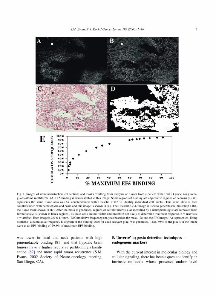
binding [23,74,75]. The analysis for each tissue
section has been extended to include quantitation of
the percentage of viable cells at each pO2, reported as
a cumulative frequency (Fig. 1). This analysis is
possible because the monoclonal antibodies are
directly conjugated to the fluorescent agents, for a
one-step detection system.The resulting binding
levels are calibrated to an absolute fluorescence
standard and are corrected for the patient drug
exposure [73] and the maximal tissue binding [23,
74,75]. Similar analyses can be performed using flow
cytometry. Based on flow cytometric studies of
human and rodent cells incubated with EF5 under
various oxygen conditions, we have estimated that
physiologic hypoxia (approx. 10% oxygen), results in
approximately 1% of maximum EF5 binding, modest
hypoxia (approx. 2.5% oxygen) results in approxi-
mately 3% of maximum EF5 binding; moderate
hypoxia (approx. 0.5% oxygen) results in approxi-
mately 10% of maximum EF5 binding and severe
hypoxia (approx. 0.1% oxygen) results in approxi-
mately 30% of maximum EF5 binding (Koch and
Evans, 2002 unpublished data).
Use of immunohistochemical markers for hypoxia
detection has allowed the analyses of the relationships
between, for example, hypoxia and other biological
endpoints such as vessels (for other examples see
Table 2). Such analyses are important for many
reasons, including the future selection of hypoxia-
directed therapies. The three-dimensional relationship
between vessels and hypoxic regions should provide
information regarding the type of hypoxia present.
This is important because treatments directed at
diffusion-limited hypoxia (‘chronic’) might not be
successful in tumors that are hypoxic due to vascular
changes (‘acute’ or perfusion-limited hypoxia). For
example, the use of a therapy such as meta-
iodobenzylguanidine (MIBG) [80] to decrease cellu-
lar oxygen utilization would only be effective in
tumors with diffusion-limited hypoxia.
At this time, there are only minimal data
regarding these agents as prognostic biomarkers.
Small studies suggest that locoregional recurrence
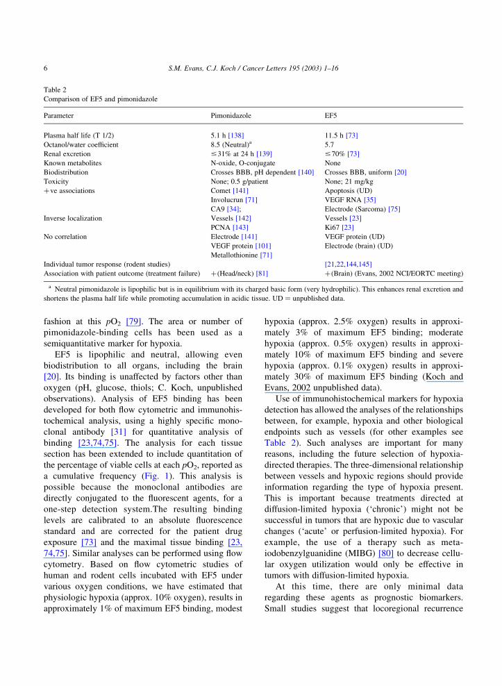
Table 2
Comparison of EF5 and pimonidazole
Parameter Pimonidazole EF5
Plasma half life (T 1/2) 5.1 h [138] 11.5 h [73]
Octanol/water coefficient 8.5 (Neutral)a 5.7
Renal excretion #31% at 24 h [139] #70% [73]
Known metabolites N-oxide, O-conjugate None
Biodistribution Crosses BBB, pH dependent [140] Crosses BBB, uniform [20]
Toxicity None; 0.5 g/patient None; 21 mg/kg
þve associations Comet [141] Apoptosis (UD)
Involucrun [71] VEGF RNA [35]
CA9 [34]; Electrode (Sarcoma) [75]
Inverse localization Vessels [142] Vessels [23]
PCNA [143] Ki67 [23]
No correlation Electrode [141] VEGF protein (UD)
VEGF protein [101] Electrode (brain) (UD)
Metallothionine [71]
Individual tumor response (rodent studies) [21,22,144,145]
Association with patient outcome (treatment failure) þ(Head/neck) [81] þ(Brain) (Evans, 2002 NCI/EORTC meeting)
a Neutral pimonidazole is lipophilic but is in equilibrium with its charged basic form (very hydrophilic). This enhances renal excretion and
shortens the plasma half life while promoting accumulation in acidic tissue. UD ¼ unpublished data.
S.M. Evans, C.J. Koch / Cancer Letters 195 (2003) 1–166
was lower in head and neck patients with high
pimonidazole binding [81] and that hypoxic brain
tumors have a higher recursive partitioning classifi-
cation [82] and more rapid tumor recurrence (S.M.
Evans, 2002 Society of Neuro-oncology meeting,
San Diego, CA).
5. ‘Inverse’ hypoxia detection techniques—
endogenous markers
With the current interest in molecular biology and
cellular signaling, there has been a quest to identify an
intrinsic molecule whose presence and/or level
Fig. 1. Images of immunohistochemical sections and masks resulting from analysis of tissues from a patient with a WHO grade 4/4 glioma,
glioblastoma multiforme. (A) EF5 binding is demonstrated in this image. Some regions of binding are adjacent to regions of necrosis (n). (B)
represents the same tissue area as (A), counterstained with Hoescht 33342 to identify individual cell nuclei. This same slide is then
counterstained with hematoxylin and eosin and this image is shown in (C). The Hoescht 33342 image is used to generate (in Photoshop 4.0w)
the tissue mask shown in (D). After the mask is generated, regions of cellular necrosis, as identified by a neuropathologist are removed from
further analysis (shown as black regions), as these cells are not viable and therefore not likely to determine treatment response. n ¼ necrosis,
a ¼ artifact. Each image is 2.0 £ 1.4 mm. (E) Cumulative frequency analyses based on the mask, (D) and the EF5 image, (A) is presented. Using
Matlabw, a cumulative frequency histogram of the binding level for each relevant pixel was generated. Thus, 95% of the pixels in the image
were at an EF5 binding of 79.8% of maximum EF5 binding.
S.M. Evans, C.J. Koch / Cancer Letters 195 (2003) 1–16 7

reflects tissue oxygenation. Oxygen levels have long
been known to modulate the release of chemical
messengers in the body; the classical example is the
release of erythropoietin by the kidney under
conditions of physiological or pathological hypoxia
[83]. HIF is a key regulator maintaining oxygen
homeostasis (for review see [57]). Many genes that
are known to be induced by HIF activation are also
expressed at higher levels in human cancers compared
to normal tissue counterparts. Examples include
VEGF [56,84–86], nitric oxide synthase-2 [87,88],
insulin-like growth factor [89], and transforming
growth factor [90]. Other factors that have been
associated with hypoxia, although not necessarily via
HIF regulation include carbonic anhydrase 9 (CA9 or
CA IX) [91 – 93], thrombospondin [94,95] and
osteopontin [96]. Several of these molecules, as well
as HIF itself, have been evaluated as intrinsic markers
of hypoxia both in serum and tissue samples.
The difficulty in using a measurement of tumor
HIF as an intrinsic marker of hypoxia is that its
activation can occur in the presence of oxygen. For
example, immunohistochemical HIF staining has
been demonstrated in renal cell tumors with extensive
vascularization due to loss of pVHL [97]. In fact, the
upregulation of HIF may be a general indicator of
poor outcome, independent of hypoxia [98]. Since
other cytokines such as VEGF and CA9 (see below)
also have non-oxygen based regulation, it is critical
that comparative studies with specific hypoxia
markers be carried out.
The relationship between hypoxia, as measured by
2-nitroimidazole binding and other biologic endpoints
has been studied (Table 2). In studies using hypoxia
and EF5 in tumor cell spheroids [99] and pimonida-
zole in rat livers [100], VEGF is upregulated,
consistent with previously observed predictions in
vitro. However, studies in human squamous cell
carcinomas [101] and glioma xenografts [102]
suggested that VEGF protein was not regulated in
concert with hypoxia, an unexpected conclusion.
Subsequent studies using EF5 immunohistochemistry
and VEGF mRNA in situ hybridization in several
human tumor types detected VEGF mRNA co-
localized with regional maxima of EF5 binding,
which were often adjacent to regions of tissue
necrosis. High EF5 binding occurred in tumor tissues
corresponding to regions roughly less than 0.3%
oxygen [35]. Two explanations for the different
findings in the human squamous cell and the glioma
xenograft studies versus this latter report are (1)
VEGF protein is secreted and may not stay ‘localized’
to hypoxic regions; and (2) the use of EF5 versus
pimonidazole. The precise relationship between
radiation response, pO2, and VEGF regulation
remains unclear. In vitro suspension culture studies
of three cervix cancer cell lines suggest that the Km of
VEGF upregulation was between 1 and 3% oxygen,
which is substantially more oxic than the Km for
radiation resistance and the above described regions
of EF5 binding [35,103].
CA9 is a zinc metalloenzyme responsible for the
reversible conversion of carbon dioxide to carbonic
acid and water. The membrane-linked isoforms CA9
and CA12 were identified as genes that were down
regulated by the von Hippel–Lindau protein (pVHL)
[104]. As pVHL is a critical element in the regulation
of HIF complex, it has been shown that the CA9 gene
was hypoxia-inducible and dependent on HIF [34].
CA9 has been found to be a tumor marker in ovarian,
endometrial and cervical cancer [105], an independent
predictive factor for overall survival in invasive breast
cancer [93] and an independent prognostic indicator
of overall survival and metastasis-free survival for
patients treated with radiation therapy for squamous
cell cancer of the cervix [106]. CA9 staining is
preferentially located in perinecrotic regions [91]. A
significant positive correlation between tumor
hypoxia (% of values less than 5 mmHg based on
Eppendorf electrode studies) and the extent of CA9
expression has been shown in cervix cancers [106]. In
a separate study, a substantial, although incomplete
overlap with pimonidazole staining was shown [34].
Similarly, Olive et al. [107] demonstrated co-
localization of CA9 and pimonidazole in cervix
cancer, although the area of the tumor section that
bound anti-CA9 antibodies represented double the
number of cells that bound anti-pimonidazole anti-
bodies. In studies of bladder and skin cancer, five of
the 20 patients studied had more CA9 staining than
pimonidazole staining [34]. Possible explanations for
these findings include transient (acute) hypoxia or that
pimonidazole only labels cells at less than 1.3%
oxygen, whereas upregulation of cytokines and
molecular markers occur at pO2 values over a larger
range, 0.2–2% oxygen [92].
S.M. Evans, C.J. Koch / Cancer Letters 195 (2003) 1–168
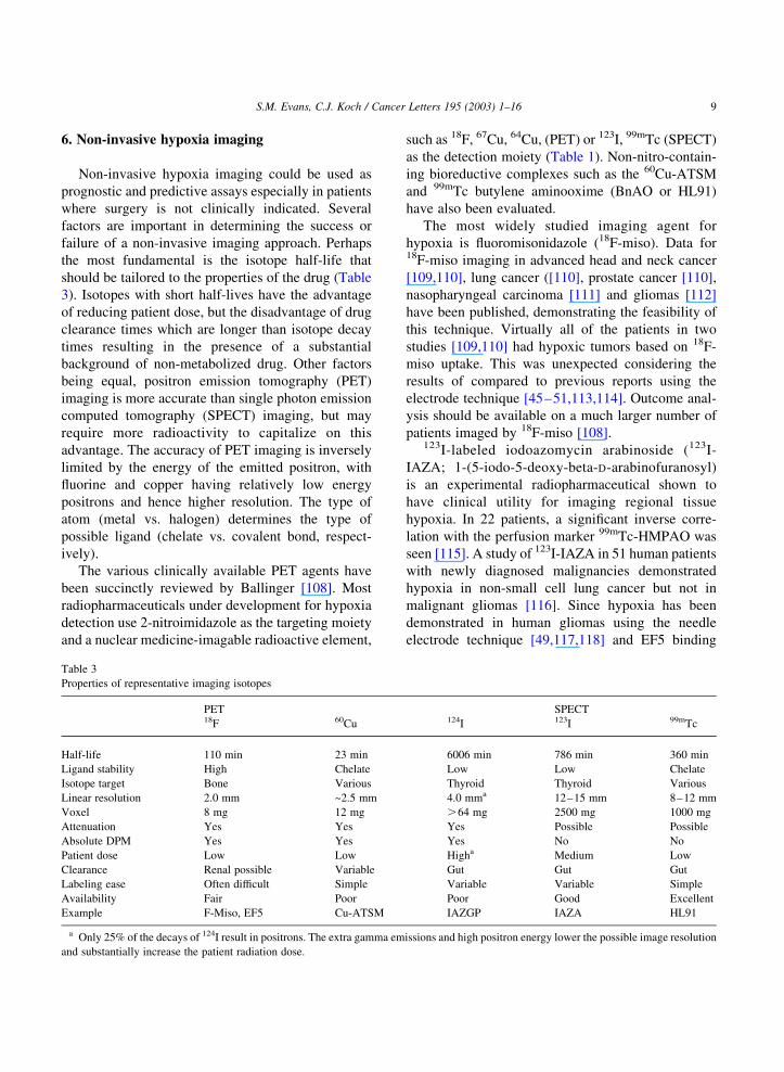
6. Non-invasive hypoxia imaging
Non-invasive hypoxia imaging could be used as
prognostic and predictive assays especially in patients
where surgery is not clinically indicated. Several
factors are important in determining the success or
failure of a non-invasive imaging approach. Perhaps
the most fundamental is the isotope half-life that
should be tailored to the properties of the drug (Table
3). Isotopes with short half-lives have the advantage
of reducing patient dose, but the disadvantage of drug
clearance times which are longer than isotope decay
times resulting in the presence of a substantial
background of non-metabolized drug. Other factors
being equal, positron emission tomography (PET)
imaging is more accurate than single photon emission
computed tomography (SPECT) imaging, but may
require more radioactivity to capitalize on this
advantage. The accuracy of PET imaging is inversely
limited by the energy of the emitted positron, with
fluorine and copper having relatively low energy
positrons and hence higher resolution. The type of
atom (metal vs. halogen) determines the type of
possible ligand (chelate vs. covalent bond, respect-
ively).
The various clinically available PET agents have
been succinctly reviewed by Ballinger [108]. Most
radiopharmaceuticals under development for hypoxia
detection use 2-nitroimidazole as the targeting moiety
and a nuclear medicine-imagable radioactive element,
such as 18F, 67Cu, 64Cu, (PET) or 123I, 99mTc (SPECT)
as the detection moiety (Table 1). Non-nitro-contain-
ing bioreductive complexes such as the 60Cu-ATSM
and 99mTc butylene aminooxime (BnAO or HL91)
have also been evaluated.
The most widely studied imaging agent for
hypoxia is fluoromisonidazole (18F-miso). Data for18F-miso imaging in advanced head and neck cancer
[109,110], lung cancer ([110], prostate cancer [110],
nasopharyngeal carcinoma [111] and gliomas [112]
have been published, demonstrating the feasibility of
this technique. Virtually all of the patients in two
studies [109,110] had hypoxic tumors based on 18F-
miso uptake. This was unexpected considering the
results of compared to previous reports using the
electrode technique [45–51,113,114]. Outcome anal-
ysis should be available on a much larger number of
patients imaged by 18F-miso [108].123I-labeled iodoazomycin arabinoside (123I-
IAZA; 1-(5-iodo-5-deoxy-beta-D-arabinofuranosyl)
is an experimental radiopharmaceutical shown to
have clinical utility for imaging regional tissue
hypoxia. In 22 patients, a significant inverse corre-
lation with the perfusion marker 99mTc-HMPAO was
seen [115]. A study of 123I-IAZA in 51 human patients
with newly diagnosed malignancies demonstrated
hypoxia in non-small cell lung cancer but not in
malignant gliomas [116]. Since hypoxia has been
demonstrated in human gliomas using the needle
electrode technique [49,117,118] and EF5 binding
Table 3
Properties of representative imaging isotopes
PET SPECT18F 60Cu 124I 123I 99mTc
Half-life 110 min 23 min 6006 min 786 min 360 min
Ligand stability High Chelate Low Low Chelate
Isotope target Bone Various Thyroid Thyroid Various
Linear resolution 2.0 mm ~2.5 mm 4.0 mma 12–15 mm 8–12 mm
Voxel 8 mg 12 mg .64 mg 2500 mg 1000 mg
Attenuation Yes Yes Yes Possible Possible
Absolute DPM Yes Yes Yes No No
Patient dose Low Low Higha Medium Low
Clearance Renal possible Variable Gut Gut Gut
Labeling ease Often difficult Simple Variable Variable Simple
Availability Fair Poor Poor Good Excellent
Example F-Miso, EF5 Cu-ATSM IAZGP IAZA HL91
a Only 25% of the decays of 124I result in positrons. The extra gamma emissions and high positron energy lower the possible image resolution
and substantially increase the patient radiation dose.
S.M. Evans, C.J. Koch / Cancer Letters 195 (2003) 1–16 9

[23], this finding suggests that the IAZA molecule
may not access the brain even in the face of a tumor-
induced break in the blood brain barrier. Preliminary
observations suggested that patients with 123I-IAZA-
avid neck metastases from head/neck cancer had a
decreased local control at 3 months, but follow-up
data have not been published. Newer agents based on
the azomycin-nucleoside structure (such as beta-D-
iodinated azomycin galactopyranoside, IAZGP) have
been developed [119], but have not yet been studied in
humans.99mTechnecium (99mTc) is commonly used in
nuclear medicine imaging because it is inexpensive,
easy to chelate to various ligands and has a near-
optimal half life (Table 3). In the 1990s, BMS181321
(Oxo [[3,3,9,9-tetramethyl1-1-(2-nitro-1H-imidazole-
1-yl)-4,8-diaza) and butylene amine oxime (oxime
(2,2′-[1,4-diaminobutane) bis [2-methyl-3-
butanone] dioxime; Prognox, HL91) were developed
as 99mTc-based hypoxia markers. Initial animal
studies with HL91 as a hypoxia detection agent
showed promise despite the absence of a rationally
developed hypoxia-binding moiety. Although a num-
ber of Phase II studies have been performed with
HL91, it is no longer in commercial development.
Fujibayashi and colleagues have developed a 62Cu
labeled diacetyl-bis (N 4-methylthiosemicarbazone)
(Cu ATSM) for imaging hypoxic tissues [120].
Studies performed in the 9L gliosarcoma rat model
compared the needle oxygen electrode and 64Cu
ATSM PET imaging before and after the tissue
oxygen concentration was modulated by hydralazine
or oxygen breathing. A correlation between low pO2
and high 64Cu-ATSM accumulation was observed
[121]. Studies of 60Cu ATSM have been performed in
patients with lung cancer [122] and cervix cancer
(Perry Grigsby, personal communication, 2002) with
promising results.18F-EF5 is unique in that it is the only hypoxia-
detection agent where the same molecule can be
measured both invasively (flow cytometry and/or
immunohistochemistry) and non-invasively (PET) in
the same tumor by administering cold drug as a carrier
for the radioactive agent. Studies in tumor bearing rats
[123] and dogs (B. Kaser-Hotz, personal communi-
cation, 2002) support the concept that hypoxic regions
can be identified with PET studies and confirmed with
immunohistochemistry studies.
7. Conclusions
It is clear from published studies using each of the
measuring techniques discussed that hypoxia is
present in a subset of human tumors. However, the
percentage of, for example, cervix cancer patients,
having a ‘hypoxic’ tumor seems to be dependent on
the technique used and how hypoxia is defined. All of
these studies agree that there is substantial inter- and
intra-tumoral heterogeneity within tumors of similar
histology and site, emphasizing the importance of
measuring hypoxia in individual patients. Although
the published (Eppendorf needle electrode) results are
statistically highly significant for hypoxia as a
prognostic marker, the sensitivity and specificity of
these assays are not optimal, e.g. there are substantial
patients with ‘hypoxic’ tumors who do well and
patients with ‘oxic’ tumors who do poorly. There are
at least two approaches to improving these results, and
both of these should be pursued.
Measurements and analysis of existing techniques
need to be refined to accommodate the observation
that hypoxia is continuous, not a binary process. The
simple division of patients above and below a median
value is likely to be artificial. As we are beginning to
understand the actual pO2 s at which pathophysiolo-
gical processes occur, correlation of hypoxia
measurements, biological processes and patient out-
come should be possible. Measurement of the exact
pO2 values in tissues would allow a more reasoned
classification of patients based on knowledge of the
pO2 where specific biological processes occur (such
as radiation resistance, upregulation of cytokines).
Studies discussed earlier in this review [17,18,20,23,
74,75] all support the idea that moderately, not
severely hypoxic cells may be most important for
determining biological resistance, perhaps because
severely hypoxic cells are destined to die. Such
analyses require that the hypoxia detection methods
be calibrated to a known standard. This process is
complex and tedious but may be initially necessary in
order to correctly interpret data.
It is highly unlikely that only one factor is pivotal
in determining therapy response. Multiple clinical and
biological factors are apt to play important roles in
patients’ outcome. Thus, consideration of other
factors, which, along with oxygen will be clinically
important, is appropriate. One example of a critical
S.M. Evans, C.J. Koch / Cancer Letters 195 (2003) 1–1610

biological factor that must be considered along with
oxygen content for radiation response is non-protein
thiol concentration since tumor cells with moderate
hypoxia but high non-protein thiols are as radiation
resistant as cells with severe hypoxia [13].
8. Summary
A clinically relevant method for evaluating the
presence and pattern of hypoxia in human tumors can
improve patient prognosis and treatment planning.
Intrinsic markers, such as VEGF, CA9 or HIF-1alpha
have the potential advantage that patients would only
require a biopsy. It is also possible that secreted
markers of hypoxia could be monitored by a blood
sample. In order for these markers to be optimized, a
better understanding of the microenvironmental
conditions modifying their regulation is required. At
present, it appears that the endogenous markers are
not solely regulated by oxygen (e.g. HIF-1alpha
upregulation in patients with VHL syndrome). A
more quantitative approach may be possible using
hypoxia-detecting 2-nitroimidazoles and/or electro-
des. These methods allow an understanding of the
presence, level, and patterns of hypoxia as well as
defining the biology of different disease sites. Labeled
2-nitroimidazoles and other redox sensitive com-
pounds can monitor tissue hypoxia non-invasively.
Such methods do not have the same resolution as the
invasive approaches but have obvious advantages,
particularly in their ability to monitor the whole tumor
and its local or distant spread, should these regions
also be hypoxic. Clearly, each technique has its
advantages and disadvantages and we need to
recognize and understand both. It may be possible
that specific tumor types will be best monitored by a
particular method and this may vary among different
tumor types, grades or stages.
Acknowledgements
Drs Richard Hill and Anthony Fyles for helpful
discussions regarding needle electrode studies.
References
[1] J.D. Chapman, A.J. Franko, C.J. Koch, The fraction of
hypoxic clonogenic cells in tumor populations, in Biological
Bases and Clinical Implications of Tumor Radioresistance,
1983, pp. 61–73.
[2] J.E. Moulder, D.F. Martin, Hypoxic fraction determinations
in the BA1112 rat sarcoma: variations within and among
assay techniques, Radiat. Res. (1984) 99.
[3] K.A. Kennedy, B.A. Teicher, S. Rockwell, A.C. Sartorelli,
The hypoxic tumor cell: a target for selective cancer
chemotherapy, Biochem. Pharmacol. 29 (1980) 1–8.
[4] J.M. Henk, Late results of a trial of hyperbaric oxygen and
radiotherapy in head and neck cancer: a rationale for hypoxic
cell sensitizers, Int. J. Radiat. Oncol. Biol. Phys. 12 (1986)
1339–1341.
[5] J.M. Henk, C.W. Smith, Radiotherapy and hyperbaric
oxygen in head and neck cancer: interim report of the second
clinical trial, Lancet 2 (1977) 104–105.
[6] J. Overgaard, H. Hansen, A.P. Sandersen, M. Hjelm-Hansen,
K. Jorgensen, E. Sandberg, et al., Misonidazole combined
with split-course radiotherapy in the treatment of invasive
carcinoma of the larynx and pharynx: report from the
DAHANCA 2 study, Int. J. Radiat. Oncol. Biol. Phys. 16
(1989) 1065–1068.
[7] J. Overgaard, Importance of tumor hypoxia in radiotherapy.
A meta-analysis of controlled clinical trials, Radiother.
Oncol. 24 (1992) S64.
[8] T.L. Phillips, T.H. Wasserman, R.J. Johnson, V.A. Levin, G.
VanRaalte, Final report on the United States phase I clinical
trial of the hypoxic cell radiosensitizer, misonidazole (RO-
07-0582; NSC #261037), Cancer 48 (1981) 1687–1704.
[9] R.C. Urtasun, C.N. Coleman, T.H. Wasserman, T.L. Phillips,
Clinical trials with hypoxic cell sensitizers: time to retrench
or time to push forward?, Int. J. Radiat. Oncol., Biol., Phys.
10 (1984) 1691–1696.
[10] M.S. Lesniak, R. Langer, H. Brem, Drug delivery to tumors
of the central nervous system, Curr. Neurol. Neurosci. Rep. 1
(2001) 210–216.
[11] R.K. Jain, Vascular and interstitial barriers to delivery of
therapeutic agents in tumors, Cancer Metastasis Rev. 9
(1990) 253–266.
[12] D.M. Brown, R. Gonsalez-Mendez, J.M. Brown, Factors
influencing intracellular uptake and radiosensitization by 2-
nitroimidazoles in vitro, Radiat. Res. 93 (1983) 492–505.
[13] A.D. Horan, C.J. Koch, The K(m) for radiosensitization of
human tumor cells by oxygen is much greater than 3 mmHg
and is further increased by elevated levels of cysteine, Radiat.
Res. 156 (2001) 388–398.
[14] C.J. Koch, S.M. Evans, Cysteine concentrations in rodent
tumors: unexpectedly high values may cause therapy
resistance, Int. J. Cancer 67 (1996) 661–667.
[15] C.J. Koch, K.A. Skov, Enhanced radiation-sensitivity by
preincubation with nitroimidazoles: effect of glutathione
depletion, Int. J. Radiat. Oncol., Biol., Phys. 29 (1994)
345–349.
[16] C. Ling, H. Michaels, E. Epp, E. Peterson, Interaction of
S.M. Evans, C.J. Koch / Cancer Letters 195 (2003) 1–16 11
misonidazole and oxygen in the radiosensitization of
mammalian cells, Int. J. Radiat. Oncol. Biol. Phys. 6
(1980) 583–589.
[17] P. Olive, Range of oxygenation in SiHa tumor xenografts,
Radiat. Res. 158 (2002) 159–166.
[18] B.G. Wouters, J.M. Brown, Cells at intermediate oxygen
levels can be more important than the ‘hypoxic fraction’ in
determining tumor response to fractionated radiation therapy,
Radiat. Res. 147 (1997) 541–550.
[19] M.L. Woods, C.J. Koch, E.M. Lord, Detection of individual
hypoxic cells in multicellular spheroids by flow cytometry
using the 2-nitroimidazole. EF5, and monoclonal antibodies,
Int. J. Radiat. Oncol., Biol., Phys. 34 (1996) 93–101.
[20] K.M. Laughlin, S.M. Evans, W.T. Jenkins, M. Tracy, C.Y.
Chan, E.M. Lord, et al., Biodistribution of the nitroimidazole
EF5 (2-[2-nitro-1H-imidazol-1-yl]-N-(2,2,3,3,3-pentafluoro-
propyl) acetamide) in mice bearing subcutaneous EMT6
tumors, J. Pharmacol. Exp. Therap. 277 (1996) 1049–1057.
[21] S.M. Evans, W.T. Jenkins, B. Joiner, E.M. Lord, C.J. Koch,
2-Nitroimidazole (EF5) binding predicts radiation resistance
in individual 9L s.c. tumors, Cancer Res. 56 (1996) 405–411.
[22] C.J. Koch, P.R. Oprysko, A.L. Shuman, W.T. Jenkins, G.
Brandt, S.M. Evans, Radiosensitization of hypoxic tumor
cells by dodecafluoropentane: a gas-phase perfluorochemical
emulsion, Cancer Res. 62 (2002) 3626–3629.
[23] S. Evans, S. Hahn, D. Magarelli, C. Koch, Hypoxic
heterogeneity in human tumors: EF5 binding, vasculature,
necrosis and proliferation, Am. J. Clin. Oncol. 24 (2001)
467–472.
[24] C. Koch, Measurement of absolute oxygen levels in cells and
tissues using oxygen sensors and the 2-nitroimidazole EF5, in
Antioxidants and Redox Signaling, Packer, Editor. 2001,
Academic Press: San Diego, California.
[25] D.M. Grzybicki, S.A. Moore, Implications of prognostic
markers in brain tumors, Clin. Lab. Med. 19 (1999) 833–847.
[26] H.B. Stone, J.M. Brown, T.L. Phillips, R.M. Sutherland,
Oxygen in human tumors: correlations between methods of
measurement and response to therapy. Summary of a
workshop held November 19–20, 1992, at the National
Cancer Institute, Bethesda, Maryland, Radiat. Res. 136
(1993) 422–434.
[27] J. Overgaard, Clinical evaluation of nitroimidazoles as
modifiers of hypoxia in solid tumors, Oncol. Res. 6 (1994)
509–518.
[28] J.H. Kaanders, L.A. Pop, H.A. Marres, I. Bruaset, F.J. van
den Hoogen, M.A. Merkx, et al., Experience in 215 patients
with advanced head-and-neck cancer, Int. J. Radiat. Oncol.,
Biol., Phys. 52 (2002) 769–778.
[29] M.J. Dorie, M.S. Kovacs, E.C. Gabalski, M. Adam, Q.T. Le,
D.A. Bloch, et al., Damage measured by the comet assay in
head and neck cancer patients treated with tirapazamine,
Neoplasia (New York) 1 (1999) 461–467.
[30] J. Del Rowe, C. Scott, M. Werner-Wasik, J.P. Bahary, W.J.
Curran, R.C. Urtasun, et al., Single-arm, open-label phase II
study of intravenously administered tirapazamine and
radiation therapy for glioblastoma multiforme, J. Clin.
Oncol. 18 (2000) 1254–1259.
[31] E.M. Lord, L. Harwell, C.J. Koch, Detection of hypoxic cells
by monoclonal antibody recognizing 2-nitroimidazole
adducts, Cancer Res. 53 (1993) 5271–5276.
[32] B.M. Fenton, S.F. Paoni, J. Lee, C.J. Koch, E.M. Lord,
Quantification of tumour vasculature and hypoxia by
immunohistochemical staining and HbO2 saturation
measurements, Br. J. Cancer 79 (1999) 464–471.
[33] N.J. Taylor, H. Baddeley, K.A. Goodchild, M.E. Powell, M.
Thoumine, L.A. Culver, et al., BOLD MRI human tumor
oxygenation during carbogen breathing, J. Magnet. Reson.
Imaging 14 (2001) 156–163.
[34] C.C. Wykoff, N.J. Beasley, P.H. Watson, K.J. Turner, J.
Pastorek, A. Sibtain, et al., Hypoxia-inducible expression of
tumor-associated carbonic anhydrases, Cancer Res. 60
(2000) 7075–7083.
[35] L. Ziemer, C. Koch, A. Maity, D. Magarelli, A. Horan, S.
Evans, Hypoxia and VEGF mRNA expression in human
tumors, Neoplasia 6 (2001) 500–508.
[36] S. Kaluz, M. Kaluzova, A. Chrastina, P.L. Olive, S.
Pastorekova, J. Pastorek, et al., Lowered oxygen tension
induces expression of the hypoxia marker MN/carbonic
anhydrase IX in the absence of hypoxia-inducible factor 1
stabilization, Cancer Res. 62 (2002) 4469–4477.
[37] S. Walenta, A. Salameh, H. Lyng, J.F. Evensen, M. Mitze,
E.K. Rofstad, et al., Correlation of high lactate levels in head
and neck tumors with incidence of metastasis, Am. J. Pathol.
150 (1997) 409–415.
[38] J.S. Nelson, Y. Tsukada, D. Schoenfeld, K. Fulling, J.
Lamarche, N. Peress, Necrosis as a prognostic criterion in
malignant supratentorial, astrocyticgliomas, Cancer 52
(1983) 550–554.
[39] J. Costa, R.A. Wesley, E. Glatstein, S.A. Rosenberg, The
grading of soft tissue sarcomas. Results of a clinicohisto-
pathologic correlation in a series of 163 cases, Cancer 53
(1984) 530–541.
[40] P. Kolstad, Vascularization, Oxygen Tension and Radio-
curability in Cancer of the Cervix, Oslo: Oslo, Universitets-
forlaget, Oslo, Norway, 1964.
[41] R.A. Gatenby, L.R. Coia, M.P. Richter, H. Katz, P.J.
Moldofsky, P. Engstrom, et al., Oxygen tension in human
tumors: in vivo mapping using CT-guided probes, Radiology
156 (1985) 211–214.
[42] P. Wendling, R. Manz, G. Thews, P. Vaupel, Heterogeneous
oxygenation of rectal carcinomas in humans. A critical
parameter for pre-operative irradiation, Adv. Exp. Med. Biol.
180 (1984) 293–300.
[43] M. Nozue, I. Lee, F. Yuan, B.A. Teicher, D.M. Brizel, M.W.
Dewhirst, et al., Interlaboratory variation in oxygen tension
measurement by Eppendorf ‘Histograph’ and comparison
with hypoxic marker, J. Surg. Oncol. 66 (1997) 30–38.
[44] W.T. Jenkins, S.M. Evans, C.J. Koch, Hypoxia and necrosis
in rat 9L glioma and Morris 7777 hepatoma tumors:
comparative measurements using EF5 binding and the
Eppendorf needle electrode, Int. J. Radiat. Oncol., Biol.,
Phys. 46 (2000) 1005–1017.
[45] M. Hockel, K. Schlenger, C. Knoop, P. Vaupel, Oxygenation
of carcinomas of the uterine cervix: evaluation by computer-
S.M. Evans, C.J. Koch / Cancer Letters 195 (2003) 1–1612
ized O2 tension measurements, Cancer Res. 51 (1991)
6098–6102.
[46] D.M. Brizel, G.L. Rosner, J. Harrelson, L.R. Prosnitz, M.W.
Dewhirst, Pretreatment oxygenation profiles of human soft
tissue sarcomas, Int. J. Radiat. Oncol., Biol., Phys. 30 (1994)
635–642.
[47] M.F. Adam, E.C. Gabalski, D.A. Bloch, J.W. Oehlert, J.M.
Brown, A.A. Elsaid, et al., Tissue oxygen distribution in head
and neck cancer patients, Head Neck 21 (1999) 146–153.
[48] B. Movsas, J.D. Chapman, E.M. Horwitz, W.H. Pinover,
R.E. Greenberg, A.L. Hanlon, et al., Hypoxic regions exist in
human prostate carcinoma, Urology 53 (1999) 11–18.
[49] R. Rampling, G. Cruickshank, A.D. Lewis, S.A. Fitzsim-
mons, P. Workman, Direct measurement of pO2 distribution
and bioreductive enzymes in human malignant brain tumors,
Int. J. Radiat. Oncol., Biol., Phys. 29 (1994) 427–431.
[50] A.C. Koong, V.K. Mehta, Q.T. Le, G.A. Fisher, D.J. Terris,
J.M. Brown, A.J. Bastidas, M. Vierra, Pancreatic tumors
show high levels of hypoxia, Int. J. Radiat. Oncol., Biol.,
Phys. 48 (2000) 919–922.
[51] E. Lartigau, H. Randrianarivelo, M.F. Avril, A. Margulis, A.
Spatz, F. Eschwege, et al., Intratumoral oxygen tension in
metastatic melanoma, Melanoma Res. 7 (1997) 400–406.
[52] M. Hockel, C. Knoop, K. Schlenger, B. Vorndran, E.
Baussmann, M. Mitze, et al., Intratumoral pO2 predicts
survival in advanced cancer of the uterine cervix, Radiother.
Oncol. 26 (1993) 45–50.
[53] M. Hockel, K. Schlenger, B. Aral, M. Mitze, U. Schaffer, P.
Vaupel, Association between tumor hypoxia and malignant
progression in advanced cancer of the uterine cervix, Cancer
Res. 56 (1996) 4509–4515.
[54] M. Nordsmark, M. Hoyer, J. Keller, O.S. Nielsen, O.M.
Jensen, J. Overgaard, The relationship between tumor
oxygenation and cell proliferation in human soft tissue
sarcomas, Int. J. Radiat. Oncol., Biol., Phys. 35 (1996)
701–708.
[55] D.M. Brizel, S.P. Scully, J.M. Harrelson, L.J. Layfield, J.M.
Bean, L.R. Prosnitz, et al., Tumor oxygenation predicts for
the likelihood of distant metastases in human soft tissue
sarcoma, Cancer Res. 56 (1996) 941–943.
[56] J.H. Marxsen, O. Schmitt, E. Metzen, W. Jelkmann, T.
Hellwig-Burgel, Vascular endothelial growth factor gene
expression in the human breast cancer cell line MX-1 is
controlled by O2 availability in vitro and in vivo, Ann. Anat.
183 (2001) 243–249.
[57] P.H. Maxwell, C.W. Pugh, P.J. Ratcliffe, Activation of the
HIF pathway in cancer, Curr. Opin. Genet. Dev. 11 (2001)
293–299.
[58] T.G. Graeber, C. Osmanian, T. Jacks, D.E. Housman, C.J.
Koch, S.W. Lowe, et al., Hypoxia-mediated selection of cells
with diminished apoptotic potential in solid tumours [see
comments]. Hypoxia-mediated selection of cells with
diminished, Nature 379 (1996) 88–91.
[59] S.D. Young, R.S. Marshall, R.P. Hill, Hypoxia induces DNA
overreplication and enhances metastatic potential of murine
tumor cells, Proc. Natl. Acad. Sci. USA 85 (1988)
9533–9537.
[60] A. Fyles, Tumor hypoxia has independent predictor impact
only in patients with node-negative cervix cancer. [see
comments.], (2002).
[61] J.D. Chapman, Hypoxic sensitizers—implications for radi-
ation therapy, New Engl. J. Med. 301 (1979) 1429–1432.
[62] A.J. Varghese, S. Gulyas, J.K. Mohindra, Hypoxia-depen-
dent reduction of 1-(2-nitro-1-imidazolyl)-3-methoxy-2-pro-
panol by Chinese hamster ovary cells and KHT tumor cells in
vitro and in vivo, Cancer Res. 36 (1976) 3761–3765.
[63] J.A. Raleigh, C.J. Koch, Importance of thiols in the reductive
binding of 2-nitroimidazoles to macromolecules, Biochem.
Pharmacol. 40 (1990) 2457–2464.
[64] C.J. Koch, A. thin-film, culturing technique allowing rapid
gas-liquid equilibration (6 sec) with no toxicity to mamma-
lian cells, Radiat. Res. 97 (1984) 434–442.
[65] Y.C. Taylor, A.M. Rauth, Differences in the toxicity and
metabolism of the 2-nitroimidazole misonidazole (Ro-07-
0582) in HeLa and Chinese hamster ovary cells, Cancer Res.
38 (1978) 2745–2752.
[66] J.S. Rasey, Z. Grunbaum, K. Krohn, N. Nelson, L. Chin,
Comparison of binding of [3H]misonidazole and [14C]mis-
onidazole in multicell spheroids, Radiat. Res. 101 (1985)
473–479.
[67] B.M. Garrecht, J.D. Chapman, The labelling of EMT-6
tumors in balb/c mice with 14C-misonidazole, Br. J. Radiol.
56 (1983) 745–753.
[68] A.J. Franko, J.D. Chapman, Binding of 14C-misonidazole to
hypoxic cells in V79 spheroids, Br. J. Cancer 45 (1982)
694–699.
[69] M.B. Parliament, L.I. Wiebe, A.J. Franko, Nitroimidazole
adducts as markers for tissue hypoxia: mechanistic studies in
aerobic normal tissues and tumour cells, Br. J. Cancer 66
(1992) 1103–1108.
[70] J.D. Chapman, C.J. Gillespie, Radiation-induced events and
their time scale in mammalian cells, Adv. Radiat. Biol. 9
(1981) 143–198.
[71] J.A. Raleigh, S.C. Chou, D.P. Calkins-Adams, C.A. Ballen-
ger, D.B. Novotny, M.A. Varia, A clinical study of hypoxia
and metallothionein protein expression in squamous cell
carcinomas, Clin. Cancer Res. 6 (2000) 855–862.
[72] K.I. Wijffels, J.H. Kaanders, P.F. Rijken, J. Bussink, F.J. van
den Hoogen, H.A. Marres, et al., Vascular architecture and
hypoxic profiles in human head and neck squamous cell
carcinomas, Br. J. Cancer 83 (2000) 674–683.
[73] C.J. Koch, S.M. Hahn, K. Rockwell Jr., J.M. Covey, W.G.
McKenna, S.M. Evans, Pharmacokinetics of EF5 [2-(2-nitro-
1-H-imidazol-1-yl)-N-(2,2,3,3,3-pentafluoropropyl) aceta-
mide] in human patients: implications for hypoxia measure-
ments in vivo by 2-nitroimidazoles, Cancer Chemother.
Pharmacol. 48 (2001) 177–187.
[74] S.M. Evans, S. Hahn, D.R. Pook, W.T. Jenkins, A.A.
Chalian, P. Zhang, et al., Detection of hypoxia in human
squamous cell carcinoma by EF5 binding, Cancer Res. 60
(2000) 2018–2024.
[75] S.M. Evans, S.M. Hahn, D.P. Magarelli, P.J. Zhang, W.T.
Jenkins, D.L. Fraker, et al., Hypoxia in human intraperitoneal
S.M. Evans, C.J. Koch / Cancer Letters 195 (2003) 1–16 13
and extremity sarcomas, Int. J. Radiat. Oncol., Biol., Phys. 49
(2001) 587–596.
[76] M.F. Dennis, M.R. Stratford, P. Wardman, M.E. Watts,
Cellular uptake of misonidazole and analogues with acidic or
basic functions, Int. J. Radiat. Biol. Relat. Stud. Phys., Chem.
Med. 47 (1985) 629–643.
[77] S.C. Chou, P.M. Flood, J.A. Raleigh, Marking hypoxic cells
for complement and cytotoxic T lymphocyte-mediated lysis:
using pimonidazole, Br. J. Cancer Suppl. 27 (1996)
S213–S216.
[78] J.A. Raleigh, G.G. Miller, A.J. Franko, C.J. Koch, A.F.
Fuciarelli, D.A. Kelly, Fluorescence immunohistochemical
detection of hypoxic cells in spheroids and tumours, Br.
J. Cancer 56 (1987) 395–400.
[79] M.W. Gross, U. Karbach, K. Groebe, A.J. Franko, W.
Mueller-Klieser, Calibration of misonidazole labeling by
simultaneous measurement of oxygen tension and labeling
density in multicellular spheroids, Int. J. Cancer 61 (1995)
567–573.
[80] J.E. Biaglow, Y. Manevich, D. Leeper, B. Chance, M.W.
Dewhirst, W.T. Jenkins, et al., MIBG inhibits respiration:
potential for radio- and hyperthermic sensitization, Int.
J. Radiat. Oncol., Biol., Phys. 42 (1998) 871–876.
[81] J. Kaanders, K. Wijffels, H. Marres, A. Ljungkvist, L. Pop,
F.v.d. Hoogen, et al., Pimonidazole binding and tumor
vascularity predict for treatment outcome in head and neck
cancer, Cancer Res. 62 (2002) 7066–7074.
[82] W.J. Curran Jr., C.B. Scott, J. Horton, J.S. Nelson, A.S.
Weinstein, A.J. Fischbach, et al., Recursive partitioning
analysis of prognostic factors in three Radiation Therapy
Oncology Group malignant glioma trials [see comments],
J. Natl. Cancer Inst. 85 (1993) 704–710.
[83] D.P. Jones, Renal metabolism during normoxia, hypoxia, and
ischemic injury, Annu. Rev. Physiol. 48 (1986) 33–50.
[84] M.W. Pedersen, S. Holm, E.L. Lund, L. Hojgaard, P.E.
Kristjansen, Coregulation of glucose uptake and vascular
endothelial growth factor (VEGF) in two small-cell lung
cancer (SCLC) sublines in vivo and in vitro, Neoplasia (New
York) 3 (2001) 80–87.
[85] Y. Tsuzuki, D. Fukumura, B. Oosthuyse, C. Koike, P.
Carmeliet, R.K. Jain, Vascular endothelial growth factor
(VEGF) modulation by targeting hypoxia-inducible factor-
1alpha(hypoxia response element(VEGF cascade differen-
tially regulates vascular response and growth rate in tumors,
Cancer Res. 60 (2000) 6248–6252.
[86] G. Xia, Y. Kageyama, T. Hayashi, S. Kawakami, M.
Yoshida, K. Kihara, Regulation of vascular endothelial
growth factor transcription by endothelial PAS domain
protein 1 (EPAS1) and possible involvement of EPAS1 in
the angiogenesis of renal cell carcinoma, Cancer 91 (2001)
1429–1436.
[87] D.S. Tendler, C. Bao, T. Wang, E.L. Huang, E.A. Ratovitski,
D.A. Pardoll, C.J. Lowenstein, Intersection of interferon and
hypoxia signal transduction pathways in nitric oxide-induced
tumor apoptosis, Cancer Res. 61 (2001) 3682–3688.
[88] J. Genius, J. Fandrey, Nitric oxide affects the production of
reactive oxygen species in hepatoma cells: implications for
the process of oxygen sensing, Free Radic. Biol. Med. 29
(2000) 515–521.
[89] J. Sugawara, D.S. Suh, G.H. Faessen, L.F. Suen, T. Shibata,
F. Kaper, et al., Regulation of insulin-like growth factor-
binding protein-1 by nitric oxide under hypoxic conditions,
J. Clin. Endocrinol. Metab. 85 (2000) 2714–2721.
[90] K. Kondo, W.G. Kaelin Jr., The von Hippel–Lindau tumor
suppressor gene, Exp. Cell Res. 264 (2001) 117–125.
[91] C.C. Wykoff, N. Beasley, P.H. Watson, L. Campo, S.K. Chia,
R. English, et al., Expression of the hypoxia-inducible and
tumor-associated carbonic anhydrases in ductal carcinoma in
situ of the breast, Am. J. Pathol. 158 (2001) 1011–1019.
[92] N.J. Beasley, C.C. Wykoff, P.H. Watson, R. Leek, H. Turley,
K. Gatter, et al., Carbonic anhydrase IX, an endogenous
hypoxia marker, expression in head and neck squamous cell
carcinoma and its relationship to hypoxia, necrosis, and
microvessel density, Cancer Res. 61 (2001) 5262–5267.
[93] S.K. Chia, C.C. Wykoff, P.H. Watson, C. Han, R.D. Leek, J.
Pastorek, et al., Prognostic significance of a novel hypoxia-
regulated marker, carbonic anhydrase IX, in invasive breast
carcinoma, J. Clin. Oncol. 19 (2001) 3660–3668.
[94] M. Detmar, Tumor angiogenesis, J. Invest. Dermatol. Symp.
Proc. 5 (2000) 20–23.
[95] K.R. Laderoute, R.M. Alarcon, M.D. Brody, J.M. Calaoagan,
E.Y. Chen, A.M. Knapp, et al., Opposing effects of hypoxia
on expression of the angiogenic inhibitor thrombospondin 1
and the angiogenic inducer vascular endothelial growth
factor, Clin. Cancer Res. 6 (2000) 2941–2950.
[96] C.P. Sodhi, S.A. Phadke, D. Batlle, A. Sahai, Hypoxia
stimulates osteopontin expression and proliferation of
cultured vascular smooth muscle cells: potentiation by high
glucose, Diabetes 50 (2001) 1482–1490.
[97] D. Zagzag, H. Zhong, J.M. Scalzitti, E. Laughner, J.W.
Simons, G.L. Semenza, Expression of hypoxia-inducible
factor 1alpha in brain tumors: association with angiogenesis,
invasion, and progression, Cancer 88 (2000) 2606–2618.
[98] D.M. Aebersold, P. Burri, K.T. Beer, J. Laissue, V. Djonov,
R.H. Greiner, et al., Expression of hypoxia-inducible factor-
1alpha: a novel predictive and prognostic parameter in the
radiotherapy of oropharyngeal cancer, Cancer Res. 61 (2001)
2911–2916.
[99] N.S. Waleh, M.D. Brody, M.A. Knapp, H.L. Mendonca,
E.M. Lord, C.J. Koch, et al., Mapping of the vascular
endothelial growth factor-producing hypoxic cells in multi-
cellular tumor spheroids using a hypoxia-specific marker,
Cancer Res. 55 (1995) 6222–6226.
[100] O. Rosmorduc, D. Wendum, C. Corpechot, B. Galy, N.
Sebbagh, J. Raleigh, et al., Hepatocellular hypoxia-induced
vascular endothelial growth factor expression and angiogen-
esis in experimental biliary cirrhosis, Am. J. Pathol. 155
(1999) 1065–1073.
[101] J.A. Raleigh, D.P. Calkins-Adams, L.H. Rinker, C.A.
Ballenger, M.C. Weissler, W.C. Fowler Jr., et al., Hypoxia
and vascular endothelial growth factor expression in human
squamous cell carcinomas using pimonidazole as a hypoxia
marker, Cancer Res. 58 (1998) 3765–3768.
[102] M.B. Parliament, M.J. Allalunis-Turner, A.J. Franko, P.L.
S.M. Evans, C.J. Koch / Cancer Letters 195 (2003) 1–1614

Olive, R. Mandyam, C. Santos, et al., Vascular endothelial
growth factor expression is independent of hypoxia in human
malignant glioma spheroids and tumours, Br. J. Cancer 82
(2000) 635–641.
[103] J.A. Chiarotto, R.P. Hill, A quantitative analysis of the
reduction in oxygen levels required to induce up-regulation
of vascular endothelial growth factor (VEGF) mRNA in
cervical cancer cell lines, Br. J. Cancer 80 (1999)
1518–1524.
[104] S.V. Ivanov, I. Kuzmin, M.H. Wei, S. Pack, L. Geil, B.E.
Johnson, et al., Down-regulation of transmembrane carbonic
anhydrases in renal cell carcinoma cell lines by wild-type von
Hippel–Lindau transgenes, Proc. Natl. Acad. Sci. USA 95
(1998) 12596–12601.
[105] J. Zavada, Z. Zavadova, S. Pastorekova, F. Ciampor, J.
Pastorek, V. Zelnik, Expression of MaTu-MN protein in
human tumor cultures and in clinical specimens, Int. J. Cancer
54 (1993) 268–274.
[106] J.A. Loncaster, A.L. Harris, S.E. Davidson, J.P. Logue, R.D.
Hunter, C.C. Wycoff, et al., Carbonic anhydrase (CA IX)
expression, a potential new intrinsic marker of hypoxia:
correlations with tumor oxygen measurements and prognosis
in locally advanced carcinoma of the cervix, Cancer Res. 61
(2001) 6394–6399.
[107] P.L. Olive, C. Aquino-Parsons, S.H. MacPhail, S.Y. Liao,
J.A. Raleigh, M.I. Lerman, et al., Carbonic anhydrase 9 as an
endogenous marker for hypoxic cells in cervical cancer,
Cancer Res. 61 (2001) 8924–8929.
[108] J.R. Ballinger, Imaging hypoxia in tumors, Semin. Nuclear
Med. 31 (2001) 321–329.
[109] D. Rischin, L. Peters, R. Hicks, P. Hughes, R. Fisher, R. Hart,
et al., Phase I trial of concurrent tirapazamine, cisplatin, and
radiotherapy in patients with advanced head and neck cancer,
J. Clin. Oncol. 19 (2001) 535–542.
[110] J.S. Rasey, W.J. Koh, M.L. Evans, L.M. Peterson, T.K.
Lewellen, M.M. Graham, et al., Quantifying regional
hypoxia in human tumors with positron emission tomography
of 18F fluoromisonidazole: a pretherapy study of 37 patients,
Int. J. Radiat. Oncol., Biol., Phys. 36 (1996) 417–428.
[111] S.H. Yeh, R.S. Liu, L.C. Wu, D.J. Yang, S.H. Yen, C.W.
Chang, et al., Fluorine-18 fluoromisonidazole tumour to
muscle retention ratio for the detection of hypoxia in
nasopharyngeal carcinoma, Eur. J. Nucl. Med. 23 (1996)
1378–1383.
[112] P.E. Valk, C.A. Mathis, M.D. Prados, J.C. Gilbert, T.F.
Budinger, Hypoxia in human gliomas: demonstration by PET
with fluorine-18-fluoromisonidazole., J. Nuclear Med. 33
(1992) 2133–2137.
[113] E. Lartigau, A.M. Le Ridant, P. Lambin, P. Weeger, L.
Martin, R. Sigal, et al., Oxygenation of head and neck
tumors, Cancer 71 (1993) 2319–2325.
[114] K. Sundfor, H. Lyng, U.L. Kongsgard, C. Trope, E.K.
Rofstad, Polarographic measurement of pO2 in cervix
carcinoma, Gynecol. Oncol. 64 (1997) 230–236.
[115] D. Groshar, A.J. McEwan, M.B. Parliament, R.C. Urtasun,
L.E. Golberg, M. Hoskinson, et al., Imaging tumor hypoxia
and tumor perfusion., J. Nuclear Med. 34 (1993) 885–888.
[116] R.C. Urtasun, M.B. Parliament, A.J. McEwan, J.R. Mercer,
R.H. Mannan, L.I. Wiebe, et al., Measurement of hypoxia in
human tumours by non-invasive spect imaging of iodoazo-
mycin arabinoside, Br. J. Cancer Suppl. 27 (1996)
S209–S212.
[117] G.S. Cruickshank, R.P. Rampling, W. Cowans, Direct
measurement of the PO2 distribution in human malignant
brain tumours, Adv. Exp. Med. Biol. 345 (1994) 465–470.
[118] D.R. Collingridge, J.M. Piepmeier, S. Rockwell, J.P. Knisely,
Polarographic measurements of oxygen tension in human
glioma and surrounding peritumoural brain tissue, Radiother.
Oncol. 53 (1999) 127–131.
[119] R.V. Iyer, P.T. Haynes, R.F. Schneider, B. Movsas, J.D.
Chapman, Marking hypoxia in rat prostate carcinomas with
beta-D-and 125I azomycin galactopyranoside., J. Nuclear
Med. 42 (2001) 337–344.
[120] Y. Fujibayashi, H. Taniuchi, Y. Yonekura, H. Ohtani, J.
Konishi, A. Yokoyama, Copper-62-ATSM: a new hypoxia
imaging agent with high membrane permeability and low
redox potential., J. Nuclear Med. 38 (1997) 1155–1160.
[121] J.S. Lewis, T.L. Sharp, R. Laforest, Y. Fujibayashi, M.J.
Welch, Tumor uptake of copper-diacetyl-bis(N(4)-
methylthiosemicarbazone): effect of changes in tissue
oxygenation., J. Nuclear Med. 42 (2001) 655–661.
[122] F. Dehdashti, M. Mintum, J. Lewis, et al., Evaluation of
tumor hypoxia with Cu-ATSM and PET (abstr.), J. Nuclear
Med. 41 (2000) 34.
[123] L. Ziemer, E. Sm, K. Aa, S. La, C. C, J. Wt, K. Js, A. A,
W. Dolbier, K. CJ. Non invasive imaging of tumor hypoxia
using the 2-nitroimidazole [18F] EF5 in rats. Eur. J. Nuclear
Med. 2002 (in Press).
[124] A.W. Fyles, M. Milosevic, R. Wong, M.C. Kavanagh, M.
Pintilie, A. Sun, et al., Oxygenation predicts radiation
response and survival in patients with cervix cancer
[published erratum appears in Radiother. Oncol. 1999 Mar;
50(3):371], Radiother. Oncol. 48 (1998) 149–156.
[125] G. Pitson, A. Fyles, M. Milosevic, J. Wylie, M. Pintilie, R.
Hill, Tumor size and oxygenation are independent predictors
of nodal diseases in patients with cervix cancer, Int. J. Radiat.
Oncol., Biol., Phys. 51 (2001) 699–703.
[126] K. Sundfor, H. Lyng, E.K. Rofstad, Tumour hypoxia and
vascular density as predictors of metastasis in squamous cell
carcinoma of the uterine cervix, Br. J. Cancer 78 (1998)
822–827.
[127] K. Sundfor, H. Lyng, C.G. Trope, E.K. Rofstad, Treatment
outcome in advanced squamous cell carcinoma of the uterine
cervix: relationships to pretreatment tumor oxygenation and
vascularization, Radiother. Oncol. 54 (2000) 101–107.
[128] E.K. Rofstad, K. Sundfor, H. Lyng, C.G. Trope, Hypoxia-
induced treatment failure in advanced squamous cell
carcinoma of the uterine cervix is primarily due to
hypoxia-induced radiation resistance rather than hypoxia-
induced metastasis, Br. J. Cancer 83 (2000) 354–359.
[129] T.H. Knocke, H.D. Weitmann, H.J. Feldmann, E. Selzer, R.
Potter, Intratumoral pO2-measurements as predictive assay
in the treatment of carcinoma of the uterine cervix, Radio-
ther. Oncol. 53 (1999) 99–104.
S.M. Evans, C.J. Koch / Cancer Letters 195 (2003) 1–16 15
[130] M. Nordsmark, M. Overgaard, J. Overgaard, Pretreatment
oxygenation predicts radiation response in advanced squa-
mous cell carcinoma of the head and neck, Radiother. Oncol.
41 (1996) 31–39.
[131] D.M. Brizel, G.S. Sibley, L.R. Prosnitz, R.L. Scher, M.W.
Dewhirst, Tumor hypoxia adversely affects the prognosis of
carcinoma of the head and neck, Int. J. Radiat. Oncol., Biol.,
Phys. 38 (1997) 285–289.
[132] D.M. Brizel, R.K. Dodge, R.W. Clough, M.W. Dewhirst,
Oxygenation of head and neck cancer: changes during
radiotherapy and impact on treatment outcome, Radiother.
Oncol. 53 (1999) 113–117.
[133] P. Stadler, A. Becker, H.J. Feldmann, G. Hansgen, J. Dunst,
F. Wurschmidt, et al., Influence of the hypoxic subvolume on
the survival of patients with head and neck cancer, Int.
J. Radiat. Oncol., Biol., Phys. 44 (1999) 749–754.
[134] V. Rudat, B. Vanselow, P. Wollensack, C. Bettscheider, S.
Osman-Ahmet, M.J. Eble, et al., Repeatability and prog-
nostic impact of the pretreatment pO(2) histography in
patients with advanced head and neck cancer, Radiother.
Oncol. 57 (2000) 31–37.
[135] V. Rudat, P. Stadler, A. Becker, B. Vanselow, A. Dietz, M.
Wannenmacher, et al., Predictive value of the tumor
oxygenation by means of pO2 histography in patients with
advanced head and neck cancer, Strahlenther. Onkol. 177
(2001) 462–468.
[136] D.J. Terris, Head and neck cancer: the importance of oxygen,
Laryngoscope 110 (2000) 697–707.
[137] M. Nordsmark, J. Alsner, J. Keller, O.S. Nielsen, O.M.
Jensen, M.R. Horsman, et al., Hypoxia in human soft tissue
sarcomas: adverse impact on survival and no association with
p53 mutations, Br. J. Cancer 84 (2001) 1070–1075.
[138] M.I. Saunders, P.J. Anderson, M.H. Bennett, S. Dische, A.
Minchinton, M.R. Stratford, et al., The clinical testing of Ro
03-8799—pharmacokinetics, toxicology, tissue and tumor
concentrations, Int. J. Radiat. Oncol., Biol., Phys. 10 (1984)
1759–1763.
[139] J.T. Roberts, N.M. Bleehen, M.I. Walton, P. Workman, A
clinical phase I toxicity study of Ro 03-8799: plasma, urine,
tumour and normal brain pharmacokinetics, Br. J. Radiol. 59
(1986) 107–116.
[140] H.F. Newman, N.M. Bleehen, R. Ward, P. Workman,
Hypoxic cell radiosensitizers in the treatment of high grade
gliomas: a new direction using combined Ro 03-8799
(pimonidazole) and SR 2508 (etanidazole), Int. J. Radiat.
Oncol., Biol., Phys. 15 (1988) 677–684.
[141] P.L. Olive, J.P. Banath, C. Aquino-Parsons, Measuring
hypoxia in solid tumours—is there a gold standard?, Acta
Oncol. 40 (2001) 917–923.
[142] A.C. Begg, H. Janssen, D. Sprong, I. Hofland, G. Blommes-
tijn, J.A. Raleigh, et al., Hypoxia and perfusion measure-
ments in human tumors—initial experience with
pimonidazole and IUdR, Acta Oncol. 40 (2001) 924–928.
[143] A.S. Kennedy, J.A. Raleigh, G.M. Perez, D.P. Calkins, D.E.
Thrall, D.B. Novotny, et al., Proliferation and hypoxia in
human squamous cell carcinoma of the cervix: first report of
combined immunohistochemical assays, Int. J. Radiat.
Oncol., Biol., Phys. 37 (1997) 897–905.
[144] M.C. Kavanagh, V. Tsang, S. Chow, C. Koch, D. Hedley, S.
Minkin, et al., comparison in individual murine tumors of
techniques for measuring oxygen levels, Int. J. Radiat.
Oncol., Biol., Phys. 44 (1999) 1137–1146.
[145] B.G. Siim, D.R. Menke, M.J. Dorie, J.M. Brown, Tirapaza-
mine-induced cytotoxicity and DNA damage in transplanted
tumors: relationship to tumor hypoxia, Cancer Res. 57 (1997)
2922–2928.
S.M. Evans, C.J. Koch / Cancer Letters 195 (2003) 1–1616