prevalence of shoulder subluxation among the stroke patients
TRANSCRIPT
PREVALENCE OF SHOULDER SUBLUXATION AMONG THE
STROKE PATIENTS ATTENDED AT CRP
Jotishko Biswas
Bachelor of Science in Physiotherapy (BSc.PT)
Session: 2005-2006
BHPI, CRP, Savar, Dhaka
Bangladesh Health Professions Institute (BHPI)
[The academic Institute of CRP]
Department of Physiotherapy
CRP, Savar, Dhaka-1343
Bangladesh
February 2012
We the under signed certify that we have carefully read and recommended to the Faculty
of Medicine, University of Dhaka, for the acceptance of this dissertation entitled
PREVALENCE OF SHOULDER SUBLUXATION AMONG THE
STROKE PATIENTS AT CRP
Submitted by Jotishko Biswas, for the partial fulfillment of the requirements for the
degree of Bachelor of Science in Physiotherapy (B.Sc.PT).
………………………….
Nasirul Islam B.Sc. PT (Hons.), MPH
Assistant professor
Department of Physiotherapy
BHPI, CRP, Savar, Dhaka
Supervisor
…………………………..
Md. Shorab Hossain
B.Sc. PT (Hons.), Dip. Ortho. Med, MPH
Assistant professor of Physiotherapy &
Head,Department of physiotherapy
BHPI, CRP, Savar, Dhaka
…………………………
Mohammad Anwar Hossain B.Sc.PT (Hons.),Dip. Ortho. Med, MPH
Assistant professor
Department of Physiotherapy
BHPI, CRP, Savar, Dhaka.
………………………….. ……………………………
Md. Shofiqul Islam Md.Obaidul Haque B.Sc.PT (Hons.), MPH B.Sc PT (Hons.), Dip.ortho. Med., MPH
Lcturer Assistant professor and Course Coordinator
Department of Physiotherapy Deparment of Physiotherapy
BHPI, CRP, Savar, Dhaka BHPI, CRP, Savar, Dhaka
DECLERATION
I declare that the work presented here is my own. All sources used have been cited
appropriately. Any mistakes or inaccuracies are my own. I also decline that for any
publication, presentation or dissemination of information of the study. I would be bound
to take written consent of my supervisor.
Signature- Date-
Jotishko Biswas
Bachelor of Science in Physiotherapy (B.Sc. PT)
Session: 2005-2006
BHPI, CRP, Savar, Dhaka- 1343
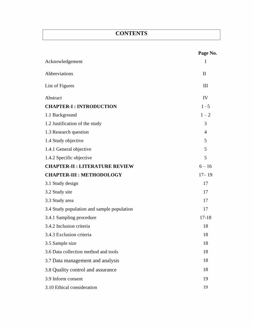
CONTENTS
Page No.
Acknowledgement
I
Abbreviations
II
List of Figures
III
Abstract IV
CHAPTER-I : INTRODUCTION 1 –5
1.1 Background 1 – 2
1.2 Justification of the study 3
1.3 Research question 4
1.4 Study objective 5
1.4.1 General objective 5
1.4.2 Specific objective 5
CHAPTER-II : LITERATURE REVIEW 6 – 16
CHAPTER-III : METHODOLOGY 17– 19
3.1 Study design 17
3.2 Study site 17
3.3 Study area 17
3.4 Study population and sample population 17
3.4.1 Sampling procedure 17-18
3.4.2 Inclusion criteria 18
3.4.3 Exclusion criteria 18
3.5 Sample size 18
3.6 Data collection method and tools 18
3.7 Data management and analysis 18
3.8 Quality control and assurance 18
3.9 Inform consent 19
3.10 Ethical consideration 19

CHAPTER-IV: RESULT 20 – 29
CHAPTER-V: DISCUSSION 30 – 31
CHAPTER-VI : CONCLUSION & RECOMMENDATION 32-33
REFERENCES 34-36
APPENDIX 37-40
i
ACKNOWLEDGEMENT
First of all, I would like to pay my gratitude to Almighty God who given me the ability to
complete this project in time with great success. I would like to pay my gratitude towards
my parents who constantly encouraged me to carry out this project. I would like to
express my gratitude to my respected teacher Md. Shofiqul Islam, Lecturer, Department
of Physiotherapy, Bangladesh Health Professions Institute (BHPI) and my friend Zahid
Bin Sultan Rana and Saiful Islam for their tired less effort with excellent guidance and
support.I would like to thanks all participants for helping me at the time of data
collection. I would also like to thanks librarian of Bangladesh Health Professions Institute
(BHPI) and their associates for their kind support to find out related books, journals and
also access to internet. Finally, my deepest gratefulness goes to my honorable research
supervisor Nasirul Islam for his keen supervision and excellent guidance without which I
could not able to complete this project.
ii
ABBREVIATIONS
ADL: Activities of Daily Living
BHPI: Bangladesh Heath Professions Institute
CRP: Center for the Rehabilitation of the Paralyzed
CVA: Cardio Vascular Accident
SPSS: Statistical Package of Social Science
WHO: World Health Organization
iii
LIST OF FIGURES
Page No
Figure-1: Age range of the participants
19
Figure-2: Prevalence of Shoulder subluxation
20
Figure-3: Male-Female Ratio 21
Figure-4: Occupation of the patients 22
Figure-5: Living area 23
Figure-6: Type of stroke 24
Figure-7: Affected side 26
Figure-8: Past medical history 27
Figure-9: History of mal handling 28
Figure-10: Past history of physical exercises
29

iv
Abstract
Purpose: To identify the prevalence of shoulder subluxation among the stroke patients
attended at CRP. Objectives: To find out the prevalence of shoulder subluxation among
the stroke patients, to identify the male female ratio ,to identify the more affected age
group, to find the occupation of patients with shoulder subluxation, to find out the past
medical history among the patients with shoulder subluxation. Methodology: A
quantitative cross-sectional study design was chosen to accomplish the objectives of the
study. 35 subjects were selected through simple random sampling technique from the
outpatient’s neurology physiotherapy department of CRP. A structural questionnaire was
developed through searching of literature. The participants were requested to answer
according to the developed format of the question. The answers were entered into SPSS
16 software and analyzed as descriptive statistics. Results: The study showed that 62.85%
(n=22) participants had shoulder subluxation among the stroke patients. Their mean age
was 54±8.4. The most of the participants 31.82% (n=7) who had shoulder subluxation in
between 53-60 years of age group among them most of the participants 27.27% (n=6)
were service holder and 63.64% (n=14) were male. More than half of the participants
54.55% (n=12) came from urban area and 54.55% (n=12) participants had hemorrhagic
stroke among stroke patients who had shoulder subluxation. 68.18% (n=15) had the
history of hypertension, 31.82% (n=7) had diabetes mellitus, 77.27% (n=17) had no past
history of physical exercises and 59.09% (n=13) had history of mal handling among the
stroke patients with shoulder subluxation. Conclusion: The result of the study
demonstrates that the prevalence of shoulder subluxation was 62.85% (n=22) and they
had certain positive exposure including diabetes mellitus, hypertension, history of mal
handling, no past history of physical exercise.
1
CHAPTER-I INTRODUCTION
1.1 Background
The brain is an exciting area in neurology as it is complex in anatomy and in function.
With the advancement of age in addition to decay, the brain becomes more prone to
get many complicated life threatening diseases, these will need appropriate attention
in time. Stroke is one of such condition which is the burning topic in this new
millennium since it is not only a major killer but also a cause of disability in the world
as well as in Bangladesh (Mohammad, 2011).
“Stroke has afflicted man kind since earliest times. Studying the remains of ancient
Egyptian mummies has shown that individuals of this era suffered strokes. In the past,
strokes were referred to as apoplexy meaning a sudden shock to the senses.
Hippocrates, the father of western medicine, wrote: “It is impossible to remove a
stroke attack of apoplexy and not easy to remove a weak attack”, this bleak statement
demonstrates the pessimistic view once held about strokes, but much has changed
since Hippocrates time. Having a stroke does not mean that one should give up all
hope and be resigned to a life of disability. Modern and surgical techniques, state of
the art rehabilitation programs and knowledge of risk factors control now make this
even truer (Bierman, 2009).
But now, in our country the real situation is totally different. Every year the number
of attack is increasing cerebrovascular disease or stroke is the first major cause of
death resulted from neurological diseases and the most frequent cause of all death
cases. At least 50% of neurological disorders in a general hospital are strokes. A study
shows that, stroke is more common in male and above the age of 50 years where male
to female ratio is 2.3:1, and death due to stroke is 34.74% of all death cases (Rahman,
et al. 2002 ).
So before taking a critical situation and to prevent disability resulted from stroke a
multidisciplinary team approach should start working, where this will consist of
Neurologist, Physiotherapist, Occupational therapist, Psychologist Nurse and social
2
workers, this will be required as, rehabilitation is the ultimate aim of treatment and
therapist, each have a definite role in stroke which should be started as early as
possible (Mohammad, 2011).
To manage inferior subluxed hemiplegic shoulder in patient with CVA, physio-
therapists use different treatment techniques of different approaches. Two different
proprioceptive facilitator’s techniques (therapeutic vibration and heavy joint
compression) of Roods approach have been selected in this study. Physiotherapists
usually use these techniques along with other techniques in the management of
subluxed hemiplegic shoulder in patient with CVA. This study examines the effects of
therapeutic vibration and heavy joint compression as treatment of inferior subluxed
hemiplegic subluxed hemiplegic shoulder in patient with CVA. To identify
effectiveness of both heavy joint compression and therapeutic vibration as a treatment
in the case of subluxed shoulder of hemiplegic patient. To find out treatment
technique that is better for the patient of subluxed shoulder for maximum achievement
(Hansen & Atchinson, 2002).
In the case of shoulder subluxation of hemiplegic patient many treatment techniques
are applied. The duration to get improvement duffers in the case of each of the
different techniques. If occupational therapisr khows the effectiveness od these two
treatment techniques, it will be easy to compare the results and find out which is the
better treatment technique. This will be less time consuming as will as helpful for the
patient. No research has been found on this area and for the development of
profession, this type of clinical study is important situation. It can be used as evidence
for using the appropriate treatment technique for the hemiplegic patient with shoulder
subluxation. To measure the effectiveness of heavy joint compression and therapeutic
vibration as a treatment in the case of subluxed shoulder of hemiplegic patient. To
compare both results of heavy joint compression and therapeutic vibration to find out
the technique with the maximum outcome (Mohammad, 2011).
3
1.2 Justification of the study
Although some studies have dealt with shoulder subluxation among the stroke
patients in other countries, the exact nature and prevalence of this important health
problem has not been studied before in Bangladesh. This study was formulated to fill
the gap of knowledge in this area. The aims of the study were to assess the pattern of
shoulder among stroke patients and to identify the impact of demographic,
occupational, psychological and social factors on them. Beside this it will help to
established right guidelines for patients, equipment and environmental conditions
which are mandatory for stroke patients. This study will also help to discover the
lacking area of a career, especially about their posture before doing any activities.
Beside this it will help to professional development which is mandatory for current
situation. The identification of prevalence of shoulder subluxation gives proper
education about stroke and shoulder subluxation. It will help to discover the role and
importance of physiotherapy in every sector of Bangladesh.

4
1.3 Research Question
What is the prevalence of shoulder subluxation among the stroke patients?
5
1.4 Study objective
General objective:
To identify the prevalence of shoulder Subluxation among the stroke
patients.
Specific objectives:
To find out the prevalence of shoulder subluxation among the stroke
patients.
To identify the male female ratio
To identify the more affected age group.
To find the occupation of patients with shoulder subluxation.
To find out the past medical history among the patients with shoulder
subluxation.
To identify the influencing demographic factors for such exposure group
in relation to age, sex, occupation, living area, past medical history, past
history of mal handling, past history of physical exercises
6
CHAPTER-II : LITERATURE REVIEW
The importance of the reorganization of the cortico spinal tract (CST) originating
from the damaged hemisphere for recovery of hand function has implications for the
development of physiotherapy programs for stroke patients. Reorganization within the
CNS (central nervous system) must be due to some form of synaptic plasticity. The
synaptic changes underling the recovery process may be similar to those that are
responsible for learning. Enhanced synaptic transmission has been postulated as a
mechanism for learning and memory processes. Synapses are strengthened when
afferent fibers receive repetitive input. The repetitive input induces molecular changes
in pre synaptic and post synaptic terminals which in turn change their effectiveness.
Similarly, the molecular that occur to change the strength of synapses may also lead
to an increase or decrease in the number of pre synaptic terminals. If therapy was
directed at repeatedly accessing the corticospinal neurons and their connections
remaining in the damaged hemisphere after stroke, the recovery of patients hand
function may be improved (Turton, 2010).
Cerebral vascular accident (CVA) or stroke is the most common disabling
neurological disease of adulthood. (Pedretti, 2007). It may be defined as an
interruption in the blood flow so that an adequate supply of oxygen and nutrients fail
to reach portion of the brain. Medical practitioners use the term cerebrovascular
accident, often abbreviated as CVA, for stroke. A stroke can occur in any part of the
brain the cerebral hemispheres, the cerebellum or the brainstem ( Bierman, 2009). A
cerebrovascular accide3nt is a rapidly developed clinical sign of a focal disturbance of
cerebral function of presumed vascular origin and o more than 24 hours duration
(WHO, 1986 cited in Turner, Foster, and Johnson, 1996). Clinical signs of stroke
develop suddenly due to interruption of blood flow to the brain and lasts more than 24
hours. Warlow (2010) defined the stroke or CVA as rapidly developing clinical
symptoms and or signs of focal time’s global loss of cerebral function with symptoms
lasting more than 24 hours leading to death with no apparent cause other than that
vascular origin. World health Organization (WHO) supports this definition of CVA.
When the severity of stroke last less than 24 hours, it is known as transient ischemic
7
attack (TIA).it is not a stroke but a warning for a forthcoming stroke. In TIA no
symptoms are found (Pedretti, 2007). Stroke or cerebrovascular accident (CVA) does
not represent a single disorder but rather a variety of disorders characterized by the
sudden onset of neurological deficits brought about by vascular injury to the brain
(Rolok &Rokey, 1990). The most typical manifestation of CVA is hemipheresis or
hemiplegia on the side of the body contralayeral to the site of CVA. One study on the
people of Bangladesh shows that the 75.59% of all stroke patients are men and 24.1%
are women where due to large artery atherosclerosis 21.25%, small artery occlusion
17.32%, cardio embolism 18.1% other determined etiology 26.7% and undetermined
causes 16.53% (Hayee et al.,2002). Stroke can be classified into two main types-
Ischemic and Hemorrhagic. Ischemic stroke includes artherothrombotic, lacunar and
embolic infarction .Hemorrhagic stroke includes intracerebral and subarachnoid
hemorrhage (warlow, 1993).
Ischemic stroke
The most common type of stroke and it is responsible for about 80% of all first ever
in a life time stroke (Warlow, 1993). This takes place when a clot blocks blood
vessels or become too narrow for blood to flow within the brain due to reduction in
blood supply, brain cells die from lack of oxygen (Nayan, 2003).
Atherothrombotic
Cerebral thrombosis occurs when a blood clot forms in one of the arteries supplying
the brain, causing vascular obstruction at the point of its formation. The size and
location of the infract depends on which vessel is occluded and the amount of
collateral circulation. Thrombosis occurs most frequently in blood vessels that have
already been damaged by atherosclerosis (an abnormal condition of the arteries in
which a thick, rough deposit forms on the inner wall of the arteries and gradually
narrows the passageway so that the blood flow slowed). Large vessel atherosclerosis
accounts for 60% of ischemic stroke (Pedretti, 2007).
8
Lacunar
These are small infracts usually lying in the deep neocortical parts of the cerebrum
and brainstems including the basal ganglia, thalamus , Pons, internal capsule and deep
white matter . Within a few months of onset of a lacunar stroke, a small cavity is left.
It results from an occlusion of small branches of large cerebral arteries-middle
cerebral, posterior cerebral,basilar, and a lesser extent ,anterior cerebral and vertebral
arteries. Lacunar infracts range in size from 2 to 15 mm. due to their small size,
usually only minimal neurological symptom result and may go undetected .statistics
shows that about 18% of ischemic stroke are lacunar (Warlow, 1993).
Embolism
Embolism occurs when a clot that has been formed elsewhere (thrombus ) breaks of
(embolus ) and travels up the bloodstream until it reaches an artery that is too small
for it pass and it blocks the artery. At this point its effects are similar to those
produced by thrombosis. Approximately 5% to 14% of strokes appear to be the result
of this process (warlow, 1993).
Hemorrhagic stroke
Hemorrhagic strokes are caused by a rupture in a blood vessel or an aneurysm with
resultant bleeding into or around cerebral tissue. These types of stroke have a much
higher fatality rate than those caused by clots. In a hemorrhagic stroke, the blood
vessel burst and the blood spread out over the brain causing damage of the brain cells
(Hansen & Atchinson, 2011).
Subarachnoid Hemorrhage
Subarachnoid hemorrhages account for about 7% of all strokes. Their most common
cause is leakage of blood from aneurysms. A combination of congenital and
degenerative factors, usually at the point of origin or bifurcation of arteries, can
participate in formation of an aneurysm. Blood may break through the weak point of
the aneurism at any time because of the force of arterial pressure spread quickly into
the cerebrospinal fluid surrounding the brain. A subarachnoid hemorrhage may also
be caused by bleeding from an arteriovenous malformation, which is an abnormal
9
collection of vessels near the surface of the brain. Other less common causes of
subarachnoid hemorrhages are hemophilia, excessive anticoagulation therapy, and
trauma to the skull and brain (Warlow, 1993).
Intracerebral Hemorrhage
Intracerebral hemorrhage accounts for about 10% of all strokes (Warlow, 2010). It
usually begins with bleeding from small, deep penetrating vessels under arteriolar or
capillary pressure as opposed to arterial pressure as with subarachnoid hemorrhage.
Therefore onset of symptoms from intracerebral hemorrhages develops gradually over
minutes, hours or sometimes days. Release of blood into brain tissue and surrounding
edema will then disrupt the function of that particular brain region. Hypertension is
the most common cause of bleeding into the brain. Severity of stroke varies with
individual. Every individual does not get stroke with same severity .some experience
mild effects, which take a short time to improve. On the other hand some suffer with
severe problems, which last for months or years. The effect of stroke will depend on
the part of brain that has been injured or damaged. It has been found that person who
have experienced a stroke and who are at any one time,10% will fully recover, 40%
will be left with a mild disability and 50% will be severely disable which may require
institutional care (Bierman, 2009). As no static has been found in aspect of
Bangladesh, foreign statistic has been added to feel the effects of stroke. The
incidence of stroke is about 1.8 to 2.0 per 1000 of population per annum. About 70%
of all strokes occur in people over 70 years of age. Approximately 80% show some
useful recovery and are able to return home; 60% of the total number regain
independence in activities of daily living (ADL) and 30% are able to resume normal
activities. The risk of mortality increases with age and the presence of associated
conditions such as heart disease (Jackson, 2006).
Motor disturbances after CVA
One side distribution and includes musculature of the trunk and limb on the affected
side: The muscles of face and mouth also may be involved. Increase muscle tone,
hypo tonicity may be apparent. Coordination or smooth rhythmic movement is lost.
Normal postural control mechanism is disturbed. Normal righting, equilibrium and
10
protective reactions are lost on the affected side. Loss of adaptive changes of muscle
tome as a protection against the forces of gravity. Loss of selective, discriminative
and isolated movement occurs after CVA. Inability to dorsiflexion the ankle and toes.
Inability to flex knee while hip is extended (Pedretti, Smith, and Pendleton, 2007).
Motor recovery after CVA
Recovery of motor function following a stroke is thought to be complete after 3 to 6
months of a stroke and can be continued for months or years (Ryerson, 2001).
Spontaneous recovery of voluntary motor function occurs primarily in the first 3
months after the onset of the CVA. Motor recovery may continue up to 1 year and in
rare instances somewhat longer. This fact does not imply that motor behavior cannot
be influenced by appropriate therapy after a year. Improvement in functional
performance may continue for years following stroke (Pedrett, Smith, and Pendleton,
2007).
In another study it has shown that most recovery occur within the first 8weeks but
10% show some improvement in their walking beyond that and almost 30% show
improvement on their activities of daily living score over the initial 6 months and also
suggests that further recovery after 6 months is much slower (Wade, 2005). Again
another study by Warlow (2010) has shown that the rate of recovery of all impairment
is macula in the first few weeks and slows down after 2 or 3 months and probably
stops after about 6 to 12 months of stroke (Turner, Foster and Johnson, 1996). The
recovery of upper limb function varies on the severity of the symptoms or damage and
maximum recovery occur in the initial 2 to 3 months of the stroke. It is typically
reported that most potential recovery of the upper limb takes place within 3 months
(Wade et al. 2005). The severely affected upper limb functions after demonstrated less
recovery. Most reports of upper limb function after acute brain lesion suggests that
recovery is minimal of patients with an initially affected limb (Wade et al. 2005).
Basmajuan and colleagues (2006) put the percentage of patients regarding full arm
and hand function after stroke at 5% and the percentage with no functional use at
20%. Gross movement of upper limb comes earlier than fine movements. Turton
(2010) stated that shoulder and elbow movements recover earlier than hand
11
movements and it is common for fine finger movements to remain permanently
disrupted (Turner, Foster and Johnson, 1996).
Sensory motor, cognitive and psychosocial components are almost always affected by
a cerebro vascular accident. Deficits in these areas and any secondary complications
profoundly affect an individual’s occupational performance in work, leisure and
activities of daily living. Secondary condition are important manifestations to
consider in regards to the patients recovery and rehabilitation as it may actually be
more disabling than the stroke itself. It is important to be aware of these
complications so that they may possibly be prevented. There are lots of secondary
conditions which make the situation complex and affect improvement (Turner, Foster
and Johnson, 1996). Glenohumeral subluxation basically is defined as a partial or
incomplete dislocation that usually stems from changes in the mechanical integrity of
the joint. Subluxation is a common problem in patient with hemiplegic, especially
during the flaccid stage, and often occurs within 3 weeks post stroke (Gould, 2002).
Subluxation is a common concern related to motor function involves the shoulder.
Common problems include subluxation, pain and immobility (Hansen & Atchinson,
2011). Subluxation is related to a change in the angle of the glenoid fossa (Ryerson,
2001). Glenohumeral joint misalignment and subluxation is reported to occur in
patients with little or no voluntary movement after stroke (Sils and Schenkman,
1985). The main feature of the shoulder joint is mobility rather than stability, making
a wide range of movement possible. As a result the shoulder is easily traumatized and
misalignment is common (Turner, Foster and Johnson, 1996).
Causes of shoulder subluxation
Shoulder occurs when any of the biomechanical factors contributing to glenohumeral
joint stability are interrupted Muscle weakness (Ryerson, 2001). Secondarily to
spasticity or flaccidity of the glenohumeral and or scapular muscles. Supraspinatus
weakness has also been found correlated with radiographic evidence of subluxation
(Trombly, 2007). Changes in muscle tone and movement, the position of scapula and
joint capsule stability. When the weight of the arm and pull of gravity draws the head
of the hummers out of the glenoid fossa of scapula (Hansen & Atchinson, 2011). Can
12
occur spontaneously when patient start sitting or standing against gravity. The weight
of the flaccid arm applying direct mechanical stretch to the joint capsule as well as
traction to unsupportive muscles of the shoulder (Gould, 2002). Subluxation is not
painful but results in changes of muscle length tension relationships, muscle
shortening and permanent of the joint capsule (Ryerson, 2001). Subluxation has been
proposed as a contributing factor in the development of shoulder pain (Chaco & Wolf,
2004). The mechanism suggested is one of traction on the rotator cuff and superior
joint capsule resulting in stretch causing pain. However there has been no explanation
of why this should cause pain and there is no direct evidence linking subluxation with
pain. On cause of pain is movement could be the pinching of the lengthened joint
capsule, caught between joint surfaces during movement in certain parts of rang. This
appears evidence in some patients as a sharp pain, which can relieved by gentle
distraction of the joint during assisted movement of the limb (Turner, Foster and
Johnson, 1996).
Although subluxation has been implicated in shoulder pain, several studies report
patients with subluxation who have no pain, even when subluxation was moderate to
severe (Smith et al., 2005). Subluxation has also been reported to be associated with
an increased incidence of RSD (Carr & Shepherd, 2000). Subluxation in itself is not
painful but is vulnerable and be easily traumatized. A dragging or ache is sometimes
reported if limb left hanging, easily rectified with correct positioning.
Mechanism of subluxation
Two thirds of the humeral head is not covered by the glenoid fossa. This lack of
stability is partly compensated for by a strong surrounding musculature. In the normal
orientation of scapula, there is an upward slope of the glenoid fossa, ethic plays an
important role in preventing downward dislocation of the homeruns. The humeral
head would have to be moved laterally in order to move downward. Then the arm is
adducted, the superior part of the capsule and the coracohumeral ligament are taut,
which prevents lateral movement of the humeral head. These safeguard against
downward displacement. The supraspinatus muscle reinforces the horizontal tension
of the capsule. The infranspinatus and posterior portion of the deltoid also play an
13
important role in preventing subluxation, because of their horizontal fibers. When the
homeruns is abducted sideways or flexed forward, the superior capsule becomes lax,
eliminating the support and joint stability must then be provided by muscle
contraction. The integrity of the joint then depends almost exclusively on the rotator
cuff muscles. In hemiplegia, patients have lost the voluntary movement in relative
muscles. These include the supraspinatus, infraspinatus and posterior fibers of the
deltoid (Hansen & Atchinson, 2011).
In addition, the muscles that support the scapula in its normal alignment are affected,
allowing a change in angulations of the glenoid fossa. Subluxation is therefore
inevitable(Hansen & Atchinson, 2011). In the frontal plane the scapula is normally
held at an angle of 40 degrees. When the slope of the glenoid fossa becomes less
oblique and more vertical, the humerus slide down and out of the fossa(Ryerson,
2001).
“In a normal shoulder the glenoid fossa is oriented upward, forward and lateral, so
that, the head od humerus remains locked in contact with it, the hemiplegic
orientation of the glenoid fossa is downward, backward and medial due to the
scapular retraction and downward rotation that are part of the common spastic pattern.
This position nullifies the “locking mechanism” and allows gravity ot pull the head of
the numerous out of the fossa (Trombly, 2007). The mechanism of subluxation is
generally considered to include the downward rotation of the scapula caused by the
weight of the limb, which positions the scapula more vertically (Carr & Shepherd,
2000). However the dowmward rotation of the scapula may not be a significant
feature and in a recent report no evidence of a relationship between scapula and
humeral orientation and glenohumeral subluxation was found (Carr & Shepherd,
2000). Evidence has been shown that low tone in scapular muscles not contribute to
downwards rotation of scapula as has been suggested (Devis, 2003). In subluxation
the shoulder girdle drops due to decreased tone to elevators of scapula, the scapula is
depressed and retracted, so that, scapula lies closer to the vertebra, inferior angle
adducted and depressed. Vertebral border of scapula pulled away from ribs and the
humerus slips downwards (Turner, Foster & Johnson, 1996).
14
Types of subuxation
Ryerson and levit (2001) first described three types of subluxation in clients with
hemiplegia; inferior, anterior and superior.
Inferior subluxation
The most common type of subluxation is an inferior subluxation. Reported incidences
of shoulder subluxation in cerebrovascular accident survivors vary from 17% to 66%
(Zorowitz, et al.1995 cited in Morley, 2002). It occurs in clients with severe weakness
and is present in the acute stage. Weakness and the weight of a heavy arm result in
downward rotation of the scapula. Downward rotation orients the glenoid fossa
vertically, the unlocking mechanism of the scapula is lost and the numerous
subluxates interiorly with internal rotation. As the humerus internally rotates, the
bicipital tuberosity rolls anteriorly; this anterior prominence is often confused with an
anterior subuxation. As subluxation occurs, the shoulder capsule is vulnerable to
stretch, especially when the humerus is dependent and resting by the side of the body.
In this position, the capsule is taut superiorly, so any downward distraction of the
humerus will place an immediate stretch on the upper part of the capsule. The
superior portion of the capsule is reinforced by the coracohumeral ligament, which is
crucial for shoulder stability. The implications of rupture of this ligament as a result o
forced abnormal passive motion as a cause of shoulder pain in subluxation (Hansen &
Atchinson, 2002).
Anterior subluxation
It occurs when the humeral head separates anteriorly from the glenoid fossa. Anterior
shoulder subluxation occurs when the downwardly rotated scapula elevates and tilts
forward on the rib cage and the humerus hyper-extends with internal rotation. In a
anterior subluxation, as tension increases on the proximal biceps tendon, the elbow
flexes and the forearm supinated. The subluxation is found in clients with atypical
patterns of return and trunk rotational asymmetries
15
Superior subluxation
Superior subluxation occurs when the humeral head lodges under the coracoids
process in a position of internal rotation and slight abduction. The humeral is locked
in this position, so that every movement of the humerus is accompanied by scapula
movement. The scapula position in this subluxation is one of the abduction, elevation
and neutral rotation. The forearm abducts across the body as the humeral abduction
and elbow flexion. A superior subluxation occurs in clients with inappropriate muscle
firing and co-contraction (Ryerson, 2001).
Prevention
Prevention of subluxation requires: Proper assessment of secondary alignment
problems (ribs cage/scapula/humeral position). Early reeducation of trunk/arm linked
patterns in sitting and standing. Prevention of shoulder capsule stretch, including
support and positioning as the client sits, stands and practices walking (Ryerson,
2001). Loading of the glenohumeral joint should be avoided as long as the affected
limb is flaccid (Trombly, 2007).
Intervention
The management of the subluxed shoulder in hemiplegia is controversial at best.
Treatment methods and prevention techniques advocated for the hemiplegic shoulder
include neuromuscular electric stimulation, proprioceptive facilitation techniques,
correct positioning, correct handling and avoidance of over vigorous movements
(Body, pepin and Hartin, 2009). If subluxation exists, the therapist reduces the
subluxation by correcting trunk, scapula and humeral alignment patterns before
attempting to re-educate arm movement patterns. As the client learns to move the arm
in patterns of functional coordination, subluxation and associated arm posturing
decrease (Ryerson, 2001). Treatment should involve correct positioning at all times to
help prevent subluxation. Subluxation cuffs, which grip the upper arm and re-
approximate the glenohumeral joint by lifting the humerus upwards can be made or
purchased (Turner, Foster and Johnson, 1996). Proprioceptive stimulation refers to the
facilitation of muscle spindles, Golgi tendon organs, joint receptors and the vestibular
apparatus. In general, proprioceptive stimulation gives the therapist more control over
16
motor response. Proprioceptors adapt more slowly than exterocptorsand can produce
sustained postural patterns. There is little or no recruitment in the proproiceptive
system. Therefore the motor response lasts as long as the stimulus is applied. There
are considerable amount of techniques that are used for facilitation. But, of those,
heavy joint compression and therapeutic vibration are widely used in the management
of subluxed hemiplegic shoulder in patient with CVA (Hansen & Atchinson, 2011).
17
CHAPTER III METHODOLOGY
3.1 Study design
The purpose of the study was to find out the prevalence shoulder subluxation among
the stroke patients. The cross section study was conducted to find out the objectives.
This design involves identifying group of people and then collecting the information
that researcher requires when they use the particular service. This type of data can be
used to assess the prevalence of acute or chronic conditions in a population. Survey
research is one of the most common forms of research that involves the asking a large
group of people questions about a particular topic or issue and these are related to the
interest of the participant. Survey is a method of collecting data which involves the
measuring relevant sample variables (often using s questionnaire) without any form of
manipulation or systemic intervention .The idea with the survey usually approaches a
sample of target group of interest, interviews them or ask them questionnaire .
3.2 Study population and sample population
A population is the total group or set of events or totality of the observation on which
a research is carried out. In this study, sample population were selected from the
participant of Centre for the Rehabilitation of the paralysed (CRP), Dhaka.
3.3.1 Study site
Centre for the Rehabilitation of paralyzed (CRP).
3.3.2 Study area
Neurological conditions of the patients.
3.4 Sample Size
The expected sample size to conduct the research was 236. But the researcher could
manage just 35 subjects because of having resource constrain.
The equation of sample size calculation are given below-
18
Here,
= 1.96
p= 0.81(Here P=Prevalence and P=81%)
q= 1-p
=1-0.25
=0.75
d= 0.05
3.5 Inclusion and Exclusion criteria
3.5.1 Inclusion criteria:
Both male and female selected who had stroke.
Subject was selected from Centre for the Rehabilitation of the
paralyzed (CRP) at Savar, Dhaka.
All age group was selected.
Subject who were willing to participate in the study.
3.5.2 Exclusion Criteria
Medically unstable patient
Patients who have cognitive problem.
Patients who are not able to communicate.
3.6 Sampling technique
Samples were selected as simple random sampling from Centre for the
Rehabilitation of the paralyzed (CRP) at Savar, Dhaka. There are a lot of
patients in Bangladesh, from this population it was selected 35 samples, according to
the inclusion and exclusion criteria.
3.7 Data collection tools
Questionnaire, consent forms, pen, papers, pen drive, SPSS (Statistical Package for the
Social Sciences) software to analyze data, Harvard Referencing 2012 and computer.
19
3.8 Data analysis
Data was inserted into SPSS 16 and descriptive statistics was used to analyze the
collected data.
3.9 Informed consent
In this study interested subjects were given consent forms and the purpose of the
research and consent forms were explained to the subject verbally. They were told
that participation is fully voluntary and they have the right to withdraw at any time.
They were also told that confidentiality will be maintained. Information might be
published in any presentations or writing but they will not be identified. The study
results might not have any direct effects on them but the members of Physiotherapy
population may be benefited from the study in future. They would not be embarrassed
by the study. At any time the researcher will be available to answer any additional
questions in regard to the study.
3.10 Ethical consideration
Permission was taken from BHPI ethical committee for research project then
permission was taken from physiotherapy department for data collection. The
participants were explained the purpose and goals of the study. This study followed the
World Health Organization (WHO) & Bangladesh Medical Research Council (BMRC)
guidelines and strictly maintained the confidentiality. Meanwhile, it was purely an
observation research, so nothing was intervene through which the research is
considered as limited ethical issue.
20
CHAPTER-IV RESULT
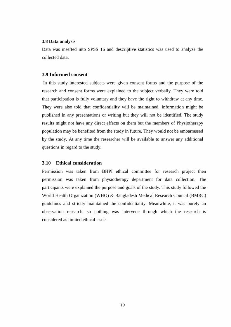
4.1 Age range of the participants
Analysis showed that 22.73% (n=5) participants had shoulder subluxation in between
41-46 years, 22.73% (n=5) was in between 47-52 years, 31.82% (n=7) was between
53-60 years, 22.73% (n=5) was more than 60 years of age out of 35 participants and
mean age of the participants was 54 (SD ±8.4) years. (Figure-1).
Fig-1: Age range of the participants
22.73%
22.73%
31.82%
22.73%
41-46 years
47-52 years
53-60 years
>60 years
21
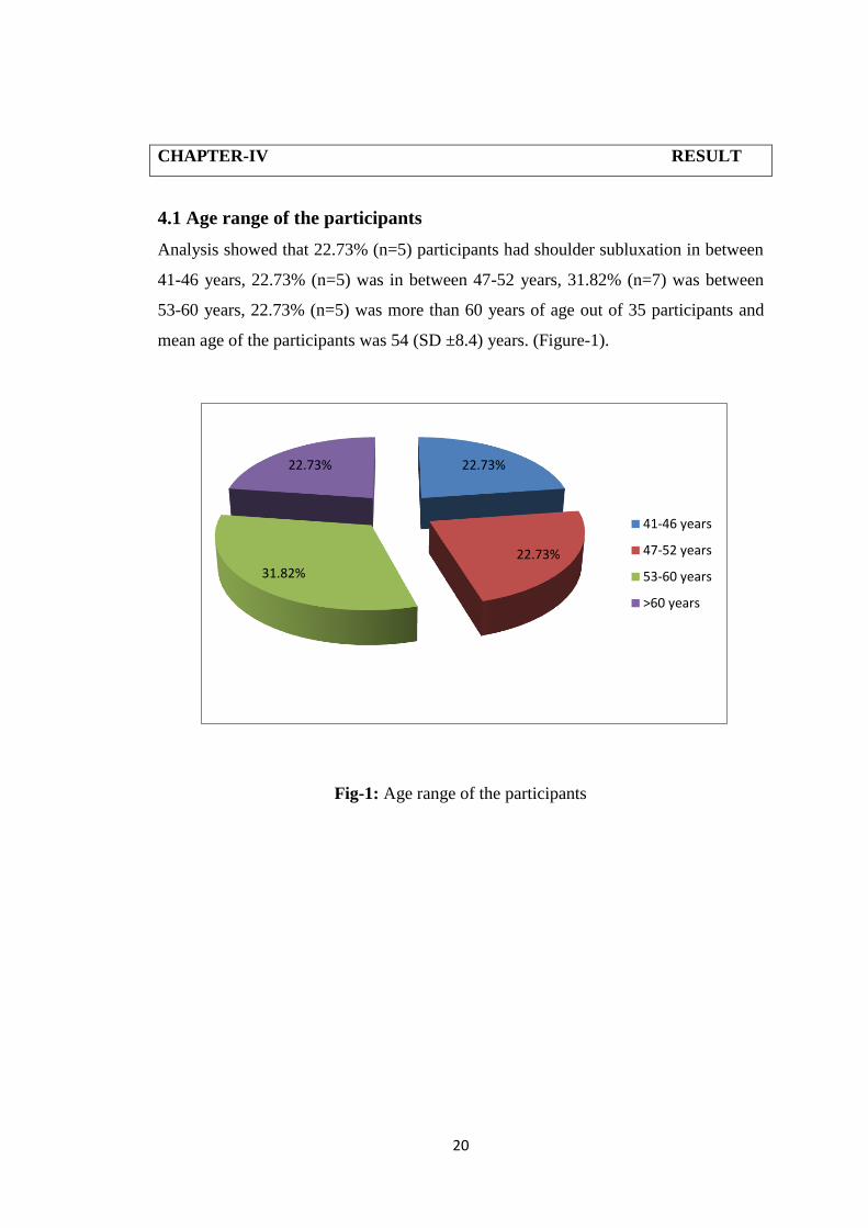
4.2 Prevalence of Shoulder subluxation
Outcome reveals that 62.85% (n=22) had shoulder subluxation out of 35 participants
(Figure-2).
Fig-2: Prevalence of Shoulder subluxation
0.00%
10.00%
20.00%
30.00%
40.00%
50.00%
60.00%
70.00%
Yes No
62.85%
37.15%
22
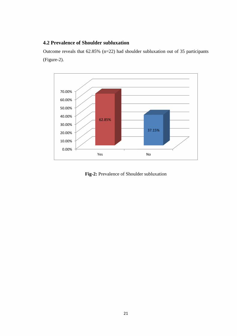
4.3 Male-Female Ratio
Among the 22 participants with shoulder subluxation 63.64% (14) were male and
36.36% (n=8) were female .Result shows that male is more affected by shoulder
subluxation than male (Figure-3).
Fig-3: Male-Female Ratio
63.64%
36.36%
Male
Female
23
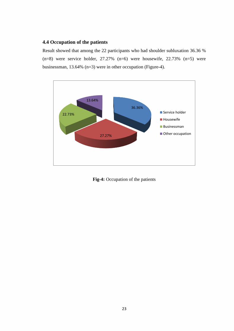
4.4 Occupation of the patients
Result showed that among the 22 participants who had shoulder subluxation 36.36 %
(n=8) were service holder, 27.27% (n=6) were housewife, 22.73% (n=5) were
businessman, 13.64% (n=3) were in other occupation (Figure-4).
Fig-4: Occupation of the patients
36.36%
27.27%
22.73%
13.64%
Service holder
Housewife
Businessman
Other occupation
24
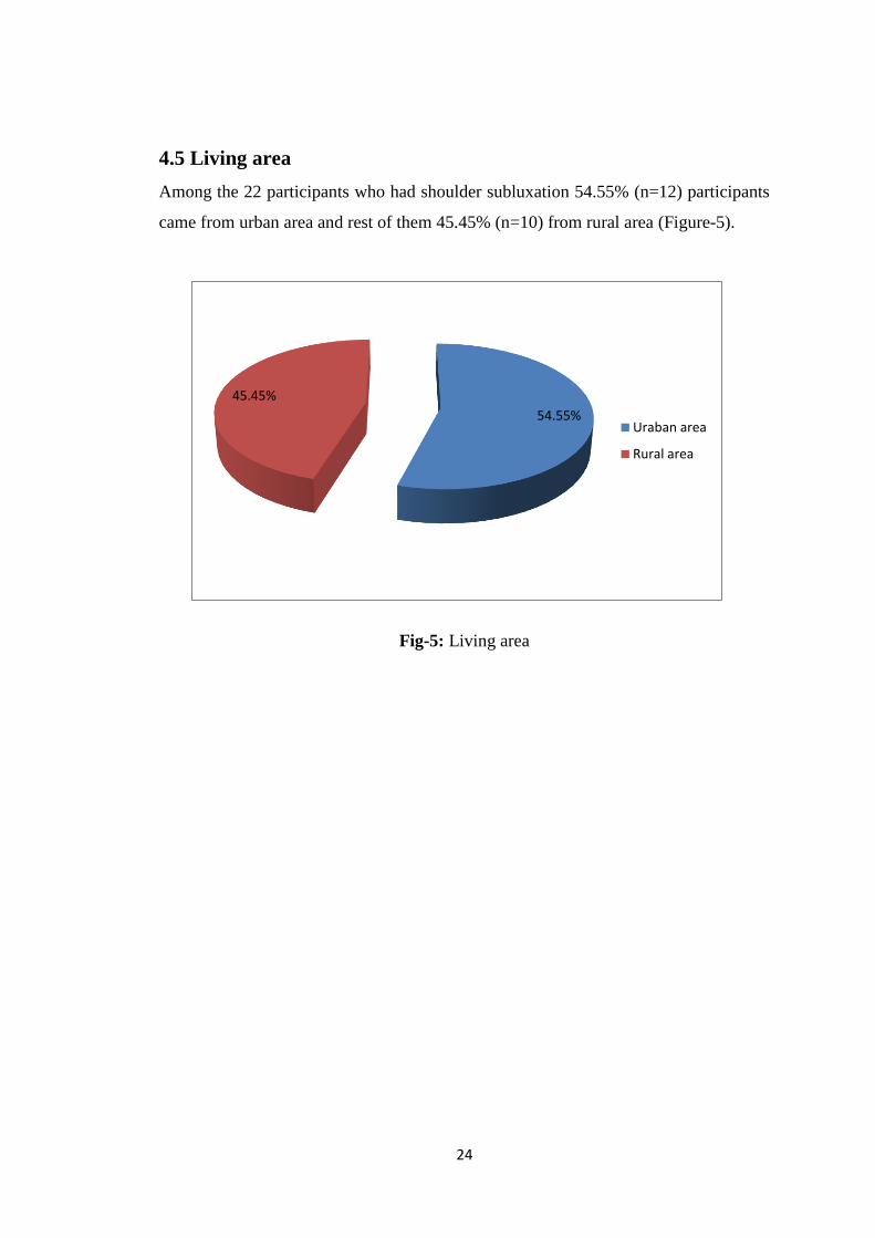
4.5 Living area
Among the 22 participants who had shoulder subluxation 54.55% (n=12) participants
came from urban area and rest of them 45.45% (n=10) from rural area (Figure-5).
Fig-5: Living area
54.55%
45.45%
Uraban area
Rural area
25
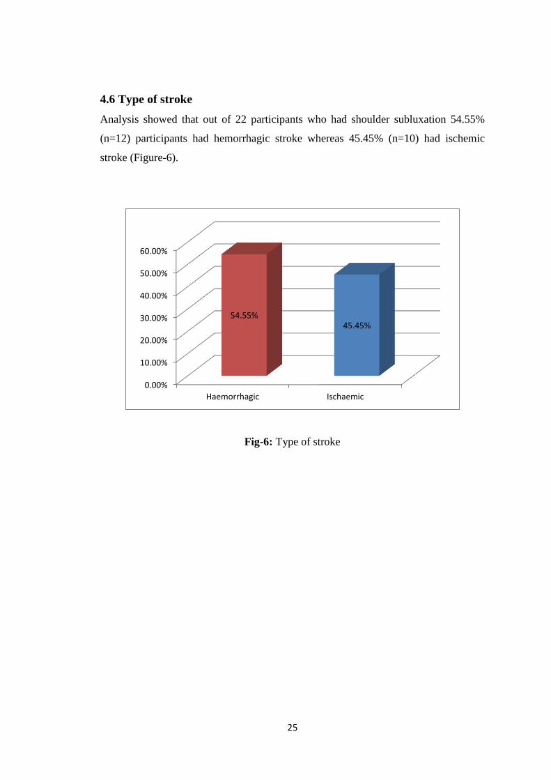
4.6 Type of stroke
Analysis showed that out of 22 participants who had shoulder subluxation 54.55%
(n=12) participants had hemorrhagic stroke whereas 45.45% (n=10) had ischemic
stroke (Figure-6).
Fig-6: Type of stroke
0.00%
10.00%
20.00%
30.00%
40.00%
50.00%
60.00%
Haemorrhagic Ischaemic
54.55% 45.45%
26
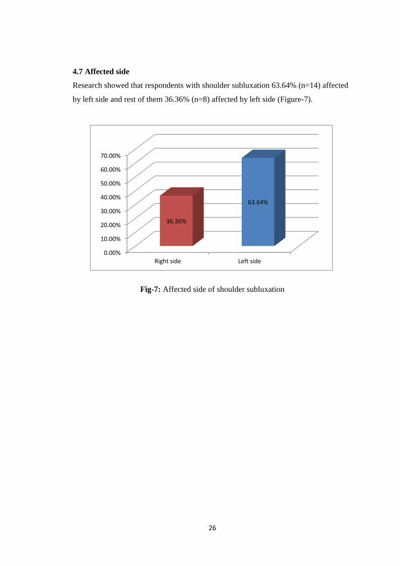
4.7 Affected side
Research showed that respondents with shoulder subluxation 63.64% (n=14) affected
by left side and rest of them 36.36% (n=8) affected by left side (Figure-7).
Fig-7: Affected side of shoulder subluxation
0.00%
10.00%
20.00%
30.00%
40.00%
50.00%
60.00%
70.00%
Right side Left side
36.36%
63.64%
27
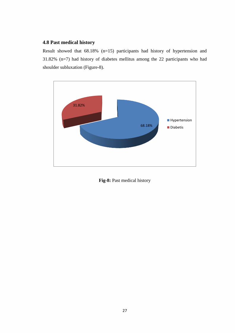
4.8 Past medical history
Result showed that 68.18% (n=15) participants had history of hypertension and
31.82% (n=7) had history of diabetes mellitus among the 22 participants who had
shoulder subluxation (Figure-8).
Fig-8: Past medical history
68.18%
31.82%
Hypertension
Diabetis
28
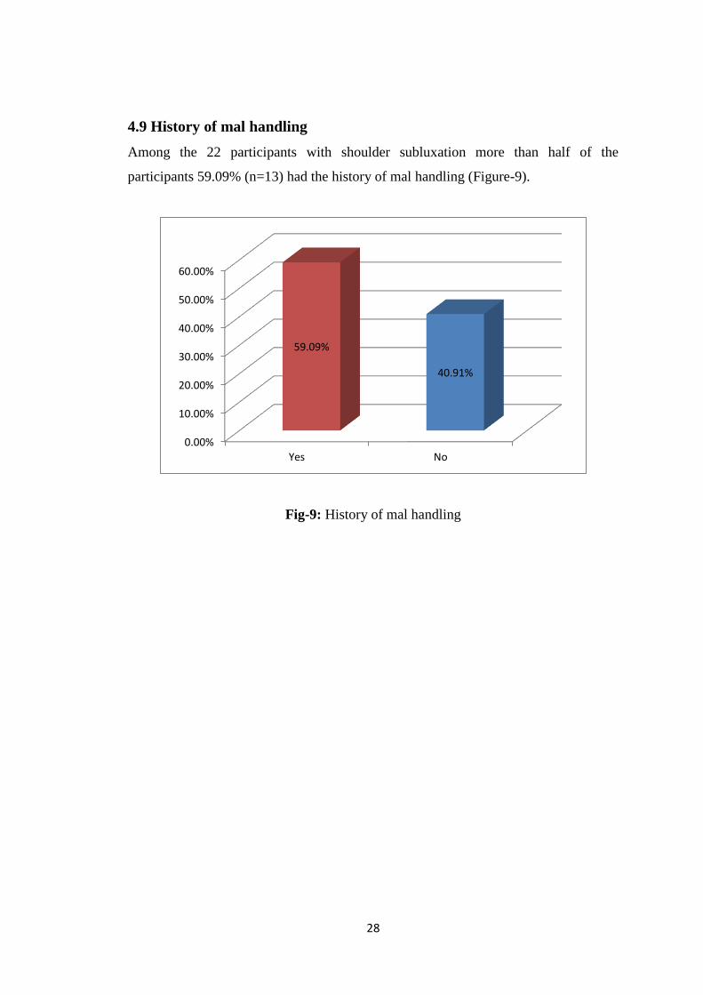
4.9 History of mal handling
Among the 22 participants with shoulder subluxation more than half of the
participants 59.09% (n=13) had the history of mal handling (Figure-9).
Fig-9: History of mal handling
0.00%
10.00%
20.00%
30.00%
40.00%
50.00%
60.00%
Yes No
59.09%
40.91%
29
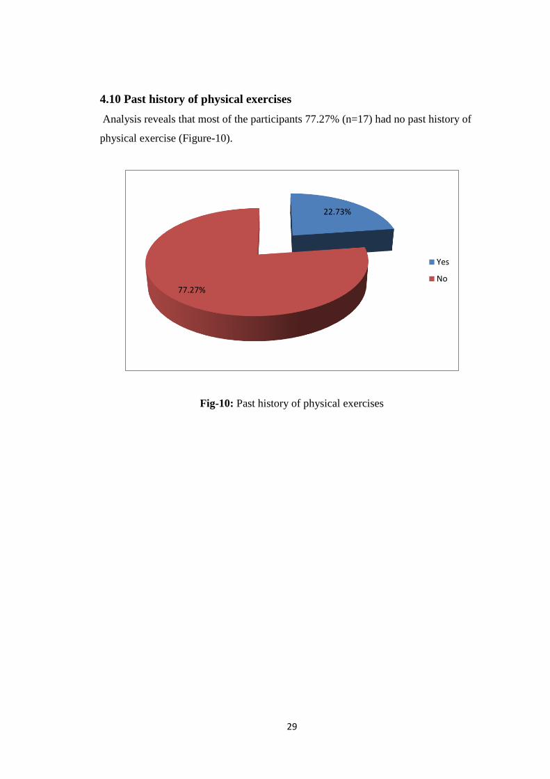
4.10 Past history of physical exercises
Analysis reveals that most of the participants 77.27% (n=17) had no past history of
physical exercise (Figure-10).
Fig-10: Past history of physical exercises
22.73%
77.27%
Yes
No
30
CHAPTER-V DISCUSSION
The objectives of the study was to find out the prevalence of shoulder subluxation
among stroke patients attended at CRP along with distribution with age, gender , past
exposure and so one. However the study findings show that almost 62.85% of the
participants had shoulder subluxation among the stroke patients. Shakoor et al.,
(2009) stated that the prevalence of shoulder subluxation was 81% among the stroke
patients in Sydney.
In this study almost 31.82% of the participants were age group 53-60 years. In United
States a study about epidemiology of shoulder subluxation by Zhang and Jordan
(2008) shows that the age standardized prevalence of shoulder subluxation after
stroke in adults age ≥ 45 was 25.2% among the participants in the Framingham Study
and 27.8% in the Johnston County.
Analysis showed that almost 63.4% of the participants were male who had shoulder
subluxation among the stroke patients. Reijman et al. (2007) stated that (55%)
majority were male who developed shoulder subluxation after stroke.
The study showed that 54.55% the participants came from urban area that had
shoulder subluxation. People in urban area are more prone (60%) to stroke than rural
area in United States (Gelber et al. 2000).
Result showed that 54.555% participants had hemorrhagic stroke among stroke
patients who had shoulder subluxation. Reijman et al. (2007) stated that hemorrhagic
stroke were (23%) more common in adults in Netherlands.
Analysis showed that 68.18% participants had the history of hypertension among the
stroke patients with shoulder subluxation. Gelber et al., (2000) stated that people who
had hypertension had 10% greater chance for developing shoulder subluxation after
stroke.
31
The study showed that 59.09% participants had had the history of mal handling
among the participants with shoulder subluxation. Mal handling is more common risk
factor for developing shoulder subluxation among the stroke patients (Gelber et al.,
2000).
Analysis showed that 77.27% participants had no past history of physical exercises
among the stroke patients who had shoulder subluxation. Magee (1997) shown in
their research that physically in active person are more (65%) prone to developing
shoulder subluxation after stroke.
Analysis showed that 31.32 participant had the history of diabetics mellitus and
45.45% ischemic stroke among the participant with shoulder subluxation.
32
CHAPTER-VI CONCLUSION AND RECOMMENDATION
6.1 Conclusion
From the data base, it was found that two third of the participants had shoulder
subluxation among the stroke patients. Percentages of shoulder subluxation were
higher in male and among them most of the participants were service holder and one
third of the people were in between 53-60 age groups. The participants who had
shoulder subluxation, more than half had the history of mal handling and two third of
them had no past history of physical exercises.
Physiotherapist should follow some intervention and consider high prevalence factors
to avoid and prevent shoulder subluxation. To identify associated factors of this
condition would be beneficial in preventive strategy and proper interventions may
reduce the impact on the quality of life and functional ability of stroke patients.
33
6.2 Recommendation
The purpose of the study was to identify the prevalence of shoulder subluxation among
the stroke patients. The researcher identified some further step that might be taken into
consideration for the better accomplishment of further research. For the ensuring of the
generalizibility of the research it is recommended to investigate large sample.
34
REFERENCES
Bierman, S.N.,1993.Cerebrovascular accident in hansen,R.A and Atchison, B.
(eds.) Conditions in Occupational Therapy: Effect on Occupational
performance. Williams &Wilkins .
Bobath, B.,1977. Treatment of Adult Hemiplegia. Physiotherapy .
Boyd et al., 1999)Shoulder supports revisited: A Canadian follow-up servey.
Canadian Journal of Occupational Therapy 66(2), 161-167.
Brocklehurst, K.,1978. How much physical Therapy for the patients with
stroke? British Medical Journal 1, 1307-1310.
Carr, J., Shepherd , R., 2000. Neurological Rehabilitation : Optimizing Motor
Performance. Butter worth Heinemann.
David, M.D., Zi-Ping, F., 2000. Effects of FES on Post Stroke shoulder
Dysfunction
Duponti , e.m. et al., 2001.clinical trials of bion , injectable neuromuscular
stimulators.
Gelber , A.C., Hochberg, M.C., Mead, L.A., Wang, N., Wigley, F.M., Klag,
M.J., 1998, joint injury in young Adults and Risk for shoulder subluxation,
retrieved on 4th
Jan 2012.
<http//www.annals.orgg/content/133/5/321.full.pdf+html>.
Hall, J.,Dudgeon , B., Guthrie , M .,1995. Validity of clinical Measures in
Adults With poststroke Hemiplegia . The American Journal of Occupational
Therapy .
Hansen, R.A., Atchison , B., 1993. Conditions in Occupational Therapy:
Effect on Occupational Therapy performance.
Hayee, et al., 2002. Aetiology of Yong Ischaemic stroke in Bangladesh.
Bangladesh Journal of Neuroscience .
Hicks, C.M .,2000. Rearch Methods for Clinical Therapist:applied project
design and analysis , 3rd ed. Churchill Livingstone.
Hopkins, H.L., Smith, H.D.,1993. Willard And Spackman’s Occupational
Therapy, 8th.ed.J.B.Lippincott, Philadelphia .
35
Jackson, S.,1996. Cerebrovascular accident in Turner, A. Foster , M. and
Jonson , S.E (eds.) Occupational Therapy and physical Dysfunction: Principles
, skills and practice, 4th ed. Churchill Livingstone.
Magee, D.J., 1997. Orthopedic Physical Assessment, 3rd
edition. W.B
Saunders Company, Philadelphia.
Mohammad, Q.D.,2001. Stroke : prevention and treatment in the new
milennium. 1st National Conference and scientific seminar, 1st Dhaka
Sheraton Hotel , Bangladesh, 4th May. soceity of Neurologist of Bangladesh
Morely, et al., 2002. Management of the Subluxed Low Tone Shoulder .
Physiotherapy 88(4), 208-215.
Nayan, j.,2003. Functional Performance afetr Stroke : A study of shoulder pain
and techniques to improve functional performance.
Pedretti, L.W., Smith, J.A., Pendleton, H.M.,1996. Cerebral Vascular accident
in Pedretti, L.W. (ed.) Occupational therapy: Practice Skill For Physical
Dysfunction.
Pedretti, L.W., Zoltan, B.,1990. Occupational Therapy: Practice skill For
Physical Dysfunction , 3rd ed. Mosby, Toronto.
Rahman, K.M., et al.,2002 .Study on Neurological disease in 7689 Hospital
admitted patients- A Restropective Study in Mymensingh Medical Collage
Hospital Bangladesh .Bangladesh Journal Of Neuroscience 18(1/2) 28-31.
Reijman, M., Pols, HAP, Bergink, AP, Hazes, JMW, Belo, JN, Lievense, AM
and Bierma-Zeinstra, SMA 2007, ‘Body mass index associated with onset and
progression of Crebro vascular diseases: the Rotterdam Study’ Annals of the
rheumatic diseases, retrieved on 9th
Feb 2012.
<http//www.ard.bmj.com/content/66/158/abstract> .
Ryerson, S.D.,2001. Hemiplegia in Umphred , D.A (ed.) Neurological
Rehabilitation.
Shakoor, M.A., Taslim, M.A., Ahmed, M.S., Hasan, S.A., 2009. Clinical
profile of patients with Shoulder subluxation: A study of 162 cases, retrieved
on 13th
Feb 2012. <http://www.ijpmr.com>.
36
Shepherd, R.B. and Carr, J.H., 1998. The shoulder following stroke:
Preserving Musculoskeletal Integrity for Function . Topics in stroke
Rehabilitation 4(4).
Smith, R.G., et al., 1982. Malalignment of the ahoulder after stroke. British
Medical Journal.
Spauding , S.J.,1999. Biomechanical analysis of four supports for the
subluxed hemiparetic shoulder . Canadian Journal of Occupational Therapy.
Trombly, C.A., 1989. Occupational Therapy for Physical Dysfunction, 3rd ed.
Williams & Wilkins, Baltimore .
Turner, A., Forster, M., Johnson, S.E., 1996. Physical Therapy and Physical
Dysfunction: Principles , Skills and Practice, 4th ed. Chuechill Livingstone,
New York.
Turton, A.,1998. Mechanisms for recovery of hand arm function after stroke:
a review of evidence from studies using non-invasive investigative
techniques. British Journal Occupational Therapy.
Uddin, M.T.,2002., Basic Electrotherapy . Asian colour printing.
Ullah, M.M.,2003. Parents as Co-Therapists In Intervention Programme: A
family centered approach.
Umphred, D . A.,2001. Neurological Rehabilitation , 4th ed. Mosby.
Umphred, D.A.,1990. Neurological Rehabilitation, 3rd ed. Mosby, 118.
Wade, J.P.H.,1992. Clinical aspects of stroke in Dowine, P.A. (ed.) Cash’s
Textbook of Neurology for Physiotherapist With a Forward by Dame Cicely
Saun , 4th ed. Wolfe, 240-252.
Warlow, C.,1993. Disorders of the cerebral circulation in Walton, J.(ed.)
Brain disesase.
Zhang, Y., Jordan, J.M., 2008. Epidemiology of stroke, journal of North
America retrieved on 6th
Feb 2012.
Zorowitz, R. D. et al.,1996. shoulder pain and subluxation after stroke:
correlation or coincidence. The American Journal of Physical Therapy .