prevalence of human-active and variant 1 strains of the tick-borne pathogen anaplasma...
TRANSCRIPT
Am. J. Trop. Med. Hyg., 91(2), 2014, pp. 302–309doi:10.4269/ajtmh.13-0525Copyright © 2014 by The American Society of Tropical Medicine and Hygiene
Prevalence of Human-Active and Variant 1 Strains of the Tick-Borne Pathogen
Anaplasma phagocytophilum in Hosts and Forests of Eastern North America
Felicia Keesing,* Diana J. McHenry, Michelle Hersh, Michael Tibbetts, Jesse L. Brunner, Mary Killilea, Kathleen LoGiudice,Kenneth A. Schmidt, and Richard S. Ostfeld
Bard College, Annandale-on-Hudson, New York; Sarah Lawrence College, Bronxville, New York; Washington State University,Pullman, Washington; New York University, New York, New York; Union College, Schenectady, New York;
Texas Tech University, Lubbock, Texas; Cary Institute of Ecosystem Studies, Millbrook, New York
Abstract. Anaplasmosis is an emerging infectious disease caused by infection with the bacterium Anaplasmaphagocytophilum. In the eastern United States, A. phagocytophilum is transmitted to hosts through the bite of theblacklegged tick, Ixodes scapularis. We determined the realized reservoir competence of 14 species of common vertebratehosts for ticks by establishing the probability that each species transmits two important strains of A. phagocytophilum(A. phagocytophilum human-active, which causes human cases, and A. phagocytophilum variant 1, which does not) tofeeding larval ticks. We also sampled questing nymphal ticks from ~150 sites in a single county over 2 years and sampledover 6 years at one location. White-footed mice (Peromyscus leucopus) and Eastern chipmunks (Tamias striatus) werethe most competent reservoirs for infection with the A. phagocytophilum human-active strain. Across the county,prevalence in ticks for both strains together was 8.3%; ticks were more than two times as likely to be infected withA. phagocytophilum human-active as A. phagocytophilum variant 1.
INTRODUCTION
Anaplasmosis is a rapidly emerging infectious disease in theUnited States, with over 1,700 cases reported in 2010—a 50%increase in the number of cases reported in the previous yearand five times the number reported in 2000.1 Most reportedcases of anaplasmosis in the United States are concentrated innorth central and northeastern states, although cases havebeen documented in 35 of 50 states.2,3 Anaplasmosis has alsobeen documented throughout Europe and Asia.4–8
Patients with anaplasmosis typically present with non-specificfebrile symptoms, including fever, chills, headache, and myal-gia,9,10 particularly during summer months.2,3 Most cases ofanaplasmosis respond well to antibiotic treatment, but 17–56%of patients with anaplasmosis are hospitalized, and an esti-mated 1% of cases prove fatal.10 Because of difficulties in diag-nosis and lack of awareness of anaplasmosis by physicians andthe public, many cases are misdiagnosed,11 and national statis-tics almost certainly dramatically underreport this disease.3,12
Anaplasmosis is caused by a rickettsial bacteriumAnaplasmaphagocytophilum, groups of which form dense aggregates(morulae) in granulocytes.13,14 The bacterium is passed fromhost to host through the bite of an infected ixodid tick—Ixodes scapularis in the eastern and central United States,I. pacificus in the western United States, and other ixodidticks in Europe and Asia.10,15,16 A number of vertebratespecies has been shown through serology to be exposed toA. phagocytophilum in nature.17–24 However, the relevance ofserological data to transmission dynamics is limited, becauseseropositive individuals might not be currently infected andtherefore, might not be infectious to feeding ticks. In the north-eastern United States, actual rates of infection or transmissionfrom infected wild hosts to naıve ticks have recently been eval-uated for a suite of hosts from eastern deciduous forests.25 Thismeasure, called the realized reservoir competence, combinesthe probability that a particular host species will be infectedand the probability that it will transmit the infection to feeding
ticks.25 Short-tailed shrews (Blarina brevicauda), white-footed
mice (Peromyscus leucopus), and Eastern chipmunks (Tamias
striatus) had the highest mean realized reservoir competence,
infecting 10–15% of feeding ticks. Other hosts, including
American robins (Turdus migratorius), raccoons (Procyon
lotor), and opossums (Didelphis virginiana), were poor reser-
voirs by this metric, infecting < 5% of feeding ticks. All species
tested were capable of transmitting A. phagocytophilum to
feeding ticks, which is in contrast to earlier reports,26 although
earlier results were based on smaller sample sizes.From an epidemiological point of view, not all strains of
A. phagocytophilum are equally important. There are multi-
ple strains of A. phagocytophilum in vertebrate hosts,27–31 but
only one strain seems to infect humans in the northeastern
United States.30 White-footed mice have been reported to be
competent reservoirs for the human-infectious strain, called
A. phagocytophilum human-active (A. phagocytophilum-ha),
but not the other major strain, A. phagocytophilum variant 1
(A. phagocytophilum-v1). This conclusion was reached, because
mice did not transmit A. phagocytophilum-v1 to feeding ticks,despite previous exposure to that strain in the laboratory.29 In
contrast, ticks feeding on white-tailed deer (Odocoileus
virginianus) are infected with A. phagocytophilum-v1 more
frequently than with A. phagocytophilum-ha.32 This result
may be because deer cannot support an infection with
A. phagocytophilum-ha. In one study, almost 40% of ticks
collected from deer were infected with A. phagocytophilum;
of these ticks, 26% were carrying the A. phagocytophilum-ha
strain.32 However, blood samples collected from the same deer
were never positive for A. phagocytophilum-ha.32
Despite the growing health concern about anaplasmosis in
the United States and elsewhere, relatively little is known about
interspecies variation in transmission ofA. phagocytophilum-ha
and A. phagocytophilum-v1, and little is known about the
relative abundances of the two strains in questing ticks. We
determined the realized reservoir competence for strains
A. phagocytophilum-ha and A. phagocytophilum-v1 of 14 spe-
cies of common vertebrate hosts for ticks. We then compared
these realized reservoir competence data with the relative
abundances of the two strains in host-seeking ticks collected*Address correspondence to Felicia Keesing, Bard College, PO Box5000, Annandale-on-Hudson, NY 12504. E-mail: [email protected]
302
from 146 different locations in a single county over 2 years in theHudson Valley of New York, where anaplasmosis is endemic.
METHODS
Collecting ticks from hosts. To determine realized reservoircompetence for the two strains, we trapped host individualsfrom 10 mammal species and 4 bird species using the methodsdescribed in detail elsewhere.33 Briefly, hosts were capturedon the property of the Cary Institute of Ecosystem Studies inMillbrook, New York during the peak abundance of larvalblack-legged ticks (I. scapularis) from July to September in2008, 2009, and 2010. Captured individuals were held for3 days in cages with wire mesh floors suspended over panslined with wet paper towels so that ticks could feed to reple-tion and drop from hosts.In some cases, if hosts did not drop > 10 ticks within 3 days,
we infested them with unfed larval ticks. In these cases, eachhost was inoculated with larval ticks that had been eithercollected in the field or hatched from eggs in the laboratory.Larvae hatched from eggs were the offspring of adult ticksthat had been collected from the area of the study and sub-sequently fed on rabbits. Transovarial transmission ofA. phagocytophilum is not known to occur,34 and therefore,larval ticks are uninfected; these infestations should not affecthost exposure to the pathogen. During infestations, mice andbirds were restrained by hand, whereas all other hosts wererestrained in nylon mesh handling cones. Infestations wereconducted by placing ticks on the host’s neck and head with a#00 paintbrush. Hosts that had been infested were held for anadditional 4 days, and engorged ticks were collected each day.Engorged larvae were held in moistened glass vials until
they molted into the nymphal stage. Newly molted nymphswere flash-frozen in liquid nitrogen and stored at −80°C. Allanimal care and husbandry were conducted with approvalfrom the Cary Institute of Ecosystem Studies InstitutionalAnimal Care and Use Committee.In general, we only assessed realized reservoir competence
of hosts that produced a minimum of 10 newly molted nymphs.However, three species—Glaucomys volans, Sorex cinereus,and Mephitis mephitis—had low body burdens. For thesespecies, we tested ticks from individuals with greater thanfour newly molted nymphs; we consider these data provisionalbecause of the low number of ticks per individual host.Collecting ticks from forests. Between 2007 and 2012, we
collected questing nymphal ticks from three long-term plotsat the Cary Institute of Ecosystem Studies in Millbrook,New York. Forests at the Cary Institute are typical of theeastern deciduous forests of New York and New England.Plots are dominated by oaks (Quercus rubra and Q. prinus)in the overstory, with primarily oak and sugar maple (Acersaccharum) seedlings, maple-leaved viburnum (Viburnumacerifolium), witch hazel (Hamamelis virginiana), and iron-wood (Ostrya virginiana) in the understory. One 2.25-ha plot(150 + 150 m) was established in 1991, and two more plotswere added in 1995 to comprise three plots, with more than700 m separating pairs.We also sampled questing nymphal ticks in June of 2011
(148 sites) and June of 2012 (78 sites) in forested locationsthroughout Dutchess County, New York. For county locations,we selected sites using a Geographic Information Systems(GIS) map of forested and non-forested land cover digitized
from aerial orthophotos generated in 2009. We generated aninitial candidate list of 2,500 random points using a randompoint overlay. These points were then stratified by the per-centage of forest cover in the surrounding landscape to pro-vide equal representation along a gradient of forest coverfrom extensively forested to highly fragmented. We elimi-nated sites when access was poor or property owners couldnot be located or recruited.At all sites in all years, we collected questing nymphal ticks
by drag sampling.35 Corduroy cloths (1 m2) were dragged along400-m transects in each site one or two times in a given yearduring the annual peak in nymphal questing activity. Tickswere collected from the cloths every 15–30 m. Questing nymphswere flash-frozen on collection as described above. All sitessampled for questing nymphs were in eastern deciduous forests.To estimate prevalence at each site, we tested 10–30 ticks.Extracting and amplifying DNA. Total genomic DNA was
extracted from ticks using either the DNeasy or DNeasy96 Blood and Tissue Kit (Qiagen, Hilden, Germany) or theGentra PureGene Tissue Kit (Qiagen, Hilden, Germany). Toamplify extracted DNA, we followed established protocols.25,36
Briefly, we used primers ApMSP2f and ApMSP2r andprobe ApMSP2p, which are specific to the msp2 gene ofA. phagocytophilum and generate a 77-base pair (bp) fragment.Real-time polymerase chain reaction (PCR) was performed in aBio-Rad CFX96 Real-Time PCR System (Bio-Rad, Hercules,CA). We used DNA extractions from unfed larval ticks andultrapure water as negative controls. The cloned 77-bp targetwas used as a positive control. Barrier pipette tips were usedthroughout the process to prevent contamination.For each tick, we conducted three replicate assays using PCR.
As described fully elsewhere,25 ticks were considered positivefor A. phagocytophilum if any one of three replicate sampleswas called positive by default settings, meaning that it amplifiedrelative to negative controls. We conducted additional confir-matory tests for any ticks with marginal results (i.e., moderatefluorescence). If any replicate was positive in the confirmatorytests, ticks were considered positive for A. phagocytophilum;if all three replicates in the confirmatory test were marginal ornegative, ticks were considered negative.Determining strain identity. Ticks that were positive for
A. phagocytophilum in these initial tests were subsequentlytested for the identity of the strain that they carried usingnested PCR. In the primary round, we used primers ge3a andge237 to amplify a 546-bp segment of the 16S gene. The prod-uct from this round was used as a template in a second roundof PCR, in which primers ge9f37 and ge9r (5¢-TTA CTC ACCCGT CTG CCA CT-3¢; designed for this study) were used toamplify a 58-bp product. Primary-round PCRs were performedin 20-mL volumes with final concentrations of 1 + PromegaPCRMaster Mix (Promega, Madison,WI) and 0.5 mMprimers.Thermal cycling was performed with an initial denaturingperiod of 95°C for 2 minutes, then 40 cycles of 94°C for30 seconds, 55°C for 1 minute, and 72°C for 1 minute, and afinal extension of 72°C for 5 minutes. Secondary-round PCRswere performed in 20-mL volumes with final concentrations of1 + iQ SYBR Green Supermix (Bio-Rad, Hercules, CA) and0.5 mM primers. Thermal cycling was performed with an ini-tial denaturing period of 95°C for 10 minutes, then 40 cyclesof 95°C for 15 seconds and 60°C for 1 minute followed by amelt curve analysis, in which the temperature increased from76°C to 85°C at 0.2°C every 10 seconds.
ECOLOGY OF ANAPLASMA PHAGOCYTOPHILUM STRAINS 303
The melt temperature forA. phagocytophilum-ha was 78.6–79.8°C, whereas for A. phagocytophilum-v1, it was 81.6–83.4°C. We did not observe consistent melt temperaturesother than those melt temperatures in the ranges indicatedabove. To verify these melt temperatures, we sequenced theDNA from 10 questing nymphs from each strain category. EachDNA segment was cloned before sequencing; the number ofclones per DNA ranged from 25 to 40 (A. phagocytophilum-ha:mean = 32.6; A. phagocytophilum-v1: mean = 37.6). For sam-ples identified as A. phagocytophilum-v1, 98% of clones wereidentical to A. phagocytophilum-v1 (accession no. AY193887in GenBank) (Supplemental Table 1). The remaining 2% ofclones differed from the A. phagocytophilum-v1 sequence by1 bp (Supplemental Table 1), and none of the clones matchedthe sequence identified as A. phagocytophilum-ha. ForA. phagocytophilum-ha, 70% of 317 total clones were identicalto A. phagocytophilum-ha (accession no. U02521 in GenBank)(Supplemental Table 1). The remaining clones differed fromA. phagocytophilum-ha in GenBank by 1 or 2 bp, and noneof the clones matched the sequence identified asA. phagocytophilum-v1. The identification of clones that dif-fered from the A. phagocytophilum-ha reference sequence butare not A. phagocytophilum-v1 suggests the possibility of acomplex of strains of A. phagocytophilum-ha. Our method didnot allow detection of coinfection of both strains in one tick.Estimating infection prevalence. Realized reservoir compe-
tence for each host species was calculated as the mean per-centage of ticks infected per individual host. This measureincorporates natural variation among species in infection withA. phagocytophilum as well as variation among species in theprobability of transmitting infection to feeding ticks.38 Weused likelihood-based methods to separately estimate preva-lence of infection of both strains among hosts as well as eachhost’s propensity to transmit the infection to ticks given aninfection.38 At our long-term plots at the Cary Institute, weused the pool of ticks collected on the three plots to calculatethe mean site-wide infection prevalence of questing nymphalticks for each year. At our county sites, we calculated themean infection prevalence of questing nymphs from each site,including only those sites where we were able to collect ³ 10nymphs in either year. To determine if the infectionprevalence of questing nymphal ticks at county sites was cor-
related between years, we calculated the correlation coeffi-cient of tick infection prevalence for each strain. For thisanalysis, we excluded sites at which no evidence of eitherA. phagocytophilum strain was detected in either year.
RESULTS
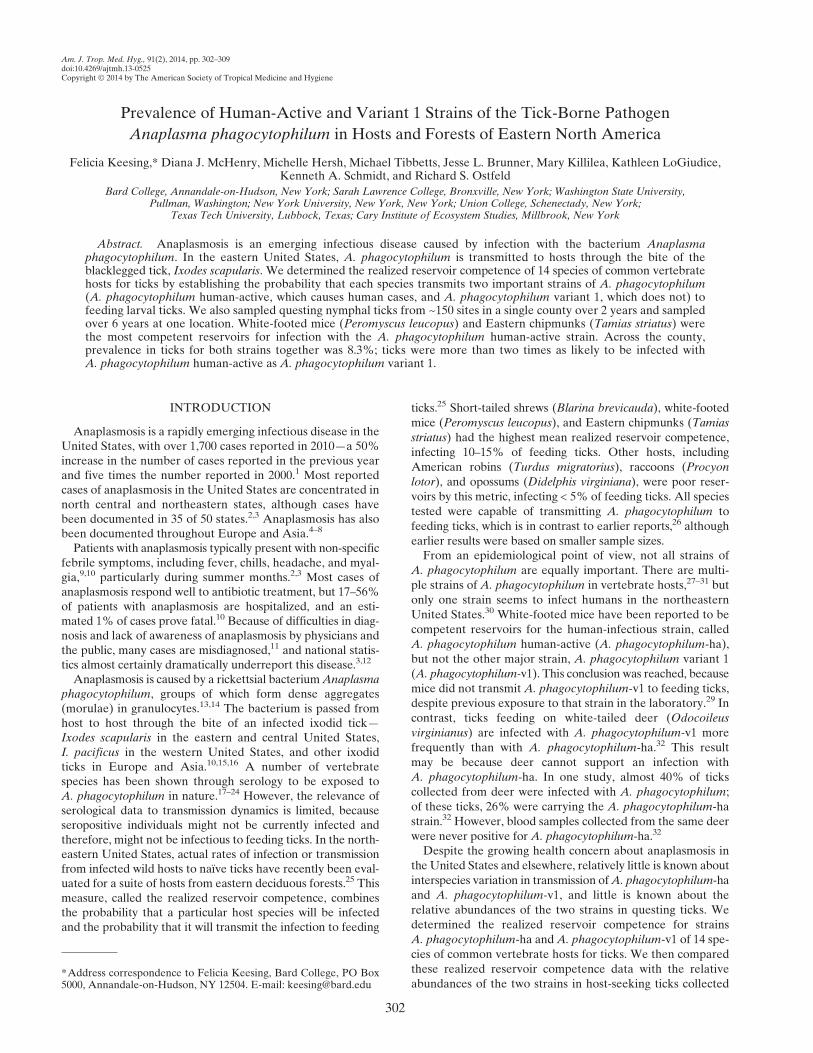
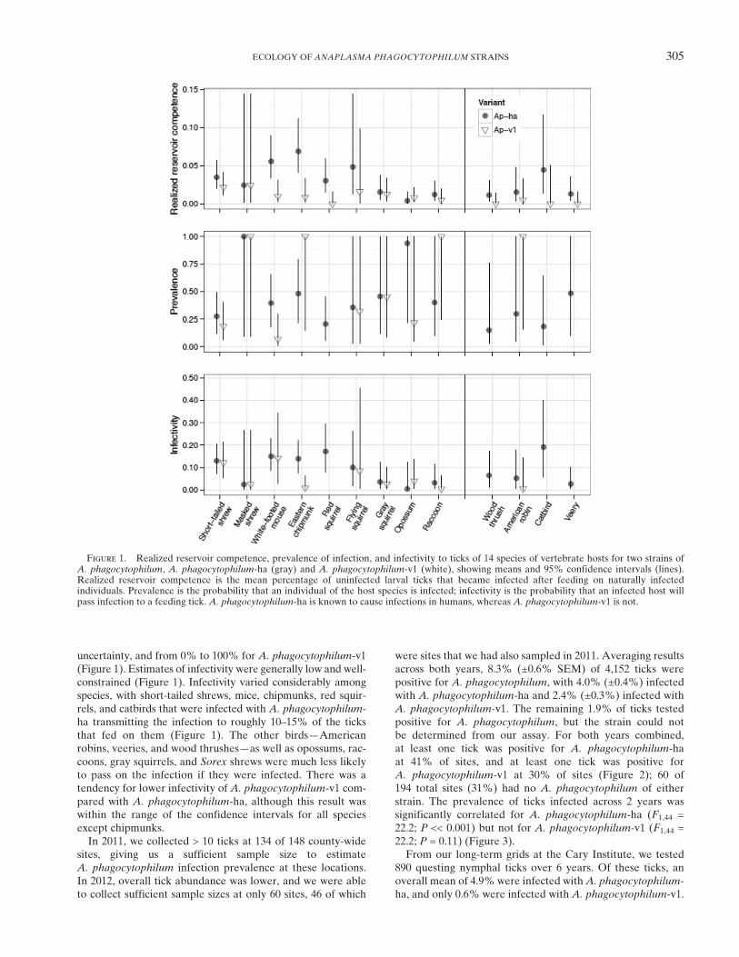
To determine realized reservoir competence of host speciesfor the two strains of A. phagocytophilum, we tested a totalof 5,098 nymphal ticks from 281 individuals of 14 hostspecies. Overall, 2.7% of ticks were infected with strainA. phagocytophilum-ha, and 0.8% of ticks were infected withstrain A. phagocytophilum-v1. As previously reported, hostspecies varied in the probability that they would transmitA. phagocytophilum to feeding ticks (Figure 1).25 Hosts alsovaried in the proportion of ticks that they infected with thetwo strains (Figure 1). White-footed mice and Easternchipmunks, for example, were relatively likely to transmitA. phagocytophilum-ha, with mice infecting 6.7% (±0.6%SEM) and chipmunks infecting 6.8% (±2.0%) of feedinglarval ticks. However, these species were relatively unlikelyto transmit A. phagocytophilum-v1, with both species infecting< 1% of feeding larvae (Table 1). In contrast, all ticks thatacquired infection from striped skunks and the majority of ticksthat acquired infection from Virginia opossums and southernflying squirrels were infected with A. phagocytophilum-v1. Of281 hosts from which we tested ticks, 10 individuals transmittedboth A. phagocytophilum-ha and A. phagocytophilum-v1 tofeeding ticks. These 10 individuals represented 7 of 14 speciesof hosts that we identified (Table 1). In particular, the twospecies of shrews were more likely than other taxa to transmitboth strains (10% of Blarina and 17% of Sorex) (Table 1). Inaddition, 9% of chipmunks transmitted both strains. Theremaining host species, including mice and three species ofground-nesting songbirds, either never or rarely transmittedboth strains (Table 1). For 2% of infected ticks, we detectedinfection with A. phagocytophilum but could not determinestrain identity using our assay.We differentiated realized reservoir competence into its
two components, prevalence and infectivity, as described inMethods. Estimates of prevalence for each species varied from15% to 100% for A. phagocytophilum-ha, with a great deal of
Table 1
Sample size, realized reservoir competence, and transmission probabilities for 14 common hosts for two strains of A. phagocytophilum:A. phagocytophilum-ha (Ap-ha) and A. phagocytophilum-v1 (Ap-v1)
Species N hosts N ticks
Mean (SE) realized reservoir competence Mean percentage of individual hosts transmitting
Ap-ha Ap-v1 Ap-ha Ap-v1 Both
Eastern chipmunk T. striatus 23 369 6.78% (2.00%) 0.58% (0.31%) 26.1% 8.7% 8.7%White-footed mouse P. leucopus 38 748 6.65% (1.98%) 0.94% (0.61%) 15.8% 2.6% 2.6%Red squirrel Tamiasciurus hudsonicus 15 297 4.92% (4.22%) 0.00% (0.00%) 20.0% 0.00% 0.00%Short-tailed shrew B. brevicauda 29 546 3.64% (1.51%) 2.22% (1.05%) 20.7% 13.8% 10.3%Catbird Dumetella carolinensis 18 299 3.33% (1.92%) 0.00% (0.00%) 5.6% 0.0% 0.0%Flying squirrel* G. volans 7 87 2.00% (1.85%) 2.38% (2.20%) 14.3% 14.3% 0.0%Gray squirrel Sciurus carolinensis 20 358 1.74% (0.79%) 1.02% (0.55%) 20.0% 15.0% 5.0%Masked shrew* S. cinereus 6 41 1.67% (1.67%) 1.67% (1.67%) 16.7% 16.7% 16.7%Raccoon P. lotor 26 503 1.40% (0.60%) 0.38% (0.24%) 15.4% 7.7% 3.9%American robin T. migratorius 20 345 1.40% (0.76%) 0.42% (0.32%) 10.0% 5.0% 0.0%Veery Catharus fuscescens 22 445 1.17% (0.55%) 0.00% (0.00%) 13.6% 0.0% 0.0%Wood thrush Hylocichla mustelina 28 496 0.89% (0.59%) 0.00% (0.00%) 7.1% 0.0% 0.0%Opossum D. virginiana 27 533 0.53% (0.37%) 1.05% (0.68%) 7.4% 11.1% 3.7%Striped skunk* Mephitis mephitis 2 31 0.00% (0.00%) 4.76% (0.00%) 0.0% 50.0% 0.0%
Realized reservoir competence is measured as the mean percentage of uninfected ticks that became infected from feeding on an individual wild host.*Small sample size.
304 KEESING AND OTHERS
uncertainty, and from 0% to 100% for A. phagocytophilum-v1(Figure 1). Estimates of infectivity were generally low and well-constrained (Figure 1). Infectivity varied considerably amongspecies, with short-tailed shrews, mice, chipmunks, red squir-rels, and catbirds that were infected with A. phagocytophilum-ha transmitting the infection to roughly 10–15% of the ticksthat fed on them (Figure 1). The other birds—Americanrobins, veeries, and wood thrushes—as well as opossums, rac-coons, gray squirrels, and Sorex shrews were much less likelyto pass on the infection if they were infected. There was atendency for lower infectivity of A. phagocytophilum-v1 com-pared with A. phagocytophilum-ha, although this result waswithin the range of the confidence intervals for all speciesexcept chipmunks.In 2011, we collected > 10 ticks at 134 of 148 county-wide
sites, giving us a sufficient sample size to estimateA. phagocytophilum infection prevalence at these locations.In 2012, overall tick abundance was lower, and we were ableto collect sufficient sample sizes at only 60 sites, 46 of which
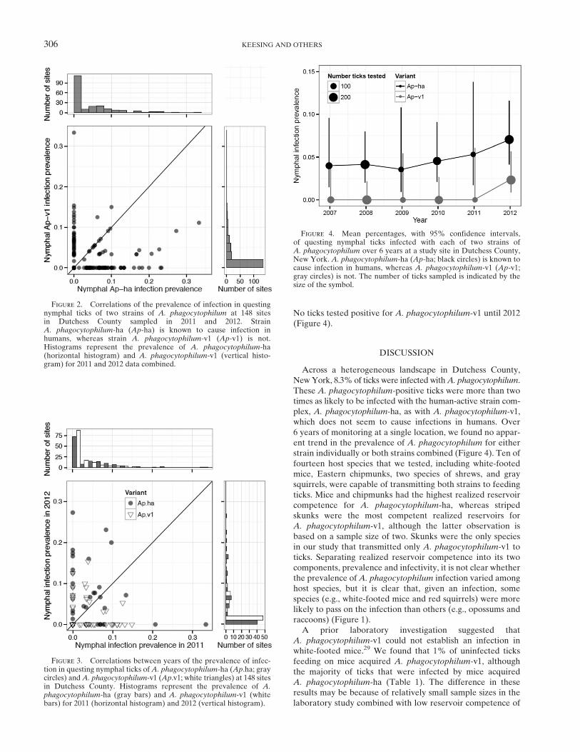
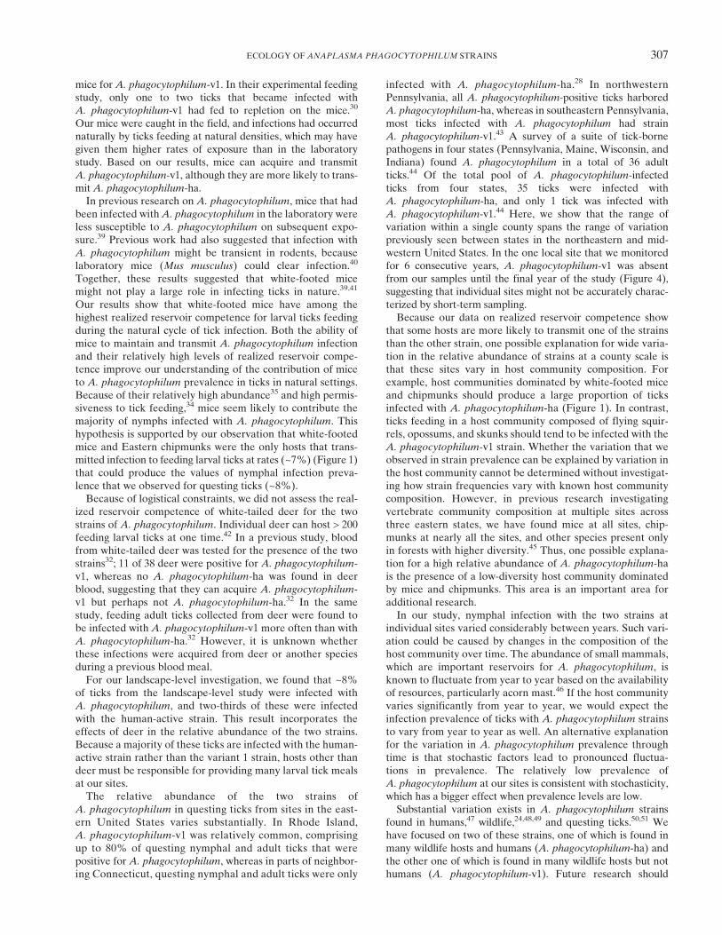
were sites that we had also sampled in 2011. Averaging resultsacross both years, 8.3% (±0.6% SEM) of 4,152 ticks werepositive for A. phagocytophilum, with 4.0% (±0.4%) infectedwith A. phagocytophilum-ha and 2.4% (±0.3%) infected withA. phagocytophilum-v1. The remaining 1.9% of ticks testedpositive for A. phagocytophilum, but the strain could notbe determined from our assay. For both years combined,at least one tick was positive for A. phagocytophilum-haat 41% of sites, and at least one tick was positive forA. phagocytophilum-v1 at 30% of sites (Figure 2); 60 of194 total sites (31%) had no A. phagocytophilum of eitherstrain. The prevalence of ticks infected across 2 years wassignificantly correlated for A. phagocytophilum-ha (F1,44 =22.2; P << 0.001) but not for A. phagocytophilum-v1 (F1,44 =22.2; P = 0.11) (Figure 3).From our long-term grids at the Cary Institute, we tested
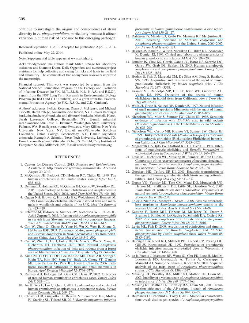
890 questing nymphal ticks over 6 years. Of these ticks, anoverall mean of 4.9% were infected withA. phagocytophilum-ha, and only 0.6% were infected with A. phagocytophilum-v1.
Figure 1. Realized reservoir competence, prevalence of infection, and infectivity to ticks of 14 species of vertebrate hosts for two strains ofA. phagocytophilum, A. phagocytophilum-ha (gray) and A. phagocytophilum-v1 (white), showing means and 95% confidence intervals (lines).Realized reservoir competence is the mean percentage of uninfected larval ticks that became infected after feeding on naturally infectedindividuals. Prevalence is the probability that an individual of the host species is infected; infectivity is the probability that an infected host willpass infection to a feeding tick. A. phagocytophilum-ha is known to cause infections in humans, whereas A. phagocytophilum-v1 is not.
ECOLOGY OF ANAPLASMA PHAGOCYTOPHILUM STRAINS 305
No ticks tested positive for A. phagocytophilum-v1 until 2012(Figure 4).
DISCUSSION
Across a heterogeneous landscape in Dutchess County,NewYork, 8.3% of ticks were infected withA. phagocytophilum.These A. phagocytophilum-positive ticks were more than twotimes as likely to be infected with the human-active strain com-plex, A. phagocytophilum-ha, as with A. phagocytophilum-v1,which does not seem to cause infections in humans. Over6 years of monitoring at a single location, we found no appar-ent trend in the prevalence of A. phagocytophilum for eitherstrain individually or both strains combined (Figure 4). Ten offourteen host species that we tested, including white-footedmice, Eastern chipmunks, two species of shrews, and graysquirrels, were capable of transmitting both strains to feedingticks. Mice and chipmunks had the highest realized reservoircompetence for A. phagocytophilum-ha, whereas stripedskunks were the most competent realized reservoirs forA. phagocytophilum-v1, although the latter observation isbased on a sample size of two. Skunks were the only speciesin our study that transmitted only A. phagocytophilum-v1 toticks. Separating realized reservoir competence into its twocomponents, prevalence and infectivity, it is not clear whetherthe prevalence of A. phagocytophilum infection varied amonghost species, but it is clear that, given an infection, somespecies (e.g., white-footed mice and red squirrels) were morelikely to pass on the infection than others (e.g., opossums andraccoons) (Figure 1).A prior laboratory investigation suggested that
A. phagocytophilum-v1 could not establish an infection inwhite-footed mice.29 We found that 1% of uninfected ticksfeeding on mice acquired A. phagocytophilum-v1, althoughthe majority of ticks that were infected by mice acquiredA. phagocytophilum-ha (Table 1). The difference in theseresults may be because of relatively small sample sizes in thelaboratory study combined with low reservoir competence of
Figure 2. Correlations of the prevalence of infection in questingnymphal ticks of two strains of A. phagocytophilum at 148 sitesin Dutchess County sampled in 2011 and 2012. StrainA. phagocytophilum-ha (Ap-ha) is known to cause infection inhumans, whereas strain A. phagocytophilum-v1 (Ap-v1) is not.Histograms represent the prevalence of A. phagocytophilum-ha(horizontal histogram) and A. phagocytophilum-v1 (vertical histo-gram) for 2011 and 2012 data combined.
Figure 3. Correlations between years of the prevalence of infec-tion in questing nymphal ticks ofA. phagocytophilum-ha (Ap.ha; graycircles) andA. phagocytophilum-v1 (Ap.v1; white triangles) at 148 sitesin Dutchess County. Histograms represent the prevalence of A.phagocytophilum-ha (gray bars) and A. phagocytophilum-v1 (whitebars) for 2011 (horizontal histogram) and 2012 (vertical histogram).
Figure 4. Mean percentages, with 95% confidence intervals,of questing nymphal ticks infected with each of two strains ofA. phagocytophilum over 6 years at a study site in Dutchess County,New York. A. phagocytophilum-ha (Ap-ha; black circles) is known tocause infection in humans, whereas A. phagocytophilum-v1 (Ap-v1;gray circles) is not. The number of ticks sampled is indicated by thesize of the symbol.
306 KEESING AND OTHERS
mice for A. phagocytophilum-v1. In their experimental feedingstudy, only one to two ticks that became infected withA. phagocytophilum-v1 had fed to repletion on the mice.30
Our mice were caught in the field, and infections had occurrednaturally by ticks feeding at natural densities, which may havegiven them higher rates of exposure than in the laboratorystudy. Based on our results, mice can acquire and transmitA. phagocytophilum-v1, although they are more likely to trans-mit A. phagocytophilum-ha.In previous research on A. phagocytophilum, mice that had
been infected withA. phagocytophilum in the laboratory wereless susceptible to A. phagocytophilum on subsequent expo-sure.39 Previous work had also suggested that infection withA. phagocytophilum might be transient in rodents, becauselaboratory mice (Mus musculus) could clear infection.40
Together, these results suggested that white-footed micemight not play a large role in infecting ticks in nature.39,41
Our results show that white-footed mice have among thehighest realized reservoir competence for larval ticks feedingduring the natural cycle of tick infection. Both the ability ofmice to maintain and transmit A. phagocytophilum infectionand their relatively high levels of realized reservoir compe-tence improve our understanding of the contribution of miceto A. phagocytophilum prevalence in ticks in natural settings.Because of their relatively high abundance35 and high permis-siveness to tick feeding,34 mice seem likely to contribute themajority of nymphs infected with A. phagocytophilum. Thishypothesis is supported by our observation that white-footedmice and Eastern chipmunks were the only hosts that trans-mitted infection to feeding larval ticks at rates (~7%) (Figure 1)that could produce the values of nymphal infection preva-lence that we observed for questing ticks (~8%).Because of logistical constraints, we did not assess the real-
ized reservoir competence of white-tailed deer for the twostrains of A. phagocytophilum. Individual deer can host > 200feeding larval ticks at one time.42 In a previous study, bloodfrom white-tailed deer was tested for the presence of the twostrains32; 11 of 38 deer were positive for A. phagocytophilum-v1, whereas no A. phagocytophilum-ha was found in deerblood, suggesting that they can acquire A. phagocytophilum-v1 but perhaps not A. phagocytophilum-ha.32 In the samestudy, feeding adult ticks collected from deer were found tobe infected with A. phagocytophilum-v1 more often than withA. phagocytophilum-ha.32 However, it is unknown whetherthese infections were acquired from deer or another speciesduring a previous blood meal.For our landscape-level investigation, we found that ~8%
of ticks from the landscape-level study were infected withA. phagocytophilum, and two-thirds of these were infectedwith the human-active strain. This result incorporates theeffects of deer in the relative abundance of the two strains.Because a majority of these ticks are infected with the human-active strain rather than the variant 1 strain, hosts other thandeer must be responsible for providing many larval tick mealsat our sites.The relative abundance of the two strains of
A. phagocytophilum in questing ticks from sites in the east-ern United States varies substantially. In Rhode Island,A. phagocytophilum-v1 was relatively common, comprisingup to 80% of questing nymphal and adult ticks that werepositive for A. phagocytophilum, whereas in parts of neighbor-ing Connecticut, questing nymphal and adult ticks were only
infected with A. phagocytophilum-ha.28 In northwesternPennsylvania, all A. phagocytophilum-positive ticks harboredA. phagocytophilum-ha, whereas in southeastern Pennsylvania,most ticks infected with A. phagocytophilum had strainA. phagocytophilum-v1.43 A survey of a suite of tick-bornepathogens in four states (Pennsylvania, Maine, Wisconsin, andIndiana) found A. phagocytophilum in a total of 36 adultticks.44 Of the total pool of A. phagocytophilum-infectedticks from four states, 35 ticks were infected withA. phagocytophilum-ha, and only 1 tick was infected withA. phagocytophilum-v1.44 Here, we show that the range ofvariation within a single county spans the range of variationpreviously seen between states in the northeastern and mid-western United States. In the one local site that we monitoredfor 6 consecutive years, A. phagocytophilum-v1 was absentfrom our samples until the final year of the study (Figure 4),suggesting that individual sites might not be accurately charac-terized by short-term sampling.Because our data on realized reservoir competence show
that some hosts are more likely to transmit one of the strainsthan the other strain, one possible explanation for wide varia-tion in the relative abundance of strains at a county scale isthat these sites vary in host community composition. Forexample, host communities dominated by white-footed miceand chipmunks should produce a large proportion of ticksinfected with A. phagocytophilum-ha (Figure 1). In contrast,ticks feeding in a host community composed of flying squir-rels, opossums, and skunks should tend to be infected with theA. phagocytophilum-v1 strain. Whether the variation that weobserved in strain prevalence can be explained by variation inthe host community cannot be determined without investigat-ing how strain frequencies vary with known host communitycomposition. However, in previous research investigatingvertebrate community composition at multiple sites acrossthree eastern states, we have found mice at all sites, chip-munks at nearly all the sites, and other species present onlyin forests with higher diversity.45 Thus, one possible explana-tion for a high relative abundance of A. phagocytophilum-hais the presence of a low-diversity host community dominatedby mice and chipmunks. This area is an important area foradditional research.In our study, nymphal infection with the two strains at
individual sites varied considerably between years. Such vari-ation could be caused by changes in the composition of thehost community over time. The abundance of small mammals,which are important reservoirs for A. phagocytophilum, isknown to fluctuate from year to year based on the availabilityof resources, particularly acorn mast.46 If the host communityvaries significantly from year to year, we would expect theinfection prevalence of ticks with A. phagocytophilum strainsto vary from year to year as well. An alternative explanationfor the variation in A. phagocytophilum prevalence throughtime is that stochastic factors lead to pronounced fluctua-tions in prevalence. The relatively low prevalence ofA. phagocytophilum at our sites is consistent with stochasticity,which has a bigger effect when prevalence levels are low.Substantial variation exists in A. phagocytophilum strains
found in humans,47 wildlife,24,48,49 and questing ticks.50,51 Wehave focused on two of these strains, one of which is found inmany wildlife hosts and humans (A. phagocytophilum-ha) andthe other one of which is found in many wildlife hosts but nothumans (A. phagocytophilum-v1). Future research should
ECOLOGY OF ANAPLASMA PHAGOCYTOPHILUM STRAINS 307
continue to investigate the origins and consequences of straindiversity in A. phagocytophilum, particularly because it affectsvariation in human risk of exposure to this emerging pathogen.
Received September 11, 2013. Accepted for publication April 17, 2014.
Published online May 27, 2014.
Note: Supplemental table appears at www.ajtmh.org.
Acknowledgments: The authors thank Mitch LeSage for laboratoryassistance and Shannon Duerr, Kelly Oggenfuss, and numerous projectassistants for help collecting and caring for ticks and hosts in the fieldand laboratory. The comments of two anonymous reviewers improvedthe manuscript.
Financial support: This work was supported by a grant from theNational Science Foundation Program on the Ecology and Evolutionof Infectious Diseases (to F.K., M.T., J.L.B., K.L., K.A.S., and R.S.O.),a grant from the NSF Long Term Research in Environmental BiologyProgram (to RSO and C.D. Canham), and a grant from the Environ-mental Protection Agency (to F.K., R.S.O., and C.D. Canham).
Authors’ addresses: Felicia Keesing, Diana J. McHenry, and MichaelTibbetts, Bard College, Annandale-on-Hudson, NY, E-mails: [email protected], [email protected], and [email protected]. Michelle Hersh,Sarah Lawrence College, Bronxville, NY, E-mail: [email protected]. Jesse L. Brunner, Washington State University,Pullman,WA, E-mail: [email protected] Killilea, NewYorkUniversity, New York, NY, E-mail: [email protected]. KathleenLoGiudice, Union College, Schenectady, NY, E-mail: [email protected]. Kenneth A. Schmidt, Texas Tech University, Lubbock, TX,E-mail: [email protected]. Richard S. Ostfeld, Cary Institute ofEcosystem Studies, Millbrook, NY, E-mail: [email protected].
REFERENCES
1. Centers for Disease Control, 2013. Statistics and Epidemiology.Available at: http://www.cdc.gov/anaplasmosis/stats/. AccessedAugust 20, 2013.
2. McQuiston JH, Paddock CD, Holman RC, Childs JE, 1999. Thehuman ehrlichioses in the United States. Emerg Infect Dis 5:635–642.
3. Demma LJ, HolmanRC,McQuiston JH, Krebs JW, SwerdlowDL,2005. Epidemiology of human ehrlichiosis and anaplasmosis inthe United States, 2001–2002.Am J Trop Med Hyg 73: 400–409.
4. Ogden NH, Brown K, Horrocks BK, Woldehiwet Z, Bennett M,1998. Granulocytic ehrlichia infection in ixodid ticks and mam-mals in woodlands and uplands of the U.K. Med Vet Entomol12: 423–429.
5. Petrovec M, Bidovec A, Sumner JW, Nicholson WL, Childs JE,Avsic-Zupanc T, 2002. Infection withAnaplasma phagocytophilain cervids from Slovenia: evidence of two genotype lineages.Wien Klin Wochenschr Middle Eur J Med 114: 641–647.
6. Cao W, Zhao Q, Zhang P, Yang H, Wu X, Wen B, Zhang X,Habbema JDF, 2003. Prevalance of Anaplasma phagocytophilaand Borrelia burgdorferi in Ixodes persulcatus ticks from north-eastern China. Am J Trop Med Hyg 68: 547–550.
7. Cao W, Zhan L, He J, Foley JE, De Vlas SJ, Wu X, Yang H,Richardus JH, Habbema JDF, 2006. Natural Anaplasmaphagocytophilum infection of ticks and rodents from a forestarea of Jilin Province, China.Am J TropMed Hyg 75: 664–668.
8. Kim CM, Yi YH, Yu DH, LeeMJ, Cho MR, Desai AR, Shringi S,Klein TA, Kim HC, Song JW, Baek LJ, Chong ST, O’guinnML, Lee JS, Lee IY, Park JH, Foley J, Chae JS, 2006. Tick-borne rickettsial pathogens in ticks and small mammals inKorea. Appl Environ Microbiol 72: 5766–5776.
9. Ramsey AH, Belongia EA, Gale CM, Davis JP, 2002. Outcomesof treated human granulocytic ehrlichiosis cases. Emerg InfectDis 8: 398–401.
10. Jin H, Wei F, Liu Q, Qian J, 2012. Epidemiology and control ofhuman granulocytic anaplasmosis: a systematic review. VectorBorne Zoonotic Dis 12: 269–274.
11. Chowdri HR, Gugliotta JL, Berardi VP, Goethert HK, MolloyPJ, Sterling SL, Telford SR, 2013. Borrelia miyamotoi infection
presenting as human granulocytic anaplasmosis: a case report.Ann Intern Med 159: 21–27.
12. Dahlgren FS, Mandel EJ, Krebs JW, Massung RF, McQuiston JH,2011. Increasing incidence of Ehrlichia chaffeensis andAnaplasma phagocytophilum in the United States, 2000–2007.Am J Trop Med Hyg 85: 124.
13. Bakken JS, Krueth J, Wilson-Nordskog C, Tilden RL, AsanovichK, Dumler JS, 1996. Clinical and laboratory characteristics ofhuman granulocytic ehrlichiosis. JAMA 275: 199–205.
14. Dumler JS, Choi KS, Garcia-Garcia JC, Barat NS, Scorpio DG,Garyu JW, Grab DJ, Bakken JS, 2005. Human granulocyticanaplasmosis and Anaplasma phagocytophilum. Emerg InfectDis 11: 1828–1834.
15. Hodzic E, Fish D, Maretzki CM, De Silva AM, Feng S, BartholdSW, 1998. Acquisition and transmission of the agent of humangranulocytic ehrlichiosis by Ixodes scapularis ticks. J ClinMicrobiol 36: 3574–3578.
16. Kramer VL, Randolph MP, Hui LT, Irwin WE, Gutierrez AG,Vugia DJ, 1999. Detection of the agents of humanehrlichioses in ixodid ticks from California. Am J Trop MedHyg 60: 62–65.
17. Walls JJ, Greig B, Neitzel DF, Dumler JS, 1997. Natural infectionof small mammal species in Minnesota with the agent of humangranulocytic ehrlichiosis. J Clin Microbiol 35: 853–855.
18. Nicholson WL, Muir S, Sumner JW, Childs JE, 1998. Serologicevidence of infection with Ehrlichia spp. in wild rodents(Muridae: Sigmodontinae) in the United States. J Clin Microbiol36: 695–700.
19. Nicholson WL, Castro MB, Kramer VI, Sumner JW, Childs JE,1999. Dusky-footed wood rats (Neotoma fuscipes) as reservoirsof granulocytic ehrlichiae (Rickettsiales: Ehrlichieae) in north-ern California. J Clin Microbiol 37: 3323–3327.
20. Magnarelli LA, Ijdo JW, Stafford KC III, Fikrig E, 1999. Infec-tions of granulocytic ehrlichiae and Borrelia burgdorferi inwhite-tailed deer in Connecticut. J Wildl Dis 35: 266–274.
21. Levin ML, Nicholson WL, Massung RF, Sumner JW, Fish D, 2002.Comparison of the reservoir competence of medium-sized mam-mals and Peromyscus leucopus for Anaplasma phagocytophilumin Connecticut. Vector Borne Zoonotic Dis 2: 125–136.
22. Goethert HK, Telford SR III, 2003. Enzootic transmission ofthe agent of human granulocytic ehrlichiosis among cottontailrabbits. Am J Trop Med Hyg 68: 633–637.
23. Dugan VG, Yabsley MJ, Tate CM, Mead DG, Munderloh UG,Herron MJ, Stallknecht DE, Little SE, Davidson WR, 2006.Evaluation of white-tailed deer (Odocoileus virginianus) asnatural sentinels forAnaplasma phagocytophilum. Vector BorneZoonotic Dis 6: 192–207.
24. Foley J, Nieto NC, Madigan J, Sykes J, 2008. Possible differentialhost tropism in Anaplasma phagocytophilum strains in thewestern United States. Ann N Y Acad Sci 1149: 94–97.
25. Keesing F, Hersh MH, Tibbetts M, McHenry DJ, Duerr S,Brunner J, Killilea M, LoGiudice K, Schmidt KA, Ostfeld RS,2012. Reservoir competence of vertebrate hosts for Anaplasmaphagocytophilum. Emerg Infect Dis 18: 2013–2016.
26. Levin ML, Fish D, 2000. Acquisition of coinfection and simulta-neous transmission of Borrelia burgdorferi and Ehrlichiaphagocytophila by Ixodes scapularis ticks. Infect Immun 68:2183–2186.
27. Belongia EA, Reed KD, Mitchell PD, Kolbert CP, Persing DH,Gill JS, Kazmierczak JK, 1997. Prevalence of granulocyticehrlichia infection among white-tailed deer in Wisconsin. JClin Microbiol 35: 1465–1468.
28. de la Fuente J, Massung RF, Wong SJ, Chu FK, Lutz H, Meli M,Loewenich FD, Grzeszczuk A, Torina A, Caracappa S,Mangold AJ, Naranjo V, Stuen S, Kocan KM, 2005. Sequenceanalysis of the msp4 gene of Anaplasma phagocytophilumstrains. J Clin Microbiol 43: 1309–1317.
29. Massung RF, Priestley RA, Miller NJ, Mather TN, Levin ML,2003. Inability of a variant strain ofAnaplasma phagocytophilumto infect mice. J Infect Dis 188: 1757–1763.
30. Massung RF, Mather TN, Priestley RA, Levin ML, 2003. Trans-mission efficiency of the AP-variant 1 strain of Anaplasmaphagocytophila. Ann N Y Acad Sci 990: 75–79.
31. Rejmanek D, Bradburd G, Foley J, 2012. Molecular characteriza-tion reveals distinct genospecies ofAnaplasma phagocytophilum
308 KEESING AND OTHERS
from diverse North American hosts. J Med Microbiol 61:204–212.
32. Massung RF, Courtney JW, Hiratzka SL, Pitzer VE, Smith G,Dryden RL, 2005. Anaplasma phagocytophilum in white-taileddeer. Emerg Infect Dis 11: 1604–1606.
33. Hersh MH, Tibbetts M, Strauss M, Ostfeld RS, Keesing F, 2012.Reservoir competence of wildlife host species for Babesiamicroti. Emerg Infect Dis 18: 1951–1957.
34. Keesing F, Brunner J, Duerr S, Killilea M, LoGiudice K, SchmidtK, Vuong H, Ostfeld RS, 2009. Hosts as ecological traps for thevector of Lyme disease. Proc Biol Sci 276: 3911–3919.
35. Ostfeld RS, Canham CD, Oggenfuss K, Winchcombe RJ,Keesing F, 2006. Climate, deer, rodents, and acorns as deter-minants of variation in Lyme-disease risk. PLoS Biol 4: e145.
36. Courtney JW, Kostelnik LM, Zeidner NS, Massung RF, 2004.Multiplex real-time PCR for detection of Anaplasmaphagocytophilum and Borrelia burgdorferi. J Clin Microbiol42: 3164–3168.
37. Massung RF, Slater K, Owens JH, Nicholson WL, Mather TN,Solberg VB, Olson JG, 1998. Nested PCR assay for detectionof granulocytic ehrlichiae. J Clin Microbiol 36: 1090–1095.
38. Brunner JL, LoGiudice K, Ostfeld RS, 2008. Estimating reservoircompetence of Borrelia burgdorferi hosts: prevalence and infec-tivity, sensitivity, and specificity. J Med Entomol 45: 139–147.
39. Levin ML, Fish D, 2000. Immunity reduces reservoir host compe-tence of Peromyscus leucopus for Ehrlichia phagocytophila.Infect Immun 68: 1514–1518.
40. Sun W, Ijdo JW, Telford SR, Hodzic E, Zhang Y, Barthold SW,Fikrig E, Telford SR III, 1997. Immunization against the agentof Human Granulocytic Ehrlichiosis in a murine model. J ClinInvest 100: 3014–3018.
41. Hodzic E, Fish D, Maretzki CM, De Silva AM, Feng S, BartholdSW, 1998. Acquisition and transmission of the agent of humangranulocytic ehrlichiosis by Ixodes scapularis ticks. J ClinMicrobiol 36: 3574–3578.
42. LoGiudice K, Ostfeld RS, Schmidt KA, Keesing F, 2003. Theecology of infectious disease: effects of host diversity and com-munity composition on Lyme disease risk. Proc Natl Acad SciUSA 100: 567–571.
43. Courtney JW, Dryden RL, Montgomery J, Schneider BS,Smith G, Massung RF, 2003. Molecular characterization of
Anaplasma phagocytophilum and Borrelia burgdorferi inIxodes scapularis ticks from Pennsylvania. J Clin Microbiol41: 1569–1573.
44. Steiner FE, Pinger RR, Vann CN, Grindle N, Civitello D, Clay K,Fuqua C, 2008. Infection and co-infection rates of Anaplasmaphagocytophilum variants, Babesia spp., Borrelia burgdorferi,and the rickettsial endosymbiont in Ixodes scapularis (Acari:Ixodidae) from sites in Indiana, Maine, Pennsylvania, andWisconsin. J Med Entomol 45: 289–297.
45. LoGiudice K, Duerr ST, Newhouse MJ, Schmidt KA, KillileaME, Ostfeld RS, 2008. Impact of host community compositionon Lyme disease risk. Ecology 89: 2841–2849.
46. Ostfeld RS, 2011.LymeDisease: The Ecology of a Complex System.Oxford, United Kingdom: Oxford University Press.
47. Dunning Hotopp JC, Lin M, Madupu R, Crabtree J, Angiuoli SV,Eisen JA, Seshadri R, Ren Q, Wu M, Utterback TR, Smith S,Lewis M, Khouri H, Zhang C, Niu H, Lin Q, Ohashi N, ZhiN, Nelson W, Brinkac LM, Dodson RJ, Rosovitz MJ,Sundaram J, Daugherty SC, Davidsen T, Durkin AS, GwinnM, Haft DH, Selengut JD, Sullivan SA, Zafar N, Zhou L,Benahmed F, Forberger H, Halpin R, Mulligan S, Robinson J,White O, Rikihisa Y, Tettelin H, 2006. Comparative genomicsof emerging human ehrlichiosis agents. PLoS Genet 2: e21.
48. Barbet AF, Lundgren AM, Alleman AR, Stuen S, Bjoersdorff A,Brown RN, Drazenovich NL, Foley JE, 2006. Structure of theexpression site reveals global diversity in MSP2 (P44) variantsin Anaplasma phagocytophilum. Infect Immun 74: 6429–6437.
49. Scharf W, Schauer S, Freyburger F, Petrovec M, Schaarschmidt-Kiener D, Liebisch G, Runge M, Ganter M, Kehl A, DumlerJS, Garcia-Perez AL, Jensen J, Fingerle V, Meli ML, Ensser A,Stuen S, von Loewenich FD, 2011. Distinct host species corre-late with Anaplasma phagocytophilum ankA gene clusters. JClin Microbiol 49: 790–796.
50. Bown KJ, Lambin X, Ogden NH, Begon M, Telford G,Woldehiwet Z, Birtles RJ, 2009. Delineating Anaplasmaphagocytophilum ecotypes in coexisting, discrete enzooticcycles. Emerg Infect Dis 15: 1948–1954.
51. Portillo A, Perez-Martinez L, Santibanez S, Santibanez P,Palomar AM, Oteo JA, 2011. Anaplasma spp. in wild mammalsand Ixodes ricinus from the North of Spain. Vector BorneZoonotic Dis 11: 3–8.
ECOLOGY OF ANAPLASMA PHAGOCYTOPHILUM STRAINS 309
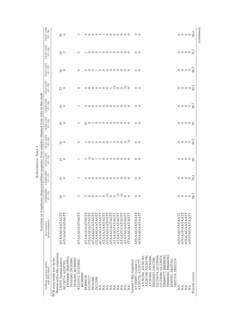
Supple
men
talTabl
e1
Variationin
Anaplasm
aphagocytophilum
sequence
from
samplesobtainedfrom
ticksin
thisstudy
GenBankaccessionnumber
with100%
Ident*
Rawsequence
betw
eenprimers†
LE000032001
(N=30)
LE640115007
(N=27)
LE120-11019
(N=25)
LE121-11018
(N=38)
LE200-11013
(N=36)
LE298-11050
(N=39)
LE324-11019
(N=28)
LE433-11011
(N=27)
LE433-11013
(N=38)
LE475-11048
(N=38)
PCR
assayresultwasAp-ha
Humanactive-likesequences
U02521(humanactive)
ATAAAGAATAGTT
20
19
15
32
18
19
23
26
29
26
AF136714,AJ242783,
FN390880,HQ629914,
JN181083,JN
181069,
JN181081,KC800983
ATGAAGAATAGTT
10
00
00
00
20
AY035312,EU839849,
KC800985
ATAGAGAATAGTT
22
10
22
00
23
DQ088128
ATAAGGAATAGTT
26
21
10
40
01
4GU391317
GTAAAGAGTAGTT
00
1‡
00
00
00
0JN
181066
ATAAAGGATAGTT
00
03
28
00
00
JN217094
GTAAAGAATAGTT
00
10
00
01
00
NA
ACAAAGAATAGTT
20
10
46
00
14
NA
ATAAAAAATAGTT
00
00
00
00
01
NA
ATAAAGAATGGTT
1‡
00
00
00
00
0NA
ATAAAGAGTAGTT
00
41
00
10
30
NA
ATAAAGTATAGTT
00
00
00
2‡
00
0NA
ATAACGAATAGTT
1‡
00
00
00
00
0NA
ATAATGGATAGTT
1‡
00
00
00
00
0NA
ATATAGAATAGTT
00
00
00
2‡
00
0NA
TTAAAGAATAGTT
00
01‡
00
00
00
Variant1-likesequences
AY193887(variant1)
ATGAAGAATAATT
00
00
00
00
00
AY281776,AY281777,
AY281780,AY281783,
AY281788,AY281790,
AY281804,AY281806,
DQ104443,FJ812390,
GU236534,GU236544,
GU236566,GU236574,
HM480377,HM480381,
HM480382,HM480383,
JF893931,JX
627361,
JX627374,JX
627374
ACGAAGAATAATT
00
00
00
00
00
NA
ATGAAGTATAATT
00
00
00
00
00
NA
ATGAGGAATAATT
00
00
00
00
00
NA
ATGGAGAATAATT
00
00
00
00
00
NA
ATGTAGAATAATT
00
00
00
00
00
Percentcorrect
66.7
70.4
60
84.2
50
48.7
82.1
96.3
76.3
68.4
(continued)
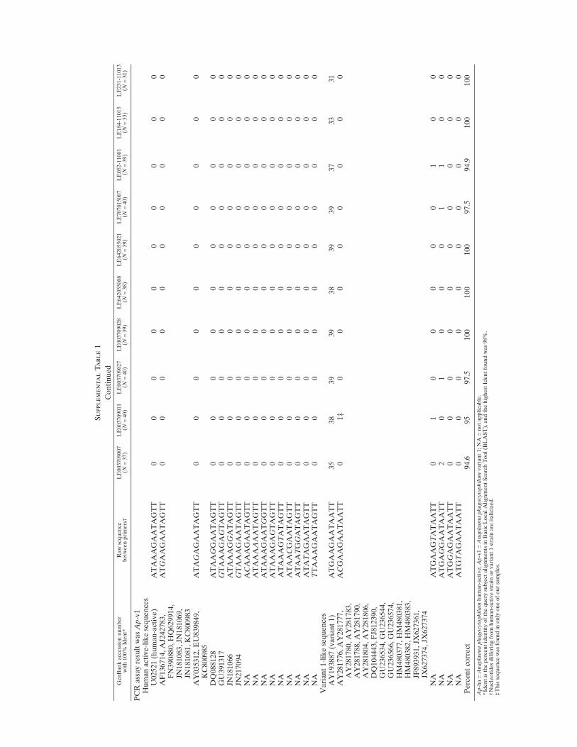
Supple
men
talTabl
e1
Continued
GenBankaccessionnumber
with100%
Ident*
Rawsequence
betw
eenprimers†
LE003709007
(N=37)
LE003709011
(N=40)
LE003709027
(N=40)
LE003709028
(N=39)
LE642055008
(N=38)
LE642055021
(N=39)
LE707015007
(N=40)
LE052-11001
(N=39)
LE184-11015
(N=33)
LE231-11013
(N=31)
PCR
assayresultwasAp-v1
Humanactive-likesequences
U02521(human-active)
ATAAAGAATAGTT
00
00
00
00
00
AF136714,AJ242783,
FN390880,HQ629914,
JN181083,JN
181069,
JN181081,KC800983
ATGAAGAATAGTT
00
00
00
00
00
AY035312,EU839849,
KC800985
ATAGAGAATAGTT
00
00
00
00
00
DQ088128
ATAAGGAATAGTT
00
00
00
00
00
GU391317
GTAAAGAGTAGTT
00
00
00
00
00
JN181066
ATAAAGGATAGTT
00
00
00
00
00
JN217094
GTAAAGAATAGTT
00
00
00
00
00
NA
ACAAAGAATAGTT
00
00
00
00
00
NA
ATAAAAAATAGTT
00
00
00
00
00
NA
ATAAAGAATGGTT
00
00
00
00
00
NA
ATAAAGAGTAGTT
00
00
00
00
00
NA
ATAAAGTATAGTT
00
00
00
00
00
NA
ATAACGAATAGTT
00
00
00
00
00
NA
ATAATGGATAGTT
00
00
00
00
00
NA
ATATAGAATAGTT
00
00
00
00
00
NA
TTAAAGAATAGTT
00
00
00
00
00
Variant1-likesequences
AY193887(variant1)
ATGAAGAATAATT
35
38
39
39
38
39
39
37
33
31
AY281776,AY281777,
AY281780,AY281783,
AY281788,AY281790,
AY281804,AY281806,
DQ104443,FJ812390,
GU236534,GU236544,
GU236566,GU236574,
HM480377,HM480381,
HM480382,HM480383,
JF893931,JX
627361,
JX627374,JX
627374
ACGAAGAATAATT
01‡
00
00
00
00
NA
ATGAAGTATAATT
01
00
00
01
00
NA
ATGAGGAATAATT
20
10
00
11
00
NA
ATGGAGAATAATT
00
00
00
00
00
NA
ATGTAGAATAATT
00
00
00
00
00
Percentcorrect
94.6
95
97.5
100
100
100
97.5
94.9
100
100
Ap-ha=Anaplasm
aphagocytophilum
human-active;Ap-v1=Anaplasm
aphagocytophilum
variant1;NA
=notapplicable.
*Identisthepercentidentity
ofthequery
subject
alignmentsin
BasicLocalAlignmentSearchTool(B
LAST),andthehighestIdentfoundwas98%
.†Nucleotidesdifferingfrom
human-activestrain
orvariant1strain
are
italicized.
‡Thissequence
wasfoundin
only
oneofoursamples.