preparation of core–shell fe3o4@poly(dopamine) magnetic nanoparticles for biosensor construction
TRANSCRIPT
Journal ofMaterials Chemistry B
PAPER
Publ
ishe
d on
25
Nov
embe
r 20
13. D
ownl
oade
d by
UN
IVE
RSI
DA
D D
E L
A L
AG
UN
A o
n 19
/12/
2013
10:
52:2
4.
View Article OnlineView Journal
aNeurochemistry and Neuroimaging group
Laguna, Tenerife, Spain. E-mail: psalazar@bAtlantica Biomedica, Tenerife, SpaincInformatica y Equipamiento Medico de CandDepartment of Analytical Chemistry, Facult
Madrid, 28040, Madrid, Spain
† Electronic supplementary informa10.1039/c3tb21171a
Cite this: DOI: 10.1039/c3tb21171a
Received 21st August 2013Accepted 21st November 2013
DOI: 10.1039/c3tb21171a
www.rsc.org/MaterialsB
This journal is © The Royal Society of
Preparation of core–shell Fe3O4@poly(dopamine)magnetic nanoparticles for biosensorconstruction†
Miriam Martın,ab Pedro Salazar,*ac Reynaldo Villalonga,d Susana Campuzano,d
Jose Manuel Pingarrond and Jose Luis Gonzalez-Moraa
Novel core–shell Fe3O4@poly(dopamine) magnetic nanoparticles were prepared through an in situ self-
polymerization method. The hybrid nanomaterial showed an average core diameter of 11 � 3 nm and a
polymer thin film thickness of 1.8 � 0.2 nm. The core–shell nanoparticles were employed as solid
supports for the covalent immobilization of horseradish peroxidase (HRP), and the resulting
biofunctionalized magnetic nanoparticles were employed to construct an amperometric biosensor for
H2O2. The enzyme biosensor showed a high sensitivity of 442.14 mA M�1 cm�2, a low limit of detection
of 182 nM, a wide linear range from 6.0 � 10�7 to 8.0 � 10�4 M and high stability for 1 month.
Introduction
During recent decades, iron oxide-based magnetic nano-particles have attracted much attention in biomedical, analyt-ical, environmental and industrial applications due to their easyand controlled synthesis and surface modication, highbiocompatibility and low toxicity.1–4 Moreover, their magneticproperties add a new dimension for biomedical and analyticalapplications such as in immunoprecipitation and separationtechniques, drug delivery, magnetic resonance imaging andhyperthermia treatments.5–10
Magnetic nanoparticles have been widely employed in elec-trochemical biosensors as nanosized supports for the immo-bilization of analytical biomolecules.11–14 In particular, theimmobilization of enzymes on the surface of these nano-particles offers numerous advantages including enhancementof the enzymatic activity and reduction of the mass-transferprocesses associated with the recognition of substrates byenzymes. Iron oxide nanoparticles also provide a favorablemicroenvironment for electrochemical devices where enzymesmay exchange electrons directly with the transducer, improvingthe sensitivity and selectivity of electrochemical biosensors.15–17
In this way, a great number of electrochemical biosensors usingmodied Fe3O4 nanoparticles have been reported in the
, Faculty of Medicine, University of La
ull.edu.es
arias, Tenerife, Spain
y of Chemistry, Complutense University of
tion (ESI) available. See DOI:
Chemistry 2014
literature for sulte, phenolic compounds, glucose, lactate,H2O2, etc.17–21
Enzyme-modied magnetic nanoparticles can be preparedby adsorption of proteins on the nanoparticle surface throughthe combined contribution of electrostatic interactions,hydrogen bonding, van-der-Waals forces and hydrophobicinteractions. However, these interactions can be affected by pH,ionic strength, temperature and polarity of solvents. Conse-quently, such an immobilization procedure oen yields lessstable catalysts due to the lack of enzyme molecules on thesupport.22 Stable enzyme–magnetic nanoparticle adducts can beprepared by covalent immobilization approaches. In general,these procedures require several reaction steps for nanoparticlesurface modication and enzyme immobilization, increasingthe complexity of the enzyme-modied nanoparticle synthesisand thus affecting the catalytic activity of the nal adduct.22 Forthese reasons, the development of novel methods for thecovalent functionalization of magnetic nanoparticles withenzymes receives considerable attention.
Dopamine (DA) is a catecholamine neurotransmitter relatedto many physiological processes and neurophysiologicaldisorders such as schizophrenia, Parkinson's and Alzheimer'sdiseases, as well as several social and addiction behaviors.23–25
In addition to its biomedical relevance, DA has been recentlyproposed as a novel organic coating material. Messersmith'sgroup reported that DA can be self-polymerized in aeratedbasic solutions, forming an adherent poly(dopamine) (pDA)lm over a wide variety of organic and inorganic surfaces.26,27
Although the structure of this polymeric lm has not been yetelucidated, it is probably formed by reaction of the primaryamino groups and quinone groups of oxidized DA via theformation of Schiff bases and/or Michaelis additionadducts.26,27
J. Mater. Chem. B

Journal of Materials Chemistry B Paper
Publ
ishe
d on
25
Nov
embe
r 20
13. D
ownl
oade
d by
UN
IVE
RSI
DA
D D
E L
A L
AG
UN
A o
n 19
/12/
2013
10:
52:2
4.
View Article Online
The reactive quinones at the surface of pDA lms could beused as anchoring points for further chemical modicationand/or immobilization of biologically active macromolecules(antibodies, enzymes, etc.).28–30 Poly(dopamine)-mediatedsurface modication has been demonstrated to provide versa-tile platforms for DNA immobilization,31 efficient and reliablemanipulation of human neural stem cells (NSCs),32 growthfactor immobilization, hematopoietic cell adhesion, anti-bacterial surface preparations33 and hydroxyapatite crystalliza-tion.34 Other approaches, using non-polymerized DA or L-dopa,have been demonstrated to be useful for the functionalizationof magnetic nanoparticles, allowing their use as solid supportsfor enzyme immobilization.35,36 However these protocols arelaborious and lengthy (4–5 working days)35 and require glutar-aldehyde as a coupling agent.36
In this work we describe the preparation and characteriza-tion of core–shell Fe3O4@pDAmagnetic nanoparticles and theiruse in the construction of an electrochemical enzymebiosensor. In contrast to other reported protocols,35,36 themethodology described in this work is rapid (3 h) and direct, notrequiring further surface activation or treatment with addi-tional coupling agents. As a proof-of-concept, horseradishperoxidase (HRP) was immobilized on the pDA coated nano-particles via Schiff base formation (Fe3O4@pDA/HRP), andfurther employed to functionalize a glassy carbon electrode(GCE) for amperometric H2O2 determination.
Materials and methodsReagents and solutions
HRP (type VI, EC 1.11.1.7, 269 Umg�1 solid), DA, hydroquinone,H2O2 and all the rest of the chemicals were obtained fromSigma. Electrochemical experiments were performed in 10 mMsodium phosphate, 2.7 mM KCl, 137 mM NaCl, and pH 7.4buffer solution (PBS). DA solution was prepared in PBS solution(pH 8.5) before use.
Instruments
Glassy carbon electrodes (GCEs, 3 mm diameter) werepurchased from CH Instruments Inc., USA. Electrochemicalmeasurements were performed with a DRP-STAT200 potentio-stat and data were acquired with Dropview soware (DropSens).For all electrochemical measurements, an Ag/AgCl (3M KCl) anda Pt wire were used as reference and counter electrodes,respectively. Field emission scanning electron micrographs(FE-SEMs) and energy dispersive X-ray spectra (EDS) wereobtained using a JEOL JSM-6300 microscope. High-resolutiontransmission electron microscopy (HRTEM) was performed witha JEOL JEM-3000 F microscope. Transmittance spectra (UV-Vis)of nanoparticles, pDA, HRP solutions and enzymatic studieswere recorded in the range of 200–1000 nm with respect to waterusing a Transpec®photodiode array spectrophotometer. FT-IRspectra were recorded with respect to air, using a Varian 670-IRspectrophotometer in the range of 4000–400 cm�1. The X-raydiffraction (XRD) pattern was recorded using a Philips Pan-alytical X'Pert powder diffractometer with Cu Ka (l ¼ 1.540 A)
J. Mater. Chem. B
radiation. Thermogravimetric analyses were performed with aPerkin Elmer Pyris Diamond TG/DTA apparatus.
Preparation of Fe3O4@pDA/HRP nanoparticles
Fe3O4 nanoparticles were prepared by the chemical coprecipi-tation method under a N2 atmosphere.30,37,38 FeCl3 (3.24 g) andFeSO4$7H2O (2.78 g) were dissolved in 200 mL of 1.2 mM HClsolution by ultrasound treatment. Then 1.25 M aqueous NaOHsolution (300 mL) was added dropwise under vigorous stirringfor 30 min, forming a black Fe3O4 precipitate. Aer vigorousstirring for another 30 min, the precipitate was magneticallydecanted and washed thoroughly with ultrapure water till thesupernatant solution reached neutrality (pH � 7). The resultingblack powder was dried in an oven at 40 �C overnight.
For coating with pDA, 500 mg of Fe3O4 were dispersed undercontinuous stirring in 25 mL of 10 mM DA solution (PBS, pH8.5) for 3 h. Aer that, polydopamine-modied nanoparticles(Fe3O4@pDA) were magnetically decanted, washed thoroughlywith ultrapure water to remove the non-reacted DA, and furtherdispersed in 25 mL of water. To immobilize the enzyme, 50 mgof Fe3O4@pDA were dispersed in 1 mg mL�1 HRP solution(2.5 mL PBS, pH 7.4) for 3 h under agitation at room tempera-ture. The HRP-modied nanoparticles (Fe3O4@pDA/HRP) weremagnetically decanted and washed thoroughly with PBS toremove the non-reacted HRP, and further re-dispersed in PBS at50 mg mL�1
nal concentration. The enzyme-modied nano-particles were kept at 4 �C until use.
Preparation of the enzyme electrode
GCEs were rst polished with 0.05 mm alumina slurry, rinsedthoroughly with water, then sonicated in water and acetone andnally dried with N2. GCEs were modied with differentvolumes of Fe3O4@pDA/HRP solution. Finally, biosensors werecured for 1 h at 37 �C.
Electrochemical measurements
A conventional three-electrode system was used for cyclic vol-tammetry (CV), chronoamperometry (CA) and constant potentialamperometry (CPA) analyses with the biosensor as the workingelectrode, a platinum wire as an auxiliary electrode, and an Ag/AgCl (3 M KCl) electrode as a reference electrode. CV and CPAstudies were done in 15 mL PBS containing hydroquinone (HQ)as an electron mediator under constant magnetic stirring.
Enzymatic activity measurements
The free and immobilized HRP-activity was measured by thereaction with H2O2 using TMB as a chromogenic substrate. Theactivity was measured at 25 �C by monitoring the absorbancechange at 650 nm with time. 5 mL of 10 mg mL�1 Fe3O4@pDA/HRP nanoparticles were dispersed in 2.5 mL of PBS and 25 mL ofthe chromogenic solution (0.1% TMB, 1% H2O2) were addedand the absorbance was recorded each 30 s. The free HRP-activity was checked using the same protocol but adding 0.9 mgHRP in PBS, the equivalent amount of HRP as that used in theformer experiment.
This journal is © The Royal Society of Chemistry 2014
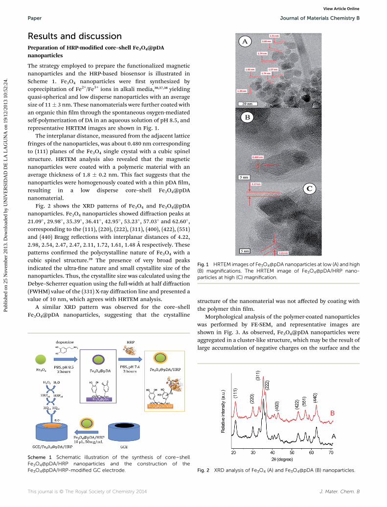
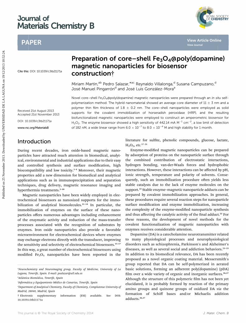
Fig. 1 HRTEM images of Fe3O4@pDA nanoparticles at low (A) and high(B) magnifications. The HRTEM image of Fe3O4@pDA/HRP nano-particles at high (C) magnification.
Paper Journal of Materials Chemistry B
Publ
ishe
d on
25
Nov
embe
r 20
13. D
ownl
oade
d by
UN
IVE
RSI
DA
D D
E L
A L
AG
UN
A o
n 19
/12/
2013
10:
52:2
4.
View Article Online
Results and discussionPreparation of HRP-modied core–shell Fe3O4@pDAnanoparticles
The strategy employed to prepare the functionalized magneticnanoparticles and the HRP-based biosensor is illustrated inScheme 1. Fe3O4 nanoparticles were rst synthesized bycoprecipitation of Fe2+/Fe3+ ions in alkali media,30,37,38 yieldingquasi-spherical and low disperse nanoparticles with an averagesize of 11� 3 nm. These nanomaterials were further coated withan organic thin lm through the spontaneous oxygen-mediatedself-polymerization of DA in an aqueous solution of pH 8.5, andrepresentative HRTEM images are shown in Fig. 1.
The interplanar distance, measured from the adjacent latticefringes of the nanoparticles, was about 0.480 nm correspondingto (111) planes of the Fe3O4 single crystal with a cubic spinelstructure. HRTEM analysis also revealed that the magneticnanoparticles were coated with a polymeric material with anaverage thickness of 1.8 � 0.2 nm. This fact suggests that thenanoparticles were homogenously coated with a thin pDA lm,resulting in a low disperse core–shell Fe3O4@pDAnanomaterial.
Fig. 2 shows the XRD patterns of Fe3O4 and Fe3O4@pDAnanoparticles. Fe3O4 nanoparticles showed diffraction peaks at21.09�, 29.98�, 35.39�, 36.41�, 42.95�, 53.23�, 57.03� and 62.60�,corresponding to the (111), (220), (222), (311), (400), (422), (551)and (440) Bragg reections with interplanar distances of 4.22,2.98, 2.54, 2.47, 2.47, 2.11, 1.72, 1.61, 1.48 A respectively. Thesepatterns conrmed the polycrystalline nature of Fe3O4 with acubic spinel structure.39 The presence of very broad peaksindicated the ultra-ne nature and small crystallite size of thenanoparticles. Thus, the crystallite size was calculated using theDebye–Scherrer equation using the full-width at half diffraction(FWHM) value of the (331) X-ray diffraction line and presented avalue of 10 nm, which agrees with HRTEM analysis.
A similar XRD pattern was observed for the core–shellFe3O4@pDA nanoparticles, suggesting that the crystalline
Scheme 1 Schematic illustration of the synthesis of core–shellFe3O4@pDA/HRP nanoparticles and the construction of theFe3O4@pDA/HRP-modified GC electrode.
This journal is © The Royal Society of Chemistry 2014
structure of the nanomaterial was not affected by coating withthe polymer thin lm.
Morphological analysis of the polymer-coated nanoparticleswas performed by FE-SEM, and representative images areshown in Fig. 3. As observed, Fe3O4@pDA nanoparticles wereaggregated in a cluster-like structure, which may be the result oflarge accumulation of negative charges on the surface and the
Fig. 2 XRD analysis of Fe3O4 (A) and Fe3O4@pDA (B) nanoparticles.
J. Mater. Chem. B

Journal of Materials Chemistry B Paper
Publ
ishe
d on
25
Nov
embe
r 20
13. D
ownl
oade
d by
UN
IVE
RSI
DA
D D
E L
A L
AG
UN
A o
n 19
/12/
2013
10:
52:2
4.
View Article Online
adsorption of Fe3O4@pDA nanoparticles from the surroundingsto achieve inter-molecular force equilibrium. As expected, EDSmicroanalysis revealed the presence of Fe and O atoms in thenanoparticle structure.
Thermal analysis was performed to conrm and estimate therelative composition of Fe3O4 and Fe3O4@pDA nanoparticles,and the results are shown in Fig. SM1 (see ESI† for additionaldetails). The weight loss of unmodied Fe3O4 at 200 �C wasestimated to be 3% of the initial weight, and could be ascribedto the removal of physically adsorbed water. Meanwhile thedecrease in the mass of Fe3O4@pDA nanoparticles in the samerange of temperature was close to 5%, with this change beingassociated with the weight loss of adsorbed water and DAmonomers. The next range of temperature, between 200 and400 �C, presented a weight loss of 5% for Fe3O4 and corre-sponded to bonded water, but Fe3O4@pDA nanoparticles pre-sented a weight loss of about 14% with a broad exothermic peakat 300 �C. This transformation could be ascribed to the thermaldecomposition of the chemically attached pDA lm from thesurface of the Fe3O4 nanoparticles, conrming the successfulpolymer coverage formed on the nanoparticles. Finally, theweight loss of about 1% (both nanoparticles) above 500 �C couldbe associated with the phase transformation of Fe3O4 to Fe2O3.
HRP was further immobilized on the core–shell Fe3O4@pDAnanoparticles by dispersing the nanosized solid supports in astirred solution of 1 mg mL�1 HRP for 3 h. The presence of freequinone groups on the surface of the pDA-coated nanoparticlesfavored the covalent immobilization of the enzyme through theformation of stable Schiff base linkages. The amount ofimmobilized enzyme was estimated to be�18 mg mg�1 support.
Fig. 3 FE-SEM image (A) and EDS analysis (B) of Fe3O4@pDAnanoparticles.
J. Mater. Chem. B
This immobilization step was studied by UV-Vis and FT-IRspectroscopy. In this sense, UV-Vis analyses were done for eachcomponent (Fe3O4, pDA and HRP) and for the nal modiednanoparticles (Fe3O4@pDA/HRP), and the results are shown inFig. 4A. The enzyme solution presented a characteristicabsorption band at around 420 nm,21 meanwhile the pDAsolution showed a typical absorption band around 350 nm,which results from the formation of the quinone intermediateand the polymerized lm.40 The surface modication of theFe3O4 nanoparticles with these adducts can be conrmed bycomparing the spectra of the raw and modied nanoparticlesolutions. UV-Vis analysis of Fe3O4@pDA/HRP samples revealedthat two peaks at 350 nm and 420 nmwere superimposed on thecharacteristic broad absorption peak of Fe3O4 nanoparticles,suggesting that the nanoparticles were successfully coveredwith the pDA lm and further modied with the HRP enzyme.
Fig. 4B shows the FT-IR analysis of the Fe3O4-based nano-materials. FT-IR spectra of all Fe3O4-based nanomaterialsshowed a main absorption band around 580 cm�1 assigned tothe Fe–O stretching modes of the magnetite. pDA-coatednanoparticles showed additional bands in the range of 1000–1700 cm�1 which may be ascribed to the aromatic rings of pDA(1614 cm�1), and the amide I, amide II and C–N stretchingbands (1639, 1535, and 1230 cm�1, respectively).29,40 The spectraof HRP-modied nanoparticles were characterized by a broadabsorption band around 1200 cm�1, which could be ascribed tothe C–N stretching in the new Schiff bases formed during theimmobilization step. It should be highlighted that Fe3O4
nanoparticles retained their magnetic properties aer coatingwith pDA and further functionalization with HRP, as illustratedin Fig. SM2.†
Fig. 4 (A) UV-Vis absorption spectra of free HRP, pDA, Fe3O4 andFe3O4@pDA/HRP nanoparticle solutions. (B) FT-IR transmittancespectra of Fe3O4, Fe3O4@pDA and Fe3O4@pDA/HRP nanoparticles.
This journal is © The Royal Society of Chemistry 2014

Paper Journal of Materials Chemistry B
Publ
ishe
d on
25
Nov
embe
r 20
13. D
ownl
oade
d by
UN
IVE
RSI
DA
D D
E L
A L
AG
UN
A o
n 19
/12/
2013
10:
52:2
4.
View Article Online
HRP-modied nanoparticles were characterized by HRTEM.Fe3O4@pDA/HRP nanoparticles (Fig. 1C) presented a similarsize and distribution to those mentioned above. The lmthickness showed a slight increase aer HRP-immobilizationbut the amorphous nature of the pDA lm hindered theobservation of evident changes in the HRP-modiednanoparticles.
On the other hand, the specic activity of the enzyme wasslightly reduced (from 20.8 � 1.4 U mg�1 to 18.5 � 1.1) aercovalent immobilization on the core–shell Fe3O4@pDA nano-particles. Therefore, it can be concluded that the tridimensionalenzyme conformation is not signicantly altered by the immo-bilization hence retaining its catalytic activity.
Fig. 5 (A) CVs of GCE/Fe3O4@pDA/HRP recorded at 10, 50, 100, 200and 300mV s�1 in PBS solution, pH 7.4 containing 1 mMHQ. (B) Lineardependence of anodic and cathodic peak currents (Iox and Ird) vs. thesquare-root of the scan rate. (C) CVs for GCE/Fe3O4@pDA/HRPrecorded in PBS solution, pH 7.4 containing 0, 0.25, 0.5, 0.75 mM HQ(scan rate: 100 mV s�1). (D) CVs of GCE/Fe3O4@pDA/HRP recorded inPBS solution, pH 7.4, containing 1.5 mMHQ and 0, 0.2, 0.6, 1 mMH2O2
(scan rate: 100 mV s�1).
Preparation of the Fe3O4@pDA/HRP based biosensor
The potential analytical use of the magnetic core–shellFe3O4@pDA/HRP nanoparticles was evaluated by constructingan electrochemical enzyme biosensor for H2O2. For thispurpose, GCEs were functionalized with HRP-modied nano-particles and hydroquinone (HQ) was used as an electro-chemical mediator due to its ability to act as an electron shuttlefrom the redox center of the HRP molecules to the GCE surface,according to the following mechanism:41
HRP + H2O2 / HRP-I + H2O (1)
HRP-I + HQred / HRP-II + HQox (2)
HRP-II + HQred / HRP + HQox (3)
HQox + 2e� / HQred (4)
Net reaction: H2O2 + 2e� + 2H+ / 2H2O (5)
where HRP, HRP-I and HRP-II represent the enzyme peroxidasein the native form and in its two oxidized forms, respectively;and HQred and HQox represent HQ in its reduced and oxidizedforms, respectively.
Fig. 5A shows the CVs recorded for the working electrode(GCE/Fe3O4@pDA/HRP) in PBS solution, pH 7.4, and contain-ing 1 mM HQ at different scan rates. Both the anodic and thecathodic current peaks (Iox and Ird) increased linearly when thesquare-root of the scan rate increased (see Fig. 5B) which ischaracteristic of a diffusion-controlled process.
Fig. 5C shows the CVs of the GCE/Fe3O4@pDA/HRP basedbiosensor in PBS (pH 7.4) in the absence and the presence ofHQ at different concentrations. Only a single pair of reversibleoxidation/reduction peaks is observed, and peak currentsincreased with the increase of HQ concentration. This repre-sented the typical electrochemical behavior of HQ.41 In addi-tion, a noticeable increase of the reduction current and aconcomitant decrease of the oxidation current were observedaer addition of H2O2, Fig. 5D, in agreement with the electro-catalytic mechanism described above. Analogous experimentsto those shown in Fig. 5D were done for GCE/Fe3O4@pDAconguration. No signicant change in the voltammetricresponses was observed aer H2O2 addition (Fig. SM3†)
This journal is © The Royal Society of Chemistry 2014
indicating that, as expected, the presence of HRP prompted thecatalytic response to H2O2.
The amount of loaded nanoparticles during the preparationof the enzyme electrode was optimized in order to achieve themaximum sensitivity for the amperometric biosensor.
The effect of the modied-magnetic bead amount immobi-lized on the top of the electrode surface on the amperometricsignal is shown in Fig. SM4A.† As can be seen, an increase in theamperometric signal was observed up to 50 mg, the signaldecreasing for higher loadings, which was probably due to anincrease in the electron transfer resistance for large modied-magnetic bead loadings. In addition, the most favourableworking conditions for the enzyme biosensor were optimized.The effect of the applied potential on the amperometricresponse of the enzyme electrode was evaluated in the range of�250 mV to 100 mV (Fig. SM4B†). The electrode showed apractically constant and higher current value from �250 mV to�150 mV, then the cathodic current decreased steadily withincreasing the applied potential. In view of these results,�150 mV was selected as the optimum value for the appliedpotential. The effect of pH on the amperometric response of theelectrode was also checked in the 5.0–9.0 pH range(Fig. SM4C†). The biosensor current exhibited an almost bell-shaped behavior with the highest response at pH 7.4. Finally,the optimum concentration of the electrochemical mediatorwas also determined. As illustrated in Fig. SM4D,† the cathodiccurrent increased with the concentration of HQ reaching aplateau at 1.2 mM, and this HQ concentration was selected forfurther studies.
Finally, the biosensor response against temperature wasstudied in the range of 15 �C to 55 �C (Fig. SM5A†). Theamperometric response gradually increased with the tempera-ture, reaching a maximum value at 55 �C. It was furtherconrmed that the cathodic response of the electrode totemperature in the range of 15–55 �C followed an Arrhenius-
J. Mater. Chem. B

Fig. 6 Calibration curves and linear fitting (inset) obtained using GCE/HRP (triangles) and GCE/Fe3O4@pDA/HRP (squares) configurations inPBS (pH 7.4) containing 1.5 mM HQ (CPA, applied potential: �0.15 V).
Journal of Materials Chemistry B Paper
Publ
ishe
d on
25
Nov
embe
r 20
13. D
ownl
oade
d by
UN
IVE
RSI
DA
D D
E L
A L
AG
UN
A o
n 19
/12/
2013
10:
52:2
4.
View Article Online
type behavior, as shown in Fig. SM5B.† Accordingly, the acti-vation energy for this enzymatic reaction was estimated to be55.53 kJ mol�1. Higher working temperatures led to a signi-cant decrease in the electrocatalytic response of the biosensor,which should be attributed to the denaturation/inactivation ofthe enzyme at these higher temperatures.
Chronoamperometry was used to obtain more informationabout the electrocatalytic behavior of the developed enzymaticbiosensor. Fig. SM6A† shows the current–time plots for theGCE/Fe3O4@pDA/HRP conguration recorded following step-ping the applied potential from open circuit conditions to�0.15 V in the absence and presence of H2O2 in the concen-tration range of 0.0–1.0 mM. The time evolution of the semi-innite linear diffusion-controlled current is described by theCottrell equation:42
I ¼ nFAC(D/pt)1/2 (6)
where n is the number of electrons consumed (n ¼ 2), F is theFaraday constant, A is the electrochemical surface area(0.07 cm2), C is the concentration (mol cm�3) of the electro-active molecule and D is the diffusion coefficient (cm2 s�1). Theplot of I vs. t�1/2 at each concentration of H2O2 was linear(Fig. SM6B†), and the slopes (mi) of the resulting straight lineswere plotted vs. H2O2 concentration. The slope of the resultingstraight line was then used to estimate the diffusion coefficientof H2O2 (Fig. SM6C†), presenting a value of 2.3 � 10�6 cm2 s�1
fairly consistent with data reported elsewhere.42
Chronoamperometry was also used for the evaluation of thecatalytic rate constant, kcat. At intermediate times, when thecurrent is dominated by the rate of the electrocatalyzed reduc-tion of H2O2, the catalytic current (Icat) can be written asfollows:42
Icat ¼ IL(kcatpCt)1/2 (7)
where IL is the current of the biosensor in the absence of H2O2
and Icat is the catalytic current due to different H2O2 concen-trations. Over a limited time frame, the values of Icat/IL werelinearly dependent on t1/2, and from its slope kcat was calculated.Finally kcat presents a value of 1.7 � 104 M�1 s�1 which is ingood agreement with data reported in the literature.43
In order to demonstrate the benet of the present congu-ration, we compared our optimized conguration (GCE/Fe3O4@pDA/HRP) with a basic biosensor conguration, where10 mL of HRP (1 mg mL�1) were adsorbed directly onto the GCEsurface (GCE/HRP). Fig. 6 shows the calibration curves of thesetwo congurations. GCE/Fe3O4@pDA/HRP showed a sensitivityof 442.14 mA M�1 cm�2, a limit of detection, LOD (S/N ¼ 3), of182 nM and an excellent linear range from 0.6 to 800 mM (R2:0.999) and acceptable response time (t90% ¼ 10 s). Meanwhilethe basic conguration (GCE/HRP) showed a lower sensitivity(100.25 mA M�1 cm�2) and linearity (R2: 0.972) in the samerange of concentration with a higher LOD (798 nM). Thesedifferences of sensitivities may be justied by taking intoaccount the divergence in enzymatic loading in the twocongurations expressed as Imax (calibration plateau ormaximum current measured under saturated substrate
J. Mater. Chem. B
conditions) �10.11 and 88.23 mA for GCE/HRP and GCE/Fe3O4@pDA/HRP, respectively. Finally, thanks to the synergismbetween the higher surface (nanoparticles) and higher enzy-matic retention (pDA), the present conguration showed bettersensitivity21,41 and LOD44–47 than previous publications reportedin the literature.
In order to study the enzyme kinetics as well as to evaluatethe affinity of the immobilized HRP towards H2O2 the apparentMichaelis–Menten (Kmapp) constant was calculated from theelectrochemical version of the Lineweaver–Burk equation:41
1/Iss ¼ 1/Imax + Kmapp/ImaxC (8)
where Iss is the steady state current aer the addition of thesubstrate, C is the bulk concentration of the substrate, and Imax
is the maximum current. The Kmapp value of the GCE/Fe3O4@pDA/HRP biosensor was determined by the steady stateamperometric response curve, which is equal to 3.05 mM,smaller than the values reported previously by other authorsusing the HRP–ZrO2 thin lm on an Au electrode (8.01 mM),46
HRP immobilized in the sol–gel-derived ceramic–carbonnanotube nanocomposite lm (23.85 mM)44 and the Fe3O4/chitosan modied GCE (21.4 mM).45 This Kmapp value, evensmaller than that obtained for the free enzyme (Kmapp ¼11 mM),48,49 conrmed that the immobilized HRP possesseshigh catalytic efficiency for the reduction of H2O2.
The stability of Fe3O4@pDA/HRP nanoparticles over timewas studied by storing them in PBS (pH 7.4) at 4 �C for a timeperiod of 28 days. Over this time, the response of the modiedelectrode to 800 mM H2O2 was evaluated. As can be seen inFig. 7, no signicant loss of the amperometric response wasobserved for at least 28 days thus conrming that both thepolydopamine lm and the immobilized-HRP were stable onthe nanoparticle surface as it was suggested previously.30,34,36
The selectivity of the GCE/Fe3O4@pDA/HRP biosensor waschecked under the experimental conditions specied above.The potential interferents tested were glucose, lactate, L-gluta-mate, choline, uric acid and ascorbic acid. The degree ofinterference was evaluated by comparing the amperometricresponses obtained for 1 mM H2O2 in the absence or in the
This journal is © The Royal Society of Chemistry 2014

Fig. 7 Control chart constructed to check the stability of Fe3O4@pDA/HRP nanoparticles stored in PBS (pH 7.4) at 4 �C. The measurementscorrespond to the mean value of three measurements for 800 mMH2O2. The upper and lower control limits (dashed lines) were set at�3 SD of the initial value.
Paper Journal of Materials Chemistry B
Publ
ishe
d on
25
Nov
embe
r 20
13. D
ownl
oade
d by
UN
IVE
RSI
DA
D D
E L
A L
AG
UN
A o
n 19
/12/
2013
10:
52:2
4.
View Article Online
presence of each interferent at 0.1 mM concentration. Theresults listed in Table SM1† demonstrate that no interferencewas found from glucose, lactate, L-glutamate and choline.However, both uric and ascorbic acids may reduce the HQproduced in the HRP catalyzed reaction and thus interfere withthe determination of H2O2.
The reproducibility of the amperometric responses obtainedwith different biosensors constructed following the sameprotocol was tested for 0.6 mM H2O2. The results for vedifferent biosensors yielded a RSD value of 3.9%, thus provingthat their fabrication procedure (including both the preparationof Fe3O4@pDA/HRP and their magnetic capture on the GCE)was reliable.
Conclusions
In the present work we have developed and characterized anovel functionalized core–shell magnetic nanoparticle(Fe3O4@pDA). The present approach is an easy method tomodify nanoparticles. The reactive quinones at the surface ofthe Fe3O4@pDA offer a great opportunity to anchor biologicallyactive macromolecules such as antibodies and enzymes. In thisway, HRP was used as a proof-of-concept for further modica-tions (Fe3O4@pDA/HRP). Finally, the newly prepared enzymaticnanoconjugates were employed to construct a second-genera-tion biosensor (GCE/Fe3O4@pDA/HRP) for H2O2 sensing.Experimental variables were optimized in order to improve thebiosensor response. All the results discussed above demon-strated that the new H2O2 biosensor based on the use ofFe3O4@pDA/HRP compares advantageously (in terms of sensi-tivity, reproducibility and simplicity of the fabrication process)with other biosensor designs.
Acknowledgements
This work has been supported by the programmes INNCORPORAand FEDER (INC-TU-2011-1621), Ministerio de Industria,
This journal is © The Royal Society of Chemistry 2014
Turismo y Comercio (TSI-020100-2011-189 and TSI-020100-2010-346), Ministerio de Ciencia e Investigacion (TIN2011-28146;CTQ2011-24355), Ministerio de Economıa y Competitividad(Research Project CTQ2012-34238) and Comunidad de MadridS2009/PPQ-1642 programme AVANSENS. R. Villalonga acknowl-edges Ramon & Cajal contract from the Spanish Ministry ofScience and Innovation. We thank Dr Pablo Lorenzo Luis (Inor-ganic Department of ULL) for helping in UV-Vis measurements.
References
1 S. C. N. Tang and I. M. C. Lo,Water Res., 2013, 47, 2613–2632.2 D. Alcantara and L. Josephson, in NanobiotechnologyInorganic Nanoparticles vs Organic Nanoparticles, ed. J. M.de la Fuente and V. Grazzu, Elsevier, 2012, vol. 4, pp. 269–289.
3 P. Tartaj, M. P. Morales, T. Gonzalez-Carreno,S. Veintemillas-Verdaguer and C. J. Serna, J. Magn. Magn.Mater., 2005, 290–291, 28–34.
4 S. Singh, K. C. Barick and D. Bahadur, J. Hazard. Mater.,2011, 192, 1539–1547.
5 J. Chomoucka, J. Drbohlavova, D. Huska, V. Adam, R. Kizekand J. Hubalek, Pharmacol. Res., 2010, 62, 144–149.
6 S. Laurent, S. Dutz, U. O. Hafeli and M. Mahmoudi, Adv.Colloid Interface Sci., 2011, 166, 8–23.
7 M. Mahmoudi, S. Sant, B. Wang, S. Laurent and T. Sen, Adv.Drug Delivery Rev., 2011, 63, 24–46.
8 O. Veiseh, J. W. Gunn and M. Zhang, Adv. Drug Delivery Rev.,2010, 62, 284–304.
9 C. Sun, J. S. H. Lee and M. Zhang, Adv. Drug Delivery Rev.,2008, 60, 1252–1265.
10 J. R. McCarthy and R. Weissleder, Adv. Drug Delivery Rev.,2008, 60, 1241–1251.
11 J. Huang, B. Han, W. Yue and H. Yan, J. Mater. Chem., 2007,17, 3812–3818.
12 J. Huang, X. Li, Y. Zheng, Y. Zhang, R. Zhao, X. Gao andH. Yan, Macromol. Biosci., 2008, 8, 508–515.
13 G. Bayramoglu, M. Yılmaz, A. U. Senel and M. Y. Arıca,Biochem. Eng. J., 2008, 40, 262–274.
14 J. Wang, D. Song, H. Zhang, J. Zhang, Y. Jin, H. Zhang,H. Zhou and Y. Sun, Colloids Surf., B, 2013, 102, 165–170.
15 E. Katz, I. Willner and J. Wang, Electroanalysis, 2004, 16, 19–44.
16 N. Jaffrezic-Renault, C. Martelet, Y. Chevolot andJ.-P. Cloarec, Sensors, 2007, 7, 589–614.
17 R. Rawal, S. Chawla and C. S. Pundir, Biosens. Bioelectron.,2012, 31, 144–150.
18 X. Chen, J. Zhu, Z. Chen, C. Xu, Y. Wang and C. Yao, Sens.Actuators, B, 2011, 159, 220–228.
19 S. Wang, Y. Tan, D. Zhao and G. Liu, Biosens. Bioelectron.,2008, 23, 1781–1787.
20 H. Teymourian, A. Salimi and R. Hallaj, Biosens. Bioelectron.,2012, 33, 60–68.
21 Y.-H. Won, D. Aboagye, H. S. Jang, A. Jitianu andL. A. Stanciu, J. Mater. Chem., 2010, 20, 5030–5034.
22 A. Sassolas, L. J. Blum and B. D. Leca-Bouvier, Biotechnol.Adv., 2012, 30, 489–511.
J. Mater. Chem. B

Journal of Materials Chemistry B Paper
Publ
ishe
d on
25
Nov
embe
r 20
13. D
ownl
oade
d by
UN
IVE
RSI
DA
D D
E L
A L
AG
UN
A o
n 19
/12/
2013
10:
52:2
4.
View Article Online
23 G. Remington, in Serotonin–Dopamine Interaction:Experimental Evidence and Therapeutic Relevance, ed. G. DiGiovanni, V. Di Matteo and E. Esposito, Elsevier, 2008, vol.172, pp. 117–140.
24 M. J. Hurley and P. Jenner, Pharmacol. Ther., 2006, 111, 715–728.
25 J. L. Katz, A. H. Newman and S. Izenwasser, Pharmacol.,Biochem. Behav., 1997, 57, 505–512.
26 H. Lee, S. M. Dellatore, W. M. Miller and P. B. Messersmith,Science, 2007, 318, 426–430.
27 H. Lee, J. Rho and P. B. Messersmith, Adv. Mater., 2009, 21,431–434.
28 R. Luo, L. Tang, J. Wang, Y. Zhao, Q. Tu, Y. Weng, R. Shenand N. Huang, Colloids Surf., B, 2013, 106, 66–73.
29 L.-P. Zhu, J.-H. Jiang, B.-K. Zhu and Y.-Y. Xu, Colloids Surf., B,2011, 86, 111–118.
30 Y. Li, C. Qin, C. Chen, Y. Fu, M. Ma and Q. Xie, Sens.Actuators, B, 2012, 168, 46–53.
31 H. O. Ham, Z. Liu, K. H. A. Lau, H. Lee andP. B. Messersmith, Angew. Chem., Int. Ed., 2011, 50, 732–736.
32 K. Yang, J. S. Lee, J. Kim, Y. B. Lee, H. Shin, S. H. Um,J. B. Kim, K. I. Park, H. Lee and S.-W. Cho, Biomaterials,2012, 33, 6952–6964.
33 S. M. Kang, N. S. Hwang, J. Yeom, S. Y. Park,P. B. Messersmith, I. S. Choi, R. Langer, D. G. Andersonand H. Lee, Adv. Funct. Mater., 2012, 22, 2949–2955.
34 J. Ryu, S. H. Ku, H. Lee and C. B. Park, Adv. Funct. Mater.,2010, 20, 2132–2139.
J. Mater. Chem. B
35 C. Xu, K. Xu, H. Gu, R. Zheng, H. Liu, X. Zhang, Z. Guo andB. Xu, J. Am. Chem. Soc., 2004, 126, 9938–9939.
36 H. Peng, X. Zhang, K. Huang and H. Xu, J. Wuhan Univ.Technol., Mater. Sci. Ed., 2008, 23, 480–485 LA-English.
37 L.-L. Pang, J.-S. Li, J.-H. Jiang, Y. Le, G. L. Shen and R.-Q. Yu,Sens. Actuators, B, 2007, 127, 311–316.
38 C. Zou, Y. Fu, Q. Xie and S. Yao, Biosens. Bioelectron., 2010,25, 1277–1282.
39 H. E. Ghandoor, H. M. Zidan, M. M. H. Khalil andM. I. M. Ismail, Int. J. Electrochem. Sci., 2012, 7, 5734–5745.
40 C. Cheng, S. Li, W. Zhao, Q. Wei, S. Nie, S. Sun and C. Zhao,J. Membr. Sci., 2012, 417-418, 228–236.
41 X. Wei, T. Liu, J. Li and X. Chen, Int. J. Electrochem. Sci., 2011,6, 4953–4966.
42 P. Salazar, M. Martın, R. D. O'Neill, R. Roche andJ. L. Gonzalez-Mora, J. Electroanal. Chem., 2012, 674, 48–56.
43 A. Karyakin, E. E. Karyakina and L. Gorton, J. Electroanal.Chem., 1998, 456, 97–104.
44 H. Chen and S. Dong, Biosens. Bioelectron., 2007, 22, 1811–1815.
45 X. Tan, J. Zhang, S. Tan, D. Zhao, Z. Huang, Y. Mi andZ. Huang, Electroanalysis, 2009, 21, 1514–1520.
46 Z. Tong, R. Yuan, Y. Chai, Y. Xie and S. Chen, J. Biotechnol.,2007, 128, 567–575.
47 X. Li, J. Wu, N. Gao, G. Shen and R. Yu, Sens. Actuators, B,2006, 117, 35–42.
48 S. Q. Liu and H. X. Ju, Anal. Biochem., 2002, 307, 110–116.49 Y. Xiao, H. X. Ju and H. Y. Chen, Anal. Chim. Acta, 1999, 391,
299–306.
This journal is © The Royal Society of Chemistry 2014