potential targets of 3 - hydroxy - 3 - methylglutaryl coenzyme a reductase inhibitor for multiple...
TRANSCRIPT
of June 13, 2013.This information is current as
TherapyReductase Inhibitor for Multiple Sclerosis3-Hydroxy-3-Methylglutaryl Coenzyme A Potential Targets of
Singh and Inderjit SinghNarender Nath, Shailendra Giri, Ratna Prasad, Avtar K.
http://www.jimmunol.org/content/172/2/12732004; 172:1273-1286; ;J Immunol
Referenceshttp://www.jimmunol.org/content/172/2/1273.full#ref-list-1
, 27 of which you can access for free at: cites 64 articlesThis article
Subscriptionshttp://jimmunol.org/subscriptions
is online at: The Journal of ImmunologyInformation about subscribing to
Permissionshttp://www.aai.org/ji/copyright.htmlSubmit copyright permission requests at:
Email Alertshttp://jimmunol.org/cgi/alerts/etocReceive free email-alerts when new articles cite this article. Sign up at:
Print ISSN: 0022-1767 Online ISSN: 1550-6606. Immunologists All rights reserved.Copyright © 2004 by The American Association of9650 Rockville Pike, Bethesda, MD 20814-3994.The American Association of Immunologists, Inc.,
is published twice each month byThe Journal of Immunology
by guest on June 13, 2013http://w
ww
.jimm
unol.org/D
ownloaded from
Potential Targets of 3-Hydroxy-3-Methylglutaryl Coenzyme AReductase Inhibitor for Multiple Sclerosis Therapy
Narender Nath,* Shailendra Giri,* Ratna Prasad,* Avtar K. Singh, † and Inderjit Singh 2*
The 3-hydroxy-3-methylglutaryl coenzyme A (HMG-CoA) reductase inhibitors or statins are newly identified immunomodulators.In vivo treatment of SJL/J mice with lovastatin reduced the duration and clinical severity of active and passive experimentalautoimmune encephalomyelitis (EAE), an animal model for multiple sclerosis. Lovastatin induced the expression of GATA3 andthe phosphorylation of STAT6, whereas it inhibited tyrosine phosphorylation of Janus kinase 2, tyrosine kinase 2, and STAT4.Inhibition of the Janus kinase-STAT4 pathway by lovastatin modulated T0 to Th1 differentiation and reduced cytokine (IFN-�and TNF-�) production, thus inducing Th2 cytokines (IL-4, IL-5, and IL-10). It inhibited T-bet (T box transcription factor) andNF-�B in activated T cells and significantly reduced infiltration of CD4- and MHC class II-positive cells to CNS. Further, itstabilized IL-4 production and GATA-3 expression in differentiated Th2 cells, whereas in differentiated Th1 cells it inhibited theexpression of T-bet and reduced the production of IFN-�. Moreover, lovastatin-exposed macrophage and BV2 (microglia) inallogeneic MLRs induced the production of the anti-inflammatory cytokine IL-10. These observations indicate that the anti-inflammatory effects of lovastatin are mediated via T cells as well as APCs, because it modulates the polarization patterns of naiveT cell activation in an APC-independent system. Together, these findings reveal that lovastatin may have possible therapeutic valueinvolving new targets (in both APCs and T cells) for the treatment of multiple sclerosis and other inflammatory diseases.TheJournal of Immunology, 2004, 172: 1273–1286.
M ultiple sclerosis (MS)3 is an inflammatory disease lim-ited to CNS white matter. The CNS inflammation con-sists of a variable degree of T lymphocytes, macro-
phage, B lymphocytes, and Abs at the leading edge of the whitematter destruction (1–4). A substantial percentage of MS patientsdevelop clinical paralysis, and there is no curative therapy for MS(1–4). Experimental autoimmune encephalomyelitis (EAE) sharesmany of the clinical and histopathological features of MS and thusserves as a useful animal model (5). Although the disease is me-diated by Th1 cells secreting the proinflammatory cytokines IFN-�and TNF-�, genetic targeting of these cytokines in many casesaggravates clinical disease (6–9). Neutralization of lymphotoxinand TNF-� prevents transfer of EAE, but they still have T cellinfiltration (10, 11). Current literature describing the anti-inflammatory cytokines, such as IL-10 and TGF-�, suggest a rangeof divergent and sometimes paradoxical effects on EAE (12–15).EAE can be induced in susceptible strains of rodents by immuniz-ing the animals with whole brain homogenate or purified neuralAgs, such as myelin basic protein, proteolipid protein (PLP), ormyelin oligodendrocyte glycoprotein. Transfer of T cells specific
to antigenic epitopes of neural Ags are sufficient to induce thedisease (16–18).
The 3-hydroxy-3-methylglutaryl coenzyme A (HMG-CoA) re-ductase inhibitors, or statins, are potent inhibitors of cholesterolbiosynthesis, with benefits in the primary and secondary preven-tion of coronary heart disease (19, 20). Recent experimental andclinical evidence indicates that some of the cholesterol-indepen-dent, or so-called pleiotropic, effects of statins involve improvingor restoring endothelial function, enhancing the stability of athero-sclerotic plaques, and decreasing oxidative stress and inflammation(21–23). The therapeutic potential of lovastatin has been well rec-ognized with its antioxidant, antitumor, and anti-inflammatory ac-tivities and is under clinical trial for the treatment of cancer andsome inflammatory diseases (21, 24–28). Previously (29–31) wehave shown that anti-inflammatory activity of lovastatin was as-sociated with inhibition of proinflammatory cytokine production,such as IFN-�, TNF-�, IL-1, IL-6, and inducible NO synthase.Other studies have shown that statins inhibit the expression ofMHC-II on APCs, and thereby inhibit the recognition of Ag by Tcells in vitro (32). Statins change the molecular confirmation ofLFAs, facilitating binding to ICAM-1 (33). Oral administration ofatorvastatin prevented paralysis in mice by inducing Th2-biasedimmune responses (34). The exact mechanisms involved in theanti-inflammatory activity of statins are yet to be defined. In thisstudy we elucidated the effect of lovastatin on T cells in vitro aswell in vivo involving the transcription factors T-bet (T box tran-scription factor), and NF-�B in attenuation of EAE. T-bet a newlyidentified molecule in Th1 cells, is a master switch for IFN-� pro-duction (35). Lovastatin inhibited T-bet expression in Th1 cells,thus inhibiting the generation of IFN-�. Lovastatin inhibitedNF-�B translocation to nucleus, which regulates the translation ofvarious proinflammatory cytokines. It inhibited the phosphoryla-tion of STAT4 by inhibiting the upstream kinases of the Januskinase family (Jak2) and tyrosine kinase 2 (Tyk2). Lovastatin in-duced the expression of the GATA3 (master controller for IL-4)transcription factor of Th2 cells (36). Moreover, it stabilized its
*Department of Pediatrics, Medical University of South Carolina, and †Department ofPathology, Ralph H. Johnson Veterans Affairs Medical Center, Charleston, SC 29425
Received for publication February 27, 2003. Accepted for publication November4, 2003.
The costs of publication of this article were defrayed in part by the payment of pagecharges. This article must therefore be hereby marked advertisementin accordancewith 18 U.S.C. Section 1734 solely to indicate this fact.1 This work was supported in parts by National Institutes of Health Grants NS22576,NS34741, NS37766, and NS-40144.2 Address correspondence and reprint requests to Dr. Inderjit Singh, Medical Univer-sity of South Carolina, 96 Jonathan Lucas Street, Clinical Science Building, Suite316, Charleston, SC 29425. E-mail address: [email protected] Abbreviations used in this paper: MS, multiple sclerosis; DLN, draining lymphnode; EAE, experimental autoimmune encephalomyelitis; HMG-CoA, 3-hydroxy-3-methylglutaryl coenzyme A; Jak, Janus kinase; m, mouse; MMCS, maximum meanclinical score; PLP, proteolipid protein; T-bet, T box transcription factor; Tyk, ty-rosine kinase; IKK, I�B kinase.
The Journal of Immunology
Copyright © 2004 by The American Association of Immunologists, Inc. 0022-1767/04/$02.00
by guest on June 13, 2013http://w
ww
.jimm
unol.org/D
ownloaded from
expression of GATA-3 and production of IL-4 in previously dif-ferentiated Th2 cells, whereas T-bet was inhibited and IFN-� gen-eration was reduced in differentiated Th1 cells. Further, we showedthat lovastatin exerted its anti-inflammatory effects on APCs (peri-toneal macrophage and BV2, microglia cells) by potently inhibit-ing the production of TNF-� in allogeneic MLR, whereas the ex-pression of IL-10 was induced.
These observations strongly support the idea that lovastatinshifts T0 cells to Th2 via inhibition of T-bet and up-regulation ofGATA3 and thus promotes attenuation of EAE. Further, these re-sults emphasize that statins act on APCs as well as T cells of theimmune system and therefore may prove to be of therapeutic valuein MS and other Th1 cell-mediated inflammatory diseases.
Materials and MethodsMice
Female SJL/J mice, 4- to 5-wk-old, were purchased from The JacksonLaboratory (Bar Harbor, ME) and Harlan Laboratories (Indianapolis, IN).Mice were housed in the Medical University of South Carolina animal carefacility and received standard laboratory food and water ad libitum. Six- to10-wk-old mice were used for all experiments. Paralyzed mice requiredalternate food sources and were fed with surgical Transgel (Charles RiverLaboratories, Wilmington, MA) and water.
Peptide, reagents, and cell line
Myelin PLP139–151 was purchased from Peptide International (Louisville,KY). Lovastatin was purchased from Calbiochem (La Jolla, CA). Recom-binant murine IL-2, IL-4, and IL-12 were purchased from BD PharMingen(San Diego, CA). The purified (NA/LE) anti-CD3� (145-2C11) and CD28(37.51) Abs were purchased from BD PharMingen. For FACS, anti-mouseCD4 (FITC; H129.19), IAp (FITC; 7-16.17), and CD16/CD32 (Fc�III/IIR,2.4G2) were used. The anti-pSTAT4/STAT4 and pSTAT6/STAT6 Abswere purchased from Zymed Laboratories (San Francisco, CA), and T-bet,GATA3, Jak2, Tyk2, and pI�B/I�B Abs were obtained from (Santa CruzBiotechnology, Santa Cruz, CA). Mouse BV-2 microglial cells were a giftfrom V. Bocchini (University of Perugia, Perugia, Italy).
Clinical evaluation of peptide-induced EAE
For PLP(139–151)-induced EAE, 6- to 8-wk-old female mice were immu-nized with an emulsion (200 �l s.c.) containing 200 �g of M. tuberculosisH37Ra (Difco, Detroit, MI) and 100 �g of myelin PLP139–151, distributedover two spots on the flank with a booster given on day 7. Each mouseadditionally received pertussis toxin (200 ng i.v.; Sigma-Aldrich, St. Louis,MO) in 200 �l of PBS on days 0 and 7 postimmunization. Individualanimals were observed daily, and clinical scores were assessed in a blindedfashion on a 0–5 scale as follows: 0 � no abnormality, 1 � limp tail, 2 �limp tail, 2.5 � hind limb weakness (legs slip through cage top), 3 �hindlimb paralysis, 4 � hind- and forelimb paralysis, and 5 � moribund(37). The data are reported as the mean daily clinical score � SEM for allanimals in a particular group and/or as the mean peak clinical score �SEM, i.e., the mean clinical score for all animals at the peak of disease. Allmice were age- and sex-matched for each experiment.
Initiation of EAE by adoptive transfer
Female donor mice (6–10 wk old) were immunized with an emulsion (200�l s.c.) containing 200 �g of Mycobacterium tuberculosisH37Ra and 100�g of myelin PLP139–151 distributed over two spots on the flank on days 0and 7. Draining lymph nodes (DLN) were harvested from donor mice onday 10 for in vitro stimulation. DLN cells were cultured (5 � 106 cells/ml)in RPMI-complete (containing RPMI 1640 (Life Technologies, Gaithers-burg, MD), 10% FBS, and 100 �g/ml streptomycin and penicillin (AtlantaBiologicals Norcross, GA), 1 mM glutamine, 1 mM nonessential aminoacids and 5 � 10�5 M 2-ME (Sigma-Aldrich), and 10 ng/ml mouse rIL-12(rmIL-12; BD PharMingen)). After 96-h incubation, cells were harvested,washed, counted, and resuspended (30 � 106 DLN cells/0.3 ml) in bufferedsalt solution. On day 0, 6- to 8-wk-old female naive SJL/J mice wereinjected i.p. with 30 � 106 T cells/mouse (in 300 �l). Recipient micereceived 200 ng of pertussis toxin in 200 �l of PBS i.p. on days 0 and 2 (37,38). Individual animals were observed daily, and clinical scores were as-sessed as described above.
Treatment of EAE with lovastatin
The mice were treated with lovastatin (2 and 5 mg/kg body weight i.p.;injection volume, 200 �l/mouse) daily from 0–60 days after induction ofactive or passive EAE. Lovastatin was dissolved in distilled, deionizedwater, and 1 N NaOH was added to create the open-ring structure of lo-vastatin (29, 30). Mice in the control group received 200 �l of PBS. Theclinical score in active and passive EAE was graded as previouslydescribed.
In vivo and in vitro T cell proliferation assays
The DLN cells were removed on day 10 from the PLP139–151-immunizedand lovastatin-treated (2 and 5 mg/kg body weight) mice, stimulated with1, 2, 5, and 10 �g/ml of PLP139–151, and incubated for 72 h. The effect oflovastatin on neural Ag-induced T cell proliferation was measured by[3H]TdR incorporation assay. Myelin PLP139–151-immune DLN cells (2 �105/100 �l/well) were cultured in 96-well, round-bottom microcultureplates (Falcon Labware, Oxnard, CA) in 0.1 ml of RPMI-complete in thepresence of 1–10 �g/ml PLP139–151 peptide and lovastatin (10–50 �M).[3H]TdR (1 �Ci/well) was added at 48 h, and the uptake of radioactivitywas measured after 72 h with a Top Count Microplate Scintillation Counter(Packard Bioscience, Mississauga, Canada). The naive T cells were iso-lated from SJL/J mice and purified by T cell enrichment columns (R&DSystems, Minneapolis, MN), and their purity was assessed by FACS. Theanti-CD3� and -CD28 Abs were coated at 2 �g/ml in 96-well microtiterplates at 4°C overnight in RPMI 1640. After washing the plates with PBS,T cells were added to the wells (2 � 105 cells/well) and cultured in RPMI-complete. The naive T cells were pretreated with different concentrationsof lovastatin (10 and 20 �M) before being added to the plates. For prolif-eration, [3H]TdR (1 �Ci/well) was added at 48 h, and the uptake of radio-activity was measured after 72 h using a Top Count Microplate Scintilla-tion Counter (Packard Bioscience). The results are expressed as counts perminute (� mean counts per minute).
Long term T cell lines
Long term PLP139–51 T cell lines were established from the DLN of SJL/Jmice primed 10 days earlier with 100 �g of the myelin PLP139–51 peptideemulsified in CFA supplemented with 200 �g of M.tuberculosisH37Ra.Lines were propagated by repeated in vitro stimulation of 1 � 106 T cellswith 5 � 106 irradiated syngeneic splenic APCs and 5 �g/ml of the pep-tide. T cells were restimulated every 3–4 wk with fresh spleen cells andAg. T cell lines were maintained in RPMI-complete and rmIL-2 (10 ng/ml). Peptide-specific restimulation was repeated every 15–30 days (15).
Effect of lovastatin on PLP139–151-specific T cells and APCs
Spleens were collected from naive, SJL/J mice as indicated. RBCs wereremoved by Pharmalyse (1�), and the remaining cells were used as APCs.The APCs were irradiated (3000 rad), washed, and cultured in 96-wellmicrotiter plates at a density of 5 � 105 cells/well. Different concentrationsof PLP139–151 peptide were added to the APCs. A PLP139–151-specific Tcell line (105 cells/well) was cocultured with the APCs and Ags in a totalvolume of 100 �l with 10 and 20 �M lovastatin. For the effect on APCs,the peritoneal macrophage (5 � 105 cells/well) were pretreated with 10 and20 �M lovastatin, PLP139–151 (5 �g/ml) was added, and cells were furtherincubated for 2 h, after that PLP139–151-specific T cells (105 cells/well)were added to them. The cells were incubated for 96 h, and were pulsedwith 1 �Ci/well [3H]TdR for the final 24 h of the 96-h incubation period.[3H]TdR uptake was detected as given above, and results are expressed asthe mean of triplicate cultures � SD (37). The long term, PLP139–151-specific T cell line was derived from a SJL/J mice primed with PLP139–151
as described above.
In vivo and in vitro Th1 and Th2 cytokine generation and effectof lovastatin
For in vivo cytokine analysis, the DLN cells from PLP139–151-immunizedand lovastatin-treated (2 and 5 mg/kg body weight) mice were cultured for48 h (Th1, IFN-�) and 96 h (Th2, IL-4). For in vitro analysis, the myelinPLP139–151-immunized DLN cells (5 � 106/ml) were cultured in RPMI-complete in 24-well plates with 5 �g/ml PLP139–151 in the presence oflovastatin (10 and 20 �M). Naive T cells were isolated from SJL/J miceand purified by T cell enrichment columns, and the purity of cells wasassessed by FACS. The anti-CD3� and -CD28 Abs were coated in 24-wellplates at 2 �g/ml in RPMI 1640 at 4°C overnight. After washing the plateswith PBS, T cells were added (1 � 106 cells/well) and cultured in RPMI-complete. The naive T cells were pretreated with different concentrationsof lovastatin (10 and 20 �M) before being adding to the plates. The culture
1274 LOVASTATIN THERAPY IN T CELL-MEDIATED AUTOIMMUNE DISEASE
by guest on June 13, 2013http://w
ww
.jimm
unol.org/D
ownloaded from
supernatants were collected at different time points (48 and 120 h) andchecked for cytokine production. The levels of Th1 (IFN-� and TNF-�)and Th2 (IL-4, IL-5, and IL-10) cytokines in culture supernatants weremeasured by the OPTEIA system and cytometric bead array (BD PharM-ingen) according to the manufacturer’s instructions. Briefly, in the cyto-metric bead array system, cell supernatants were mixed with the beads foreach cytokine labeled with PE and then acquired by FACS and analyzed onCellQuest program (BD PharMingen).
Effect of lovastatin on differentiated Th1 and Th2 cells
Naive T cells were isolated from SJL/J mice, CD4�T cells were purified byCD4� enrichment columns, and the purity of the cells was assessed byFACS (�95%). The anti-CD3� and -CD28 Abs were coated at 2 �g/ml in24-well plates at 4°C overnight in RPMI 1640. After washing the plateswith PBS, CD4� T cells were suspended in RPMI 1640-complete andadded to 24-well plates (1 � 106 cells/well), and the cytokine and Abmixture was added for Th1 (rmIL-12, 10 ng/ml; anti-IL-4, 10 �g/ml) andTh2 (rmIL-4, 10 ng/ml; anti-IFN-� and IL-12, 5 �g/ml) cell generation.After 96 h Th1 and Th2 cells (ascertained by cytokine analysis; data notshown) were harvested, and then these cells were restimulated with anti-CD3� and -CD28 for Th1 (IFN-�) and Th2 (IL-4) cytokines and T-bet andGATA-3 analysis. To determine the effect of lovastatin, the cells werepretreated with different concentrations of lovastatin (10 and 20 �M) be-fore being added to the plates. The culture supernatants were collected at48 h and checked for cytokine levels as mentioned above, and cells wereanalyzed for T-bet and GATA-3 by Western blot.
APC-dependent priming of cytokine production
APCs (peritoneal macrophage from C57BL/6 mice and BV2 cells, a mi-croglia cell line) were used to generate an allogeneic T cell stimulatoryresponse. T cells were isolated from the lymph nodes of BALB/c mice.APCs were pretreated with lovastatin for 2 h at 10 �M, and then T cellswere added at a 1/1 ratio (2 � 104 cells of each T cell vs APC) in 96-wellplates. Cells were labeled with tritiated thymidine (1 �Ci/well) for �18 hbefore harvesting, and [3H]TdR uptake was detected as in the proliferationassay. Supernatant was obtained from cell cultures before the addition oftritiated thymidine and analyzed for cytokines (IL-10 and TNF-�) byELISA (BD PharMingen) (39).
Western blot analysis
Cells were incubated in the presence or the absence of different stimuli,harvested, washed with Hanks’ buffer, and sonicated in 50 mM Tris-HCl(pH 7.4) containing protease inhibitors (1 mM PMSF, 5 �g/ml aprotinin,5 �g/ml antipain, 5 �g/ml pepstatin A, and 5 �g/ml leupeptin). Proteinswere resolved by 8–16% gradient SDS-PAGE and transferred onto nitro-cellulose membranes. The membranes were then blocked for 1 h in 5%nonfat dry milk/TTBS (20 mM Tris, 500 mM NaCl, and 0.1% Tween 20,pH 7.5) and incubated for 1.5 h in primary antisera (pSTAT4/STAT4,pSTAT6/STAT6, GATA3, and T-bet) containing 5% nonfat dry milk.Blots were washed with TTBS (four times, 5 min/wash) and incubated for45 min at room temperature. HRP-conjugated anti-rabbit or anti-mousesecondary Ab was added at a dilution of 1/5000. The blots were washedthree times in TTBS and developed with an ECL detection system (40).
Immunoprecipitation and in vitro kinase assay
Briefly, T cells (107/lane) were pretreated with different concentrations oflovastatin for 2 h and then stimulated with anti-CD3� and -CD28 at 37°Cfor 15 min. The cells were washed in PBS and solubilized in 25 mMHEPES, 2 mM Na3VO4, 0.1% Triton X-100, 0.5 mM DTT, 1 mM PMSF,10 �g/ml aprotinin, and 10 �g/ml leupeptin, pH 7.4. The soluble proteins(200 �g) were incubated with 5 �g of rabbit anti-Jak2, and Tyk2 Ab at 4°Cfor 4 h. Protein G-Sepharose was added and incubated for an additionalhour. Jak2/Tyk2 immune complexes on protein G-Sepharose were washedthree times with 50 mM HEPES, 150 mM NaCl, 0.1% Triton X-100, and0.5 mM DTT, pH 7.6, and then once with 50 mM HEPES, 100 mM NaCl,6.25 mM MnCl2, 0.1% Triton X-100, and 0.5 mM DTT, pH 7.6 (HNHT).The Jak2/Tyk2 protein immobilized on protein G-Sepharose beads wasmixed with 25 �l of phosphorylation buffer (HNHT containing 20 �g/mlaprotinin and 20 �g/ml leupeptin); 20 �Ci of [�-32P]ATP was then addedto 10 �M ATP and 5 mM MnCl2. After 15 min at 30°C, the reaction wasstopped with the addition of 1� SDS sample buffer and resolved on 8–16%gradient SDS-PAGE. The gels were dried and exposed to autoradiograph(41).
EMSA
Nuclear extracts from stimulated or unstimulated T cells were prepared asdescribed previously (40). EMSAs were performed with the NF-�B con-sensus sequence end-labeled with [32P]ATP. The gels were dried and au-toradiographed at �70°C. For competition analysis, nuclear extracts werepreincubated with water or competitor DNA for 5 min at room temperature,and the samples were processed for EMSA.
TranSignal protein/DNA arrays
Purified naive T cells were stimulated with anti-CD3� and CD28 (2 �g/ml)coated on 100-mm tissue culture plates at 4°C overnight. The cells wereincubated on plates at a concentration of 10 � 106 cells/ml at 37°C for 4 hand then harvested from lovastatin-treated and untreated samples to deter-mine various transcription factors by protein/DNA arrays according to themanufacturer’s instructions (Panomics, Redwood, CA). Briefly, a set ofbiotin-labeled DNA binding oligonucleotides (TranSignal probe mix) waspreincubated with nuclear extract to allow the formation of protein/DNA(or transcription factor/DNA) complexes. The protein/DNA complexeswere than separated from the free probes. The probes in the complexeswere extracted and hybridized to the TranSignal Array. Signals were de-tected using a chemiluminescent imaging system.
Histological analysis
To assess the degree of CNS inflammation and demyelination, SJL/J micetreated with lovastatin after induction of active EAE were euthanized onday 13 (at the peak of the disease) by CO2 asphyxiation and perfused byintracardiac injection of PBS containing 4% paraformaldehyde and 1%glutaraldehyde. Four-micrometer-thick transverse sections were taken fromlumbar regions of the spinal cord (six sections per mouse). The sectionswere stained with H&E to asses leukocyte infiltration and inflammation(30, 31).
Isolation and FACS of cells from spinal cord
For phenotypic characterization of cells from the CNS, mice were anes-thetized after disease transfer. The spinal cords were isolated and mashedon a 200-mesh screen, resuspended in 30% Percoll, and overlaid on 70%Percoll. Cells were then spun for 15 min at 400 � g, and cells at the30:70% interface were collected and washed twice with PBS (38). Thecells were then stained for FITC- or PE-conjugated Abs anti-mouse CD4and anti-mouse IAp. Nonspecific staining was blocked by anti-CD16/CD32. The cells were acquired by FACS and analyzed by CellQuest(BD PharMingen).
Statistical analysis
Data are presented as the mean � SEM or the mean � SD. All statisticswere analyzed with a one-way multiple-range ANOVA test. Significances( p value) between groups were determined using the Newman-Keuls test.Values of p � 0.05 and above ( p � 0.01, very significant; p � 0.001,highly significant) were considered significant.
ResultsProphylactic and therapeutic efficacy of lovastatin in EAE
To test the protective efficacy of lovastatin in MS, we examinedeffect of lovastatin on the pathogenesis of active EAE. SJL/J micewere treated with lovastatin i.p. from days 0–60 after induction ofEAE by immunization with myelin PLP139–151. All mice in theEAE-untreated group developed clinical paralysis from day �13and reached a maximum mean clinical score (MMCS) of 3.5 onday 18 (Fig. 1a). Fifty percent of the mice treated with lovastatin(2 and 5 mg/kg) developed paralysis starting on day 14, and by day20 all mice had developed paralysis, which lasted up to day 23 (2mg) and day 28 (5 mg). EAE was delayed in lovastatin groups witha MMCS of 2.2, which was significantly less ( p � 0.001) than thatin the EAE group. After the first relapse, disease was observedonly in the EAE group, and there were no signs of relapse intreated groups. We observed significant ( p � 0.01) weight loss inthe EAE mice as disease progressed compared with healthy con-trols (Fig. 1b). In the 2 mg/kg lovastatin-treated group, weight losswas observed, but it was significantly less ( p � 0.01) than that inthe EAE group. In 5 mg/kg lovastatin-treated mice, weight losswas similar to that observed in the EAE group.
1275The Journal of Immunology
by guest on June 13, 2013http://w
ww
.jimm
unol.org/D
ownloaded from
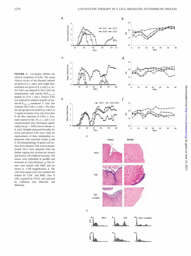
FIGURE 1. Lovastatin inhibits theclinical symptoms of EAE. The meanclinical scores of the diseased animalsare given in a, c, and e, and weight mea-surements are given in b, d, and f. a, Ac-tive EAE was induced in SJL/J mice byimmunization with myelin PLP139–151
peptide in CFA. c and e, Passive EAEwas induced by adoptive transfer of my-elin-PLP139–151-sensitized T cells intorecipient SJL/J mice. a and c, The mice(six per group) were treated i.p. with 2 or5 mg/kg lovastatin every day from days0–60 after induction of EAE; e, lova-statin started on day 10. a, c, and e, Lo-vastatin-treated mice developed signifi-cantly less (p � 0.001) severe disease. b,d, and f, Weights measured biweekly foractive and passive EAE mice. Data arerepresentative of three independent ex-periments with consistent results. g andh, The histopathology of spinal cord sec-tions from adoptive EAE and lovastatin-treated SJL/J mice prepared from thelumbar regions (six sections per mouse)and fixed in 10% buffered formalin. Thetissues were embedded in paraffin andsectioned at 5-�m thickness. g, The tis-sues were stained with H&E and areshown at �100 magnification. h, Thecells from spinal cord were isolated andstained for CD4� and MHC class IIcells, acquired by FACS, and analyzedby CellQuest (see Materials andMethods).
1276 LOVASTATIN THERAPY IN T CELL-MEDIATED AUTOIMMUNE DISEASE
by guest on June 13, 2013http://w
ww
.jimm
unol.org/D
ownloaded from
Next, we examined the in vivo effect of lovastatin on adoptiveEAE. SJL/J mice were treated with lovastatin (in the same manneras for active EAE) after induction of EAE by adoptive transfer ofmyelin PLP139–151-specific T cells. All 18 mice in the EAE-un-treated group developed clinical paralysis starting on day 8, with aMMCS of 3.2 on day 13 (Fig. 1c). Treatment with 2 and 5 mg/kglovastatin delayed disease onset by 3 days and decreased the de-velopment of paralysis (MMCS of 2 and 2.2) on day 13 during thefirst peak of EAE. Relapse was observed in lovastatin-treatedgroups on day 38 and 44, with MMCS of 0.67 and 1.61 in 2 and5 mg/kg groups, respectively. In the EAE group, MMCS was 2 andpeaked at 3.5 during the course of the study (60 days). In theadoptive EAE model, untreated animals lost weight continuouslyduring the study, whereas the 2 mg/kg lovastatin-treated group didnot show any weight loss compared with healthy controls (Fig.1d). Mice treated with 5 mg/kg lovastatin lost weight during thefirst peak of disease; subsequently, mice regained the weight andmaintained it similar to healthy controls.
To examine the therapeutic efficacy of lovastatin in ongoingEAE, SJL/J mice were treated with lovastatin (2 mg/kg i.p.) daily,and in another group it was given on alternate days from days10–60 after induction of passive EAE (as above). Day 10 waschosen for lovastatin therapy because in adoptive EAE the firstpeak started on approximately days 8 and 10. The EAE-untreatedgroup developed clinical paralysis starting on day 8, which pro-gressed to a MMCS of 2.4 on day 18 (Fig. 1e). Treatment of micewith lovastatin starting on day 10 decreased the clinical severity ofadoptive transfer EAE in the relapsing stage, but was unable tostop the first peak of disease. Lovastatin treatment on alternatedays was unable to stop both the first peak and relapsing and re-mitting EAE. In the group treated daily with lovastatin, EAEpeaked, and then relapsing and remitting disease was observed, butthe clinical severity never reached an MMCS �2, which was sig-nificantly less ( p � 0.001) than that in EAE. However, in thealternate day lovastatin-treated group, MMCS reached 3.5, similarto that in the EAE group. We observed that mice in lovastatin-treated and -untreated groups both lost weight compared with thehealthy controls (Fig. 1f).
Lovastatin decreased infiltration of cells to CNS
We examined the effect of lovastatin on the pathogenesis of in-flammation and demyelination in the CNS of mice with EAE. Spi-nal cord sections from mice treated with lovastatin after inductionof passive EAE were analyzed for the infiltration of mononuclearcells (inflammation) and myelin loss (demyelination). As shown inFig. 1g, the untreated EAE mice showed profound inflammation inthe CNS. Treatment of EAE mice with lovastatin significantly re-duced the number of inflammatory cells in both the artery andborder region (Fig. 1g). We observed fewer demyelinated regionsin lovastatin-treated animals compared with EAE mice (data notshown). Further, we examined the phenotype of infiltrating cells toCNS by FACS and found that the untreated EAE group had in-creased infiltration of CD4� T cells along with MHC class II-positive cells compared with controls. However, lovastatin treat-ment significantly reduced ( p � 0.001) the infiltration of CD4�
and MHC class II-positive cells (Fig. 1h).
Lovastatin inhibited neural Ag-specific and naive T cellproliferation
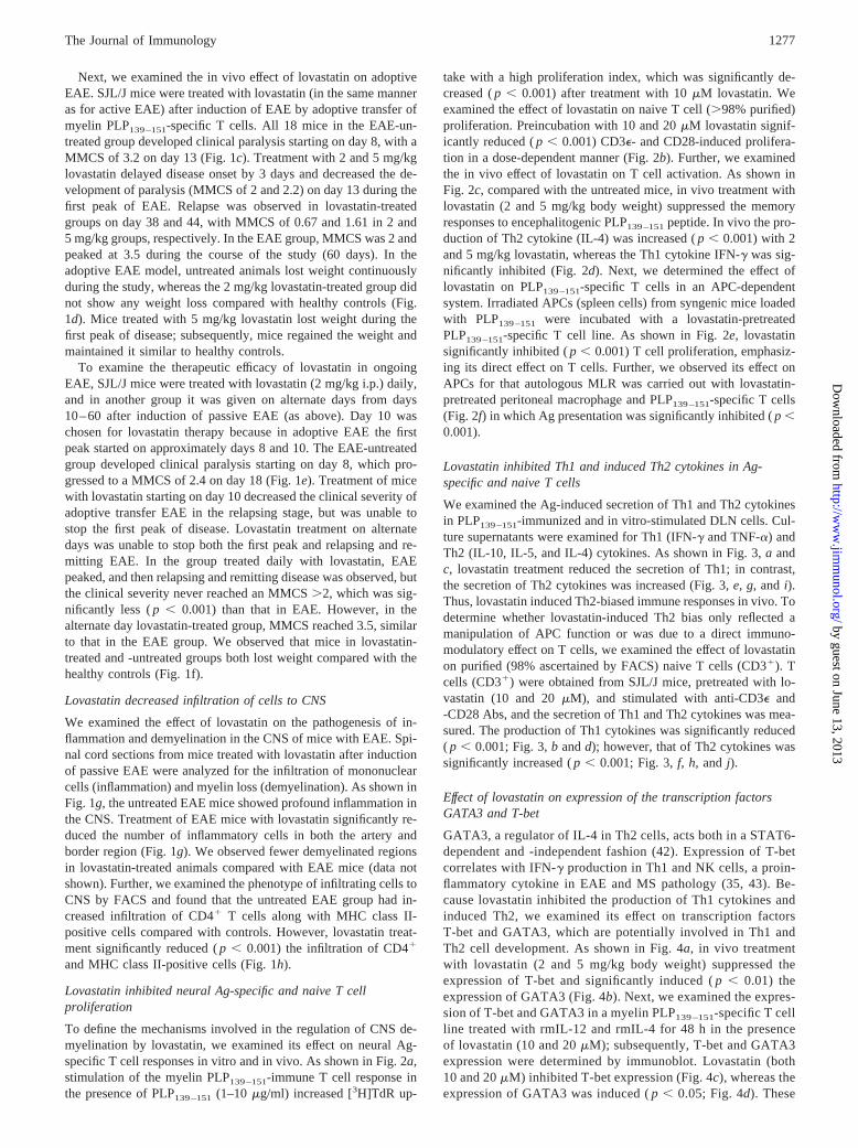
To define the mechanisms involved in the regulation of CNS de-myelination by lovastatin, we examined its effect on neural Ag-specific T cell responses in vitro and in vivo. As shown in Fig. 2a,stimulation of the myelin PLP139–151-immune T cell response inthe presence of PLP139–151 (1–10 �g/ml) increased [3H]TdR up-
take with a high proliferation index, which was significantly de-creased ( p � 0.001) after treatment with 10 �M lovastatin. Weexamined the effect of lovastatin on naive T cell (�98% purified)proliferation. Preincubation with 10 and 20 �M lovastatin signif-icantly reduced ( p � 0.001) CD3�- and CD28-induced prolifera-tion in a dose-dependent manner (Fig. 2b). Further, we examinedthe in vivo effect of lovastatin on T cell activation. As shown inFig. 2c, compared with the untreated mice, in vivo treatment withlovastatin (2 and 5 mg/kg body weight) suppressed the memoryresponses to encephalitogenic PLP139–151 peptide. In vivo the pro-duction of Th2 cytokine (IL-4) was increased ( p � 0.001) with 2and 5 mg/kg lovastatin, whereas the Th1 cytokine IFN-� was sig-nificantly inhibited (Fig. 2d). Next, we determined the effect oflovastatin on PLP139–151-specific T cells in an APC-dependentsystem. Irradiated APCs (spleen cells) from syngenic mice loadedwith PLP139–151 were incubated with a lovastatin-pretreatedPLP139–151-specific T cell line. As shown in Fig. 2e, lovastatinsignificantly inhibited ( p � 0.001) T cell proliferation, emphasiz-ing its direct effect on T cells. Further, we observed its effect onAPCs for that autologous MLR was carried out with lovastatin-pretreated peritoneal macrophage and PLP139–151-specific T cells(Fig. 2f) in which Ag presentation was significantly inhibited ( p �0.001).
Lovastatin inhibited Th1 and induced Th2 cytokines in Ag-specific and naive T cells
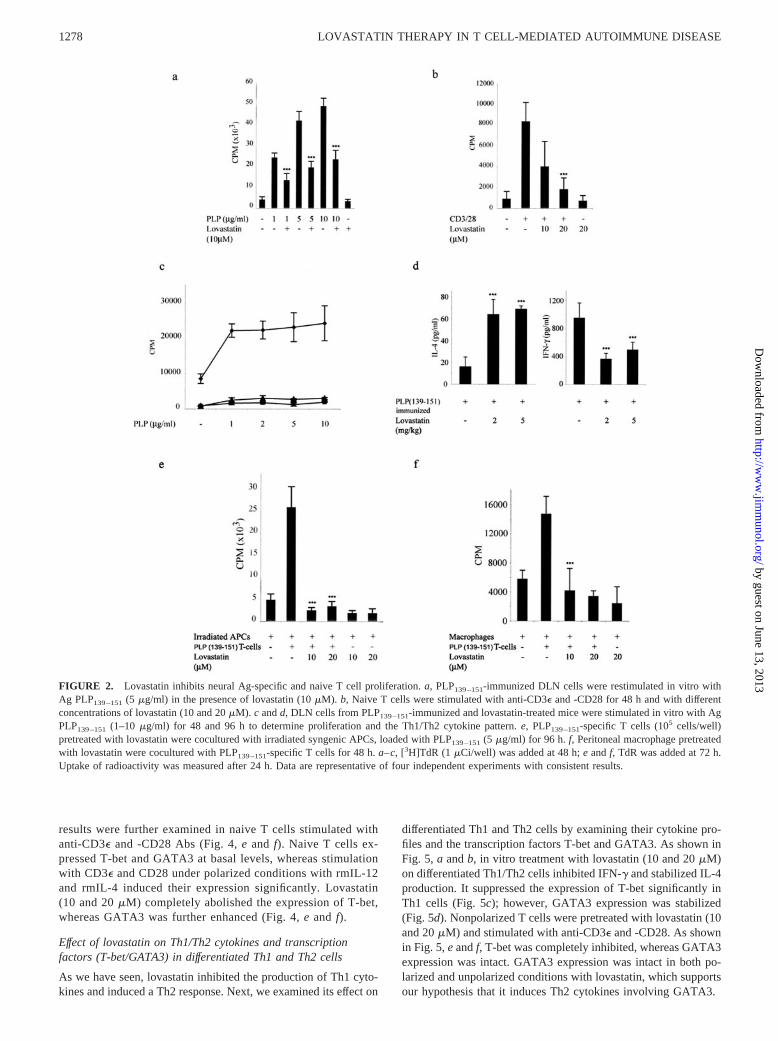
We examined the Ag-induced secretion of Th1 and Th2 cytokinesin PLP139–151-immunized and in vitro-stimulated DLN cells. Cul-ture supernatants were examined for Th1 (IFN-� and TNF-�) andTh2 (IL-10, IL-5, and IL-4) cytokines. As shown in Fig. 3, a andc, lovastatin treatment reduced the secretion of Th1; in contrast,the secretion of Th2 cytokines was increased (Fig. 3, e, g, and i).Thus, lovastatin induced Th2-biased immune responses in vivo. Todetermine whether lovastatin-induced Th2 bias only reflected amanipulation of APC function or was due to a direct immuno-modulatory effect on T cells, we examined the effect of lovastatinon purified (98% ascertained by FACS) naive T cells (CD3�). Tcells (CD3�) were obtained from SJL/J mice, pretreated with lo-vastatin (10 and 20 �M), and stimulated with anti-CD3� and-CD28 Abs, and the secretion of Th1 and Th2 cytokines was mea-sured. The production of Th1 cytokines was significantly reduced( p � 0.001; Fig. 3, b and d); however, that of Th2 cytokines wassignificantly increased ( p � 0.001; Fig. 3, f, h, and j).
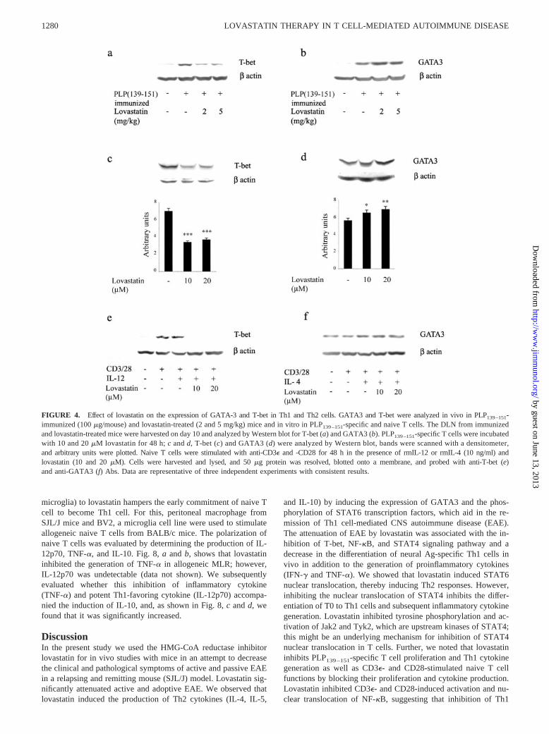
Effect of lovastatin on expression of the transcription factorsGATA3 and T-bet
GATA3, a regulator of IL-4 in Th2 cells, acts both in a STAT6-dependent and -independent fashion (42). Expression of T-betcorrelates with IFN-� production in Th1 and NK cells, a proin-flammatory cytokine in EAE and MS pathology (35, 43). Be-cause lovastatin inhibited the production of Th1 cytokines andinduced Th2, we examined its effect on transcription factorsT-bet and GATA3, which are potentially involved in Th1 andTh2 cell development. As shown in Fig. 4a, in vivo treatmentwith lovastatin (2 and 5 mg/kg body weight) suppressed theexpression of T-bet and significantly induced ( p � 0.01) theexpression of GATA3 (Fig. 4b). Next, we examined the expres-sion of T-bet and GATA3 in a myelin PLP139 –151-specific T cellline treated with rmIL-12 and rmIL-4 for 48 h in the presenceof lovastatin (10 and 20 �M); subsequently, T-bet and GATA3expression were determined by immunoblot. Lovastatin (both10 and 20 �M) inhibited T-bet expression (Fig. 4c), whereas theexpression of GATA3 was induced ( p � 0.05; Fig. 4d). These
1277The Journal of Immunology
by guest on June 13, 2013http://w
ww
.jimm
unol.org/D
ownloaded from
results were further examined in naive T cells stimulated withanti-CD3� and -CD28 Abs (Fig. 4, e and f). Naive T cells ex-pressed T-bet and GATA3 at basal levels, whereas stimulationwith CD3� and CD28 under polarized conditions with rmIL-12and rmIL-4 induced their expression significantly. Lovastatin(10 and 20 �M) completely abolished the expression of T-bet,whereas GATA3 was further enhanced (Fig. 4, e and f).
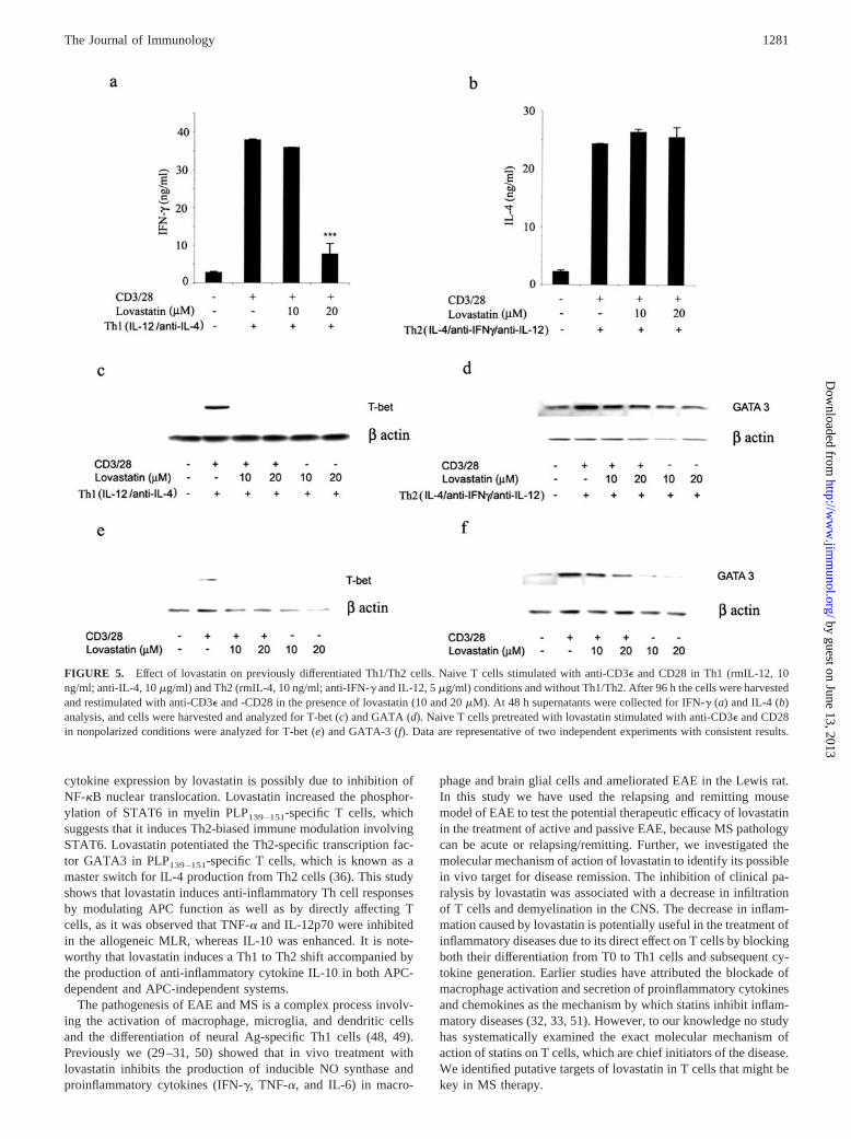
Effect of lovastatin on Th1/Th2 cytokines and transcriptionfactors (T-bet/GATA3) in differentiated Th1 and Th2 cells
As we have seen, lovastatin inhibited the production of Th1 cyto-kines and induced a Th2 response. Next, we examined its effect on
differentiated Th1 and Th2 cells by examining their cytokine pro-files and the transcription factors T-bet and GATA3. As shown inFig. 5, a and b, in vitro treatment with lovastatin (10 and 20 �M)on differentiated Th1/Th2 cells inhibited IFN-� and stabilized IL-4production. It suppressed the expression of T-bet significantly inTh1 cells (Fig. 5c); however, GATA3 expression was stabilized(Fig. 5d). Nonpolarized T cells were pretreated with lovastatin (10and 20 �M) and stimulated with anti-CD3� and -CD28. As shownin Fig. 5, e and f, T-bet was completely inhibited, whereas GATA3expression was intact. GATA3 expression was intact in both po-larized and unpolarized conditions with lovastatin, which supportsour hypothesis that it induces Th2 cytokines involving GATA3.
FIGURE 2. Lovastatin inhibits neural Ag-specific and naive T cell proliferation. a, PLP139–151-immunized DLN cells were restimulated in vitro withAg PLP139–151 (5 �g/ml) in the presence of lovastatin (10 �M). b, Naive T cells were stimulated with anti-CD3� and -CD28 for 48 h and with differentconcentrations of lovastatin (10 and 20 �M). c and d, DLN cells from PLP139–151-immunized and lovastatin-treated mice were stimulated in vitro with AgPLP139–151 (1–10 �g/ml) for 48 and 96 h to determine proliferation and the Th1/Th2 cytokine pattern. e, PLP139–151-specific T cells (105 cells/well)pretreated with lovastatin were cocultured with irradiated syngenic APCs, loaded with PLP139–151 (5 �g/ml) for 96 h. f, Peritoneal macrophage pretreatedwith lovastatin were cocultured with PLP139–151-specific T cells for 48 h. a–c, [3H]TdR (1 �Ci/well) was added at 48 h; e and f, TdR was added at 72 h.Uptake of radioactivity was measured after 24 h. Data are representative of four independent experiments with consistent results.
1278 LOVASTATIN THERAPY IN T CELL-MEDIATED AUTOIMMUNE DISEASE
by guest on June 13, 2013http://w
ww
.jimm
unol.org/D
ownloaded from
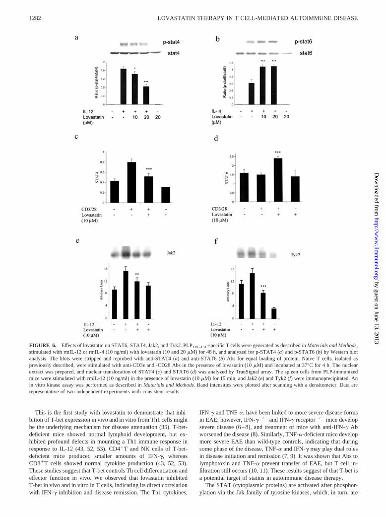
Lovastatin inhibited phosphorylation of Jak2, Tyk2, and STAT4and induced phosphorylation of STAT6 in PLP139–151-specificand naive T cells
As an activated (tyrosine-phosphorylated) STAT, STAT4 has akey role in IL-12-dependent Th1 lineage commitment (44).STAT6 is also required for IL-4-dependent Th2 lineage commit-ment (42). Thus, we examined whether lovastatin treatment sup-pressed the phosphorylation of STAT4 or induced the phosphor-ylation of STAT6 in PLP139–151-specific T cells. As shown in Fig.6, a and b, the PLP139–151-specific T cell line was stimulated withrmIL-12 and rmIL-4 in the presence of lovastatin, which reducedthe tyrosine phosphorylation of STAT4 in 15 min and inducedSTAT6 phosphorylation in 30 min ( p � 0.001).
To define the mechanisms involved in the lovastatin-mediatedregulation of T cell responses, we examined the effect of lovastatinon nuclear translocation of STAT4 and STAT6 transcription fac-tors in naive T cells that were stimulated with anti-CD3� and-CD28. Pretreatment of naive T cells with 10 �M lovastatin in-hibited the CD3�- and CD28-induced nuclear translocation ofSTAT4 (Fig. 6c) and potentiated STAT6 nuclear translocation(Fig. 6d). To determine whether the inhibition of STAT4 proteinsby lovastatin was a direct effect or a consequence of inhibition ofupstream Jak, we examined the effect of lovastatin on spleen cellsfrom PLP139–151-immunized mice. The cells were stimulated invitro with rmIL-12 (10 ng/ml) for tyrosine phosphorylation of Jak2and Tyk2. Immunoprecipitation and in vitro kinase assay showedthat the stimulation of T cells with IL-12 induced the activity ofJak2 and Tyk2 in 15 min (Fig. 6, e and f). However, pretreatmentof cells with lovastatin inhibited IL-12 and induced the activity ofJak2 ( p � 0.01) and Tyk2 ( p � 0.001).
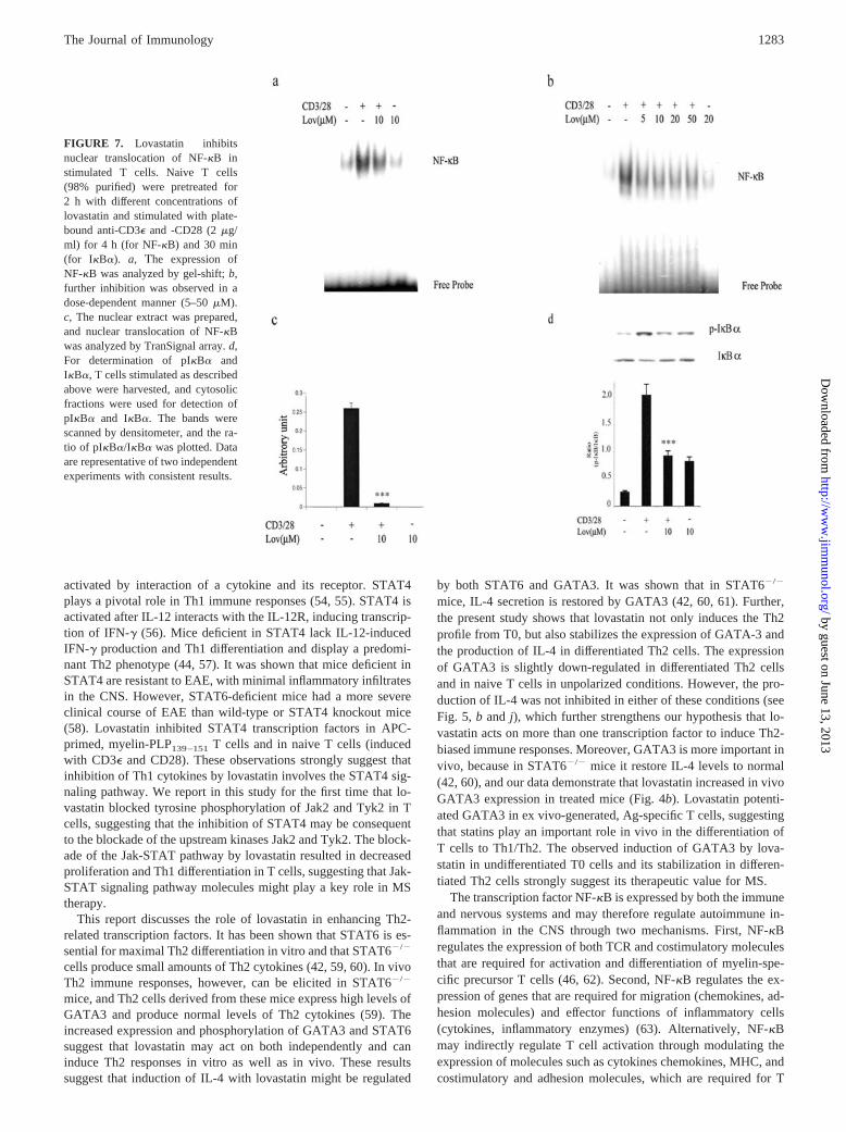
Lovastatin inhibited NF-�B pathway in activated T cells
Activation of NF-�B is necessary for cell survival and for induc-tion of IFN-�, TNF-�, IL-2, and MHC classes I and II (45, 46). Tounderstand the basis of inhibition of these cytokines in lovastatin-treated cells, we examined its effect on CD3�- and CD28-inducedactivation of NF-�B in naive T cells. In naive T cells, NF-�B(p65/p50 heterodimer) is retained in the cytoplasm by its associ-ation with I��. After stimulation of naive cells with variousagents, the cytosolic NF-�B/I�� complex dissociates, and freeNF-�B translocates to the nucleus and regulates the transcriptionof various genes. Phosphorylation of I�B� by upstream kinaseIKK is essential for the dissociation of I�B� from NF-�B and itsdegradation (46, 47). Stimulation of naive T cells with CD3� andCD28 significantly induced the nuclear translocation of NF-�B, asmeasured by gel-shift assay; however, 2-h pretreatment of T cellswith lovastatin (10 �M) significantly inhibited NF-�B ( p � 0.001;Fig. 7a). Inhibition of NF-�B by lovastatin appears to be dosedependent (Fig. 7b). The gel-shift detected a specific band in re-sponse to CD3� and CD28 stimulation that was competed off by anunlabeled probe (data not shown). These results were further sup-ported by the observation obtained from TransSignal protein/DNAarrays (Fig. 7c). Because lovastatin inhibits nuclear translocationof NF-�B, we examined the effect of lovastatin on phosphorylationand degradation of I�B�. Stimulation of naive T cells with CD3�and CD28 induced phosphorylation and degradation of I�B�,which was inhibited by lovastatin ( p � 0.001; Fig. 7d).
Lovastatin-pretreated APCs induce Th2 cells accompanied bygeneration of IL-10
We have shown earlier that lovastatin inhibits proinflammatorycytokines in glial cells in response to LPS activation (29). In thisstudy we found that exposure of APCs (peritoneal macrophage and
FIGURE 3. Induction of Th2 cytokines with lovastatin. a, c, e, g, and i,DLN cells were isolated on day 10 from PLP139–151-immunized SJL/J miceand cultured in vitro at 5 � 106 cells/ml in the presence of PLP139–151 (5�g/ml) and lovastatin (10 and 20 �M). b, d, f, h, and j, Naive T cells (98%purified) isolated from lymph nodes were cultured in anti-CD3�- and anti-CD28-precoated plates at 1 � 106 cells/ml with lovastatin (10 and 20 �M).The supernatants were collected at 48 h (for IFN-� and TNF-�) and 120 h(for IL-4, IL-5, and IL-10) for cytokine measurements. a–d, IFN-� andTNF-� are significantly reduced (p � 0.001) in both PLP-primed and naiveT cells; e–j, IL-10, IL-5, and IL-4 are significantly increased (p � 0.001).The values are the means of triplicate determinations at each point, and theerror bars represent �SD. Data are representative of four different exper-iments with consistent results.
1279The Journal of Immunology
by guest on June 13, 2013http://w
ww
.jimm
unol.org/D
ownloaded from
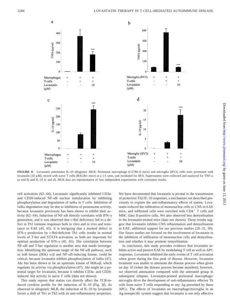
microglia) to lovastatin hampers the early commitment of naive Tcell to become Th1 cell. For this, peritoneal macrophage fromSJL/J mice and BV2, a microglia cell line were used to stimulateallogeneic naive T cells from BALB/c mice. The polarization ofnaive T cells was evaluated by determining the production of IL-12p70, TNF-�, and IL-10. Fig. 8, a and b, shows that lovastatininhibited the generation of TNF-� in allogeneic MLR; however,IL-12p70 was undetectable (data not shown). We subsequentlyevaluated whether this inhibition of inflammatory cytokine(TNF-�) and potent Th1-favoring cytokine (IL-12p70) accompa-nied the induction of IL-10, and, as shown in Fig. 8, c and d, wefound that it was significantly increased.
DiscussionIn the present study we used the HMG-CoA reductase inhibitorlovastatin for in vivo studies with mice in an attempt to decreasethe clinical and pathological symptoms of active and passive EAEin a relapsing and remitting mouse (SJL/J) model. Lovastatin sig-nificantly attenuated active and adoptive EAE. We observed thatlovastatin induced the production of Th2 cytokines (IL-4, IL-5,
and IL-10) by inducing the expression of GATA3 and the phos-phorylation of STAT6 transcription factors, which aid in the re-mission of Th1 cell-mediated CNS autoimmune disease (EAE).The attenuation of EAE by lovastatin was associated with the in-hibition of T-bet, NF-�B, and STAT4 signaling pathway and adecrease in the differentiation of neural Ag-specific Th1 cells invivo in addition to the generation of proinflammatory cytokines(IFN-� and TNF-�). We showed that lovastatin induced STAT6nuclear translocation, thereby inducing Th2 responses. However,inhibiting the nuclear translocation of STAT4 inhibits the differ-entiation of T0 to Th1 cells and subsequent inflammatory cytokinegeneration. Lovastatin inhibited tyrosine phosphorylation and ac-tivation of Jak2 and Tyk2, which are upstream kinases of STAT4;this might be an underlying mechanism for inhibition of STAT4nuclear translocation in T cells. Further, we noted that lovastatininhibits PLP139–151-specific T cell proliferation and Th1 cytokinegeneration as well as CD3�- and CD28-stimulated naive T cellfunctions by blocking their proliferation and cytokine production.Lovastatin inhibited CD3�- and CD28-induced activation and nu-clear translocation of NF-�B, suggesting that inhibition of Th1
FIGURE 4. Effect of lovastatin on the expression of GATA-3 and T-bet in Th1 and Th2 cells. GATA3 and T-bet were analyzed in vivo in PLP139–151-immunized (100 �g/mouse) and lovastatin-treated (2 and 5 mg/kg) mice and in vitro in PLP139–151-specific and naive T cells. The DLN from immunizedand lovastatin-treated mice were harvested on day 10 and analyzed by Western blot for T-bet (a) and GATA3 (b). PLP139–151-specific T cells were incubatedwith 10 and 20 �M lovastatin for 48 h; c and d, T-bet (c) and GATA3 (d) were analyzed by Western blot, bands were scanned with a densitometer,and arbitrary units were plotted. Naive T cells were stimulated with anti-CD3� and -CD28 for 48 h in the presence of rmIL-12 or rmIL-4 (10 ng/ml) andlovastatin (10 and 20 �M). Cells were harvested and lysed, and 50 �g protein was resolved, blotted onto a membrane, and probed with anti-T-bet (e)and anti-GATA3 (f) Abs. Data are representative of three independent experiments with consistent results.
1280 LOVASTATIN THERAPY IN T CELL-MEDIATED AUTOIMMUNE DISEASE
by guest on June 13, 2013http://w
ww
.jimm
unol.org/D
ownloaded from
cytokine expression by lovastatin is possibly due to inhibition ofNF-�B nuclear translocation. Lovastatin increased the phosphor-ylation of STAT6 in myelin PLP139–151-specific T cells, whichsuggests that it induces Th2-biased immune modulation involvingSTAT6. Lovastatin potentiated the Th2-specific transcription fac-tor GATA3 in PLP139–151-specific T cells, which is known as amaster switch for IL-4 production from Th2 cells (36). This studyshows that lovastatin induces anti-inflammatory Th cell responsesby modulating APC function as well as by directly affecting Tcells, as it was observed that TNF-� and IL-12p70 were inhibitedin the allogeneic MLR, whereas IL-10 was enhanced. It is note-worthy that lovastatin induces a Th1 to Th2 shift accompanied bythe production of anti-inflammatory cytokine IL-10 in both APC-dependent and APC-independent systems.
The pathogenesis of EAE and MS is a complex process involv-ing the activation of macrophage, microglia, and dendritic cellsand the differentiation of neural Ag-specific Th1 cells (48, 49).Previously we (29–31, 50) showed that in vivo treatment withlovastatin inhibits the production of inducible NO synthase andproinflammatory cytokines (IFN-�, TNF-�, and IL-6) in macro-
phage and brain glial cells and ameliorated EAE in the Lewis rat.In this study we have used the relapsing and remitting mousemodel of EAE to test the potential therapeutic efficacy of lovastatinin the treatment of active and passive EAE, because MS pathologycan be acute or relapsing/remitting. Further, we investigated themolecular mechanism of action of lovastatin to identify its possiblein vivo target for disease remission. The inhibition of clinical pa-ralysis by lovastatin was associated with a decrease in infiltrationof T cells and demyelination in the CNS. The decrease in inflam-mation caused by lovastatin is potentially useful in the treatment ofinflammatory diseases due to its direct effect on T cells by blockingboth their differentiation from T0 to Th1 cells and subsequent cy-tokine generation. Earlier studies have attributed the blockade ofmacrophage activation and secretion of proinflammatory cytokinesand chemokines as the mechanism by which statins inhibit inflam-matory diseases (32, 33, 51). However, to our knowledge no studyhas systematically examined the exact molecular mechanism ofaction of statins on T cells, which are chief initiators of the disease.We identified putative targets of lovastatin in T cells that might bekey in MS therapy.
FIGURE 5. Effect of lovastatin on previously differentiated Th1/Th2 cells. Naive T cells stimulated with anti-CD3� and CD28 in Th1 (rmIL-12, 10ng/ml; anti-IL-4, 10 �g/ml) and Th2 (rmIL-4, 10 ng/ml; anti-IFN-� and IL-12, 5 �g/ml) conditions and without Th1/Th2. After 96 h the cells were harvestedand restimulated with anti-CD3� and -CD28 in the presence of lovastatin (10 and 20 �M). At 48 h supernatants were collected for IFN-� (a) and IL-4 (b)analysis, and cells were harvested and analyzed for T-bet (c) and GATA (d). Naive T cells pretreated with lovastatin stimulated with anti-CD3� and CD28in nonpolarized conditions were analyzed for T-bet (e) and GATA-3 (f). Data are representative of two independent experiments with consistent results.
1281The Journal of Immunology
by guest on June 13, 2013http://w
ww
.jimm
unol.org/D
ownloaded from
This is the first study with lovastatin to demonstrate that inhi-bition of T-bet expression in vivo and in vitro from Th1 cells mightbe the underlying mechanism for disease attenuation (35). T-bet-deficient mice showed normal lymphoid development, but ex-hibited profound defects in mounting a Th1 immune response inresponse to IL-12 (43, 52, 53). CD4�T and NK cells of T-bet-deficient mice produced smaller amounts of IFN-�, whereasCD8�T cells showed normal cytokine production (43, 52, 53).These studies suggest that T-bet controls Th cell differentiation andeffector function in vivo. We observed that lovastatin inhibitedT-bet in vivo and in vitro in T cells, indicating its direct correlationwith IFN-� inhibition and disease remission. The Th1 cytokines,
IFN-� and TNF-�, have been linked to more severe disease formsin EAE; however, IFN-��/� and IFN-� receptor�/� mice developsevere disease (6–8), and treatment of mice with anti-IFN-� Abworsened the disease (8). Similarly, TNF-�-deficient mice developmore severe EAE than wild-type controls, indicating that duringsome phase of the disease, TNF-� and IFN-� may play dual rolesin disease initiation and remission (7, 9). It was shown that Abs tolymphotoxin and TNF-� prevent transfer of EAE, but T cell in-filtration still occurs (10, 11). These results suggest of that T-bet isa potential target of statins in autoimmune disease therapy.
The STAT (cytoplasmic proteins) are activated after phosphor-ylation via the Jak family of tyrosine kinases, which, in turn, are
FIGURE 6. Effects of lovastatin on STAT6, STAT4, Jak2, and Tyk2. PLP139–151-specific T cells were generated as described in Materials and Methods,stimulated with rmIL-12 or rmIL-4 (10 ng/ml) with lovastatin (10 and 20 �M) for 48 h, and analyzed for p-STAT4 (a) and p-STAT6 (b) by Western blotanalysis. The blots were stripped and reprobed with anti-STAT4 (a) and anti-STAT6 (b) Abs for equal loading of protein. Naive T cells, isolated aspreviously described, were stimulated with anti-CD3� and -CD28 Abs in the presence of lovastatin (10 �M) and incubated at 37°C for 4 h. The nuclearextract was prepared, and nuclear translocation of STAT4 (c) and STAT6 (d) was analyzed by TranSignal array. The spleen cells from PLP-immunizedmice were stimulated with rmIL-12 (10 ng/ml) in the presence of lovastatin (10 �M) for 15 min, and Jak2 (e) and Tyk2 (f) were immunoprecipitated. Anin vitro kinase assay was performed as described in Materials and Methods. Band intensities were plotted after scanning with a densitometer. Data arerepresentative of two independent experiments with consistent results.
1282 LOVASTATIN THERAPY IN T CELL-MEDIATED AUTOIMMUNE DISEASE
by guest on June 13, 2013http://w
ww
.jimm
unol.org/D
ownloaded from
activated by interaction of a cytokine and its receptor. STAT4plays a pivotal role in Th1 immune responses (54, 55). STAT4 isactivated after IL-12 interacts with the IL-12R, inducing transcrip-tion of IFN-� (56). Mice deficient in STAT4 lack IL-12-inducedIFN-� production and Th1 differentiation and display a predomi-nant Th2 phenotype (44, 57). It was shown that mice deficient inSTAT4 are resistant to EAE, with minimal inflammatory infiltratesin the CNS. However, STAT6-deficient mice had a more severeclinical course of EAE than wild-type or STAT4 knockout mice(58). Lovastatin inhibited STAT4 transcription factors in APC-primed, myelin-PLP139–151 T cells and in naive T cells (inducedwith CD3� and CD28). These observations strongly suggest thatinhibition of Th1 cytokines by lovastatin involves the STAT4 sig-naling pathway. We report in this study for the first time that lo-vastatin blocked tyrosine phosphorylation of Jak2 and Tyk2 in Tcells, suggesting that the inhibition of STAT4 may be consequentto the blockade of the upstream kinases Jak2 and Tyk2. The block-ade of the Jak-STAT pathway by lovastatin resulted in decreasedproliferation and Th1 differentiation in T cells, suggesting that Jak-STAT signaling pathway molecules might play a key role in MStherapy.
This report discusses the role of lovastatin in enhancing Th2-related transcription factors. It has been shown that STAT6 is es-sential for maximal Th2 differentiation in vitro and that STAT6�/�
cells produce small amounts of Th2 cytokines (42, 59, 60). In vivoTh2 immune responses, however, can be elicited in STAT6�/�
mice, and Th2 cells derived from these mice express high levels ofGATA3 and produce normal levels of Th2 cytokines (59). Theincreased expression and phosphorylation of GATA3 and STAT6suggest that lovastatin may act on both independently and caninduce Th2 responses in vitro as well as in vivo. These resultssuggest that induction of IL-4 with lovastatin might be regulated
by both STAT6 and GATA3. It was shown that in STAT6�/�
mice, IL-4 secretion is restored by GATA3 (42, 60, 61). Further,the present study shows that lovastatin not only induces the Th2profile from T0, but also stabilizes the expression of GATA-3 andthe production of IL-4 in differentiated Th2 cells. The expressionof GATA3 is slightly down-regulated in differentiated Th2 cellsand in naive T cells in unpolarized conditions. However, the pro-duction of IL-4 was not inhibited in either of these conditions (seeFig. 5, b and j), which further strengthens our hypothesis that lo-vastatin acts on more than one transcription factor to induce Th2-biased immune responses. Moreover, GATA3 is more important invivo, because in STAT6�/� mice it restore IL-4 levels to normal(42, 60), and our data demonstrate that lovastatin increased in vivoGATA3 expression in treated mice (Fig. 4b). Lovastatin potenti-ated GATA3 in ex vivo-generated, Ag-specific T cells, suggestingthat statins play an important role in vivo in the differentiation ofT cells to Th1/Th2. The observed induction of GATA3 by lova-statin in undifferentiated T0 cells and its stabilization in differen-tiated Th2 cells strongly suggest its therapeutic value for MS.
The transcription factor NF-�B is expressed by both the immuneand nervous systems and may therefore regulate autoimmune in-flammation in the CNS through two mechanisms. First, NF-�Bregulates the expression of both TCR and costimulatory moleculesthat are required for activation and differentiation of myelin-spe-cific precursor T cells (46, 62). Second, NF-�B regulates the ex-pression of genes that are required for migration (chemokines, ad-hesion molecules) and effector functions of inflammatory cells(cytokines, inflammatory enzymes) (63). Alternatively, NF-�Bmay indirectly regulate T cell activation through modulating theexpression of molecules such as cytokines chemokines, MHC, andcostimulatory and adhesion molecules, which are required for T
FIGURE 7. Lovastatin inhibitsnuclear translocation of NF-�B instimulated T cells. Naive T cells(98% purified) were pretreated for2 h with different concentrations oflovastatin and stimulated with plate-bound anti-CD3� and -CD28 (2 �g/ml) for 4 h (for NF-�B) and 30 min(for I�B�). a, he expression ofNF-�B was analyzed by gel-shift; b,further inhibition was observed in adose-dependent manner (5–50 �M).c, The nuclear extract was prepared,and nuclear translocation of NF-�Bwas analyzed by TranSignal array. d,For determination of pI�B� andI�B�, T cells stimulated as describedabove were harvested, and cytosolicfractions were used for detection ofpI�B� and I�B�. The bands werescanned by densitometer, and the ra-tio of pI�B�/I�B� was plotted. Dataare representative of two independentexperiments with consistent results.
1283The Journal of Immunology
by guest on June 13, 2013http://w
ww
.jimm
unol.org/D
ownloaded from
cell activation (62–64). Lovastatin significantly inhibited CD3�-and CD28-induced NF-�B nuclear translocation by inhibitingphosphorylation and degradation of I�B� in T cells. Inhibition ofI�B� degradation may be due to inhibition of proteasome activity,because lovastatin previously has been shown to inhibit their ac-tivity (62–64). Induction of NF-�B directly correlates with IFN-�generation, and it was observed that c-Rel deficiency led to a de-fect in Th1 immune responses both in vitro and in vivo and resis-tance to EAE (45, 65). It is intriguing that a marked defect inIFN-� production by c-Rel-deficient Th1 cells results in normallevels of T-bet and STAT4 activation, as both are important foroptimal production of IFN-� (45, 65). The correlation betweenNF-�B and T-bet regulation is another area that needs investiga-tion. Identifying the upstream target of the NF-�B pathway, suchas I�B kinase (IKK) �/� and NF-�B-inducing kinase, could becritical, because lovastatin inhibits phosphorylation of I�B� (47).Akt has been shown to be an upstream kinase of IKK��, whichregulates its activity via phosphorylation (47). Akt might be a po-tential target for lovastatin, because it inhibits CD3�- and CD28-induced Akt activity in naive T cells (data not shown).
This study reports that statins can directly affect the TCR-in-duced cytokine profile for the induction of IL-10 (Fig. 3f). Asobserved in allogeneic MLR, the induction of IL-10 by lovastatinfavors a shift of Th1 to Th2 with its anti-inflammatory properties.
We have documented that lovastatin is pivotal in the transmissionof protective Th2/IL-10 responses, a mechanism not described pre-viously to explain the anti-inflammatory effects of statins. Lova-statin reduced the infiltration of mononuclear cells to CNS in EAEmice, and infiltrated cells were enriched with CD4� T cells andMHC class II-positive cells. We also observed less demyelinationin the lovastatin-treated mice (data not shown). These results sug-gest that lovastatin inhibits CNS inflammation and demyelinationin EAE, additional support for our previous studies (29–31, 50).Our future studies are focused on the involvement of lovastatin inthe inhibition of infiltration of mononuclear cells and demyelina-tion and whether it may promote remyelination.
In conclusion, this study provides evidence that lovastatin in-hibits active and passive EAE by modulating T cell as well as APCresponses. Lovastatin inhibited the early events of T cell activationwhen given during the first peak of disease. However, lovastatintreatment was unable to attenuate the disease process when givenon day 10 (when the disease process became manifest); however,we observed attenuation compared with the untreated group insubsequent relapses. Lovastatin-primed peritoneal macrophage/microglia drive the development of anti-inflammatory effector Thcells from naive T cells responding to any Ag presented by theseAPCs. The effects of lovastatin on macrophage/microglia in anAg-nonspecific system suggest that lovastatin is not only effective
FIGURE 8. Lovastatin potentiates IL-10 allogeneic MLR. Peritoneal macrophage (C57BL/6 mice) and microglia (BV2) cells were pretreated withlovastatin (10 �M), mixed with naive T cells (BALB/c mice) at a 1/1 ratio, and incubated for 48 h. Supernatants were collected and analyzed for TNF-�(a and b) and IL-10 (c and d). MLR data are representative of two independent experiments with consistent results.
1284 LOVASTATIN THERAPY IN T CELL-MEDIATED AUTOIMMUNE DISEASE
by guest on June 13, 2013http://w
ww
.jimm
unol.org/D
ownloaded from
in EAE, but may also prove to be effective in other inflammatorydiseases. Further, the possible mechanism involved in disease pro-gression is the expression of T-bet, STAT4, and NF-�B, whichcontribute to the induction of Th1 cytokines. Lovastatin inhibitedthese transcription factors (T-bet, NF-�B, and STAT4), which areresponsible for CNS inflammation via induction of Th1 cell dif-ferentiation and cytokine (IFN-� and TNF-�) production. We havedemonstrated that lovastatin induced the expression of GATA3and STAT6 in Th2 cells, which promoted the remission of thedisease by enhancing T0 to Th2 cell differentiation and cytokine(IL-4, IL-5, and IL-10) generation by affecting both APCs and Tcells. The stabilization of IL-4/GATA3 in differentiated Th2 cellsand the concomitant down-regulation of IFN-�/T-bet in Th1 cellsby lovastatin identified these molecules as novel targets for MStherapy.
AcknowledgmentsWe thank Drs. Anne G. Gilg and Jennifer G. Schnellmann for their criticalreading of the manuscript, and Joyce Bryan and Hope Terry for laboratoryassistance.
References1. Ozawa, K., G. Suchanek, H. Breitschopf, W. Bruck, H. Budka, K. Jellinger, and
H. Lassmann. 1994. Patterns of oligodendroglia pathology in multiple sclerosis.Brain 117:1311.
2. Lassmann, H., C. S. Raine, J. Antel, and J. W. Prineas. 1998. Immunopathologyof multiple sclerosis: report on an international meeting held at the Institute ofNeurology of the University of Vienna. J. Neuroimmunol. 86:213.
3. Lucchinetti, C. F., W. Bruck, M. Rodriguez, and H. Lassmann. 1996. Distinctpatterns of multiple sclerosis pathology indicate heterogeneity on pathogenesis.Brain Pathol. 6:259.
4. Weiner, H. L., and D. J. Selkoe. 2002. Inflammation and therapeutic vaccinationin CNS diseases. Nature 420:879.
5. Swanborg, R. H. 1995. Experimental autoimmune encephalomyelitis in rodentsas a model for human demyelinating disease. Clin. Immunol. Immunopathol.77:4.
6. Ferber, I. A., S. Brocke, C. Taylor-Edwards, W. Ridgway, C. Dinisco,L. Steinman, D. Dalton, and C. G. Fathman. 1996. Mice with a disrupted IFN-�gene are susceptible to the induction of experimental autoimmune encephalomy-elitis (EAE). J. Immunol. 156:5.
7. Frei, K., H. P. Eugster, M. Bopst, C. S. Constantinescu, E. Lavi, and A. Fontana.1997. Tumor necrosis factor � and lymphotoxin � are not required for inductionof acute experimental autoimmune encephalomyelitis. J. Exp. Med. 185:2177.
8. Lublin, F. D., R. L. Knobler, B. Kalman, M. Goldhaber, J. Marini, M. Perrault,C. D’Imperio, J. Joseph, S. S. Alkan, and R. Korngold. 1993. Monoclonal anti-gamma interferon antibodies enhance experimental allergic encephalomyelitis.Autoimmunity 16:267.
9. Liu, J., M. W. Marino, G. Wong, D. Grail, A. Dunn, J. Bettadapura, A. J. Slavin,L. Old, and C. C. Bernard. 1998. TNF is a potent anti-inflammatory cytokine inautoimmune-mediated demyelination. Nat. Med. 4:78.
10. Ruddle, N. H., C. M. Bergman, K. M. McGrath, E. G. Lingenheld, M. L. Grunnet,S. J. Padula, and R. B. Clark. 1990. An antibody to lymphotoxin and tumornecrosis factor prevents transfer of experimental allergic encephalomyelitis.J. Exp. Med. 172:1193.
11. Korner, H., F. A. Lemckert, G. Chaudhri, S. Etteldorf, and J. D. Sedgwick. 1997.Tumor necrosis factor blockade in actively induced experimental autoimmuneencephalomyelitis prevents clinical disease despite activated T cell infiltration tothe central nervous system. Eur. J. Immunol. 27:1973.
12. Young, D. A., L. D. Lowe, S. S. Booth, M. J. Whitters, L. Nicholson,V. K. Kuchroo, and M. Collins. 2000. IL-4, IL-10, IL-13, and TGF-� from analtered peptide ligand-specific Th2 cell clone down-regulate adoptive transfer ofexperimental autoimmune encephalomyelitis. J. Immunol. 164:3563.
13. Croxford, J. L., M. Feldmann, Y. Chernajovsky, and D. Baker. 2001. Differenttherapeutic outcomes in experimental allergic encephalomyelitis dependent uponthe mode of delivery of IL-10: a comparison of the effects of protein, adenoviralor retroviral IL-10 delivery into the central nervous system. J. Immunol.166:4124.
14. Santambrogio, L., G. M. Hochwald, B. Saxena, C. H. Leu, J. E. Martz,J. A. Carlino, N. H. Ruddle, M. A. Palladino, L. I. Gold, and G. J. Thorbecke.1993. Studies on the mechanisms by which transforming growth factor-�(TGF-�) protects against allergic encephalomyelitis: antagonism between TGF-�and tumor necrosis factor. J. Immunol. 151:1116.
15. Legge, K. L., B. Min, J. J. Bell, J. C. Caprio, L. Li, R. K. Gregg, andH. Zaghouani. 2000. Coupling of peripheral tolerance to endogenous interleukin10 promotes effective modulation of myelin-activated T cells and amelioratesexperimental allergic encephalomyelitis. J. Exp. Med. 191:2039.
16. Ando, D. G., J. Clayton, D. Kono, J. L. Urban, and E. E. Sercarz. 1989. Enceph-alitogenic T cells in the B10. PL model of experimental allergic encephalomy-elitis (EAE) are of the Th-1 lymphokine subtype. Cell. Immunol. 124:132.
17. Zamvil, S., P. Nelson, J. Trotter, D. Mitchell, R. Knobler, R. Fritz, andL. Steinman. 1985. T-cell clones specific for myelin basic protein induce chronicrelapsing paralysis and demyelination. Nature 317:355.
18. Zamvil, S. S., and L. Steinman. 1990. The T lymphocyte in experimental allergicencephalomyelitis. Annu. Rev. Immunol. 8:579.
19. Laufs, U., K. Gertz, U. Dirnagl, M. Bohm, G. Nickenig, and M. Endres. 2002.Rosuvastatin, a new HMG-CoA reductase inhibitor, upregulates endothelial nitricoxide synthase and protects from ischemic stroke in mice. Brain Res. 942:23.
20. Zamvil, S. S., and L. Steinman. 2002. Cholesterol-lowering statins possess anti-inflammatory activity that might be useful for treatment of MS. Neurology59:970.
21. Weitz-Schmidt, G. 2002. Statins as anti-inflammatory agents. Trends Pharmacol.Sci. 23:482.
22. Sadeghi, M. M., M. Collinge, R. Pardi, and J. R. Bender. 2000. Simvastatinmodulates cytokine-mediated endothelial cell adhesion molecule induction: in-volvement of an inhibitory G protein. J. Immunol. 165:2712.
23. Kobashigawa, J. A., S. Katznelson, H. Laks, J. A. Johnson, L. Yeatman,X. M. Wang, D. Chia, P. I. Terasaki, A. Sabad, G. A. Cogert, et al. 1995. Effectof pravastatin on outcomes after cardiac transplantation. N. Engl. J. Med. 333:621.
24. Dimitroulakos, J., L. Y. Ye, M. Benzaquen, M. J. Moore, S. Kamel-Reid,M. H. Freedman, H. Yeger, and L. Z. Penn. 2001. Differential sensitivity ofvarious pediatric cancers and squamous cell carcinomas to lovastatin-inducedapoptosis: therapeutic implications. Clin. Cancer Res. 7:158.
25. Mueck, A. O., H. Seeger, and D. Wallwiener. 2001. Further evidence for directvascular actions of statins: effect on endothelial nitric oxide synthase and adhe-sion molecules. Exp. Clin. Endocrinol. Diabetes 109:181.
26. Denoyelle, C., P. Albanese, G. Uzan, L. Hong, J. P. Vannier, J. Soria, andC. Soria. 2003. Molecular mechanism of the anti-cancer activity of cerivastatin,an inhibitor of HMG-CoA reductase, on aggressive human breast cancer cells.Cell. Signalling 15:327.
27. Leung, B. P., N. Sattar, A. Crilly, M. Prach, D. W. McCarey, H. Payne,R. Madhok, C. Campbell, J. A. Gracie, F. Y. Liew, et al. 2003. A novel anti-inflammatory role for simvastatin in inflammatory arthritis. J. Immunol.170:1524.
28. Feleszko, W., I. Mlynarczuk, E. Z. Balkowiec-Iskra, A. Czajka, T. Switaj,T. Stoklosa, A. Giermasz, and M. Jakobisiak. 2000. Lovastatin potentiates anti-tumor activity and attenuates cardiotoxicity of doxorubicin in three tumor modelsin mice. Clin. Cancer Res. 6:2044.
29. Pahan, K., F. G. Sheikh, A. M. Namboodiri, and I. Singh. 1997. Lovastatin andphenylacetate inhibit the induction of nitric oxide synthase and cytokines in ratprimary astrocytes, microglia, and macrophages. J. Clin. Invest. 100:2671.
30. Stanislaus, R., K. Pahan, A. K. Singh, and I. Singh. 1999. Amelioration of ex-perimental allergic encephalomyelitis in Lewis rats by lovastatin. Neurosci. Lett.269:71.
31. Stanislaus, R., A. K. Singh, and I. Singh. 2001. Lovastatin treatment decreasesmononuclear cell infiltration into the CNS of Lewis rats with experimental al-lergic encephalomyelitis. J. Neurosci. Res. 66:155.
32. Kwak, B., F. Mulhaupt, S. Myit, and F. Mach. 2000. Statins as a newly recog-nized type of immunomodulator. Nat. Med. 6:1399.
33. Weitz-Schmidt, G., K. Welzenbach, V. Brinkmann, T. Kamata, J. Kallen,C. Bruns, S. Cottens, Y. Takada, and U. Hommel. 2001. Statins selectively inhibitleukocyte function antigen-1 by binding to a novel regulatory integrin site. Nat.Med. 7:687.
34. Youssef, S., O. Stuve, J. C. Patarroyo, P. J. Ruiz, J. L. Radosevich, E. M. Hur,M. Bravo, D. J. Mitchell, R. A. Sobel, L. Steinman, et al. 2002. The HMG-CoAreductase inhibitor, atorvastatin, promotes a Th2 bias and reverses paralysis incentral nervous system autoimmune disease. Nature 420:78.
35. Szabo, S. J., S. T. Kim, G. L. Costa, X. Zhang, C. G. Fathman, andL. H. Glimcher. 2000. A novel transcription factor, T-bet, directs Th1 lineagecommitment. Cell 100:655.
36. Zheng, W., and R. A. Flavell. 1997. The transcription factor GATA-3 is neces-sary and sufficient for Th2 cytokine gene expression in CD4 T cells. Cell 89:587.
37. Tompkins, S. M., J. Padilla, M. C. Dal Canto, J. P. Ting, L. Van Kaer, andS. D. Miller. 2002. De novo central nervous system processing of myelin antigenis required for the initiation of experimental autoimmune encephalomyelitis.J. Immunol. 168:4173.
38. Howard, L. M., and S. D. Miller. 2001. Autoimmune intervention by CD154blockade prevents T cell retention and effector function in the target organ. J. Im-munol. 166:1547.
39. Weigel, B. J., N. Nath, P. A. Taylor, A. Panoskaltsis-Mortari, W. Chen,A. M. Krieg, K. Brasel, and B. R. Blazar. 2002. Comparative analysis of murinemarrow-derived dendritic cells generated by Flt3L or GM-CSF/IL-4 and maturedwith immune stimulatory agents on the in vivo induction of antileukemia re-sponses. Blood 100:4169.
40. Giri, S., M. Jatana, R. Rattan, J. S. Won, I. Singh, and A. K. Singh. 2002.Galactosylsphingosine (psychosine)-induced expression of cytokine-mediated in-ducible nitric oxide synthases via AP-1 and C/EBP: implications for Krabbedisease. FASEB J. 16:661.
41. Bright, J. J., L. D. Kerr, and S. Sriram. 1997. TGF-� inhibits IL-2-induced ty-rosine phosphorylation and activation of Jak1 and Stat 5 in T lymphocytes. J. Im-munol. 159:175.
42. Ouyang, W., M. Lohning, Z. Gao, M. Assenmacher, S. Ranganath, A. Radbruch,and K. M. Murphy. 2000. Stat6-independent GATA-3 autoactivation directs IL-4-independent Th2 development and commitment. Immunity 12:27.
1285The Journal of Immunology
by guest on June 13, 2013http://w
ww
.jimm
unol.org/D
ownloaded from
43. Szabo, S. J., B. M. Sullivan, C. Stemmann, A. R. Satoskar, B. P. Sleckman, andL. H. Glimcher. 2002. Distinct effects of T-bet in TH1 lineage commitment andIFN-� production in CD4 and CD8 T cells. Science 295:338.
44. Thierfelder, W. E., J. M. van Deursen, K. Yamamoto, R. A. Tripp, S. R. Sarawar,R. T. Carson, M. Y. Sangster, D. A. Vignali, P. C. Doherty, G. C. Grosveld, etal. 1996. Requirement for Stat4 in interleukin-12-mediated responses of naturalkiller and T cells. Nature 382:171.
45. Hilliard, B., E. B. Samoilova, T. S. Liu, A. Rostami, and Y. Chen. 1999. Exper-imental autoimmune encephalomyelitis in NF-�B-deficient mice: roles of NF-�Bin the activation and differentiation of autoreactive T cells. J. Immunol. 163:2937.
46. Li, Q., and I. M. Verma. 2002. NF-�B regulation in the immune system. Nat. Rev.Immunol. 2:725.
47. Karin, M., and Y. Ben-Neriah. 2000. Phosphorylation meets ubiquitination: thecontrol of NF-�B activity. Annu. Rev. Immunol. 18:621.
48. Fischer, H. G., and G. Reichmann. 2001. Brain dendritic cells and macrophages/microglia in central nervous system inflammation. J. Immunol. 166:2717.
49. Aloisi, F., F. Ria, S. Columba-Cabezas, H. Hess, G. Penna, and L. Adorini. 1999.Relative efficiency of microglia, astrocytes, dendritic cells and B cells in naiveCD4� T cell priming and Th1/Th2 cell restimulation. Eur. J. Immunol. 29:2705.
50. Stanislaus, R., A. G. Gilg, A. K. Singh, and I. Singh. 2002. Immunomodulationof experimental autoimmune encephalomyelitis in the Lewis rats by Lovastatin.Neurosci. Lett. 333:167.
51. Romano, M., L. Diomede, M. Sironi, L. Massimiliano, M. Sottocorno,N. Polentarutti, A. Guglielmotti, D. Albani, A. Bruno, P. Fruscella, et al. 2000.Inhibition of monocyte chemotactic protein-1 synthesis by statins. Lab. Invest.80:1095.
52. Finotto, S., M. F. Neurath, J. N. Glickman, S. Qin, H. A. Lehr, F. H. Green,K. Ackerman, K. Haley, P. R. Galle, S. J. Szabo, et al. 2002. Development ofspontaneous airway changes consistent with human asthma in mice lacking T-bet.Science 295:336.
53. Mullen, A. C., F. A. High, A. S. Hutchins, H. W. Lee, A. V. Villarino,D. M. Livingston, A. L. Kung, N. Cereb, T. P. Yao, S. Y. Yang, et al. 2001. Roleof T-bet in commitment of TH1 cells before IL-12-dependent selection. Science292:1907.
54. Pernis, A. B., and P. B. Rothman. 2002. JAK-STAT signaling in asthma. J. Clin.Invest. 109:1279.
55. Ohashi, P. S. 2002. T-cell signalling and autoimmunity: molecular mechanismsof disease. Nat. Rev. Immunol. 2:427.
56. Jacobson, N. G., S. J. Szabo, R. M. Weber-Nordt, Z. Zhong, R. D. Schreiber,J. E. Darnell, Jr., and K. M. Murphy. 1995. Interleukin 12 signaling in T helpertype 1 (Th1) cells involves tyrosine phosphorylation of signal transducer andactivator of transcription (Stat)3 and Stat4. J. Exp. Med. 181:1755.
57. Kaplan, M. H., Y. L. Sun, T. Hoey, and M. J. Grusby. 1996. Impaired IL-12responses and enhanced development of Th2 cells in Stat4-deficient mice. Nature382:174.
58. Chitnis, T., N. Najafian, C. Benou, A. D. Salama, M. J. Grusby, M. H. Sayegh,and S. J. Khoury. 2001. Effect of targeted disruption of STAT4 and STAT6 on theinduction of experimental autoimmune encephalomyelitis. J. Clin. Invest.108:739.
59. Finkelman, F. D., S. C. Morris, T. Orekhova, M. Mori, D. Donaldson,S. L. Reiner, N. L. Reilly, L. Schopf, and J. F. Urban, Jr. 2000. Stat6 regulationof in vivo IL-4 responses. J. Immunol. 164:2303.
60. Jankovic, D., M. C. Kullberg, N. Noben-Trauth, P. Caspar, W. E. Paul, andA. Sher. 2000. Single cell analysis reveals that IL-4 receptor/Stat6 signaling is notrequired for the in vivo or in vitro development of CD4� lymphocytes with a Th2cytokine profile. J. Immunol. 164:3047.
61. Ho, I. C., and L. H. Glimcher. 2002. Transcription: tantalizing times for T cells.Cell 109(Suppl.):S109.
62. Barnes, P. J., and M. Karin. 1997. Nuclear factor-�B: a pivotal transcriptionfactor in chronic inflammatory diseases. N. Engl. J. Med. 336:1066.
63. Beg, A. A., and D. Baltimore. 1996. An essential role for NF-�B in preventingTNF-�-induced cell death. Science 274:782.
64. Ting, A. T., F. X. Pimentel-Muinos, and B. Seed. 1996. RIP mediates tumornecrosis factor receptor 1 activation of NF-�B but not Fas/APO-1-initiated apo-ptosis. EMBO J. 15:6189.
65. Hilliard, B. A., N. Mason, L. Xu, J. Sun, S. E. Lamhamedi-Cherradi, H. C. Liou,C. Hunter, and Y. H. Chen. 2002. Critical roles of c-Rel in autoimmune inflam-mation and helper T cell differentiation. J. Clin. Invest. 110:843.
1286 LOVASTATIN THERAPY IN T CELL-MEDIATED AUTOIMMUNE DISEASE
by guest on June 13, 2013http://w
ww
.jimm
unol.org/D
ownloaded from