posterior urethral valve
TRANSCRIPT

9/11/2013
Clinical Case
Case of a posterior
Urethral valve in a Viable Twin Intrauterine
Gestation Of About 23W2D, affecting the
leading Twin in a 23-year-old woman.
9/11/2013
Patient Clinical History
History :
A 23-year-old pregnant woman presented asymptomatic.
With no clinical signs of oligohydramnios
No complains of lower abdominal pains
First pregnancy and with no history of abortion.
Normal heart rate of fetus on midwife’s examination.
Abdomen was normal for GA
Family his. Of twin GA
9/11/2013
REFERRING PHYSICIAN (MIDWIFE) REQUEST
Obstetric scan
9/11/2013
Department Protocol
Ultrasound Department Manhyia District Hospital protocol with additional comments and impression stated in the exams report.
MANHYIA DISTRICT HOSPITAL 2ND AND 3RD.docx
9/11/2013
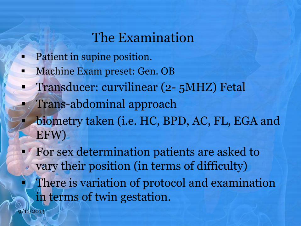
The Examination
Patient in supine position.
Machine Exam preset: Gen. OB
Transducer: curvilinear (2- 5MHZ) Fetal
Trans-abdominal approach
biometry taken (i.e. HC, BPD, AC, FL, EGA and EFW)
For sex determination patients are asked to vary their position (in terms of difficulty)
There is variation of protocol and examination in terms of twin gestation.
9/11/2013
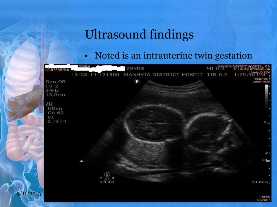
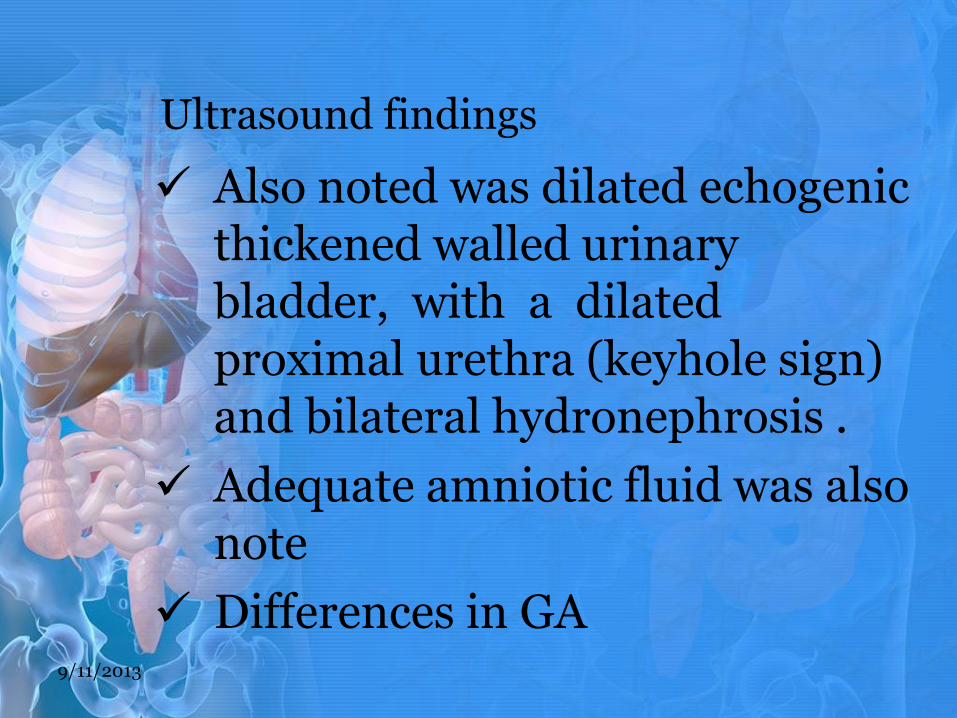
Ultrasound findings
• Noted is an intrauterine twin gestation
9/11/2013

Ultrasound findings
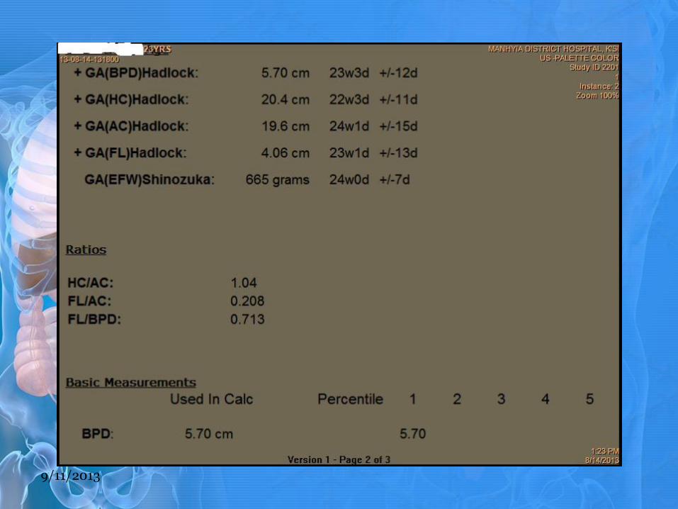
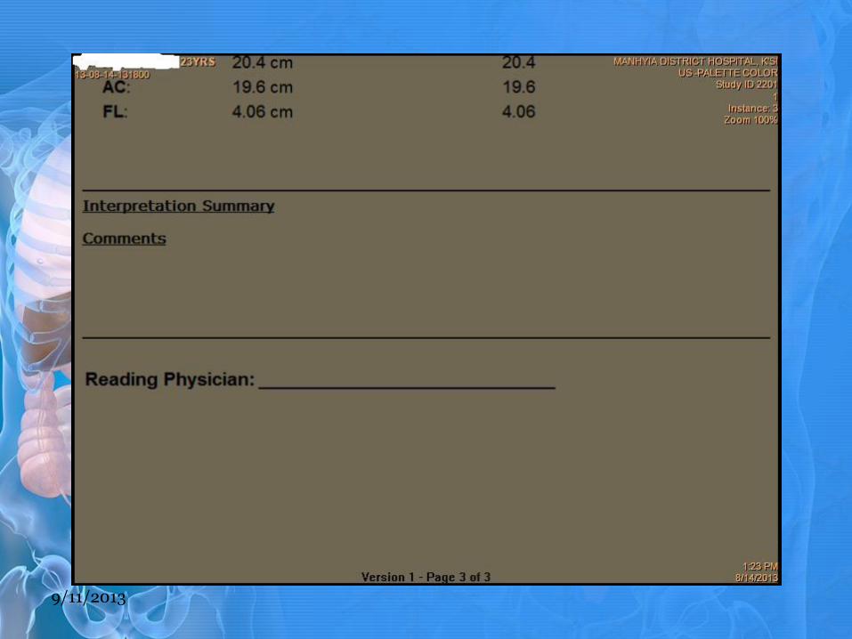
The fetal biometry of both twin
TA TB
BPD-(23W3D) -(21W6D)
HC-(22W3D) -(21W5D)
AC-(24W1D) -(22W3D)
FL-(23W1D) -(22W2D)
9/11/2013

On gray scale 2D BPD/HC
9/11/2013
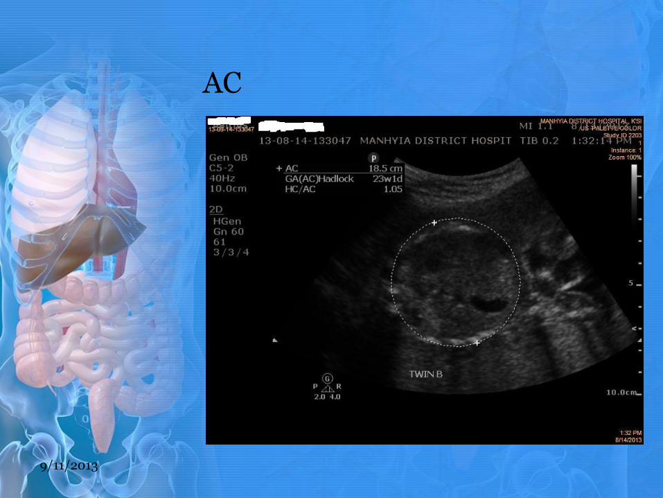
AC
9/11/2013
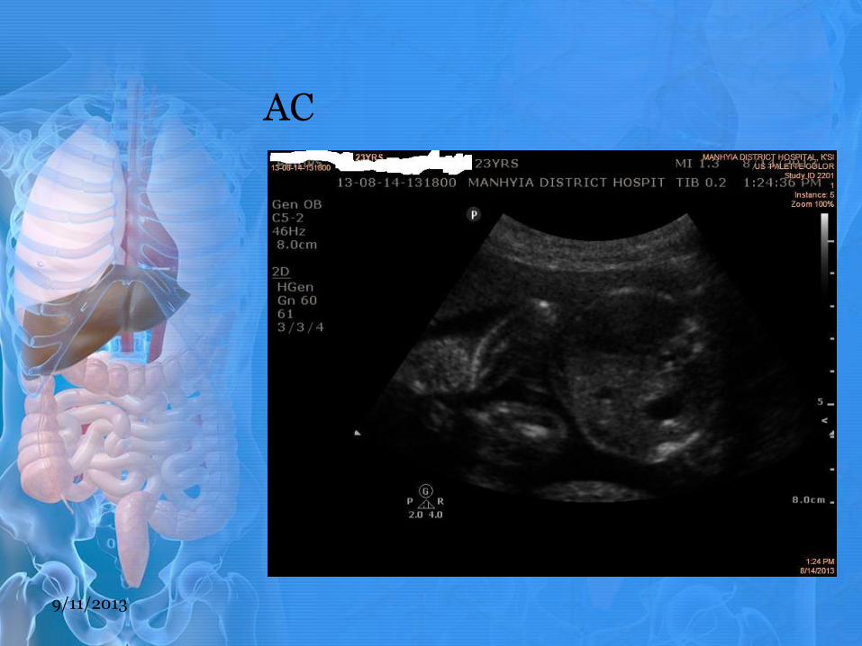
AC
9/11/2013
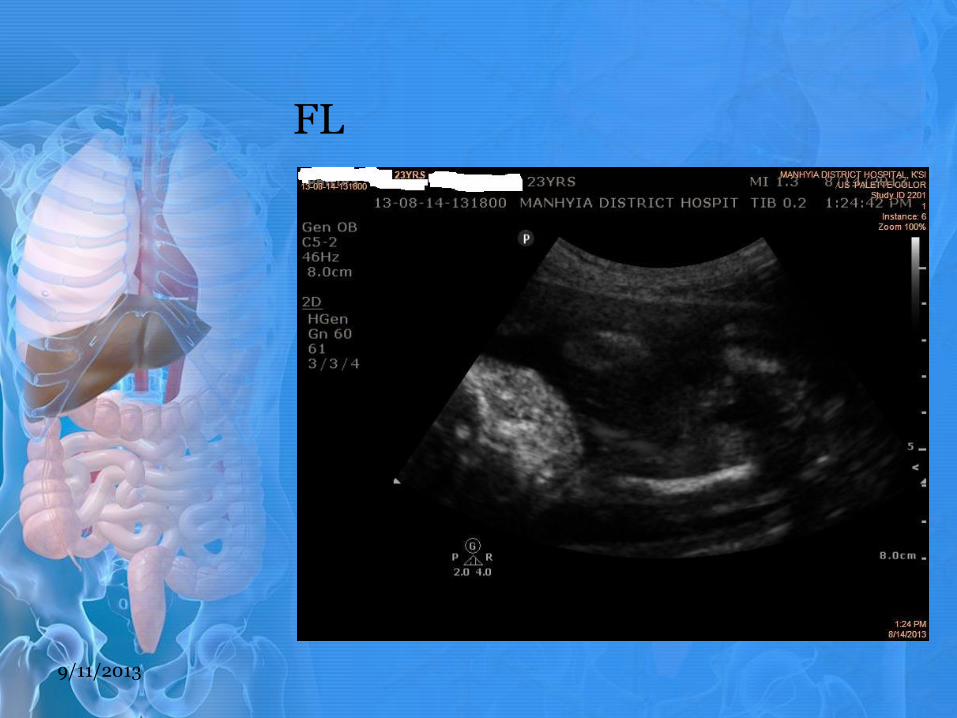
FL
9/11/2013

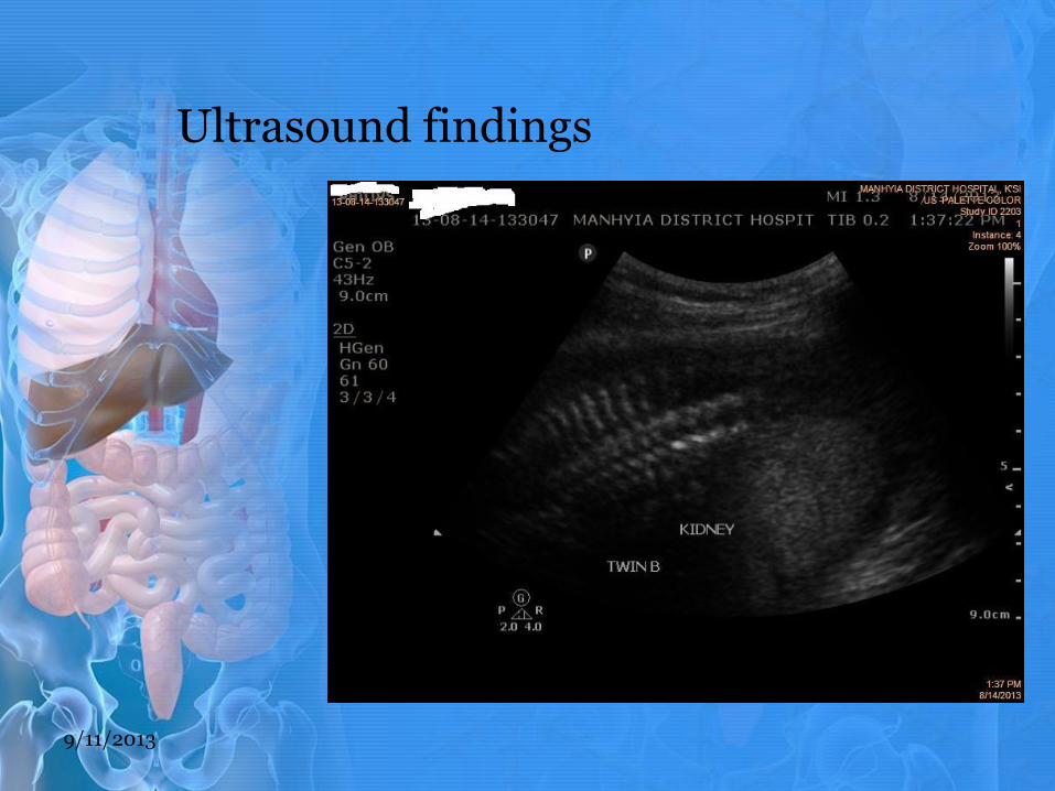
Ultrasound findings
• Twin B
• Ultrasound report MANHYIA DISTRICT HOSPITAL PM.pdf
9/11/2013
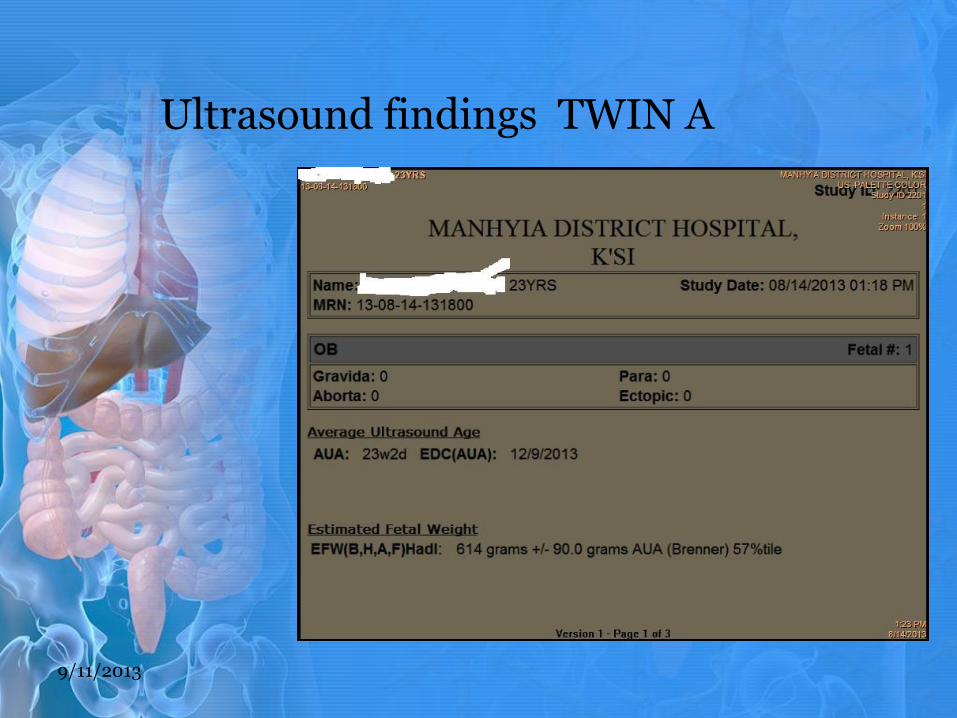
Ultrasound findings TWIN A
9/11/2013
9/11/2013
9/11/2013
Ultrasound findings
Also noted was dilated echogenic thickened walled urinary bladder, with a dilated proximal urethra (keyhole sign) and bilateral hydronephrosis .
Adequate amniotic fluid was also note
Differences in GA 9/11/2013
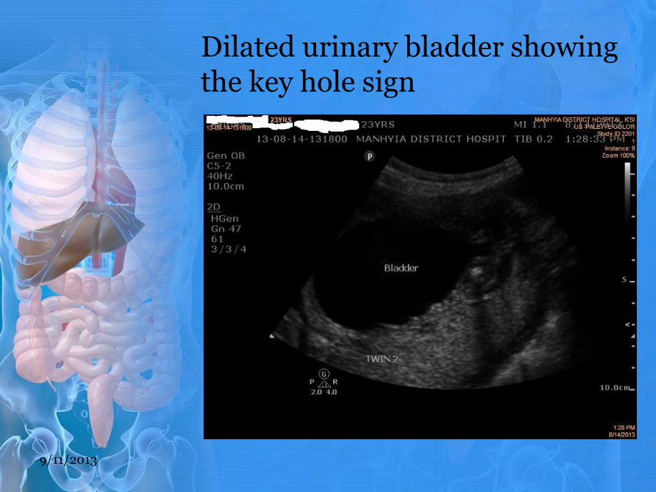
Dilated urinary bladder showing the key hole sign
9/11/2013
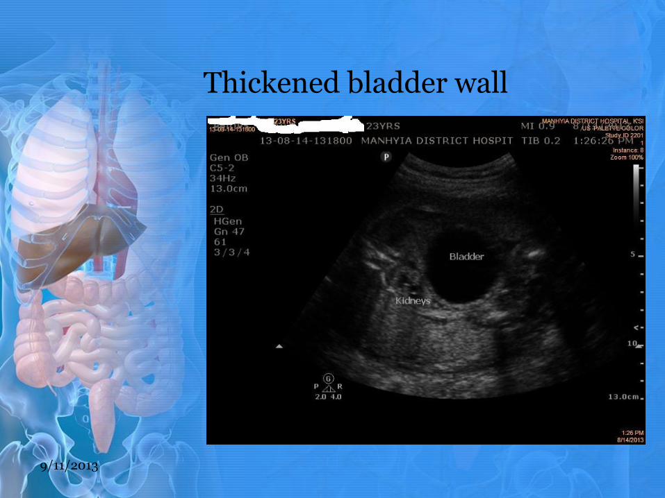
Thickened bladder wall
9/11/2013
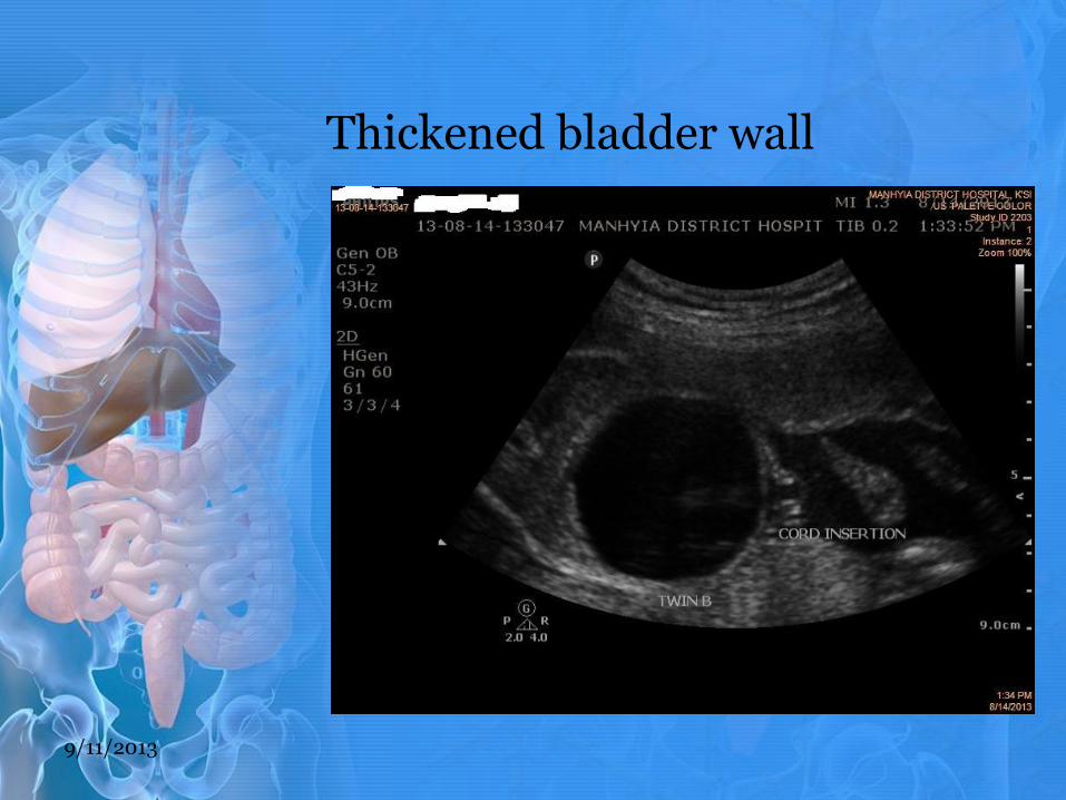
Thickened bladder wall
9/11/2013
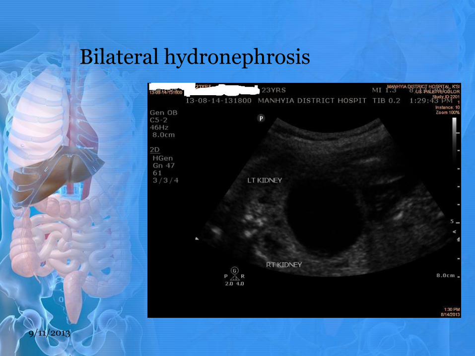
Bilateral hydronephrosis
9/11/2013
Ultrasound findings
9/11/2013
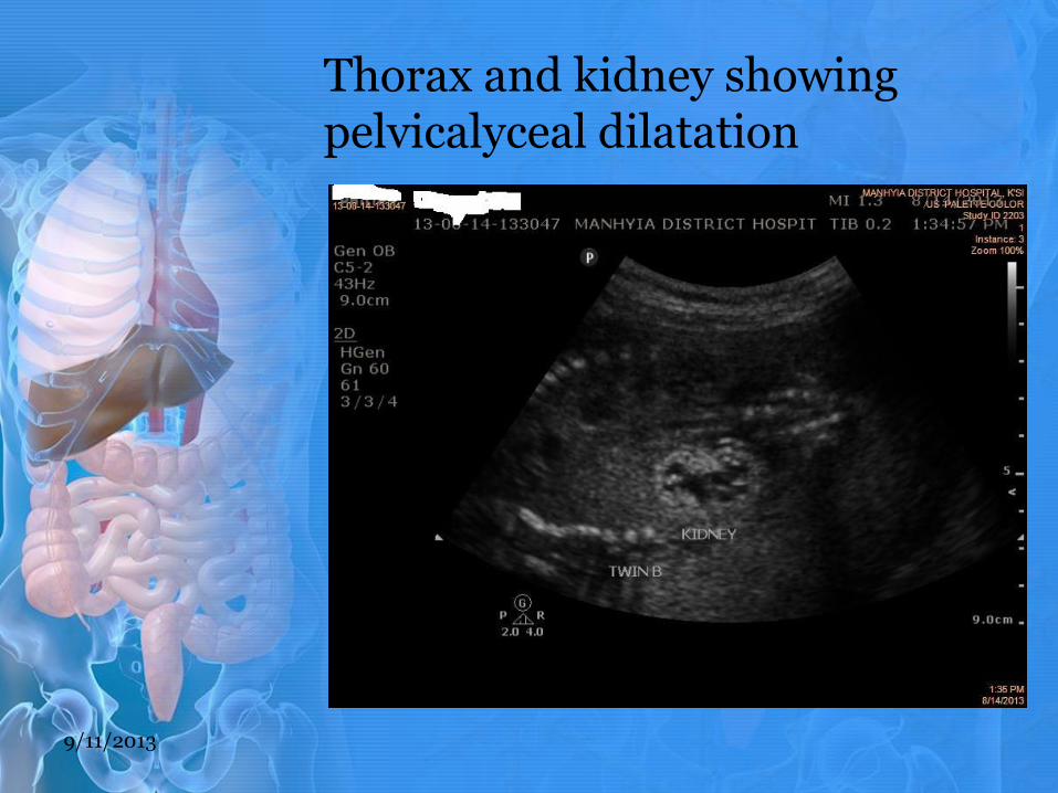
Thorax and kidney showing pelvicalyceal dilatation
9/11/2013
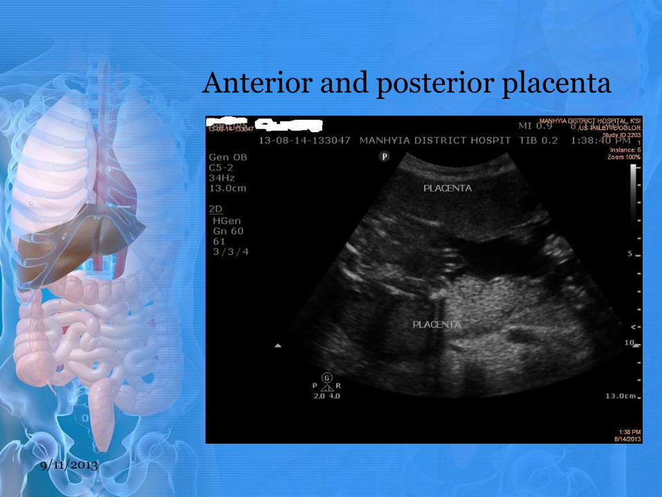
Anterior and posterior placenta
9/11/2013
Relation of encountered pathology in relation to patient presentation and our findings.
The presence of intrauterine twin gestation with twin A not affected with any obstructive uropathy, in this case is an indication of adequate amniotic fluid noted around both twins, which ruled out oligohydramnios associated with PUV. Thus the affected twin only swallows the amniotic fluid without urinating into it but the unaffected one swallows and at the same time urinates into the amnion which keeps the fluid balanced.
The absence of Oligohydramnios explains the mother’s condition of showing no signs and symptoms.
9/11/2013
Dilated urinary bladder with thickened echogenic wall and dilated proximal urethra (keyhole sign) noted on sonogram above suggests that there is an obstruction, of lower urinary tract origin preventing urine from coming out of the urinary system. This in effect builds up a backwards pressure (thrust) which pushes urine back to fill the pelvicalyceal systems which was noted as Bilateral hydronephrosis on sonogram above.
9/11/2013
Conclusions drawn:
After discussing case with my supervisor we came to a conclusion that the findings were consistent with posterior urethral valve in a twin gestation of about 23w2d affecting twin B.
9/11/2013

Ultrasound Exams Final Report
MANHYIA DISTRICT HOSPITAL.pdf
Differentials
Urethral atresia/agenesis
Neurogenic bladder
Prune-belly syndrome
9/11/2013
Follow-up
Follow up was done and patients was asked to come for antenatal care consistently and routine ultrasound examinations will be done to monitor the progress of the condition.
If fetus survive up to term, they will deliver them by appropriate method and will be handed over to a pediatrician surgeon for surgical repair of the urethral valve.
9/11/2013

Discussion:
Megacystis In the first trimester, an enlarged bladder >7 mm (megacystis) should raise the suspicion of either chromosomal abnormalities or obstructive uropathy. Approximately 20% of fetuses with megacystis measuring 7–15 mm have an underlying chromosomal abnormality such as trisomy 13 or 18. If the bladder measures >15 mm, the risk of an underlying bladder outlet obstruction (urethral atresia, posterior urethral valves, or cloacal abnormalities) is very high and is associated with a very poor prognosis. With megacystis >15 mm, it is sometimes possible to find echogenic or dysplastic kidneys and reduced liquor volume in the first trimester. From: problem-based obstetric ultrasound by Basky Thilaganathan, Shanthi Sairam, Aris T Papageorghiou and Amar Bhide.
9/11/2013
Discussion: • Obstruction of the posterior urethra by a congenital valve, a severe
condition often associated with refluxing or obstructive uropathy and renal dysplasia, mostly seen in baby boys. from Encyclopedia of Diagnostic Imaging volume 1
• Posterior urethral valves are the most common cause of lower urinary tract obstruction, followed by urethral atresia or stricture. Posterior urethral valves are seen exclusively in males and may cause total, intermittent, or partial obstruction, with variable prognosis. Most cases are sporadic, occurring in 1 in 5000 male births; and the recurrence risk is small.
• Back pressure causes a persistently dilated urinary bladder, with a dilated proximal urethra (keyhole sign). There may be thickening of the bladder wall (>2 mm) or a severely distended thin-walled bladder, bilateral tortuous hydroureters, and hydronephrosis. If the obstruction is severe and of long-standing, progressive renal parenchymal fibrosis and dysplasia develop, resulting in severe oligohydramnios, pulmonary hypoplasia, and compression deformities (Potter’s syndrome). There may be spontaneous bladder rupture with urinary ascites or a calyceal rupture with perirenal urinoma. If spontaneous decompression occurs, this “safety valve” may protect the kidneys from further prenatal damage and may diminish the degree of hydronephrosis. Rumack
9/11/2013

Discussion: CAUSES
• The etiology is unknown
• Genetics.
• Developmental anomaly in the urethra
• Abnormal embryology and persistent of a fold which remains and forms flaps or valves in the urethral passage which Normally forms in the 4th months of gestation.
• Urethral carcinoid tumors normally occur at the verumontanum.
9/11/2013
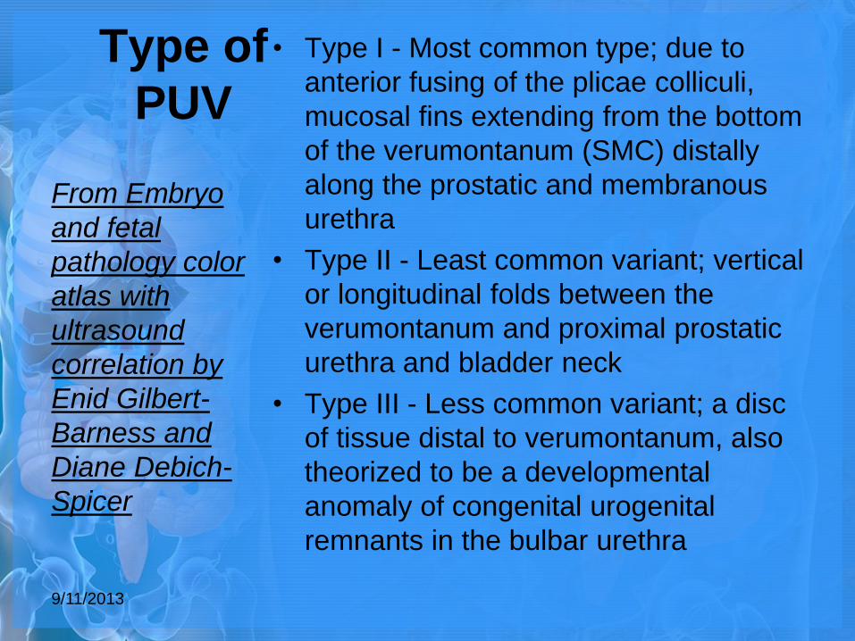
Type of
PUV
• Type I - Most common type; due to
anterior fusing of the plicae colliculi,
mucosal fins extending from the bottom
of the verumontanum (SMC) distally
along the prostatic and membranous
urethra
• Type II - Least common variant; vertical
or longitudinal folds between the
verumontanum and proximal prostatic
urethra and bladder neck
• Type III - Less common variant; a disc
of tissue distal to verumontanum, also
theorized to be a developmental
anomaly of congenital urogenital
remnants in the bulbar urethra
From Embryo
and fetal
pathology color
atlas with
ultrasound
correlation by
Enid Gilbert-
Barness and
Diane Debich-
Spicer
9/11/2013
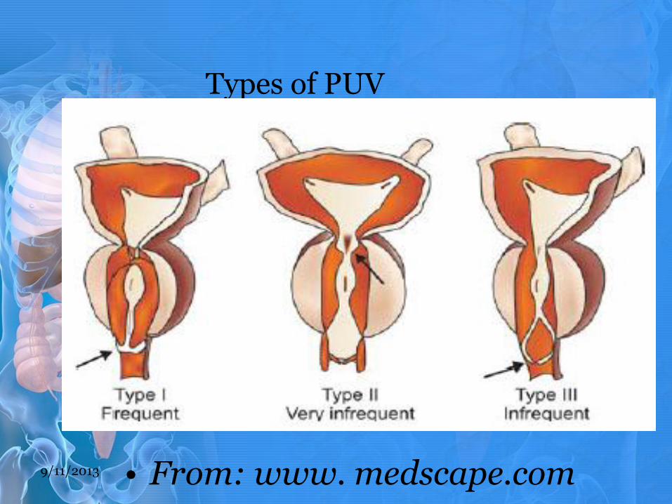
Types of PUV
• From: www. medscape.com 9/11/2013
Discussion: associate conditions Kidney failure Pulmonary hypoplasia compression deformities (Potter’s syndrome) spontaneous bladder rupture
9/11/2013
Discussion: Role of Ultrasonography in Diagnosis and assessment
Ultrasound can accurately detect fetal lower urinary tract Obstruction with a sensitivity of 95% and a specificity of 80%. The sensitivity of ultrasound screening for these abnormalities is improved because anomalies of the renal tractand kidneys are also associated with secondary findings,such as oligohydramnios. Assessment of the fetal genitourinary tract is a part of all routine ultrasound examinations. When abnormalities are detected, this should lead to a detailed assessment focusing on amniotic fluid volume, renal size and parenchyma, the collecting system, and bladder size. Dilatation of the renal pelvis and fluid-filled areas as small as 1-2mm may be visualized in utero using high-resolution real-time ultrasound. Ultrasonography may allow differentiation of obstructive and non-obstructive causes of megacystis, with the association of increased echogenicity and oligohydramnios in the presence of bladder distension being predictive of an obstructive aetiology. Similarly, in cases of hydronephrosis, Oliveira et al. showed that oligohydramnios and megacystis were predictive of an obstructive aetiology, with the sensitivity and specificity of the combination of both variables being 60 and 98% respectively. However, ultrasound is of limited value in determinng the underlying aetiology causing the LUTO. 9/11/2013
Treatment/management
Open fetal surgery
The initial approach adopted involved open hysterotomy. Harrison et al. performed the first successful in-utero decompression for hydronephrosis in 1981. The maternal morbidity associated with open surgery, and associated fetal risks of preterm labour and neurological sequelae, have led to the development of alternative minimally invasive techniques. No new cases of open fetal surgery have been reported since 1988
Vesico-amniotic shunting
Ultrasound-guided percutaneous vesico-amniotic shunting is the most commonly used method to relieve urinary tract obstruction. This technique involves the placement of a double pig-tailed catheter under ultrasound guidance
and local anaesthesia, with the distal end in the fetal
bladder and the proximal end in the amniotic cavity to
allow drainage of fetal urine.
Vesicocentesis
Fine-needle aspiration under ultrasound guidance
9/11/2013
References
• Diagnostic Ultrasound, 4th Ed vol 1; Rumack, Wilson, Charboneau, Levine
• Problem-based obstetric ultrasound by Basky Thilaganathan, Shanthi Sairam, Aris T Papageorghiou and Amar Bhide.
• Posterior%20Urethral%20Valves%20Symptoms%20and%20Treatment.htm
• Fetal lower urinary tract obstruction by David Lissauer, Rachel K. Morris, Mark D. Kilby in Seminars in Fetal & Neonatal Medicine journal (2007)12, 464e470 available at www.sciencedirect.com
9/11/2013
Cont.
• (Holzgreve W, Evans MI: Nonvascular needle and shunt placements for fetal therapy, In Fetal Medicine [Special Issue]. Westij Med 1993; 159:333-340)
• Embryo and fetal pathology color atlas with ultrasound correlation by Enid Gilbert-Barness and Diane Debich-Spicer(© Enid Gilbert-Barness and Diane Debich-Spicer 2004)
• Posterior urethral valves by Dr. Yuranga Weerakkody and Radswiki et al.
9/11/2013
9/11/2013

THANK YOU!!!
9/11/2013