polycystin-1 promotes pkcα-mediated nf-κb activation in kidney cells
TRANSCRIPT
www.elsevier.com/locate/ybbrc
Biochemical and Biophysical Research Communications 350 (2006) 257–262
BBRC
Polycystin-1 promotes PKCa-mediated NF-jB activation in kidney cells
Manuela Banzi a, Gianluca Aguiari a, Viky Trimi a, Alessandra Mangolini a, Paolo Pinton b,Ralph Witzgall c, Rosario Rizzuto b, Laura del Senno a,*
a Department of Biochemistry and Molecular Biology, University of Ferrara, Ferrara, Italyb General Pathology, Department of Experimental and Diagnostic Medicine, University of Ferrara, Ferrara, Italy
c Institute for Molecular and Cellular Anatomy, University of Regensburg, Regensburg, Germany
Received 4 September 2006Available online 18 September 2006
Abstract
Polycystin-1 (PC1), the PKD1 gene product, is a membrane receptor which regulates many cell functions, including cell proliferationand apoptosis, both typically increased in cyst lining cells in autosomal dominant polycystic kidney disease. Here we show that PC1upregulates the NF-jB signalling pathway in kidney cells to prevent cell death. Human embryonic kidney cell lines (HEK293CTT), stablyexpressing a PC1 cytoplasmic terminal tail (CTT), presented increased NF-jB nuclear levels and NF-jB-mediated luciferase promoteractivity. This, consistently, was reduced in HEK293 cells in which the endogenous PC1 was depleted by RNA interference. CTT-depen-dent NF-jB promoter activation was mediated by PKCa because it was blocked by its specific inhibitor Ro-320432. Furthermore, it wasobserved that apoptosis, which was increased in PC1-depleted cells, was reduced in HEK293CTT cells and in porcine kidney LtTA cellsexpressing a doxycycline-regulated CTT. Staurosporine, a PKC inhibitor, and parthenolide, a NF-jB inhibitor, significantly reduced theCTT-dependent antiapoptotic effect. These data reveal, therefore, a novel pathway by which polycystin-1 activates a PKCa-mediatedNF-jB signalling and cell survival.� 2006 Elsevier Inc. All rights reserved.
Keywords: Polycystin-1; NF-jB; Promoter activity; siRNA; Apoptosis; PKCa
The main pathogenic mechanism for autosomal domi-nant polycystic kidney disease (ADPKD), a commoninherited kidney disorder, is the total or even partial lossof function of either polycystin-1 (PC1) or polycystin-2(PC2) [1]. PC1 is a 500 kDa protein with a large extracellu-lar N-terminus, 11 transmembrane regions, and a smallcytoplasmic C-terminal tail involved in signal transductionpathways [1]. Recently, it has been shown that PC1 acts asa mechano-fluid stress sensor in primary cilium of kidneytubular cells [2] by interacting with the intracellular C-ter-minus of PC2, a Ca2+-permeable cation channel [3], andtransducing the cilial deflection as a transient increase incytoplasmic Ca2+ levels [4]. This observation provided
0006-291X/$ - see front matter � 2006 Elsevier Inc. All rights reserved.
doi:10.1016/j.bbrc.2006.09.042
* Corresponding author. Fax: +39 532 202723.E-mail address: [email protected] (L.del Senno).
the most convincing evidence for the functional interactionof the two proteins in the regulation of intracellular Ca2+
homeostasis and Ca2+-dependent signal transduction path-ways [4], mainly regulating epithelial cell proliferation andapoptosis [5,6], both abnormally increased in ADPKD cystlining cells [7]. However, the molecular bases underlyingthe PC1 effects on Ca2+ signalling pathways leading tocystogenesis are incomplete. In particular, the relationbetween PC1 expression, Ca2+ levels, and cell prolifera-tion/apoptosis still remains unclear.
In earlier studies we found that the overexpression of thecytoplasmic PC1 C-terminal tail (CTT) in HEK293 kidneyepithelial cells increased the ATP-evoked intracellular Ca2+
levels, providing evidence for a PC1 role in modulatingCa2+ release and/or capacitative calcium entry (CCE) [8].In these cells there was an increase in serum-induced cell
258 M. Banzi et al. / Biochemical and Biophysical Research Communications 350 (2006) 257–262
proliferation that was dependent on both Ca2+-activatedPKCa and ERK1/2 signalling pathways [9], indicating thatthe PC1 tail amplifies and strengthens the Ca2+ responseafter exposure to extracellular factors. We, therefore, pro-posed that the unbalance of the PC1/PC2 complex by theoverexpression of the PC1 tail caused a Ca2+-dependentPKCa activation inducing a cell proliferation increase, thuscounteracting the putative growth-suppression activity ofthe endogenous full length PC1 [10].
Since PKC- and Erk-dependent pathways are criticalcomponents of the cell survival cascade in epithelial cellsby inhibiting apoptosis [11], we predicted that the CTTof PC1 might increase cell survival by suppressing theapoptosis. Various extracellular signals, often convergingin common intracellular pathways, can induce apoptosisin a cell-type-specific fashion [12]. Rel/NF-jB transcriptionfactors have been demonstrated to regulate apoptosis inmany cell types, including kidney cells [13].
The present study, carried out in kidney cells either over-expressing CTT or depleted of PC1 by RNA interference,demonstrates the positive role of PC1 on NF-jB activityand provides evidence for the PC1-mediated increase in cellsurvival by NF-jB activation.
Materials and methods
Reagents. G418, staurosporine, doxycycline, and tumor necrosis factora (TNFa) were purchased from Sigma–Aldrich (Milano, Italy); Ro-320432 and parthenolide were from Calbiochem (La Jolla, CA, USA); theTdT-mediated dUTP nick end labelling (TUNEL) kit was from Promega(Milano, Italy); commercial antibodies were from Santa Cruz (DBA ItaliaSrl, Segrate, Italy), pSUPER RNAi System from OligoEngene (Seattle,WA, USA) and pNFjB-TA-Luc from Clontech (Celbio Srl, Italy).
Cells, DNA constructs, and transfections. Human embryonic kidney(HEK293), baby hamster kidney (BHK), and porcine kidney LtTA cells(LLC-PK1 cells, producing a tetracycline-controlled Trans-Activator)were cultured as previously reported [9,14].
pCDNA3/TrkPC1 construct contains human PC1 CTT fused to Trk-A transmembrane and N-terminal cDNA sequences [8]. This and thepCDNA3/Trk0 (containing the Trk-A domains) were transiently trans-fected in HEK293 or BHK cells, or stably transfected in HEK293 cells [9].From pCDNA3 constructs, CTT and Trk0 sequences were excised, clonedin pUHD3-10, and used to generate LtTACTT and LtTATrk0 clones withCTT or Trk0 expression negatively controlled by doxycycline. Positiveclones were screened by Western blotting with anti-TrkA antibody onextracts of G418-resistant cells cultured for three days in absence ofdoxycycline [14].
A PKD1 siRNA was constructed as previously reported [15], by usingthe forward PKD1 primer sequence: 5 0GATCCCCCGACAAGCAGTCCCTGACCTTCAAGAGAGGTCAGGGACTGCTTGTCGTTTTAAA3 0 and the reverse primer sequence: 5 0AGCTTTTCCAAAAACGACAAGCAGTCCCTGACCTCTCTTGAAGGTCAGGGACTGCTTGTCGGGG3 0. The annealed double-stranded DNA was cloned intothe pSUPER plasmid. HEK293 cells were co-transfected with either wildtype (pSuper) or recombinant construct (pSsiPKD1) and with pCDNA3by calcium phosphate [9], and G418-resistant positive clones(HEK293pSuper and HEK293pSsiPKD1) were screened by immunocyto-chemistry with a previously described anti-PC1rabbit polyclonal antibody[16].
Immunoblot and immunocytochemistry analysis. Cell fractions(20–100 lg) from stably transfected clones were loaded on 8% SDS–PAGE, blotted to nitrocellulose membrane, and analyzed as alreadydescribed [8].
Cells, plated in 8-well chamber slides, were allowed to grow inappropriate medium until 85–90% confluence. After fixation and perme-abilization, antibody specific binding was revealed with peroxidase-coupled secondary antibodies and diaminobenzidine reaction.
Luciferase reporter assay. Cells were seeded in 6-well plates, cultured to80% confluence in 2% FBS supplemented medium, and co-transfected,using polyethylenimine, with 250 ng/well pNFjB-TA-Luc reporter con-struct (containing four NF-jB consensus sequences upstream the fireflyluciferase reporter gene), and with 750 ng/well of recombinant plasmids.After 8 h of incubation, an equal volume of medium without FBS wasadded. Forty-eight hours later, cells were treated for 16 h with mediumalone or with TNFa (16 ng/ml). Cell extracts were assayed, in triplicate,with a luciferase assay kit (Promega). Data were expressed as relativefirefly luciferase units (RLUS) normalized by the Renilla luciferase unitsand as fold increase with respect to control cells.
Identification of apoptosis. Cells were cultured on glass coverslips inserum-deprived conditions (0.4% BSA) for 24 h, exposed to 70 J/m2 UVradiation or treated with appropriate molecules and times as indicatedin Fig. 3 legend. After 48 h from UV radiation, apoptotic cells weredetermined by TUNEL assay and staining with diaminobenzidine.Apoptosis was also measured by fluorescence after staining withHoechst 33258 (10 lg/ml). Images were acquired with a LSM 510 Zeissconfocal microscope, and fluorescence was quantified by Z Scan seriesanalysis.
Statistical analysis. The statistical significance of results were calcu-lated by the unpaired t test.
Results
NF-jB is activated in kidney cells expressing CTT
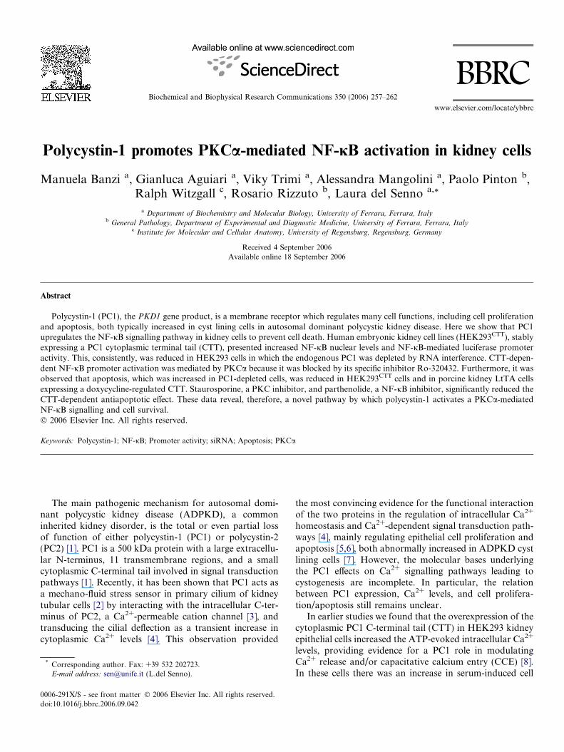
The effect of PC1 CTT on NF-jB expression was initiallyinvestigated in kidney epithelial HEK293 cells stablyexpressing the TrkA-fused CTT [9]. NF-jB p65 levels didnot significantly differ in HEK293CTT cells and HEK293Trk0
control cells; however, in the latter cells NF-jB p65 wasonly detected in the cytoplasm, while in HEK293CTT cellsit was also detected in nuclei (Fig. 1A). Furthermore, afterexposure to TNFa, a potent activator of NF-jB signalling[17], nuclear positivity to NF-jB p65 was also found insome HEK293Trk0 nuclei (data not shown), whereas thatto NF-jB p50 was found in all HEK293CTT nuclei. Overallthese results suggest a positive effect of CTT on NF-jBfunction, which was highlighted by TNFa.
We then evaluated the capacity of CTT to modulate theNF-jB DNA binding. Luciferase activity was measured inHEK293 cells which were transiently co-transfected witheither CTT or Trk0 plasmid and with the pNFjB-TA-Luc luciferase reporter plasmid, and cultured in serum-de-prived medium in absence and in presence of TNFa. Underbasal conditions, luciferase promoter activity of Trk0transfected cells was similar to that of cells transfected withthe pCDNA3 empty vector, while that of CTT transfectedcells was approximately 3-fold increased (2.994 ± 0.253,the mean ± SEM compared to Trk0 transfected cells,p < 0.05) (Fig. 1B). Moreover, TNFa treatment increasedthe CTT-dependent luciferase activity both in transientlytransfected HEK and BHK cells. Taken together, thesedata show that CTT expression in kidney cells potentiatesbasal and TNFa-induced promoter activity mediated byNF-jB.
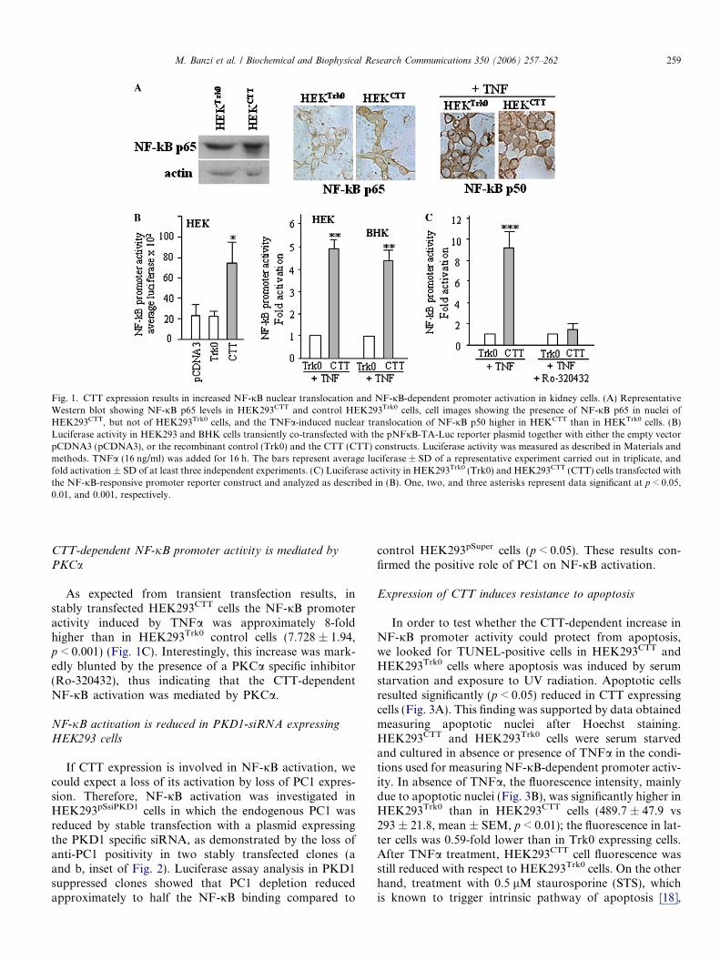
Fig. 1. CTT expression results in increased NF-jB nuclear translocation and NF-jB-dependent promoter activation in kidney cells. (A) RepresentativeWestern blot showing NF-jB p65 levels in HEK293CTT and control HEK293Trk0 cells, cell images showing the presence of NF-jB p65 in nuclei ofHEK293CTT, but not of HEK293Trk0 cells, and the TNFa-induced nuclear translocation of NF-jB p50 higher in HEKCTT than in HEKTrk0 cells. (B)Luciferase activity in HEK293 and BHK cells transiently co-transfected with the pNFjB-TA-Luc reporter plasmid together with either the empty vectorpCDNA3 (pCDNA3), or the recombinant control (Trk0) and the CTT (CTT) constructs. Luciferase activity was measured as described in Materials andmethods. TNFa (16 ng/ml) was added for 16 h. The bars represent average luciferase ± SD of a representative experiment carried out in triplicate, andfold activation ± SD of at least three independent experiments. (C) Luciferase activity in HEK293Trk0 (Trk0) and HEK293CTT (CTT) cells transfected withthe NF-jB-responsive promoter reporter construct and analyzed as described in (B). One, two, and three asterisks represent data significant at p < 0.05,0.01, and 0.001, respectively.
M. Banzi et al. / Biochemical and Biophysical Research Communications 350 (2006) 257–262 259
CTT-dependent NF-jB promoter activity is mediated by
PKCa
As expected from transient transfection results, instably transfected HEK293CTT cells the NF-jB promoteractivity induced by TNFa was approximately 8-foldhigher than in HEK293Trk0 control cells (7.728 ± 1.94,p < 0.001) (Fig. 1C). Interestingly, this increase was mark-edly blunted by the presence of a PKCa specific inhibitor(Ro-320432), thus indicating that the CTT-dependentNF-jB activation was mediated by PKCa.
NF-jB activation is reduced in PKD1-siRNA expressing
HEK293 cells
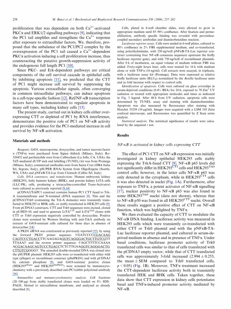
If CTT expression is involved in NF-jB activation, wecould expect a loss of its activation by loss of PC1 expres-sion. Therefore, NF-jB activation was investigated inHEK293pSsiPKD1 cells in which the endogenous PC1 wasreduced by stable transfection with a plasmid expressingthe PKD1 specific siRNA, as demonstrated by the loss ofanti-PC1 positivity in two stably transfected clones (aand b, inset of Fig. 2). Luciferase assay analysis in PKD1suppressed clones showed that PC1 depletion reducedapproximately to half the NF-jB binding compared to
control HEK293pSuper cells (p < 0.05). These results con-firmed the positive role of PC1 on NF-jB activation.
Expression of CTT induces resistance to apoptosis
In order to test whether the CTT-dependent increase inNF-jB promoter activity could protect from apoptosis,we looked for TUNEL-positive cells in HEK293CTT andHEK293Trk0 cells where apoptosis was induced by serumstarvation and exposure to UV radiation. Apoptotic cellsresulted significantly (p < 0.05) reduced in CTT expressingcells (Fig. 3A). This finding was supported by data obtainedmeasuring apoptotic nuclei after Hoechst staining.HEK293CTT and HEK293Trk0 cells were serum starvedand cultured in absence or presence of TNFa in the condi-tions used for measuring NF-jB-dependent promoter activ-ity. In absence of TNFa, the fluorescence intensity, mainlydue to apoptotic nuclei (Fig. 3B), was significantly higher inHEK293Trk0 than in HEK293CTT cells (489.7 ± 47.9 vs293 ± 21.8, mean ± SEM, p < 0.01); the fluorescence in lat-ter cells was 0.59-fold lower than in Trk0 expressing cells.After TNFa treatment, HEK293CTT cell fluorescence wasstill reduced with respect to HEK293Trk0 cells. On the otherhand, treatment with 0.5 lM staurosporine (STS), whichis known to trigger intrinsic pathway of apoptosis [18],
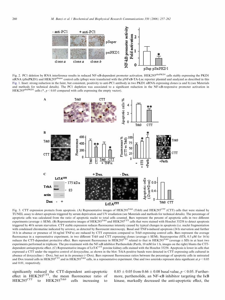
Fig. 2. PC1 deletion by RNA interference results in reduced NF-jB-dependent promoter activation. HEK293pSsiPKD1 cells stably expressing the PKD1siRNA (pSsiPKD1) and HEK293pSuper control cells (pSup) were transfected with the pNFjB-TA-Luc reporter plasmid and analyzed as described in thisFig. 1. Inset: strong reduction in the faint, but consistent, positivity to anti-PC1 antibody in two PKD1 siRNA expressing clones (a and b) (see Materialsand methods for technical details). The PC1 depletion was associated to a significant reduction in the NF-jB-responsive promoter activation inHEK293pSsiPKD1 cells (*, p < 0.05 compared with cells expressing the empty vector).
Fig. 3. CTT expression protects from apoptosis. (A) Representative images of HEK293Trk0 (Trk0) and HEK293CTT (CTT) cells that were stained byTUNEL assay to detect apoptosis triggered by serum deprivation and UV irradiation (see Materials and methods for technical details). The percentage ofapoptotic cells was calculated from the ratio of apoptotic nuclei to total cells counted. Bars represent the percent of apoptotic cells in two differentexperiments (average ± SEM). (B) Representative images of HEK293Trk0 and HEK293CTT cells that were stained with Hoechst 33258 to detect apoptosistriggered by 40 h serum starvation. CTT stable expression reduces fluorescence intensity caused by typical changes in apoptosis (i.e. nuclei fragmentationwith condensed chromatine indicated by arrows), as detected by fluorescent microscopy. Basal and TNFa-induced apoptosis (24 h starvation and further16 h in absence or presence of 16 ng/ml TNFa) are reduced by CTT expression compared to Trk0 expressing control cells. Bars represent the averagefluorescence in a representative experiment, in two different Trk0 and CTT expressing clones (average ± SEM). Staurosporine (STS, 0.5 lM for 16 h)reduces the CTT-dependent protective effect. Bars represent fluorescence in HEK293CTT related to that in HEK293Trk0 (average ± SD) in at least twoexperiments performed in triplicate. The pre-treatment with the NF-jB inhibitor Parthenolide (Parth, 10 mM for 1 h, images on the right) blunts the CTT-dependent antiapoptotic effect. (C) Representative images of LtTACTT porcine kidney cells stained with the Hoechst 33258. Apoptosis is lower in cells thatexpressed a CTT under the negative control of doxycycline, as shown in the blot: TrkA-positive bands were detected in CTT expressing cells cultured inabsence of doxycycline (�Dox), but not in its presence (+Dox). Bars represent fluorescence ratios between the percentage of apoptotic cells in untreatedand Dox treated cells in HEK293CTT and in HEK293Trk0 cells, in a representative experiment. One and two asterisks represent data significant at p < 0.05and 0.01, respectively.
260 M. Banzi et al. / Biochemical and Biophysical Research Communications 350 (2006) 257–262
significantly reduced the CTT-dependent anti-apoptoticeffect in HEK293CTT, the mean fluorescence ratio ofHEK293CTT to HEK293Trk0 cells increasing to
0.85 ± 0.05 from 0.66 ± 0.08 basal value, p < 0.05. Further-more, parthenolide, an NF-jB inhibitor targeting the IjBkinase, markedly decreased the anti-apoptotic effect, the
M. Banzi et al. / Biochemical and Biophysical Research Communications 350 (2006) 257–262 261
mean fluorescence ratio of HEK293CTT to HEK293Trk0
cells increasing to 0.98 ± 0.16, p < 0.05. These data show,therefore, that CTT is able to confer a resistance to apopto-sis, which is reduced in presence of the apoptogenic factorstaurosporine or blunted in presence of the NF-jBinhibitor.
The role of CTT on apoptosis was further investigatedin a polarized and ciliated tubule cell line: the porcine kid-ney LtTA cells [14], expressing heterologous CTT underthe doxycycline control (LtTACTT clones). Only in absenceof doxycycline (Dox) did plasma membrane proteinsbecome positive to the anti-TrkA antibody (Fig. 3C).While the majority of LtTACTT cells grown in presenceof Dox had some irregular and chromatine-condensedapoptotic nuclei, those grown in Dox absence (expressingCTT) did not. Moreover, apoptosis in cells grown withoutDox was higher in control LtTATrk0 than in LtTATRKPC1
cells, as shown by the higher ratio in fluorescence betweencells grown with and without Dox (0.79 in LtTATrk0 and0.41 in LtTATRKPC1, the ratios between apoptotic indexin treated and untreated cells).
Apoptosis is increased in PKD1-siRNA expressing HEK293
cells
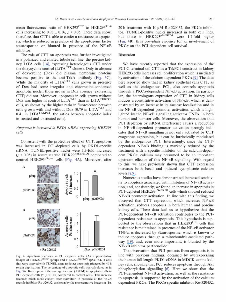
Consistent with the protective effect of CTT, apoptosiswas increased in PC1-depleted cells by PKD1-specificsiRNA: TUNEL-positive nuclei were 1.3-fold increased(p < 0.05) in serum starved HEK293pSsiPKD1 compared tocontrol HEK293pSuper cells (Fig. 4A). Moreover, after
Fig. 4. Apoptosis increases in PC1-depleted cells. (A) Representativeimages of HEK293pSuper (pSup) and HEK293pSsiPKD1 (pSsiPKD1) cellsthat were assayed with TUNEL assay to detect apoptosis triggered by 40 hserum deprivation. The percentage of apoptotic cells was calculated as inFig. 3A. Bars represent the average increase (±SEM) in apoptotic cells inPC1-depleted cells (*, p < 0.05, compared to control cells). This increasebecomes much more evident after starvation in presence of the PKCaspecific inhibitor Ro-320432, as shown by the representative images in (B).
20 h treatment with 10 lM Ro-320432, the PKCa inhibi-tor, TUNEL-positive nuclei increased in both cell lines,but those in HEK293pSsiPKD1 were 1.7-fold higher(Fig. 4B), thus providing evidence for an involvement ofPKCa on the PC1-dependent cell survival.
Discussion
We have recently reported that the expression of thePC1 C-terminal tail CTT as a TrkPC1 construct in kidneyHEK293 cells increases cell proliferation which is mediatedby activation of the calcium-dependent PKCa [9]. The datahere reported show that in kidney epithelial cells CTT, aswell as the endogenous PC1, also controls apoptosisthrough a PKCa-dependent NF-jB activation. In particu-lar, the heterologous expression of CTT in kidney cellsinduces a constitutive activation of NF-jB, which is dem-onstrated by an increase in its nuclear localization and inthe NF-jB-dependent promoter activation, which is high-lighted by the NF-jB signalling activator TNFa, in bothhuman and hamster cells. Moreover, the observation thatPC1 depletion by siRNA interference causes a reductionin NF-jB-dependent promoter activation strongly indi-cates that NF-jB signalling is not only activated by CTTexogenous expression, but can be intrinsically modulatedby the endogenous PC1. Interestingly, since the CTT-dependent NF-jB binding is markedly reduced by thetreatment with a specific inhibitor of the calcium-depen-dent PKCa, calcium may presumed to be an importantupstream effector of this NF-jB signalling. With regardto this, we have previously shown that CTT expressionincreases both basal and induced cytoplasmic calciumlevels [8,9].
Numerous studies have demonstrated increased sensitiv-ity to apoptosis associated with inhibition of NF-jB activa-tion, and, consistently, we found an increase in apoptosis inPC1-depleted HEK293pSsiPKD1 cells which showed reducedNF-jB promoter activation. In line with this finding, weobserved that CTT expression, which increases NF-jBactivation, reduces apoptosis in both human and porcinekidney cells. These data lead us to hypothesize that thePC1-dependent NF-jB activation contributes to the PC1-dependent resistance to apoptosis. This hypothesis is sup-ported by the observations that in HEK293CTT cells thisresistance is maintained in presence of the NF-jB activatorTNFa, is decreased by Staurosporine, which is known toinduce apoptosis through a mitochondria-mediated path-way [19], and, even more important, is blunted by theNF-jB inhibitor parthenolide.
The observation that PC1 protects from apoptosis is inline with previous findings, obtained by overexpressingthe human full length PKD1 cDNA in MDCK canine kid-ney cells, showing that PC1 reduces apoptosis through Aktphosphorylation signalling [6]. Here we show that thePC1-dependent NF-jB activation, as well as the resistanceto apoptosis, is supported by the activation of the calciumdependent PKCa. The PKCa specific inhibitor Ro-320432,
262 M. Banzi et al. / Biochemical and Biophysical Research Communications 350 (2006) 257–262
which, on the one hand, reduces NF-jB activity in CTTexpressing HEK293CTT cells, on the other hand doesincrease apoptosis in PC1-depleted HEK293pSsiPKD1 cells.Furthermore, the role of PKCa activation in PC1-depen-dent survival is also supported by the observation that, inCTT stably expressing HEK293CTT cells, the CTT-depen-dent protection from apoptosis is reduced by STS, a potentprotein kinase C inhibitor with a broad spectrum of activ-ity [18]. The PKCa activation, therefore, represents anadditional signalling pathway involved in the PC1-medi-ated increase in cell survival via NF-jB activation.
The calcium-dependent anti-apoptotic effect of PC1 mayseem paradoxical because calcium release from endoplas-mic reticulum may result in a mitochondrial overload ofcalcium and cell death [20]. The latter observation com-bined with the PC1-dependent NF-jB activation suggeststhat the PC1-dependent anti-apoptotic effect may becaused by a controlled calcium release, possibly constantrepetitive calcium transients, like those observed in oua-bain-treated rat proximal tubule cells [21]. With regard tothis, it is noteworthy that PC1 can regulate the calciumchannel activity of its interacting partner PC2 [2]. Further-more, it was recently reported that low doses of ouabainabolished the apoptotic effect of serum starvation and thatthe protective effect depends on IP3R-mediated calciumrelease and, subsequently, activation of NF-jB [13].
In conclusion, the present study, carried out in kidneycell lines of different species and tubule origin, either over-expressing CTT or depleted of PC1 by RNA interference,demonstrates the positive role of PC1 on NF-jB activationand provides evidence for the PC1-mediated cell survivalby NF-jB via a PKCa.
Acknowledgments
Work was supported by Cassa di Risparmio di Ferrara,the University’s Local Funds, Italian Telethon and Associ-ation for Cancer Research. We thank R.Gambari andP.Secchiero (University of Ferrara) for kindly providingus with pNFjB-TA-Luc vector and NF-jB inhibitor par-thenolide, respectively.
References
[1] A.C. Ong, P.C. Harris, Molecular pathogenesis of ADPKD: thepolycystin complex gets complex, Kidney Int. 67 (2005) 1234–1247.
[2] S.M. Nauli, F.J. Alenghat, Y. Luo, E. Williams, P. Vassilev, X. Li,A.E. Elia, W. Lu, E.M. Brown, S.J. Quinn, D.E. Ingber, J. Zhou,Polycystins 1 and 2 mediate mechanosensation in the primary ciliumof kidney cells, Nat. Genet. 33 (2003) 129–137.
[3] P. Koulen, Y. Cai, L. Geng, Y. Maeda, S. Nishimura, R. Witzgall,B.E. Ehrlich, S. Somlo, Polycystin-2 is an intracellular calcium releasechannel, Nat. Cell. Biol. 4 (2002) 191–197.
[4] P. Delmas, Polycystins: from mechanosensation to gene regulation,Cell 118 (2004) 145–148.
[5] X. Li, Y. Luo, P.G. Starremans, C.A. McNamara, Y. Pei, J. Zhou,Polycystin-1 and polycystin-2 regulate the cell cycle through the helix-loop-helix inhibitor Id2, Nat. Cell. Biol. 7 (2005) 1202–1212.
[6] M. Boca, G. Distefano, F. Qian, A.K. Bhunia, G.G. Germino, A.Boletta, Polycystin-1 induces resistance to apoptosis through thephosphatidylinositol 3-kinase/Akt signaling pathway, J. Am. Soc.Nephrol. 17 (2006) 637–647.
[7] W. Wang, C. Mei, B. Tang, H. Zhao, C. Xu, Z. Li, X. Shen, W. Fu,B. Dai, Aberrant expression of SPARC and its impact on prolifer-ation and apoptosis in ADPKD cyst-lining epithelia, Nephrol. Dial.Transplant. 21 (2006) 278–1288.
[8] G. Aguiari, M. Campanella, E. Manzati, P. Pinton, M. Banzi, S.Moretti, R. Piva, R. Rizzuto, L. del Senno, Expression of polycystin-1C-terminal fragment enhances the ATP-induced Ca2+ release inhuman kidney cells, Biochem. Biophys. Res. Commun. 301 (2003)657–664.
[9] E. Manzati, G. Aguiari, M. Banzi, M. Manzati, R. Selvatici, S.Falzarano, I. Maestri, P. Pinton, R. Rizzuto, L. del Senno, Thecytoplasmic C-terminus of polycystin-1 increases cell proliferation inkidney epithelial cells through serum-activated and Ca(2+)-dependentpathway(s), Exp. Cell. Res. 304 (2005) 391–406.
[10] A.K. Bhunia, K. Piontek, A. Boletta, L. Liu, F. Qian, P.N. Xu, F.J.Germino, G.G. Germino, PKD1 induces p21(waf1) and regulation ofthe cell cycle via direct activation of the JAK-STAT signalingpathway in a process requiring PKD2, Cell 109 (2002) 157–168.
[11] G. Nowak, Protein kinase C-alpha and ERK1/2 mediate mitochon-drial dysfunction, decreases in active Na+ transport, and cisplatin-induced apoptosis in renal cells, J. Biol. Chem. 277 (2002) 43377–43388.
[12] G.E. Sonenshein, Rel/NF-kappa B transcription factors and thecontrol of apoptosis, Semin. Cancer Biol. 8 (1997) 113–119.
[13] J. Li, S. Zelenin, A. Aperia, O. Aizman, Low doses of ouabain protectfrom serum deprivation-triggered apoptosis and stimulate kidney cellproliferation via activation of NF-kappaB, J. Am. Soc. Nephrol. 17(2006) 1848–1857.
[14] S. Hidaka, V. Konecke, L. Osten, R. Witzgall, PIGEA-14, a novelcoiled-coil protein affecting the intracellular distribution of polycy-stin-2, J. Biol. Chem. 279 (2004) 35009–35016.
[15] B. Pelucchi, G. Aguiari, A. Pignatelli, E. Manzati, R. Witzgall, L. delSenno, O. Belluzzi, Nonspecific cation current associated with nativepolycystin-2 in HEK-293 cells, J. Am. Soc. Nephrol. 17 (2006) 388–397.
[16] G. Aguiari, R. Piva, E. Manzati, E. Mazzoni, G. Augello, E. Chiari,S. Moretti, L.M. Neri, L. del Senno, K562 erythroid and HL60macrophage differentiation downregulates polycystin, a large mem-brane-associated protein, Exp. Cell Res. 244 (1998) 259–267.
[17] S. Papa, C. Bubici, F. Zazzeroni, C.G. Pham, C. Kuntzen, J.R.Knabb, K. Dean, G. Franzoso, The NF-kappaB-mediated control ofthe JNK cascade in the antagonism of programmed cell death inhealth and disease, Cell Death Differ. 13 (2006) 712–729.
[18] U.T. Ruegg, G.M. Burgess, Staurosporine, K-252 and UCN-01:potent but nonspecific inhibitors of protein kinases, Trends Pharma-col. Sci. 10 (1989) 218–220.
[19] M.R. Wieckowski, G. Szabadkai, M. Wasilewski, P. Pinton, J.Duszynski, R. Rizzuto, Overexpression of adenine nucleotide trans-locase reduces Ca2+ signal transmission between the ER andmitochondria, Biochem. Biophys. Res. Commun. 348 (2006) 393–399.
[20] G. Szabadkai, A.M. Simoni, M. Chami, M.R. Wieckowski, R.J.Youle, R. Rizzuto, Drp-1-dependent division of the mitochondrialnetwork blocks intraorganellar Ca2+ waves and protects againstCa2+-mediated apoptosis, Mol. Cell. 16 (2004) 59–68.
[21] O. Aizman, P. Uhlen, M. Lal, H. Brismar, A. Aperia, Ouabain, asteroid hormone that signals with slow calcium oscillations, Proc.Natl. Acad. Sci. USA 98 (2001) 13420–13424.