poly (i:c) therapy decreases cerebral ischaemia/reperfusion injury via tlr3-mediated prevention of...
TRANSCRIPT

Poly (I:C) therapy decreases cerebral ischaemia/reperfusion
injury via TLR3-mediated prevention of Fas/FADD interaction
Xia Zhang a, Tuanzhu Ha a, d, Chen Lu a, Fred Lam a, Li Liu e, John Schweitzer b, d,John Kalbfleisch c, d, Race L. Kao a, d, David L. Williams a, d, Chuanfu Li a, d, *
a Department of Surgery, Quillen College of Medicine, East Tennessee State University, Johnson City, TN, USAb Pathology Quillen College of Medicine, East Tennessee State University, Johnson City, TN 37614c Biometry and Medical Computing and the Center for Inflammation, Quillen College of Medicine,
East Tennessee State University, Johnson City, TN 37614d Infectious Disease and Immunity, Quillen College of Medicine,
East Tennessee State University, Johnson City, TN 37614e Department of Geriatrics5, the First Affiliated Hospital of Nanjing Medical University, Nanjing 210029, China
Received: July 23, 2014; Accepted: September 5, 2014
Abstract
Toll-like receptor (TLR)-mediated signalling plays a role in cerebral ischaemia/reperfusion (I/R) injury. Modulation of TLRs has been reported toprotect against cerebral I/R injury. This study examined whether modulation of TLR3 with poly (I:C) will induce protection against cerebral I/Rinjury. Mice were treated with or without Poly (I:C) (n = 8/group) 1 hr prior to cerebral ischaemia (60 min.) followed by reperfusion (24 hrs).Poly (I:C) pre-treatment significantly reduced the infarct volume by 57.2% compared with untreated I/R mice. Therapeutic administration ofPoly (I:C), administered 30 min. after cerebral ischaemia, markedly decreased infarct volume by 34.9%. However, Poly (I:C)-induced protectionwas lost in TLR3 knockout mice. In poly (I:C)-treated mice, there was less neuronal damage in the hippocampus compared with untreated I/Rmice. Poly (I:C) treatment induced IRF3 phosphorylation, but it inhibited NF-jB activation in the brain. Poly (I:C) also decreased I/R-inducedapoptosis by attenuation of Fas/FasL-mediated apoptotic signalling. In addition, Poly (I:C) treatment decreased microglial cell caspase-3 activity.In vitro data showed that Poly (I:C) prevented hypoxia/reoxygenation (H/R)-induced interaction between Fas and FADD as well as caspase-3and -8 activation in microglial cells. Importantly, Poly (I:C) treatment induced co-association between TLR3 and Fas. Our data suggest that Poly(I:C) decreases in cerebral I/R injury via TLR3 which associates with Fas, thereby preventing the interaction of Fas and FADD, as well as microg-lial cell caspase-3 and -8 activities. We conclude that TLR3 modulation by Poly (I:C) could be a potential approach for protection against ischae-mic stroke.
Keywords: cerebral ischaemia/reperfusion � stroke� TLR3� Poly (I:C)� apoptosis�microglial cells
Introduction
Stroke is the third leading cause of death and the leading cause oflong-term disability in the United States. Each year, 795,000 Amer-icans suffer a new or recurrent stroke [1]. Approximately, 610,000of these are initial strokes and 185,000 are recurrent strokes [1].Ischaemic stroke caused by cerebral ischaemia/reperfusion (I/R)injury accounts for ~83% of all stroke cases [1]. At present, there
is no effective treatment for cerebral I/R injury. Recent studieshave shown that Toll-like receptor (TLR)-mediated innate immuneand inflammatory responses contribute to cerebral I/R injury [2–4].TLR-mediated signalling pathways predominately activate NF-jBwhich is a critical transcription factor regulating gene expressioninvolved in innate and inflammatory responses [5]. Recent evi-dence suggests that TLRs may be important targets for develop-ment of new treatment approaches for cerebral I/R injury [6–10].For example, TLR4-deficient mice showed decreased injury follow-ing cerebral I/R [6, 10]. TLR2 has also been reported to play arole in focal cerebral ischaemic injury [10, 11]. In addition, admin-istration of CpG-ODN, a TLR9 ligand reduces cerebral I/R injury[12, 13].
*Correspondence to: Chuanfu LI, M.D.,
Department of Surgery, East Tennessee State University,
Johnson City, TN 37614, USA.
Tel.: 423-439-6349Fax: 423-439-6259
E-mail: [email protected]
ª 2014 The Authors.
Journal of Cellular and Molecular Medicine published by John Wiley & Sons Ltd and Foundation for Cellular and Molecular Medicine.
This is an open access article under the terms of the Creative Commons Attribution License, which permits use,
distribution and reproduction in any medium, provided the original work is properly cited.
doi: 10.1111/jcmm.12456
J. Cell. Mol. Med. Vol 19, No 3, 2015 pp. 555-565

Toll-like receptor 3 is located in intracellular endosomes andrecognizes double-stranded RNA (dsRNA), resulting in induction ofantiviral immune responses [14]. Polyinosinic-polycytidylic acid[Poly (I:C)], a synthetic analogue of dsRNA, stimulates TLR3-medi-ated responses [14]. TLR3 also recognizes by-products from apop-totic and necrotic cells [15]. TLR3-mediated signallingpredominantly activates IRF3 to stimulate type I interferon (IFN)production [5, 16]. Recently, Packard and Gesuete et al. reportedthat Poly (I:C)-induced preconditioning decreased cerebral I/R injury[17, 18]. Pan et al. reported that pre-treatment of mice with Poly (I:C) attenuated neurological deficits and reduced infarct volume fol-lowing cerebral I/R injury [19]. Cui et al. have shown that transientglobal cerebral ischaemia increased expression of TLR3, interferonregulatory factor and interferon beta in the hippocampus [20]. Col-lectively, the published data suggest that Poly (I:C) pre-treatmentattenuates cerebral I/R injury via a preconditioning-dependentmechanism [17, 18]. However, whether the role of TLR3 in Poly (I:C)-induced protection against cerebral I/R injury has not beeninvestigated.
Microglial cells are the resident macrophages in the central ner-vous system and they play a critical role in the induction of innateimmune and inflammatory responses [21]. Increasing evidence sug-gests that I/R-activated microglial cells induce and release pro-inflam-matory cytokines, leading to neuronal damage and dysfunction [22].Activated microglial cells may scavenge damaged neurons [23] andpromote regeneration of damaged neurons by secreting growth fac-tors [24]. We have previously reported that cerebral I/R induced acti-vation of microglial cells [25]. However, it remains unclear whetherPoly (I:C)-induced neuroprotection will involve attenuation of microg-lial activation following cerebral I/R injury.
In this study, we demonstrated that Poly (I:C) administration sig-nificantly attenuates murine cerebral I/R injury and that Poly (I:C)-induced neuroprotection is not mediated through preconditioningmechanisms. Of potentially greater clinical importance, therapeuticadministration of poly (I:C) reduces cerebral I/R injury. However, theprotection by Poly (I:C) is lost in TLR3 knockout mice. We also dem-onstrated that Poly (I:C) induces co-association between TLR3 andFas, resulting in preventing I/R-induced activation of Fas/FADD-medi-ated apoptotic signalling.
Materials and methods
Animals
Age- and weight-matched male C57BL/6 mice and TLR3 knockout(TLR3 KO) mice on the C57BL/6 background were obtained from Jack-
son Laboratory (Indianapolis, IN, USA). The TLR3 KO mice were back-
crossed with C57BL/6 for eight interbreeding generations. The micewere maintained in the Division of Laboratory Animal Resources at East
Tennessee State University (ETSU). The experiments outlined in this
study conform to the Guide for the Care and Use of Laboratory Animals
published by the National Institutes of Health (NIH Publication, 8th Edi-tion, 2011). The animal care and experimental protocols were approved
by the ETSU Committee on Animal Care.
Focal cerebral ischaemia/reperfusion
Focal cerebral I/R was induced by occlusion of the middle cerebralartery (MCA) on the left side as described in our previous studies [9,
10, 13, 25, 26]. Briefly, mice (23–25 g bodyweight) were anaesthetized
by 5.0% Isoflurane and anaesthesia was maintained by inhalation of
1.5–2% Isoflurane driven by 100% oxygen flow. Mice were intubatedand ventilated with room air using a rodent ventilator at a rate of 110
breaths per min. with a total delivered volume of 0.5 ml. Body tempera-
ture was regulated at 37.0°C by surface water heating. Following the
skin incision, the left common carotid artery (CCA), the external carotidartery (ECA) and the internal carotid artery (ICA) were carefully
exposed. Microvascular aneurysm clips were applied to the left CCA
and the ICA. A coated 6-0 filament (6023PK, Doccol Corp., Sharon, MA,USA) was introduced into an arteriotomy hole, fed distally into the ICA.
After the ICA clamp was removed, the filament was advanced 11 mm
from the carotid bifurcation, and focal cerebral ischaemia was started.
After ischaemia for 60 min., the filament and the CCA clamp weregently removed (reperfusion starts). The collar suture at the base of the
ECA stump was tightened. The skin was closed, anaesthesia discontin-
ued, and the animal allowed to recover in pre-warmed cages. Control
mice underwent a neck dissection and coagulation of the ECA, but noocclusion of the MCA.
Measurement of cerebral blood flow
Successful occlusion of the MCA was verified and recorded by laser
Doppler flowmetry (Model PeriFlux system 5000; Perimed, Stockholm,
Sweden) as described previously [13, 25]. Briefly under anaesthesia, amidline incision of the head was made and a probe holder was attached
to the skull with super-crazy glue at 6 mm lateral and 1 mm posterior
of bregma. A laser Doppler probe was connected to the probe holder
and regional cerebral blood flow (rCBF) was monitored and recorded.The data were collected continuously, stored in a computer, and analy-
sed using the Perimed data acquisition and analysis system. Regional
CBF was expressed as a percentage of pre-ischaemic baseline values.
Experimental design
To evaluate the effect of Poly (I:C) on focal cerebral I/R injury, weemployed a non-preconditioning regimen. Poly (I:C) (Catalog Cod: tlrl-
picw, InvivoGen, San Diego, CA, USA) was dissolved in sterile endo-
toxin-free 0.9% NaCl and injected intraperitoneally (i.p., 10 lg/25 g
bodyweight, n = 8) 1 hr prior to cerebral ischaemia (60 min.) followedby reperfusion for 24 hrs.
To investigate the therapeutic effect of Poly (I:C) on focal cerebral I/
R injury, Poly (I:C) (i.p., 10 lg/25 g bodyweight, n = 8) was adminis-tered by intravascular injection 30 min. after the beginning of cerebral
ischaemia. Focal cerebral ischaemia was continued for an additional
30 min. followed by reperfusion for 24 hrs.
To examine the role of TLR3 in Poly (I:C)-induced protection againstcerebral I/R injury, TLR3 KO mice (n = 7/group) were treated with or
without Poly (I:C) (10 lg/25 g bodyweight) 1 hr before the mice were
subjected to focal cerebral ischaemia (60 min.) followed by reperfusion
(24 hrs). The infarct size for all experiments was determined by triphe-nyltetrazolium chloride (TTC) staining as described below [9, 10, 13,
25, 26].
556 ª 2014 The Authors.
Journal of Cellular and Molecular Medicine published by John Wiley & Sons Ltd and Foundation for Cellular and Molecular Medicine.

Measurement of infarct volume
The infarct volume was determined as described previously [9, 10, 13,25, 26]. After completion of reperfusion, mice were killed and perfused
with ice cold PBS via the ascending aorta. Brains were removed and
sectioned coronally into 2-mm-thick slices. The slices were stained with
2% TTC solution at 37°C for 15 min. followed by fixation with 10% for-malin neutral buffer solution (pH 7.4). The infarct areas were traced and
quantified with an image-analysis system. Unstained areas (pale colour)
were defined as ischaemic lesions. The areas of infarction and the areas
of both hemispheres were calculated for each brain slice. An oedemaindex was calculated by dividing the total volume of the left hemisphere
by the total volume of the right hemisphere. The actual infarct volume
adjusted for oedema was calculated by dividing the infarct volume bythe oedema index [9, 10, 13, 25, 26]. Infarct volumes are expressed as
a percentage of the total brain volume � SEM.
Evaluation of neuronal damage in thehippocampal formation
Neuronal damage in brain sections were determined by Nissl’s method
as described previously [9, 10, 13, 25, 26]. Paraffin sections cut in the
coronal plane at ~1.5 mm behind the bregma with a thickness of 7 lmwere deparaffinized and then stained with 0.1% cresyl violet for 2 min.The sections were evaluated using light microscopy by a neuropatholo-
gist.
Immunohistochemistry double fluorescentstaining
Double fluorescent staining was performed to examine caspase-3
activity in microglial cells following cerebral I/R as described previ-
ously [9, 13]. Briefly, brain tissues were immersion-fixed in 4% buf-
fered paraformaldehyde, embedded in paraffin, cut at 7 lm, andstained with a specific anti-cleaved caspase-3 antibody which was
labelled with FITC. After washing, the sections were incubated with
anti-ionized calcium-binding adapter molecule 1 (IBA1; Santa Cruz Bio-
technology Inc., Santa Cruz, CA, USA) at 25°C for 1 hr to stain acti-vated microglial cells. After washing, the sections were incubated with
Texas Red conjugated anti-goat antibodies (sc-2783; Santa Cruz, Santa
Cruz, CA, USA) for 1 hr at 25°C. The sections were then incubatedwith DAPI for staining the nucleus. The sections were covered with
fluorescence mounting medium (Vector Labs, Burlingame, CA, USA).
The images were viewed on an EVOS-fI digital inverted fluorescent
microcopy (Advanced Microscopy Group, Bothell, WA, USA). Fields ofcortex were randomly examined using a defined rectangular field area
for analysis of microglia activation.
In vitro experiments
BV2 microglial cells were provided by Dr. Keshvara at Ohio State Uni-
versity and maintained in DMEM supplemented with 5% foetal bovineserum under 5% CO2 at 37°C as described previously [13, 25]. When
the cells reached 70–80% confluence, the medium was changed to a
hypoxia medium (NaCl 118 mmol, NaH2PO4 24 mmol, CaCl2 2.5 mmol,
EDTA 0.5 mmol, Sodium L-lactate 20 mmol, KCl 6 mmol, pH 6.2)before the cells were treated with poly (I:C) at a final concentration of
0.1 lg/ml. The cells were then subjected to hypoxia (2 hrs) followed by
reoxygenation (12 hrs) [29].
In separate experiments, the cells were treated with poly (I:C)(0.1 lg/ml) for 0, 5, 15, 30 and 60 min. with four replicates at each
time-point. The cells were harvested and cellular proteins were isolated
for examination of caspase-8 and caspase-3/7 activities by commer-cially available kits (Promega, Madison, WI, USA) as described previ-
ously [27]. Cellular proteins were also subjected to
immunoprecipitation with a specific antibody against Fas followed by
immunoblot with specific antibodies against FADD or TLR3.
Immunoprecipitation
Approximately, 800 lg of cellular proteins were immunoprecipitatedwith 2 lg of antibody against Fas (Santa Cruz Biotechnology Inc.) for
1 hr at 4°C on a rotator followed by an addition of 20 ll protein
A/G-agarose beads (Santa Cruz) as described previously [27, 30]. Theimmunoprecipitates were washed three times in lysis washing buffer,
suspended in loading buffer, and boiled for 5 min. before the immuno-
precipitates were subjected to immunoblot with primary antibodies
(anti-TLR3, 1:1000, and anti-FADD, 1:1000, Santa Cruz), respectively,followed by secondary antibody (anti-rabbit and antimouse; Sigma-
Aldrich, St. Louis, MO, USA).
Western blots
Briefly, the cellular proteins were separated by SDS-PAGE and trans-
ferred onto Hybond ECL membranes (Amersham Pharmacia, Piscata-way, NJ, USA). The ECL membranes were incubated with the
appropriate primary antibody [anti-Fas, anti-FasL, anti-FADD, anti-JNK
(Santa Cruz Biotechnology Inc.), anti-cleaved caspase-3, anti-caspase-8
(Cell Signaling Technology Inc., Danvers, MA, USA)], respectively,followed by incubation with peroxidase-conjugated secondary antibod-
ies (Cell Signaling Technology Inc., Danvers, MA, USA). The signals
were detected with the ECL system (Amersham Pharmacia). To
control for lane loading, the same membranes were probed with anti-GAPDH (glyceraldehyde-3-phosphate dehydrogenase; Biodesign, Saco,
ME, USA) after being washed with stripping buffer. The signals were
quantified using a G: Box gel imaging system (Syngene, Fredrick, MD,USA).
Caspase-3/7 and caspase-8 activities assay
Caspase-3 and caspase-8 activity in brain tissue was measured using a
Caspase-Glo assay kit (Madison, WI, USA) according to the manufac-
turer’s protocol as described previously [13].
Electrophoretic mobility shift assay
Nuclear proteins were isolated from ischaemic cerebral hemispheresas described previously [9, 10] and NF-jB binding activity was exam-
ined by Light Shift Chemiluminescent electrophoretic mobility shift
ª 2014 The Authors.
Journal of Cellular and Molecular Medicine published by John Wiley & Sons Ltd and Foundation for Cellular and Molecular Medicine.
557
J. Cell. Mol. Med. Vol 19, No 3, 2015

assay (EMSA) kit (Thermo Scientific, Waltham, MA, USA) according tothe instructions of the manufacturer.
Statistical analysis
Data are presented in figures as mean SEM for experimental groups.
Group mean levels were compared with ANOVA (one-way or multifacto-
rial as dictated by the design structure) and the least significant dif-ference procedure (as the F-test was statistically significant).
Probability levels of 0.05 or smaller were used to indicate statistical
significance.
Results
The protective effect of poly (I:C) on cerebral I/Rinjury does not require preconditioning
To examine whether Poly (I:C) can induce protection against cerebralI/R injury without preconditioning, we administrated Poly (I:C) tomice 1 hr before the mice were subjected to cerebral ischaemia(60 min.) followed by reperfusion (24 hrs). Figure 1A shows thatPoly (I:C) administration significantly reduced infarct volume by57.2% compared with untreated I/R mice. The data indicate that Poly(I:C)-induced neuroprotection occurs rapidly and does not requirepreconditioning.
Therapeutic administration of poly (I:C)decreases focal infarct volume following I/R
We also examined the therapeutic effect of Poly (I:C) on cerebral I/Rinjury. As shown in Figure 1A, therapeutic administration of Poly (I:C)30 min. after the beginning of ischaemia also significantly reducedinfarct volume by 34.9% (15.2 � 2.35 versus 24.4 � 2.67) com-pared with the untreated I/R group. The data indicate that therapeuticadministration of Poly (I:C) during ischaemia decreases I/R-inducedbrain injury.
A
B
C
Fig. 1 Poly (I:C) administration reduces infarct volume following cere-
bral I/R. (A) Poly (I:C) (10 lg/25 g bodyweight) was administered tomice 1 hr prior to or 30 min. after cerebral ischaemia. Mice were sub-
jected to cerebral ischaemia (60 min.) followed by reperfusion (24 hrs).
Infarct size was examined by TTC staining. Representative image ofinfarct size from groups are shown on the top of the bar graph. (B)Cerebral blood flow (CBF) measurement before, during and after ischae-
mia. (C) TLR3 deficiency abolishes the Poly (I:C)-induced protection
in cerebral I/R. TLR3 knockout mice were treated with or without Poly(I:C) 1 hr prior to cerebral I/R. *P < 0.05 compared with indicated
group. N = 7–8/group.
558 ª 2014 The Authors.
Journal of Cellular and Molecular Medicine published by John Wiley & Sons Ltd and Foundation for Cellular and Molecular Medicine.

Cerebral blood flow was comparable in controland poly (I:C)-treated I/R mice
It is important to confirm that the effects observed in Poly (I:C)-trea-ted mice were not because of differences in cerebral blood flow aftercerebral ischaemia followed by reperfusion. Figure 1B shows thatcerebral blood flow was significantly reduced by 80%, immediatelyfollowing occlusion of the MCA. After the occlusion was released,cerebral blood flow returned to slightly above normal levels. Therewas no significant difference in cerebral blood flow between theuntreated cerebral I/R group and the Poly (I:C)-treated group.
TLR3 deficiency abolished poly (I:C)-inducedneuroprotection in cerebral I/R injury
To determine whether TLR3 is required for Poly (I:C)-induced protec-tion against cerebral I/R injury, we treated TLR3 knockout (TLR3�/�)mice with Poly (I:C) 1 hr prior to cerebral ischaemia (60 min.) fol-lowed by reperfusion (24 hrs). Untreated TLR3�/� mice were alsosubjected to cerebral I/R. Figure 1C shows that the infarct volume inTLR3�/� mice after cerebral I/R was comparable to that in WT I/Rmice. Poly (I:C) administration did not reduce cerebral infarction inTLR3�/� mice, indicating that poly (I:C)-induced protection was lostin TLR3�/� mice. The data indicate that TLR3 is essential for mediat-ing the beneficial effect of Poly (I:C) on cerebral I/R injury.
Poly (I:C) administration attenuated neuronaldamage in the hippocampal formation
We evaluated the effect of Poly (I:C) on neuronal damage followingcerebral I/R. Nissl staining showed neuronal damage in the cornu am-monis 1 (CA1) field of the hippocampal formation (HF) characterizedby shrunken cell bodies accompanied by shrunken and pyknoticnuclei in the I/R mice (Fig. 2). Similar changes were variablyexpressed in the dentate gyrus (DG). In contrast, the neurons in theCA1 and DG fields in poly (I:C)-treated mice showed less neuronaldamage and morphology was preserved.
Poly (I:C) administration prevents NF-jB bindingactivity and increases IRF3 phosphorylation inbrain tissue following I/R
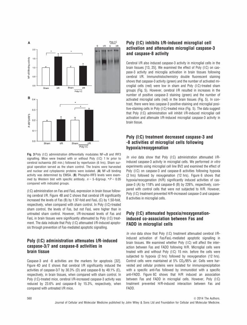
NF-jB activation plays an important role in cerebral I/R injury [29,31]. Figure 3A shows that I/R significantly increased the levels ofNF-jB binding activity by 60.7% compared with sham control. Incontrast, Poly (I:C) treatment prevented I/R-induced NF-jB bindingactivity in the brain tissues. TLR3-mediated signalling activates IRF3which controls IFN expression [5, 16]. Administration of IFN-b hasbeen demonstrated to induce protection against cerebral I/R injury[30, 31]. Figure 3B shows that Poly (I:C) administration significantlyincreased IRF3 phosphorylation levels in brain tissue following I/R. In
contrast, I/R did not induce IRF3 phosphorylation in brain tissue. Thedata indicate that Poly (I:C) administration differentially modulatesNF-jB and IRF3 signalling pathways. Specifically, Poly (I:C) preventsI/R-induced NF-jB activation and promotes activation of IRF3-medi-ated signalling.
Poly (I:C) administration attenuates I/R-inducedapoptosis in brain tissues
Cerebral I/R-induced apoptosis plays a role in brain tissue injury inresponse to I/R [32]. We examined whether administration of Poly(I:C) will attenuate I/R-induced apoptosis in brain tissues. TUNELstaining showed that there were greater numbers of TUNEL-stainedpositive apoptotic cells in the CA1 field following cerebral I/R com-pared with the sham control (Fig. 4A). However, in Poly (I:C)-treatedmice, fewer TUNEL-positive apoptotic cells were observed, indicatingthat Poly (I:C) administration attenuates cerebral I/R-induced neuronalapoptosis.
Poly (I:C) administration attenuates Fas andFasL levels in brain tissue following cerebral I/R
The Fas-mediated apoptotic signalling pathway plays an importantrole in cerebral ischaemic injury [33]. We examined the effect of Poly
Fig. 2 Poly (I:C) treatment attenuates neuronal damage in the HF follow-
ing cerebral I/R. Mice were treated with or without Poly (I:C) 1 hr priorto cerebral ischaemia (60 min.) followed by reperfusion (24 hrs). Sham
surgical operation served as the sham control. Brains were harvested,
sectioned and stained with 0.1% cresyl violet (n = 4/group). HF indi-cates hippocampal formation.
ª 2014 The Authors.
Journal of Cellular and Molecular Medicine published by John Wiley & Sons Ltd and Foundation for Cellular and Molecular Medicine.
559
J. Cell. Mol. Med. Vol 19, No 3, 2015
(I:C) administration on Fas and FasL expression in brain tissue follow-ing cerebral I/R. Figure 4B and C shows that cerebral I/R significantlyincreased the levels of Fas (B) by 1.97-fold and FasL (C) by 1.50-fold,respectively, when compared with sham control. In Poly (I:C)-treatedsham control, the levels of Fas, but not FasL were higher than inuntreated sham control. However, I/R-increased levels of Fas andFasL in brain tissues were significantly attenuated by Poly (I:C) treat-ment. The data indicate that Poly (I:C) attenuated I/R-induced apopto-sis through prevention of Fas-mediated apoptotic signalling.
Poly (I:C) administration attenuates I/R-inducedcaspase-3/7 and caspase-8 activities inbrain tissue
Caspase-3 and -8 activities are the markers for apoptosis [32].Figure 4D and E shows that cerebral I/R significantly induced theactivities of caspase-3/7 by 30.3% (D) and caspase-8 by 49.1% (E),respectively, in brain tissues, when compared with sham control. InPoly (I:C)-treated mice, cerebral I/R-increased caspase-3 activity wasreduced by 23.6% and caspase-8 by 15.3%, respectively, whencompared with untreated I/R mice.
Poly (I:C) inhibits I/R-induced microglial cellactivation and attenuates microglial caspase-3and caspase-8 activity
Cerebral I/R also induced caspase-3 activity in microglial cells in thebrain tissues [13, 25]. We examined the effect of Poly (I:C) on cas-pase-3 activity and microglia activation in brain tissues followingcerebral I/R. Immunohistochemistry double fluorescent stainingshows that caspase-3 activity (green) and the number of activated mi-croglial cells (red) were low in sham and Poly (I:C)-treated shamgroups (Fig. 5). However, cerebral I/R resulted in increases in thenumber of positive caspase-3 staining (green) and the number ofactivated microglial cells (red) in the brain tissues (Fig. 5). In con-trast, there were less caspase-3 positive-staining and microglial posi-tive-staining cells in Poly (I:C)-treated mice (Fig. 5). The data suggestthat Poly (I:C) administration will inhibit I/R-induced microglial cellactivation and attenuate I/R-induced microglial caspase-3 activity inbrain tissue.
Poly (I:C) treatment decreased caspase-3 and-8 activities of microglial cells followinghypoxia/reoxygenation
In vivo data show that Poly (I:C) administration attenuated I/R-induced caspase-3 activity in microglial cells. We performed in vitroexperiments using microglial cell line BV2 and examined the effect ofPoly (I:C) on caspase-3 and caspase-8 activities following hypoxia(2 hrs) followed by reoxygenation (12 hrs). Figure 6 shows thathypoxia/reoxygenation (H/R) significantly induced activities of cas-pase-3 (A) by 118% and caspase-8 (B) by 226%, respectively, com-pared with control cells that were not subjected to H/R. However,Poly (I:C) treatment prevented H/R-increased caspase-3 and caspase-8 activities in microglial cells.
Poly (I:C) attenuated hypoxia/reoxygenation-induced co-association between Fas andFADD in microglial cells
In vivo data show that Poly (I:C) treatment attenuated cerebral I/R-induced activation of Fas/FasL-mediated apoptotic signalling inbrain tissues. We examined whether Poly (I:C) will affect the inter-action between Fas and FADD following H/R. Microglial cells weretreated with and without Poly (I:C) 15 min. before the cells weresubjected to hypoxia (2 hrs) followed by reoxygenation (12 hrs).Control cells were maintained at 5% CO2/95% air. Cells were har-vested and cellular proteins were isolated for immunoprecipitationwith a specific anti-Fas followed by immunoblot with a specificanti-FADD. Figure 6C shows that H/R induced an associationbetween Fas and FADD in microglial cells. However, Poly (I:C)treatment prevented H/R-induced interaction between Fas andFADD.
A
B
Fig. 3 Poly (I:C) administration differentially modulates NF-jB and IRF3signalling. Mice were treated with or without Poly (I:C) 1 hr prior to
cerebral ischaemia (60 min.) followed by reperfusion (6 hrs). Sham sur-
gical operation served as the sham control. The brains were harvested
and nuclear and cytoplasmic proteins were isolated. (A) NF-jB bindingactivity was determined by EMSA. (B) Phospho-IRF3 levels were exam-
ined by Western blot with specific antibody. n = 5–6/group. *P < 0.05
compared with indicated groups.
560 ª 2014 The Authors.
Journal of Cellular and Molecular Medicine published by John Wiley & Sons Ltd and Foundation for Cellular and Molecular Medicine.

Poly (I:C) treatment induced a co-associationbetween TLR3 and Fas in cultured microglialcells
We then examined whether there is an association between TLR3 andFas following Poly (I:C) treatment, thus preventing the interaction ofFas with FADD. Microglial cells were treated with Poly (I:C) for 0, 15,
30 and 60 min., respectively. The cells were harvested and cellularproteins were isolated for immunoprecipitation with a specific anti-Fas antibody followed by immunoblot with a specific anti-TLR3 anti-body. Figure 6D shows that Poly (I:C) treatment rapidly induced anassociation between TLR3 and Fas as demonstrated by the presenceof TLR3 in the immunoprecipitates with anti-Fas. TLR3/Fas co-associ-ation peaked at 15 min. and rapidly decreased thereafter. The data
A
B C
D E
Fig. 4 Poly (I:C) treatment attenuates I/R-
induced apoptosis in brain tissues. Mice
were treated with or without Poly (I:C)
1 hr prior to cerebral ischaemia (60 min.)followed by reperfusion (6 hrs). Sham
surgical operation served as the sham
control. The brains were harvested andsectioned. Cellular proteins were prepared
from the remaining brain tissues. (A)Apoptosis in brain tissue was examined
by TUNEL assay. N = 4/group. Poly (I:C)treatment decreased the levels of Fas (B)and FasL (C) in brain tissues following
cerebral I/R. n = 5–6/group. Poly (I:C)
attenuates I/R-induced caspase-3/7 (D)and caspase-8 (E) activities which were
measured using caspase-3/7 and caspase-
8 activity kits. n = 4–6/group. *P < 0.05
compared with indicated groups.
ª 2014 The Authors.
Journal of Cellular and Molecular Medicine published by John Wiley & Sons Ltd and Foundation for Cellular and Molecular Medicine.
561
J. Cell. Mol. Med. Vol 19, No 3, 2015

suggest that Poly (I:C) administration induced an association betweenTLR3 and Fas. TLR3/Fas co-association served to attenuate the H/R-induced Fas with FADD interaction, leading to decreased caspase-8and caspase-3 activation in microglial cells.
Discussion
This study showed that Poly (I:C) administration significantly reducedcerebral I/R injury, but the neuroprotective mechanism did not requirepreconditioning. More significantly, therapeutic administration of Poly(I:C), 30 min. after cerebral ischaemia, also decreased focal infarctvolume. However, the protective effect of Poly (I:C) was lost in TLR3-deficient mice, indicating that TLR3 is required for Poly (I:C)-inducedprotection. Cerebral I/R-induced neuronal apoptosis, microglial acti-vation and microglial caspase-3 activity were significantly attenuatedby Poly (I:C) administration. In vitro data showed that Poly (I:C)administration induced co-association between TLR3 and Fas inmicroglial cells, thus preventing the interaction of Fas with FADD andsubsequent caspase-3 and -8 activation in microglial cells. We con-clude that Poly (I:C)-induced protection against cerebral I/R injury ismediated by TLR3. Mechanistically, Poly (I:C) stimulates TLR3
association with Fas which prevents Fas/FADD-mediated apoptoticsignalling.
Recently, published literature indicates that TLR-mediated signal-ling plays an important role in cerebral I/R injury [6, 10, 11]. TLR4deficiency [6, 8] or TLR2 modulation [9, 25] protects the brain fromI/R injury. Hyakkoku et al. reported that TLR3 deficiency did notinduce a neuroprotective effect against cerebral I/R [34], indicatingthat TLR3 may be required for the induction of protection againstcerebral I/R injury. Indeed, we demonstrated in this study that thePoly (I:C)-induced neuroprotective effect was lost in TLR3-deficientmice. Our observation suggests that poly I:C-induced neuroprotectionis mediated by a TLR3-dependent mechanism. Packard et al. reportedthat Poly (I:C)-induced preconditioning decreased cerebral injury inresponse to I/R [17]. Pan et al. also reported that pre-treatment ofmice with Poly (I:C) reduced infarct volume [19]. In this study, weobserved that Poly (I:C) administration rapidly induces protectionagainst cerebral I/R injury without preconditioning. Of greaterimportance, therapeutic administration of Poly (I:C) to mice alsodecreased infarct volume following cerebral I/R injury. Poly (I:C) canbe recognized by TLR3 [14] which mediates signalling through aTRIF-dependent pathway to stimulate production of IFNs [16]. Huaet al. and Famakin et al. reported that TRIF knockout mice did not
Fig. 5 Poly (I:C) administration attenuatesI/R-induced caspase-3 activity in microgli-
al cells in the brain tissues. Mice were
treated with or without Poly (I:C) 1 hrprior to cerebral ischaemia (60 min.) fol-
lowed by reperfusion (6 hrs). Sham surgi-
cal operation served as the sham control.
The brains were harvested and sectioned.Activation of microglia was examined
with specific antibody against Iba1 (red).
Caspase-3 activity was stained with anti-
cleaved caspase-3 antibody (Green).Nuclei were stained with DAPI (blue).
Caspase-3 activity (green) in activated
microglial cells (red) was a yellow colour
(merge). N = 4/group. I/R indicate ischae-mia/reperfusion. *P < 0.05 compared with
indicated groups.
562 ª 2014 The Authors.
Journal of Cellular and Molecular Medicine published by John Wiley & Sons Ltd and Foundation for Cellular and Molecular Medicine.

show a reduction in cerebral infarction and neurological deficits fol-lowing cerebral I/R [35, 36], indicating that TLR3-mediated TRIF-dependent IFN signalling may serve a protective role in cerebral I/Rinjury. Indeed, Marsh et al. reported that LPS-induced precondition-ing decreased cerebral I/R injury through IFN-b production [30].Administration of IFN-b locally protected the brain from ischaemicinjury [31]. We observed that administration of Poly (I:C) inducedIRF3 phosphorylation in brain tissue, indicating that Poly (I:C) admin-istration activates TLR3-mediated TRIF-dependent IRF3 signalling[16].
Cerebral I/R-induced apoptosis plays a role in brain tissueinjury in response to I/R [32]. In our previous studies, we observedthat cerebral I/R induced apoptosis in the CA1 and cortex. Thisstudy showed that Poly (I:C) treatment markedly reduced positiveapoptotic staining cells in the CA1 field. Poly (I:C) treatment alsodecreased caspase-3 activity, which is a marker for apoptosis in thecortex. The data indicate that Poly (I:C) administration can attenuateI/R-induced apoptosis. It is well known that activation of Fas byextracellular FasL triggers the recruitment of FADD, which directlyactivates caspase-8. Activated caspase-8 in turn stimulates caspase-3
A B
C
D
Fig. 6 Poly I:C administration prevents hypoxia/reoxygenation (H/R)-induced caspase-3 and caspase-8 activities in microglial cells (Bv2). Microglial
cells were treated with and without Poly (I:C) 15 min. before the cells were subjected to hypoxia (2 hrs) followed by reoxygenation (12 hrs). Cellularproteins were isolated for analysis of caspase-3 (A) and caspase-8 (B) activities by Western blot using anti-cleaved caspase-3 and cleaved caspase-
8 antibodies, respectively. (C) Poly (I:C) administration prevented H/R-induced interaction of Fas with FADD. Immunoprecipitation (IP) was per-
formed with anti-Fas. The immunoprecipitates were subjected to immunoblot (IB) with anti-FADD and anti-Fas, respectively. (D) Poly (I:C) treatment
induced an association between TLR3 and Fas. BV2 cells were treated with or without Poly (I:C) for 0, 15, 30 and 60 min., respectively. Cellular pro-teins were isolated and immunoprecipitation was performed with specific anti-Fas followed by immunoblot using a specific antibody against TLR3.
There were four replicates in each group. Representative blots are shown. M: hypoxia medium; P: Poly (I:C); H/R: Hypoxia/Reoxygenation.
*P < 0.05 compared with indicated groups.
ª 2014 The Authors.
Journal of Cellular and Molecular Medicine published by John Wiley & Sons Ltd and Foundation for Cellular and Molecular Medicine.
563
J. Cell. Mol. Med. Vol 19, No 3, 2015

activity [37]. We observed that Poly (I:C) administration markedlyattenuated cerebral I/R-induced increases in Fas and FasL levels aswell as caspase-8 and caspase-3 activities in the brain. Activated IRF3translocates to the nucleus and stimulates the expression of type Iinterferon genes [16] which are essential for mammalian host defenceagainst viruses. Type I IFNs also have a suppressive effect onimmune and inflammatory responses [31, 38]. In addition, the acti-vated IRF3-mediated IFN pathway is frequently associated with a pro-survival phenotype. Seimon et al. [39] have shown that treatment ofER-stressed macrophages with a small dose of LPS induced cell sur-vival through IRF3/IFN signalling. However, blocking TRIF/IRF3 sig-nalling, the cells underwent death by activating JNK-mediatedapoptotic signalling. The data indicate that IRF3-mediated signallingpromotes cell survival, while suppression of IRF3-mediated signallingleads cell apoptosis. Our data indicate that Poly (I:C) attenuated I/R-induced apoptosis in the brain which could be an important mecha-nism by which Poly (I:C) induces protection against cerebral I/Rinjury.
Microglia activation plays an important role in cerebral I/R injury[24]. Microglial cells are active sensors and versatile effector cells inpathophysiological brain injury [40]. Microglia cells express mostTLRs [41], including TLR3 which recognizes Poly (I:C) and double-stranded RNA (dsRNA) [42]. We have previously shown cerebral I/Rinduces caspase-3 and microglia activation in the brain [13, 25].Recently, Burguillos et al. reported that activation of caspase-8 isassociated with microglial activation [43]. Activated microglia releasesubstances that cause neuronal injury [21, 40]. We observed in thisstudy that Poly (I:C) treatment attenuated cerebral I/R-induced cas-pase-3 activity and microglia activation in the brain, indicating thatPoly (I:C) may attenuate microglia activation and apoptosis inresponse to I/R stimulation.
It has been reported that inhibition of caspase activity in mi-croglial cells protects against neuronal damage in several animalmodels of brain diseases, including hypoxic or ischaemic strokeand acute bacterial meningitis [43]. Inhibition of caspase activity inmicroglial cells resulted in a neuroprotective effect [44]. To furtherdetermine the role of Poly (I:C) in attenuation of I/R-induced micro-glia activation and apoptosis, we performed in vitro experimentsusing the microglial cell line BV2. We observed that Poly (I:C)
treatment attenuated H/R-induced caspase-3/7 and caspase-8 activ-ities. Caspase-8 can be activated by Fas-mediated apoptotic signal-ling through an interaction with FADD [37]. We observed that H/Rinduced an association between Fas and FADD as demonstrated bythe presence of FADD in anti-Fas immunoprecipitates. However, theH/R-induced interaction of Fas with FADD was prevented by Poly(I:C) administration. Poly (I:C) can be recognized by TLR3 whichexpresses on microglial cells [21]. To elucidate the mechanism bywhich Poly (I:C) administration prevented H/R-induced interactionof Fas with FADD, we examined whether Poly (I:C) treatment wouldpromote TLR3 interaction with Fas, thereby, preventing the interac-tion of Fas with FADD following H/R stimulation. We observed thatPoly (I:C) administration induced co-association between TLR3and Fas as demonstrated by the presence of TLR3 in the immuno-precipitates by anti-Fas. On the basis of these observations, wepropose a new neuroprotective mechanism in which Poly (I:C) pro-motes TLR3 interaction with Fas, thus preventing the interaction ofFas with FADD, thereby, attenuating H/R-induced activation of cas-pase-8 and caspase-3/7 in microglial cells.
In summary, therapeutic administration of Poly (I:C) significantlyreduced cerebral I/R-induced infarct volume via a mechanism thatdoes not involve preconditioning. The mechanisms involve attenua-tion of Fas/FasL-mediated apoptotic signalling. Specifically, Poly (I:C)administration induces a co-association between TLR3 and Fas, thuspreventing the interaction of Fas with FADD and the activation of cas-pase-3 and -8 in microglial cells. The data suggest that Poly (I:C)could be a potential approach for management and treatment ofstroke patients.
Acknowledgements
This work was supported, in part, by NIH HL071837 to C.L., NIH GM083016 to
C.L. and D.L.W., NIH GM53522 to D.L.W., NIH GM093878 to RLK.
Conflicts of interest
There was no conflict of interest for the authors in this study.
References
1. Lloyd-Jones D, Adams RJ, Brown TM, et al.Heart disease and stroke statistics–2010update. Circulation. 2010; 121: e46–215.
2. Wang Q, Tang XN, Yenari MA. The inflam-matory response in stroke. J Neuroimmunol.
2007; 184: 53–68.3. Stoll G. Inflammatory cytokines in the ner-
vous system: multifunctional mediators inautoimmunity and cerebral ischemia. Rev
Neurol. 2002; 158: 887–91.4. del Zoppo GJ. Acute anti-inflammatory
approaches to ischemic stroke. Ann N Y
Acad Sci. 2010; 1207: 143–8.
5. Aderem A, Ulevitch RJ. Toll-like receptors inthe induction of the innate immune
response. Nature. 2000; 406: 782–7.6. Caso J, Pradillo J, Hurtado O, et al. Toll-like
receptor 4 is involved in brain damage and
inflammation after experimental stroke. Cir-
culation. 2007; 115: 1599–608.7. Tang SC, Arumugam TV, Xu X, et al. Pivotal
role for neuronal Toll-like receptors in ische-
mic brain injury and functional deficits. Proc
Natl Aad Sci USA. 2007; 104: 13798–803.8. Hua F, Ma J, Ha T, et al. Activation of Toll-
like receptor 4 signaling contributes to hip-
pocampal neuronal death following global
cerebral ischemia/reperfusion. J Neuroim-
munol. 2007; 190: 101–11.9. Hua F, Ma J, Ha T, et al. Preconditioning
with a TLR2 specific ligand increases resis-
tance to cerebral ischemia/reperfusion
injury. J Neuroimmunol. 2008; 199: 75–82.10. Hua F, Ma J, Ha T, et al. Differential roles of
TLR2 and TLR4 in acute focal cerebral ische-
mia/reperfusion injury in mice. Brain Res.
2009; 1262: 100–8.11. Lehnardt S, Lehmann S, Kaul D, et al. Toll-
like receptor 2 mediates CNS injury in focal
564 ª 2014 The Authors.
Journal of Cellular and Molecular Medicine published by John Wiley & Sons Ltd and Foundation for Cellular and Molecular Medicine.

cerebral ischemia. J Neuroimmunol. 2007;190: 28–33.
12. Stevens SL, Ciesielski TM, Marsh BJ, et al.Toll-like receptor 9: a new target of ischemic
preconditioning in the brain. J Cereb BloodFlow Metab. 2008; 28: 1040–7.
13. Lu C, Ha T, Wang X, et al. The TLR9 ligand,
CpG-ODN, induces protection against cere-bral ischemia/reperfusion injury via activa-
tion of PI3K/Akt signaling. J Am Heart
Assoc. 2014; 3: e000629.
14. Matsumoto M, Seya T. TLR3: interferoninduction by double-stranded RNA including
poly(I:C). Adv Drug Deliv Rev. 2008; 60:
805–12.15. Cavassani KA, Ishii M, Wen H, et al. TLR3
is an endogenous sensor of tissue necrosis
during acute inflammatory events. J Exp
Med. 2008; 205: 2609–21.16. Yamamoto M, Sato S, Hemmi H, et al. Role
of adaptor TRIF in the MyD88-independent
toll-like receptor signaling pathway. Science.
2003; 301: 640–3.17. Packard AE, Hedges JC, Bahjat FR, et al.
Poly-IC preconditioning protects against
cerebral and renal ischemia-reperfusion
injury. J Cereb Blood Flow Metab. 2012; 32:242–7.
18. Gesuete R, Packard AE, Vartanian KB,et al. Poly-ICLC preconditioning protects
the blood-brain barrier against ischemicinjury in vitro through type 1 interferon sig-
naling. J Neurochem. 2012; 123: 75–85.19. Pan LN, Zhu W, Li C, et al. Toll-like receptor
3 agonist Poly I: C protects against simu-
lated cerebral ischemia in vitro and in vivo.
Acta Pharmacol Sin. 2012; 33: 1246–53.20. Cui G, Ye X, Zuo T, et al. Chloroquine pre-
treatment inhibits toll-like receptor 3 signal-
ing after stroke. Neurosci Lett. 2013; 548:
101–4.21. Olson JK, Miller SD. Microglia initiate cen-
tral nervous system innate and adaptive
immune responses through multiple TLRs. J
Immunol. 2004; 173: 3916–24.22. Yenari MA, Kauppinen TM, Swanson RA.
Microglial activation in stroke: therapeutic
targets. Neurotherapeutics. 2010; 7: 378–91.
23. Madinier A, Bertrand N, Mossiat C, et al.Microglial involvement in neuroplastic
changes following focal brain ischemia in
rats. PLoS ONE. 2009; 4: e8101.
24. Lai AY, Todd KG. Microglia in cerebralischemia: molecular actions and interac-
tions. Can J Physiol Pharmacol. 2006; 84:
49–59.25. Lu C, Liu L, Chen Y, et al. TLR2 ligand
induces protection against cerebral ische-
mia/reperfusion injury via activation of phos-
phoinositide 3-kinase/Akt signaling. JImmunol. 2011; 187: 1458–66.
26. Lu C, Hua F, Liu L, et al. Scavenger recep-tor class-A has a central role in cerebral
ischemia/reperfusion injury. J Cereb BloodFlow Metab. 2010; 30: 1972–81.
27. Li C, Ha T, Kelley J, et al. Modulating Toll-
like receptor mediated signaling by (1–>3)-b-D-glucan rapidly induces cardioprotection.
Cardiovasc Res. 2004; 61: 538–47.28. Ha T, Hu Y, Liu L, et al. TLR2 ligands
induce cardioprotection against ischemia/reperfusion injury through a PI3K/Akt-
dependent mechanism. Cardiovasc Res.
2010; 87: 694–703.29. Schneider A, Martin-Villalba A, Weih F,
et al. NF-jB is activated and promotes cell
death in focal cerebral ischemia. Nat Med.
1999; 5: 554–9.30. Marsh B, Stevens SL, Packard AEB, et al.
Systemic lipopolysaccharide protects the
brain from ischemic injury by reprogram-
ming the response of the brain to stroke: acritical role for IRF3. J Neurosci. 2009; 29:
9839–49.31. Veldhuis WB, Derksen JW, Floris S, et al.
Interferon-beta blocks infiltration of inflam-matory cells and reduces infarct volume
after ischemic stroke in the rat. J Cereb
Blood Flow Metab. 2003; 23: 1029–39.32. Broughton BRS, Reutens DC, Sobey CG.
Apoptotic mechanisms after cerebral ische-
mia. Stroke. 2009; 40: e331–9.33. Rosenbaum DM, Gupta G, D’Amore J, et al.
Fas (CD95/APO-1) plays a role in the
pathophysiology of focal cerebral ischemia.
J Neurosci Res. 2000; 61: 686–92.
34. Hyakkoku K, Hamanaka J, Tsuruma K,et al. Toll-like receptor 4 (TLR4), but not
TLR3 or TLR9, knock-out mice have
meuroprotective effects against focal cere-
bral ischemia. Neuroscience. 2010; 171:258–67.
35. Hua F, Wang J, Sayeed I, et al. The TRIF-
dependent signaling pathway is not requiredfor acute cerebral ischemia/reperfusion
injury in mice. Biochem Biophys Res Com-
mun. 2009; 390: 678–83.36. Famakin BM, Mou Y, Ruetzler CA, et al.
Disruption of downstream MyD88 or TRIF
Toll-like receptor signaling does not protect
against cerebral ischemia. Brain Res. 2011;
1388: 148–56.37. Chinnaiyan AM, O’Rourke K, Tewari M,
et al. FADD, a novel death domain-contain-
ing protein, interacts with the death domainof Fas and initiates apoptosis. Cell. 1995; 81:
505–12.38. Reboldi A, Dang EV, McDonald JG, et al. 25-
Hydroxycholesterol suppresses interleukin-1-driven inflammation downstream of type I
interferon. Science. 2014; 345: 679–84.39. Seimon TA, Obstfeld A, Moore KJ, et al.
Combinatorial pattern recognition receptorsignaling alters the balance of life and death
in macrophages. Proc Natl Aad Sci USA.
2006; 103: 19794–9.40. Weinstein JR, Koerner IP, Moller T. Micro-
glia in ischemia brain injury. Future Neurol.
2010; 5: 227–46.41. Jack CS, Arbour N, Manusow J, et al. TLR
signaling tailors innate immune responses in
human microglia and astrocytes. J Immunol.
2005; 175: 4320–30.42. Town T, Jeng D, Alexopoulou L, et al. Mi-
croglia recognize double-stranded RNA via
TLR3. J Immunol. 2006; 176: 3804–12.43. Burguillos MA, Dejerborg T, Kavanagh E,
et al. Caspase signalling controls microgliaactivation and neurotoxicity. Nature. 2011;
472: 319–24.44. Braun JS, Novak R, Herzog KH, et al. Neu-
roprotection by a caspase inhibitor in acute
bacterial meningitis. Nat Med. 1999; 5: 298–302.
ª 2014 The Authors.
Journal of Cellular and Molecular Medicine published by John Wiley & Sons Ltd and Foundation for Cellular and Molecular Medicine.
565
J. Cell. Mol. Med. Vol 19, No 3, 2015