plasmodium falciparum malaria challenge by the bite of aseptic anopheles stephensi mosquitoes:...
TRANSCRIPT

Plasmodium falciparum Malaria Challenge by the Bite ofAseptic Anopheles stephensi Mosquitoes: Results of aRandomized Infectivity TrialKirsten E. Lyke1*, Matthew Laurens1,2, Matthew Adams1, Peter F. Billingsley3, Adam Richman3, Mark
Loyevsky3, Sumana Chakravarty3, Christopher V. Plowe1,2, B. Kim Lee Sim3,4, Robert Edelman1,
Stephen L. Hoffman3
1 Center for Vaccine Development, University of Maryland School of Medicine, Baltimore, Maryland, United States of America, 2 Howard Hughes Medical Institute,
University of Maryland School of Medicine, Baltimore, Maryland, United States of America, 3 Sanaria, Inc., Rockville, Maryland, United States of America, 4 Protein Potential
LLC, Rockville, Maryland, United States of America
Abstract
Background: Experimental infection of malaria-naıve volunteers by the bite of Plasmodium falciparum-infected mosquitoesis a preferred means to test the protective effect of malaria vaccines and drugs. The standard model relies on the bite of fiveinfected mosquitoes to induce malaria. We examined the efficacy of malaria transmission using mosquitoes raisedaseptically in compliance with current Good Manufacturing Practices (cGMPs).
Methods and Findings: Eighteen adults aged 18–40 years were randomized to receive 1, 3 or 5 bites of Anopheles stephensimosquitoes infected with the chloroquine-sensitive NF54 strain of P. falciparum. Seventeen participants developedmalaria; fourteen occurring on Day 11. The mean prepatent period was 10.9 days (9–12 days). The geometric meanparasitemia was 15.7 parasites/mL (range: 4–70) by microscopy. Polymerase chain reaction (PCR) detected parasites 3.1(range: 0–4) days prior to microscopy. The geometric mean sporozoite load was 16,753 sporozoites per infected mosquito(range: 1,000–57,500). A 1-bite participant withdrew from the study on Day 13 post-challenge and was PCR and smearnegative.
Conclusions: The use of aseptic, cGMP-compliant P. falciparum-infected mosquitoes is safe, is associated with a preciseprepatent period compared to the standard model and appears more efficient than the standard approach, as it led toinfection in 100% (6/6) of volunteers exposed to three mosquito bites and 83% (5/6) of volunteers exposed to one mosquitobite.
Trial Registration: ClinicalTrials.gov NCT00744133
Citation: Lyke KE, Laurens M, Adams M, Billingsley PF, Richman A, et al. (2010) Plasmodium falciparum Malaria Challenge by the Bite of Aseptic Anophelesstephensi Mosquitoes: Results of a Randomized Infectivity Trial. PLoS ONE 5(10): e13490. doi:10.1371/journal.pone.0013490
Editor: Laurent Renia, BMSI-A*STAR, Singapore
Received May 14, 2010; Accepted September 17, 2010; Published October 21, 2010
Copyright: � 2010 Lyke et al. This is an open-access article distributed under the terms of the Creative Commons Attribution License, which permits unrestricteduse, distribution, and reproduction in any medium, provided the original author and source are credited.
Funding: The conduct of the trial was supported by a Vaccine Testing and Evaluation Unit contract N01-AI25461 from the National Institute of Allergy andInfectious Diseases. Mosquito rearing and infection by cultured NF54 parasites was supported by the study sponsor, Sanaria, Inc. KEL is supported by a Doris DukeClinical Scientist Development Award. MBL is supported by the Burroughs-Wellcome Fund/American Society of Tropical Medicine and Hygiene PostdoctoralFellowship in Tropical Infectious Diseases. CVP is supported by a Doris Duke Distinguished Clinical Scientist Award from the Doris Duke Charitable Foundation andCVP, MBL and MA are supported by the Howard Hughes Medical Institute. The Malaria Vaccine Initiative (MVI) supported the development by Sanaria, Inc. of aBiologics Master File for Aseptic Anopheles stephensi Mosquitoes Infected with Plasmodium falciparum Sporozoites. PFB, AR, ML, SC, BKLS, and SLH are employeesof Sanaria, Inc. and/or Protein Potential, LLC representing the sponsors of the study and participated in development of study design, data collection and analysis,publication decisions and manuscript publication. NIAID/DMID/VTEU, the funders of the study, had no role in study design, data collection and analysis, decisionto publish, or preparation of the manuscript.
Competing Interests: PFB, AR,ML, SC, BKLS, and SLH are employees of Sanaria, Inc. and/or Protein Potential, LLC representing the sponsors of the study andparticipated in development of study design, data collection and analysis, publication decisions and manuscript publication. The funding sponsor, however, wasNIAID/DMID/VTEU and members of this organization played no role in the development of the manuscript or publication. These affiliations do not alter theauthors’ adherence to all the PLoS ONE policies on sharing data and materials.
* E-mail: [email protected]
Introduction
Experimental infection of malaria-naıve volunteers by the bite
of Plasmodium falciparum-infected mosquitoes has been a preferred
means to test the protective effect of malaria vaccine and drug
candidates in malaria-naıve volunteers. Experimental induction of
malaria by the bite of infected mosquitoes has been reported in the
literature for nearly 90 years [1,2]. The conduct of challenge trials
is complicated by the need for a mosquito insectary and expertise
of personnel in rearing the insects, the transport of infected
mosquitoes to the study site, and tight restrictions on mosquito-
rearing and infection to coincide with vaccine or drug adminis-
tration. If sporozoites could be manufactured, vialed, and
transported to clinical trial centers around the world, and used
to infect volunteers by needle and syringe inoculation, this would
dramatically increase the capacity to assess new anti-malaria
PLoS ONE | www.plosone.org 1 October 2010 | Volume 5 | Issue 10 | e13490

vaccine and drug candidates. Such capacity is of critical
importance as the need for malaria challenge facilities is expected
to grow with the advent of new malaria vaccine constructs and
new antimalarial drugs [3].
Despite the current challenges to using infected mosquitoes for
challenge, recent data from 532 volunteers has demonstrated that
experimental infection of malaria-naıve volunteers is safe and
reliable [4], and more than 1450 volunteers have been challenged
by this method during the past 25 years [5]. The challenge model
effectively predicted clinical efficacy for the RTS,S malaria
vaccine prior to clinical trials in malaria-endemic areas [6].
Except in settings with very high malaria transmission intensity,
individuals are rarely bitten by more than one infected mosquito
per night under natural conditions. Malaria sporozoite challenge
studies have traditionally relied upon the bites of five infected
mosquitoes to induce malaria [7,8,9,10]. Fewer bites by mosqui-
toes raised in non-sterile conditions, have not reliably induced
malaria in volunteers [11,12,13], and increased bites may provide
an unrealistic and overwhelming challenge in which even
irradiated-sporozoite immunized volunteers fail to be protected
from malaria challenge [14].
The aseptic rearing of P. falciparum sporozoite-infected mosqui-
toes in compliance with current Good Manufacturing Practices
(cGMPs) and the demonstration that these mosquitoes contain
fully infectious sporozoites that can transmit malaria infection is
the first step toward developing a method to manufacture vialed
sporozoites for parenteral administration. Furthermore, use of
such mosquitoes to infect volunteers may reduce the variability of
sporozoite load among mosquito lots, improve reproducibility of
the pre-patent period and infection rate, and decrease the
theoretical risk to human volunteers of infection by co-infecting
microorganisms in the mosquito salivary glands. While laboratory-
reared Anopheles typically have higher salivary gland sporozoite
loads than wild-caught anophelines [15], they have highly variable
loads [16,17] which may result in a variable inoculum of
sporozoites with each mosquito bite [17]. An increase in
sporozoite and liver stage parasite burden may decrease the
prepatent period [12,18], and a decrease in burden could increase
the prepatent period. In our experience A. stephensi mosquitoes
reared in standard insectaries are contaminated with bacteria and
fungi, which may reduce mosquito fitness and infection by the
malaria parasite. There is no evidence from challenge studies that
coincidental transmission of these agents to humans occurs but
there is the theoretical risk of mechanical transmission of fungi and
bacteria from the proboscis during feeding by traditionally-reared
mosquitoes.
The production of aseptic mosquitoes, reared in compliance
with cGMPs has been established for the manufacture of the
metabolically active, non-replicating (radiation attenuated), asep-
tic, purified, vialed P. falciparum sporozoites (PfSPZ) used in the
PfSPZ Vaccine [19,20]. In this study we have used non-irradiated
mosquitoes manufactured by the identical procedure as those used
for the PfSPZ Vaccine to establish the infectivity of aseptic
sporozoites transmitted by aseptically reared mosquitoes. We
sought to determine the minimum number of A. stephensi bites
required to safely achieve 100% volunteer infection with special
attention to prepatent periods, sporozoite loads and parasitemia.
Methods
ObjectivesThe primary objective of this study was to evaluate the safety
and tolerability of a new human malaria challenge model using
aseptic A. stephensi mosquitoes infected with the chloroquine-
sensitive NF54 isolate of P. falciparum and reared in compliance
with cGMPs. Secondary objectives were to investigate the
minimum number of A. stephensi bites required to safely achieve
100% volunteer infectivity, to study the character of the malaria
infection and ascertain any differences between infection conferred
by aseptic sporozoites and that described in traditional malaria
challenge events, and to assess the role of molecular diagnostic
techniques for accurate real-time diagnosis of P. falciparum
infection. The protocol for this trial and supporting CONSORT
checklist are available as supporting information; see Checklist S1
and Protocol S1.
Study population and designThe clinical study was conducted at the Center for Vaccine
Development (CVD), at the University of Maryland School of
Medicine in Baltimore, Maryland. Eighteen malaria-naive adults
aged 19–39 years were randomized in a 1:1:1 ratio to receive 1, 3
or 5 bites of A. stephensi mosquitoes infected with P. falciparum
(Figure 1). Participants were randomized by an online random
allocation sequence generated by the EMMES Corporation and
accessed by the study nurse coordinator. Once randomized, study
personnel administering the challenge were not blinded as to the
bite assignation. Participants were previously screened for good
health and submitted blood for laboratories including hepatitis and
HIV serologies and urine for pregnancy testing if applicable.
Baseline complete blood counts (CBC), creatinine, glucose,
aspartate aminotransferase (AST) and alanine aminotransferase
(ALT) were screened and only volunteers with tightly defined
normal values were enrolled. Additionally, volunteers were pre-
screened for those with significant cardiovascular risk (i.e., .10%,
5 year risk)[21]. Risk factors include sex, age (years), systolic blood
pressure (mm Hg), smoking status (current vs. past or never), body
mass index (kg/mm2), reported diabetes status, or current
treatment for raised blood pressure. A 12-lead ECG was
performed and read by a staff cardiologist. Exclusion criteria
included known history of malaria infection, long-term residence
(.5 years) in a malaria-endemic area, travel to a malaria-endemic
area within the previous 6 months, and splenectomy.
EthicsThe trial was conducted in compliance with the Declaration of
Helsinki. All volunteers signed an informed consent form after
hearing a detailed explanation of the study and passing a written
examination designed to ascertain if they understood the risks of
malaria infection. Study protocols were reviewed and approved by
institutional review boards of the University of Maryland and the
National Institute of Allergy and Infectious Diseases/Division of
Microbiology and Infectious Diseases. The trial was monitored by
PPD, Inc. (Wilmington, NC).
Dosage, Preparation and Administration of StudyProduct
A. stephensi mosquitoes infected with P. falciparum parasites of the
NF54 strain were used for challenge utilizing methods developed
by Sanaria, Inc (Rockville, MD). Briefly, eggs from A. stephensi
mosquitoes were disinfected and placed in a custom medium for
growth to pupae. Adult female mosquitoes were fed P. falciparum
gametocytes in transfusion-quality human erythrocytes and serum.
A proportion of the eggs, pupae, blood meal, and mosquitoes were
cultured to assess for microbial growth. Only confirmed aseptic
material was used in the study. Mosquitoes were transported under
aseptic conditions to CVD and maintained aseptically at
appropriate temperature and humidity. Prior to human challenge,
Aseptic Challenge Model
PLoS ONE | www.plosone.org 2 October 2010 | Volume 5 | Issue 10 | e13490

one, three or five female mosquitoes were placed in a pint,
cylindrical cardboard container with a mesh top. The mosquitoes
were placed on exposed forearms and allowed to feed for 5 minutes,
after which they were removed and dissected to determine whether
the mosquito had 1) fed and 2) salivary gland sporozoites. A
mosquito would be categorized as ‘‘fed’’ if blood was found within
the mid-gut after challenge. Entomologist also dissected out the
paired salivary glands and quantified the sporozoite load. If
necessary, additional mosquitoes were used until the requisite
numbers of infected mosquitoes had fed upon the participant. The
techniques utilized were identical to those used as part of the
development of the metabolically-active, replication deficient,
whole-organism malaria vaccine (PfSPZ Vaccine).
Quantification of salivary gland sporozoite loadBy convention, the number of sporozoites present in mosquito
salivary glands is categorized as: 0 (no sporozoites), 1 (1–10), 2 (11–
100), 3 (101–1000), 4 (.1000) [17]. In this study, a hemocytom-
eter was used to quantify the sporozoite load per mosquito
(Chakravarty et al., manuscript in preparation). The method used
had a lower limit of detection of 250 sporozoites per mosquito.
Post-challenge AssessmentParticipants were monitored for 30 minutes after challenge and
asked to maintain symptom diaries until Day 7. They were
evaluated on Days 5–7 and were admitted to an in-patient ward
on Day 8 prior to the expected time of blood stage parasitemia.
Daily histories, vital signs, physical exams, blood smears and real-
time quantitative polymerase chain reaction (RTQ-PCR) assays
were performed. Asymptomatic individuals were provided with
pagers for rapid contact, allowed to leave the ward during the day,
and return in the evenings for evaluation. Symptomatic or
parasitemic individuals remained on the ward under clinical
supervision. After confirmation of parasitemia by microscopy,
volunteers received 1500 mg chloroquine base as standard first
line therapy over 48 hours. The volunteers were discharged after
documentation of three sequential negative blood smears and were
followed weekly for 4 weeks followed by a final visit on Day 56.
Assessment of safety and tolerabilityAll adverse events were graded for 1) severity (mild-easily
tolerated, moderate-interfered with daily activity, or severe-
prevented daily activity) and 2) relatedness (associated or not
Figure 1. Study flow diagram.doi:10.1371/journal.pone.0013490.g001
Aseptic Challenge Model
PLoS ONE | www.plosone.org 3 October 2010 | Volume 5 | Issue 10 | e13490
associated to the study product). Exceptions included fever (mild,
.99.5–100.4uF; moderate, .100.4–102.2uF; and severe,
.102.2uF), and erythema/induration (mild, 0–22 mm; moderate,
21–50 mm; and severe, .50 mm). The local challenge site was
assessed and general solicited symptoms or signs evaluated. Local
reactions that persisted beyond Day 2 were recorded as adverse
events based on the reasoning that erythema, pruritis and
induration are normal responses to mosquito bites during two
days post- exposure. Solicited symptoms related to the malaria
challenge (Days 2–7) (Table 2) and related to blood stage malaria
began on Day 8 (Table 3) and continued for the duration of the
volunteer follow-up or until a malaria diagnosis. Any other signs or
symptoms were considered to be unsolicited.
Due to an adverse cardiac event that occurred in the setting of
malaria challenge at another challenge center, ECGs and troponin
levels were done on day 3 and 10 after the malaria diagnosis for
exploratory purposes [22]. Of note, this cardiac event consisting of
chest pain at rest in a young female volunteer occurred in the
setting of malaria vaccine administration, subsequent active
malaria infection and malaria eradication using RiametH. The
temporal association of the event with malaria challenge was likely
circumstantial but the exact etiology of the chest pain remains
unclear. Safety labs including a CBC, creatinine, glucose, AST
and ALT were drawn on all days of a positive malaria smear, the
duration of treatment and at each of four weekly follow-ups
thereafter.
Malaria DiagnosticsBlood smears. Beginning on Day 5, daily blood smears
were performed to monitor for the presence of blood stage
parasites. Blood smear intervals were decreased to every 8–
12 hours if participants developed signs or symptoms consistent
with malaria, until a diagnosis was established. Ten mL of blood
were placed on a microscope slides in a 162 cm rectangle, heat-
fixed and Giemsa-stained for P. falciparum parasites. Two trained
investigators, blinded to randomization results, examined five
separate passes along the 1 cm axis using the 100x oil immersion
lens of calibrated microscopes. This was doubled to ten passes for
symptomatic individuals. The five passes performed by two
separate microscopists on different areas of the smear examined a
total of 0.9–1.1 mL of blood. Parasites were quantified per mL.
For positive smears, or if questions or discrepancies arose, a third
trained investigator (MBL) was called on to read and quantify
parasite burden. The minimum criterion for acceptance of a
positive smear was identification of two unquestionable P.
falciparum parasites confirmed by at least one investigator and
MBL. All therapeutic decisions were based on the results of a
positive blood smear.
Real-time quantitative DNA polymerase chain reaction
(RTQ-PCR). To evaluate and optimize methods for early
molecular diagnosis of P. falciparum malaria in challenge trials,
RTQ-PCR was performed on 0.5 mL of venous blood collected
contemporaneously with blood smears using published methods
with minor adaptation [23]. Standard curve cell counts were
determined by FacsCaliber flow cytometer. PCR primers were
based on the published sequence of the highly conserved [24],
stage specific [25] P. falciparum 18S ribosomal RNA gene. Primer
sequences were identical to the corresponding sequence of the
NF54 strain. Samples were blinded and assays were run daily.
Each sample was run in triplicate along with a water control. The
data were analyzed using the Applied Biosystem 7300 Absolute
Quantification Software. The assay sensitivity was determined to
be 40 parasites/mL. Results were not utilized for therapeutic
decisions.
StatisticsThe study was designed to be a proof-of-concept study and was
not powered for statistically significant comparisons. Each subject’s
exposure to the randomized number of bites (1, 3, or 5) was
considered a separate Bernoulli trial with ‘success’ defined as a
positive malaria smear within 56 days of exposure. The agreement
of RTQ-PCR to the blood smear analysis was conducted and
presented using Pearson’s Correlation as well as a linear regression
analysis. These analyses were conducted on matched, paired
smear and RTQ-PCR test results utilizing SAS (version 9.1.3,
Cary, N.C.). The study database was managed by the EMMES
Corporation (Rockville, MD).
Results
Study Population and Malaria Challenge EventEighteen adults aged 19–39 years (mean: 29 years) underwent
challenge in March 2009 by the bite of 1, 3 or 5 bites of P. falciparum-
infected mosquitoes on the same day (Table 1). Participants
randomized to receive 1, 3 or 5 bites required a mean presentation
of 2, 5.7 or 14.5 challenge mosquitoes, respectively. In total, 49% of
female Anopheles mosquitoes presented for challenge ingested a blood
meal and approximately 75% of the fed mosquitoes had detectable
salivary gland sporozoites.
Sporozoite load resultsMosquitoes were dissected immediately and the total sporozoite
density determined. The geometric mean sporozoite load was
16,753 (range: 1,000 to 57,500) sporozoites per infected mosquito
and did not vary appreciably between bite groups (Table 1). No
infected mosquitoes with fewer than 1,000 sporozoites were found.
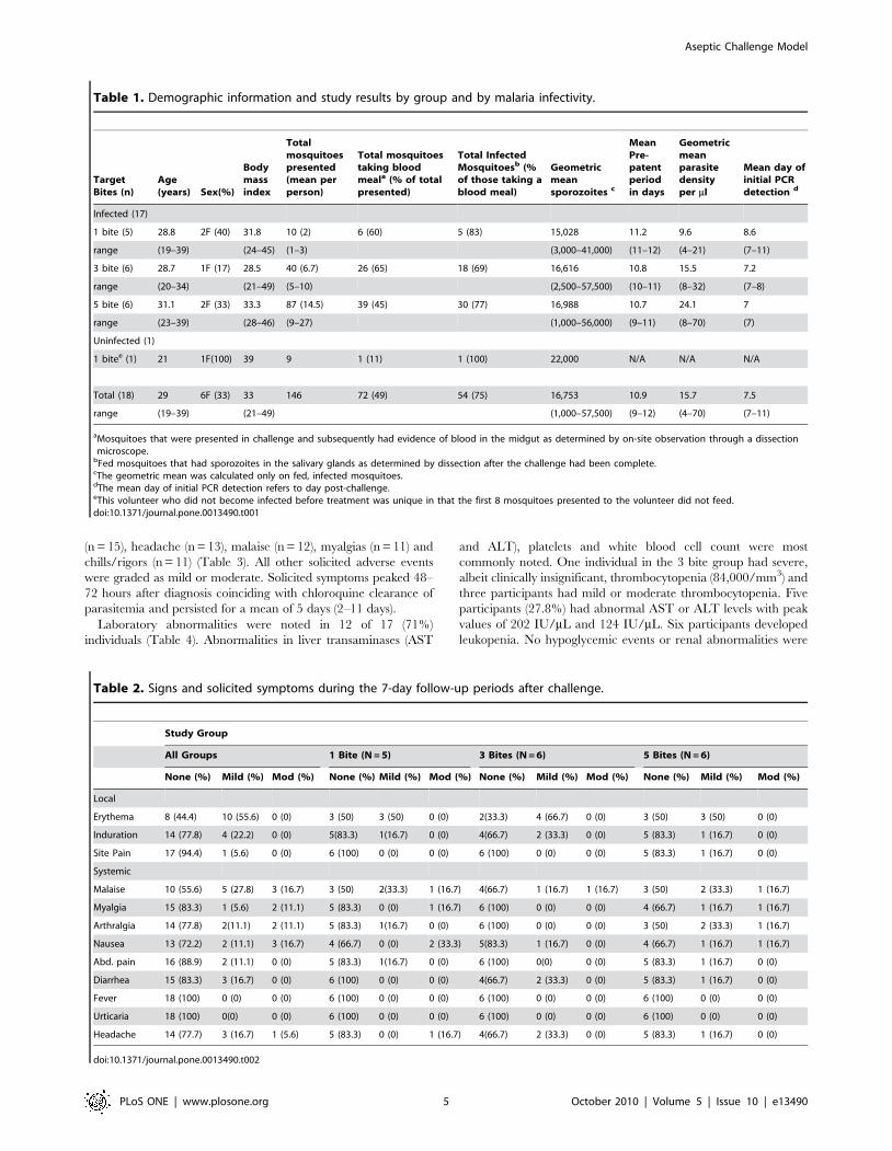
Post-Challenge Safety AssessmentSolicited adverse events were collected on local reactogenicity to
the challenge on Days 2–7 (Table 2). No severe adverse events
were reported. Fifteen local reactogenicity events in ten volunteers
were associated with the challenge. All symptoms were rated as
mild and included erythema, induration and pain at the challenge
site. There were 29 solicited adverse events with only one related
to the malaria challenge event (malaise). Three mild events
(malaise, myalgia, and arthralgia) and one moderate (arthralgia)
event occurred on Day 6 in two volunteers from the 5 bite group,
and were deemed associated with impending malaria infection and
not to the challenge event itself. The remaining solicited events
related to self-limited viral gastroenteritis experienced by several
participants before the inpatient portion of the trial. Six unsolicited
adverse events (pruritis) were reported during the post-challenge
period, five of which were deemed mild and one moderate.
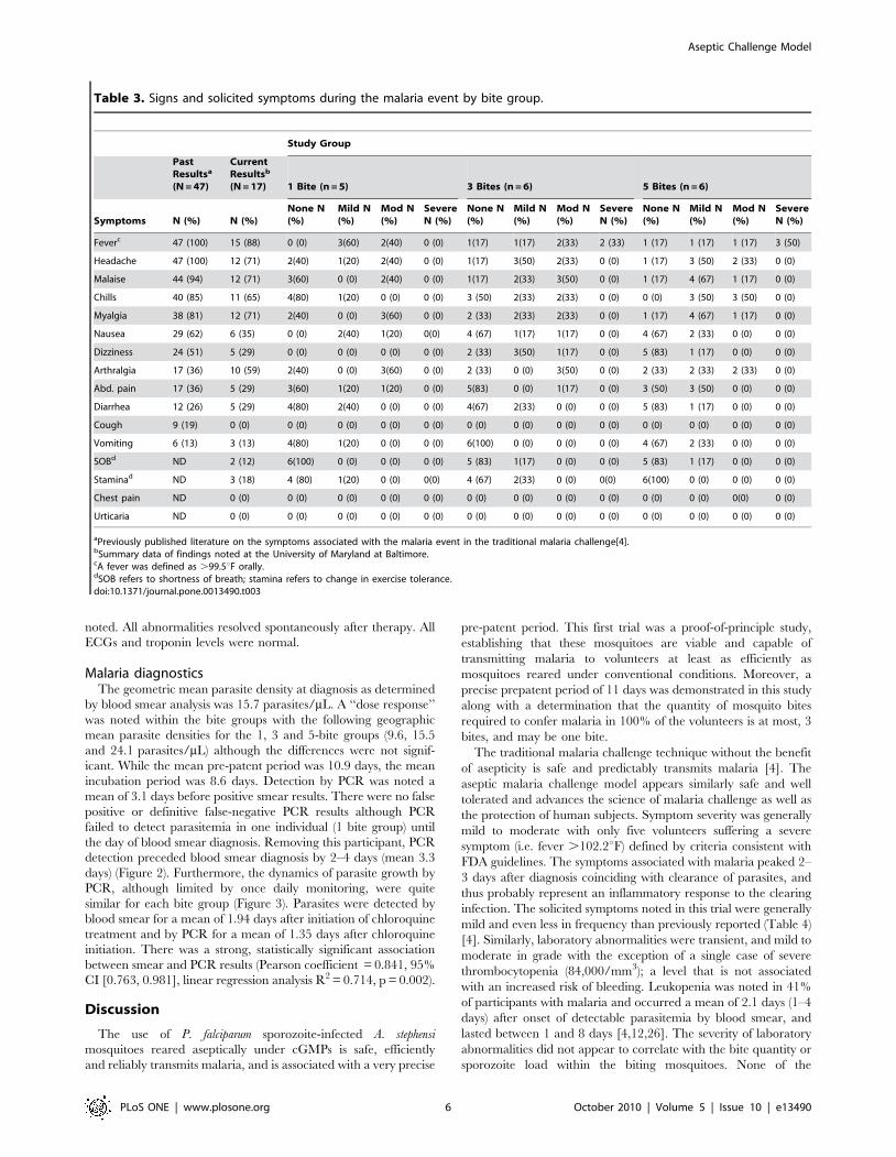
Malaria EventSeventeen participants developed parasitemia, fourteen occur-
ring on Day 11, with a mean pre-patent period of 10.9 days (9–12
days). The eighteenth participant (1 bite group) withdrew from the
study on Day 13, and was PCR and smear negative on Day 13 at
which point she was treated with chloroquine. Interestingly, it took
exposure to 9 mosquitoes for one to successfully feed on this
participant (Table 1). Six participants had mild symptoms including
malaise, headache, nausea, and diarrhea preceding malaria
diagnosis by a mean of 4.3 days (range 3–6). No individuals had
temperatures greater than 99.9uF before detection of parasitemia.
Most participants (15/18, 83.3%) reported at least one adverse
event. Fifteen individuals developed an elevated temperature, five of
which were graded as severe (.102.2uF) with a mean duration of
2.2 days. The most common solicited adverse events included fever
Aseptic Challenge Model
PLoS ONE | www.plosone.org 4 October 2010 | Volume 5 | Issue 10 | e13490
(n = 15), headache (n = 13), malaise (n = 12), myalgias (n = 11) and
chills/rigors (n = 11) (Table 3). All other solicited adverse events
were graded as mild or moderate. Solicited symptoms peaked 48–
72 hours after diagnosis coinciding with chloroquine clearance of
parasitemia and persisted for a mean of 5 days (2–11 days).
Laboratory abnormalities were noted in 12 of 17 (71%)
individuals (Table 4). Abnormalities in liver transaminases (AST
and ALT), platelets and white blood cell count were most
commonly noted. One individual in the 3 bite group had severe,
albeit clinically insignificant, thrombocytopenia (84,000/mm3) and
three participants had mild or moderate thrombocytopenia. Five
participants (27.8%) had abnormal AST or ALT levels with peak
values of 202 IU/mL and 124 IU/mL. Six participants developed
leukopenia. No hypoglycemic events or renal abnormalities were
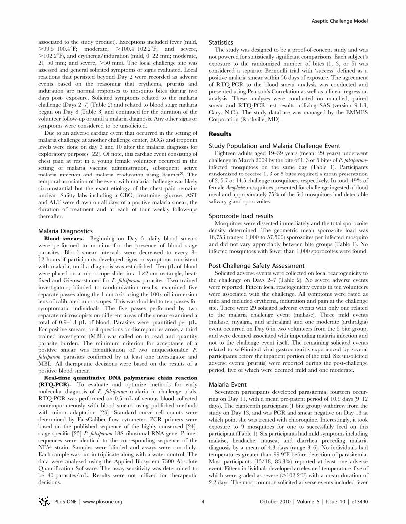
Table 1. Demographic information and study results by group and by malaria infectivity.
TargetBites (n)
Age(years) Sex(%)
Bodymassindex
Totalmosquitoespresented(mean perperson)
Total mosquitoestaking bloodmeala (% of totalpresented)
Total InfectedMosquitoesb (%of those taking ablood meal)
Geometricmeansporozoites c
MeanPre-patentperiodin days
Geometricmeanparasitedensityper ml
Mean day ofinitial PCRdetection d
Infected (17)
1 bite (5) 28.8 2F (40) 31.8 10 (2) 6 (60) 5 (83) 15,028 11.2 9.6 8.6
range (19–39) (24–45) (1–3) (3,000–41,000) (11–12) (4–21) (7–11)
3 bite (6) 28.7 1F (17) 28.5 40 (6.7) 26 (65) 18 (69) 16,616 10.8 15.5 7.2
range (20–34) (21–49) (5–10) (2,500–57,500) (10–11) (8–32) (7–8)
5 bite (6) 31.1 2F (33) 33.3 87 (14.5) 39 (45) 30 (77) 16,988 10.7 24.1 7
range (23–39) (28–46) (9–27) (1,000–56,000) (9–11) (8–70) (7)
Uninfected (1)
1 bitee (1) 21 1F(100) 39 9 1 (11) 1 (100) 22,000 N/A N/A N/A
Total (18) 29 6F (33) 33 146 72 (49) 54 (75) 16,753 10.9 15.7 7.5
range (19–39) (21–49) (1,000–57,500) (9–12) (4–70) (7–11)
aMosquitoes that were presented in challenge and subsequently had evidence of blood in the midgut as determined by on-site observation through a dissectionmicroscope.
bFed mosquitoes that had sporozoites in the salivary glands as determined by dissection after the challenge had been complete.cThe geometric mean was calculated only on fed, infected mosquitoes.dThe mean day of initial PCR detection refers to day post-challenge.eThis volunteer who did not become infected before treatment was unique in that the first 8 mosquitoes presented to the volunteer did not feed.doi:10.1371/journal.pone.0013490.t001
Table 2. Signs and solicited symptoms during the 7-day follow-up periods after challenge.
Study Group
All Groups 1 Bite (N = 5) 3 Bites (N = 6) 5 Bites (N = 6)
None (%) Mild (%) Mod (%) None (%) Mild (%) Mod (%) None (%) Mild (%) Mod (%) None (%) Mild (%) Mod (%)
Local
Erythema 8 (44.4) 10 (55.6) 0 (0) 3 (50) 3 (50) 0 (0) 2(33.3) 4 (66.7) 0 (0) 3 (50) 3 (50) 0 (0)
Induration 14 (77.8) 4 (22.2) 0 (0) 5(83.3) 1(16.7) 0 (0) 4(66.7) 2 (33.3) 0 (0) 5 (83.3) 1 (16.7) 0 (0)
Site Pain 17 (94.4) 1 (5.6) 0 (0) 6 (100) 0 (0) 0 (0) 6 (100) 0 (0) 0 (0) 5 (83.3) 1 (16.7) 0 (0)
Systemic
Malaise 10 (55.6) 5 (27.8) 3 (16.7) 3 (50) 2(33.3) 1 (16.7) 4(66.7) 1 (16.7) 1 (16.7) 3 (50) 2 (33.3) 1 (16.7)
Myalgia 15 (83.3) 1 (5.6) 2 (11.1) 5 (83.3) 0 (0) 1 (16.7) 6 (100) 0 (0) 0 (0) 4 (66.7) 1 (16.7) 1 (16.7)
Arthralgia 14 (77.8) 2(11.1) 2 (11.1) 5 (83.3) 1(16.7) 0 (0) 6 (100) 0 (0) 0 (0) 3 (50) 2 (33.3) 1 (16.7)
Nausea 13 (72.2) 2 (11.1) 3 (16.7) 4 (66.7) 0 (0) 2 (33.3) 5(83.3) 1 (16.7) 0 (0) 4 (66.7) 1 (16.7) 1 (16.7)
Abd. pain 16 (88.9) 2 (11.1) 0 (0) 5 (83.3) 1(16.7) 0 (0) 6 (100) 0(0) 0 (0) 5 (83.3) 1 (16.7) 0 (0)
Diarrhea 15 (83.3) 3 (16.7) 0 (0) 6 (100) 0 (0) 0 (0) 4(66.7) 2 (33.3) 0 (0) 5 (83.3) 1 (16.7) 0 (0)
Fever 18 (100) 0 (0) 0 (0) 6 (100) 0 (0) 0 (0) 6 (100) 0 (0) 0 (0) 6 (100) 0 (0) 0 (0)
Urticaria 18 (100) 0(0) 0 (0) 6 (100) 0 (0) 0 (0) 6 (100) 0 (0) 0 (0) 6 (100) 0 (0) 0 (0)
Headache 14 (77.7) 3 (16.7) 1 (5.6) 5 (83.3) 0 (0) 1 (16.7) 4(66.7) 2 (33.3) 0 (0) 5 (83.3) 1 (16.7) 0 (0)
doi:10.1371/journal.pone.0013490.t002
Aseptic Challenge Model
PLoS ONE | www.plosone.org 5 October 2010 | Volume 5 | Issue 10 | e13490
noted. All abnormalities resolved spontaneously after therapy. All
ECGs and troponin levels were normal.
Malaria diagnosticsThe geometric mean parasite density at diagnosis as determined
by blood smear analysis was 15.7 parasites/mL. A ‘‘dose response’’
was noted within the bite groups with the following geographic
mean parasite densities for the 1, 3 and 5-bite groups (9.6, 15.5
and 24.1 parasites/mL) although the differences were not signif-
icant. While the mean pre-patent period was 10.9 days, the mean
incubation period was 8.6 days. Detection by PCR was noted a
mean of 3.1 days before positive smear results. There were no false
positive or definitive false-negative PCR results although PCR
failed to detect parasitemia in one individual (1 bite group) until
the day of blood smear diagnosis. Removing this participant, PCR
detection preceded blood smear diagnosis by 2–4 days (mean 3.3
days) (Figure 2). Furthermore, the dynamics of parasite growth by
PCR, although limited by once daily monitoring, were quite
similar for each bite group (Figure 3). Parasites were detected by
blood smear for a mean of 1.94 days after initiation of chloroquine
treatment and by PCR for a mean of 1.35 days after chloroquine
initiation. There was a strong, statistically significant association
between smear and PCR results (Pearson coefficient = 0.841, 95%
CI [0.763, 0.981], linear regression analysis R2 = 0.714, p = 0.002).
Discussion
The use of P. falciparum sporozoite-infected A. stephensi
mosquitoes reared aseptically under cGMPs is safe, efficiently
and reliably transmits malaria, and is associated with a very precise
pre-patent period. This first trial was a proof-of-principle study,
establishing that these mosquitoes are viable and capable of
transmitting malaria to volunteers at least as efficiently as
mosquitoes reared under conventional conditions. Moreover, a
precise prepatent period of 11 days was demonstrated in this study
along with a determination that the quantity of mosquito bites
required to confer malaria in 100% of the volunteers is at most, 3
bites, and may be one bite.
The traditional malaria challenge technique without the benefit
of asepticity is safe and predictably transmits malaria [4]. The
aseptic malaria challenge model appears similarly safe and well
tolerated and advances the science of malaria challenge as well as
the protection of human subjects. Symptom severity was generally
mild to moderate with only five volunteers suffering a severe
symptom (i.e. fever .102.2uF) defined by criteria consistent with
FDA guidelines. The symptoms associated with malaria peaked 2–
3 days after diagnosis coinciding with clearance of parasites, and
thus probably represent an inflammatory response to the clearing
infection. The solicited symptoms noted in this trial were generally
mild and even less in frequency than previously reported (Table 4)
[4]. Similarly, laboratory abnormalities were transient, and mild to
moderate in grade with the exception of a single case of severe
thrombocytopenia (84,000/mm3); a level that is not associated
with an increased risk of bleeding. Leukopenia was noted in 41%
of participants with malaria and occurred a mean of 2.1 days (1–4
days) after onset of detectable parasitemia by blood smear, and
lasted between 1 and 8 days [4,12,26]. The severity of laboratory
abnormalities did not appear to correlate with the bite quantity or
sporozoite load within the biting mosquitoes. None of the
Table 3. Signs and solicited symptoms during the malaria event by bite group.
Study Group
PastResultsa
(N = 47)
CurrentResultsb
(N = 17) 1 Bite (n = 5) 3 Bites (n = 6) 5 Bites (n = 6)
Symptoms N (%) N (%)None N(%)
Mild N(%)
Mod N(%)
SevereN (%)
None N(%)
Mild N(%)
Mod N(%)
SevereN (%)
None N(%)
Mild N(%)
Mod N(%)
SevereN (%)
Feverc 47 (100) 15 (88) 0 (0) 3(60) 2(40) 0 (0) 1(17) 1(17) 2(33) 2 (33) 1 (17) 1 (17) 1 (17) 3 (50)
Headache 47 (100) 12 (71) 2(40) 1(20) 2(40) 0 (0) 1(17) 3(50) 2(33) 0 (0) 1 (17) 3 (50) 2 (33) 0 (0)
Malaise 44 (94) 12 (71) 3(60) 0 (0) 2(40) 0 (0) 1(17) 2(33) 3(50) 0 (0) 1 (17) 4 (67) 1 (17) 0 (0)
Chills 40 (85) 11 (65) 4(80) 1(20) 0 (0) 0 (0) 3 (50) 2(33) 2(33) 0 (0) 0 (0) 3 (50) 3 (50) 0 (0)
Myalgia 38 (81) 12 (71) 2(40) 0 (0) 3(60) 0 (0) 2 (33) 2(33) 2(33) 0 (0) 1 (17) 4 (67) 1 (17) 0 (0)
Nausea 29 (62) 6 (35) 0 (0) 2(40) 1(20) 0(0) 4 (67) 1(17) 1(17) 0 (0) 4 (67) 2 (33) 0 (0) 0 (0)
Dizziness 24 (51) 5 (29) 0 (0) 0 (0) 0 (0) 0 (0) 2 (33) 3(50) 1(17) 0 (0) 5 (83) 1 (17) 0 (0) 0 (0)
Arthralgia 17 (36) 10 (59) 2(40) 0 (0) 3(60) 0 (0) 2 (33) 0 (0) 3(50) 0 (0) 2 (33) 2 (33) 2 (33) 0 (0)
Abd. pain 17 (36) 5 (29) 3(60) 1(20) 1(20) 0 (0) 5(83) 0 (0) 1(17) 0 (0) 3 (50) 3 (50) 0 (0) 0 (0)
Diarrhea 12 (26) 5 (29) 4(80) 2(40) 0 (0) 0 (0) 4(67) 2(33) 0 (0) 0 (0) 5 (83) 1 (17) 0 (0) 0 (0)
Cough 9 (19) 0 (0) 0 (0) 0 (0) 0 (0) 0 (0) 0 (0) 0 (0) 0 (0) 0 (0) 0 (0) 0 (0) 0 (0) 0 (0)
Vomiting 6 (13) 3 (13) 4(80) 1(20) 0 (0) 0 (0) 6(100) 0 (0) 0 (0) 0 (0) 4 (67) 2 (33) 0 (0) 0 (0)
SOBd ND 2 (12) 6(100) 0 (0) 0 (0) 0 (0) 5 (83) 1(17) 0 (0) 0 (0) 5 (83) 1 (17) 0 (0) 0 (0)
Staminad ND 3 (18) 4 (80) 1(20) 0 (0) 0(0) 4 (67) 2(33) 0 (0) 0(0) 6(100) 0 (0) 0 (0) 0 (0)
Chest pain ND 0 (0) 0 (0) 0 (0) 0 (0) 0 (0) 0 (0) 0 (0) 0 (0) 0 (0) 0 (0) 0 (0) 0(0) 0 (0)
Urticaria ND 0 (0) 0 (0) 0 (0) 0 (0) 0 (0) 0 (0) 0 (0) 0 (0) 0 (0) 0 (0) 0 (0) 0 (0) 0 (0)
aPreviously published literature on the symptoms associated with the malaria event in the traditional malaria challenge[4].bSummary data of findings noted at the University of Maryland at Baltimore.cA fever was defined as .99.5uF orally.dSOB refers to shortness of breath; stamina refers to change in exercise tolerance.doi:10.1371/journal.pone.0013490.t003
Aseptic Challenge Model
PLoS ONE | www.plosone.org 6 October 2010 | Volume 5 | Issue 10 | e13490

symptoms or laboratory abnormalities met World Health
Organization criteria for severe malaria [27].
The traditional challenge methodology relies on the bite of five
mosquitoes to reliably transmit malaria to 100% of volunteers.
Utilizing mosquitoes raised aseptically under cGMPs, all partic-
ipants in the 3 and 5 bite groups and 5 of 6 volunteers in the 1 bite
group developed malaria. One volunteer withdrew from the study
on day 13 and was treated with chloroquine before developing
parasitemia. Excluding this volunteer, whose ultimate infection
status is unknown, 100% infectivity was achieved with a single
bite. The number of mosquitoes required to achieve a successful
blood feed varied per individual. In instances where a higher
number of mosquitoes were required to achieve a successful blood
feed, it is possible that the total number of mosquitoes to which the
volunteers were exposed was underestimated as mosquitoes can
inoculate sporozoites as they probe for blood, even if they did not
take a blood meal. However, of the volunteers randomized to the
one bite arm, two were exposed to the bite of only one mosquito
and the rest were exposed to only two or three mosquitoes (total of
10 mosquitoes for infecting 5 volunteers, Table 1) before achieving
a successful blood feed indicating that #3 mosquitoes are required
to achieve malaria infection using this technique. It should be
noted that all other studies have used the same criteria for a
successful blood feed. Thus, regardless of probing without feeding,
our data are distinct from the other studies, despite use of similar
methodology.
Of the 17 participants who were infected, 14 (82%) developed a
positive blood smear on Day 11 (range 9–12) after challenge. This
compares favorably with previous published results where 18/47
(38%) participants developed malaria on Day 11 (range 9–14,
mean 10.52 days) but the pre-patent period was more variable.
Increased sporozoite inoculation and liver burden could result in
reduced prepatent periods [18] or prolonged parasitemias [28].
Conversely, the physical characteristics of the mosquito salivary
duct may limit the number of sporozoites that can be inoculated
during probing and feeding [29]. In the P. yoelli model, sporozoite
injection has proven to be highly variable ranging from 0–1,297
Table 4. Laboratory abnormalities recorded during the malaria event.
Study Group
Parameter/unit Grade Range 1 Bite (%) 3 Bites (%) 5 Bites (%)
AST (IU/L) None 0–40 4 (80) 4 (67) 3 (50)
Mild 41–99 1 (20) 2 (33) 1 (17)
Moderate 100–199 0 (0) 0 (0) 2 (33)
Severe $200 0 (0) 0 (0) 0 (0)
ALT (IU/L) None 0–55 (=) 0–40 (R)a 5 (100) 4 (67) 3 (50)
Mild 56–137 (=) 41–99 (R) 0 (0) 2 (33) 1 (17)
Moderate 138–274 (=) 100–199 (R) 0 (0) 0 (0) 2 (33)
Severe $275 (=) $200 (R) 0 (0) 0 (0) 0 (0)
Hemoglobin (g/dL) None 12.5–17.0 (=) 11.5–15.0 (R) 5 (100) 5 (83) 6 (100)
Mild 10.6–12.4 (=) 11.1–11.4 (R) 0 (0) 1 (17) 0 (0)
Moderate 10,0–10.5 (=) 9.6–10.0 (R) 0 (0) 0 (0) 0 (0)
Severe ,10.0 (=) #9.5 (R) 0 (0) 0 (0) 0 (0)
WBC (x 103/mm3) None 4.0–10.5 3 (60) 3 (50) 4 (67)
Mild 2.5–3.9 2 (40) 2 (33) 2 (33)
Moderate 1.5–2.4 0 (0) 1 (17) 0 (0)
Severe ,1.5 0 (0) 0 (0) 0 (0)
Platelets (x 103/mm3) None $140 5 (100) 2 (33) 6 (100)
Mild 125–139 0 (0) 0 (0) 0 (0)
Moderate 100–124 0 (0) 3 (50) 0 (0)
Severe 20–99 0 (0) 1 (17) 0 (0)
a(=) represents males and (R) represents females.doi:10.1371/journal.pone.0013490.t004
Figure 2. Time to infection of volunteers measured by bloodsmear and PCR. The time to infection of all volunteers (pooledexposed to 1, 3 and 5 infectious bites) measured by blood smear (solidline, median time to infection = 11 days) and PCR (dashed line, mediantime to infection = 8 days) was significantly different (p#0.001, Mantel-Cox test).doi:10.1371/journal.pone.0013490.g002
Aseptic Challenge Model
PLoS ONE | www.plosone.org 7 October 2010 | Volume 5 | Issue 10 | e13490

Figure 3. Dynamics of parasite growth in volunteers after challenge. Each line shows the parasite density in an individual volunteer asmeasured by PCR after being bitten by 1 (top), 3 (middle) or 5 (bottom) Plasmodium falciparum-infected Anopheles stephensi mosquitoes. Allvolunteers were treated on day 11 (vertical dashed line on each panel) when parasites were detected by blood smear, except for one volunteer in the1 bite group (blue dashed line) who was positive and treated on day 12, one volunteer (grey dashed line) in the 3 bite group who was positive andtreated on day 10, and one volunteer (red dashed line) in the 5 bite group who was positive and treated on day 9. Data are presented until lastpositive identification of parasites in the blood by PCR. Values shown as 1 on the log scale were negative.doi:10.1371/journal.pone.0013490.g003
Aseptic Challenge Model
PLoS ONE | www.plosone.org 8 October 2010 | Volume 5 | Issue 10 | e13490

per bite (mean 123) and is only weakly correlated to sporozoite
gland quantity [30]. Therefore, increased numbers of sporozoites
per mosquito may not translate into increased sporozoites
inoculated. Complicating interpretation, the traditional method
of determining sporozoite loads is imprecise, relying upon
qualitative estimation of total sporozoites on salivary glands
squashes, with gland scores graded from 0 to 4+ [17]. In virtually
all previous studies, mosquitoes with a gland score of $2+ (11–100
sporozoites) were considered infectious [7]. Utilizing a more
precise counting technique, 16,753 sporozoites per mosquito
(range 1,000–57,500) were present in the challenge mosquitoes.
Our data indicate that mosquitoes raised aseptically in compliance
with cGMPs can successfully transmit malaria to 100% of
participants through the bite of 3, and likely 1, rather than 5
mosquito bites, and suggest they may lead to a more precise pre-
patent period. The mosquitoes used in this study had more
sporozoites than those used in most other studies. Thus, it is
possible that the infectivity of 1 and 3 infected mosquitoes may
have been due to an increased sporozoite inoculum when
compared to traditionally-raised mosquitoes, but experimental
data in P. yoelii [30] do not support this interpretation.
The use of PCR to achieve diagnosis earlier in malaria
challenge studies is attractive, but it also carries risks. A false
negative or false positive result in a small challenge trial of a
malaria vaccine could profoundly alter study results. There were
no false positive results in our preliminary use of this technique.
While the ability to detect low-level parasitemia days before blood
smear detection could, theoretically, avert symptoms associated
with malaria, we did not find a correlation between PCR and
symptom onset (data not shown). Moreover, the severity of
symptoms increased once therapy was initiated and peaked after
48–72 hours when parasites were no longer detectable. Perfor-
mance of the PCR assay could be increased to every 8–12 hours to
enhance detection, but PCR is, currently, more labor-intensive
procedure than is the reading of blood smears, and would require
round-the-clock staffing in a challenge setting. Further study of
diagnostic PCR in the context of volunteer challenges is required
to fully document the pre-test specificity of the assay.
The development of a metabolically active, non-replicating
(radiation attenuated) P. falciparum sporozoite vaccine is based on
the successful immunization of volunteers by the bite of irradiated,
non-aseptic mosquitoes [7]. It has been hypothesized that the
contaminants, including bacterial and fungal material, accompa-
nying these mosquitoes may provide some adjuvant effects that
enhance sporozoite-induced immunity. Our study does not give
any indication of the immunogenicity of the sporozoites
administered by the bite of aseptically-reared mosquitoes, but
establishing that the sporozoites produced under aseptic conditions
remain fully virulent and capable of eliciting malaria is nonetheless
important data for the effort to develop a metabolically active,
non-replicating whole sporozoite P. falciparum vaccine, which is
produced in aseptic A. stephensi mosquitoes using the same
methodology.
Now that the aseptic malaria challenge model has been
established with the NF54 strain of P. falciparum, which has
historically been utilized in malaria challenge studies, we plan to
develop challenges with additional P. falciparum strains. The genetic
diversity of P. falciparum poses significant challenges to vaccine
development [31], and the development of a heterologous
challenge model will permit assessment of the ability of vaccine
candidates to provide protection against genetically diverse
parasites and of anti-malarial drugs to prevent malaria caused
by P. falciparum of differing drug sensitivities.
The data from this trial demonstrate that aseptic sporozoites can
transmit malaria. This is the first step toward developing and
assessing the infectivity of aseptic, purified, cryopreserved P.
falciparum sporozoites administered by needle and syringe to infect
volunteers rather than relying on the bite of a mosquito. This
would allow institutions without the capability of rearing infectious
mosquitoes to safely, and reliably conduct malaria challenge trials,
and such a study is being planned. Ultimately, our goal is to have
multiple strains of P. falciparum parasites that are cryopreserved and
packaged for transport to be used by institutions worldwide for
testing malaria vaccines and pharmacologic agents.
Supporting Information
Checklist S1 Consort Checklist
Found at: doi:10.1371/journal.pone.0013490.s001 (0.23 MB
DOC)
Protocol S1 Aseptic Challenge Protocol
Found at: doi:10.1371/journal.pone.0013490.s002 (1.26 MB
DOC)
Acknowledgments
We thank the volunteer population from Baltimore, MD who so graciously
consented to participate in this trial. Drs. Lee F. Hall and Steven Rosenthal
and Walter Jones of the Department of Microbiology and Infectious
Diseases at the National Institute of Health offered invaluable support and
advice, although program staff from the NIAID played no role in the
collection, analysis or interpretation of data, writing the paper, or the
decision to submit it for publication. Our special thanks go to Lisa Chrisley,
Maria Johnson, and James Freeman for coordination of the trial and for
regulatory oversight, and to the entire Sanaria manufacturing team for
production of the infected mosquitoes.
Author Contributions
Conceived and designed the experiments: KEL MBL PFB AR ML SC
CVP BKLS RE SLH. Performed the experiments: KEL MBL MA PFB
BKLS RE. Analyzed the data: KEL MA PFB SC BKLS SLH. Contributed
reagents/materials/analysis tools: KEL MA AR ML SC BKLS SLH.
Wrote the paper: KEL MBL PFB CVP BKLS RE SLH.
References
1. Driver JR, Gammel JA, Darnosh LJ (1926) Malaria treatment central nervous
system ‘‘syphilis’’. ;Journal of the American Medical Association (lxxxvii), 1921,
Cited in Bunker ‘Recent treatment of general paralysis’-.
2. Brown EM (2000) Why Wagner-Juaregg won the Nobel Prize for discovering
Malaria Therapy for General Paresis of the Insane. Journal of Psychiatry xi:
371–382.
3. (2010) Malaria 2010: more ambition and accountability please. Lancet 375:
1407-.
4. Epstein JE, Rao S, Williams F, Freilich D, Luke T, et al. (2007) Safety and
clinical outcome of experimental challenge of human volunteers with Plasmodium
falciparum-infected mosquitoes: an update. J Infect Dis 196: 145–154.
5. Moorthy VS, Diggs C, Ferro S, Good MF, Herrera S, et al. (2009) Report of a
consultation on the optimization of clinical challenge trials for evaluation of
candidate blood stage malaria vaccines, 18-19 March 2009, Bethesda, MD,
USA. Vaccine 27: 5719–5725.
6. Kester KE, McKinney DA, Tornieporth N, Ockenhouse CF, Heppner DG,
et al. (2001) Efficacy of recombinant circumsporozoite protein vaccine regimens
against experimental Plasmodium falciparum malaria. J Infect Dis 183: 640–647.
7. Hoffman SL, Goh LM, Luke TC, Schneider I, Le TP, et al. (2002) Protection of
humans against malaria by immunization with radiation-attenuated Plasmodium
falciparum sporozoites. J Infect Dis 185: 1155–1164.
8. Chulay JD, Schneider I, Cosgriff TM, Hoffman SL, Ballou WR, et al. (1986)
Malaria transmitted to humans by mosquitoes infected from cultured Plasmodium
falciparum. Am J Trop Med Hyg 35: 66–68.
9. Herrington DA, Clyde DF, Murphy JR, Baqar S, Levine MM, et al. (1988) A
model for Plasmodium falciparum sporozoite challenge and very early therapy of
Aseptic Challenge Model
PLoS ONE | www.plosone.org 9 October 2010 | Volume 5 | Issue 10 | e13490

parasitaemia for efficacy studies of sporozoite vaccines. Trop Geogr Med 40:
124–127.
10. Murphy JR, Baqar S, Davis JR, Herrington DA, Clyde DF (1989) Evidence for a
6.5-day minimum exoerythrocytic cycle for Plasmodium falciparum in humans and
confirmation that immunization with a synthetic peptide representative of a
region of the circumsporozoite protein retards infection. J Clin Microbiol 27:
1434–1437.
11. Rickman LS, Jones TR, Long GW, Paparello S, Schneider I, et al. (1990)
Plasmodium falciparum-infected Anopheles stephensi inconsistently transmit malaria to
humans. Am J Trop Med Hyg 43: 441–445.
12. Verhage DF, Telgt DS, Bousema JT, Hermsen CC, van Gemert GJ, et al. (2005)
Clinical outcome of experimental human malaria induced by Plasmodium
falciparum-infected mosquitoes. Neth J Med 63: 52–58.
13. Fries LF, Gordon DM, Schneider I, Beier JC, Long GW, et al. (1992) Safety,
immunogenicity, and efficacy of a Plasmodium falciparum vaccine comprising a
circumsporozoite protein repeat region peptide conjugated to Pseudomonas
aeruginosa toxin A. Infect Immun 60: 1834–1839.
14. Clyde DF (1975) Immunization of man against falciparum and vivax malaria by
use of attenuated sporozoites. Am J Trop Med Hyg 24: 397–401.
15. Davis JR, Murphy JR, Baqar S, Clyde DF, Herrington DA, et al. (1989)
Estimate of anti-Plasmodium falciparum sporozoite activity in humans vaccinated
with synthetic circumsporozoite protein (NANP)3. Trans R Soc Trop Med Hyg
83: 748–750.
16. Githeko AK, Brandling-Bennett AD, Beier M, Atieli F, Owaga M, et al. (1992)
The reservoir of Plasmodium falciparum malaria in a holoendemic area of western
Kenya. Trans R Soc Trop Med Hyg 86: 355–358.
17. Beier JC, Davis JR, Vaughan JA, Noden BH, Beier MS (1991) Quantitation of
Plasmodium falciparum sporozoites transmitted in vitro by experimentally infected
Anopheles gambiae and Anopheles stephensi. Am J Trop Med Hyg 44: 564–570.
18. Jeffery GM, Young MD, Burgess RW, Eyles DE (1959) Early activity in
sporozoite-induced Plasmodium falciparum infections. Ann Trop Med Parasitol 53:
51–58.
19. Luke TC, Hoffman SL (2003) Rationale and plans for developing a non-
replicating, metabolically active, radiation-attenuated Plasmodium falciparum
sporozoite vaccine. J Exp Biol 206: 3803–3808.
20. Hoffman SL, Billingsley PF, James E, Richman A, Loyevsky M, et al. (2010)
Development of a metabolically active, non-replicating sporozoite vaccine toprevent Plasmodium falciparum malaria. Hum Vaccin 6: 97–106.
21. Gaziano TA, Young CR, Fitzmaurice G, Atwood S, Gaziano JM (2008)
Laboratory-based versus non-laboratory-based method for assessment ofcardiovascular disease risk: the NHANES I Follow-up Study cohort. Lancet
371: 923–931.22. Nieman AE, de MQ, Roestenberg M, Wiersma J, Pop G, et al. (2009) Cardiac
complication after experimental human malaria infection: a case report. Malar J
8: 277-).23. Spring MD, Cummings JF, Ockenhouse CF, Dutta S, Reidler R, et al. (2009)
Phase 1/2a study of the malaria vaccine candidate apical membrane antigen-1(AMA-1) administered in adjuvant system AS01B or AS02A. PLoS ONE 4:
e5254-.24. McCutchan TF, de lCV, Lal AA, Gunderson JH, Elwood HJ, et al. (1988)
Primary sequences of two small subunit ribosomal RNA genes from Plasmodium
falciparum. Mol Biochem Parasitol 28: 63–68.25. Gunderson JH, Sogin ML, Wollett G, Hollingdale M, de lCV, et al. (1987)
Structurally distinct, stage-specific ribosomes occur in Plasmodium. Science 238:933–937.
26. Church LW, Le TP, Bryan JP, Gordon DM, Edelman R, et al. (1997) Clinical
manifestations of Plasmodium falciparum malaria experimentally induced bymosquito challenge. J Infect Dis 175: 915–920.
27. World Health Organization (2000) Severe falciparum malaria. CommunicableDiseases Cluster. Trans R Soc Trop Med Hyg 94 Suppl 1: S1–90.
28. Murphy JR, Clyde DF, Herrington DA, Baqar S, Davis JR, et al. (1990)Continuation of chloroquine-susceptible Plasmodium falciparum parasitemia in
volunteers receiving chloroquine therapy. Antimicrob Agents Chemother 34:
676–679.29. Jin Y, Kebaier C, Vanderberg J (2007) Direct microscopic quantification of
dynamics of Plasmodium berghei sporozoite transmission from mosquitoes to mice.Infect Immun 75: 5532–5539.
30. Medica DL, Sinnis P (2005) Quantitative dynamics of Plasmodium yoelii sporozoite
transmission by infected anopheline mosquitoes. Infect Immun 73: 4363–4369.31. Takala SL, Plowe CV (2009) Genetic diversity and malaria vaccine design,
testing and efficacy: preventing and overcoming ‘vaccine resistant malaria’.Parasite Immunol 31: 560–573.
Aseptic Challenge Model
PLoS ONE | www.plosone.org 10 October 2010 | Volume 5 | Issue 10 | e13490