pirfenidone prevents capsular contracture after mammary implantation
TRANSCRIPT
ORIGINAL ARTICLE
Pirfenidone Prevents Capsular Contracture After MammaryImplantation
Matias Gancedo Æ Luis Ruiz-Corro ÆAdriana Salazar-Montes Æ Ana Rosa Rincon ÆJuan Armendariz-Borunda
Published online: 30 October 2007
� Springer Science+Business Media, LLC 2007
Abstract
Background Pirfenidone (PFD), a new antifibrotic and
antiinflammatory agent, prevents and resolves fibrous tis-
sue. This study evaluated the effect of PFD on adverse
events in mammary implants using an animal model.
Mammary implantation, the most frequent aesthetic sur-
gery, may present several complications after surgery such
as swelling, capsule contracture, hardness, and pain.
Methods Wistar rats underwent submammary implanta-
tion with either smooth or textured silicone gel implants
and were administrated 200 mg/kg of PFD daily. The
control group received saline. The animals were killed at 8
weeks. The capsular tissue of both implants was removed
for histologic and molecular analyses.
Results Typical postaugmentation periimplant capsules
with opacity on adjacent tissues developed 8 weeks after
silicone implantation. No significant differences were
observed between the textured and smooth implants in any
analyzed parameter. Clearly, PFD reduced capsule
thickness around submmamary tissue, fibroblast-like cell
proliferation, and recruitment of inflammatory cells. The
total cell numbers per field were reduced as well. In con-
trast, the control group presented abundant mononuclear
cell infiltration and fibroblast-like cell proliferation. The
total content of collagen in the PFD group was 50% less
than in the control group. Fibroblast cells displayed 45%
less activated phenotype in the PFD group than in the
control group, as determined by immunohistochemistry
techniques. In the PFD animals, transforming growth fac-
tor-b (TGF-b) decreased 85% and collagen 1 gene
expression 60%, compared with the control group.
Conclusion The findings show a positive effect of PFD
on mammary contracture in 10 rats. Despite the small
number of animals, the differences found in 10 control rats
encourage the authors to propose a larger study later and to
suggest PFD as a potential preventive strategy in human
mammary implantation surgery.
Keywords Capsular contracture �Mammary implantation � Pirfenidone � PFD
Pirfenidone (PFD) may be used as a coadjuvant mechanism
to reduce undesirable events after breast implantation in
humans. We support this observation on the basis of the
following facts. Concepts about beauty have increased
mammary surgery demand, with reconstructive and beauty
goals. However, despite the great utility of this medical
proceeding, swelling and capsule contracture around the
implant represent infrequent postsurgery complications.
These complications may cause malformation, hardness,
and pain in the breast, with physical and psychological
alterations in some patients.
Presented at the Latinoamerican meeting on plastic surgery. Buenos
Aires, Argentina, April 2006.
M. Gancedo
Instituto Jalisciense de Cirugıa Reconstructiva ‘‘Dr. Jose
Guerrerosantos’’, Guadalajara, Jalisco 44281, Mexico
L. Ruiz-Corro � A. Salazar-Montes � A. R. Rincon �J. Armendariz-Borunda
Department of Molecular Biology and Genomics, Instituto de
Biologıa Molecular en Medicina y Terapia Genica, CUCS,
Universidad de Guadalajara, Apdo. Postal 2-123, Guadalajara,
Jal 44281, Mexico
J. Armendariz-Borunda (&)
OPD Hospital Civil de Guadalajara, Guadalajara, Jal, Mexico
e-mail: [email protected]
123
Aesth Plast Surg (2008) 32:32–40
DOI 10.1007/s00266-007-9051-4
The causes and pathogenesis of capsular contracture
have not been completely understood. Different publica-
tions mention a variable incidence of capsules ranging
from 0% to 74% [1], depending on the implant shell, sur-
face texture, and anatomic implantation site (subglandular
or subpectoral) [2]. The causes of these capsules could be
accumulation of tissue fluids in the implant pocket, inten-
sive inflammatory response, subclinical infection, age of
the patient, foreign materials, and alteration of cellular and
molecular mechanisms in the implantation area.
When an implant is placed, the body reacts by encap-
sulating it, starting a rejection [3–6]. This immunologic
response is mediated by cytokines and growth factors such
as interleukin-1 (IL-1), IL-6, tumor necrosis factor-a (TNF-
a?), platelet-derived growth factor, and transforming
growth factor-b1 (TGF- b1) [7, 8]. The presence of myo-
fibroblasts in the contracted capsule has been reported
together with a-smooth muscle actin (a-SMA) production,
in which capsules more severely deformed presented a
higher a-SMA production, suggesting a direct role of
activated myofibroblasts in the contracture development
[8]. It also has been demonstrated that the number of
myofibroblasts present in the tissue is proportional to the
contracture thickness [9].
The aforementioned data were obtained with human
beings. On the other hand, several studies have used animal
models to mimic breast contracture observed in patients
with postmammary implants. Animals such as pigs, rabbits,
rats, and mice have been used with variable results. Some
animal models involve the addition of an inductor agent to
the implant to accelerate contracture development. All of
them show fibrosis development, fibroblast activation,
inflammation, and capsule thickness [10–16].
In previous studies using rabbit and mice models, the
preinstillation of the implant pocket with sodium 2-mer-
captoethane sulfonate (mesna) and mitomycin C reduced
capsule thickness, the number of fibroblasts, and collagen
deposition [10, 11]. Nonetheless, these drugs are not
commonly used currently in clinical practice. To reduce
fibroblast activation and wound retraction, steroid infiltra-
tion into the wound and the implant interior has been
reported with few complications. Steroids too are not fre-
quently used currently, nor are they recommended by the
implant manufacturers. The complications include thinning
skin, tissue atrophy, striations, blue skin, and implant
exposition [17–25].
On the other hand, new antifibrotic drugs have been
reported recently for various medical problems. Pirfeni-
done (5 methyl-1-phenyl-2-[1H] pyridine) (PFD), a new
antifibrotic agent, has proved to be effective both in vitro
and in vivo, preventing and resolving fibrous tissue in
experimental models of lung fibrosis [26], peritoneal
adhesion [27], human and experimental liver cirrhosis [28,
29], uterine fibromyomas [30], kidney fibrosis [31], and
keloid scars [32]. In addition, PFD is able to inhibit
fibroblast growth factor and transforming growth factor-b(TGF-b) production in human fibroblasts, blocking the G1
phase of the cell cycle.
Because mammary implants induce fibrosis and
inflammation and PFD has been shown to have antifibrotic
and antiinflammatory properties, we evaluated the effect of
PFD on scar and capsular contracture of mammary
implants in a rat model. This animal model of silicone
implant placement for 8 weeks is similar to what others
have used in the past [13]. In the current study, the con-
comitant use of PFD for 8 weeks after silicone implant
placement caused a decrease in fibroblast activation, col-
lagen deposition, and gene expression of profibrogenic
molecules. Therefore, PFD might be used as a coadjuvant
mechanism to reduce undesirable events after breast
implants in humans.
Materials and Methods
Materials
Pirfenidone was provided by Cell Therapy and Technology
(Mexico). Droperidol and ketamine were purchased from
Virbac Inc. (France). The a-SMA antibody was obtained
from Boeringer Manheim (Germany), and theel implants
were provided by NAGOR GFX (Ireland).
Animals
The 20 female Wistar rats used in this study were obtained
from Charles Rivers Inc. These animals, weighing 250 g,
were housed according to the principles and procedures
outlined in the National Institute of Health’s Guide for the
Care and Use of Laboratory Animals. The rats, divided into
two groups of 10 rats each, underwent submammary
implantation with smooth and textured silicone gel
implants, each with pore size of 350 lg. A group of 10
animals was administered 200 mg/kg of PFD orally in 1 ml
of volume per day, starting on the day of surgery. The
control group (10 animals) was administered the same
volume of vehicle. The animals were killed at 8 weeks.
Capsular tissue of both implants was removed and pre-
served for later histologic and molecular analysis.
Surgical Procedure
The rats were anesthetized with 100 ll/100 g weight of
droperidol and ketamine intramuscularly. An abdominal
Aesth Plast Surg (2008) 32:32–40 33
123
paramedian incision was made next to the mammary
glands, and submammary pockets were dissected. A
smooth implant then was inserted into the right side and a
textured one was placed in the left side. Both incisions
were closed using 3-0 silk sutures. After surgery, the ani-
mals were allowed to recover and fed ad libitum.
Histologic Analysis
For histologic evaluation, tissue was fixed in formaldehyde
buffered with a phosphate solution (0.1 M, pH 7.4) at room
temperature. Next, 5-lm-thick sections were obtained, and
slides were stained with hematoxylin-eosin. Histologic
evaluation was determined using a computer-assisted
automated image analyzer (Image-Pro Plus 4.0 Media
Cybernetics Inc., MD, USA) to analyze 10 random fields in
different areas per slide at 9200 magnification.
Determination of Fibrosis Index
Capsular tissue sections were immediately fixed by
immersion in 10% paraformaldehyde diluted in phosphate
saline buffer, dehydrated in graded ethylic alcohol, and
embedded in paraffin. The 5-lm-thick sections were
stained with Masson’s trichrome. For these slides, the
percentage of capsular tissue affected by fibrosis was
determined using a computer-assisted automated image
analyzer (Image-Pro Plus) to analyze 15 random fields per
slide and calculate the ratio of connective tissue against the
whole capsule area [33].
a-SMA Immunohistochemistry
For immunohistochemistry analysis, capsular tissue sec-
tions were mounted in silane-covered slides and
deparaffinized. The endogenous activity of peroxidase was
quenched with 3% H2O2 in absolute methanol. The slides
were incubated overnight at room temperature with mouse
monoclonal antibodies against a-SMA (Boeringer Man-
heim). The antibody was detected with peroxidase-labeled
rabbit polyclonal antibodies against mouse immunoglobu-
lins, stained with diaminobenzidine, and counterstained
with hematoxylin. For quantification analysis, 10 random
fields were evaluated at 9400 magnification. Immunohis-
chemical positive and negative cells were counted by an
automated image analyzer (Image Pro Plus), and the data
were expressed as the percentage of positive cells.
Total RNA Extraction
Isolation of total RNA from capsular tissue was performed
according to the modified method described by Chom-
czynski and Sacchi [34]. Briefly, capsular tissue containing
collagen-producing cells was obtained and homogenized
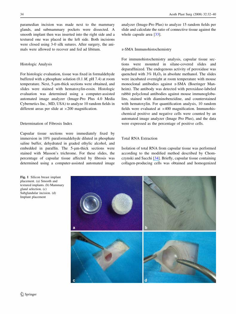
Fig. 1 Silicon breast implant
placement. (a) Smooth and
textured implants. (b) Mammary
gland selection. (c)
Subglandular incision. (d)
Implant placement
34 Aesth Plast Surg (2008) 32:32–40
123
using the Polytron System (Brinkmann, Switzerland) in the
presence of Trizol (Invitrogen). Chloroform was added, the
aqueous phase was obtained, and the RNA was precipitated
with isopropanol at 4�C overnight. The quantity and
intactness of RNA were routinely tested by determining the
absorbance at 260/280 and the ethidium bromide fluores-
cence of RNA electrophoresis in 1% formaldehyde-
containing agarose gels.
Reverse Transcription
Altogether, 2 lg of RNA extracted from all the samples
were added with 240 ng of random primers, 5 mM of DTT,
1 mM of dNTP mix, 40 U of RNase out inhibitor, and 200
U of RT Superscript III, then incubated for 10 min at 25�C,
60 min at 37�C, and 10 min at 95�C. The samples were
stored at –70�C until real-time polymerase chain reaction
(PCR) was performed.
Real-Time PCR
Quantitative real-time PCR was performed using a Rotor
Gene RG 3000 Sequence Detector (Corbett Research,
Sydney Australia) as follows: 1 cycle at 50�C for 2 min, 1
cycle at 95�C for 10 min, and 40 cycles at 95�C for 30 s
and 60�C for 40 s. The total reaction was designed to occur
in 10 ll containing 2 ll of cDNA, 1X of Universal PCR
Master Mix (Applied Biosystems), and 1X of final con-
centration of primers and TaqMan probe from
experimental and control genes synthesized by Applied
Biosystems (assay on demand). Multiplex PCR real time
for collagen aI and TGF-b were performed in duplicate for
each animal using glyceraldehyde phosphate dehydroge-
nase (GAPDH) as a housekeeping gene.
Data analysis was performed according to user bulletin
number 2 of Applied Biosystems [35]. Using sequence
detection software, we calculated the threshold cycle (Ct)
for each reaction, which then was used to quantify the
amount of starting template reaction. A difference in Ct
values (DCt) was calculated for each gene by duplicate.
The amount of the target gene normalized to the endoge-
nous gene GAPDH and relative calibrator is given as
2-DDCt, where DDCt is DCt - DCtGAPDH and DCt is
CTgene target - CtGAPDH.
Statistical Analyses
Results are expressed as mean ± standard deviation. Stu-
dent’s t test was used. All p values less than 0.05 were
considered to indicate a significant difference between
groups.
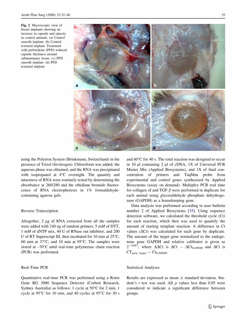
Fig. 2 Macroscopic view of
breast implants showing an
increase in capsule and opacity
in control animals. (a) Control
smooth implant. (b) Control
textured implant. Treatment
with pirfenidone (PFD) reduced
capsule thickness around
submammary tissue. (c) PFD
smooth implant. (d) PFD
textured implant
Aesth Plast Surg (2008) 32:32–40 35
123
Results
In this study. submammary silicone breast implants were
placed in female Wistar rats to evaluate the effect of
PFD has on contracture and fibrosis development after 8
weeks (Fig 1). It is important to mention that no animal
treated with PFD presented adverse effects such as
diarrhea or weight loss. After 8 weeks, the animals
presented with an augmented capsule around the implant,
with an increase in the opacity on adjacent tissues,
making it difficult to visualize the morphology of the
implant surface. An exacerbated amount of extracellular
matrix also was evident. We did not observe differences
between textured and smooth implants (Fig. 2a and b).
The PFD treatment caused a reduction in capsule
thickness around submammary tissue without evidence of
a real capsule and with a clear decrease in the amount of
extracellular matrix, a fact confirmed later with specific
staining.
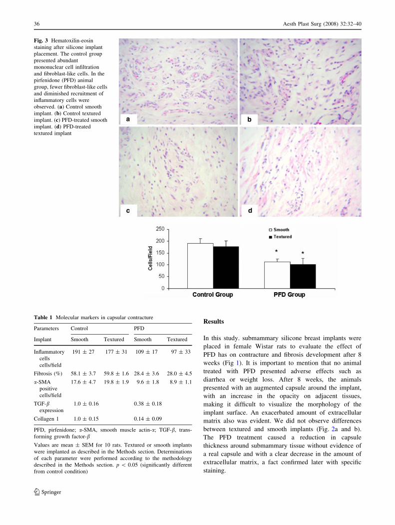
Fig. 3 Hematoxilin-eosin
staining after silicone implant
placement. The control group
presented abundant
mononuclear cell infiltration
and fibroblast-like cells. In the
pirfenidone (PFD) animal
group, fewer fibroblast-like cells
and diminished recruitment of
inflammatory cells were
observed. (a) Control smooth
implant. (b) Control textured
implant. (c) PFD-treated smooth
implant. (d) PFD-treated
textured implant
Table 1 Molecular markers in capsular contracture
Parameters Control PFD
Implant Smooth Textured Smooth Textured
Inflammatory
cells
cells/field
191 ± 27 177 ± 31 109 ± 17 97 ± 33
Fibrosis (%) 58.1 ± 3.7 59.8 ± 1.6 28.4 ± 3.6 28.0 ± 4.5
a-SMA
positive
cells/field
17.6 ± 4.7 19.8 ± 1.9 9.6 ± 1.8 8.9 ± 1.1
TGF-bexpression
1.0 ± 0.16 0.38 ± 0.18
Collagen 1 1.0 ± 0.15 0.14 ± 0.09
PFD, pirfenidone; a-SMA, smooth muscle actin-a; TGF-b, trans-
forming growth factor-b
Values are mean ± SEM for 10 rats. Textured or smooth implants
were implanted as described in the Methods section. Determinations
of each parameter were performed according to the methodology
described in the Methods section. p \ 0.05 (significantly different
from control condition)
36 Aesth Plast Surg (2008) 32:32–40
123
These downregulated events were noticeable to the same
extent in rats with both textured and smooth implants
(Fig. 2c and d). By using hematoxylin-eosin staining, we
could demonstrate that PFD-treated animal groups pre-
sented lower fibroblast- like cell proliferation and reduced
recruitment of inflammatory cells. In contrast, the control
group showed abundant mononuclear cell infiltration, lar-
ger fibroblast-like cell proliferation, and the presence of
neovascularization. Total cell numbers per microscopic
field were considerably reduced when the animals were
treated with PFD (n = 10; p\0.05; Fig. 3a–d; Table 1). To
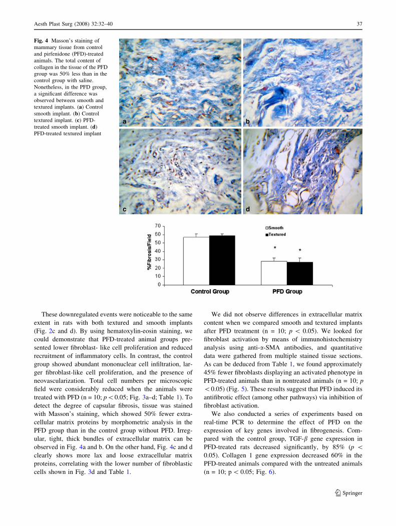
detect the degree of capsular fibrosis, tissue was stained
with Masson’s staining, which showed 50% fewer extra-
cellular matrix proteins by morphometric analysis in the
PFD group than in the control group without PFD. Irreg-
ular, tight, thick bundles of extracellular matrix can be
observed in Fig. 4a and b. On the other hand, Fig. 4c and d
clearly shows more lax and loose extracellular matrix
proteins, correlating with the lower number of fibroblastic
cells shown in Fig. 3d and Table 1.
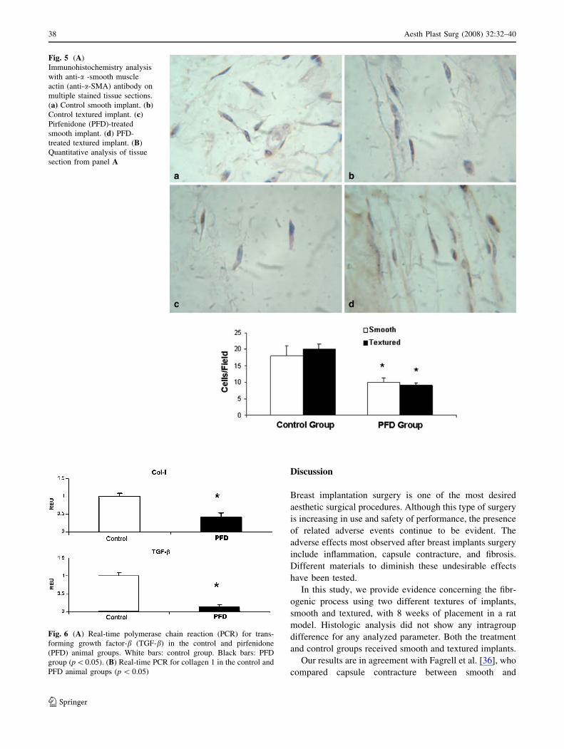
We did not observe differences in extracellular matrix
content when we compared smooth and textured implants
after PFD treatment (n = 10; p \ 0.05). We looked for
fibroblast activation by means of immunohistochemistry
analysis using anti-a-SMA antibodies, and quantitative
data were gathered from multiple stained tissue sections.
As can be deduced from Table 1, we found approximately
45% fewer fibroblasts displaying an activated phenotype in
PFD-treated animals than in nontreated animals (n = 10; p
\0.05) (Fig. 5). These results suggest that PFD induced its
antifibrotic effect (among other pathways) via inhibition of
fibroblast activation.
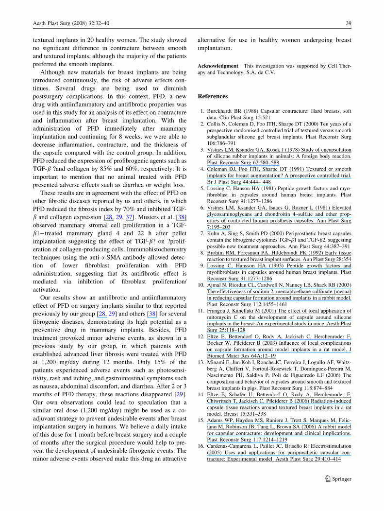
We also conducted a series of experiments based on
real-time PCR to determine the effect of PFD on the
expression of key genes involved in fibrogenesis. Com-
pared with the control group, TGF-b gene expression in
PFD-treated rats decreased significantly, by 85% (p \0.05). Collagen 1 gene expression decreased 60% in the
PFD-treated animals compared with the untreated animals
(n = 10; p \ 0.05; Fig. 6).
Fig. 4 Masson’s staining of
mammary tissue from control
and pirfenidone (PFD)-treated
animals. The total content of
collagen in the tissue of the PFD
group was 50% less than in the
control group with saline.
Nonetheless, in the PFD group,
a significant difference was
observed between smooth and
textured implants. (a) Control
smooth implant. (b) Control
textured implant. (c) PFD-
treated smooth implant. (d)
PFD-treated textured implant
Aesth Plast Surg (2008) 32:32–40 37
123
Discussion
Breast implantation surgery is one of the most desired
aesthetic surgical procedures. Although this type of surgery
is increasing in use and safety of performance, the presence
of related adverse events continue to be evident. The
adverse effects most observed after breast implants surgery
include inflammation, capsule contracture, and fibrosis.
Different materials to diminish these undesirable effects
have been tested.
In this study, we provide evidence concerning the fibr-
ogenic process using two different textures of implants,
smooth and textured, with 8 weeks of placement in a rat
model. Histologic analysis did not show any intragroup
difference for any analyzed parameter. Both the treatment
and control groups received smooth and textured implants.
Our results are in agreement with Fagrell et al. [36], who
compared capsule contracture between smooth and
Fig. 5 (A)
Immunohistochemistry analysis
with anti-a -smooth muscle
actin (anti-a-SMA) antibody on
multiple stained tissue sections.
(a) Control smooth implant. (b)
Control textured implant. (c)
Pirfenidone (PFD)-treated
smooth implant. (d) PFD-
treated textured implant. (B)
Quantitative analysis of tissue
section from panel A
Fig. 6 (A) Real-time polymerase chain reaction (PCR) for trans-
forming growth factor-b (TGF-b) in the control and pirfenidone
(PFD) animal groups. White bars: control group. Black bars: PFD
group (p\0.05). (B) Real-time PCR for collagen 1 in the control and
PFD animal groups (p \ 0.05)
38 Aesth Plast Surg (2008) 32:32–40
123
textured implants in 20 healthy women. The study showed
no significant difference in contracture between smooth
and textured implants, although the majority of the patients
preferred the smooth implants.
Although new materials for breast implants are being
introduced continuously, the risk of adverse effects con-
tinues. Several drugs are being used to diminish
postsurgery complications. In this context, PFD, a new
drug with antiinflammatory and antifibrotic properties was
used in this study for an analysis of its effect on contracture
and inflammation after breast implantation. With the
administration of PFD immediately after mammary
implantation and continuing for 8 weeks, we were able to
decrease inflammation, contracture, and the thickness of
the capsule compared with the control group. In addition,
PFD reduced the expression of profibrogenic agents such as
TGF-b ?and collagen by 85% and 60%, respectively. It is
important to mention that no animal treated with PFD
presented adverse effects such as diarrhea or weight loss.
These results are in agreement with the effect of PFD on
other fibrotic diseases reported by us and others, in which
PFD reduced the fibrosis index by 70% and inhibited TGF-
b and collagen expression [28, 29, 37]. Musters et al. [38]
observed mammary stromal cell proliferation in a TGF-
b1-treated mammary gland 4 and 22 h after pellet
implantation suggesting the effect of TGF-b? on ?prolif-
eration of collagen-producing cells. Immunohistochemistry
techniques using the anti-a-SMA antibody allowed detec-
tion of lower fibroblast proliferation with PFD
administration, suggesting that its antifibrotic effect is
mediated via inhibition of fibroblast proliferation/
activation.
Our results show an antifibrotic and antiinflammatory
effect of PFD on surgery implants similar to that reported
previously by our group [28, 29] and others [38] for several
fibrogenic diseases, demonstrating its high potential as a
preventive drug in mammary implants. Besides, PFD
treatment provoked minor adverse events, as shown in a
previous study by our group, in which patients with
established advanced liver fibrosis were treated with PFD
at 1,200 mg/day during 12 months. Only 15% of the
patients experienced adverse events such as photosensi-
tivity, rash and itching, and gastrointestinal symptoms such
as nausea, abdominal discomfort, and diarrhea. After 2 or 3
months of PFD therapy, these reactions disappeared [29].
Our own observations could lead to speculation that a
similar oral dose (1,200 mg/day) might be used as a co-
adjuvant strategy to prevent undesirable events after breast
implantation surgery in humans. We believe a daily intake
of this dose for 1 month before breast surgery and a couple
of months after the surgical procedure would help to pre-
vent the development of undesirable fibrogenic events. The
minor adverse events observed make this drug an attractive
alternative for use in healthy women undergoing breast
implantation.
Acknowledgment This investigation was supported by Cell Ther-
apy and Technology, S.A. de C.V.
References
1. Burckhardt BR (1988) Capsular contracture: Hard breasts, soft
data. Clin Plast Surg 15:521
2. Collis N, Coleman D, Foo ITH, Sharpe DT (2000) Ten years of a
prospective randomised controlled trial of textured versus smooth
subglandular silicone gel breast implants. Plast Reconstr Surg
106:786–791
3. Vistnes LM, Ksander GA, Kosek J (1978) Study of encapsulation
of silicone rubber implants in animals: A foreign body reaction.
Plast Reconstr Surg 62:580–588
4. Coleman DJ, Foo ITH, Sharpe DT (1991) Textured or smooth
implants for breast augmentation? A prospective controlled trial.
Br J Plast Surg 44:444– 448
5. Lossing C, Hanson HA (1981) Peptide growth factors and myo-
fibroblast in capsules around human breast implants. Plast
Reconstr Surg 91:1277–1286
6. Vistnes LM, Ksander GA, Isaacs G, Rozner L (1981) Elevated
glycosaminoglycans and chondroitin 4–sulfate and other prop-
erties of contracted human prosthesis capsules. Ann Plast Surg
7:195–203
7. Kuhn A, Sing S, Smith PD (2000) Periprosthetic breast capsules
contain the fibrogenic cytokines TGF-b1 and TGF-b2, suggesting
possible new treatment approaches. Ann Plast Surg 44:387–391
8. Brohim RM, Foresman PA, Hildebrandt PK (1992) Early tissue
reaction to textured breast implant surfaces. Ann Plast Surg 28:354
9. Lossing C, Hansson HA (1993) Peptide growth factors and
myofibroblasts in capsules around human breast implants. Plast
Reconstr Surg 91:1277–1286
10. Ajmal N, Riordan CL, Cardwell N, Nanney LB, Shack RB (2003)
The effectiveness of sodium 2–mercaptoethane sulfonate (mesna)
in reducing capsular formation around implants in a rabbit model.
Plast Reconstr Surg 112:1455–1461
11. Frangou J, Kanellaki M (2001) The effect of local application of
mitomycin C on the development of capsule around silicone
implants in the breast: An experimental study in mice. Aesth Plast
Surg 25:118–128
12. Eltze E, Bettendorf O, Rody A, Jackisch C, Herchenroder F,
Bocker W, Pfleiderer B (2003) Influence of local complications
on capsule formation around model implants in a rat model. J
Biomed Mater Res 64A:12–19
13. Minami E, Jun Koh I, Ronche JC, Ferreira J, Logullo AF, Waitz-
berg A, Chifferi V, Fortoul-Rosewick T, Domınguez-Pereira M,
Nascimento PH, Saldiva P, Poli de Figueiredo LF (2006) The
composition and behavior of capsules around smooth and textured
breast implants in pigs. Plast Reconstr Surg 118:874–884
14. Eltze E, Schafer U, Bettendorf O, Rody A, Herchenroder F,
Chiwritsch T, Jackisch C, Pfleiderer B (2006) Radiation-induced
capsule tissue reactions around textured breast implants in a rat
model. Breast 15:331–338
15. Adams WP, Haydon MS, Raniere J, Trott S, Marques M, Felic-
iano M, Robinson JB, Tang L, Brown SA (2006) A rabbit model
for capsular contracture: development and clinical implications.
Plast Reconstr Surg 117:1214–1219
16. Cardenas-Camarena L, Paillet JC, Briseno R: Electrostimulation
(2005) Uses and applications for periprosthetic capsular con-
tracture: Experimental model. Aesth Plast Surg 29:410–414
Aesth Plast Surg (2008) 32:32–40 39
123
17. Peterson HD, Burt GB Jr (1974) The role of steroid in prevention
of circumferential capsular scarring in augmentation mamma-
plasty. Plast Reconstr Surg 54:28
18. Perrin ER (1976) The use of soluble steroid within inflatable
breast prostheses. Plast Reconstr Surg 57:163
19. Vinnik CA (1976) Spherical contracture of fibrous capsules
around breast implants: Prevention and treatment. Plast Reconstr
Surg 58:555
20. Ellemberg AH (1977) Marked thinning of the breast skin flaps
after the insertion of implants containing triamcinolone. Plast
Reconstr Surg 60:755
21. Carrico TJ, Cohen IK (1979) Capsular contracture and steroid-
related complication after augmentation mammaplasty: A pre-
liminary study. Plast Reconstr Surg 64:377
22. Baker JL Jr (1981) The effectiveness of alpha-tocopherol (vita-
min E) in reducing the incidence of spherical contracture around
breast implants. Plast Reconstr Surg 68:696
23. Cucin RL, Guthrie RH, Graham M (1982) Rate of diffusion of
Solu-Medrol across the silastic membranes of breast prostheses:
An in vivo study. Ann Plast Surg 9:228
24. Cafee HH (1984) The effects of intraprosthetic methylpredniso-
lone on implants capsules and surrounding soft tissue. Ann Plast
Surg 12:348
25. Gayou R, Rudolph R (1979) Capsular contraction around silicone
mammary prostheses. Ann Plast Surg 2:62
26. Iyer SN, Wild JS, Schiedt MJ, Hyde DM, Margolin SB, Giri SN
(1995) Dietary intake of pirfenidone ameliorates bleomycin-
induced lung fibrosis in hamsters. J Lab Clin Med 125:779–785
27. Al-Took S, Murray C, Tulandi T (1998) Effects of pirfenidone
and dermoid cyst fluid on adhesion formation. Fertil Steril
69:341–342
28. Garcıa L, Hernandez I, Sandoval A, Salazar, Garcia J, Vera J,
Grijalva G, Muriel P, Margolin S, Armendariz-Borunda J (2002)
Pirfenidone affectively reverses experimental liver fibrosis. J
Hepatol 32:797–805
29. Armendariz-Borunda J, Islas-Carbajal MC, Meza-Garcıa E,
Rincon AR, Sandoval AS, Salazar A, Berumen J, Alvarez A,
Alvarez A, Covarrubias A, Arechiga G, Garcıa L (2006) A pilot
study in cirrhotic patients using a new antiinflammatory and
antifibrotic agent, pirfenidone. Gut 55:1663–1665
30. Lee B-S, Margolin SB, Nowak AR (1998) Pirfenidone: A novel
pharmacological agent that inhibits leiomyoma cell proliferation
and collagen production. J Clin Endocrinol Metab 83:219–223
31. Shimizu T, Kuroda T, Hata S, Fukagawa M, Margolin SB,
Kurokawa K (1998) Pirfenidone improves renal function and
fibrosis in the postobstructed kidney. Kidney Int 54:99–109
32. Shetlar MR, Shetlar DJ, Bloom RF, Shetlar CL, Margolin SB
(1998) Involution of keloid implants in athymic mice treated with
pirfenidone or with triamcinolone. J Lab Clin Med 132:491–496
33. Callaway JK, Beart PM, Jarott B (1998) A reliable procedure for
comparison of antioxidants in rat brain homogenates. J Pharma-
col Toxicol Methods 39:155–162
34. Chomczynski P, Sacchi N (1987) Single-step method of RNA
isolation by acid guanidinium thiocyanate-phenol chloroform
extraction. Anal Biochem 162:156–159
35. Retrieved from http://www.dna-9.int-med.uiowa.edu/
RealtimePCRdocs/Bulletin 2 Applied Byosystem
36. Fagrell D, Berggren A, Tarpila E (2001) Capsular contracture
around saline-filled fine textured and smooth mammary implants:
A prospective 7.5-year follow-up. Plast Reconstr Surg 108:2108
37. Lee B-S, Margolin SB, Nowak AR (1998) Pirfenidone: A novel
pharmacological agent that inhibits leiomyoma cell proliferation
and collagen production. J Clin Endocrinol Metab 83:219–223
38. Musters S, Coughlan K, McFadden T, Mapple R, Mulvey T,
Plaunt K (2004) Exogenous TGF-b1 promotes stromal develop-
ment in the heifer mammary gland. J Dairy Sci 87:896–904
40 Aesth Plast Surg (2008) 32:32–40
123