physiotherapy in critically ill patients
TRANSCRIPT
Clinical Rehabilitation27(4) 336 –346© The Author(s) 2012Reprints and permissions: sagepub.co.uk/journalsPermissions.navDOI: 10.1177/0269215512458940cre.sagepub.com
CLINICALREHABILITATION
458940 CRE27410.1177/0269215512458940Clinical RehabilitationVenturelli et al.2012
1Department of Medical and Surgical Sciences, University of Modena, Pavullo-Modena, Italy2Villa Pineta Hospital, Pavullo n/F (Modena), Italy3Auxilium Vitae Rehabilitation Center, Volterra, Pisa, Italy4IRCCS-Maugeri Foundation of Veruno, Novara, Italy5IRCCS-Maugeri Foundation of Lumezzane, Brescia, Italy6San Giuseppe Hospital, Milano, Italy
Efficacy of temporary positive expiratory pressure (TPEP) in patients with lung diseases and chronic mucus hypersecretion. The UNIKO® project: a multicentre randomized controlled trial
Elena Venturelli1, Ernesto Crisafulli2, Assunta DeBiase2, Daniela Righi2, Daniele Berrighi3, Pier Paolo Cavicchioli3, Guido Vagheggini3, Francesco Dabrosca4, Bruno Balbi4, Mara Paneroni5, Luca Bianchi5, Michele Vitacca5, Vittoria Galimberti6, Michele Zaurino6, Giorgio Schiavoni6, Andrea Iattoni1, Nicolino Ambrosino3 and Enrico M Clini1,2
AbstractObjective: To evaluate whether temporary positive expiratory pressure provides benefit in patients with lung diseases and chronic hypersecretion.Design: Single blind multicentre randomized trial.Setting: Five Italian rehabilitation centres.Participants: Ninety-eight patients with chronic obstructive pulmonary disease and/or chronic bronchitis (n=78), or bronchiectasis (n=20), with a peak cough expiratory flow >150 l/min and sputum production >30 ml/day, randomly included into two treatment groups.Interventions: For 10 consecutive days, the active group performed twice a day 20-minute cycles of manually assisted breathing techniques in sequence with the addition of 15 minutes of temporary positive expiratory pressure, while the control group was treated by manually assisted breathing techniques alone.Measures: Within and between group changes of arterial oxygenation index, lung volumes and respiratory muscles strength were recorded at enrolment and after 3 and 10 treatment sessions.
Article
Corresponding author:EM Clini, University of Modena and Villa Pineta Hospital, Via Gaiato 127, Pavullo (MO), 41026, Italy. Email: [email protected]
Venturelli et al. 337
Introduction
Chronic mucus hypersecretion is frequently observed in many respiratory diseases, such as chronic airway obstruction in chronic obstructive pulmonary disease, including chronic bronchitis and bronchiectasis,1 with a negative impact on both function and survival.2,3
To date, chest physiotherapy techniques by manually assisted breathing techniques remain the gold standard for those patients with normal cough reflex.1 In addition, positive expiratory pressure delivered by hand-held devices is considered a valid technique to manage excessive airway secre-tion, enhancing expectoration and modifying muco-rheological properties.4 However, results supporting the use and the effectiveness of these tools to assist airways clearance are still controver-sial in literature.
Recently, a new modality to mechanically deliver a low positive expiratory pressure level at the mouth during spontaneous breathing, so-called temporary positive expiratory pressure, has become available in the market, and it has been proposed in assisting patients with chronic mucus hypersecretion. This technique produces a 1 cmH2O increase in airway pressure along the respiratory cycle until immedi-ately before the end of expiration. Although the level of applied pressure with temporary positive expiratory pressure is several times lower than that (5 to 15 cmH2O) used and considered effective with
other positive expiratory pressure and/or oscilla-tory-positive expiratory pressure devices,1 prelimi-nary results have shown that an expiratory pressure ≤1 cm H2O applied for a fraction of the expiratory phase may improve the distribution of alveolar ven-tilation and prevent mechanical stress injury, which is expected to occur in the bronchial tree or lung parenchyma at a higher pressure.5
The aim of our study was to evaluate the clinical effectiveness of temporary positive expiratory pres-sure as a new positive low-pressure technique to clear excessive mucus in stable patients with hyper-secretion and normal cough reflex. We therefore tested the hypothesis that temporary positive expi-ratory pressure may provide additional clinical ben-efits over conventional manually assisted breathing techniques, considered as the reference techniques in this condition.
Methods
Patients
We prospectively recruited 98 chronic mucus hyper-secretion patients admitted from July 2008 to December 2010 to five Italian centres (Villa Pineta Hospital, Pavullo n/F, Auxilium Vitae Rehabilitation Center, Volterra, IRCCS Maugeri Foundation, Centres of Veruno and Lumezzane, and San Giuseppe
Pre-to-post treatment change of sputum volume and bronchial encumbrance (Δ-visual analog scale), sputum density and purulence were compared daily within the study period.Results: No significant changes were recorded for the oxygenation index, while dynamic lung volumes and respiratory muscle strength significantly (P <0.05) improved in the active group. The group comparison analysis of the pre-to-post change showed that inspiratory capacity was significantly higher in the active than in the control group (+19.5% and +2.2%, P=0.044) at day 10. A greater improvement in Δ-visual analog scale was recorded in the active group at day 3 and 8.Conclusions: These preliminary data suggest that temporary positive expiratory pressure improves lung volumes and speeds up the improvement of bronchial encumbrance in patients with lung diseases and hypersecretion.
KeywordsHypersecretion, bronchial drainage, temporary positive expiratory pressure, cough, rehabilitation
Received: 8 May 2012; accepted: 31 July 2012
338 Clinical Rehabilitation 27(4)
Hospital, Milano) for an inpatient pulmonary reha-bilitation. Villa Pineta Hospital acted as the study coordinator.
The institutional review board and ethical com-mittee at each hospital approved the study, which was conducted according to the declaration of Helsinki.6
All patients were in a stable clinical condition and free from any acute exacerbation for least four weeks at the time of inclusion; chronic mucus hypersecretion was specifically related to chronic obstructive pulmonary disease or bronchiectasis. Forty per cent of them had concomitant chronic respiratory failure leading to regular domiciliary oxygen therapy (long term oxygen therapy) (see Table 1) but not precluding them from participation in the trial. Diagnosis and severity of chronic obstructive pulmonary disease were confirmed on a clinical basis and by means of spirometry.7,8
Chronic mucus hypersecretion was defined as a sputum volume production of greater than 30 ml/day.1 A peak cough expiratory flow >150 l/min con-firmed the patients were able to cough without the need of any additional support.9
Patients with limitations regarding exercise or with other associated conditions (i.e. severe concom-itant cardiovascular disease or cancer) which may have limited or impaired the response to training, and those patients under domiciliary mechanical ventilation or taking any active drugs supposed to have a mucoregulatory action (i.e. aminophyllines, N-acetyl-cysteine), were excluded from the study. Finally, patients who were deemed unable to use the temporary positive expiratory pressure device were also excluded.
Patients were considered as having dropped out of the study when clinical signs of a new exacerba-tion occurred. Patients dropping out were nonethe-less included in the analysis as being registered and part of the trial.
DesignThis was a single blind randomized trial with randomization list by blocks for each centre; each centre had a randomization list provided, which indicated the group for each patient’s allocation.
The protocol was recorded on the ClinicalTrial.gov website with identification code NCT00700388.
Rehabilitation programAppropriate selection of patients and the program content were according to the American Thoracic Society/European Respiratory Society joint state-ment on pulmonary rehabilitation.10,11 All the eligi-ble patients were instructed to use temporary positive expiratory pressure for acclimatization in a 2-hour training period in the lung laboratory before inclusion into the trial.
The study group (active) performed twice a day 20-minute cycles of manually assisted breathing techniques plus 15 minutes of temporary positive expiratory pressure (UNIKO®, Medical Products Research, Legnano, Italy) each day.
Controls were treated by manually assisted breathing techniques alone with the same modali-ties as in the active group. In all patients interven-tion lasted 10 consecutive days, within the period of the in-hospital rehabilitation course starting at least 48–72 hours following admission and initial assessment.
Manually assisted breathing techniques con-sisted of a cycle of assisted exercises; among these we performed Expiration Lente Totale avec Glotte Ouverte en Infralateral and forced expira-tion,1,5 which were delivered in sequence to all patients up to the time limit; a short preliminary description of the procedure was given by the attending physiotherapist. Temporary positive expiratory pressure, when appropriate, was deliv-ered continuously to the subjects throughout the programmed period. Spontaneous cough to elimi-nate secretions following treatment periods was not assisted by personnel, and patients were instructed to collect sputum in a single plastic pot (changed on a daily basis) to calculate the total volume per day. The temporary positive expira-tory pressure device (UNIKO® Medical Products Research, Legnano, Italy) delivered a fixed posi-tive pressure (1 cmH2O or 0.0977 kPa) only in the expiratory phase. This increase in low pressure was created through a pulsatile flow approxi-mately 42 Hz in frequency.
Venturelli et al. 339
Physiotherapists from each centre and involved in this research were previously instructed to stan-dardize the type and duration of all activities. They knew which group each patient was in, but none of them was aware of the study purpose.
Measurements and outcomes
At enrolment, patients’ anthropometric and physiolog-ical characteristics and main diagnosis were recorded. Cough peak expiratory flow by a hand-held device
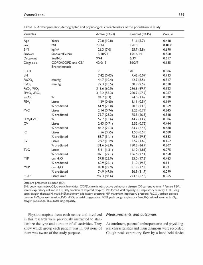
Table 1. Anthropometric, demographic and physiological characteristics of the population in study.
Variables Active (n=53) Control (n=45) P-value
Age Years 70.0 (10.8) 71.6 (8.7) 0.448Sex M/F 29/24 35/10 0.017BMI kg/m2 26.3 (7.0) 25.7 (5.8) 0.690Smoker Smoker/Ex/No 13/18/22 15/16/14 0.560Drop-out Yes/No 9/44 6/39 0.617Diagnosis COPD/COPD and CB/
Bronchiectasis40/0/13 36/2/7 0.185
LTOT 19 20 0.386pH 7.42 (0.03) 7.42 (0.04) 0.733PaCO2 mmHg 44.7 (10.4) 42.7 (8.5) 0.817PaO2 mmHg 72.3 (10.5) 68.9 (9.5) 0.510PaO2 /FiO2 318.6 (60.0) 296.6 (69.7) 0.123SPaO2 /FiO2 313.2 (57.3) 280.7 (67.7) 0.087SatO2 % 94.7 (2.3) 94.0 (1.6) 0.520FEV1 Litres 1.29 (0.60) 1.11 (0.54) 0.149 % predicted 61.9 (25.0) 50.3 (24.8) 0.069FVC Litres 2.14 (0.74) 2.25 (0.79) 0.345 % predicted 79.7 (23.2) 75.8 (26.3) 0.848FEV1/FVC % 52.7 (15.6) 44.2 (13.7) 0.006CV Litres 2.43 (0.71) 2.52 (0.72) 0.444 % predicted 85.2 (22.3) 83.7 (27.5) 0.588IC Litres 1.56 (0.55) 1.58 (0.59) 0.680 % predicted 83.7 (34.1) 73.6 (29.9) 0.883RV Litres 2.97 (1.19) 3.52 (1.65) 0.162 % predicted 131.6 (48.8) 150.5 (64.4) 0.307TLC Litres 5.41 (1.31) 6.10 (1.81) 0.075 % predicted 102.1 (22.1) 106.6 (27.1) 0.658MIP cm H2O 57.8 (25.9) 55.0 (17.5) 0.463 % predicted 60.9 (26.1) 51.0 (19.3) 0.131MEP cm H2O 83.0 (29.9) 81.9 (37.3) 0.921 % predicted 74.9 (47.0) 56.9 (31.7) 0.099PCEF Litres /min 247.3 (83.6) 223.3 (67.8) 0.065
Data are presented as mean (SD).BMI, body mass index; CB, chronic bronchitis; COPD, chronic obstructive pulmonary disease; CV, current volume; F, female; FEV1, forced expiratory volume in 1 s; FiO2, fraction of inspired oxygen; FVC, forced vital capacity; IC, inspiratory capacity; LTOT, long term oxygen therapy; M, male; MEP, maximum expiratory pressure; MIP, maximum inspiratory pressure; PaCO2, carbon dioxide tension; PaO2, oxygen tension; PaO2 /FiO2, arterial oxygenation; PCEF, peak cough expiratory flow; RV, residual volume; SatO2, oxygen saturation; TLC, total lung capacity.
340 Clinical Rehabilitation 27(4)
was recorded in all patients to confirm their cough competence and reflex initially.9
Outcome measures were taken at study entry (day 0), and after three and 10 days of treatment. Data on sputum volume and characteristics, and individuals’ perceived symptoms, were recorded on a daily basis.
One physiotherapist, different from those who treated the patients and unaware of the study pur-pose, was in charge in each centre for all the mea-surements at each time point.
Primary outcome – Arterial blood gas analysis was obtained from an arterial blood sample taken from the radial artery with the patient in resting con-dition and breathing room air or oxygen (when appropriate) at the prescribed flow rate: parameters were corrected by the Mays’ graph.12
The derived arterial oxygen tension to inspira-tory oxygen fraction ratio (PaO2/FiO2) was consid-ered as the primary outcome based on previous data using a device to treat a condition of chronic mucus hypersecretion in severely affected patients.13
Secondary outcomes – Lung function was assessed by means of an automated spirometer: dynamic and static volumes were expressed as % of their predicted value.14 Respiratory muscle strength (maximal inspiratory and expiratory pressure) was then performed by means of a specific module recording maximal pressures against an occlusive mouth resistance at both total lung capacity (for maximal expiratory pressure) and functional resid-ual volume (for maximal inspiratory pressure); values were recorded as absolute and as a percent-age of predicted values according to the reference equations.15 Both measures of lung function were recorded with the patient in a sitting position; the best of three measurements was recorded.
The perceived sensation of bronchial encum-brance was reported by patients as a discomfort and/or appearance of substantial amount of secretions leading to dyspnoea; it was measured each day by a visual analogic scale16 (days 1 to 10). The change in sputum volume and percent visual analogic scale were recorded as study outcomes.
Sputum density and purulence were also recorded on a daily basis by means of a specific three-point semi-quantitative scale (where 0=fluid for density
or light for purulence, 1= dry for density or yellow-ish for purulence, 2= thick for density or dark for purulence).17 The patients were instructed to collect their secretions in a small container every day in the morning and following the chest physiotherapy (120 minutes after the second daily session) in both groups and for the whole study period. The same nurse, not involved in the study aims, recorded these variables.
Statistical analysisSample size. Although this study addresses several outcome measures, only one was considered for sample size determination. Changes in daytime oxygenation ratio (PaO2/FiO2) under usual breath-ing conditions was the outcome selected to deter-mine a minimum sample able to ensure powerful testing of treatment effect.13 In order to ensure 80% power to detect a group difference ≥25 points in oxygenation ratio after treatment period as signifi-cant at the 0.05 level, 42 patients per group were needed for the study. Considering a probability of 15% drop-out rate of randomized patients, the mini-mum target sample size was fixed at 49 patients per group.
Efficacy analysis. Analyses were carried out using SPSS software (SPSS 8.0 for Windows; SPSS, Chi-cago, Illinois, USA) and applied according to the current methodology.18
Qualitative and quantitative variables are pre-sented as count, percentage (%), and mean (± stan-dard deviation, SD), respectively. A preliminary test for normal distribution of data was made by the Kolmogorov–Smirnov test.
Absolute values and changes (Δ) in each out-come variable were compared by means of ANOVA and Student t-test. Wilcoxon and Kruskal–Wallis tests were applied for non-parametric variables. Comparisons among groups and times were per-formed following the intention to treat model; last observation carried forward was used as the method of analysis and data are presented accordingly.
A further analysis for confirming results has been added; indeed, the general linear model for repeated measures (mixed-model ANOVA) was
Venturelli et al. 341
applied to all the variables in the study to calculate the statistical interaction between groups, time and groups at each time point.
All results were considered to be statistically sig-nificant at a level of P <0.05.
Results
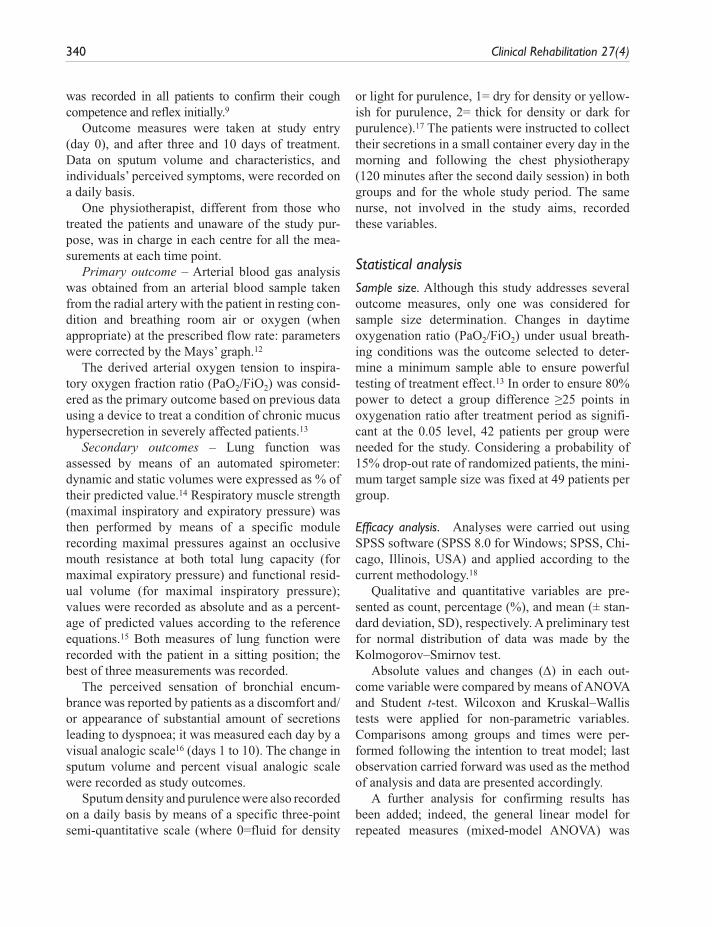
The study flow diagram is depicted in Figure 1. Main descriptive anthropometric, clinical and
functional characteristics of patients included are shown in Table 1; no differences among centres were reported. Sixty-four patients were male (65%) and 15 of the whole population presented a Body Mass Index >30 kg/m2, with no difference between groups. Gender distribution was different among groups. Chronic respiratory failure needing long term oxygen therapy occurred in 39 patients (40%). Overall, the study population was characterized by a moderate airflow obstruction with concomitant hyperinflation. The drop-out rate from the study
Figure 1. Flow chart of the study.
342 Clinical Rehabilitation 27(4)
was 17% (n=9) and 11% (n=6) in active and control groups respectively (not significant); in all cases, patients withdrew due to a recurrent exacerbation of their respiratory condition.
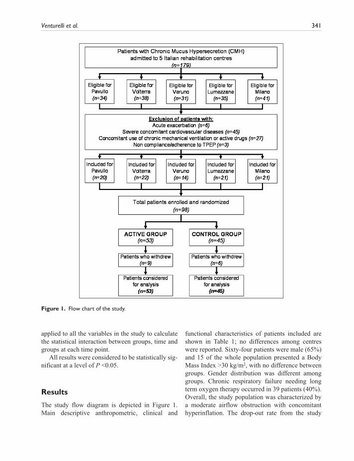
Table 2 shows the changes in arterial blood gases, lung volumes, muscle strength and bronchial encumbrance (visual analogic scale %) over time. No significant changes were observed for the blood gases or for the oxygenation ratio in both groups. There were significant (P < 0.05) increases by T10 in forced expiratory volume in the first second (+3.0%), forced vital capacity (+4.4%), current vol-ume (+4.2%), and inspiratory capacity (+19.6%) in the active but not in the control group.
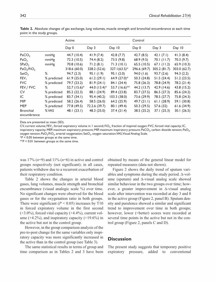
However, in the group comparison analysis of the pre-to-post change for the same variables only inspi-ratory capacity was more significantly increased in the active than in the control group (see Table 3).
The same statistical results in terms of group and time comparison as in Tables 2 and 3 have been
obtained by means of the general linear model for repeated measures (data not shown).
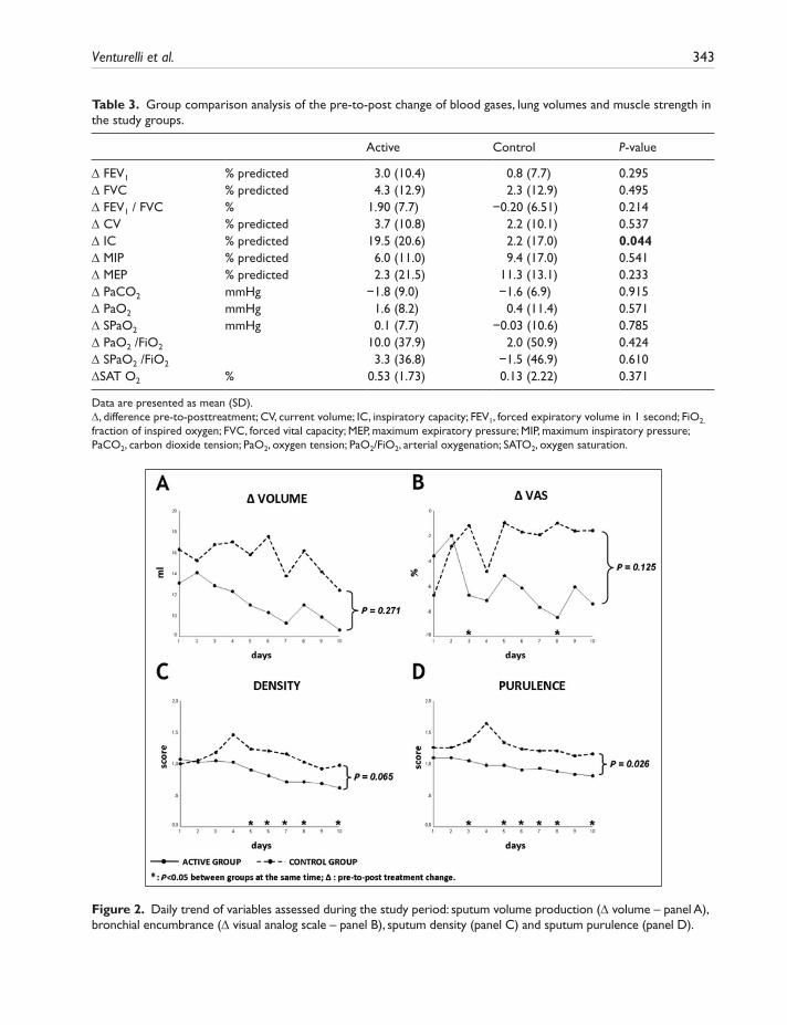
Figure 2 shows the daily trend of sputum vari-ables and symptoms during the study period. Δ-vol-ume (sputum) and Δ-visual analog scale showed similar behaviour in the two groups over time; how-ever, a greater improvement in Δ-visual analog scale after intervention was recorded at day 3 and 8 in the active group (Figure 2, panel B). Sputum den-sity and purulence showed a similar and significant trend to improvement over time in both groups; however, lower (=better) scores were recorded at several time points in the active but not in the con-trol group (Figure 2, panels C and D).
Discussion
The present study suggests that temporary positive expiratory pressure, added to conventional
Table 2. Absolute changes of gas exchange, lung volumes, muscle strength and bronchial encumbrance at each time point in the study groups.
Active Control
Day 0 Day 3 Day 10 Day 0 Day 3 Day 10
PaCO2 mmHg 44.7 (10.4) 41.9 (7.4) 42.8 (7.7) 42.7 (8.5) 42.1 (7.1) 41.3 (8.4)PaO2 mmHg 72.3 (10.5) 74.4 (8.2) 73.5 (9.8) 68.9 (9.5) 70.1 (11.7) 70.3 (9.7)SPaO2 mmHg 70.8 (10.6) 71.2 (8.1) 71.3 (10.1) 65.5 (10.5) 67.1 (11.2) 65.9 (10.3)PaO2/FiO2 318.6 (60.0) 328.2 (52.6) 327.1(63.5)* 296.6 (69.7) 305.2 (81.7) 303.0 (62.7)SatO2 % 94.7 (2.3) 95.1 (1.9) 95.1 (2.0) 94.0 (1.6) 93.7 (2.6) 94.3 (2.2)FEV1 % predicted 61.9 (25.0) 61.2 (29.1) 64.9 (27.0)* 50.3 (24.8) 51.5 (24.4) 51.2 (23.5)FVC % predicted 79.7 (23.2) 81.9 (24.1) 84.1 (24.4) 75.8 (26.3) 78.8 (24.9) 78.2 (21.4)FEV1/ FVC % 52.7 (15.6)* 44.0 (13.4)* 53.7 (16.6)** 44.2 (13.7) 42.9 (14.6) 43.8 (15.2)CV % predicted 85.2 (22.3) 88.1 (24.9) 89.4 (23.8) 83.7 (27.5) 86.5 (27.3) 85.6 (24.2)IC % predicted 83.7 (34.1) 95.4 (40.2) 103.3 (38.0) 73.6 (29.9) 78.5 (27.7) 75.8 (24.3)MIP % predicted 58.2 (26.4) 58.5 (26.0) 64.2 (25.9) 49.7 (21.1) 61.1 (28.9) 59.1 (30.8)MEP % predicted 77.8 (49.5) 72.2.6 (39.7) 80.1 (49.4) 50.3 (29.5) 57.6 (32) 61.6 (34.9)Bronchial encumbrance
% VAS 48.1 (23.1) 48.3 (25.0) 37.4 (21.4) 38.5 (25.2) 37.1 (25.3) 30.1 (26.5)
Data are presented as mean (SD).CV, current volume; FEV1, forced expiratory volume in 1 second; FiO2, fraction of inspired oxygen; FVC, forced vital capacity; IC, inspiratory capacity; MEP, maximum expiratory pressure; MIP, maximum inspiratory pressure; PaCO2, carbon dioxide tension; PaO2, oxygen tension; PaO2/FiO2, arterial oxygenation; SatO2, oxygen saturation; VAS, Visual Analog Scale.*P < 0.05 between groups at the same time.**P < 0.01 between groups at the same time.
Venturelli et al. 343
Table 3. Group comparison analysis of the pre-to-post change of blood gases, lung volumes and muscle strength in the study groups.
Active Control P-value
Δ FEV1 % predicted 3.0 (10.4) 0.8 (7.7) 0.295Δ FVC % predicted 4.3 (12.9) 2.3 (12.9) 0.495Δ FEV1 / FVC % 1.90 (7.7) −0.20 (6.51) 0.214Δ CV % predicted 3.7 (10.8) 2.2 (10.1) 0.537Δ IC % predicted 19.5 (20.6) 2.2 (17.0) 0.044Δ MIP % predicted 6.0 (11.0) 9.4 (17.0) 0.541Δ MEP % predicted 2.3 (21.5) 11.3 (13.1) 0.233Δ PaCO2 mmHg −1.8 (9.0) −1.6 (6.9) 0.915Δ PaO2 mmHg 1.6 (8.2) 0.4 (11.4) 0.571Δ SPaO2 mmHg 0.1 (7.7) −0.03 (10.6) 0.785Δ PaO2 /FiO2 10.0 (37.9) 2.0 (50.9) 0.424Δ SPaO2 /FiO2 3.3 (36.8) −1.5 (46.9) 0.610ΔSAT O2 % 0.53 (1.73) 0.13 (2.22) 0.371
Data are presented as mean (SD).Δ, difference pre-to-posttreatment; CV, current volume; IC, inspiratory capacity; FEV1, forced expiratory volume in 1 second; FiO2, fraction of inspired oxygen; FVC, forced vital capacity; MEP, maximum expiratory pressure; MIP, maximum inspiratory pressure; PaCO2, carbon dioxide tension; PaO2, oxygen tension; PaO2/FiO2, arterial oxygenation; SATO2, oxygen saturation.
Figure 2. Daily trend of variables assessed during the study period: sputum volume production (Δ volume – panel A), bronchial encumbrance (Δ visual analog scale – panel B), sputum density (panel C) and sputum purulence (panel D).
344 Clinical Rehabilitation 27(4)
manually assisted chest physiotherapy to clear excessive mucus in patients with lung diseases and chronic hypersecretion, improves ventilatory func-tion and speeds up the improvement of perceived bronchial encumbrance.
Airway clearance can be effectively performed through different manually assisted or unassisted methods.19 Recently, temporary positive expiratory pressure, as a positive pressure device, has been developed for those patients having a valid cough reflex. This is the first controlled study assessing the short-term (10 days) clinical effectiveness of tem-porary positive expiratory pressure in a consistent population of patients with lung diseases and chronic mucus hypersecretion.
The preliminary and experimental use of tempo-rary positive expiratory pressure in pathology has been performed in chronic respiratory diseases, including chronic obstructive pulmonary disease, asthma and cystic fibrosis, showing that symptoms and static and dynamic lung volumes improved after two weeks of treatment.20 In addition, tempo-rary positive expiratory pressure has been success-fully used for 30 minutes twice a day for five days in the preparation of 28 chronic obstructive pulmo-nary disease patients for major abdominal surgery.21 According to those findings,20,21 it has been specu-lated that lung ventilation was less inhomogeneous and arterial blood gases significantly improved in patients treated by temporary positive expiratory pressure, who also reported a significant and pro-gressive reduction in mucus production and in per-ceived bronchial encumbrance.
Our results have confirmed those preliminary observations in terms of lung ventilation, but not the expected effect on gas exchange. The lack of improvement in the primary outcome is possibly due to two main problems. First, the expected change PaO2/FiO2 ratio was estimated on the basis of a previous study using a different mechanical device to remove secretions;13 indeed, expected changes in arterial blood gases with temporary posi-tive expiratory pressure were lacking based on the available data. Second, it is more likely that, due to the previous reason, the sample size was under-powered to show any significant difference in oxy-genation ratio between the two groups.
Notably, we have found that temporary positive expiratory pressure causes a significant change in pulmonary function, as documented by the signifi-cant improvement of most variables at T10 (see Table 3) in the active but not in the control group. In particular, the analysis of group difference in vari-able changes confirmed the significant effect of tem-porary positive expiratory pressure on the increase in inspiratory capacity (see Table 3), which is likely to prove both the reduction in airway obstruction and the recruitment of collapsed or obstructed peripheral airways and lung parenchyma.
This beneficial effect on lung function has also so far been assessed by clinicians aiming at evaluat-ing the effectiveness of different types of positive expiratory pressure techniques.22,23 The theoretical benefit of these techniques, indeed, is the ability to enhance mucus clearance by either stenting the air-ways and preventing airway collapse, or increasing intrathoracic pressure and collateral ventilation dis-tal to retained secretions, or decreasing functional residual capacity of the lung.24
Another relevant finding in our study was the effect following the use of temporary positive expi-ratory pressure on the characteristics of mucus in the population treated. Indeed, a trend towards a greater reduction of sputum over time in the active group compared with the control group was reported (see Figure 2, panel A). The use of tempo-rary positive expiratory pressure also enhanced the improvement of the score for density and for puru-lence in the active group at several time points (see Figure 2, panels C and D). It is likely that, for a given reduction in the overall volume of sputum produced over the study period, the reduction in sputum density may reflect and parallel the reduc-tion of perceived bronchial encumbrance that patients experienced early, three days after begin-ning temporary positive expiratory pressure (see Figure 2, panel B).
Previous studies have challenged the effect of conventional chest physiotherapy and different pos-itive expiratory pressure techniques on the individ-ual’s ability to expectorate, and found a likely benefit, probably due to change in the operational lung volumes and flow.4 Some authors have specifi-cally tested positive expiratory pressure mask and
Venturelli et al. 345
manually assisted breathing techniques in improv-ing lung function and maintaining the effect over a one-year period when compared with postural drainage.25,26 However, the significant advantage of one technique over another was not clear, even if used for a long period.27
Although this study reports some interesting and new findings in relation to the use of tempo-rary positive expiratory pressure, some limita-tions should be carefully taken into account by readers. First, the study failed to test the hypoth-esis that the addition of temporary positive expi-ratory pressure may gain in terms of oxygenation, and this was probably due to a type-2 statistical error for an under-powered sample size (see dis-cussion above). Second, temporary positive expi-ratory pressure was delivered for only a 10-day period and we cannot exclude the possibility that a longer period would have enhanced and/or sus-tained the positive effects; this preliminary study is unlikely to show the optimal regimen of tempo-rary positive expiratory pressure in terms of either frequency or duration. Third, despite the low level of pressure delivered by temporary pos-itive expiratory pressure, we cannot exclude that a different effect could have been generated in patients with chronic obstructive pulmonary dis-ease or bronchiectasis; however, the limited pop-ulation in the study does not allow this question to be answered. Therefore, our clinical findings can only generate hypotheses that should be fur-ther tested in physiological studies with an appro-priate design and aim.
Finally, we did not study any functional out-come (e.g. the six minute walk test) to see whether the recorded improvements might have been related to a better general clinical condition of these patients.
Despite this, the trial has the strength of being based on a multicentre randomized controlled design that guarantees an elegant and correct sci-entific method of research. Moreover, the popula-tion in the study is quite representative of patients suffering from lung diseases and chronic mucus hypersecretion in real life, thus making this research a realistic base for future studies in the same field.
Clinical messages
• Temporary positive expiratory pressure improves lung function and symptoms in patients with chronic lung disease and mucus hypersecretion.
• The clinical advantage of temporary posi-tive expiratory pressure over conventional chest physiotherapy consists in a faster recovery from symptoms during treatment.
Authors’ contributions
Conception and design of the study: EV, EC, EMCDrafting the article: EV, ECAnalysis of data: EC, EVAcquisition of data: DR, AD, DB, PC, FD, MP, VG, MZ, GS, AIRevising paper and the final draft: BB, NA, EMC
Acknowledgments
The authors would like to thank Dr Adrian H Kendrick (University Hospital, Bristol, UK) for his valuable help in editing and revising the manuscript. We are very grateful to the physiotherapy staff in all the centres involved for taking outstanding care of our patients during the study period.
Funding
This work was supported by a one-year bursary (10 000 Euro) from Vivisol Italia srl to EV for study monitoring.
References 1. Bott J, Blumenthal S, Buxton M, et al., on behalf of the
British Thoracic Society Physiotherapy Guideline Develop-ment Group. Guidelines for the physiotherapy management of the adult, medical, spontaneously breathing patient. Tho-rax 2009; 64: i1-i52.
2. Vestbo J, Prescott E and Lange P. Association of chronic mucus hypersecretion with FEV1 decline and chronic obstructive pulmonary disease morbidity. Copenhagen City Heart Study Group. Am J Respir Crit Care Med 1996; 153: 1530–1535.
3. Prescott E, Lange P and Vestbo J. Chronic mucus hyperse-cretion in COPD and death from pulmonary infection. Eur Respir J 1995; 8: 1333–1338.
346 Clinical Rehabilitation 27(4)
4. Holland AE and Button BM. Is there a role for airway clear-ance techniques in chronic obstructive pulmonary disease? Chron Respir Dis 2006; 3: 83–91.
5. Hyatt RE and Wilson TA. Forced expiration. In: Crystal RG, West JB, Weibel ER and Barnes PJ. (eds) The Lung. Philadelphia: Lippincott–Raven, 1997, p. 1400.
6. World Medical Association Declaration of Helsinki, Ethical Principles for Medical Research involving human subjects. Available from: http://www.wma.net/e/policy/b3.htm [Last accessed 15 June 2011].
7. American Thoracic Society. Definitions and classification of chronic bronchitis, asthma, and pulmonary emphysema: American Thoracic Society. Am Rev Respir Dis 1962; 85: 762–768.
8. Global Initiative for Chronic Obstructive Pulmonary Dis-ease. Global strategy for the diagnosis, management and prevention of chronic obstructive pulmonary disease: NHLBI/WHO workshop report, NIH Publication 2701. Bethesda, updated November 2006. Available from: http://www.goldcopd.com [Last accessed 10 January 2011].
9. Chatwin M, Ross E, Hart N, Nickol AH, Polkey MI and Simonds AK. Cough augmentation with mechanical insuf-flation/exsufflation in patients with neuromuscular weak-ness. Eur Respir J 2003, 21: 502–508.
10. Nici L, Donner C, Wouters E, et al.; ATS/ERS Pulmonary Rehabilitation Writing Committee. American Thoracic Society/European Respiratory Society statement on pulmo-nary rehabilitation. Am J Respir Crit Care Med 2006; 173 (Suppl. 12): 1390–1413.
11. Casaburi R and ZuWallack R. Pulmonary rehabilitation for management of chronic obstructive pulmonary disease. N Engl J Med 2009; 360: 1329–1335.
12. Mays EE. An arterial blood gas diagram for clinical use. Chest 1973; 63: 793–800.
13. Clini EM, Degli Antoni F, Vitacca M, et al. Intrapulmonary percussive ventilation in tracheostomized patients: a random-ized controlled trial. Intensive Care Med 2006; 34: 1994–2001.
14. Quanjer PH, Tammeling GJ, Cotes JE, Pedersen OF, Pes-lin R and Yernault JC. Lung volumes and forced ventila-tory flows. Report Working Party Standardization of Lung Function Tests, European Community for Steel and Coal. Official Statement of the European Respiratory Society. Eur Respir J Suppl 1993; 6: 5–40.
15. Bruschi C, Cerveri I, Zoia MC, et al. Reference values of maximal respiratory mouth pressures: a population-based study. Am Rev Respir Dis 1992; 146 (Suppl. 3): 790–793.
16. Aitken RC. Measurement of feelings using Visual Ana-logue Scales. Proc R Soc Med 1969; 62: 989–993.
17. Vitacca M, Clini EM, Foglio K, et al. Hygroscopic condenser humidifiers in chronically tracheostomized patients who breathe spontaneously. Eur Resp J 1994; 7: 2026–2032.
18. Altman DG. Statistics in medical journals: some recent trends. Stat Med 2000;19(23): 3275–3289.
19. Kendrick AH. Airway clearance techniques in cystic fibro-sis: physiology, devices and the future. J R Soc Med 2007; 100: 3–23.
20. Medical Products Research. Available from:www.mpr-italy.it [Last accessed 25 September 2010].
21. Fazzi P, Girolami G, Albertelli R, Grana M, Mosca F and Giuntini C. IPPB with temporary expiratory pressure (TPEP) in surgical patients with COPD. Eur Respir J 2008; 32 (Suppl. 52): 577s.
22. Groth S, Stafanger G, Dirksen H, Andersen JB, Falk M and Kelstrup M. Positive expiratory pressure (PEP-mask) physiotherapy improves ventilation and reduces volume of trapped gas in cystic fibrosis. Bull Eur Physiopathol Respir 1985; 21: 339–343.
23. Garrard CS and Shah M. The effects of expiratory positive airway pressure on functional residual capacity in normal subjects. Crit Care Med 1978; 6: 320–322.
24. Nunn JF. Nunn’s Applied Respiratory Physiology, 5th ed. Oxford: Butterworth-Heinemann, 2000.
25. Christensen EF, Nedergaard T and Dahl R. Long-term treatment of chronic bronchitis with positive expiratory pressure mask and chest physiotherapy. Chest 1990; 97: 645–650.
26. Mcllwaine PM, Wong LT, Peacock D and Davidson AGF. Long-term comparative trial of conventional postural drain-age and percussion versus positive expiratory pressure physiotherapy in the treatment of cystic fibrosis. J Pediatr 1997; 131: 570–574.
27. Gaskin L, Corey M, Shin J, Reisman JJ, Thomas J and Tullis DE. Long term trial of conventional postural drainage and percussion versus positive expiratory pressure. Pediatr Pulmonol 1998; (Suppl. 17): 345.