physiological aspects of blastocyst uterine interaction
TRANSCRIPT

J. Biosci., Vol. 6, Supplement 2, July 1984, pp. 23–31. © Printed in India. Physiological aspects of blastocyst uterine interaction
S. K. DEY, D. L. DAVIS*, R. M. HERSEY**, JUDITH WEISZ**,D. C. JOHNSON and P. L. PAKRASI Department of Obstetrics-Gynecology and Physiology, University of Kansas Medical Center, Ralph L. Smith Research Center, Kansas City, Kansas 66103, USA
*Department of Animal Sciences and Industry, Kansas State University, Manhattan,Kansas 66506, USA
**Department of Obstetrics and Gynecology, Milton S. Hershey Medical Center, Hershey, Pennsylvania 17033, USA Abstract. An interaction between the blastocyst and the uterus is essential for establishment of pregnancy. Because maternal estrogen is not an absolute requirement, estrogen of embryonic origin has been implicated in this process in the pig and the rabbit. Furthermore, estrogen forming capacity has been documented in the blastocyst of these species. However, while the complete machinery for steroid synthesis in the pig balstocyst has been de- monstrated, the issue is still unresolved for the rabbit blastocyst. In the present communication we have shown that 17α-hydroxylase and C17-20-lyase, enzymes involved in the formation of androgens (C19-steroids) from C21-steroids (progestins), are present in day-6 rabbit blasto- cysts. C17-20-lyase activity was undetectable to low in day-5 and increased in day-6 balstocysts. The activity was further increased in day-6 blastocysts cultured for 24 h. Because prosta- glandins have been implicated in uterine vascular changes at about the time of implantation and pregnancy establishment, and because catechol estrogens are more potent than phenolic estrogens in stimulating prostaglandin synthesis in the blastocyst and the uterus, we determined catechol estrogen forming capacity in the rabbit and pig blastocyst. Catechol estrogen forming capacity (estrogen-2/4-hydroxylase) in the pig blastocyst appears on day 10 of pregnancy, peaks on day 12 and then declines. Our preliminary experiments also indicate that day-6 rabbit blastocysts have catechol estrogen forming capacity. On the basis of our present findings and of others, we propose that catechol estrogens of embryonic origin mediate the stimulatory effect of estrogens on prostaglandin synthesis in the embryo and/or the uterus and thus participate in the process of establishment of pregnancy. Keywords. Blastocysts; implantation; pregnancy recognition; catecholestrogens; prostaglan- dins; aromatase.
Introduction Corner (1947) wrote, ". . . the uterine chamber is actually a less favorable place for early embryos than, say, the anterior chamber of the eye, except when the hormones of the ovary act upon it and change it to a place of superior efficiency for its new functions". More recent is the realization that the establishment of pregnancy results from the culmination of an intimate interaction between the developing embryo and the differentiating uterus. However, although conceptually accepted, the nature and timing Abbreviations used: PGs, Prostaglandins; P5, pregnenolone; 2-OH-E2, 2-hydroxyestradiol; 4-OH-E2, 4-hydroxyestradiol.
23

24 Dey et al. of such a two-way interaction between the blastocyst and the uterus are still a challenging question. The establishment of early pregnancy consists of several synchronized and precisely controlled embryonic and maternal components: (a) the migration, spacing and orientation of the embryo into the uterus, (b) apposition and adhesion of the trophoblast with the uterine epithelium followed by attachment to the uterus (the process of implantation), (c) increased capillary permeability and blood flow in the uterine vascular bed, and (d) prolongation of ovarian luteal function for maintenance of progesterone secretion.
Although these events present intricate problems in endocrine control and involve the participation of the embryo, the endocrine capabilities of the conceptus have not been fully appreciated in the past. This issue deserves special attention in the case of certain animals in which the establishment of early pregnancy including implantation can occur in the absence of maternal estrogen. Pigs, sheep, rabbits and hamsters fall into this category. Two possibilities have been considered: (i) either estrogen is not required for pregnancy establishment in these species, or (ii) if required, this is provided by the blastocyst. Recently, both estrogen and prostaglandins (PGs) have been implicated in this process (George and Wilson, 1978; Heap et al., 1981; Hoversland et al., 1982; Pakrasi and Dey, 1982; Davis et al., 1983). The synthesis of estrogen by the pig blastocyst was first reported in 1973 (Perry et al., 1973) and subsequent experiments have confirmed this finding (Heap et al., 1981). Although the question of estrogen formation in the rabbit blastocyst has been a subject of great debate, the estrogen forming capacity in the blastocyst of this species has recently been documented independently by three different laboratories (George and Wilson, 1978; Hoversland et al., 1982; Wu and Lin, 1982). However, while the complete machinery for steroid synthesis in the pig blastocyst has been established (Heap et al., 1981), the issue is still unresolved for the rabbit blastocyst. Also unanswered is the physiological significance of embryonic estrogen in the control of early pregnancy. Recently, it has been observed that catechol estrogens, the major metabolites of phenolic estrogens, are more potent than the latter in stimulating PG synthesis in the embryo and the uterus in vitro (Kelly and Abel, 1980, 1981; Pakrasi and Dey, 1983). In order to find answers to the above questions, we have investigated whether the rabbit-blastocyst has the capacity to convert progestins to androgens (17 α-hydroxylase and C17-20-lyase activities) and whether the pig and rabbit blastocysts have catechol estrogen forming capacity. On the basis of our findings and of others we will discuss the possible role of embryonic catechol estrogens in the establishment of early prenancy through generation of PGs in the blastocyst and/or the uterus. Rabbits and pigs were chosen because of the availability of a relatively large amount of embryonic tissues with which to work.
Materials and methods Preparation of tissues Rabbit: New Zealand white rabbits were induced to superovulate (Mukherjee et al.,1978) and blastocysts were recovered on days 5 and 6 of pregnancy (day 1 = 24 h post coitum). In addition, day-6 blastocysts were cultured for 24 h in RPM 1-1640 medium supplemented with crystalline bovine serum albumin (Sigma Chem. Co., St. Louis,
Physiological aspects of blastocyst uterine interaction 25 Missouri, USA) under an atmosphere of 90 % N2, 5 % CO2 and 5 % O2 at 37°C in order to obtain embryonic age approximately equivalent to day-7 of pregnancy. At the end of incubation, the blastocysts were washed in 0·1 Μ HEPES buffer (pH 7·4) containing NADPH and stored at –80°C until assayed. After thawing, blastocysts werehomogenized in assay buffer using a micro-ultrasonic cell disruptor with a 2·5 mm tip (Kontes, Evanston, Illinois, USA). The homogenate was centrifuged at 105,000g for60 min at 4°C. The pellet was resuspended in assay mixture for determining 17α- hydroxylase and C17-20-lyase activities. Pig: Yorkshire and Yorkshire × Duroc gilts were checked once daily for estrus andmated on the first and second days of estrus. Blastocysts were recovered surgically from the uterus on days 10–14 of pregnancy (Davis and Day, 1978; Davis et al., 1983). The onset of estrus was considered as day 0 of pregnancy. Blastocysts were examined under a microscope, and those with apparently normal morphology were placed in 12 × 75 glass tubes and kept frozen at – 80°C until assayed for estrogen 2-/4-hydroxylase.
Assay procedures 17α-hydroxylase: The assay is based on the liberation of [3H]-OH from 17α-[3H]- pregnenolone (P5) as a result of 17α-hydroxylase activity (Kremers, 1976; Tsai-Morris and Johnson, 1982). The [3H]-OH liberated enzymatically was distilled under vacuum and counted in a liquid scintillation counter. The reaction mixture in a final volume of 200 µl contained 100 µΜ 17α-[3H] -pregnenolone (l·34 mCi/mmol, prepared and characterized by Dr. P. Kremers, University of Liege), an NADPH generating system (Hoversland et al., 1982) and blastocyst tissue extract. The incubation was carried out in 16 × 75 mm polycarbonate tubes for 2 h at 37°C in a Dubnoff metabolic water bath. At the end of the incubations, the steroids in each sample were extracted with 2 ml of diethyl ether. The aqueous phase was distilled to determine the amount of pre- gnenolone consumed, while the ether phase was subjected to Sephadex L-20 column chromatography to separate 17-OH-pregnenolone from pregnenolone. The fraction with 17-OH-pregnenolone was measured by radioimmunoassay (Grotjan and Johnson, 1974; Johnson, 1979). Enzyme activity was calculated, after subtracting blank values obtained with boiled tissue extracts or without tissue extracts. C17-20, lyase: The assay is based on the liberation of [C14]-acetic acid from 21-[14C]- progesterone as a result of C17-20-lyase. The assay method used in determining the lyase activity in the blastocyst has been adopted from Chasalow et al. (1982) and modified by Johnson and Griswold (1983). Although 17-OH-progesterone has been the substrate of choice in the past, the above groups of investigators have shown that progesterone is the preferred substrate for this enzyme.
The reaction mixture in a final volume of 200-300 µl in 0·1 Μ HEPES buffer (pH 7·3) contained 40 µΜ 21-[14C]-progesterone (48·9 mCi/mmol, New England Nuclear Corp., Boston, Massachussetts, USA), an NADPH generating system (Johnson and Griswold, 1983) and blastocyst tissue extracts. The incubation was carried out in 16 × 75 mm polycarbonate tubes at 37°C for 1–2 h. The reaction was stopped by the addition of 400 µl of 0·01Ν HCl. The tubes were centrifuged at 2300 g for 15 min, the supernatant fluid was distilled under reduced pressure and the distillate containing

26 Dey et al. [C14]-acetic acid was counted in a liquid scintillation spectrometer with a 91% efficiency for [14C]. The radioactivity in the blank samples, lacking only the tissue extracts, was subtracted from the experimental values. The enzyme activity was expressed as picrogram of substrate consumed per embryo per hour.
Estrogen 2-/4- hydroxylase The assay is based on isolation of 2-hydroxyestradiol (2-OH-E2) and 4-hydroxy- estradiol (4-OH-E2) by a two-step separation procedure involving the use of neutral alumina column followed by thin layer chromatography as developed by Hersey et al. (1981). Blastocysts were sonicated in 1–2 volumes of 0·3 Μ sucrose. Duplicate samples and blanks were incubated for 10 min at 30°C in a final volume of 150 µl 0·1 Μ HEPES buffer (pH 7·4) containing 10 µΜ [6·7-3H]-estradiol (40–60 Ci/mmol, New England Nuclear Corp.), 10 µΜ ascorbic acid and 3·3 µΜ 2-OH-E2. The reaction was terminated by the addition of cold buffer containing [4-14C]-2-OH-E2 which was used to correct for losses of both 2- and 4-hydroxyestradiol. Tritiated 2-OH-E2 and 4-OH- E2 were isolated by adsorption to neutral alumina and separated from each other by silica gel thin layer chromatography. Activity was expressed as pmol/mg protein/10 min. Blank values were obtained by measuring activity of tissues heated for 10 min at 100°C. Results
17 α-hydroxylase activity in the rabbit blastocyst The activity of 17α-hydroxylase (pg substrate consumed/blastocyst/2 h) in day-6 blastocysts cultured for 24 h was 45·10 ± 6·8 as determined from the liberation of[3H]-OH (4 pools of 879 blastocysts, 120–280 blastocyst in each pool). The assay of 17- OH-pregnenolone by radioimmunoassay, following column chromatography, was about 13% of pregnenolone (without correction for the recovery losses) that was hydroxylated. It is possible that pregnenolone was metabolized to androgens via 17α- hydroxylase-C17-20-lyase complex or alternatively, pregnenolone was converted to progesterone via Δ5-3ß-hydroxysteroid dehydrogenase (EC1.1.1.145). We alsomeasured 17α-hydroxylase activity in the 100,000 g pellet of one batch of 170 day-6 blastocysts; the activity was 50·48 pg substrate consumed per blastocyst per hour.
C17-20-lyase activity in the rabbit blastocyst As shown in table 1, lyase activity was undetectable to low on day-5 and increased on day-6. The activity was further increased in day-6 blastocysts cultured for 24 h. It should be noted that a considerable variation in enzyme activities was observed between batches of blastocysts of the same embryonic age. This variation in enzyme activity could result from storage of blastocysts for several weeks in order to collect sufficient numbers, a great variation in the sizes of the blastocysts or from interassay variations. Indeed, in three occasions, we have failed to detect any lyase activity in comparable number of day-6 blastocysts. Nonetheless the enzyme activity was

Physiological aspects of blastocyst uterine interaction 27
Table 1. C17-20-lyase activity in rabbit blastocysts.
Values are mean ± S.E.M. * Activity was undetected in another experiment with 72 blastocysts. ** Day 6 blastocysts cultured for 24 h.
detectable in most of the cases and we are confident that lyase activity is present in the preimplantation rabbit blastocysts. Estrogen 2-/4-hydroxylase activity in the pig blastocyst As shown in table 2, total estrogen 2-/4-hydroxylase was detectable on day-10 of pregnancy, peaked on day-12 and then declined. In this experiment, 4-hydroxylation ranged from 15 to 25 % of 2-hydroxylation. The activity was concentrated in the extra- embryonic portion of the blastocyst and is similar to that observed for aromatase activity (table 3).
Table 2. Estrogen 2-/4-hydroxylase activity in pig blastocyst.
Values are mean ± S.E.M. 5–9 blastocysts from a single pig were used in each experiment. The increase in activity between days 10 and 12 and the decrease between days 12 and 14 were statistically significant (P < 0.05).
Discussion Although an indication of conversion of [C21]-steroids to [C19]-steroids (androgens) by the preimplantation rabbit blastocyst was obtained (Huff and Eik-Nes, 1966), no conclusive studies regarding the activities of 17 α-hydroxylase and C17-20-lyase required for conversion of [C21]-steroids to androgens have been reported. Because ofthe participation of these enzymes in the formation of androgens which then can be

28 Dey et al.
Table 3. Aromatase and estrogen 2-/4-hydroxylase activities in embryonic and extra-embryonic tissues from pig blastocysts.
Aromatase activity was determined by measuring the amount of 3H2O formed during incubation with [lß-3H]-androstenedione (Weisz et al., 1982).Measurement of estrogen-2-/4-hydroxylase was performed as described in methods and material. Enzyme activities were measured in an equal number of embryonic and extraembryonic tissue samples obtained from the same blasto- cysts (2–9 blastocysts per sample). Activity is expressed as mean ± S.E.M., obtained from the number of samples indicated (n).ND-Not detectable; less than two times blank activity.
aromatized to estrogens, the study of these ezymes deserved special attention, especially in the light of the present findings of aromatase activity in the rabbit blastocyst (George and Wilson, 1978; Hoversland et al., 1982, Wu and Lin, 1982). The results of our present investigation demonstrate that both 17α-hydroxylase and C17-20-lyase are present in the rabbit blastocyst. It should also be noted that the C17-20-lyase activity in the blastocyst of this species can be detected by using progesterone as the substrate. Recent investigations (Chasalow et al., 1982; Johnson and Griswold, 1983) on rat testis and ovary have also shown that progesterone is the preferred substrate over 17- hydroxyprogesterone for C17-20-lyase activity. This raises the possibility that the formation of 17-hydroxyprogesterone may not be an obligatory step in the formation of andro stenedione from progesterone. Further investigation, however, will be required to answer this problem.
The failure of other investigators to show androgen or estrogen forming capacity in the rabbit blastocyst could be due to the use of an inadequate number of blastocysts in the reaction mixture, an unsaturating concentration of the substrate, the lack of addition of optimal concentration of co-factors, or the technique used. In most of the studies in the past, conventional techniques of product isolation by chromatography were used and very little care was taken to prevent further metabolism of the products to be identified. A failure to use saturating concentrations of substrates may be true for several steroidogenic studies with rabbit blastocysts. Furthermore, the use of a large number of blastocysts appears to be required in order to obtain measurable amounts of steroid production.
Although it appears from this present and earlier investigations that the rabbit blastocyst has the capacity to form estrogen from progesterone in vitro, we do not know whether they synthesize androgens or estrogens in vivo. On the other hand, the evidence for formation of estrogen in the pig blastocyst both in vivo and in vitro has been documented (Heap et al., 1981). The next question is: if both rabbit and pig blastocysts are capable of synthesizing estrogens, what is the fate and physiological significance of
Physiological aspects of blastocyst uterine interaction 29 this embryonic estrogen? PGs are considered to participate in the process of pregnancy establishment including implantation and estrogen is a known modulator of PG synthesis in the uterus in vivo (Ham et al., 1975; Pakrasi et al., 1983). Despite the popular agreement on the receptor mediated uterotrophic action of estrogen, the mechanism by which this steroid stimulates PG synthesis in the uterus is not clearly understood. The studies of Castracane and Jordan (1976), where stimulation of PG production in the rat uterus by estrogen was unaffected by inhibitors of protein and RNA synthesis or an antiestrogen, support a non-genomic function. Furthermore, the inability of estrogen to stimulate PG production in the uterus in vitro (Pakrasi and Dey, 1983), in contrast to its effects in vivo, suggests that the metabolism of estrogen in vivo may be involved in PG synthesis. Indeed, recent reports indicate that catechol estrogens, the major metabolites of phenolic estrogens, are more potent than the latter in stimulating PG synthesis in the uterus and blastocyst (Kelly and Abel, 1980, 1981; Pakrasi and Dey, 1983). Therefore, our findings of catechol estrogen forming capacity in the pig blastocyst appears interesting. We have also preliminary evidence for catechol estrogen forming capacity in the rabbit blastocyst. The timing of peak catechol estrogen forming capacity in day-12 pig blastocyst coincides with the appearance of aromatase activity (Heap et al., 1981) and of PGs in the embryo (Davis et al., 1983) as well as with changes in uterine blood flow (Ford and Christenson, 1979). The surge of estrogen-2-/4- hydroxylase (catechol estrogen forming capacity) in the pig blastocyst appears at a critical time i.e. just after the completion of the process of migration, spacing and orientation of the blastocyst in the uterus (Dhindsa et al., 1967; Heuser and Streeter, 1929) and just before the adhension of the trophoblast to the uterine epithelium and its definitive attachment on day 18 of pregnancy (Crombie, 1972). Because changes in uterine blood flow requires the presence of the embryo, the PGs that appear in both the blastocyst and in uterine flushings at this time are thought to be responsible for uterine vascular changes (Davis et al., 1983).
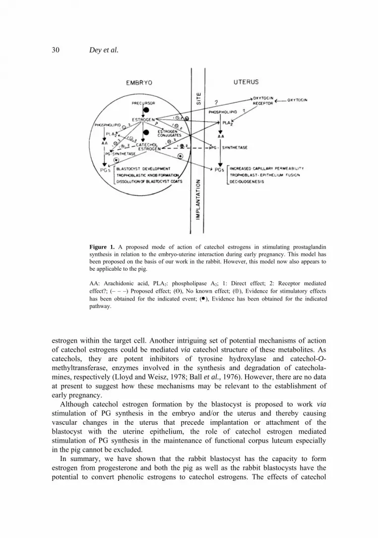
The idea that catechol estrogens formed by the embryo act at or close to their site of synthesis, is compatible with the recent view regarding function of this class of estrogen metabolites (Merriam and Lipsett, 1983). The rapid clearance of catechol estrogens from the circulation makes it unlikely that they function as circulating hormones (Merriam et al., 1980). It is more likely that catechol estrogens serve as a local or paracrine function acting on the cells in which they are formed or on those in their close proximity. Estrogen, the substrate for estrogen-2-/4-hydroxylase, should be available locally since aromatase activity has been identified in the preimplantation blastocysts of pig and rabbit. Moreover, both aromatase and estrogen-2/4-hydroxylase are con- centrated in the same extra-embryonic portion of the pig blastocyst. These various findings lead us to propose that the stimulation of PG synthesis responsible for the vascular changes in the uterus and attributed to phenolic estrogens, may in fact be mediated by their catechol metabolites (figure 1).
Stimulation of PG synthesis is only one of several mechanisms through which catechol estrogen may mediate their function. Like the phenolic estrogens, catechol estrogens can act through the receptor (Hersey et al., 1982). The addition of a second hydroxyl group to the Α-ring of estrogens, however, reduces the affinity of these steroids for the cytosolic estrogen receptor (Martucci and Fishman, 1976). Therefore, catechol estrogen formation could serve as a means to reduce the potency of the parent
30 Dey et al.
Figure 1. A proposed mode of action of catechol estrogens in stimulating prostaglandin synthesis in relation to the embryo-uterine interaction during early pregnancy. This model has been proposed on the basis of our work in the rabbit. However, this model now also appears to be applicable to the pig. AA: Arachidonic acid, PLA2: phospholipase A2; 1: Direct effect; 2: Receptor mediated effect?; (– – –) Proposed effect; (Ө), No known effect; (⊕), Evidence for stimulatory effects has been obtained for the indicated event; (•), Evidence has been obtained for the indicatedpathway.
estrogen within the target cell. Another intriguing set of potential mechanisms of action of catechol estrogens could be mediated via catechol structure of these metabolites. As catechols, they are potent inhibitors of tyrosine hydroxylase and catechol-O- methyltransferase, enzymes involved in the synthesis and degradation of catechola- mines, respectively (Lloyd and Weisz, 1978; Ball et al., 1976). However, there are no data at present to suggest how these mechanisms may be relevant to the establishment of early pregnancy.
Although catechol estrogen formation by the blastocyst is proposed to work via stimulation of PG synthesis in the embryo and/or the uterus and thereby causing vascular changes in the uterus that precede implantation or attachment of the blastocyst with the uterine epithelium, the role of catechol estrogen mediated stimulation of PG synthesis in the maintenance of functional corpus luteum especially in the pig cannot be excluded.
In summary, we have shown that the rabbit blastocyst has the capacity to form estrogen from progesterone and both the pig as well as the rabbit blastocysts have the potential to convert phenolic estrogens to catechol estrogens. The effects of catechol
Physiological aspects of blastocyst uterine interaction 31 estrogens are likely to include modulation of PG synthesis but further work will be required to establish this hypothesis.
Acknowledgements We thank Renee Becka and Debra Coco for their expert technical assistance. This research was supported in part by NICHHD grants (HD-12122 and HD-09734). P.L.P. is a Rockefeller Foundation Postdoctoral Fellow. Published as contribution No. 84- 178-J of the Kansas Agricultural Experiment Station. References Ball, P., Knuppen, R., Haupt, Μ. and Breuer, Η. (1976) J. Clin. Endocrinol., 34, 736. Castracane, V. D. and Jordon, V. D. (1976) Prostaglandins, 12, 243.Chasalow, F. I., Marr, Η. and Taylor, G. (1982) Steroids, 39, 617. Corner, G. W. (1947) in The Hormones in Human Reproduction , ed. G. W. Corner (New Jersey: Princeton
University Press, Princeton), p. 259. Crombi, P. R. (1972) The Morphology and Ultrastructure of the Pig's Placenta Throughout Pregnancy, Ph. D.
Thesis, University of Cambridge, UK. Davis, D. L. and Day, B. N. (1978) J. Anim. Sci. 46, 1043. Davis, D. L., Pakrasi, P. L. and Dey, S. K. (1983) Biol. Reprod., 28, 1114. Dhindsa, D. S, Dzuik, P. L. and Norton, Η. W. (1967) Anat. Rec., 159, 325. Ford, S. P. and Christenson, R. K. (1979) Biol. Reprod., 21, 617. George, F. W. and Wilson, J. D. (1978) Science, 199, 200. Grotjan, H. E., Jr. and Johnson, D. C. (1974) Proc. Soc. Exp. Biol. Med., 145, 610. Ham, Ε. Α., Cirillo, V. J., Janetti, Μ. Ε. and Kuehl, F. Α., Jr. (1975) Proc. Natl. Acad. Sci., USA, 72, 1420. Heap, R. Β., Flint, Α. Ρ. F. and Gadsby,J. E. (1981) in Cellular and Molecular Aspects of Implantation, eds S. R.
Glasser and D. W. Bullock (New York: Plenum Press) p. 311. Heuser, C. H. and Streeter, G. L. (1929) Contr. Embryol. Carnegie Inst., 20, 1. Hoversland, R. C, Dey, S. K. and Johnson, D. C. (1982) J. Reprod. Fert., 66, 259. Hersey, R. M., Williams, Κ. Ι. Η. and Weisz, J. (1981) Endocrinology, 35, 374. Huff, R. L. and Eik-Nes, K. B. (1966) J. Reprod. Fert., 11, 57. Johnson, D. C. (1979) J. Reprod. Fert., 56, 263. Johnson, D. C. and Griswold, T. (1984) J. Steroid Biochem., 20, 733. Kelly, R. W. and Abel, M. H. (1980) Prostaglandins, 20, 613. Kelly, R. W. and Abel, M. H. (1981) J. Steroid Biochem., 14, 787. Kremers, P. (1976) Eur. J. Biochem., 61, 481. Lloyd, Τ. and Weisz, J. (1978) J. Biol. Chem., 253, 4841. Martucci, C. and Fishman, J. (1976) Steroids, 27, 325. Merriam, G. R., Brandon, D. D., Kono, S., Davis, S. E., Loriaux, D. L. and Lipsett, M. B. (1980) J. Clin.
Endocrinol. Metab., 51, 1211. Merriam, G. R. and Lipsett, Μ. Β. (1983) in Catechol Estrogens, eds. G. R. Merriam and M. B. Lipsett (New
York: Raven Press), p. 7. Mukherjee, Α., Dey, S. K., Sen Gupta, J., Ramadoss, C. S. and Dickmann, Ζ. (1978) J. Reprod. Fert., 53,77. Pakrasi, P. L, Cheng, Η. C. and Dey, S. K. (983). Prostaglandins, 26, 991. Pakrasi, P. L. and Dey, S. K. (1982) Prostaglandins, 24, 73. Pakrasi, P. L. and Dey, S. K. (1983) Biol. Reprod., 29, 347. Perry, J. S., Heap, Ε. C. and Amoroso, Ε. C. (1973) Nature (London), 245, 45. Tsai-Morris, C-H and Johnson, D. C. (1982) J. Steroid Biochem., 17, 407. Weisz, J., Brown, B. L. and Ward, I. (1982) Neuroendocrinology, 109, 1912. Wu, J. Τ. and Lin, G. M. (1982) J. Reprod. Fert., 66, 655.