photo-inhibition of aβ fibrillation mediated by a newly designed fluorinated oxadiazole
TRANSCRIPT
RSC Advances
PAPER
Photo-inhibition
aInstitute of Biophysics, National Research CbDepartment of Biological Chemical and Ph
University of Palermo, Palermo, ItalycEuro-Mediterranean Institute of Science andDepartment of Neurology, University of TexeDepartment of Life and Environmental Scien
for the Physical Sciences of Matter, MarchefInstitute of Biomedicine and Molecular Im
Palermo, Italy
† Electronic supplementary informa10.1039/c4ra13556c
‡ The two authors have contributed equa
Cite this: RSC Adv., 2015, 5, 16540
Received 31st October 2014Accepted 27th January 2015
DOI: 10.1039/c4ra13556c
www.rsc.org/advances
16540 | RSC Adv., 2015, 5, 16540–1654
of Ab fibrillation mediated by anewly designed fluorinated oxadiazole†
M. R. Mangione,‡*a A. Palumbo Piccionello,‡abc C. Marino,ad M. G. Ortore,e P. Picone,f
S. Vilasi,a M. Di Carlo,f S. Buscemi,ab D. Bulonea and P. L. San Biagioa
Uncontrolled aggregation of amyloid beta peptide (Ab) is the main cause of Alzheimer's disease.
Therapeutic approaches to intervention in amyloid diseases include the use of small molecules able to
stabilize the soluble Ab conformation, or to redirect the amyloidogenic pathway towards non-toxic and
non-fibrillar states. Fluorometric measurements revealed that 3-(40-trifluoromethylphenyl)-5-(40-methoxyphenyl)-1,2,4-oxadiazole, when irradiated, is able to interact with the monomeric Ab peptide
readdressing the aggregation pathway toward the formation of amorphous aggregates as evidenced by
CD, AFM, and SAXS measurements. We hypothesize that this compound, under radiation, forms a
reactive intermediate that sticks on the Ab peptide by interfering with its fibrillation process. Cytotoxicity
assays performed on LAN5 neuroblastoma cells suggest that the presence of oxadiazole reduces the
toxicity of Ab. This finding might be the start of innovative therapies against Alzheimer's disease.
Introduction
Alzheimer's Disease (AD) is a neurodegenerative pathologyrepresenting the most common form of dementia in the elderly.The affected individuals present progressive loss of memory,mood changes, communication and reasoning problems.1–3 Thepathology is characterized by the formation of extracellularamyloid plaques and intracellular neurobrillary tangles in thebrain.4 All these deposits are composed by well-ordered, b-sheetrich, bers of the Amyloid b peptides (Ab) produced by prote-olysis of Amyloid Precursor Protein (APP) in residues of 39–43amino acids. The evidence of the simultaneous presence ofaccumulated bers and the cerebral tissue loss brought to theinitial hypothesis that the proteinaceous aggregates areresponsible for the neurodegenerative damage.5–7 However, ithas been shown that the species formed in the initial state ofthe aggregation kinetics could determine severe damage in thecerebral cells.8–10 Much experimental evidence, in fact, high-lighted the role of these species in the alteration of membranes,
ouncil, Palermo, Italy
armaceutical Sciences and Technologies,
d Technology, Palermo, Italy
as Medical Branch, Galveston, TX, USA
ces, National Interuniversity Consortium
Polytechnic University, Ancona, Italy
munology, National Research Council,
tion (ESI) available. See DOI:
lly to the present work.
8
particularly of their solubility and permeability.11,12 Moreover,the pathological presynaptic alteration was observed in trans-genic mice brain before the neuronal degeneration associatedwith bers accumulation. These observations suggest that theseverity of the pathology is uncorrelated with the concentrationof the mature bers.13–17 Despite much effort and the numerousstudies conducted, the mechanism and the pathogenesis of ADare still a matter of debate. The problem is also compounded bythe instability of the peptide both in the monomeric and earlyaggregated forms and by the presence of numerous poly-morphic aggregates and bers. In fact, Ab aggregation is acomplex process that seems to involve more than a simpleconversion from an unstable monomer to mature brils.18,19
Some therapeutic approaches trigger the disaggregation ofthe amyloid deposits and aggregates. Graphene-basedmaterialsor gold nanoparticles, with high affinity for deposits of Abbers, were used to redissolve them, by using the local heat.20–22
Over the last years, increasing interest has been focused onthe study of natural or synthetic molecules capable of inter-fering with the aggregation and brillation of Ab peptide.23–25
The search of small inhibitors of the toxic effects of Ab hasproduced a wide number of approaches and promisingdrugs.26–32
Particularly, growing interest has been addressed to mole-cules able to drive the unstable monomers towards the forma-tion of harmless and stable aggregates.33 Indeed, the deviationof the aggregation mechanism toward off-pathway productsseems to be a new promising strategy for the treatment ofamyloid diseases.
For example, the inhibitor effects of curcumin on the bril-lation have been investigated by different authors.25,27,34,35
This journal is © The Royal Society of Chemistry 2015
Paper RSC Advances
Curcumin is a natural molecule characterized by anti-inammatory and antioxidant activity. However, it crosses theblood–brain barrier (BBB) only when injected, and its effec-tiveness is reduced when added to the diet.36,37 These limita-tions have prompted the synthesis of new derivatives having amajor efficacy on the inhibition of aggregation.38,39
The ferulic acid is another example of natural molecule within vitro inhibitory activity. In fact, this molecule seems tointerfere with the oligomer formation through the destabiliza-tion of Asp23–Lys28 salt bridge that is at “the basis of brilstability”.40,41
All these compounds share a common phenolic structurethat seems to be responsible for the bril destabilization.
A new branch of research in this eld aims to the control ofthe amyloid aggregation through photochemical methods. Forinstance, it has been synthesized a “photo-clickable” Ab1–42peptide with increased stability and solubility, whose primarystructure is earned aer photo-stimulation.39 Very recently, thephotochemical approach also allowed to control the peptideaggregation by means of riboavin-mediated photooxygenationof Ab1–42, resulting into a decrease of Abeta toxicity and areduction of the uncontrolled self-assembly.42,43
The photo-stimulation is also used to trap, detect and char-acterize lowmolecular weight oligomers of Ab-peptide using thePICUP technique (photo-induced protein cross-linking ofunmodied proteins). This intriguing photo-chemical methodseems to be well appropriate for structural studies of the tran-sient and dynamic equilibrium between small oligomericspecies of Ab.44 A characteristic of this technique is the shortirradiation time required (few seconds) because of the involvedmetal complexes characterized by high photo-reactivity.However the short time of irradiation can make somehowdifficult to redirect accurately the aggregation pattern towardthe formation of small oligomers.44 Moreover, the toxicity ofinvolved metal complexes avoids the perspective of pharma-ceutical development.
In this context, we decided to design a new not toxic photo-active molecule able to interact with Ab in order to modulate itsaggregation pattern by means of photo-stimulation.
Recent studies indicated 1,2,4-oxadiazoles as promisingprobes for detection of amyloid plaques in vivo due to their highaffinity toward Ab.45 These interesting heterocycles, utilized inmany pharmaceutical applications,46–49 present a well knownphotochemical reactivity50,51 characterized by the formation ofintermediates able to induce by photostimulation an electrontransfer (PET) with amines or an energy transfer (ET) witharomatic species.52–54 In this study we designed, synthesized,characterized and tested a new uorinated oxadiazoliccompound which interfere with the Ab self-assembly. Theactivation of the oxadiazolic compound at an appropriatewavelength is responsible for the formation of photo-activespecies exerting an inhibitory effect on Ab brillation. Ourexperimental investigations were achieved bymeans of differenttechniques that are uorescence, circular dichroism (CD),atomic force microscopy (AFM), and small angle X-ray scat-tering (SAXS). We aim to evidence with different structuralresolutions the effects of the compound on Ab aggregation
This journal is © The Royal Society of Chemistry 2015
pathway. Finally, preliminary investigation relative to thetoxicity of the oxadiazolic compound were also examined by invitro cellular experiments.
Materials and methodsSynthesis of 3-(40-triuoromethylphenyl)-5-(40-methoxyphenyl)-1,2,4-oxadiazole
Pyridine (1.1 eq.) and 4-methoxybenzoyl chloride 2 (1.1 eq.) wereadded to a suspension of 4-triuoromethylphenylamidoxime 1(4.9 mmol, 1.0 g) in toluene (100 mL). The reaction mixture wasreuxed for 8 hours. The solvent was removed under reducedpressure and the residue was chromatographed givingcompound 3 (84%).
3-(40-Triuoromethylphenyl)-5-(40-methoxyphenyl)-1,2,4-oxadiazole 3. Mp 145–147 �C. FT-IR (nujol) v: 1613 cm�1.1HNMR (300 MHz, CDCl3) d: 3.92 (s, 3H, OCH3), 7.78 (d, J¼ 6.9 Hz,2H, Ar), 7.83 (d, J ¼ 8.1 Hz, 2H, Ar), 8.17 (d, 2H, J ¼ 6.9 Hz, Ar),8.26 (d, 2H, J ¼ 8.1 Hz, Ar). GC-MS (m/z): 320 (M+, 100%).Elemental analysis for C16H11F3N2O2 theor: C, 60.00; H, 3.46; N,8.75. Exper.: C, 60.03; H, 3.48; N, 8.78.
Sample preparation. The oxadiazole was solubilized inethanol at 1 mM concentration and then diluted to 10 mM in 10mM phosphate buffer (PB) solution (NaH2PO4/Na2HPO4-RIE-DEL DE HAEN in water MilliQ) at pH 7.4.
The lyophilized synthetic peptide Ab1–40 (Polypeptide) wassolubilized with NaOH 5 mM (Sigma-Aldrich), pH 10, sonicatedand already lyophilized according to Fezoui et al. treatment.55
Aer this procedure the peptide was dissolved in PB 10 mM andthen ltered with two lters in series having diameter of 0.20mm (Whatman) and 0.02 mm (Millex-Lg) respectively, in order toeliminate whatever aggregates. The sample preparation wasoperated in a cool room at 4 �C. Ab concentration (50 mM for allsamples) was determined by tyrosine absorption at 276 nmusing an extinction coefficient of 1390 cm�1 M�1.
The aggregation kinetics were followed at controlledtemperature (37 �C) and under stirring (200 rpm) for 24 hours.Samples prepared for AFM images were aged at 37 �C for 4 daysbefore the deposition on the mica surface. The same agedsamples were concentrated at 150 mM using Millipore lterswith a cut-off of 3 kDa before X-ray scattering measurements, inorder to obtain the necessary scattering intensity. Samples forcytotoxicity assays were collected at 0, 5 and 24 hours of kineticexperiments in order to select aggregates at different states. Allwithdrawals were performed using a laminar ow hood to keepsterility.
Spectrouorometric measurements. Emission spectra wererecorded using a spectrouorometer Jasco FP-6500. The oxadi-azole emission spectra was recorded in ethanol and in PB–EtOH99 : 1 using a quartz cell with 10 mm path length. The wave-length of excitation was 260 nm and the emission range was250–500 nm.
Circular dichroism spectroscopy. The secondary structure ofthe Ab1–40 was studied by using a JASCO J-815 CD Spectrometer.Particularly, withdrawals of samples at appropriate time duringthe aggregation kinetic were observed. Spectra were recorded at20 �C using a quartz cell with 0.2 mm path length. Each
RSC Adv., 2015, 5, 16540–16548 | 16541

Fig. 1 Synthesis of 1,2,4-oxadiazole 3.
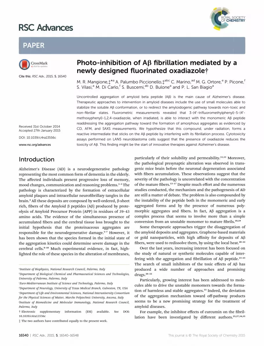
Fig. 2 Emission spectra at different kinetic times of compound 3 (10mM) in: (A) ethanol with continue photo-excitation; (B) in PB–ethanol(99 : 1) with continue photo-excitation; (C) in PB–ethanol (99 : 1)without continue photo-excitation.
RSC Advances Paper
spectrum measurement was obtained by averaging over eightscans and subtracting the blank solvent contribution.
AFM analysis. Aliquots of 50 mM representative samples atthe end of the aggregation kinetics were deposited onto freshlycleaved mica surfaces (Agar Scientic, Assing, Italy) and incu-bated for up to 2 minutes before rinsing with deionized waterand drying with gentle nitrogen ow. AFM experiments wereachieved with a Nanowizard II system (JPK Instruments, Ger-many), operating in tapping mode in air and at room temper-ature. Single beam silicon cantilevers (TESPA, NanoAndMore,USA) with a nominal spring constant of 42 Nm�1 and resonancefrequency of 320 kHz, were used. Images with scan sizes of 2� 2mm2 were acquired on different areas on each sample. Analysisof the aggregates size was performed by using the data pro-cessing soware provided by JPK Instruments.
Small angle X-ray scattering. SAXS experiments were per-formed at ID2 beamline at the European Synchrotron RadiationFacility in Grenoble, France. The sample temperature wasmaintained at 20 �C. SAXS patterns were recorded using a beroptically coupled two-dimension detector, FReLoN (Fast-Readout, Low-Noise). The distance from the sample and thedetector was set to 1.5 m, in order to obtain a Q-range (Q ¼4p sin q/l, where 2q is the scattering angle and l ¼ 0.995 A theX-ray wavelength) from 0.01 to 0.34 A�1. We recorded simulta-neously the incident and transmitted intensities to the purposeof obtaining data in an absolute scale, hence the normalizedSAXS patterns were azimuthally averaged to obtain the one-dimension proles of scattered intensity. The buffer contribu-tion was subtracted from protein solution data for each inves-tigated condition, considering the correction for the proteinvolume fraction. To prevent radiation damage, all solutionswere degassed before transferring into the capillary. Eachmeasurement was performed for 100 ms, and followed by adead time of 3 s in order to avoid radiation damage. Thisstrategy was repeated for 50 times in order to obtain a satis-factory signal-to-noise ratio, despite the low protein concen-tration which was investigated.
Cell cultures, treatment and cell viability determination.Cells were cultured in 96 well plates at 1.5� 106 cells per well inRPMI 1640medium (Celbio srl, Milan, Italy) supplemented with10% fetal bovine serum (Gibco-Invitrogen, Milan, Italy) and 1%penicillin, 1% streptomycin (50 mg mL). Cells were maintainedin a humidied 5% CO2 atmosphere at 37 �C. LAN5 cells weretreated for 72 h with samples of Ab without or with compound 3taken according to sample preparation. The nal concentra-tions were 6.25 mM for Ab and 1.25 mM for 3. Cell viability wasmeasured by MTS assay (Promega Italia, S.r.l., Milan, Italy).MTS [3-(4,5-dimethylthiazol-2-yl)-5-(3-carboxymethoxyphenyl)-2-(4-sulphophenyl)-2H-tetrazolium] was utilized according tothe manufacturer's instructions. Aer cell treatments, 20 mL ofthe MTS solution was added to each well, and the incubationwas continued at 37 �C for 4 h. The absorbance was read at 490nm on the Microplate reader WallacVictor 2 1420 MultilabelCounter (PerkinElmer, Inc. Monza, Italy). Results wereexpressed as the percentage MTS reduction in the control cells.The control and treated cells were morphologically analyzed bymicroscopy inspection using Axio scope 2 microscope (Zeiss).
16542 | RSC Adv., 2015, 5, 16540–16548
Results and discussionSynthesis and photochemical characterization of 1,2,4-oxadiazole 3
The compound 3-(40-triuoromethylphenyl)-5-(40-methoxyphenyl)-1,2,4-oxadiazole 3 was synthesized accordingly to the generalscheme of the amidoxime route, by reuxing the tri-uoromethylbenzamidoxime 1 with the appropriate benzoylchloride in the presence of pyridine (Fig. 1).
The photochemical features and photostability of thiscompound were studied by time course measurement of uo-rescence emission spectra in different solvents. The Fig. 2Ashows the kinetic evolution of the emission spectrum of 1,2,4-oxadiazole 3 diluted in ethanol. The emission spectra present aGaussian-like shape characterized by a time-dependent emis-sion quenching.
Otherwise the oxadiazole dissolved in a solution of PB–ethanol at 99 : 1 ratio, has an emission spectra (Fig. 2B) with aninitial intensity quench coupled with a change of the shape. Indetail, the emission signal, aer about 45 minutes, shows a nestructure with 3 characteristic bands: an hypsochromic bandaround 340–350 nm, an halfway band around 360 nm and abathochromic band near 380 nm.
To investigate if the well-known photochemical reactivity ofoxadiazoles50–54 is due to the photo-excitation, a sample wasincubated at 37 �C under stirring (200 rpm) in dark conditions.Interestingly, the emission spectra of sample registered atdifferent times starting from the dissolution in phosphatebuffer (Fig. 2C) display minor changes in intensity and shape.Overall, these results indicate that while the solvent is respon-sible for the emission maximum shi, the appearance of a nestructure in aqueous solution is a secondary phenomenon dueto photo-stimulation of the compound.
This journal is © The Royal Society of Chemistry 2015
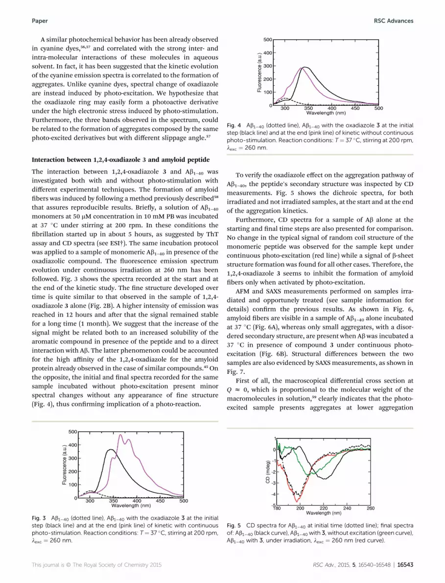
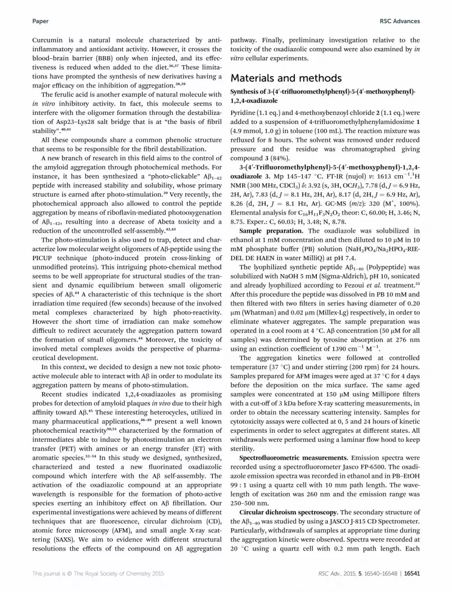
Fig. 4 Ab1–40 (dotted line), Ab1–40 with the oxadiazole 3 at the initialstep (black line) and at the end (pink line) of kinetic without continuousphoto-stimulation. Reaction conditions: T¼ 37 �C, stirring at 200 rpm,
Paper RSC Advances
A similar photochemical behavior has been already observedin cyanine dyes,56,57 and correlated with the strong inter- andintra-molecular interactions of these molecules in aqueoussolvent. In fact, it has been suggested that the kinetic evolutionof the cyanine emission spectra is correlated to the formation ofaggregates. Unlike cyanine dyes, spectral change of oxadiazoleare instead induced by photo-excitation. We hypothesize thatthe oxadiazole ring may easily form a photoactive derivativeunder the high electronic stress induced by photo-stimulation.Furthermore, the three bands observed in the spectrum, couldbe related to the formation of aggregates composed by the samephoto-excited derivatives but with different slippage angle.57
lexc ¼ 260 nm.
Interaction between 1,2,4-oxadiazole 3 and amyloid peptide
The interaction between 1,2,4-oxadiazole 3 and Ab1–40 wasinvestigated both with and without photo-stimulation withdifferent experimental techniques. The formation of amyloidbers was induced by following amethod previously described58
that assures reproducible results. Briey, a solution of Ab1–40monomers at 50 mM concentration in 10 mM PB was incubatedat 37 �C under stirring at 200 rpm. In these conditions thebrillation started up in about 5 hours, as suggested by ThTassay and CD spectra (see ESI†). The same incubation protocolwas applied to a sample of monomeric Ab1–40 in presence of theoxadiazolic compound. The uorescence emission spectrumevolution under continuous irradiation at 260 nm has beenfollowed. Fig. 3 shows the spectra recorded at the start and atthe end of the kinetic study. The ne structure developed overtime is quite similar to that observed in the sample of 1,2,4-oxadiazole 3 alone (Fig. 2B). A higher intensity of emission wasreached in 12 hours and aer that the signal remained stablefor a long time (1 month). We suggest that the increase of thesignal might be related both to an increased solubility of thearomatic compound in presence of the peptide and to a directinteraction with Ab. The latter phenomenon could be accountedfor the high affinity of the 1,2,4-oxadiazole for the amyloidprotein already observed in the case of similar compounds.45 Onthe opposite, the initial and nal spectra recorded for the samesample incubated without photo-excitation present minorspectral changes without any appearance of ne structure(Fig. 4), thus conrming implication of a photo-reaction.
Fig. 3 Ab1–40 (dotted line), Ab1–40 with the oxadiazole 3 at the initialstep (black line) and at the end (pink line) of kinetic with continuousphoto-stimulation. Reaction conditions: T¼ 37 �C, stirring at 200 rpm,lexc ¼ 260 nm.
This journal is © The Royal Society of Chemistry 2015
To verify the oxadiazole effect on the aggregation pathway ofAb1–40, the peptide's secondary structure was inspected by CDmeasurements. Fig. 5 shows the dichroic spectra, for bothirradiated and not irradiated samples, at the start and at the endof the aggregation kinetics.
Furthermore, CD spectra for a sample of Ab alone at thestarting and nal time steps are also presented for comparison.No change in the typical signal of random coil structure of themonomeric peptide was observed for the sample kept undercontinuous photo-excitation (red line) while a signal of b-sheetstructure formation was found for all other cases. Therefore, the1,2,4-oxadiazole 3 seems to inhibit the formation of amyloidbers only when activated by photo-excitation.
AFM and SAXS measurements performed on samples irra-diated and opportunely treated (see sample information fordetails) conrm the previous results. As shown in Fig. 6,amyloid bers are visible in a sample of Ab1–40 alone incubatedat 37 �C (Fig. 6A), whereas only small aggregates, with a disor-dered secondary structure, are present when Abwas incubated a37 �C in presence of compound 3 under continuous photo-excitation (Fig. 6B). Structural differences between the twosamples are also evidenced by SAXSmeasurements, as shown inFig. 7.
First of all, the macroscopical differential cross section atQ z 0, which is proportional to the molecular weight of themacromolecules in solution,59 clearly indicates that the photo-excited sample presents aggregates at lower aggregation
Fig. 5 CD spectra for Ab1–40 at initial time (dotted line); final spectraof: Ab1–40 (black curve), Ab1–40 with 3, without excitation (green curve),Ab1–40 with 3, under irradiation, lexc ¼ 260 nm (red curve).
RSC Adv., 2015, 5, 16540–16548 | 16543
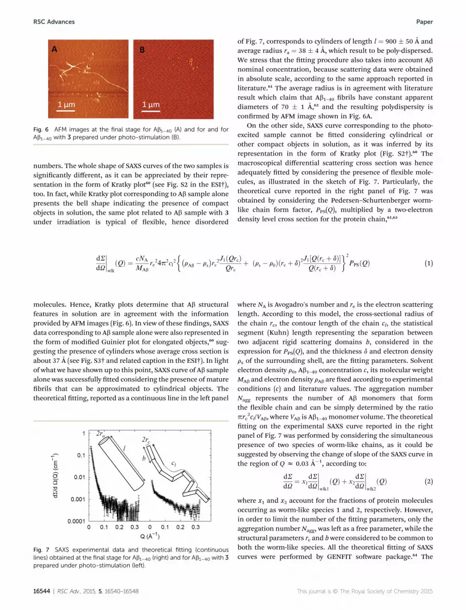
Fig. 6 AFM images at the final stage for Ab1–40 (A) and for and forAb1–40 with 3 prepared under photo-stimulation (B).
RSC Advances Paper
numbers. The whole shape of SAXS curves of the two samples issignicantly different, as it can be appreciated by their repre-sentation in the form of Kratky plot60 (see Fig. S2 in the ESI†),too. In fact, while Kratky plot corresponding to Ab sample alonepresents the bell shape indicating the presence of compactobjects in solution, the same plot related to Ab sample with 3under irradiation is typical of exible, hence disordered
dS
dU
����wlk
ðQÞ ¼ cNA
MAb
re24p2cl
2
��rAb � rs
�rc
2J1ðQrcÞQrc
þ ðrs � r0Þðrc þ dÞ2J1½Qðrc þ dÞ�Qðrc þ dÞ
�2
PPSðQÞ (1)
molecules. Hence, Kratky plots determine that Ab structuralfeatures in solution are in agreement with the informationprovided by AFM images (Fig. 6). In view of these ndings, SAXSdata corresponding to Ab sample alone were also represented inthe form of modied Guinier plot for elongated objects,60 sug-gesting the presence of cylinders whose average cross section isabout 37 A (see Fig. S3† and related caption in the ESI†). In lightof what we have shown up to this point, SAXS curve of Ab samplealone was successfully tted considering the presence of maturebrils that can be approximated to cylindrical objects. Thetheoretical tting, reported as a continuous line in the le panel
Fig. 7 SAXS experimental data and theoretical fitting (continuouslines) obtained at the final stage for Ab1–40 (right) and for Ab1–40 with 3prepared under photo-stimulation (left).
16544 | RSC Adv., 2015, 5, 16540–16548
of Fig. 7, corresponds to cylinders of length l ¼ 900 � 50 A andaverage radius ra ¼ 38 � 4 A, which result to be poly-dispersed.We stress that the tting procedure also takes into account Abnominal concentration, because scattering data were obtainedin absolute scale, according to the same approach reported inliterature.61 The average radius is in agreement with literatureresult which claim that Ab1–40 brils have constant apparentdiameters of 70 � 1 A,62 and the resulting polydispersity isconrmed by AFM image shown in Fig. 6A.
On the other side, SAXS curve corresponding to the photo-excited sample cannot be tted considering cylindrical orother compact objects in solution, as it was inferred by itsrepresentation in the form of Kratky plot (Fig. S2†).60 Themacroscopical differential scattering cross section was henceadequately tted by considering the presence of exible mole-cules, as illustrated in the sketch of Fig. 7. Particularly, thetheoretical curve reported in the right panel of Fig. 7 wasobtained by considering the Pedersen–Schurtenberger worm-like chain form factor, PPS(Q), multiplied by a two-electrondensity level cross section for the protein chain,61,63
where NA is Avogadro's number and re is the electron scatteringlength. According to this model, the cross-sectional radius ofthe chain rc, the contour length of the chain cl, the statisticalsegment (Kuhn) length representing the separation betweentwo adjacent rigid scattering domains b, considered in theexpression for PPS(Q), and the thickness d and electron densityrs of the surrounding shell, are the tting parameters. Solventelectron density r0, Ab1–40 concentration c, its molecular weightMAb and electron density rAb are xed according to experimentalconditions (c) and literature values. The aggregation numberNagg represents the number of Ab monomers that formthe exible chain and can be simply determined by the ratioprc
2cl/VAb, where VAb is Ab1–40 monomer volume. The theoreticaltting on the experimental SAXS curve reported in the rightpanel of Fig. 7 was performed by considering the simultaneouspresence of two species of worm-like chains, as it could besuggested by observing the change of slope of the SAXS curve inthe region of Q z 0.03 A�1, according to:
dS
dU¼ x1
dS
dU
����wlk1
ðQÞ þ x2
dS
dU
����wlk2
ðQÞ (2)
where x1 and x2 account for the fractions of protein moleculesoccurring as worm-like species 1 and 2, respectively. However,in order to limit the number of the tting parameters, only theaggregation number Nagg, was le as a free parameter, while thestructural parameters rc and bwere considered to be common toboth the worm-like species. All the theoretical tting of SAXScurves were performed by GENFIT soware package.64 The
This journal is © The Royal Society of Chemistry 2015
Table 1 Fitting parameters of the SAXS curve corresponding to the photoexcited sample, reported in Fig. 7, right panel
Aggregation number Nagg Chain radius rc (A) Kuhn length b (A) Weight fraction x (%)
Worm-like 1 2.0 � 0.4 4.3 � 0.3 10 � 2 30 � 5Worm-like 2 11.2 � 0.5 70 � 5
Paper RSC Advances
resulting parameters are reported in Table 1, including theweight fraction of each worm-like population.
The aggregation numbers and the low value of the crosssection radius conrm that the photo-excited sample does notpresent big aggregates, as evidenced by AFM images. Moreover,the shortness of the statistical segment length is consistent withthe lack of secondary structure revealed by CD results.
The mechanism of inhibition of the oxadiazolic compoundon Ab brillogenesis could be traced to its photo-reactivity. Wehypothesize that the photo-stimulation induces the formationof a reactive intermediate. The latter may react with the Ab1–40amino acid backbone through an electron and/or energytransfer mechanism and induce a structural modicationresponsible for brillogenesis inhibition, as pictorially repre-sented in Fig. 8.
We suggest a mechanism analogous to the PICUP methodused for stabilizing metastable amyloid oligomers previouslydescribed.44 More in details, the method is based on the photo-stimulation of ruthenium or palladium complexes which formradicals capable of extracting an electron from nearby amyloidpeptides. The formed reactive species of Ab may carry out analternative cross-linking with other neighboring Ab peptides.44
Particularly, we suggest that the electron-transfer from Tyrresidues could initially generate the coupling of two peptidechain, forming a cross-linked dimers.
We would like to stress that a PICUP-like mechanism isstrongly suggested by nding that the oxadiazolic compoundexerts an inhibitory effect only under photo-stimulation. Obvi-ously, further experiments will clarify the specic molecularmechanism behind the interaction between amyloid peptideand the oxadiazolic compound.
Fig. 8 Pictorial representation of photo-induced Ab1–40 modification.
This journal is © The Royal Society of Chemistry 2015
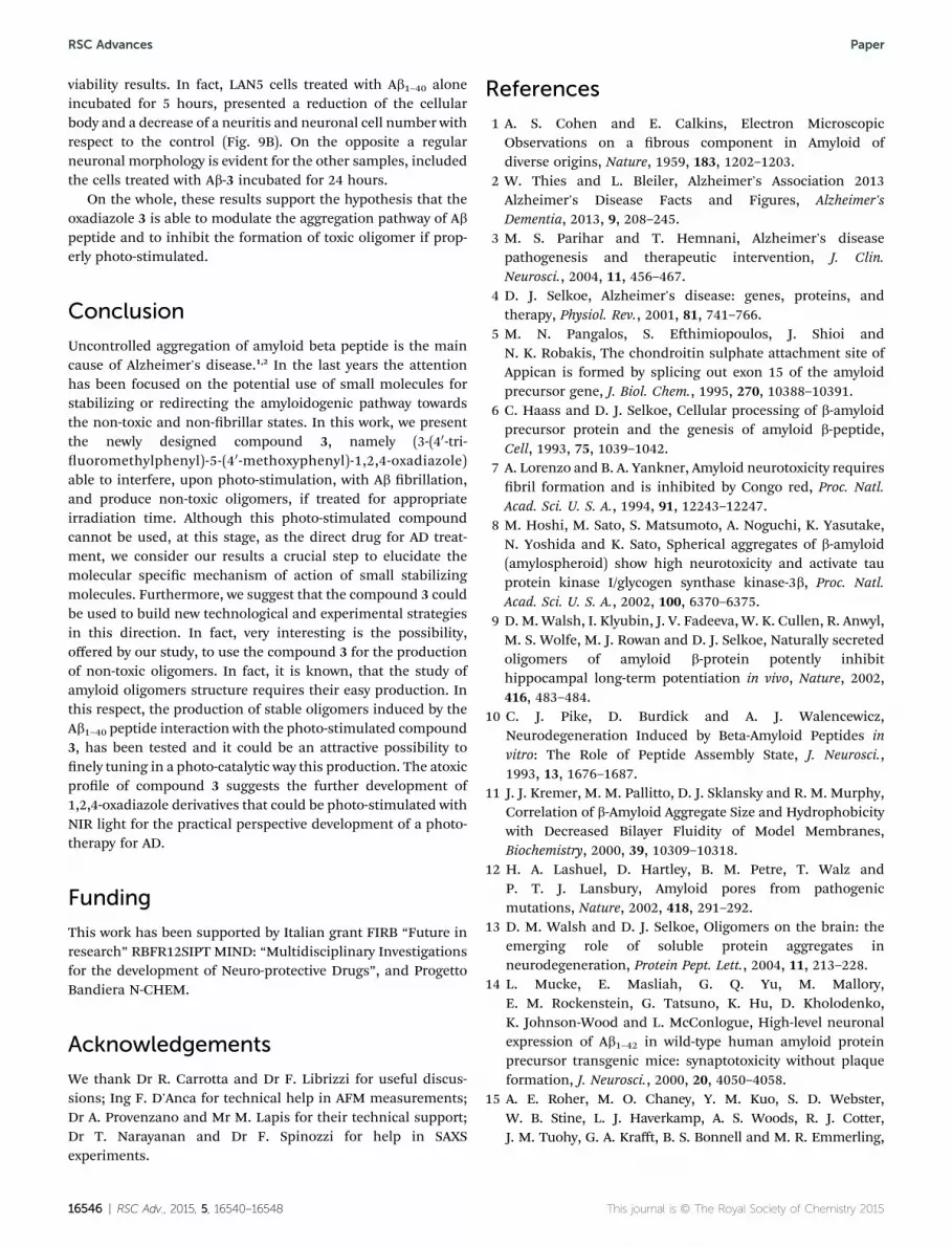
In vitro cytotoxicity assay
We further investigate the cytotoxicity of compound 3 and itspossible effect on the formation of neurotoxic Ab1–40 aggregates,MTS assay was realized on LAN5 neuroblastoma cell line.
LAN5 cells were treated using sample withdrawals taken at 0,5 and 24 hours of aggregation kinetic of Ab1–40 alone or inpresence of the oxadiazole 3, and their viability was estimated.As shown in Fig. 9A, the main toxicity was caused by the sampleof Ab1–40 alone incubated for 5 hours, which, accordingly to ourprevious studies58 should contain only oligomeric species. TheAb1–40 sample at the initial time and the bers obtained aer 24hours do not result as toxic as the 3 compound. To verify theeffect of oxadiazole 3 on the toxicity ofAb1–40 photo-stimulatedsamples for 5 and 24 hours in presence of the compound 3were dosed on LAN5 cells. No toxic effect was observed for cellstreated with samples incubated 5 hours. On the contrary, thesamples photo-stimulated for 24 hours cause a reduction on thecell viability if compared with Ab bers. However the speciesformed aer this irradiation time result less toxic if comparedwith Ab aggregates obtained aer 5 hours. This evidencesuggests a different structural organization obtained in pres-ence of photo-stimulated compound 3. Microscopic imagesacquired on the same samples, before MTS assay, showeddifferent cell morphology (Fig. 9B), consistently with cell
Fig. 9 (A) Viability for LAN5 cells after 72 h of incubation with sampleat different kinetic time of Ab1–40 (t ¼ 0, 5, and 24 h) and Ab1–40 withoxadiazole 3 (t¼ 5, and 24 h) compared with control and oxadiazole 3.(B) Microscopic images for the same samples.
RSC Adv., 2015, 5, 16540–16548 | 16545
RSC Advances Paper
viability results. In fact, LAN5 cells treated with Ab1–40 aloneincubated for 5 hours, presented a reduction of the cellularbody and a decrease of a neuritis and neuronal cell number withrespect to the control (Fig. 9B). On the opposite a regularneuronal morphology is evident for the other samples, includedthe cells treated with Ab-3 incubated for 24 hours.
On the whole, these results support the hypothesis that theoxadiazole 3 is able to modulate the aggregation pathway of Abpeptide and to inhibit the formation of toxic oligomer if prop-erly photo-stimulated.
Conclusion
Uncontrolled aggregation of amyloid beta peptide is the maincause of Alzheimer's disease.1,2 In the last years the attentionhas been focused on the potential use of small molecules forstabilizing or redirecting the amyloidogenic pathway towardsthe non-toxic and non-brillar states. In this work, we presentthe newly designed compound 3, namely (3-(40-tri-uoromethylphenyl)-5-(40-methoxyphenyl)-1,2,4-oxadiazole)able to interfere, upon photo-stimulation, with Ab brillation,and produce non-toxic oligomers, if treated for appropriateirradiation time. Although this photo-stimulated compoundcannot be used, at this stage, as the direct drug for AD treat-ment, we consider our results a crucial step to elucidate themolecular specic mechanism of action of small stabilizingmolecules. Furthermore, we suggest that the compound 3 couldbe used to build new technological and experimental strategiesin this direction. In fact, very interesting is the possibility,offered by our study, to use the compound 3 for the productionof non-toxic oligomers. In fact, it is known, that the study ofamyloid oligomers structure requires their easy production. Inthis respect, the production of stable oligomers induced by theAb1–40 peptide interaction with the photo-stimulated compound3, has been tested and it could be an attractive possibility tonely tuning in a photo-catalytic way this production. The atoxicprole of compound 3 suggests the further development of1,2,4-oxadiazole derivatives that could be photo-stimulated withNIR light for the practical perspective development of a photo-therapy for AD.
Funding
This work has been supported by Italian grant FIRB “Future inresearch” RBFR12SIPT MIND: “Multidisciplinary Investigationsfor the development of Neuro-protective Drugs”, and ProgettoBandiera N-CHEM.
Acknowledgements
We thank Dr R. Carrotta and Dr F. Librizzi for useful discus-sions; Ing F. D'Anca for technical help in AFM measurements;Dr A. Provenzano and Mr M. Lapis for their technical support;Dr T. Narayanan and Dr F. Spinozzi for help in SAXSexperiments.
16546 | RSC Adv., 2015, 5, 16540–16548
References
1 A. S. Cohen and E. Calkins, Electron MicroscopicObservations on a brous component in Amyloid ofdiverse origins, Nature, 1959, 183, 1202–1203.
2 W. Thies and L. Bleiler, Alzheimer's Association 2013Alzheimer's Disease Facts and Figures, Alzheimer'sDementia, 2013, 9, 208–245.
3 M. S. Parihar and T. Hemnani, Alzheimer's diseasepathogenesis and therapeutic intervention, J. Clin.Neurosci., 2004, 11, 456–467.
4 D. J. Selkoe, Alzheimer's disease: genes, proteins, andtherapy, Physiol. Rev., 2001, 81, 741–766.
5 M. N. Pangalos, S. Ehimiopoulos, J. Shioi andN. K. Robakis, The chondroitin sulphate attachment site ofAppican is formed by splicing out exon 15 of the amyloidprecursor gene, J. Biol. Chem., 1995, 270, 10388–10391.
6 C. Haass and D. J. Selkoe, Cellular processing of b-amyloidprecursor protein and the genesis of amyloid b-peptide,Cell, 1993, 75, 1039–1042.
7 A. Lorenzo and B. A. Yankner, Amyloid neurotoxicity requiresbril formation and is inhibited by Congo red, Proc. Natl.Acad. Sci. U. S. A., 1994, 91, 12243–12247.
8 M. Hoshi, M. Sato, S. Matsumoto, A. Noguchi, K. Yasutake,N. Yoshida and K. Sato, Spherical aggregates of b-amyloid(amylospheroid) show high neurotoxicity and activate tauprotein kinase I/glycogen synthase kinase-3b, Proc. Natl.Acad. Sci. U. S. A., 2002, 100, 6370–6375.
9 D. M. Walsh, I. Klyubin, J. V. Fadeeva, W. K. Cullen, R. Anwyl,M. S. Wolfe, M. J. Rowan and D. J. Selkoe, Naturally secretedoligomers of amyloid b-protein potently inhibithippocampal long-term potentiation in vivo, Nature, 2002,416, 483–484.
10 C. J. Pike, D. Burdick and A. J. Walencewicz,Neurodegeneration Induced by Beta-Amyloid Peptides invitro: The Role of Peptide Assembly State, J. Neurosci.,1993, 13, 1676–1687.
11 J. J. Kremer, M. M. Pallitto, D. J. Sklansky and R. M. Murphy,Correlation of b-Amyloid Aggregate Size and Hydrophobicitywith Decreased Bilayer Fluidity of Model Membranes,Biochemistry, 2000, 39, 10309–10318.
12 H. A. Lashuel, D. Hartley, B. M. Petre, T. Walz andP. T. J. Lansbury, Amyloid pores from pathogenicmutations, Nature, 2002, 418, 291–292.
13 D. M. Walsh and D. J. Selkoe, Oligomers on the brain: theemerging role of soluble protein aggregates inneurodegeneration, Protein Pept. Lett., 2004, 11, 213–228.
14 L. Mucke, E. Masliah, G. Q. Yu, M. Mallory,E. M. Rockenstein, G. Tatsuno, K. Hu, D. Kholodenko,K. Johnson-Wood and L. McConlogue, High-level neuronalexpression of Ab1–42 in wild-type human amyloid proteinprecursor transgenic mice: synaptotoxicity without plaqueformation, J. Neurosci., 2000, 20, 4050–4058.
15 A. E. Roher, M. O. Chaney, Y. M. Kuo, S. D. Webster,W. B. Stine, L. J. Haverkamp, A. S. Woods, R. J. Cotter,J. M. Tuohy, G. A. Kra, B. S. Bonnell and M. R. Emmerling,
This journal is © The Royal Society of Chemistry 2015
Paper RSC Advances
Morphology and toxicity of Ab-(1–42) dimer derived fromneuritic and vascular amyloid deposits of Alzheimer'sdisease, J. Biol. Chem., 1996, 271, 20631–20635.
16 M. P. Lambert, A. K. Barlow, B. A. Chromy, C. Edwards,R. Freed, M. Liosatos, T. E. Morgan, I. Rozovsky,B. Trommer, K. L. Viola, P. Wals, C. Zhang, C. E. Finch,G. A. Kra and W. L. Klein, Diffusible, nonfribrillar ligandsderived from Ab1–42 are potent central nervous systemneurotoxins, Proc. Natl. Acad. Sci. U. S. A., 1998, 95, 6448–6453.
17 A. Y. Hsia, E. Masliah, L. McConlogue, G. Q. Yu, G. Tatsuno,K. Hu, D. Kholodenko, R. C. Malenka, R. A. Nicoll andL. Mucke, Plaque-independent disruption of naturalcircuits in Alzheimer's disease mouse models, Proc. Natl.Acad. Sci. U. S. A., 1999, 96, 3228–3233.
18 M. Necula, S. Kayed, S. Milton and C. G. Glabe, SmallMolecule Inhibitors of Aggregation Indicate That Amyloidb Oligomerization and Fibrillization Pathways AreIndependent and Distinct, J. Biol. Chem., 2007, 282, 10311–10324.
19 A. Jan, D. M. Hartley and H. A. Lashuel, Preparation andcharacterization of toxic Ab aggregates for structural andfunctional studies in Alzheimer's disease research, Nat.Protoc., 2010, 5, 1186–1209.
20 M. Li, X. Yang, J. Ren, K. Qu and X. Qu, Using GrapheneOxide High Near-Infrared Absorbance for PhotothermalTreatment of Alzheimer's Disease, Adv. Mater., 2012, 24,1722–1728.
21 M. J. Kogan, N. G. Bastus, R. Amigo, D. Grillo-Bosch,E. Araya, A. Turiel A. Labarta, E. Giralt and V. F. Puntes,Nanoparticle-Mediated Local and Remote Manipulation ofProtein Aggregation, 2006, vol. 6, pp. 110–115.
22 E. Araya, I. Olmedo, N. G. Bastus, S. Guerrero, V. F. Puntes,E. Giralt and M. J. Kogan, Gold Nanoparticles andMicrowave Irradiation Inhibit Beta-AmyloidAmyloidogenesis, Nanoscale Res. Lett., 2008, 3, 435–443.
23 I. W. Hamley, The Amyloid Beta Peptide: A Chemist'sPerspective. Role in Alzheimers's and Fibrillation, Chem.Rev., 2012, 112, 5147–5192.
24 C. A. Hawkes, V. Ng and J. McLaurin, Small moleculeinhibitors of Ab-aggregation and neurotoxicity, Drug Dev.Res., 2009, 70, 111–124.
25 A. Sgarbossa, Natural Biomolecules and ProteinAggregation: Emerging Strategies against Amyloidogenesis,Int. J. Mol. Sci., 2012, 13, 17121–17137.
26 Y. Goodman, M. R. Steiner, S. M. Steiner and M. P. Mattson,Nordihydroguaiaretic acid protects hippocampal-neuronsagainst amyloid beta-peptide toxicity, and attenuates free-radical and calcium accumulation, Brain Res., 1994, 654,171–176.
27 M. Garcia-Alloza, L. A. Borrelli, A. Rozkalne, B. T. Hyman andB. J. Bacskai, Curcumin labels amyloid pathology in vivo,disrupts existing plaques, and partially restores distortedneurites in an Alzheimer mouse model, J. Neurochem.,2007, 102, 1095–1104.
28 M. Akagawa and K. Suyama, Amine oxidase-like activity ofpolyphenols – mechanism and properties, Eur. J. Biochem.,2001, 268, 1953–1963.
This journal is © The Royal Society of Chemistry 2015
29 E. Graf, Antioxidant potential of ferulic acid, Free RadicalBiol. Med., 1992, 13, 435–448.
30 H. S. Kim, J. Y. Cho, D. H. Kim, J. J. Yan, H. K. Lee, H. V. Suhand D. K. Song, Inhibitory effects of long-termadministration of ferulic acid on microglial activationinduced by intracerebroventricular injection of beta-amyloid peptide (1–42) in mice, Biol. Pharm. Bull., 2004,27, 120–121.
31 S. Butterels, M. Hejjaoui, B. Fauvet, L. Awad andH. A. Lashuel, Chemical Strategies for Controlling ProteinFolding and Elucidating the Molecular Mechanisms ofAmyloid Formation and Toxicity, J. Mol. Biol., 2012, 421,204–236.
32 D. M. Walsh, M. Townsend, M. B. Podlisny, G. M. Shankar,J. V. Fadeeva, O. El Agnaf, D. M. Hartley and D. J. Selkoe,Certain Inhibitors of Synthetic Amyloid b-Peptide (Ab)Fibrillogenesis Block Oligomerization of Natural Ab andThereby Rescue Long-Term Potentiation, J. Neurosci., 2005,25, 2455–2462.
33 V. N. Uversky, Mysterious oligomerization of theamyloidogenic proteins, FEBS J., 2010, 277, 2940–2953.
34 K. Ono, K. Hasegawa, H. Naiki and M. Yamada, Curcuminhas potent anti-amyloidogenic effects for Alzheimers beta-amyloid brils in vitro, J. Neurosci. Res., 2004, 75, 742–750.
35 A. N. Begum, M. R. Jones, G. P. Lim, T. Morihara, P. Kim,D. D. Heath, C. L. Rock, M. A. Pruitt, F. Yang, B. Hudspeth,S. Hu, K. F. Faull, B. Teter, G. M. Cole and S. A. Frautschy,Curcumin structure-function, bioavailability, and efficacy inmodels of neuroinammation and Alzheimer's disease, J.Pharmacol. Exp. Ther., 2008, 326, 196–208.
36 P. Anand, A. B. Kunnumakkara, R. A. Newman andB. B. Aggarwal, Bioavailability of curcumin: problems andpromises, Mol. Pharm., 2007, 4, 807–818.
37 C. S. Yang, S. Sang, J. D. Lambert and M. J. Lee,Bioavailability issues in studying the health effects of plantpolyphenolic compounds, Mol. Nutr. Food Res., 2008, 52,139–151.
38 R. Narlawar, M. Pickhardt, S. Leuchtenberger, K. Baumann,S. Krause, T. Dyrks, S. Weggen, E. Mandelkow andB. Schmidt, Curcumin-derived pyrazoles and isoxazoles:Swiss army knives or blunt tools for Alzheimer's disease?,ChemMedChem, 2008, 3, 165–172.
39 R. A. Orlando, A. M. Gonzales, R. E. Royer, L. M. Deck andD. L. Vander Jagt, A Chemical Analog of Curcumin as anImproved Inhibitor of Amyloid Abeta, PLoS One, 2012, 7,e31869, DOI: 31810.31371/journal.pone.0031869.
40 A. Sgarbossa, S. Monti, F. Lenci, E. Bramanti, R. Bizzarri andV. Barone, The effects of ferulic acid on b-amyloid brillarstructures investigated through experimental andcomputational techniques, Biochim. Biophys. Acta, 2013,1830, 2924–2937.
41 B. Tarus, J. E. Straub and D. Thirumalai, Dynamics of Asp23–Lys28 salt-bridge formation in A beta(10–35) monomers, J.Am. Chem. Soc., 2006, 128, 16159–16168.
42 A. Taniguchi, Y. Sohma, M. Kimura, T. Okada, K. Ikeda,Y. Hayashi, T. Kimura, S. Hirota, K. Matsuzaki and Y. Kiso,“Click Peptide” Based on the “O-Acyl Isopeptide Method”:
RSC Adv., 2015, 5, 16540–16548 | 16547
RSC Advances Paper
Control of Ab1–42 Production from a Photo-Triggered Ab1–42Analogue, J. Am. Chem. Soc., 2005, 128, 696–697.
43 A. Taniguchi, M. Skwarczynski, Y. Sohma, T. Okada,K. Ikeda, H. Prakash, H. Mukai, Y. Hayashi, T. Kimura,S. Hirota, K. Matsuzaki and Y. Kiso, Controlled Productionof Amyloid b Peptide from a Photo-Triggered, Water-Soluble Precursor “Click Peptide”, ChemBioChem, 2008, 9,3055–3065.
44 G. Bitan and D. B. Teplow, Rapid Photochemical CrossLinking – A New Tool for Studies of Metastable,Amyloidogenic Protein Assemblies, Acc. Chem. Res., 2004,37, 357–364.
45 M. Ono, M. Haratake, H. Saji and M. Nakayama,Development of novel b-amyloid probes based on 3,5-diphenyl-1,2,4-oxadiazole, Bioorg. Med. Chem., 2008, 16,6867–6872.
46 A. Terenzi, G. Barone, A. Palumbo Piccionello, G. Giorgi,A. Guarcello, P. Portanova, G. Calvaruso, S. Buscemi,N. Vivona and A. Pace, Synthesis, characterization, cellularuptake and interaction with native DNA of a bis(pyridyl)-1,2,4-oxadiazole copper(II) complex, Dalton Trans., 2010,9140–9145.
47 A. Palumbo Piccionello, R. Musumeci, C. Cocuzza,C. G. Fortuna, A. Guarcello, P. Pierro and A. Pace,Synthesis and preliminary antibacterial evaluation oflinezolid-like 1,2,4-oxadiazole derivatives, Eur. J. Med.Chem., 2012, 50, 441–448.
48 C. G. Fortuna, C. Bonaccorso, A. Bulbarelli, G. Caltabiano,L. Rizzi, L. Goracci, G. Musumarra, A. Pace, A. PalumboPiccionello, A. Guarcello, P. Pierro, C. E. A. Cocuzza andR. Musumeci, New linezolid-like 1,2,4-oxadiazoles activeagainst Gram-positive multiresistant pathogens, Eur. J.Med. Chem., 2013, 65, 533–545.
49 L. Lentini, R. Mel, A. Di Leonardo, A. Spinello, G. Barone,A. Pace, A. Palumbo Piccionello and I. Pibiri, Toward aRationale for the PTC124 (Ataluren) PromotedReadthrough of Premature Stop Codons: A ComputationalApproach and GFP-Reporter Cell-Based Assay, Mol. Pharm.,2014, 11, 653–664.
50 N. Vivona, S. Buscemi, I. Pibiri, A. Palumbo Piccionello andA. Pace, Synthesis of Heteroaromatics via RearrangementReactions, 2010.
51 S. Buscemi, N. Vivona and T. Caronna, A generalized andefficient synthesis of 3-amino-5-alkyl-1,2,4-oxadiazoles, 3-(n-alkylamino)-5-alkyl-1,2,4-oxadiazoles, 3-(n,n-dialkylamino)-5-alkyl-1,2,4-oxadiazoles by irradiation of 3-alkanoylamino-4-phenyl-1,2,5-oxadiazoles (furazans), Synthesis, 1995, 8, 917–919.
16548 | RSC Adv., 2015, 5, 16540–16548
52 S. Buscemi, A. Pace, A. Palumbo Piccionello, I. Pibiri andN. Vivona, Fluorinated heterocyclic compounds. Aphotochemical approach to a synthesis of uorinatedquinazolin-4-ones, Heterocycles, 2004, 63, 1619–1628.
53 S. Buscemi, A. Pace, A. Palumbo Piccionello, I. Pibiri andN. Vivona, Fluorinated heterocyclic compounds. Aphotochemical approach to a synthesis of polyuoroaryl-1,2,4-triazoles, Heterocycles, 65, 387–394.
54 S. Buscemi, A. Pace, N. Vivona, T. Caronna and A. Galia,Photoinduced single electron transfer on 5-aryl-1,2,4-oxadiazoles: Some mechanistic investigations in thesynthesis of quinazolin-4-ones, J. Org. Chem., 1999, 64,728–733.
55 Y. Fezoui, D. M. Hartley, J. D. Harper, R. Khurana,D. M. Walsh, M. M. Condron, D. J. Selkoe, P. T. Lansbury,A. L. Fink and D. B. Teplow, An improved method ofpreparing the amyloid beta-protein for brillogenesis andneurotoxicity experiments, Amyloid, 2000, 7, 166–178.
56 A. Marini, A. Munoz-Losa, A. Biancardi and B. Mennucci,What is Solvatochromism?, J. Phys. Chem. B, 2010, 114,17128–17135.
57 J. S. Kim, R. Kodagahally, L. Strekowski and G. Patonay, Astudy of intramolecular H-complexes of novelbis(heptamethine cyanine) dyes, Talanta, 2005, 67, 947–954.
58 C. Corsale, R. Carrotta, M. R. Mangione, S. Vilasi,A. Provenzano, G. Cavallaro, D. Bulone and P. L. SanBiagio, Entrapment of Ab1–40 peptide in unstructuredaggregates, J. Phys.: Condens. Matter, 2012, 24, 244103.
59 E. Mylonas and D. I. Svergun, Accuracy of molecular massdetermination of proteins in solution by small-angle X-rayscattering, J. Appl. Crystallogr., 2007, 40, 245–249.
60 I. Pilz, O. Glatter and O. Kratky, Small-angle X-ray scattering,Methods Enzymol., 1979, 61, 148–249.
61 M. G. Ortore, F. Spinozzi, S. Vilasi, I. Sirangelo, G. Irace,T. Narayanan, R. Sinibaldi and P. Mariani, Time resolvedsmall angle X scattering study of the early stage of amyloidformation of an apomyoglobin mutant, Phys. Rev. E, 2011,84, 061904.
62 J. X. Lu, W. Qiang, W. M. Yau, C. D. Schwieters,S. C. Meredith and R. Tycko, Molecular Structure of b-Amyloid Fibrils in Alzheimer's Disease Brain Tissue, Cell,2013, 154, 1257–1268.
63 J. S. Pedersen and P. Schurtenberger, Scattering Functions ofSemiexible Polymers with and without Excluded VolumeEffects, Macromolecules, 1996, 29, 7602–7612.
64 F. Spinozzi, C. Ferrero, M. G. Ortore, A. De Maria Antolinosand P. Mariani, GENFIT: soware for the analysis of small-angle X-ray and neutron scattering data of macromoleculesin solution, J. Appl. Crystallogr., 2014, 47, 1132–1139.
This journal is © The Royal Society of Chemistry 2015