phosphorylation of elf-4e and initiation of protein synthesis in p19 embryonal carcinoma cells
TRANSCRIPT

Journal of Cellular Biochemistry 59:443-452 (1 995)
Phosphorylation of elF-4E and Initiation of Protein Synthesis in P19 Embryonal Carcinoma Cells Miranda Kleijn, Harry 0. Voorma, and Adri A.M. Thomas
Department of Molecular Cell Biology, University of Utrecht, 3584 CH Utrecht, The Netherlands.
Abstract Mitogenic stimulation of protein synthesis is accompanied by an increase in elF-4E phosphorylation. The effect o n protein synthesis by induction of differentiation is less well known. We treated PI 9 embryonal carcinoma cells with the differentiating agent retinoic acid and found that protein synthesis increased during the first hour of addition. However, the phosphorylation state, as well as the turnover of phosphate on elF-4E, remained unchanged. Apparently, the change in protein synthesis after RA addition is regulated by another mechanism than elF-4E phosphorylation.
By using P I 9 cells overexpressing the EGF receptor, we show that the signal transduction pathway that leads to phosphorylation of elF-4E is present in P I 9 cells; the ECF-induced change in phosphorylation of elF-4E in these cells is likely to be regulated by a change in elF-4E phosphatase activity.
These results suggest that the onset of retinoic acid-induced differentiation is triggered by a signal transduction pathway which involves changes in protein synthesis, but not elF-4E phosphorylation.
Key words: protein synthesis, retinoic acid, ECF, NGF, differentiation, phosphatase, elF-4E
c 1995 WiIey-Liss, Inc
Eukaryotic translation initiation is regulated by phosphorylation of initiation factors. It has been shown that changes in the phosphoryla- tion state of four of these eukaryotic initiation factors (elF), elF-2, elF-2B, elF-4B, and elF-4E, coincide with changes in protein synthesis [Red- path and Proud, 19941. The first two proteins act to deliver Met-tRNA onto the 40s ribosomal subunit: elF-2 by complexing to Met-tRNA and GTP, and elF-2B by replacing GDP on elF-2 for GTP. Phosphorylation of the a-subunit of elF-2 impaired the GDP-GTP exchange by sequestra- tion of elF-2B [Konieczny and Safer, 1983; Sali- mans et al., 1984; Siekierka et al., 19821. In vitro experiments showed that phosphorylation of the €-subunit of elF-2B increased the GDP/GTP exchange activity, enabling a higher protein syn- thesis rate [Dholakia and Wahba, 1988; Singh et al., 1994; Welsh and Proud, 19921.
elF-4E binds the cap-structure on the mRNA [Sonenberg et al., 1978; Rhoads, 19881. Subse- quent unwinding of the secondary structure of the 5’untranslated region (UTR) [Rozen et al.,
Received April 12,1995; accepted May 10, 1995. Address reprint requests to Miranda Kleijn, Department of Molecular Cell Biology, University of Utrecht, Padualaan 8, 3584 CH Utrecht, The Netherlands.
i 1995 Wiley-Liss, Inc.
19901 enables binding of the 40s . elF-2 . Met- tRNA . GTP complex followed by formation of the 80s ribosomal complex.
elF-4A, together with elF-4B, is able to un- wind double-stranded RNA or RNA/DNA du- plexes [Lawson et al., 1989; Rozen et al., 19901. elF-4B can be multiply phosphorylated in re- sponse to mitogens [Duncan and Hershey, 1985; Wolthuis et al., 19931. A decrease in phosphory- lation of elF-4B is observed after serum starva- tion of cells. The role of phosphorylation of elF-4B on the protein synthesis rate remains to be determined.
Phosphorylation of the cap-binding protein elF-4E closely follows the protein synthetic rate. Phosphorylation of elF-4E is decreased after adenovirus infection [Huang and Schneider, 19911, heat shock [Lamphear and Panniers, 19911, and mitosis [Bonneau and Sonenberg, 19871. In all cases a decrease in protein synthe- sis was detected. An increase in elF-4E phosphor- ylation was shown after treatment of cells with mitogens, like phorbol esters [Frederickson et al., 1991; Morley and Traugh, 1989; Rychlik et al., 19901, and lipopolysaccharide (LPS) [Rych- lik et al., 19901, treatment with insulin [Man- zella et al., 1991; Morley and Traugh, 19901, serum [Kaspar et al., 19901, epidermal growth

444 Kleijn et al.
factor (EGF) [Donaldson et al., 19911, tumor necrosis factor (TNF) [Marino et al., 19911, plate- let-derived growth factor (PDGF) [Bu and Hage- dorn, 1991; Frederickson et al., 19911, nerve growth factor (NGF) [Frederickson et al., 19921, and overexpression of pp6OSrc [Frederickson et al., 19911 orp21'" [Rinker-Schaeffer et al., 19921. After treatment of cells with phorbol esters, serum, and after ~ 2 1 ' " ~ transformation an in- crease in protein synthesis was detected. Thus, the numerous examples indicate that mitogenic stimulation of cells leads to an increased phos- phorylation status of elF-4E and an increased protein synthesis.
Phosphorylation of elF-4E seems to be neces- sary for efficient translation initiation. First, 80 to 85% of elF-4E present on the 48s initiation complex was phosphorylated [Joshi-Barve et al., 1990; Lamphear and Panniers, 19901; in con- trast, only 50% of the free elF-4E was phosphory- lated. Second, phosphorylated elF-4E binds m7GTP, m7GpppG, and globin mRNA 3 to 4 times more efficiently than nonphosphorylated elF-4E [Minich et al., 19941.
An increase in elF-4E phosphorylation is be- lieved to enhance translation of mRNAs with an extensive secondary structure in their 5'UTR by an increase in the activity of elF-4E. The orni- thine decarboxylase G/C-rich 5'UTR was more efficiently translated when cells were stimulated with insulin or phorbol ester [Manzella et al., 19911. Furthermore, mRNAs in which second- ary structure was increased by cloning comple- mentary regions in the 5'UTR were poorly trans- lated in normal cells, but translation was relatively efficient in cells overexpressing elF-4E [Koromilas et al., 19921.
The amount of elF-4E present in the cell limits the initiation process [Duncan et al., 1987; Hiremath et al., 19851. It is expected that mRNAs with extensive folding are only translated under conditions when more phosphorylated elF-4E is present. Proteins such as oncogene products, growth factors, and growth factor receptors are often encoded by mRNAs with a complex second- ary structure in the 5'UTR. These proteins play a critical role in growth, differentiation, and development [Kozak, 1987; Lazaris-Karatzas et al., 19921.
Pheochromocytoma (PC12) cells can be differ- entiated with NGF. Treatment of PC12 cells with NGF led to a rapid increase in elF-4E phosphorylation, suggesting that elF-4E phos- phorylation plays a role in the change of gene
expression at the very early onset of differentia- tion [Frederickson et al., 19921. Inhibition o f ras-activity in PC12 cells by a dominant nega- tive mutant led to a decreased elF-4E phosphor- ylation upon NGF addition. It was also found that ras-inhibition led to a marked repression of the neurite outgrowth of PC12 cells after NGF treatment. These two findings suggest that elF-4E phosphorylation and ras-activation are necessary for differentiation of PC12 cells [Fre- derickson et al., 19921. The question can be raised whether phosphorylation of elF-4E i s obligatory for differentiation in general The amount of phosphorylated elF-4E may be able to regulate cell growth and differentiation: overex- pression of elF-4E in HeLa cells [de Benedetti and Rhoads, 19901, NIH 3T3, and rat 2 fibro- blasts [Lazaris-Karatzas et al., 19901 led to aber- rant growth of the cells. Stimulation of transla- tion initiation of mRNAs with a complex 5'UTR by phosphorylation of elF-4E suggests that elF-4E may be involved in the regulation of differentiation [Lazaris-Karatzas et al., 1992 I. To determine whether differentiation coincides with a change in elF-4E phosphorylation, P19 embryonal carcinoma cells were treated with the differentiation-inducing agent retinoic acid (RA). P19 cells are malignant stem cells of tera- tocarcinomas. Depending on the chemical agent (retinoic acid [RAI or dimethyl sulfoxide [DMSOI) and the culture conditions used [Jones- Villeneuve et al., 1982; McBurney et al., 1982 I, P19 cells can differentiate into cells represent- ing the three germ layers of the mouse embryo: endoderm, ectoderm, and mesoderm [Martin, 19801. In this respect, P19 cells are very similar to the pluripotent inner cell mass of early em- bryos. P19 cells which overexpress a human EGF receptor (EGF-R) were treated with EGF to determine whether the signal transduction pathway leading to elF-4E phosphorylation is present in undifferentiated P19 cells.
MATERIALS AND METHODS Materials
Acrylamidelbisacrylamide and biolytes were from Biorad, Veenendaal, the Netherlands; CHAPS from Boehringer, Almere, the Nether- lands; urea and G418 from Gibco/BRL, Breta, the Netherlands; all-trans retinoic acid and SS- NGF from Sigma, Brunschwigchemie, Amster- dam, the Netherlands; and murine EGF from Harlan, CPB, Zeist, the Netherlands.

Phosphorylation of elF-4E in P19 Cells 445
Cell Culture
P19 cells were cultured in a 1: l mixture of Ham F-12 and Dulbecco modified Eagle’s me- dium (DF) supplemented with 7.5% fetal calf serum. P19-8-39 cells were obtained by cotrans- fection of pSV2neo and pSV2HERc, an expres- sion vector for the human EGF-R [den Hertog et al., 1991al. P19-8-39 cells were cultured as P19 cells, but in the presence of G418 (200 pg/ml). P19-8-39 cells will be referred to as P19-EGF cells.
Treatment of Cells With Retinoic Acid or ECF and Measurement of the Protein Synthesis
P19 cells and P19-EGF cells were grown in 6-cm-diameter tissue culture dishes to a conflu- ency of 70%. The cells were deprived of serum in the presence of 10 kg transferrin per ml for the times indicated in the figure legends before treat- ment with RA M) or EGF (50 ng/ml). Cells were washed twice with phosphate-buffered sa- line (PBS) and harvested in sample buffer for one-dimensional (1D) iso-electric focusing, con- taining 9.5 M urea, 80 mM CHAPS (3-[(3- cholamidopropyl)dimethylammoniol- l-propane- sulfonate), 0.75% Biolytes 3/10, 2.25% Biolytes 4/6, and 700 mM P-mercaptoethanol. Before harvesting, the cells were labeled with 12.5 pCi of [35Slmethionine/cysteine (Amersham, Den Bosch, the Netherlands) per ml for the times indicated in the figure legends. Incorporation of [35S]methionine/cysteine into protein was mea- sured by trichloroacetic acid precipitation.
Detection of the Phosphorylation State of elF-4E by 1 D-IEF
Cell lysates were run on a denaturing 1D iso-electric focusing gel (6% acrylamide/bisacryl- amide (19:1), 9 M urea, 30 mM CHAPS, 0.75% Biolyte 3/10, 2.25% Biolyte 4/61 as described [Maurides et al., 19891. The system was modi- fied for a 7 x 8 cm-gel system. Electrophoresis time was 3.5 h. The gel was blotted onto a PVDF membrane using a semidry apparatus (Hoefer, San Francisco, CAI. elF-4E was detected by us- ing a purified polyclonal antibody raised against a elF-4E peptide (EPETTPTTNPPPAEEEKT) [Jaramillo et al., 19913. Antibodies were purified by binding to elF-4E-peptide coupled to Sepha- rose. Bound antibodies were eluted with 0.1 M NaHC03, pH 11.5, and immediately neutralized with 1 M HAc. The blot was developed with the alkaline phosphatase method and the percent-
age of phosphorylated elF-4E over the total amount of elF-4E was determined with a densi- tometer (Molecular Dynamics).
Treatment of PC12 Cells With NGF
PC12 cells were grown in DF medium contain- ing 7.5% fetal calf serum in 6-cm-diameter tis- sue culture dishes to a confluency of 50%. The cells were deprived of serum for 2 h, before treatment with NGF (30 ng/ml). Cell lysates were prepared, run on a 1D iso-electric focusing gel, and analyzed as described above.
Incorporation of [32Pl-Labeled Orthophosphate Into elF-4E
P19 cells and P19-EGF cells were grown to a confluency of 70%, and deprived of serum as described above. Cells were washed with phos- phate-free DF and incubated in the same me- dium containing 100 FCi of carrier-free [32P]- labeled orthophosphate (Amersham) per ml. Cells were labeled for the times indicated in Figure 3 up to 3 h, to determine the optimal labeling conditions for elF-4E. After washing with PBS, the cells were lysed with 400 p1 10 mM 4-(2-Hydroxyethyl)-l-piperazineethanesul- fonic acid (HEPES), pH 7.4, adjusted with KOH, 50 mM P-glycerophosphate, 0.2 mM EDTA, 0.5% Nonidet-P40, 100 mM KC1, 7 mM P-mercapto- ethanol, 0.2 mM Na3V04, 0.2 mM benzamidine, and 4 Fg leupeptin per ml. The lysates were centrifuged at 10,OOOg to remove cell debris and the supernatant was used to purify elF-4E. The supernant was incubated with 5 ~1 packed m7GTP Sepharose, supplemented with 15 pl packed Sepharose 4B to increase the pellet vol- ume. After 1 h the beads were washed 3 times with lysis buffer and elF-4E was eluted with 10 mM HEPES-KOH, pH 7.5, 0.2 mM EDTA, 100 mM KAc, and 75 pM m7GTP. The eluant was analyzed by sodium dodecyl sulphate (13.5%) polyacrylamide gelelectrophoresis (SDS-PAGE) and phosphorylation of elF-4E was quantified with a phospho-imager (Molecular Dynamics).
Turnover of Phosphorylated 4E Measured by [32P]orthophosphate Labeling
P19 and P19-EGF cells were grown and la- beled as described above. After 3 h of labeling, medium was replaced by DF with or without RA or EGF. Cells were harvested at the times indi- cated in the legend. elF-4E was purified with

446 Kleijn eta!.
m7GTP Sepharose and analyzed by SDS-PAGE (13.5%) as described above.
RESULTS Protein Synthesis and the elF-4E Phosphorylation
State in P19 Cells
Serum deprivation of P I 9 cells for 1.5 h caused a decrease in protein synthesis to 90% (Table I). After 24 h of serum starvation, protein synthe- sis was 23% of that of the control cells. The phosphorylation state of elF-4E was measured at the same time points by using 1D iso-electric focusing. During the first 24 h of serum starva- tion no change was detected in the elF-4E phos- phorylation state, in spite of the change in pro- tein synthesis. Only after 48 h a decrease in the elF-4E phosphorylation state was detected from 30 to 12%. Apparently, no correlation exists between protein synthesis and the elF-4E phos- phorylation state after serum deprivation of P19 cells. After the indicated periods of serum starva- tion, the cells were treated with RA or serum for 30 min. After 1.5 h of serum deprivation both parameters were unchanged. After 16 to 40 h of starvation, both RA and serum treatment caused a small but reproducible increase in protein synthesis. However, the phosphorylation state of elF-4E remained at the same level. No correla- tion was found between protein synthesis and the elF-4E phosphorylation state in P19 cells during serum starvation or after treatment with RA or serum.
Table I shows the results of treatment of P19 cells with RA at one time point of induction only (30 mid . To determine whether elF-4E phos- phorylation occurred transiently as also found in PC12 cells [Frederickson et al., 19921, P19 cells were treated with RA for up to 4 h (Fig. 1). The control lane (Fig. 1, lane 3) showed that 31% of elF-4E was phosphorylated after serum
deprivation for 24 h. The phosphorylation state after serum starvation was the same as found for nontreated cells (Fig. 1, lane 1). Treatment with RA (lanes 4-10) did not lead to a significant change in the phosphorylation pattern. How- ever, protein synthesis increased 50% in the first hour, as also shown in Table I, and de- creased again after 90 min. Stimulation of je- rum-deprived cells with serum led to an increase in protein synthesis, but no significant change in the phosphorylation state was detected (Fig:. 1, lane 2).
The phosphorylation state of elF-4E was also followed during 3 days of differentiation after RA addition. No changes were detected, al- though protein synthesis gradually declined from 100 to 30%. This decrease in protein synthesis coincided with a decrease in cell growth and DNA synthesis. Together with the change in morphology, this showed that RA treatment caused differentiation of P19 cells (data not shown).
Differentiation Induced by Different Agents
The induction of differentiation in P19 cells with RA did not lead to a change in the elF-4E phosphorylation state, although protein synthe- sis was increased transiently (Fig. 1). This result, indicated that elF-4E phosphorylation may not. be a prerequisite for cellular differentiation, a conclusion that may be derived from the results of NGF addition to PC12 cells [Frederickson et al., 19921. The lack of inducingelF-4E phosphor- ylation in P19 cells during differentiation may be due to the absence of a component of the signal transduction pathway leading to elF-4E phosphorylation. This question was addressed by using the P19-derivatives, expressing the hu- man EGF-receptor (P19-EGF cells) with EGF [den Hertog et al., 1991al. Depending on the
TABLE I. The Effect of Retinoic Acid and Serum on the Phosphorylation State of elF-4E and on Translation Initiation in P19 Cells
~ _ _ ~~
Duration of serum-deprivation (h)” 0 1.5 16 24 40
--
~.
Addition T 4E-P T 4E-P T 4E-P T 4E-P T 4E-P
None 100 30 90 30 30 30 23 30 15 12 FL4b 100 28 90 33 45 34 34 32 23 12 Serumb 100 ND ND ND 50 32 35 26 24 10
%Protein synthesis was measured after TCA precipitation (in %). The cells were labeled with 35S-methionine/cysteine during 15 min before harvesting as described in the text. The phosphorylation state of elF-4E was examined as described in Materials and Methods, and is given as percentage of phosphorylated elF-4E over the total amount of elF-4E. bLysates were prepared after 30 min of induction.

Phosphorylation of elF-4E in P19 Cells
I 2 3 4 c h 7 P a i n
447
5 9+ 6 -3 -b
0- 3E-P e 4E
serumcontrol + addition of serum +
incubation (minutes) 30 0 20 30 45 60 90 120 240 protein synthesis ("0) 330 1 SO 100 90 I50 130 160 100 110 I10 eIF-4E-P (Yo) 30 29 31 30 32 38 29 30 26 23
,t RA i + i + + + t +
Fig. 1. Time-course of RA treatment of P I 9 cells. PI 9 cells were deprived of serum for 24 h. The cells were treated with serum for 30 min (lane 2 ) or with RA M) for up to 4 h (lanes 3-10). The sample from serum-fed cells is in lane 1. Protein synthesis was measured by [35Slmethionine/cysteine
incorporation, and lysates were run on a 1 D iso-electric focus- ing gel. elF-4E was detected by western blotting and the amount of phosphorylated elF-4E was quantified by densitometry. The p H of the gel is indicated on the left.
culture conditions, P19-EGF cells respond to
the parental P19 cells [den Hertog et al., 1991al. I 1 2 3 1 1 4 5 6 1 EGF addition by differentiation, in contrast to PI 9-EGF PC12
Therefore, these cells can be used to determine whether the signal transduction pathway lead- ing to elF-4E phosphorylation is present in un- differentiated P19 cells.
Treatment of P19-EGF cells with EGF (Fig. 2, lanes 1-3) led to a 20% increase of protein synthesis in the first hour. The ratio of phos- phorylated to unphosphorylated elF-4E changed from 1:5.7 (Fig. 2, lane 1) to 1:1.6 (lane 31, a 3.6-fold increase.
P19-EGF cells were also treated with RA to show whether the expression of the EGF recep-
4- - - .-rr 4 - 4 E - P 4- 4E -- -- ___I - - - --
tor influenced the effect of RA on elF-4E pho+ phorylation. P19-EGF cells responded to RA similar to P19 cells, which shows that the EGF receptor did not change the response to RA. P19 cells were also treated with EGF. As expected from literature data [den Hertog et al., 1991a1, P19 cells did not respond to EGF (data not shown).
Treatment of PC12 cells with the growth and differentiation agent NGF caused elF-4E phos- phorylation [Frederickson et al., 19921. We re- peated the experiment after a serum deprivation of 2 h, and measured protein synthesis and the elF-4E phosphorylation state (Fig. 2, lanes 4-61.
L 0 30 60 I I 0 30 60 I minutes
EGF NGF Fig. 2. Phosphorylation of elF-4E in P19-EGF and PC12 cells. P19-EGF cells were deprived of serum and treated with ECF (50 ngiml) for 0 (lane I ) , 30 (lane 2) , and 60 min (lane 3). PC12 cells were deprived of serum and treated with NGF (30 ngiml) for 0 (lane 4) , 30 (lane 5), and 60 min (lane 6). The cells were labeled with [35Slmethionine/cysteine for 30 min before harvest- ing, and the incorporation of [35S]methionine/cysteine into protein was detected by trichloroacetic acid precipitation. Cells were harvested in sample buffer and run on a 1 D iso-electric focusing gel. Left: Lane 1, 15% phosphorylated elF-4E; lane 2, 21%; and lane 3, 38%. Right: Lane 4: 47% phosphorylated elF-4E, lane 5: 60%, and lane 6: 70%.
448 Kleijn et a[.
hour
RA
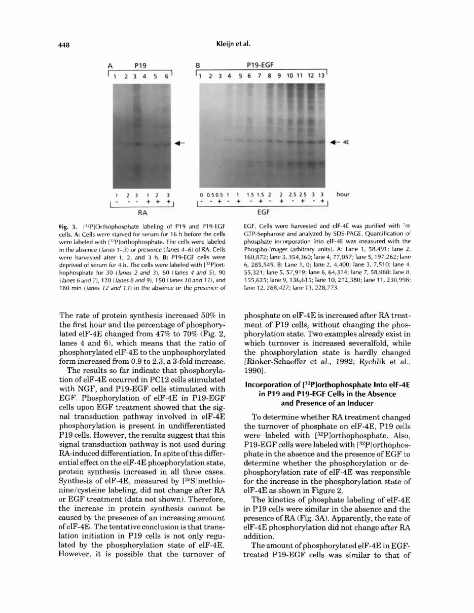
Fig. 3. [32P]Orthophosphate labeling of PI9 and P19-ECF cells. A: Cells were starved for serum for 16 h before the cells were labeled with [32Plorthophosphate. The cells were labeled in the absence (lanes 1-3) or presence (lanes 4-6) of RA. Cells were harvested after 1, 2, and 3 h. B: P19-ECF cells were deprived of serum for 4 h. The cells were labeled with [?*P]ort- hophosphate for 30 (lanes 2 and 3), 60 (lanes 4 and 5), 90 (lanes 6 and 7), 120 (lanes 8 and 9), 1 50 (lanes 70 and 1 I ) , and 180 min (lanes 12 and 13) in the absence or the presence of
EGF
ECF. Cells were harvested and elF-4E was purified with 'm CTP-Sepharose and analyzed by SDS-PACE. Quantification of phosphate incorporation into elF-4E was measured with th r Phospho-imager (arbitrary units). A: Lane 1, 58,491; lane 2. 160,872; lane 3, 354,360; lane 4, 77,057; lane 5, 197,262; lane 6, 285,545. B: Lane 1, 0; lane 2, 4,400; lane 3, 7,510; lane 4. 55,321; lane 5, 57,919; lane 6, 64,314; lane 7, 58,960; lane 8, 155,625; lane 9, 136,615; lane 10,212,380; lane 11, 230,998; lane 12, 268,427; lane 13, 228,773.
The rate of protein synthesis increased 50% in the first hour and the percentage of phosphory- lated elF-4E changed from 47% to 70% (Fig. 2, lanes 4 and 61, which means that the ratio of phosphorylated elF-4E to the unphosphorylated form increased from 0.9 to 2.3, a 3-fold increase.
The results so far indicate that phosphoryla- tion of elF-4E occurred in PC12 cells stimulated with NGF, and P19-EGF cells stimulated with EGF. Phosphorylation of elF-4E in P19-EGF cells upon EGF treatment showed that the sig- nal transduction pathway involved in elF-4E phosphorylation is present in undifferentiated P19 cells. However, the results suggest that this signal transduction pathway is not used during RA-induced differentiation. In spite of this differ- ential effect on the elF-4E phosphorylation state, protein synthesis increased in all three cases. Synthesis of elF-4E, measured by [35Slmethio- nineicysteine labeling, did not change after RA or EGF treatment (data not shown). Therefore, the increase in protein synthesis cannot be caused by the presence of an increasing amount of elF-4E. The tentative conclusion is that trans- lation initiation in PI9 cells is not only regu- lated by the phosphorylation state of elF-4E. However, it is possible that the turnover of
phosphate on elF-4E is increased after RA treat- ment of P19 cells, without changing the phos- phorylation state. Two examples already exist in which turnover is increased severalfold, while the phosphorylation state is hardly changed [Rinker-Schaeffer et al., 1992; Rychlik et a].. 19901.
Incorporation of [32P]orthophosphate Into elF-4E in P19 and P19-ECF Cells in the Absence
and Presence of an Inducer
To determine whether RA treatment changed the turnover of phosphate on elF-4E, P19 cells were labeled with [32Pl~rthopho~phate. Also, P19-EGF cells were labeled with [32P]orthophos- phate in the absence and the presence of EGF to determine whether the phosphorylation or de- phosphorylation rate of elF-4E was responsible for the increase in the phosphorylation state of elF-4E as shown in Figure 2.
The kinetics of phosphate labeling of elF-4E in P19 cells were similar in the absence and the presence of RA (Fig. 3A). Apparently, the rate of elF-4E phosphorylation did not change after RA addition.
The amount of phosphorylated elF-4E in EGF- treated P19-EGF cells was similar to that of

Phosphorylation of elF-4E in P19 Cells 449
control cells (Fig. 3B). As the elF-4E phosphory- lation state in P19-EGF cells increased after EGF treatment (Fig. 21, the results indicate that the rate of dephosphorylation of elF-4E is influ- enced by EGF.
Dephosphorylation of elF-4E After Induction of Differentiation
P19 cells and P19-EGF cells were deprived of serum and supplemented during the last 3 h of the serum deprivation with [32Pl-labeled ortho- phosphate. After the labeled medium was re- moved, cells were treated with RA or EGF for the times indicated in Figure 4.
The amount of [32Pl-labeled phosphate on elF-4E in P19 cells decreased in the control cells, as well as in the RA treated cells in a similar way (Fig. 4). The half-life of the phosphate on elF-4E in the absence and in the presence of RA was about 41 min. As expected from the previous results, the rate of elF-4E dephosphorylation was not influenced by the addition of RA.
The half-life of a phosphate on elF-4E in un- treated P19-EGF cells (Fig. 4, lanes 2-4) was similar to the one found in P19 cells (41 min). In contrast, the amount of [32Pl-labeled elF-4E did not decrease but even increased 1.6-fold, when
P19-EGF cells were chased in the presence of EGF (Fig. 4, lanes 12-14). This result together with the changed phosphorylation state and the unchanged elF-4E phosphorylation rate indi- cates that EGF modulates elF-4E dephosphory- lation.
During a chase of P19-EGF cells after [32Plor- thophosphate labeling in the absence and the presence of RA, the same results were obtained as shown for P19 cells (data not shown).
DISCUSSION
P19 cells were treated with the differentiation agent RA to measure whether differentiation coincided with a change in the rate of protein synthesis and elF-4E phosphorylation. RA treat- ment led to an increase in protein synthesis within 30-60 min (Table I). However, elF-4E phosphorylation state (Fig. 11, the turnover of phosphate on elF-4E (Figs. 3 and 4), and the synthesis of elF-4E (data not shown) remained the same. These results show that the very early onset of differentiation of P19 cells does not require changes in elF-4E phosphorylation.
RA can influence cell growth [Sporn and Rob- erts, 19831, pattern formation in limb develop- ment and regeneration [Maden, 1982; Takaha-
Fig. 4. Turnover of phosphate on elF-4E in PI9 and P19-ECF cells. PI 9 cells and P19-ECF cells were starved for serum. During the last 3 h, [32P]-labeled orthophosphate was added. Label was removed and the cells were chased in the absence or presence of RA (P19) (left) or ECF (P19-ECF) (right). PI 9 cells were harvested after 0 (lane 7), 20 (lanes 2 and 3 ) , 40 (lanes 4 and 5), and 60 min (lanes 6 and 7). P19-ECF cells were harvested after 0 (lane 8), 20 (lanes 9 and 72), 40 ( lanes 70 and
13), and 60 min (lanes 7 7 and 74). elF-4E was purified and analyzed by SDS-PACE. Incorporation of phosphate into elF-4E was measured with a Phospho-imager (arbitrary units). Lane 1, 201,484; lane 2, 148,409; lane 3, 148,729; lane 4, 120,292; lane 5, 103,310; lane 6, 71,873; lane 7, 96,418; lane 8, 21 6,598; lane 9, 232,557; lane 10, 183,833; lane 11, 96,566; lane 12,235,042; lane 13, 314,975; lane 14, 309,956.

450 Kleijn eta!.
shi et al., 19751, and fetal development [Soprano et al., 1986; Tickle et al., 19851. Furthermore, RA activates diacylglycerol, and causes a translo- cation of PKC from the cytosol to the membrane within minutes. This suggests a rapid signal transduction from the nuclear RA-receptor to the cytoplasm [Kurie et al., 19931, which is supported by the fast increase in protein synthe- sis after RA addition (Table I).
Increased protein synthesis in RA-treated cells cannot be explained by increased elF-4E phos- phorylation, and regulation of other initiation factors must be involved. One possible explana- tion for increased protein synthesis is by regula- tion of the recently discovered 4E-BP1 or PHAS-I protein [Pause et al., 19941. Phosphorylation of 4E-BP1 influenced the activity of elF-4E. The effect of elF-4E phosphorylation on the regula- tion by 4E-BP1 was not determined, and 4E-BP1 regulation would not require elF-4E phosphory- lation per se. It was suggested that 4E-BP1 phosphorylation dissociates the 4E-BP1-elF-4E complex and liberates elF-4E, enabling cap bind- ing and initiation of protein synthesis [Pause et al., 19941.
To measure whether the unchanged elF-4E phosphorylation state was due to the absence of the signal transduction pathway leading to elF-4E phosphorylation in undifferentiated P19 cells, P19-EGF cells were treated with EGF. Upon EGF treatment an increase in the phos- phorylation state of elF-4E occurred (Fig. 2). Therefore, the signal transduction pathway is present in undifferentiated P19 cells. However, this pathway was not used when P19 cells were differentiated with RA. Apparently, elF-4E phos- phorylation is not an absolute prerequisite to induce differentiation after RA-addition.
Undifferentiated P19 cells do not express de- tectable levels of epidermal growth factor recep- tors (EGF-R). However, after 2 days of RA- induced differentiation increased levels of EGF-R mRNA and protein has been found [Joh et al., 19921. Reduced expression of EGF-R in differen- tiating P19 cells inhibited the ability to differen- tiate [Wu and Adamson, 19931. Undifferenti- ated P19 cells overexpressing the EGF receptor could be stimulated to differentiate upon EGF treatment when grown in aggregates. However, under our cell culture conditions-growing the cells in monolayers-differentiation does not occur upon EGF addition [den Hertog et al., 1991al. Therefore, the increase in protein syn- thesis and elF-4E phosphorylation as shown in Figure 2 was due to growth stimulation.
Besides having an effect on differentiation, NGF can stimulate proliferation of PC12 cells [Boonstra et al., 19831. Therefore, the effect of NGF addition on elF-4E phosphorylation in PC12 cells could be explained by the growth stimulatory effect of NGF. Treatment of P19- EGF cells with the growth inducer EGF also led to elF-4E phosphorylation and increased pro- tein synthesis (Fig. 2). This indicates that elF-4E phosphorylation coincides with growth stimula- tion.
Treatment of P19-EGF cells with EGF in- creased the phosphorylation state of elF-4E (Fig. 2). This increase was solely due to a decreased dephosphorylation rate and not due to modula- tion of the elF-4E phosphorylation rate (Figs. 3 and 4). This result indicates that an elF-4E phosphatase is influenced after EGF treatment. Regulation of the elF-4E phosphorylation state by phosphatase activity has been described be- fore. Treatment of human mammary epithelial cells (184NlA4) with EGF led to an elevation of the elF-4E phosphorylation state of 2-3-fold [Donaldson et al., 19911. Okadaic acid, a potent inhibitor of phosphatase 1 and 2A, also led to an increase in elF-4E phosphorylation. Treatment of the cells with EGF and okadaic acid simulta- neously did not result in a further elevation of elF-4E phosphorylation [Donaldson et al., 19911. This may suggest that both EGF and okadaic acid act on the same phosphatase.
The turnover of phosphates on elF-4E was similar in P19 and P19-EGF cells. The half-life of the phosphate on elF-4E was about 41 min. Zhang et al. [19941 described that the half-life of a phosphate on elF-4E in 293 cells was 32-36 min, a value similar to the one found here with P19 cells. Although only based on two observa- tions, this finding suggests that the turnover rate of phosphorylated elF-4E is similar in differ- ent cell types.
Treatment of PC12 cells with NGF led to a 4-fold increase in the amount of phosphorylated elF-4E [Frederickson et al., 19921. Treatment of 184NlA4 cells with EGF led to a 2-3-fold in- crease in elF-4E phosphorylation [Donaldson et al., 19911. The ratio of phosphorylated elF-4E to the unphosphorylated form was 0.17 in control P19-EGF cells, and 0.61 in EGF-treated cells, a 3.6-fold increase (Fig. 2). The total amount of EGF receptors on 184NlA4 cells (300,000 recep- tors/cell) [Donaldson et al., 19911 is 10-fold higher than the amount of EGF receptors on P19-EGF cells [den Hertog et al., 1991b1, and PC12 cells expose 58,000 NGF receptors on

Phosphorylation of elF-4E in P 1 9 Cells 451
their surface [Herrup and Thoenen, 19791. The difference in receptor type and in the amount of receptors does not influence the extent of the stimulation of the elF-4E phosphorylation.
The retinoic acid receptor is a nuclear recep- tor and a member of the steroidithyroid hor- mone receptor family [Giguere et al., 1987; Pet- kovich et al., 19871. EGF and NGF both bind to a membrane bound tyrosine lunase receptor and have the ability to increase protein synthesis as well as to increase elF-4E phosphorylation. RA induces the cell via a nuclear receptor and in- creased protein synthesis only. Apparently the nuclear signal is transmitted to the cytoplasmic translational apparatus. The result may imply that an increase in elF-4E phosphorylation oc- curs after activation of receptor tyrosine kinases or after activation of one of the intermediates of the tyrosine kinase signal transduction path- way.
ACKNOWLEDGMENTS
We thank the Hubrecht Laboratory for the P19 and the P19-8-39 cells, Andrea Flynn and Chris Proud for their help with the 1D iso- electric focusing and purification of the elF-4E antibodies, and Gert Scheper for his help with the manuscript.
REFERENCES
Bonneau A, Sonenberg N (1987): Involvement of the 24-kDa cap-binding protein in regulation of protein synthesis in mitosis. J Biol Chem 262:11134-11139.
Boonstra J , Moolenaar WH, Harrison PH, Moed P, van der Saag PT, de Laat SW (1983): Ionic strength and growth stimulation induced by nerve growth factor and epidermal growth factor in rat pheochromocytoma (PC12) cells. J Cell Biol97:92-98.
Bu X, Hagedorn CH (1991): Platelet-derived growth factor stimulates phosphorylation of the 25 kDa mRNA cap binding protein (elF-4E) in human lung fibroblasts. FEBS Lett 283:219-222.
de Benedetti A, Rhoads RE (1990): Overexpression of eukary- otic protein synthesis initiation factor 4E in HeLa cells results in aberrant growth and morphology. Proc Natl Acad Sci USA 87:8212-8216.
den Hertog J, de Laat SW, Schlessinger J, Kruijer W (1991a): Neuronal differentiation in response to epidermal growth factor of transfected murine P19 embryonal carcinoma cells expressing human epidermal growth factor recep- tors. Cell Growth Differ 2:155-164.
den Hertog J, Eman R, Tertoolen LGJ, de Laat SW, Kruijer W (1991b): Characterization of a tyrosine kinase signal- ling pathway in undifferentiated P19 embryonal carci- noma cells. Exp Cell Res 196:226-232.
Dholakia JN, Wahba AJ (1988): Phosphorylation of the guanine nucleotide exchange factor from rabbit reticulo- cytes regulates its activity in polypeptide chain initiation. Proc Natl Acad Sci USA 85:51-54.
Donaldson RW, Hagedorn CH, Cohen S (1991): Epidermal growth factor or okadaic acid stimulates phosphorylation of eukaryotic initiation factor 4F. J Biol Chem 266:3162- 3166.
Duncan R, Hershey JWB (1985): Regulation of initiation factors during translational repression caused by serum depletion. J Biol Chem 260:5493-5497.
Duncan R, Milburn SC, Hershey JWB (1987): Regulated phosphorylation and low abundance of HeLa cell initia- tion factor elF-4F suggest a role in translational control. J Biol Chem 262:380-388.
Frederickson RM, Montine KS, Sonenberg N (1991): Phos- phorylation of eukaryotic translation initiation factor 4E is increased in Src-transformed cell lines. Mol Cell Biol
Frederickson RM, Mushynski W, Sonenberg N (1992): Phos- phorylation of translation initiation factor elF-4E is in- duced in a ras-dependent manner during nerve growth factor-mediated PC12 cell differentiation. Mol Cell Biol
Giguere V, Ong ES, Segui P, Evans RM (1987): Identifica- tion of a receptor for the morphogen retinoic acid. Nature
Herrup K, Thoenen H (1979): Properties ofthe nerve growth factor receptor of a clonal line of rat pheochromocytoma (PC12) cells. Exp Cell Res 121:71-78.
Hiremath LS, Webb NR, Rhoads RE (1985): Immunological detection of the messenger RNA cap-binding protein. J Biol Chem 260:7843-7849.
Huang J, Schneider R J (1991): Adenovirus inhibition of cellular protein synthesis involves inactivation of cap- binding protein. Cell 65:271-280.
Jaramillo M, Pelletier J, Edery I, Nielsen PJ, Sonenberg N (1991): Multiple mRNAs encode the murine translation initiation factor elF-4E. J Biol Chem 266:10446-10451.
Joh T , Darland T, Samuels M, Wu J-X, Adamson ED (1992): Regulation of the epidermal growth factor receptor gene in murine embryonal carcinoma cells. Cell Growth Differ 3:315-325.
Jones-Villeneuve EMV, McBurney MW, Rogers KA, Kalnins VI (1982): Retinoic acid induces embryonal carcinoma cells to differentiate into neurons and glial cells. J Cell Biol
Joshi-Bane S, Rychlik W, Rhoads RE (1990): Alteration of the major phosphorylation site of eukaryotic protein syn- thesis initiation factor 4E prevents its association with the 48 S initiation complex. J Biol Chem 265:2979-2983.
Kaspar RL, Rychlik W, White MW, Rhoads RE, Morris DR (1990): Simultaneous cytoplasmic redistribution of ribo- somal protein L32 mRNA and phosphorylation of eukary- otic initiation factor 4E after mitogenic stimulation of Swiss 3T3 cells. J Biol Chem 265:3619-3622.
Konieczny A, Safer B (1983): Purification of the eukaryotic initiation factor 2-eukaryotic initiation factor 2B complex and characterization of its guanine nucleotide exchange activity during protein synthesis initiation. J Biol Chem 258:3402-3408.
Koromilas AE, Lazaris-Karatzas A, Sonenberg N (1992): mRNAs encoding extensive secondary structure in their 5' noncoding region translate efficiently in cells overex- pressing initiation factor elF-4E. EMBO J 11:4153-4158.
Kozak M (1987): An analysis of 5'-noncoding sequences from 699 vertebrate messenger RNAs. Nucleic Acids Res 15:8125-8148.
11:2896-2900.
12:1239-1247.
330~624-629.
941253-262.

452 Kleijn et a[.
Kurie JM, Younes A, Miller WH, Burchert M, Chiu C-F, Kolesnick R, Dmitrovsky E (1993): Retinoic acid stimu- lates the protein kinase C pathway before activation of its p-nuclear receptor during human teratocarcinoma differ- entiation. Biochim Biophys Acta 1179:203-207.
Lamphear BJ, Panniers R (1990): Cap binding protein com- plex that restores protein synthesis in heat-shocked Ehrlich cell lysates contains highly phosphorylated elF- 4E. J Biol Chem 265:5333-5336.
Lamphear BJ, Panniers R (1991): Heat shock impairs the interaction of cap-binding protein complex with 5' mRNA cap. J Biol Chem 266:2789-2794.
Lawson TG, Lee KA, Maimone MM, Abramson RD, Dever TE, Merrick WC, Thach RE (1989): Dissociation of double- stranded polynucleotide helical structures by eukaryotic initiation factors, as revealed by a novel assay. Biochemis-
Lazaris-Karatzas A, Montine KS, Sonenberg N (1990): Ma- lignant transformation by a eukaryotic initiation factor subunit that binds to mRNA 5' cap. Nature 345:544-547.
Lazaris-Karatzas A, Smith MR, Frederickson RM, Jaramillo ML, Liu YL, Kung HF, Sonenberg N (1992): Ras mediates translation initiation factor-4E-induced malignant trans- formation. Gene Dev 6:1631-1642.
Maden M (1982): Vitamin A and pattern formation in the regenerating limb. Nature 295:672-675.
Manzella JM, Rychlik W, Rhoads RE, Hershey JWB, Black- shear PJ (1991): Insulin induction of ornithine decarbox- ylase: Importance of mRNA secondary structure and phos- phorylation of eucaryotic initiation factors elF-4B and elF-4E. J Biol Chem 266:2383-2389.
Marino MW, Feld LJ, Jaffe EA, Pfeffer LM, Han H-M, Donner DB (1991): Phosphorylation of the proto-onco- gene product eukaryotic initiation factor 4E is a common cellular response to tumor necrosis factor. J Biol Chem
Martin GR (1980): Teratocarcinomas and mammalian em- bryogenesis. Science 209:768-776.
Maurides PA, Akkaraju GR, Jagus R (1989): Evaluation of protein phosphorylation state by a combination of vertical slab isoelectric focusing and immunoblotting. Anal Bio- chem 183:144-151.
McBurney MW, Jones-Villeneuve EMV, Edwards MKS, Anderson P J (1982): Control of cell muscle and neuronal differentiation in a cultured embryonal carcinoma cell line. Nature 299:165-167.
Minich WB, Balasta ML, Goss DJ, Rhoads RE (1994): Chro- matographic resolution of in uiuo phosphorylated and nonphosphorylated eukaryotic translation factor elF-4E: Increased cap affinity of the phosphorylated form. Proc Natl Acad Sci USA 91:7668-7672.
Morley SJ, Traugh JA (1989): Phorbol esters stimulate phosphorylation of eukaryotic initiation factors 3,4B, and 4F. J Biol Chem 264:2401-2404.
Morley SJ, Traugh J A (1990): Differential stimulation of phosphorylation of initiation factors elF-4E, elF-4B, elF-3, and ribosomal protein S6 by insulin and phorbol esters. J Biol Chem 265:10611-10616.
Pause A, Belsham GJ, Gingras A-C, Donze 0, Lin T-A, Lawrence J r JC, Sonenberg N (1994): Insulin-dependent stimulation of protein synthesis by phosphorylation of a regulator of 5'-cap function. Nature 371:762-767.
Petkovich M, Brand NJ, Krust A, Chambon P (1987): A human retinoic acid receptor which belongs to the family of nuclear receptors. Nature 330:444-450.
try 28~4729-4734.
266:2685-2688.
Redpath NT, Proud CG (1994): Molecular mechanisms in the control of translation by hormones and growth fac- tors. Biochim Biophys Acta 1220:147-162.
Rhoads RE (1988): Cap recognition and the entry of mRXA into the protein synthesis initiation cycle. Trends Bio- chem Sci 13:52-56.
Rinker-Schaeffer CW, Austin V, Zimmer S, Rhoads RE (1992): rus transformation of cloned rat embryo fibro- blasts results in increased rates of protein synthesis and phosphorylation of eukaryotic initiation factor 4E. J Fl~ol Chem 267: 10659-10664.
Rozen F, Edery I, Meerovitch K, Dever TE, Merrick WC, Sonenberg N (1990): Bidirectional RNA helicase act11 ity of eucaryotic translation initiation factors 4A and 4F. No1 Cell Biol 10:1134-1144.
Rychlik W, Rush JS, Rhoads RE, Waechter C J 11990:: Increased rate of phosphorylation-dephosphorylation of the translational initiation factor elF-4E correlates Kith the induction of protein and glycoprotein biosynthesis in activated B lymphocytes. J Biol Chem 265:19467-19471.
Salimans M, Goumans H, Amesz H, Benne R, Voorma HO (1984): Regulation of protein synthesis in eukaryotes. Eur J Biochem 145:91-98.
Siekierka J, Mauser L, Ochoa S (1982): Mechanism of poly- peptid chain initiation in eukaryotes and its control by phosphorylation of the cx subunit of initiation factor 2. Proc Natl Acad Sci USA 79:2537-2540.
Singh LP, Aroor AR, Wahba AJ (1994): Phosphorylation of the guanine nucleotide exchange factor and eukaryotic initiation factor 2 by casein kinase I1 regulates guanine nucleotide binding and GDPiGTP exchange. Biochemis- try 33:9152-9157.
Sonenberg N, Morgan MA, Merrick WC, Shatkin AJ I 1978 I: A polypeptide in eukaryotic initiation factors t h a t crosslinks specifically to the 5'-terminal cap in mRNA. Proc Natl Acad Sci USA 75:4843-4847.
Soprano DR, Soprano KJ, Goodman DS (1986): Retinol- binding protein and transthyretin mRNA levels in visceral yolk sac and liver during fetal development in the rat. Proc Natl Acad Sci USA 83:7330-7334.
Sporn MB, Roberts AB (1983): Role of retinoids in differen- tiation and carcinogenesis. Cancer research 43:3034-- 3040.
Takahashi YI, Winick JE, Goodman DS (1975) deficiency and fetal growth and development Nutr 105:1299-1310.
Tickle C, Lee J , Eichele G (1985): A quantitative analysis of' the effect of all-trans retinoic acid on the pattern of chick limb development. Dev Biol 109:82-95.
Welsh GI, Proud CG (1992): Regulation of protein synthesis in Swiss 3T3 fibroblasts-Rapid activation of the guaninr- nucleotide-exchange factor by insulin and growth factcirs. Biochem J 284:19-23.
Wolthuis RMF, Cremers AFM, Kasperaitis MAM, van der Mast CA, Voorma HO, Boonstra J (1993): Epidermal growth factor stimulates phosphorylation of eukaryotic initiation factor 4B, independently of protein kinase C . Biochim Biophys Acta 1177:160-166.
Wu J, Adamson ED (1993): Inhibition of differentiation in P19 embryonal carcinoma cells by the expression of vec- tors encoding truncated or antisense EGF receptor. Dev Biol 159:208-222.
Zhang Y, Feigenblum D, Schneider R J (1994): A late adeno- virus factor induces elF-4E dephosphorylation and inhibi- tion of cell protein synthesis. J Virol68:7040-7050.