patterns of hippocampal-neocortical interactions in the retrieval of episodic autobiographical...
TRANSCRIPT
Patterns of Hippocampal–Neocortical Interactions in theRetrieval of Episodic Autobiographical Memories
Across the Entire Life-Span of Aged Adults
Armelle Viard,1 Karine Lebreton,1 Gael Chetelat,1 Beatrice Desgranges,1 Brigitte Landeau,1
Alan Young,1 Vincent De La Sayette,1 Francis Eustache,1,2 and Pascale Piolino1,2,3*
ABSTRACT: We previously demonstrated that episodic autobiographi-cal memories (EAMs) rely on a network of brain regions comprising themedial temporal lobe (MTL) and distributed neocortical regions regard-less of their remoteness. The findings supported the model of memoryconsolidation, which proposes a permanent role of MTL during EAM re-trieval (multiple-trace theory or MTT) rather than a temporary role(standard model). Our present aim was to expand the results by examin-ing the interactions between the MTL and neocortical regions (or MTL–neocortical links) during EAM retrieval with varying retention intervals.We used an experimental paradigm specially designed to engage agedparticipants in the recollection of EAMs, extracted from five differenttime-periods, covering their whole life-span, in order to examine corre-lations between activation in the MTL and neocortical regions. The na-ture of the memories was checked at debriefing by means of behavioralmeasures to control the degree of episodicity and properties of memo-ries. Targeted correlational analyses carried out on the MTL, frontal, lat-eral temporal, and posterior regions revealed strong links between theMTL and neocortex during the retrieval of both recent and remoteEAMs, challenging the standard model of memory consolidation andsupporting MTT instead. Further confirmation was given by resultsshowing that activation in the left and right hippocampi significantlycorrelated during the retrieval of both recent and remote memories.Correlations among extra-MTL neocortical regions also emerged for alltime-periods, confirming the critical role of the prefrontal, temporal(lateral temporal cortex and temporal pole), precuneus, and posteriorcingulate regions in EAM retrieval. Overall, this paper emphasizes therole of a bilateral network of MTL and neocortical areas whose activa-tion correlate during the recollection of rich phenomenological recentand remote EAMs. VVC 2009 Wiley-Liss, Inc.
KEY WORDS: autobiographical memory; consolidation; correlation;hippocampus; neuroimaging
INTRODUCTION
Autobiographical memory (AM) refers to information and memoriesof personal life events. It is composed of different types of representa-tions, from general knowledge about oneself (semantic component, also
referred to as ‘‘personal semantics’’) to very specificpersonal events (episodic component) (Tulving et al.,1988; Conway, 2001). The episodic component [epi-sodic autobiographical memory (EAM)] is charac-terized by spatio-temporal specificity, mental visualimagery, and emotion (Brewer, 1996; Conway andPleydell-Pearce, 2000; Conway, 2001; Tulving, 2001),as well as by a particular self-reflective mental state,termed autonoetic consciousness, which implies thatthe subject recollects his memories with a sense ofreliving (re-experiencing), by mentally ‘‘traveling backin time’’ (Wheeler et al., 1997; Tulving, 2001; Piolinoet al., in press). Thus, the central tenet of episodicAM revolves around phenomenological re-experienc-ing and the sense of self in time. The semantic com-ponent is characterized by a state of consciousness,termed noetic consciousness, which enables one toretrieve general facts about a personal event withoutre-experiencing it.
Neuroimaging studies have detected an overall left-lateralized cerebral network associated with the re-trieval of EAMs, including, in particular, prefrontal,medial, and lateral temporal cortices, as well as poste-rior regions (Maguire, 2001; Moscovitch et al., 2005;Svoboda et al., 2006). Little is known, however, aboutinteractions between these regions during autobio-graphical retrieval. Connectivity (Maguire et al., 2000;Addis et al., 2004a) or correlational (Greenberg et al.,2005) analyses can be used to address this issue.Maguire et al. (2000) showed increased connectivitybetween the hippocampus and the parahippocampalgyrus during the recognition of autobiographicalevents relative to other memory subtypes (autobio-graphical events, autobiographical facts, public events,or general facts). Similarly, Addis et al. (2004a)showed that the left and right hippocampi were func-tionally connected during AM retrieval, as well aswith the right parahippocampal gyrus. These findingssuggest a role of the medial temporal lobe (MTL) inthe retrieval of EAMs, but do not consider its involve-ment according to memory remoteness and the phe-nomenological properties of the memories retrieved.
A central debate today concerns the role over timeof the MTL, in particular the hippocampus, in AMretrieval and two conflicting theories of memory con-
1 Inserm-EPHE-Universite de Caen/Basse-Normandie, Unite U923, GIPCyceron, CHU Cote de Nacre, Caen, France; 2Universite Paris Des-cartes, Institut de Psychologie, Paris, France; 3CNRS, UMR 8189, Labo-ratoire Psychologie et Neurosciences Cognitives, Paris, France*Correspondence to: Dr. Pascale Piolino, Inserm-EPHE-Universite deCaen/Basse-Normandie, Unite U923, GIP Cyceron, CHU Cote de Nacre,14033 Caen Cedex, France. E-mail: [email protected] for publication 10 February 2009DOI 10.1002/hipo.20601Published online 31 March 2009 in Wiley InterScience (www.interscience.wiley.com).
HIPPOCAMPUS 20:153–165 (2010)
VVC 2009 WILEY-LISS, INC.
solidation have been proposed. The ‘‘standard model’’ suggeststhat the MTL is initially implicated in the encoding and con-solidation of AMs, but with time, AM retrieval becomes inde-pendent of this region and relies only on neocortical regions(Squire and Alvarez, 1995; Bayley and Squire, 2005). Thus,the retrieval of recent memories relies on interactions betweenthe MTL and neocortical regions (or MTL–neocortical links),while the retrieval of remote memories depends solely on neo-cortical interactions. Moreover, this theory does not distinguishbetween the two components of AM (episodic and semantic)and assumes that both are subject to the same consolidationprocess. Alternatively, the ‘‘multiple trace theory’’ (MTT)(Nadel and Moscovitch, 1997; Moscovitch et al., 2005; Nadelet al., 2007) concurs with the standard model for the semanticAMs, but suggests that MTL–neocortical links are permanentlyrequired for the retrieval of EAMs. Of note, according toMTT, it is both the hippocampus and its related structures,including the parahippocampal gyrus, which are hypothesizedto interact permanently with neocortical regions during the re-trieval of both recent and remote EAMs.
In a continuation of our previous activation study (Viardet al., 2007), which favored MTT, we addressed these issues byexploring the patterns of coactivation between different MTLand neocortical regions during the retrieval of EAMs takenfrom five time-periods and covering the entire life-span ofhealthy aged adults. Our previous neuroimaging data hadshown that a network, including mainly the left hippocampus,left superior frontal gyrus, bilateral precuneus and posterior cin-gulate gyrus, was commonly active for all time-periods. Behav-iorally, all memories were characterized by specificity and ahigh level of details, hence were episodic (i.e., spatiotemporaluniqueness and details). However, some differences emergedamong intermediate periods which were rated stronger than themost recent and most remote periods, in terms of the phenom-enological attributes of memory (i.e., emotion, mental visualimagery, and autonoetic consciousness) and recruited addition-ally the right hippocampus.
In the present paper, using the same data set, we examined cor-relations in the activation of medial temporal and neocorticalregions during the retrieval of recent and remote EAMs, in orderto further test the two models of memory consolidation. In ouranalyses, we included a priori regions known to be particularlyinvolved in the retrieval of EAMs (Cabeza and St Jacques, 2007),namely the MTL, prefrontal and, posterior regions (precuneusand posterior cingulate gyrus), as well as lateral temporal cortices.Indeed, substantial evidence implicates the MTL in the retrievalof EAMs, in particular the hippocampus and parahippocampalgyrus (Maguire, 2001; Ryan et al., 2001; Maguire and Frith,2003a,b; Piefke et al., 2003; Piolino et al., 2004; 2008; Addiset al., 2004b; Gilboa et al., 2004; Greenberg et al., 2005; Viardet al., 2007), as well as the amygdala which is known for its rolein the processing of emotional AMs (Markowitsch et al., 2000,2003; Daselaar et al., 2008; for review, see Phelps, 2004). A closelink between memory and emotion is suggested by studies show-ing a preferential recall of emotional events (Brewer, 1986; Dolanet al., 2000). The prefrontal cortex (PFC) is crucial for the recon-
struction of EAMs from the initial search to the maintenance ofa specific memory in mind and is thought to be involved in thecontrolled retrieval of information from posterior regions (Mayesand Roberts, 2001; Simons and Spiers, 2003; Gilboa, 2004;Cabeza and St Jacques, 2007; Piolino et al., 2008). Prefrontalregions are also hypothesized to play a role in the emergence ofautonoetic consciousness, an essential characteristic of EAM re-trieval (Levine et al., 1998; Piolino et al., 2005). Posteriorregions, such as the precuneus or the posterior cingulate gyrus,have been associated with access to sensory–perceptual details, inparticular via their role in mental visual imagery (Fletcher et al.,1995; Cavanna and Trimble, 2006). Lateral temporal activationsare involved in semantic retrieval processes (Maguire, 2001; Pio-lino et al., 2007). Indeed, autobiographical retrieval is often initi-ated by first browsing through the general levels of autobiograph-ical knowledge before accessing an episodic event (Conway andPleydell-Pearce, 2000; Conway et al., 2001).
Concerning our main predictions and according to MTT,we expected activation in the MTL (hippocampus, parahippo-campal gyrus, amygdala) and neocortical regions (frontal, tem-poral, and posterior regions) to be correlated during the re-trieval of EAMs, whether they belonged to recent or remotetime-periods (Nadel and Moscovitch, 1997; Moscovitch et al.,2005; Nadel et al., 2007). Furthermore, we predicted that themore richly recollected intermediate periods (compared to themost recent and remote ones) would involve a larger bilateralMTL–neocortical correlational network.
MATERIALS AND METHODS
Participants
Twelve right-handed (as measured by the Edinburgh handed-ness inventory) healthy females [mean age 6 standard deviation(SD) 5 67.2 6 5.2 yr; ranging from 60 to 75 yr old] with nohistory of psychiatric or neurological disorder were recruitedthrough a university, a retirement association, or a newspaperadvertisement. To obtain a homogeneous group, we recruitedonly females. The study was approved by the Regional EthicsCommittee and written informed consent was obtained fromall subjects prior to their participation in the study. Participantshad no abnormality on their T1-weighted high-resolution mag-netic resonance imaging (MRI). They underwent a battery ofneuropsychological tests to assess their cognitive abilities and allperformed in the normal range (see Viard et al., 2007, for afull description). Each participant resided at home and all wereactive in cultural pursuits, continuing education, or withresponsibilities in diverse associations.
Task and Experimental Design
The experimental procedure was divided in two sessions (formore details, see Viard et al., 2007). A few weeks before theexperimental phase, the first session was carried out with a close
154 VIARD ET AL.
Hippocampus
family member who was interviewed on the participant’s spe-cific life-events. In the second session, a training period pre-ceded the functional scanning which was followed by a debrief-ing. Personal sentence-cues were elaborated from the familymember’s prior interview and cues were visually presented inwhite on a black background, using Superlab software (3.0 ver-sion, Cedrus). Participants were given precise instructions torecall ‘‘a personal event which occurred only once, at a particu-lar place and date, and lasted several minutes or hours, but lessthan a day, with as many details as they could.’’ The scanningperiod consisted of five functional runs, randomly intermixedacross subjects, each corresponding to one time-period andcomposed of five intermixed experimental and control blocks.In the experimental condition, participants viewed sentence-cues presented for 5 s, followed by 19 s of blank screen duringwhich they had to mentally retrieve the corresponding specificpersonal event (e.g., the wedding of Pierre; the visit to the Eif-fel Tower). Since they could start their mental evocation whilethe cue was still on the screen, the maximum retrieval time was24 s. They were asked to press on a button as soon as theygained access to the prompted event. Twenty-five sentences-cues were presented per subject, corresponding to the five dif-ferent time-periods (P1: 0–17 yr; P2: 18–30 yr; P3: > 30 yrold except for the last 5 yr; P4: last 5 yr except the last12 months; P5: last 12 months). In the control condition, par-ticipants were asked to detect the presence of two consecutiveletters (‘‘mb’’) in pseudowords of six letters (for example,‘‘speugr’’ or ‘‘mbieha’’) and were instructed to press on a buttonwhen ‘‘mb’’ was present in the pseudoword. This low-level taskwas chosen as a baseline condition in order to control for read-ing operations, mental processing of visual cues and motorprocessing, common to both experimental and control tasks.
Following the scanning session, a debriefing took place inwhich participants retrieved all events again and rated them onbehavioral scales. We specifically assessed episodic AM, takinginto account not only the objective specificity of the personalevents that are recalled (uniqueness, spatiotemporal location,details), but also the subjective experience of remembering theencoding context. Indeed, episodic AM relies not only on theability to recall a specific event and locate it in time and space,but also on the ability to recollect specific details which distin-guish that event from similar ones. As it is possible to rebuild aspecific event from one’s personal semantic AM withoutactually reliving sensory–perceptual episodic details, it is vitalto gauge the specificity of details from the encoding contextthrough the sense of re-experiencing. The encoding contextencompasses time and space (i.e., the specificity of event), sen-sory–perceptual–affective–cognitive details (i.e., the specificityof details), the subjective experience (i.e., autonoetic conscious-ness) and the visual experience (i.e., self-perspective) (Piolinoet al., 2006, in press).
Participants were first given precise instructions to recallagain the personal events from the five different time periodsin the scan which were rated using strict objective criteria. Thesubjective reports of memories were then assessed using theRemember/Know procedure (Tulving, 1985; Gardiner, 1988),
the quantification of mental visual imagery and the remember-er’s self-perspective known as the Field/Observer perspectiveparadigm (Nigro and Neisser, 1983; Robinson and Swanson,1993), which makes it possible to differentiate between epi-sodic and semantic AM retrieval. In total, our procedure madeit possible to measure episodic AM characterized by unique-ness, specificity and details, and as enabling someone to ‘‘travelback in time,’’ relive specific events and view these events asthey would originally have been seen through his or her owneyes (see also Crawley and French, 2005).
Accordingly, episodicity was estimated (1) with an‘‘objective’’ measure using an episodic scale which takes intoaccount uniqueness, specificity and details of each memory and(2) with ‘‘subjective’’ measures of remembering using analogicalscales (see below). More precisely, the specificity of each evoca-tion was measured by the investigators using a validated fine-grained five-point scale (Piolino et al., 2003, 2004, 2006,2007, in press; Viard et al., 2007), taking into account thespecificity of the content (single or repeated event), the spatio-temporal situation and the presence of details (perceptions,thoughts, feelings). A specific event with sensory details situatedin time and space was given a score of 4. A specific event with-out any details but situated in time and space was scored 3. Arepeated or extended event was scored 2 if it was situated intime and space or 1 if it was not. An absence of memory, oronly general information about a theme, was scored 0. A totalscore (strictly episodic or EM score) was recorded per time pe-riod which took into account the number of specific anddetailed memories scoring 4. Besides, in order to specify thedifferent aspects of the recollective experience, participants wereasked to rate their memories on several analogical scales (10-cmlines; subjective measurement), known to be crucial to controlthe degree of episodic re-experiencing (Piolino et al., 2004, inpress; Viard et al., 2007). These scales evaluated the emotionalintensity and state of consciousness at retrieval (as measured bythe Remember/Know paradigm), as well as various attributes ofmental visual imagery, such as the mental strategy used duringretrieval, the mental image quality and the number of mentalimages retrieved and the field viewpoint perspective.
As our previous study (Viard et al., 2007) showed that theobjective measure of episodicity was comparable across the fivetime-periods and involved the hippocampus and neocorticalregions whatever the time-periods, we did not focus the presentanalyses on this score. By contrast, since the subjective measuresdiffered across the five time-periods, for the purpose of thepresent study, we calculated for each time-period an index ofphenomenology (i.e., phenomenological score) that combinesall behavioral subjective measures collected at debriefing (i.e.,emotional intensity, state of consciousness, mental visual strat-egy, mental image quality and number of mental images, view-point perspective). Indeed, our interest was to answer the ques-tion of MTL–neocortical interaction over the course of time inepisodic recollection which is based on the combination ofthree attributes, e.g., emotion, autonoetic consciousness andvisual imagery (see results of Viard et al., 2007, and Piolinoet al., in press for an extended theoretical account).
HIPPOCAMPAL–NEOCORTICAL INTERACTIONS DURING EAM RETRIEVAL 155
Hippocampus
fMRI Data Acquisition
A blocked functional MRI design was used. Lying in thescanner, participants viewed the display via a mirror to anactive matrix video projector. Stimulus onset was synchronizedwith the acquisition of the first slice. Anatomical and functionalMRIs were acquired on a General Electrics Signa 1.5 TeslaMRI scanner (GE, BUC, France). First, a high-resolution T1-weighted MRI scan (T1-MRI) was acquired with a three-dimensional inversion recovery spoiled gradient echo sequence(matrix size 5 256 3 256 3 128; slice thickness 5 1.5 mm).Second, a proton density/T2-weighted MRI scan (PD-MRI,T2-MRI) was acquired with 32 axial slices covering the entirebrain and the superior part of the cerebellum (slice thickness 53.8 mm). Finally, functional images were acquired with echoplanar imaging blood oxygen level dependent (BOLD)sequence (repetition time 5 6 s, echo time 5 60 ls, flip angle5 908, matrix size 5 64 3 64 3 32, 50 volumes, 3.8-mm-thick slices) covering the same field of view as the T2-MRIacquisition.
Construction of an Old-Adult Template
Using voxel-based morphometry (VBM) (Good et al.,2001), each individual T1-MRIs were segmented according tothe unified segmentation procedure (Ashburner and Friston,2005) with spatial normalization included. Mean templateswere calculated based on the individual segmented and normal-ized T1-MRIs, creating three separate old-adult templatesaccording to tissue type (e.g., gray and white matters, CSF)which were then spatially smoothed using an 8-mm FWHMGaussian kernel.
Functional Image Pre-Processing
Functional images were processed and analyzed using theStatistical Parametric Mapping software (SPM5; WellcomeDepartment of Cognitive Neurology, London, United King-dom; http://www.fil.ion.ucl.ac.uk/spml). The first six volumesof the functional acquisition were discarded, allowing for signalstabilization, and differences in slice acquisition timing werecorrected. Images were realigned to correct for interscan move-ment with creation of resliced mean functional volumes (mean-fMRI). For intermodalities registration, rigid registration matri-ces (mean-fMRI onto T2-MRI and PD-MRI onto T1-MRIand T1-MRI onto the old-adult template) were computed,combined and then applied to fMRI volumes. Individual T1-MRIs were then segmented using the old-adult templates aspriors (obtained previously, one for each tissue type; see above)and normalized. In order to set the fMRI volumes into ourold-adult space, functional MRI images were resampled usingthe normalization parameters obtained in the segmentationstep. Finally, data were spatially smoothed with an 8-mm3
FWHM Gaussian kernel, leading to an image smoothness of�11 mm in the 3 directions.
fMRI Data Analysis
A fixed-effect (within-subject) model was applied to thetime-series of each subject. After filtering (high-pass filter:96 s), t-statistic maps were generated for the contrasts‘‘memory minus control,’’ for each period. A priori ten bilat-eral anatomical VOIs selected from the aal template of SPM5(Tzourio-Mazoyer et al., 2002; Piolino et al., 2008) wereresampled to the old-adult template, resulting in twentyregions. The VOIs selected were the hippocampus, parahip-pocampal gyrus (BAs 30, 34, 35, 36), amygdala, superiorfrontal gyrus (approximately BAs 6, 8, 9, including the supe-rior orbital and medial frontal gyri, BAs 10, 11; correspond-ing, in other terminologies, approximately to the ventro-medial PFC), middle frontal gyrus (approximately BAs 6, 8,9, 10, 46, including the orbital middle frontal gyrus, BA 11;corresponding approximately to the dorso-lateral PFC), infe-rior frontal gyrus (including the orbital, opercular and trian-gular inferior frontal gyrus, BAs 44, 45, 47; corresponding tothe ventro-lateral PFC), the precuneus (BA 7, 31), posteriorcingulate gyrus (BAs 31, 30), as well as the lateral temporalcortex (including the superior, middle and inferior temporalgyri, BAs 20, 21, 22, 42) and temporal pole (including thesuperior and inferior temporal poles, BA 38). For each partic-ipant, mean activation values corresponding to the differencein BOLD activation between the experimental and controltasks, were extracted within each VOI for each time-periodusing the ‘‘anatomical VOI analysis’’ of the fMRIroi SPMtoolbox.
For the purpose of our study, we were mainly interested inidentifying correlations between brain regions and used bothsimple univariate (see also Greenberg et al., 2005 who use asimilar approach to ours) and multi-variate methods (seebelow). We performed correlational analyses between thetwenty regions for each time-period independently. Bravais-Pearson coefficients were calculated for each VOI crossedwith all other regions. The Bonferroni correction was appliedto correct for multiple comparisons. Results were, thus, con-sidered significant at P < 0.000125 (i.e., 0.05 divided by 203 20).
Additionally, we conducted two types of stepwise regressionanalyses (mutli-variate method) to study the relationshipsbetween activation in the different brain regions and the‘‘phenomenological score’’ which combines various criticalattributes of EAMs collected during the debriefing session(i.e., emotional intensity, autonoetic consciousness, mentalvisual strategy, mental image quality and number of mentalimages retrieved, viewpoint perspective) for each period sepa-rately. First, we conducted stepwise regression analyses toexamine which brain regions best predicted the phenomeno-logical score (combining all phenomenological attributes ofepisodic memories) taken as dependent variable, for eachtime-period. Second, we carried out other stepwise regressionanalyses to study the relationships between activation in bothhippocampi and other brain regions, for each period sepa-rately, equated for their episodic characteristics. Mean activa-
156 VIARD ET AL.
Hippocampus
tion values within both hippocampal VOIs were entered asdependent variables and those within the other VOIs as inde-pendent ones.
RESULTS
Behavioral Results
Behavioral results reported previously (Viard et al., 2007)showed that memories from all time-periods were characterizedby specificity and detail (as measured by the EM score) attest-ing of their episodic nature, as well as by the use of a visualmental strategy and the retrieval of numerous mental images.Differences among periods included memories from P2, P3,and P4 which were emotionally more intense at encoding (forP2 and P3) or at retrieval (for P3 and P4), compared to mem-ories from P1 and P5, and memories from P1 which were lessautonoetic and mental image quality less clear than the otherperiods. Further analyses [analysis of variance (ANOVA)] car-ried out on the phenomenological score confirmed a significanteffect of time-periods [F(4,44) 5 3.06, P < 0.05]. Post hoctests (PLSD Fisher) indicated that periods P1 and P5 werescored lower than the remote periods P2 and P3 and the recentperiod P4 (P < 0.05). Alternatively, the periods P2, P3 and P4were equivalent (data not shown).
Correlational Analyses
Results show similarities, as well as differences between peri-ods. Of note, all significant correlations are positive at a verystringent threshold (P < 0.000125). For more clarity, resultsare separated, first, in terms of correlations in the activationbetween the MTL and neocortical regions and among subre-gions of the MTL (Table 1), then among extra-MTL neocorti-cal regions (Table 2).
Correlations Between Activation in the MTLand Neocortical Regions and AmongSubregions of the MTL
Results, depicted on Table 1 and Figure 1A, show that acti-vation in the MTL correlates significantly with activation inneocortical regions (lateral temporal cortex, temporal pole,precuneus) or with other MTL regions, for the retrieval ofboth recent (P1, P2, and P3) and remote (P4 and P5) memo-ries. More precisely, concerning interactions between theMTL and neocortical regions, activation in the MTL (hippo-campus, parahippocampal gyrus) correlates significantly withactivation in the lateral temporal cortex, on the left (for P3and P4) and activity in the left amygdala correlates with thatin the right temporal pole (for P1), left precuneus (for P1) orleft lateral temporal cortex (for P4). Concerning interactions
among MTL regions, activation in the left hippocampus cor-relates significantly with activation in the left parahippocam-pal gyrus (for P1, P3, and P5), in the left amygdala (for P3,P4, and P5) or right amygdala (for P3 and P4). On the right,activation in the hippocampus correlates significantly withactivation in the amygdala for P3. Additionally, significantbi-hemispheric correlations emerge between both hippocampi(for P3 and P4), both parahippocampal gyri (for P2 and P4)and both amygdale (for P5). Thus, interactions between theMTL and neocortical areas are limited mainly to lateral tem-poral regions (temporal cortex or temporal pole). Altogether,there are as many intrahemispheric as there are interhemi-spheric correlations between the MTL and neocortical regionsor among MTL regions.
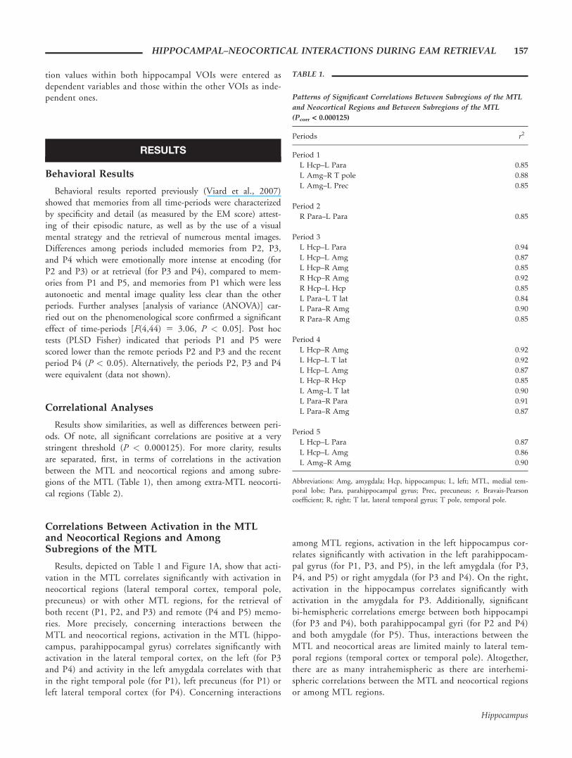
TABLE 1.
Patterns of Significant Correlations Between Subregions of the MTL
and Neocortical Regions and Between Subregions of the MTL
(Pcorr < 0.000125)
Periods r2
Period 1
L Hcp–L Para 0.85
L Amg–R T pole 0.88
L Amg–L Prec 0.85
Period 2
R Para–L Para 0.85
Period 3
L Hcp–L Para 0.94
L Hcp–L Amg 0.87
L Hcp–R Amg 0.85
R Hcp–R Amg 0.92
R Hcp–L Hcp 0.85
L Para–L T lat 0.84
L Para–R Amg 0.90
R Para–R Amg 0.85
Period 4
L Hcp–R Amg 0.92
L Hcp–L T lat 0.92
L Hcp–L Amg 0.87
L Hcp–R Hcp 0.85
L Amg–L T lat 0.90
L Para–R Para 0.91
L Para–R Amg 0.87
Period 5
L Hcp–L Para 0.87
L Hcp–L Amg 0.86
L Amg–R Amg 0.90
Abbreviations: Amg, amygdala; Hcp, hippocampus; L, left; MTL, medial tem-poral lobe; Para, parahippocampal gyrus; Prec, precuneus; r, Bravais-Pearsoncoefficient; R, right; T lat, lateral temporal gyrus; T pole, temporal pole.
HIPPOCAMPAL–NEOCORTICAL INTERACTIONS DURING EAM RETRIEVAL 157
Hippocampus
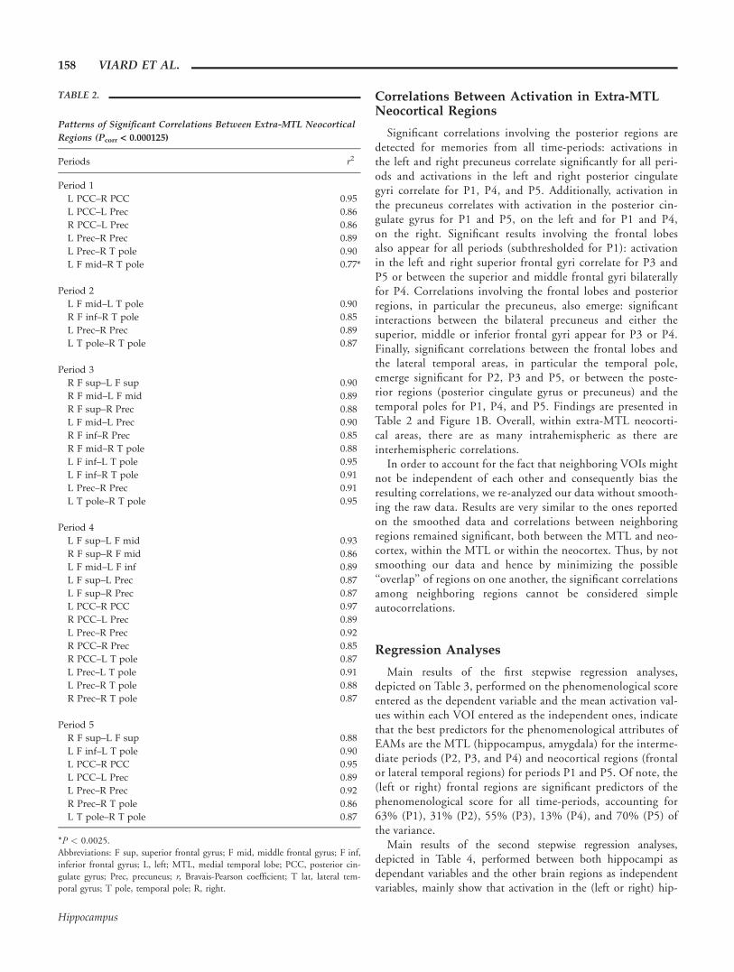
Correlations Between Activation in Extra-MTLNeocortical Regions
Significant correlations involving the posterior regions aredetected for memories from all time-periods: activations inthe left and right precuneus correlate significantly for all peri-ods and activations in the left and right posterior cingulategyri correlate for P1, P4, and P5. Additionally, activation inthe precuneus correlates with activation in the posterior cin-gulate gyrus for P1 and P5, on the left and for P1 and P4,on the right. Significant results involving the frontal lobesalso appear for all periods (subthresholded for P1): activationin the left and right superior frontal gyri correlate for P3 andP5 or between the superior and middle frontal gyri bilaterallyfor P4. Correlations involving the frontal lobes and posteriorregions, in particular the precuneus, also emerge: significantinteractions between the bilateral precuneus and either thesuperior, middle or inferior frontal gyri appear for P3 or P4.Finally, significant correlations between the frontal lobes andthe lateral temporal areas, in particular the temporal pole,emerge significant for P2, P3 and P5, or between the poste-rior regions (posterior cingulate gyrus or precuneus) and thetemporal poles for P1, P4, and P5. Findings are presented inTable 2 and Figure 1B. Overall, within extra-MTL neocorti-cal areas, there are as many intrahemispheric as there areinterhemispheric correlations.
In order to account for the fact that neighboring VOIs mightnot be independent of each other and consequently bias theresulting correlations, we re-analyzed our data without smooth-ing the raw data. Results are very similar to the ones reportedon the smoothed data and correlations between neighboringregions remained significant, both between the MTL and neo-cortex, within the MTL or within the neocortex. Thus, by notsmoothing our data and hence by minimizing the possible‘‘overlap’’ of regions on one another, the significant correlationsamong neighboring regions cannot be considered simpleautocorrelations.
Regression Analyses
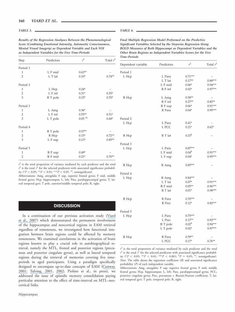
Main results of the first stepwise regression analyses,depicted on Table 3, performed on the phenomenological scoreentered as the dependent variable and the mean activation val-ues within each VOI entered as the independent ones, indicatethat the best predictors for the phenomenological attributes ofEAMs are the MTL (hippocampus, amygdala) for the interme-diate periods (P2, P3, and P4) and neocortical regions (frontalor lateral temporal regions) for periods P1 and P5. Of note, the(left or right) frontal regions are significant predictors of thephenomenological score for all time-periods, accounting for63% (P1), 31% (P2), 55% (P3), 13% (P4), and 70% (P5) ofthe variance.
Main results of the second stepwise regression analyses,depicted in Table 4, performed between both hippocampi asdependant variables and the other brain regions as independentvariables, mainly show that activation in the (left or right) hip-
TABLE 2.
Patterns of Significant Correlations Between Extra-MTL Neocortical
Regions (Pcorr < 0.000125)
Periods r2
Period 1
L PCC–R PCC 0.95
L PCC–L Prec 0.86
R PCC–L Prec 0.86
L Prec–R Prec 0.89
L Prec–R T pole 0.90
L F mid–R T pole 0.77*
Period 2
L F mid–L T pole 0.90
R F inf–R T pole 0.85
L Prec–R Prec 0.89
L T pole–R T pole 0.87
Period 3
R F sup–L F sup 0.90
R F mid–L F mid 0.89
R F sup–R Prec 0.88
L F mid–L Prec 0.90
R F inf–R Prec 0.85
R F mid–R T pole 0.88
L F inf–L T pole 0.95
L F inf–R T pole 0.91
L Prec–R Prec 0.91
L T pole–R T pole 0.95
Period 4
L F sup–L F mid 0.93
R F sup–R F mid 0.86
L F mid–L F inf 0.89
L F sup–L Prec 0.87
L F sup–R Prec 0.87
L PCC–R PCC 0.97
R PCC–L Prec 0.89
L Prec–R Prec 0.92
R PCC–R Prec 0.85
R PCC–L T pole 0.87
L Prec–L T pole 0.91
L Prec–R T pole 0.88
R Prec–R T pole 0.87
Period 5
R F sup–L F sup 0.88
L F inf–L T pole 0.90
L PCC–R PCC 0.95
L PCC–L Prec 0.89
L Prec–R Prec 0.92
R Prec–R T pole 0.86
L T pole–R T pole 0.87
*P < 0.0025.Abbreviations: F sup, superior frontal gyrus; F mid, middle frontal gyrus; F inf,inferior frontal gyrus; L, left; MTL, medial temporal lobe; PCC, posterior cin-gulate gyrus; Prec, precuneus; r, Bravais-Pearson coefficient; T lat, lateral tem-poral gyrus; T pole, temporal pole; R, right.
158 VIARD ET AL.
Hippocampus
pocampus is best predicted by activation in the MTL regions(parahippocampal gyrus and amygdala) and then neocorticalregions (frontal except for P5, temporal or posterior regions:
precuneus and posterior cingulate cortex). Overall, the regres-sion analyses show a pattern of significant MTL–neocorticalinteraction regardless of the time-period.
FIGURE 1. Schematic illustration of results from the correla-tional analyses between (A) subregions of the MTL and neocorticalregions and among MTL regions, and (B) extra-MTL neocorticalregions, for each time-period. Numeric values represent significantcorrelational coefficients (r 2) between the two subregions connectedby arrows (Pcorr < 0.000125). Abbreviations: Amg, amygdala; F sup,
superior frontal gyrus; F mid, middle frontal gyrus; F inf, inferiorfrontal gyrus; Hcp, hippocampus; L, left; Para, parahippocampalgyrus; PCC, posterior cingulate gyrus; Prec, precuneus; R, right; Tlat, lateral temporal gyrus; T pole, Temporal pole. [Color figure canbe viewed in the online issue, which is available atwww.interscience.wiley.com.]
HIPPOCAMPAL–NEOCORTICAL INTERACTIONS DURING EAM RETRIEVAL 159
Hippocampus
DISCUSSION
In a continuation of our previous activation study (Viardet al., 2007) which demonstrated the permanent involvementof the hippocampus and neocortical regions in EAM retrievalregardless of remoteness, we investigated how functional inte-gration between brain regions could be affected by memoryremoteness. We examined correlations in the activation of brainregions known to play a crucial role in autobiographical re-trieval, namely the MTL, frontal and posterior regions (precu-neus and posterior cingulate gyrus), as well as lateral temporalregions during the retrieval of memories covering five time-periods in aged participants. Using a paradigm specificallydesigned to encompass up-to-date concepts of EAM (Conway,2001; Tulving, 2001, 2002; Piolino et al., in press), weaddressed the issue of episodic memory consolidation payingparticular attention to the effect of time-interval on MTL–neo-cortical links.
TABLE 3.
Results of the Regression Analyses Between the Phenomenological
Score (Combining Emotional Intensity, Autonoetic Consciousness,
Mental Visual Imagery) as Dependent Variable and Each VOI
as Independent Variables for the Five Time-Periods
Step Predictors r2 Total r2
Period 1
1 L F mid 0.63** –
2 L T lat 0.10* 0.74**
Period 2
1 L Hcp 0.24* –
2 L F inf 0.31* 0.55*
3 R T pole 0.15* 0.70*
Period 3
1 L Amg 0.36* –
2 L F inf 0.55** 0.51*
3 L T pole 0.05 ns 0.68*
Period 4
1 R T pole 0.57** –
2 R Hcp 0.15* 0.72**
3 L F sup 0.13* 0.85**
Period 5
1 R F sup 0.49* –
2 R F mid 0.21* 0.70**
r2 is the total proportion of variance mediated by each predictor and the totalr2 is the total r2 for the selected predictors with associated significance probabil-ity (*P < 0.05; **P < 0.01; ***P < 0.05, ns, nonsignificant).Abbreviations: Amg, amygdala; F sup, superior frontal gyrus; F mid, middlefrontal gyrus; Hcp, hippocampus; L, left; Para, parahippocampal gyrus; T, lat-eral temporal gyri; T pole, anterior/middle temporal pole; R, right.
TABLE 4.
Final Multiple Regression Model Performed on the Predictive
Significant Variables Selected by the Stepwise Regression Using
BOLD Measures of Both Hippocampi as Dependent Variables and the
Other Brain Regions as Independent Variables Scores for the Five
Time-Periods
Dependent variable Predictors r2 Total r2
Period 1
L Hcp L Para 0.71*** –
L T lat 0.17** 0.88***
L F mid 0.06* 0.94***
R F inf 0.02* 0.97***
R Hcp L Amg 0.58** –
R F inf 0.27** 0.85**
R F sup 0.06* 0.91***
R Para 0.04* 0.95***
Period 2
L Hcp L Para 0.41* –
L PCC 0.21* 0.62*
R Hcp R T lat 0.23$ –
Period 3
L Hcp L Para 0.87*** –
L F mid 0.04$ 0.91***
L F sup 0.04* 0.95***
R Hcp R Amg 0.85*** –
Period 4
L Hcp R Amg 0.84*** –
L T lat 0.07* 0.91***
R F mid 0.05** 0.96***
R T lat 0.01* 0.98***
R Hcp R Para 0.70*** –
R Prec 0.12* 0.82***
Period 5
L Hcp L Para 0.75*** –
L Prec 0.17** 0.92***
R T pole 0.02$ 0.94***
L T pole 0.02* 0.97***
R Hcp R Para 0.59** –
L PCC 0.17* 0.76**
r2 is the total proportion of variance mediated by each predictor and the totalr2 is the total r2 for the selected predictors with associated significance probabil-ity (*P < 0.05; **P < 0.01; ***P < 0.001; $P 5 0.05; ns, nonsignificant).Note: The table shows the regression coefficient (B) and associated significanceprobability (P) of each independent variable.Abbreviations: Amg, amygdala; F sup, superior frontal gyrus; F mid, middlefrontal gyrus; Hcp, hippocampus; L, left; Para, parahippocampal gyrus; PCC,posterior cingulate gyrus; Prec, precuneus; r, Bravais-Pearson coefficient; T, lat-eral temporal gyri; T pole, temporal pole; R, right.
160 VIARD ET AL.
Hippocampus
Our previous findings had shown that the recollection ofEAMs from the five time-periods triggered activation in a cir-cumscribed network, including the left hippocampus and supe-rior frontal gyrus, as well as the precuneus and posterior cingu-late gyrus, bilaterally (Viard et al., 2007). Behavioral resultsindicated that regardless of the age of memories, recollectionwas characterized by specificity, attesting of their episodic na-ture (i.e., spatiotemporal uniqueness and details). Differencesamong periods included memories from P2, P3, and P4 whichwere rated stronger than the other periods (P1 and P5) interms of the phenomenological attributes of memories andrecruited, additionally, the right hippocampus.
New findings based on correlational and regression analysesshow a pattern of significant MTL–neocortical or MTL-MTLinteractions for all time-periods, as well as strong correlationswithin extra-MTL neocortical regions. These results will be dis-cussed in light of the two conflicting models of long-termmemory consolidation, first, in terms of activation betweenMTL and neocortical regions, then among subregions of theMTL and, finally, among neocortical regions.
Correlations Between Activation in the MTL andNeocortical Regions
Activations in the MTL and neocortex (lateral temporal cor-tex, temporal pole, precuneus) strongly correlate for bothremote (P1 and P3) and recent (P4) periods, indicating thatMTL–neocortical links are still present during the retrieval ofepisodic memories whatever their remoteness. This result isconcordant with the proposed permanent link between medialtemporal and neocortical regions in the retrieval of EAMs irre-spective of their remoteness (MTT; Nadel and Moscovitch,1997; Nadel et al., 2007) and refutes the standard model whichwould have predicted greater MTL–neocortical correlations forrecent compared to remote AMs. Instead, MTT postulates thatthe MTL as a whole (i.e., including the hippocampus, parahip-pocampal gyrus and amygdala) interacts permanently with neo-cortical regions during the retrieval of both recent and remoteepisodic AMs. Our results are also in line with an increasingnumber of studies using the voxel-based approach which haveshown the permanent involvement of the MTL in the retrievalof EAMs whatever their remoteness (Ryan et al., 2001; Addiset al., 2004b; Gilboa et al., 2004; Piolino et al., 2004; Rekkasand Constable, 2005; Steinvorth et al., 2006; Nadel et al.,2007; Viard et al., 2007; Daselaar et al., 2008; Piolino et al.,2008). Further support to MTT is brought by recent patientstudies which suggest that MTL damage impairs remote mem-ory retrieval to a greater extent than was previously thought(Steinvorth et al., 2006; Noulhiane et al., 2007), although theimportance of retrograde amnesia depends on the extent of thelesion (Hepner et al., 2007; Kirwan et al., 2008).
Interestingly, no significant correlations were detectedbetween activation in the frontal and medial temporal regions,possibly due to the use of a stringent threshold, but consistentwith previous findings (Markowitsch, 1995). Based on neuro-psychological observations, Markowitsch (1995) proposed that
prefrontal regions communicate with the lateral (and not themedial) part of the temporal lobes, in its polar area, and this ismade possible via a bundle of fibers, the uncinate fasciculus,which unites the frontal and temporal lobes. Indeed, our resultsindicate that activation in the medial temporal regions signifi-cantly correlate with the lateral temporal areas, both for remote(P1 and P3) and recent (P4) periods. This is also consistentwith previous connectivity findings which showed that the rec-ollection of autobiographical events caused increased connectiv-ity between the parahippocampal gyrus and the lateral temporalregions (lateral temporal cortex and temporal pole), relative topublic events (Maguire et al., 2000). We can extrapolate thesefindings to pathology in which an interruption of circuits con-necting frontal and temporo-polar regions produces a deficit inrecall, even with intact MTL structures, resulting in a discon-nection syndrome (Markowitsch, 1995; Levine et al., 1998;Piolino et al., 2005).
Correlations Among Subregions of the MTL
Activation in the hippocampus correlates significantly withactivation in other MTL regions (parahippocampal gyrus,amygdala) during the retrieval of recent (P1 and P3) andremote (P4 and P5) EAMs. Additionally, we detected signifi-cant correlations in the activation of both hippocampi forremote (P3) and recent (P4) periods, suggesting that MTL–MTL links are permanently required for the retrieval of long-term EAMs regardless of memory age. These findings are thusconcordant with MTT which would predict long-lasting inter-actions among MTL subregions (Nadel et al., 2007) and refutepredictions of the standard model which would predict greaterMTL-MTL interactions for recent than for remote memories(Squire and Alvarez, 1995). Results also showed significant cor-relations involving the parahippocampal gyrus, either bi-hemi-spheric correlations (for P2 and P4) or correlations with otherMTL subregions (hippocampus or amygdala) for both remote(P1, P3) and recent (P4, P5) periods. Similarly, Addis et al.(2004a) showed that the left and right hippocampi were bothfunctionally connected during AM retrieval, as well as with theright parahippocampal gyrus. While these findings supportMTT and confirm the importance of the hippocampus inEAM retrieval whatever memory remoteness, they also under-line the role of the parahippocampal gyrus and an interactionbetween both structures to support the recollection of personalpast events (Maguire et al., 2000; Tsukiura et al., 2002; Okudaet al., 2003; St Jacques et al., 2008). Tsukiura et al. (2002)suggest that the parahippocampal gyrus, particularly on theright, may be implicated in the retrieval of topographical orspatial EAMs (Niki and Luo, 2002; Moscovitch et al., 2005).They propose that right parahippocampal activation could berelated to the recruitment of posterior visual areas during theretrieval of older episodic memories. Although we did notdetect a direct link between the parahippocampal gyrus andposterior areas, activation in the latter strongly correlated withother neocortical regions (see below).
HIPPOCAMPAL–NEOCORTICAL INTERACTIONS DURING EAM RETRIEVAL 161
Hippocampus
Our results highlight the involvement of the amygdala andits interaction with the hippocampus and parahippocampalgyrus, particularly during the retrieval of memories from peri-ods P3 and P4. Behaviorally, these periods were rated higher interms of emotional intensity at retrieval compared to memoriesfrom the other periods. Emotion is an important phenomeno-logical quality of persistent and vivid EAMs (Christianson,1992; Talarico et al., 2004). Interestingly, our regressions analy-ses showed that the phenomenological attributes of EAMs(phenomenological score) are best predicted by the amygdalafor P3, period which in our sample was rated the most intenseemotionally, both at encoding and at retrieval. Much evidencesuggests that the enhanced memory capability observed foremotional events is due, at least in part, to the amygdala’sinfluence on encoding and storage of hippocampal-dependentmemories (for review, see Phelps, 2004), as suggested by manystudies detecting amygdalar activation during EAM retrieval(Fink et al., 1996; Markowitsch et al., 2000, 2003; Maguireand Frith, 2003; Addis et al., 2004; Greenberg et al., 2005;Daselaar et al., 2008). Functional interactions have beendetected between the amygdala and the hippocampus duringencoding (Hamann et al., 1999; Dolcos et al., 2004), as well asduring retrieval (Dolcos et al., 2005) especially if recall isaccompanied by a sense of recollection (see also, Sharot et al.,2004). Thus, our results further highlight the role of the amyg-dala in phenomenological processes and the crucial role ofemotion in the reviviscence of EAMs.
General Discussion on the MTL–NeocorticalInteractions
Overall, there are as many intrahemispheric as there areinterhemispheric correlations between the MTL and neocorticalregions and the same is true within the MTL alone. This isconcordant with a growing number of recent activation studieswhich detected bilateral MTL activation when participantswere engaged in the retrieval of specific AMs (i.e., EAMs) ratedstrongly in terms of vividness, richness of detail, emotionality,re-experiencing or personal significance (Ryan et al., 2001;Okuda et al., 2003; Piefke et al., 2003; Gilboa et al., 2004;Mayes et al., 2004; Piolino et al., 2004; Greenberg et al.,2005; Steinvorth et al., 2006; Viard et al., 2007). Thus, com-bined with voxel-based studies, our results stress the idea that abilateral interplay between MTL–neocortical regions character-izes rich EAM recollection (see also Piolino et al., 2008).
Our regression analyses provided further insights about therelationship between the phenomenological attributes of EAMs(via the phenomenological score which combines emotional in-tensity, autonoetic consciousness, mental visual strategy, mentalimage quality, number of mental images and viewpoint per-spective) and certain brain regions. Interestingly, the phenome-nological score was best predicted by the MTL (hippocampusand amygdala) for intermediate periods (P2, P3, and P4), whilethe score was best predicted by neocortical regions (frontal andlateral temporal regions) for periods P1 and P5. This might beexplained by the fact that memories from intermediate periods
were phenomenologically different compared to the very recent(P1) and very remote memories (P5). Indeed, although memo-ries from all time-periods were specific (i.e., episodic, character-ized by spatiotemporal uniqueness and details, as measured bythe objective EM score), some memories showed modulationson certain subjective scales and were thus phenomenologicallydifferent according to the period considered (e.g., higher emo-tional intensity at retrieval for P3 and P4 compared to P1 andP5; lower autonoetic consciousness and quality of mentalimages for P1). In fact, the objective EM score taps a differentaspect of episodicity than the subjective recollective ratings.Indeed, one has to reach a certain threshold of episodicity, inorder for an event to be episodic. Then, above the threshold,there are graduations or modulations on certain episodic qual-ities (Piolino et al., 2006; Viard et al., 2007).
Altogether, our results suggest that the retention interval(e.g., memory remoteness) is not the only factor which influen-ces cerebral activity and interactions: richness of recollection(i.e., the quality of memories retrieved) has a crucial role onbrain activity and brain interactions. This conclusion is actuallyin line with (and confirms) recent neuroimaging studies:beyond memory remoteness, richness of recollection (emotion,visual imagery, vividness, level of detail, personal importance)strongly influences patterns of brain activations (Ryan et al.,2001; Addis et al., 2004; Gilboa et al., 2005; for reviews, seeMoscovitch et al., 2005; Cabeza and St Jacques, 2007). Over-all, our results are very much in accordance with predictions ofMTT which stress that all the phenomenological features ofEAM retrieval are crucial for the continuous involvement ofMTL–neocortical associations (see Moscovitch et al., 2005;Nadel et al., 2007).
Correlations Between Activation in Extra-MTLNeocortical Regions
Our results indicate a continuous interaction between differ-ent neocortical sites for recent and remote periods, as predictedby both models of memory consolidation. Indeed, the standardmodel and MTT both suggest that cortico-cortical connectionspersist through time and will be strengthened as the memoriesare consolidated (Squire and Alvarez, 1995; Nadel andMoscovitch, 1997; Nadel et al., 2007). Here, we show that, forall time-periods, interactions among neocortical regions involveboth anterior and posterior regions.
Strong correlations involving posterior regions are detectedbetween the bilateral precuneus (for all time-periods) andbetween the precuneus and posterior cingulate gyrus (for P1,P4, and P5). Previous findings suggest a role of the precuneus,and the adjacent posterior cingulate cortex, in self-referentialprocesses (Fink et al., 1996; Maddock et al., 2001). Along withother cortical midline structures (or CMS), both regions arehypothesized to play a role in generating a model of the self(Northoff and Bermpohl, 2004). In our study, the stimuliselected were highly self-relevant and targeted specific andunique AMs in the participants’ lives. Furthermore, a role ofthe precuneus in visual mental imagery during episodic mem-
162 VIARD ET AL.
Hippocampus
ory retrieval has previously been shown (Shallice et al., 1994;Fletcher et al., 1995; Cavanna and Trimble, 2006). Visualmental imagery increases the recall of EAMs: detailed memoriesare often accompanied by strong imagery reports (Brewer,1986, 1996; Dewhurst and Conway, 1994; Greenberg andRubin, 2003). Indeed, our behavioral data indicate that, for alltime-periods, the strategy used to retrieve memories was mas-sively visual, possibly reflecting access to event-specific knowl-edge (ESK, Conway and Pleydell-Pearce, 2000).
Significant correlations involving frontal regions (superior,middle or inferior frontal gyri) are detected for all time-periods(subthresholded for P1), either fronto-frontal (for P3, P4, andP5) or between the frontal lobes and other neocortical regions(e.g., lateral temporal cortex or temporal pole for P1, P2, P3,and P5; precuneus for P3 and P4). Neuroimaging studies haveprovided extensive evidence that links episodic memory andAM to distinct functions of the frontal lobes, such as strategicretrieval processing, self referential processing, monitoringrelated to self processing (for reviews, see Cabeza and Nyberg,2000; Gilboa, 2004; Svoboda et al., 2006; Cabeza and StJacques, 2007). The prefrontal lobe is one of the criticalregions for the emergence of autonoetic consciousness (Wheeleret al., 1997) and is also essential in self-awareness present atretrieval (Levine et al., 1998; Piolino et al., 2005). Lesion stud-ies have shown that damage to the (right) prefrontal lobes canlead to disruption in the way individuals think about them-selves: patients know what happened to them, but are uncon-cerned or indifferent, and their memories seem to lack personalsignificance (Wheeler et al., 1997). Our behavioral findingsshow that memories from all time-periods were recollectedwith an autonoetic state of consciousness. Moreover, strongcorrelations were detected among frontal regions probably sup-porting the rise of autonoetic consciousness in the retrievalprocess. Further confirmation was provided by the stepwiseregression analyses which showed that, for all time-periods (leftor right) frontal regions were good predictors of the phenome-nological score (which encompasses, in particular, autonoeticconsciousness). Overall, our results highlight the crucial role ofautonoetic consciousness in the reviviscence of EAMs and pro-vide further evidence of a role of the frontal regions in access-ing phenomenologically rich EAMs.
Finally, strong correlations involving the lateral temporalregions (temporal pole or lateral temporal cortex) emerged dur-ing the retrieval of memories from all time-periods, either cor-relations with frontal regions (for P2, P3, and P5) or with theprecuneus (for P1, P4, and P5) or bi-hemispheric interactionsbetween the left and right temporal poles (for P2, P3, and P5).Increased connectivity among lateral temporal areas has beendetected during the recognition of general knowledge and pub-lic events (Maguire et al., 2000), both semantic in nature, sug-gesting a role of these regions in semantic processes. In fact,evidence has linked the functions of the lateral temporal lobesto personal semantic memory processes (Mummery et al.,1996; Lee et al., 2002; Piolino et al., 2007; for review, seeSvoboda et al., 2006). It has been hypothesized that the tempo-ral pole acts as a convergence zone and integrates information
from hippocampal structures and posterior association regions(Damasio, 1989; Maguire et al., 2000; Svoboda et al., 2006).Temporopolar activations are often reported in studies of AMretrieval and lesion to this region may cause focal retrogradeamnesia (Wheeler and McMillan, 2001). Lateral temporalregions are also functionally connected with the hippocampusduring the retrieval of both unique (specific) and repeated (gen-eral) autobiographical events (Addis et al., 2004a). Similarly,our regression analyses show that the lateral temporal regionsare good predictors of hippocampal activation for all time-peri-ods. Hence, these findings stress the role of semantic knowledgein accessing EAMs. More generally, our findings substantiatethat episodic and semantic memory processes are integral partsof AM recollection and access to episodic events is often man-aged by first sifting through the general autobiographicalknowledge (Conway and Bekerian, 1987; Conway andPleydell-Pearce, 2000; Conway et al., 2001; Levine et al.,2004; Svoboda et al., 2006).
CONCLUSIONS
Our main results indicate that activation in medial temporaland neocortical regions correlate during the retrieval of allEAMs, whether they are recent or remote, emphasizing a pat-tern of significant MTL–neocortical interaction regardless ofthe passage of time. We also show that MTL–MTL correlationsremain constant for both recent and remote periods. Both setsof results are concordant with the proposed permanent linkbetween MTL and neocortical regions and the persistence ofMTL interactions for memories of all ages, as hypothesized byMTT. Our results also showed that richness of recollection(i.e., quality of memories retrieved) has a crucial role on brainactivity and its interactions: richly recollected memories (fromintermediate periods) recruited a larger bilateral MTL–neocorti-cal correlational network. Our main results point that MTT,and more generally models of memory consolidation, could becomplemented and possibly strengthened by considering moreprecisely the modulation of the MTL–neocortical interactionsas a function of the phenomenological features of specificmemories retrieved, such as emotion, mental visual imageryand, state of consciousness.
An aspect which we could not address in this study (due todesign limitations) is the directionality of interaction. Effectiveconnectivity methods, such as dynamic causal modeling(DCM), are particularly appealing to determine causal out-comes. It would also be interesting, in future studies, to traceindividualized ROIs to better delineate finer subregions in themedial temporal lobe, for example, in the parahippocampalgyrus (parahippocampal, entorhinal and perirhinal cortices), inorder to distinguish the separate contributions of each of thesesubregions in EAM retrieval, across time. Further research is,thus, needed to clarify the influence that the regions of theEAM network exert over one another and the contribution ofthe finer subregions of this system.
HIPPOCAMPAL–NEOCORTICAL INTERACTIONS DURING EAM RETRIEVAL 163
Hippocampus
Acknowledgments
A.V. was supported by the Association France Alzheimer’sFellowships for Young Researchers.
REFERENCES
Addis DR, McIntosh AR, Moscovitch M, Crawley AP, McAndrewsMP. 2004a. Characterizing spatial and temporal features of auto-biographical memory retrieval networks: A partial least squaresapproach. Neuroimage 23:1460–1471.
Addis DR, Moscovitch M, Crawley AP, McAndrews MP. 2004b. Rec-ollective qualities modulate hippocampal activation during autobio-graphical memory retrieval. Hippocampus 14:752–762.
Ashburner J, Friston KJ. 2005. Unified segmentation. Neuroimage26:839–851.
Bayley PJ, Squire LR. 2005. Failure to acquire new semantic knowl-edge in patients with large medial temporal lobe lesions. Hippo-campus 15:273–280.
Brewer W. 1986. What is autobiographical memory? In: Rubin DC,editor. Autobiographical Memory. Cambridge: Cambridge Univer-sity Press. pp 25–49.
Brewer W. 1996. What is recollective memory? In: Rubin DC, editor.Remembering Our Past: Studies in Autobiographical Memory.Cambridge: Cambridge University Press. pp 19–66.
Cabeza R, Nyberg L. 2000. Imaging cognition. II. An empirical reviewof 275 PET and fMRI studies. J Cogn Neurosci 12:1–47.
Cabeza R, St Jacques P. 2007. Functional neuroimaging of autobio-graphical memory. Trends Cogn Sci 11:219–227.
Cavanna AE, Trimble MR. 2006. The precuneus: A review of its func-tional anatomy and behavioural correlates. Brain 129:564–583.
Conway MA. 2001. Sensory–perceptual episodic memory and its con-text: Autobiographical memory. Philos Trans R Soc Lond B BiolSci 356:1375–1384.
Conway MA, Bekerian DA. 1987. Organization in autobiographicalmemory. Mem Cognit 15:119–132.
Conway MA, Pleydell-Pearce CW. 2000. The construction of autobio-graphical memories in the self-memory system. Psychol Rev107:261–288.
Conway MA, Pleydell-Pearce CW, Whitecross SE. 2001. The neuroan-atomy of autobiographical memory: A slow cortical potentials(SCPs) study of autobiographical memory retrieval. J Mem Lang45:493–524.
Conway MA, Pleydell-Pearce CW, Whitecross SE, Sharpe H. 2003.Neurophysiological correlates of memory for experienced and imag-ined events. Neuropsychologia 41:334–340.
Crawley SE, French CC. 2005. Field and observer viewpoint inremember-know memories of personal childhood events. Memory13:673–681.
Damasio AR. 1989. Time-locked multiregional retroactivation: A sys-tems-level proposal for the neural substrates of recall and recogni-tion. Cognition 33:25–62.
Daselaar SM, Rice HJ, Greenberg DL, Cabeza R, Labar KS, RubinDC. 2008. The spatiotemporal dynamics of autobiographicalmemory: Neural correlates of recall, emotional intensity, and reliv-ing. Cerebral Cortex 18:217–229.
Dewhurst SA, Conway MA. 1994. Pictures, images, and recollectiveexperience. J Exp Psychol Learn Mem Cogn 20:1088–1098.
Dolan RJ, Lane R, Chua P, Fletcher P. 2000. Dissociable temporallobe activations during emotional episodic memory retrieval.NeuroImage 11:203–209.
Dolcos F, LaBar KS, Cabeza R. 2004. Interaction between the amyg-dala and the medial temporal lobe memory system predicts bettermemory for emotional events. Neuron 42:855–863.
Dolcos F, LaBar KS, Cabeza R. 2005. Remembering one year later:role of the amygdala and the medial temporal lobe memory systemin retrieving emotional memories. Proc Natl Acad Sci USA102:2626–2631.
Fink GR, Markowitsch HJ, Reinkemeier M, Bruckbauer T, Kessler J,Heiss WD. 1996. Cerebral representation of one’s own past: Neu-ral networks involved in autobiographical memory. J Neurosci16:4275–4282.
Fletcher PC, Frith CD, Baker SC, Shallice T, Frackowiak RS, DolanRJ. 1995. The mind’s eye-precuneus activation in memory relatedimagery. Neuroimage 2:195–200.
Gilboa A. 2004. Autobiographical and episodic memory-one and thesame? Evidence from prefrontal activation in neuroimaging studies.Neuropsychologia 42:1336–1349.
Gilboa A, Winocur G, Grady CL, Hevenor SJ, Moscovitch M. 2004.Remembering our past: Functional neuroanatomy of recollection ofrecent and very remote personal events. Cereb Cortex 14:1214–1225.
Good CD, Johnsrude IS, Ashburner J, Henson RN, Friston KJ, Frack-owiak RS. 2001. A voxel-based morphometric study of ageing in465 normal adult human brains. Neuroimage 14:21–36.
Greenberg DL, Rubin DC. 2003. The neuropsychology of autobio-graphical memory. Cortex 39:687–728.
Greenberg DL, Rice HJ, Cooper JJ, Cabeza R, Rubin DC, Labar KS.2005. Co-activation of the amygdala, hippocampus and inferiorfrontal gyrus during autobiographical memory retrieval. Neuropsy-chologia 43:659–674.
Hepner IJ, Mohamed A, Fulham MJ, Miller LA. 2007. Topographical,autobiographical and semantic memory in a patient with bilateralmesial temporal and retrosplenial infarction. Neurocase 13:97–114.
Kirwan CB, Bayley PJ, Galvan VV, Squire LR. 2008. Detailed recol-lection of remote autobiographical memory after damage to themedial temporal lobe. Proc Natl Acad Sci USA 105:2676–2680.
Lee AC, Robbins TW, Graham KS, Owen AM. 2002. ‘‘Pray or prey?’’dissociation of semantic memory retrieval from episodic memoryprocesses using positron emission tomography and a novel homo-phone task. Neuroimage 16:724–735.
Levine B, Black SE, Cabeza R, Sinden S, Meintosh AR, Toth JP, Tulv-ing E, Stuss DT. 1998. Episodic memory and the self in a case ofisolated retrograde amnesia. Brain 121:1951–1973.
Levine B, Turner GR, Tisserand D, Hevenor SJ, Graham SJ, McIntoshAR. 2004. The functional neuroanatomy of episodic and semanticautobiographical remembering: A prospective functional MRIstudy. J Cogn Neurosci 16:1633–1646.
Maddock RJ, Garrett AS, Buonocore MH. 2001. Remembering famil-iar people: The posterior cingulate cortex and autobiographicalmemory retrieval. Neuroscience 104:667–676.
Maguire EA. 2001. Neuroimaging studies of autobiographical eventmemory. Philos Trans R Soc Lond B Biol Sci 356:1441–1451.
Maguire EA, Frith CD. 2003a. Lateral asymmetry in the hippocampalresponse to the remoteness of autobiographical memories. J Neuro-sci 23:5302–5307.
Maguire EA, Frith CD. 2003b. Aging affects the engagement of thehippocampus during autobiographical memory retrieval. Brain126:1511–1523.
Maguire EA, Mummery CJ, Buchel C. 2000. Patterns of hippocam-pal–cortical interaction dissociate temporal lobe memory subsys-tems. Hippocampus 10:475–482.
Markowitsch HJ. 1995. Which brain regions are critically involved inthe retrieval of old episodic memory? Brain Res Rev 21:117–127.
Markowitsch HJ, Thiel A, Reinkemeier M, Kessler J, Koyuncun A,Heiss WD. 2000. Right amygdalar and temporofrontal activationduring autobiographic, but not fictitious memory retrieval. BehavNeurol 12:181–190.
Markowitsch HJ, Vandekerckhove MM, Lanfermann H, Russ MO.2003. Engagement of lateral and medial prefrontal areas in the
164 VIARD ET AL.
Hippocampus
ecphory of sad and happy autobiographical memories. Cortex39:643–665.
Mayes AR, Roberts N. 2001. Theories of episodic memory. PhilosTrans R Soc Lond B Biol Sci 356:1395–1408.
Mayes AR, Montaldi D, Spencer TJ, Roberts N. 2004. Recalling spa-tial information as a component of recently and remotely acquiredepisodic or semantic memories: An fMRI study. Neuropsychology18:426–441.
Moscovitch M, Rosenbaum RS, Gilboa A, Addis DR, Westmacott R,Grady C, McAndrews MP, Levine B, Black S, Winocur G, NadelL. 2005. Functional neuroanatomy of remote episodic, semanticand spatial memory: A unified account based on multiple tracetheory. J Anat 207:35–66.
Mummery CJ, Patterson K, Hodges JR, Wise RJ. 1996. Generating‘‘tiger’’ as an animal name or a word beginning with T: Differencesin brain activation. Philos Trans R Soc Lond B Biol Sci 263:989–995.
Nadel L, Moscovitch M. 1997. Memory consolidation, retrograde am-nesia and the hippocampal complex. Curr Opin Neurobiol 7:217–227.
Nadel L, Campbell J, Ryan L. 2007. Autobiographical memory re-trieval and hippocampal activation as a function of repetition andthe passage of time. Neural Plast 2007:90472
Niki K, Luo J. 2002. An fMRI study on the time-limited role of themedial temporal lobe in long-term topographical autobiographicmemory. J Cogn Neurosci 14:500–507.
Northoff G, Bermpohl F. 2004. Cortical midline structures and theself. Trends Cogn Sci 8:102–107.
Noulhiane M, Piolino P, Hasboun D, Baulac M, Samson S. 2007.Autobiographical memory after temporal lobe resection: Neuropsy-chological and MRI volumetric findings. Brain 130:3184–3199.
Okuda J, Fujii T, Ohtake H, Tsukiura T, Tanji K, Suzuki K, Kawa-shima R, Fukuda H, Itoh M, Yamadori A. 2003. Thinking of thefuture and past: The roles of the frontal pole and the medial tem-poral lobes. Neuroimage 19:1369–1380.
Phelps EA. 2004. Human emotion and memory: interactions of theamygdala and hippocampal complex. Curr Opin Neurobiol14:198–202.
Piefke M, Weiss PH, Zilles K, Markowitsch HJ, Fink GR. 2003. Dif-ferential remoteness and emotional tone modulate the neural corre-lates of autobiographical memory. Brain 126:650–668.
Piolino P, Desgranges B, Benali K, Eustache F. 2002. Episodic andsemantic remote autobiographical memory in ageing. Memory10:239–257.
Piolino P, Giffard-Quillon G, Desgranges B, Chetelat G, Baron JC,Eustache F. 2004. Re-experiencing old memories via hippocampus:A PET study of autobiographical memory. Neuroimage 22:1371–1383.
Piolino P, Hannequin D, Desgranges B, Girard B, Beaunieux H, Gif-fard B, Lebreton K, Eustache F. 2005. Right ventral frontal hypo-metabolism and abnormal sense of self in a case of disproportion-ate retrograde amnesia. Cogn Neuropsychol 22:1005–1034.
Piolino P, Desgranges B, Clarys D, Guillery-Girard B, Taconnat L,Isingrini M, Eustache F. 2006. Autobiographical memory, autono-etic consciousness and self-perspective in aging. Psychol Aging21:510–525.
Piolino P, Chetelat G, Matuszewski V, Landeau B, Mezenge F, ViaderF, de la Sayette V, Eustache F, Desgranges B. 2007. In search ofautobiographical memories: A PET study in the frontal variant offrontotemporal dementia. Neuropsychologia 45:2730–2743.
Piolino P, Desgranges B, Hubert V, Bernard F, Chetelat G, Baron JC,Eustache F. 2008. Reliving lifelong episodic autobiographical mem-ories via the hippocampus: A correlative resting PET study inhealthy middle-aged subjects. Hippocampus 18:445–459.
Piolino P, Desgranges B, Eustache F. Episodic autobiographical mem-ory over the course of time: Cognitive, neuropsychological andneuroimaging findings. Special Issue on Episodic memory and thebrain [theoretical review paper]. Neuropsychologia, in press.
Rekkas PV, Constable RT. 2005. Evidence that autobiographic mem-ory retrieval does not become independent of the hippocampus:An fMRI study contrasting very recent with remote events. J CognNeurosci 17:1950–1961.
Robinson JA, Swanson KL. 1993. Field and observer modes ofremembering. Memory 1:169–184.
Rubin DC, Schrauf RW, Greenberg DL. 2003. Belief and recollectionof autobiographical memories. Mem Cognit 31:887–901.
Ryan L, Nadel L, Keil K, Putnam K, Schnyer D, Trouard T,Moscovitch M. 2001. Hippocampal complex and retrieval of recentand very remote autobiographical memories: Evidence from func-tional magnetic resonance imaging in neurologically intact people.Hippocampus 11:707–714.
Shallice T, Fletcher P, Frith CD, Grasby P, Frackowiack RSJ, Dolan R.1994. Brain regions associated with acquisition and retrieval ofverbal episodic memory. Nature 121:561–579.
Sharot T, Delgado MR, Phelps EA. 2004. How emotion enhances thefeeling of remembering. Nat Neurosci 7:1376–1380.
Simons JS, Spiers HJ. 2003. Prefrontal and medial temporal lobeinteractions in long-term memory. Nature Rev Neurosci 4:637–648.
Squire LR, Alvarez P. 1995. Retrograde amnesia and memory consoli-dation: A neurobiological perspective. Curr Opin Neurobiol5:169–177.
St Jacques P, Rubin DC, LaBar KS, Cabeza R. 2008. The short andlong of it: Neural correlates of temporal-order memory for autobio-graphical events. J Cogn Neurosci 20:1327–1341.
Steinvorth S, Corkin S, Halgren E. 2006. Ecphory of autobiographicalmemories: An fMRI study of recent and remote memory retrieval.Neuroimage 30:285–298.
Svoboda E, McKinnon MC, Levine B. 2006. The functional neuroan-atomy of autobiographical memory: A meta-analysis. Neuropsycho-logia 44:2189–2208.
Talarico JM, LaBar KS, Rubin DC. 2004. Emotional intensity predictsautobiographical memory experience. Mem Cognit 32:1118–1132.
Tsukiura T, Fujii T, Okuda J, Ohtake H, Kawashima R, Itoh M,Fukuda H, Yamadori A. 2002. Time-dependent contribution ofthe hippocampal complex when remembering the past: A PETstudy. Neuroreport 13:2319–2323.
Tulving E. 2001. Episodic memory and common sense: How farapart? Philos Trans R Soc Lond B Biol Sci 356:1505–1515.
Tulving E. 2002. Episodic memory: From mind to brain. Ann RevPsychol 53:1–25.
Tulving E, Schacter DL, McLachlan DR, Moscovitch M. 1988. Pri-ming of semantic autobiographical knowledge: A case study of ret-rograde amnesia. Brain Cogn 8:3–20.
Tzourio-Mazoyer N, Landeau B, Papathanassiou D, Crivello F, EtardO, Delcroix N, Mazoyer B, Joliot M. 2002. Automated anatomicallabeling of activations in SPM using a macroscopic anatomical par-cellation of the MNI MRI single-subject brain. Neuroimage15:273–289.
Viard A, Piolino P, Desgranges B, Chetelat G, Lebreton K, LandeauB, Young A, de la Sayette V, Eustache F. 2007. Hippocampal acti-vation for autobiographical memories over the entire lifetime inhealthy aged subjects: An fMRI study. Cereb Cortex 17:2453–2467.
Wheeler MA, McMillan CT. 2001. Focal retrograde amnesia and the ep-isodic-semantic distinction. Cogn Affect Behav Neurosci 1:22–36.
Wheeler MA, Stuss DT, Tulving E. 1997. Toward a theory of episodicmemory: The frontal lobes and autonoetic consciousness. PsycholBull 121:331–354.
HIPPOCAMPAL–NEOCORTICAL INTERACTIONS DURING EAM RETRIEVAL 165
Hippocampus