patient-to-patient transmission of hepatitis c at iranian thalassemia centers shown by genetic...
TRANSCRIPT

Patient-to-Patient Transmission of Hepatitis C at Iranian Thalassemia Centers Shown by Genetic Characterization of Viral Strains
Katayoun Samimi-Rad 1, *, Freshteh Asgari 2, Mohsen Nasiritoosi 3, Abdoulreza Esteghamati 4, Azar Azarkeyvan 5, Seyedeh Masoomeh Eslami 6, Farhad Zamani 7, Lars Magnius 8, Seyed Moayed Alavian 8, Heléne Norder 9
1 Department of Virology, School of Public Health, Tehran University of Medical Sciences, Tehran, IR Iran 2 Center for Disease Control, Deputy of Health, Ministry of Health and Medical Education, Tehran, IR Iran 3 Department of Internal Medicine, Gastroenterology and Hepatology Section, Tehran University of Medical Sciences, Tehran, IR Iran 4 Department of Pediatric, Tehran University of Medical Sciences, Tehran, IR Iran 5 Iranian Blood Transfusion Organization (IBTO), Thalassemia Center, Tehran, IR Iran 6 Gastrointestinal and Liver Disease Research Center, Firoozgar Hospital, Tehran University of Medical Sciences, Tehran, IR Iran 7 Department of Microbiology, Tumor and Cell Biology (MTC), Karolinska Institute, Solna, Sweden 8 Research Center for Gastroenterology and Liver Disease, Baqiatallah University of Medical Sciences, Tehran, IR Iran 9 Department of Infectious Diseases, University of Gothenburg, Gothenburg, Sweden
* Corresponding author: Katayoun Samimi-Rad, Department of Virology, School of Public Health, Tehran University of Medical Sciences, P. O. Box: 6446, Tehran, IR Iran. Tel.: +98-2188950595, Fax: +98-2166462267, E-mail: [email protected].
A B S T R A C T
Background: Hepatitis C is prevalent among thalassemia patients in Iran. It is mainly transfusion mediated, in particular among patientstreated before 1996 when blood screening was introduced.Objectives: The current study aimed to investigate why patients still seroconvert to anti-HCV in Iranian thalassemia centers.Patients and Methods: During 2006-2007 sera were sampled from 217 anti-HCV positive thalassemia patients at nine thalassemia centersin Tehran and Amol city, where 34 (16%) patients had been infected after 1996. The HCV subtype could be determined by sequencing andphylogenetic analysis of partial NS5B and/or 5׳NCR-core region in 130 strains.Results: 1a (53%) was predominant followed by 3a (30%), 1b (15%), and one strain each of 2k, 3k and 4a. Phylogenetic analysis revealed 19 cladeswith up to five strains diverging with less than six nucleotides from each other within subtypes 1a and 3a. Strains in seven clades were fromnine patients infected between 1999 and 2005 and similar to strains from eight patients infected before 1996, indicating ongoing transmissionat the centers. Further epidemiological investigation revealed that 28 patients infected with strains within the same clade had frequentlybeen transfused at the same shift sitting on the same bed. An additional eight patients with related strains had frequently been transfusedsimultaneously in the same room.Conclusions: The results suggest nosocomial transmission at these thalassemia centers both before and after the introduction of bloodscreening. Further training of staff and strict adherence to preventive measures are thus essential to reduce the incidence of new HCVinfections.
Keywords: Hepatitis C; Thalassemia; Iran Copyright © 2013, Kowsar Corp.; Published by Kowsar Corp.
Article type: Research Article; Received: 12 Aug 2012, Revised: 10 Nov 2012, Accepted: 12 Nov 2012; DOI: 10.5812/hepatmon.7699
Implication for health policy/practice/research/medical education:This is the first report on the nosocomial HCV transmission at Iranian thalassemia centers. This study highly recommends adherence to protective measures in order to reduce the risk of acquiring HCV through nosocomial transmission at these centers.
Please cite this paper as:Samimi-Rad K, Asgari F, Nasiritoosi M, Esteghamati A, Azarkeyvan A, Eslami SM, Zamani F, Magnius L, Alavian SM, Norder H. Patientto-Patient Transmission of Hepatitis C at Iranian Thalassemia Centers Shown by Genetic Characterization of Viral Strains. HepatMon. 2013;13(1):e7699. DOI: 10.5812/hepatmon.7699
Copyright © 2013, Kowsar Corp.; Published by Kowsar Corp.This is an Open Access article distributed under the terms of the Creative Commons Attribution License (http://creativecommons.org/licenses/by/3.0), which permits unrestricted use, distribution, and reproduction in any medium, provided the original work is properly cited.

HCV Transmission at Iranian Thalassemia CentersSamimi-Rad K et al.
Hepat Mon. 2013;13(1):e7699
1. Background Thalassemia major and thalassemia intermedia are
both common transfusion dependent anemias in Iran.There are around 18000 known thalassemia patients in the country (1). Reports from different regions of Iran estimate that 18% of the thalassemia patients are positive for anti-HCV (1-3), which is far higher than the positivity rate of 0.5% found in the general Iranian population (4). Most of the thalassemia patients live in Tehran (n = 2,850) and Mazandaran (n = 2,880) provinces. Transfusion of unscreened blood was previously the main risk factor for HCV infection in thalassemia patients, but thisrisk was reduced after 1996 when screening for anti-HCV was introduced at all blood banks in Iran (5, 6). In 2005the Ministry of Health and Medical education implemented a national plan to decrease the rate of infection in thalassemia patients by offering free anti-HCV testing and anti-viral treatment to those found positive. Thissupplementary strategy has decreased the number of newly infected, although new infections still occur at lowrate. This may be due to inaccurate blood screening or bytransfusion of blood collected from hepatitis C infected donors during the window period before anti-HCV appears. Other nosocomial exposures may also play a rolein HCV transmissions. Sequence analysis of the infecting HCV strains is now a powerful tool to trace infectious sources and thereby also transmission routes (6).
2. Objectives The current study aimed to characterize HCV strains
from patients at thalassemia centers in Tehran in Tehran province and in Amol City in Mazandaran provinceto investigate possible nosocomial transmission at these centers.
3. Patients and Methods
3.1. Study Population Two hundred seventeen thalassemia patients positive
for anti-HCV were included in the study. Out of these, 112 patients were from seven thalassemia centers in Tehran, Zafar adult thalassemia clinic, Childrens׳ medical center, Sodeh clinic, Aliasghar Hospital, Mofid Hospital, SpecialMedical center, Boali Hospital and from Shahid Bahonar hospital in Karaj City. The other 105 patients were from Amol thalassemia center at Imam Reza hospital in northern Iran. All staff members in Amol and Zafar adult thalassemia centers were anti-HCV negative. Table 1 indicates the patients` information, age and gender. The patients received blood transfusions, every two to four weeks and had been regularly tested for anti-HCV every six months or once a year at least since 2001. Information was obtainedby a questionnaire on date of birth, gender, age at first transfusion, duration of transfusion therapy, number of transfusions received until the time of sampling, risk factors, and date of admission to the thalassemia center. Retrospectively, further interviews were conducted in 2008 on 36 patients found infected with similar HCV strains. The purpose of the study was explained to the patients or to the parents of the children and informed consent was obtained before sampling. This study was conducted incompliance with the World Medical Association Declaration of Helsinki and was approved by Tehran University of Medical Sciences Ethics Committee. All anti-HCV positive patients diagnosed as thalassemic were included inthe study except those also seropositive for hepatitis Bvirus (HBV) or immunodeficiency virus (HIV). All the patients fromTehran and Amol were bled during 2006 and 2007, respectively.
Table 1. Characteristics of 217 Thalassemia Patients Transfused in Tehran and Amol and PCR and Sequencing Results of Their HCV Strains
Region No. Males/Females
Age, y, (min-max)
Mean ± SD HCV PCR Positive No. of Sequenced Strains
NS5B, No.(%)
Core, No.(%)
NS5B, No.(%)
Core, No.(%)
Tehran 112 56/56 12-47 25.2 ± 7.0 60 (54) 13 (24)a 58 (97) 11 (85)a
Amol 105 56/49 11-63 21.5 ± 7.4 58 (55) 3 (6) 58 (100) 3 (100)
Total 217 112/105 11-63 23.4 ± 7.4 118 (54) 16 (16) 116 (98) 14 (88) a Including two samples which could be amplified but not sequenced based on NS5B region
3.2. RNA Extraction and PCR ously described (7, 8) to give a 380-bp product betweenUTR- core region׳positions 8258 and 8687 of NS5B. The 5
100 µl plasma was added to 500 µl lysis buffers (0.5% was also amplified for several strains using the primers SDS and 10mM EDTA) and 500 µl water saturated phenol. 186/NCR3 and 186/univ-1 (8). RNA was precipitated with isopropanol. The pellet waswashed with 70% ethanol and dissolved in 20 µl distilled 3.3. Sequencing and Phylogenetic Analysis water. cDNA synthesis and semi-nested PCR were per-
UTR-core׳The products obtained from the NS5B and 5formed with primers hep101, hep102 and hep105 as previ
2

HCV Transmission at Iranian Thalassemia Centers Samimi-Rad K et al.
Hepat Mon. 2013;13(1):e7699
regions were purified using GFX PCR DNA and Gel band purification kit (GE Healthcare, Buckinghamsure, UK).The sequencing reaction was carried out with the ABIPRISM TM Big Dye TM Terminator Cycle Sequencing Reaction Kit (Applied Biosystem, Foster city, CA, USA, version 3.1) using purified PCR products as templates. PCR products amplified within the NS5B region were sequencedwith hep105, and those amplified within the 5’UTR-coreregion were sequenced with primers univ-1 and 186. The obtained sequences were aligned with 347 sequences ofthe corresponding region retrieved from Gen Bank (seeaccession numbers in Figure 1). The genetic distances between the aligned sequences were calculated using theF84 model in DNADIST in the Phylip program packageversion 3.66c. Phylogenetic trees were constructed usingthe UPGMA algorithm in the program NEIGHBOUR in thePhylip program package. The sequences obtained in this work are deposited in GenBank with accession numbersKC118130- KC118333.
3.4. Statistical Analysis Statistical analysis was performed with Fisher’s test. P <
0.05 was considered statistically significant.
4. Results Most of the patients had been hepatitis C infected before
1996, when testing for anti-HCV and blood screening wasintroduced in Iran. However, 34 (16 %) patients had been infected after 1996, most of them, 26 (76 %), were infected after 2000 (Table 2). Fourteen of these patients were from Tehran and 20 from Amol. The thalassemia centers have separate transfusion rooms for men and women, and several patients are simultaneously transfused at the same
shift in the same room. HCV RNA was detected in sera from 132 (61 %) patients. The subtype could be determinedin 130 of them, 69 from Tehran and 61 from Amol (Table 1). The NS5B region was amplified and sequenced for 116 strains and the core region only for 14 strains (Table 1).The most common subtype found from the Tehran centers was 1a in 34 (49%) patients followed by 1b in 17 (25%)and 3a in 15 (22 %). Strains of subtypes 2k, 3k, and 4a were found infecting one patient each (Table 3). Patients at the center in Amol were also mainly infected by 1a strains (n= 35; 57 %), whereas 1b was found only in two, which was significantly less thanthat of Tehran (P < 0.001). Subtype 3a was somewhat more frequent in Amol compared toTehran, 39 % versus 22% (Table 3). In the NS5B region, therewere significantly more similar 1a strains, diverging withless than six nucleotides from each other, in Amol center than in Tehran centers, 24/35; 69 % versus 6/34; 18 % (P ≤ 0.001). Phylogenetic analysis of the NS5B region revealed high divergence between the strains. However, strains from 57 patients diverged with less than six nucleotidesfrom strains of at least one other patient, and formed 19 clades (Figure 1a-d). Seventeen (89 %) of these clades were formed by strains from patients of the same sex (Table 4).The majority of the patients with similar strains, 47 (82 %), had been infected before 1996. The strains from the 10 patients infected after 1996 were found in eight of the 19 clades. They did not form separate clades, and weremostly similar to strains from patients infected before 1996 (Table 4). Clades one to 12 were formed by 33 (52.3%) of the 65 subtype 1a strains in this study (Figure 1a-b, Table 4). Four of these (clades 2, 4, 9, and 11) were formed exclusively by strains from Tehran patients. The patients infected with strains in three of these clades (4, 9 and 11) had frequently been transfused in the same room at thesame shift (Table 4).
Table 2. Years of Start of Transfusion and of Seroconversion to anti-HCV in 34 Thalassemia Patients Infected After 1996 in Tehran and Amol
Region Total, No. of Patients
Cases Infected After 1996, No. (%)
Start of Transfusion Year of Seroconversion to anti-HCV
HCV RNA Positve, No.(%)Before 1996,
No. (%) After1996,No. (%)
1996-2000,No. (%)
2001-2006,No. (%)
Tehran 112 14 (12.5) 13 (93) 1 (7) 3 (21) 11 (79) 6 (43)
Amol 105 20 (19) 15 (75) 5 (25) 5 (25) 15 (75) 10 (50)
Total 217 34 (16) 28 (82) 6 (18) 8 (24) 26 (76) 16 (47)
Table 3. Results from Genotyping 130 HCV Strains from Tehran and Amol
Region No. of genotyped strains
Genotype
1a, No. (%) 1b, No. (%) 2k, No. (%) 3a, No. (%) 3k, No. (%) 4a, No. (%)
Tehran 69 34 (49.3)a 17 (24.7)b 1 (1.4) 15 (21.8) 1 (1.4)c 1 (1.4)
Amol 61 35 (57.4)c 2 (3.3) 0 24 (39.3)d 0 0
Total 130 69 (53 ) 19 (14.6) 1 (0.8) 39 (30) 1 (0.8 ) 1 (0.8 ) Number of strains typed only by sequencing of the core region: a 4 strains; b 7 strains; c 1 strain; d 2 strains
3

HCV Transmission at Iranian Thalassemia CentersSamimi-Rad K et al.
Hepat Mon. 2013;13(1):e7699
Tabl
e 4.
Stra
in D
esig
natio
ns a
nd C
hara
cter
istic
s of P
atie
nts I
nfec
ted
with
Rel
ated
1a a
nd 3a
HCV
Clad
e St
rain
N
ucle
otid
edi
ffer
ence
Ag
e/se
x Ye
ar st
art t
ran
sfus
ion
/Yea
r fi
rst a
nti
-HCV
pos
itiv
e O
rigi
n
Clad
e St
rain
N
ucle
otid
edi
ffer
ence
Ag
e/Se
x Ye
ar S
tart
tran
s fus
ion
/ ye
ar fi
rst a
nti
-HCV
pos
itiv
e O
rigi
n
110
72
4 30
/F
1984
c Am
ol
11 79
4a 0
30/M
19
76c
Tehr
an
1001
27
/M
1981
c 75
2 40
/M
1966
/1999
2 85
0 4
20/M
19
88c
Tehr
an
12
1053
b 5
17/M
1 19
90/2
003
Amol
785
27/M
19
80c
1000
9/
M
1988
c
3 10
19b
3 18
/M
1989
c Te
hran
13
78
0 1
23/M
19
83
Tehr
an
1089
29
/M
1992
c 77
9 22
/F
1984
c
786
4-5
22/M
19
85c
Amol
10
28b
0 19
/F
1987
c Am
ol
1036
4-
5 12
/M
2000
/200
1Am
ol
1083
21
/F
1989
c
4 85
4a 1
27/M
19
79c
Tehr
an
14
1083
b 3
21/F
19
89c
Amol
843
28/M
19
79c
1011
29/F
19
98/19
99
5 99
5b 2
22/M
19
84
Amol
10
67
5 18
/F
1990
Am
ol
1075
17
/M
1993
c 10
62
24/F
19
83/2
001
6 10
87b
3 28
/M
1980
/1996
Am
ol
15
727
0 26
/M
1980
c Te
hran
1031
13
/M
1996
/1999
73
3 31
/M
1975
c
996b
2 18
/M
1991
cAm
ol
16
1099
b 2
26/M
19
82
Amol
1065
16
/M
1991
c 10
12
18/M
19
91/2
005
1091
b 2
16/F
19
89c
Amol
10
03
3-5
22/M
19
85
Amol
1047
18
/F
1991
10
59
4-5
35/M
19
73c
7 10
50b
0 23
/F
1989
c Am
ol
17
1078
5
22/M
19
81c
Amol
1046
25
/F
1982
10
82
27/M
19
81c
8 10
10b
2 27
/F
1982
c Am
ol
18
1034
b 0
21/F
19
81c
Amol
1093
27
/F
1984
c 10
37
27/F
19
86c
1015
4-
5 30
/M
1978
c Am
ol
1037
b 1
27/F
19
86c
Amol
1060
20
/F
1989
c
994
3-4
13/M
19
94/2
002
Amol
1088
b29
/M
1981
c Am
ol
1025
2-
5 25
/M
1983
c Am
ol
1090
25
/M
1983
c
9 83
3a 4
22/F
19
88c
Tehr
an
19
746a
5 24
/F
1990
c Te
hran
777
21/F
19
88c
871
30/F
19
79/2
005
10
1066
4
21/F
19
86c
Amol
1048
29
/F
1985
c
a The
pat
ient
s fre
quen
tly tr
ansf
used
at t
he sa
me
shift
in th
e sa
me
room
b The
pat
ient
s wer
e fr
eque
ntly
sim
ulta
neou
sly t
rans
fuse
d w
hile
sitt
ing
next
to e
ach
othe
r on
the
sam
e be
d at
the
sam
e sh
ift.
c The
pat
ient
was
hep
atiti
s C in
fect
ed b
efor
e 19
96.
4

HCV Transmission at Iranian Thalassemia Centers Samimi-Rad K et al.
5 Hepat Mon. 2013;13(1):e7699
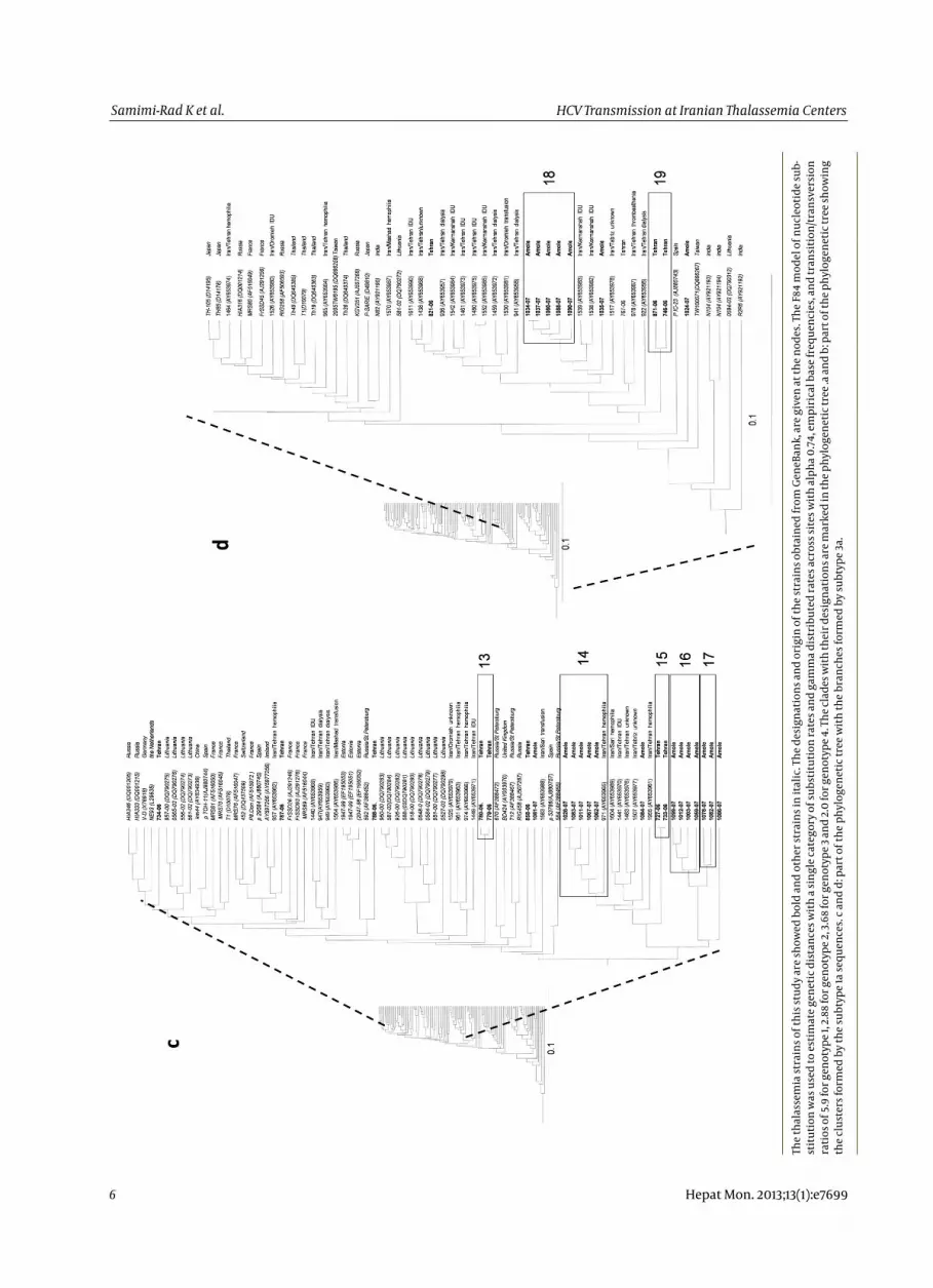
Fi
gure
1. U
PGM
A de
ndro
gram
bas
ed o
n 32
5 nu
cleo
tide
of th
e N
S5B
regi
on o
f 379
HCV
stra
ins
HCV Transmission at Iranian Thalassemia CentersSamimi-Rad K et al.
Hepat Mon. 2013;13(1):e7699
6
The t
hala
ssem
ia st
rain
s of t
his s
tudy
are
show
ed b
old
and
othe
r str
ains
in it
alic
. The
des
igna
tions
and
ori
gin
of th
e str
ains
obt
aine
d fr
om G
eneB
ank,
are
giv
en a
t the
nod
es. T
he F8
4 m
odel
of n
ucle
otid
e sub
st
itutio
n w
as u
sed
to es
timat
e gen
etic
dis
tanc
es w
ith a
sing
le ca
tego
ry o
f sub
stitu
tion
rate
s and
gam
ma d
istr
ibut
ed ra
tes a
cros
s site
s with
alph
a 0.74
, em
piri
cal b
ase f
requ
enci
es, a
nd tr
ansi
tion/
tran
sver
sion
ra
tios o
f 5.9
for g
enot
ype 1
, 2.8
8 fo
r gen
otyp
e 2, 3
.68
for g
enot
ype 3
and
2.0
for g
enot
ype 4
. The
clad
es w
ith th
eir d
esig
natio
ns ar
e mar
ked
in th
e phy
loge
netic
tree
.a an
d b:
par
t of t
he p
hylo
gene
tic tr
ee sh
owin
g th
e cl
uste
rs fo
rmed
by t
he su
btyp
e 1a
sequ
ence
s. c a
nd d
: par
t of t
he p
hylo
gene
tic tr
ee w
ith th
e br
anch
es fo
rmed
by s
ubty
pe 3a
.
HCV Transmission at Iranian Thalassemia Centers Samimi-Rad K et al.
7 Hepat Mon. 2013;13(1):e7699
Strain 752 in clade 11 was from a patient infected in1999 and identical to strain 794 from a patient infectedbefore 1996. Seven clades were formed by strains onlyfrom Amol (clades 1, 5, 6, 7, 8, 10 and 12; Figure 1a-b). Anadditional clade, 3, was formed by strains mainly fromAmol, but also with one strain from Tehran. Strains in six of these 1a clades were from 16 patients (eight pairs) who had been transfused simultaneously sitting on the samebed at the center in Amol (Table 4). Two of the eight pairs were formed by one patient each infected before and after 1996. Seven clades were formed by 22 of the 37 (59%) subtype 3a strains in the current study study (Figure 1c-d).Three clades were formed by strains from Tehran (clades 13, 15 and 19) and four by strains from Amol (clades 14, 16, 17and 18; Table 4; Figure 1 c-d). There were significantly moreclosely related 3a strains among patients from Amol ascompared to those of Tehran, 73% versus 40%, respectively (P < 0.001). Strains in three of these clades (14, 16 and 18) were from 12 patients (six pairs) who had been transfusedat the same shift sitting next to each other on the samebed in Amol. Two patients with strains in clade 14 and one patient with a strain in clade 16 had been infectedbetween 1999 and 2005, while the other patient in thepairs had been infected before 1996. Clade 19 was formedby two strains from patients who had frequently beentransfused at the same shift in the same room in a center in Tehran (Table 4; Figure 1c-d). One of the male patients in this clade had been infected in 2005. The 12 subtype 1b strains all diverged with more than five nucleotidesfrom each other and did not form any clades, but wereinterdispersed among1b strains derived world-wide. Thiswas significantly different as compared to the other subtypes (P < 0.001) when all strains were considered. However, most of the 1b strains, 10, were from Tehran centers, which generally had fewer similar strains circulatingthan the center in Amol (P = 0.015). When only the strainsfrom Tehran centers were considered, the lack of similar 1b strains as compared to strains belonging to othersubtypes was less significant (0 versus 15; P = 0.05). Thepatient infected with the 4a strain had been transfusedfor 8 years in the United Arab Emirates, and had probably been infected there.
5. Discussion The frequencies of subtype 1b and 3a in hepatitis C in
fected thalassemia patients from Tehran were 25% and 22%, respectively, which were in contrast with the findings from the thalassemia centers in Amol, where 1a and3a were the most prevalent subtypes. In addition, datafrom earlier Iranian studies showed subtypes 1a and 3a were the most common ones in the country with geographical differences in their distribution, and 1a wasfrequent among patients infected through blood transfusion (9-11). Several factors may be responsible for theintroduction of various subtypes and strains that may infect patients at the thalassemia centers in Tehran. First,
25% of the hepatitis C infected patients had been treated in other cities or abroad for several years before startingtreatment in Tehran, such as the patient infected with 4a, who had been treated in the United Arab Emirates (12, 13).Second, due to a high demand for blood in Tehran, it is unavoidable for the centers to use blood supplied from other Iranian cities, which might result the introduction of various HCV subtypes. Third, sampling in Tehran was done at several thalassemia centers which may differ with regard to the number of patients, amount of blood provided from the other parts of Iran, rate of HCV infections and number of patients previously transfused in other cities or abroad. The number of new HCV infected patients decreased when blood screening for anti-HCVwas introduced in Iran in 1996, the current study alsoindicated that 86 % of the patients had been infected before 1996. However, 34 (16 %) of the patients had become hepatitis C infected after 1996. Most of them had been infected in 2001 or later, and only eight had been infected between 1996 and 2000. This might have been an under-estimate, since there was no testing for anti-HCV in thalassemia centers on a regular basis these years. Most patients from Tehran have been tested for anti-HCV since 1998, while there was no routinely anti HCV testing in Amol until 1999 or 2000. Phylogenetic analysis ofthe NS5B region revealed a high genetic variability of the strains at different centers. Although 54 % of the 1a and 3a strains formed clades of similar strains, the strains from one clade were highly divergent from strains of the same subtype forming another clade. The variability betweenthe strains and the clades may be due to several factorsincluding transfusion of unscreened blood before 1996, blood obtained from other cities particularly for patientsfrom Tehran, and the fact that 24% of the thalassemia patients had started their treatment in other cities. All these circumstances could lead to multiple entries of HCV lineages into the centers. However, in different centers there were closely related strains forming clades. Further epidemiological investigations revealed that 36 of these strains were isolated from patients frequently transfusedat the same shift in the same room. Moreover, the number of related 1a and 3a strains from Amol was higher and these strains formed larger clades than the ones fromTehran. In addition, 71% of the patients infected with similar strains from Amol had been transfused while sittingnext to each other on the same bed. This indicates that nosocomial transmission was more common in Amol thalassemia center than in those of Tehran. This could be due to Amol Thalassemia center was the only centerin the city, and the number of patients often exceeded the number of beds, which could influence the patientto-patient transmission of hepatitis C. The circulation of few similar strains, as in Amol, may also facilitate the identification of nosocomial transmission. The patients in Tehran were transfused at several centers, and could have been exposed to more divergent HCV strains. Some of the patients were also transfused and infected abroad,
HCV Transmission at Iranian Thalassemia CentersSamimi-Rad K et al.
Hepat Mon. 2013;13(1):e7699
as the patient infected with 4a and probably the majority of those infected with 1b strains. These 1b strains were less related to each other and more similar to the strains from Western Europe (14), while the 1a and 3a strains were more similar to strains from other Iranian high risk groups. This suggests multiple entries of 1b strains from abroad into the thalassemia centers in Tehran. The high similarity of the majority of the 1a and 3a strains suggests nosocomial transmission of HCV at the thalassemia centers. In a study on five new hepatitis C cases infected atZafar adult thalassemia clinic in 2004, all the blood donors whom could be traced, 69 %, were anti-HCV negative indicating patient-to patient transmission, although it was not shown by sequencing and genetic analysis of the patient strains (15). Nosocomial transmission of hepatitis C has previously been described between patients and atdifferent hospital settings outside Iran (8, 16-21). However, phylogenetic analysis of the HCV strains was performed only in few of the studies to identify common source of infection or route of transmission (8, 18, 19). Until now, nosocomial HCV transmission based on sequencing and phylogenetic analysis of patient HCV strains at thalassemia centers has not previously been reported. The geneticanalysis of the infecting strains in combination with further epidemiological analysis clearly indicated patient-topatient transmission of hepatitis C at the centers under study. More transmission events may have occurred than those identified, since some patients may have moved to other thalassemia centers or have died. Also, if the transmission occurred a long time ago, the strains may have diverged beyond a detectable association of the strains due to genetic drift. The exact mode of infection was not investigated, although the most likely factors may be the use of contaminated medical equipment or other poor enforcement of safety guidelines. Violation of safety procedures resulting in the spread of hepatitis C in hospitals has previously been documented (16, 22). The presentstudy supports that nosocomial transmission plays a possible role in HCV spread at Iranian thalassemia centers.Education of staff and implementation of strict infection control practices are thus necessary in order to reduce the number of new cases of hepatitis C at these centers.
Acknowledgements The authors gratefully acknowledge their gratitude to
Dr.Bashir Hajibeigy (Research Center of Iranian Blood Transfusion Organization, Tehran, Iran), the staff and the patients of the thalassemia centers for their excellent cooperation in data and sample collection.
Authors’ Contribution KS and HN were responsible for the conception, design
and analysis of the study. KS collected the samples, performed the experiments and wrote the draft of the paper. LM did the critical revision of the paper. HN, LM, SMA and
KS prepared the final version of the paper. FA carried out the statistical analysis. MN, AE, AA, SME, FZ and SMA provided the study subjects and data collection.
Financial Disclosure None declared.
Funding/Support This study was supported by the grant from the Swed
ish Research Links 348-2005-6250 and from the Swedish Research Council VR521-2006-2753.
References 1. Rezvan H, Abolghassemi H, Kafiabad SA. Transfusion-transmit
ted infections among multitransfused patients in Iran: a review. Transfus Med.2007;17(6):425-33.
2. Abolghasemi H, Amid A, Zeinali S, Radfar MH, Eshghi P, Rahiminejad MS, et al. Thalassemia in Iran: epidemiology, prevention, and management. J Pediatr Hematol Oncol.2007;29(4):233-8.
3. Alavian S, Tabatabaei S, Lankarani K. Epidemiology of HCV infection among thalassemia patients in eastern Mediterraneancountries: a quantitative review of literature. Iranian Red Crescent Medical Journal.2010;12(4):365-76.
4. Merat S, Rezvan H, Nouraie M, Jafari E, Abolghasemi H, RadmardAR. Seroprevalence of hepatitis C virus: the first population-based study from Iran. Int J Infect Dis.2010;14 Suppl 3:e113-6.
5. Samimi-Rad K, Hosseini M, Mobeini G, Asgari F, Alavian SM, Tahaei ME, et al. Hepatitis C virus infection among multi-transfused patients and personnel in haemodialysis units in centralIslamic Republic of Iran. East Mediterr Health J.2012;18(3):227-35.
6. Mirmomen S, Alavian SM, Hajarizadeh B, Kafaee J, Yektaparast B, Zahedi MJ. Epidemiology of hepatitis B, hepatitis C, and human immunodeficiency virus infecions in patients with beta-thalassemia in Iran: a multicenter study. Arch Iran Med.2006;9(4):31923.
7. Kalinina O, Norder H, Vetrov T, Zhdanov K, Barzunova M, Plotnikova V, et al. Shift in predominating subtype of HCV from 1b to 3a in St. Petersburg mediated by increase in injecting drug use. J Med Virol.2001;65(3):517-24.
8. Norder H, Bergstrom A, Uhnoo I, Alden J, Weiss L, Czajkowski J, et al. Confirmation of nosocomial transmission of hepatitis C virus by phylogenetic analysis of the NS5-B region. J Clin Micro-biol.1998;36(10):3066-9.
9. Samimi-Rad K, Shahbaz B. Hepatitis C virus genotypes among patients with thalassemia and inherited bleeding disorders inMarkazi province, Iran. Haemophilia.2007;13(2):156-63.
10. Samimi-Rad K, Nategh R, Malekzadeh R, Norder H, Magnius L. Molecular epidemiology of hepatitis C virus in Iran as reflected by phylogenetic analysis of the NS5B region. J Med Vi-rol.2004;74(2):246-52.
11. Alavian SM, Miri SM, Keshvari M, Elizee PK, Behnava B, Tabatabaei SV, et al. Distribution of hepatitis C virus genotype in Iranian multiply transfused patients with thalassemia. Transfu-sion.2009;49(10):2195-9.
12. Shobokshi OA, Serebour FE, Skakni LI. Hepatitis C genotypes/subtypes among chronic hepatitis patients in Saudi Arabia. Saudi Med J.2003;24 Suppl 2:S87-91.
13. Chamberlain RW, Adams N, Saeed AA, Simmonds P, Elliott RM. Complete nucleotide sequence of a type 4 hepatitis C virus variant, the predominant genotype in the Middle East. J Gen Virol.1997;78(Pt 6):1341-7.
14. Simmonds P. The origin and evolution of hepatitis viruses in humans. J Gen Virol.2001;82(Pt 4):693-712.
15. Azar KA, Hajibeygi B, Nasiri TM, Amini Kafiabad S, Maghsoudlou M, Shadman AH. Blood (Khoon).2010;7(3):156-61.
16. Krause G, Trepka MJ, Whisenhunt RS, Katz D, Nainan O, Wiersma
8

HCV Transmission at Iranian Thalassemia Centers Samimi-Rad K et al.
9 Hepat Mon. 2013;13(1):e7699
ST, et al. Nosocomial transmission of hepatitis C virus associated with the use of multidose saline vials. Infect Control Hosp Epide-miol.2003;24(2):122-7.
17. Januszkiewicz-Lewandowska D, Wysocki J, Rembowska J, Pernak M, Lewandowski K, Nowak T, et al. Transmission of HCV infection among long-term hospitalized onco-haematological patients. J Hosp Infect.2003;53(2):120-3.
18. Gonzalez-Candelas F, Guiral S, Carbo R, Valero A, Vanaclocha H, Gonzalez F, et al. Patient-to-patient transmission of hepatitis C virus (HCV) during colonoscopy diagnosis. Virol J.2010;7:217.
19. Dencs A, Hettmann A, Martyin T, Jekkel C, Banyai T, Takacs M. Phylogenetic investigation of nosocomial transmission of hepatitis
C virus in an oncology ward. J Med Virol.2011;83(3):428-36. 20. Comstock RD, Mallonee S, Fox JL, Moolenaar RL, Vogt TM, Perz JF,
et al. A large nosocomial outbreak of hepatitis C and hepatitis B among patients receiving pain remediation treatments. Infect Control Hosp Epidemiol.2004;25(7):576-83.
21. Bronowicki JP, Venard V, Botte C, Monhoven N, Gastin I, Chone L, et al. Patient-to-patient transmission of hepatitis C virus during colonoscopy. N Engl J Med.1997;337(4):237-40.
22. Perz JF, Thompson ND, Schaefer MK, Patel PR. US outbreak investigations highlight the need for safe injection practices and basic infection control. Clin Liver Dis.2010;14(1):137-51. x.