pathogen translocation across the bloodâbrain barrier
TRANSCRIPT
Seediscussions,stats,andauthorprofilesforthispublicationat:https://www.researchgate.net/publication/26791247
Pathogentranslocationacrosstheblood-brainbarrier
ArticleinFEMSImmunology&MedicalMicrobiology·September2009
DOI:10.1111/j.1574-695X.2009.00594.x·Source:PubMed
CITATIONS
21
READS
42
3authors:
Someoftheauthorsofthispublicationarealsoworkingontheserelatedprojects:
APVV14-0218Decipheringtheligand-receptorinteractionsinvolvedincentralnervous
systeminvasionbypathogensanddevelopmentoftargetedtherapeuticstrategyagainst
neuroinfectionsViewproject
Upgradingtheresearchperformanceinmolecularmedicine-ERAChairFP7Viewproject
LuciaPulzova
UniversityofVeterinaryMedicineandPhar…
39PUBLICATIONS57CITATIONS
SEEPROFILE
MangeshBhide
UniversityofVeterinaryMedicineinKosice…
97PUBLICATIONS612CITATIONS
SEEPROFILE
AndrejKovac
SlovakAcademyofSciences
31PUBLICATIONS288CITATIONS
SEEPROFILE
Allin-textreferencesunderlinedinbluearelinkedtopublicationsonResearchGate,
lettingyouaccessandreadthemimmediately.
Availablefrom:AndrejKovac
Retrievedon:30September2016
M I N I R E V I E W
Pathogen translocationacross theblood^brain barrierLucia Pulzova1,2, Mangesh R. Bhide1,2 & Kovac Andrej2
1Laboratory of Biomedical Microbiology and Immunology, Department of Microbiology and Immunology, University of Veterinary Medicine, Kosice,
Slovakia; and 2Institute of Neuroimmunology, Slovak Academy of Sciences, Bratislava, Slovakia
Correspondence: Mangesh R. Bhide,
Laboratory of Biomedical Microbiology and
Immunology, Department of Microbiology
and Immunology, University of Veterinary
Medicine, Komenskeho 73, 04181 Kosice,
Slovakia. Tel.: 1421 90 446 1705; fax: 1421
55 632 3173; e-mail:
Received 29 June 2009; accepted 27 July 2009.
Final version published online 1 September
2009.
DOI:10.1111/j.1574-695X.2009.00594.x
Editor: Willem van Leeuwen
Keywords
blood–brain barrier; neuroinvasion;
plasminogen; tight junction; microbial
translocation.
Abstract
Neurological manifestations caused by neuroinvading pathogens are typically
attributed to penetration of the blood–brain barrier (BBB) and invasion of the
central nervous system. However, the mechanisms used by many pathogens (such
as Borrelia) to traverse the BBB are still unclear. Recent studies revealed that
microbial translocation across the BBB must involve a repertoire of microbial–host
interactions (receptor–ligand interactions). However, the array of interacting
molecules responsible for the borrelial translocation is not yet clearly known.
Pathogens bind several host molecules (plasminogen, glycosaminoglycans, factor
H, etc.) that might mediate endothelial interactions in vivo. This review sum-
marizes our current understanding of the pathogenic mechanisms involved in the
translocation of the BBB by neuroinvasive pathogens.
Introduction
The blood–brain barrier (BBB) is a regulatory interface
between the peripheral circulation and the central nervous
system (CNS) (Kim, 2008). The neurological symptoms of
some diseases are associated with the mode of traversal of
this barrier and penetration into the brain by several
pathogens (bacteria, fungi, parasites and viruses). Interest-
ingly, some of the neuroinvasive pathogens, for example
Chlamydiophila pneumoniae (MacIntyre et al., 2002) and
Borrelia burgdorferi sensu lato (Miklossy et al., 2004; Batinac
et al., 2007), are reported to be associated with multiple
sclerosis and Alzheimer’s disease (AD), respectively. How-
ever, the relationship between these organisms and diseases
remains unclear. Pathogens exploit several mechanisms
that enable them to reach the CNS, such as the traversal of
BBB or penetration through neurons by axonal flow. Many
pathogens have the potential to infect the CNS, but it is
unclear why only a relatively small number of pathogens
account for most clinical cases with nervous disorders. A
comprehensive understanding of BBB crossing mechanisms
is pivotal for drug and vaccine development against neu-
roinvading pathogens.
The BBB
The BBB is a structural and functional barrier that regulates
the passage of blood-borne substances and cells into the
brain and thus maintains the homeostasis of the neural
microenvironment that is crucial for normal neuronal
activity and function (Abbott et al., 2006). BBB is formed
by brain microvascular endothelial cells (BMECs) that line
the cerebral microvessels. The periendothelial structures of
the BBB include pericytes (related to smooth muscle cells,
surround the endothelium, reduce endothelial apoptosis
and stabilize the endothelium), astrocytes (induce many
BBB features and support the tissue of the CNS) and a basal
membrane. The BMECs utilize unique features that distin-
guish them from the peripheral endothelial cells. Most
prominent among these are as follows: (1) numerous inter-
cellular ‘tight junctions (TJs)’ that possess high transen-
dothelial electrical resistance and retard paracellular flux; (2)
the absence of fenestrae and a reduced level of fluid-phase
endocytosis; and (3) asymmetrically localized enzymes
and carrier-mediated transport systems (Biegel et al., 1995).
The BMECs express several influx/efflux transporters that
transport important nutrients such as glucose (GLUT-1
FEMS Immunol Med Microbiol 57 (2009) 203–213 c� 2009 Federation of European Microbiological SocietiesPublished by Blackwell Publishing Ltd. All rights reserved
IMM
UN
OLO
GY
& M
EDIC
AL
MIC
ROBI
OLO
GY
transporter), amino acids (LAT1 transporter) and nucleo-
sides (e.g. ENT1 and CNT1) (de Boer et al., 2003). Because
of the negatively charged abluminal membrane, negatively
charged molecules face obscurity while entering or exiting
the endothelial cells. There are two families of multispecific
anion transporters that mediate the influx and/or the efflux
of these compounds: the organic anion transporter poly-
peptide family and the organic anion transporter (Sekine
et al., 2000). Transporters for organic cations OCNT1 (Tsuji,
2005) and monocarboxylic acids MCT1 and MCT2 (Gerhart
et al., 1989; Price et al., 1998) have also been described on
BMEC. Peptides and proteins such as insulin or transferrin
are transported through the BBB by receptor-mediated
transport (Davidson et al., 1990; Moos et al., 2007).
To protect the nervous system from xenobiotics, BMEC
express several efflux transporters. The most important are
members of the ATP-binding cassette (ABC) family, for
example P-glycoprotein (ABCB1), multidrug resistance pro-
teins (MRP1–6) and brain multidrug resistance protein
(Deli et al., 2005). Furthermore, metabolizing enzymes such
as members of the cytochrome P450 family effectively
restrict the entry of xenobiotics into the CNS (el-Bacha &
Minn, 1999).
The TJs
Polarized epithelial cells carry out functions such as the
transport of ions and nutrients, secretion of protein products
and protection of the interior of the organism from invading
microorganisms. Cell polarity is observed in the functionally
distinct portions of the plasma membrane, known as the
apical domain and the basolateral domain (Miyoshi & Takai,
2005). The apical domain contains anion channels, H1/K1-
ATPase and transporters, whereas the lateral portion of the
basolateral domain contains proteins involved in attachment
to neighboring cells and cell–cell communication. The basal
portion of the basolateral domain contains the binding sites
for constituents of the basal lamina, and receptors for
hormones and other signaling molecules that regulate the
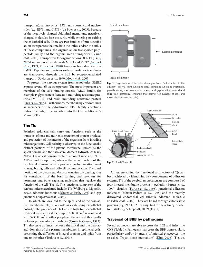
function of the cell (Fig. 1). The junctional complexes of the
cerebral microvasculature include TJs (Wolburg & Lippoldt,
2002), adherens junctions (Schulze & Firth, 1993) and gap
junctions (Nagasawa et al., 2006).
TJs, which are localized to the apical end of the basolat-
eral membrane, play a key role in establishing endothelial
polarity. The presence of TJs leads to high transendothelial
electrical resistance values of up to 2000O cm2 as compared
with 3–33O cm2 in other peripheral tissues, and this results
in lower paracellular permeability (Crone & Olesen, 1982).
TJs also serve as fences between the apical and the basolat-
eral domains of the plasma membranes in epithelial cells,
preventing the diffusion of integral proteins and lipids from
one to the other (Tsukita et al., 2001).
An understanding the functional architecture of TJs has
been achieved by identifying key components of adhesion
systems. TJs of the cerebral microvasculate are composed of
four integral membrane proteins – occludin (Furuse et al.,
1994), claudins (Furuse et al., 1998), junctional adhesion
molecules (Martin-Padura et al., 1998) and the recently
discovered endothelial cell-selective adhesion molecule
(Nasdala et al., 2002). These are linked through cytoplasmic
proteins (e.g. ZO-1, -2, -3, cingulin) to the actin cytoskele-
ton (Wolburg & Lippoldt, 2002) (Fig. 2).
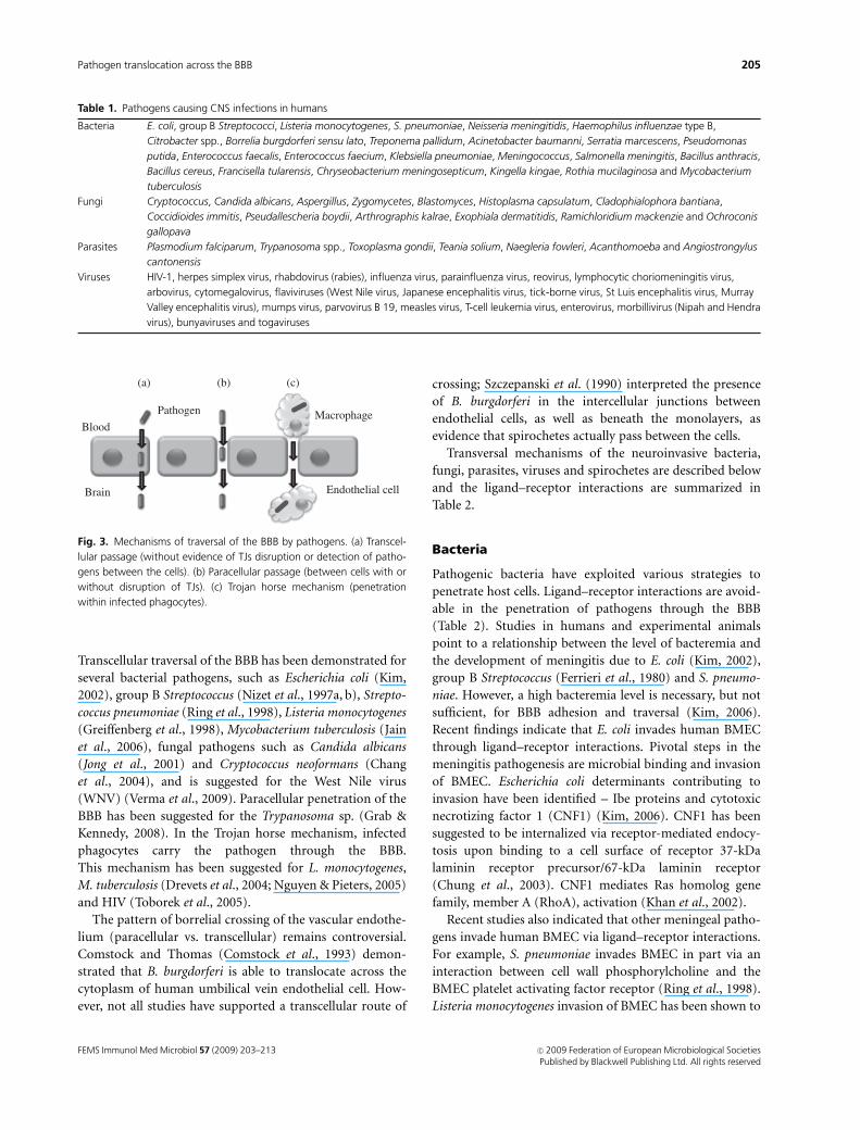
Traversal of BBB by pathogens
Several pathogens are able to cross the BBB and infect the
CNS (Table 1). Pathogens may cross the BBB transcellulary,
paracellulary and/or by means of infected phagocytes (the
so-called Trojan horse mechanism) (Kim, 2006) (Fig. 3).
Lateral membrane
Apical membrane
Basal membrane
Fig. 1. Organization of the intercellular junctions. Cell attached to the
adjacent cell via tight junctions (arc), adherens junctions (rectangle,
provide strong mechanical attachment) and gap junctions (round-end
rods, free intercellular channels that permit free passage of ions and
molecules between the cells).
Endothelial cell
Brain blood vessel
Tight junctions
Pericytes
Astrocyte end-feet
ZO-1
Occludin
ClaudinZO-2
ZO-3JAM
Intercellularspace
Fig. 2. The BBB and TJ.
FEMS Immunol Med Microbiol 57 (2009) 203–213c� 2009 Federation of European Microbiological SocietiesPublished by Blackwell Publishing Ltd. All rights reserved
204 L. Pulzova et al.
Transcellular traversal of the BBB has been demonstrated for
several bacterial pathogens, such as Escherichia coli (Kim,
2002), group B Streptococcus (Nizet et al., 1997a, b), Strepto-
coccus pneumoniae (Ring et al., 1998), Listeria monocytogenes
(Greiffenberg et al., 1998), Mycobacterium tuberculosis (Jain
et al., 2006), fungal pathogens such as Candida albicans
(Jong et al., 2001) and Cryptococcus neoformans (Chang
et al., 2004), and is suggested for the West Nile virus
(WNV) (Verma et al., 2009). Paracellular penetration of the
BBB has been suggested for the Trypanosoma sp. (Grab &
Kennedy, 2008). In the Trojan horse mechanism, infected
phagocytes carry the pathogen through the BBB.
This mechanism has been suggested for L. monocytogenes,
M. tuberculosis (Drevets et al., 2004; Nguyen & Pieters, 2005)
and HIV (Toborek et al., 2005).
The pattern of borrelial crossing of the vascular endothe-
lium (paracellular vs. transcellular) remains controversial.
Comstock and Thomas (Comstock et al., 1993) demon-
strated that B. burgdorferi is able to translocate across the
cytoplasm of human umbilical vein endothelial cell. How-
ever, not all studies have supported a transcellular route of
crossing; Szczepanski et al. (1990) interpreted the presence
of B. burgdorferi in the intercellular junctions between
endothelial cells, as well as beneath the monolayers, as
evidence that spirochetes actually pass between the cells.
Transversal mechanisms of the neuroinvasive bacteria,
fungi, parasites, viruses and spirochetes are described below
and the ligand–receptor interactions are summarized in
Table 2.
Bacteria
Pathogenic bacteria have exploited various strategies to
penetrate host cells. Ligand–receptor interactions are avoid-
able in the penetration of pathogens through the BBB
(Table 2). Studies in humans and experimental animals
point to a relationship between the level of bacteremia and
the development of meningitis due to E. coli (Kim, 2002),
group B Streptococcus (Ferrieri et al., 1980) and S. pneumo-
niae. However, a high bacteremia level is necessary, but not
sufficient, for BBB adhesion and traversal (Kim, 2006).
Recent findings indicate that E. coli invades human BMEC
through ligand–receptor interactions. Pivotal steps in the
meningitis pathogenesis are microbial binding and invasion
of BMEC. Escherichia coli determinants contributing to
invasion have been identified – Ibe proteins and cytotoxic
necrotizing factor 1 (CNF1) (Kim, 2006). CNF1 has been
suggested to be internalized via receptor-mediated endocy-
tosis upon binding to a cell surface of receptor 37-kDa
laminin receptor precursor/67-kDa laminin receptor
(Chung et al., 2003). CNF1 mediates Ras homolog gene
family, member A (RhoA), activation (Khan et al., 2002).
Recent studies also indicated that other meningeal patho-
gens invade human BMEC via ligand–receptor interactions.
For example, S. pneumoniae invades BMEC in part via an
interaction between cell wall phosphorylcholine and the
BMEC platelet activating factor receptor (Ring et al., 1998).
Listeria monocytogenes invasion of BMEC has been shown to
Table 1. Pathogens causing CNS infections in humans
Bacteria E. coli, group B Streptococci, Listeria monocytogenes, S. pneumoniae, Neisseria meningitidis, Haemophilus influenzae type B,
Citrobacter spp., Borrelia burgdorferi sensu lato, Treponema pallidum, Acinetobacter baumanni, Serratia marcescens, Pseudomonas
putida, Enterococcus faecalis, Enterococcus faecium, Klebsiella pneumoniae, Meningococcus, Salmonella meningitis, Bacillus anthracis,
Bacillus cereus, Francisella tularensis, Chryseobacterium meningosepticum, Kingella kingae, Rothia mucilaginosa and Mycobacterium
tuberculosis
Fungi Cryptococcus, Candida albicans, Aspergillus, Zygomycetes, Blastomyces, Histoplasma capsulatum, Cladophialophora bantiana,
Coccidioides immitis, Pseudallescheria boydii, Arthrographis kalrae, Exophiala dermatitidis, Ramichloridium mackenzie and Ochroconis
gallopava
Parasites Plasmodium falciparum, Trypanosoma spp., Toxoplasma gondii, Teania solium, Naegleria fowleri, Acanthomoeba and Angiostrongylus
cantonensis
Viruses HIV-1, herpes simplex virus, rhabdovirus (rabies), influenza virus, parainfluenza virus, reovirus, lymphocytic choriomeningitis virus,
arbovirus, cytomegalovirus, flaviviruses (West Nile virus, Japanese encephalitis virus, tick-borne virus, St Luis encephalitis virus, Murray
Valley encephalitis virus), mumps virus, parvovirus B 19, measles virus, T-cell leukemia virus, enterovirus, morbillivirus (Nipah and Hendra
virus), bunyaviruses and togaviruses
(a) (b) (c)
Pathogen MacrophageBlood
Brain Endothelial cell
Fig. 3. Mechanisms of traversal of the BBB by pathogens. (a) Transcel-
lular passage (without evidence of TJs disruption or detection of patho-
gens between the cells). (b) Paracellular passage (between cells with or
without disruption of TJs). (c) Trojan horse mechanism (penetration
within infected phagocytes).
FEMS Immunol Med Microbiol 57 (2009) 203–213 c� 2009 Federation of European Microbiological SocietiesPublished by Blackwell Publishing Ltd. All rights reserved
205Pathogen translocation across the BBB
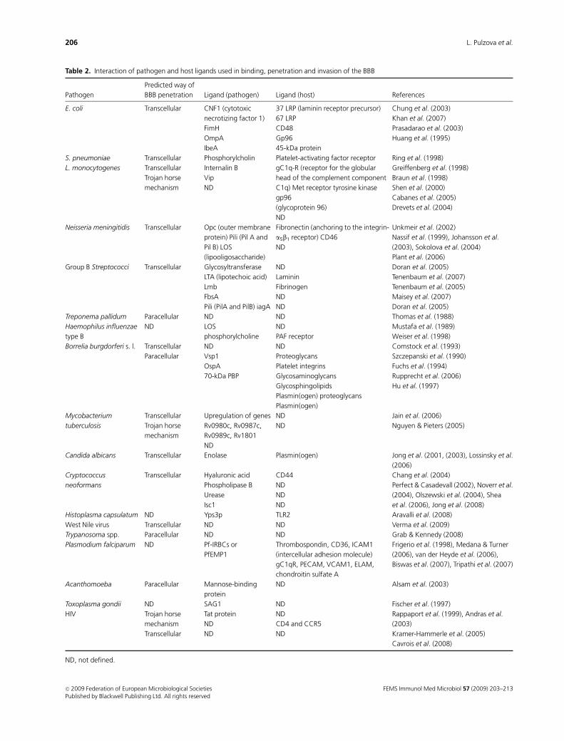
Table 2. Interaction of pathogen and host ligands used in binding, penetration and invasion of the BBB
Pathogen
Predicted way of
BBB penetration Ligand (pathogen) Ligand (host) References
E. coli Transcellular CNF1 (cytotoxic
necrotizing factor 1)
FimH
OmpA
IbeA
37 LRP (laminin receptor precursor)
67 LRP
CD48
Gp96
45-kDa protein
Chung et al. (2003)
Khan et al. (2007)
Prasadarao et al. (2003)
Huang et al. (1995)
S. pneumoniae Transcellular Phosphorylcholin Platelet-activating factor receptor Ring et al. (1998)
L. monocytogenes Transcellular
Trojan horse
mechanism
Internalin B
Vip
ND
gC1q-R (receptor for the globular
head of the complement component
C1q) Met receptor tyrosine kinase
gp96
(glycoprotein 96)
ND
Greiffenberg et al. (1998)
Braun et al. (1998)
Shen et al. (2000)
Cabanes et al. (2005)
Drevets et al. (2004)
Neisseria meningitidis Transcellular Opc (outer membrane
protein) Pili (Pil A and
Pil B) LOS
(lipooligosaccharide)
Fibronectin (anchoring to the integrin-
a5b1 receptor) CD46
ND
Unkmeir et al. (2002)
Nassif et al. (1999), Johansson et al.
(2003), Sokolova et al. (2004)
Plant et al. (2006)
Group B Streptococci Transcellular Glycosyltransferase
LTA (lipotechoic acid)
Lmb
FbsA
Pili (PilA and PilB) iagA
ND
Laminin
Fibrinogen
ND
ND
Doran et al. (2005)
Tenenbaum et al. (2007)
Tenenbaum et al. (2005)
Maisey et al. (2007)
Doran et al. (2005)
Treponema pallidum Paracellular ND ND Thomas et al. (1988)
Haemophilus influenzae
type B
ND LOS
phosphorylcholine
ND
PAF receptor
Mustafa et al. (1989)
Weiser et al. (1998)
Borrelia burgdorferi s. l. Transcellular
Paracellular
ND
Vsp1
OspA
70-kDa PBP
ND
Proteoglycans
Platelet integrins
Glycosaminoglycans
Glycosphingolipids
Plasmin(ogen) proteoglycans
Plasmin(ogen)
Comstock et al. (1993)
Szczepanski et al. (1990)
Fuchs et al. (1994)
Rupprecht et al. (2006)
Hu et al. (1997)
Mycobacterium
tuberculosis
Transcellular
Trojan horse
mechanism
Upregulation of genes
Rv0980c, Rv0987c,
Rv0989c, Rv1801
ND
ND
ND
Jain et al. (2006)
Nguyen & Pieters (2005)
Candida albicans Transcellular Enolase Plasmin(ogen) Jong et al. (2001, (2003), Lossinsky et al.
(2006)
Cryptococcus
neoformans
Transcellular Hyaluronic acid
Phospholipase B
Urease
Isc1
CD44
ND
ND
ND
Chang et al. (2004)
Perfect & Casadevall (2002), Noverr et al.
(2004), Olszewski et al. (2004), Shea
et al. (2006), Jong et al. (2008)
Histoplasma capsulatum ND Yps3p TLR2 Aravalli et al. (2008)
West Nile virus Transcellular ND ND Verma et al. (2009)
Trypanosoma spp. Paracellular ND ND Grab & Kennedy (2008)
Plasmodium falciparum ND Pf-IRBCs or
PfEMP1
Thrombospondin, CD36, ICAM1
(intercellular adhesion molecule)
gC1qR, PECAM, VCAM1, ELAM,
chondroitin sulfate A
Frigerio et al. (1998), Medana & Turner
(2006), van der Heyde et al. (2006),
Biswas et al. (2007), Tripathi et al. (2007)
Acanthomoeba Paracellular Mannose-binding
protein
ND Alsam et al. (2003)
Toxoplasma gondii ND SAG1 ND Fischer et al. (1997)
HIV Trojan horse
mechanism
Transcellular
Tat protein
ND
ND
ND
CD4 and CCR5
ND
Rappaport et al. (1999), Andras et al.
(2003)
Kramer-Hammerle et al. (2005)
Cavrois et al. (2008)
ND, not defined.
FEMS Immunol Med Microbiol 57 (2009) 203–213c� 2009 Federation of European Microbiological SocietiesPublished by Blackwell Publishing Ltd. All rights reserved
206 L. Pulzova et al.
be mediated by internalin B (Greiffenberg et al., 1998).
Neisseria meningitidis invasion of human BMEC is mediated
by a bacterial outer membrane protein, OspC, binding to
fibronectin, thereby anchoring the bacteria to the integrin
a5b1 receptor on the human BMEC surface (Unkmeir et al.,
2002). Further studies are needed to understand the con-
tribution of these interactions to BMEC invasion and
BBB traversal.
Previous studies revealed that internalized bacteria are
found within membrane-bounded vacuoles of BMEC and
transmigrate without multiplication and are protected from
fusion with lysosomes (Kim, 2003, 2006). Electron micro-
scopy studies have shown that E. coli, M. tuberculosis and
group B Streptococcus invasion is associated with microvilli-
like protrusions at the entry site on the surface of human
BMEC (Nizet et al., 1997a, b), suggesting a rearrangement of
the host cell actin cytoskeleton. Actin cytoskeleton rearran-
gements are necessary for BMEC invasion by meningitis-
causing bacteria, but the signaling mechanisms involved in
actin differ among meningitis-causing bacterial species.
Citrobacter spp. are gram-negative bacteria and are asso-
ciated with neonatal meningitis (Badger et al., 1999). The
unique feature of meningitis caused by Citrobacter spp. is
their frequent association with brain abscess formation. The
pathogenesis of Citrobacter spp. meningitis and brain
abscess is not well characterized. Citrobacter freundii is able
to invade and cross human BMECs in vitro. Invasion of
BMECs by C. freundii was found to be dependent on
microfilaments, microtubules, endosome acidification and
de novo protein synthesis. In contrast to other meningitis-
causing bacteria, C. freundii is able to multiply within
human BMECs. This may be a mechanism whereby
C. freundii traverses the BBB (Huang et al., 2000).
Fungi
Several fungi have been shown to cause CNS infections in
humans. In HIV-endemic areas C. neoformans (Gordon et al.,
2000) and C. albicans (Issel, 1971) are the most frequently
isolated yeasts from patients with CNS involvement.
Cryptococcus neoformans is a common cause of culture-
proven meningitis in areas where HIV-1 is endemic (Perfect
& Casadevall, 2002). Brain invasion does not require the
recruitment of host inflammatory cells (Chretien et al.,
2002; Chang et al., 2004), which eliminates the possibility
of the Trojan horse mechanism. Recent studies indicate that
C. neoformans uses a transcellular mechanism of BMEC
(Chang et al., 2004) and requires protein kinase C-a activa-
tion (Jong et al., 2008). The CPS1 gene is required for
C. neoformans adherence to the surface protein CD44 of
human brain microvascular endothelial cells (HBMECs)
(Jong et al., 2008).
Candida albicans is able to adhere, invade and transcytose
across HBMEC without affecting the integrity of the mono-
layers (Jong et al., 2001) by a poorly understood process. A
Candida enolase interacting with the plasminogen system
contributes to C. albicans invasion and traversal of human
BMEC (Jong et al., 2001).
Histoplasma capsulatum is a common cause of fungal
infection, and although most infections are asymptomatic, it
is capable of causing histoplasmosis in immunocompro-
mised individuals (Aravalli et al., 2008). Histoplasma capsu-
latum may cause meningitis in 5–25% of its victims who
have AIDS. The interaction of H. capsulatum Yps3p with
microglial cells leads to nuclear factor-kB activation via the
Toll-like receptor 2 (TLR2) pathway (Aravalli et al., 2008). A
deeper understanding of the host–Histoplasma interaction is
needed.
Parasites
Malaria seems to be a major public health problem in many
parts of the tropical world. One of the important virulence
mechanisms in Plasmodium falciparum is the ability of
P. falciparum trophozoites and schizonts to sequester in
the vasculature of diverse host organs (brain) (MacPherson
et al., 1985; Silamut et al., 1999). Plasmodium falciparum-
infected RBCs use the 32-kDa human protein gC1qR/
HABP1/p32 as a receptor to bind to HBMECs (Biswas
et al., 2007).
The neurological manifestations of sleeping sickness in
humans caused by Trypanosoma brucei gambiense and
Trypanosoma brucei rhodesiense are attributed to the pene-
tration of the CNS by trypanosomes, but how trypanosomes
cross the human BBB remains unclear (Grab & Kennedy,
2008). The forms of trypanosomes found in the blood-
stream efficiently cross HBMECs by a paracellular route
(Grab et al., 2004). In rodent models, the parasite can pass
through the BBB across or between endothelial cells. Inter-
feron-g has been shown to play an important role in
regulating trypanosome trafficking into the brain (Masocha
et al., 2007). A trypanosome apoptotic factor expressed by
T. brucei that mediates apoptosis in mouse-brain and hu-
man-brain vascular endothelial cells (HBVECs) was identi-
fied and characterized (Stiles et al., 2004).
Pathogenic Acanthomoeba is the common cause of kera-
titis and rare, but fatal granulomatous amoebic encephalitis.
The mechanism that the pathogen uses to cross the BBB is
unclear. Some studies revealed the ability of several geno-
types of Acanthomoeba to bind human BMEC and cause
cytotoxicity in BMEC (Alsam et al., 2003).
Encephalitis is a serious complication of the infection
with the obligate intracellular parasite Toxoplasma gondii.
Additional studies are needed to elucidate the mechanism
involved in toxoplasmosis of the CNS.
FEMS Immunol Med Microbiol 57 (2009) 203–213 c� 2009 Federation of European Microbiological SocietiesPublished by Blackwell Publishing Ltd. All rights reserved
207Pathogen translocation across the BBB
Viruses
The penetration of HIV into the CNS through neurons by
axonal flow, which occurs with the herpes virus and the
rabies virus, is less probable because the CD4 receptor, the
main receptor that enables HIV to infect the cell, is absent
on neurons (Gendelman et al., 1998). The Trojan horse
mechanism of transport across the BBB is considered to play
a crucial role in the pathogenesis of viral meningitidis in the
late phase of AIDS. Although this model gained rapid favor,
recent studies challenge this model by showing that the vast
majority of virions transmitted in trans originates from the
plasma membrane rather than from intracellular vesicles
(Cavrois et al., 2008).
The mechanisms of BBB disruption during retroviral-
associated pathologies are not fully understood yet. Most of
the studies are focused on the effect of soluble molecules
secreted by infected lymphocytes on BBB functions and
intercellular TJ organization. In the case of HIV infection,
the viral protein Tat has been shown to induce an inflam-
matory process in brain endothelial cells, or endothelial cell
apoptosis (Andras et al., 2003; Kim & Langridge, 2004), and
to be able to disrupt the intercellular TJs.
WNV-associated encephalitis is characterized by disrup-
tion of the BBB, enhanced infiltration of immune cells into
the CNS, microglia activation, inflammation and eventual
loss of neurons (Glass et al., 2005; Sitati et al., 2007). WNV
gains entry into the CNS via the transcellular pathway,
without compromising the BBB integrity, instead of the
paracellular pathway, in which case, an increase in WNV
RNA at earlier time points would be expected, due to passive
diffusion (Verma et al., 2009). WNV does not induce the
cytopathic effect and induces an expression of claudin-1 and
upregulation of vascular cell adhesion molecule-1 and E-
selectin (Verma et al., 2008).
Spirochetes
Neurosyphilis and neuroborreliosis are prototypes for
spirochete infection of the CNS, but their pathogenesis
remains unclear.
Treponema pallidum can invade through the intercellular
junction of aortic endothelial cells (Thomas et al., 1988),
which suggests the usage of a paracellular mechanism of
penetration of the vascular endothelium, but it is unclear
whether a similar mechanism is involved in T. pallidum
penetration of the BBB.
Borrelia strains traverse human BMEC without obvious
changes in the integrity of the host cells (Grab et al., 2005).
This translocation is facilitated by host proteases, which
are involved in the plasminogen activation system and
fibrinolysis (Coleman et al., 1995, 1997, 2001; Coleman &
Benach, 2000, 2003), like in other pathogens of low genomic
capacity. The fibrinolytic system linked by an activation
cascade may lead to focal and transient degradation of TJ
proteins, allowing B. burgdorferi to invade the CNS.
Although the role of plasmin in infection is important,
there are other host proteases that could also be used to
enhance the translocation of the host barriers such as matrix
metalloproteinases (MMPs) (Gebbia et al., 2001).
Neuroborreliosis and the transversal mechanisms
exploited by Borrelia are discussed below in detail.
Neuroborreliosis and neuroinvasive/transversal mechanisms
Lyme borreliosis (LB) is the most common tick-borne
disease in the Northern hemisphere. Neuroborreliosis can
arise at any time during the course of LB. For early
neuroborreliosis, aseptic meningitis and an involvement of
cranial and peripheral nerves are typical (Steere, 2001).
Neuroborreliosis occurs in 15–25% of patients with loca-
lized erythema migrans. The most pronounced clinical
symptom is pain, as a result of radiculoneuritis, headaches
and facial palsy. During the course of neuroborreliosis, facial
nerves are most frequently affected, resulting in unilateral or
bilateral peripheral facial palsy (Nigrovic et al., 2008).
Several bacteria express their own proteases, which digest
extracellular matrices in order to invade tissues, but other
bacteria, such as B. burgdorferi, appear to utilize the fibrino-
lytic system of the host to disseminate. Borrelia burgdorferi
does not produce any collagenase, elastase, hyaluronidase or
plasminogen activators (Klempner et al., 1995). Borrelia
burgdorferi is able to bind both human plasminogen and
plasmin via various binding structures, mainly via OspA
(Fuchs et al., 1994) and a 70-kDa protein (Hu et al., 1997).
OspA expression is downregulated almost immediately after
a blood meal by the tick vector and OspA is expressed
minimally at all times of early infection in the mouse
(Schwan et al., 1995).
Plasminogen bonded on the bacterial surface can be
converted in plasmin by host activators (Berge & Sjobring,
1993; Young et al., 1998). Plasmin bonded to the surface of
the bacterial cell is stabilized and protected against inactiva-
tion by a1- and a2-antiplasmin (Perides et al., 1996). The
protection of cell surface-bonded plasmin from physiologi-
cal inhibitors may allow the spirochete to traverse normal
tissue barriers, to colonize organs and to propagate patho-
logical processes within the affected tissues. However, the
fact that there is no difference in plasminogen binding
between infectious and noninfectious strains of B. burgdor-
feri suggests that surface binding of plasmin or plasminogen
is not the only determinant of virulence (Hu et al., 1995).
Borrelia burgdorferi induces the expression and secretion
of the urokinase-type plasminogen activator (uPA) and the
expression of the uPA receptor (uPAR; CD87) by a variety of
cell types, including monocytes (Coleman et al., 2001;
FEMS Immunol Med Microbiol 57 (2009) 203–213c� 2009 Federation of European Microbiological SocietiesPublished by Blackwell Publishing Ltd. All rights reserved
208 L. Pulzova et al.
Coleman & Benach, 2003). The uPAR synthesis can be
induced through CD14 and TLR2 signaling, which estab-
lishes a new functional link between the PAS and the innate
immune system (Coleman & Benach, 2003).
The fibrinolytic system can directly digest components of
the extracellular matrix (Coleman et al., 1999). It can also
activate other proteases, including MMPs. Borrelia burgdor-
feri is capable of upregulating and activating human inflam-
matory cell MMPs (Gebbia et al., 2001). Mononuclear cells
and neutrophils may express many different MMPs that are
often involved in host tissue destruction in various inflam-
matory diseases. These molecules could be used to penetrate
various host barriers by an enhanced penetration across
collagen I, laminin and collagen IV (Gebbia et al., 2001),
which are important constituents of the BBB.
The pathway for B. burgdorferi to activate or elicit an
increase in the release of MMP-9 by host cells could either be
direct or indirect (i.e. either by acting on cells via receptors)
(Gebbia et al., 2004) to increase their production of MMPs
or by increasing the release of other mediators, such as
cytokines, which could stimulate the release of MMPs (Zhao
et al., 2007). The upregulation of MMP-9 by B. burgdorferi
could involve a CD14 signaling pathway. MMP-9 but not
MMP-1 was specifically induced in monocytic cells through
TLR2 by B. burgdorferi (Gebbia et al., 2004).
Further investigations and challenges
This review summarizes and extends our understanding of
how pathogens use different mechanisms to cross the BBB and
invade the CNS. The neuroinvasive mechanisms used by many
pathogens (such as Borrelia) are still unclear. Nevertheless, new
pathogens are emerging with the potential of effective neu-
roinvasion, like the recent reports of neurofrancisellosis. The
possible linkage between neuroinvasion by pathogens and
disease conditions such as AD or MS has provided new and
challenging research areas in neuroimmunology.
Acknowledgements
The research activities and authors of this review are sup-
ported by the research grants VEGA-1/0621/09, 1/0608/09,
1/4395/07 and EU structural funds for centers of excellence
–26220120002 (INFEKTZOON). We thank Dr Olga Dvorska
(Language Department, UVL, Kosice) and Lee Dixon for the
English language editing, and Dr Chakurkar (ICAR Complex,
Old Goa, India) for critically reviewing the manuscript.
References
Abbott NJ, Ronnback L & Hansson E (2006) Astrocyte–
endothelial interactions at the blood–brain barrier. Nat Rev
Neurosci 7: 41–53.
Alsam S, Kim KS, Stins M, Rivas AO, Sissons J & Khan NA (2003)
Acanthamoeba interactions with human brain microvascular
endothelial cells. Microb Pathogenesis 35: 235–241.
Andras IE, Pu H, Deli MA, Nath A, Hennig B & Toborek M
(2003) HIV-1 Tat protein alters tight junction protein
expression and distribution in cultured brain endothelial cells.
J Neurosci Res 74: 255–265.
Aravalli RN, Hu S, Woods JP & Lokensgard JR (2008)
Histoplasma capsulatum yeast phase-specific protein Yps3p
induces Toll-like receptor 2 signaling. J Neuroinflamm 5: 30.
Badger JL, Stins MF & Kim KS (1999) Citrobacter freundii invades
and replicates in human brain microvascular endothelial cells.
Infect Immun 67: 4208–4215.
Batinac T, Petranovic D, Zamolo G & Ruzic A (2007) Lyme
borreliosis and multiple sclerosis are associated with primary
effusion lymphoma. Med Hypotheses 69: 117–119.
Berge A & Sjobring U (1993) PAM, a novel plasminogen-binding
protein from Streptococcus pyogenes. J Biol Chem 268:
25417–25424.
Biegel D, Spencer DD & Pachter JS (1995) Isolation and culture of
human brain microvessel endothelial cells for the study of
blood–brain barrier properties in vitro. Brain Res 692:
183–189.
Biswas AK, Hafiz A, Banerjee B, Kim KS, Datta K & Chitnis CE
(2007) Plasmodium falciparum uses gC1qR/HABP1/p32 as a
receptor to bind to vascular endothelium and for platelet-
mediated clumping. PLoS Pathog 3: 1271–1280.
Braun L, Ohayon H & Cossart P (1998) The InIB protein of
Listeria monocytogenes is sufficient to promote entry into
mammalian cells. Mol Microbiol 27: 1077–1087.
Cabanes D, Sousa S, Cebria A, Lecuit M, Garcia-del Portillo F &
Cossart P (2005) Gp96 is a receptor for a novel Listeria
monocytogenes virulence factor, Vip, a surface protein. EMBO J
24: 2827–2838.
Cavrois M, Neidleman J & Greene WC (2008) The achilles heel of
the Trojan horse model of HIV-1 trans-infection. PLoS Pathog
4: e1000051.
Chang YC, Stins MF, McCaffery MJ et al. (2004) Cryptococcal
yeast cells invade the central nervous system via transcellular
penetration of the blood–brain barrier. Infect Immun 72:
4985–4995.
Chretien F, Lortholary O, Kansau I, Neuville S, Gray F & Dromer
F (2002) Pathogenesis of cerebral Cryptococcus neoformans
infection after fungemia. J Infect Dis 186: 522–530.
Chung JW, Hong SJ, Kim KJ et al. (2003) 37-kDa laminin
receptor precursor modulates cytotoxic necrotizing factor 1-
mediated RhoA activation and bacterial uptake. J Biol Chem
278: 16857–16862.
Coleman JL & Benach JL (2000) The generation of enzymatically
active plasmin on the surface of spirochetes. Methods 21:
133–141.
Coleman JL & Benach JL (2003) The urokinase receptor can be
induced by Borrelia burgdorferi through receptors of the innate
immune system. Infect Immun 71: 5556–5564.
FEMS Immunol Med Microbiol 57 (2009) 203–213 c� 2009 Federation of European Microbiological SocietiesPublished by Blackwell Publishing Ltd. All rights reserved
209Pathogen translocation across the BBB
Coleman JL, Sellati TJ, Testa JE, Kew RR, Furie MB & Benach JL
(1995) Borrelia burgdorferi binds plasminogen, resulting in
enhanced penetration of endothelial monolayers. Infect
Immun 63: 2478–2484.
Coleman JL, Gebbia JA, Piesman J, Degen JL, Bugge TH &
Benach JL (1997) Plasminogen is required for efficient
dissemination of B. burgdorferi in ticks and for enhancement
of spirochetemia in mice. Cell 89: 1111–1119.
Coleman JL, Roemer EJ & Benach JL (1999) Plasmin-coated
Borrelia burgdorferi degrades soluble and insoluble
components of the mammalian extracellular matrix. Infect
Immun 67: 3929–3936.
Coleman JL, Gebbia JA & Benach JL (2001) Borrelia burgdorferi
and other bacterial products induce expression and release of
the urokinase receptor (CD87). J Immunol 166: 473–480.
Comstock LE, Fikrig E, Shoberg RJ, Flavell RA & Thomas DD
(1993) A monoclonal antibody to OspA inhibits association of
Borrelia burgdorferi with human endothelial cells. Infect
Immun 61: 423–431.
Crone C & Olesen SP (1982) Electrical resistance of brain
microvascular endothelium. Brain Res 241: 49–55.
Davidson DA, Bohannon NJ, Corp ES et al. (1990) Evidence for
separate receptors for insulin and insulin-like growth factor-I
in choroid plexus of rat brain by quantitative autoradiography.
J Histochem Cytochem 38: 1289–1294.
de Boer AG, van der Sandt IC & Gaillard PJ (2003) The role of
drug transporters at the blood–brain barrier. Annu Rev
Pharmacol 43: 629–656.
Deli MA, Abraham CS, Kataoka Y & Niwa M (2005) Permeability
studies on in vitro blood–brain barrier models: physiology,
pathology, and pharmacology. Cell Mol Neurobiol 25: 59–127.
Doran KS, Engelson EJ, Khosravi A et al. (2005) Blood–brain
barrier invasion by group B Streptococcus depends upon
proper cell-surface anchoring of lipoteichoic acid. J Clin Invest
115: 2499–2507.
Drevets DA, Dillon MJ, Schawang JS, Van Rooijen N, Ehrchen J,
Sunderkotter C & Leenen PJ (2004) The Ly-6Chigh monocyte
subpopulation transports Listeria monocytogenes into the brain
during systemic infection of mice. J Immunol 172: 4418–4424.
el-Bacha RS & Minn A (1999) Drug metabolizing enzymes in
cerebrovascular endothelial cells afford a metabolic protection
to the brain. Cell Mol Biol 45: 15–23.
Ferrieri P, Burke B & Nelson J (1980) Production of bacteremia
and meningitis in infant rats with group B streptococcal
serotypes. Infect Immun 27: 1023–1032.
Fischer HG, Nitzgen B, Reichmann G, Gross U & Hadding U
(1997) Host cells of Toxoplasma gondii encystation in infected
primary culture from mouse brain. Parasitol Res 83: 637–641.
Frigerio S, Gelati M, Ciusani E, Corsini E, Dufour A, Massa G &
Salmaggi A (1998) Immunocompetence of human
microvascular brain endothelial cells: cytokine regulation of
IL-1beta, MCP-1, IL-10, sICAM-1 and sVCAM-1. J Neurol
245: 727–730.
Fuchs H, Wallich R, Simon MM & Kramer MD (1994) The outer
surface protein A of the spirochete Borrelia burgdorferi is a
plasmin(ogen) receptor. P Natl Acad Sci USA 91:
12594–12598.
Furuse M, Itoh M, Hirase T, Nagafuchi A, Yonemura S, Tsukita S
& Tsukita S (1994) Direct association of occludin with ZO-1
and its possible involvement in the localization of occludin at
tight junctions. J Cell Biol 127: 1617–1626.
Furuse M, Fujita K, Hiiragi T, Fujimoto K & Tsukita S (1998)
Claudin-1 and -2: novel integral membrane proteins localizing
at tight junctions with no sequence similarity to occludin.
J Cell Biol 141: 1539–1550.
Gebbia JA, Coleman JL & Benach JL (2001) Borrelia spirochetes
upregulate release and activation of matrix metalloproteinase
gelatinase B (MMP-9) and collagenase 1 (MMP-1) in human
cells. Infect Immun 69: 456–462.
Gebbia JA, Coleman JL & Benach JL (2004) Selective induction of
matrix metalloproteinases by Borrelia burgdorferi via toll-like
receptor 2 in monocytes. J Infect Dis 189: 113–119.
Gendelman HE, Zheng J, Coulter CL et al. (1998) Suppression of
inflammatory neurotoxins by highly active antiretroviral
therapy in human immunodeficiency virus-associated
dementia. J Infect Dis 178: 1000–1007.
Gerhart DZ, LeVasseur RJ, Broderius MA & Drewes LR (1989)
Glucose transporter localization in brain using light and
electron immunocytochemistry. J Neurosci Res 22: 464–472.
Glass WG, Lim JK, Cholera R, Pletnev AG, Gao JL & Murphy PM
(2005) Chemokine receptor CCR5 promotes leukocyte
trafficking to the brain and survival in West Nile virus
infection. J Exp Med 202: 1087–1098.
Gordon SB, Walsh AL, Chaponda M et al. (2000) Bacterial
meningitis in Malawian adults: pneumococcal disease is
common, severe, and seasonal. Clin Infect Dis 31: 53–57.
Grab DJ & Kennedy PG (2008) Traversal of human and animal
trypanosomes across the blood–brain barrier. J Neurovirol 14:
344–351.
Grab DJ, Nikolskaia O, Kim YVet al. (2004) African trypanosome
interactions with an in vitro model of the human blood–brain
barrier. J Parasitol 90: 970–979.
Grab DJ, Perides G, Dumler JS et al. (2005) Borrelia burgdorferi,
host-derived proteases, and the blood–brain barrier. Infect
Immun 73: 1014–1022.
Greiffenberg L, Goebel W, Kim KS et al. (1998) Interaction of
Listeria monocytogenes with human brain microvascular
endothelial cells: InlB-dependent invasion, long-term
intracellular growth, and spread from macrophages to
endothelial cells. Infect Immun 66: 5260–5267.
Hu LT, Perides G, Noring R & Klempner MS (1995) Binding of
human plasminogen to Borrelia burgdorferi. Infect Immun 63:
3491–3496.
Hu LT, Pratt SD, Perides G, Katz L, Rogers RA & Klempner MS
(1997) Isolation, cloning, and expression of a 70-kilodalton
plasminogen binding protein of Borrelia burgdorferi. Infect
Immun 65: 4989–4995.
Huang SH, Wass C, Fu Q, Prasadarao NV, Stins M & Kim KS
(1995) Escherichia coli invasion of brain microvascular
endothelial cells in vitro and in vivo: molecular cloning and
FEMS Immunol Med Microbiol 57 (2009) 203–213c� 2009 Federation of European Microbiological SocietiesPublished by Blackwell Publishing Ltd. All rights reserved
210 L. Pulzova et al.
characterization of invasion gene ibe10. Infect Immun 63:
4470–4475.
Huang SH, Stins MF & Kim KS (2000) Bacterial penetration
across the blood–brain barrier during the development of
neonatal meningitis. Microbes Infect 2: 1237–1244.
Issel W (1971) Candida albicans infection of the central nervous
system. Arzneimittelforschung 21: 34–43.
Jain SK, Paul-Satyaseela M, Lamichhane G, Kim KS & Bishai WR
(2006) Mycobacterium tuberculosis invasion and traversal
across an in vitro human blood–brain barrier as a pathogenic
mechanism for central nervous system tuberculosis. J Infect Dis
193: 1287–1295.
Johansson L, Rytkonen A, Bergman P et al. (2003) CD46 in
meningococcal disease. Science 301: 373–375.
Jong A, Wu CH, Prasadarao NV et al. (2008) Invasion of
Cryptococcus neoformans into human brain microvascular
endothelial cells requires protein kinase C-alpha activation.
Cell Microbiol 10: 1854–1865.
Jong AY, Stins MF, Huang SH, Chen SH & Kim KS (2001)
Traversal of Candida albicans across human blood–brain
barrier in vitro. Infect Immun 69: 4536–4544.
Jong AY, Chen SH, Stins MF, Kim KS, Tuan TL & Huang SH
(2003) Binding of Candida albicans enolase to plasmin(ogen)
results in enhanced invasion of human brain microvascular
endothelial cells. J Med Microbiol 52: 615–622.
Khan NA, Wang Y, Kim KJ, Chung JW, Wass CA & Kim KS (2002)
Cytotoxic necrotizing factor-1 contributes to Escherichia coli
K1 invasion of the central nervous system. J Biol Chem 277:
15607–15612.
Khan NA, Kim Y, Shin S & Kim KS (2007) FimH-mediated
Escherichia coli K1 invasion of human brain microvascular
endothelial cells. Cell Microbiol 9: 169–178.
Kim KS (2002) Strategy of Escherichia coli for crossing the
blood–brain barrier. J Infect Dis 186 (suppl 2): S220–S224.
Kim KS (2003) Pathogenesis of bacterial meningitis: from
bacteraemia to neuronal injury. Nat Rev Neurosci 4: 376–385.
Kim KS (2006) Microbial translocation of the blood–brain
barrier. Int J Parasitol 36: 607–614.
Kim KS (2008) Mechanisms of microbial traversal of the
blood–brain barrier. Nat Rev Microbiol 6: 625–634.
Kim TG & Langridge WH (2004) Synthesis of an HIV-1 Tat
transduction domain-rotavirus enterotoxin fusion protein in
transgenic potato. Plant Cell Rep 22: 382–387.
Klempner MS, Noring R, Epstein MP, McCloud B, Hu R,
Limentani SA & Rogers RA (1995) Binding of human
plasminogen and urokinase-type plasminogen activator to the
Lyme disease spirochete, Borrelia burgdorferi. J Infect Dis 171:
1258–1265.
Kramer-Hammerle S, Rothenaigner I, Wolff H, Bell JE & Brack-
Werner R (2005) Cells of the central nervous system as targets
and reservoirs of the human immunodeficiency virus. Virus
Res 111: 194–213.
Lossinsky AS, Jong A, Fiala M, Mukhtar M, Buttle KF & Ingram
M (2006) The histopathology of Candida albicans invasion in
neonatal rat tissues and in the human blood–brain barrier in
culture revealed by light, scanning, transmission and
immunoelectron microscopy. Histol Histopathol 21:
1029–1041.
MacIntyre A, Hammond CJ, Little CS, Appelt DM & Balin BJ
(2002) Chlamydia pneumoniae infection alters the junctional
complex proteins of human brain microvascular endothelial
cells. FEMS Microbiol Lett 217: 167–172.
MacPherson GG, Warrell MJ, White NJ, Looareesuwan S &
Warrell DA (1985) Human cerebral malaria. A quantitative
ultrastructural analysis of parasitized erythrocyte
sequestration. Am J Pathol 119: 385–401.
Maisey HC, Hensler M, Nizet V & Doran KS (2007) Group B
streptococcal pilus proteins contribute to adherence to and
invasion of brain microvascular endothelial cells. J Bacteriol
189: 1464–1467.
Martin-Padura I, Lostaglio S, Schneemann M et al. (1998)
Junctional adhesion molecule, a novel member of the
immunoglobulin superfamily that distributes at intercellular
junctions and modulates monocyte transmigration. J Cell Biol
142: 117–127.
Masocha W, Rottenberg ME & Kristensson K (2007) Migration of
African trypanosomes across the blood–brain barrier. Physiol
Behav 92: 110–114.
Medana IM & Turner GD (2006) Human cerebral malaria and the
blood–brain barrier. Int J Parasitol 36: 555–568.
Miklossy J, Khalili K, Gern L et al. (2004) Borrelia burgdorferi
persists in the brain in chronic lyme neuroborreliosis and may
be associated with Alzheimer disease. J Alzheimers Dis 6:
639–649; discussion 673–681.
Miyoshi J & Takai Y (2005) Molecular perspective on tight-
junction assembly and epithelial polarity. Adv Drug Deliver Rev
57: 815–855.
Moos T, Rosengren Nielsen T, Skjorringe T & Morgan EH (2007)
Iron trafficking inside the brain. J Neurochem 103: 1730–1740.
Mustafa MM, Ramilo O, Olsen KD, Franklin PS, Hansen EJ,
Beutler B & McCracken GH Jr (1989) Tumor necrosis factor in
mediating experimental Haemophilus influenzae type B
meningitis. J Clin Invest 84: 1253–1259.
Nagasawa K, Chiba H, Fujita H, Kojima T, Saito T, Endo T &
Sawada N (2006) Possible involvement of gap junctions in the
barrier function of tight junctions of brain and lung
endothelial cells. J Cell Physiol 208: 123–132.
Nasdala I, Wolburg-Buchholz K, Wolburg H et al. (2002) A
transmembrane tight junction protein selectively expressed on
endothelial cells and platelets. J Biol Chem 277: 16294–16303.
Nassif X, Pujol C, Morand P & Eugene E (1999) Interactions of
pathogenic Neisseria with host cells. Is it possible to assemble
the puzzle? Mol Microbiol 32: 1124–1132.
Nguyen L & Pieters J (2005) The Trojan horse: survival tactics of
pathogenic mycobacteria in macrophages. Trends Cell Biol 15:
269–276.
Nigrovic LE, Thompson AD, Fine AM & Kimia A (2008) Clinical
predictors of Lyme disease among children with a peripheral
facial palsy at an emergency department in a Lyme disease-
endemic area. Pediatrics 122: e1080–e1085.
FEMS Immunol Med Microbiol 57 (2009) 203–213 c� 2009 Federation of European Microbiological SocietiesPublished by Blackwell Publishing Ltd. All rights reserved
211Pathogen translocation across the BBB
Nizet V, Gibson RL & Rubens CE (1997a) The role of group B
streptococci beta-hemolysin expression in newborn lung
injury. Adv Exp Med Biol 418: 627–630.
Nizet V, Kim KS, Stins M, Jonas M, Chi EY, Nguyen D & Rubens
CE (1997b) Invasion of brain microvascular endothelial cells
by group B streptococci. Infect Immun 65: 5074–5081.
Noverr MC, Williamson PR, Fajardo RS & Huffnagle GB (2004)
CNLAC1 is required for extrapulmonary dissemination of
Cryptococcus neoformans but not pulmonary persistence. Infect
Immun 72: 1693–1699.
Olszewski MA, Noverr MC, Chen GH, Toews GB, Cox GM,
Perfect JR & Huffnagle GB (2004) Urease expression by
Cryptococcus neoformans promotes microvascular
sequestration, thereby enhancing central nervous system
invasion. Am J Pathol 164: 1761–1771.
Perfect JR & Casadevall A (2002) Cryptococcosis. Infect Dis Clin
N Am 16: 837–874, v–vi.
Perides G, Noring R & Klempner MS (1996) Inhibition of
Borrelia burgdorferi-bound fibrinolytic enzymes by alpha2-
antiplasmin, PAI-1 and PAI-2. Biochem Bioph Res Co 219:
690–695.
Plant L, Sundqvist J, Zughaier S, Lovkvist L, Stephens DS &
Jonsson AB (2006) Lipooligosaccharide structure contributes
to multiple steps in the virulence of Neisseria meningitidis.
Infect Immun 74: 1360–1367.
Prasadarao NV, Srivastava PK, Rudrabhatla RS, Kim KS, Huang
SH & Sukumaran SK (2003) Cloning and expression of the
Escherichia coli K1 outer membrane protein A receptor, a gp96
homologue. Infect Immun 71: 1680–1688.
Price NT, Jackson VN & Halestrap AP (1998) Cloning and
sequencing of four new mammalian monocarboxylate
transporter (MCT) homologues confirms the existence of a
transporter family with an ancient past. Biochem J 329:
321–328.
Rappaport J, Joseph J, Croul S, Alexander G, Del Valle L, Amini S
& Khalili K (1999) Molecular pathway involved in HIV-1-
induced CNS pathology: role of viral regulatory protein, Tat.
J Leukocyte Biol 65: 458–465.
Ring A, Weiser JN & Tuomanen EI (1998) Pneumococcal
trafficking across the blood–brain barrier. Molecular analysis
of a novel bidirectional pathway. J Clin Invest 102: 347–360.
Rupprecht TA, Koedel U, Heimerl C, Fingerle V, Paul R, Wilske B
& Pfister HW (2006) Adhesion of Borrelia garinii to neuronal
cells is mediated by the interaction of OspA with
proteoglycans. J Neuroimmunol 175: 5–11.
Schulze C & Firth JA (1993) Immunohistochemical localization
of adherens junction components in blood–brain barrier
microvessels of the rat. J Cell Sci 104: 773–782.
Schwan TG, Piesman J, Golde WT, Dolan MC & Rosa PA (1995)
Induction of an outer surface protein on Borrelia burgdorferi
during tick feeding. P Natl Acad Sci USA 92: 2909–2913.
Sekine T, Cha SH & Endou H (2000) The multispecific organic
anion transporter (OAT) family. Pflug Arch 440: 337–350.
Shea JM, Kechichian TB, Luberto C & Del Poeta M (2006) The
cryptococcal enzyme inositol phosphosphingolipid-
phospholipase C confers resistance to the antifungal effects of
macrophages and promotes fungal dissemination to the
central nervous system. Infect Immun 74: 5977–5988.
Shen Y, Naujokas M, Park M & Ireton K (2000) InIB-dependent
internalization of Listeria is mediated by the Met receptor
tyrosine kinase. Cell 103: 501–510.
Silamut K, Phu NH, Whitty C et al. (1999) A quantitative analysis
of the microvascular sequestration of malaria parasites in the
human brain. Am J Pathol 155: 395–410.
Sitati E, McCandless EE, Klein RS & Diamond MS (2007)
CD40–CD40 ligand interactions promote trafficking of CD81
T cells into the brain and protection against West Nile virus
encephalitis. J Virol 81: 9801–9811.
Sokolova O, Heppel N, Jagerhuber R, Kim KS, Frosch M,
Eigenthaler M & Schubert-Unkmeir A (2004) Interaction of
Neisseria meningitidis with human brain microvascular
endothelial cells: role of MAP- and tyrosine kinases in invasion
and inflammatory cytokine release. Cell Microbiol 6:
1153–1166.
Steere AC (2001) Lyme disease. New Engl J Med 345: 115–125.
Stiles JK, Whittaker J, Sarfo BY, Thompson WE, Powell MD &
Bond VC (2004) Trypanosome apoptotic factor mediates
apoptosis in human brain vascular endothelial cells. Mol
Biochem Parasit 133: 229–240.
Szczepanski A, Furie MB, Benach JL, Lane BP & Fleit HB (1990)
Interaction between Borrelia burgdorferi and endothelium in
vitro. J Clin Invest 85: 1637–1647.
Tenenbaum T, Bloier C, Adam R, Reinscheid DJ & Schroten H
(2005) Adherence to and invasion of human brain
microvascular endothelial cells are promoted by fibrinogen-
binding protein FbsA of Streptococcus agalactiae. Infect Immun
73: 4404–4409.
Tenenbaum T, Spellerberg B, Adam R, Vogel M, Kim KS &
Schroten H (2007) Streptococcus agalactiae invasion of human
brain microvascular endothelial cells is promoted by the
laminin-binding protein Lmb. Microbes Infect 9: 714–720.
Thomas DD, Navab M, Haake DA, Fogelman AM, Miller JN &
Lovett MA (1988) Treponema pallidum invades intercellular
junctions of endothelial cell monolayers. P Natl Acad Sci USA
85: 3608–3612.
Toborek M, Lee YW, Flora G et al. (2005) Mechanisms of the
blood–brain barrier disruption in HIV-1 infection. Cell Mol
Neurobiol 25: 181–199.
Tripathi AK, Sullivan DJ & Stins MF (2007) Plasmodium
falciparum-infected erythrocytes decrease the integrity of
human blood–brain barrier endothelial cell monolayers.
J Infect Dis 195: 942–950.
Tsuji A (2005) Small molecular drug transfer across the
blood–brain barrier via carrier-mediated transport systems.
NeuroRx 2: 54–62.
Tsukita S, Furuse M & Itoh M (2001) Multifunctional strands in
tight junctions. Nat Rev Mol Cell Bio 2: 285–293.
Unkmeir A, Latsch K, Dietrich G et al. (2002) Fibronectin
mediates Opc-dependent internalization of Neisseria
FEMS Immunol Med Microbiol 57 (2009) 203–213c� 2009 Federation of European Microbiological SocietiesPublished by Blackwell Publishing Ltd. All rights reserved
212 L. Pulzova et al.
meningitidis in human brain microvascular endothelial cells.
Mol Microbiol 46: 933–946.
van der Heyde HC, Nolan J, Combes V, Gramaglia I & Grau GE
(2006) A unified hypothesis for the genesis of cerebral malaria:
sequestration, inflammation and hemostasis leading to
microcirculatory dysfunction. Trends Parasitol 22: 503–508.
Verma S, Molina Y, Lo YY, Cropp B, Nakano C, Yanagihara R &
Nerurkar VR (2008) In vitro effects of selenium deficiency on
West Nile virus replication and cytopathogenicity. Virol J 5:
66–78.
Verma S, Lo Y, Chapagain M et al. (2009) West Nile virus
infection modulates human brain microvascular endothelial
cells tight junction proteins and cell adhesion molecules:
transmigration across the in vitro blood–brain barrier. Virology
385: 425–433.
Weiser JN, Pan N, McGowan KL, Musher D, Martin A & Richards
J (1998) Phosphorylcholine on the lipopolysaccharide of
Haemophilus influenzae contributes to persistence in the
respiratory tract and sensitivity to serum killing mediated by
C-reactive protein. J Exp Med 187: 631–640.
Wolburg H & Lippoldt A (2002) Tight junctions of the
blood–brain barrier: development, composition and
regulation. Vasc Pharmacol 38: 323–337.
Young KC, Shi GY, Wu DH, Chang LC, Chang BI, Ou CP & Wu
HL (1998) Plasminogen activation by streptokinase via a
unique mechanism. J Biol Chem 273: 3110–3116.
Zhao Z, Fleming R, McCloud B & Klempner MS (2007) CD14
mediates cross talk between mononuclear cells and fibroblasts
for upregulation of matrix metalloproteinase 9 by Borrelia
burgdorferi. Infect Immun 75: 3062–3069.
FEMS Immunol Med Microbiol 57 (2009) 203–213 c� 2009 Federation of European Microbiological SocietiesPublished by Blackwell Publishing Ltd. All rights reserved
213Pathogen translocation across the BBB