palynological characters and their systematic ... - core
TRANSCRIPT
Palynological characters and their systematic significance in Naucleeae (Cinchonoideae,
Rubiaceae)
By: YanFeng Kuanga,b,d
, Bruce K. Kirchoff c, YuanJiang Tang
a,b, YuanHui Liang
a, and
JingPing Liao a,b,*
Kuang, Yan-Feng, and B. K. Kirchoff, Yuan-Jiang Tang, Yuan-Hui Liang, and Jing-Ping Liao.
2008. Palynological characters and their systematic significance in Naucleeae
(Cinchonoideae, Rubiaceae). Review of Palaeobotany and Palynology 151: 123-135
Made available courtesy of ELSEVIER:
http://www.elsevier.com/wps/find/journaldescription.cws_home/503359/description#description
***Note: Figures may be missing from this format of the document
Abstract:
Phylogenetic studies have improved Naucleeae classification, but the relationships among the
subtribes remain largely unresolved. This can be explained by the inadequate number of
synapomorphies shared among these lineages. Of the 49 morphological characters used in
phylogenetic analyses, none were from pollen. It has been proposed that H-shaped endoapertures
form a synapomorphy of the Naucleeae. Further study of Naucleeae pollen is needed to test this
hypothesis as the endoapertures of many Naucleeae genera are unknown.
Pollen morphology of 24 species was examined using scanning electron and light microscopy.
Naucleeae pollen is very small to small, with a spheroidal to subprolate shape in equatorial view.
Three compound apertures are present, each comprised of a long ectocolpus, a lolongate to
(sub)circular mesoporus, and an often H-shaped endoaperture. The sexine ornamentation is
microreticulate to striate, rugulate, or perforate. Pollen wall ultrastructure of five species was
studied with transmission electron microscopy. The exine is composed of a perforated tectum,
short columellae, and a thick nexine. The nexine is often differentiated into a foot layer and an
endexine, and thickened into costae towards the aperture. The intine often protrudes from the
aperture forming a protruding oncus. Our observations support the phylogenetic delimitation of
the Naucleeae sensu Razafimandimbison and Bremer, but pollen morphology is of little value in
distinguishing the subtribes and genera of the Naucleeae.
Ancestral state reconstruction using MacClade is unambiguous in showing that the possession of
an H-shaped endoaperture and protruding onci (a new character for the tribe) form
morphological synapomorphies of the clade Hymenodictyon+ Naucleeae.
Article:
1. INTRODUCTION
* a South China Botanical Garden, Chinese Academy of Sciences, Guangzhou, 510650, China
b Key Laboratory of Digital Botanical Garden in Guangdong, Guangzhou, 510650, China
cDepartment of Biology, University of North Carolina at Greensboro, 312 Eberhart, P. O. Box 26170, Greensboro,
NC 27412, USA d Graduate School of Chinese Academy of Sciences, Beijing, 100039, China
CORE Metadata, citation and similar papers at core.ac.uk
Provided by The University of North Carolina at Greensboro
The Naucleeae are a predominantly palaeotropical tribe of the subfamily Cinchonoideae
(Rubiaceae) comprising 26 genera and 179 species (Razafimandimbison and Bremer, 2002). The
center of distribution of the tribe is Southeast Asia (Ridsdale,1978). Members of the Naucleeae
are characterized by numerous-flowered globose inflorescences, and epigynous floral nectaries
deeply embedded in hypanthia. Members of the tribe occur in various habitats ranging from
terrestrial (rainforests, deciduous dry forests, and savannas) to wet (swampy forests and stable or
running rivers) (Bremer et al., 1995; Razafimandimbison and Bremer, 2001, 2002;
Razafimandimbison, 2002).
The intratribal classification of the Naucleeae is controversial (Haviland,1897; Verdcourt, 1958;
Bremekamp,1966; Ridsdale,1978; Robbrecht,1994). In order to test the monophyly of previous
subtribal circumscriptions, Razafimandimbison and Bremer (2001, 2002) conducted
phylogenetic analyses based on three molecular data sets (ITS, rbcL, trnT-F), and 49
morphological characters. Their results strongly suggest a much broader circumscription for the
Naucleeae than previously proposed, including not only all members of the Naucleeae sensu
Ridsdale, but also Cephalanthus, Hallea, Mitragyna, Uncaria, Corynanthe and Pausinystalia.
Their analyses also showed that the Naucleeae can be subdivided into six highly supported and
morphologically distinct subtribes: Breoniinae, Cephalanthinae, Corynantheinae, Naucleinae,
Mitragyninae, and Uncarinae. A seventh tribe Adininae, is poorly supported. The Cephalanthinae
occur in a basal position, and are sister to the remaining subtribes, which are placed in a large
clade. Unfortunately, the relationships among the subtribes of this clade are largely unresolved.
This can be explained by the inadequate number of synapomorphies shared among these
lineages, which are mostly united by homoplastic characters. Of the 49 morphological characters
used in the analyses, no pollen morphological characters are included.

Pollen morphological characters have proved to be particularly informative in elucidating
evolutionary relationships in many groups of Rubiaceae (e.g., Johansson,1987; Andersson, 1993;
Persson,1993; Rova and Andersson, 1995; Delprete, 1996; De Block and Robbrecht, 1998;
Huysman et al., 1998; Huysmans et al., 1999; Dessein et al., 2002). They are frequently
incorporated into morphological cladistic analyses, and can be useful in supporting or rejecting
molecular phylogenetic hypotheses (Huysmans et al., 1994; Piesschaert et al., 2000; Dessein et
al., 2005a).
Pollen morphology of the Naucleeae, was first investigated by Leroy (1975), who conducted an
extensive study of Hallea and Mitragyna pollen. Huysmans et al. (1994) completed this work by
investigating the pollen of all ten species of these genera. They concluded that the genera could
not be recognized based solely on pollen morphology. Pollen morphology of ten species
belonging to seven genera occurring in China was briefly described by Liang (1982) and Wang
et al. (1995). More recently, Verellen et al. (2007) have surveyed Naucleeae pollen. Their results
support the broader delimitation of the Naucleeae sensu Razafim. and Bremer, but cannot
provide unambiguous support for subtribal or generic delimitations because of lack of variation
in pollen characters. Verellen et al. (2007) also proposed that an H-shaped endoaperture forms a
synapomorphy of the Naucleeae. Further study of the Naucleeae pollen is needed to test this
hypothesis, as the endoaperture of many Naucleeae genera remains unknown. The goals of the
present paper are: (1) to complement existing palynological data on the Naucleeae including
surveying for the presence of a protruding oncus, a character that has been reported in other
Rubiaceae taxa; (2) to test the hypothesis that the possession of H-shaped endoapatures forms a
synapomorphy of the tribe; (3) to find additional pollen morphological synapomorphies for the
tribe.
2. MATERIALS AND METHODS
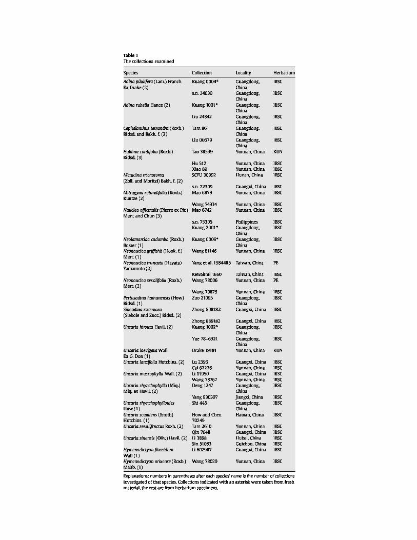
Forty-two collections of 22 species from 11 genera of the Naucleeae, and of two species of
Hymenodictyon occurring in China were examined (Table 1). Polliniferous anthers were
collected from living plants growing in the South China Botanical Garden (SCBG), and/or from
herbarium specimens from the following herbaria: IBSC, KUN and PE. Voucher specimens of
the fresh material are deposited in IBSC (Table 1).
For light (LM) and scanning electron microscopy (SEM), the polliniferous anthers were softened
by soaking in glacial acetic acid (Reitsma,1969), and dissected with tweezers to release the
pollen. The separation of the pollen from the remaining anther material was accomplished with a
sieve (pore diameter 50 µm). Each pollen sample was then split into two parts. One part was
acetolysed (three to five minutes in a heating block at 98 °C) according to the method of
Erdtman (1960) and, transferred to 70% ethanol. The other part was transferred directly to 70%
ethanol. Both parts were washed three times in 70% ethanol in an ultrasonic bath, for ten minutes
each time. For LM, the pollen was mounted on slides in glycerine jelly, and coverslips sealed to
the slides with paraffin. LM photographs were taken at a magnification of 1000×. For SEM, the
pollen was mounted on copper stubs, air-dried, and coated with gold in a JFC-1600 sputter coater
(JEOL Ltd, Tokyo, Japan). Observations and digital images were collected with a JEOL JSM-
6360LV SEM (JEOL Ltd, Tokyo, Japan).
Pollen wall ultrastructure was investigated with fresh pollen from Adina pilulifera, Neolamarckia
cadamba, and Uncaria hirsuta, and pollen from herbarium specimens of Metadina trichotoma,
and Pertusadina hainanensis. For transmission electron microscopy (TEM), hydrated,
unacetolysed pollen was fixed in 2.5% glutaraldehyde at pH 7.2, rinsed in 0.1 M phosphate
buffer for 2 h, then postfixed in 1 % osmium tetroxide for 2 h or more. The pollen was then
washed in phosphate buffer, dehydrated in an acetone series, embedded in Spurr's resin, and
cured at 70 °C. Ultrathin sections (80 nm) were cut using a Leica-Ultracut S ultramicrotome
(Leitz Inc., Wiesbaden, Germany), and stained with uranyl acetate and lead citrate. Transmission
electron micrographs were taken with a JEM-1010 (JEOL Ltd, Tokyo, Japan) transmission
electron microscope at 100 KV.
Measurements of the polar axis (P) and equatorial diameter (E) were made with LM from 10–20
pollen grains per specimen. All other measurements were made on digital SEM images with
JEOL's Smile View 2.2.6.1 software (JEOL Ltd, Tokyo, Japan). The colpi/polar axis ratio
multiplied by 100 (=LC/P× 100) was used to express the relative length of the colpi.
Measurements of ectocolpus width refer to the transverse diameter of the mesoporus, if present,
or to the widest opening of the ectocolpus, if there is no mesoporus. Measurements of the layers
of the pollen wall were made on 10–20 TEM images (10–20
pollen grains) of each taxon.
Palynological terminology follows that of Punt et al. (2007). Pollen size, and shape classes in
equatorial view refer to Erdtman (1969). The generic delimitations and infratribal taxa adopted
here are as circumscribed by Razafimandimbison and Bremer (2002). Unfortunately, these
authors did not place the two genera Diyaminauclea and Khasiaclunea, and the two genera
Haldina and Sinoadina are only provisionally accommodated in the poorly supported subtribe
Adiniae.
In order to investigate their potential as synapomorphies, the characters ―H-shaped endoaperture‖
and ―protruding oncus‖ were mapped on the phylogenetic tree of the Naucleeae-
Hymenodictyoneae clade (Razafimandimbison and Bremer, 2001, 2002), and their ancestral
states were reconstructed from unordered characters with the software MacClade 4.06 (Maddison
and Maddison, 2003). In the analyses of Razafimandimbison and Bremer (2001, 2002) the genus
Luculia, which had been shown to be basal in the Rubiaceae by Bremer et al. (1999), was used to
root the tree while the genera Ex-ostema and Cinchona were used as additional outgroups. For
our analyses we replaced the terminals (species) of Razafimandimbison and Bremer (2001,
2002) with the corresponding genera. The occurrence of H-shaped endoapertures in a genus,
whether distinct,indistinct or incomplete, were coded as the character state ―H-shaped
endoapertures present,‖ regardless of whether or not they occur in all members of a genus. For
example, H-shaped endoapertures are coded as present in Uncaria, even though they have only
been reported in U. rhynchophylla. Endoapertures in other patterns are coded as ―H-shaped
endoapertures absent.‖ Distinct and indistinct protruding onci, and protruding oncus remnants,
are coded as the character state ―protruding oncus present,‖ regardless of whether or not they
occur in all members of a genus.
3. RESULTS
3.1. General features
The pollen grains are always monads, radiosymmetric, and very small (E<10 µm) to small (E
10–25 µm). Their shape in equatorial view is spheroidal (P/E 0.88–1.14) to subprolate (P/E
1.14–1.33). The spheroidal condition can be subdivided into oblate spheroidal (P/E 0.88–1), and
prolate spheroidal (P/E 1–1.14). The outline in polar view (amb) is usually (sub)circular with
sunken colpi. Three compound apertures are found in each grain, each comprised of a long
ectocolpus, a lolongate to slightly circular mesoporus, and an often H-shaped endoaperture. H-
shaped endoapertures are the inner of a three-part compound aperture, and have two cross
members, one above and one below the porus. The ectocolpus membrane is usually granular,
though it is not visible in some species due to the narrowness of the ectocolpus. The mesoporus
is always located in the middle of the ectocolpus, at the equator. The sexine ornamentation is
microreticulate to striate, rugulate, or perforate; the lumina of microreticulations are usually
irregularly polygonal and (sub)circular. There is usually no differentiation of the sexine towards
the poles and/or colpi. Exine and intine are both obvious under LM and TEM. In TEM, the exine
is composed of a perforated tectum, relatively short columellae, and a thick nexine. The nexine is
often differentiated into a foot layer and an endexine, and thickened into costae towards the
aperture. The intine of some species is thickened in the apertural region, and protrudes from the
aperture forming a protruding oncus (Plate IV). This character can be variable in taxa in which it
occurs, so that some grains possess it and others do not.
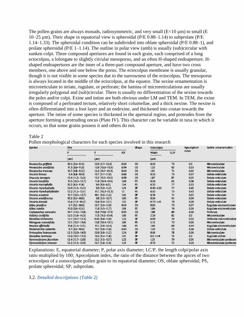
Table 2
Pollen morphological characters for each species involved in this research
Explanations: E, equatorial diameter; P, polar axis diameter; LC/P, the length colpi/polar axis
ratio multiplied by 100; Apocolpium index, the ratio of the distance between the apices of two
ectocolpus of a zonocolpate pollen grain to its equatorial diameter; OS, oblate spheroidal; PS,
prolate spheroidal; SP, subprolate.
3.2. Detailed descriptions (Table 2)
Generic descriptions are given for two genera, (Neonauclea, and Uncaria) in order to concisely
indicate the variation among the species investigated in these genera. Generic descriptions of the
other ten genera are not given because only one or two species were studied in each genus. The
pollen morphology of nine species (indicated with asterisks, e.g., Neonauclea sessilifolia*)
belonging to four genera is reported for the first time. The remaining 15 species (e.g.,
Neonauclea griffithii, without any symbol) belonging to 11 genera are described only by
important palynological characters that have not been reported previously (Table 2).
Neonauclea Merr. (65 species; 3 investigated): Pollen very small to small, P 10.5 (9.4–1.9) lam,
E 10.9 (9.7–12.6) lam; shape in equatorial view oblate spheroidal, amb slightly triangular,
subcircular to circular. Apertures 3; colporate, with long, narrow ectocolpi, each 0.59– 1.22 lam
wide, and with a relative length ranging from 62 (Neonauclea sessilifolia) to 73 (Neonauclea
griffithii), ends acute, membrane granular; mesoporus subcircular to lolongate; incompletely H-
shaped endoaperture presents in N. sessilifolia; protruding onci occur in N. griffithii. Sexine
sculpture microreticulate, without differentiation towards the poles and colpi, lumina of
microreticulations irregularly oblong or polygonal. There is little variation in pollen size, shape,
aperture number, or sexine ornamentation in the genus.
N. griffithii (Plate I, A; Plate III, A): Ectocolpus ends acute, membrane granular; mesoporus
subcircular, with protruding oncus; endoaperture not H-shaped; lumina of microreticulations
irregularly oblong or polygonal.
N. sessilifolia* (Plate I, B–C; Plate III, B): Ectocolpus ends acute to obtuse, membrane granular;
mesoporus lolongate (Plate III, B), protruding oncus absent; endoaperture faint, incompletely H-
shaped (Plate III, B). Sexine sculpture rugulate to microreticulate (Plate I, C), without
differentiation towards the colpi, but with slightly larger lumina at poles, lumina irregularly
oblong or polygonal.
N. truncata* (Plate I, D; Plate III, C): Ectocolpus ends acute to obtuse, membrane granular;
mesoporus lolongate (Plate III, C), protruding oncus absent; endoaperture not H-shaped; lumina
of microreticulations irregularly oblong or polygonal.
Uncaria Schreber (34 species; 9 investigated) Pollen very small to small, P (8.5–13.8) lam, E
(8.6–13.5) lam; shape in equatorial view spheroidal to subprolate, the spheroidal condition
subdivided into oblate spheroidal (P/E 0.88–1) and prolate spheroidal (P/E 1–1.14), amb circular
with three lobes due to the sunken colpi. Apertures 3; colporate, with long and very narrow (slit-
like) ectocolpi, width ranging from 0.28 lam (Uncaria sessilifructus) to 1.87 lam (Uncaria
laevigata), relative length 61–87, ends acute to obtuse, membrane granular, although not visible
in some species; mesoporus circular to lolongate; indistinctly H-shaped endoaperture present in
U. rhynchophylla, endoapertures in other species not H-shaped; in some species distinct or
indistinct protruding onci present. The sexine pattern may equally well be described as striate to
reticulate with interwoven muri, or rugulate with slender, long striae on the reticulum, without
differentiation towards poles or colpi; lumina of microreticulations irregularly oblong or
subcircular. There is little variation in pollen size, shape, sexine ornamentation and aperture
morphology in this genus.
U. hirsuta (Plate I, E–F; Plate III, D): Ectocolpus ends acute, membrane not visible; mesoporus
lolongate (Plate III, D), distinct protruding oncus present, (Plate I, E–F); endoaperture not H-
shaped; lumina of microreticulations irregularly oblong or subcircular.
U. laevigata* (Plate I, G): Ectocolpus ends acute to obtuse, membrane granular; mesoporus
circular to lolongate, protruding oncus present; endoaperture unknown; lumina of
microreticulations irregularly oblong or subcircular.
U. lancifolia* (Plate I, H; Plate III, E): Ectocolpus ends acute, membrane granular; mesoporus
lolongate (Plate III, E), protruding oncus absent; endoaperture not H-shaped; lumina of
microreticulations irregularly oblong or subcircular.
U. macrophylla (Plate I, I; Plate III, F): Ectocolpus ends acute, membrane not visible; mesoporus
subcircular, with indistinct protruding oncus (Plate III, F); endoaperture not H-shaped; lumina of
microreticulations irregularly oblong or subcircular.
U. rhynchophylla (Plate I, J; Plate III, G): Ectocolpus ends acute to obtuse, membrane granular;
mesoporus subcircular (Plate III, G), protruding oncus present (Plate I, J); endoaperture faint,
indistinctly H-shaped (Plate III, G); lumina of microreticulations irregularly oblong or
subcircular.
U. rhynchophylloides* (Plate I, K): Ectocolpus ends acute, membrane not visible; mesoporus
subcircular, without protruding oncus; endoaperture unknown; lumina of microreticulations
irregularly oblong or subcircular.
U. scandens* (Plate I, L; Plate III, H): Ectocolpus ends acute, membrane granular; mesoporus
subcircular (Plate III, H), protruding oncus present (Plate I, L); endoaperture not H-shaped;
lumina of microreticulations irregularly oblong or subcircular.
U. sessilifructus (Plate I, M–N; Plates III, I): Ectocolpus ends acute, membrane not visible;
mesoporus lolongate, with protruding oncus (Plates III, I); endoaperture not H-shaped; lumina of
microreticulations irregularly oblong or subcircular.
U. sinensis* (Plate I, O; Plate III, J): Ectocolpus ends acute, membrane granular; mesoporus
subcircular (Plate III, J), protruding oncus present (Plate I, O; Plate III, J); endoaperture not H-
shaped; lumina of microreticulations irregularly oblong or subcircular.
Adina pilulifera (Plate II, A; Plate III, K): Ectocolpus ends acute, membrane granular; mesoporus
circular (Plate III, K), with protruding oncus or oncus remnants (Plate II, A); endoaperture faint,
indistinctly H-shaped; lumina of microreticulations irregularly polygonal.
A. rubella (Plate II, B): Ectocolpus ends acute, membrane granular; mesoporus subcircular,
protruding oncus present; endoaperture unknown; lumina of microreticulations irregularly
polygonal.
Cephalanthus tetrandra (Plate II, C–D; Plate III, L): Ectocolpus protuberant in the middle (Plate
II, C), ends acute, membrane granular; mesoporus circular (Plate III, L), with indistinct
protruding oncus (Plate II, C); endoaperture distinct, clearly H-shaped (Plate III, L);
lumina/perforations of microreticulations irregularly subcircular or polygonal.
Haldina cordifolia (Plate II, E–F; Plate III, M): Ectocolpus ends obtuse to acute, membrane
granular; mesoporus lolongate (Plate III, M), with distinct protruding oncus (Plate II, E);
endoaperture faint, incompletely H-shaped; lumina of microreticulations irregularly polygonal.
Metadina trichotoma (Plate II, G; Plate III, N): Ectocolpus ends acute, membrane not visible;
mesoporus lolongate (Plate III, N), with protruding oncus (Plate II, G); endoaperture distinct,
clearly H-shaped (Plate III, N); lumina of microreticulations usually irregularly polygonal.
Mitragyna rotundifolia (Plate II, H; Plate III, O): Ectocolpus ends acute, membrane granular;
mesoporus circular (Plate III, O), protruding oncus absent; endoaperture distinct, clearly H-
shaped (Plate III, O); lumina/perforations of microreticulations irregularly oblong or polygonal.
Nauclea officinalis (Plates II, I; Plate III, P): Ectocolpus ends acute to obtuse, membrane
granular; mesoporus lolongate (Plate III, P), with distinct protruding oncus (Plates II, I);
endoaperture indistinct, not H-shaped; lumina of microreticulations irregularly polygonal.
Neolamarckia cadamba (Plate II, J): Ectocolpus ends acute, membrane granular; mesoporus
circular, distinct protruding oncus present; endoaperture unknown; lumina of microreticulations
irregularly polygonal.
Pertusadina hainanensis* (Plate II, K; Plate III, Q): Ectocolpus protuberant in the middle (Plate
II, K), ends acute, membrane not visible; mesoporus circular, with protruding oncus or
protruding oncus remnants (Plate III, Q); endoaperture indistinct, not H-shaped; lumina of
microreticulations irregularly oblong or polygonal.
Sinoadina racemosa (Plate II, L; Plate III, R): Ectocolpus ends acute, membrane not visible;
mesoporus subcircular (Plate III, R), with protruding oncus or remnants (Plate II, L);
endoaperture faint, indistinctly H-shaped; lumina of microreticulations irregularly polygonal.
Hymenodictyon flaccidum* (Plate II, M–N; Plate III, S): Ectocolpus ends acute, membrane
granular; mesoporus lolongate, with protruding oncus or remnants (Plate II, N); endoaperture H-
shaped (Plate III, S); lumina of microreticulations irregularly oblong or polygonal.
H. orixense (Plate II, O): Ectocolpus ends acute, membrane granular; mesoporus subcircular,
protruding oncus absent; endoaperture unknown; lumina of microreticulations irregularly oblong
or polygonal.
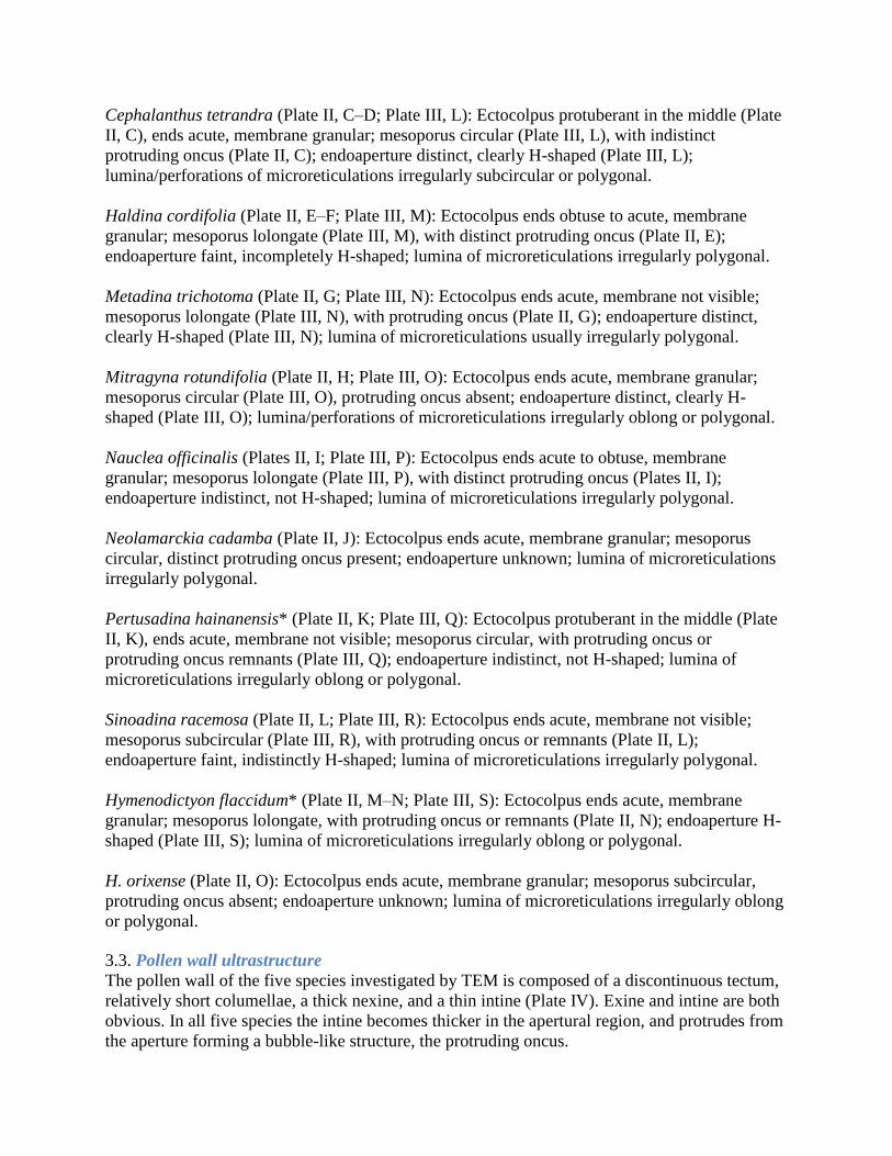
3.3. Pollen wall ultrastructure
The pollen wall of the five species investigated by TEM is composed of a discontinuous tectum,
relatively short columellae, a thick nexine, and a thin intine (Plate IV). Exine and intine are both
obvious. In all five species the intine becomes thicker in the apertural region, and protrudes from
the aperture forming a bubble-like structure, the protruding oncus.

Adina pilulifera (Plate IV, A–B): Tectum 0.2–0.39 μm thick; columellae 0.11–0.25 μm thick;
nexine differentiated into a foot layer and endexine, the foot layer separated from the endexine
by a single line, which is little different from these layers in electron density. Nexine 0.19–0.28
μm thick in mesocolpial region, becoming thinner near the aperture and then thickened into
costae surrounding the aperture. Intine usually 0.07–0.28 μm thick, thicker (ca. 0.69 μm) near the
aperture, and forming a protruding oncus.
Metadina trichotoma (Plate IV, C–D): Tectum 0.16–0.35 μm thick; columellae 0.04–0.25 μm
thick, electron-dense material (possibly lipidic) occurring occasionally between the columellae.
Nexine not differentiated into a foot layer and endexine, usually 0.06–0.24 (- 0.61) μm thick in
mesocolpial region, thickened into costae around the aperture. Intine usually 0.07–0.38 μm thick,
forming protruding onci. A less fibrillar zone occurs beneath the aperturate intine.
Neolamarckia cadamba (Plate IV, E–F): Tectum indistinct, 0.18– 0.38 μm thick; columellae
indistinct, 0.01–0.05 μm thick, electron- dense material (possibly lipidic and much wider than the
columellae), occurring occasionally between columellae. Nexine differentiated into a foot layer
and a very thin endexine, the foot layer separated from the endexine by a single white line.
Nexine 0.11–0.18 μm thick in mesocolpial region, thickened into costae around the aperture.
Intine usually 0.02–0.06 μm thick, forming a protruding oncus ca. 1.6 μm in diameter. Oncus
composed of a bi-layered ectintine with an electron- dense outer layer and a thick electron-lucent
inner layer; and separated from the cytoplasm of the pollen cell by intine material.
Pertusadina hainanensis (Plate IV, G–H): Tectum 0.16–0.29 μm thick; columellae indistinct,
0.04–0.16 μm thick, electron-dense material (possibly lipidic) occurring occasionally between
the columellae. Nexine not clearly differentiated into a foot layer and endexine, 0.07–0.27 μm
thick in mesocolpial region, becoming thinner near the aperture, and then thickened into costae
surrounding the aperture. Intine usually 0.01–0.18 μm thick in the apertural region, and forming
a protruding oncus; oncus composed of a bi-layered ectintine: an electron-dense outer layer, and
a thick electron-lucent inner layer.
Uncaria hirsuta (Plate IV, I–J): Tectum 0.16–0.48 μm thick; columellae 0.1–0.16 μm thick,
nexine differentiated into a foot layer and endexine, the foot layer separated from the endexine
by a single white line. Nexine 0.16–0.35 μm thick in mesocolpial region, becoming thinner near
the aperture, and then thickened into costae immediately surrounding the aperture. Intine usually
0.18–0.32 μm thick, forming a subcircular oncus, ca. 2.5 μm in diameter. The oncus is composed
of a bi-layered ectintine: an electron-dense outer layer, and a thick electron-lucent inner layer.
Cytoplasmic components, e.g., a vacuole (Plate IV, J) and starch (unpublished images), are
sometimes found in the onci. 3.4. Ancestral character state reconstruction 3.4.1. H-shaped
endoaperture (Fig. 1)
3.4. Ancestral character state recognition
3.4.1. H-shaped apertures
H-shaped apertures occur in all the genera of the subtribes Cephalanthinae, Mitragyninae,
Uncarinae, and Corynantheinae, and in five genera (including Haldina and Sinoadina) of
subtribe Adininae. One genus of subtribe Breoniinae also possesses H- shaped endoapertures,
while the condition in the other three genera remains unknown. The endoapertures in two genera
of the subtribe Naucleinae are not H-shaped; the condition in the other two genera remains
unknown. Hymenodictyon of the tribe Hymenodictyeae, the sister group of the Naucleeae, has H-
shaped endoapertures (Fig. 1).
Parsimony reconstruction suggests that all the ancestral nodes in the outgroups lack H-shaped
endoapatures, while the ancestor of the clade containing Hymenodictyon and the Naucleeae
possesses them. Many of the internal nodes of the Naucleeae also possess H-shaped
endoapertures, though the polytomies make the reconstruction of the more derived nodes
difficult.
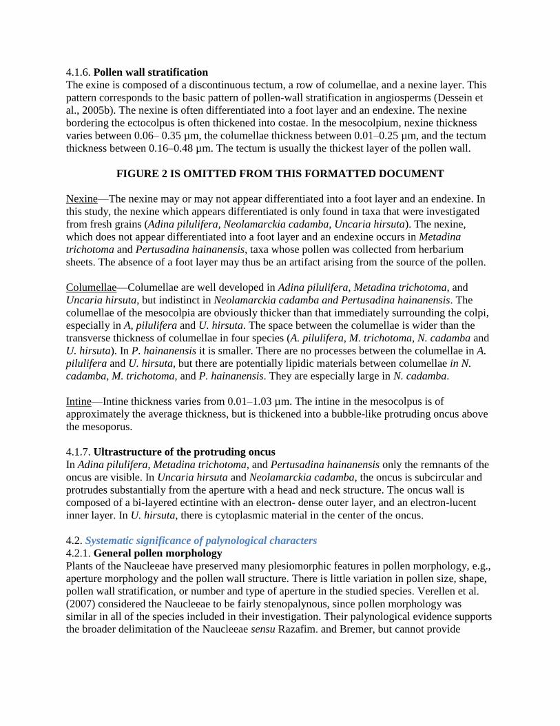
3.4.2. Protruding oncus (Fig. 2)
Of the 12 genera that have been investigated, protruding onci occur in six genera (including
Haldina and Sinoadina) of subtribe Adininae, two of subtribe Naucleinae, the single genus
Uncaria of subtribe Uncarinae, the single genus Cephalanthus of subtribe Cephalanthinae, and
Hymenodictyon of tribe Hymenodictyoeae. Only the genus Mitragyna s.s. of Mitragyninae lacks
this characteristic. The condition in the remaining 13 genera distributed in subtribes Adininae,
Breoniinae, Corynantheinae, Mitragyninae, and Naucleinae remain unknown (Fig. 2).
Parsimony reconstruction suggests that a protruding oncus is a synapomorphy of the clade
Hymenodictyon+ Naucleeae, though the reconstruction for the subtribes Mitragyninae and
Corynantheinae is equivocal. The remaining nodes all reconstruct as possessing a protruding
oncus.
4. DISCUSSION
4.1. Pollen morphological characteristics
4.1.1. Size
Pollen grain size is rather uniform throughout the species investigated. The average equatorial
diameter (E) varies from 9.01 µm in Uncaria macrophylla to 15.85 µm in Cephalanthus
tetrandra. We found that Cephalanthus has the largest pollen grains in the tribe Naucleeae, in
agreement with Verellen et al. (2007).
4.1.2. Shape
In equatorial view, pollen shape is described by the P/E ratio. In the studied species, all of the
pollen grains are speroidal (P/E 0.88–1.14), or subprolate (P/E 1.14–1.33). The spheroidal
condition can be further divided into oblate spheroidal (P/E 0.88–1.0) and prolate spheroidal (P/
E 1.0–1.14). The amb is (sub)circular, and often somewhat lobed due to sunken colpi, which
occur in most members of the Rubiaceae (Dessein et al., 2005a,b). Slightly triangular pollen
grains are only found in Neonauclea griffithii.
4.1.3. Aperture
Number—The number of colpi is always three. Triaperturate pollen grains are common in the
Rubiaceae, and are the plesiomorphic condition in the family (Dessein et al., 2005b). However,
the number of apertures varies considerably within tribe Spermacoceae (Dessein et al., 2002).
Position—In most Rubiaceae, pollen is angulaperturate (the apertures situated at the angles of the
outline in polar view), or zonoaperturate (the apertures situated only at the equator) (Dessein et
al., 2005b). All the species studied are zonoaperturate, the apertures all arranged along the
equator, but a tendency towards an angulaperture is seen in some pollen grains of Neonauclea
griffithii.
Type—Apertures of Naucleeae are always compound, built up by three components that are
situated in three layers of the pollen wall. The aperture is built up of an ectocolpus, a mesoporus,
and an endoaperture. This type of compound aperture is believed to be plesiomorphic in the
Rubiaceae (Dessein et al., 2005b).
Ectocolpus—The length of the ectocolpus is very variable in the Rubiaceae (Dessein et al.,
2005b). For instance, the relative length of the colpus (LC/P ratio) ranges from 6–62 in African
Spermacoce species (Dessein et al., 2002). In the Naucleeae, the variation in the LC/P ratio is
relatively minor, ranging from 61 in Uncaria sessilifructus to 87 in U. laevigata. The
apocolpium index is also very low, varying from 0.25 in U. laevigata to 0.53 in Neonauclea
sessilifolia. The colpi are often slit-like in the genera Uncaria, Metadina, Pertusadina, and
Sinoadina, sometimes becoming wider around the mesoporus. The width varies from 0.28 µm in
U. sessilifructus to 2.29 µm in Haldina cordifolia. The ectocolpus membrane is often granular,
except in Uncaria hirsuta, U. macrophylla, U. rhynchophylloides, U. sessilifructus, Metadina
trichotoma, Pertusadina hainanensis, and Sinoadina racemosa. The ectocolpus membrane of
these species is not visible, as the colpi are slit-like and deep-set.
The colpi of all species have distinct, regular margins. The ectocolpus is protuberant in
Cephalanthus tetrandra and Pertusadina hainanensis.
Endoaperture—The variation in endoapertures in Rubiaceae is large and has significant
systematic value. Endocolpi (Dessein et al., 2000) and endocinguli (Dessein et al., 2002) are the
most common endoapertures in the family, but H-shaped endoapertures have also been reported
in some taxa (Robbrecht,1985; Huysmans et al., 1994; Verellen et al., 2007). H-shaped
endoapertures have a thin H-shaped zone surrounding the porus. They can be clearly observed
with SEM in broken pollen grains, while they appear as a brighter zone surrounding the porus in
LM. The downstrokes of the ―H‖ are parallel to the ectocolpus, while the equatorial connections
may be weak or missing (Huysmans et al., 1994), making the ―H‖ incomplete.
Distinct H-shaped endoapertures occur in Neonauclea, Uncaria, Adina, Cephalanthus, Haldina,
Metadina, Mitragyna, Sinoadina, and Hymenodictyon, while indistinct ones occur in Nauclea
and Pertusadina. H-shaped endoapertures are first observed here in Adina, Cephalanthus,
Metadina, and Sinoadina. In Haldina, Neonuclea, Sinoadina and Uncaria, the ―H‖ is incomplete.
H-shaped endoapertures have also been previously reported in Breonadia, Corynanthe, Haldina,
Neonauclea, Pausinystalia, Pseudocinchona, Uncaria (Verellen et al., 2007), and
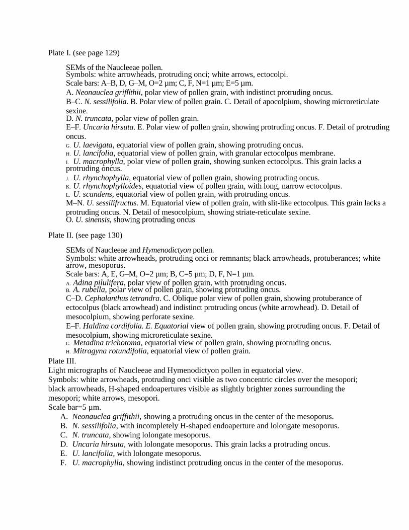
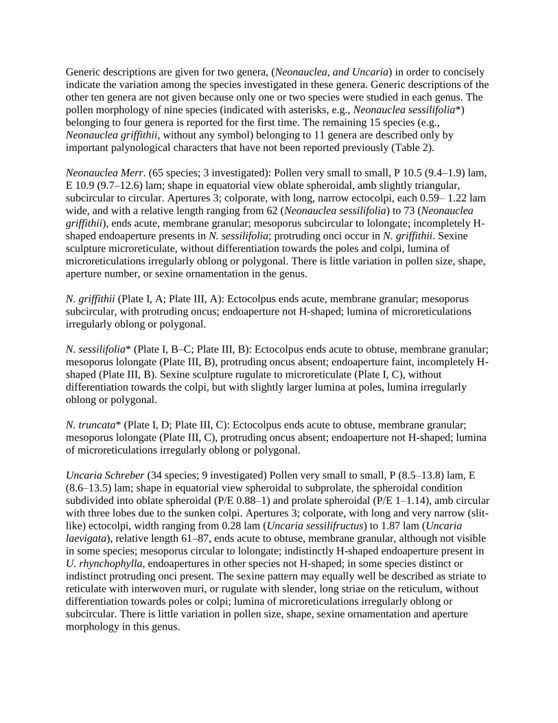
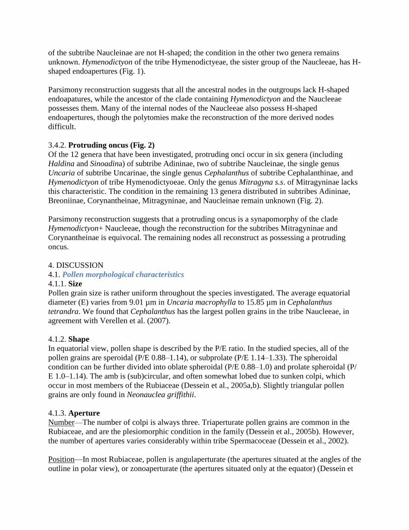
Plate I. (see page 129)
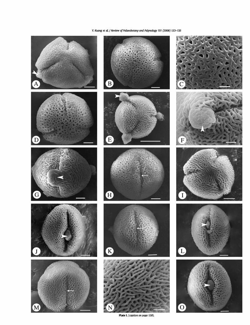
SEMs of the Naucleeae pollen. Symbols: white arrowheads, protruding onci; white arrows, ectocolpi. Scale bars: A–B, D, G–M, O=2 µm; C, F, N=1 µm; E=5 µm.
A. Neonauclea griffithii, polar view of pollen grain, with indistinct protruding oncus.
B–C. N. sessilifolia. B. Polar view of pollen grain. C. Detail of apocolpium, showing microreticulate
sexine. D. N. truncata, polar view of pollen grain. E–F. Uncaria hirsuta. E. Polar view of pollen grain, showing protruding oncus. F. Detail of protruding
oncus. G. U. laevigata, equatorial view of pollen grain, showing protruding oncus. H. U. lancifolia, equatorial view of pollen grain, with granular ectocolpus membrane. I. U. macrophylla, polar view of pollen grain, showing sunken ectocolpus. This grain lacks a protruding oncus.
J. U. rhynchophylla, equatorial view of pollen grain, showing protruding oncus. K. U. rhynchophylloides, equatorial view of pollen grain, with long, narrow ectocolpus. L. U. scandens, equatorial view of pollen grain, with protruding oncus. M–N. U. sessilifructus. M. Equatorial view of pollen grain, with slit-like ectocolpus. This grain lacks a
protruding oncus. N. Detail of mesocolpium, showing striate-reticulate sexine. O. U. sinensis, showing protruding oncus
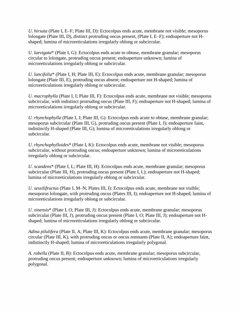
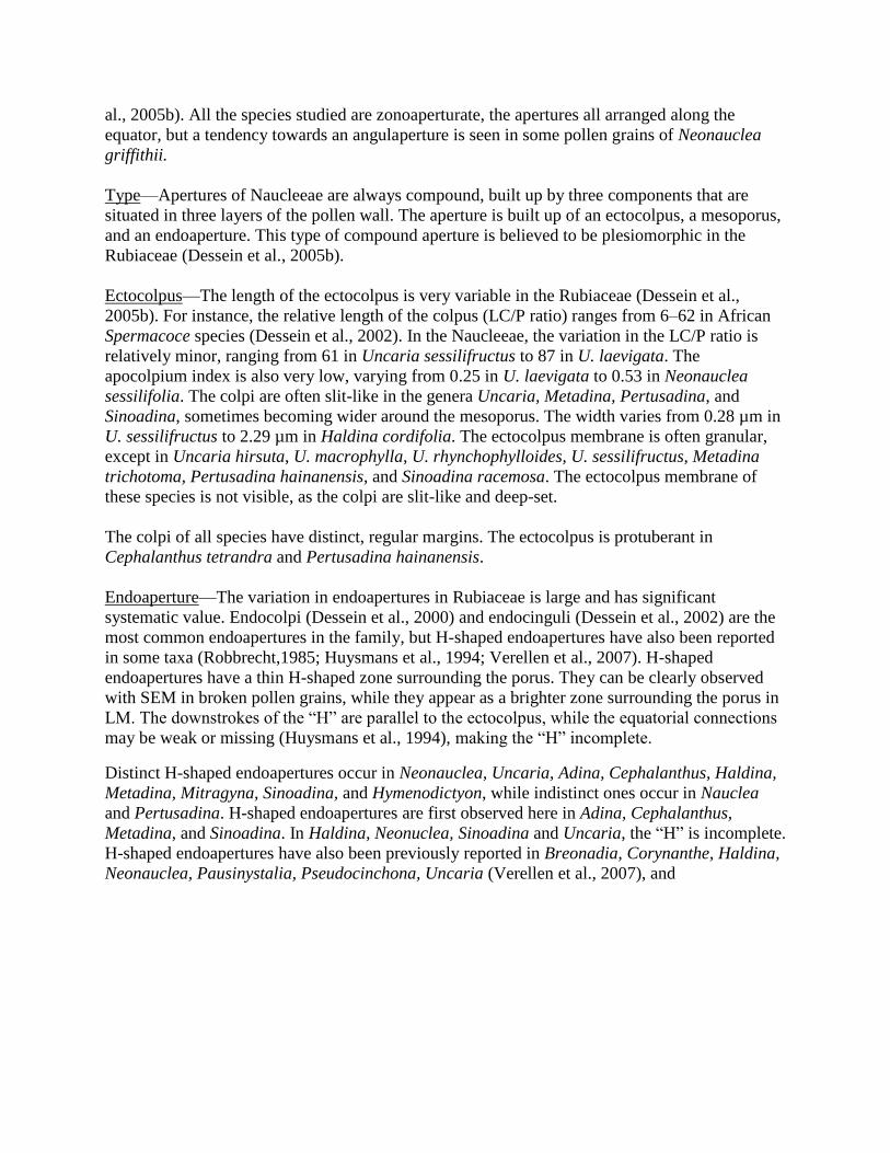
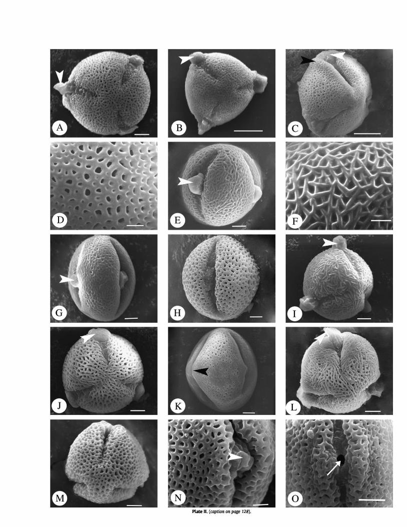
Plate II. (see page 130)
SEMs of Naucleeae and Hymenodictyon pollen. Symbols: white arrowheads, protruding onci or remnants; black arrowheads, protuberances; white arrow, mesoporus. Scale bars: A, E, G–M, O=2 µm; B, C=5 µm; D, F, N=1 µm. A. Adina pilulifera, polar view of pollen grain, with protruding oncus. B. A. rubella, polar view of pollen grain, showing protruding oncus. C–D. Cephalanthus tetrandra. C. Oblique polar view of pollen grain, showing protuberance of
ectocolpus (black arrowhead) and indistinct protruding oncus (white arrowhead). D. Detail of
mesocolpium, showing perforate sexine.
E–F. Haldina cordifolia. E. Equatorial view of pollen grain, showing protruding oncus. F. Detail of
mesocolpium, showing microreticulate sexine. G. Metadina trichotoma, equatorial view of pollen grain, showing protruding oncus. H. Mitragyna rotundifolia, equatorial view of pollen grain. I. J. K. L. Nauclea officinalis, oblique polar view of pollen grain, showing protruding oncus. M. Neolamarckia cadamba, polar view of pollen grain, showing protruding oncus. N. Pertusadina hainanensis, equatorial view of pollen grain, showing protuberance of ectocolpus. This grain lacks a protruding oncus. O. Sinoadina racemosa, polar view of pollen grain, show protruding oncus remnants. M–N. Hymenodictyon,fiaccidum. M. Oblique polar view of pollen grain. N. Equatorial
view of pollen grain, showing protruding oncus remnants. O. H. orixense, equatorial
view of pollen grain, showing mesoporus.
Plate IV. (see page 132) TEMs of Naucleeae pollen. Abbreviations: c, columellae; co, costa; en, endexine; fl, foot layer; in, intine; lm, lipidic material; n, nexine; re, remnant; t, tectum; v, vacuole. Symbols: black
arrowheads, electron-dense outer layer of oncus; star, electron-lucent inner layer of oncus. Scale bars: A, E–F, H–I=200 nm; B, D, J=500 nm; C, G=100 nm. A–B. Adina pilulifera. A. Pollen wall ultrastructure in non-apertural region. B. Detail of pollen wall structure towards apertural region, showing protruding oncus
remnant and costae. C–D. Metadina trichotoma. C. Pollen wall ultrastructure in non-apertural region, and possible lipidic material between columellae. D. Detail of pollen wall structure towards
apertural region. E–F. Neolamarckia cadamba. E. Detail of pollen wall stratification in non-apertural region. Note possible lipidic material between columellae. F. Oncus separated from the
cytoplasm of the vegetative cell by intine material. G–H. Pertusadina hainanensis. G. Detail of pollen wall structure in non-apertural region. Note possible lipidic material between columellae. H. Protruding oncus and
costae in apertural region.
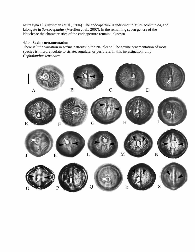
Plate III.
Light micrographs of Naucleeae and Hymenodictyon pollen in equatorial view.
Symbols: white arrowheads, protruding onci visible as two concentric circles over the mesopori;
black arrowheads, H-shaped endoapertures visible as slightly brighter zones surrounding the
mesopori; white arrows, mesopori.
Scale bar=5 µm.
A. Neonauclea griffithii, showing a protruding oncus in the center of the mesoporus.
B. N. sessilifolia, with incompletely H-shaped endoaperture and lolongate mesoporus.
C. N. truncata, showing lolongate mesoporus.
D. Uncaria hirsuta, with lolongate mesoporus. This grain lacks a protruding oncus.
E. U. lancifolia, with lolongate mesoporus.
F. U. macrophylla, showing indistinct protruding oncus in the center of the mesoporus.
G. U. rhynchophylla, with indistinctly H-shaped endoaperture and subcircular mesoporus.
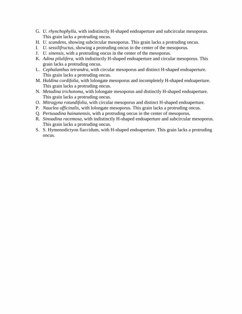
This grain lacks a protruding oncus.
H. U. scandens, showing subcircular mesoporus. This grain lacks a protruding oncus.
I. U. sessilifructus, showing a protruding oncus in the center of the mesoporus.
J. U. sinensis, with a protruding oncus in the center of the mesoporus.
K. Adina pilulifera, with indistinctly H-shaped endoaperture and circular mesoporus. This
grain lacks a protruding oncus.
L. Cephalanthus tetrandra, with circular mesoporus and distinct H-shaped endoaperture.
This grain lacks a protruding oncus.
M. Haldina cordifolia, with lolongate mesoporus and incompletely H-shaped endoaperture.
This grain lacks a protruding oncus.
N. Metadina trichotoma, with lolongate mesoporus and distinctly H-shaped endoaperture.
This grain lacks a protruding oncus.
O. Mitragyna rotundifolia, with circular mesoporus and distinct H-shaped endoaperture.
P. Nauclea officinalis, with lolongate mesoporus. This grain lacks a protruding oncus.
Q. Pertusadina hainanensis, with a protruding oncus in the center of mesoporus.
R. Sinoadina racemosa, with indistinctly H-shaped endoaperture and subcircular mesoporus.
This grain lacks a protruding oncus.
S. S. Hymenodictyon fiaccidum, with H-shaped endoaperture. This grain lacks a protruding
oncus.
Mitragyna s.l. (Huysmans et al., 1994). The endoaperture is indistinct in Myrmeconauclea, and
lalongate in Sarcocephalus (Verellen et al., 2007). In the remaining seven genera of the
Naucleeae the characteristics of the endoaperture remain unknown.
4.1.4. Sexine ornamentation
There is little variation in sexine patterns in the Naucleeae. The sexine ornamentation of most
species is microreticulate to striate, rugulate, or perforate. In this investigation, only
Cephalanthus tetrandra
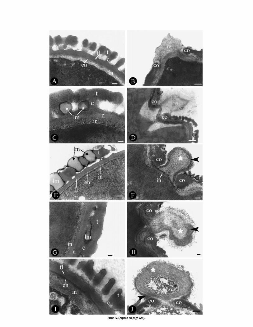
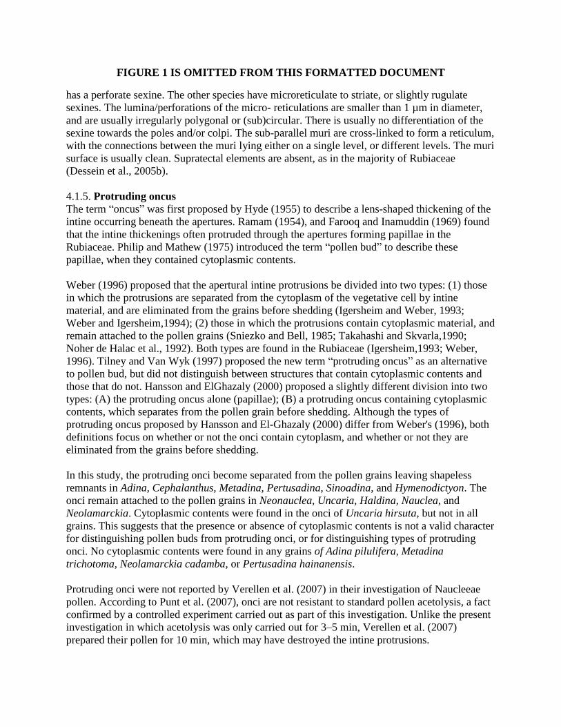
FIGURE 1 IS OMITTED FROM THIS FORMATTED DOCUMENT
has a perforate sexine. The other species have microreticulate to striate, or slightly rugulate
sexines. The lumina/perforations of the micro- reticulations are smaller than 1 µm in diameter,
and are usually irregularly polygonal or (sub)circular. There is usually no differentiation of the
sexine towards the poles and/or colpi. The sub-parallel muri are cross-linked to form a reticulum,
with the connections between the muri lying either on a single level, or different levels. The muri
surface is usually clean. Supratectal elements are absent, as in the majority of Rubiaceae
(Dessein et al., 2005b).
4.1.5. Protruding oncus
The term ―oncus‖ was first proposed by Hyde (1955) to describe a lens-shaped thickening of the
intine occurring beneath the apertures. Ramam (1954), and Farooq and Inamuddin (1969) found
that the intine thickenings often protruded through the apertures forming papillae in the
Rubiaceae. Philip and Mathew (1975) introduced the term ―pollen bud‖ to describe these
papillae, when they contained cytoplasmic contents.
Weber (1996) proposed that the apertural intine protrusions be divided into two types: (1) those
in which the protrusions are separated from the cytoplasm of the vegetative cell by intine
material, and are eliminated from the grains before shedding (Igersheim and Weber, 1993;
Weber and Igersheim,1994); (2) those in which the protrusions contain cytoplasmic material, and
remain attached to the pollen grains (Sniezko and Bell, 1985; Takahashi and Skvarla,1990;
Noher de Halac et al., 1992). Both types are found in the Rubiaceae (Igersheim,1993; Weber,
1996). Tilney and Van Wyk (1997) proposed the new term ―protruding oncus‖ as an alternative
to pollen bud, but did not distinguish between structures that contain cytoplasmic contents and
those that do not. Hansson and ElGhazaly (2000) proposed a slightly different division into two
types: (A) the protruding oncus alone (papillae); (B) a protruding oncus containing cytoplasmic
contents, which separates from the pollen grain before shedding. Although the types of
protruding oncus proposed by Hansson and El-Ghazaly (2000) differ from Weber's (1996), both
definitions focus on whether or not the onci contain cytoplasm, and whether or not they are
eliminated from the grains before shedding.
In this study, the protruding onci become separated from the pollen grains leaving shapeless
remnants in Adina, Cephalanthus, Metadina, Pertusadina, Sinoadina, and Hymenodictyon. The
onci remain attached to the pollen grains in Neonauclea, Uncaria, Haldina, Nauclea, and
Neolamarckia. Cytoplasmic contents were found in the onci of Uncaria hirsuta, but not in all
grains. This suggests that the presence or absence of cytoplasmic contents is not a valid character
for distinguishing pollen buds from protruding onci, or for distinguishing types of protruding
onci. No cytoplasmic contents were found in any grains of Adina pilulifera, Metadina
trichotoma, Neolamarckia cadamba, or Pertusadina hainanensis.
Protruding onci were not reported by Verellen et al. (2007) in their investigation of Naucleeae
pollen. According to Punt et al. (2007), onci are not resistant to standard pollen acetolysis, a fact
confirmed by a controlled experiment carried out as part of this investigation. Unlike the present
investigation in which acetolysis was only carried out for 3–5 min, Verellen et al. (2007)
prepared their pollen for 10 min, which may have destroyed the intine protrusions.
4.1.6. Pollen wall stratification
The exine is composed of a discontinuous tectum, a row of columellae, and a nexine layer. This
pattern corresponds to the basic pattern of pollen-wall stratification in angiosperms (Dessein et
al., 2005b). The nexine is often differentiated into a foot layer and an endexine. The nexine
bordering the ectocolpus is often thickened into costae. In the mesocolpium, nexine thickness
varies between 0.06– 0.35 µm, the columellae thickness between 0.01–0.25 µm, and the tectum
thickness between 0.16–0.48 µm. The tectum is usually the thickest layer of the pollen wall.
FIGURE 2 IS OMITTED FROM THIS FORMATTED DOCUMENT
Nexine—The nexine may or may not appear differentiated into a foot layer and an endexine. In
this study, the nexine which appears differentiated is only found in taxa that were investigated
from fresh grains (Adina pilulifera, Neolamarckia cadamba, Uncaria hirsuta). The nexine,
which does not appear differentiated into a foot layer and an endexine occurs in Metadina
trichotoma and Pertusadina hainanensis, taxa whose pollen was collected from herbarium
sheets. The absence of a foot layer may thus be an artifact arising from the source of the pollen.
Columellae—Columellae are well developed in Adina pilulifera, Metadina trichotoma, and
Uncaria hirsuta, but indistinct in Neolamarckia cadamba and Pertusadina hainanensis. The
columellae of the mesocolpia are obviously thicker than that immediately surrounding the colpi,
especially in A, pilulifera and U. hirsuta. The space between the columellae is wider than the
transverse thickness of columellae in four species (A. pilulifera, M. trichotoma, N. cadamba and
U. hirsuta). In P. hainanensis it is smaller. There are no processes between the columellae in A.
pilulifera and U. hirsuta, but there are potentially lipidic materials between columellae in N.
cadamba, M. trichotoma, and P. hainanensis. They are especially large in N. cadamba.
Intine—Intine thickness varies from 0.01–1.03 µm. The intine in the mesocolpus is of
approximately the average thickness, but is thickened into a bubble-like protruding oncus above
the mesoporus.
4.1.7. Ultrastructure of the protruding oncus
In Adina pilulifera, Metadina trichotoma, and Pertusadina hainanensis only the remnants of the
oncus are visible. In Uncaria hirsuta and Neolamarckia cadamba, the oncus is subcircular and
protrudes substantially from the aperture with a head and neck structure. The oncus wall is
composed of a bi-layered ectintine with an electron- dense outer layer, and an electron-lucent
inner layer. In U. hirsuta, there is cytoplasmic material in the center of the oncus.
4.2. Systematic significance of palynological characters
4.2.1. General pollen morphology
Plants of the Naucleeae have preserved many plesiomorphic features in pollen morphology, e.g.,
aperture morphology and the pollen wall structure. There is little variation in pollen size, shape,
pollen wall stratification, or number and type of aperture in the studied species. Verellen et al.
(2007) considered the Naucleeae to be fairly stenopalynous, since pollen morphology was
similar in all of the species included in their investigation. Their palynological evidence supports
the broader delimitation of the Naucleeae sensu Razafim. and Bremer, but cannot provide
unambiguous support for subtribal or generic delimitations because of a lack of variation in
pollen characters. Our palynological results agree well with those of Verellen et al. (2007).
The pollen morphology of tribe Hymenodictyeae is very similar to that of the Naucleeae.
Razafimandimbison and Bremer (2001) placed the Hymenodictyeae as sister to the Naucleeae.
Based on this result, Andersson and Antonelli (2005) submerged the Hymenodictyon into the
Naucleeae for molecular analysis. Razafimandimbison and Bremer (2006) showed that the two
tribes can still be distinguished from each other by their inflorescence morphology, floral disk
morphology, and fruit characteristics. Verellen et al. (2007) concluded that the sister relationship
between the two tribes was supported by the shared presence of H-shaped endoapertures, but
they also noted that their palynological evidence also supported the submersion of Hymeno-
dictyeae in Naucleeae. H-shaped endoapertures, and a protruding oncus, occur in at least some
members of the Hymenodictyeae and Naucleeae included in this paper, and their pollen
morphology in other respects also shows much similarity (e.g., sexine ornamentation, aperture
morphology). The lack of clear synapomorphies for the two tribes, and the shared presence of H-
shaped endoapertures and a protruding oncus suggest that Anderson and Antonelli's (2005)
analysis may be correct. 4.2.2. H-shaped endoaperture
The presence of H-shaped endoapertures was proposed as a synapomorphy of the Naucleeae by
Dessein et al. (2005b), and Verellen et al. (2007) agreed. To test this hypothesis, we mapped the
occurrence of H-shaped endoapertures at the generic level on the phylogenetic tree of
Razafimandimbison and Bremer (2001, 2002), and reconstructed the ancestral states at each
node. H-shaped endoapertures are widespread in five subtribes of the Naucleeae, and in the
Hymenodictyeae, and form a putative synapomorphy of the clade Hymenodictyon+ Naucleeae.
4.2.3. Protruding oncus
The protruding oncus was previously reported in the following Rubiaceae taxa: Stephegyne
parviflora (Ramam, 1954), Oldenlandia nudicaulis (Farooq and Inamuddin, 1969),
Ophiorrhizeae (Philip and Mathew, 1975; Mathew and Philip, 1987), Isertieae (Priyadarshan and
Ramachandran, 1984), and Vanguerieae (Tilney and Van Wyk, 1997), Mitriostigma axillare
(Hansson and El-Ghazaly, 2000), Hedyotideae (Ma et al., 2005), and Tarenna gracilipes
(Vinckier and Smets, 2005). These taxa are not closely related according to the summary
cladogram of Rubiaceae (Dessein et al., 2005b), which suggests that the character has evolved
several times independently.
In the Hymenodictyeae-Naucleeae the presence of the protruding oncus has only been
investigated in 12 genera. Of the seven subtribes, only the Mitragyninae probably lack it. The
Adininae, Naucleinae, Uncarinae, and Cephalanthinae all possess this characteristic, which
implies a relatively close relationship between these four subtribes. The condition in the
Corynantheinae and Breoniinae remains unknown, though parsimony analysis suggests that the
Breoniinae will be found to possess it. The sister tribe Hymenodictyeae possesses a protruding
oncus, while the outgroups lack it. These results lead us to suggest that the protruding oncus is a
synapomorphy of the Hymenodictyeae-Naucleeae clade. The generic-level ancestral state
reconstruction supports this suggestion.
5. CONCLUSIONS
The Naucleeae is a stenopalynous tribe, characterized by very small to small pollen grains,
tricolporate pollen with an ectocolpus, a subcircular to lolongate mesoporus, and often an H-
shaped endoaperture. Sexine patterns are microreticulate to striate, rugulate, or perforate. Our
pollen morphological observations in Chinese Naucleeae species support the delimitation of the
Naucleeae sensu Razafim. and Bremer. Naucleeae have preserved many plesiomorphic features
in pollen morphology, and pollen morphology is of little value in distinguishing the subtribes and
genera of the Naucleeae. Ancestral state reconstruction at the generic level is unambiguous in
showing that the possession of H-shaped endoapertures and protruding onci form morphological
synapomorphies of the clade Hymenodictyon+ Naucleeae.
Acknowledgements
We thank Ms. XinLan Xu and Ms. XiaoYing Hu for assistance with electron microscopy; Ms.
FeiYan Zeng for permission to remove pollen from herbarium specimens; Laura Lagomarsino
and Dr. Chelsea Specht for assistance with MacClade; the two anonymous reviewers and the
editors for their helpful comments and suggestions on an earlier draft of this paper. Funding for
this work was provided by National Natural Science Foundation of China (39870087, 30370099,
40332021), and the Science and Technology Project of Guangzhou Municipality (2006Z2-
E0011).
REFERENCES
Andersson, L., 1993. Pollen characteristics of the tribes Calycophylleae, Cinchoneae and
Hillieae (Rubiaceae). Nordic Journal of Botany 13, 405–417.
Andersson, L., Antonelli, A., 2005. Phylogeny of the tribe Cinchoneae (Rubiaceae), its
position in Cinchonoideae, and description of a new genus, Ciliosemina. Taxon 54, 17–
28.
Bremekamp, C.E.B.,1966. Remarks on the position, the delimitation and the subdivision
of the Rubiaceae. Acta Botanica Neerlandica 15,1–33.
Bremer, B., Andreasen, K., Olsson, D., 1995. Subfamilial and tribal relationships in the
Rubiaceae based on rbcL sequence data. Annals of the Missouri Botanical Garden 82,
383–397.
Bremer, B., Jansen, R.J., Oxelman, B., Backlund, M., Lantz, H., Kim, K.J., 1999. More
characters or more taxa for a robust phylogeny-case study from the coffee family
(Rubiaceae). Systematic Biology 48, 413–435.
De Block, P., Robbrecht, E.,1998. Pollen of the Pavetteae (Rubiaceae, Ixoroideae) and its
taxonomic significance. Grana 37, 260–275.
Delprete, P.G., 1996. In: Robbrecht, E., Puff, C., Smets, E. (Eds.), Evaluation of the
tribes Chiococceae, Condamineeae and Catesbaceae (Rubiaceae) Based on
Morphological Characters. Second International Rubiaceae Conference Proceedings,
Opera Botanica Belgica, vol. 7. Nationale Plantentuin van Belgie, Meise, pp. 165–192.
Dessein, S., Huysmans, S., Robbrecht, E., Smets, E., 2002. Pollen of African Spermacoce
species (Rubiaceae) — morphology and evolution aspects. Grana 41, 69–89.
Dessein, S., Ochoterena, H., De Block, P., Lens, F., Robbrecht, E., Schols, P., Smets, E.,
Vinckier, S., Huysmans, S., 2005a. Palynological characters and their phylogenetic signal
in Rubiaceae. Botanical Review 71,354–414.

Dessein, S., Harwood, R., Smets, E., Robbrecht, E., 2005b. Pollen of Spermacoce
(Rubiaceae) species from the Northern Territory of Australia: morphology and taxonomic
significance. Australian Systymatic Botany 18, 367–382.
Dessein, S., Scheltens, A., Huysmans, S., Robbrecht, E., Smets, E., 2000. Pollen
morphological survey of Pentas (Rubiaceae-Rubioideae) and its closest allies. Review of
Palaeobotany and Palynology 112,189–205.
Erdtman, G., 1960. The acetolysis method. A revised description. Svensk Botanisk
Tidskrift 54, 561–564.
Erdtman, G., 1969. Handbook of Palynology. An Introduction to the Study of Pollen
Grains and Spores. Munksgaard, Copenhagen.
Farooq, M., Inamuddin, M., 1969. The embryology of Oldenlandia nudicaulis Roth.
Journal of Indian Botany Society 48,166–173.
Hansson, T., El-Ghazaly, G., 2000. Develoμment and cytochemistry of pollen and
tapetum in Mitriostigma axillare (Rubiaceae). Grana 39, 65–89.
Haviland, G.D.,1897. A revision of the tribe Naucleeae (Nat. Ord. Rubiaceae). Journal of
Linnean Society 33,1–94.
Huysmans, S., Robbrecht, E., Smets, E., 1994. Are the genera Hallea and Mitragyna
(Rubiaceae: Coptosapelteae) pollen morphologically distinct? Blumea 39, 321–340.
Huysman, S., Robbrecht, E., Smets, E., 1998. A collapsed tribe revised: pollen
morphology of the Iserteae (Cinchonoideae-Rubiaceae). Review of Palaeobotany and
Palynology 104, 85–113.
Huysmans, S., Robbrecht, E., Delprete, P., Smets, E.,1999. Pollen morphological support
for the Catesbaeeae-Chiococceae-Exostma-complex (Rubiaceae). Grana 38, 325–338.
Hyde, H.A., 1955. Oncus, a new term in pollen morphology. New Phytologist 54, 255–
256.
Igersheim, A.,1993. The character states of the Caribbean monotypic endemic Strumpfia
(Rubiaceae). Nordic Journal of Botany 13, 545–559.
Igersheim, A., Weber, M.,1993. Pollen bud formation in Ophiorrhiza (Rubiaceae). Opera
Botanica Belgica 6, 51–59.
Johansson, J.T.,1987. Pollen morphology of the tribe Morindeae (Rubiaceae). Grana 26,
134–150.
Leroy, J.F.,1975. Taxogénétique dans le genre Hallea sur lasous-tribu des Mitragyninae
(Rubiaceae-Naucleeae).Adansonia 2,65–88.
Liang YuanHui, 1982. Rubiaceae. In: Institute of Botany and South China Institute of
Botany, Academia Sinica. (Eds), Angiosperm pollen flora of tropic and subtropic China.
Science Press, Beijing, pp. 305–325. (in Chinese).
Ma, QiXia, Wang, RuiJiang, Chen, BingHui, 2005. Pollen morphology of Spiradiclis Bl.
(Rubiaceae). Journal of Tropical and Subtropical Botany 13, 159–166 (in Chinese, with
English Abstract).
Maddison, D.R., Maddison, W.P., 2003. MacClade 4.06. Sinauer Associates, Sunderland,
MA.
Mathew, P.M., Philip, O., 1987. Develoμment and systematic significance of pollen bud
formation in Ophiorrhiza Linn. New Botanist 14, 47–54.
Noher de Halac, I., Fama, G., Cismondi, I.A., 1992. Changes in lipids and
polysaccharides during pollen ontogeny in Oenothera anthers. Sexual Plant Reproduction
5, 110–116.
Persson, C., 1993. Pollen morphology of the Gardenieae-Gardeniinae (Rubiaceae).
Nordic Journal of Botany 13, 561–582.
Philip, O., Mathew, P.M., 1975. Cytology of exceptional develoμment of the male
gametophyte in Ophiorrhiza mungos. Canadian Journal of Botany 53, 2032–2037.
Piesschaert, F., Huysmans, S., Jaimes, I., Robbrecht, E., Smets, E., 2000. Morphological
evidence for an extended tribe Coccocypseleae (Rubiaceae-Rubioideae). Plant Biology 2,
536–546.
Priyadarshan, P.M., Ramachandran, K., 1984. Cytology and exceptional pollen
develoμment in Mussaenda Linn. Cytologia 49,407–413.
Punt, W., Hoen, P.P., Blackmore, S., Nilsson, S., Le Thomas, A., 2007. Glossary of
pollen and spore terminology. Review of Palaeobotany and Palynology 143,1–81.
Ramam, S.S.,1954. Gametogenesis and fertilization of Stephegyne parviflora Korth. Agra
University Journal of Research (Science) 3, 243–348.
Razafimandimbison, S.G., 2002. A systematic revision of Breonia (Rubiaceae-
Naucleeae). Annals of the Missouri Botanical Garden 89, 1–37.
Razafimandimbison, S.G., Bremer, B., 2001. Tribal delimitation of Naucleeae (Cincho-
noideae, Rubiaceae): inference from molecular and morphological data. Systymatics and
Geography of Plants 71, 515–538.
Razafimandimbison, S.G., Bremer, B., 2002. Phylogeny and classification of Naucleeae
s.l. (Rubiaceae) inferred from molecular (ITS, rbcL, and trnT-F) and morphological data.
American Journal of Botany 89,1027–1041.
Razafimandimbison, S.G., Bremer, B., 2006. Taxonomic revision of the tribe Hymeno-
dictyeae (Rubiaceae, Cinchonoideae). Botanical Journal of the Linnean Society 152,
331–386.
Reitsma, T.,1969. Size modifications of recent pollen grains under different treatments.
Review of Palaeobotany and Palynology 9,175–202.
Ridsdale, C.E.,1978. A revision of the tribe Naucleeae s.s. (Rubiaceae). Blumea 24,307–
366. Robbrecht, E.,1985. Further observations on the pollen morphology of the South
African genus Carpacoce (Rubiaceae-Anthospermeae). Review of Palaeobotany and
Palynology 45, 361–371.
Robbrecht, E., 1994. Supplement to the 1988 outline of the classification of the
Rubiaceae. Index to genera. Opera Botanica Belgica 6,173–196.
Rova, J.H.E., Andersson, L., 1995. A reevaluation of the tribes Hippotideae and
Tammsieae (Rubiaceae). Nordic Journal of Botany 15, 269–284.
Sniezko, R., Bell, P.R., 1985. The develoμment of the colpus in the pollen grain of Oe-
nothera suaveolens. Pollen and Spores 27,135–144.
Takahashi, M., Skvarla, J.J., 1990. Pollen develoμment in Oenothera biennis
(Onagraceae). American Journal of Botany 77,1142–1148.
Tilney, P.M., Van Wyk, A.E., 1997. Pollen morphology of Canthium, Keetia and Psydrax
(Rubiaceae: Vanguerieae) in South Africa. Grana 36, 249–260.
Verdcourt, B., 1958. Remarks on the classification of the Rubiaceae. Bulletin de Jardin
Botanique d'Etat Bruxelles 28, 209–281.
Verellen, J., Dessein, S., Razafimandimbison, S.G., Smets, E., Huysmans, S., 2007.
Pollen morphology of the tribes Naucleeae and Hymendictyeae (Rubiaceae-Cinchonoi-
deae) and its phylogenetic significance. Botanical Journal of Linnean Society 153, 329–
341.
Vinckier, S., Smets, E., 2005. A histological study of microsporogenesis in Tarenna
gracilipes (Rubiaceae). Grana 44, 30–44.
Wang, FuXiong, Qian, NanFen, Zhang, YuLong, Yang, HuiQiu,1995. Pollen flora of
China, 2nd ed. Science Press, Beijing. (in Chinese).
Weber, M., 1996. Apertural chambers in Geranium: develoμment and ultrastructure.
Sexual Plant Reproduction 9,102–106.
Weber, M., Igersheim, A.,1994. ―Pollen Buds‖ in Ophiorrhiza (Rubiaceae) and their role
in pollenkitt release. Botanica Acta 107, 257–262.