pain management for veterinary technicians and nurses
TRANSCRIPT
Pain Management for Veterinary Technicians and Nurses, First Edition. Edited by Mary Ellen Goldberg and Nancy Shaffran. © 2015 John Wiley & Sons, Inc. Published 2015 by John Wiley & Sons, Inc. www.wiley.com/go/goldbergpainmanagement
1
216
c h a p t e r
Chapter No.: 1 Title Name: Goldberg 0002163917.inddComp. by: KIRUPANAND N Date: 06 Aug 2014 Time: 06:29:57 PM Stage: Proof WorkFlow:CSW Page Number: 216
13Analgesia in Exotic Animals
Kate Lafferty, Stephen J. Cital, and Mary Ellen Goldberg
Small Mammal AnalgesiaKate Lafferty and Mary Ellen Goldberg
Small mammals have been defined to include all nondomestic mammalian species under 1 foot in length including the head and tail. They may be kept as (“pocket”) pets owned by clients, used for teaching purposes in schools or zoos, and even used competitively in exhibitions. Small mammals also make up the vast majority of lab-oratory animals in research medicine (Greenacre 2008). Because of the strict requirements in laboratory animal medicine, a large amount of information has been collected about analgesia in small mammals. Regardless of the habitat, all captive small mammals are considered equally to domesticated species and should be afforded the same standard of pain and stress management.
Rodent Analgesia
Parameters for measuring rodent responses to painful stimuli fall into three general categories: physiological, biochemical, and behavioral. Heart
rate and respiratory rate have been used as an indirect measure of pain, with increases in both thought to accompany pain states (Colpaert et al. 2001). Any stress or excitement, even handing the animal, will increase both heart rate and respiratory rate (Livingston and Chambers 2000). Therefore, these parameters must be used with educated caution. Biochemical parameters—including levels of corticosteroids, catecholamines, and various hormones—are also frequently employed to assess pain or distress in laboratory animals (Mayer 2007). Behavioral changes are often the earliest signs of pain for animal care staff (ILAR 1992). Not only can behavioral parameters be effective tools for detecting or grading pain, but the use of these parameters also avoids the induction of pain or stress sometimes inherent in collecting biochemical or even physiological data (Mayer 2007). In many cases, researchers and animal care personnel must make an adequate assessment of pain and discomfort based upon behavioral observations alone.
An understanding of normal species behavior is essential to correctly assessing painful behavior.
0002163917.indd 216 8/6/2014 6:29:57 PM
Chapter 13 Analgesia in Exotic Animals 217
Chapter No.: 1 Title Name: Goldberg 0002163917.inddComp. by: KIRUPANAND N Date: 06 Aug 2014 Time: 06:29:57 PM Stage: Proof WorkFlow:CSW Page Number: 217
Without knowing the range of normal behavior, an observer will find it extremely difficult to detect abnormal behavior, especially in prey species, which often only demonstrate cryptic and subtle changes (Mayer 2007).
Species-Specific Signs
Mice
After procedures that cause pain, mice may increase their sleeping times. Reduced food and water intake with resultant weight loss, dehydration, and wasting of the muscles on the back may be observed. Piloerection (hair standing on end) and a hunched appearance indicate pain or distress. The animal fails to groom but scratches more fre-quently. Sick mice are often isolated from the remainder of the group. Aggressive vocalization is observed in the early stages, decreasing where pain or stress reduces the ability to move and respond.
The eyes appear sunken, and ocular and nasal discharge may be noted as the animal’s condition worsens. The respiration rate increases and breathing may be forced or labored. Defecation and urination are immediate reactions to stress in the mouse and increase or decrease as stress con-tinues. The movement of vibrissae (muscle hairs) becomes less evident as pain or stress continues. Affected mice become more timid and apprehen-sive; however, as pain or stress increases, they may become aggressive, with a tendency to bite. The animal may attempt to bite the source of pain or affected area and may self-mutilate at the affected site.
Writhing movements are noted when the pain is abdominal. There is gradual assumption of a hunched, “sleeping posture” away from any light source. Where limbs or feet are affected, sudden running movements are exhibited as an escape mechanism; there is increasing difficulty in main-taining posture. The mouse may show unsteady gait, difficulty in moving in straight line, and circling movements where balance is affected. A rolling gait is often noted with developing ascites.
As its condition worsens, the animal becomes quiet and unresponsive, separates from the group, and eventually becomes unaware of its surround-ings. Hypothermia is observed with increasing deterioration in condition; the animal feels “cold” to the touch.
Rats
Rats are generally docile and less aggressive than mice toward members of their own species and humans. Acute pain or distress is usually accompanied by constant vocalization and struggling. Rats will often lick or guard a pain-ful area. Increased scratching can indicate chronic pain. A rat in pain will often sit crouched with its head turned into its abdomen. Sleeping periods will be disturbed and increase if pain or distress is present. An elevated respiratory rate associated with sneezing occurs when the respiratory system is affected. Increasing piloerection (staring coat) is noted, along with an increasingly untidy appearance as the animal fails to groom itself. There may be some hair loss. The animal ceases to eat and drink normally. There is poor skin tone as well as evidence of muscle wasting along the back—an indication of dehydration and weight loss.
During repeated painful or distressing proce-dures, animals may become more aggressive and resist handling, which will increase with increasing pain or distress. The eyelids rapidly assume a half-closed or almost-closed position. The eyes may appear sunken, and ocular discharge is common, often progressing to red-colored hematopor-phyrin exudate (“porphyrin staining”), which may encircle the eye. Nasal discharge, if present, may be red colored as well.
Constipation or diarrhea may occur depending on the organ system(s) affected. Urination decreases
Key SignS 13.1 Mice: Withdrawal, biting response, piloerection, hunched back, sunken eyes and abdomen, dehydration, and weight loss.
0002163917.indd 217 8/6/2014 6:29:57 PM
218 Pain Management for Veterinary Technicians and Nurses
Chapter No.: 1 Title Name: Goldberg 0002163917.inddComp. by: KIRUPANAND N Date: 06 Aug 2014 Time: 06:29:57 PM Stage: Proof WorkFlow:CSW Page Number: 218
with reduced water intake; however, frequency may increase where urinary infection or hormonal disturbance is present. Animals in pain initially show increased awareness/aggressive responses and a tendency to bite but eventually become depressed and unresponsive. Exploratory behavior lessens. Aversive behavior is shown toward other animals. There is possible self-mutilation of affected parts in later stages. Abdominal contrac-tion and stilted movements may occur if abdom-inal pain is present. There may be increasing pain associated with locomotion. Lameness in one of the limbs or simply careful gait may be noted. A “waddling” gait occurs where abdominal enlargement takes place as a result of intestinal obstruction or ascites. Circling often occurs where balance is disturbed.
Initially, stressed or painful rats exhibit increased angry or aggressive vocalization, espe-cially on handling. There is a gradual reduction in vocal response as the pain or stress continues and movement ceases unless a sudden painful stim-ulus is experienced. Hypothermia indicates significant deterioration in the animal’s condition. A pale appearance indicates anemia or blood loss.
Guinea Pigs
Guinea pigs are alert but timid and apprehensive animals that will try to avoid capture and restraint. Rarely is there any aggression toward humans. Any sign of acceptance (willingness to capture) indicates the animal is unwell. Loud vocalization will accompany even minor and transient pain. Guinea pigs often appear sleepy when in pain. Initially, there is an increased level of response to painful or stressful stimuli. However, this gradually subsides and the animal becomes unresponsive. It gradually appears more apprehensive. The eyes may be sunken and dull. The respiratory rate increases as a painful or
stressful stimulus increases or continues; where the respiratory system is affected, respirations become increasingly forced and labored. Often loss of weight occurs as well as hair loss, scaly skin, and dehydration. When the gastrointestinal (GI) tract is affected, there may be evidence of diarrhea. There is a tendency toward “barbering” under dietary stress with failure to eat or drink. Barbering includes plucking of fur or whiskers from cage mates (hetero-barbering) or oneself (self-barbering) and is common in mice and guinea pigs (Kalueff et al. 2006). Group aggres-sion may occur and damage to the skin of the back may result from fighting. There is excessive salivation when abnormal teeth cause eating difficulties, a tendency to an arched back where abdominal pain is present, and failure of the “righting” reflex in seriously ill animals. There may be pain associated with locomotion, lame-ness, and careful gait due to sore feet in older animals.
Gerbils
Gerbils are highly active, nervous animals and usually attempt to avoid restraint. Signs of pain and distress are difficult to assess, as gerbils apparently object to any interference. There is an increased level of response under painful or stressful stimuli. Ocular discharge is common. Under stressful conditions, the eyelids may be half closed, with dry matting of the eyelids. The increased respiratory rate associated with lung involvement is difficult to assess by eye. Loss of coat condition occurs. Loss of hair from the tail may be seen in overcrowded animals. Facial lesions and sores may result from excessive bur-rowing in the corners of the cage.
Dehydration is rarely seen, since the gerbil’s normal metabolism enables full utilization of the
Key SignS 13.2 Rats: Vocalization, struggling, licking/guarding, weight loss, piloerection, hunched position, and hypothermia.
Key SignS 13.3 Guinea Pigs: Withdrawal, vocalization, failure to resist restraint, staring coat, and unresponsive.
0002163917.indd 218 8/6/2014 6:29:57 PM
Chapter 13 Analgesia in Exotic Animals 219
Chapter No.: 1 Title Name: Goldberg 0002163917.inddComp. by: KIRUPANAND N Date: 06 Aug 2014 Time: 06:29:57 PM Stage: Proof WorkFlow:CSW Page Number: 219
water content of the diet. Only small quantities of urine are voided under normal conditions. Feces are normally firm, dry pellets. Constipation is rare. Diarrhea, if it occurs, may quickly lead to death from fluid loss. Gerbils are normally extremely active and nervous. Under severe stress, there may be temporary collapse and apparent shock syndrome; however, the animals recover, given time. Changes in exploratory behavior and increased aggressive response may occur. A hunching up and arching of the back may be observed, especially with abdominal involvement. Abnormal gait is associated with locomotion or abdominal involvement.
Hamsters
Under normal conditions, hamsters will sleep for long periods during the day, and little activity will be seen. They often appear aggressive toward their cage mates and emit loud screeching noises, disproportionate to the degree of interference, when handled. This response increases under painful or stressful stimuli. Ocular discharge is commonly associated with stress. An increased respiratory rate is associated with lung involve-ment. Loss of coat condition is seen where the diet is deficient in vitamin E and short-chain fatty acids. Loss of body condition occurs with decreased food and water intake. Constipation is unusual in the hamster. Diarrhea, when it occurs, is profuse and liquid, staining the perineal region. Increasing depression takes place when the animal is left undisturbed. Daytime sleep periods may be extended and increasing lassitude may be seen except when the animal is being handled. Exploratory behavior is reduced. A hunched appearance is noted, as is an unwillingness to move, especially where abdominal organs are involved. Lateral recumbency can indicate that the animal is moribund. Normal gait is affected
when pain is associated with locomotion. Stilted movements are sometimes associated with abdominal involvement, for example, ascites fol-lowing cirrhosis of the liver.
Pain Scoring
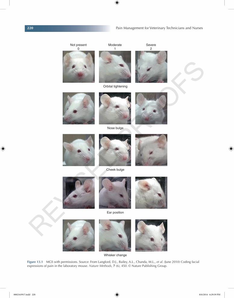
The Mouse Grimace Scale (MGS) is taken from the paper of Langford et al. (2010). In the MGS, intensity of each feature is coded on a three-point scale (Figure 13.1). For each of the five features, images of mice exhibiting behavior corresponding to the three values are shown (Langford et al. 2010).
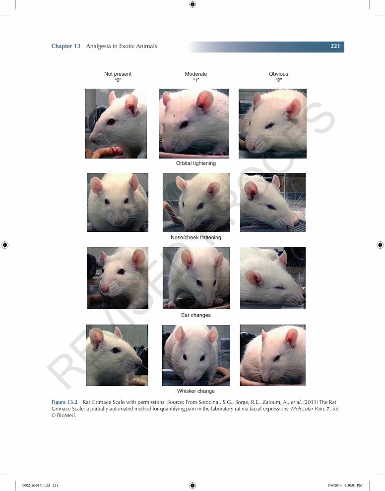
The Rat Grimace Scale has been adapted from Sotocinal et al. (2011) (Figure 13.2). The four action units of the Rat Grimace Scale are shown in Table 13.1 and Table 13.2.
Limited work has been performed to date to create pain assessment scales for guinea pigs. Hamsters and gerbils could be evaluated using a combination of the aforementioned scales.
Medications
Analgesic dose rates based on body weight in rodents tend to be relatively high compared with other mammals, largely because of their small body size and fast metabolic rate (Miller and Richardson 2011). Analgesia should be administered if a painful condition is suspected or a painful procedure has been performed (Longley 2008b). The analgesics most commonly used for rodents and other pet or labora-tory small animal species are opioids and nonsteroidal anti-inflammatory drugs (NSAIDs). Not only preoperative but also intermediate and immediate postoperative analgesic admini-stration is important for adequate pain relief in postsurgical rodents. Intermediate doses of
Key SignS 13.4 Gerbils: Hunched appearance, weight loss, and shock syndrome.
Key SignS 13.5 Hamsters: Weight loss, hunched appearance, increased aggression or depression, and extended sleep periods.
0002163917.indd 219 8/6/2014 6:29:57 PM
220 Pain Management for Veterinary Technicians and Nurses
Chapter No.: 1 Title Name: Goldberg 0002163917.inddComp. by: KIRUPANAND N Date: 06 Aug 2014 Time: 06:29:57 PM Stage: Proof WorkFlow:CSW Page Number: 220
Not present0
Moderate1
Orbital tightening
Nose bulge
Cheek bulge
Ear position
Whisker change
Severe2
Figure 13.1 MGS with permissions. Source: From Langford, D.J., Bailey, A.L., Chanda, M.L., et al. (June 2010) Coding facial expressions of pain in the laboratory mouse. Nature Methods, 7 (6), 450. © Nature Publishing Group.
0002163917.indd 220 8/6/2014 6:29:59 PM
Chapter 13 Analgesia in Exotic Animals 221
Chapter No.: 1 Title Name: Goldberg 0002163917.inddComp. by: KIRUPANAND N Date: 06 Aug 2014 Time: 06:29:57 PM Stage: Proof WorkFlow:CSW Page Number: 221
Not present“0”
Moderate“1”
Orbital tightening
Nose/cheek flattening
Ear changes
Whisker change
Obvious“2”
Figure 13.2 Rat Grimace Scale with permissions. Source: From Sotocinal, S.G., Sorge, R.E., Zaloum, A., et al. (2011) The Rat Grimace Scale: a partially automated method for quantifying pain in the laboratory rat via facial expressions. Molecular Pain, 7, 55. © BioMed.
0002163917.indd 221 8/6/2014 6:30:01 PM
222 Pain Management for Veterinary Technicians and Nurses
Chapter No.: 1 Title Name: Goldberg 0002163917.inddComp. by: KIRUPANAND N Date: 06 Aug 2014 Time: 06:29:57 PM Stage: Proof WorkFlow:CSW Page Number: 222
morphine (3 mg/kg) given parenterally 30 min before and 30 min and 2 h after a simple abdom-inal surgery provide adequate pain relief over a 2-day period. Administration of this same dose
later in the postsurgical period provides only transient relief (Gonzalez et al. 2000; Gaertner et al. 2008). Presurgical administration of anal-gesics will reduce the amount of anesthesia needed during surgery improving recovery scores (Penderis and Franklin 2005). Investigators along with consulting laboratory animal veteri-narians must evaluate individual animals to determine what agent and dose is appropriate for a particular species and protocol (Gaertner et al. 2008).
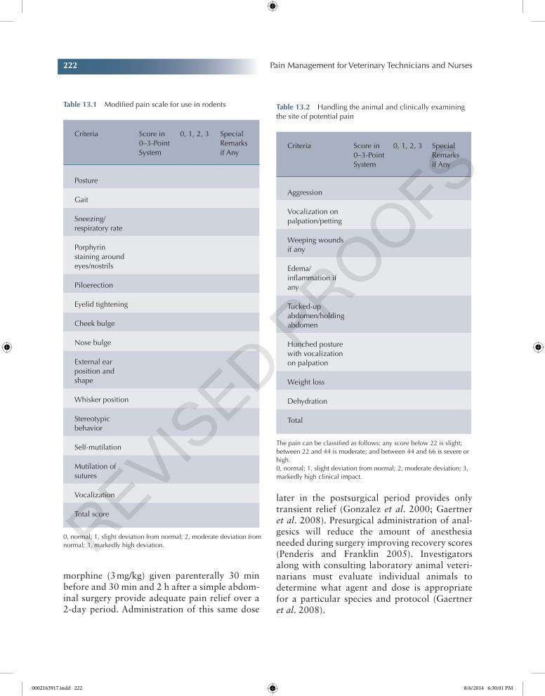
Table 13.1 Modified pain scale for use in rodents
Criteria Score in 0–3-Point System
0, 1, 2, 3 Special Remarks if Any
Posture
Gait
Sneezing/respiratory rate
Porphyrin staining around eyes/nostrils
Piloerection
Eyelid tightening
Cheek bulge
Nose bulge
External ear position and shape
Whisker position
Stereotypic behavior
Self-mutilation
Mutilation of sutures
Vocalization
Total score
0, normal; 1, slight deviation from normal; 2, moderate deviation from normal; 3, markedly high deviation.
Table 13.2 Handling the animal and clinically examining the site of potential pain
Criteria Score in 0–3-Point System
0, 1, 2, 3 Special Remarks if Any
Aggression
Vocalization on palpation/petting
Weeping wounds if any
Edema/inflammation if any
Tucked-up abdomen/holding abdomen
Hunched posture with vocalization on palpation
Weight loss
Dehydration
Total
The pain can be classified as follows: any score below 22 is slight; between 22 and 44 is moderate; and between 44 and 66 is severe or high.0, normal; 1, slight deviation from normal; 2, moderate deviation; 3, markedly high clinical impact.
0002163917.indd 222 8/6/2014 6:30:01 PM

Chapter 13 Analgesia in Exotic Animals 223
Chapter No.: 1 Title Name: Goldberg 0002163917.inddComp. by: KIRUPANAND N Date: 06 Aug 2014 Time: 06:29:57 PM Stage: Proof WorkFlow:CSW Page Number: 223
Opioids in Rodents
Regardless of the route of administration, mor-phine has a relatively short duration of action, thus limiting its use in a laboratory animal setting where 24 h intensive care is not routinely provided (Gaertner et al. 2008). Buprenorphine is a preferred analgesic to relieve moderate postsurgical pain in rodents. It has been observed that self-medication of buprenorphine (0.5 mg/kg in flavored gelatin) may provide pain relief adequate to maintain food and water intake, avoiding postsurgical weight loss in rats (Liles et al. 1998; Flecknell et al. 1999; Gaertner et al. 2008). Fentanyl may be applied topically to nonhaired skin in an aqueous cream base, and in this form, it improved wound healing, wound contracture, cellular proliferation, and angiogen-esis in rats (Poonawala et al. 2005). In mice and rats, butorphanol is seldom used because it has a short duration (1–2 h) of analgesia and relieves only minor pain (Gades et al. 2000).
NSAIDs in Rodents
The NSAID agents most frequently used in rodents include carprofen, meloxicam, and ketoprofen. NSAIDs traditionally have been recommended to alleviate mild pain; however, as the potency and cyclooxygenase (COX) selectivity of the newer agents have improved, they can now be used to alleviate more painful conditions. Unlike opioids, NSAIDs are not controlled drugs and may also provide a longer duration of action than many of the opioid agents (Miller and Richardson 2011).
Local Anesthetics in Rodents
To minimize the risk when using local anes-thetics, it is advisable to draw up the maximum safe dose before administration, approximately 10 mg/kg for lidocaine and 2 mg/kg for bupiva-caine (Flecknell 2009).
Laboratory studies are usually performed in young, healthy, adult strains of mice and rats. Dose
rate may require adjustment for companion animal rodents, which are often geriatric and may have concurrent diseases (Miller and Richardson 2011). To minimize stress, rodents should be housed away from the sight and smells of their natural preda-tors, including dogs, cats, ferrets, and raptors (Table 13.3, Table 13.4, and Table 13.5).
Rabbit Analgesia
In the USA and Europe, rabbits are common household pets, with rabbits being the third most popular mammalian pet in the UK (Wenger 2012). Rabbits are widely used as a research animal model in areas ranging from infectious disease to orthopedic surgery (Weaver et al. 2010). As prey species, rabbits are inclined to hide their painful condition to maintain a normal appearance, thereby avoiding predation. The presence of an observer may result in complete immobility with no apparent pain-related behav-iors (Flecknell 2006). To accurately assess changes indicative of pain and distress, an under-standing of normal species behavior is essential.
Rabbits
The rabbit presents significant difficulties in rec-ognition of pain and distress, as it often quietly accepts apparently painful or distressing proce-dures; this may relate to its feral behavior where concealment is important to survival. Even healthy rabbits may not move frequently or indulge in exploratory behavior. Pain is usually characterized by a reduction in food and water intake (and thus weight loss and dehydration) and limited movement. Although rabbits fre-quently become ill and distressed without show-ing much apparent loss of condition, careful examination will reveal a loss of muscle mass on the lower back. Ocular discharge is a common response to stress in the rabbit, with protrusion of the nictitating membrane.
Under continued pain or stress, rabbits assume a “sleepy” appearance. The animal exhibits increased
0002163917.indd 223 8/6/2014 6:30:01 PM
224 Pain Management for Veterinary Technicians and Nurses
Chapter No.: 1 Title Name: Goldberg 0002163917.inddComp. by: KIRUPANAND N Date: 06 Aug 2014 Time: 06:29:57 PM Stage: Proof WorkFlow:CSW Page Number: 224
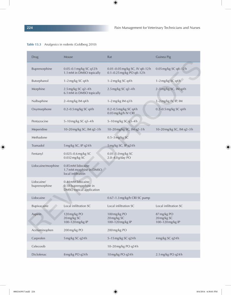
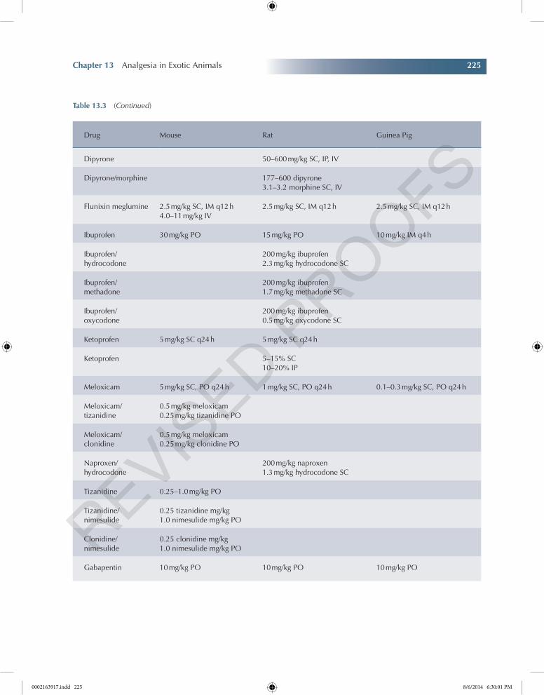
Table 13.3 Analgesics in rodents (Goldberg 2010)
Drug Mouse Rat Guinea Pig
Buprenorphine 0.05–0.1 mg/kg SC q12 h1.1 mM in DMSO topically
0.01–0.05 mg/kg SC, IV q8–12 h0.1–0.25 mg/kg PO q8–12 h
0.05 mg/kg SC q8–12 h
Butorphanol 1–2 mg/kg SC q4 h 1–2 mg/kg SC q4 h 1–2 mg/kg SC q4 h
Morphine 2.5 mg/kg SC q2–4 h6.1 mM in DMSO topically
2.5 mg/kg SC q2–4 h 2–5 mg/kg SC, IM q4 h
Nalbuphine 2–4 mg/kg IM q4 h 1–2 mg/kg IM q3 h 1–2 mg/kg IV, IP, IM
Oxymorphone 0.2–0.5 mg/kg SC q4 h 0.2–0.5 mg/kg SC q4 h0.03 mg/kg/h IV CRI
0.2–0.5 mg/kg SC q4 h
Pentazocine 5–10 mg/kg SC q3–4 h 5–10 mg/kg SC q3–4 h
Meperidine 10–20 mg/kg SC, IM q2–3 h 10–20 mg/kg SC, IM q2–3 h 10–20 mg/kg SC, IM q2–3 h
Methadone 0.5–3 mg/kg SC
Tramadol 5 mg/kg SC, IP q24 h 5 mg/kg SC, IP q24 h
Fentanyl 0.025–0.6 mg/kg SC0.032 mg/kg SC
0.01–1.0 mg/kg SC2.0–4.0 g/day PO
Lidocaine/morphine 0.85 mM lidocaine1.7 mM morphine in DMSO local infiltration
Lidocaine/buprenorphine
0.44 mM lidocaine0.18 buprenorphine in DMSO topical application
Lidocaine 0.67–1.3 mg/kg/h CRI SC pump
Bupivacaine Local infiltration SC Local infiltration SC Local infiltration SC
Aspirin 120 mg/kg PO20 mg/kg SC100–120 mg/kg IP
100 mg/kg PO20 mg/kg SC100–120 mg/kg IP
87 mg/kg PO20 mg/kg SC100–120 mg/kg IP
Acetaminophen 200 mg/kg PO 200 mg/kg PO
Carprofen 5 mg/kg SC q24 h 5–15 mg/kg SC q24 h 4 mg/kg SC q24 h
Celecoxib 10–20 mg/kg PO q24 h
Diclofenac 8 mg/kg PO q24 h 10 mg/kg PO q24 h 2.1 mg/kg PO q24 h
0002163917.indd 224 8/6/2014 6:30:01 PM
Chapter 13 Analgesia in Exotic Animals 225
Chapter No.: 1 Title Name: Goldberg 0002163917.inddComp. by: KIRUPANAND N Date: 06 Aug 2014 Time: 06:29:57 PM Stage: Proof WorkFlow:CSW Page Number: 225
Table 13.3 (Continued)
Drug Mouse Rat Guinea Pig
Dipyrone 50–600 mg/kg SC, IP, IV
Dipyrone/morphine 177–600 dipyrone3.1–3.2 morphine SC, IV
Flunixin meglumine 2.5 mg/kg SC, IM q12 h4.0–11 mg/kg IV
2.5 mg/kg SC, IM q12 h 2.5 mg/kg SC, IM q12 h
Ibuprofen 30 mg/kg PO 15 mg/kg PO 10 mg/kg IM q4 h
Ibuprofen/hydrocodone
200 mg/kg ibuprofen2.3 mg/kg hydrocodone SC
Ibuprofen/methadone
200 mg/kg ibuprofen1.7 mg/kg methadone SC
Ibuprofen/oxycodone
200 mg/kg ibuprofen0.5 mg/kg oxycodone SC
Ketoprofen 5 mg/kg SC q24 h 5 mg/kg SC q24 h
Ketoprofen 5–15% SC10–20% IP
Meloxicam 5 mg/kg SC, PO q24 h 1 mg/kg SC, PO q24 h 0.1–0.3 mg/kg SC, PO q24 h
Meloxicam/tizanidine
0.5 mg/kg meloxicam0.25 mg/kg tizanidine PO
Meloxicam/clonidine
0.5 mg/kg meloxicam0.25 mg/kg clonidine PO
Naproxen/hydrocodone
200 mg/kg naproxen1.3 mg/kg hydrocodone SC
Tizanidine 0.25–1.0 mg/kg PO
Tizanidine/nimesulide
0.25 tizanidine mg/kg1.0 nimesulide mg/kg PO
Clonidine/nimesulide
0.25 clonidine mg/kg1.0 nimesulide mg/kg PO
Gabapentin 10 mg/kg PO 10 mg/kg PO 10 mg/kg PO
0002163917.indd 225 8/6/2014 6:30:01 PM
226 Pain Management for Veterinary Technicians and Nurses
Chapter No.: 1 Title Name: Goldberg 0002163917.inddComp. by: KIRUPANAND N Date: 06 Aug 2014 Time: 06:29:57 PM Stage: Proof WorkFlow:CSW Page Number: 226
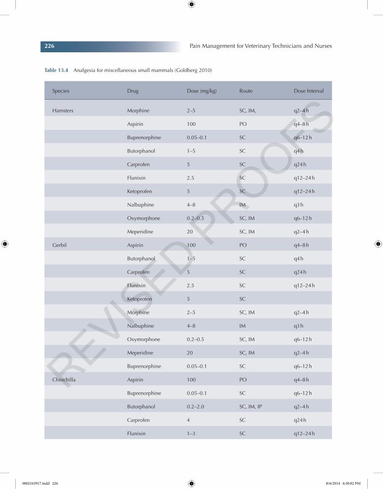
Table 13.4 Analgesia for miscellaneous small mammals (Goldberg 2010)
Species Drug Dose (mg/kg) Route Dose Interval
Hamsters Morphine 2–5 SC, IM, q2–4 h
Aspirin 100 PO q4–8 h
Buprenorphine 0.05–0.1 SC q6–12 h
Butorphanol 1–5 SC q4 h
Carprofen 5 SC q24 h
Flunixin 2.5 SC q12–24 h
Ketoprofen 5 SC q12–24 h
Nalbuphine 4–8 IM q3 h
Oxymorphone 0.2–0.5 SC, IM q6–12 h
Meperidine 20 SC, IM q2–4 h
Gerbil Aspirin 100 PO q4–8 h
Butorphanol 1–5 SC q4 h
Carprofen 5 SC q24 h
Flunixin 2.5 SC q12–24 h
Ketoprofen 5 SC
Morphine 2–5 SC, IM q2–4 h
Nalbuphine 4–8 IM q3 h
Oxymorphone 0.2–0.5 SC, IM q6–12 h
Meperidine 20 SC, IM q2–4 h
Buprenorphine 0.05–0.1 SC q6–12 h
Chinchilla Aspirin 100 PO q4–8 h
Buprenorphine 0.05–0.1 SC q6–12 h
Butorphanol 0.2–2.0 SC, IM, IP q2–4 h
Carprofen 4 SC q24 h
Flunixin 1–3 SC q12–24 h
0002163917.indd 226 8/6/2014 6:30:02 PM
Chapter 13 Analgesia in Exotic Animals 227
Chapter No.: 1 Title Name: Goldberg 0002163917.inddComp. by: KIRUPANAND N Date: 06 Aug 2014 Time: 06:29:57 PM Stage: Proof WorkFlow:CSW Page Number: 227
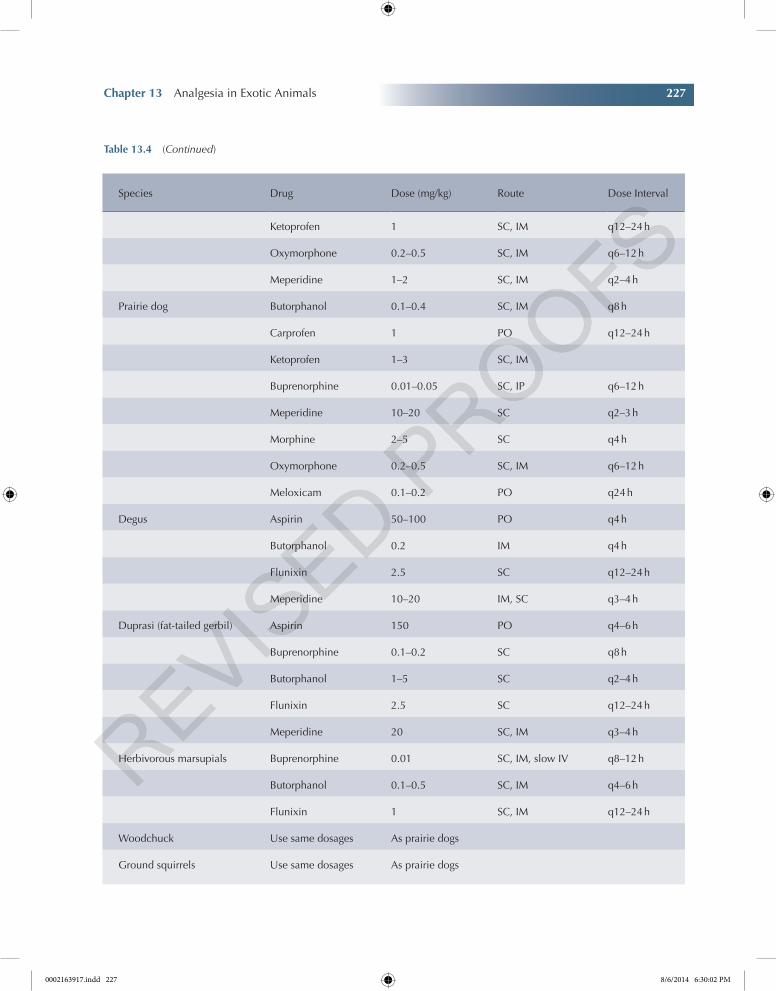
Species Drug Dose (mg/kg) Route Dose Interval
Ketoprofen 1 SC, IM q12–24 h
Oxymorphone 0.2–0.5 SC, IM q6–12 h
Meperidine 1–2 SC, IM q2–4 h
Prairie dog Butorphanol 0.1–0.4 SC, IM q8 h
Carprofen 1 PO q12–24 h
Ketoprofen 1–3 SC, IM
Buprenorphine 0.01–0.05 SC, IP q6–12 h
Meperidine 10–20 SC q2–3 h
Morphine 2–5 SC q4 h
Oxymorphone 0.2–0.5 SC, IM q6–12 h
Meloxicam 0.1–0.2 PO q24 h
Degus Aspirin 50–100 PO q4 h
Butorphanol 0.2 IM q4 h
Flunixin 2.5 SC q12–24 h
Meperidine 10–20 IM, SC q3–4 h
Duprasi (fat-tailed gerbil) Aspirin 150 PO q4–6 h
Buprenorphine 0.1–0.2 SC q8 h
Butorphanol 1–5 SC q2–4 h
Flunixin 2.5 SC q12–24 h
Meperidine 20 SC, IM q3–4 h
Herbivorous marsupials Buprenorphine 0.01 SC, IM, slow IV q8–12 h
Butorphanol 0.1–0.5 SC, IM q4–6 h
Flunixin 1 SC, IM q12–24 h
Woodchuck Use same dosages As prairie dogs
Ground squirrels Use same dosages As prairie dogs
Table 13.4 (Continued)
0002163917.indd 227 8/6/2014 6:30:02 PM
228 Pain Management for Veterinary Technicians and Nurses
Chapter No.: 1 Title Name: Goldberg 0002163917.inddComp. by: KIRUPANAND N Date: 06 Aug 2014 Time: 06:29:57 PM Stage: Proof WorkFlow:CSW Page Number: 228
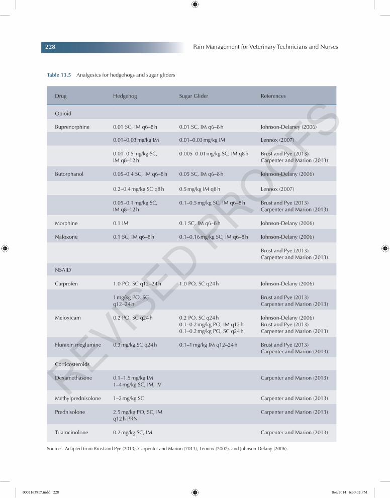
Table 13.5 Analgesics for hedgehogs and sugar gliders
Drug Hedgehog Sugar Glider References
Opioid
Buprenorphine 0.01 SC, IM q6–8 h 0.01 SC, IM q6–8 h Johnson-Delaney (2006)
0.01–0.03 mg/kg IM 0.01–0.03 mg/kg IM Lennox (2007)
0.01–0.5 mg/kg SC, IM q8–12 h
0.005–0.01 mg/kg SC, IM q8 h Brust and Pye (2013)Carpenter and Marion (2013)
Butorphanol 0.05–0.4 SC, IM q6–8 h 0.05 SC, IM q6–8 h Johnson-Delany (2006)
0.2–0.4 mg/kg SC q8 h 0.5 mg/kg IM q8 h Lennox (2007)
0.05–0.1 mg/kg SC, IM q8–12 h
0.1–0.5 mg/kg SC, IM q6–8 h Brust and Pye (2013)Carpenter and Marion (2013)
Morphine 0.1 IM 0.1 SC, IM q6–8 h Johnson-Delany (2006)
Naloxone 0.1 SC, IM q6–8 h 0.1–0.16 mg/kg SC, IM q6–8 h Johnson-Delany (2006)
Brust and Pye (2013)Carpenter and Marion (2013)
NSAID
Carprofen 1.0 PO, SC q12–24 h 1.0 PO, SC q24 h Johnson-Delany (2006)
1 mg/kg PO, SC q12–24 h
Brust and Pye (2013)Carpenter and Marion (2013)
Meloxicam 0.2 PO, SC q24 h 0.2 PO, SC q24 h0.1–0.2 mg/kg PO, IM q12 h0.1–0.2 mg/kg PO, SC q24 h
Johnson-Delany (2006)Brust and Pye (2013)Carpenter and Marion (2013)
Flunixin meglumine 0.3 mg/kg SC q24 h 0.1–1 mg/kg IM q12–24 h Brust and Pye (2013)Carpenter and Marion (2013)
Corticosteroids
Dexamethasone 0.1–1.5 mg/kg IM1–4 mg/kg SC, IM, IV
Carpenter and Marion (2013)
Methylprednisolone 1–2 mg/kg SC Carpenter and Marion (2013)
Prednisolone 2.5 mg/kg PO, SC, IM q12 h PRN
Carpenter and Marion (2013)
Triamcinolone 0.2 mg/kg SC, IM Carpenter and Marion (2013)
Sources: Adapted from Brust and Pye (2013), Carpenter and Marion (2013), Lennox (2007), and Johnson-Delany (2006).
0002163917.indd 228 8/6/2014 6:30:02 PM
Chapter 13 Analgesia in Exotic Animals 229
Chapter No.: 1 Title Name: Goldberg 0002163917.inddComp. by: KIRUPANAND N Date: 06 Aug 2014 Time: 06:29:57 PM Stage: Proof WorkFlow:CSW Page Number: 229
depression, progressive unawareness, and lack of response. The animal will often face the back of cage, away from light. An increased respiratory rate is associated with either apprehension or lung involvement. There is fecal staining of the coat. Nighttime pellet production may be interrupted. Constipation and diarrhea are common responses to pain or stress. Excessive self-grooming may precipitate hair balls in the stomach. Where foot soreness is involved, weight may be thrown for-ward or backward to reduce discomfort. Body stretching and lying flat are common indications of abdominal discomfort. Pain may be associated with locomotion, especially with sore feet.
In situations where pain is to be expected (e.g., after surgery or dental treatment), it should be assumed that rabbits feel pain even if they do not express recognized behavioral signs of pain.
Untreated pain has many undesirable effects such as activation of complement cascade, cytokine systems, and arachidonic acid cascade, in addition to activation of the sympathetic nervous system. Sympathetic nervous system activation may result in tachycardia, arrhythmias, vasoconstriction, altered cardiac output, and increased myocardial oxygen demand. Alterations in organ perfusion, fluid, electrolyte, and acid–base balance may occur. Pain may alter respiratory rate and reduce tidal volume, which may intensify any existing respiratory compromise. Other detrimental effects of untreated pain include inducing a catabolic state, reduced appetite or anorexia, delayed wound healing, lowered immune responses, and prolonged hospital stays. Pain can also reduce GI motility. Concurrent reductions in food intake, dehydration, and disease processes such as enterotoxemia and hepatic lipidosis could further promote ileus and may induce life-threatening problems in the rabbit. Critically ill or traumatized patients have the fewest physiologic reserves to deal with these additional insults. Untreated pain increases morbidity and potentially increases mortality in many species and
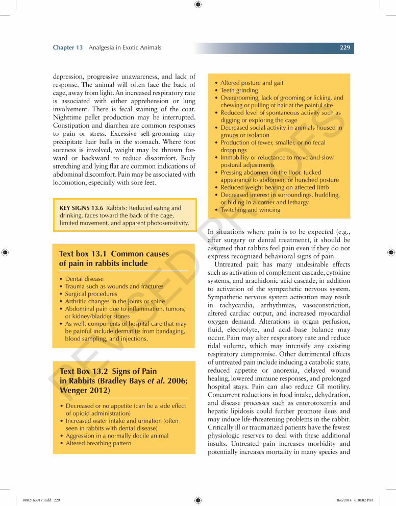
Key SignS 13.6 Rabbits: Reduced eating and drinking, faces toward the back of the cage, limited movement, and apparent photosensitivity.
Text box 13.1 Common causes of pain in rabbits include
•Dental disease•Trauma such as wounds and fractures•Surgical procedures•Arthritic changes in the joints or spine•Abdominal pain due to inflammation, tumors,
or kidney/bladder stones•As well, components of hospital care that may
be painful include dermatitis from bandaging, blood sampling, and injections.
Text Box 13.2 Signs of Pain in Rabbits (Bradley Bays et al. 2006; Wenger 2012)
•Decreased or no appetite (can be a side effect of opioid administration)
• Increased water intake and urination (often seen in rabbits with dental disease)
•Aggression in a normally docile animal•Altered breathing pattern
•Altered posture and gait•Teeth grinding•Overgrooming, lack of grooming or licking, and
chewing or pulling of hair at the painful site•Reduced level of spontaneous activity such as
digging or exploring the cage•Decreased social activity in animals housed in
groups or isolation•Production of fewer, smaller, or no fecal
droppings• Immobility or reluctance to move and slow
postural adjustments•Pressing abdomen on the floor, tucked
appearance to abdomen, or hunched posture•Reduced weight bearing on affected limb•Decreased interest in surroundings, huddling,
or hiding in a corner and lethargy•Twitching and wincing
0002163917.indd 229 8/6/2014 6:30:02 PM
230 Pain Management for Veterinary Technicians and Nurses
Chapter No.: 1 Title Name: Goldberg 0002163917.inddComp. by: KIRUPANAND N Date: 06 Aug 2014 Time: 06:29:57 PM Stage: Proof WorkFlow:CSW Page Number: 230
maybe more so in a prey species like the rabbit. In the extreme, there are anecdotal reports of painful rabbits going into shock and dying despite their underlying illness or injury not appearing to have been life-threatening (Barter 2011).
Nursing Care
While hospitalized, rabbits should be provided with appropriate housing that is away from the sight, smell, and sound of predatory species. The area should be quiet and the animals kept clean and dry. Attention should be paid to making sure that animals have easy access to food and water, particularly if they have mobility issues. Appropriate nursing care by attending to wounds, bandages, urination, and defecation will also help to improve patient comfort. Animals should be
handled carefully and restrained appropriately to avoid unnecessary stress. Some animals that are normally housed with another rabbit may find the social isolation stressful, and consideration should be given to bringing their “buddy” rabbit into hospital with them (Barter 2011).
Pain Scoring
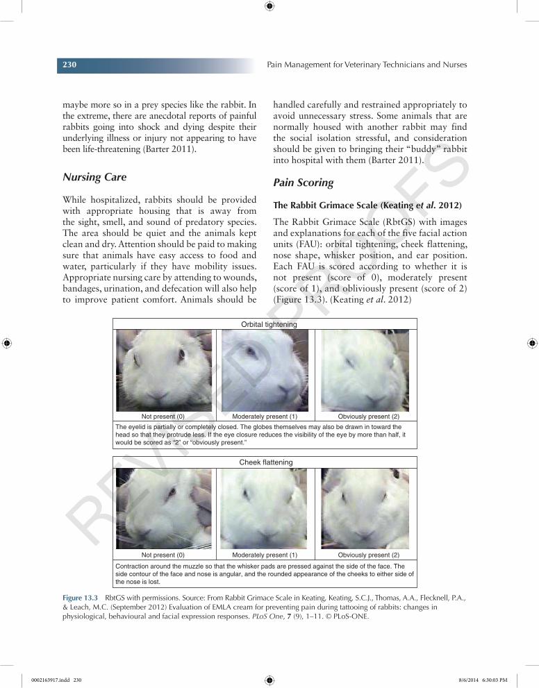
The Rabbit grimace Scale (Keating et al. 2012)
The Rabbit Grimace Scale (RbtGS) with images and explanations for each of the five facial action units (FAU): orbital tightening, cheek flattening, nose shape, whisker position, and ear position. Each FAU is scored according to whether it is not present (score of 0), moderately present (score of 1), and obliviously present (score of 2) (Figure 13.3). (Keating et al. 2012)
Figure 13.3 RbtGS with permissions. Source: From Rabbit Grimace Scale in Keating, Keating, S.C.J., Thomas, A.A., Flecknell, P.A., & Leach, M.C. (September 2012) Evaluation of EMLA cream for preventing pain during tattooing of rabbits: changes in physiological, behavioural and facial expression responses. PLoS One, 7 (9), 1–11. © PLoS-ONE.
Orbital tightening
Cheek flattening
Not present (0) Moderately present (1) Obviously present (2)
Not present (0) Moderately present (1) Obviously present (2)
The eyelid is partially or completely closed. The globes themselves may also be drawn in toward thehead so that they protrude less. If the eye closure reduces the visibility of the eye by more than half, itwould be scored as “2” or “obviously present.”
Contraction around the muzzle so that the whisker pads are pressed against the side of the face. Theside contour of the face and nose is angular, and the rounded appearance of the cheeks to either side ofthe nose is lost.
0002163917.indd 230 8/6/2014 6:30:03 PM
Chapter 13 Analgesia in Exotic Animals 231
Chapter No.: 1 Title Name: Goldberg 0002163917.inddComp. by: KIRUPANAND N Date: 06 Aug 2014 Time: 06:29:57 PM Stage: Proof WorkFlow:CSW Page Number: 231
Medications
Opioids in Rabbits
For pharmacological properties of opioids, refer to Chapter 4.
Buprenorphine, butorphanol, nalbuphine, and pentazocine develop a ceiling effect. This find-ing is consistent with the agonist–antagonist (e.g., butorphanol) or partial-agonist opioids (buprenorphine) displaying a “ceiling effect” in
Not present (0) Moderately present (1) Obviously present (2)
Not present (0) Moderately present (1) Obviously present (2)
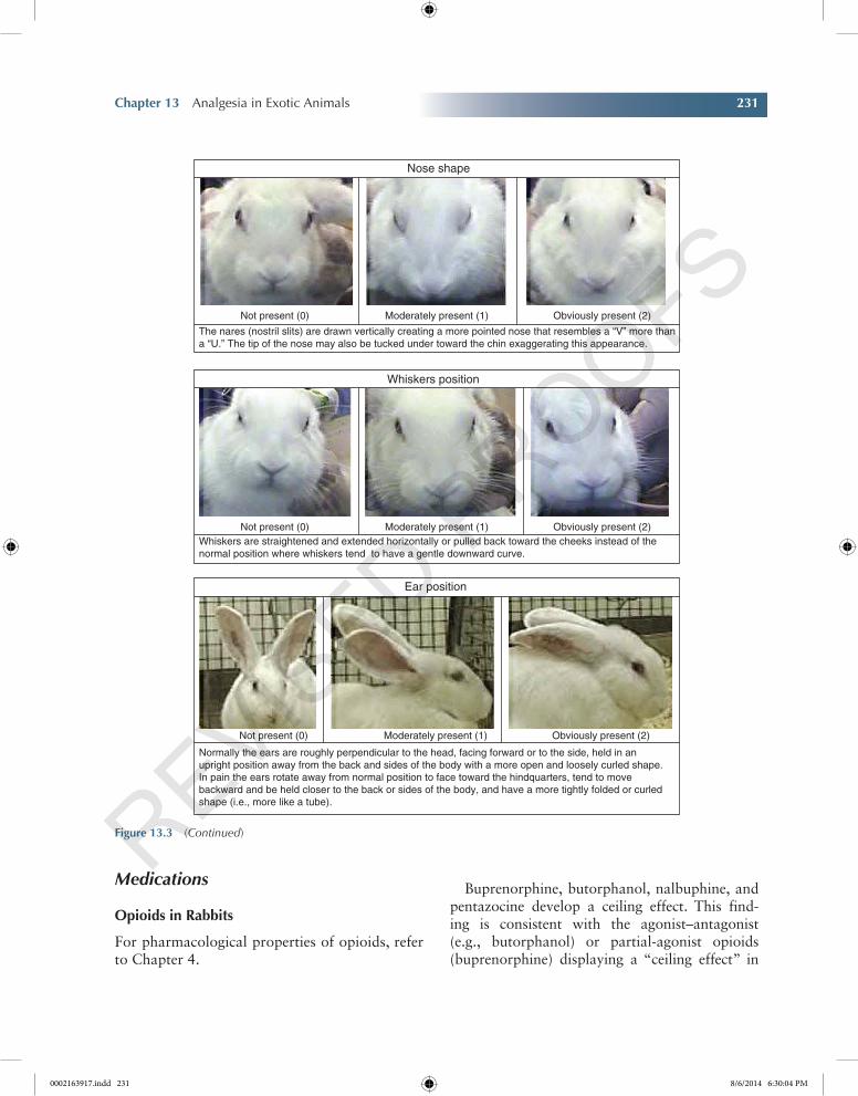
Whiskers are straightened and extended horizontally or pulled back toward the cheeks instead of thenormal position where whiskers tend to have a gentle downward curve.
Normally the ears are roughly perpendicular to the head, facing forward or to the side, held in anupright position away from the back and sides of the body with a more open and loosely curled shape.In pain the ears rotate away from normal position to face toward the hindquarters, tend to movebackward and be held closer to the back or sides of the body, and have a more tightly folded or curledshape (i.e., more like a tube).
Whiskers position
Ear position
Nose shape
Not present (0) Moderately present (1) Obviously present (2)
The nares (nostril slits) are drawn vertically creating a more pointed nose that resembles a “V” more thana “U.” The tip of the nose may also be tucked under toward the chin exaggerating this appearance.
Figure 13.3 (Continued)
0002163917.indd 231 8/6/2014 6:30:04 PM
232 Pain Management for Veterinary Technicians and Nurses
Chapter No.: 1 Title Name: Goldberg 0002163917.inddComp. by: KIRUPANAND N Date: 06 Aug 2014 Time: 06:29:57 PM Stage: Proof WorkFlow:CSW Page Number: 232
the magnitude of analgesia they provide (Gutstein and Akil 2010). Due to this ceiling effect, opi-oids of this category are suitable for the treatment of mild to moderate pain (Barter 2011). Buprenorphine can be given transmucosally. Transmucosal (TM) administration of buprenor-phine is an uncomplicated method for pet owners to give pain medication at home over a several-day time period without having to give injections. Transdermal administration of fentanyl results in variable plasma levels. Rabbits tolerate fentanyl transdermal patches well, but hair regrowth may hinder dermal absorption of fentanyl after 24 h of administration (Wenger 2012). Conversely, the application of a depilatory agent can cause early and rapid absorption of fentanyl, thereby leading to unwanted sedation and lack of sustained therapeutic plasma concentration levels over the 3-day treatment period (Foley et al. 2001). Tramadol has apparent clinical efficacy in rabbits with osteoarthritis for which NSAIDs are not a good option. It should be noted, however, that tramadol is extremely unpalatable when com-pounded, so strong flavoring agents should be used to maximize acceptance of the drug. Ideally, practicing veterinarians should work with a licensed compounding pharmacist to find the flavor combination that works well. Crushed tab-lets in an oral compounding agent flavored with nonalcoholic piña colada mix have been reported to be palatable to rabbits (Johnston 2005).
nSAiDs in Rabbits
For pharmacologic properties of NSAIDs, see Chapter 4.
The two most common NSAIDs used in rabbit medicine are carprofen and meloxicam. Carprofen and meloxicam are available both in oral and injectable forms and can safely be used preoperatively, assuming there is adequate renal perfusion (Robertson 2001).
Oral administration of meloxicam is well tol-erated by rabbits (Leach et al. 2009; Barter 2011). Due to the side effect profile of NSAIDs in other species, consideration should be given to
monitoring rabbits receiving chronic NSAID therapy with serum biochemistry profiles and fecal occult blood tests (Barter 2011).
Regional Anesthesia and Analgesia
Local anesthetics can be used topically, via direct infiltration into soft tissue containing nerve end-ings, intra-articularly (not practical in ferrets or rabbits), intravenously, or epidurally (Johnston 2005). Creams containing local anesthetics, for example, EMLA 5% cream, can be applied topi-cally to the skin before placing intravenous (IV) catheters (Johnston 2005). It is advantageous to clip the hair before applying the EMLA cream. Incisional line blocks and wound infiltration are simple, cost-effective means to provide analgesia for surgical procedures. Important nerve blocks for dental surgery include infraorbital, mental, mandibular, and maxillary (Lichtenberger and Ko 2007). The same techniques, discussed under sec-tion “Rabbit Analgesia”, can be applied to rabbits. Intratesticular injection of local anesthetics can be performed to provide additional analgesia for neutering (Lichtenberger and Ko 2007). Epidural analgesia has been described in guinea pigs and rabbits (Greenaway et al. 2001). Volumes of local anesthetic agent used for epidural injection vary between 0.1 and 0.2 ml/kg (Johnston 2005).
An effective analgesic plan has both pharmaco-logic and nonpharmacologic components. Analgesic therapy should target multiple loca-tions within the pain pathway and is an integral part of the management of surgical and trauma patients as well as acute and chronic medical con-ditions in the rabbit (Barter 2011) (Table 13.6).
Ferret Analgesia
Pain tolerance likely varies greatly between individual ferrets just like in other species. There is a strong supposition that solitary-living ani-mals and prey animals are masters of disguising pain. Different types of pain might induce differ-ent types of pain-related behaviors, complicating
0002163917.indd 232 8/6/2014 6:30:04 PM
Chapter 13 Analgesia in Exotic Animals 233
Chapter No.: 1 Title Name: Goldberg 0002163917.inddComp. by: KIRUPANAND N Date: 06 Aug 2014 Time: 06:29:57 PM Stage: Proof WorkFlow:CSW Page Number: 233
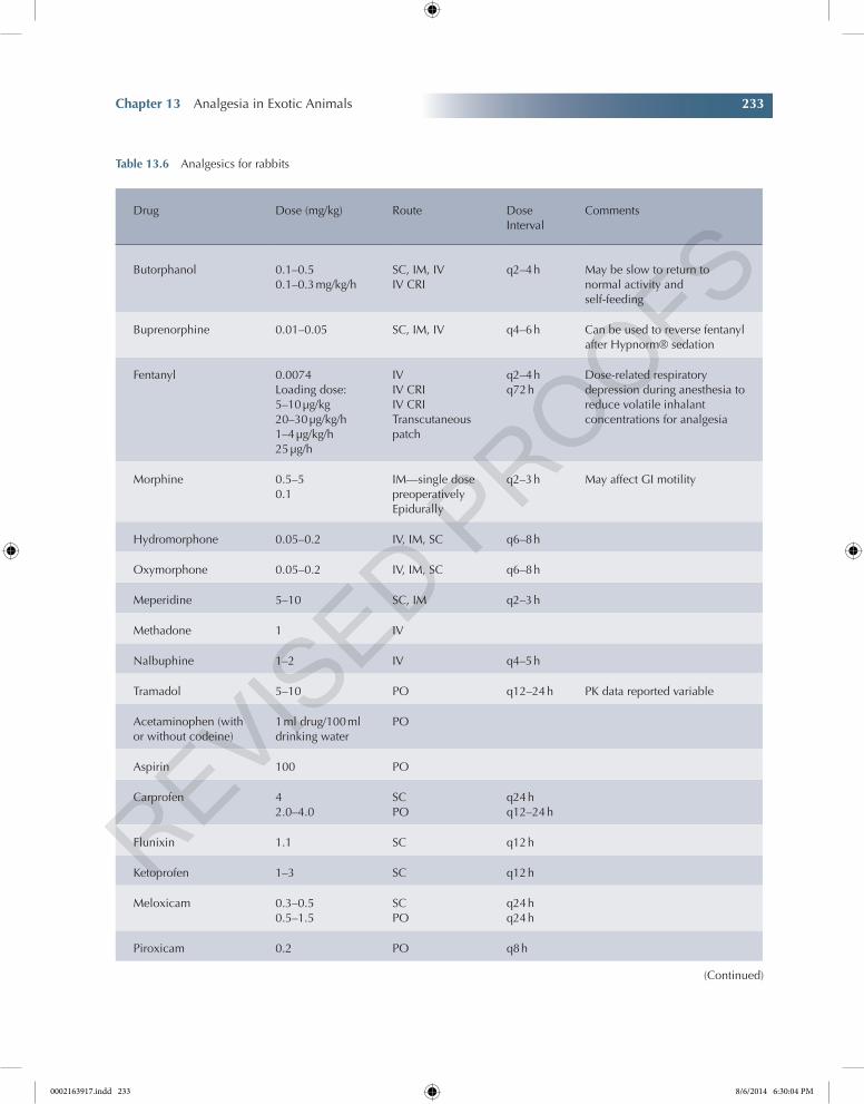
Table 13.6 Analgesics for rabbits
Drug Dose (mg/kg) Route Dose Interval
Comments
Butorphanol 0.1–0.50.1–0.3 mg/kg/h
SC, IM, IVIV CRI
q2–4 h May be slow to return to normal activity and self-feeding
Buprenorphine 0.01–0.05 SC, IM, IV q4–6 h Can be used to reverse fentanyl after Hypnorm® sedation
Fentanyl 0.0074Loading dose: 5–10 µg/kg20–30 µg/kg/h1–4 µg/kg/h25 µg/h
IVIV CRIIV CRITranscutaneous patch
q2–4 hq72 h
Dose-related respiratory depression during anesthesia to reduce volatile inhalant concentrations for analgesia
Morphine 0.5–50.1
IM—single dose preoperativelyEpidurally
q2–3 h May affect GI motility
Hydromorphone 0.05–0.2 IV, IM, SC q6–8 h
Oxymorphone 0.05–0.2 IV, IM, SC q6–8 h
Meperidine 5–10 SC, IM q2–3 h
Methadone 1 IV
Nalbuphine 1–2 IV q4–5 h
Tramadol 5–10 PO q12–24 h PK data reported variable
Acetaminophen (with or without codeine)
1 ml drug/100 ml drinking water
PO
Aspirin 100 PO
Carprofen 42.0–4.0
SCPO
q24 hq12–24 h
Flunixin 1.1 SC q12 h
Ketoprofen 1–3 SC q12 h
Meloxicam 0.3–0.50.5–1.5
SCPO
q24 hq24 h
Piroxicam 0.2 PO q8 h
(Continued)
0002163917.indd 233 8/6/2014 6:30:04 PM
234 Pain Management for Veterinary Technicians and Nurses
Chapter No.: 1 Title Name: Goldberg 0002163917.inddComp. by: KIRUPANAND N Date: 06 Aug 2014 Time: 06:29:57 PM Stage: Proof WorkFlow:CSW Page Number: 234
the recognition of pain even more. Therefore, pain scales should include behavioral parameters that are species specific, can be objectively assessed, and indicate different forms of pain, for example, pain due to trauma, acute postoperative pain, pain from the musculoskeletal system, vis-ceral pain, and inflammatory pain (van Oostrom et al. 2011). To determine specific behavioral signs of pain in ferrets, one should be familiar with the normal behavior of this species (Flecknell 1998; Johnston 2005). Ferrets spend 70% of their time during the day sleeping with short periods of activity (van Oostrom 2011).
Drug Dose (mg/kg) Route Dose Interval
Comments
Ketamine 0.510 µg/kg/min2 µ/kg/min
IVIV CRIIV CRI
Before surgeryDuring surgeryFor 24 h postoperatively
Lidocaine <2 SC—local infiltration
Bupivacaine <1.5 SC—local infiltration
Sources: Adapted from (Goldberg 2010) and Hawkins and Pascoe (2012).
Table 13.6 (Contiuned)
Key SignS 13.7 Ferrets: Stiff posture, demented behavior, lack of grooming, hunched head and neck, and inappetence.
Text Box 13.3 Behavioral characteristics of ferrets in pain (van Oostrom 2011)
•Diminished general activity and exploratory behavior in a new environment
•Altered posture (in many animals, a hunched posture, but in ferrets, the normal hunchback is now absent)
Text Box 13.4 Pain-related behaviors specific in ferrets include (Johnston 2005)
•Preferring inactivity•Staying curled in a ball•Exhibiting aggressive biting behavior or
teeth-bearing when disturbed
•Altered gait, such as lameness•Uncharacteristic aggression in animals that are
otherwise very friendly•Apathy in animals that are otherwise fierce•Vocalizations that differ in pitch and pattern
from the normal interactive sounds•Hiding in the back of the cage facing away
from the observer•Lack of grooming behavior resulting in
ruffled and unkempt appearance of the hair coat
•Diminished food and water intake, especially in dental or GI pain
•Bruxism, especially in abdominal pain•Aversive response to external palpation of the
animal
0002163917.indd 234 8/6/2014 6:30:05 PM
Chapter 13 Analgesia in Exotic Animals 235
Chapter No.: 1 Title Name: Goldberg 0002163917.inddComp. by: KIRUPANAND N Date: 06 Aug 2014 Time: 06:29:57 PM Stage: Proof WorkFlow:CSW Page Number: 235
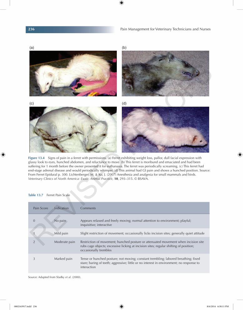
The most likely causes of pain in ferrets are arthritis, cancer, or dental problems (Figure 13.4 and Table 13.7).
Medications
Opioids in Ferrets
For pharmacologic properties of opioids, see Chapter 4.
The most commonly used mixed agonists–antagonists in ferrets are butorphanol and buprenorphine. Butorphanol has agonistic effects mainly at κ receptors, with minimal to no μ opioid effects, hence it is classified as a μ antagonist (Johnston 2005). Butorphanol should be used for sedative effects in ferrets because its analgesic effects seem to be limited (Johnston 2005). Clinically, buprenorphine appears to last 6–10 h after subcutaneous (SC) administration in ferrets. TM absorption of buprenorphine has been used
successfully in ferrets (Johnston 2005). Fentanyl can be used as a constant rate infusion (CRI) in ferrets. Fentanyl CRI is most often used during surgery and in the postoperative period but can also be useful in trauma patients who need potent analgesia (van Oostrom 2011). Frequent reassess-ment, including ventilatory status and effective-ness of pain management, is mandatory in these patients. Fentanyl citrate has an effect for only approximately 30 min after a single IV injection and is thus most commonly used as a CRI during the perianesthetic and postoperative periods (Criado and de Segura 2003). Remifentanil is an ultra-short-acting μ receptor agonist, which makes it very suitable for CRI (Hawkins and Pascoe 2012). Tramadol is a very weak μ agonist, but the O-desmethyl metabolite (M1) is a much more potent agonist. It has been used in ferrets successfully (Johnston 2005).
nSAiDs in Ferrets
For pharmacologic properties of NSAIDs, refer to Chapter 4.
Based on the substantial number of reports on the successful use of NSAIDs in ferrets, these drugs can be considered a valuable additive to analgesic therapy in this species (van Oostrom 2011). Ferrets are prone to GI ulcers and GI disease. A GI protectant, antihistamine/antacid therapy should accompany administration of NSAIDs (Johnson-Delany 2009).
Regional Anesthesia and Analgesia in Ferrets
For pharmacologic properties of local anes-thetics, refer to Chapter 5.
Intratesticular blocks can be used for castration at a concentration of 1 mg/kg of 2% lidocaine per testicle (Lichtenberger and Ko 2007). Sufficient time (a minimum of 5 min) has to be allowed for the local anesthetic to migrate to the spermatic cord to benefit from this technique. Toxicity is prevented by using appropriate concentrations and volumes. Lidocaine (2%) solution often must be diluted to achieve a suitable injection volume and should not exceed a total dose of 4 mg/kg (Lichtenberger and
Text Box 13.5 Ferrets with visceral pain
•Μay have decreased appetite•Μay demonstrate bruxism when presented
with food•Μay have inadequate postoperative analgesia,
which may be associated with shivering in the presence of a normal body temperature
Text Box 13.6 Other signs of pain in ferrets include
•Α bristling of the tail fur, so that the tail resembles a pipe cleaner
•Ηalf-closed eyelids•Focal muscle fasciculations (individual or
group of muscles)•High-pitched vocalization or grunting when
handled•Lameness•General malaise
0002163917.indd 235 8/6/2014 6:30:05 PM
236 Pain Management for Veterinary Technicians and Nurses
Chapter No.: 1 Title Name: Goldberg 0002163917.inddComp. by: KIRUPANAND N Date: 06 Aug 2014 Time: 06:29:57 PM Stage: Proof WorkFlow:CSW Page Number: 236
Figure 13.4 Signs of pain in a ferret with permissions. (a) Ferret exhibiting weight loss, pallor, dull facial expression with glassy look to eyes, hunched abdomen, and reluctance to move. (b) This ferret is moribund and emaciated and had been suffering for 1 month before the owner presented it for euthanasia. The ferret was periodically screaming. (c) This ferret had end-stage adrenal disease and would periodically whimper. (d) This animal had GI pain and shows a hunched position. Source: From Ferret Epidural p. 300. Lichtenberger, M. & Ko, J. (2007) Anesthesia and analgesia for small mammals and birds. Veterinary Clinics of North America: Exotic Animal Practice, 10, 293–315. © BSAVA.
(a) (b)
(c) (d)
Table 13.7 Ferret Pain Scale
Pain Score Indication Comments
0 No pain Appears relaxed and freely moving; normal attention to environment; playful; inquisitive; interactive
1 Mild pain Slight restriction of movement; occasionally licks incision sites; generally quiet attitude
2 Moderate pain Restriction of movement; hunched posture or attenuated movement when incision site rubs cage objects; excessive licking at incision sites; regular shifting of position; occasionally trembles
3 Marked pain Tense or hunched posture; not moving; constant trembling; labored breathing; fixed stare; baring of teeth; aggressive; little or no interest in environment; no response to interaction
Source: Adapted from Sladky et al. (2000).
0002163917.indd 236 8/6/2014 6:30:11 PM
Chapter 13 Analgesia in Exotic Animals 237
Chapter No.: 1 Title Name: Goldberg 0002163917.inddComp. by: KIRUPANAND N Date: 06 Aug 2014 Time: 06:29:57 PM Stage: Proof WorkFlow:CSW Page Number: 237
Ko 2007). Techniques that can be used in ferrets are incisional line blocks, local infiltration, ring blocks, splash blocks, topical application, conductive nerve blocks, and even epidural anesthesia (Lichtenberger and Ko 2007; van Oostrom 2011). Adding opioids, such as morphine or buprenorphine, to a local block is considered to increase the duration of anal-gesia significantly (Lichtenberger and Ko 2007). A 25- to 27-gauge needle should be used for these blocks (Table 13.8 and Table 13.9).
Other Analgesics Used in Small Mammals
For Dental Disease
Analgesia is an important part of dental disease management, especially involving the tooth roots where pain can be chronic. In rabbits and large rodents, injectable agents like butorphanol, at 0.5–1 mg/kg IM q6–8 h, or buprenorphine, at 0.05 mg/kg IM q12 h, are good choices. Oral analgesics have not been extensively studied, but some clinicians use a compounded carprofen suspension at 2 mg/kg PO q12 h to manage pain short term (Hoefer 2001).
Regional Analgesia
Dental Blocks in Small Mammals (Johnson-Delany 2009; Hawkins and Pascoe 2012)There are five important dental blocks for small mam-mals (e.g., rodents, rabbits, and ferrets). All five blocks incorporate the lidocaine–bupivacaine mixture as dis-cussed previously for the ring block technique. The total dose of the mixture is drawn up into a syringe, and one-fifth of the total dose is given into each of the five sites. A 25- to 27-gauge needle is used with a 0.5- to 1-cc syringe (Lichtenberger and Ko 2007).
Infraorbital Nerve Block The infraorbital nerve arises from the maxillary branch of the trigeminal nerve. This nerve provides sensory fibers to the upper incisor teeth, upper lip, and adjacent soft tis-sues. The zygomatic nerve also arises from the max-illary nerve just proximal to the infraorbital nerve. This nerve also supplies sensory fibers to the lateral
aspect of the face. The infraorbital foramen is located approximately 5–12 mm dorsal to the crestal bone adjacent to the upper first premolar (cheek) tooth at the lateral aspect of the skull. The foramen is not as easily palpated as in the dog. The facial tuber is a palpable bony prominence at the mesial (rostral) aspect of the zygomatic bone and is approximately 4–10 mm ventral to the infraorbital canal. The infraorbital nerve can be blocked by infusion of local anesthetic at this foramen.
Mental Nerve Block The mental nerve arises from the mandibular nerve as it extends into the mental foramina to form the mental nerve. The mental nerve supplies sensory fibers to the ventral and lateral aspect of the mandible, lip, and lower incisor and motor fibers to local muscles. The mental nerve exits the mental foramen located at the dorsal lateral aspect of the body of the man-dible. The foramen is rostral (2–4 mm) to the first mandibular premolar (cheek) tooth and is located ventrally in the dorsal one-third of the body of the mandible. The mental nerve can be blocked by infusion of local anesthetic at this foramen.
Mandibular Nerve Block The mandibular nerve arises from the trigeminal nerve and supplies sensory and motor fibers to the ventral mandible as well as to the muscles of mastication. The man-dibular nerve provides sensory fibers to the man-dibular molar and premolar (cheek) teeth as well as to adjacent tissues. The mandibular nerve enters the mandibular foramen on the medial surface of the mandible. An extraoral approach should be made with great care to avoid neurovascular structures (facial vessels and nerves) at the ventral aspect of the mandible. The mandibular foramen is approximately midway between the distal aspect of the last molar (cheek) tooth and the ventral aspect of the mandible. Additionally, the foramen is approximately 2–5 mm distal to the third molar tooth. After this location is determined, an infu-sion needle of appropriate length can be “walked along” the medial aspect of the mandible to the mandibular foramen for infusion of the local anesthetic. This effectively blocks the mandibular premolar and molar (cheek) teeth.
0002163917.indd 237 8/6/2014 6:30:11 PM
Chapter No.: 1 Title Name: Goldberg 0002163917.inddComp. by: KIRUPANAND N Date: 06 Aug 2014 Time: 06:29:57 PM Stage: Proof WorkFlow:CSW Page Number: 238
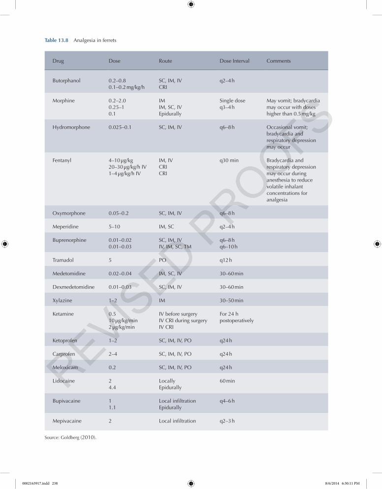
Table 13.8 Analgesia in ferrets
Drug Dose Route Dose Interval Comments
Butorphanol 0.2–0.80.1–0.2 mg/kg/h
SC, IM, IVCRI
q2–4 h
Morphine 0.2–2.00.25–10.1
IMIM, SC, IVEpidurally
Single doseq3–4 h
May vomit; bradycardia may occur with doses higher than 0.5 mg/kg
Hydromorphone 0.025–0.1 SC, IM, IV q6–8 h Occasional vomit; bradycardia and respiratory depression may occur
Fentanyl 4–10 µg/kg20–30 µg/kg/h IV1–4 µg/kg/h IV
IM, IVCRICRI
q30 min Bradycardia and respiratory depression may occur during anesthesia to reduce volatile inhalant concentrations for analgesia
Oxymorphone 0.05–0.2 SC, IM, IV q6–8 h
Meperidine 5–10 IM, SC q2–4 h
Buprenorphine 0.01–0.020.01–0.03
SC, IM, IVIV, IM, SC, TM
q6–8 hq6–10 h
Tramadol 5 PO q12 h
Medetomidine 0.02–0.04 IM, SC, IV 30–60 min
Dexmedetomidine 0.01–0.03 SC, IM, IV 30–60 min
Xylazine 1–2 IM 30–50 min
Ketamine 0.510 µg/kg/min2 µg/kg/min
IV before surgeryIV CRI during surgeryIV CRI
For 24 h postoperatively
Ketoprofen 1–2 SC, IM, IV, PO q24 h
Carprofen 2–4 SC, IM, IV, PO q24 h
Meloxicam 0.2 SC, IM, IV, PO q24 h
Lidocaine 24.4
LocallyEpidurally
60 min
Bupivacaine 11.1
Local infiltrationEpidurally
q4–6 h
Mepivacaine 2 Local infiltration q2–3 h
Source: Goldberg (2010).
0002163917.indd 238 8/6/2014 6:30:11 PM
Chapter 13 Analgesia in Exotic Animals 239
Chapter No.: 1 Title Name: Goldberg 0002163917.inddComp. by: KIRUPANAND N Date: 06 Aug 2014 Time: 06:29:57 PM Stage: Proof WorkFlow:CSW Page Number: 239
Maxillary Nerve Block The maxillary nerve supplies sensory fibers to the upper premolar and molar (cheek) teeth and adjacent tissues. The syringe is aspirated to ensure that the needle is not in a vessel lumen. Firm digital pressure is placed over the rostral end of the infraorbital canal while slowly infusing the local anesthetic. This block anesthetizes all ipsilateral premolar and molar (cheek) teeth and the adjacent periodontal tissues.
Palatine Nerve Block The sphenopalatine nerve ends within the sphenopalatine ganglion. Three nerves extend from the ganglion to regional tis-sues. The nasal cavity is innervated by the nasal rami, the rostral or anterior hard palate by the nasopalatine nerve, and the posterior hard palate by the anterior palatine nerve. The oral cavity of the rabbit limits easy visualization; however, the anterior palatine nerve can be blocked as it exits the larger palatine foramen. This foramen is located halfway between the palatal aspect of the third upper premolar (cheek) tooth and the pal-atal midline. Infusion of a local anesthetic blocks this nerve and the palate of the ipsilateral side.
epidurals
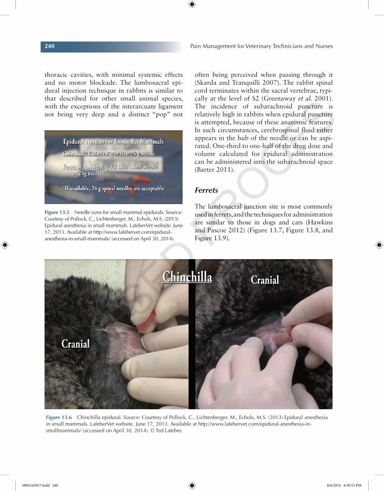
Rodents
Although technically challenging, the epidural technique in rodents is possible (Cheol et al. 2007). CRIs also avoid “peaks and valleys” in drug concentration and are a valuable compo-nent of multimodal analgesia in many veterinary species (National Academies Press 2009). Agents that may be used include opioids, ketamine, and a2 adrenoceptor agonists (Figure 13.5 and Figure 13.6). Although the use of CRIs is not fre-quently reported in companion rodents, this technique is used in the research environment (Franken et al. 2008).
Rabbits
Epidural administration of preservative-free morphine may be useful for perioperative anal-gesia in rabbits or for undergoing orthopedic sur-gery or extensive surgery of the abdominal or
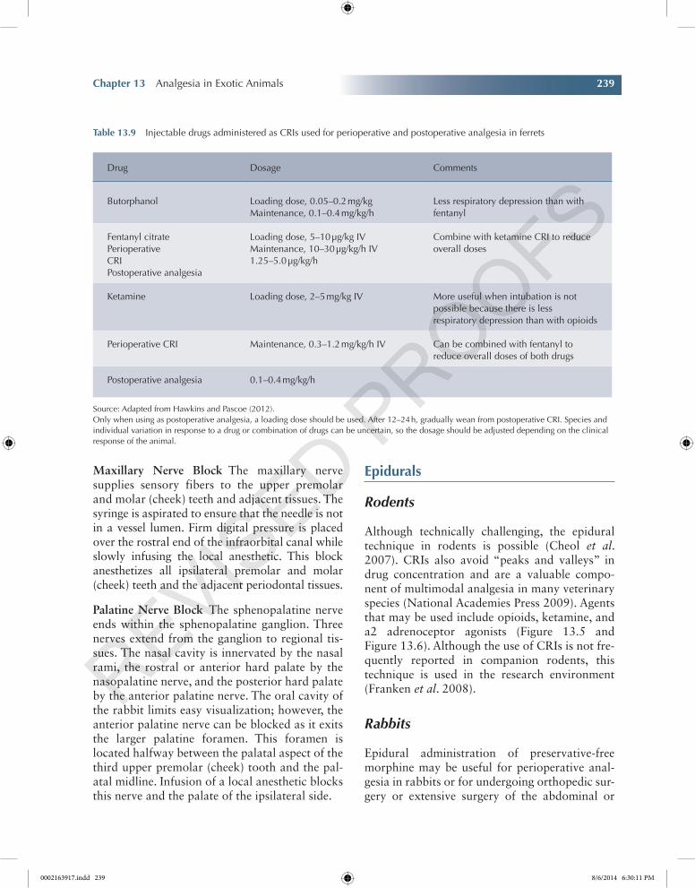
Table 13.9 Injectable drugs administered as CRIs used for perioperative and postoperative analgesia in ferrets
Drug Dosage Comments
Butorphanol Loading dose, 0.05–0.2 mg/kgMaintenance, 0.1–0.4 mg/kg/h
Less respiratory depression than with fentanyl
Fentanyl citratePerioperativeCRIPostoperative analgesia
Loading dose, 5–10 µg/kg IVMaintenance, 10–30 µg/kg/h IV1.25–5.0 µg/kg/h
Combine with ketamine CRI to reduce overall doses
Ketamine Loading dose, 2–5 mg/kg IV More useful when intubation is not possible because there is less respiratory depression than with opioids
Perioperative CRI Maintenance, 0.3–1.2 mg/kg/h IV Can be combined with fentanyl to reduce overall doses of both drugs
Postoperative analgesia 0.1–0.4 mg/kg/h
Source: Adapted from Hawkins and Pascoe (2012).Only when using as postoperative analgesia, a loading dose should be used. After 12–24 h, gradually wean from postoperative CRI. Species and individual variation in response to a drug or combination of drugs can be uncertain, so the dosage should be adjusted depending on the clinical response of the animal.
0002163917.indd 239 8/6/2014 6:30:11 PM
240 Pain Management for Veterinary Technicians and Nurses
Chapter No.: 1 Title Name: Goldberg 0002163917.inddComp. by: KIRUPANAND N Date: 06 Aug 2014 Time: 06:29:57 PM Stage: Proof WorkFlow:CSW Page Number: 240
thoracic cavities, with minimal systemic effects and no motor blockade. The lumbosacral epi-dural injection technique in rabbits is similar to that described for other small animal species, with the exceptions of the interarcuate ligament not being very deep and a distinct “pop” not
often being perceived when passing through it (Skarda and Tranquilli 2007). The rabbit spinal cord terminates within the sacral vertebrae, typi-cally at the level of S2 (Greenaway et al. 2001). The incidence of subarachnoid puncture is relatively high in rabbits when epidural puncture is attempted, because of these anatomic features. In such circumstances, cerebrospinal fluid either appears in the hub of the needle or can be aspi-rated. One-third to one-half of the drug dose and volume calculated for epidural administration can be administered into the subarachnoid space (Barter 2011).
Ferrets
The lumbosacral junction site is most commonly used in ferrets, and the techniques for administration are similar to those in dogs and cats (Hawkins and Pascoe 2012) (Figure 13.7, Figure 13.8, and Figure 13.9).
Figure 13.6 Chinchilla epidural. Source: Courtesy of Pollock, C., Lichtenberger, M., Echols, M.S. (2013) Epidural anesthesia in small mammals. LafeberVet website. June 17, 2013. Available at http://www.lafebervet.com/epidural-anesthesia-in- smallmammals/ (accessed on April 30, 2014). © Ted Lafeber.
Figure 13.5 Needle sizes for small mammal epidurals. Source: Courtesy of Pollock, C., Lichtenberger, M., Echols, M.S. (2013) Epidural anesthesia in small mammals. LafeberVet website. June 17, 2013. Available at http://www.lafebervet.com/epidural-anesthesia-in-small-mammals/ (accessed on April 30, 2014).
0002163917.indd 240 8/6/2014 6:30:13 PM
Chapter 13 Analgesia in Exotic Animals 241
Chapter No.: 1 Title Name: Goldberg 0002163917.inddComp. by: KIRUPANAND N Date: 06 Aug 2014 Time: 06:29:57 PM Stage: Proof WorkFlow:CSW Page Number: 241
epidural Anesthesia in a Ferret
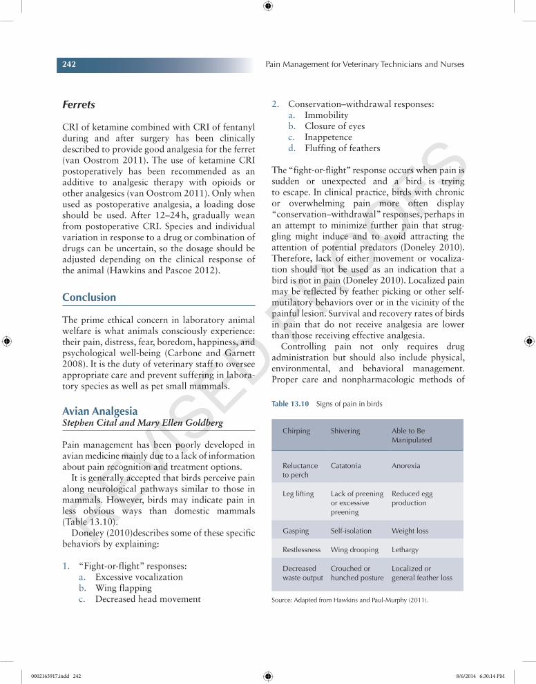
For epidural anesthesia, a 25-gauge needle is placed in the distal two-thirds of the Lumbosacral (LS) space. The black line indi-cates the seventh lumbar vertebrae. The “x” marks indicate the wings of the ilium. The black dot indicates the first sacral vertebrae (Lichtenberger and Ko 2007).
Constant Rate infusions
Rodents
Rats and mice have received CRI via a tail-vein catheter connected to a syringe pump (Franken et al. 2008). Morphine at 0.04 mg/kg/h has been reported in rats (Sadiq et al. 2011). Lidocaine has been reported as a CRI in rats for the prevention of neuropathic pain at 0.67 or 1.3 mg/kg/h (Smith et al. 2002). A study states that rac-ketamine was administered at a constant rate of 10 mg/kg/min for 5 min (Edwards and Mather 2001).
Rabbits
Fentanyl CRIs have been used intraoperatively to provide analgesia to rabbits and subjectively allow lowering of anesthetic vaporizer settings. However, respiratory depression at higher doses can be marked, necessitating positive pressure ventilation. Dose requirements, efficacy, and anesthetic-sparing and hemodynamic effects vary between species and have not been reported in rabbits (Barter 2011).
Till date, no reports of lidocaine CRI (mg/kg/h or min) are found on PubMed.
Ketamine bolus of 1 mg/kg BW of ketamine is administered IV followed immediately by a CRI at 40 mg/kg BW/min using a syringe pump (Gianotti et al. 2012).
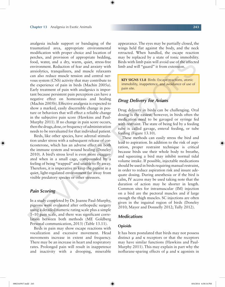
Figure 13.9 Ferret epidural view 2. Source: Courtesy of Pollock, C., Lichtenberger, M., Echols, M.S. (2013) Epidural anesthesia in small mammals. LafeberVet website. June 17, 2013. Available at http://www.lafebervet.com/epidural-anesthesia- in-smallmammals/ (accessed on April 30, 2014). © Ted Lafeber.
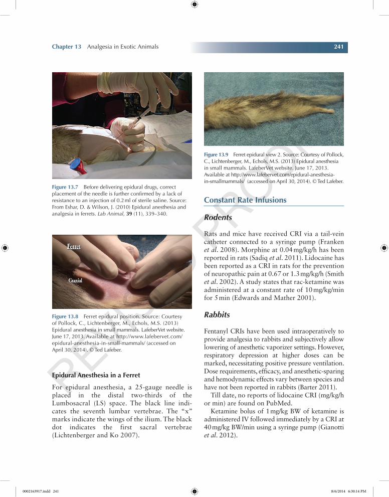
Figure 13.7 Before delivering epidural drugs, correct placement of the needle is further confirmed by a lack of resistance to an injection of 0.2 ml of sterile saline. Source: From Eshar, D. & Wilson, J. (2010) Epidural anesthesia and analgesia in ferrets. Lab Animal, 39 (11), 339–340.
Figure 13.8 Ferret epidural position. Source: Courtesy of Pollock, C., Lichtenberger, M., Echols, M.S. (2013) Epidural anesthesia in small mammals. LafeberVet website. June 17, 2013. Available at http://www.lafebervet.com/epidural-anesthesia-in-small-mammals/ (accessed on April 30, 2014). © Ted Lafeber.
0002163917.indd 241 8/6/2014 6:30:14 PM
242 Pain Management for Veterinary Technicians and Nurses
Chapter No.: 1 Title Name: Goldberg 0002163917.inddComp. by: KIRUPANAND N Date: 06 Aug 2014 Time: 06:29:57 PM Stage: Proof WorkFlow:CSW Page Number: 242
Ferrets
CRI of ketamine combined with CRI of fentanyl during and after surgery has been clinically described to provide good analgesia for the ferret (van Oostrom 2011). The use of ketamine CRI postoperatively has been recommended as an additive to analgesic therapy with opioids or other analgesics (van Oostrom 2011). Only when used as postoperative analgesia, a loading dose should be used. After 12–24 h, gradually wean from postoperative CRI. Species and individual variation in response to a drug or combination of drugs can be uncertain, so the dosage should be adjusted depending on the clinical response of the animal (Hawkins and Pascoe 2012).
Conclusion
The prime ethical concern in laboratory animal welfare is what animals consciously experience: their pain, distress, fear, boredom, happiness, and psychological well-being (Carbone and Garnett 2008). It is the duty of veterinary staff to oversee appropriate care and prevent suffering in labora-tory species as well as pet small mammals.
Avian AnalgesiaStephen Cital and Mary Ellen Goldberg
Pain management has been poorly developed in avian medicine mainly due to a lack of information about pain recognition and treatment options.
It is generally accepted that birds perceive pain along neurological pathways similar to those in mammals. However, birds may indicate pain in less obvious ways than domestic mammals (Table 13.10).
Doneley (2010)describes some of these specific behaviors by explaining:
1. “Fight-or-flight” responses:a. Excessive vocalizationb. Wing flappingc. Decreased head movement
2. Conservation–withdrawal responses:a. Immobilityb. Closure of eyesc. Inappetenced. Fluffing of feathers
The “fight-or-flight” response occurs when pain is sudden or unexpected and a bird is trying to escape. In clinical practice, birds with chronic or overwhelming pain more often display “conservation–withdrawal” responses, perhaps in an attempt to minimize further pain that strug-gling might induce and to avoid attracting the attention of potential predators (Doneley 2010). Therefore, lack of either movement or vocaliza-tion should not be used as an indication that a bird is not in pain (Doneley 2010). Localized pain may be reflected by feather picking or other self-mutilatory behaviors over or in the vicinity of the painful lesion. Survival and recovery rates of birds in pain that do not receive analgesia are lower than those receiving effective analgesia.
Controlling pain not only requires drug administration but should also include physical, environmental, and behavioral management. Proper care and nonpharmacologic methods of
Table 13.10 Signs of pain in birds
Chirping Shivering Able to Be Manipulated
Reluctance to perch
Catatonia Anorexia
Leg lifting Lack of preening or excessive preening
Reduced egg production
Gasping Self-isolation Weight loss
Restlessness Wing drooping Lethargy
Decreased waste output
Crouched or hunched posture
Localized or general feather loss
Source: Adapted from Hawkins and Paul-Murphy (2011).
0002163917.indd 242 8/6/2014 6:30:14 PM
Chapter 13 Analgesia in Exotic Animals 243
Chapter No.: 1 Title Name: Goldberg 0002163917.inddComp. by: KIRUPANAND N Date: 06 Aug 2014 Time: 06:29:57 PM Stage: Proof WorkFlow:CSW Page Number: 243
analgesia include support or bandaging of the traumatized area, appropriate environmental modification with proper choice and location of perches, and provision of appropriate bedding, food, water, and a dry, warm, quiet, stress-free environment. Reduction of fear and anxiety with anxiolytics, tranquilizers, and muscle relaxants can also reduce muscle tension and central ner-vous system (CNS) activity that may contribute to the experience of pain in birds (Machin 2005a). Early treatment of pain with analgesics is impor-tant because persistent pain perception can have a negative effect on homeostasis and healing (Machin 2005b). Effective analgesia is expected to show a marked, easily discernible change in pos-ture or behaviors that will effect a reliable change in the subjective pain score (Hawkins and Paul-Murphy 2011). If no change in pain score occurs, then the drugs, dose, or frequency of administration needs to be reevaluated for that individual patient.
Birds, like other species, have adrenal stimula-tion under stress with a subsequent release of cor-ticosterone, which has an adverse effect on both the immune system and wound healing (Doneley 2010). A bird’s stress level is even more exagger-ated when in a small cage, compounded by a feeling of being “trapped” and unable to fly away. Therefore, it is imperative to keep the patient in a quiet, light-regulated environment far away from visible predatory species or other stressors.
Pain Scoring
In a study completed by Dr. Joanne Paul-Murphy, pigeons were evaluated after orthopedic surgery using a detailed numeric rating scale plus a simple 1–10 pain scale, and there was significant corre-lation between both methods (ME Goldberg Personal communication, 2013) (Table 13.11).
Birds in pain may show escape reactions with vocalization and excessive movement. Head movements increase in extent and frequency. There may be an increase in heart and respiratory rates. Prolonged pain will result in inappetence and inactivity with a drooping, miserable
appearance. The eyes may be partially closed, the wings held flat against the body, and the neck retracted. When handled, the escape reaction may be replaced by a state of tonic immobility. Birds with limb pain will avoid use of the affected limb and will “guard” it from extension.
Drug Delivery for Avians
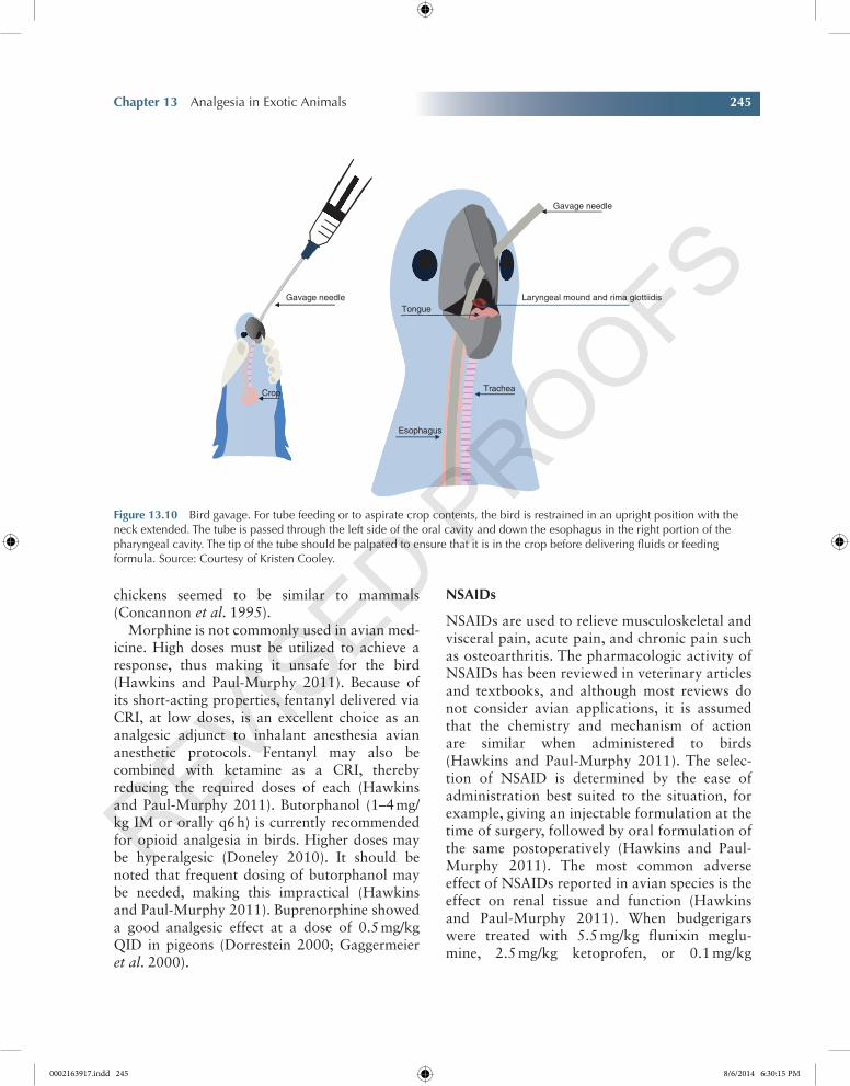
Drug delivery in birds can be challenging. Oral dosing is the easiest; however, in birds often the medication need to be gavaged or syringe fed with restraint. The state of being fed by a feeding tube is called gavage, enteral feeding, or tube feeding (Figure 13.10).
These methods can easily stress the bird and lead to aspiration. In addition to the risk of aspi-ration, proper restraint technique is critical because birds use their whole body to breathe, and squeezing a bird may inhibit normal tidal volume intake. If possible, injectable medications should be used in birds requiring manual restraint, in order to reduce aspiration risk and insure ade-quate dosing. During anesthesia or if the bird is calm, IV access may be used taking note that the duration of action may be shorter in length. Common sites for intramuscular (IM) injection on a bird are the pectoral muscles and if large enough the thigh muscles. SC injections are often given in the inguinal region of birds (Doneley 2010; Mayer and Donnelly 2012; Tully 2012).
Medications
Opioids
It has been postulated that birds may not possess distinct μ and κ receptors or that the receptors may have similar functions (Hawkins and Paul-Murphy 2011). This may explain in part why the isoflurane-sparing effects of μ and κ agonists in
Key SignS 13.8 Birds: Escape reactions, atonic immobility, inappetence, and avoidance of use of pain site.
0002163917.indd 243 8/6/2014 6:30:14 PM
Chapter No.: 1 Title Name: Goldberg 0002163917.inddComp. by: KIRUPANAND N Date: 06 Aug 2014 Time: 06:29:57 PM Stage: Proof WorkFlow:CSW Page Number: 244
Table 13.11 Pigeon Orthopedic Scale Index
Fecal output ________________
Date ________________
Time ________________
Bird number _______________
Amount of time before or after anesthesia ____________________
Observations Score Criteria
Eyes 0 Open, alert
1 Partially closed
2 Closed
Activity 0 Walking/preening
1 Not walking but body movements
2 No activity
Posture 0 Standing
1 Sitting upright
2 Sitting on hocks
Appetite 0 Observed eating or evidence of food bowl mess
2 No evidence of eating
Appearance 0 Well groomed, not fluffed
1 Slightly fluffed
2 Not well groomed, fluffed
Shivering 0 Not shivering
2 Intermittent shivers
4 Constant shivering
Attitude 0 Alert, responsive
2 Slightly depressed, less responsive
4 Depressed, unresponsive
Total:_________
Visual analog scale
No pain Worst pain possible
Created by Dr. Joanne Paul-Murphy.Source: Modified based on the “Avian Pain Scale” of ME Goldberg.
0002163917.indd 244 8/6/2014 6:30:14 PM
Chapter 13 Analgesia in Exotic Animals 245
Chapter No.: 1 Title Name: Goldberg 0002163917.inddComp. by: KIRUPANAND N Date: 06 Aug 2014 Time: 06:29:57 PM Stage: Proof WorkFlow:CSW Page Number: 245
chickens seemed to be similar to mammals (Concannon et al. 1995).
Morphine is not commonly used in avian med-icine. High doses must be utilized to achieve a response, thus making it unsafe for the bird (Hawkins and Paul-Murphy 2011). Because of its short-acting properties, fentanyl delivered via CRI, at low doses, is an excellent choice as an analgesic adjunct to inhalant anesthesia avian anesthetic protocols. Fentanyl may also be combined with ketamine as a CRI, thereby reducing the required doses of each (Hawkins and Paul-Murphy 2011). Butorphanol (1–4 mg/kg IM or orally q6 h) is currently recommended for opioid analgesia in birds. Higher doses may be hyperalgesic (Doneley 2010). It should be noted that frequent dosing of butorphanol may be needed, making this impractical (Hawkins and Paul-Murphy 2011). Buprenorphine showed a good analgesic effect at a dose of 0.5 mg/kg QID in pigeons (Dorrestein 2000; Gaggermeier et al. 2000).
nSAiDs
NSAIDs are used to relieve musculoskeletal and visceral pain, acute pain, and chronic pain such as osteoarthritis. The pharmacologic activity of NSAIDs has been reviewed in veterinary articles and textbooks, and although most reviews do not consider avian applications, it is assumed that the chemistry and mechanism of action are similar when administered to birds (Hawkins and Paul-Murphy 2011). The selec-tion of NSAID is determined by the ease of administration best suited to the situation, for example, giving an injectable formulation at the time of surgery, followed by oral formulation of the same postoperatively (Hawkins and Paul-Murphy 2011). The most common adverse effect of NSAIDs reported in avian species is the effect on renal tissue and function (Hawkins and Paul-Murphy 2011). When budgerigars were treated with 5.5 mg/kg flunixin meglu-mine, 2.5 mg/kg ketoprofen, or 0.1 mg/kg
Trachea
Esophagus
Tongue
Crop
Gavage needle
Gavage needle
Laryngeal mound and rima glottiidis
Figure 13.10 Bird gavage. For tube feeding or to aspirate crop contents, the bird is restrained in an upright position with the neck extended. The tube is passed through the left side of the oral cavity and down the esophagus in the right portion of the pharyngeal cavity. The tip of the tube should be palpated to ensure that it is in the crop before delivering fluids or feeding formula. Source: Courtesy of Kristen Cooley.
0002163917.indd 245 8/6/2014 6:30:15 PM
246 Pain Management for Veterinary Technicians and Nurses
Chapter No.: 1 Title Name: Goldberg 0002163917.inddComp. by: KIRUPANAND N Date: 06 Aug 2014 Time: 06:29:57 PM Stage: Proof WorkFlow:CSW Page Number: 246
meloxicam for either 3 or 7 days, plasma uric acid and protein levels did not change, but a low frequency of glomerular congestion, degenera-tion, and dilation of tubules occurred (Pereira and Werther 2007). Piroxicam has been used clinically for long-term treatment of chronic arthritis in cranes (Hanley et al. 2005; Hawkins and Paul-Murphy 2011).
Commonly used NSAIDs include (Doneley 2010):
• Meloxicam 0.2–0.5 mg/kg IM or orally q12 h
• Carprofen 2–4 mg/kg orally q12 h• Ketoprofen 2 mg/kg IM q8–24 h
Regional Anesthesia and Analgesia
Regional infiltration using a local line or splash block is the most common method used in birds. The SC space in most avian species is very thin, so making several SC injections with a small-gauge needle is recommended (Hawkins and Paul-Murphy 2011). Local anesthetics in the form of transdermal patches and creams, epidural infusions, spinal blocks, and IV blocks have not been reported. Systemic uptake of the local anes-thetics can be rapid in birds, and metabolism may be prolonged, increasing the potential for toxic reactions (Hawkins and Paul-Murphy 2011).
Lidocaine can be used safely in birds at doses below 2 mg/kg. When giving 1–2 mg/kg to small birds, the commercially available concentration of lidocaine should be diluted at least to a ratio of 1:10. Lidocaine can be mixed with bupiva-caine 0.5% (1 mg/kg). This mixed dose can be injected IM or infiltrated locally at the surgery or wound site (Bowles et al. 2007). Bupivacaine has been used in birds, but concerns have been about toxicity. It has been used at 2 mg/kg in mallard ducks (Hawkins and Paul-Murphy 2011). Higher doses of 8 mg/kg had no adverse effects when used for brachial plexus block in ducks (Brenner et al. 2010). Intra-articular bupivacaine (3 mg in 0.3 ml saline) was effective for treating arthritic pain in 1.5 kg chickens (Hocking et al. 1997). A
1:1 mixture of bupivacaine and dimethyl sulf-oxide was applied to amputated chicken beaks immediately after amputation and feed intake was improved (Glatz et al. 1992).
Although local anesthesia reduces pain percep-tion, it does nothing to protect the patient from stress. With many avian patients, it may be better to induce general anesthesia rather than stress the bird with restraint and local anesthesia (Doneley 2010).
Additional Analgesics
Tramadol is active at opiate, alpha adrenergic, and serotonergic receptors (Scott and Perry 2000). Until specific analgesic plasma concentrations are known, it is difficult to predict how these differ-ences may affect appropriate dosing frequency after repeated doses. Mild transient bradycardia was observed immediately after IV administration in three of six birds but was not considered clini-cally significant (Souza et al. 2009).
Gabapentin has been used to treat neuropathic pain in humans for almost a decade. In three reports showed that in birds, self-mutilation appeared to be relieved after the addition of gab-apentin to the therapeutic regime (Hawkins and Paul-Murphy 2011) (see Table 3).
Multimodal analgesia should be considered in every avian patient. Increasing analgesic efficacy and decreasing single drug toxicity are shown in Table 13.12.
Large Flightless Birds
The use of analgesics in large flightless birds like penguins, ostrich, emu, rheas, and cassowaries comes from extrapolation of doses and tech-niques used in other avian species. Very little research has been performed on the efficacy or appropriate dosage ranges in most types of medication in these species. Analgesic drugs, such as butorphanol, carfentanil, and fentanyl, used during anesthesia in these species do offer some pain-relieving effects. Blindfolding is a highly recommended technique in stress relief for larger birds (West et al. 2007).
0002163917.indd 246 8/6/2014 6:30:15 PM
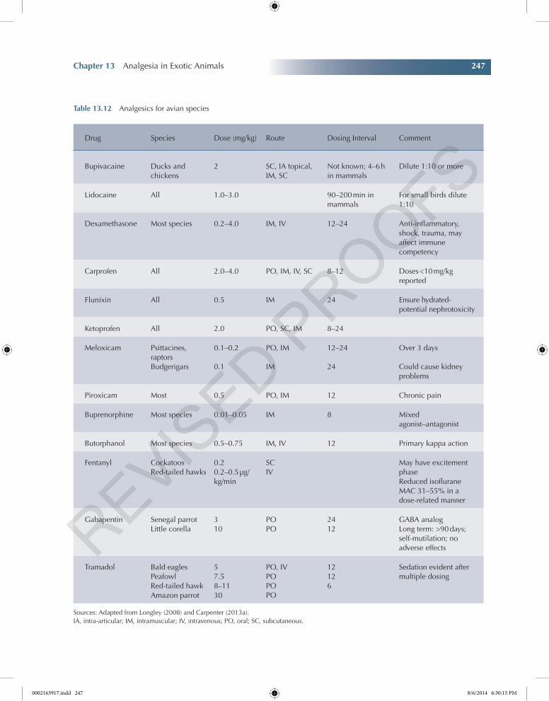
Chapter 13 Analgesia in Exotic Animals 247
Chapter No.: 1 Title Name: Goldberg 0002163917.inddComp. by: KIRUPANAND N Date: 06 Aug 2014 Time: 06:29:57 PM Stage: Proof WorkFlow:CSW Page Number: 247
Table 13.12 Analgesics for avian species
Drug Species Dose (mg/kg) Route Dosing Interval Comment
Bupivacaine Ducks and chickens
2 SC, IA topical, IM, SC
Not known; 4–6 h in mammals
Dilute 1:10 or more
Lidocaine All 1.0–3.0 90–200 min in mammals
For small birds dilute 1:10
Dexamethasone Most species 0.2–4.0 IM, IV 12–24 Anti-inflammatory, shock, trauma, may affect immune competency
Carprofen All 2.0–4.0 PO, IM, IV, SC 8–12 Doses <10 mg/kg reported
Flunixin All 0.5 IM 24 Ensure hydrated-potential nephrotoxicity
Ketoprofen All 2.0 PO, SC, IM 8–24
Meloxicam Psittacines, raptorsBudgerigars
0.1–0.2
0.1
PO, IM
IM
12–24
24
Over 3 days
Could cause kidney problems
Piroxicam Most 0.5 PO, IM 12 Chronic pain
Buprenorphine Most species 0.01–0.05 IM 8 Mixed agonist–antagonist
Butorphanol Most species 0.5–0.75 IM, IV 12 Primary kappa action
Fentanyl CockatoosRed-tailed hawks
0.20.2–0.5 µg/kg/min
SCIV
May have excitement phaseReduced isoflurane MAC 31–55% in a dose-related manner
Gabapentin Senegal parrotLittle corella
310
POPO
2412
GABA analogLong term: >90 days; self-mutilation; no adverse effects
Tramadol Bald eaglesPeafowlRed-tailed hawkAmazon parrot
57.58–1130
PO, IVPOPOPO
12126
Sedation evident after multiple dosing
Sources: Adapted from Longley (2008) and Carpenter (2013a).IA, intra-articular; IM, intramuscular; IV, intravenous; PO, oral; SC, subcutaneous.
0002163917.indd 247 8/6/2014 6:30:15 PM
248 Pain Management for Veterinary Technicians and Nurses
Chapter No.: 1 Title Name: Goldberg 0002163917.inddComp. by: KIRUPANAND N Date: 06 Aug 2014 Time: 06:29:57 PM Stage: Proof WorkFlow:CSW Page Number: 248
Reptile AnalgesiaStephen Cital
Reptiles are increasing in small animal practice. They may be owned by private collectors, may be zoological collections, or may be wildlife casualties.
Initial treatment involves providing a suitable thermal environment away from predator species, with small hiding boxes or toweling. When pos-sible, painful areas should be immobilized (Eatwell 2010).
Reptiles encompass more than 8000 different species and 4 different orders: Sphenodontia, Crocodilia, Squamata, and Chelonia. Within these varying orders are huge physiologic and behavioral adaptations, making pain assessment difficult and highly subjective. For this reason, analgesia is sometimes overlooked in these animals and it can
be difficult to clinically quantify the benefits of analgesia (Eatwell 2010; Mosely 2011). Yet, if there is a question as to whether a procedure or condition of the patient is painful, the patient should receive the benefit of analgesia. A physical exam with baseline vitals should be conducted. Similarly to birds, observation from a distance may be necessary to accurately assess respiratory rates. In some species of reptiles, such as snakes, heartbeat can be observed through the skin.
Because they are prey species, often reptiles exhibit no obvious signs of pain other than changes in behavior. Remote observation of such patients can help to identify subtle behavioral changes. In order to accurately assess pain, it is imperative to know the normal behavioral pat-terns of each particular species. Thorough client communication is used to acquire this information through detailed patient history.
There are some behavioral differences specific to acute versus chronic pain syndromes in reptiles. Acute pain often results in vigorous activity at the time of the insult (e.g., trying to bite when injected), but, subsequently, in the case of chronic pain, reptiles may become immobile (e.g., after surgery).
Text Box 13.7 Avian analgesia
Animal and Personnel SafetyOne of the most dangerous aspects of treating a bird is handling. Birds often struggle and flap their wings, which can lead to serious injury including breaking a wing. Other species that have longer legs, like flamingo, can easily break their legs while struggling. When holding a bird, it is important to not squeeze the patient too tight. Applying too much pressure to the bird can not only injure internal organs but also inhibit the bird from breathing normally.
As far as human hazards are concerned, birds can deliver quite the bite as well as scratches. Pecking can be a dangerous behavior if precautions are not taken. An eye is an easy target with a thrashing unhappy bird. Zoonosis is also higher on the list of hazards of working with birds. It is generally ideal to wear gloves, long sleeves, and eye protection when handling birds. For giant species, like ostrich or emu, dangers include fractures to the handler from viscous kicks by the bird (Fowler 2007; Tully 2012).
Key SignS 13.9 Reptiles: Flinching and muscle contractions, weight loss, and anorexia.
Text Box 13.8 Black mamba venom
Black mamba venom was recently found to have excellent analgesic properties and even able to abolish pain in the research setting. “Mambalgins,” as scientists call them, have been shown to have analgesic effects when compared to morphine in mice with fewer negative side effects such as respiratory depression (Diochot et al. 2012).
0002163917.indd 248 8/6/2014 6:30:15 PM

Chapter 13 Analgesia in Exotic Animals 249
Chapter No.: 1 Title Name: Goldberg 0002163917.inddComp. by: KIRUPANAND N Date: 06 Aug 2014 Time: 06:29:57 PM Stage: Proof WorkFlow:CSW Page Number: 249
Acute Pain
Signs of acute pain include withdrawal of painful limbs (e.g., in chelonians and lizards), escape behavior, avoidance, restlessness, aggression, and increased respiratory rate. In those species with melanophores, acute pain can be identified by a darkening of the skin over the affected areas. This can be seen following injections in chameleons or water dragons, for example, and owners should be made aware of this. In some cases, florid displays of pain can be seen with biting or scratching of the region in question and vocalization (Eatwell 2010).
Chronic Pain
Chronic pain is less likely to evoke marked clinical signs. Instead, immobility or stinting of the area may be seen (e.g., snakes may not curl in the affected region), and some animals may become aggressive if handling or manipulation is attempted (i.e., acute pain overlying chronic pain) (Eatwell 2010).
Causes of Pain in Reptiles
The following are some of the more commonly observed conditions that may be painful to reptiles (Wenger 2007):
• Trauma including bite wounds, fractures, and heat damage
• Surgical procedures, that is, ovariectomy or digit amputation
• Chronic disease processes such as gout, metabolic bone disease, or neoplasia
• Urolithiasis• Intestinal impaction• Dystocia and follicular stasis• Necrotic stomatitis• Peritonitis• Osteomyelitis and abscesses
Analgesic Medications
Reptiles have an interesting and variable neuroan-atomic and receptor pathology, resulting in vari-able systemic use of drugs in general. A study in
red-eared sliders discovered that the femoral veins drained directly into the abdominal vein, which went straight into the liver. The liver, being a filter, had notable presystemic extraction of the drugs, reducing bioavailability (Sladky et al. 2009; Baker et al. 2011). Reptiles have a renal portal system, which moves blood from the back half of a reptile through the kidneys before returning to the heart. This system has been linked to filtering of medica-tions before systemic dispersion occurs. Therefore, it is generally more effective to give injections in the front half of patients with renal portal systems.
Delivery of analgesics to reptiles is similar to mammalian administration. SC, IM, and oral administrations are the more common methods. For oral administration of medications, reptiles may need to have their mouths manually opened with a slender semisoft object (e.g., guitar pick or credit card). IM injections may be most effective when administered into the front end of a reptile. IV access is also possible in reptiles but is less practical and more difficult. Reptiles also can be given intracoelomic (IC) injections (Tully 2012).
Opioids
Opioid analgesics are used frequently in reptiles.Opioid effectiveness is typically assessed by
anesthetic-sparing effects or by pain studies. Numerous reports provide conflicting results; therefore, the efficacy of many agents remains questionable. Butorphanol tartrate is the most commonly administered opioid analgesic drug in reptiles (Sladky and Mans 2012). Butorphanol has been used at doses of 0.4–28 mg/kg in a variety of species (Eatwell 2010). Morphine has been shown to be effective in both lizards and crocodil-ians at doses of 1–2 mg/kg. Time to onset of action appears to be prolonged (2–8 h) after morphine and butorphanol administration (Kanui and Hole 1992; Sladky et al. 2008), and this must be taken into consideration. Gamble (2009) found that serum levels following the use of transdermal fen-tanyl patches in skinks were comparable to those providing analgesia in humans.
The pharmacokinetics (PK) of buprenorphine have been studied in red-eared sliders (Kummrow
0002163917.indd 249 8/6/2014 6:30:16 PM
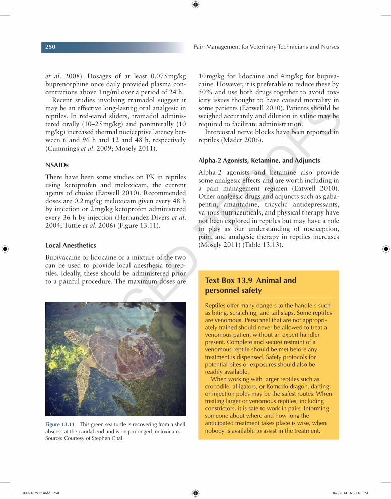
250 Pain Management for Veterinary Technicians and Nurses
Chapter No.: 1 Title Name: Goldberg 0002163917.inddComp. by: KIRUPANAND N Date: 06 Aug 2014 Time: 06:29:57 PM Stage: Proof WorkFlow:CSW Page Number: 250
et al. 2008). Dosages of at least 0.075 mg/kg buprenorphine once daily provided plasma con-centrations above 1 ng/ml over a period of 24 h.
Recent studies involving tramadol suggest it may be an effective long-lasting oral analgesic in reptiles. In red-eared sliders, tramadol adminis-tered orally (10–25 mg/kg) and parenterally (10 mg/kg) increased thermal nociceptive latency bet-ween 6 and 96 h and 12 and 48 h, respectively (Cummings et al. 2009; Mosely 2011).
nSAiDs
There have been some studies on PK in reptiles using ketoprofen and meloxicam, the current agents of choice (Eatwell 2010). Recommended doses are 0.2 mg/kg meloxicam given every 48 h by injection or 2 mg/kg ketoprofen administered every 36 h by injection (Hernandez-Divers et al. 2004; Tuttle et al. 2006) (Figure 13.11).
Local Anesthetics
Bupivacaine or lidocaine or a mixture of the two can be used to provide local anesthesia to rep-tiles. Ideally, these should be administered prior to a painful procedure. The maximum doses are
10 mg/kg for lidocaine and 4 mg/kg for bupiva-caine. However, it is preferable to reduce these by 50% and use both drugs together to avoid tox-icity issues thought to have caused mortality in some patients (Eatwell 2010). Patients should be weighed accurately and dilution in saline may be required to facilitate administration.
Intercostal nerve blocks have been reported in reptiles (Mader 2006).
Alpha-2 Agonists, Ketamine, and Adjuncts
Alpha-2 agonists and ketamine also provide some analgesic effects and are worth including in a pain management regimen (Eatwell 2010). Other analgesic drugs and adjuncts such as gaba-pentin, amantadine, tricyclic antidepressants, various nutraceuticals, and physical therapy have not been explored in reptiles but may have a role to play as our understanding of nociception, pain, and analgesic therapy in reptiles increases (Mosely 2011) (Table 13.13).
Figure 13.11 This green sea turtle is recovering from a shell abscess at the caudal end and is on prolonged meloxicam. Source: Courtesy of Stephen Cital.
Text Box 13.9 Animal and personnel safety
Reptiles offer many dangers to the handlers such as biting, scratching, and tail slaps. Some reptiles are venomous. Personnel that are not appropri-ately trained should never be allowed to treat a venomous patient without an expert handler present. Complete and secure restraint of a venomous reptile should be met before any treatment is dispensed. Safety protocols for potential bites or exposures should also be readily available.
When working with larger reptiles such as crocodile, alligators, or Komodo dragon, darting or injection poles may be the safest routes. When treating larger or venomous reptiles, including constrictors, it is safe to work in pairs. Informing someone about where and how long the anticipated treatment takes place is wise, when nobody is available to assist in the treatment.
0002163917.indd 250 8/6/2014 6:30:16 PM
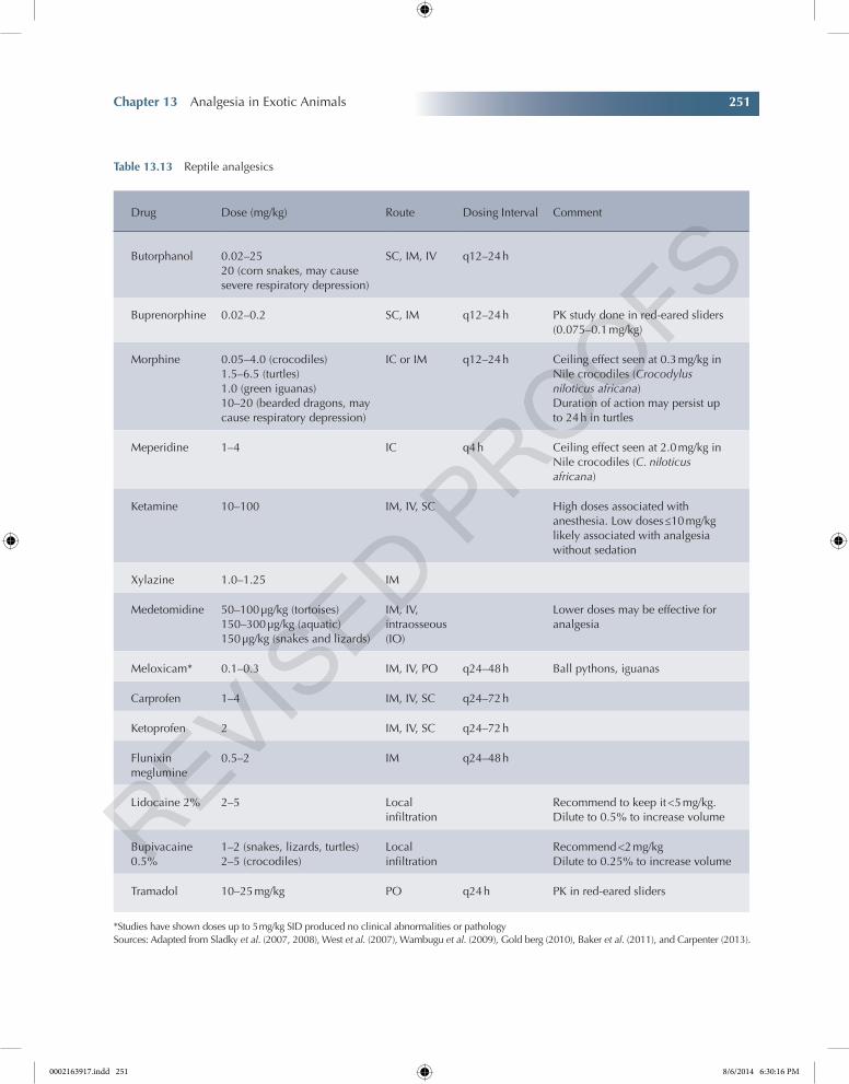
Chapter 13 Analgesia in Exotic Animals 251
Chapter No.: 1 Title Name: Goldberg 0002163917.inddComp. by: KIRUPANAND N Date: 06 Aug 2014 Time: 06:29:57 PM Stage: Proof WorkFlow:CSW Page Number: 251
Table 13.13 Reptile analgesics
Drug Dose (mg/kg) Route Dosing Interval Comment
Butorphanol 0.02–2520 (corn snakes, may cause severe respiratory depression)
SC, IM, IV q12–24 h
Buprenorphine 0.02–0.2 SC, IM q12–24 h PK study done in red-eared sliders (0.075–0.1 mg/kg)
Morphine 0.05–4.0 (crocodiles)1.5–6.5 (turtles)1.0 (green iguanas)10–20 (bearded dragons, may cause respiratory depression)
IC or IM q12–24 h Ceiling effect seen at 0.3 mg/kg in Nile crocodiles (Crocodylus niloticus africana)Duration of action may persist up to 24 h in turtles
Meperidine 1–4 IC q4 h Ceiling effect seen at 2.0 mg/kg in Nile crocodiles (C. niloticus africana)
Ketamine 10–100 IM, IV, SC High doses associated with anesthesia. Low doses ≤10 mg/kg likely associated with analgesia without sedation
Xylazine 1.0–1.25 IM
Medetomidine 50–100 µg/kg (tortoises)150–300 µg/kg (aquatic)150 µg/kg (snakes and lizards)
IM, IV, intraosseous (IO)
Lower doses may be effective for analgesia
Meloxicam* 0.1–0.3 IM, IV, PO q24–48 h Ball pythons, iguanas
Carprofen 1–4 IM, IV, SC q24–72 h
Ketoprofen 2 IM, IV, SC q24–72 h
Flunixin meglumine
0.5–2 IM q24–48 h
Lidocaine 2% 2–5 Local infiltration
Recommend to keep it <5 mg/kg. Dilute to 0.5% to increase volume
Bupivacaine 0.5%
1–2 (snakes, lizards, turtles)2–5 (crocodiles)
Local infiltration
Recommend <2 mg/kgDilute to 0.25% to increase volume
Tramadol 10–25 mg/kg PO q24 h PK in red-eared sliders
*Studies have shown doses up to 5 mg/kg SID produced no clinical abnormalities or pathologySources: Adapted from Sladky et al. (2007, 2008), West et al. (2007), Wambugu et al. (2009), Gold berg (2010), Baker et al. (2011), and Carpenter (2013).
0002163917.indd 251 8/6/2014 6:30:16 PM
252 Pain Management for Veterinary Technicians and Nurses
Chapter No.: 1 Title Name: Goldberg 0002163917.inddComp. by: KIRUPANAND N Date: 06 Aug 2014 Time: 06:29:57 PM Stage: Proof WorkFlow:CSW Page Number: 252
Analgesia in AmphibiansStephen Cital
With more than 4000 known species of amphib-ians and a large population of these animals used in research as well as kept as pets, it is prudent that proper care and treatment is offered to this exotic species group. Based on general anatomy and behavior, it might be easy to assume we could treat these patients the same way we would treat reptiles. However, amphibians unlike reptiles go through different stages of metamorphosis dur-ing their life span. Amphibians go from having gills to lungs, being aquatic to semiaquatic, and having no legs to four or two and now with envi-ronmental contaminants sometimes six, more or less. This means at two different times in their lives, we may treat amphibians differently. At one point like a fish and at another more like any other terrestrial creature.
Amphibians are considered a “lesser verte-brate,” which means they fall lower on the evo-lutionary development chain. Scientists have more recently analyzed nociceptive pathways of amphibians and concluded there are no major distinctions between nociceptive afferent path-ways in either mammals or amphibians; yet, other comparative neurology suggests potential pain may be less substantial or recognized as such with these lesser vertebrates than in humans. With this said, amphibians still do possess a well-developed endogenous opioid system with corresponding binding sites, primarily kappa-like receptors. Not only do opioids provide analgesia but also there has been potential shown with other drugs like ketamine, alpha-2s, and tricaine methanesulfonate (Machin 1999; Stevens 2011). It is important to note that amphibians have a reduced rate of hepatic metabolism of certain medications and appear to require greater doses of opioids for systemic effect than mammals. Although nociceptive receptors and the seemingly effective treatment of perceived pain exist with amphibians, there is but one problem: no thalamus to cortex net-working to the cerebral cortex exists. Why?
Because the structure makes its first appearance in reptiles. What this means is amphibians still feel pain, but it may be much different than the way mammals may appreciate it. In human patients, where cortical neurons have lesions or have been affected by surgical painful stimuli, it has been described as being aware of the pain, but it does not necessarily bother them (Mosely 2011; Stevens 2011).
In a study conducted using eastern red-spotted newts, the animals underwent a bilateral forelimb amputation. One group received systemic buprenorphine, while another had a bath appli-cation of butorphanol and the control group received no analgesics. The animals were then closely monitored for common signs of pain or discomfort associated with other species, such as poor appetite, spontaneous movement, and other species-specific behaviors. The research did discover animals receiving the postoperative analgesics experienced shorter time to “normal” behaviors than the nontreatment animals. This suggested that pain potential is significant in amphibian species and can at least be partially controlled with opioid analgesics (Stevens 2011).
Treatment
One of the advances in veterinary pain management is the use of transdermal patches that deliver anal-gesia through the skin. In amphibians, this has always been the case. Common anesthetic prac-tices include anesthesia administration through the skin and thus are the same with analgesics (West et al. 2007).
A more controversial but still practiced form of anesthesia and analgesia is induced hypo-thermia. Possible animal welfare concerns can be made with this method, but one study has shown
Key SignS 13.10 Amphibians: Muscular movements, closed eyes, color changes, rapid respirations, immobility, and anorexia.
0002163917.indd 252 8/6/2014 6:30:17 PM
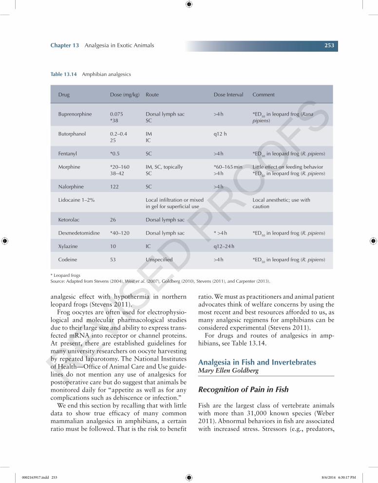
Chapter 13 Analgesia in Exotic Animals 253
Chapter No.: 1 Title Name: Goldberg 0002163917.inddComp. by: KIRUPANAND N Date: 06 Aug 2014 Time: 06:29:57 PM Stage: Proof WorkFlow:CSW Page Number: 253
analgesic effect with hypothermia in northern leopard frogs (Stevens 2011).
Frog oocytes are often used for electrophysio-logical and molecular pharmacological studies due to their large size and ability to express trans-fected mRNA into receptor or channel proteins. At present, there are established guidelines for many university researchers on oocyte harvesting by repeated laparotomy. The National Institutes of Health—Office of Animal Care and Use guide-lines do not mention any use of analgesics for postoperative care but do suggest that animals be monitored daily for “appetite as well as for any complications such as dehiscence or infection.”
We end this section by recalling that with little data to show true efficacy of many common mammalian analgesics in amphibians, a certain ratio must be followed. That is the risk to benefit
ratio. We must as practitioners and animal patient advocates think of welfare concerns by using the most recent and best resources afforded to us, as many analgesic regimens for amphibians can be considered experimental (Stevens 2011).
For drugs and routes of analgesics in amp-hibians, see Table 13.14.
Analgesia in Fish and invertebratesMary Ellen Goldberg
Recognition of Pain in Fish
Fish are the largest class of vertebrate animals with more than 31,000 known species (Weber 2011). Abnormal behaviors in fish are associated with increased stress. Stressors (e.g., predators,
Table 13.14 Amphibian analgesics
Drug Dose (mg/kg) Route Dose Interval Comment
Buprenorphine 0.075*38
Dorsal lymph sacSC
>4 h *ED50 in leopard frog (Rana pipiens)
Butorphanol 0.2–0.425
IMIC
q12 h
Fentanyl *0.5 SC >4 h *ED50 in leopard frog (R. pipiens)
Morphine *20–16038–42
IM, SC, topicallySC
*60–165 min>4 h
Little effect on feeding behavior*ED50 in leopard frog (R. pipiens)
Nalorphine 122 SC >4 h
Lidocaine 1–2% Local infiltration or mixed in gel for superficial use
Local anesthetic; use with caution
Ketorolac 26 Dorsal lymph sac
Dexmedetomidine *40–120 Dorsal lymph sac * >4 h *ED50 in leopard frog (R. pipiens)
Xylazine 10 IC q12–24 h
Codeine 53 Unspecified >4 h *ED50 in leopard frog (R. pipiens)
* Leopard frogsSource: Adapted from Stevens (2004), West et al. (2007), Goldberg (2010), Stevens (2011), and Carpenter (2013).
0002163917.indd 253 8/6/2014 6:30:17 PM
254 Pain Management for Veterinary Technicians and Nurses
Chapter No.: 1 Title Name: Goldberg 0002163917.inddComp. by: KIRUPANAND N Date: 06 Aug 2014 Time: 06:29:57 PM Stage: Proof WorkFlow:CSW Page Number: 254
water quality, and disease) can easily shift well-being to distress. Depending on the nature and severity of a stressor and on the animal’s current state of being, the animal may adapt suc-cessfully to a stress (adaptive behaviors), or it may become distressed in a way that threatens its well-being or health (maladaptive behaviors). Adaptive behaviors include normal feeding, schooling for appropriate groups, breeding, and nest building. Maladaptive behaviors include abnormal feeding, absence or diminution of grooming, and changes in social interaction (aggression, withdrawal) (Table 13.15).
Fish possess mammalian-like anatomic, biochemical, and functional components for the transmission, mediation, and central modula-tion of painful stimuli (Beitz 1992). This includes (Storms and Mylniczenko 2004) biochemicals that can activate opioid receptors (Ng and Chan 1990); proteins that have similarities to GABA benzodiazepine receptors (Friedl et al. 1988); P-like substances possibly contributing to noci-ception (Weld and Maler 1992); presence of μ, δ, and κ opioid receptors; and endogenous opioid activity that modulates response to stressors (Pert et al. 1974; Chervova and Lapshin 2000). Teleost fish possess COX enzymes iden-tical to those of mammals (Grosser et al. 2002).
Fish neuroanatomy and behavioral responses indicate that these animals feel pain. It would then be logical to conclude that an animal that can feel pain can also experience distress (Posner 2009).
While signs of pain in fish are subjective (body orientation and swimming, feeding, hiding, and/or spatial positioning) (near aeration or heater), others are objective (environmental quality parameters, heart rate, opercular or respiratory rate, lethargy, fin clamping, darkening, blood diagnostic values, and/or physical examination findings) (Weber 2011). It is difficult to deter-mine the nature of the response to pain in fish. Although they exhibit a pronounced response to injuries or to contact with irritants, their response to chronic stimuli may be small or absent. Fish with wounds that are severe enough to cause
immobility in a mammal will often appear to behave normally and even resume feeding. Fish will react to noxious stimuli such as a hypodermic needle stick, by strong muscular movements. When exposed to a noxious environment, such as a strong acid, fish show abnormal swimming behavior with attempts to jump from the water. Their coloring becomes darker and opercular movements become more rapid. Such effects are indicative of some degree of distress; however, it is not possible to describe these unequivocally as signs of pain. The most predominant sign of distress or pain in fish is anorexia.
For common analgesics used in fish, see Table 13.16.
Whether fish feel pain, are simply fear aversive, or are experiencing nociception to any noxious stimulus caused from disease, poor husbandry, handling, or transport, it is a veterinary responsibility to help amelio-rate this stress and/or distress in a similar way to practical veterinary solutions employed for other livestock, pet, laboratory animal, and wild populations.
Weber (2011)
Recognition of Pain in Invertebrates
Very little is actually known about pain in inver-tebrates. For example, it is unclear if a wasp smashed with a flyswatter or a worm on a fish-hook feels pain. All animal species can experience tissue damage. Invertebrates are a group of ani-mals composing more than 95% of earth’s species identified by not possessing a vertebral column (Lewbart 2012). They have mechanisms that
Key SignS 13.11 Fish: Abnormal swimming behavior, attempting to jump out of water, rapid opercular movements, clamped fins, pale or darkened color, and hiding—anorexia is the first sign.
0002163917.indd 254 8/6/2014 6:30:17 PM
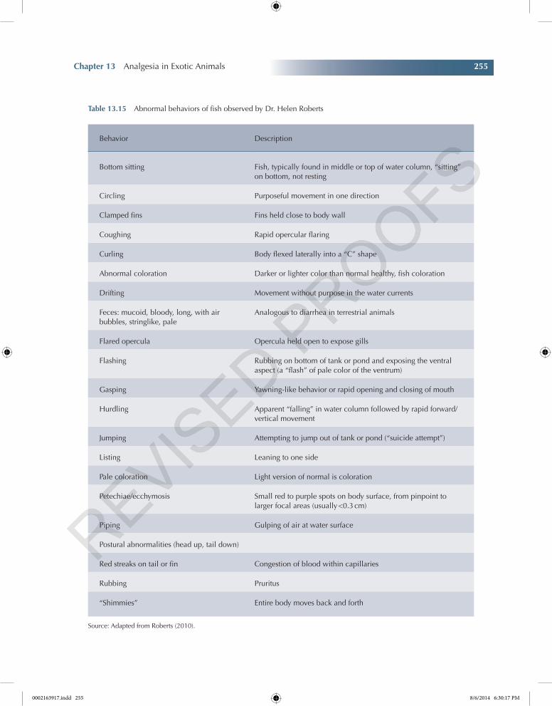
Chapter 13 Analgesia in Exotic Animals 255
Chapter No.: 1 Title Name: Goldberg 0002163917.inddComp. by: KIRUPANAND N Date: 06 Aug 2014 Time: 06:29:57 PM Stage: Proof WorkFlow:CSW Page Number: 255
Table 13.15 Abnormal behaviors of fish observed by Dr. Helen Roberts
Behavior Description
Bottom sitting Fish, typically found in middle or top of water column, “sitting” on bottom, not resting
Circling Purposeful movement in one direction
Clamped fins Fins held close to body wall
Coughing Rapid opercular flaring
Curling Body flexed laterally into a “C” shape
Abnormal coloration Darker or lighter color than normal healthy, fish coloration
Drifting Movement without purpose in the water currents
Feces: mucoid, bloody, long, with air bubbles, stringlike, pale
Analogous to diarrhea in terrestrial animals
Flared opercula Opercula held open to expose gills
Flashing Rubbing on bottom of tank or pond and exposing the ventral aspect (a “flash” of pale color of the ventrum)
Gasping Yawning-like behavior or rapid opening and closing of mouth
Hurdling Apparent “falling” in water column followed by rapid forward/vertical movement
Jumping Attempting to jump out of tank or pond (“suicide attempt”)
Listing Leaning to one side
Pale coloration Light version of normal is coloration
Petechiae/ecchymosis Small red to purple spots on body surface, from pinpoint to larger focal areas (usually <0.3 cm)
Piping Gulping of air at water surface
Postural abnormalities (head up, tail down)
Red streaks on tail or fin Congestion of blood within capillaries
Rubbing Pruritus
“Shimmies” Entire body moves back and forth
Source: Adapted from Roberts (2010).
0002163917.indd 255 8/6/2014 6:30:17 PM
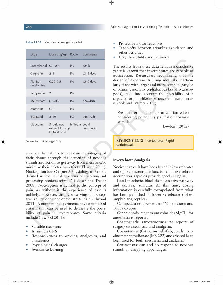
256 Pain Management for Veterinary Technicians and Nurses
Chapter No.: 1 Title Name: Goldberg 0002163917.inddComp. by: KIRUPANAND N Date: 06 Aug 2014 Time: 06:29:57 PM Stage: Proof WorkFlow:CSW Page Number: 256
enhance their ability to maintain the integrity of their tissues through the detection of noxious stimuli and action to get away from them and/or minimize their deleterious effects (Elwood 2011). Nociception (see Chapter 3 Physiology of Pain) is defined as “the neural processes of encoding and processing noxious stimuli” (Loeser and Treede 2008). Nociception is central to the concept of pain, as without it the experience of pain is unlikely. However, simply observing a nocicep-tive ability does not demonstrate pain (Elwood 2011). A number of experiments have established criteria that can be used to delineate the possi-bility of pain in invertebrates. Some criteria include (Elwood 2011):
• Suitable receptors• A suitable CNS• Responsiveness to opioids, analgesics, and
anesthetics• Physiological changes• Avoidance learning
• Protective motor reactions• Trade-offs between stimulus avoidance and
other activities• Cognitive ability and sentience
The results from these data remain inconclusive yet it is known that invertebrates are capable of nociception. Researchers recommend that the design of experiments using mollusks, particu-larly those with larger and more complex ganglia or brains (especially cephalopods but also gastro-pods), take into account the possibility of a capacity for pain-like experience in these animals (Crook and Walters 2011).
We must err on the side of caution when considering potentially painful or noxious stimuli.
Lewbart (2012)
invertebrate Analgesia
Nociceptive cells have been found in invertebrates and opioid systems are functional in invertebrate nociception. Opioids provide good analgesia.
Local anesthetics block the nociceptive pathway and decrease stimulus. At this time, dosing information is carefully extrapolated from what has been published on lower vertebrates (fishes, amphibians, reptiles).
Centipedes: only reports of 5% isoflurane and 100% oxygen.
Cephalopods: magnesium chloride (MgCl2) for anesthesia is reported.
Chaetognaths (arrowworms): no reports of surgery or anesthesia and analgesia.
Coelenterates (flatworms, jellyfish, corals): tric-aine methanesulfonate (MS-222) and ethanol have been used for both anesthesia and analgesia.
Crustaceans: can and do respond to noxious stimuli by dropping appendages.
Table 13.16 Multimodal analgesia for fish
Drug Dose (mg/kg) Route Comments
Butorphanol 0.1–0.4 IM q24 h
Carprofen 2–4 IM q3–5 days
Flunixin meglumine
0.25–0.5 IM q3–5 days
Ketoprofen 2 IM
Meloxicam 0.1–0.2 IM q24–48 h
Morphine 0.3 IM
Tramadol 5–10 PO q48–72 h
Lidocaine Should not exceed 1–2 mg/kg total dose
Infiltrate Local anesthesia
Source: From Goldberg (2010). Key SignS 13.12 Invertebrates: Rapid withdrawal.
0002163917.indd 256 8/6/2014 6:30:17 PM
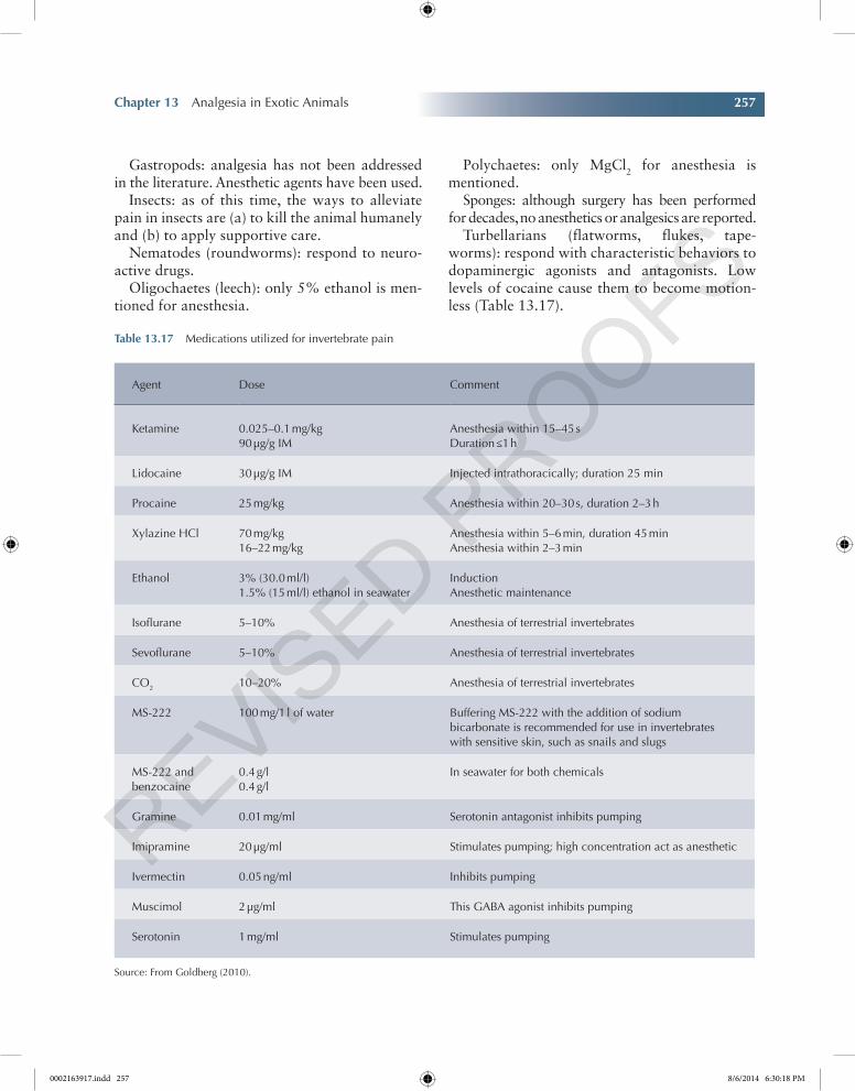
Chapter 13 Analgesia in Exotic Animals 257
Chapter No.: 1 Title Name: Goldberg 0002163917.inddComp. by: KIRUPANAND N Date: 06 Aug 2014 Time: 06:29:57 PM Stage: Proof WorkFlow:CSW Page Number: 257
Gastropods: analgesia has not been addressed in the literature. Anesthetic agents have been used.
Insects: as of this time, the ways to alleviate pain in insects are (a) to kill the animal humanely and (b) to apply supportive care.
Nematodes (roundworms): respond to neuro-active drugs.
Oligochaetes (leech): only 5% ethanol is men-tioned for anesthesia.
Polychaetes: only MgCl2 for anesthesia is mentioned.
Sponges: although surgery has been performed for decades, no anesthetics or analgesics are reported.
Turbellarians (flatworms, flukes, tape-worms): respond with characteristic behaviors to dopaminergic agonists and antagonists. Low levels of cocaine cause them to become motion-less (Table 13.17).
Table 13.17 Medications utilized for invertebrate pain
Agent Dose Comment
Ketamine 0.025–0.1 mg/kg90 µg/g IM
Anesthesia within 15–45 sDuration ≤1 h
Lidocaine 30 µg/g IM Injected intrathoracically; duration 25 min
Procaine 25 mg/kg Anesthesia within 20–30 s, duration 2–3 h
Xylazine HCl 70 mg/kg16–22 mg/kg
Anesthesia within 5–6 min, duration 45 minAnesthesia within 2–3 min
Ethanol 3% (30.0 ml/l)1.5% (15 ml/l) ethanol in seawater
InductionAnesthetic maintenance
Isoflurane 5–10% Anesthesia of terrestrial invertebrates
Sevoflurane 5–10% Anesthesia of terrestrial invertebrates
CO2 10–20% Anesthesia of terrestrial invertebrates
MS-222 100 mg/1 l of water Buffering MS-222 with the addition of sodium bicarbonate is recommended for use in invertebrates with sensitive skin, such as snails and slugs
MS-222 and benzocaine
0.4 g/l0.4 g/l
In seawater for both chemicals
Gramine 0.01 mg/ml Serotonin antagonist inhibits pumping
Imipramine 20 µg/ml Stimulates pumping; high concentration act as anesthetic
Ivermectin 0.05 ng/ml Inhibits pumping
Muscimol 2 µg/ml This GABA agonist inhibits pumping
Serotonin 1 mg/ml Stimulates pumping
Source: From Goldberg (2010).
0002163917.indd 257 8/6/2014 6:30:18 PM
258 Pain Management for Veterinary Technicians and Nurses
Chapter No.: 1 Title Name: Goldberg 0002163917.inddComp. by: KIRUPANAND N Date: 06 Aug 2014 Time: 06:29:57 PM Stage: Proof WorkFlow:CSW Page Number: 258
References
Baker, B., Sladky, K. & Johnson, S. (2011) Evaluations of the analgesic effects of oral and subcutaneous tramadol administration in red-eared slider turtles. The Journal of the American Veterinary Medical Association, 238 (2), 220–227.
Barter, L.A. (2011) Rabbit Analgesia. Veterinary Clinics of North America: Exotic Animal Practice, 14 (1), 93–104.
Beitz, A.J. (1992) Anatomic and chemical organization of descending pain modulation systems. C.E. Short & A. Van Poznak (eds), Animal Pain, Churchill Livingstone, New York, pp. 31–62.
Bowles, H., Lichtenberger, M. & Lennox, A. (2007) Emergency and Critical Care of Pet Birds. Veterinary Clinics of North America: Exotic Animal Practice, 10, 345–394.
Bradley Bays, T., Lightfoot, T. & Mayer, J. (2006) Exotic Pet Behavior. Saunders Elsevier, St. Louis, MO.
Brenner, D.J., Larsen, R.S., Dickinson, P.J., Wack, R.F., Williams, D.C. & Pascoe, P.J. (2010) Development of an avian brachial plexus nerve block technique for perioperative analgesia in mallard ducks (Anas platyrhynchos). Journal of Avian Medicine and Surgery, 24, 24–34.
Brust, D.M. & Pye, G.W. (2013) Chapter 6 Sugar Gliders. Carpenter JW (ed), Exotic Animal Formulary, 4th edn, Elsevier/Saunders, St. Louis, p. 443.
Carbone, L. & Garnett, N. (2008) Ethical issues in anesthesia and analgesia in laboratory animals. R.E. Fish, M.J. Brown, P.J. Danneman, & A.Z. Karas (eds), Anesthesia and Analgesia in Laboratory Animals. American College of Laboratory Animal Medicine Series, Academic Press, San Diego, p. 561.
Carpenter, J.W. Hawkins, M., Barron, H.W., Speer, B.L., & Pollock, C. (2013) Chapter 5 Birds. Exotic Animal Formulary, 4th edn, Elsevier, St. Louis, pp. 256–281.
Cheol Jin, H., Keller, A.J., Kwon Jung, J., Subieta, A., & Brennan, T.J. (2007) Epidural tezampanel, an AMPA/kainite receptor antagonist, produces postoperative analgesia in rats. Anesthesia Analgesia, 105, 1152–1159.
Carpenter, J.W. & Marion, C.J. (2013) Chapter 7 Hedgehogs. J.W. Carpenter (ed), Exotic Animal Formulary, 4th edn, Elsevier/Saunders, St. Louis, p. 463.
Chervova, L.S. & Lapshin, D.N. (2000) Opioid modu-lation of pain threshold in fish. Doklady Biological Sciences, 375, 590–591.
Colpaert, F.C., Tarayre, J.P., Alliaga, M., Bruins Slot, L.A., Attal, N. & Koek, W. (2001) Opiate self- administration as a measure of chronic nociceptive pain in arthritic rats. Pain, 91, 33–45.
Committee on Recognition and Alleviation of Pain in Laboratory Animals (2009) Effective pain management. Recognition and alleviation of pain in laboratory animals, National Academies Press, Washington, DC, pp. 71–118.
Concannon, K.T., Dodam, J.R. & Hellyer, P.W. (1995) Influence of a mu- and kappa-opioid agonist on iso-flurane minimal anesthetic concentration in chickens. American Journal of Veterinary Research, 56, 806–811.
Criado, A.B. & Gomez de Segura, I.A. (2003) Reduction of isoflurane MAC by fentanyl or remi-fentanil in rats. Veterinary Anaesthesia and Analgesia, 30, 250–256.
Crook, R.J. & Walters, E.T. (2011) Nociceptive behavior and physiology of molluscs: animal welfare implications. ILAR Journal, 52 (2), 185–195.
Cummings, B.B., Sladky, K.K. & Johnson, S.M. (2009) Tramadol analgesic and respiratory effects in red-eared slider turtles (Trachemys scripta). American Association of Zoo Veterinarians and American Association of Wildlife Veterinarians Joint Conference, Tulsa, p. 115.
Doneley, B. (2010) ed. Anesthesia and Analgesia. Avian Medicine and Surgery in Practice: Companion and Aviary Birds, Manson Publishing, London, p. 245.
Dorrestein, G.M. (2000) Nursing the Sick Bird. T.N. Tully, G.M. Dorrestein & A.K. Jones (eds), Handbook of Avian Medicine, 2nd edn, Butterworth-Heinemann, Oxford, p. 101.
Eatwell, K. (July/August 2010) Options for analgesia and anaesthesia in reptiles. In Practice, 32, 306–311.
Edwards, S.R. & Mather, L.E. (2001) Tissue uptake of ketamine and norketamine enantiomers in the rat indirect evidence for extrahepatic metabolic inversion. Life Sciences, 69, 2051–2066.
Elwood, R.W. (2011) Pain and suffering in inverte-brates? ILAR Journal, 52 (2), 175–184.
Eshar, D. & Wilson, J. (2010) Epidural anesthesia and analgesia in ferrets. Lab Animal, 39 (11), 339–340.
Flecknell, P.A. (1998) Analgesia in small mammals. Seminars in Avian and Exotic Pet Medicine, 7, 41–47.
0002163917.indd 258 8/6/2014 6:30:18 PM
Chapter 13 Analgesia in Exotic Animals 259
Chapter No.: 1 Title Name: Goldberg 0002163917.inddComp. by: KIRUPANAND N Date: 06 Aug 2014 Time: 06:29:57 PM Stage: Proof WorkFlow:CSW Page Number: 259
Flecknell, P. (2006) Anaesthesia and perioperative care. A. Meredith & P. Flecknell (eds), BSAVA Manual of Rabbit Medicine and Surgery, 2nd edn, Gloucester, BSAVA Press, pp. 154–165.
Flecknell, P.A. (2009) ed. Analgesia and post-operative care. Laboratory animal anaesthesia, 3rd edn, Elsevier, London, pp. 139–180.
Flecknell, P.A., Roughan, J.V. & Stewart, R. (1999) Use of oral buprenorphine (‘buprenorphine jello’) for postoperative analgesia in rats—a clinical trial. Laboratory Animal, 33 (2), 169–174.
Foley, P.L., Henderson, A.L., Bisonette, E.A., Wimer, G.R. & Feldman, S.H. (2001) Evaluation of fentanyl transdermal patches in rabbits: blood concentra-tions and physiologic response. Comparative Medicine, 51, 239–244.
Fowler, M. (2007) Zoo and Wild Animal Medicine Current Therapy, 6 edn. Saunders, St. Louis, MO.
Franken, N.D., van Oostrom, H., Stienen, P.J., Doornenbal, A. & Hellebrekers, L.J. (2008) Evaluation of analgesic and sedative effects of con-tinuous infusion of dexemetomidine by measuring somatosensory- and auditory-evoked potentials in the rat. Veterinary Anaesthesthesia and Analgesia, 35, 424–431.
Friedl, W., Hebebrand, J., Rabe, S., & Propping, P. (1988) Phylogenetic conservation of the benzodiaz-epine binding sites: pharmacologic evidence. Neuropharmacology, 27, 163–170.
Gades, N.M., Danneman, P.J., Wixson, S.K. & Tolley, E.A. (2000) The magnitude and duration of the analgesic effect of morphine, butorphanol, and buprenorphine in rats and mice. Contemporary Topics in Laboratory Animal Science, 39 (2), 8–13.
Gaertner, D.J., Hallman, T.M., Hankenson, F.C., & Batchelder, M.A. (2008) Anesthesia and analgesia for laboratory rodents. R.E. Fish, M.J. Brown, P.J. Danneman, & A.Z. Karas (eds), Anesthesia and Analgesia in Laboratory Animals. American College of Laboratory Animal Medicine Series, Academic Press, San Diego, pp. 239–297.
Gaggermeier, B., Henke, J., Schatzmann, H., et al. (2000) Lihtersachunjen en Schmerzlinderung unit Buprenorphin bei Hanstauben Columba livia, Gurel, 1739, var. dornestica. Proceedings of the XII Conference on Avian Disease, Munich, p. 21.
Gamble, K.C. (2009) Plasma fentanyl concentrations achieved after transdermal fentanyl patch application in prehensile tailed skinks, Corucia zebrata. Journal of Herpetological Medicine and Surgery, 18, 81–85.
Gianotti, G., Valverde, A., Sinclair, M., et al. (2012) Prior determination of baseline minimum alveolar concentration (MAC) of isoflurane does not influence the effect of ketamine on MAC in rabbits. The Canadian Journal of Veterinary Research, 76, 261–267.
Glatz, P.C., Murphy, L.B. & Preston, A.P. (1992) Analgesic therapy of beak-trimmed chickens. Australian Veterinary Journal, 69, 18.
Goldberg, M.E. (2010) The fourth vital sign in all crea-tures great and small. The NAVTA Journal, Winter, 31–54.
Gonzalez, M.I., Field, M.J., Bramwell, S., McCleary, S. & Singh, L. (2000) Ovariohysterectomy in the rat: a model of surgical pain for evaluation of pre-emptive analgesia? Pain, 88 (1), 79–88.
Greenacre, C.B. (2008) Pain management in small mammal patients. CVC in Kansas City Proceedings; Advanstar http://veterinarycalendar.dvm360.com/avhc/Pain+Management+Center/Pain-management-i n - s m a l l - m a m m a l - p a t i e n t s - P r o c e e d i n /ArticleStandard/Article/detail/568367 (accessed on April 21, 2014).
Greenaway, J.B., Partlow, G.D., Gonsholt, N.L. & Fisher, K.R. (2001) Anatomy of the lumbosacral spinal cord in rabbits. Journal of the American Animal Hospital Association, 37 (1), 27–34.
Grosser, T., Yusuff, S., Cheskis, E., Pack, M.A. & Fitzgerald, G.A. (2002) Developmental expression of functional cyclooxygenases in zebrafish. Proceedings of the National Academy of Sciences of the United States of America, 99, 8418–8423.
Gutstein, H.B. & Akil, H. (2010) Chapter 21. Opioid analgesics. L.L. Brunton, J.S. Lazo & K.L. Parker (eds), Goodman & Gilman’s the pharmacological basis of therapeutics, 11th edn, McGraw-Hill Companies, New York, pp. 547–590.
Hanley, C.S., Thomas, N.J., Paul-Murphy, J. & Hartup, B.K. (2005) Exertional myopathy in whooping cranes (Grus americana) with prognostic guidelines. Journal of Zoo and Wildlife Medicine, 36, 489–497.
Hawkins, M.G. & Pascoe, P.J. (2012) Anesthesia, analgesia and sedation of small mammals. K.E. Quesenberry & J.W. Carpenter (eds), Ferrets, Rabbits and Rodents: Clinical Medicine and Surgery, 3rd edn, Elsevier/Saunders, St. Louis, pp. 429–451.
Hawkins, M.G. & Paul-Murphy, J. (2011) Avian anal-gesia. Veterinary Clinics of North America: Exotic Animal Practice, 14 (1), 62–76.
0002163917.indd 259 8/6/2014 6:30:18 PM
260 Pain Management for Veterinary Technicians and Nurses
Chapter No.: 1 Title Name: Goldberg 0002163917.inddComp. by: KIRUPANAND N Date: 06 Aug 2014 Time: 06:29:57 PM Stage: Proof WorkFlow:CSW Page Number: 260
Hernandez-Divers, S.J., Mcbride, M., Koch, T.F., Perpinan, D., Wilson, G.H., & Stedman, N.L. (2004) Single dose oral and intravenous pharmacokinetics of meloxicam in the green iguana (Iguana iguana). Proceedings of the 11th Annual Conference of the Association of Reptile and Amphibian Veterinarians, Naples, May 8 to 11, pp. 106–107.
Hocking, P.M., Gentle, M.J., Bernard, R. & Dunn, L.N. (1997) Evaluation of a protocol for deter-mining the effectiveness of pretreatment with local analgesics for reducing experimentally induced articular pain in domestic fowl. Research in Veterinary Science, 63, 263–267.
Hoefer, H.L. (2001) Small mammal dentistry. Atlantic Coast Veterinary Conference. Institute of Laboratory Animal Resources, National Research Council. Recognition and Alleviation of Pain and Distress in Laboratory Animals. National Academy Press, Washington, DC, 1992.
Institute of Laboratory Animal Resources, National Research Council. (1992) Recognition and Alleviation of Pain and Distress in Laboratory Animals. National Academy Press, Washington, DC.
Johnston, M.S. (2005) Clinical approaches to anal-gesia in ferrets and rabbits. Seminars in Avian and Exotic Pet Medicine, 14 (4), 229–235.
Johnson-Delany, C.A. (2006) Common procedures in hedgehogs, praire dogs, exotic rodents and companion marsupials. Veterinary Clinics Exotic Animal, 9, 415–435.
Johnson-Delany, C.A. (2009) Ferret anaesthesia and analgesia. E. Keeble & A. Meredith (eds), BSAVA Manual of Rodents and Ferrets, BSAVA, Quedgeley, Glouster, p. 245.
Kalueff, A.V., Minasyan, A., Keisala, T., Shah, Z. & Tuohimaa, P. (2006) Hair barbering in mice: impli-cations for neurobehavioural research. Behavioural Processes, 71, 8–15.
Kanui, T.I. & Hole, K. (1992) Morphine and pethidine antinociception in the crocodile. Journal of Veterinary Pharmacology and Therapeutics, 15, 101–103.
Keating, S.C.J., Thomas, A.A., Flecknell, P.A., & Leach, M.C. (September 2012) Evaluation of EMLA cream for preventing pain during tattooing of rabbits: changes in physiological, behavioural and facial expression responses. PLoS One, 7 (9), 1–11.
Kummrow, M.S., Tseng, F., Hesse, L. & Court, M. (2008) Pharmacokinetics of buprenorphine after sin-gle-dose subcutaneous administration in red-eared
sliders (Trachemys scripta elegans). Journal of Zoo and Wildlife Medicine, 39, 590–595.
Langford, D.J., Bailey, A.L., Chanda, M.L., et al. (June 2010) Coding facial expressions of pain in the labo-ratory mouse. Nature Methods, 7 (6), 448.
Leach, M.C., Allweiler, S., Richardson, C., Roughan, J.V., Narbe, R. & Flecknell, P.A. (2009) Behavioural effects of ovariohysterectomy and oral administration of meloxicam in laboratory housed rabbits. Research Veterinary Science, 87 (2), 336–347.
Lennox, A. (2007) Emergency and critical care proce-dures in sugar gliders, African Hedgehogs and Prairie Dogs. Veterinary Clinics Exotic Animal, 10, 533–555.
Lewbart, G.A. (2012) Chapter 1 Introduction. G.A. Lewbart (ed), Invertebrate Medicine, John Wiley & Sons, Inc, Ames, p. 3.
Lichtenberger, M. & Ko, J. (2007) Anesthesia and analgesia for small mammals and birds. Veterinary Clinics of North America: Exotic Animal Practice, 10, 293–315.
Liles, J.H., Flecknell, P.A., Roughan, J. & Cruz-Madorran, I. (1998) Influence of oral buprenor-phine, oral naltrexone or morphine on the effects of laparotomy in the rat. Laboratory Animal, 32 (2), 149–161.
Livingston, A. & Chambers, P. (2000) The physiology of pain. P. Flecknell & A. Waterman-Pearson (eds), Pain Management in Animals, WB Saunders, London, pp. 9–20.
Loeser, J.D. & Treede, R.D. (2008) The Kyoto protocol of IASP Basic Pain Terminology. Pain, 137, 473–477.
Longley, L.A. (2008a) ed. Avian anaesthesia. Anaesthesia of Exotic Pets, Saunders/Elsevier, London, p. 162.
Longley, L.A. (2008b) Rodent anaesthesia. Anaesthesia of Exotic Pets, Saunders/Elsevier, London, p. 69.
Machin, K. (1999) Review: amphibian pain and anal-gesia. Journal of Zoo and Wildlife Medicine, 30, 2–10.
Machin, K.L. (2005a) Avian Analgesia. Seminars in Avian and Exotic Pet Medicine, 14 (4), 236–242.
Machin, K.L. (2005b) Controlling avian pain. Compendium April 2005, VetLearn Publications, Yardley, PA, pp. 299–309.
Mader, D. (2006) Reptile medicine and surgery. 2nd edn, Saunders Elsevier, St. Louis, MO.
Mayer, J. (2007) Use of behavior analysis to recognize pain in small mammals. Lab Animal, 36 (6), 43.
0002163917.indd 260 8/6/2014 6:30:19 PM
Chapter 13 Analgesia in Exotic Animals 261
Chapter No.: 1 Title Name: Goldberg 0002163917.inddComp. by: KIRUPANAND N Date: 06 Aug 2014 Time: 06:29:57 PM Stage: Proof WorkFlow:CSW Page Number: 261
Mayer, J. & Donnelly, T. (2012) Clinical Veterinary Advisor: Birds and Exotic Pets, 1 Har/Psc edn. Saunders, St. Louis, MO.
Miller, A.L. & Richardson, C.A. (2011) Rodent anal-gesia. Veterinary Clinics of North America: Exotic Animal Practice, 14 (1), 84.
Mosely, C. (2011) Pain and nociception in reptiles. Veterinary Clinics: Exotic Animals, 14, 45–60.
Ng, T.B. & Chan, S.T.H. (1990) Adrenocorticotropin-like and opiate-like materials in the brain of the red grouper (Epinephelus akaara). Comparative Biochemistry and Physiology, 95C, 159–162.
Penderis, J. & Franklin, R.J. (2005) Effects of pre- versus post-anaesthetic buprenorphine on propofol-anaesthetized rats. Veterinary Anaesthesia and Analgesia, 32 (5), 256–260.
Pereira, M.E. & Werther, K. (2007) Evaluation of the renal effects of flunixin meglumine, ketoprofen and meloxicam in budgerigars Melopsittacus undulatus). Veterinary Record, 160, 844–846.
Pert, D.B., Aposhian, D. & Snyder, S.H. (1974) Phylogenetic distribution of opiate receptor binding. Brain Research, 75, 356–361.
Pollock, C., Lichtenberger, M., & Echols, M.S. (2013) Epidural anesthesia in small mammals. LafeberVet website. June 17, 2013. Available at http://www.lafebervet.com/epidural-anesthesia-in-small- mammals/ (accessed on April 30, 2014).
Poonawala, T., Levay-Young, B.K., Hebbel, R.P. & Gupta, K. (2005) Opioids heal ischemic wounds in the rat. Wound Repair Regeneration., 13 (2), 165–174.
Posner, L.P. (2009) Pain and distress in fish: a review of the evidence. ILAR Journal, 50 (4), 327–328.
Roberts, H.E. (2010) Chapter 15 Physical Examination of the Fish. Fundamentals of Ornamental Fish Health, Blackwell Publishing, Ames, pp. 161–165.
Robertson, S.A. (2001) Analgesia and analgesic tech-niques. Veterinary Clinics of North America: Exotic Animal Practice, 1 (4), 1–18.
Sadiq, M.W., Salehpour, M., Forsgard, N., Possnert, G. & Hammarlund-Udenaes, M. (2011) Morphine brain pharmacokinetics at very low concentrations studied with accelerator mass spectrometry and liquid chroma tography-tandem mass spectrometry. Drug Metabolism and Disposition, 39 (2), 174–179.
Scott, L.J. & Perry, C.M. (2000) Tramadol: a review of its use in perioperative pain. Drugs, 60, 139–176.
Skarda, R.T. & Tranquilli, W.J. (2007) Local and regional anesthetic and analgesic techniques: cats. W.J. Tranquilli, J.C. Thurmon & K.A. Grimm (eds),
Lumb & Jones’ veterinary anesthesia and analgesia, 4th edn, Blackwell Publishing, Ames, pp. 595–603.
Sladky, K.K., Horne, W.A., Goodrowe, K.L., Stoskopf, M.K., Loomis, M.R. & Harms, C.A. (2000) Evaluation of Epidural Morphine for Postoperative Analgesia in Ferrets (Mustela putorius furo). Contemporary Topics, 39 (6), 33–38.
Sladky, K., Miletic, V., Paul-Murphy, J. et al. (2007) Analgesic efficacy and respiratory effects of butor-phanol and morphine in turtles. Journal of the American Veterinary Medical Association, 230 (9), 1356–1362.
Sladky, K.K., Kinney, M.E. & Johnson, S.M. (2008) Analgesic efficacy of butorphanol and morphine in bearded dragons and corn snakes. Journal of the American Veterinary Medical Association, 233, 267–273.
Sladky, K., Kinney, M. & Johnson, S. (2009) Effects of opioid receptor activation on thermal antinocicpet-ion in red-eared slider turtles. American Journal of Veterinary Research, 70 (9), 1072–1078.
Sladsky, K.K. & Mans, C. (2012) Clinical analgesia in reptiles. Journal of Exotic Pet Medicine, 21, 158–167.
Smith, L.J., Shih, A., Miletic, G. & Miletic, V. (2002) Continual systemic infusion of lidocaine provides analgesia in an animal model of neuropathic pain. Pain, 97, 267–273.
Sotocinal, S.G., Sorge, R.E., Zaloum, A. et al. (2011) The Rat Grimace Scale: A partially automated method for quantifying pain in the laboratory rat via facial expressions. Molecular Pain, 7, 55.
Souza, M.J., Martin-Jimenez, T., Jones, M.P. & Cox, S.K. (2009) Pharmacokinetics of intravenous and oral tramadol in the bald eagle (Haliaeetus leuco-cephalus). Journal of Avian Medicine and Surgery, 23, 247–252.
Stevens, C. (2004) Opioid research in amphibians: and alternative pain model yielding insights on the evo-lution of opioid receptors. Brain Research Review, 46 (2), 204–215.
Stevens, C. (January 2011). Analgesia in amphibians: preclinical studies and clinical applications. Veterinary Clinics of North America: Exotic Animal Practice, 14 (1), 33–44.
Storms, T.N. & Mylniczenko, N.D. (2004) Pain and analgesia in fish: unanswered questions. Proceedings AAZV, AAWV, WDA Joint Conference, San Diego, CA, August 28-September 3, 2004.
Tully, T. (2012) A Veterinary Technician’s Guide to Exotic Animal Care, 2nd edn. AAHA Press.
0002163917.indd 261 8/6/2014 6:30:19 PM
262 Pain Management for Veterinary Technicians and Nurses
Chapter No.: 1 Title Name: Goldberg 0002163917.inddComp. by: KIRUPANAND N Date: 06 Aug 2014 Time: 06:29:57 PM Stage: Proof WorkFlow:CSW Page Number: 262
Tuttle, A.D., Papich, M., Lewbart, G.A., Christian, S., Gunkel, C. & Harms, C.A. (2006) Pharmacokinetics of ketoprofen in the green iguana (Iguana iguana) follow-ing single intravenous and intramuscular injections. Journal of Zoo and Wildlife Medicine, 37, 567–570, Lakewood, CO.
van Oostrom, H., Schoemaker, N.J., & Uilenreef, J.J. (2011) Pain management in ferrets. Veterinary Clinics of North America: Exotic Animal Practice, 14 (1). W.B. Saunders Co. Philadelphia, PA, pp. 105–116.
Wambugu, S.N., Towett, P.K., Kiama, S.G., Abelson, K.S.P. & Kanui, T.I. (2009) Effects of opioids in the formalin test in the Speke’s hinged tortoise. Journal of Veterinary Pharmacology and Therapeutics, 33, 347–351.
Weaver, L.A., Blaze, C.A., Linder, D.E., Andrutis, K.A. & Karas, A.Z. (2010) A model for clinical evaluation of perioperative analgesia in rabbits (Oryctolagus
cuniculus). Journal of the American Association for Laboratory Animal Science, 49 (6), 845–851.
Weber, E.S. (2011) Fish analgesia: pain, stress, fear aversion or nociception? Veterinary Clinics of North America: Exotic Animal Practice, 14 (1), 21–32.
Weld, M.M. & Maler, L. (1992) Substance P-like immunoreactivity in the brain of the gymnotiform fish Apteronotus leptorhynchus: presence of sex dif-ferences. Journal of Chemical Neuroanatomy, 5, 107–129.
Wenger, S. (2007) Analgesia in reptiles. Clinic for Zoo Animals, Exotic Pets and Wildlife, Vetsuisse Faculty, Zurich, Switzerland. Available at http://www.ava.eu.com/ (accessed on April 29, 2014).
Wenger, S. (2012) Anesthesia and analgesia in rabbits and rodents. Journal of Exotic Pet Medicine, 21, 7–16.
West, G., Heard, D. & Caulkett, N. (2007) Zoo Animal and Wildlife Immobilization and Anesthesia, 1st edn. Wiley-Blackwell. Ames, IA.
0002163917.indd 262 8/6/2014 6:30:19 PM