osteoimmunology of bone loss in inflammatory rheumatic
TRANSCRIPT
HAL Id: hal-02460095https://hal.archives-ouvertes.fr/hal-02460095
Submitted on 10 Feb 2020
HAL is a multi-disciplinary open accessarchive for the deposit and dissemination of sci-entific research documents, whether they are pub-lished or not. The documents may come fromteaching and research institutions in France orabroad, or from public or private research centers.
L’archive ouverte pluridisciplinaire HAL, estdestinée au dépôt et à la diffusion de documentsscientifiques de niveau recherche, publiés ou non,émanant des établissements d’enseignement et derecherche français ou étrangers, des laboratoirespublics ou privés.
Osteoimmunology of Bone Loss in InflammatoryRheumatic Diseases
Fabienne Coury, Olivier Peyruchaud, Irma Machuca-Gayet
To cite this version:Fabienne Coury, Olivier Peyruchaud, Irma Machuca-Gayet. Osteoimmunology of Bone Lossin Inflammatory Rheumatic Diseases. Frontiers in Immunology, Frontiers, 2019, 10, pp.679.10.3389/fimmu.2019.00679. hal-02460095
MINI REVIEWpublished: 03 April 2019
doi: 10.3389/fimmu.2019.00679
Frontiers in Immunology | www.frontiersin.org 1 April 2019 | Volume 10 | Article 679
Edited by:
Claudine Blin-Wakkach,
UMR7370 Laboratoire de Physio
Médecine Moléculaire (LP2M), France
Reviewed by:
Patrizia D’Amelio,
University of Turin, Italy
Yong-Gil Kim,
University of Ulsan College of
Medicine, South Korea
Mascha Koenen,
University of Ulm, Germany
*Correspondence:
Irma Machuca-Gayet
orcid.org/0000-0002-3010-9774
Specialty section:
This article was submitted to
Inflammation,
a section of the journal
Frontiers in Immunology
Received: 08 January 2019
Accepted: 12 March 2019
Published: 03 April 2019
Citation:
Coury F, Peyruchaud O and
Machuca-Gayet I (2019)
Osteoimmunology of Bone Loss in
Inflammatory Rheumatic Diseases.
Front. Immunol. 10:679.
doi: 10.3389/fimmu.2019.00679
Osteoimmunology of Bone Loss inInflammatory Rheumatic DiseasesFabienne Coury 1,2,3, Olivier Peyruchaud 1,2 and Irma Machuca-Gayet 1,2*
1 INSERM, UMR1033 LYOS, Lyon, France, 2University Claude Bernard Lyon I, Lyon, France, 3Department of Rheumatology,
Lyon Sud Hospital, Lyon, France
Over the past two decades, the field of osteoimmunology has emerged in response
to a range of evidence demonstrating the reciprocal relationship between the immune
system and bone. In particular, localized bone loss, in the form of joint erosions and
periarticular osteopenia, as well as systemic osteoporosis, caused by inflammatory
rheumatic diseases including rheumatoid arthritis, the prototype of inflammatory arthritis
has highlighted the importance of this interplay. Osteoclast-mediated resorption at
the interface between synovium and bone is responsible for the joint erosion seen in
patients suffering from inflammatory arthritis. Clinical studies have helped to validate
the impact of several pathways on osteoclast formation and activity. Essentially, the
expression of pro-inflammatory cytokines as well as Receptor Activator of Nuclear factor
κB Ligand (RANKL) is, both directly and indirectly, increased by T cells, stimulating
osteoclastogenesis and resorption through a crucial regulator of immunity, the Nuclear
factor of activated T-cells, cytoplasmic 1 (NFATc1). Furthermore, in rheumatoid arthritis,
autoantibodies, which are accurate predictors both of the disease and associated
structural damage, have been shown to stimulate the differentiation of osteoclasts,
resulting in localized bone resorption. It is now also evident that osteoblast-mediated
bone formation is impaired by inflammation both in joints and the skeleton in rheumatoid
arthritis. This review summarizes the substantial progress that has been made in
understanding the pathophysiology of bone loss in inflammatory rheumatic disease and
highlights therapeutic targets potentially important for the cure or at least an alleviation
of this destructive process.
Keywords: inflammatory rheumatic diseases, rheumatoid arthritis, spondyloarthritis, bone erosion, inflammatory
bone loss, osteoclast
INTRODUCTION
The close relationship between the immune and bone systems has long been noted sincepioneering work on soluble immune cell-derived osteoclast-activating factors performed in theearly 1970s (1, 2) and was termed osteoimmunology (3). The most significant osteoimmunologicalexample arose from the observation of osteoclast-mediated bone loss in inflammatory rheumaticdiseases. Inflammatory rheumatic diseases encompass more than 100 heterogeneous multisystemdisorders which can affect joints and lead to disability. However, rheumatoid arthritis (RA) andthe spondyloarthritis group (SpA) are the most common inflammatory rheumatic diseases thatpreferentially affect joints and cause tenderness, swelling, and destruction of joints. Consequently,in this review, we will confine the term “inflammatory rheumatic diseases” to these particular
Coury et al. Bone Loss in Inflammatory Rheumatic Diseases
diseases. SpA, also termed “seronegative” as they do notproduce rheumatoid factor nor the anti-citrullinated peptideantibodies (ACPA) observed in RA, represent a groupof diseases with common genetic and clinical features,including ankylosing spondylitis (AS), reactive arthritis,psoriatic arthritis (PsA), and SpA associated with inflammatorybowel disease.
RA is considered to be the prototype of destructiveinflammatory arthritis with bone loss at sites of articular andperi-articular inflammation. SpA also causes inflammation of theaxial skeleton and extra-articular entheses leading to not onlybone degradation but also to ectopic bone formation—whichin some cases can even lead to bony ankylosis of the joint.Genetic and experimental evidence has associated the activationof IL23-IL17 axis with inflammation and entheseal new bone.The ectopic bone formation aspect of SpA will not be discussedfurther, as herein review focus is restricted to bone loss, formationis reviewed elsewhere (4). This dissimilarity in the anatomicalsites of bone affected and in bone formation patterns highlightsthe differences in pathophysiological mechanisms involved inthese conditions.
Herein, we briefly highlight the key concepts and recentadvances in the osteoimmunology field within the context ofbone loss in inflammatory rheumatic diseases.
DIFFERENTIAL BONE LOSS ININFLAMMATORY RHEUMATIC DISEASES
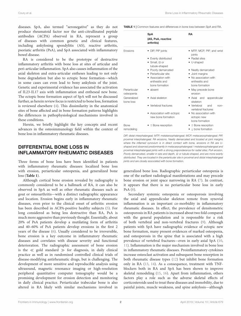
Three forms of bone loss have been identified in patientswith inflammatory rheumatic diseases: localized bone losswith erosion, periarticular osteopenia, and generalized boneloss (Table 1).
Although cortical bone erosion revealed by radiography iscommonly considered to be a hallmark of RA, it can also beobserved in SpA as well as other rheumatic diseases such asgout or osteoarthritis—with a distinct radiographic appearanceand location. Erosion begins early in inflammatory rheumaticdiseases, even prior to the clinical onset of arthritis: erosionhas been described in ACPA-positive healthy subjects (5). Forlong considered as being less destructive than RA, PsA ismuchmore aggressive than previously thought. Essentially, about20% of PsA patients develop a mutilating form of arthritisand 40–60% of PsA patients develop erosions in the first 2years of the disease (6). Usually considered to be irreversible,bone erosion is a key outcome in inflammatory rheumaticdiseases and correlates with disease severity and functionaldeterioration. The radiographic assessment of bone erosionis the ≪ gold standard ≫ for diagnosis, in daily clinicalpractice as well as in randomized controlled clinical trials ofdisease-modifying antirheumatic drugs, but is challenging. Thedevelopment of more sensitive and reproducible analysis usingultrasound, magnetic resonance imaging or high-resolutionperipheral quantitative computer tomography would be apromising development for erosion detection and monitoringin daily clinical practice. Periarticular trabecular bone is alsoaltered in RA likely with similar mechanisms involved in
TABLE 1 | Common features and differences in bone loss between SpA and RA.
SpA RA
(AS, PsA, reactive
arthritis)
Erosions • DIP, PIP joints
• Evenly distributed
• Small, or
tubule-shaped
• Poorly demarcated
• Periarticular site
• Association with
enthesitis and
bone formation
• MTP, MCP, PIP, and wrist
joints
• Radial sites
• U-shaped
• Neatly demarcated
• Joint margins
• No association with
enthesitis and
bone formation
Periarticular
osteopenia
• absent • May precede bone
erosion
Generalized
bone loss
• Axial skeleton
• Vertebral fractures
• Association with ectopic
new bone formation
• Axial and appendicular
skeleton
• Vertebral and non-
vertebral fractures
• No association with
ectopic new
bone formation
Bone
remodeling
• ↑ Bone resorption • ↑ Bone resorption
• ↓ bone formation
DIP, distal interphalangeal; MTP, metatarsophalangeal; MCP, metacarpophalangeal; PIP,
proximal interphalangeal. RA erosions, Neatly demarcated and located at joint margins
where the inflamed synovium is in direct contact with bone, erosions in RA are U-
shaped and observed predominantly in metacarpophalangeal / metatarsophalangeal and
proximal interphalangeal joints with a strong preponderance for radial sites; PsA erosion,
Poorly demarcated, smaller in size and depth, Ω or tubule-shaped, and are more evenly
distributed. They are located in the periarticular site in proximal and distal interphalangeal
joints and are closely associated with bone formation.
generalized bone loss. Radiographic periarticular osteopenia isone of the earliest radiological manifestations and may precedebone erosion or joint space narrowing in RA (7). In contrast,it appears that there is no periarticular bone loss in earlyPsA (8).
Secondary systemic osteopenia or osteoporosis involvingthe axial and appendicular skeleton remote from synovialinflammation is an important co-morbidity in inflammatoryrheumatic diseases. In effect, the prevalence of densitometricosteoporosis in RA patients is increased about two fold comparedwith the general population and is responsible for a riskof both vertebral and non-vertebral fractures (9). Althoughpatients with SpA have radiographic evidence of ectopic newbone formation, many present evidences of marked osteopenia,and osteoporosis in the spine that is associated with a highprevalence of vertebral fractures—even in early axial SpA (10,11). Inflammation is the major mechanism involved in bone lossin inflammatory rheumatic diseases. Proinflammatory cytokinesincrease osteoclast activation and subsequent bone resorption inboth rheumatic disease types (12) but inhibit bone formationonly in RA (13, 14). As a consequence, treatment with TNF-blockers both in RA and SpA has been shown to improveskeletal remodeling (15, 16). Apart from inflammation, othersfactors play a role such as the adverse skeletal effects ofcorticosteroids used to treat these diseases and immobility, due topainful joints, muscle weakness, and spine ankylosis—although
Frontiers in Immunology | www.frontiersin.org 2 April 2019 | Volume 10 | Article 679
Coury et al. Bone Loss in Inflammatory Rheumatic Diseases
bone loss is observed well-before the development of spinalimmobility (17–19).
OSTEOCLAST DIFFERENTIATION ANDFUNCTION IN INFLAMMATORYRHEUMATIC DISEASES
Osteoclasts are responsible for bone erosion and have beenidentified at sites of focal erosion at the pannus-bone interfaceboth in RA patients (20, 21) and animal models of arthritis(22–26). This role was definitively demonstrated by osteoclast-deficient mouse models of arthritis which were shown tobe fully protected from bone erosion (25, 26). Osteoclastsare multinucleated bone resorbing cells which originatefrom the fusion of mononucleated cells belonging to themyeloid lineage in the presence of macrophage colony-stimulating factor (M-CSF) and Receptor Activator ofNuclear factor-κB Ligand (RANKL). Osteoclast formationis governed by a regulatory triad, the receptor activator ofNF-κB (RANK), its ligand RANKL and a decoy receptorosteoprotegerin (OPG) also known as osteoclastogenesisinhibitory factor. OPG binds to RANKL hampering RANK-RANKL interaction, though RANKL/OPG ratio determinesosteoclast number, lifespan and activity. Activation of RANKon mononuclear osteoclast precursors initiates a transcriptionalcascade culminating in osteoclast differentiation. Interestingly,transcription factors important for osteoclast differentiationare key regulators of immune responses—such as NF-κB andnuclear factor of activated T cells cytoplasmic 1 (NFATc1).RANKL signaling in osteoclasts is strengthened by thesynergistic activation of Immunoreceptor tyrosine-basedactivation motif (ITAM)-containing proteins, DNAX-activating protein of 12 kDa (DAP12) and Fc gamma receptor(FcRγ) (27, 28).
RANKL expression is high in synovial tissue from RA, PsA,and SpA peripheral joint disease patients (29–32). Treatmentwith non-biologic disease-modifying anti-rheumatic drugs(DMARDs) or glucocorticoids decreases the RANKL/OPG ratioin RA synovium and is satisfyingly associated with improvedradiographic scores (17, 33). In addition, pharmacologicalinhibition of osteoclasts either by bisphosphonate zolendronicacid, or denosumab, a RANKL-specific monoclonal blockingantibody, also demonstrated some efficacy in impairingthe progression of bone erosion in both arthritic mice andRA patients (34–38). However, these anti-resorptive drugstargeting osteoclasts are inadequate because they also alterphysiological bone remodeling, necessitating the discovery ofnew targets.
ROLE OF T CELLSIN OSTEOCLASTOGENESIS
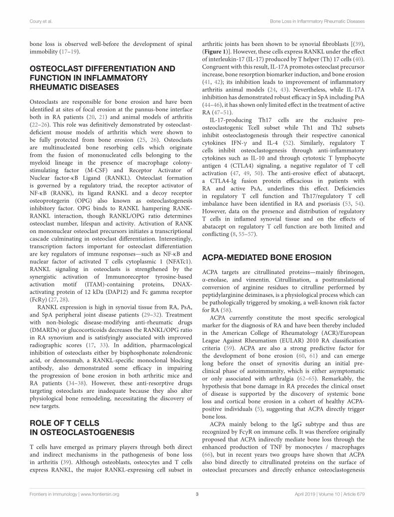
T cells have emerged as primary players through both directand indirect mechanisms in the pathogenesis of bone lossin arthritis (39). Although osteoblasts, osteocytes and T cellsexpress RANKL, the major RANKL-expressing cell subset in
arthritic joints has been shown to be synovial fibroblasts [(39),(Figure 1)]. However, these cells express RANKL under the effectof interleukin-17 (IL-17) produced by T helper (Th) 17 cells (40).Congruent with this result, IL-17A promotes osteoclast precursorincrease, bone resorption biomarker induction, and bone erosion(41, 42); its inhibition leads to improvement of inflammatoryarthritis animal models (24, 43). Nevertheless, while IL-17Ainhibition has demonstrated robust efficacy in SpA including PsA(44–46), it has shown only limited effect in the treatment of activeRA (47–51).
IL-17-producing Th17 cells are the exclusive pro-osteoclastogenic Tcell subset while Th1 and Th2 subsetsinhibit osteoclastogenesis through their respective canonicalcytokines IFN-γ and IL-4 (52). Similarly, regulatory Tcells inhibit osteoclastogenesis through anti-inflammatorycytokines such as IL-10 and through cytotoxic T lymphocyteantigen 4 (CTLA4) signaling, a negative regulator of T cellactivation (47, 49, 50). The anti-erosive effect of abatacept,a CTLA4-Ig fusion protein efficacious in patients withRA and active PsA, underlines this effect. Deficienciesin regulatory T cell function and Th17/regulatory T cellimbalance have been identified in RA and psoriasis (53, 54).However, data on the presence and distribution of regulatoryT cells in inflamed synovial tissue and on the effects ofabatacept on regulatory T cell function are both limited andconflicting (8, 55–57).
ACPA-MEDIATED BONE EROSION
ACPA targets are citrullinated proteins—mainly fibrinogen,α-enolase, and vimentin. Citrullination, a posttranslationalconversion of arginine residues to citrulline performed bypeptidylarginine deiminases, is a physiological process which canbe pathologically triggered by smoking, a well-known risk factorfor RA (58).
ACPA currently constitute the most specific serologicalmarker for the diagnosis of RA and have been thereby includedin the American College of Rheumatology (ACR)/EuropeanLeague Against Rheumatism (EULAR) 2010 RA classificationcriteria (59). ACPA are also a strong predictive factor forthe development of bone erosion (60, 61) and can emergelong before the onset of synovitis during an initial pre-clinical phase of autoimmunity, which is either asymptomaticor only associated with arthralgia (62–65). Remarkably, thehypothesis that bone damage in RA precedes the clinical onsetof disease is supported by the discovery of systemic boneloss and cortical bone erosion in a cohort of healthy ACPA-positive individuals (5), suggesting that ACPA directly triggerbone loss.
ACPA mainly belong to the IgG subtype and thus arerecognized by FcγR on immune cells. It was therefore originallyproposed that ACPA indirectly mediate bone loss through theenhanced production of TNF by monocytes / macrophages(66), but in recent years two groups have shown that ACPAalso bind directly to citrullinated proteins on the surface ofosteoclast precursors and directly enhance osteoclastogenesis
Frontiers in Immunology | www.frontiersin.org 3 April 2019 | Volume 10 | Article 679
Coury et al. Bone Loss in Inflammatory Rheumatic Diseases
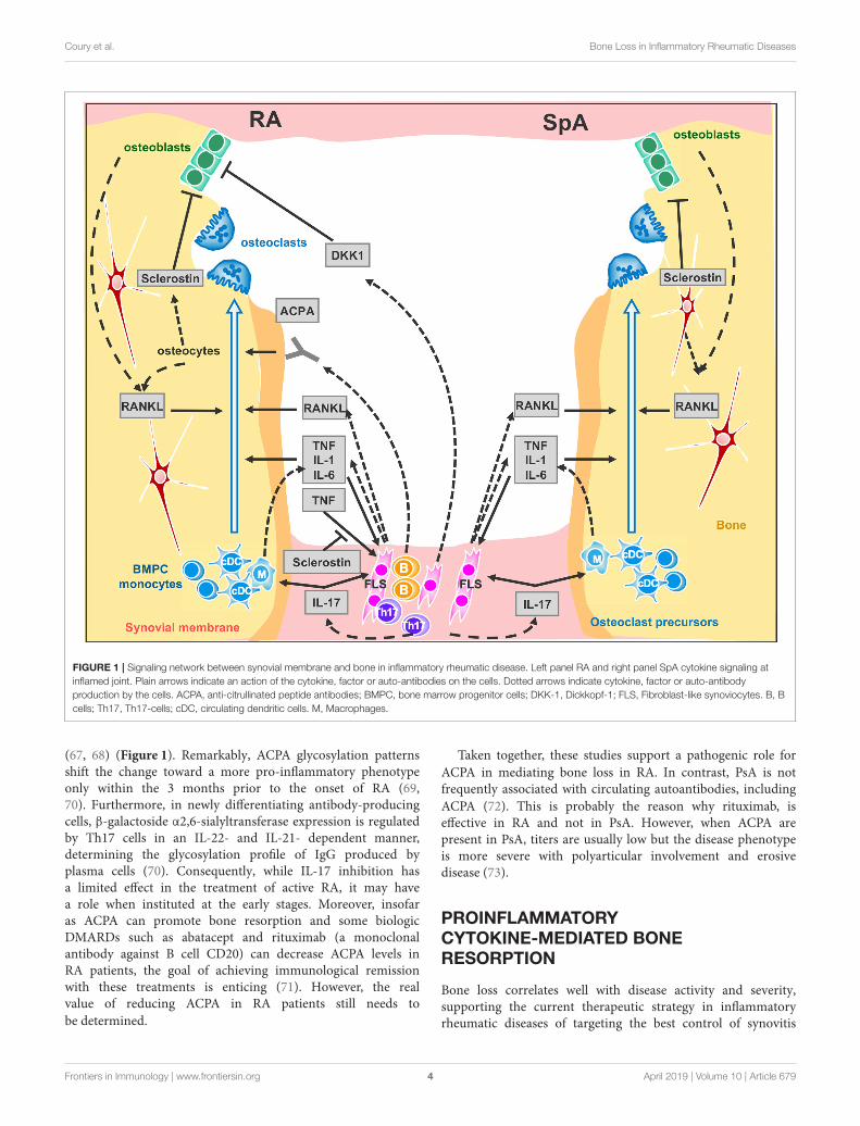
FIGURE 1 | Signaling network between synovial membrane and bone in inflammatory rheumatic disease. Left panel RA and right panel SpA cytokine signaling at
inflamed joint. Plain arrows indicate an action of the cytokine, factor or auto-antibodies on the cells. Dotted arrows indicate cytokine, factor or auto-antibody
production by the cells. ACPA, anti-citrullinated peptide antibodies; BMPC, bone marrow progenitor cells; DKK-1, Dickkopf-1; FLS, Fibroblast-like synoviocytes. B, B
cells; Th17, Th17-cells; cDC, circulating dendritic cells. M, Macrophages.
(67, 68) (Figure 1). Remarkably, ACPA glycosylation patternsshift the change toward a more pro-inflammatory phenotypeonly within the 3 months prior to the onset of RA (69,70). Furthermore, in newly differentiating antibody-producingcells, β-galactoside α2,6-sialyltransferase expression is regulatedby Th17 cells in an IL-22- and IL-21- dependent manner,determining the glycosylation profile of IgG produced byplasma cells (70). Consequently, while IL-17 inhibition hasa limited effect in the treatment of active RA, it may havea role when instituted at the early stages. Moreover, insofaras ACPA can promote bone resorption and some biologicDMARDs such as abatacept and rituximab (a monoclonalantibody against B cell CD20) can decrease ACPA levels inRA patients, the goal of achieving immunological remissionwith these treatments is enticing (71). However, the realvalue of reducing ACPA in RA patients still needs to
be determined.
Taken together, these studies support a pathogenic role for
ACPA in mediating bone loss in RA. In contrast, PsA is notfrequently associated with circulating autoantibodies, including
ACPA (72). This is probably the reason why rituximab, iseffective in RA and not in PsA. However, when ACPA arepresent in PsA, titers are usually low but the disease phenotypeis more severe with polyarticular involvement and erosivedisease (73).
PROINFLAMMATORYCYTOKINE-MEDIATED BONERESORPTION
Bone loss correlates well with disease activity and severity,supporting the current therapeutic strategy in inflammatoryrheumatic diseases of targeting the best control of synovitis
Frontiers in Immunology | www.frontiersin.org 4 April 2019 | Volume 10 | Article 679
Coury et al. Bone Loss in Inflammatory Rheumatic Diseases
and the biological inflammatory syndrome. Indeed, conventionalDMARDs, such as methotrexate, enable protection from boneerosion simply by their ability to reduce synovitis (74). However,some RA patients in sustained clinical remission or low diseaseactivity still continue to accrue bone erosions (38, 75), likelybecause of subclinical synovial inflammation (76). This evolutionis probably similar in SpA, but it has not yet been clearlydemonstrated in the absence of well-defined remission criteria.
TNF overexpression is sufficient to induce arthritis in mice(77). TNF operates by several mechanisms: it promotes boneresorption indirectly in conjunction with IL-6 by up-regulatingRANKL expression in synovial fibroblasts (78, 79) and directlyby aiding the differentiation of osteoclasts from mononuclearprecursors in synovial tissues in synergy with RANKL (80)(Figure 1). Recent evidence suggests that combinations ofcytokines, such as TNF plus IL-6, may drive RANK/RANKL-independent osteoclast formation (81) but this process still needsconfirmation using other models. TNF also expands the pool ofosteoclast precursor cells (82). Additionally, IL-1 is a mediator ofTNF-induced osteoclastogenesis (83) while IL-6 is an importantfactor for Th17 differentiation. Accordingly, clinical trials—onlyin RA—with TNF blockers (16) and the Il-6 receptor blockade(84), have confirmed the impact of pro-inflammatory cytokineson osteoclastogenesis as they can retard or arrest the occurrenceof bone erosion. As for the IL-1 blockade, despite having alimited effect on swelling, it protects from bone erosion inRA (85).
OSTEOFORMATION ANDEROSION REPAIR
In RA only, the inflammatorymilieu also impairs bone formationand erosion repair. TNF is the instrumental cytokine thatunbalances bone homeostasis, blocking osteoblast differentiationand maturation through Wingless (Wnt) ligand signaling (86).Bone formation is governed by Wnt pathways which arecritical for the osteoblast transcriptional differentiation programthrough the canonical β catenin-dependent activation. The Wntligands interact with the membrane-bound co-receptor frizzledand the low–density lipoprotein receptor-related proteins LRP-5 or LRP-6. This activated receptor complex stabilizes β catenintranscription factor, allowing its translocation to the nucleus todirectly coactivate Runx2 and OPG (87, 88). In inflammatoryrheumatic diseases, bone erosion repair is scarcely observed, evenunder biologic therapies such as TNF or IL-6 receptor blockers,and manifests only as apposition of new bone (sclerosis) at thebase of the erosion (89, 90). Paradoxically, analysis of histologicalsections of arthritic samples, either from humans or frommurinemodels, has shown the presence of osteoblast lineage cells closeto the eroded bone once inflammation resolves (21, 91). Inaddition, intermittent parathyroid hormone (PTH) treatment -an anabolic agent for bone- used for treatment of osteoporosis,fails to reduce erosion volume in patients with established RAwith disease activity controlled by TNF blockers (92). By contrastto humans, treatment of hTNFtg mice with a combined therapyconsisting of anti-TNF together with intermittent PTH led to
regression of local bone erosion and bone repair, demonstratingnew bone formation (93). An alternative to anabolic treatmentaiming at increasing bone formation and repair, is to block boneformation antagonists. Indeed, Wnt pro-osteogenic function iscontrolled and tempered by several physiological antagonists:Dickkopf proteins (DKK-1 and 2), soluble frizzled-relatedproteins (sFRPs) (94, 95) and sclerostin that—in the presenceof Wnt ligands—antagonizes LRP-6 internalization (96, 97).In RA, TNF lessens osteoformation by up-regulating DKK-1expression, for instance DKK-1 level is found to be elevatedin RA patients’ sera and in hTNFtg mice, CIA, and GPI-induced arthritis mice, (98, 99). In hTNFtg mice only, DKK-1 inhibition is able to prevent bone erosion and to promotebone formation, generating osteophytes around inflamed joints(99). Soluble frizzled-related proteins sFRP1 and sFRP2 areWnt antagonist that sequestrate Wnt ligands, preventing themto activate frizzle/Lpr5 receptors, were also found elevated insynovial fluids of KBxN serum transfert inflammation inducedmice model (91). Among the Wnt ligand antagonists, sclerostinis an attractive therapeutic target for bone loss pathologies.Sclerostin-neutralizing antibodies have been shown to havestrong bone-building effects in mice, rats, monkeys, and humans(97–101). This treatment prevents the decrease of bone mineraldensity and bone volume at axial and appendicular sites inCollagen-Induced Arthritis mice but does not protect fromerosion on the periarticular bone and fails to repair focalerosions (102). On the other hand, in hTNFtg mice, TNFinduced sclerostin expression in inflammatory synoviocytes,unexpectedly, the absence of sclerostin in hTNFtg/ Sost−/− mice,instead of reversing the inflammatory bone destruction, elicitedexacerbation of the disease. These observations suggest thatsclerostin may be involved in regulating other pathways besidesWnt signaling or has an anti-osteoclastogenic effect in TNF-dependent chronic arthritis (103). In line with this paradigm ofuncovered sclerostin functions, recent findings surprisingly showthat overexpressing sclerostin in murine skeletal stem cells formsovergrown bones when engrafted. This observation indicates thatsclerostin could have an osteoforming effect on skeletal stem cells(104). Moreover, a recent study using non-inflammatory boneloss mouse models, unveiled a compensatory mechanism leadingto increased expression of sclerostin when DKK-1 is inhibited. Itwould therefore perhaps be prudent before embarking upon anti-sclerostin treatments for RA, to conduct further studies in animalmodels of RA using Sost tissue-specific ablation to help obtain abetter understanding of the precise role of sclerostin in chronicinflammatory diseases.
In contrast to RA, bone formation is observed in SpAat entheseal sites, resulting in endochondral bone formations.IL32γ, among others pro-inflammatory cytokines, is foundelevated in SpA synovial fluid, it is proposed that IL32γ enhancesosteoblast differentiation via DKK-1 suppression, thereafterpromoting abnormal bone formation (105). Indeed, lower levelsof DKK-1 in AS and PsA patients and sclerostin in ASpatients have been reported, potentially explaining the non-impediment of osteoblast activity (99, 106, 107). In conflictwith the above report, a recent meta-analysis showed nosignificant difference in sclerostin serum levels in AS and RA
Frontiers in Immunology | www.frontiersin.org 5 April 2019 | Volume 10 | Article 679
Coury et al. Bone Loss in Inflammatory Rheumatic Diseases
patients vs. healthy controls which suggests that sclerostinmay not be associated with the pathogenesis of AS andRA (108). Last, a recent and challenging study revealed thatvesicular RANK produced by mature osteoclasts stimulateearly osteoblast differentiation through osteoblastic RANKLreverse signaling (109). Consequently, the development ofa biological compound to trigger RANKL reverse signalingin osteoblast would be a new promising lead to promotebone formation.
CONCLUSIONS
In inflammatory rheumatic diseases, systemic and local boneloss constitute a common key outcome in terms of functionalcapacity and reflects the tight interaction between the immunesystem and bone, leading to an increase in osteoclast activityand a consequent uncoupling of bone resorption from formation.Once established, bone erosions are at present, still irreversible. It
is to be hoped that a better future understanding of the molecularpathways involved in bone loss and bone formation—particularlyin the context of inflammation—will enable the development ofnew therapies that can selectively and directly halt, or even repair,bone erosion.
AUTHOR CONTRIBUTIONS
All authors listed have made a substantial, direct andintellectual contribution to the work, and approved itfor publication.
FUNDING
This work was supported by grants from INSERM andthe University Claude Bernard Lyon-1 (OP), the ComitéDépartemental de la Loire de la Ligue Contre le Cancer (OP), theANR grant LYSBONE (OP) (Grant n. ANR-15-CE14-0010-01).
REFERENCES
1. Horton JE, Raisz LG, Simmons HA, Oppenheim JJ, Mergenhagen SE. Bone
resorbing activity in supernatant fluid from cultured human peripheral blood
leukocytes. Science. (1972) 177:793–5.
2. Mundy GR, Raisz LG, Cooper RA, Schechter GP, Salmon SE. Evidence for
the secretion of an osteoclast stimulating factor in myeloma. N Engl J Med.
(1974) 291:1041–6. doi: 10.1056/NEJM197411142912001
3. Arron JR, Choi Y. Bone versus immune system. Nature. (2000) 408:535–6.
doi: 10.1038/35046196
4. Gravallese EM, Schett G. Effects of the IL-23-IL-17 pathway on
bone in spondyloarthritis. Nat Rev Rheumatol. (2018) 14:631–40.
doi: 10.1038/s41584-018-0091-8
5. Kleyer A, Finzel S, Rech J, Manger B, Krieter M, Faustini F, et al. Bone
loss before the clinical onset of rheumatoid arthritis in subjects with
anticitrullinated protein antibodies. Ann Rheum Dis. (2014) 73:854–60.
doi: 10.1136/annrheumdis-2012-202958
6. Gladman DD, Antoni C, Mease P, Clegg DO, Nash P. Psoriatic arthritis:
epidemiology, clinical features, course, and outcome.Ann RheumDis. (2005)
64 Suppl 2:ii14–7. doi: 10.1136/ard.2004.032482
7. Brower AC. Use of the radiograph to measure the course of rheumatoid
arthritis. The gold standard versus fool’s gold. Arthritis Rheum.
(1990) 33:316–24.
8. Szentpetery A, Heffernan E, Haroon M, Kilbane M, Gallagher P, McKenna
MJ, et al. Striking difference of periarticular bone density change in
early psoriatic arthritis and rheumatoid arthritis following anti-rheumatic
treatment as measured by digital X-ray radiogrammetry. Rheumatology.
(2016) 55:891–6. doi: 10.1093/rheumatology/kev443
9. Haugeberg G, Uhlig T, Falch JA, Halse JI, Kvien TK. Bone mineral
density and frequency of osteoporosis in female patients with
rheumatoid arthritis: results from 394 patients in the Oslo County
Rheumatoid Arthritis register. Arthritis Rheum. (2000) 43:522–30.
doi: 10.1002/1529-0131(200003)43:3<522::AID-ANR7>3.0.CO;2-Y
10. van derWeijdenMA, van Denderen JC, LemsWF, HeymansMW, Dijkmans
BA, van der Horst-Bruinsma IE. Low bone mineral density is related to male
gender and decreased functional capacity in early spondylarthropathies. Clin
Rheumatol. (2011) 30:497–503. doi: 10.1007/s10067-010-1538-8
11. van der Weijden MA, van der Horst-Bruinsma IE, van Denderen JC,
Dijkmans BA, Heymans MW, Lems WF. High frequency of vertebral
fractures in early spondylarthropathies. Osteoporos Int. (2012) 23:1683–90.
doi: 10.1007/s00198-011-1766-z
12. Redlich K, Smolen JS. Inflammatory bone loss: pathogenesis and therapeutic
intervention. Nat Rev Drug Discov. (2012) 11:234–50. doi: 10.1038/nrd3669
13. Bultink IE, Vis M, van der Horst-Bruinsma IE, Lems WF. Inflammatory
rheumatic disorders and bone. Curr Rheumatol Rep. (2012) 14:224–30.
doi: 10.1007/s11926-012-0252-8
14. Gao Y, Grassi F, Ryan MR, Terauchi M, Page K, Yang X, et al. IFN-gamma
stimulates osteoclast formation and bone loss in vivo via antigen-driven T
cell activation. J Clin Invest. (2007) 117:122–32. doi: 10.1172/JCI30074
15. Allali F, Breban M, Porcher R, Maillefert JF, Dougados M, Roux C. Increase
in bone mineral density of patients with spondyloarthropathy treated
with anti-tumour necrosis factor alpha. Ann Rheum Dis. (2003) 62:347–9.
doi: 10.1136/ard.62.4.347
16. Vis M, Havaardsholm EA, Haugeberg G, Uhlig T, Voskuyl AE, van de
Stadt RJ, et al. Evaluation of bone mineral density, bone metabolism,
osteoprotegerin and receptor activator of the NFkappaB ligand serum levels
during treatment with infliximab in patients with rheumatoid arthritis. Ann
Rheum Dis. (2006) 65:1495–9. doi: 10.1136/ard.2005.044198
17. Hartmann K, Koenen M, Schauer S, Wittig-Blaich S, Ahmad M, Baschant
U, et al. Molecular actions of glucocorticoids in cartilage and bone
during health, disease, and steroid therapy. Physiol Rev. (2016) 96:409–47.
doi: 10.1152/physrev.00011.2015
18. Mandl P, Kainberger F, Friberg Hitz M. Imaging in osteoporosis in
rheumatic diseases. Best Pract Res Clin Rheumatol. (2016) 30:751–65.
doi: 10.1016/j.berh.2016.08.010
19. Boling EP. Secondary osteoporosis: underlying disease and the risk for
glucocorticoid-induced osteoporosis. Clin Ther. (2004) 26:1–14.
20. Bromley M, Woolley DE. Chondroclasts and osteoclasts at subchondral sites
of erosion in the rheumatoid joint. Arthritis Rheum. (1984) 27:968–75.
21. Gravallese EM, Harada Y, Wang JT, Gorn AH, Thornhill TS, Goldring SR.
Identification of cell types responsible for bone resorption in rheumatoid
arthritis and juvenile rheumatoid arthritis. Am J Pathol. (1998) 152:943–51.
22. Kong YY, Feige U, Sarosi I, Bolon B, Tafuri A, Morony S, et al. Activated
T cells regulate bone loss and joint destruction in adjuvant arthritis
through osteoprotegerin ligand. Nature. (1999) 402:304–9. doi: 10.1038/
46303
23. Romas E, Sims NA, Hards DK, Lindsay M, Quinn JW, Ryan PF,
et al. Osteoprotegerin reduces osteoclast numbers and prevents bone
erosion in collagen-induced arthritis. Am J Pathol. (2002) 161:1419–27.
doi: 10.1016/S0002-9440(10)64417-3
24. Lubberts E, Koenders MI, Oppers-Walgreen B, van den Bersselaar L,
Coenen-de Roo CJ, Joosten LA, et al. Treatment with a neutralizing
anti-murine interleukin-17 antibody after the onset of collagen-
induced arthritis reduces joint inflammation, cartilage destruction,
and bone erosion. Arthritis Rheum. (2004) 50:650–9. doi: 10.1002/art.
20001
Frontiers in Immunology | www.frontiersin.org 6 April 2019 | Volume 10 | Article 679
Coury et al. Bone Loss in Inflammatory Rheumatic Diseases
25. Pettit AR, Ji H, von Stechow D, Muller R, Goldring SR, Choi Y, et al.
TRANCE/RANKL knockout mice are protected from bone erosion in
a serum transfer model of arthritis. Am J Pathol. (2001) 159:1689–99.
doi: 10.1016/S0002-9440(10)63016-7
26. Redlich K, Hayer S, Ricci R, David JP, Tohidast-Akrad M, Kollias G, et al.
Osteoclasts are essential for TNF-alpha-mediated joint destruction. J Clin
Invest. (2002) 110:1419-27. doi: 10.1172/JCI15582
27. Mocsai A, Humphrey MB, Van Ziffle JA, Hu Y, Burghardt A, Spusta SC,
et al. The immunomodulatory adapter proteins DAP12 and Fc receptor
gamma-chain (FcRgamma) regulate development of functional osteoclasts
through the Syk tyrosine kinase. Proc Natl Acad Sci USA. (2004) 101:6158–63.
doi: 10.1073/pnas.0401602101
28. Koga T, Inui M, Inoue K, Kim S, Suematsu A, Kobayashi E, et al.
Costimulatory signals mediated by the ITAM motif cooperate with RANKL
for bone homeostasis. Nature. (2004) 428:758–63. doi: 10.1038/nature02444
29. Crotti TN, Smith MD, Weedon H, Ahern MJ, Findlay DM, Kraan M, et al.
Receptor activator NF-kappaB ligand (RANKL) expression in synovial tissue
from patients with rheumatoid arthritis, spondyloarthropathy, osteoarthritis,
and from normal patients: semiquantitative and quantitative analysis. Ann
Rheum Dis. (2002) 61:1047–54. doi: 10.1136/ard.61.12.1047
30. Ritchlin CT, Haas-Smith SA, Li P, Hicks DG, Schwarz EM. Mechanisms of
TNF-alpha- and RANKL-mediated osteoclastogenesis and bone resorption
in psoriatic arthritis. J Clin Invest. (2003) 111:821–31. doi: 10.1172/JCI16069
31. Haynes DR, Barg E, Crotti TN, Holding C, Weedon H, Atkins GJ, et al.
Osteoprotegerin expression in synovial tissue from patients with rheumatoid
arthritis, spondyloarthropathies and osteoarthritis and normal controls.
Rheumatology. (2003) 42:123–34. doi: 10.1093/rheumatology/keg047
32. van Tuyl LH, Voskuyl AE, Boers M, Geusens P, Landewe RB, Dijkmans
BA, et al. Baseline RANKL:OPG ratio and markers of bone and
cartilage degradation predict annual radiological progression over 11
years in rheumatoid arthritis. Ann Rheum Dis. (2010) 69:1623–8.
doi: 10.1136/ard.2009.121764
33. Haynes D, Crotti T, Weedon H, Slavotinek J, Au V, Coleman M, et al.
Modulation of RANKL and osteoprotegerin expression in synovial tissue
from patients with rheumatoid arthritis in response to disease-modifying
antirheumatic drug treatment and correlation with radiologic outcome.
Arthritis Rheum. (2008) 59:911–20. doi: 10.1002/art.23818
34. Jarrett SJ, Conaghan PG, Sloan VS, Papanastasiou P, Ortmann CE,
O’Connor PJ, et al. Preliminary evidence for a structural benefit of the
new bisphosphonate zoledronic acid in early rheumatoid arthritis. Arthritis
Rheum. (2006) 54:1410–4. doi: 10.1002/art.21824
35. Herrak P, Gortz B, Hayer S, Redlich K, Reiter E, Gasser J, et al.
Zoledronic acid protects against local and systemic bone loss in tumor
necrosis factor-mediated arthritis. Arthritis Rheum. (2004) 50:2327–37.
doi: 10.1002/art.20384
36. Sims NA, Green JR, Glatt M, Schlict S, Martin TJ, Gillespie MT, et al.
Targeting osteoclasts with zoledronic acid prevents bone destruction
in collagen-induced arthritis. Arthritis Rheum. (2004) 50:2338–46.
doi: 10.1002/art.20382
37. Deodhar A, Dore RK, Mandel D, Schechtman J, Shergy W, Trapp R, et al.
Denosumab-mediated increase in hand bone mineral density associated
with decreased progression of bone erosion in rheumatoid arthritis patients.
Arthritis Care Res. (2010) 62:569–74. doi: 10.1002/acr.20004
38. Cohen G, Gossec L, Dougados M, Cantagrel A, Goupille P, Daures JP, et al.
Radiological damage in patients with rheumatoid arthritis on sustained
remission. Ann Rheum Dis. (2007) 66:358–63. doi: 10.1136/ard.2006.057497
39. Danks L, Komatsu N, Guerrini MM, Sawa S, Armaka M, Kollias G, et al.
RANKL expressed on synovial fibroblasts is primarily responsible for bone
erosions during joint inflammation. Ann Rheum Dis. (2016) 75:1187–95.
doi: 10.1136/annrheumdis-2014-207137
40. Kotake S, Udagawa N, Hakoda M, Mogi M, Yano K, Tsuda E, et al.
Activated human T cells directly induce osteoclastogenesis from
human monocytes: possible role of T cells in bone destruction in
rheumatoid arthritis patients. Arthritis Rheum. (2001) 44:1003–12.
doi: 10.1002/1529-0131(200105)44:5<1003::AID-ANR179>3.0.CO;2
41. Lubberts E, Oppers-Walgreen B, Pettit AR, Van Den Bersselaar L, Joosten
LA, Goldring SR, et al. Increase in expression of receptor activator of
nuclear factor kappaB at sites of bone erosion correlates with progression of
inflammation in evolving collagen-induced arthritis.Arthritis Rheum. (2002)
46:3055–64. doi: 10.1002/art.10607
42. Adamopoulos IE, Suzuki E, Chao CC, Gorman D, Adda S, Maverakis E,
et al. IL-17A gene transfer induces bone loss and epidermal hyperplasia
associated with psoriatic arthritis. Ann Rheum Dis. (2015) 74:1284–92.
doi: 10.1136/annrheumdis-2013-204782
43. Bush KA, Farmer KM, Walker JS, Kirkham BW. Reduction of joint
inflammation and bone erosion in rat adjuvant arthritis by treatment with
interleukin-17 receptor IgG1 Fc fusion protein. Arthritis Rheum. (2002)
46:802–5. doi: 10.1002/art.10173
44. McInnes IB, Mease PJ, Kirkham B, Kavanaugh A, Ritchlin CT, Rahman P,
et al. Secukinumab, a human anti-interleukin-17A monoclonal antibody,
in patients with psoriatic arthritis (FUTURE 2): a randomised, double-
blind, placebo-controlled, phase 3 trial. Lancet. (2015) 386:1137–46.
doi: 10.1016/S0140-6736(15)61134-5
45. Mease PJ, McInnes IB, Kirkham B, Kavanaugh A, Rahman P, van der Heijde
D, et al. Secukinumab Inhibition of Interleukin-17A in Patients with Psoriatic
Arthritis. N Engl J Med. (2015) 373:1329–39. doi: 10.1056/NEJMoa14
12679
46. van der Heijde D, Gladman DD, Kishimoto M, Okada M, Rathmann
SS, Moriarty SR, et al. Efficacy and Safety of Ixekizumab in Patients
with Active Psoriatic Arthritis: 52-week Results from a Phase III
Study (SPIRIT-P1). J Rheumatol. (2018) 45:367–77doi: 10.3899/jrheum.
170429
47. Hueber W, Patel DD, Dryja T, Wright AM, Koroleva I, Bruin G,
et al. Effects of AIN457, a fully human antibody to interleukin-17A, on
psoriasis, rheumatoid arthritis, and uveitis. Sci Transl Med. (2010) 2:52ra72.
doi: 10.1126/scitranslmed.3001107
48. Genovese MC, Durez P, Richards HB, Supronik J, Dokoupilova E,
Mazurov V, et al. Efficacy and safety of secukinumab in patients
with rheumatoid arthritis: a phase II, dose-finding, double-blind,
randomised, placebo controlled study. Ann Rheum Dis. (2013) 72:863–9.
doi: 10.1136/annrheumdis-2012-201601
49. Genovese MC, Fleischmann R, Furst D, Janssen N, Carter J, Dasgupta
B, et al. Efficacy and safety of olokizumab in patients with rheumatoid
arthritis with an inadequate response to TNF inhibitor therapy: outcomes
of a randomised Phase IIb study. Ann Rheum Dis. (2014) 73:1607–15.
doi: 10.1136/annrheumdis-2013-204760
50. Tlustochowicz W, Rahman P, Seriolo B, Krammer G, Porter B, Widmer
A, et al. Efficacy and safety of subcutaneous and intravenous loading
dose regimens of secukinumab in patients with active rheumatoid arthritis:
results from a randomized phase II study. J Rheumatol. (2016) 43:495–503.
doi: 10.3899/jrheum.150117
51. Blanco FJ, Moricke R, Dokoupilova E, Codding C, Neal J, AnderssonM, et al.
Secukinumab in active rheumatoid arthritis: a phase III randomized, double-
blind, active comparator- and placebo-controlled study.Arthritis Rheumatol.
(2017) 69:1144–53. doi: 10.1002/art.40070
52. Sato K, Suematsu A, Okamoto K, Yamaguchi A, Morishita Y, Kadono Y,
et al. Th17 functions as an osteoclastogenic helper T cell subset that links
T cell activation and bone destruction. J Exp Med. (2006) 203:2673–82.
doi: 10.1084/jem.20061775
53. Flores-Borja F, Jury EC, Mauri C, Ehrenstein MR. Defects in
CTLA-4 are associated with abnormal regulatory T cell function in
rheumatoid arthritis. Proc Natl Acad Sci USA. (2008) 105:19396–401.
doi: 10.1073/pnas.0806855105
54. Sugiyama H, Gyulai R, Toichi E, Garaczi E, Shimada S, Stevens
SR, et al. Dysfunctional blood and target tissue CD4+CD25high
regulatory T cells in psoriasis: mechanism underlying unrestrained
pathogenic effector T cell proliferation. J Immunol. (2005) 174:164–73.
doi: 10.4049/jimmunol.174.1.164
55. Cao D, van Vollenhoven R, Klareskog L, Trollmo C, Malmstrom V.
CD25brightCD4+ regulatory T cells are enriched in inflamed joints of
patients with chronic rheumatic disease. Arthritis Res Ther. (2004) 6:R335–
46. doi: 10.1186/ar1192
56. Bonelli M, Goschl L, Bluml S, Karonitsch T, Hirahara K, Ferner E, et al.
Abatacept (CTLA-4Ig) treatment reduces T cell apoptosis and regulatory T
cell suppression in patients with rheumatoid arthritis. Rheumatology. (2016)
55:710–20. doi: 10.1093/rheumatology/kev403
Frontiers in Immunology | www.frontiersin.org 7 April 2019 | Volume 10 | Article 679
Coury et al. Bone Loss in Inflammatory Rheumatic Diseases
57. Alvarez-Quiroga C, Abud-Mendoza C, Doniz-Padilla L, Juarez-
Reyes A, Monsivais-Urenda A, Baranda L, et al. CTLA-4-Ig therapy
diminishes the frequency but enhances the function of Treg cells in
patients with rheumatoid arthritis. J Clin Immunol. (2011) 31:588–95.
doi: 10.1007/s10875-011-9527-5
58. Klareskog L, Stolt P, Lundberg K, Kallberg H, Bengtsson C, Grunewald
J, et al. A new model for an etiology of rheumatoid arthritis: smoking
may trigger HLA-DR (shared epitope)-restricted immune reactions to
autoantigens modified by citrullination. Arthritis Rheum. (2006) 54:38–46.
doi: 10.1002/art.21575
59. Aletaha D, Neogi T, Silman AJ, Funovits J, Felson DT, Bingham CO, 3rd,
et al. 2010 rheumatoid arthritis classification criteria: an American College
of Rheumatology/European League Against Rheumatism collaborative
initiative. Ann Rheum Dis. (2010) 69:1580–8. doi: 10.1136/ard.2010.138461
60. Arkema EV, Goldstein BL, Robinson W, Sokolove J, Wagner CA, Malspeis
S, et al. Anti-citrullinated peptide autoantibodies, human leukocyte antigen
shared epitope and risk of future rheumatoid arthritis: a nested case-control
study. Arthritis Res Ther. (2013) 15:R159. doi: 10.1186/ar4342
61. Jilani AA, Mackworth-Young CG. The role of citrullinated protein
antibodies in predicting erosive disease in rheumatoid arthritis: a systematic
literature review and meta-analysis. Int J Rheumatol. (2015) 2015:728610.
doi: 10.1155/2015/728610
62. Rantapaa-Dahlqvist S, de Jong BA, Berglin E, Hallmans G, Wadell G,
Stenlund H, et al. Antibodies against cyclic citrullinated peptide and IgA
rheumatoid factor predict the development of rheumatoid arthritis. Arthritis
Rheum. (2003) 48:2741–9. doi: 10.1002/art.11223
63. Nielen MM, van Schaardenburg D, Reesink HW, van de Stadt RJ, van der
Horst-Bruinsma IE, de KoningMH, et al. Specific autoantibodies precede the
symptoms of rheumatoid arthritis: a study of serial measurements in blood
donors. Arthritis Rheum. (2004) 50:380–6. doi: 10.1002/art.20018
64. Wigerblad G, Bas DB, Fernades-Cerqueira C, Krishnamurthy A,
Nandakumar KS, Rogoz K, et al. Autoantibodies to citrullinated
proteins induce joint pain independent of inflammation via a
chemokine-dependent mechanism. Ann Rheum Dis. (2016) 75:730–8.
doi: 10.1136/annrheumdis-2015-208094
65. van der Woude D, Rantapaa-Dahlqvist S, Ioan-Facsinay A, Onnekink C,
Schwarte CM, Verpoort KN, et al. Epitope spreading of the anti-citrullinated
protein antibody response occurs before disease onset and is associated with
the disease course of early arthritis. Ann Rheum Dis. (2010) 69:1554–61.
doi: 10.1136/ard.2009.124537
66. Lu MC, Lai NS, Yu HC, Huang HB, Hsieh SC, Yu CL. Anti-
citrullinated protein antibodies bind surface-expressed citrullinated Grp78
on monocyte/macrophages and stimulate tumor necrosis factor alpha
production. Arthritis Rheum. (2010) 62:1213–23. doi: 10.1002/art.27386
67. Harre U, Lang SC, Pfeifle R, Rombouts Y, Fruhbeisser S, Amara K, et al.
Glycosylation of immunoglobulin G determines osteoclast differentiation
and bone loss. Nat Commun. (2015) 6:6651. doi: 10.1038/ncomms7651
68. Krishnamurthy A, Joshua V, Haj Hensvold A, Jin T, Sun M,
Vivar N, et al. Identification of a novel chemokine-dependent
molecular mechanism underlying rheumatoid arthritis-associated
autoantibody-mediated bone loss. Ann Rheum Dis. (2016) 75:721–9.
doi: 10.1136/annrheumdis-2015-208093
69. Rombouts Y, Ewing E, van de Stadt LA, Selman MH, Trouw LA, Deelder
AM, et al. Anti-citrullinated protein antibodies acquire a pro-inflammatory
Fc glycosylation phenotype prior to the onset of rheumatoid arthritis. Ann
Rheum Dis. (2015) 74:234–41. doi: 10.1136/annrheumdis-2013-203565
70. Pfeifle R, Rothe T, Ipseiz N, Scherer HU, Culemann S, Harre U, et al.
Regulation of autoantibody activity by the IL-23-TH17 axis determines
the onset of autoimmune disease. Nat Immunol. (2017) 18:104–13.
doi: 10.1038/ni.3579
71. Wunderlich C, Oliviera I, Figueiredo CP, Rech J, Schett G. Effects of
DMARDs on citrullinated peptide autoantibody levels in RA patients-
A longitudinal analysis. Semin Arthritis Rheum. (2017) 46:709–14.
doi: 10.1016/j.semarthrit.2016.09.011
72. Alenius GM, Berglin E, Rantapaa Dahlqvist S. Antibodies against
cyclic citrullinated peptide (CCP) in psoriatic patients with or
without joint inflammation. Ann Rheum Dis. (2006) 65:398–400.
doi: 10.1136/ard.2005.040998
73. Perez-Alamino R, Garcia-Valladares I, Cuchacovich R, Iglesias-Gamarra
A, Espinoza LR. Are anti-CCP antibodies in psoriatic arthritis patients
a biomarker of erosive disease? Rheumatol Int. (2014) 34:1211–6.
doi: 10.1007/s00296-014-2956-8
74. Rich E, Moreland LW, Alarcon GS. Paucity of radiographic progression in
rheumatoid arthritis treated with methotrexate as the first disease modifying
antirheumatic drug. J Rheumatol. (1999) 26:259–61.
75. Molenaar ET, Voskuyl AE, Dinant HJ, Bezemer PD, Boers M, Dijkmans BA.
Progression of radiologic damage in patients with rheumatoid arthritis in
clinical remission. Arthritis Rheum. (2004) 50:36–42. doi: 10.1002/art.11481
76. Brown AK, Conaghan PG, Karim Z, Quinn MA, Ikeda K, Peterfy CG, et al.
An explanation for the apparent dissociation between clinical remission and
continued structural deterioration in rheumatoid arthritis. Arthritis Rheum.
(2008) 58:2958–67. doi: 10.1002/art.23945
77. Keffer J, Probert L, Cazlaris H, Georgopoulos S, Kaslaris E, Kioussis D,
et al. Transgenic mice expressing human tumour necrosis factor: a predictive
genetic model of arthritis. EMBO J. (1991) 10:4025–31.
78. Tunyogi-Csapo M, Kis-Toth K, Radacs M, Farkas B, Jacobs JJ, Finnegan
A, et al. Cytokine-controlled RANKL and osteoprotegerin expression by
human and mouse synovial fibroblasts: fibroblast-mediated pathologic
bone resorption. Arthritis Rheum. (2008) 58:2397–408. doi: 10.1002/art.
23653
79. Hashizume M, Hayakawa N, Mihara M. IL-6 trans-signalling directly
induces RANKL on fibroblast-like synovial cells and is involved in RANKL
induction by TNF-alpha and IL-17. Rheumatology. (2008) 47:1635–40.
doi: 10.1093/rheumatology/ken363
80. Lam J, Takeshita S, Barker JE, Kanagawa O, Ross FP, Teitelbaum SL.
TNF-alpha induces osteoclastogenesis by direct stimulation of macrophages
exposed to permissive levels of RANK ligand. J Clin Invest. (2000) 106:1481–
8. doi: 10.1172/JCI11176
81. Yokota K, Sato K, Miyazaki T, Kitaura H, Kayama H, Miyoshi F, et al.
Combination of tumor necrosis factor alpha and interleukin-6 induces
mouse osteoclast-like cells with bone resorption activity both in vitro
and in vivo. Arthritis Rheumatol. (2014) 66:121–9. doi: 10.1002/art.
38218
82. Yao Z, Li P, Zhang Q, Schwarz EM, Keng P, Arbini A, et al. Tumor
necrosis factor-alpha increases circulating osteoclast precursor numbers
by promoting their proliferation and differentiation in the bone marrow
through up-regulation of c-Fms expression. J Biol Chem. (2006) 281:11846–
55. doi: 10.1074/jbc.M512624200
83. Wei S, Kitaura H, Zhou P, Ross FP, Teitelbaum SL. IL-1 mediates
TNF-induced osteoclastogenesis. J Clin Invest. (2005) 115:282–90.
doi: 10.1172/JCI23394
84. Nishimoto N, Hashimoto J, Miyasaka N, Yamamoto K, Kawai S, Takeuchi T,
et al. Study of active controlled monotherapy used for rheumatoid arthritis,
an IL-6 inhibitor (SAMURAI): evidence of clinical and radiographic
benefit from an x ray reader-blinded randomised controlled trial of
tocilizumab. Ann Rheum Dis. (2007) 66:1162–7. doi: 10.1136/ard.2006.0
68064
85. Jiang Y, Genant HK, Watt I, Cobby M, Bresnihan B, Aitchison R,
et al. A multicenter, double-blind, dose-ranging, randomized, placebo-
controlled study of recombinant human interleukin-1 receptor antagonist
in patients with rheumatoid arthritis: radiologic progression and correlation
of Genant and Larsen scores. Arthritis Rheum. (2000) 43:1001–9.
doi: 10.1002/1529-0131(200005)43:5<1001::AID-ANR7>3.0.CO;2-P
86. Findlay DM, Atkins GJ. TWEAK and TNF regulation of sclerostin: a novel
pathway for the regulation of bone remodelling. Adv Exp Med Biol. (2011)
691:337–48doi: 10.1007/978-1-4419-6612-4_34
87. Holmen SL, Giambernardi TA, Zylstra CR, Buckner-Berghuis BD, Resau
JH, Hess JF, et al. Decreased BMD and limb deformities in mice carrying
mutations in both Lrp5 and Lrp6. J Bone Miner Res. (2004) 19:2033–40.
doi: 10.1359/JBMR.040907
88. Hu H, Hilton MJ, Tu X, Yu K, Ornitz DM, Long F. Sequential roles
of Hedgehog and Wnt signaling in osteoblast development. Development.
(2005) 132:49–60. doi: 10.1242/dev.01564
89. Finzel S, Englbrecht M, Engelke K, Stach C, Schett G. A comparative study of
periarticular bone lesions in rheumatoid arthritis and psoriatic arthritis. Ann
Rheum Dis. (2011) 70:122–7. doi: 10.1136/ard.2010.132423
Frontiers in Immunology | www.frontiersin.org 8 April 2019 | Volume 10 | Article 679
Coury et al. Bone Loss in Inflammatory Rheumatic Diseases
90. Finzel S, Rech J, Schmidt S, Engelke K, Englbrecht M, Schett G.
Interleukin-6 receptor blockade induces limited repair of bone erosions in
rheumatoid arthritis: a micro CT study. Ann Rheum Dis. (2013) 72:396–400.
doi: 10.1136/annrheumdis-2011-201075
91. Matzelle MM, Gallant MA, Condon KW, Walsh NC, Manning CA, Stein
GS, et al. Resolution of inflammation induces osteoblast function and
regulates the Wnt signaling pathway. Arthritis Rheum. (2012) 64:1540–50.
doi: 10.1002/art.33504
92. Solomon DH, Kay J, Duryea J, Lu B, Bolster MB, Yood RA, et al.
Effects of teriparatide on joint erosions in rheumatoid arthritis: a
randomized controlled trial. Arthritis Rheumatol. (2017) 69:1741–50.
doi: 10.1002/art.40156
93. Redlich K, Gortz B, Hayer S, Zwerina J, Doerr N, Kostenuik P, et al. Repair
of local bone erosions and reversal of systemic bone loss upon therapy
with anti-tumor necrosis factor in combination with osteoprotegerin or
parathyroid hormone in tumor necrosis factor-mediated arthritis. Am J
Pathol. (2004) 164:543–55. doi: 10.1016/S0002-9440(10)63144-6
94. Ai M, Holmen SL, Van Hul W, Williams BO, Warman ML. Reduced affinity
to and inhibition by DKK1 form a common mechanism by which high
bone mass-associated missense mutations in LRP5 affect canonical Wnt
signaling.Mol Cell Biol. (2005) 25:4946–55. doi: 10.1128/MCB.25.12.4946-49
55.2005
95. Bourhis E, Wang W, Tam C, Hwang J, Zhang Y, Spittler D, et al.
Wnt antagonists bind through a short peptide to the first beta-propeller
domain of LRP5/6. Structure. (2011) 19:1433–42. doi: 10.1016/j.str.2011.
07.005
96. Binnerts ME, Kim KA, Bright JM, Patel SM, Tran K, Zhou M, et al. R-
Spondin1 regulates Wnt signaling by inhibiting internalization of LRP6.
Proc Natl Acad Sci USA. (2007) 104:14700–5. doi: 10.1073/pnas.0702
305104
97. Kedlaya R, Veera S, Horan DJ, Moss RE, Ayturk UM, Jacobsen CM,
et al. Sclerostin inhibition reverses skeletal fragility in an Lrp5-deficient
mouse model of OPPG syndrome. Sci Transl Med. (2013) 5:211ra158.
doi: 10.1126/scitranslmed.3006627
98. Ma Y, Zhang X, Wang M, Xia Q, Yang J, Wu M, et al. The serum
level of Dickkopf-1 in patients with rheumatoid arthritis: a systematic
review and meta-analysis. Int Immunopharmacol. (2018) 59:227–32.
doi: 10.1016/j.intimp.2018.04.019
99. Diarra D, Stolina M, Polzer K, Zwerina J, Ominsky MS, Dwyer D, et al.
Dickkopf-1 is a master regulator of joint remodeling. Nat Med. (2007)
13:156–63. doi: 10.1038/nm1538
100. Ominsky MS, Vlasseros F, Jolette J, Smith SY, Stouch B, Doellgast G, et al.
Two doses of sclerostin antibody in cynomolgus monkeys increases bone
formation, bonemineral density, and bone strength. J BoneMiner Res. (2010)
25:948–59. doi: 10.1002/jbmr.14
101. Cosman F, Crittenden DB, Adachi JD, Binkley N, Czerwinski
E, Ferrari S, et al. Romosozumab Treatment in Postmenopausal
Women with Osteoporosis. N Engl J Med. (2016) 375:1532–43.
doi: 10.1056/NEJMoa1607948
102. Marenzana M, Vugler A, Moore A, Robinson M. Effect of sclerostin-
neutralising antibody on periarticular and systemic bone in a murine model
of rheumatoid arthritis: a microCT study. Arthritis Res Ther. (2013) 15:R125.
doi: 10.1186/ar4305
103. Wehmeyer C, Frank S, Beckmann D, Bottcher M, Cromme C,
Konig U, et al. Sclerostin inhibition promotes TNF-dependent
inflammatory joint destruction. Sci Transl Med. (2016) 8:330ra35.
doi: 10.1126/scitranslmed.aac4351
104. Chan CKF, Gulati GS, Sinha R, Tompkins JV, Lopez M, Carter AC, et al.
Identification of the human skeletal stem cell. Cell. (2018) 175:43–56 e21.
doi: 10.1016/j.cell.2018.07.029
105. Lee EJ, Lee EJ, Chung YH, Song DH, Hong S, Lee CK, et al. High
level of interleukin-32 gamma in the joint of ankylosing spondylitis is
associated with osteoblast differentiation. Arthritis Res Ther. (2015) 17:350.
doi: 10.1186/s13075-015-0870-4
106. Appel H, Ruiz-Heiland G, Listing J, Zwerina J, Herrmann M, Mueller R,
et al. Altered skeletal expression of sclerostin and its link to radiographic
progression in ankylosing spondylitis. Arthritis Rheum. (2009) 60:3257–62.
doi: 10.1002/art.24888
107. Fassio A, Idolazzi L, Viapiana O, Benini C, Vantaggiato E, Bertoldo
F, et al. In psoriatic arthritis Dkk-1 and PTH are lower than in
rheumatoid arthritis and healthy controls. Clin Rheumatol. (2017) 36:2377–
81. doi: 10.1007/s10067-017-3734-2
108. Shi J, YingH, Du J, Shen B. Serum sclerostin levels in patients with ankylosing
spondylitis and rheumatoid arthritis: a systematic review and meta-analysis.
Biomed Res Int. (2017) 2017:9295313. doi: 10.1155/2017/9295313
109. Ikebuchi Y, Aoki S, Honma M, Hayashi M, Sugamori Y, Khan M, et al.
Coupling of bone resorption and formation by RANKL reverse signalling.
Nature. (2018) 561:195–200. doi: 10.1038/s41586-018-0482-7
Conflict of Interest Statement: The authors declare that the research was
conducted in the absence of any commercial or financial relationships that could
be construed as a potential conflict of interest.
Copyright © 2019 Coury, Peyruchaud and Machuca-Gayet. This is an open-access
article distributed under the terms of the Creative Commons Attribution License (CC
BY). The use, distribution or reproduction in other forums is permitted, provided
the original author(s) and the copyright owner(s) are credited and that the original
publication in this journal is cited, in accordance with accepted academic practice.
No use, distribution or reproduction is permitted which does not comply with these
terms.
Frontiers in Immunology | www.frontiersin.org 9 April 2019 | Volume 10 | Article 679