optimization and characterization of silver nanoparticle by endophytic fungi penicillium sp....
TRANSCRIPT
Research ArticleOptimization and Characterization of SilverNanoparticle by Endophytic Fungi Penicillium sp.Isolated from Curcuma longa (Turmeric) and ApplicationStudies against MDR E. coli and S. aureus
Dattu Singh, Vandana Rathod, Shivaraj Ninganagouda, Jyothi Hiremath,Ashish Kumar Singh, and Jasmine Mathew
Department of Microbiology, Gulbarga University, Gulbarga, Karnataka 585106, India
Correspondence should be addressed to Vandana Rathod; [email protected]
Received 26 October 2013; Accepted 16 December 2013; Published 3 February 2014
Academic Editor: Zhe-Sheng Chen
Copyright © 2014 Dattu Singh et al. This is an open access article distributed under the Creative Commons Attribution License,which permits unrestricted use, distribution, and reproduction in any medium, provided the original work is properly cited.
Development of ecofriendly and reliable processes for the synthesis of nanoparticles has attracted considerable interest innanotechnology because of its tremendous impetus in modulating metals into nanosize to their potential use for human benefits.In this study an endophytic fungus, Penicillium sp., isolated from healthy leaves of Curcuma longa (turmeric) was subjected toextracellular biosynthesis of silver nanoparticles (AgNps) and their activity against MDR E. coli and S. aureus. The biosynthesizedAgNps optimization was studied and characterized by UV-visible spectroscopy, Fourier transform infrared spectroscopy (FTIR),and transmission electronmicroscopy (TEM).Then producedAgNpswere tested againstMDRE. coli and S. aureus.The endophyticfungus Penicillium sp. from healthy leaves of C. longa (turmeric) was found to be a good producer of AgNps. Parametricoptimization showed maximum absorbance of 420–425 nm at pH-7, 25∘C with 1mM AgNO
3
concentration and 15–20 g of wetbiomass. Further TEM revealed the formation of spherical, well-dispersed nanoparticles with size ranging between 25 and 30 nmand FTIR shows the bands at 1644 and 1538 cm−1 corresponding to the binding vibrations of amide I and II bands of proteins,respectively. Antibacterial activity against MDR E. coli and S. aureus showed good results showing maximum zone of inhibition of17mm and 16mm, respectively, at 80 𝜇L of AgNps.
1. Introduction
Resistance in human pathogens is a big challenge in fieldslike pharmaceutical and biomedicine. Antibiotic resistanceprofiles lead to fear about the emergence and reemergenceof multidrug-resistant (MDR) pathogens [1]. These MDRpathogens require multiple treatments of broad-spectrumantibiotics, which are less effective, more toxic, and moreexpensive [2]. Therefore, development of or modification inantimicrobial compounds to improve bactericidal potential isa priority area of research in the modern era [3]. Nanotech-nology is an emerging field with its application in science andtechnology for the purpose of synthesis and development ofnanomaterials at the nanoscale level [4]. The use of metalnanoparticles is gaining impetus in the present century due
to their optical, electrical, and catalytic properties. To utilizeand optimize physical properties of nanosizedmetal particleslarge spectrums of research have been focused to controlthe size and shape, which is crucial in tuning their physical,chemical, and optical properties [5–7].
Various techniques, such as chemical, physical, andmechanical techniques, have been developed to preparemetal nanoparticles, as these methods are costly, toxic, andnonecofriendly. A green synthesis of nanoparticles with thehelp of biological sources like plant and microorganismsis carried out because they are less toxic to human andenvironment [8]. Different types of metal nanoparticlesare produced, copper, zinc, titanium [9], magnesium, gold[10], alginate [11], and silver; of them silver nanoparticles(AgNps) have proved to be most effective as they have good
Hindawi Publishing CorporationBioinorganic Chemistry and ApplicationsVolume 2014, Article ID 408021, 8 pageshttp://dx.doi.org/10.1155/2014/408021
2 Bioinorganic Chemistry and Applications
antimicrobial efficacy against bacteria, viruses, and othereukaryotic microorganisms [12]. The researchers are movingtowards nanoparticles especially silver nanoparticles to solvethe problem of emerging human pathogens [13, 14]. Silvernanoparticles are more effective because of the high surfacearea to volume ratio so that a large proportion of silvernanoparticles are in direct contact with their environment[15]. By using fungi and bacteria various studies on biosyn-thesis of silver nanoparticles have been demonstrated [16,17]; one such clique of microbes is the endophytes whosepotential biosynthesis of nanoparticles has not been studiedcompletely. Bacon et al. [18] defined endophytes as “microbesthat colonize living internal tissues of plants without causingany immediate, overt negative effects,” whereas Strobel andcolleagues [19] suggested that the relationship can range frommutualistic to bordering on pathogens. Very few reportsare available wherein endophytic fungi were used for thesynthesis of nanoparticles. Therefore, in the present study,we have used endophytic fungi Penicillium sp. isolated fromhealthy leaves of Curcuma longa (turmeric) for the extra-cellular biosynthesis of silver nanoparticles, its optimization,and characterization studies; to obtain monodispersed silvernanoparticles its efficacy against MDR E. coli and S. aureusstrains, was studied.
2. Materials and Methods
2.1. Isolation of Endophytic Fungi. Healthy leaves of Curcumalonga (turmeric) were collected from the Department ofBotany, Gulbarga University, Gulbarga. The leaves broughtto the laboratory were washed several times under runningtap water and cut into small pieces.These pieces were surfacesterilized by sequential rinsing into 70% ethanol (C
2H5OH)
for 30 sec, 0.01% mercuric chloride (HgCl2) for 5min, 0.5%
sodium hypochlorite (NaOCl), and 2-3 minutes with steriledistilled water and then allowed to dry under sterile condi-tions.The cut surface of the segment was placed on petri dishcontaining (potato dextrose agar) PDA supplemented withstreptomycin sulfate (250 𝜇g/mL), incubated at 28∘C for 3-4days, and monitored every day for the growth of endophyticfungal colony from leaf segment. The fungi which grew outfrom leaf segmentwere isolated and brought into pure cultureonto other PDA plates. The fungal isolate was identifiedbased on itsmorphological and reproductive characters usingstandard identification manual [20].
2.2. Extracellular Synthesis of Silver Nanoparticles. The iso-lated endophytic fungus, Penicillium sp., was grown in250mL Erlenmeyer flask. About 100mL of malt glucose yeastpeptone (MGYP) broth [21] contained yeast extract andmalt extract, 0.3% each, glucose, 1%, and peptone, 0.5%, at25∘C in static position. After 72 h of incubation the mycelialbiomass was separated by filtration and then extensivelywashed with distilled water to remove the traces of mediacomponents. This biomass was taken into flasks containing100mL distilled water and incubated at the same positionfor 48 h. The suspension was filtered with Whatman filter
paper number 1 and was used. Further, the fungal filtrate wasmixed with aqueous solution AgNO
3of 1mM concentration
for reduction [20].
2.3. Optimization Studies for Silver Nanoparticles Production.There is always a continuous interaction between organismand the environment in which they live. The environmentalconditions exert an influence on growth and development oforganism. The enzyme production by fungi is influenced bythe condition in which the organisms are cultivated [21, 22].Therefore, optimization studies will not only support goodgrowth but also enhance product yield.
2.3.1. Effect of AgNO3Concentration. The production of
nanoparticles is also dependent on substrate concentration.The concentration of AgNO
3from 0.5 to 2.0mMwas studied
with a difference of 0.5mM. The optimum concentrationfor the synthesis of nanosilver is confirmed by UV-visibleabsorption spectroscopy.
2.3.2. Effect of pH. pH has a strong influence on growth andenzyme production which is required for the biosynthesis ofAgNps. Different pH ranging from 4.0 to 8.0 was used withthe difference of 1.0 to study the influence of pH on AgNpsproduction from endophytic fungus, Penicillium sp.
2.3.3. Effect of Temperature. Temperature plays a very impor-tant role in all reactions. Optimization studies with respectto temperature were carried out with temperature rangingfrom 20∘C to 40∘C with difference of 5∘C on endophyticfungus, Penicillium sp. for AgNps production. The samplewas analyzed with UV-visible absorption spectroscopy andfurther effect of temperature on nanoparticles was studied.
2.3.4. Effect of Biomass Concentration. The effect of biomassconcentration on the extracellular synthesis of AgNps wasstudied by exposing 5 to 20 g of wet biomass with a differenceof 5 g of endophytic fungi Penicillium sp. Biosynthesis ofnanosilver particles at different biomass concentrations wascharacterized by UV-visible absorption spectroscopy.
2.4. Characterization Studies for Silver Nanoparticles
2.4.1. UV-Visible Spectroscopy. The formation of AgNps waspreliminarily confirmedby visual observation of color changefrom pale white to reddish brown, further by UV-visiblespectra at different time intervals. Sharp peak given by UV-visible spectrum confirms silver nanoparticle at the absorp-tion range between 400 and 450 nm.
2.4.2. Transmission Electron Microscopy (TEM). Character-ization of AgNps was done by TEM (Hitachi-H-7500) toknow the size and shape of nanoparticles. The sampleswere prepared by drop-coating the AgNps solution onto thecarbon-coated copper grid and were loaded onto a specimenholder. TEM micrographs were taken and then sizes andshape of AgNps were confirmed.
Bioinorganic Chemistry and Applications 3
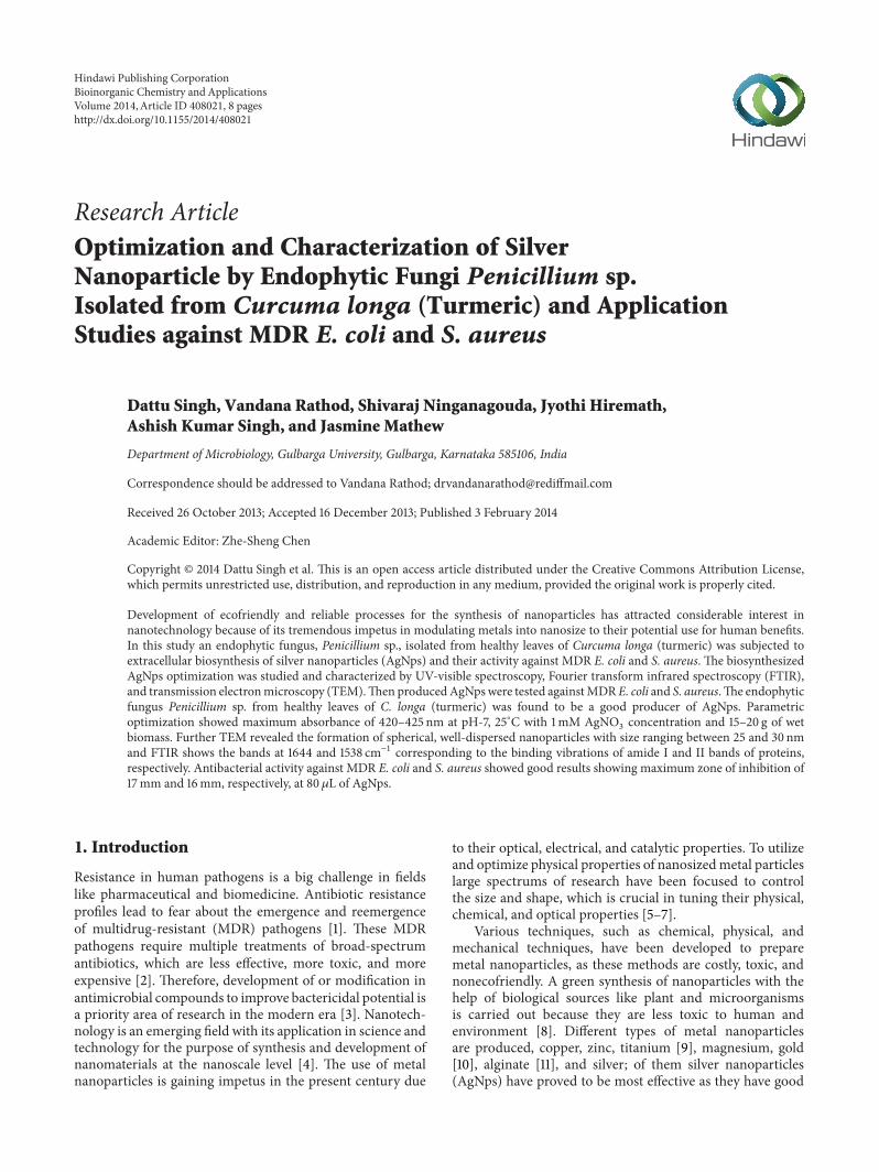
(a) (b)
Figure 1: (a) Endophytic fungi grown from sterilized leaf segment of Curcuma longa on PDA. (b) Microscopic image of endophytic fungi,Penicillium sp.
2.4.3. Fourier Transform Infrared Spectroscopy (FTIR). TheAgNps synthesized were air-dried at room temperature andwere subjected to FTIR analysis in the range of 500 to4000 cm−1.The probable biomolecules responsible for reduc-tion, capping, and effective stabilization of the AgNps wererecorded using FTIR spectrophotometer at diffuse reflectancemode.
2.5. Analysis of Antibacterial Activity of Silver Nanoparticlesagainst MDR E. coli and S. aureus Strains
2.5.1. Antimicrobial Susceptibility Assay of Clinical Isolates.The clinical isolates were collected from the diagnostic labsof the Gulbarga district and tested for the sensitivity againstdifferent antibiotics by subculturing the bacterial cultures,incubated at 37∘C for 5-6 h to moderate turbidity. Thenlawn of pathogenic bacteria was prepared on nutrient agarplate using sterile swabs and then antibiotic disc was keptaseptically on the lawn plate and incubated at 37∘C for 24 hand then results were monitored.
2.5.2. Antibacterial Assay of Silver Nanoparticles against MDRE. coli and S. aureus. Antibacterial assay was done on MDRE. coli and S. aureus strains using agar well diffusion assaymethod [23, 24]. The test organisms were grown in nutrientbroth for 5-6 h lawn of MDR bacteria which was prepared onnutrient agar plates using sterile swabs. Agar wells weremadeon nutrient agar plates using gel puncture and each well wasloaded with 20𝜇L, 40 𝜇L, 60𝜇L, and 80 𝜇L, respectively, ofAgNps solution and incubated at 37∘C for 24 h. The zone ofinhibition was measured.
3. Results and Discussion
3.1. Isolation of Endophytic Fungi. The surface sterilized leafsegments ofC. longa (turmeric) were placed on PDAmediumand incubated for 72 h. The fungi grown out from tissuewere brought into pure culture, identified by microscopicobservation and morphological characteristic features. Stud-ies revealed that the fungus is Penicillium sp. Figure 1.
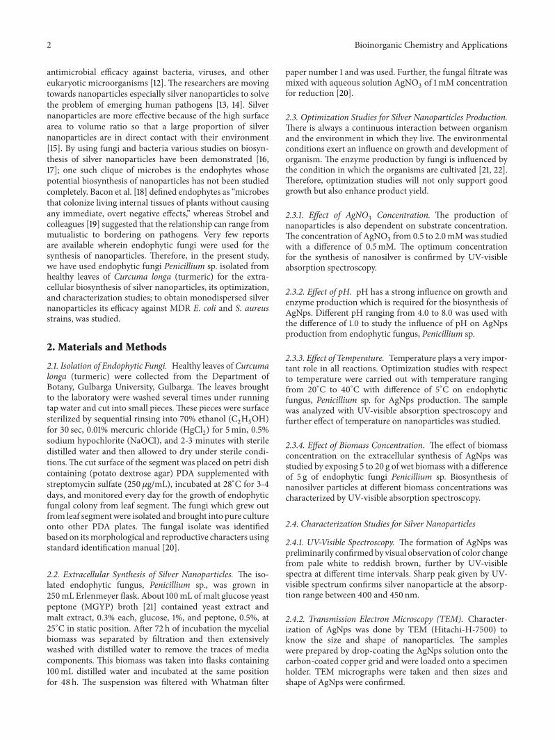
3.2. Extracellular Synthesis of Silver Nanoparticles. Fungal fil-trate was treated with equal volume of 1mMAgNO
3solution
after 24 h incubation; appearance of color change from palewhite to brown is a clear indication for the formation of silver
Figure 2: Color change to reddish brown after treating filtrate ofendophytic fungi, Penicillium sp., with 1mM AgNO
3
.
nanoparticles in the reaction mixture. The intensity of thecolor was increased with the period of incubation Figure 2.The appearance of the brown color was due to the excitationof surface plasmon vibrations [25].
3.3. Optimization Studies for Silver Nanoparticles Production.Environmental conditions profoundly modulate the growthand metabolism of fungi. Culture conditions have beenthe critical components, directly affecting the productivityand also the process economics. Optimization of physicalparameters will not only support good growth but alsoenhance the product yield [26]. The growth conditions, suchas substrate concentration, pH, temperature, and inoculumsize, directly monitor the rate of enzyme activity whichreflects the synthesis of AgNps.
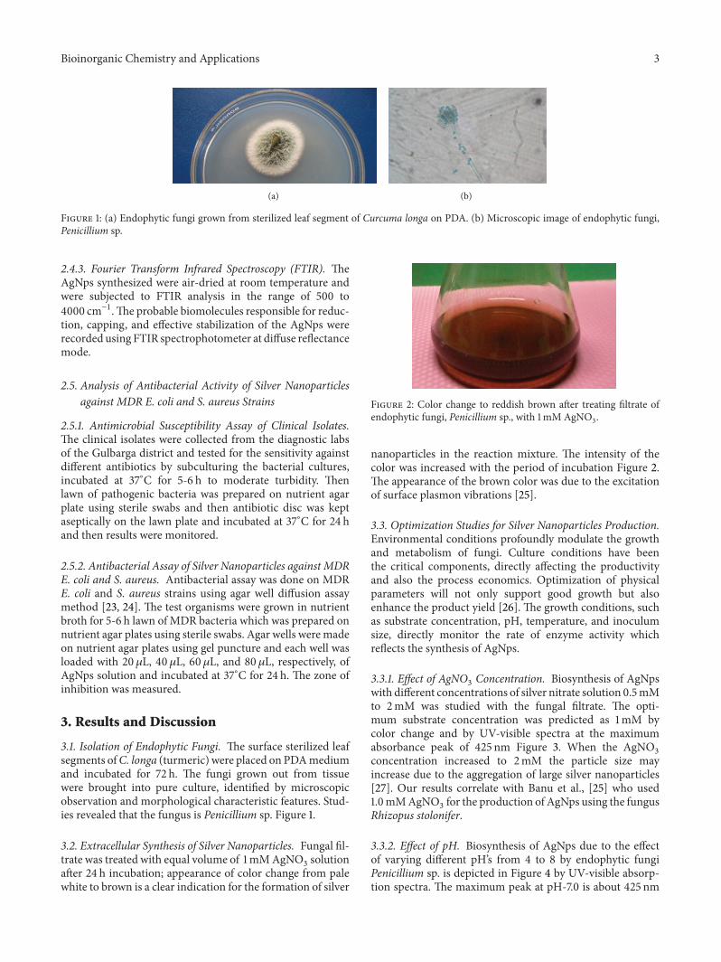
3.3.1. Effect of AgNO3Concentration. Biosynthesis of AgNps
with different concentrations of silver nitrate solution 0.5mMto 2mM was studied with the fungal filtrate. The opti-mum substrate concentration was predicted as 1mM bycolor change and by UV-visible spectra at the maximumabsorbance peak of 425 nm Figure 3. When the AgNO
3
concentration increased to 2mM the particle size mayincrease due to the aggregation of large silver nanoparticles[27]. Our results correlate with Banu et al., [25] who used1.0mMAgNO
3for the production of AgNps using the fungus
Rhizopus stolonifer.
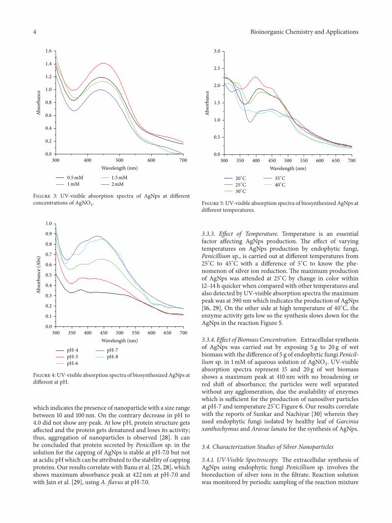
3.3.2. Effect of pH. Biosynthesis of AgNps due to the effectof varying different pH’s from 4 to 8 by endophytic fungiPenicillium sp. is depicted in Figure 4 by UV-visible absorp-tion spectra. The maximum peak at pH-7.0 is about 425 nm
4 Bioinorganic Chemistry and Applications
1.6
1.4
1.2
1.0
0.8
0.6
0.4
0.2
0.0
300 400 500 600 700
Abso
rban
ce
Wavelength (nm)
0.5mM1mM
1.5mM2mM
Figure 3: UV-visible absorption spectra of AgNps at differentconcentrations of AgNO
3
.
1.0
0.9
0.8
0.7
0.6
0.5
0.4
0.2
0.3
0.1
0.0
300 350 400 450 500 550 600 650 700
pH-4pH-5pH-6
pH-7pH-8
Abso
rban
ce (A
bs)
Wavelength (nm)
Figure 4: UV-visible absorption spectra of biosynthesized AgNps atdifferent at pH.
which indicates the presence of nanoparticle with a size rangebetween 10 and 100 nm. On the contrary decrease in pH to4.0 did not show any peak. At low pH, protein structure getsaffected and the protein gets denatured and loses its activity;thus, aggregation of nanoparticles is observed [28]. It canbe concluded that protein secreted by Penicillum sp. in thesolution for the capping of AgNps is stable at pH-7.0 but notat acidic pHwhich can be attributed to the stability of cappingproteins. Our results correlate with Banu et al. [25, 28], whichshows maximum absorbance peak at 422 nm at pH-7.0 andwith Jain et al. [29], using A. flavus at pH-7.0.
3.0
2.5
2.0
1.5
1.0
0.5
0.0
300 350 400 450 500 550 600 650 700
Abso
rban
ce
20∘C25∘C30∘C
35∘C40∘C
Wavelength (nm)
Figure 5: UV-visible absorption spectra of biosynthesized AgNps atdifferent temperatures.
3.3.3. Effect of Temperature. Temperature is an essentialfactor affecting AgNps production. The effect of varyingtemperatures on AgNps production by endophytic fungi,Penicillium sp., is carried out at different temperatures from25∘C to 45∘C with a difference of 5∘C to know the phe-nomenon of silver ion reduction. The maximum productionof AgNps was attended at 25∘C by change in color within12–14 h quicker when compared with other temperatures andalso detected by UV-visible absorption spectra themaximumpeak was at 390 nmwhich indicates the production of AgNps[16, 29]. On the other side at high temperature of 40∘C, theenzyme activity gets low so the synthesis slows down for theAgNps in the reaction Figure 5.
3.3.4. Effect of Biomass Concentration. Extracellular synthesisof AgNps was carried out by exposing 5 g to 20 g of wetbiomass with the difference of 5 g of endophytic fungi Penicil-lium sp. in 1mM of aqueous solution of AgNO
3. UV-visible
absorption spectra represent 15 and 20 g of wet biomassshows a maximum peak at 410 nm with no broadening orred shift of absorbance; the particles were well separatedwithout any agglomeration, due the availability of enzymeswhich is sufficient for the production of nanosilver particlesat pH-7 and temperature 25∘C Figure 6. Our results correlatewith the reports of Sunkar and Nachiyar [30] wherein theyused endophytic fungi isolated by healthy leaf of Garciniaxanthochymus and Aravae lanata for the synthesis of AgNps.
3.4. Characterization Studies of Silver Nanoparticles
3.4.1. UV-Visible Spectroscopy. The extracellular synthesis ofAgNps using endophytic fungi Penicillium sp. involves thebioreduction of silver ions in the filtrate. Reaction solutionwas monitored by periodic sampling of the reaction mixture
Bioinorganic Chemistry and Applications 5
300 400 500 600 700
1.4
1.2
1.0
0.8
0.6
0.4
0.2
0.0
Abso
rban
ce
Wavelength (nm)
20 g15 g
10 g5g
Figure 6: UV-visible absorption spectra of AgNps at differentbiomass concentrations.
300 350 400 450 500 550 600 650 700
Wavelength (nm)
2.0
1.8
1.6
1.4
1.2
1.0
0.8
0.6
0.4
0.2
0.0
Abso
rban
ce
120h96h72h48h
24h8h2h1h
Figure 7: UV-visible absorption spectra of biosynthesized AgNps atdifferent time intervals.
at regular time intervals by using UV-visible spectroscopy.Synthesized AgNps showed maximum absorbance peak at420 nm Figure 7. The AgNps formed were highly stable upto 120 h after the reaction.The AgNps were characterized andconfirmed by TEM analysis. Similar results were observed byNinganagouda et al., [24] who revealed plasma resonance ofAgNps between 380 and 450 nm, and Sunkar and Nachiyar,who [31] revealed absorption peak at 400 nm and 423 nmby endophytic fungi isolated from leaf samples of GarciniaXanthochymus and Aravae lanata.
50nm
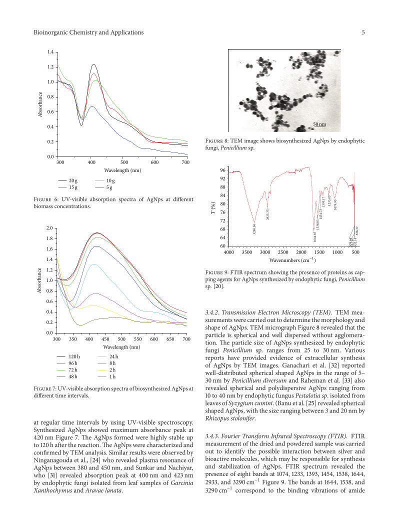
Figure 8: TEM image shows biosynthesized AgNps by endophyticfungi, Penicillium sp.
96
92
88
84
80
76
72
68
64
60
T(%
)
4000 3500 3000 2500 2000 1500 1000 500
Wavenumbers (cm−1)3290.50
2923.35
1644.65
1538.031454.73
1393.17
1233.05
1074.95
539.64
535.97
532.14
528.37
Figure 9: FTIR spectrum showing the presence of proteins as cap-ping agents for AgNps synthesized by endophytic fungi, Penicilliumsp. [20].
3.4.2. Transmission Electron Microscopy (TEM). TEM mea-surementswere carried out to determine themorphology andshape of AgNps. TEM micrograph Figure 8 revealed that theparticle is spherical and well dispersed without agglomera-tion. The particle size of AgNps synthesized by endophyticfungi Penicillium sp. ranges from 25 to 30 nm. Variousreports have provided evidence of extracellular synthesisof AgNps by TEM images. Ganachari et al. [32] reportedwell-distributed spherical shaped AgNps in the range of 5–30 nm by Penicillium diversum and Raheman et al. [33] alsorevealed spherical and polydispersive AgNps ranging from10 to 40 nm by endophytic fungus Pestalotia sp. isolated fromleaves of Syzygium cumini. (Banu et al. [25] revealed sphericalshaped AgNps, with the size ranging between 3 and 20 nm byRhizopus stolonifer.
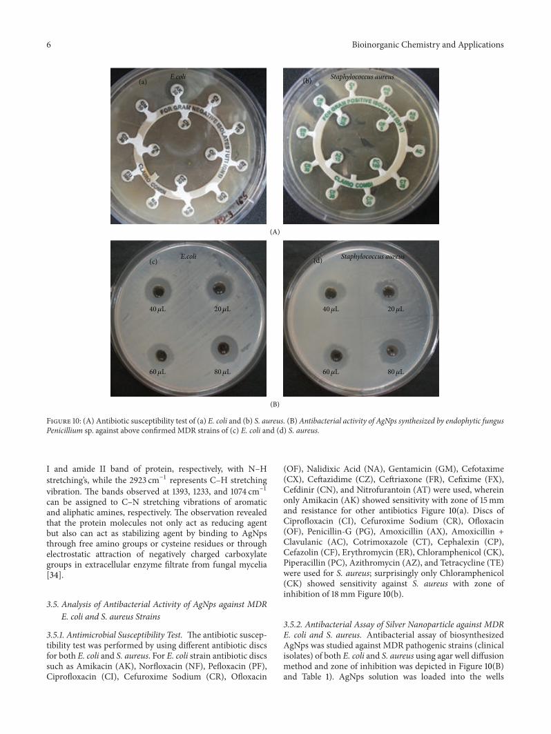
3.4.3. Fourier Transform Infrared Spectroscopy (FTIR). FTIRmeasurement of the dried and powdered sample was carriedout to identify the possible interaction between silver andbioactive molecules, which may be responsible for synthesisand stabilization of AgNps. FTIR spectrum revealed thepresence of eight bands at 1074, 1233, 1393, 1454, 1538, 1644,2933, and 3290 cm−1 Figure 9. The bands at 1644, 1538, and3290 cm−1 correspond to the binding vibrations of amide
6 Bioinorganic Chemistry and Applications
E.coli Staphylococcus aureus(a)
(A)
(b)
(B)
E.coli Staphylococcus aureus(c) (d)
40𝜇L 20𝜇L
60𝜇L 80𝜇L
40𝜇L 20𝜇L
60𝜇L 80𝜇L
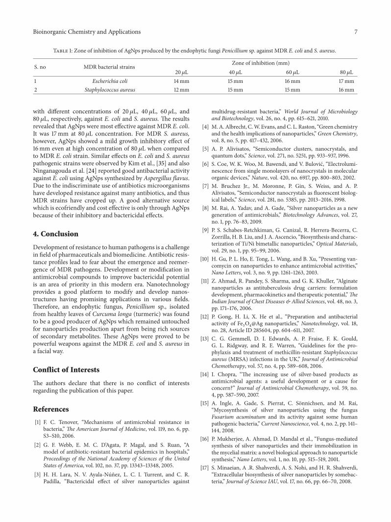
Figure 10: (A) Antibiotic susceptibility test of (a) E. coli and (b) S. aureus. (B)Antibacterial activity of AgNps synthesized by endophytic fungusPenicillium sp. against above confirmed MDR strains of (c) E. coli and (d) S. aureus.
I and amide II band of protein, respectively, with N–Hstretching’s, while the 2923 cm−1 represents C–H stretchingvibration. The bands observed at 1393, 1233, and 1074 cm−1can be assigned to C–N stretching vibrations of aromaticand aliphatic amines, respectively. The observation revealedthat the protein molecules not only act as reducing agentbut also can act as stabilizing agent by binding to AgNpsthrough free amino groups or cysteine residues or throughelectrostatic attraction of negatively charged carboxylategroups in extracellular enzyme filtrate from fungal mycelia[34].
3.5. Analysis of Antibacterial Activity of AgNps against MDRE. coli and S. aureus Strains
3.5.1. Antimicrobial Susceptibility Test. The antibiotic suscep-tibility test was performed by using different antibiotic discsfor both E. coli and S. aureus. For E. coli strain antibiotic discssuch as Amikacin (AK), Norfloxacin (NF), Pefloxacin (PF),Ciprofloxacin (CI), Cefuroxime Sodium (CR), Ofloxacin
(OF), Nalidixic Acid (NA), Gentamicin (GM), Cefotaxime(CX), Ceftazidime (CZ), Ceftriaxone (FR), Cefixime (FX),Cefdinir (CN), and Nitrofurantoin (AT) were used, whereinonly Amikacin (AK) showed sensitivity with zone of 15mmand resistance for other antibiotics Figure 10(a). Discs ofCiprofloxacin (CI), Cefuroxime Sodium (CR), Ofloxacin(OF), Penicillin-G (PG), Amoxicillin (AX), Amoxicillin +Clavulanic (AC), Cotrimoxazole (CT), Cephalexin (CP),Cefazolin (CF), Erythromycin (ER), Chloramphenicol (CK),Piperacillin (PC), Azithromycin (AZ), and Tetracycline (TE)were used for S. aureus; surprisingly only Chloramphenicol(CK) showed sensitivity against S. aureus with zone ofinhibition of 18mm Figure 10(b).
3.5.2. Antibacterial Assay of Silver Nanoparticle against MDRE. coli and S. aureus. Antibacterial assay of biosynthesizedAgNps was studied against MDR pathogenic strains (clinicalisolates) of both E. coli and S. aureus using agar well diffusionmethod and zone of inhibition was depicted in Figure 10(B)and Table 1). AgNps solution was loaded into the wells
Bioinorganic Chemistry and Applications 7
Table 1: Zone of inhibition of AgNps produced by the endophytic fungi Penicillium sp. against MDR E. coli and S. aureus.
S. no MDR bacterial strains Zone of inhibition (mm)20 𝜇L 40 𝜇L 60 𝜇L 80 𝜇L
1 Escherichia coli 14mm 15mm 16mm 17mm2 Staphylococcus aureus 12mm 15mm 15mm 16mm
with different concentrations of 20𝜇L, 40 𝜇L, 60𝜇L, and80 𝜇L, respectively, against E. coli and S. aureus. The resultsrevealed that AgNps were most effective against MDR E. coli.It was 17mm at 80 𝜇L concentration. For MDR S. aureus,however, AgNps showed a mild growth inhibitory effect of16mm even at high concentration of 80𝜇L when comparedto MDR E. coli strain. Similar effects on E. coli and S. aureuspathogenic strains were observed by Kim et al., [35] and alsoNinganagouda et al. [24] reported good antibacterial activityagainst E. coli using AgNps synthesized by Aspergillus flavus.Due to the indiscriminate use of antibiotics microorganismshave developed resistance against many antibiotics, and thusMDR strains have cropped up. A good alternative sourcewhich is ecofriendly and cost effective is only through AgNpsbecause of their inhibitory and bactericidal effects.
4. Conclusion
Development of resistance to human pathogens is a challengein field of pharmaceuticals and biomedicine. Antibiotic resis-tance profiles lead to fear about the emergence and reemer-gence of MDR pathogens. Development or modification inantimicrobial compounds to improve bactericidal potentialis an area of priority in this modern era. Nanotechnologyprovides a good platform to modify and develop nanos-tructures having promising applications in various fields.Therefore, an endophytic fungus, Penicillium sp., isolatedfrom healthy leaves of Curcuma longa (turmeric) was foundto be a good producer of AgNps which remained untouchedfor nanoparticles production apart from being rich sourcesof secondary metabolites. These AgNps were proved to bepowerful weapons against the MDR E. coil and S. aureus ina facial way.
Conflict of Interests
The authors declare that there is no conflict of interestsregarding the publication of this paper.
References
[1] F. C. Tenover, “Mechanisms of antimicrobial resistance inbacteria,” The American Journal of Medicine, vol. 119, no. 6, pp.S3–S10, 2006.
[2] G. F. Webb, E. M. C. D’Agata, P. Magal, and S. Ruan, “Amodel of antibiotic-resistant bacterial epidemics in hospitals,”Proceedings of the National Academy of Sciences of the UnitedStates of America, vol. 102, no. 37, pp. 13343–13348, 2005.
[3] H. H. Lara, N. V. Ayala-Nunez, L. C. I. Turrent, and C. R.Padilla, “Bactericidal effect of silver nanoparticles against
multidrug-resistant bacteria,” World Journal of Microbiologyand Biotechnology, vol. 26, no. 4, pp. 615–621, 2010.
[4] M.A.Albrecht, C.W. Evans, andC. L. Raston, “Green chemistryand the health implications of nanoparticles,” Green Chemistry,vol. 8, no. 5, pp. 417–432, 2006.
[5] A. P. Alivisatos, “Semiconductor clusters, nanocrystals, andquantum dots,” Science, vol. 271, no. 5251, pp. 933–937, 1996.
[6] S. Coe, W. K. Woo, M. Bawendi, and V. Bulovic, “Electrolumi-nescence from single monolayers of nanocrystals in molecularorganic devices,” Nature, vol. 420, no. 6917, pp. 800–803, 2002.
[7] M. Bruchez Jr., M. Moronne, P. Gin, S. Weiss, and A. P.Alivisatos, “Semiconductor nanocrystals as fluorescent biolog-ical labels,” Science, vol. 281, no. 5385, pp. 2013–2016, 1998.
[8] M. Rai, A. Yadav, and A. Gade, “Silver nanoparticles as a newgeneration of antimicrobials,” Biotechnology Advances, vol. 27,no. 1, pp. 76–83, 2009.
[9] P. S. Schabes-Retchkiman, G. Canizal, R. Herrera-Becerra, C.Zorrilla, H. B. Liu, and J. A. Ascencio, “Biosynthesis and charac-terization of Ti/Ni bimetallic nanoparticles,” Optical Materials,vol. 29, no. 1, pp. 95–99, 2006.
[10] H. Gu, P. L. Ho, E. Tong, L. Wang, and B. Xu, “Presenting van-comycin on nanoparticles to enhance antimicrobial activities,”Nano Letters, vol. 3, no. 9, pp. 1261–1263, 2003.
[11] Z. Ahmad, R. Pandey, S. Sharma, and G. K. Khuller, “Alginatenanoparticles as antituberculosis drug carriers: formulationdevelopment, pharmacokinetics and therapeutic potential,”TheIndian Journal of Chest Diseases & Allied Sciences, vol. 48, no. 3,pp. 171–176, 2006.
[12] P. Gong, H. Li, X. He et al., “Preparation and antibacterialactivity of Fe
3
O4
@Ag nanoparticles,” Nanotechnology, vol. 18,no. 28, Article ID 285604, pp. 604–611, 2007.
[13] C. G. Gemmell, D. I. Edwards, A. P. Fraise, F. K. Gould,G. L. Ridgway, and R. E. Warren, “Guidelines for the pro-phylaxis and treatment of methicillin-resistant Staphylococcusaureus (MRSA) infections in the UK,” Journal of AntimicrobialChemotherapy, vol. 57, no. 4, pp. 589–608, 2006.
[14] I. Chopra, “The increasing use of silver-based products asantimicrobial agents: a useful development or a cause forconcern?” Journal of Antimicrobial Chemotherapy, vol. 59, no.4, pp. 587–590, 2007.
[15] A. Ingle, A. Gade, S. Pierrat, C. Sonnichsen, and M. Rai,“Mycosynthesis of silver nanoparticles using the fungusFusarium acuminatum and its activity against some humanpathogenic bacteria,” Current Nanoscience, vol. 4, no. 2, pp. 141–144, 2008.
[16] P. Mukherjee, A. Ahmad, D. Mandal et al., “Fungus-mediatedsynthesis of silver nanoparticles and their immobilization inthemycelial matrix: a novel biological approach to nanoparticlesynthesis,” Nano Letters, vol. 1, no. 10, pp. 515–519, 2001.
[17] S. Minaeian, A .R. Shahverdi, A. S. Nohi, and H. R. Shahverdi,“Extracellular biosynthesis of silver nanoparticles by somebac-teria,” Journal of Science IAU, vol. 17, no. 66, pp. 66–70, 2008.
8 Bioinorganic Chemistry and Applications
[18] C. W. Bacon, D. M. Hinton, J. K. Porter, A. E. Glenn, andG. Kuldau, “Fusaric acid, a Fusarium verticillioides metabolite,antagonistic to the endophytic biocontrol bacterium Bacillusmojavensis,” Canadian Journal of Botany, vol. 82, no. 7, pp. 878–885, 2004.
[19] G. Strobel, X. Yang, J. Sears, R. Kramer, R. S. Sidhu, and W.M. Hess, “Taxol from Pestalotiopsis microspora, an endophyticfungus of Taxus wallachiana,” Microbiology, vol. 142, no. 2, pp.435–440, 1996.
[20] D. Singh, V. Rathod, S. Ninganagouda, J. Herimath, and P.Kulkarni, “Biosynthesis of silver nanoparticle by endophyticfungi Penicillium sp. isolated from Curcuma longa (turmeric)and its antibacterial activity against pathogenic gram negativebacteria,” Journal of Pharmacy Research, vol. 7, no. 5, pp. 448–453, 2013.
[21] M. Karbasian, S. M. Atyabi, S. D. Siadat, S. B. Momen, andD. Norouzian, “Optimizing nano-silver formation by Fusariumoxysporum PTCC 5115 employing response surface method-ology,” The American Journal of Agricultural and BiologicalScience, vol. 3, no. 1, pp. 433–437, 2008.
[22] P. K. Lekha and B. K. Lonsane, “Production and application oftannin acyl hydrolsase: state of the art,” Advances in AppliedMicrobiology, vol. 44, no. 27, pp. 215–260, 1997.
[23] S. C. Bell and W. E. Grundy, “Preparation of agar wells forantibiotic assay,” Applied Microbiology, vol. 16, no. 10, pp. 1611–1612, 1968.
[24] S. Ninganagouda, V. Rathod, H. Jyoti, D. Singh, K. Prema, andManzoor-Ul-Haq, “Extracellular biosynthesis of silver nanopar-ticles using Aspergillus flavus and their antimicrobial activityagainst gram negative MDR strains,” International Journal ofPharma and Bio Sciences, vol. 4, no. 2, pp. 222–229, 2013.
[25] A. Banu, V. Rathod, and E. Ranganath, “Silver nanoparticleproduction by Rhizopus stolonifer and its antibacterial activ-ity against extended spectrum 𝛽-lactamase producing (ESBL)strains of Enterobacteriaceae,” Materials Research Bulletin, vol.46, no. 9, pp. 1417–1423, 2011.
[26] J. Zhang and R. Greasham, “Chemically defined media forcommercial fermentations,” Applied Microbiology and Biotech-nology, vol. 51, no. 4, pp. 407–421, 1999.
[27] C. R. K. Rao and D. C. Trivedi, “Synthesis and characterizationof fatty acids passivated silver nanoparticles—their interactionwith PPy,” Synthetic Metals, vol. 155, no. 2, pp. 324–327, 2005.
[28] A. Banu and V. Rathod, “Synthesis and chacterzation of silvernanoparticles by Rhizopus stolonifer,” International Journal ofBiomedical andAdvance Research, vol. 2, no. 5, pp. 148–158, 2011.
[29] N. Jain, A. Bhargava, S. Majumdar, J. C. Tarafdar, and J. Pan-war, “Extracellular biosynthesis and characterization of silvernanoparticles using Aspergillus flavus NJP08: a mechanismperspective,” Nanoscale, vol. 3, no. 2, pp. 635–641, 2011.
[30] S. Sunkar and C. V. Nachiyar, “Endophytic fungi medi-ated extracellular silver nanoparticles as effective antibacterialagents,” International Journal of Pharmacy and PharmaceuticalScience, vol. 5, no. 2, pp. 95–100, 2013.
[31] S. Sunkar and C. V. Nachiyar, “Biogenesis of antibacterial silvernanoparticles using the endophytic bacterium Bacillus cereusisolated from Garcinia xanthochymus,” Asian Pacific Journal ofTropical Biomedicine, vol. 2, no. 12, pp. 953–959, 2012.
[32] S. V. Ganachari, R. Bhat, R. Deshpande, and A. Venkataraman,“Extracellular biosynthesis of silver nanoparticles using fungiPenicillium diversum and their antimicrobial activity studies,”BioNanoScience, vol. 2, no. 4, pp. 316–321, 2012.
[33] F. Raheman, S. Deshmukh, A. Ingle, A. Gade, and M. Rai,“Silver nanoparticles: novel antimicrobial agent synthesizedfrom an endophytic fungus Pestalotia sp. isolated from leaves ofSyzygium cumini (L),” Nano Biomedicine and Engineering, vol.3, no. 3, pp. 174–178, 2011.
[34] A. Gole, C. Dash, V. Ramakrishnan et al., “Pepsin-gold colloidconjugates: preparation, characterization, and enzymatic activ-ity,” Langmuir, vol. 17, no. 5, pp. 1674–1679, 2001.
[35] J. S. Kim, E. Kuk, K. N. Yu et al., “Antimicrobial effects of silvernanoparticles,” Nanomedicine, vol. 3, no. 1, pp. 95–101, 2007.

Submit your manuscripts athttp://www.hindawi.com
Hindawi Publishing Corporationhttp://www.hindawi.com Volume 2014
Inorganic ChemistryInternational Journal of
Hindawi Publishing Corporation http://www.hindawi.com Volume 2014
International Journal ofPhotoenergy
Hindawi Publishing Corporationhttp://www.hindawi.com Volume 2014
Carbohydrate Chemistry
International Journal of
Hindawi Publishing Corporationhttp://www.hindawi.com Volume 2014
Journal of
Chemistry
Hindawi Publishing Corporationhttp://www.hindawi.com Volume 2014
Advances in
Physical Chemistry
Hindawi Publishing Corporationhttp://www.hindawi.com
Analytical Methods in Chemistry
Journal of
Volume 2014
Bioinorganic Chemistry and ApplicationsHindawi Publishing Corporationhttp://www.hindawi.com Volume 2014
SpectroscopyInternational Journal of
Hindawi Publishing Corporationhttp://www.hindawi.com Volume 2014
The Scientific World JournalHindawi Publishing Corporation http://www.hindawi.com Volume 2014
Medicinal ChemistryInternational Journal of
Hindawi Publishing Corporationhttp://www.hindawi.com Volume 2014
Chromatography Research International
Hindawi Publishing Corporationhttp://www.hindawi.com Volume 2014
Applied ChemistryJournal of
Hindawi Publishing Corporationhttp://www.hindawi.com Volume 2014
Hindawi Publishing Corporationhttp://www.hindawi.com Volume 2014
Theoretical ChemistryJournal of
Hindawi Publishing Corporationhttp://www.hindawi.com Volume 2014
Journal of
Spectroscopy
Analytical ChemistryInternational Journal of
Hindawi Publishing Corporationhttp://www.hindawi.com Volume 2014
Journal of
Hindawi Publishing Corporationhttp://www.hindawi.com Volume 2014
Quantum Chemistry
Hindawi Publishing Corporationhttp://www.hindawi.com Volume 2014
Organic Chemistry International
ElectrochemistryInternational Journal of
Hindawi Publishing Corporation http://www.hindawi.com Volume 2014
Hindawi Publishing Corporationhttp://www.hindawi.com Volume 2014
CatalystsJournal of