oncogenic kit signaling and therapeutic intervention in a mouse model of gastrointestinal stromal...
TRANSCRIPT

Oncogenic Kit signaling and therapeutic interventionin a mouse model of gastrointestinal stromal tumorFerdinand Rossi*, Imke Ehlers*, Valter Agosti*†, Nicholas D. Socci‡, Agnes Viale§, Gunhild Sommer*, Yasemin Yozgat*,Katia Manova*¶, Cristina R. Antonescu*�, and Peter Besmer*,**††‡‡
Departments of *Developmental Biology and §Molecular Biology, ‡Computational Biology Center, and ¶Molecular Cytology Facility, Sloan–KetteringInstitute, New York, NY 10021; and �Department of Pathology, Memorial Sloan–Kettering Cancer Center, **Gerstner Sloan–Kettering Graduate School ofBiomedical Sciences, and ††Cornell University Weill Graduate School of Medical Sciences, New York, NY 10021
Edited by Joseph Schlessinger, Yale University School of Medicine, New Haven, CT, and approved June 20, 2006 (received for review December 30, 2005)
Kit receptor-activating mutations are critical in the pathogenesis ofgastrointestinal stromal tumors (GIST). We investigated mecha-nisms of oncogenic Kit signaling and the consequences of thera-peutic intervention in a mouse model of human GIST. Treatment ofGIST mice with imatinib decreased cell proliferation and increasedapoptosis in the tumor. Analysis of tumor tissue from imatinib-treated mice showed diminished phosphatidylinositol 3-kinase(PI3-kinase) and mammalian target of rapamycin (mTOR) signalingsuggesting that oncogenic Kit signaling critically contributes to thetranslational response in GIST. Treatment with RAD001 (everoli-mus), an mTOR inhibitor, diminished the translational responseand cell proliferation in tumor lesions, pointing to mTOR inhibitionas a therapeutic approach for imatinib-resistant GIST. Analysis ofRNA expression profiles in GIST lesions with and without imatinibtreatment showed changes in expression of IFN-inducible genesand cell cycle regulators. These results convincingly show thatKitV558�/� mice represent a unique faithful mouse model of humanfamilial GIST, and they demonstrate the utility of these mice forpreclinical investigations and to elucidate oncogenic signalingmechanisms by using genetic approaches and targeted pharmaco-logical intervention.
imatinib � Kit receptor tyrosine kinase � signal transduction
The Kit receptor tyrosine kinase has a critical role in the normaldevelopment and function of the interstitial cells of Cajal (ICC),
as well as in hematopoietic cell populations, gametogenesis, andmelanogenesis during embryonic development and in the postnatalorganism (1–3). The finding of Kit receptor-activating mutations inhuman tumors, including gastrointestinal stromal tumor (GIST),seminomas, and mastocytosis, as well as some acute myelogenousleukemias, suggested a role for Kit in oncogenesis. GIST is the mostcommon mesenchymal tumor of the gastrointestinal tract. GISTsexpress Kit and are thought to derive from a Kit� or Kitlow ICCprogenitor or ICC. The vast majority of GISTs contain Kit recep-tor-activating mutations (4, 5). Kit-activating mutations in GIST arefound predominantly in the juxtamembrane domain of the Kitreceptor, but mutations in the extracellular and kinase domains ofKit have been described as well (6, 7). Imatinib mesylate (Gleevec,STI571), an inhibitor of the Kit, PDGFR, and BCR-ABL tyrosinekinases, is used to treat patients with GIST and chronic myeloge-nous leukemia. In GIST, it elicits a partial response or stable diseasein a majority of patients with metastatic or recurrent disease. Theclinical response correlates with a decrease in tumor cellularity andmyxoid degeneration of the tumor. Although the clinical responseto imatinib is quite well described, the molecular response of Kitinhibition by imatinib in GIST is poorly understood. Imatinib ismost effective in GISTs with Kit-activating mutations in the jux-tamembrane domain, some kinase domain mutations, or extracel-lular domain mutations. But Kit mutations that destabilize theinactive form of the kinase are resistant to inhibition by imatinib.Unfortunately, long-term treatment with imatinib is associated withthe development of drug resistance, and in some cases, resistanceappears to derive from second site mutations in the Kit receptor (8,
9). Therefore, the development of new strategies for the treatmentof GIST is highly relevant.
Several cases of human familial GIST syndrome with associatedinterstitial cells of Cajal hyperplasia, hyperpigmentation, and�orurticaria pigmentosa with germ-line Kit mutations have beenreported (10, 11). Based on these findings, we produced a mousecarrying a Kit-activating mutation, KitV558�/�, in the germ line (12).The KitV558�/� mutation is located in the juxtamembrane domainof Kit (exon 11), a negative regulatory region of the receptor wherethe majority of the somatic GIST mutations in patients occur.Heterozygous mutant KitV558�/� mice develop symptoms of diseaseand eventually die from pathology in the gastrointestinal (GI) tract.Patchy hyperplasia of Kit-positive cells is observed within themyenteric plexus of the GI tract, and neoplastic lesions indistin-guishable from human GIST are found with complete penetrancein the cecum of mutant mice (12). Thus the KitV558�/� mice providea unique opportunity to investigate the development of GIST andthe consequences of therapeutic intervention.
The Kit receptor has a role in distinct cellular responses, includ-ing cell proliferation, survival, adhesion, chemotaxis, and secretoryresponses, as well as the desensitization of the activated receptor invarious cell types in vitro and in vivo. The downstream signalingcascades that are known to be activated by Kit include the Ras�MAP kinase, Rac�Rho-JNK, phosphatidylinositol 3-kinase (PI3-kinase)�AKT�PDK1�FOXO, and src family kinase (SFK)�STATsignaling networks. Cell-type-specific responses depend in part onthe cellular context and the presence of signaling components, andtherefore signaling cascades that may be activated by Kit vary indifferent cell types. Consequently, disruption of Kit-specific signal-ing pathways by knock-in mutations produces cell-specific effects,e.g., disruption of Kit-induced PI3-kinase signaling was shown toimpair male fertility, whereas melanogenesis and hematopoiesiswere not affected (13), and disruption of SFK binding shows mainlyage-dependent lymphopoietic defects (14). We have used imatinibto investigate oncogenic Kit signaling in mouse GIST in vivo. Weshow that treatment of GIST mice with imatinib abolished cell cycleprogression concomitant with an increase in apoptosis in the tumorlesions. Analysis of gene expression profiles in placebo- and ima-tinib-treated mice revealed roles for cell cycle regulators andIFN-inducible genes in GIST. Biochemical analysis of tumor tissuefrom imatinib-treated mice showed diminished PI3-kinase andmammalian target of rapamycin (mTOR) signaling, implying a rolefor the translational response in oncogenic Kit signaling in GIST.To investigate the role of the translational response in GIST, micewere treated with the mTOR inhibitor RAD001 (everolimus).
Conflict of interest statement: No conflicts declared.
This paper was submitted directly (Track II) to the PNAS office.
Abbreviations: GIST, gastrointestinal stromal tumor; PI3-kinase, phosphatidylinositol3-kinase; ABC, ATP-binding cassette; mTOR, mammalian target of rapamycin.
†Present address: Department of Experimental and Clinical Medicine, University MagnaGraecia, University Campus, Germaneto, 88100 Catanzaro, Italy.
‡‡To whom correspondence should be addressed. E-mail: [email protected].
© 2006 by The National Academy of Sciences of the USA
www.pnas.org�cgi�doi�10.1073�pnas.0511076103 PNAS � August 22, 2006 � vol. 103 � no. 34 � 12843–12848
MED
ICA
LSC
IEN
CES
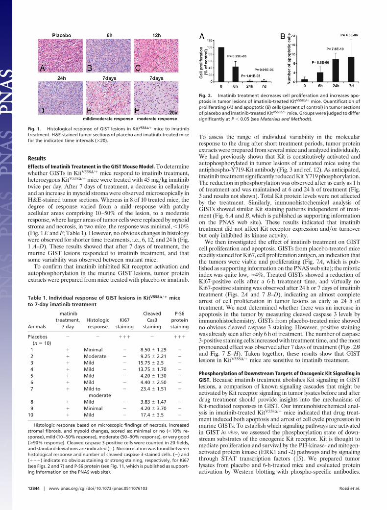
ResultsEffects of Imatinib Treatment in the GIST Mouse Model. To determinewhether GISTs in KitV558�/� mice respond to imatinib treatment,heterozygous KitV558�/� mice were treated with 45 mg�kg imatinibtwice per day. After 7 days of treatment, a decrease in cellularityand an increase in myxoid stroma were observed microscopically inH&E-stained tumor sections. Whereas in 8 of 10 treated mice, thedegree of response varied from a mild response with patchyacellular areas comprising 10–50% of the lesion, to a moderateresponse, where larger areas of tumor cells were replaced by myxoidstroma and necrosis, in two mice, the response was minimal, �10%(Fig. 1 E and F; Table 1). However, no obvious changes in histologywere observed for shorter time treatments, i.e., 6, 12, and 24 h (Fig.1 A–D). These results showed that after 7 days of treatment, themurine GIST lesions responded to imatinib treatment, and thatsome variability was observed between mutant mice.
To confirm that imatinib inhibited Kit receptor activation andautophosphorylation in the murine GIST lesions, tumor proteinextracts were prepared from mice treated with placebo or imatinib.
To assess the range of individual variability in the molecularresponse to the drug after short treatment periods, tumor proteinextracts were prepared from several mice and analyzed individually.We had previously shown that Kit is constitutively activated andautophosphorylated in tumor lesions of untreated mice using theantiphospho-Y719-Kit antibody (Fig. 3 and ref. 12). As anticipated,imatinib treatment significantly reduced Kit Y719 phosphorylation.The reduction in phosphorylation was observed after as early as 1 hof treatment and was maintained at 6 and 24 h of treatment (Fig.3 and results not shown). Total Kit protein levels were not affectedby the treatment. Similarly, immunohistochemical analysis ofGISTs showed similar Kit staining patterns independent of treat-ment (Fig. 6 A and B, which is published as supporting informationon the PNAS web site). These results indicated that imatinibtreatment did not affect Kit receptor expression and�or turnoverbut only inhibited its kinase activity.
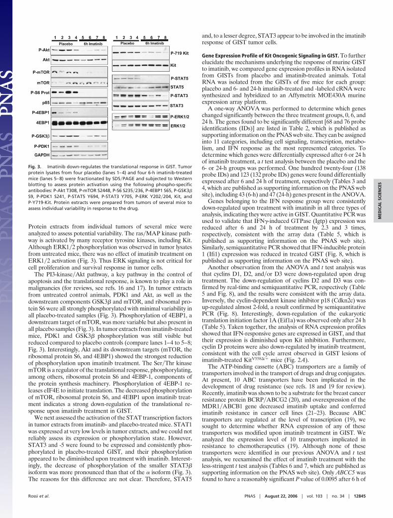
We then investigated the effect of imatinib treatment on GISTcell proliferation and apoptosis. GISTs from placebo-treated micereadily stained for Ki67, cell proliferation antigen, an indication thatthe tumors were viable and proliferating (Fig. 7A, which is pub-lished as supporting information on the PNAS web site); the mitoticindex was quite low, �4%. Treated GISTs showed a reduction ofKi67-positive cells after a 6-h treatment time, and virtually noKi67-positive staining was observed after 24 h or 7 days of imatinibtreatment (Figs. 2A and 7 B–D), indicating an almost completearrest of cell proliferation in tumor lesions as early as 24 h oftreatment. We next determined whether there was an increase inapoptosis in the tumor by measuring cleaved caspase 3 levels byimmunohistochemistry. GISTs from placebo-treated mice showedno obvious cleaved caspase 3 staining. However, positive stainingwas already seen after only 6 h of treatment. The number of caspase3-positive staining cells increased with treatment time, and the mostpronounced effect was observed after 7 days of treatment (Figs. 2Band Fig. 7 E–H). Taken together, these results show that GISTlesions in KitV558�/� mice are sensitive to imatinib treatment.
Phosphorylation of Downstream Targets of Oncogenic Kit Signaling inGIST. Because imatinib treatment abolishes Kit signaling in GISTlesions, a comparison of known signaling cascades that might beactivated by Kit receptor signaling in tumor lysates before and afterdrug treatment should provide insights into the mechanisms ofKit-mediated responses in GIST. Our immunohistochemical anal-ysis in imatinib-treated KitV558�/� mice indicated that drug treat-ment induced both apoptosis and arrest of cell cycle progression inmurine GISTs. To establish which signaling pathways are activatedin GIST in vivo, we assessed the phosphorylation state of down-stream substrates of the oncogenic Kit receptor. Kit is thought tomediate proliferation and survival by the PI3-kinase- and mitogen-activated protein kinase (ERK1 and -2) pathways and by signalingthrough STAT transcription factors (15). We prepared tumorlysates from placebo and 6-h-treated mice and evaluated proteinactivation by Western blotting with phospho-specific antibodies.
Fig. 1. Histological response of GIST lesions in KitV558�/� mice to imatinibtreatment. H&E-stained tumor sections of placebo and imatinib-treated micefor the indicated time intervals (�20).
Table 1. Individual response of GIST lesions in KitV558��� miceto 7-day imatinib treatment
Animals
Imatinibtreatment,
7 dayHistologicresponse
Ki67staining
CleavedCas3
staining
P-S6proteinstaining
Placebos(n � 10)
� � ��� � ���
1 � Minimal � 8.50 � 1.29 �
2 � Moderate � 9.25 � 2.21 �
3 � Mild � 15.75 � 2.5 �
4 � Mild � 13.75 � 1.70 �
5 � Mild � 4.20 � 1.30 �
6 � Mild � 4.40 � 2.50 �
7 � Mild tomoderate
� 23.4 � 1.51 �
8 � Mild � 3.83 � 1.47 �
9 � Minimal � 4.20 � 3.70 �
10 � Mild � 17.4 � 3.5 �
Histologic response based on microscopic findings of necrosis, increasedstromal fibrosis, and myxoid changes, scored as: minimal or no (�10% re-sponse), mild (10–50% response), moderate (50–90% response), or very good(�90% response). Cleaved caspase 3-positive cells were counted in 20 fields,and standard deviations are indicated (�). No correlation was found betweenhistological response and number of cleaved caspase 3-stained cells. (�) and(���) indicate no obvious staining or strong staining, respectively, for Ki67(see Figs. 2 and 7) and P-S6 protein (see Fig. 11, which is published as support-ing information on the PNAS web site).
Fig. 2. Imatinib treatment decreases cell proliferation and increases apo-ptosis in tumor lesions of imatinib-treated KitV558�/� mice. Quantification ofproliferating (A) and apoptotic (B) cells (percent of control) in tumor sectionsof placebo and imatinib-treated KitV558�/� mice. Groups were judged to differsignificantly at P � 0.05 (see Materials and Methods).
12844 � www.pnas.org�cgi�doi�10.1073�pnas.0511076103 Rossi et al.
Protein extracts from individual tumors of several mice wereanalyzed to assess potential variability. The ras�MAP kinase path-way is activated by many receptor tyrosine kinases, including Kit.Although ERK1�2 phosphorylation was observed in tumor lysatesfrom untreated mice, there was no effect of imatinib treatment onERK1�2 activation (Fig. 3). Thus ERK signaling is not critical forcell proliferation and survival response in tumor cells.
The PI3-kinase�Akt pathway, a key pathway in the control ofapoptosis and the translational response, is known to play a role inmalignancies (for reviews, see refs. 16 and 17). In tumor extractsfrom untreated control animals, PDK1 and Akt, as well as thedownstream components GSK3� and mTOR, and ribosomal pro-tein S6 were all strongly phosphorylated with minimal variability inall placebo-treated samples (Fig. 3). Phosphorylation of 4EBP1, adownstream target of mTOR, was more variable but also present inall placebo samples (Fig. 3). In tumor extracts from imatinib-treatedmice, PDK1 and GSK3� phosphorylation was still visible butreduced compared to placebo controls (compare lanes 1–4 to 5–8;Fig. 3). Interestingly, Akt and its downstream targets (mTOR, theribosomal protein S6, and 4EBP1) showed the strongest reductionof phosphorylation upon imatinib treatment. The Ser�Thr kinasemTOR is a regulator of the translational response, phosphorylating,among others, ribosomal protein S6 and 4EBP-1, components ofthe protein synthesis machinery. Phosphorylation of 4EBP-1 re-leases eIF4E to initiate translation. The decreased phosphorylationof mTOR, ribosomal protein S6, and 4EBP1 upon imatinib treat-ment indicates a strong down-regulation of the translational re-sponse upon imatinib treatment in GIST.
We next assessed the activation of the STAT transcription factorsin tumor extracts from imatinib- and placebo-treated mice. STAT1was expressed at very low levels in tumor extracts, and we could notreliably assess its expression or phosphorylation state. However,STAT3 and -5 were found to be expressed and consistently phos-phorylated in placebo-treated GIST, and their phosphorylationappeared to be diminished upon treatment with imatinib. Interest-ingly, the decrease of phosphorylation of the smaller STAT3�isoform was more pronounced than that of the � isoform (Fig. 3).The reasons for this difference are not clear. Therefore, STAT5
and, to a lesser degree, STAT3 appear to be involved in the imatinibresponse of GIST tumor cells.
Gene Expression Profile of Kit Oncogenic Signaling in GIST. To furtherelucidate the mechanisms underlying the response of murine GISTto imatinib, we compared gene expression profiles in RNA isolatedfrom GISTs from placebo and imatinib-treated animals. TotalRNA was isolated from the GISTs of five mice for each group:placebo and 6- and 24-h imatinib-treated and -labeled cRNA weresynthesized and hybridized to an Affymetrix MOE430A murineexpression array platform.
A one-way ANOVA was performed to determine which geneschanged significantly between the three treatment groups, 0, 6, and24 h. The genes found to be significantly different [68 and 76 probeidentifications (IDs)] are listed in Table 2, which is published assupporting information on the PNAS web site. They can be assignedinto 11 categories, including cell signaling, transcription, metabo-lism, and IFN response as the most represented categories. Todetermine which genes were differentially expressed after 6 or 24 hof imatinib treatment, a t test analysis between the placebo and the6- or 24-h groups was performed. One hundred twenty-four (138probe IDs) and 123 (132 probe IDs) genes were found differentiallyexpressed after 6 and 24 h of treatment, respectively (Tables 3 and4, which are published as supporting information on the PNAS website), including 43 (6-h) and 47 (24-h) genes present in the ANOVA.
Genes belonging to the IFN response group were consistentlydown-regulated upon treatment with imatinib in all three types ofanalysis, indicating they were active in GIST. Quantitative PCR wasused to validate that IFN�-induced GTPase (Igtp) expression wasreduced after 6 and 24 h of treatment by 2.3 and 3 times,respectively, consistent with the array data (Table 5, which ispublished as supporting information on the PNAS web site).Similarly, semiquantitative PCR showed that IFN-inducible protein1 (Ifi1) expression was reduced in treated GIST (Fig. 8, which ispublished as supporting information on the PNAS web site).
Another observation from the ANOVA and t test analysis wasthat cyclins D1, D2, and�or D3 were down-regulated upon drugtreatment. The down-regulation of cyclins D2 and D3 was con-firmed by real-time and semiquantitative PCR, respectively (Table5 and Fig. 8), and the results were consistent with the array data.Inversely, the cyclin-dependent kinase inhibitor p18 (Cdkn2c) wasup-regulated almost 2-fold, a result confirmed by semiquantitativePCR (Fig. 8). Interestingly, down-regulation of the eukaryotictranslation initiation factor 1A (Eif1a) was observed only after 24 h(Table 5). Taken together, the analysis of RNA expression profilesshowed that IFN-responsive genes are expressed in GIST, and thattheir expression is diminished upon Kit inhibition. Furthermore,cyclin D proteins were also down-regulated by imatinib treatment,consistent with the cell cycle arrest observed in GIST lesions ofimatinib-treated KitV558�/� mice (Fig. 2A).
The ATP-binding cassette (ABC) transporters are a family oftransporters involved in the transport of drugs and drug conjugates.At present, 10 ABC transporters have been implicated in thedevelopment of drug resistance (see refs. 18 and 19 for review).Recently, imatinib was shown to be a substrate for the breast cancerresistance protein BCRP�ABCG2 (20), and overexpression of theMDR1�ABCB1 gene decreased imatinib uptake and conferredimatinib resistance in cancer cell lines (21–23). Because ABCtransporters are regulated at the level of transcription (19), wesought to determine whether RNA expression of any of thesetransporters was modified upon imatinib treatment in GIST. Weanalyzed the expression level of 10 transporters implicated inresistance to chemotherapeutics (19). Although none of thesetransporters were identified in our previous ANOVA and t testanalysis, we reexamined the effect of imatinib treatment with theless-stringent t test analysis (Tables 6 and 7, which are published assupporting information on the PNAS web site). Only ABCC5 wasfound to have a reasonably significant P value of 0.0095 after 6 h of
Fig. 3. Imatinib down-regulates the translational response in GIST. Tumorprotein lysates from four placebo (lanes 1–4) and four 6-h imatinib-treatedmice (lanes 5–8) were fractionated by SDS�PAGE and subjected to Westernblotting to assess protein activation using the following phospho-specificantibodies: P-Akt T308, P-mTOR S2448, P-S6 S235�236, P-4EBP1 S65, P-GSK3�
S9, P-PDK1 S241, P-STAT5 Y694, P-STAT3 Y705, P-ERK Y202�204, Kit, andP-Y719-Kit. Protein extracts were prepared from tumors of several mice toassess individual variability in response to the drug.
Rossi et al. PNAS � August 22, 2006 � vol. 103 � no. 34 � 12845
MED
ICA
LSC
IEN
CES

treatment, but MDR1�ABCB1 and BCRP�ABCG2 were also foundto be up-regulated, with P values of 0.0016 and 0.0040, respectively,after 24 h of treatment. These results were validated by real-timePCR (Table 5) and, whereas the three genes were found to beup-regulated after 6 and 24 h of imatinib treatment, the highstandard deviation indicated variability between tumors for theregulation of these genes. These results may suggest that imatinibtreatment can induce an up-regulation of ABC transporters inGIST, and this could contribute to the development of imatinibresistance.
Human GISTs have been shown to have rather distinct geneexpression profiles. To further validate whether the GIST mousemodel replicates the human disease, we compared the expressionprofile of murine GIST with the human GIST signature derivedfrom the comparison of 181 different sarcomas (24). The humanGIST signature represents a list of 295 weighted genes and amongthem, 173 are present on the MOE430A chip and thus could becompared with the mouse GIST expression profile. Importantly,144 genes from the human GIST signature were found to beexpressed in the mouse GIST, and 29 were absent (Table 8, whichis published as supporting information on the PNAS web site). Thetop discriminators of the human signature, including KIT,HRASLS3, PLAT, GHR, and IGF2 (24), were also present in mouseGIST. This indicates a substantial degree of similarity betweenhuman and mouse GIST expression profiles. Interestingly, none ofthe human signature genes found in the mouse GIST expressionprofiles are affected by imatinib treatment.
Effects of mTOR Inhibition in GIST. Our previous results indicated thatAkt signaling by mTOR may be involved in oncogenic Kit signalingin GIST. To determine whether inhibition of mTOR is sufficient toreproduce the effects of imatinib on murine GIST, we treatedKitV558�/� mice with the mTOR inhibitor RAD001. Mice weretreated by gavage with 5 mg�kg RAD001 daily. Tumor extractswere prepared after 24 h and characterized by Western blotanalysis. We observed that ribosomal protein S6 phosphorylationwas completely abolished after treatment, indicating that the doseadministered was sufficient to block mTOR signaling in GIST (Fig.4). We confirmed that phosphorylation of upstream components ofmTOR signaling, including Y719 phosphorylation of Kit andphosphorylation of Akt, were not affected by RAD001 treatment;only a variable slight increase in Akt phosphorylation was observed(Fig. 4 and results not shown). These results indicate rapid andeffective inhibition of mTOR signaling in GIST by RAD001.
Despite the effect of RAD001 blocking mTOR activity,KitV558�/� mice treated with 5 mg�kg RAD001 for up to 4 weeksdid not show a histologic response in tumor sections prepared fromtreated mice, and no significant decrease in tumor cellularity orincrease in myxoid stroma was observed compared to control mice
(Fig. 5 A–D). Similarly, RAD001 treatment did not produceapoptosis, as demonstrated by the absence of cleaved caspase 3staining in treated tumor sections (Fig. 5 E–H). However, RAD001did affect cell proliferation in GIST. Ki67 staining was decreased by24 h of treatment, which became more pronounced after 7 days andled to an almost complete absence of Ki67 staining by 4 weeks (Fig.5 I–L and Fig. 9C, which is published as supporting information onthe PNAS web site). Treatment of mice with a higher dose of 10mg�kg RAD001 still produced no effect on histology or apoptosisin a group of five treated animals (Fig. 9A and results not shown),but near-complete arrest of cell cycle progression was evident afteronly 7 days of treatment (Figs. 9 B and C). Of note is that the onsetof cell cycle arrest upon treatment with RAD001 is delayedcompared to cell cycle arrest induced by imatinib.
Combination Treatment of GIST with Imatinib and RAD001. Thepreceding results showed that mTOR inhibition by RAD001 down-regulated S6 phosphorylation and impaired cell cycle progression inthe mouse GIST model. They also showed that signaling throughmTOR is strongly affected by imatinib treatment. To determinewhether imatinib treatment synergized with RAD001 in GIST,mice were treated with 45 mg�kg imatinib and 10 mg�kg RAD001for 7 days. The histology of tumor sections showed a response to thetreatment with a decrease in cellularity and increase in myxoidstroma (Fig. 10, which is published as supporting information on thePNAS web site). However, the response observed in the eight micetreated with both imatinib and RAD001 varied from mild tomoderate and was similar to that obtained by imatinib treatmentalone (Fig. 1 E and F). Similarly, cleaved caspase 3 staining revealedthat the number of apoptotic cells in the combination treatment wassimilar to that in imatinib treatment alone (not shown). Weconclude that the two drugs do not synergize in mouse GIST.
DiscussionThe protein tyrosine kinase inhibitors imatinib, erlotinib, andgefitinib, inhibitors of the Kit, PDGFR, Bcr-Abl, and EGFRtyrosine kinases are being used successfully to treat patients withGIST, chronic myelogenous leukemia, and lung adenocarcinomas,
Fig. 4. RAD001 treatment inhibits mTOR signaling in GIST of KitV558�/� mice.Tumor extracts from four placebo and four RAD001-treated mice were frac-tionated by SDS�PAGE and subjected to immunoblot analysis with antibodiesfor phospho-Akt T308 and Akt proteins (A) and phospho-ribosomal protein S6S235�236 and GAPDH (B).
Fig. 5. RAD001 treatment induces cell cycle arrest in GIST lesions of KitV558�/�
mice. (A–D) H&E staining of tumor sections of placebo and RAD001-treatedmice (5 mg�kg). (E–L) Immunostaining of tumor sections of placebo andRAD001-treated mice (5 mg�kg) with antibody for cleaved caspase 3 (E–H) andKi67 (I–L). The quantification of proliferating cells is presented in Fig. 9.
12846 � www.pnas.org�cgi�doi�10.1073�pnas.0511076103 Rossi et al.

respectively. However, the development of drug resistance is alimiting factor in targeted single-agent therapy. Resistance ofteninvolves the acquisition of second-site receptor tyrosine kinasemutations, which interfere with tyrosine kinase inhibition. A de-tailed understanding of the signaling pathways involved in thedevelopment and maintenance of GIST may help to identifyeffector molecules that could be targeted with other specificinhibitors and to uncover combinational therapies to more effec-tively block critical signaling pathways involved in tumor growth andmaintenance. In the present study, we used a mouse model of GISTto investigate Kit-mediated oncogenic signaling pathways and eval-uate the consequences of imatinib inhibition on posttranscriptionalmodifications and gene expression in vivo. In our GIST mousemodel, the juxtamembrane domain KitV558� mutation found in ahuman familial GIST case was introduced into the mouse germ lineby using a knock-in strategy. The KitV558�/� mice develop GISTwith complete penetrance and indistinguishably from the humandisease. We show that the response to imatinib in our murine GISTmodel is similar to the histologic response in human GIST patients,with replacement of cellular areas by myxoid stroma and focalnecrosis. These features provided a rationale to investigate theconsequences of imatinib treatment in the GIST mice.
Normal Kit ligand-induced Kit receptor signaling is known toactivate several signaling molecules and cascades in vitro, includingthe PI3-kinase signaling network, the Ras�-MAP kinase cascade,Src kinase family signaling, SHP1�2 signaling, and the E3 ubiquitinligase c-cbl. The characterization of posttranslational modificationsof signaling molecules suspected to have a role in Kit signaling inGIST indicates strong activation of the PI3-kinase signaling cas-cades, including the translational response and down-regulation ofthe activating modifications upon drug treatment. Furthermore,phosphorylation of the STAT transcription factors, STAT3 andSTAT5, was inhibited by imatinib as well. Surprisingly, the Ras-MAP kinase pathway, although activated in GIST samples, was notaffected by imatinib treatment. Because all tumor samples showedhistologic and biochemical evidence of response to imatinib, thelack of an effect on ERK1�2 activation suggests this pathway isinsufficient for oncogenic Kit signaling. However, it is possible thatqualitatively superior responses could be seen if ERK1�2 activationwere inhibited in imatinib-treated mice. The availability of suchinhibitors will help to clarify the role of MAP kinase signaling inGIST.
The analysis of gene expression profiles revealed that thegenes affected by imatinib fall into several categories. First,imatinib treatment affects the expression of cell cycle regulators.The cyclins E and D and their associated kinases are positiveregulators of cell division. The cyclin D family members (D1, D2,and D3) are expressed in various combinations in different celltypes. They bind and activate the cyclin-dependent kinases Cdk4and –6, which lead to the phosphorylation and inactivation ofpRb and subsequent transcription of E2Fs-dependent genesrequired for S phase entry. Two families of inhibitors restraincyclin�cdk activity: the ink4 and Cip�Kip families (for reviews,see refs. 25–27). Our gene expression profiling experimentsrevealed that the three members of the cyclin D family weredown-regulated upon imatinib treatment, whereas the inhibitorp18ink4c was up-regulated. This is in agreement with a decreasein cell proliferation, and it underlines the importance of cyclinD in oncogenic Kit signaling in GIST. Cyclin D expression isinduced by growth factors and mitogenic signals to mediateprogression of the cell cycle, and their overexpression is ob-served in several cancer types (25). In agreement with ourbiochemical results showing diminished STAT5 phosphorylationupon imatinib treatment, STAT5 has been shown to bind andactivate the cyclin D1 promoter (28). Small molecule inhibitorsof cyclin�cdk activity have been developed recently as possibletherapeutics (29, 30). It will be interesting to know whether GIST
patients resistant to imatinib therapy could benefit from theseinhibitors.
Both Imatinib and RAD001 similarly down-regulate the trans-lational response in GIST, as demonstrated by the greatlydiminished ribosomal protein S6 phosphorylation, and bothdrugs induced cell cycle arrest. But, in contrast to imatinib,RAD001 treatment did not induce apoptosis. This differencecould be explained by the fact that mTOR forms two differentcomplexes, the raptor–mTOR and the rictor–mTOR complex.The raptor–mTOR complex has been shown to directly phos-phorylate the hydrophobic motif site of S6K1 and to regulate cellgrowth (31) by ribosomal protein S6 phosphorylation. Thiscomplex is sensitive to rapamycin and RAD001 inhibition. Incontrast, rapamycin does not associate with the rictor–mTORcomplex (32), which has been shown to be a kinase for Akt inDrosophila and human cells (33) and therefore may play animportant role in Akt activation also in GIST. One can hypoth-esize that in GIST, RAD001 inhibits the raptor–mTOR complex,which is consistent with a decrease in cell proliferation observedin mice treated for 7 days but leaves the rictor–mTOR complexfree to activate Akt.
Whereas mTOR inhibition by RAD001 induced cell cyclearrest, no concomitant histological or apoptotic response wasobserved in tumor lesions. In contrast, imatinib inhibited cellcycle progression and induced an increase in apoptosis as well asa histological response in GIST. This indicates that mTORinhibition induces cell cycle arrest but, to achieve better thera-peutic efficacy, other components of oncogenic Kit signalinghave to be targeted. These components may lie upstream ofmTOR in the PI-3 kinase or STAT pathways. In addition toactivating the translational response, the PI3-kinase effector Aktalso phosphorylates and inhibits the proapoptotic BAD proteinand the forkhead transcription factor, which may account for thelack of induction of apoptosis.
Rapamycin has been reported to have a weak antitumoractivity in vivo. In mouse models of lymphoma and chronicmyelogenous leukemia, rapamycin alone did not improve sur-vival of transplanted mice significantly. However, when rapa-mycin was used in combination with cytotoxic agents or withimatinib, synergistic antitumor activity was observed (34, 35).Our results show no benefit of using the rapamycin derivativeRAD001 in combination with imatinib to improve the histolog-ical response in GIST, indicating that RAD001 does not improvethe therapeutic efficacy of imatinib inhibition in GIST carryingthe KitV558� mutation. Targeting of other pathways in additionto mTOR may improve the efficacy of GIST treatment. Highmitotic index values are associated with malignant behavior ofhuman GISTs and poor prognosis. By virtue of abolishing tumorcell proliferation, RAD001 may be of useful in the treatment ofpatients with imatinib-resistant GIST. Our previous descriptionand the current study convincingly show that KitV558�/� micerepresent a unique faithful mouse model of human familialGIST. The current study demonstrates the utility of these micefor preclinical investigations and for elucidating oncogenic sig-naling mechanisms by using genetic approaches and targetedpharmacological intervention.
Materials and MethodsMice. Heterozygous KitV558�/� mice were described in ref. 12. TheKitV558�/� mice used in these experiments were backcrossed withC57BL�6J for seven to nine generations. Mice selected for treat-ment had no apparent signs of disease and were 3–5 months old.
Drug Treatment of Heterozygous KitV558�/� Mice. Imatinib (Gleevec,STI571) was kindly provided by Novartis. Imatinib was dissolvedin water as a 10-mM solution and stored at �20°C. HeterozygousKitV558�/� mice were treated by i.p. injection with 45 mg�kgimatinib, once per day for 6- and 12-h treatments or twice per day
Rossi et al. PNAS � August 22, 2006 � vol. 103 � no. 34 � 12847
MED
ICA
LSC
IEN
CES

for 24 h, 7 days, and 3 weeks. RAD001 (everolimus) [40-O-(2-hydroxyethyl)-rapamycin] was also provided by Novartis as a20-mg�g emulsion. An emulsion placebo was also provided. Theemulsion was diluted in 5% sucrose before administration bygavage, twice for a 24-h treatment and once per day for 7-day and4-week treatment. After the indicated treatment times, micewere killed 6 h after the last administration of the drug, and thetumors were quickly harvested and fixed in freshly prepared 4%paraformaldehyde for histology and immunohistochemistry orsnap-frozen in liquid nitrogen for protein analyses. Five to 10mice per group of treatment were used.
Microarray Expression Analysis. Total RNA was prepared fromGIST by the TRIzol method (Invitrogen). Biotin-labeled c-RNAprepared from 5 �g of total RNA was fragmented and hybridizedto oligonucleotide microarrays MOE430A (Affymetrix). The imagefiles were quantitated with Affymetrix’ MAS 5 software. Initially,the lists of genes were filtered to remove those with �25% presentcalls in at least one group (placebo, 6 h, 24 h). An ANOVA test wasdone to find genes whose expression value differed among thegroups. Then, to find genes differentially expressed at a given timepoint, a standard t test was used. To correct for multiple testing, theFalse Discovery Rate method was used.
Real-Time PCR. Two micrograms of total RNA was reverse-transcribed at 42°C for 30 min using the iScript cDNA Synthesis kit(Bio-Rad). Forty nanograms of resultant cDNA was used in aquantitative PCR by using an iCycler (Bio-Rad) and predesignedTaqMan gene expression assays (Supporting Text, which is pub-lished as supporting information on the PNAS web site). Triplicatecycle threshold values were averaged, and amounts of target wereinterpolated from the standard curves and normalized to hypoxan-thine phosphoribosyltransferase.
Statistical Analysis. The mitotic index (percentage of proliferatingcells) in placebo-treated tumors was obtained by counting total and
Ki67-positive cells by using MetaMorph software in 20 pictures of10 placebo-treated tumors. To determine cell proliferation andapoptosis in tumors, Ki67 and cleaved caspase 3-positive cells werecounted under a microscope on 20 fields of at least five differenttumors for each time point. Student’s t test assuming unequalvariances between the two samples was used to determine thesignificance of differences of proliferating and apoptotic cellsbetween the placebo and imatinib or RAD001-treated GISTs.Groups were judged to differ significantly at P � 0.05.
Histological and Immunohistochemical Analyses. For microscopicanalysis, 5-�m sections from paraffin-embedded tissue were stainedwith H&E. Immunohistochemistry antibodies were purchasedfrom Cell Signaling Technology (no. 9661) for cleaved caspase 3,Novacastra (no. NCL-Ki67P) for Ki67, Oncogene (no. PC34) forKit, and Cell Signaling Technology (no. 2211) for phospho-S6ribosomal protein.
Immunoprecipitation and Western Blotting. Western blotting andimmunoprecipitation of tumor lysates were performed as described(12). Anti-p85 rabbit polyclonal antibody was purchased fromUpstate Biotechnology, and anti-GAPDH were purchased fromGeneTex. All other antibodies were rabbit polyclonals obtainedfrom Cell Signaling Technology.
We thank Sandra Gonzales, Craig Farrell, and Ahmed Fadl of theMolecular Cytology Facility for help with histological analyses and Drs.Yuhong She and Ju Haiying of the Antitumor Assessment Facility fortechnical assistance with drug administration. We thank Drs. ElisabethBuchdunger and Heidi Lane of the Novartis Institutes for BioMedicalResearch and Oncology, Basel, respectively, for providing STI571 andRAD001. We also thank Drs. Eva Besmer and Joe Scandura and RobertMaki for critical reading and comments of this manuscript. This work wassupported by the National Cancer Institute and National Institutes ofHealth Grants CA 102774 and HL�DK55748 (to P.B.).
1. Huizinga, J. D., Thuneberg, L., Kluppel, M., Malysz, J., Mikkelsen, H. B. &Bernstein, A. (1995) Nature 373, 347–349.
2. Maeda, H., Yamagata, A., Nishikawa, S., Yoshinaga, K., Kobayashi, S. & Nishi,K. (1992) Development (Cambridge, U.K.) 116, 369–375.
3. Torihashi, S., Ward, S. M., Nishikawa, S., Nishi, K., Kobayashi, S. & Sanders,K. M. (1995) Cell Tissue Res. 280, 97–111.
4. Antonescu, C. R., Sommer, G., Sarran, L., Tschernyavsky, S. J., Riedel, E.,Woodruff, J. M., Robson, M., Maki, R., Brennan, M. F., Ladanyi, M., et al.(2003) Clin. Cancer Res. 9, 3329–3337.
5. Hirota, S., Isozaki, K., Moriyama, Y., Hashimoto, K., Nishida, T., Ishiguro, S.,Kawano, K., Hanada, M., Kurata, A., Takeda, M., et al. (1998) Science 279,577–580.
6. Lasota, J., Wozniak, A., Sarlomo-Rikala, M., Rys, J., Kordek, R., Nassar, A.,Sobin, L. H. & Miettinen, M. (2000) Am. J. Pathol. 157, 1091–1095.
7. Rubin, B. P., Singer, S., Tsao, C., Duensing, A., Lux, M. L., Ruiz, R., Hibbard,M. K., Chen, C. J., Xiao, S., Tuveson, D. A., et al. (2001) Cancer Res. 61,8118–8121.
8. Antonescu, C. R., Besmer, P., Guo, T., Arkun, K., Hom, G., Koryotowski, B.,Leversha, M. A., Jeffrey, P. D., Desantis, D., Singer, S., et al. (2005) Clin. CancerRes. 11, 4182–4190.
9. Debiec-Rychter, M., Cools, J., Dumez, H., Sciot, R., Stul, M., Mentens, N.,Vranckx, H., Wasag, B., Prenen, H., Roesel, J., et al. (2005) Gastroenterology128, 270–279.
10. Nishida, T., Hirota, S., Taniguchi, M., Hashimoto, K., Isozaki, K., Nakamura,H., Kanakura, Y., Tanaka, T., Takabayashi, A., Matsuda, H., et al. (1998) Nat.Genet. 19, 323–324.
11. Robson, M. E., Glogowski, E., Sommer, G., Antonescu, C. R., Nafa, K., Maki,R. G., Ellis, N., Besmer, P., Brennan, M. & Offit, K. (2004) Clin. Cancer Res.10, 1250–1254.
12. Sommer, G., Agosti, V., Ehlers, I., Rossi, F., Corbacioglu, S., Farkas, J., Moore,M., Manova, K., Antonescu, C. R. & Besmer, P. (2003) Proc. Natl. Acad. Sci.USA 100, 6706–6711.
13. Kissel, H., Timokhina, I., Hardy, M. P., Rothschild, G., Tajima, Y., Soares, V.,Angeles, M., Whitlow, S. R., Manova, K. & Besmer, P. (2000) EMBO J. 19,1312–1326.
14. Agosti, V., Corbacioglu, S., Ehlers, I., Waskow, C., Sommer, G., Berrozpe, G.,Kissel, H., Tucker, C. M., Manova, K., Moore, M. A., et al. (2004) J. Exp. Med.199, 867–878.
15. Timokhina, I., Kissel, H., Stella, G. & Besmer, P. (1998) EMBO J. 17, 6250–6262.16. Luo, J., Manning, B. D. & Cantley, L. C. (2003) Cancer Cell 4, 257–262.17. Wendel, H. G. & Lowe, S. W. (2004) Cell Cycle 3, 847–849.18. Borst, P. & Elferink, R. O. (2002) Annu. Rev. Biochem. 71, 537–592.19. Scotto, K. W. (2003) Oncogene 22, 7496–7511.20. Burger, H., van Tol, H., Boersma, A. W., Brok, M., Wiemer, E. A., Stoter, G.
& Nooter, K. (2004) Blood 104, 2940–2942.21. Hamada, A., Miyano, H., Watanabe, H. & Saito, H. (2003) J. Pharmacol. Exp.
Ther. 307, 824–828.22. Mahon, F. X., Belloc, F., Lagarde, V., Chollet, C., Moreau-Gaudry, F.,
Reiffers, J., Goldman, J. M. & Melo, J. V. (2003) Blood 101, 2368–2373.23. Thomas, J., Wang, L., Clark, R. E. & Pirmohamed, M. (2004) Blood 104, 3739–3745.24. Baird, K., Davis, S., Antonescu, C. R., Harper, U. L., Walker, R. L., Chen, Y.,
Glatfelter, A. A., Duray, P. H. & Meltzer, P. S. (2005) Cancer Res. 65, 9226–9235.25. Sherr, C. J. & Roberts, J. M. (1999) Genes Dev. 13, 1501–1512.26. Sherr, C. J. & Roberts, J. M. (2004) Genes Dev. 18, 2699–2711.27. Weinberg, R. A. (1995) Cell 81, 323–330.28. Bromberg, J. F. (2001) BioEssays 23, 161–169.29. Malumbres, M. & Barbacid, M. (2001) Nat. Rev. Cancer 1, 222–231.30. Swanton, C. (2004) Lancet Oncol. 5, 27–36.31. Burnett, P. E., Barrow, R. K., Cohen, N. A., Snyder, S. H. & Sabatini, D. M.
(1998) Proc. Natl. Acad. Sci. USA 95, 1432–1437.32. Sarbassov, D. D., Ali, S. M., Kim, D. H., Guertin, D. A., Latek, R. R.,
Erdjument-Bromage, H., Tempst, P. & Sabatini, D. M. (2004) Curr. Biol. 14,1296–1302.
33. Sarbassov, D. D., Guertin, D. A., Ali, S. M. & Sabatini, D. M. (2005) Science307, 1098–1101.
34. Mohi, M. G., Boulton, C., Gu, T. L., Sternberg, D. W., Neuberg, D., Griffin,J. D., Gilliland, D. G. & Neel, B. G. (2004) Proc. Natl. Acad. Sci. USA 101,3130–3135.
35. Wendel, H. G., De Stanchina, E., Fridman, J. S., Malina, A., Ray, S., Kogan, S.,Cordon-Cardo, C., Pelletier, J. & Lowe, S. W. (2004) Nature 428,332–337.
12848 � www.pnas.org�cgi�doi�10.1073�pnas.0511076103 Rossi et al.