omental milky spots develop in the absence of lymphoid tissue-inducer cells and support b and t cell...
TRANSCRIPT

Immunity
Article
Omental Milky Spots Develop in the Absenceof Lymphoid Tissue-Inducer Cells and SupportB and T Cell Responses to Peritoneal AntigensJavier Rangel-Moreno,1,3 Juan E. Moyron-Quiroz,2,3 Damian M. Carragher,2 Kim Kusser,1 Louise Hartson,1 Amy Moquin,2
and Troy D. Randall1,*1Division of Allergy, Immunology and Rheumatology, University of Rochester, Rochester, NY 14642, USA2Trudeau Institute, Saranac Lake, NY 12983, USA3These authors contributed equally to this work
*Correspondence: [email protected]
DOI 10.1016/j.immuni.2009.03.014
SUMMARY
The omentum is a site of B1 cell lymphopoiesis andimmune responsiveness to T cell-independent anti-gens. However, it is unknown whether it supportsimmune responses independently of conventionallymphoid organs. We showed that the omentumcollected antigens and cells from the peritoneal cavityand supported T cell-dependent B cell responses,including isotype switching, somatic hypermutation,and limited affinity maturation, despite the lack ofidentifiable follicular dendritic cells. The omentumalso supported CD4+ and CD8+ T cell responses toperitoneal antigens and recruited effector T cellsprimed in other locations. Unlike conventionallymphoid organs, milky spots in the omentum devel-oped in the absence of lymphoid tissue-inducer cells,but required the chemokine CXCL13. Although thelymphoid architecture of milky spots was disruptedin lymphotoxin-deficient mice, normal architecturewas restored by reconstitution with lymphotoxin-sufficient hematopoietic cells. These results indicatethat the milky spots of the omentum function asunique secondary lymphoid organs that promoteimmunity to peritoneal antigens.
INTRODUCTION
The omentum is a fatty tissue that connects the spleen, stomach,
pancreas, and colon (Williams and White, 1986) and often
occludes wounds in the peritoneal cavity, including hernias,
inflamed appendices, tumors, and other infected or inflamed
sites (Morrison, 1906). Surgeons appreciate the immunological
and wound-healing properties of the omentum and take advan-
tage of these properties in reconstructive procedures or to close
large surgical incisions (Williams and White, 1986). The advan-
tages of the omentum for surgical closure include its enormous
angiogenic potential (Goldsmith et al., 1984), large surface area
(Das, 1976), and apparent immunological activity (Roberts,
1955; Walker and Rogers, 1961).
The omentum contains milky spots (MSs), which are clusters of
leukocytes embedded in the omental tissue (Krist et al., 1995a).
The MSs also collect fluids, particulates, and cells from the peri-
toneal cavity (Fedorko et al., 1971; Gerber et al., 2006; Hodel,
1970), and the frequency and size of MSs increase in the omenta
of patients undergoing peritoneal dialysis (Beelen et al., 2005; Di
Paolo et al., 2005). Plasma cell responses to some T cell-depen-
dent antigens are observed in the omenta of mice immunized i.p.
(Dux et al., 1977, 1986; Hajdu et al., 1972), and the surgical
removal of the omentum in rabbits reduces the antibody
response to i.p. SRBC by 75% (Portis, 1924), suggesting that
the MSs may be secondary lymphoid organs. However, the
MSs of naive animals consist primarily of macrophages and B1
cells, with few T cells (Beelen et al., 1980; Krist et al., 1995b;
Van Vugt et al., 1996). Because they also seem to lack inter-
digitating dendritic cells and follicular dendritic cells (FDCs)
(Van Vugt et al., 1996), and because some studies were unable
to elicit T cell-dependent immune responses in the omentum
(Szaniawska, 1974, 1975), some investigators conclude that
MSs are not true secondary lymphoid tissues (Szaniawska,
1974, 1975; Van Vugt et al., 1996). Moreover, even in studies
showing omental plasma cell responses, it is unclear whether
these cells were originally primed in the omentum or in other
secondary lymphoid organs. Thus, the immunological function
of the MSs is unclear.
Other data indicate that B1 cells initially develop from hemato-
poietic progenitors in the fetal omentum and fetal liver and are
then maintained by a process of self-renewal in the peritoneal
cavity (Solvason et al., 1992; Solvason and Kearney, 1992). In
fact, the leukocytes in the MSs are similar in composition to those
in the peritoneal cavity, with a predominance of B1 cells and
macrophages (Ansel et al., 2002; Beelen et al., 1980). Importantly,
B1 cells express a unique repertoire of antigen receptors,
including the T15 idiotype, which recognizes phosphorylcholine,
a cell surface component of some bacteria (Benedict and Kear-
ney, 1999; Vakil et al., 1991). Intestinal leakage or the intraperito-
neal delivery of bacteria leads to rapid activation of B1 cells and
promotes T cell-independent antibody responses (Ansel et al.,
2002; Ha et al., 2006). Moreover, cells in the MSs are highly
responsive to bacterial products like LPS (Cui et al., 2002; Ha
et al., 2006), suggesting that B1 cells in the peritoneal cavity and
omentum are specialized to provide natural immunity to bacterial
pathogens. Consistent with this idea, Cxcl13�/�mice make poor
Immunity 30, 731–743, May 22, 2009 ª2009 Elsevier Inc. 731

Immunity
Milky Spots Form without Lymphoid Tissue Inducers
Figure 1. Antibody Responses to Peritoneal Antigens in the Absence of Conventional Lymphoid Organs(A and B) WT and SLP mice were i.p. immunized with NP-OVA. The serum titers of NP-specific IgM and total IgG (A) as well as NP-specific IgG1, IgG2a, IgG2b,
and IgG3 (B) were determined by ELISA. This experiment is representative of two experiments with five mice in each group.
(C) WT and SLP mice were immunized with TNP-KLH. The serum titers of TNP-specific IgG were determined by ELISA and the concentration of TNP-specific IgG
was determined by comparison to a monoclonal anti-TNP standard. This experiment was performed twice with similar results.
(D) C57BL/6 and splenectomized Lta�/� mice were immunized with NP-OVA. The serum titers of NP-specific IgG were determined by ELISA.
antibody responses to bacterial antigens in the peritoneal cavity
and have low titers of natural antibody, including the T15 idiotype
(Ansel et al., 2002). Thus, the cell types in the peritoneal cavity and
omentum appear specialized to provide innate immunity to T cell-
independent bacterial antigens.
Despite the immunological potential of the MSs, many investi-
gators use intraperitoneal injection to immunize laboratory mice
with T cell-dependent antigens and then assay those responses
in the spleen or perithymic lymph nodes (LNs)—ignoring any
possible role for the omentum. More importantly, the MSs of
the omentum collect metastasizing tumor cells (Gerber et al.,
2006; Krist et al., 1998), fluid from peritoneal dialysis (Beelen
et al., 2005), bacteria from intestinal perforations (Ha et al.,
2006), and antigens from abdominal injuries (Morrison, 1906)—
particularly when the omental tissue is used in reconstructive
surgeries (Williams and White, 1986). Thus, it is essential that
we understand the immunological properties of the MSs.
Here we tested whether the MSs of the omentum were formed
by the same developmental pathways used by conventional
lymphoid organs and whether they could independently support
immune responses to peritoneal antigens. We found that, unlike
conventional lymphoid organs, MSs developed in the absence
of lymphoid tissue-inducer cells, but required the chemokine
CXCL13. Although the lymphoid architecture of milky spots was
disrupted in lymphotoxin (LT)-deficient mice, normal architecture
could be restored by reconstitution with LT-sufficient hematopoi-
etic cells. We also found that MSs lacked networks of follicular
dendritic cells; however, they were capable of supporting T cell-
dependent B cell responses, including isotype switching, somatic
hypermutation, and limited affinity maturation. The omentum also
supported CD4+ and CD8+ T cell responses to peritoneal antigens
and recruited effector T cells primed in other locations. These
results indicate that the milky spots of the omentum function as
unique secondary lymphoid organs that promote immunity to
peritoneal antigens.
732 Immunity 30, 731–743, May 22, 2009 ª2009 Elsevier Inc.
RESULTS
Mice Lacking Conventional Lymphoid Organs GenerateB Cell Responses to Peritoneal AntigensTo test whether functional local lymphoid tissues were formed in
response to antigenic challenge in the peritoneal cavity, we intra-
peritoneally (i.p.) immunized wild-type (WT) as well as spleen
lymph node and Peyer’s patch-deficient (SLP) mice with
(4-hydroxy-3-nitrophenyl)-acetyl(15)-OVA (NP-OVA) adsorbed
to alum and measured the serum titers of NP-specific antibody.
We found that WT and SLP mice produced similar titers of
NP-specific IgM that peaked early after immunization and were
subsequently maintained at low amounts (Figure 1A). The titers
of NP-specific total IgG rapidly increased in both WT and SLP
mice and stayed very high for nearly 2 months after immunization
(Figure 1A). Both WT and SLP mice predominantly generated
NP-specific IgG1 and to a lesser extent IgG2a (Figure 1B). Very
little IgG2b or IgG3 were observed in either WT or SLP mice.
We also immunized WT and SLP mice with 2,4,6-trinitro-
phenyl(30)-KLH (TNP-KLH) adsorbed to alum and boosted the
mice on day 28. As observed in the previous experiment, the
SLP mice were slower to generate antigen-specific IgG, but
the titerswereequivalentbyday14after immunization (Figure1C).
In addition, the titers of TNP-specific IgG rapidly increased after
the booster immunization in both groups (Figure 1C). Because
IgG titers do not give a good sense of how much antigen-specific
IgG was produced, we repeated the ELISAs with anti-TNP stan-
dards and found that the SLP mice were slow to generate high
titers of anti-TNP IgG, but that both groups ultimately generated
more than 1 mg/ml of serum anti-TNP IgG. Together, these data
demonstrate that conventional lymphoid organs—spleen, LNs,
and Peyer’s patches—are not necessary for B cell responses to
haptenated proteins in the peritoneal cavity.
We were initially surprised that SLP mice made such a robust
antibody response to NP-OVA, given that lymphotoxin-deficient

Immunity
Milky Spots Form without Lymphoid Tissue Inducers
(Lta�/�) mice make such poor antibody responses to peritoneal
antigens (Banks et al., 1995; Matsumoto et al., 1996b) because
of their lack of lymph nodes and disrupted splenic architecture
(Banks et al., 1995; de Togni et al., 1994). To reconcile our
data with published results, we i.p. immunized C57BL/6 and
splenectomized Lta�/� mice with NP-OVA adsorbed to alum
and measured the titers of NP-specific IgG. We found that the
IgG response in splenectomized Lta�/� mice was both delayed
and significantly (p < 0.01) reduced compared to that in
C57BL/6 mice (Figure 1D). Because both SLP and splenectom-
ized Lta�/�mice lack spleen and lymph nodes, we conclude that
the major immune defect in Lta�/�mice is due to the loss of LTa,
rather than the loss of spleen and lymph nodes.
The Lymphoid Areas of the Omentum ExhibitCharacteristics of Secondary Lymphoid OrgansAlthough spleen and lymph nodes are absent from SLP mice, it
was possible that local lymphoid tissues, such as MSs, remained
that could be responsible for generating antibody responses to
peritoneal antigens. To test whether MSs were present in the
omentum of naive SLP mice, we examined whole mounts of
omentum. We found MSs that consisted of large clusters of B cells
with PNAd-expressing vessels in the omenta of both WT and SLP
chimeric mice (Figures 2A and 2B). To test whether the MSs
collected haptenated OVA, we injected OVA labeled with Alexa
Fluor 594 into the peritoneal cavity and examined the MSs by
immunofluorescence. We found that labeled OVA was easily
detected in the MSs as early as 2 hr after injection (Figure 2C)
and was highly concentrated by 4 hr after injection (Figure 2D).
To determine whether particulates in the peritoneal cavity
were actively carried to the MSs via phagocytic cells or were
passively collected, we next transferred CFSE-labeled SRBCs
to the peritoneal cavity. We found that peritoneal SRBCs rapidly
collected in the MSs (Figure 2E). At higher magnification, it was
clear that many of the CFSE-labeled SRBCs had been engulfed
by CD11b-expressing phagocytic cells (Figure 2E, arrows). Flow
cytometry revealed that the majority of SRBCs in the omentum
were internalized by phagocytic cells (Figure 2F) although a small
proportion of SRBCs were not associated with other cells
(Figure 2F, gated population). The SRBC transfer also changed
the composition and cellularity of the omentum (Figure 2G),
with the largest increase in cell frequency occurring in the
CD11c�CD11bhi population (Figure 2G, box E). The total number
of cells in the omentum increased about 4-fold upon SRBC
administration, with significant increases (p < 0.05) occurring in
all cell populations—even the lymphocyte population (Figure 2G,
box C), which was the only population that did not phagocytose
SRBCs (Figure S1 available online). These data confirm previous
reports that activation of peritoneal cells promotes their migra-
tion to the omentum (Ha et al., 2006) and demonstrate that at
least some particulates can be passively collected by the
omentum in addition to being captured by phagocytic cells.
To further test the idea that there is bulk flow from the peritoneal
cavity through the MSs, we injected EL4 lymphoma cells that had
been transfected with green fluorescent protein (GFP). We found
that EL4-GFP cells rapidly accumulated in the omentum
(Figure 2I) and were found in close association with B cells in
the MSs (Figure 2J). This accumulation was largely unaffected
by pretreatment with pertussis toxin (PTX) (Figures 2I and 2J),
although there was a small decrease in the frequency and number
of PTX-treated EL4 cells that accumulated in the omentum
(Figures 2K and 2L). To test whether this was also true of lympho-
cytes, we purified B cells from green fluorescent protein (GFP)
transgenic mice and purified T cells from C57BL/6 mice, which
we labeled with BODIPY 558-568. These cells were mixed, incu-
bated with or without PTX, and injected into the peritoneal
cavity. We found that whereas B and T cells accumulated in the
omentum regardless of whether they were pretreated with PTX,
the untreated cells rapidly entered MSs and segregated into
B and T cell areas (Figure 2M). In contrast, cells pretreated with
PTX collected in the omentum, but the B and T cells were
scattered and did not form compact follicular structures. As
with the EL4 cells, the frequency and number of lymphocytes
that migrated to the omentum was reduced by PTX treatment
(Figures 2N and 2O). Thus, peritoneal cells clearly use PTX-sensi-
tive mechanisms to actively migrate to the omentum, but can also
accumulate in the omentum by other mechanisms. However, the
segregation of B and T cells and the formation of follicular struc-
tures in the MSs are controlled by PTX-sensitive mechanisms.
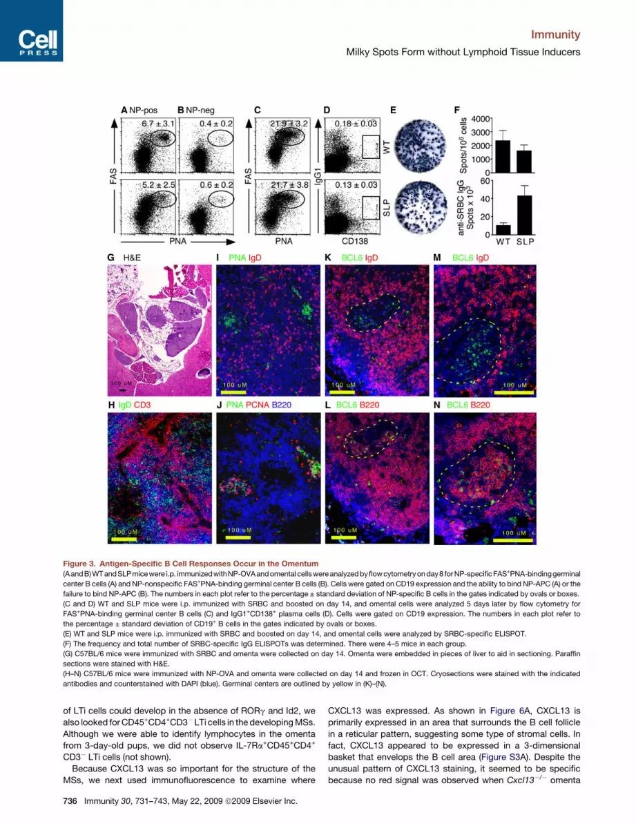
Milky Spots Support T Cell-Dependent ImmuneResponses to Peritoneal AntigensBecause peritoneal antigens and cells rapidly collected in the
MSs, we next wanted to test whether the MSs supported immune
responses to T cell-dependent antigens. Therefore, we i.p. immu-
nized WT and SLP mice with NP-OVA adsorbed to alum and
examined the response of NP-specific B cells in the omentum 7
days later. By using NP-conjugated allophycocyanin to identify
NP-specific B cells, we found that both WT and SLP mice were
able to generate NP-specific germinal center B cells in the
omentum (Figure 3A). We also gated on B cells that were not
NP specific in these same animals and found that the frequency
of germinal center B cells was much lower (Figure 3B). In fact,
the frequency of germinal center B cells in the NP-nonspecific
B cell population was similar to that seen in omental B cells
from naive mice (Figure S2). We also immunized WT and SLP
mice with SRBC and boosted them on day 28. Five days after
the boost, we found very high frequencies of germinal center
B cells (Figure 3C) and isotype-switched plasma cells (Figure 3D)
in the omenta of both groups. Although we could not demonstrate
antigen specificity of these cells by flow cytometry, we found
similar frequencies of SRBC-specific IgG-secreting cells in the
omenta of both groups (Figure 3E) and significantly (p < 0.01)
higher numbers of IgG-secreting cells in SLP mice (Figure 3F).
We next tested whether we could define germinal centers in
MSs by histology. To examine the histology of immunized
omenta, we folded individual omenta in pieces of liver, fixed
them in formalin, and embedded them in paraffin. Sections of
omenta embedded with this method could be easily stained
with H&E to see the bluish areas of the lymphocyte-filled MSs
and the lacy areas of fatty tissue surrounded by the supporting
liver (Figure 3G). By using cryosections, we also found separated
B and T cell areas in omenta from SRBC-immunized mice
(Figure 3H), as well as clusters of IgD�PNA-binding B cells
(Figure 3I). In serial sections, we found that the clusters of PNA-
binding B cells were also proliferating as demonstrated by
PCNA expression (Figure 3J). To confirm the presence of bona
fide germinal centers in the omenta of immunized mice, we also
Immunity 30, 731–743, May 22, 2009 ª2009 Elsevier Inc. 733

Immunity
Milky Spots Form without Lymphoid Tissue Inducers
Figure 2. The Milky Spots of the Omentum Collect Peritoneal Cells and Antigens
(A and B) Omenta were obtained from naive WT and SLP mice and whole mounts were probed with antibodies to B220 and PNAd. Images were obtained as
optical sections with the Zeiss Apotome.
(C and D) C57BL/6 mice were i.p. immunized with ovalbumin coupled to Alexa Fluor 594. Omenta were collected 2 or 4 hr later and whole mounts were probed
with antibodies to CD11b.
(E) 1 3 106 CFSE-labeled SRBC were i.p. injected and the omenta were collected 4 hr later. Whole mounts were probed with antibodies to CD11b. The high-
magnification image is an optical section obtained with the Zeiss Apotome.
(F) Omental cells from SRBC-injected and control mice were analyzed by flow cytometry. The plots show all events collected. The inset in the top panel shows the
profile of CFSE-SRBC prior to injection. The numbers in each plot refer to the percentage ± standard deviation of events in the CFSE-SRBC gate.
(G) Omental cells from SRBC-injected and control mice were analyzed by flow cytometry. The plots show live leukocytes and gated into five populations based on
CD11c and CD11b expression.
734 Immunity 30, 731–743, May 22, 2009 ª2009 Elsevier Inc.

Immunity
Milky Spots Form without Lymphoid Tissue Inducers
probed cryosections with antibodies to BCL6 (Figures 3K–3N),
which is a marker of germinal center B cells (Cattoretti et al.,
1995). We found clusters of BCL6+IgD� cells surrounded by
BCL6�IgD+ cells (Figures 3K and 3M). Serial sections revealed
that these clusters consisted of large blast cells that expressed
B220 on the cell surface and BCL6 in the nucleus (Figures 3L
and 3N). These data demonstrate that the MSs support local
germinal center B cell responses to peritoneal antigens.
We next tested whether CFSE-labeled OVA-specific CD4+ T
cells could respond in the MSs. We found that OTII cells in both
WT and SLP mice immunized with alum and LPS (no OVA)
predominantly expressed low amounts of CD25 (Figure 4A, top
row) and high amounts of CD62L (Figure 4B, top row). In contrast,
OTII cells in WT and SLP mice that were immunized with NP-OVA
36 hr previously rapidly increased their expression of CD25 and
reduced their expression of CD62L (Figures 4A and 4B, middle
rows). Changes in surface expression of these markers coincided
with the first cell divisions of the OTII cells, although cell division
was not required for either increased CD25 expression or
decreased CD62L expression (Figures 4A and 4B, middle rows).
OTII proliferation continued rapidly in both WT and SLP mice and
most cells had divided once or twice by 42 hr after immunization
(Figure 4C). We also tested whether CD8+ T cells responded to
NP-OVA. As shown in Figure 4D, OVA-specific CD8+ T cells could
be found in the MSs of both WT and SLP chimeras 7 days after
immunization with NP-OVA in alum. The frequency of OVA-
responding CD8+ T cells was relatively low, probably because
NP-OVA is an exogenous antigen that is poorly presented by
cross-priming. These data demonstrate that T cell responses
can also occur in the MSs of the omentum, even in mice that
lack spleen, LNs, and Peyer’s patches.
We next wanted to determine whether effector or memory
T cell generated in other locations recirculated through the peri-
toneal cavity and the omentum. To test this possibility, we first
intranasally infected C57BL/6 mice with influenza virus and
looked in the peritoneal cavity and omentum for recirculating
influenza-specific CD8+ T cells. We found influenza-specific
(tetramer-binding) CD8 T cells in both the peritoneal lavage and
omentum of mice that had been infected with influenza 21 days
previously (Figure 4E). We also orally infected IL-4 reporter
(4-get) mice with larvae of the intestinal helminth Heligmoso-
moides polygyrus and looked in the peritoneal lavage and
omentum for recirculating Th2 cells. Because the 4-get mice
are transgenic for a reporter construct that expresses GFP under
the control of the IL-4 promoter (Mohrs et al., 2001), Th2 cells
should express GFP. We found GFP-expressing CD4+ T cells in
both the peritoneal lavage and omentum on day 28 after infection
(Figure 4F). Thus, antigen-experienced CD4+ and CD8+ T cells
recirculate through the peritoneal cavity and omentum, even
when these cells were primed outside of the peritoneal cavity.
Development and Architecture of Milky SpotsWe next examined the role of lymphotoxin-a (LTa) and the homeo-
static chemokines CXCL13, CCL21, and CCL19 in the develop-
ment and organization of the MSs. We found that the MSs were
well developed in both C57BL/6 and PLT mice, which are homo-
zygous for the paucity of lymph node T cells (plt) mutation, which
inactivates the CCL19 and CCL21 genes that are expressed
in secondary lymphoid organs (Nakano and Gunn, 2001).
However, the MSs were much smaller or even absent in Lta�/�
and Cxcl13�/� mice (Figures 5A–5C), consistent with previous
observations (Ansel et al., 2002). In particular, the omenta of
Lta�/� and Cxcl13�/� mice had very small or absent B cell areas
and lacked detectable PNAd+ HEVs (Figure 5C). Given that MSs
are found in SLP mice (Figure 2B), it seems that the reconstitution
of Lta�/�mice with normal bone marrow restores the structure of
the MSs, suggesting that the defects in the MSs of Lta�/�mice are
not due to an absolute blockade in development.
Because the MSs in the omenta of Lta�/� and Cxcl13�/�mice
were architecturally abnormal, we hypothesized that LTa and
CXCL13 were involved in a positive feedback loop similar to
that described in other lymphoid organs (Ansel et al., 2000). To
test this possibility, we extracted RNA from the omenta of naive
C57BL/6, Lta�/�, Cxcl13�/�, and PLT mice and analyzed chemo-
kine mRNA expression by quantitative PCR. To our surprise, we
found that the expression of CXCL12, CCL21, CCL19, and
CXCL13 as well as LTb and TNF-a was essentially normal in
the omentum of Lta�/� mice (Figure 5D). In contrast, we found
that whereas the expression of CXCL12 and CCL21 was normal
in the omenta of Cxcl13�/� mice, the expression of CCL19,
CXCL13, LTb, and TNF-a were very reduced (Figure 5D). Finally,
we found that the expression of CCL19 and CCL21 was reduced
in the omenta of PLT mice, consistent with the plt mutation, but
that the expression of the other chemokines and cytokines that
we tested was normal in the omentum of PLT mice (Figure 5D).
These data demonstrate that even though CXCL13 is very
important for the development of the MSs, its expression is not
controlled by LTa.
We next tested whether the formation of the MSs required LTi
cells, which are absent in mice deficient in retinoic acid related
orphan receptor-g (Rorc�/�mice) and in mice that lack the inhib-
itor of differentiation-2 (Id2�/� mice) (Sun et al., 2000; Yokoto
et al., 1999). We found well-developed MSs with large B cell folli-
cles in both Rorc�/� mice and Id2�/� mice (Figures 5E and 5F),
suggesting that the development of MSs in the omentum occurs
independently of LTi cells. Because it is possible that some types
(H) Total cells from the omenta of SRBC-injected and control mice were enumerated and the number of cells in each of the gated populations was calculated.
(I) GFP-expressing EL4 cells were cultured in vitro for 2 hr with or without pertussis toxin (PTX) and then i.p. injected into C57BL/6 recipients. Omenta were
collected 2 or 4 hr later and whole mounts were examined by fluorescence microscopy.
(J) Whole mounts of omenta from EL4-GFP-injected mice were probed with B220 to visualize MSs. Images are optical sections obtained with the Zeiss Apotome.
(K) The frequency of PTX-treated and untreated EL4-GFP cells in the omenta was determined by flow cytometry.
(L) The number of PTX-treated and untreated EL4-GFP cells in the omenta was calculated. There were 4–5 mice per group.
(M) B cells were purified from GFP-transgenic mice and mixed 2:1 with purified, bodipy-labeled T cells from C57BL/6 mice. The cell mixture was cultured for 2 hr
with or without PTX and 1 3 107 total cells/mouse were i.p. injected into C57BL/6 recipients. The omenta were collected 2 and 4 hr after injection and whole
mounts were analyzed by fluorescence microscopy.
(N) The frequencies of PTX-treated and untreated B and T cells in the omenta were determined by flow cytometry.
(O) The numbers of PTX-treated and untreated B and T cells in the omenta were calculated. There were 4–5 mice per group.
Immunity 30, 731–743, May 22, 2009 ª2009 Elsevier Inc. 735
Immunity
Milky Spots Form without Lymphoid Tissue Inducers
Figure 3. Antigen-Specific B Cell Responses Occur in the Omentum
(A andB) WT andSLPmicewere i.p. immunizedwithNP-OVAandomental cellswere analyzed byflowcytometry onday8 forNP-specific FAS+PNA-bindinggerminal
center B cells (A) and NP-nonspecific FAS+PNA-binding germinal center B cells (B). Cells were gated on CD19 expression and the ability to bind NP-APC (A) or the
failure to bind NP-APC (B). The numbers in each plot refer to the percentage ± standard deviation of NP-specific B cells in the gates indicated by ovals or boxes.
(C and D) WT and SLP mice were i.p. immunized with SRBC and boosted on day 14, and omental cells were analyzed 5 days later by flow cytometry for
FAS+PNA-binding germinal center B cells (C) and IgG1+CD138+ plasma cells (D). Cells were gated on CD19 expression. The numbers in each plot refer to
the percentage ± standard deviation of CD19+ B cells in the gates indicated by ovals or boxes.
(E) WT and SLP mice were i.p. immunized with SRBC and boosted on day 14, and omental cells were analyzed by SRBC-specific ELISPOT.
(F) The frequency and total number of SRBC-specific IgG ELISPOTs was determined. There were 4–5 mice in each group.
(G) C57BL/6 mice were immunized with SRBC and omenta were collected on day 14. Omenta were embedded in pieces of liver to aid in sectioning. Paraffin
sections were stained with H&E.
(H–N) C57BL/6 mice were immunized with NP-OVA and omenta were collected on day 14 and frozen in OCT. Cryosections were stained with the indicated
antibodies and counterstained with DAPI (blue). Germinal centers are outlined by yellow in (K)–(N).
of LTi cells could develop in the absence of RORg and Id2, we
also looked for CD45+CD4+CD3�LTi cells in the developing MSs.
Although we were able to identify lymphocytes in the omenta
from 3-day-old pups, we did not observe IL-7Ra+CD45+CD4+
CD3� LTi cells (not shown).
Because CXCL13 was so important for the structure of the
MSs, we next used immunofluorescence to examine where
736 Immunity 30, 731–743, May 22, 2009 ª2009 Elsevier Inc.
CXCL13 was expressed. As shown in Figure 6A, CXCL13 is
primarily expressed in an area that surrounds the B cell follicle
in a reticular pattern, suggesting some type of stromal cells. In
fact, CXCL13 appeared to be expressed in a 3-dimensional
basket that envelops the B cell area (Figure S3A). Despite the
unusual pattern of CXCL13 staining, it seemed to be specific
because no red signal was observed when Cxcl13�/� omenta

Immunity
Milky Spots Form without Lymphoid Tissue Inducers
were stained with the same antibodies used in Figure 6A
(Figure S3B). In addition, we did not observe any red signal
when C57BL/6 omenta were stained with either the secondary
antibody alone (Figure S3C) or with a different primary antibody
followed by the secondary antibody (Figure S3D). CXCL13
expression is often associated with FDCs (Cyster et al., 2000),
so we next tested whether markers of FDCs colocalized with
CXCL13 via serial sections (Figures 6A–6F). We found that
FDCM1 was expressed in a reticular pattern surrounding the
B cell area (Figure 6B) similar to where we observed CXCL13
expression. However, we observed scattered staining only with
FDCM2 (Figure 6C). Moreover, CD21 was primarily expressed
on B cells and was only expressed on a few scattered non-B
cells (Figure 6D). Interestingly, a few CD35-expressing cells
were observed in the center of the B cell area (Figure 6E). Despite
the highly unusual staining patterns of FDC markers in omentum,
Figure 4. Antigen-Specific T Cell Responses
in the Omentum
(A–C) Purified OTII CD4+ T cells were labeled with
CFSE and i.p. injected into WT and SLP recipient
mice. Recipients were immunized 4 hr later with
100 mg OVA and 10 mg LPS adsorbed to alum or
with 10 mg LPS and alum as indicated. Omenta
were obtained 36 or 42 hr later and omental cells
were analyzed by flow cytometry for CD4, CFSE,
and CD25 (A) or CD4, CFSE, and CD62L (B). The
plots shown are gated on CD4+ T cells. CFSE dilu-
tion in the transferred CD4 T cells was measured
by flow cytometry (C). There were 4–5 mice per
group and omenta were pooled prior to analysis.
The data shown are representative of three inde-
pendent experiments.
(D) WT and SLP mice were immunized with 100 mg
OVA and 10 mg LPS adsorbed to alum. OVA-
specific CD8+ T cells were identified by tetramer
binding in the omenta of immunized mice. The
plots shown are gated on CD8+ T cells.
(E) C57BL/6 mice were intranasally infected with
influenza, and single-cell suspensions from perito-
neal cavity and omenta were analyzed by flow
cytometry 21 days after infection. Histograms
show the frequency of CD8+ T cells in the live
leukocyte population. Dot plots were gated on
CD8+ T cells and the boxes indicate influenza
nucleoprotein-specific cells.
(F) 4-get IL-4 reporter mice were orally infected
with H. polygyrus larvae, and single-cell suspen-
sions from peritoneal cavity and omenta were
analyzed by flow cytometry 28 days after infection.
Histograms show the frequency of CD4+ T cells in
the live leukocyte population. Dot plots were gated
on CD4+ T cells and the boxes indicate Th2 cells
based on GFP expression (B).
the same combination of antibodies used
at the same time on splenic sections
revealed typical staining patterns of
FDCs in the center of B cell follicles
(Figures 6G–6J). Despite the apparent
absence of conventional FDC networks,
however, we did observe a network
of ERTR7+ stromal cells throughout the
MSs (Figure 6K). Interestingly, CD11b+ macrophages, which
are known to express CXCL13 in the peritoneal cavity (Ansel
et al., 2002), were found around the outside of the MSs (Figures
6F and 6L), whereas CD11c+ cells were found in the center of the
MSs similar to where the B cells are located (Figure 6D). Thus,
the cellular architecture and pattern of chemokine expression
in the MSs are different than in conventional lymphoid organs.
Milky Spots Support Somatic Hypermutation and AffinityMaturationThe lack of a well-defined conventional FDC network made us
question whether B cells responding to antigen could be selected
for high-affinity antigen receptors in the MSs. To test whether
somatic hypermutation and affinity selection were occurring in
the MSs of the omentum, we immunized WT and SLP mice with
NP-OVA on day 0 and amplified NP-specific IgG1 heavy chain
Immunity 30, 731–743, May 22, 2009 ª2009 Elsevier Inc. 737

Immunity
Milky Spots Form without Lymphoid Tissue Inducers
Figure 5. Milky Spot Development Requires LTa and CXCL13, but Not LTi Cells
(A) Omenta were obtained from naive C57BL/6, Lta�/�, Cxcl13�/�, or PLT mice as indicated and whole mounts were probed with antibodies to B220 and CD11b.
(B) Paraffin sections of omenta were stained with H&E.
(C) Paraffin sections of omenta were probed with antibodies to B220 and PNAd.
(D) RNA was extracted from the omenta of naive C57BL/6, Lta�/�, Cxcl13�/�, or PLT mice as indicated and assayed for the expression of CXCL12, CXCL13,
CCL19, CCL21, LTb, and TNF-a by quantitative PCR. The expression of each mRNA was normalized first to GAPDH and then normalized to the expression
in WT animals, which was set at 1.
(E and F) Omenta were obtained from naive Rorc�/� or Id2�/�mice as indicated and paraffin sections were stained with H&E (E) or probed with antibodies to B220
and PNAd (F). Images are representative of 3–5 mice per group.
V regions by using primers specific for V186.2 and the IgG1
constant region on day 7, day 14, and day 19 (5 days after boost-
ing) and sequenced individual clones. Although we observed only
a few mutations in the V regions of IgG heavy chains from either
WT or SLP mice on day 7, more sequences in both groups had
accumulated mutations by days 14 and 19 after immunization
(Figure 7A). However, more of the sequences from WT mice
had mutations and the number of mutations in each V region
was also higher in sequences from WT mice than those from
SLP mice on days 14 and 19 (Figure 7A). Furthermore, 16/25
738 Immunity 30, 731–743, May 22, 2009 ª2009 Elsevier Inc.
sequences from WT mice on day 14 exhibited the W to L mutation
at position 33 that confers about a 10-fold higher affinity for NP
(Figures S4A and S4B). In contrast, only 5/22 sequences from
SLP mice exhibited this mutation at this time. Strikingly, 11/13
sequences from WT mice exhibited the W to L mutation at day 19,
whereas 0/12 sequences from SLP mice had this mutation
(Figures S4A and S4B). The differences in sequence were not
limited to the patterns of mutations, as shown by the fact that
16/25 sequences from WT mice on day 14 had the YYYG motif
at the V-D junction, which is characteristic of NP-binding

Immunity
Milky Spots Form without Lymphoid Tissue Inducers
Figure 6. CXCL13 Is Expressed Surrounding the B Cell Areas
(A–F) Omenta were obtained from naive C57BL/6 mice and cryosections were probed with the indicated antibodies.
(G–J) Spleens were obtained from naive C57BL/6 mice and cryosections were probed with the indicated antibodies.
Cryosections in (B)–(E) were stained at the probed at the same time, under the same conditions as the corresponding cryosections in (G)–(J).
(K and L) Omenta were obtained from naive C57BL/6 mice and whole mounts were probed with the indicated antibodies. Images were generated by a combi-
nation of fluorescence (optical sections with the Zeiss apotome) and DIC. Control primary and secondary antibodies were negative. Images are representative of
3–5 mice.
sequences, whereas only 7/22 sequences from SLP mice had
this motif. Interestingly, all of the W to L mutations in sequences
from SLP mice were from those that contained the YYYG motif.
Thus, although somatic hypermutation is still evident in SLP
mice, the process of clonal selection seems to be very different.
To directly determine whether B cells responding to NP-OVA in
the MSs underwent affinity maturation, we performed NP-specific
ELISAs with plates coated with either NP2BSA or NP16BSA and
compared the ratio of IgG that bound to each as a measure of
the relative affinity of the antibody. As shown in Figure 7B, the
affinity of NP-specific IgG from both WT and SLP mice was rela-
tively low at day 10 after immunization. The affinity of NP-specific
IgG increased rapidly in WT mice and increased somewhat more
slowly in SLP mice (Figure 7B). Ultimately, however, the relative
affinity of NP-specific IgG reached similar levels in both groups
as measured in this assay. Together, these data demonstrate
that the MSs of the omentum support some aspects of T cell-
dependent B cell responses, although the process of clonal selec-
tion is unusual.
DISCUSSION
Previous work suggested that the omentum had an immunolog-
ical function and showed that plasma cells could be found in the
omentum after intraperitoneal immunization with T cell-indepen-
dent antigens (Ansel et al., 2002; Ha et al., 2006) as well as classic
T cell-dependent antigens (Dux et al., 1977; Hajdu et al., 1972;
Van Vugt et al., 1996). Nevertheless, some authors conclude
that the MSs should not be classified as secondary lymphoid
organs (Szaniawska, 1974, 1975; Van Vugt et al., 1996). Given
the lack of FDC networks in MSs (Van Vugt et al., 1996), the prev-
alence of B1 cells in the peritoneal cavity and omentum (Ansel
et al., 2002), and the altered repertoire and affinity selection of
B cells responding to antigen in the MSs of mice that lack conven-
tional lymphoid organs, we would agree with Van Vugt et al.
(1996) that the MSs are not conventional secondary lymphoid
organs. However, Van Vugt’s conclusion that they should rather
be classified as perivascular infiltrates is, in our opinion, too
limiting. Our data clearly show that the MSs of the omentum are
Immunity 30, 731–743, May 22, 2009 ª2009 Elsevier Inc. 739

Immunity
Milky Spots Form without Lymphoid Tissue Inducers
Figure 7. Somatic Hypermutation and
Affinity Selection Occurs in the Absence of
Conventional Secondary Lymphoid Organs
(A) WT and SLP mice were i.p. immunized with NP-
OVA and boosted on day 14. RNA was extracted
from omenta on days 7, 14, and 19 after immuniza-
tion. The V regions of B cells with V186.2 heavy
chains that had switched to IgG1 were amplified
by PCR, subcloned, and sequenced. The pie
charts indicate the relative proportion of
sequences that were unmutated (0) or contained
the indicated number of mutations.
(B) The titers of NP-specific total IgG were deter-
mined with ELISA plates coated with either NP16-
BSA or NP2-BSA. The relative affinity of the NP-
specific IgG is expressed as a ratio of binding to
the NP2-BSA versus NP16-BSA. There were five
mice in each group and this experiment is repre-
sentative of three independent experiments.
present prior to immunization and support B cell responses
(albeit unusual ones) as well as CD4+ and CD8+ T cell responses
to antigens that are administered i.p. Moreover, B cells as well as
CD4+ and CD8+ T cells primed in distal locations recirculate
through the peritoneal cavity and omentum, indicating that the
MSs of the omentum are sites of immune surveillance for
a wide spectrum of antigens. Together, these data suggest that
the omentum functions much more broadly as a secondary
lymphoid organ than previously realized, even though it is struc-
turally, developmentally, and functionally unique.
Unlike encapsulated LNs, which acquire antigen via afferent
lymphatics, or mucosal lymphoid tissues, which transport
antigen across mucosal epithelium, the MSs of the omentum
open directly to the peritoneal cavity and collect cells and anti-
gens suspended in the peritoneal fluid (Cui et al., 2002; Gerber
et al., 2006; Hodel, 1970). Although LPS-activated peritoneal
B1 cells use chemokine receptors to rapidly migrate to the MSs
(Ha et al., 2006), i.p. administered naive B and T cells also collect
in the MSs—even when treated with pertussis toxin, suggesting
that at least some of the movement from the peritoneal cavity
through the omentum occurs via bulk flow rather than specific
homing. Consistent with this, free SRBCs in the peritoneal cavity
can be collected in the omentum, although most are rapidly
phagocytosed by macrophages and DCs. The presence of
PNAd-expressing HEVs in the MSs also suggests that naive B
and T cells can also be recruited to the MSs from the blood. In
fact, a recent study shows that B2 cells use a4b7-MAdCAM inter-
actions to enter the MSs of the omentum from the blood (Berber-
ich et al., 2008), and other studies show that fibronectin as well as
various cell adhesion molecules are important for the entry and
migration of peritoneal cells (Cui et al., 2002). Together, these
data suggest a model in which recirculating T and B cells entering
the MSs from the blood are confronted with a parade of antigens
and cells from the peritoneal cavity, such as those released by
abdominal surgery, peritoneal dialysis, peritoneal tumor metas-
tases, and peritoneal infections. This intersection of recirculating
lymphocytes and peritoneal drainage makes the MSs ideal sites
for the initiation of local immune responses.
Unlike conventional lymphoid organs, which have B cell folli-
cles arranged around networks of FDCs (Endres et al., 1999;
Tew et al., 1997), the B cell areas of the MSs appear to lack central
740 Immunity 30, 731–743, May 22, 2009 ª2009 Elsevier Inc.
FDC networks and instead are surrounded by reticular networks
of CD11b+ and FDCM1+ cells. Although CXCL13 is normally
associated with a central reticular network in the B cell follicles
of conventional lymphoid organs (Cyster et al., 2000), CXCL13
is expressed around the outside of the B cell areas of MSs in
the same area that contains CD11b+ cells. It is likely that the
CD11b+ cells are macrophages, which are known to express
CXCL13 in the peritoneal cavity (Ansel et al., 2002), but it is
unclear whether the FDCM1+ cells are also macrophages, FDC
precursors, or some other cell type. Given that other FDC
markers do not stain in the same area and also do not detect
a well-defined reticular network in the center of the B cell areas,
it is difficult to make a firm conclusion regarding the status of
FDCs in the omentum. However, the structure of the MSs seems
inside-out relative to that of conventional lymphoid organs or
even ectopic lymphoid follicles.
Despite the unusual architecture of the milky spots, we
observe somatic hypermutation and some degree of affinity
maturation in B cell responses from mice that lack conventional
lymphoid organs, but retain MSs. Interestingly, the repertoire of
V186.2 IgG heavy chains from the omenta of NP-OVA-immu-
nized SLP mice contains some unusual features, such as a rela-
tive paucity of sequences with the YYYG motif at the V-D junction
and very few sequences that encode the W to L mutation,
despite the relatively robust accumulation of mutations overall.
Although it is tempting to conclude that affinity maturation failed
in these animals, the relative avidity for NP2BSA does increase in
SLP mice over the first 21 days after immunization, albeit more
slowly than in WT mice. Unfortunately, the ELISA-based assay
of relative affinity has a relatively narrow dynamic range and
cannot differentiate between antibodies with affinities higher
than about 10�7 M. Thus, the NP-specific antibodies generated
in WT mice could ultimately have much higher affinities than
those generated in SLP mice. Interestingly, the few sequences
from SLP mice that acquired the W to L mutation all had the
canonical YYYG motif at the V-D junction. Thus, an alternative
explanation is that the initial repertoire of B cells recruited into
the NP response is different in WT and SLP mice and that
many of the heavy chains from SLP mice lacking the YYYG motif
and the W to L substitution accumulate different mutations that
also confer higher affinity for NP.

Immunity
Milky Spots Form without Lymphoid Tissue Inducers
Immunity 30, 731–743, May 22, 2009 ª2009 Elsevier Inc. 741
The difference in frequency of W to L substitutions between
sequences from WT and SLP mice is also striking in another
way. If we assume that the sequences obtained from SLP mice
are typical of the V regions that are selected in the omentum,
then the high frequency of B cells with the W to L mutation in WT
mice must mean that those B cells were originally selected in other
lymphoid organs and that their progeny recirculated back to the
omentum. Because the sequences that have the W to L substitu-
tion vastly outnumber those that lack the W to L substitution in WT
mice, this suggests that the clones that are selected in conven-
tional lymphoid organs and recirculate to the omentum out-
compete those clones that are selected in situ. If competitive
fitness is determined by relative affinity (Dal Porto et al., 2002),
then the B cells selected in conventional lymphoid organs contain-
ing the W to L substitution would be of higher affinity that those
selected locally. If this were true, however, we should still observe
in SLP mice an increase in the frequency of sequences with the
W to L mutation between day 14 and 19 after immunization. The
fact that we do not observe this increase means that clones with
the W to L mutation are not selectively advantaged in SLP mice
and that they are competing with clones of equivalent affinity or
that affinity selection is not occurring. Although some degree of
affinity maturation can occur in the absence of either germinal
centers or immune complex deposition on FDCs (Hannum et al.,
2000; Koni and Flavell, 1999) and can occur in the disorganized
spleens of Lta�/� and Ltbr�/� mice (Futterer et al., 1998; Matsu-
moto et al., 1996a), it has long been assumed that immune
complexes trapped on the surface of FDCs facilitate B cell selec-
tion and affinity maturation. Thus, the lack of clearly identifiable
FDCs in MSs may contribute to poor affinity-based selection.
Unlike the development of the majority of secondary lymphoid
organs, the development of the MSs occurs independently of
RORg-dependent or Id2-dependent LTi cells, suggesting that
MSs use alternative cell types to trigger the differentiation of local
mesenchymal cells into stromal cells that support lymphoid
architecture. Of course the stromal elements in the MSs are very
different than those in conventional lymphoid tissues, because
the MSs lack clearly identifiable FDCs and maintain only an
ERTR7+ stromal network. Another unusual feature of milky spot
development and organization is the minimal role for LTa.
Although the MSs are clearly compromised in the absence of
LTa, their structure is completely restored in adult mice after
reconstitution with Lta+/+ hematopoietic cells, suggesting that
the defects in the MSs ofLta�/�miceare due tonondevelopmental
defect, such as the poor differentiation of HEVs (Browning et al.,
2005). This is reminiscent of the nasal associated lymphoid tissue
(NALT), which develops independently of LTa, but requires LTa to
maintain normal architecture because of its ability to promote the
expression of chemokines and trigger the differentiation of PNAd-
expressing HEVs (Fukuyama et al., 2002; Harmsen et al., 2002;
Rangel-Moreno et al., 2005). Surprisingly, however, the expres-
sion of homeostatic chemokines is normal in the omenta of Lta�/�
mice, just like the induced expression of these chemokines in the
lungs of Lta�/� mice during influenza infection (Moyron-Quiroz
et al., 2004). However, despite the minimal role of LTa in the
expression of CXCL13, CCL19, and CCL21, CXCL13 is very
important for the development of the MSs. Essentially, no B cells
are found in the omenta of Cxcl13�/�mice and the expression of
CCL19, LTb, and TNF-a is severely impaired (Ansel et al., 2002).
Taken altogether, these data demonstrate that the MSs of the
omentum are unique secondary lymphoid organs that sample
antigens from the peritoneal cavity and promote local, albeit
unusual, immune responses. These results will be important for
the understanding of how the omentum functions in response
to antigens from abdominal surgeries, peritoneal dialysis, intes-
tinal perforations, and even peritoneal tumor metastases.
EXPERIMENTAL PROCEDURES
Mice and Generation of Bone Marrow Chimeras
C57BL/6 mice and C57BL/6.129Ltatm1Dch (Lta�/�mice) (de Togni et al., 1994)
were obtained from the Jackson laboratory. Cxcl13�/� (Ansel et al., 2000)
and PLT (Nakano et al., 1997) mice were a kind donation of J. Cyster (UCSF).
Rorc�/� (Sun et al., 2000) and Id2�/� (Yokoto et al., 1999) mice were obtained
from D. Littman (NYU). IL-4 reporter (4-get) mice (Mohrs et al., 2001) were
obtained from M. Mohrs (Trudeau Institute). WT and SLP chimeric mice were
generated by lethally irradiating C57BL/6 and splenectomized Lta�/� mice
and reconstituting them with 1 3 107 whole bone marrow cells from C57BL/6
mice as described (Moyron-Quiroz et al., 2004, 2006). Mice were irradiated
with 1000 rads from a 137Cs source at 93 rad/min in two doses. Mice were
allowed to reconstitute for at least 6 weeks prior to immunization. All gene-tar-
geted mice were on the C57BL/6 background and bred in the animal facility of
Trudeau Institute. All procedures involving animals were approved by the Tru-
deau Institute Institutional Animal Care and Use Committee and were con-
ducted according to the principles outlined by the National Research Council.
Immunization and Infection
Mice were immunized i.p. with 100 mg NP-OVA or 100 mg TNP-KLH adsorbed
to alum or with 1 3 106 SRBCs in PBS or with 100 mg Alexa-594-OVA. Mice
were intranasally infected with 100 egg infectious units of A/PR8/34 influenza
or orally infected with 200 Heligmosomoides polygyrus larvae.
Isolation of RNA, PCR, and Sequencing
RNA was extracted from whole omentum via an RNeasy kit (QIAGEN). 2 mg of
DNase-treated RNA were reverse transcribed with random hexamers and
Superscript II (Invitrogen). Quantitative PCR was performed with Taqman
master mix, according to the Applied Biosystems protocol. Primers and
probes for CXCL12, CXCL13, CCL19, LTb, and TNF-a were obtained from
Applied Biosystems. Primers for CCL21 (50-AGACTCAGGAGCCCAAAGCA-30
and 50-GTTGAAGCAGGGCAAGGGT-30) and GAPDH (50CTCGTCCCGTAGA
CAAAATGG-30 and 50-AATCTCCACTTTGCCACTGCA-30) were synthesized
by IDT. The probes for CCL21 (50-FAM-CCACCTCATGCTGGCCTCCGT-
BHQ-30 ) and for GAPDH (50-FAM-CGGATTTGGCCGTATTGGGCG-BHQ-30)
were synthesized by Biosearch Technology. The ABI Prism 7700 Taqman
instrument was available through the molecular biology core facility at the
Trudeau Institute. cDNAs of VH regions from NP-specific BCRs were amplified
with a forward primer specific for V186.2 NP 5-1 CATGCTCTTCTTGGCAGC
AACAGC and a reverse primer specific for IgG1 NP 3-1 GTGCACACCGCTGG
ACAGGGATCC. PCR reactions were performed for 1 min at 94�C, 1.5 min
at 58�C, and 2 min at 72�C for 25 cycles with Expand High-Fidelity Taq
polymerase from Roche. The PCR product was cloned into TOPO vector
(Invitrogen) and DNA from individual colonies was purified and sequenced.
Sequencing was performed by Polymorphic DNA Technologies and sequence
data was aligned with Sequencher software.
Cell Purification and Labeling
B cells, total T cells, and CD4+ T cells were purified by positive selection with
MACS beads. For B cells and CD4+ T cells, anti-B220 or anti-CD4 magnetic
beads (Miltenyi Biotec) were added directly to single-cell suspensions at
25 ml beads per 1 3 108 total cells in 100 ml final volume and incubated on
ice for an additional 15 min on ice. For total T cells, cells were first incubated
with biotinylated anti-CD4 and anti-CD8 followed by streptavidin magnetic
beads (Miltenyi Biotec). Cells were washed, filtered, and passed over a type
LS+ magnetic column (Miltenyi Biotec). After washing, the magnetically bound
cells were eluted from the column with the supplied plunger. Purified OTII
CD4+ T cells were labeled with 5 mM carboxyfluoroscein succinimidyl ester

Immunity
Milky Spots Form without Lymphoid Tissue Inducers
(CFSE) in PBS for 10 min at 37�C. Total T cells were labeled with 0.2 mM 4, 4
difluoro-5(2thienyl)-4-bora-3a, 4a-diaza-s-indacene-3-propionic acid, succi-
nimidyl ester (BODIPY 558/568) in PBS for 10 min at 37�C. In some cases, cells
were incubated at 3 3 107/ml with 100 ng/ml pertussis toxin for 2 hr at 37�C in
complete RPMI with 2% FBS, 10 mM HEPES, 2 mM glutamine, 100 mg/ml
streptomycin, and 100 U/ml penicillin.
Flow Cytometry
Omenta were digested with Collagenase D (Roche) at 37�C for 1 hr and
mechanically disrupted by passage through a wire mesh, and live leukocytes
were obtained by density gradient centrifugation with Lympholyte-Poly (Cedar-
lane). Fc receptors were blocked with 10 mg/ml 2.4G2, followed by staining with
antibodies or MHC class I tetramers. The H-2Db class I tetramer containing
NP366-374 peptide were generated by the Trudeau Institute Molecular Biology
Core Facility as described (Flynn et al., 1998; Murali-Krishna et al., 1998).
NP-specific B cells were identified with NP-allophycocyanin (NP-APC). Fluoro-
chrome-labeled antibodies to CD8, CD4, CD62L, CD138, FAS, and CD19 were
obtained from BD Biosciences. FITC-labeled PNA was obtained from Sigma.
ELISAs and ELISPOTs
Plates were coated with NP(16)-BSA, NP(2)-BSA, or TNP(5)-BSA at 1 mg/ml and
blocked with 10 mg/ml BSA. Serum samples were initially diluted 100-fold in
PBS with 10 mg/ml BSA and 0.1% Tween-20 and then serially diluted in
3-fold steps in the same buffer and applied to the coated plates. Bound Ig
was detected with horseradish peroxidase-conjugated goat anti-mouse IgM
or goat anti-mouse IgG from Southern Biotechnology Associates. IgG1,
IgG2a, IgG2b, and IgG3 were detected with HRP-conjugated isotype-specific
antibodies from BD Biosciences. Purified monoclonal TNP antibodies (A111-3
and G155-178 BD Biosciences) were used as standards for the anti-TNP
ELISAs. Antibody titers are defined as the dilution required to reduce a positive
signal to 3-fold above background. ELISPOTs were performed as described
(Rangel-Moreno et al., 2005).
Immunofluorescence
Whole omenta were placed in 24-well plates and blocked with 10 mg/ml 2.4G2
in PBS with 5% BSA for 1 hr at 4�C while rocking. Fluorochrome-conjugated
antibodies were then added for an additional 4 hr at 4�C while rocking. Omen-
tums were washed 3 times for 15 min each in PBS with 5% BSA and then wet
mounted in PBS with 5% BSA. Omenta were also prepared for histology by
folding strips of omentum into a piece of liver, fixing the tissue with 10% neutral
buffered formalin, and embedding in paraffin. Paraffin sections were treated
with Antigen Retrieval Solution (DAKO) at 96�C for 20 min and then cooled at
room temperature for 20 min before blocking and staining as described above.
Sections probed with fluorescent antibodies were counterstained with DAPI.
Cryosections were obtained by cutting tissues in the presence of dry ice so
that the fatty tissue of the omentum could be cut. All tissues were viewed
with a Zeiss Axiovert 200 m microscope with Apotome. Green and red fluores-
cence was captured with appropriate filter sets and in some cases, combined
with images obtained with DIC. Some images were obtained as optical sections
with the Apotome. Images were recorded with a Zeiss AxioCam digital
camera with Zeiss AxioVision 4.5 software and saved as TIFF files. Antibodies
to CD21, CD11b, CD11C, B220, CD35, and PNAd were obtained from BD
Biosciences and conjugated to either Alexa Fluor 488 or Alexa Fluor with kits
from Invitrogen. Purified goat anti-mouse CXCL13 (R&D Systems)was detected
with Alexa Fluor 594-conjugated donkey anti-goat IgG (Molecular Probes).
SUPPLEMENTAL DATA
Supplemental Data include four figures and can be found with this article online
at http://www.cell.com/immunity/supplemental/S1074-7613(09)00186-1.
ACKNOWLEDGMENTS
The authors would like to thank F. Lund for helpful discussions during the
course of this work and M. Tighe for help with histology and microscopy.
This work was supported by the Trudeau Institute and NIH grants HL69409,
AI072689, and AI061511.
742 Immunity 30, 731–743, May 22, 2009 ª2009 Elsevier Inc.
Received: June 18, 2007
Revised: September 16, 2008
Accepted: March 6, 2009
Published online: May 7, 2009
REFERENCES
Ansel, K.M., Ngo, V.N., Hayman, P.L., Luther, S.A., Forster, R., Sedgwick, J.D.,
Browning, J.L., Lipp, M., and Cyster, J.G. (2000). A chemokine-driven positive
feedback loop organizes lymphoid follicles. Nature 406, 309–314.
Ansel, K.M., Harris, R.B., and Cyster, J.G. (2002). CXCL13 is required for B1
cell homing, natural antibody production, and body cavity immunity. Immunity
16, 67–76.
Banks, T.A., Rouse, B.T., Kerley, M.K., Blair, P.J., Godfrey, V.L., Kuklin, N.A.,
Bouley, D.M., Thomas, J., Kanangat, S., and Mucenski, M.L. (1995). Lympho-
toxin a deficient mice: effects on secondary lymphoid organ development and
humoral immune responsiveness. J. Immunol. 155, 1685–1693.
Beelen, R.H., Fluitsma, D.M., and Hoefsmit, E.C. (1980). The cellular composi-
tion of omentum milky spots and the ultrastructure of milky spot macrophages
and reticulum cells. J. Reticuloendothel. Soc. 28, 585–599.
Beelen, R.H., Oosterling, S.J., van Egmond, M., van den Born, J., and Zareie,
M. (2005). Omental milky spots in peritoneal pathophysiology (spots before
your eyes). Perit. Dial. Int. 25, 30–32.
Benedict, C.L., and Kearney, J.F. (1999). Increased junctional diversity in fetal
B cells results in a loss of protective anti-phosphorylcholine antibodies in adult
mice. Immunity 10, 607–617.
Berberich, S., Dahne, S., Schippers, A., Peters, T., Muller, W., Kremmer, E.,
Forster, R., and Pabst, O. (2008). Differential molecular and anatomical basis
for B cell migration into the peritoneal cavity and omental milky spots. J. Immu-
nol. 180, 2196–2203.
Browning, J.L., Allaire, N., Ngam-Ek, A., Notidis, E., Hunt, J., Perrin, S., and
Fava, R.A. (2005). Lymphotoxin-beta receptor signaling is required for the
homeostatic control of HEV differentiation and function. Immunity 23, 539–550.
Cattoretti, G., Chang, C.C., Cechova, K., Zhang, J., Ye, B.H., Falini, B., Louie,
D.C., Offit, K., Chaganti, R.S., and Dalla-Favera, R. (1995). BCL-6 protein is
expressed in germinal-center B cells. Blood 86, 45–53.
Cui, L., Johkura, K., Liang, Y., Teng, R., Ogiwara, N., Okouchi, Y., Asanuma, K.,
and Sasaki, K. (2002). Biodefense function of omental milky spots through cell
adhesion molecules and leukocyte proliferation. Cell Tissue Res. 310, 321–330.
Cyster, J.G., Ansel, K.M., Rief, K., Hyman, P.L., Tang, H.L., Luther, S.A., and
Ngo, V.N. (2000). Foliicular stromal cells and lymphocyte homing to follicles.
Immunol. Rev. 176, 181–193.
Dal Porto, J.M., Haberman, A.M., Kelsoe, G., and Shlomchik, M.J. (2002). Very
low affinity B cells form germinal centers, become memory B cells, and partic-
ipate in secondary immune responses when higher affinity competition is
reduced. J. Exp. Med. 195, 1215–1221.
Das, S.K. (1976). The size of the human omentum and methods of lengthening
it for transplantation. Br. J. Plast. Surg. 29, 170–244.
de Togni, P., Goellner, J., Ruddle, N.H., Streeter, P.R., Fick, A., Mariathasan,
S., Smith, S.C., Carlson, R., Shornick, L.P., Strauss-Schoenberger, J., et al.
(1994). Abnormal development of peripheral lymphoid organs in mice deficient
in lymphotoxin. Science 264, 703–707.
Di Paolo, N., Sacchi, G., Garosi, G., Sansoni, E., Bargagli, L., Ponzo, P.,
Tanganelli, P., and Gaggiotti, E. (2005). Omental milky spots and peritoneal
dialysis—review and personal experience. Perit. Dial. Int. 25, 48–57.
Dux, K., Janik, P., and Szaniawska, B. (1977). Kinetics of proliferation, cell
differentiation and IgM secretion in the omental lymphoid organ of B10/Sn
mice following intraperitoneal immunization with sheep erythrocytes. Cell.
Immunol. 32, 97–109.
Dux, K., Rouse, R.V., and Kyewski, B. (1986). Composition of the lymphoid cell
populations from omental milky spots during the immune response in C57BL/
Ka mice. Eur. J. Immunol. 16, 1029–1032.
Endres, R., Alimzhanov, M.B., Plitz, T., Futterer, A., Kosco-Vilbois, M.H.,
Nedospasov, S.A., Rajewski, K., and Pfeffer, K. (1999). Mature follicular

Immunity
Milky Spots Form without Lymphoid Tissue Inducers
dendritic cell networks depend on expression of lymphotoxin b receptor by
radioresistant stromal cells and of lymphotoxin b and tumor necrosis factor
by B cells. J. Exp. Med. 189, 159–167.
Fedorko, M.E., Hirsch, J.G., and Fried, B. (1971). Studies on transport of macro-
molecules and small particles across mesothelial cells of the mouse omentum.
II. Kinetic features and metabolic requirements. Exp. Cell Res. 69, 313–323.
Flynn, K.J., Belz, G.T., Altman, J.D., Ahmed, R., Woodland, D.L., and Doherty,
P.C. (1998). Virus-specific CD8+ T cells in primary and secondary influenza
pneumonia. Immunity 8, 683–691.
Fukuyama, S., Hiroi, T., Yokota, Y., Rennert, P.D., Yanagita, M., Kinoshita, N.,
Terawaki, S., Shikina, T., Yamamoto, M., Kurono, Y., and Kiyono, H. (2002).
Initiation of NALT organogenesis is independent of the IL-7R, LTbetaR, and
NIK signaling pathways but requires the Id2 gene and CD3(-)CD4(+)CD45(+)
cells. Immunity 17, 31–40.
Futterer, A., Mink, K., Luz, A., Kosco-Vilbois, M.H., and Pfeffer, K. (1998). The
lymphotoxin b receptor controls organogenisis and affinity maturation in
peripheral lymphoid tissues. Immunity 9, 59–70.
Gerber, S.A., Rybalko, V.Y., Bigelow, C.E., Lugade, A.A., Foster, T.H., Frelin-
ger, J.G., and Lord, E.M. (2006). Preferential attachment of peritoneal tumor
metastases to omental immune aggregates and possible role of a unique
vascular microenvironment in metastatic survival and growth. Am. J. Pathol.
169, 1739–1752.
Goldsmith, H.S., Griffith, A.L., Kupferman, A., and Catsimpoolas, N. (1984).
Lipid angiogenic factor from omentum. JAMA 252, 2034–2036.
Ha, S.A., Tsuji, M., Suzuki, K., Meek, B., Yasuda, N., Kaisho, T., and Fagara-
san, S. (2006). Regulation of B1 cell migration by signals through Toll-like
receptors. J. Exp. Med. 203, 2541–2550.
Hajdu, I., Holub, M., and Trebichavsky, I. (1972). The sequence of appearance
of antibodies in mouse omentum plasma cells. Exp. Cell Res. 75, 219–230.
Hannum, L.G., Haberman, A.M., Anderson, S.M., and Shlomchik, M.J. (2000).
Germinal center initiation, variable gene region hypermutation, and mutant B
cell selection without detectable immune complexes on follicular dendritic
cells. J. Exp. Med. 192, 931–942.
Harmsen, A., Kusser, K., Hartson, L., Tighe, M., Sunshine, M.J., Sedgwick,
J.D., Choi, Y., Littman, D.R., and Randall, T.D. (2002). Organogenesis of nasal
associated lymphoid tissue (NALT) occurs independently of lymphotoxin-
a (LTa) and retinoic acid receptor-related orphan receptor-g, but the organiza-
tion of NALT is LTa-dependent. J. Immunol. 168, 986–990.
Hodel, C. (1970). Ultrastructural studies on the absorption of protein markers
by the greater omentum. Eur. Surg. Res. 2, 435–449.
Koni, P.A., and Flavell, R.A. (1999). Lymph node germinal centers form in the
absence of follicular dendritic cells networks. J. Exp. Med. 189, 855–864.
Krist, L.F., Eestermans, I.L., Steenbergen, J.J., Hoefsmit, E.C., Cuesta, M.A.,
Meyer, S., and Beelen, R.H. (1995a). Cellular composition of milky spots in
the human greater omentum: an immunochemical and ultrastructural study.
Anat. Rec. 241, 163–174.
Krist, L.F., Kerremans, M., Koenen, H., Blijleven, N., Eestermans, I.L., Calame,
W., Meyer, S., and Beelen, R.H. (1995b). Novel isolation and purification
method permitting functional cytotoxicity studies of macrophages from milky
spots in the greater omentum. J. Immunol. Methods 184, 253–261.
Krist, L.F., Kerremans, M., Broekhuis-Fluitsma, D.M., Eestermans, I.L., Meyer,
S., and Beelen, R.H. (1998). Milky spots in the greater omentum are predom-
inant sites of local tumour cell proliferation and accumulation in the peritoneal
cavity. Cancer Immunol. Immunother. 47, 205–212.
Matsumoto, M., Lo, S.F., Carruthers, C.J.L., Min, J., Mariathasan, S., Huang,
G., Plas, D.R., Martin, S.M., Geha, R.S., Nahm, M.H., and Chaplin, D.D.
(1996a). Affinity maturation without germinal centers in lymphotoxin a deficient
mice. Nature 382, 462–466.
Matsumoto, M., Mariathasan, S., Nahm, M.H., Baranyay, F., Peschon, J.J.,
and Chaplin, D.D. (1996b). Role of lymphotoxin and the type 1 TNF receptor
in the formation of germinal centers. Science 271, 1289–1291.
Mohrs, M., Shinkai, K., Mohrs, K., and Locksley, R.M. (2001). Analysis of type 2
immunity in vivo with a bicistronic IL-4 reporter. Immunity 15, 303–311.
Morrison, R. (1906). On the functional aspects of the greater omentum. BMJ 1,
76–78.
Moyron-Quiroz, J.E., Rangel-Moreno, J., Kusser, K., Hartson, L., Sprague, F.,
Goodrich, S., Woodland, D.L., Lund, F.E., and Randall, T.D. (2004). Role of
inducible bronchus associated lymphoid tissue (iBALT) in respiratory immu-
nity. Nat. Med. 10, 927–934.
Moyron-Quiroz, J.E., Rangel-Moreno, J., Hartson, L., Kusser, K., Tighe, M.P.,
Klonowski, K.D., Lefrancois, L., Cauley, L.S., Harmsen, A.G., Lund, F.E., and
Randall, T.D. (2006). Persistence and responsiveness of immunologic memory
in the absence of secondary lymphoid organs. Immunity 25, 643–654.
Murali-Krishna, K., Altman, J.D., Suresh, M., Sourdive, D.J., Zajac, A.J., Miller,
J.D., Slansky, J., and Ahmed, R. (1998). Counting antigen-specific CD8 T cells:
A reevaluation of bystander activation during viral infection. Immunity 8,
177–187.
Nakano, H., and Gunn, M.D. (2001). Gene duplications at the chemokine locus
on mouse chromosome 4: Multiple strain-specific haplotypes and the deletion
of secondary lymphoid-organ chemokine and EBI-1 ligand chemokine genes
in the plt mutation. J. Immunol. 166, 361–369.
Nakano, H., Tamura, T., Yoshimoto, T., Yagita, H., Miyasaka, M., Butcher,
E.C., Nariuchi, H., Kakiuchi, T., and Matsuzawa, A. (1997). Genetic defect in
T lymphocyte-specific homing into peripheral lymph nodes. Eur. J. Immunol.
27, 215–221.
Portis, B. (1924). Role of omentum of rabbits, dogs and guinea-pigs in antibody
production. J. Infect. Dis. 34, 159–185.
Rangel-Moreno, J., Moyron-Quiroz, J., Kusser, K., Hartson, L., Nakano, H.,
and Randall, T.D. (2005). Role of CXC chemokine ligand 13, CC chemokine
ligand (CCL) 19, and CCL21 in the organization and function of nasal-associ-
ated lymphoid tissue. J. Immunol. 175, 4904–4913.
Roberts, K.B. (1955). Antibody formation in the omentum. Br. J. Exp. Pathol.
36, 357–362.
Solvason, N., and Kearney, J.F. (1992). The human fetal omentum: A site of
B cell generation. J. Exp. Med. 175, 397–404.
Solvason, N., Chen, X., Shu, F., and Kearney, J.F. (1992). The fetal omentum in
mice and humans. A site enriched for precursors of CD5 B cells early in devel-
opment. Ann. N Y Acad. Sci. 651, 10–20.
Sun, Z., Unutmaz, D., Zou, Y.-R., Sunshine, M.J., Pierani, A., Brenner-Morton,
S., Mebius, R.E., and Littman, D.R. (2000). Requirement for RORg in thymo-
cyte survival and lymphoid organ development. Science 288, 2369–2373.
Szaniawska, B. (1974). Changes in the greater omentum of mice of different
strains after intraperitoneal immunization with sheep erythrocytes. I. Produc-
tion of IgM immunoglobulins in milky spots. Arch. Immunol. Ther. Exp. (Warsz.)
22, 585–593.
Szaniawska, B. (1975). Changes in the greater omentum of mice of different
strains following intraperitoneal strains following intraperitoneal immunization
with sheep erythrocytes. III. Determination of the percentage of thymus-
dependent cells in the omental milky spots in mice by the application of
anti-o serum. Arch. Immunol. Ther. Exp. (Warsz.) 23, 19–24.
Tew, J.G., Wu, J., Qin, D., Helm, S., Burton, G.F., and Szakal, A.K. (1997).
Follicular dendritic cells and presentation of antigen and costimulatory signals
to B cells. Immunol. Rev. 156, 39–52.
Vakil, M., Briles, D.E., and Kearney, J.F. (1991). Antigen-independent selection
of T15 idiotype during B-cell ontogeny in mice. Dev. Immunol. 1, 203–212.
Van Vugt, E., Van Rijthoven, E.A., Kamperdijk, E.W., and Beelen, R.H. (1996).
Omental milky spots in the local immune response in the peritoneal cavity of
rats. Anat. Rec. 244, 235–245.
Walker, F.C., and Rogers, A.W. (1961). The greater omentum as a site of anti-
body synthesis. Br. J. Exp. Pathol. 42, 222–231.
Williams, R., and White, H. (1986). The greater omentum: Its applicability to
cancer surgery and cancer therapy. Curr. Probl. Surg. 23, 789–865.
Yokoto, Y., Mansouri, A., Mori, S., Sugawara, S., Adachi, S., Nishikawa, S.I.,
and Gruss, P. (1999). Development of peripheral lymphoid organs and natual
killer cells depends on the helix-loop-helix inhibitor Id2. Nature 397, 702–706.
Immunity 30, 731–743, May 22, 2009 ª2009 Elsevier Inc. 743