nursing care plans - core
TRANSCRIPT
CRITICAL CARE NURSING CARE PLANS
Sheree Corner
Contributor:
Barbara Sage1 R.N., M.s., C.C.R.N.
DELMAR -- THOMSON LEARNING
Africa Australia Canada Denmark Japan Mexico New Zealand Philippines Puerto Rico Singapore Spain United Kingdom United States
NOTICE TO THE READER
Publisher does not warrant or guarantee any of the products described herein or perform any independent analysis in connection with any of the product information contained herein. Publisher does not assume, and expressly disclaims, any obligation to obtain and include information other than that provided to it by the manufacturer. The reader is expressly warned to consider and adopt all safety precautions that might be indicated by the activities herein and to avoid all potential hazards. By following the instructions contained herein, the reader willingly assumes all risks in connection with such instructions. The Publisher makes no representation or warranties of any kind, including but not limited to, the warranties of fitness for particular purpose or merchantability, nor are any such representations implied with respect to the material set forth herein, and the publisher takes no responsibility with respect to such material. The publisher shall not be liable for any special, consequential, or exemplary damages resulting, in whole or part, from the readers' use of, or reliance upon, this material.
COPYRIGHT 0 1998 Delmar, a division of Thomson Learning, Inc. The Thomson Learningm is a trademark used herein under license.
Printed in the United States of America 2 3 4 5 6 7 8 9 1 0 X X X 0 5 0 4 0 3 0 2 0 1 0 0
For more information, contact Delmar, 3 Columbia Circle, PO Box 15015, Albany, NY 12212-0515; or find us on the World Wide Web at http://www.delmar.com
International Division List Asia AustraliaINew Zealand: Latin America: Thomson Learning NelsonfI'homson Learning Thomson Learning 60 Albert Street, #15-01 102 Dodds Street Seneca, 53 Albert Complex South Melbourne, Victoria 3205 Colonia Polanco Singapore 189969 Australia 11560 Mexico D.F. Mexico Tel: 65 336 6411 Fax: 65 336 7411
Tel: 61 39 685 4111 Fax: 61 39 685 4199
Tel: 525-281-2906 F a : 525-281-2656
Japan: UWEuropeMiddle East Canada: Thomson Learning Thomson Learning NelsonlThomson Learning Palaceside Building 5F Berkshire House 1120 Birchmount Road 1-1-1 Hitotsubashi, Chiyoda-ku 168-173 High Holbom Scarborough, Ontario Tokyo 100 0003 Japan London Canada M1 K 5G4 Tel: 813 5218 6544 WClV 7AA United Kingdom Tel: 416-752-9100 Fax: 813 5218 6551 Tel: 44 171 497 1422 F a : 416-752-8102
Fax: 44 171 497 1426
ALL RIGHTS RESERVED. No part of this work covered by the copyright hereon may be reproduced or used in any form or by any means-graphic, electronic, or mechanical, including photocopying, recording, taping, Web distribution or information storage and retrieval systems-without the written permission of the publisher.
For permission to use material from this text or product contact us by Tel (800) 730-2214; Fax (800) 730-2215; www.thomsonrights.com
Library of Congress Cataloging-in-Publication Data
ISBN: 1-56930-035-6

1
TABLE OF CONTENTS

This Page Intentionally Left Blank
CARDlOUASCUwl SYSTEM .................................................................................................................................... 1
Congestive Heart Failure ............................................................................................................................................................. 3 Myocardial Infaction (MI) ........................................................................................................................................................ 13 Pericarditis ................................................................................................................................................................................. 25 Infective Endocarditis (IE) ......................................................................................................................................................... 31 Hypertension ............................................................................................................................................................................. 39 Thrombophlebitis ...................................................................................................................................................................... 47 Intra-Aortic Balloon Pump (IABP) ............................................................................................................................................ 53
Aortic Aneurysm ........................................................................................................................................................................ 77
Pacemakers ................................................................................................................................................................................ 59 Cardiac Surgery ......................................................................................................................................................................... 67
RESPlRlYrORY SYSTEM ........................................................................................................................................ 83
Adult Respiratory Distress Syndrome (ARDS) .......................................................................................................................... 85 Chronic Obstructive Pulmonary Disease (C 0 P D) ................................................................................................................. 91
Pneumonia .............................................................................................................................................................................. 105 Pneumothorax ......................................................................................................................................................................... 111 Status Asthmaticus ................................................................................................................................................................... 117 Mechanic al Ventilation ............................................................................................................................................................ 121
Pulmonary Embolism ................................................................................................................................................................ 99
NEUROLOGICAL SYSTEM ..................................................................................................................................... 137
C V A ...................................................................................................................................................................................... 139 Head Injuries ........................................................................................................................................................................... 147 Spinal Cord Injuries ................................................................................................................................................................. 159 Guillain-Barre Syndrome ......................................................................................................................................................... 169 Status Epilepticus ..................................................................................................................................................................... 175 Meningitis ............................................................................................................................................................................... 181 Ventriculostomy/ICP Monitoring ............................................................................................................................................ 185 Endarterectomy ....................................................................................................................................................................... 189
GASTROMTESTINAUHEPATIC SYSTEM .................................................................................................................. 191
Gastrointestinal Bleeding ......................................................................................................................................................... 193 Esophageal Varices ................................................................................................................................................................... 199 Hepatitis .................................................................................................................................................................................. 203 Pancreatitis .............................................................................................................................................................................. 207 Acute Abdomen/ Abdominal Trauma ...................................................................................................................................... 211 Liver Failure ............................................................................................................................................................................. 217
HEMATOLOGIC SYSTEM ..................................................................................................................................... 223
Disseminated Intravascular Coagulation (DIC) ....................................................................................................................... 225 H E L L P Syndrome ............................................................................................................................................................... 229 Anemia .................................................................................................................................................................................... 233
iv
RENWDOCRINE SYSTEMS .............................................................................................................................. 237
Acute Renal Failure (ARF) ....................................................................................................................................................... 239 Diabetic Ketoacidosis (D K A) ................................................................................................................................................ 251 Hyperglycemic Hyperosmolar Nonketotic Coma (H H N K) ................................................................................................ 261 Syndrome of Inappropriate ADH Secretion (SIADH) ............................................................................................................. 265 Diabetes Insipidus (DI) ........................................................................................................................................................... 269 Pheochromocytoma ................................................................................................................................................................. 273 Thyrotoxicosis (Thyroid Storm) ............................................................................................................................................. 281
MUSCULOSKELETAL SYSTEM .............................................................................................................................. 289
Fractures .................................................................................................................................................................................. 291 Amputation ............................................................................................................................................................................. 299 Fat Embolism .......................................................................................................................................................................... 307
IHTEGUMarrARY SYSTEM ................................................................................................................................... 311
Frostbite/Hypothermia ............................................................................................................................................................ 313 Malignant Hyperthermia ......................................................................................................................................................... 319 Burns/Thermal Injuries ........................................................................................................................................................... 323
OTHER ............................................................................................................................................................. 329
Multiple Organ Dysfunction Syndrome (MODS) ................................................................................................................... 331 Acute Poisoning/Drug Overdose .............................................................................................................................................. 337 Snakebite ................................................................................................................................................................................. 343 Transplants .............................................................................................................................................................................. 349 Cardiogenic Shock ................................................................................................................................................................... 357
INDEN OF NURSING DIAGNOSES ........................................................................................................................... 361
REFERENCES ..................................................................................................................................................... 365
CARDIOVASCULAR SYSTEM 1
Congestive Heart Failure
Myocardial Infarction (MI)
Pericarditis
Infective Endocarditis (IE)
Hypertension
Thrombophlebitis
Intra-Aortic Balloon Pump (IABP)
Pacemakers
Cardiac Surgery
Aortic Aneurysm

This Page Intentionally Left Blank
CARDIOVASCULAR SYSTEM 3
Congestive Heart Failure Heart failure is the inability of the heart to supply blood f l ~ w to meet physiologic demands, without utilizing compensatory changes. There may be failure involving one or both sides of the heart, and over time, causes the development of pulmonary and systemic congestion and complica- tions. Congestive heart failure, or CHF, is a common complication after myocardial infarction and can be attributed to one-third of the deaths of patients with MIs. Usually following MI, the heart failure is left-sided since most infarctions involve damage to the left ventricle.
Heart failure can also be classified as acute or chronic. In chronic heart failure, the body experi- ences a gradual development as the heart becomes unable to pump a sufficient amount of blood to meet the body’s demands. Chronic heart failure can become acute without any overt cause.
Often, the patient will have no early symptoms of left-sided heart failure. Symptoms of decreased cardiac output will develop once the heart fails to pump enough blood into the systemic circulation. The pressure in the left ventricle increases, which in turn causes retrograde increases of pressure in the left atrium because of the increased difficulty for blood to enter the atrium from the pulmonary veins. Blood backs up in the lung vasculature, and when the pulmonary capillary pressure is exceeded by the oncotic pressure of the proteins in the plasma fluid (usually > 30 mmHg), the fluid leaks into the interstitial spaces. When this fluid moves into the alveoli, shortness of breath, coughing, and crackles (rales) occur, and the patient progresses into overt pulmonary edema, with the classic sign of coughing up copious amounts of pink frothy sputum.
Right-sided heart failure is usually caused by left- sided heart failure, but can also be caused by pulmonary emboli, pulmonary hypertension, COPD, and the presence of right ventricular infarctions.
The lungs can accept a certain amount of fluid build-up, but eventually, if no intervention is taken, the pressure in the lungs increases to the point whereby the right ventricle cannot eject its blood into the lungs. The right ventricle fails and then the blood in the right atrium cannot drain completely, and thus cannot accept the total amount of blood from the vena cavae. Venous pooling occurs with the impairment of venous blood flow, and eventually the organs become congested with venous blood.
Treatment of heart failure involves attempts to improve contractility of the ventricle by use of positive inotropic drugs, decrease of afterload by the use of nitrates and vasodilators, and in some instances, by use of the IABP, and decrease of pre- load by the use of diuretics, IV nitroglycerin, and fluid/sodium restrictions.
Oxygen: to increase available oxygen supply
Morphine: used to induce vasodilation, decrease venous return to the heart, reduce pain and anxi- ety, and decrease myocardial oxygen consumption
Cardiac glycosides: digitalis (Digoxin, Lanoxin) PO or IV to increase the force and strength of ventricular contractions and to decrease rate of contractions in order to increase cardiac output
Diuretics: furosemide (Lasix) PO or IV, chloroth- iazide (Diuril) PO, bumetanide (Bumex) PO or IV to promote excess fluid removal, to decrease edema and pulmonary venous pressure by preventing sodium and water reabsorption
4 CRITICAL CARE NURSING CARE PLANS
Vasodilators: hydralazine (Apresoline) PO or IV, isosorbide dinitrate (Isordil) SL or PO, prazosin (Minipress) PO, minoxidil (Loniten) PO, diazox- ide (Hyperstat) IV, sodium nitroprusside (Nipride) IV, nitroglycerine (Nitrostat, Tridil) PO, SL, IV to relax vascular smooth muscle, decrease preload and afterload, decrease oxygen demand, decrease systemic vascular resistance, and increase venous capacitance
Renin-angiotensin system inhibitors: captopril (Capoten) PO used to inhibit angiotensin converting enzyme to reduce the production of angiotensin I1 to enable the decrease in vasocon- striction and to reduce afterload
Inotropic agents: dopamine, dobutamine (Dobutrex) IV, amrinone (Inocor) IV used to increase myocardial contractility, without increas- ing the heart rate, to produce peripheral vasodilation and decrease preload and afterload
Electrolytes: mainly potassium to replace that which is lost during diuretic therapy
Laboratory: electrolyte levels to monitor for imbalances; renal profiles to monitor for kidney function problems; digoxin levels to monitor for toxicity; platelet count to monitor for thrombocy- topenia from amrinone
Chest x-ray: shows any enlargement of the heart and pulmonary vein, presence of pulmonary ederna or pleural effusion
Electrocardiography: used to monitor for dysrhythmias which may occur as a result of the heart failure or as a result of digitalis toxicity
Echocardiography: used to study structural abnor- malities and blood flow through the heart
Intra-aortic balloon pump: decreases the workload on the heart, decreases myocardial oxygen demand, increases coronary perfusion,
decreases afterload, decreases preload, improves cardiac output and tissue perfusion
NURSING CARE PLANS Fluid volume excess
Related to: increased sodium and water retention, decreased organ perfusion, compromised regulatory mechanisms, decreased cardiac output, increased ADH production
Defining characteristics: edema, weight gain, intake greater than output, increased blood pres- sure, increased heart rate, shortness of breath, dyspnea, orthopnea, crackles (rales), S 3 gallop, oliguria, jugular vein distention, pleural effusion, specific gravity changes, altered electrolyte levels
Outcome Criteria
Blood pressure will be maintained within normal limits and edema will be absent or minimal in all body parts.
Fluid volume will be stabilized with balanced intake and output.
INTERVENTIONS RATIONALES
Monitor vital signs and hemo- dynamic readings if available.
Fluid volume excess will cause increases in blood pressure, and CVP and pulmonary artery pressures, and these changes will be reflected from the development of pulmonary congestion and heart failure.
Auscultate lungs for presence of crackles (rales), or other adventitious breath sounds. Observe for presence of cough, increased dyspnea, tachypnea, orthopnea or paroxysmal noc- turnal dyspnea.
May indicate pulmonary edema from cardiac decornpensation and pulmonary congestion. Pulmon- ary edema symptoms reflect left- sided hearr failure. Right- sided heart failure may have slower onset, but symptoms of dyspnea, orthopnea, and cough are more difficult to reverse.
CARDIOVASCULAR SYSTEM 5
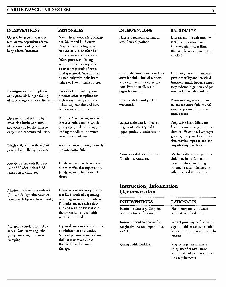
INTERVENTIONS RATIONALES INTERVENTIONS RATIONALES Observe for jugular vein dis- tention and dependent edema. Note presence of generalized body edema (anasarca).
Investigate abrupt complaints of dyspnea, air hunger, feeling of impending doom or suffocation.
Determine fluid balance by measuring intake and output, and observing for decreases in output and concentrated urine.
Weigh daily and notify M D of greater than 2 lblday increase.
Provide patient with fluid in- take of 2 Llday, unless fluid restriction is warranted.
Administer diuretics as ordered (furosemide, hydralazine, spiro- lactone with hydrochlorothiazide).
Monitor electrolyte for imbal- ances. Note increasing lethar- gy, hypotension, or musde cramping.
May indicate impending conges- tive failure and fluid excess. Peripheral edema begins in feet and ankles, or other de- pendent areas and ascends as failure progresses. Pitting will usually occur only after 10 or more pounds of excess fluid is retained. Anasarca will be seen only with right heart failure or bi-ventricular failure.
Excessive fluid build-up can promote other complications such as pulmonary edema or pulmonary embolus and inter- vention must be immediate.
Renal perfusion is impaired with excessive fluid volume, which causes decreased cardiac output leading to sodium and water retention and oliguria.
Abrupt changes in weight usually indicate excess fluid.
Fluids may need to be restricted due to cardiac decompensation. Fluids maintain hydration of tissues.
Drugs may be necessary to cor- rect fluid overload depending on emergent nature of problem. Diuretics increase urine flow rate and may inhibit reabsorp- tion of sodium and chloride in the renal tubules.
Hypokalemia can occur with the administration of diuretics. Signs of potassium and sodium deficits may occur due to fluid shifts with diuretic therapy.
Place and maintain patient in semi-Fowler's position.
Auscultate bowel sounds and ob- serve for abdominal distention, anorexia, nausea, or constipa- tion. Provide small, easily- digestible meals.
Measure abdominal girth if warranted.
Palpate abdomen for liver en- largement; note any right upper quadrant tenderness or pain.
Assist with dialysis or hemo- filtration as warranted.
Diuresis may be enhanced by recumbent position due to increased glomerular filtra- tion and decreased production ofADH.
C H F progression can impair gastric motility and intestinal function. Small, frequent meals may enhance digestion and pre- vent abdominal discomfort.
Progressive right-sided heart failure can cause fluid to shift into the peritoneal space and cause ascites.
Progressive heart failure can lead to venous congestion, ab- dominal distention, liver engor- gement, and pain. Liver func- tion may be impaired and can impede drug metabolism.
Mechanically removing excess fluid may be performed to rapidly reduce circulating volume in cases refractory to other medical therapeutics.
Instruction, Information, Demonstration
INTERVENTIONS RATIONALES Instruct patient regarding diet- ary restrictions of sodium.
Fluid retention is increased with intake of sodium.
Instruct patient to observe for weight changes and report these to MD.
Weight gain may be firsr overt sign of fluid excess and should be monitored to prevent compli- cations.
Consult with dietitian. May be required to ensure adequacy of caloric intake with fluid and sodium resrric- tion requirements.
6 CRITICAL CARE NURSING CARE PLANS
Outcome Criteria INTERVENTIONS RATIONALES
Instruct patient in medications
dose, effect, side effects, . . . .
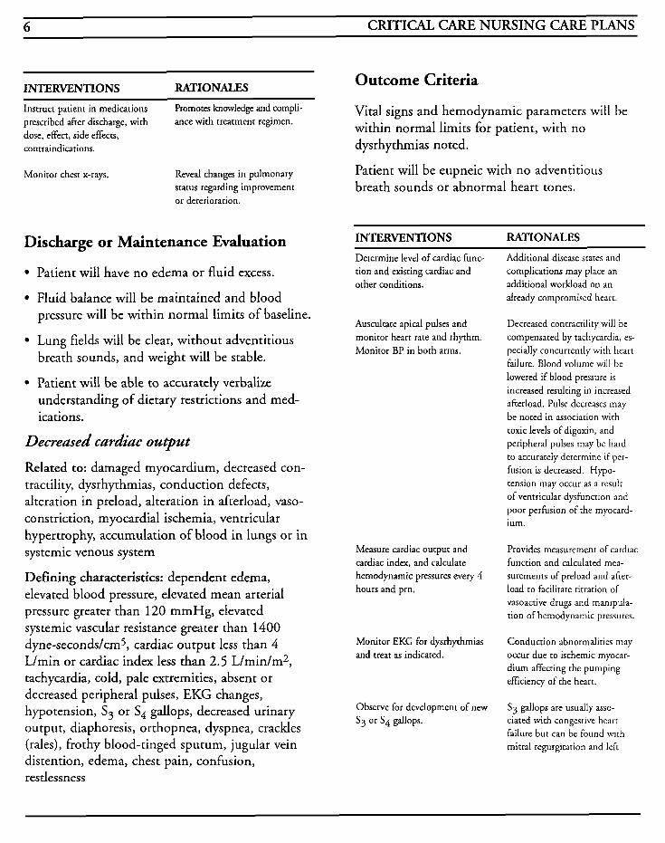
Promotes knowledge and compli- Vital signs and hemodynamic parameters will be within normal limits for patient, with no prescribed after discharge, with ance with treatment regimen.
contraindications. dysrhythmias noted.
Monitor chest x-rays. Reveal changes in pulmonary status regarding improvement or deterioration.
Patient will be eupneic with no adventitious breath sounds or abnormal heart tones.
INTERVENTIONS RATIONALES Discharge or Maintenance Evaluation
Patient will have no edema or fluid excess.
Fluid balance will be maintained and blood pressure will be within normal limits of baseline.
Lung fields will be clear, without adventitious breath sounds, and weight will be stable.
Patient will be able to accurately verbalize understanding of dietary restrictions and med- ications.
Decreased cardiac output
Related to: damaged myocardium, decreased con- tractility, dysrhythmias, conduction defects, alteration in preload, alteration in afterload, vaso- constriction, myocardial ischemia, ventricular hypertrophy, accumulation of blood in lungs or in systemic venous system
Defining characteristics: dependent edema, elevated blood pressure, elevated mean arterial pressure greater than 120 mmHg, elevated systemic vascular resistance greater than 1400 dyne-secondslcm5, cardiac output less than 4 L/min or cardiac index less than 2.5 L/min/m2, tachycardia, cold, pale extremities, absent or decreased peripheral pulses, EKG changes, hypotension, S 3 or S, gallops, decreased urinary output, diaphoresis, orthopnea, dyspnea, crackles (rales), frothy blood-tinged sputum, jugular vein distention, edema, chest pain, confusion, restlessness
Determine level of cardiac func- tion and existing cardiac and other conditions.
Additional disease states and complications may place an additional workload on an already compromised heart.
Auscultate apical pulses and monitor heart rate and rhythm. Monitor BP in both arms.
Decreased contractility will be compensated by tachycardia, es- pecially concurrently with heart failure. Blood volume will be lowered if blood pressure is increased resulting in increased afterload. Pulse decreases may be noted in association with toxic levels of digoxin, and peripheral pulses may be hard to accurately determine if per- fusion is decreased. Hypo- tension may occur as a result of ventricular dysfunction and poor perfusion of rhe myocard- ium.
Measure cardiac output and cardiac index, and calculate hernodynamic pressures every 4 hours and prn.
Monitor EKG for dysrhythmias and treat as indicated.
Observe for development of new S3 or S4 gallops.
Provides measurement of cardiac function and calculated mea- surements of preload and after- load to facilitate titration of vasoactive drugs and manipula- tion of hemodynamic pressures.
Conduction abnormalities may occur due to ischemic myocar- dium affecting the pumping efficiency of the heart.
S3 gallops are usually asso- ciated with congestive hearr failure but can be found with rnitral regurgitation and left
CARDIOVASCULAR SYSTEM 7
INTERVENTIONS RATIONALES INTERVENTIONS RATIONALES
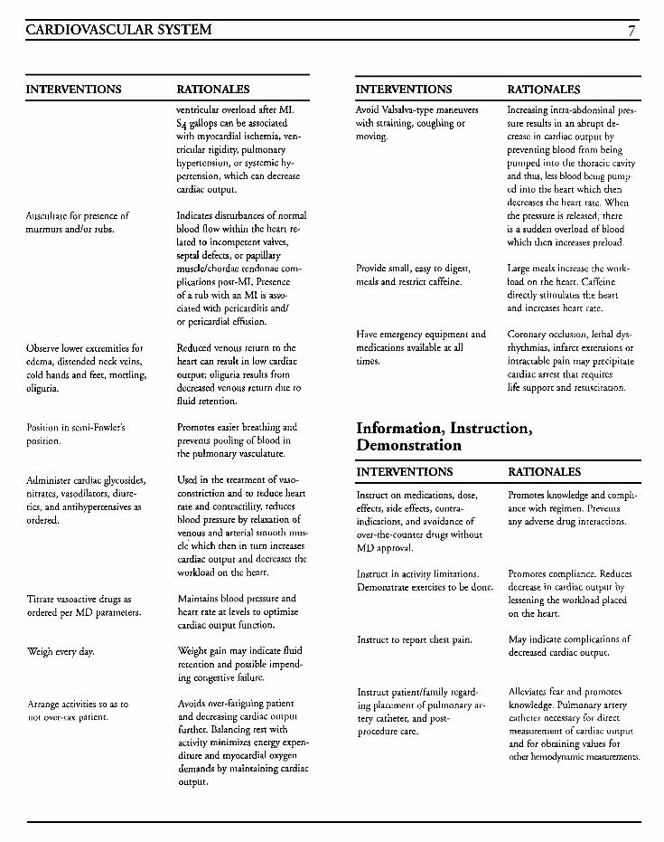
ventricular overload after MI. S4 gallops can be associated with myocardial ischemia, ven- tricular rigidity, pulmonary hypertension, or systemic hy- pertension, which can decrease cardiac output.
Avoid Valsalva-type maneuvers with straining, coughing or moving.
Increasing intra-abdominal pres- sure results in an abrupt de- crease in cardiac output by preventing blood from being pumped into the thoracic cavity and thus, less blood being pump- ed into the heart which then decreases the heart rate. When the pressure is released, there is a sudden overload of blood which then increases preload.
Auscultate for presence of murmurs andlor rubs.
Indicates disturbances of normal blood flow within the heart re- lated to incompetent valves, septal defects, or papillary muscle/chordae tendonae com- plications post-MI. Presence of a rub with an MI is asso- ciated with pericarditis and/ or pericardial effusion.
Provide small, easy to digest, meals and restrict caffeine.
Large meals increase the work- load on the heart. Caffeine directly stimulates the heart and increases heart rate.
Coronary occlusion, lethal dys- rhythmias, infarct extensions or intractable pain may precipitate cardiac arrest that requires life support and resuscitation.
Have emergency equipment and medications available at all times.
Observe lower extremities for edema, distended neck veins, cold hands and feet, mottling, oliguria.
Reduced venous return to the heart can result in low cardiac output; oliguria results from decreased venous return due to fluid retention.
Information, Instruction, Demonstration
Position in semi-Fowler’s position.
Promotes easier breathing and prevents pooling of blood in the pulmonary vasculature.
INTERVENTIONS RATIONALES Administer cardiac glycosides, nitrates, vasodilators, diure- tics, and antihypertensives as ordered.
Used in the treatment of vaso- constriction and to reduce heart rate and contractility, reduces blood pressure by relaxation of venous and arterial smooth mus- cle’ which then in turn increases cardiac output and decreases the workload on the heart.
Instruct on medications, dose, effects, side effects, contra- indications, and avoidance of over-the-counter drugs without MD approval.
Promotes knowledge and compli- ance with regimen. Prevents any adverse drug interactions.
Instruct in activity limitations. Demonstrate exercises to be done.
Promotes compliance. Reduces decrease in cardiac output by lessening the worMoad placed on the heart.
Maintains blood pressure and heart rate at levels to optimize cardiac output function.
Titrate vasoactive drugs as ordered per MD parameters.
Instruct to report chest pain. May indicate complications of decreased cardiac output. Weigh every day. Weight gain may indicate fluid
retention and possible impend- ing congestive failure.
Instruct patiendfamily regard- ing placement of pulmonary ar- tery catheter, and post- procedure care.
Alleviates fear and promotes knowledge. Pulmonary artery catheter necessary for direct measurement of cardiac output and for obtaining values for other hemodynamic measurements.
Arrange activities so as to not over-tax patient.
Avoids over-fatiguing patient and decreasing cardiac output further. Balancing rest with activity minimizes energy expen- diture and myocardial oxygen demands by maintaining cardiac output.
8 CRITICAL CARE NURSING CARE PLANS
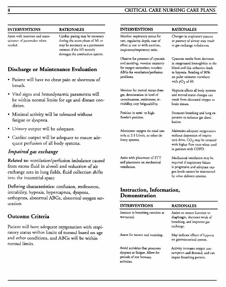
INTERVENTIONS RATIONALES INTERVENTIONS RATIONALES Assist with insertion and main- tainance of pacemaker when needed.
Cardiac pacing may be necessary during the acute phase of MI or may be necessary as a permanent measure if the MI severely damages the conduction system.
Discharge or Maintenance Evaluation
Patient will have no chest pain or shortness of breath.
Vital signs and hemodynamic parameters will be within normal limits for age and disease con- dition.
Minimal activity will be tolerated without fatigue or dyspnea.
Urinary output will be adequate.
Cardiac output will be adequate to ensure ade- quate perfusion of all body systems.
Impaired gas exchange
Related to: ventilationlperfusion imbalance caused from excess fluid in alveoli and reduction of air exchange area in lung fields, fluid collection shifts into the interstitial space
Defining characteristics: confusion, restlessness, irritability, hypoxia, hypercapnea, dyspnea, orthopnea, abnormal ABGs, abnormal oxygen sat- uration
Outcome Criteria
Patient will have adequate oxygenation with respi- ratory status within limits of normal based on age and other conditions, and ABGs will be within normal limits.
Monitor respiratory status for rate, regularity, depth, ease of effort at rest or with exertion, inspiratory/expiratory ratio.
Observe for presence of cyanosis and mottling; monitor oximetry for oxygen saturation; monitor ABGs for ventilation/perfusion problems.
Monitor for mental status chan- ges, deterioration in level of consciousness, restlessness, ir- ritability, easy fatigueability.
Position in semi- or high- Fowler's position.
Administer oxygen via nasal can- nula at 2-3 L/min, or other de- livery systems.
Assist with placement of ETT and placement on mechanical ventilation.
Changes in respiratory pattern or patency of airway may result in gas exchange imbalances.
Cyanosis results from decreases in oxygenated hemoglobin in the blood and this reduction leads to hypoxia. Reading of 90% on pulse oximeter correlates with pO2 of GO.
Hypoxia affects all body systems and mental status changes can result from decreased oxygen to brain tissues.
Promotes breathing and lung ex- pansion to enhance gas distri- bution.
Maintains adequate oxygenation without depression of respira- tory drive. CO2 may be retained with higher flow rates when used in patients with COPD.
Mechanical ventilation may be required if respiratory failure is progressive and adequate oxy- gen levels cannot be maintained by other delivery systems.
Instruction, Information, Demonstration
INTERVENTIONS RATIONALES Instruct in breathing exercises as warranted. diaphragm, decreases work of
Assists to restore function to
breathing, and improves gas exchange.
Assess for nausea and vomiting. May indicate effects of hypoxia on gastrointestinal system.
Avoid activities that promotes dyspnea or fatigue. Allow for periods of rest between activities.
Activity increases oxygen con- sumption and demand, and can impair breathing pattern.
CARDIOVASCULAR SYSTEM 9
INTERVENTIONS RATIONALES Outcome Criteria Instruct in safety concerns with oxygen use. and provides knowledge.
Promotes safety with oxygen
Instruct patiendfamily in need for placement on mechanical ventilation, what to expect, unknown. what benefits are to be received, what potential problems may be encountered.
Promotes knowledge and decreases anxiety and fear of the
Discharge or Maintenance Evaluation
Patient will exhibit no ventilation/perfusion imbalances.
Patient will be eupneic with no adventitious breath sounds.
ABGs will be within acceptible ranges for patient with adequate oxygenation of all tissues.
Patient will be able to verbalize/demonstrate the correct use of oxygen.
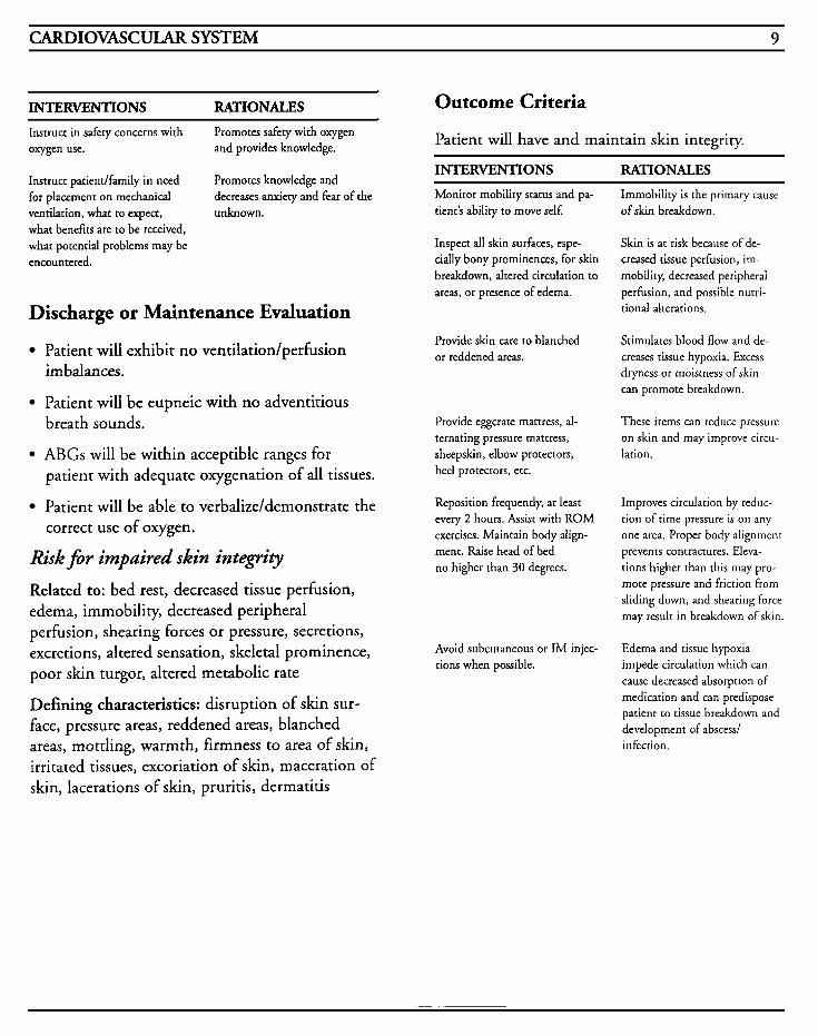
Risk f i r impaired skin intephy
Related to: bed rest, decreased tissue perfusion, edema, immobility, decreased peripheral perfusion, shearing forces or pressure, secretions, excretions, altered sensation, skeletal prominence, poor skin turgor, altered metabolic rate
Defining characteristics: disruption of skin sur- face, pressure areas, reddened areas, blanched areas, mottling, warmth, firmness to area of skin, irritated tissues, excoriation of skin, maceration of skin, lacerations of skin, pruritis, dermatitis
Patient will have and maintain skin integrity.
INTERVENTIONS RATIONALES Monitor mobility status and pa- tient's ability to move self.
Inspect all skin surfaces, espe- cially bony prominences, for skin breakdown, altered circulation to areas, or presence of edema.
Provide skin care to blanched or reddened areas.
Provide eggcrate mattress, al- ternating pressure mattress, sheepskin, elbow protectors, heel protectors, etc.
Reposition frequently, at least every 2 hours. Assist with ROM exercises. Maintain body align- ment. Raise head of bed no higher than 30 degrees.
Avoid subcutaneous or IM injec- tions when possible.
Immobility is the primary cause of skin breakdown.
Skin is at risk because of de- creased tissue perfusion, im- mobility, decreased peripheral perfusion, and possible nutri- tional alterations.
Stimulates blood flow and de- creases tissue hypoxia. Excess dryness or moistness of skin can promote breakdown.
These items can reduce pressure on skin and may improve circu- lation.
Improves circulation by reduc- tion of time pressure is on any one area. Proper body alignment prevents contractures. Eleva- tions higher than this may pro- mote pressure and friction from sliding down, and shearing force may result in breakdown of skin.
Edema and tissue hypoxia impede circulation which can cause decreased absorption of medication and can predispose patient to tissue breakdown and development of abscess/ infection.
10 CRITICAL CARE NURSING CARE PLANS
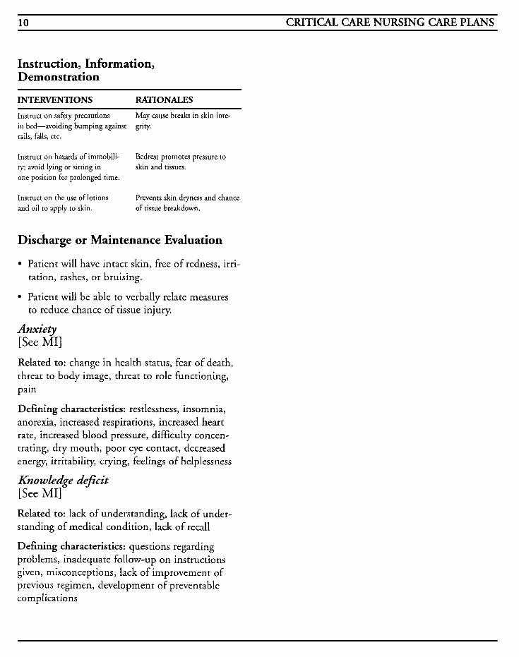
Instruction, Information, Demonstration
INTERVENTIONS RATIONALES
Instruct on safety precautions in bed-avoiding bumping against grity. rails, falls, etc.
May cause breaks in skin inte-
Instruct on hazards of immobili- ty; avoid lying or sitting in one position for prolonged time.
Instruct on the use of lotions and oil to apply to skin.
Bedrest promotes pressure to skin and tissues.
Prevents skin dryness and chance of tissue breakdown.
Discharge or Maintenance Evaluation
Patient will have intact skin, free of redness, irri- tation, rashes, or bruising.
Patient will be able to verbally relate measures
Anxiety [See MI] Related to: change in health status, fear of death, threat to body image, threat to role functioning,
to reduce chance of tissue injury.
pain
Defining characteristics: restlessness, insomnia, anorexia, increased respirations, increased heart rate, increased blood pressure, difficulty concen- trating, dry mouth, poor eye contact, decreased energy, irritability, crying, feelings of helplessness
Knowledge deficit [See MI] Related to: lack of understanding, lack of under- standing of medical condition, lack of recall
Defining characteristics: questions regarding problems, inadequate follow-up on instructions given, misconceptions, lack of improvement of previous regimen, development of preventable complications
CARDIOVASCULAR SYSTEM 11
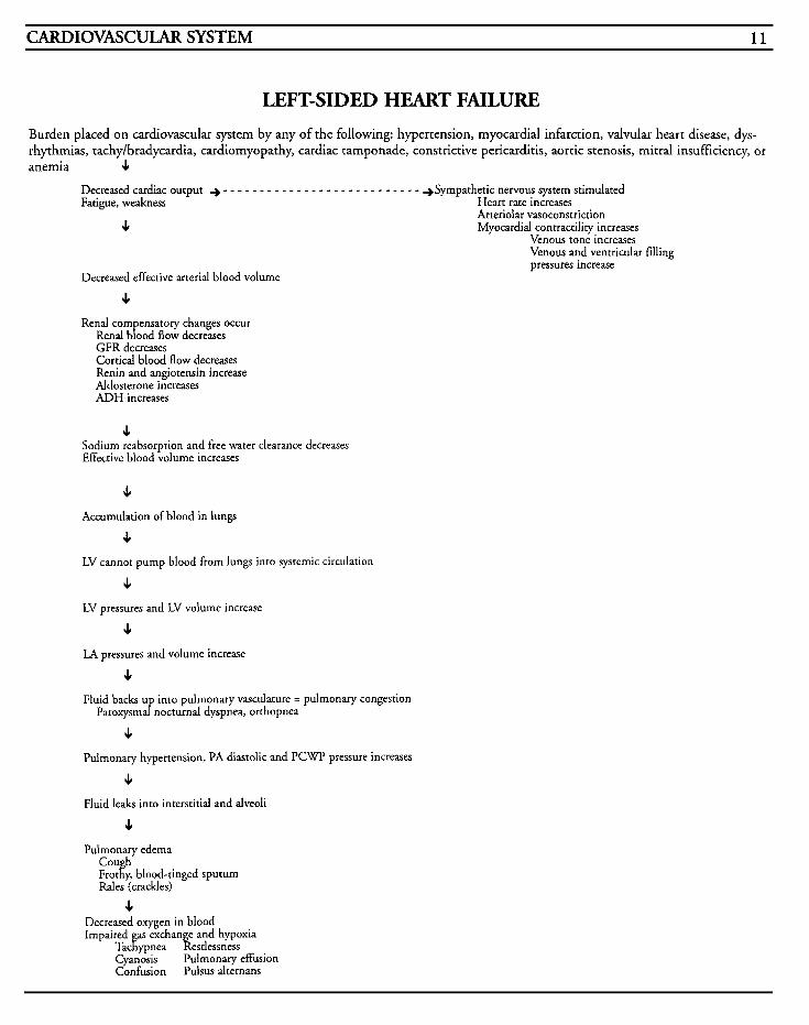
LEFT-SIDED HEART FAILURE Burden placed on cardiovascular system by any of the following: hypertension, myocardial infarction, valvular heart disease, dys- rhythmias, tachy/bradycardia, cardiomyopathy, cardiac tamponade, constrictive pericarditis, aortic stenosis, mitral insufficiency, or anemia 4
Decreased cardiac output + - - - - - - - - - - - - - - - - - - - - - - - - - - - +Sympathetic nervous system stimulated Fatigue, weakness Heart rate increases
Arteriolar vasoconstriction 4 Myocardial contractility increases
Venous tone increases Venous and ventricular filling pressures increase
Decreased effective arterial blood volume
J,
Renal com ensatory changes occur Rend bfood flow decreases GFR decreases Cortical blood flow decreases Renin and angiotensin increase Aldosterone increases ADH increases
J, Sodium reabsorption and free water clearance decreases Effective blood volume increases
4 Accumulation of blood in lungs
J,
LV cannot pump blood from lungs into systemic circulation
J LV pressures and LV volume increase
4
J,
LA pressures and volume increase
Fluid backs U into pulmonary vasculature = pulmonary congestion Paroxysmafnocturnal dyspnea, orthopnea
4 Pulmonary hypertension, PA diastolic and PCWP pressure increases
6
4 Pulmonary edema
Cou h Frotl& blood-tinged sputum Rales (crackles)
Fluid leaks into interstitial and alveoli
4 Decreased oxygen in blood Impaired as exchange and hypoxia
Tackypnea Restlessness Cyanosis Pulmonary effusion Confusion Pulsus alternans
12 CRITICAL CARE NURSING CARE PLANS
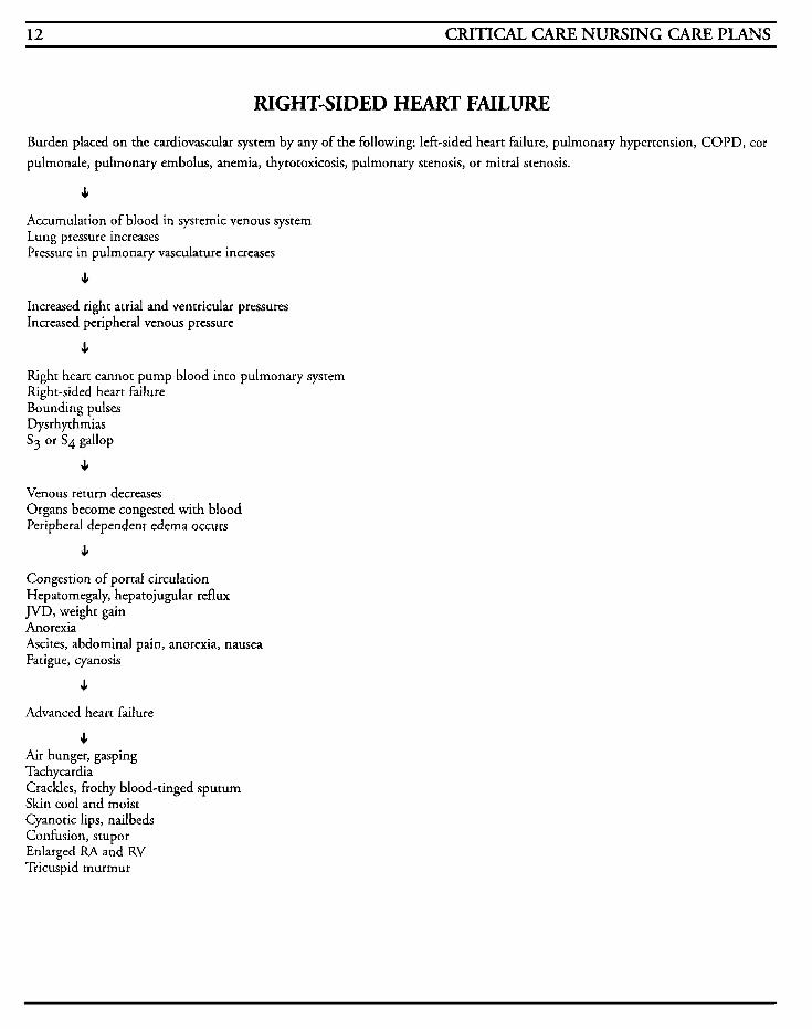
RIGHT-SIDED HEART FAILURE
Burden placed on the cardiovascular system by any of the following: left-sided heart failure, pulmonary hypertension, COPD, cor pulmonale, pulmonary embolus, anemia, thyrotoxicosis, pulmonary stenosis, or mitral stenosis.
4 Accumulation of blood in systemic venous system Lung pressure increases Pressure in pulmonary vasculature increases
4
Increased right atrial and ventricular pressures Increased peripheral venous pressure
J,
Right heart cannot pump blood into pulmonary system Right-sided heart failure Bounding pulses Dysrhythmias S j or S4 gallop
4 Venous return decreases Organs become congested with blood Peripheral dependent edema occurs
J,
Congestion of portal circulation Hepatomegaly, hepatojugular reflux JVD, weight gain Anorexia Ascites, abdominal pain, anorexia, nausea Fatigue, cyanosis
4
Advanced heart failure
4 Air hunger, gasping Tachycardia Crackles, frothy blood-tinged sputum Skin cool and moist Cyanotic lips, nailbeds Confusion, stupor Enlarged RA and RV Tricuspid murmur
CARDIOVASCULAR SYSTEM 13
MYoeardial Infaretion (MD Myocardial infarction (MI) is a critical emergency that requires timely management to save heart muscle and limit damage that may evolve over several hours. Blood flow is abruptly decreased or stopped through the coronary arteries and results in ischemia and necrosis to the myocardium if not treated. Many people die prior to receiving med- ical care due to the denial that anything may be wrong and postponement of seeking medical care. Cardiac dysrhythmias, mainly ventricular fibrilla- tion, is usually the cause of death in these individuals. An MI is diagnosed based on type of chest pain, electrocardiographic changes, and increase of cardiac enzymes, such as CK, SGOT, and LDH. Precordial pain is similar to but usually more intense and prolonged than anginal pain, and in the instance of MI, the chest pain is usu- ally constant and not relieved with nitroglycerin or rest.
Atherosclerosis of the arteries is usually the most common finding in patients. Atherosclerosis and arteriosclerosis are used interchangeably when dis- cussing the fatty plaques that adhere to the inner layer of the arteries. The continuous build-up of these plaques, as well as the potential for hemor- rhage at the intimal layer may result in alterations of the blood flow through the coronary arteries and abnormalities in platelet aggregation may con- tribute to changes in coronary perfusion. Infarction may occur without coronary artery dis- ease or occlusion, and if the patient has developed an adequate collateral circulation, coronary occlu- sion may occur without infarction.
MI is usually a disease involving the left ventricle but the damage may extend to other areas, such as the atria or right ventricle. A right ventricular myocardial infarction usually has high right ven-
tricular filling pressures and often has severe tri- cuspid regurgitation. Transmural infarcts involve the entire thickness of the myocardium and are characterized by Q waves on the electrocardiogram. Nontransmural infarcts are characterized by S-T segment and T wave changes. Subendocardial infarcts usually involve the inner portion of the myocardium where wall tension is highest and the blood flow is most vul- nerable to circulatory problems. Occlusion of the right coronary artery will result in an inferior infarction that may also include posterior portions of the heart. Occlusion of the left main artery, known as “the widow maker,” usually results in death due to the extensive damage. Occlusion of the left anterior descending artery results in an anterior infarction and may include some inferior parts of the heart, and occlusion of the circumflex artery results in a lateral infarction.
Precipitating factors that preclude MIs include heredity, age, gender, presence of hypertension, presence of diabetes mellitus, cigarette smoking, hyperlipidemia, obesity, sedentary lifestyles, and stress.
The main goals in treating myocardial infarction are to increase blood flow to the coronary arteries and thus decrease infarction size, increase oxygen supply and decrease oxygen demand to prevent myocardial death or injury, and control or correct dy s r h yt hm ias .
MEDICAL CARE Oxygen: to increase available oxygen supply
Analgesics: morphine is the drug of choice, given in incremental doses IV every 5 minutes as needed; IM injections are avoided because they can raise the enzyme levels and do not act as quickly
14 CRITICAL CARE NURSING CARE PLANS
Thrombolytic agents: Streptokinase, Urokinase, or Tissue Plasminogen Activator (tPa) given either intracoronary or intravenously to activate the body’s own fibrinolytic system to dissolve the clot and resume coronary blood perfusion
Cardiac glycosides: digitalis to increase force and strength of ventricular contractions and to decrease the conduction and rate of contractions in order to increase cardiac output; usually not used in the acute phase
Diuretics: furosemide (Lasix) to promote excess fluid removal, to decrease edema and pulmonary venous pressure by preventing sodium and water reabsorption
Vasodilators: hydralazine (Apresoline), nifedipine (Procardia, Adalat), nitroglycerin (Nitropaste, Nitrodur, Nitrostat, Tridil, Nitroglycerine), prazosin (Minipres), captopril (Capoten)-used to relax venous and/or arterial smooth muscle to decrease preload, decrease afterload, and decrease oxygen demand
Beta-adrenergic blockers: used to decrease blood pressure, decrease elevated plasma renins, and with non-selective blockers, may do so without related reflex tachycardias; used to treat ventricular dys- rhythmias and for the prophylaxis of angina
Aspirin: used to decrease platelet aggregation and helps with vasodilation of peripheral vessels
Thrombolytics: used in the treatment of acute MI; acts by activating mechanisms for conversion of plasminogen to plasmin which is able to dissolve the clot; commonly used are streptokinase, urokinase, alteplase, or anistreplase
Heparin: used with thrombolytic protocols, and in the treatment of MI; prevents conversion of fib- rinogen to fibrin and prothrombin to thrombin by its action on antithrombin I11
Laboratory: leukocyte count, sed rate and blood glucose may be elevated; creatinine phosphokinase (CK, CPK) will normally increase within 4-6 hours, peak between 12-24 hours, and last 2-3 days but should not be used as sole indicator due to possibility of elevation with other problems such as surgery or trauma; lactate dehydrogenase (LDH) will normally increase within 8-12 hours, peak between 2-4 days, and last 10-14 days but should not be used as sole indicator due to possi- bility of elevation with other problems such as liver failure; serum glutamic oxaloacetic transami- nase (SGOT) is occasionally used as an infarct indicator; isoenzymes of CPK are very specific with CPK-MB most specific for MI, and levels will not rise with transient chest pain or in surgi- cal procedures; a definitive level for CPK-MB is greater than or equal to 4% of the total CDK; LDH isoenzymes, specifically LDHl is more spe- cific for MI; if the total LDH is elevated and LDHl is most predominant, MI is confirmed; both CPK-MB and LDHl will return to normal 72-96 hours after elevation
Chest x-ray: shows any enlargement of the heart and pulmonary vein, presence of pulmonary edema or pleural effusion
Electrocardiography: shows indicative changes associated with sites of acute infarcts using Q waves, S-T segment elevation, and T wave inver- sion. Also reveals changes with atrial and ventricular enlargement, rhythm and conduction abnormalities, ischemia, electrolyte abnormalities, drug toxicity, and presence of dysrhythmias
Echocardiography: used to study structural abnor- malities and blood flow through the heart; M-mode echocardiography measures structures with a single ultrasonic beam that provides a narrow view of the heart; two-dimensional (2D) echocardiography shows a two-dimensional and
CARDIOVASCULAR SYSTEM 15
wider look at the heart that is more useful in diag- nosing right ventricular infarcts; documents increased right ventricular size, performance and segmental wall motion abnormalities, and blood flow through the heart
Nuclear cardiologic testing: MUGA (multiple gated acquisition study) provides information that approximates ejection fractions and the analysis of the ventricular wall motion; 99mTc (Technetium- 99 pyrophosphate scan) shows infarcted areas as increased levels of radioactivity, or “hot spots’’ that appear 12-36 hours after infarct and remain for 4- 7 days; PET (positron emission tomography) allows measurement of myocardial blood flow, fatty acid and glucose metabolism, and blood volume; thallium scans can determine size and location of damage as a “cold spot”
Magnetic resonance imaging (MRI): provides a three-dimensional view that can detect changes in tissues before structural damage is done and is safe for pregnant women and children
Cardiac catheterization: used to assess pathophys- iology of the patient‘s cardiovascular disorder, to provide left ventricular function information, to allow for measurement of heart pressures and car- diac output, to evaluate stenotic lesions, and to measure blood gas content
Intra-aortic balloon pump (IABP): decreases the workload on the heart, decreases myocardial oxygen demand, increases coronary perfusion, decreases afterload, decreases preload, and helps to limit infarct size if quickly initiated, improves car- diac output and tissue perfusion; used in cardiogenic shock, for support post cardiac surgery, intractable chest pain, and in cardiac catheterizations or other cardiovascular procedures of high-risk patients
Ventricular assist device (VAD): used on either or both ventricles to provide total support to the heart and circulation in order to allow recovery to the heart; usually indicated in patients who are awaiting cardiac transplantation or in those patients with cardiogenic shock and ventricular failure; may be used in conjunction with IABP
Pacemakers: either temporary or permanent, used in anticipation of lethal dysrhythmias andlor con- duction problems
Surgery: coronary artery bypass grafting to reroute the coronary blood flow around the dis- eased vessel to enable coronary perfusion
NURSING CARE PLANS Alteration in comfort
Related to: chest pain due to decreased blood flow to myocardium, myocardial ischemia or infarct, post-procedure discomfort, chest wall pain post-surgery, pericarditis
Defining characteristics: chest pain with or with- out radiation, facial grimacing, clutching of hands or chest, restlessness, diaphoresis, changes in pulse and blood pressure, dyspnea, dizziness
Outcome Criteria
Chest pain will be relieved or controlled to patient’s satisfaction.
INTERVENTIONS RATIONALES Evaluate chest pain as to type, location, severity, relief, change with activity or rest, other symp- toms concurrenrly noted, such as pallor, diaphoresis, radiation of pain, nausea, vomiting, shortness of breath, and vital sign changes.
Variations may occur with patients regarding speci- fic complaints and beha- vior. Most MI patients look acutely ill and can only focus on their pain. Respirations may be in- creased as a result of an- xiety and pain. Heart rate
16 CRITICAL CARE NURSING CARE PLANS
INTERVENTIONS RATIONALES INTERVENTIONS RATIONALES
Obtain description of intensity using 0-10 scale, with 0 being no pain and 10 being the worst pain experienced.
Obtain history (when possible) of previous cardiac pain and familial history of cardiac problems.
Administer oxygen by nasal cannula or mask as indicated.
Administer analgesic as ordered, such as morphine sulfate, meperi- dine (Demerol), or Dilaudid IV.
Administer beta-blockers as or- dered (such as atenolol, pindolol, and propranolol).
may increase due to in- creased catecholamines, stress, and pain, which can also increase blood pres- sure.
Administer calcium-channel bloc- kers as ordered (such as verapa- mil, diltiazem, or nifedipine).
These drugs can increase coronary blood flow and collateral circulation, reduce preload and myocar- dial oxygen demands, which can decrease pain due to ischemia.
Pain is a subjective ex- perience and personal to that patient. Intensity scales are useful to gauge improvement or deteriora- tion as perceived by the patient.
This provides information that may help to differen- tiate current pain from previous problems, as well as identify new problems and complications.
Supplemental oxygen can in- crease the available oxygen and can relieve pain asso- ciated with myocardial is- chemia.
Morphine is the drug of choice to control MI pain, but other analgesics may be used to reduce pain and reduce the workload on the heart. IM injec- tions should be avoided because they can alter cardiac enzymes and are not absorbed well in tissue that is non- or under-perfused.
These d rug block sym- pathetic stimulation, re- duce heart rate and sys- tolic blood pressure, and thus lowers the myocardid oxygen demand. Beta- blockers should not be given in severely impaired contractility states due to the negative inotropic properties.
Maintain bedrest during pain, with position of comfort; nurse to stay with patient during pain.
Maintain relaxing environment to promote calmness.
Reduces oxygen consumption, and demand; alleviates fear and provides caring atmos- phere.
Reduces competing stimuli and reduces anxiety.
Information, Instruction, Demonstration
INTERVENTIONS RATIONALES
Instruct to notify nurse imme- diately of any chest pain.
Instruct in relaxation tech- niques, deep breathing, guided imagery, visualization, etc.
Instruct in nitroglycerin SL ad- ministration after hospitalization; 1 q5 minutes up to 3 times, and if pain is unrelieved, patient should seek emergency medical care.
Instruct in activity alterations and limitations.
Instruct in medication effects, side effects, contraindications, and symptoms to report.
Delay in notification can delay pain relief and may require increased amounts of medication in order to finally achieve relief. Pain can cause further damage to an already- injured myocardium, and may signal extension of MI, spasm, or other com- plication.
Helps to decrease pain and anxiety and provides dis- traction from pain.
Knowledge facilitates co- operation and compliance with medical regimen. Pain unrelieved with NTG may be indicative of MI.
Decreases myocardial oxygen demand and workload on the heart.
Promores knowledge and com- pliance with therapeutic regimen. Alleviates fear of unknown.
CARDIOVASCULAR SYSTEM 17
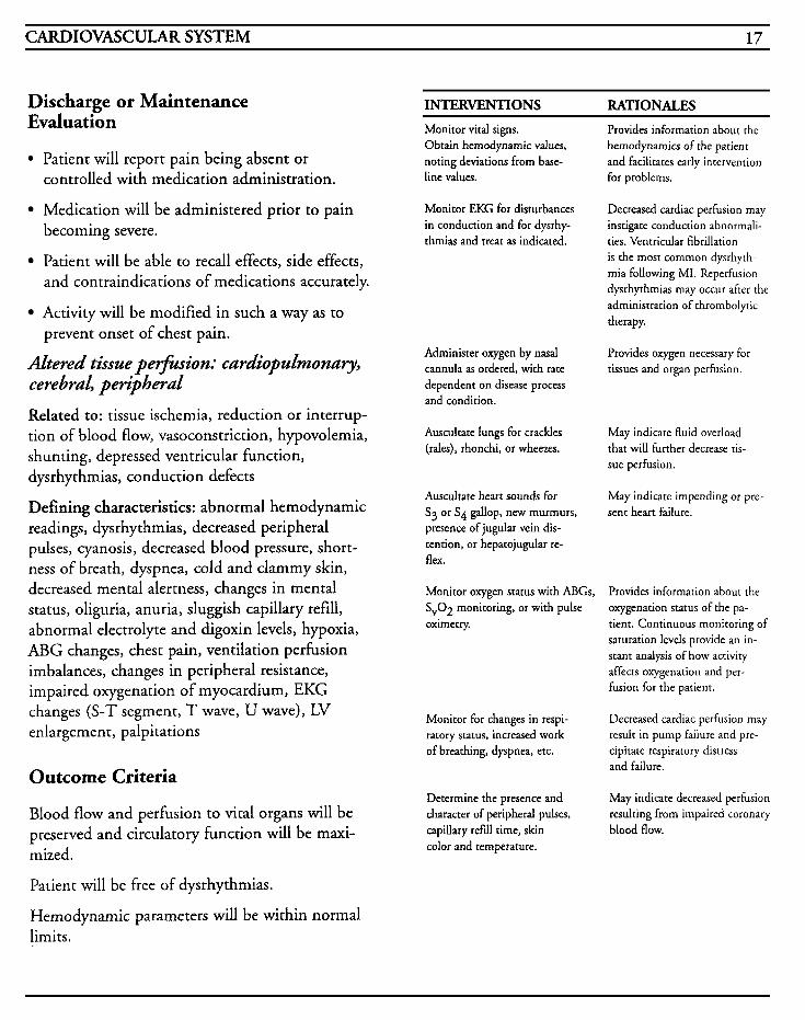
Discharge or Maintenance Evaluation
Patient will report pain being absent or controlled with medication administration.
Medication will be administered prior to pain becoming severe.
Patient will be able to recall effects, side effects, and contraindications of medications accurately.
Activity will be modified in such a way as to
Altered tissue pe+ion: cardiopulmonary, cerebral, peripheral
Related to: tissue ischemia, reduction or interrup- tion of blood flow, vasoconstriction, hypovolemia, shunting, depressed ventricular function, dysrhythmias, conduction defects
Defining characteristics: abnormal hemodynamic readings, dysrhythmias, decreased peripheral pulses, cyanosis, decreased blood pressure, short- ness of breath, dyspnea, cold and clammy skin, decreased mental alertness, changes in mental status, oliguria, anuria, sluggish capillary refill, abnormal electrolyte and digoxin levels, hypoxia, ABG changes, chest pain, ventilation perfusion imbalances, changes in peripheral resistance, impaired oxygenation of myocardium, EKG changes (S-T segment, T wave, U wave), LV enlargement, palpitations
prevent onset of chest pain.
Outcome Criteria
Blood flow and perfusion to vital organs will be preserved and circulatory function will be maxi- mized.
Patient will be free of dysrhythmias.
Hemodynamic parameters will be within normal limits.
INTERVENTIONS RATIONALES Monitor vital signs. Obtain hemodynamic values, noting deviations from base- line values. for problems.
Monitor EKG for disturbances in conduction and for dysrhy- thmias and treat as indicated.
Provides information about the hernodynamics of the patient and facilitates early intervention
Decreased cardiac perfusion may instigate conduction abnormali- ties. Ventricular fibrillation is the most common dysrhyth- mia following MI. Reperfusion dysrhythmias may occur after the administration of thrombolytic therapy.
Administer oxygen by nasal cannula as ordered, with rate dependent on disease process and condition.
Provides oxygen necessary for tissues and organ perfusion.
Auscultate lungs for crackles (rales), rhonchi, or wheezes.
May indicate fluid overload that will further decrease tis- sue perfusion.
Auscultate heart sounds for S3 or S4 gallop, new murmurs, presence of jugular vein dis- tention, or hepatojugular re- flex.
May indicate impending or pre- sent heart failure.
Monitor oxygen status with ABGs, Sv02 monitoring, or with pulse oximetry. tient. Continuous monitoring of
Provides information about the oxygenation status of the pa-
saturation levels provide an in- stant analysis of how activity affects oxygenation and per- fusion for the patient.
Monitor for changes in respi- ratory status, increased work of breathing, dyspnea, etc.
Determine the presence and character of peripheral pulses, capillary refill time, skin color and temperature.
Decreased cardiac perfusion may result in pump failure and pre- cipitate respiratory distress and failure.
May indicate decreased perfusion resulting from impaired coronary blood flow.
18 CRITICAL CARE NURSING CARE PLANS
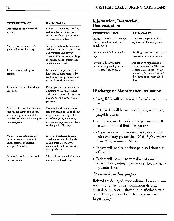
INTERVENTIONS RATIONALES
Discourage any non-essential activity.
Assist patient with planned, graduated levels of activity.
Titrate vasoactive drugs as ordered.
Administer thrombolytic drugs as ordered.
Auscultate for bowel sounds and monitor for complaints of nau- sea, vomiting, anorexia, abdo- minal distention, abdominal pain, or constipation.
Monitor urine output for ade- quate amounts, character of urine, presence of sediment, and specific gravity.
Monitor labwork such as renal or liver profiles.
Ambulation, exercise, transfers, and Valsalva-type maneuvers can increase blood pressure and decrease tissue perfusion.
Allows for balance between rest and activity to decrease myocar- dial workload and oxygen demand. Gradual increases help to increase patient tolerance to activity without pain.
Maintain blood pressure and heart rate at parameters set by MD for optimal perfusion with minimal workload on heart.
Drugs Iyse the clot that may be occluding the coronary artery and promote restoration of oxy- gen and blood flow to increase perfusion.
Decreased perfusion to mesen- tery may result in loss or change in peristalsis, resulting in GI use of analgesics, and change in surroundings may contribute to changes in GI status.
Decreased perfusion to renal arteries may result in oliguria. Dehydration secondary to nausea and vomiting may affect renal perfusion.
May indicate organ dysfunction and decreased perfusion.
Information, Instruction, Demonstration
INTERVENTIONS RATIONALES Instruct on medications, dosage, effects, side effects, and con- traindications.
Promotes compliance with regimen and knowledge base.
Instruct to refrain from smok- ing. with can decrease perfusion.
Instruct in dietary require- ments, menu planning, sodium restrictions, foods to avoid.
Smoking causes vasoconstriction
Reduction of high-cholesterol and sodium foods will help to control atherosclerosis, hyper- lipidemia, fluid retention, and the effects on coronary blood flow.
Discharge or Maintenance Evaluation
Lung fields will be clear and free of adventitious breath sounds.
Extremities will be warm and pink, with easily palpable pulses.
Vital signs and hemodynamic parameters will be within normal limits for patient.
Oxygenation will be optimal as evidenced by pulse oximetry greater than 90%, SvOz greater than 75%, or normal ABGs.
Patient will be free of chest pain and shortness of breath.
Patient will be able to verbalize information accurately regarding medications, diet and activ- ity limitations.
Decreased cardiac output
Related to: damaged myocardium, decreased con- tractility, dysrhythmias, conduction defects, alteration in preload, alteration in afterload, vaso- constriction, myocardial ischemia, ventricular hypertrophy
CARDIOVASCULAR SYSTEM 19
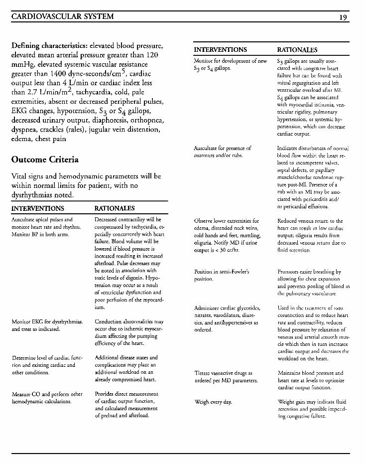
Defining characteristics: elevated blood pressure, elevated mean arterial pressure greater than 120 mmHg, elevated systemic vascular resistance greater than 1400 dyne-seconds/cm5, cardiac output less than 4 L/min or cardiac index less than 2.7 L/min/m2, tachycardia, cold, pale extremities, absent or decreased peripheral pulses, EKG changes, hypotension, S3 or S 4 gallops, decreased urinary output, diaphoresis, orthopnea, dyspnea, crackles (rales), jugular vein distention, edema, chest pain
Outcome Criteria
Vital signs and hemodynamic parameters will be within normal limits for patient, with no dysrhythmias noted.
INTERVENTIONS RATIONALES Auscultate apical pulses and monitor heart rate and rhythm. Monitor BP in both arms.
Monitor EKG for dysrhythmias. and treat as indicated.
Determine level of cardiac func- tion and existing cardiac and other conditions.
Measure CO and perform other hemodynamic calculations.
Decreased contractility will be compensated by tachycardia, es- pecially concurrently with heart failure. Blood volume will be lowered if blood pressure is increased resulting in increased afterload. Pulse decreases may be noted in association with toxic levels of digoxin. Hypo- tension may occur as a result of ventricular dysfunction and poor perfusion of the myocard- ium.
Conduction abnormalities may occur due to ischemic myocar- dium affecting the pumping efficiency of the heart.
Additional disease states and complications may place an additional workload on an already compromised heart.
Provides direct measurement of cardiac output function, and calculated measurement of preload and afterload.
INTERVENTIONS RATIONALES Monitor for development of new Sg or S4 gallops.
Auscultate for presence of murmurs andlor rubs.
Observe lower extremities for edema, distended neck veins, cold hands and feet, mottling, oliguria. Notify M D if urine output is < 30 cclhr.
Position in semi-Fowler's position.
Administer cardiac glycosides, nitrates, vasodilators, diure- tics, and antihypertensives as ordered.
Titrate vasoactive drugs as ordered per M D parameters.
Weigh every day.
Sg gallops are usually asso- ciated with congestive heart failure but can be found with mitral regurgitation and left ventricular overload after MI. S4 gallops can be associated with myocardial ischemia, ven- tricular rigidity, pulmonary hypertension, or systemic hy- pertension, which can decrease cardiac output.
Indicates disturbances of normal blood flow within the heart re- lated to incompetent valves, sepia1 defects, or papillary muscle/chordae tendonae rup- ture post-MI. Presence of a rub with an MI may be asso- ciated with pericarditis and/ or pericardial effusions.
Reduced venous return to the heart can resulr in low cardiac output; oliguria results from decreased venous return due to fluid retention.
Promotes easier breathing by allowing for chest expansion and prevents pooling of blood in the pulmonary vasculature.
Used in the treatment of vaso- constriction and 10 reduce heart rate and contractility, reduces blood pressure by relaxation of venous and arterial smooth mus- cle which then in turn increases cardiac output and decreases the workload on the heart.
Maintains blood pressure and heart rate at levels to optimize cardiac output function.
Weight gain may indicate fluid retention and possible impend- ing congestive failure.
20 CRITICAL CARE NURSING CARE PLANS
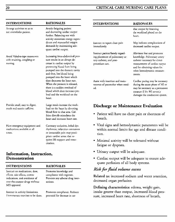
INTERVENTIONS RATIONALES INTERVENTIONS MTIONALES Arrange activities so as to not overwhelm patient.
Avoids fatiguing patient and decreasing cardiac output further. Balancing rest with activity minimizes energy expen- diture and myocardial oxygen demands by maintaining ade- quate cardiac output.
diac output by lessening the workload placed on the heart.
Instruct to report chest pain immediately, decreased cardiac output.
May indicate complications of
Avoid Valsalva-type maneuvers with straining, coughing or moving.
Provide small, easy to digest, meals and restrict caffeine.
Have emergency equipment and medications availabIe at all times.
Increasing intra-abdominal pres- sure results in an abrupt de- crease in cardiac output by preventing blood from being pumped into the thoracic cavity and thus, less blood being pumped into the heart which then decreases the heart rate. When the pressure is released, there is a sudden overload of blood which then increases pre- load and the workload on the heart.
Large meals increase the work- load on the heart by diverting blood flow to that area. Caf- feine directly stimulates the heart and increases heart rate.
Coronary occlusion, lethal dys- rhythmias, infarction extensions or intractable pain may preci- pitate cardiac arrest that re- quires life support and resus- citation.
Information, Instruction, Demonstration
INTERVENTIONS RATIONALES Instruct on medications, dose, effects, side effects, contra- indications, and avoidance of over-the-counter drugs without actions. MD approval.
Promotes knowledge and compliance with regimen. Prevents any adverse drug inter-
Instruct in activity limitations. Demonstrate exercises to be done.
Promotes compliance. Reduces potential for decrease in car-
Instruct patientlfamily regard- ing placement of pulmonary ar- tery catheter, and post- procedure care.
Alleviates fear and promotes knowledge. Pulmonary artery catheter necessary for direct measurement of cardiac output and for obtaining values for other hemodynamic measure- ments.
Assist with insertion and main- tenance of pacemaker when need- ed.
Cardiac pacing may be necessary during the acute phase of MI or may be necessary as a permanent measure if the MI severely damages the conduction system.
Discharge or Maintenance Evaluation
0 Patient will have no chest pain or shortness of breath.
Vital signs and hemodynamic parameters will be within normal limits for age and disease condi- tion.
Minimal activity will be tolerated without fatigue or dyspnea.
Urinary output will be adequate.
Cardiac output will be adequate to ensure ade- quate perfusion of all body systems.
Risk for fluid volume excess
Related to: increased sodium and water retention, decreased organ perfusion
Defining characteristics: edema, weight gain, intake greater than output, increased blood pres- sure, increased heart rate, shortness of breath,
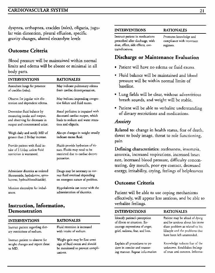
CARDIOVASCULAR SYSTEM 21
dyspnea, orthopnea, crackles (rales), oliguria, jugu- lar vein distention, pleural effusion, specific gravity changes, altered electrolyte levels
Outcome Criteria
Blood pressure will be maintained within normal limits and edema will be absent or minimal in all body parts.
INTERVENTIONS RATIONALES Auscultate lungs for presence of crackles (rales).
Observe for jugular vein dis- tention and dependent edema.
Determine fluid balance by measuring intake and output, and observing for decreases in output and concentrated urine.
Weigh daily and notify MD of greater than 2 Ib/day increase.
Provide patient with fluid in- take of 2 L/day, unless fluid restricrion is warranted.
Administer diuretics as ordered (furosemide, hydralazine, spiro- lactone, hydrochlorothiazide).
Monitor electrolyte for imbal- ances.
May indicate pulmonary edema from cardiac decompensation.
May indicate impending conges- tive failure and fluid excess.
Renal perfusion is impaired with decreased cardiac output, which leads to sodium and water reten- tion and oliguria.
Abrupt changes in weight usually indicate excess fluid.
Fluids provide hydration of tis- sues. Fluids may need to be restricted due to cardiac decom- pensation.
Drugs may be necessary to cor- rect fluid overload depending on emergent nature of problem.
Hypokalemia can occur with the administration of diuretics.
Instruction, Information, Demonstration
INTERVENTIONS RATIONALES
Instruct patient regarding diet- ary restrictions of sodium.
Fluid retention is increased with intake of sodium.
Instruct patient to observe for weight changes and report these to MD.
Weight gain may be first overt sign of fluid excess and should be monitored to prevent compli- cations.
INTERVENTIONS RATIONALES Instruct patient in medications prescribed after discharge, with dose, effect, side effects, con- traindications.
Promotes knowledge and compliance with treatment regimen.
Discharge or Maintenance Evaluation
Patient will have no edema or fluid excess.
. Fluid balance will be maintained and blood pressure will be within normal limits of baseline. . Lung fields will be clear, without adventitious breath sounds, and weight will be stable.
Patient will be able to verbalize understanding of dietary restrictions and medications.
Anxiety Related to: change in health status, fear of death, threat to body image, threat to role functioning, pain
Defining characteristics: restlessness, insomnia, anorexia, increased respirations, increased heart rate, increased blood pressure, difficulty concen- trating, dry mouth, poor eye contact, decreased energy, irritability, crying, feelings of helplessness
Outcome Criteria
Patient will be able to use coping mechanisms effectively, will appear less anxious, and be able to verbalize feelings.
INTERvENTf ONS RATIONALES Identify patient’s perception of illness or situation. En- courage expressions of anger, grief, sadness, fear, and loss.
Patient may be afraid of dying and be anxious about his imme- diate problem as related to his lifestyle and the problems that have been left unattended.
Explain all procedures to pa- tient in concise and reassur- ing manner. Repeat information
Knowledge reduces fear of the unknown. Establishes feelings of trust and concern. Informa-
22 CRITICAL CAFE NURSING CARE PLANS
INTERVENTIONS RATIONALES
as needed based on patient’s ability to comprehend.
Encourage the patient to dis- cuss his fears and feelings. Provide an atmosphere of accep- tance without judgment. Accept his use of denial, but do not reinforce false beliefs. Avoid confrontations and upsets.
Provide opportunities for the family to visit and assist with care if possible. Orient to routines.
Provide private time for pa- tient and family member(s) ro verbalize feelings.
Provide opportunities for pa- tient to control his environ- ment and activities as much as feasible based on condition.
Provide opportunity for patient to rest withour interruption as much as possible.
Adminisrer antianxiety drugs as ordered (diazepam, flurazepam, lorazepam).
tion may need to be repeated or reintbrced due to competing stimuli.
Assists the patient in verba- lizing concerns and provides the opportunity to deal with matters of import to the patient. Ac- cepting the patient’s feelings may decrease his anxiety which can facilitate a therapeutic environment for instruction. Denial can be useful to decrease anxiety but can postpone dealing with the reality of the problem. Confrontations can lead to anger and exacerbate the use of denial and decrease cooperation.
Familiar people can decrease anxiety of the patient, as well as provide a more con- ducive atmosphere for learn- ing and recovery. Predicta- bility can decrease anxiety. Supportive family members can comfort the patient and relieve worries.
Allows time for expression of concerns and feelings, and relieves tension by establish- ing a more normal routine.
Allows the patient to have some control over his situation and facilitates compliance with care of which patient is not in con- trol.
Facilitates coping mechanism by conserving energy, and by providing required rest.
Promotes rest and reduces anxi- ety.
Information, Instruction, Demonstration
INTERVENTIONS RATIONALES Instruct patient and family as to all procedures, tests, medications, and care in a factual consistent manner. Reinforce as needed.
Instruct patient in relaxa- tion techniques. Provide for diversionary activities.
Instruct about post-discharge care, activities, limitations, symptoms to report, problems that might be encountered, and goals.
Accurate information reduces anxiety, facilitates the rela- tionship between patient and nurse, and allows the patienr and family to deal with the problem in a realistic manner. Repetition, when needed, helps in the retention of information when the attention span is di- minished.
Reduces anxiety and stress.
Reduces anxiety and promores increased independence and self- confidence; decreases fear of abandonment that can occur with discharge from hospital; assists patient and family to identi+ realisric goals and decreases the chances of discour- agemenr with limitations during recuperation.
Discharge or Maintenance Evaluation
Patient is able to recognize feelings and identify mechanisms to cope and identify causes.
Patient has significant reduction in fear and anxiety and appears less tense, with normal vital signs.
Patiendfamily can appropriately utilize problem-solving skills.
Patient can verbalize concerns easily and has increased energy.
Patient can make appropriate decisions based on factual information regarding his condition and is able to discuss future plans.
CARDIOVASCULAR SYSTEM 23
Knowledge W c i t
Related to: lack of understanding, lack of under- standing of medical condition, lack of recall
Defining characteristics: verbalized questions regarding problems, inadequate follow-up on instructions given, misconceptions, lack of improvement of previous regimen, development of preventable complications
Outcome Criteria
Patient will be able to verbalize and demonstrate understanding of information given regarding condition, medications, and treatment regimen.
Information, Instruction, Demonstration
INTERVENTIONS RATIONALES Determine patient’s baseline of knowledge regarding disease pro- cess, normal physiology, and function of the heart.
Monitor patient’s readiness to learn and determine best methods to use for teaching. Attempt to incorporate family members in learning process. Rein- structlreinforce information as needed.
Provide time for individual in- teraction with patient.
Instruct patient on procedures that may be performed.
Instruct patient on medications, dose, effects, side effects, con- traindications, and signs/ symptoms to report to MD.
Provides information regarding patient$ understanding of condition as well as a baseline from which to base teaching.
Promotes optimal learning en- vironment when patient shows willingness to learn. Family members may assist with help- ing the patient to make in- formed choices regarding his treatment. Anxiety or latge volumes of instruction may impede comprehension and limit learning.
Promotes relationship between patient and nurse, and estab- blishes trus:.
Provides knowledge and pro- motes the ability to make informed choices.
Provides information to :he patient to manage medication regimen and ensure compliance.
INTERVENTIONS RATIONALES Instruct in dietary needs and restrictions, such as limiting caffeine and sodium or in- creasing potassium, etc.
Provide printed materials when possible for patientlfamily to review.
Demonstrate and instruct on technique for checking pulse rate and regularity Instruct in situations where immediate action must be taken.
Have patient demonstrate all skills that will be necessary for post-discharge.
Instructldemonstrate exercises to be performed, avoiding over- taxing activities, signs/ symptoms that may require the cessation of any activity, and to report symptoms that may require medical attention.
Patient may need to increase dietary potassium if placed on diuretics; caffeine should be limited due to the direct stimu- lant effect on the heart; so- dium should be limited due to the potential for fluid reten- tion.
Provides references for patient and family to refer to once dis- charged, and can enhance the understanding of verbally- given instructions.
Self-monitoring promotes self- independence and can provide timely intervention for abnor- malities or complications. Heart rates that exceed set parameters may require furrher medial alteration in medica- tions or regimen.
Provides information that patient has gained a full understanding of instruction and is able to demonstrate correct information.
Exercise programs are help- ful in improving cardiac function.
Discharge or Maintenance Evaluation
Patient will be able to verbalize understanding of condition, treatment regimen, and signs/symptoms to report.
Patient will be able to correctly perform all tasks prior to discharge.
Patient will be able to verbalize understanding of cardiac disease, risk factors, dietary restrictions, and lifestyle adaptations.
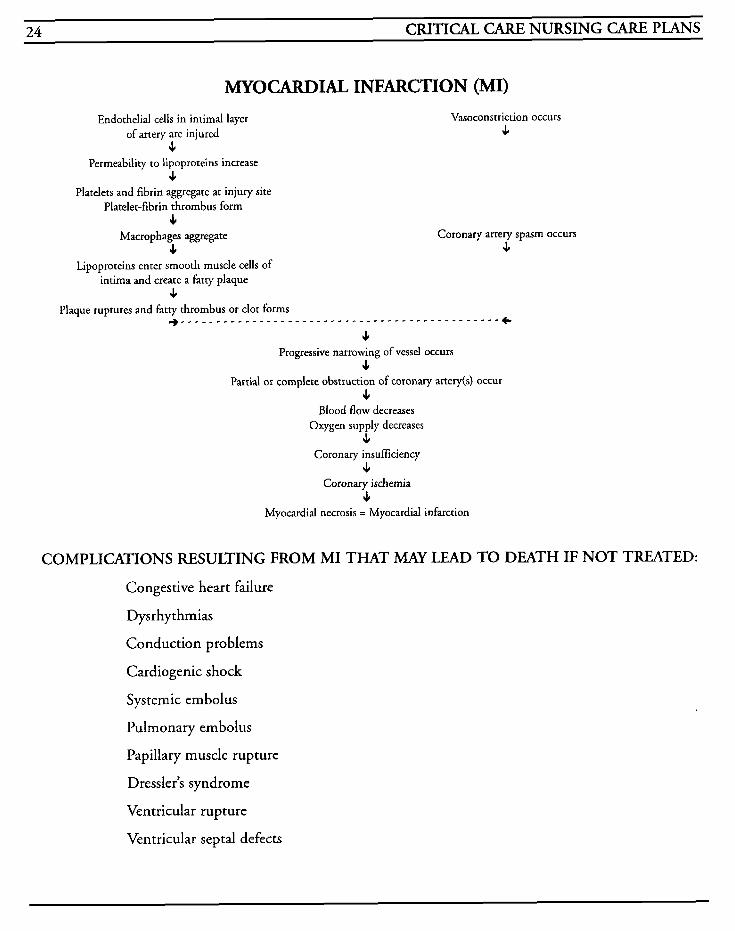
24 CRITICAL CARE NURSING CARE PLANS
MYOCARDZAL INFARCTION (MI)
Coronary artery spasm occurs s
Vasoconstriction occurs Endothelial cells in intimal layer
J, Permeability to lipoproteins increase
4 Platelets and fibrin aggregate at injury site
Platelet-fibrin thrombus form J,
Macrophages aggregate 4
Lipoproteins enter smooth muscle cells of intima and create a fatty plaque
s Plaque ruptures and fatty thrombus or clot forms
. . . . . . . . . . . . . . . . . . . . . . . . . . . . . . . . . . . . . . . . . . . . . . c
of artery are injured s
J, Progressive narrowing of vessel occurs
s Partial or complete obstruction of coronary artery(s) occur
J, Blood flow decreases
Oxygen supply decreases s
Coronary insufficiency s
Coronary ischemia J,
Myocardial necrosis = Myocardial infarction
COMPLICATIONS RESULTING FROM MI THAT MAY LEAD TO DEATH IF NOT TREATED:
Congestive heart failure
Dysrhythmias
Conduction problems
Cardiogenic shock
Systemic embolus
Pulmonary embolus
Papillary muscle rupture
Dressler’s syndrome
Ventricular rupture
Ventricular septal defects
CARDIOVASCULAR SYSTEM 25
Per Scar di t is Pericarditis is an inflammation of the pericardium that can occur due to a variety of circumstances. The inflammation is usually a manifestation of another disease process, but may be drug induced, from agents such as procainamide, hydralazine, phenytoin, penicillin, phenylbutazone, minoxidil, or daunorubicin. Other causes for pericarditis include idiopathic causes, viral, bacterial, fungal, protozoal, uremia, MI, tuberculosis, neoplasms, trauma, surgical procedures, autoimmune disor- ders (lupus, rheumatoid arthritis, scleroderma), inflammatory disorders (amyloidosis), dissecting aortic aneurysms, or radiation treatments to the thorax.
Pericarditis may be classified as acute or chronic, as well as constrictive or restrictive. Constrictive pericarditis occurs when fibrin material is deposited on the pericardium and adhesions form between the epicardium and pericardium. Restrictive pericarditis results when effusion into the pericardial sac occurs. Both types cause inter- ference with the heart’s ability to fill properly, which causes increases in systemic and pulmonary venous pressures. Eventually systemic blood pres- sure and cardiac output decrease.
The visceral pericardium is a serous membrane that is separated from a fibrous sac, or parietal pericardium, by a small (less than 50 cc) amount of fluid. If the fluid increases to the point where the heart function is compromised, pleural effu- sion occurs and cardiac tamponade becomes a critical concern. The pericardium is important because it holds the heart in a fixed position to minimize friction between it and other structures. Other functions include prevention of exercise- or hypervolemic-induced dilatation of the cardiac chambers and assistance with atrial filling during systole.
The main symptoms of pericarditis include sharp, retrosternal and/or left precordial pain that wors- ens while in a supine position, and a pericardial friction rub best auscultated at the lower left ster- nal border. The pain may be exacerbated by coughing, swallowing, breathing, or twisting. Other symptoms may be seen depending on the severity of the pericarditis and the rapidity in which the fluid accumulates. Volumes of 100 cc that accumulates quickly may produce a more life-threatening complication, cardiac tamponade, than a larger accumulation of fluid that is gener- ated over a long period of time.
MEDICAL CARE Oxygen: to increase available oxygen supply
Analgesics: morphine or meperidine used to alle- viate pain
Steroids: large doses of corticosteroids, such as prednisone, are given to reduce inflammation and control the symptoms of pericarditis
NSAIDs: aspirin or indomethacin are used to reduce fever and inflammation
IV fluids: given to help restore left ventricular fill- ing volume and to offset any compressive effects of intrapericardial pressure increases
Inotropic drugs: isoproterenol or dobutamine IV given for their positive inotropic effects as well as peripheral vasodilating properties
Laboratory: white blood cell count may be elevated, sed rate may be elevated from non-spe- cific inflammatory response; CKMB may be mildly elevated; blood cultures done to identify organism responsible for infective process and to ascertain appropriate drug for eradication; renal profile done to evaluate for uremic pericarditis and worsening renal status
26 CRITICAL CARE NURSING CARE PLANS
Electrocardiography: used to monitor for S-T ele- vation, T wave changes associated with pericarditis, and to monitor for dysrhythmias
Echocardiography: used to establish presence of pericardial fluid and an estimate of volume, any vegetation on valves, and to observe for right atrium and right ventricular dilatation
Chest x-ray: used to show cardiomegaly and to assess lung fields
Pericardiocentesis: used to relieve fluid build-up and pressure in emergency situations where the patient is deteriorating or is in shock
Surgery: open surgical drainage is usually the treatment of choice for cardiac tamponade
NURSING CARE PLANS Alterution in comfort [See MI] Related to: chest pain due to pericardial inflam- mation
Defining characteristics: chest pain with or with- out radiation, facial grimacing, clutching of hands or chest, restlessness, diaphoresis, changes in pulse and blood pressure, dyspnea
Altered tissue perfision: curdiopulmonury, renal, peripherul, cerebrul
Related to: tissue ischemia, reduction or interrup- tion of blood flow, vasoconstriction, hypovolemia, shunting, depressed ventricular function, dysrhythmias, conduction defects
Defining characteristics: abnormal hemodynamic readings, dysrhythmias, decreased peripheral pulses, cyanosis, decreased blood pressure, short- ness of breath, dyspnea, cold and clammy skin, decreased mental alertness and changes in mental status, oliguria, anuria, sluggish capillary refill, abnormal electrolyte and digoxin levels, hypoxia, ABG changes, chest pain, ventilation perfusion
imbalances, changes in peripheral resistance, impaired oxygenation of myocardium, EKG changes (S-T segment, T wave, U wave), LV enlargement, palpitations, abnormal renal function studies
Outcome Criteria
Blood flow and perfusion to vital organs will be preserved and circulatory function will be maximized.
Patient will be free of dysrhythmias.
Hemodynamic parameters will be within normal limits.
INTERVENTIONS RATIONALES
Obtain vital signs. Obtain hemodynamic values, noting deviations from base- line values.
Provides information about the hemodynamics of the patient.
Determine the presence and character of peripheral pulses, capillary refill time, skin color and temperature.
Discourage any non-essential activity.
May indicate decreased perfusion resulting from impaired coronary blood flow.
Ambulation, exercise, transfers, and Valsalva-type maneuvers can increase blood pressure and de- crease tissue perfusion.
Monitor EKG for disturbances in conduction and for dysrhy- thrnias and treat as indicated.
Decreased cardiac perfusion may instigate conduction abnormali- ties. Dysrhythmias may occur due to compromised function of ventricles due to pressure exer- ted on them by excess fluid.
Titrate vasoactive drugs as ordered.
Administer oxygen by nasal cannula as ordered, with rate dependent on disease process and condition.
Maintain blood pressure and heart rate at parameters set by M D for optimal perfusion with minimal workload on heart.
Provides oxygen necessary for tissues and organ perfusion.
CARDIOVASCULAR SYSTEM 27
INTERVENTIONS RATIONALES
Auscultate lungs for crackles (rales), rhonchi, or wheezes.
Auscultate heart sounds for S g or S4 gallop, new murmurs, presence of jugular vein dis- tention, or hepatojugular re- flex.
Monitor oxygen status with ABGs, SvO, monitoring, or with pulse oximetry.
Assist patient with planned, graduated levels of activity.
Suggestive of fluid overload that will further decrease tis- sue perfusion.
Suggestive of impending or present heart failure.
Provides information about the oxygenation status of the pa- tient. Continuous monitoring of saturation levels provide an in- stant analysis of how activity can affect oxygenation and per- fusion.
Allows for balance between rest and activity to decrease myocar- dial workload and oxygen demand. Gradual increases help to increase patient tolerance to activity without pain occurring.
Information, Instruction, Demonstration
INTERVENTIONS RATIONALES Instruct on medications, dosage, effects, side effects, and con- traindications.
Instruct to refrain from smok- ing.
Instruct in dietary require- ments, menu planning, sodium restrictions, foods to avoid.
Promotes compliance with regi- men and knowledge base.
Smoking causes vasoconstriction with can decrease perfusion.
Reduction of high-cholesterol and sodium foods will help to control atherosclerosis, hyper- lipidemia, fluid retention, and the effects on coronary blood flow.
Discharge or Maintenance Evaluation
Lung fields will be clear and free of adventitious breath sounds.
Extremities will be warm, pink, with easily pal- pable pulses of equal character.
Vital signs and hemodynamic parameters will be within normal limits for patient.
Oxygenation will be optimal as evidenced by pulse oximetry greater than 90%, Sv02 greater than 75%, or normal ABGs.
Patient will be free of chest pain and shortness of breath.
Patient will be able to verbalize information cor- rectly regarding medications, diet and activity limitations.
Decreased cardiac output [See MI] Related to: fluid in pericardial sac from pericardial effusion, potential for cardiac tamponade because of effusion, damaged myocardium, decreased con- tractility, dysrhythmias, conduction defects, alteration in preload, alteration in afterload, vaso- constriction, myocardial ischemia, ventricular hypertrophy
Defining characteristics: decreased blood pressure, tachycardia, pulsus paradoxus greater than 10 mmHg, distended neck veins, increased central venous pressure, dysrhythmias, decreased QRS voltage or electrical alternans, diminished heart sounds, dyspnea, friction rub, cardiac output less than 4 L/min, cardiac index less than 2.5 L/min/m’
Anxiety [See MI] Related to: change in health status, fear of death, threat to body image, threat to role functioning, pain
28 CRITICAL CARE NURSING CARE PLANS
Defining characteristics: restlessness, insomnia, anorexia, increased respirations, increased heart rate, increased blood pressure, difficulty concen- trating, dry mouth, poor eye contact, decreased energy, irritability, crying, feelings of helplessness
Knowledge &+&it [See MI] Related to: lack of understanding, lack of under- standing of medical condition, lack of recall
Defining characteristics: questions regarding problems, inadequate follow-up on instructions given, misconceptions, lack of improvement of previous regimen, development of preventable complications
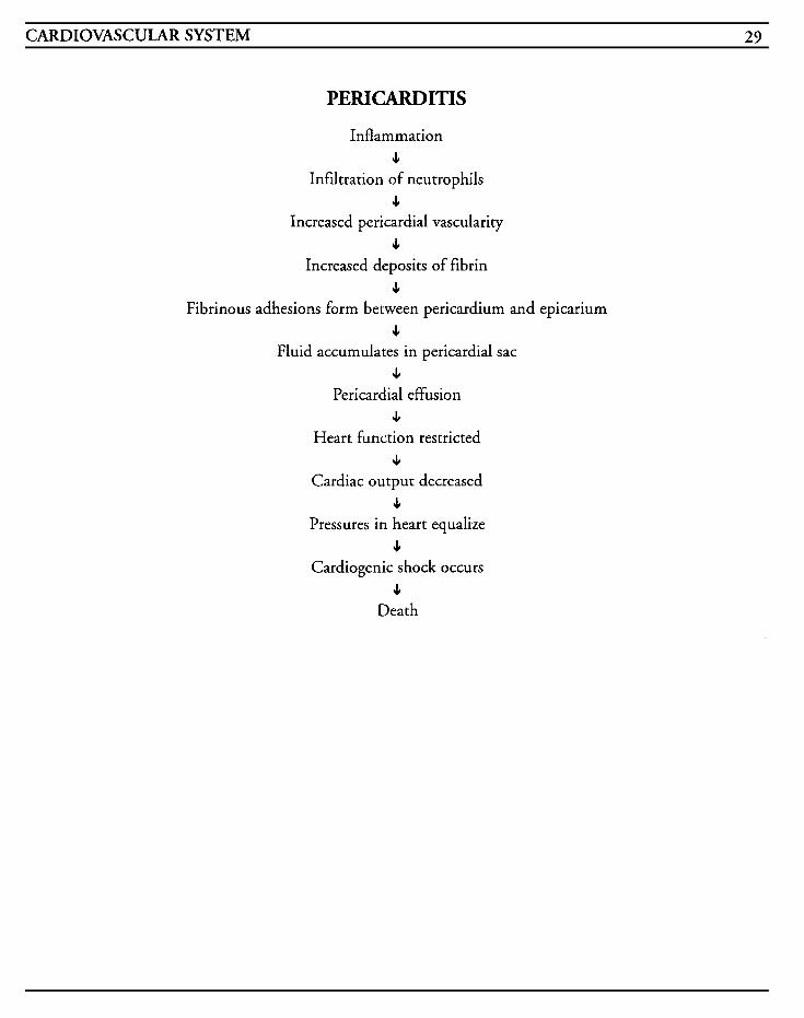
CARDIOVASCULAR SYSTEM 29
PERICARDITIS
Inflammation c
Infiltration of neutrophils c
Increased pericardial vascularity 4
Increased deposits of fibrin c
Fibrinous adhesions form between pericardium and epicarium e
Fluid accumulates in pericardial sac c
Pericardial effusion c
Heart function restricted J,
Cardiac output decreased c
Pressures in heart equalize c
Cardiogenic shock occurs c
Death

This Page Intentionally Left Blank
CARDIOVASCULAR SYSTEM 31
Bacterial endocarditis is now referred to as infec- tive endocarditis due to the presence of other organisms besides bacteria being the causative agent. It is an infection of the cardiac valves and inner lining of the heart that is characterized as a systemic illness. Endocarditis may be misdiagnosed as other infections in the early stages if signs and symptoms of cardiac involvement are not present, Common complaints range from fever with tem- perature less than 102 degrees, chills, arthralgia, lethargy, and anorexia. Acute endocarditis may result in death within a matter of hours if not treated. Antimicrobial therapy can decrease mor- tality to 15%, but heart failure secondary to valvular scarring and damage can occur after the infection is resolved.
Almost any organism can cause endocarditis but the most common ones noted have been Streptococcus viridans, Staphylococcus aureus, Enterococci, Staphylococcus epidermidis, Streptococcus pneumoniae, Pseudornonas aerugi- nosa, Candida albicans, and Aspergillus fumigatus.
Endocarditis may be subdivided into the acute and subacute classes, depending on the virulence of the organism involved and the length of duration. Acute infective endocarditis (AIE) has less than one month duration whereas subacute infective endocarditis (SIE) is usually greater than one month in duration, SIE usually involves congeni- tally-deformed or damaged heart valves, and AIE usually involves normal heart valves. Trauma in many forms can occur to the epithelia1 layer of the valves/endocardium causing injury and deposits of platelets and fibrin to adhere to this surface. This is known as nonbacterial thrombotic endocarditis
(NBTE). After this stage, the heart is then set up for vegetation to colonize from bacteria from other areas of the body during transient episodes of bac- teremia. As these organisms grow, more platelets and fibrin adhere and eventually valves are destroyed, vegetation breaks off and embolizes to other areas of the body, and a systemic immune response occurs.
Patients who are at risk for endocarditis include those with rheumatic heart disease, open-heart surgery, congenital heart defects, prosthetic valve replacements, dental procedures, gynecological surgery or procedures, genitourinary surgery or procedures, invasive tests or lines, infected periph- eral or central venous lines, IUDs, AV shunts or fistulas, skin abnormalities in preexisting cardiac disease, immunosuppressive therapy, and IV drug use.
Patients who have had prosthetic valves placed and who develop endocarditis are divided into early (occurring less than two months postoperatively) and late (occurring greater than two months post- operatively) classes, and develop chills, fever, leukocytosis, and/or a new murmur. Mortality is higher in early prosthetic valve endocarditis and is a serious problem.
MEDICAL CARE Antibiotics: penicillin is the treatment of choice for Streptococcus viridans, with cephalothin or vancomycin being alternate choices; penicillin plus gentamicin is the treatment of choice for Steptococcus faecalis; synthetic penicillins, such as oxacillin or nafcillin, cephalothin and/or gentam- icin are used in Staphylococcus epidermidis
Laboratory: a series of blood cultures is done to isolate the causative organism and sensitivity to antimicrobial agents; CBC is used to assess for anemia that may occur in up to 70% of patients,
32 CRITICAL CARE NURSING CARE PLANS
to monitor leukocyte levels, and to assess platelet counts; sedimentation rates may increase; immune titers show antigen-antibody response
Electrocardiography: shows alterations in conduc- tion, dysrhythmias, ischemia
Echocardiography: used to establish diagnosis, to determine underlying cardiac disease, to estimate myocardial contractility, and demonstrate early mitral valve closure and aortic insufficiency
Nuclear cardiologic testing: Technetium-99 scans and gallium-67 imaging used to evaluate the extent of the infective process and to evaluate potential as a surgical candidate
Surgery: valve replacement is necessary if patient develops intractable congestive heart failure with hemodynamic compromise, persistent bacteremia despite antimicrobial treatment, prosthetic valve endocarditis, major systemic emboli, gram negative or fungal infection; drainage of abscesses or empyema; repair of peripheral or cerebral mycotic aneurysms
Prophylaxis: prophylactic antibiotic therapy must be prescribed prior to dental procedures, urethral or gynecological procedures, or surgery
NURSING CARE PLANS Risk for altered tissue pe@ion: cardiopul- monary, cerebral, renal, gastrointestinal, and peripheral
Related to: valvular vegetation emboli, platelet- fibrin emboli, and immunologic responses causing allergic vasculitis; embolu
Defining characteristics: petechiae, arthritis, arthralgia, myalgias, decreased peripheral pulses, Janeway’s lesions, Roth‘s spots, Osler’s nodes, lower back pain, splinter hemorrhages to subungual
areas, hematuria, oliguria, anuria, chest pain, short- ness of breath, dyspnea, confusion, weakness, convulsions, coma, hemiplegia, aphasia, hemipare- sis, cardiac tamponade, pericardial friction rub, murmur, dysrhythmias, conduction defects, cold clammy skin, cyanosis, mental status changes, hypotension, tachycardia, decreased urinary output, increased BUN
Outcome Criteria
Patient will achieve and maintain adequate tissue perfusion to all body systems.
INTERVENTIONS RATIONALES
Determine mental status and level of consciousness. Observe for hemiparesis, paralysis, apha- sia, convulsions, visual field de- fects, or coma, and notify MD.
Symptoms may indicate emboliza- tion to cerebrum which may re- quire emergency treatment.
Monitor EKG for conduction ab- normalities, especially prolonged PR interval, new left bundle branch block, new right bundle branch block with or without left anterior hemiblo&. Treat as in- dicated per protocol.
Observe for sudden shortness of breath, tachypnea, pleurisy-type pain, pallor or cyanosis.
Evaluate chest pain, tachycar- dia, decreased blood pressure. Auscultate heart sounds for new
Due to the close proximity of aortic valve cusps to the con- duction system, bacterial in- vasion and proliferation may extend the infection process into the myocardium and cause dysrhythmias. Extension of the infection from the mitral valve to the Bundle of His and AV node may result in junctional tachycardia, Mobitz I, second degree or third degree AV blocks.
Arterial emboli may affect the heart and other vital organs. Venous congestion may result in thrombus formation in deep veins and cause embolization to lungs, or embolization of vegetation thrombi may result in pulmonary embolus.
Arterial emboli may affect the heart and cause myocardial in- farction. New murmurs may
CARDIOVASCULAR SYSTEM 33
INTERVENTIONS RATIONALES or changed murmurs, pericardial friction rubs, or abnormal lung sounds (crackles, rales).
Observe extremities for swelling, erythema, tenderness, pain, pos- itive Homans’ sign, positive Pratt’s sign. Observe for de- creased peripheral pulses, pal- lor, coldness, cyanosis.
Monitor for complaints of ab- dominal pain to lek upper abdo- men with radiation ro left shoulder, abdominal rigidity, tenderness, nausea, or vomiting.
Observe urine for hematuria, oliguria, anuria, complaints of flank or back pain.
Observe for petechiae on mucous membranes, conjunctiva, neck, wrists, and ankles. Observe for splinter hemorrhages in subungu- al areas, Osler’s nodes to distal fingers and toes, sides of fin- gers, palms or thighs, and for Janeway’s lesions to the palms, soles of feet, arms and legs.
occur as a result of valve scarring and distortion, valve aneurysm, septal rupture, papillary muscle rupture, or myocardial abscess rupture. Rupture into the pericar- dial sac can cause cardiac tarn- ponade, in which heart tones will be muffled. Pericardial friction rubs may indicate pericarditis. Abnormal lung sounds may indicate impending congestive heart failure.
Bedrest promotes venous stasis which can increase the risk of thromboembolus formation. Ac- tual vegetation emboli can mi- grate and occlude peripheral arteries, leading to tissue ischemia and necrosis.
May indicate embolization to spleen. Vegetative emboli may occlude mesenteric artery and cause bowel infarction. Spleno- megaly may be caused by anti- gen stimulation and allergic vasculitis.
Allergic vasculitis from endo- carditis can result in focal, acute, or chronic glomerulo- nephritis and progress to renal insufficiency, renal failure, and uremia.
Petechiae is one of the classic symptoms of endocarditis as a result of allergic vasculitis. Petechiae are usually 1-2 mm in diameter, flat, red with white or gray centers, non-tender, and groups fade within a few days. Petechiae may be noted in other diagnoses and they should be ruled our. Hemorrhages to the subungual areas may be seen in early infective endocarditis but may be seen in trauma, with hemo- or peritoneal dialysis, or in mitral stenosis. Osler‘s nodes are nodules that range from
INTERVENTIONS RATIONALES
Evaluate complaints of arthri- tis, arthralgia, and severe lower back pain. Medicate as needed.
Monitor blood culture and sen- sitivity reports.
Administer antimicrobials as ordered.
1-10 mm in diameter, red with white centers, overtly tender, and are usually a late sign of endocarditis, typically found in subacute endo- carditis infections. Jane- way’s lesions are non-tender reddened or pink macular lesions, 1-5 mm in diameter, and usually change to tan and fade within 2 weeks. These are usually an early sign of endocarditis.
Occur in endocarditis due to localized immune responses or in decreased perfusion.
Usually 3-6 blood cultures are done in a series ro assess for sustained bacteremia because bacteria are continually re- leased into the system in endo- carditis. The series prevents the possibility of false read- ings. Cultures determine the specific organism responsible for the bacteremia, and sensi- tivity results enable the choice of antimicrobials to be suited to the specific infection.
Antibiotics should not be start- ed until culture series is com- pleted in subacute IE, but with acute IE, empiric antibiotics are given until cultures are available. In some instances, early negative results may indicate only that the culture could not be grown due to low levels of bacteria or an unusual organism being present. Obtaining cultures after anribi- otics have been started do not give accurate information.
34 CRITICAL CARE NURSING CARE PLANS
Instruction, Information, Demonstration
~ ~ ~~~ -
INTERVENTIONS RATIONALES Instruct patient in signs/ symptoms to report to MD.
Promotes knowledge and cornpli- ance with regimen.
Discharge or Maintenance Evaluation
Patient will have adequate tissue perfusion to all body systems.
Patient will be mentally lucid, with no confusion or neurological deficits.
Patient will have adequate urinary output with no hematuria, and renal function studies will be within normal limits.
Patient will be able to recall accurately the infor- mation instructed.
Decreased cardiac output [See MI] Related to: complications with infected heart valves, potential for cardiac tamponade because of effusion, damaged myocardium, decreased contrac- tility, dysrhythmias, conduction defects, alteration in preload, alteration in afterload, vasoconstriction, myocardial ischemia, ventricular hypertrophy
Defining characteristics: decreased blood pressure, tachycardia, pulsus paradoxus greater than 10 mmHg, distended neck veins, increased central venous pressure, dysrhythmias, decreased QRS voltage or electrical alternans, diminished heart sounds, dyspnea, friction rub, cardiac output less than 5 L/min, cardiac index less than 2.5 L/min/m2, change in mental status, change or new cardiac murmur, arterial emboli, decreased urine output, cyanosis, cold clammy skin
Hyperthermia
Related to: bacteremia, allergic vasculitis, arterial occlusion/infarction, abscess
Defining characteristics: body temperature greater than normal range, flushed warm skin, chills, increased heart rate, increased respiratory rate
Outcome Criteria
Patient will maintain body temperature within normal limits and be free of infection.
INTERVENTIONS RATIONALES Monitor temperature every 2-4 hours and prn. Observe for chills and diaphoresis.
Monitor environment tempera- ture and limit or add blankets as warranted. Change linens as needed.
Monitor I&O; provide adequate fluids.
Give tepid sponge baths prn.
Place on cooling blanket as warranted.
Administer antipyretic medi- cations as warranted.
Endocarditis usually results in temperatures less than 102 degrees; temperatures grearer than this indicate an acute infective process. Chills frequently precede a tempera- ture spike.
Room temperature may be al- tered to assist with main- tenance of normal body tem- perature.
Diaphoresis and increased meta- bolic rate from temperature elevations increase fluid loss and may cause dehydration.
May assist in lowering tempera- ture by means of evaporation. Using cooler water or alcohol may cause chilling and thus increase body temperature.
Cooling blankets are usually only used for severe fever greater than 104 degrees when risk of brain damage or seizures is imminent.
Reduces fever by action on the hypothalamus. Low grade ternp- eratures may be beneficial to the body’s immune system and ability to retard the growth of organisms.
CARDIOVASCULAR SYSTEM 35
Information, Instruction, Demonstration
INTEKVENTIONS RATIONALES Instruct on procedures for de- creasing temperature.
Provides knowledge and reduces kar and enhances compliance.
Instruct to take temperature frequently and to notify MD for elevations immediately.
Temperature elevations indicate infection and prompt notifica- tion will allow for prompt treatment.
Instruct on medications, effects, side effects, contraindications, ance. symptoms to report.
Promotes knowledge and compli-
Discharge or Maintenance Evaluation
Patient will be normothermic with no overt signs/symptoms of infection.
Risk for infiction
Related to: inhibition of antibodies due to immunological system action, inflammatory processes due to vegetation growth, predisposition to bacteremia, septic emboli, myocardial abscess, occlusion of arteries leading to necrosis of body systems, invasive procedures and lines, dental pro- cedures, nosocomial infections, lack of recognition of infection, lack of prophylactic treatment, sup-
. A .
rainfection
Defining characteristics: elevated temperature, ele- vated WBC count, positive blood cultures, reddened, draining IV sites
Outcome Criteria
Patient will be free of infection, afebrile, with no over symptoms of infection or infective process noted.
INTERVENTIONS RATIONALES Monitor temperature trends.
Monitor for signs/symptoms of deterioration of patient and failure to improve within a timely manner.
Observe mouth for patches of white plaque and perineal areas for vaginal drainage or itching, and notify MD.
Inspect wounds, IV sites, cath- eter sites, invasive devices and lines, changes in drainage or body fluids.
Maintain aseptic or sterile technique as warranted.
Obtain urine, blood, sputum, wound, and invasive Iindcathe- ter specimens for culture and sensitiviry and Gram stain as warranred.
Reposition patient every 2 hours; encourage coughing and deep breathing.
Decreases in body temperature below 9G degrees may indicate advanced s h o d states and 1s a critical indicator of de- creased tissue perfusion and lack of the body’s ability to muster enough defense to raise the temperature. Tem- peratures greater than 10 1 degrees are due to the effect of endotoxins on the hypo- thdamus and of pyrogen- released endorphins.
May indicate ineffective anti- biotic therapy or abundance of resistant organisms.
Thrush or yeast infections may occur as a secondary infection when normal flora is killed by massive antibiotic therapy.
May indicate local secondary infection or inflammation.
Reduces the risk of opportun- istic infection and chances of cross-contamination.
Assists with identification of source of infection, causative organism, and antibiotic of choice to enable prompt and effective treatment.
Frequent changes in position and breathing exercises en- hance pulmonary toilet and may help to prevent pneumonia.
36 CRITICAL CARE NURSING CARE PLANS
INTERVENTIONS RATIONALES
Administer antibiotics as or- dered.
Antibiotics may be started prior to receiving final culture reports based on the likelihood of the infective organism. Spe- cific antibiotics are determined by the culture information.
Information, Instruction, Demonstration
INTERVENTIONS RATIONALES
Instruct patient to cover mouth and nose during coughing/sneezing. from airborne organisms. Good Instruct in handwashing and dis-
Prevents spread of infection
posal of contaminated materials. handwashing reduces spread of infection. Infection control procedures limit contamination and spread of infective mater- ials.
Instruct patient in good dental hygiene to use soft tooth- brush; to avoid water pik and toothpicks; to obtain regular dental exams.
Instruct patient to take tempera- ture every day for 1 month post discharge.
Prepare patient for surgery as warranted.
Instruct patient in obtaining prophylactic antibiotic therapy prior to procedures.
Avoids trauma to gums which may promote reinfection. Water pik and toothpi& may cause bleeding and promote infection.
Temperature elevations may indicate infeaion/reinfec- tion.
Surgery may be required to remove necrotic tissue or limbs and to remove purulent material in order to enhance healing. Surgery may be required to re- place damaged heart valves due to vegetative infection.
Prophylaxis will be required for any invasive procedure due to likelihood of reinfection.
Discharge or Maintenance Evaluation
Patient will have normal temperature and vital signs.
of infection.
0 Patient will be able to recall instructions accu- rately.
Patient will exhibit no overt symptoms or signs
Patient will seek prophylactic antibiotic therapy prior to any procedure and will have no evidence of reinfection.
Anxiety [See MI] Related to: change in health status, fear of death, threat to body image, threat to role functioning, pain
Defining characteristics: restlessness, insomnia, anorexia, increased respirations, increased heart rate, increased blood pressure, difficulty concen- trating, dry mouth, poor eye contact, decreased energy, irritability, crying, feelings of helplessness
Knowledge dejcit [See MI] Related to: lack of understanding, lack of under- standing of medical condition, lack of recall
Defining characteristics: questions regarding problems, inadequate follow-up on instructions given, misconceptions, lack of improvement of previous regimen, development of preventable complications
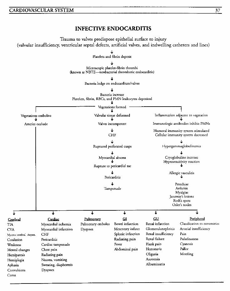
CARDIOVASCULAR SYSTEM 37
INFECTIVE ENDOCARDITIS
Trauma to valves predispose epithelia1 surface to injury (valvular insufficiency, ventricular septal defects, artificial valves, and indwelling catheters and lines)
c Platelets and fibrin deposit
c Microscopic platelet-fibrin thrombi
(known as NBTE-nonbacterial thrombotic endocarditis)
J, Bacteria lodge on endocardiumlvalves
c Bacteria increase
Platelets, fibrin, RBCs, and PMN leukocytes deposited c I 7 Vegetations formed
c Vegetations embolize
J, Valvular tissue deformed Inflammation adjacent to vegetation
4 c Arteries occlude Valves incompetent Immunologic antibodies inhibit PMNs
c CHF
e Ruptured perforated cusps
Humoral immunity system stimulated Cellular immunity system decreased
J Hypergammaglobulinemia
J, J, Myocardial abscess Cryoglobulins increase e Hypersensitivity reaction
Rupture to pericardial sac c c
Pericarditis
4 Tamponade
Allergic vasculitis c
Petechiae Arthritis Mydgias
Janeway's lesions Roth's spots
Osler's nodes I
c c J, J, e c Cerebral Cardiac Pulmonay s;L L a Perbheral TIA Myocardial ischemia Pulmonary embolus Bowel infarction Renal infarction Claudication to extremities CVA Myocardial infarction Dyspnea Mesentery infarct Glomerulonephritis Arterial insufficiency Mycoric cerebral Anysm. CHF Splenic infarction Renal insufficiency Pain Confusion Pericarditis Radiating pain Renal failure Pulselessness Weakness Cardiac tamponade Fever Flank pain Cyanosis Mental changes Chest pain Abdominal pain Hematuria Pallor Hemiparesis Radiating pain Oliguria Mottling Hemiplegia Nausea, vomiting Aphasia Convulsions Dyspnea Coma
Azotemia Sweating, diaphoresis Albuminuria

This Page Intentionally Left Blank
CARDIOVASCULAR SYSTEM 39
Hypertension Essential hypertension, which is an elevated blood pressure of unknown origin, and secondary hyper- tension, which is an elevated blood pressure resulting from a known cause, will cause inflam- mation and necrosis in the arterioles which then result in decreased blood flow to vital body organs, and places stress on the heart and vessels. Uncontrolled hypertension is associated with per- manent damage to body systems. Blood pressure is considered to be hypertension if the systolic pres- sure is greater than 140 mmHg or the diastolic pressure is greater than 90 mmHg, and is classi- fied based on the severity from a high normal to malignant hypertension. Hypertensive crisis is defined as a sustained increase in diastolic blood pressure above 120 mmHg, which is high enough to cause irreversible damage to organs and tissue death.
Hypertension may result from several origins- adrenal origin (as in pheochromocytoma, Cushing’s disease, brain tumor, etc.), renal origin (as in pyelonephritis), cardiovascular origin (as in atherosclerosis or coarctation of the aorta, etc.), or unknown origin which accounts for the majority of all known hypertension.
Untreated, hypertension will result in death due to cerebrovascular accident, congestive heart failure, intracerebral hemorrhage, kidney failure, or dis- secting aneurysms.
Systolic blood pressure is the pressure that the heart pumps against to force blood from the left side of the heart to the aorta and to major arteries. Diastolic blood pressure is the pressure required to permit filling of the ventricles before the next sys- tole cycle. The pulse pressure, which is the value of the difference between the systolic and diastolic pressures, may be used to indicate perfusion prob-
lems. The mean arterial pressure, or MAP (or MABP) is the average pressure attempting to push the blood through the circulatory system and should be greater than 60 mmHg in order to ade- quately perfuse organs.
Elevated blood pressure may occur as a result of emotional stress with as much as 40 mmHg increase, and may also result from ventilatory insufficiency, post-seizures, electroconvulsive ther- apy, intracerebral injury, CNS disorders due to the mass,ive stimulation of catecholamines, coronary artery bypass surgery, myocardial infarction, heart failure, renal insufficiency, eclampsia/toxemia, endocrine disorders, and some drugs.
Risk factors include: ages between 30 and 70 years of age, race (black), use of birth control pills, obe- sity, familial history, smoking, stress, diabetes mellitus, and sedentary lifestyle.
Treatment is aimed at lowering blood pressure by use of antihypertensive medications, diuretics to increase urinary output, and by eliminating factors that promote the elevation of blood pressure. A “stepped care” regimen is used most often, with step one involving the use of thiazide diuretics and calcium ion antagonists; step two involves the sup- plemental use of beta-adrenergic blockers; step three includes vasodilators; and step four involves guanethidine.
MEDICAL CARE Diuretics: chlorothiazide (Diuril), spironolactone (Aldactone), chlorthalidone (Hygroton), hydrochlorothiazide (Esidrix, HydroDiuril), triamterene (Dyrenium), metolazone (Zaroxolyn, Diulo), ethacrynic acid (Edecrin), furosemide (Lasix) to promote diuresis and block reabsorption of sodium and water in the kidney
Calcium ion antagonists: verapamil (Calan), dilti- azem (Cardizem), nifedipine (Procardia),
40 CRITICAL CARE NURSING CARE PLANS
nitrendipine to produce vasodilation on vascular smooth muscle
Adrenergic inhibitors: reserpine, methyldopa (Aldomet), propranolol (Inderal), prazosin hydrochloride (Minipress) used to impair synthe- sis of norepinephrine, suppression of sympathetic outflow by central alpha-adrenergic stimulation, or blocking of preganglionic to postganglionic autonomic transmission
Vasodilators: hydralazine (Apresoline), minoxidil (Loniten), nadolol (Corgard) to relax smooth muscle of arterioles and reduce peripheral vascular resistance and thus, blood pressure
ACE inhibitors: captopril (Capoten) used to lower total peripheral resistance by inhibiting angiotensin-converting enzyme
Electrolytes: potassium chloride (KCI, K Dur, K tabs) to replace potassium lost through diuresis
Chest x-ray: shows any enlargement of the heart and pulmonary vein, presence of pulmonary edema or pleural effusion
Electrocardiography: used to monitor for changes in rate and rhythm, conduction abnormalities, left ventricular hypertrophy, ischemia, electrolyte abnormalities, drug toxicity, and presence of dys- rhythmias
Laboratory: cholesterol levels and lipid profile used to determine cholesterol and triglyceride levels and their pertinence to atherosclerosis; elec- trolyte profiles used to monitor for hypokalemia and hypernatremia which may be prevalent due to diuretic therapy; CBC used to identify potential renal failure and polycythemia; glucose levels used to identify potential causes of hypertension; BUN and creatinine levels used to identify renal dysfunction; urinalysis used to identify proteinuria for possible indication of renal disease and hema-
turia for possible indication of nephrosclerosis; thyroid profile used to identify hyperthyroidism which may lead to vasoconstriction and hyperten- sion; aldosterone level used to identify primary aldosteronism; urine VMA to identify elevation of catecholamine metabolites which may indicate pheochromocytoma
Radiographic testing: IVP may be used to iden- ti$ presence of kidney disease; renal arteriogram may be used to show renal artery stenosis or other causes of hypertension
NURSING CARE PLANS Rid for decreased cardiac output
Related to: vasoconstriction, increased preload, increased afterload, ventricular hypertrophy, ischemia
Defining characteristics: elevated blood pressure, decreased cardiac output, decreased stroke volume, increased peripheral vascular resistance, increased systemic vascular resistance
Outcome Criteria
Patient will have no elevation in blood pressure above normal limits and will have adequate car- diac output, and will maintain blood pressure within acceptable limits.
INTERVENTIONS RATIONALES Monitor blood pressure every 1-2 hours, or every 5 minutes during active titration of vasoactive drugs. Measure pressure in both arms using appropriate size of cuff. When possible, obtain pressures lying, sitting, and standing.
Changes in blood pressure may indicate changes in patient status requiring prompt atten- tion. Comparing pressures in both sides provides information as to amount of vascular in- volvement. Blood pressure may vary depending on body position and postural hypotension may result in syncope.
CARDIOVASCULAR SYSTEM 41
INTERVENTIONS RATIONALES Infarmatian, Instruction, Demonstration
Monitor EKG for dysrhythmias, conduction defects, and for heart rate and rhythm changes.
Decreases in cardiac output may result in changes in car- diac perfusion causing dys- INTERVENTIONS RATIONALES
Treat as indicated. rhythmias.
Observe skin for color, temp- erature, capillary refill time, and diaphoresis.
Peripheral vasoconstriction may result in pale, cool, clammy skin, with prolonged capillary refill time due to cardiac dysfunction and de- creased cardiac output.
Auscultate lungs for adventi- tious breath sounds.
Ausculate heart tones.
Administer thiazide, loop, or potassium-sparing diuretics as ordered.
Crackles (rales) or wheezing may indicate pulmonary con- gestion due to cardiac failure as a result of in- creased blood pressure.
Instruct on fluid and diet requirements and restrictions of sodium.
Restrictions can assist with decrease in fluid retention and hypertension, thereby im- proving cardiac output.
Promotes knowledge and com- pliance with drug regimen.
Instruct on medications, ef- feas, side effects, contra- indications, signs to report.
Prepare patient for surgery if warranted.
Pheochromocytoma may require surgical intervention for re- moval of the tumor in order to correct hypertension.
Hypertensive patients often
hypertrophy. Ventricular have S4 gallops due to atrial Discharge or Maintenance Evaluation hypertrophy may result in s3 gallops.
Thiazides are used to reduce blood pressure in patients with normal renal function and these limit fluid retention. Loop diuretics inhibits reabsorption of sodium and chloride and are used in patients who have renal dys- function. Potassium-sparing di- uretics are used in conjunction with thiazides to decrease the amount of potassium lost.
Administer sympathetic inhibi- tors as ordered.
These drugs reduce blood pres- sure by decreasing peripheral resistance, reducing cardiac output, inhibiting sympathetic activity, and suppressing the release of renin which is a potent vasoconstrictor.
Administer vasodilators as ordered.
May be used in severe hyper- tension to increase coronary blood flow and decrease after- load to improve cardiac output.
Patient will be normotensive, with adequate car- diac output and index.
Medications will be taken as ordered with no side effects.
Patient will have stable heart rate, rhythm, and heart tones, with no adventitious breath sounds.
Patient will be able to verbalize instructions accurately.
Risk for altered tissue perjkion: cardiopul- monary, cerebral, renal, gastrointestinal, and perapherul
Related to: increased catecholamine stimulation, increased blood pressure, decreased cardiac output, decreased baroreceptor sensitivity, changes in cere- brospinal fluid pressure, angiotensin and aldosterone stimulation, sodium intake, environ- mental factors, genetic factors, strain on arterial wall, atherosclerosis
Administer antiadrenergic drugs Prevents blood vessels from as ordered. constricting and increasing
blood pressure.
42 CRITICAL CARE NURSING CARE PLANS
Defining characteristics: increased blood pressure, retinopathy, retina1 hemorrhage, headache, epistaxis, tachycardia, rales, S3 or S4 gallops, rest- lessness, bruits to femoral, carotids, abdominal aorta, blurred vision, chest pain, shortness of breath, optic disc papilledema, seizures, coma, nys- tagmus, mental changes
Outcome Criteria
Patient will achieve and maintain adequate tissue perfusion to all body systems.
INTERVENTIONS RATIONALES Monitor for sudden onset of chest pain.
Monitor EKG for changes in rate, rhythm, dysrhythmias, and con- duction defects. Treat as in- dicated.
Monitor hemodynamic parameters closely and titrate vasoactive drugs as warranted.
Observe for shift of point of maximal impulse (PMI) to left
Auscultate over peripheral ar- teries for bruits.
Observe extremities for swelling, erythema, tenderness, pain, pos- itive Homans’ sign, positive Pratt’s sign. Observe for de- creased peripheral pulses, pal- lor, coldness, cyanosis.
May indicate dissecting aortic aneurysm.
Decreased perfusion may result in dysrhythmias due to decrease in oxygen.
Provides immediate information regarding efficacy of medication and status of hypertension.
Shift occurs in cardiac enlarge- ment.
Atherosclerosis may cause bruits by obstructing blood flow.
Bedrest promotes venous stasis which can increase the risk of thromboembolus formation, Ac- tual vegetation emboli can mi- grate and occlude peripheral arteries, leading to tissue ischemia and necrosis.
Instruction, Information, Demonstration
INTERVENTIONS RATIONALES Instruct patient in signs/ symptoms to report to M D , such as headache upon rising, in- creased blood pressure, chest pain, shortness of breath, in- creased heart rate, weight gain of > 2 lblday or 5 Ib/wk, edema, visual changes, nosebleeds, diz- ziness, syncope, muscle cramps, nausedvomiting, impotence or decreased libido.
Promotes knowledge and compli- ance with treatment. Promotes prompt detection and facilitates prompt intervention.
Discharge or Maintenance Evaluation
Patient will have adequate tissue perfusion to all body systems.
Patient will be mentally lucid, with no confu- sion or neurological deficits.
Patient will have adequate urinary output with no hematuria, and renal function studies will be within normal limits.
0 Patient will be able to recall accurately the infor-
Anxiety [See MI] Related to: change in health status, fear of death, threat to body image, threat to role functioning, pain
Defining characteristics: restlessness, insomnia, anorexia, increased respirations, increased heart rate, increased blood pressure, difficulty concen- trating, dry mouth, poor eye contact, decreased energy, irritability, crying, feelings of helplessness
mation instructed.
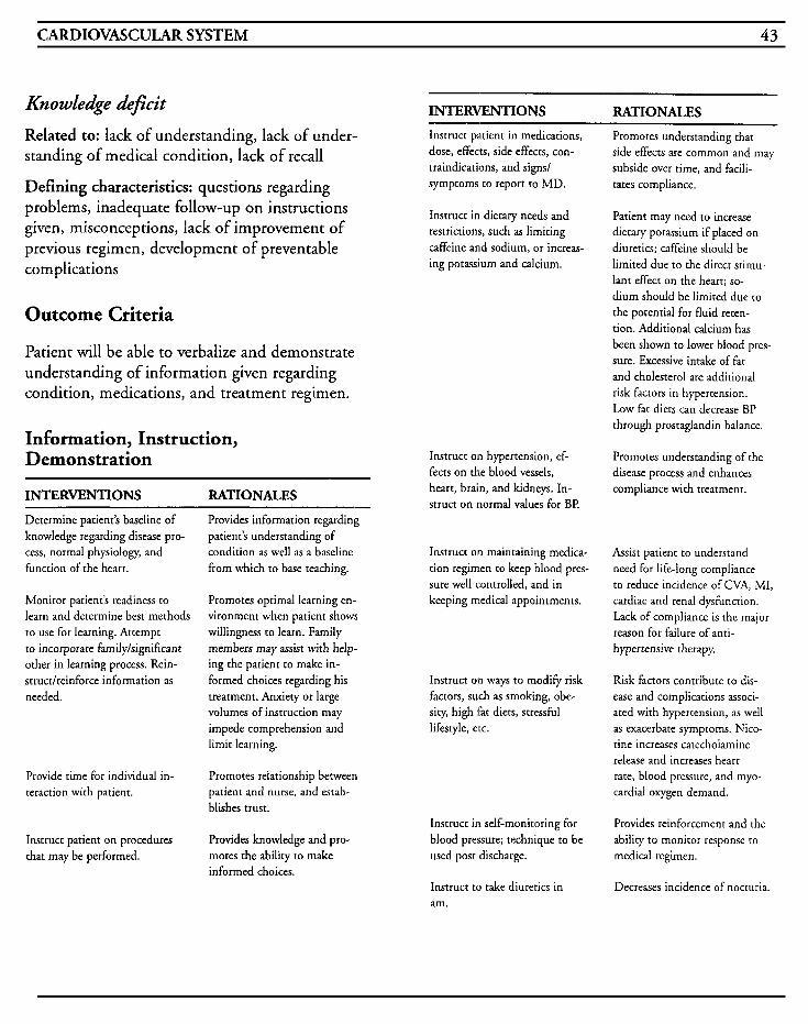
CARDIOVASCULAR SYSTEM 43
Knowledge Aficz't
Related to: lack of understanding, lack of under- standing of medical condition, lack of recall
Defining characteristics: questions regarding problems, inadequate follow-up on instructions given, misconceptions, lack of improvement of previous regimen, development of preventable complications
Outcome Criteria
Patient will be able to verbalize and demonstrate understanding of information given regarding condition, medications, and treatment regimen.
Information, Instruction, Demonstration
INTERVENTIONS RATIONALES
Determine patient's baseline of knowledge regarding disease pro- cess, normal physiology, and function of the heart.
Monitor patient's readiness to learn and determine best methods to use for learning. Attempt to incorporate family/significant other in learning process. Rein- strucdreinforce information as needed.
Provide time for individual in. teraction with patient.
Instruct patient on procedures that may be performed.
Provides information regarding patient's understanding of condition as well as a baseline from which to base teaching.
Promotes optimal learning en- vironment when patient shows willingness to learn. Family members may assist with help- ing the patient to make in- formed choices regarding his treatment. Anxiety o t large volumes of instruction may impede comprehension and limit learning.
Promotes relationship between patient and nurse, and estab- blishes trust.
Provides knowledge and pro- motes rhe ability to make informed choices.
INTERVENTIONS RATIONALES Instruct patient in medications, dose, effects, side effects, con- traindications, and signs/ symptoms to report to MD.
Promotes understanding chat side effects are common and may subside over time, and facili- tates compliance.
Instruct in dietary needs and restrictions, such as limiting caffeine and sodium, or increas- ing potassium and calcium.
Instruct on hypertension, ef- fects on the blood vessels, heart, brain, and kidneys. In- struct on normal values for BI?
Instruct on maintaining medica- tion regimen to keep blood pres- sure well controlled, and in keeping medical appointments.
Instruct on ways to modify risk factors, such as smoking, obe- sity, high Fat diets, stressful lifestyle, etc.
Instruct in self-monitoring for blood pressure; technique to be used post discharge.
Instruct to take diuretics in am.
Patient may need to increase dietary potassium if placed on diuretics; caffeine should be limited due to the direct srimu- lant effect on the heart; so- dium should be limited due to the potential for fluid reten- tion. Additional calcium has been shown to lower blood pres- sure. Excessive intake of fat and cholesterol are additional risk factors in hypertension. Low fat diets can decrease BP through prostaglandin balance.
Promotes understanding of the disease process and enhances compliance with treatment.
Assist patient to understand need for life-long compliance to reduce incidence of CVA, MI, cardiac and renal dysfunction. Lack of compliance is the major reason for failure of anti- hypertensive therapy.
Risk factors contribute to dis- ease and complications associ- ated with hypertension, as well as exacerbate symptoms. Nico- tine increases catecholamine release and increases heart rate, blood pressure, and myo- cardial oxygen demand.
Provides reinforcement and the ability to monitor response to medical regimen.
Decreases incidence of nocturia.
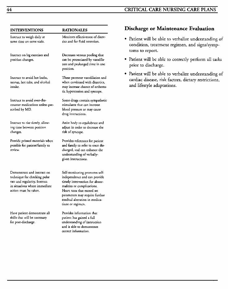
44 CRITICAL CARE NURSING CARE PLANS
INTERVENTIONS RA'TIBNALES Discharge or Maintenance Evaluation Instruct to weigh daily at same time on same scale.
Instruct on leg exercises and position changes.
Instruct to avoid hot baths, saunas, hot tubs, and alcohol intake.
Instruct to avoid over-the- counter medications unless pre- scribed by MD.
Instruct to rise slowly, allow- ing time between position changes.
Provide printed materials when possible for patiendfamily to review.
Demonstrate and instruct on technique for checking pulse rate and regularity. Instruct in situations where immediate action must be taken.
Have patient demonstrate all skills that will be necessary for post-discharge.
Monitors effectiveness of diure- tics and for fluid retention.
Decreases venous pooling that can be potentiated by vasodila- tors and prolonged time in one position.
These promote vasodilation and when combined with diuretics, may increase chance of orthosta- tic hypotension and syncope.
Some drugs contain sympathetic stimulants that can increase blood pressure or may cause drug interactions.
Assist body to equilobrate and adjust in order to decrease the risk of syncope.
Provides references for patient and family to refer to once dis- charged, and can enhance the understanding of verbally- given instructions.
Self-monitoring promotes self- independence and can provide timely intervention for abnor- malities or complications. Heart rates that exceed set parameters may require further medical alteration in medica- tions or regimen.
Provides information that patient has gained a full understanding of instruction and is able to demonstrate correct information.
Patient will be able to verbalize understanding of condition, treatment regimen, and signs/symp- toms to report.
Patient will be able to correctly perform all tasks prior to discharge.
Patient will be able to verbalize understanding of cardiac disease, risk factors, dietary restrictions, and lifestyle adaptations.
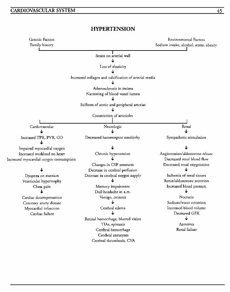
CARDIOVASCULAR SYSTEM 45
HYPERTENSION
Increased TPR, PVR, CO c
Decreased baroreceptor sensitivity Sympathetic stimulation
Impaired myocardial oxygen c c Increased workload on heart Chronic hypertension Angiotension/aldosterone release
Increased myocardial oxygen consumption c Decreased renal blood flow Decreased renal oxygenation
Ischemia of renal tissues
Chest pain Memory impairment Increased blood pressure
Changes in CSF pressures
Decrease in cerebral oxygen supply c Decrease in cerebral perfusion c
Dyspnea on exertion Ventricular hypertrophy c Renidaldosterone secretion
c Dull headache in a.m. c Cardiac decompensation Vertigo, tremors Nocturia Coronary artery disease c Sodium/water retention Myocardial infarction Cerebral edema Increased blood volume
Cardiac failure c Decreased GFR Retina1 hemorrhage, blurred vision c
TIAs, epistaxis Azotemia Cerebral hemorrhage Renal failure
Cerebral aneurysm Cerebral thrombosis, CVA

This Page Intentionally Left Blank
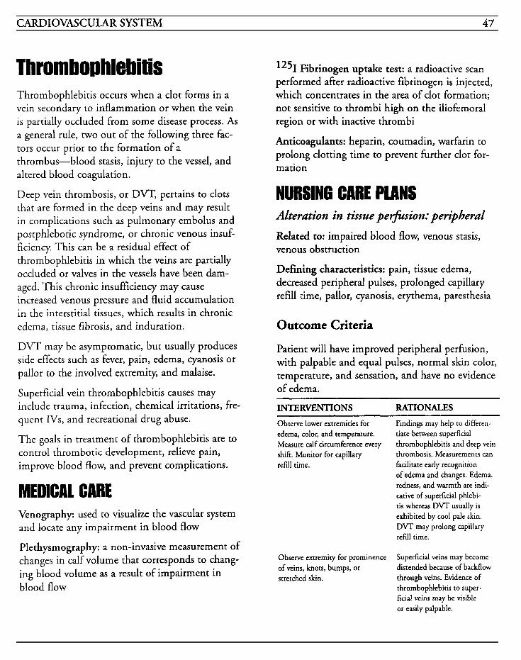
CARDIOVASCULAR SYSTEM 47
Throm bop hlebi tis Thrombophlebitis occurs when a clot forms in a vein secondary to inflammation or when the vein is partially occluded from some disease process. As a general rule, two out of the following three fac- tors occur prior to the formation of a thrombus-blood stasis, injury to the vessel, and altered blood coagulation.
Deep vein thrombosis, or DVT, pertains to clots that are formed in the deep veins and may result in complications such as pulmonary embolus and postphlebotic syndrome, or chronic venous insuf- ficiency. This can be a residual effect of thrombophlebitis in which the veins are partially occluded or valves in the vessels have been dam- aged. This chronic insufficiency may cause increased venous pressure and fluid accumulation in the interstitial tissues, which results in chronic edema, tissue fibrosis, and induration.
DVT may be asymptomatic, but usually produces side effects such as fever, pain, edema, cyanosis or pallor to the involved extremity, and malaise.
Superficial vein thrombophlebitis causes may include trauma, infection, chemical irritations, fre- quent IVs, and recreational drug abuse.
The goals in treatment of thrombophlebitis are to control thrombotic development, relieve pain, improve blood flow, and prevent complications.
MEDICAL CARE Venography: used to visualize the vascular system and locate any impairment in blood flow
Plethysmography: a non-invasive measurement of changes in calf volume that corresponds to chang- ing blood volume as a result of impairment in blood flow
1251 Fibrinogen uptake test: a radioactive scan performed after radioactive fibrinogen is injected, which concentrates in the area of clot formation; not sensitive to thrombi high on the iliofemoral region or with inactive thrombi
Anticoagulants: heparin, coumadin, warfarin to prolong clotting time to prevent further clot for- mation
NURSING CARE PLANS Alteration in tissue pe+ion: peripheral
Related to: impaired blood flow, venous stasis, venous obstruction
Defining characteristics: pain, tissue edema, decreased peripheral pulses, prolonged capillary refill time, pallor, cyanosis, erythema, paresthesia
Outcome Criteria
Patient will have improved peripheral perfusion, with palpable and equal pulses, normal skin color, temperature, and sensation, and have no evidence of edema.
INTERVENTIONS RATIONALES - Observe lower extremities for edema, color, and temperature. Measure calf circumference every shift. Monitor for capillary refill time.
Findings may help to differen- tiate between superficial thrombophlebitis and deep vein thrombosis. Measurements can facilitate early recognition of edema and changes. Ederna, redness, and warmth are indi- cative of superficial phlebi- tis whereas DVT usually is exhibited by cool pale skin. DVT may prolong capillary refill time.
Observe extremity for prominence of veins, knots, bumps, or stretched skin.
Superficial veins may become distended because of backflow through veins. Evidence of thrombophlebitis to super- ficial veins may be visible or easily palpable.
48 CRITICAL CARE NURSING CARE PLANS
INTERVENTIONS RATIONALES Maintain bedrest.
Elevate legs while in bed or sitting in chair.
Observe for positive Homanb sign (pain in calf upon dorsi- flexion of foot).
Perform active or passive ROM exercises while at bedrest.
Apply TED hose after acute phase is over. Remove for at least 1 hour every shift.
Apply warm moist soaks as or- dered.
Administer anticoagulants as ordered.
Monitor laboratory studies for PT, PTT, A M T , and CBC.
Activity limitation may mini- mize the potential for dis- lodgment of the dot.
Reduces swelling and increases venous return. Some experts believe that elevation may actually enhance the release of thrombi.
Homan’s sign may or may not be present consistently and should not be used as a sole indicator of thrombophlebitis.
Promote increased venous blood return and decrease venous stasis.
Assists to minimize postphle- botic syndrome and increases blood flow to deep veins. Re- moval allows time for compres- sion of veins to be relaxed.
Promotes vasodilation and may improve venous return and de- crease in edema.
Heparin is used initially be- cause of its action on thrombin formation and the removal of the intrinsic pathway to pre- vent hr ther clot formation. Coumadin is usually used for long-term therapy.
Monitors efficacy of anticoagu- lant therapy and potential for d o t formation due to hemocon- centration/dehydration.
Information, Instruction, Demonstration
INTERVENTIONS RATIONALES Instruct on avoidance of rubbing or massaging extremity involved.
May promote risk of dislodging clot and causing embolization.
Avoid crossing legs, prolonged positions with legs dangling, or knees bent.
Positions tend to restrict cir- culation and increases venous stasis, and increases edema.
Instruct in deep breathing Promotes emptying of large exercises. veins by increasing negative
pressure in the thorax.
Instruct on maintaining fluid intake of at least 2 Llday.
Dehydration promotes increased viscosity of blood, and in- creases venous stasis.
Prepare patient for surgery if warranted.
Surgical intervention may be required if circulation is se- verely compromised. Recurrent episodes of thrombi may require a vena caval umbrella to filter out thrombi going to lung .
Instruct on lying in a slightly reversed trendelenburg position.
Promotes blood flow to depend- ent extremities; preferable to have extremities full of blood as opposed to empty.
Discharge or Maintenance Evaluation
Patient will have palpable pulses of equal strength to all extremities.
Skin will be within normal limits of coloration, temperature, and sensation.
Patient will be able to recall all instructions accurately.
Patient will have no complications from antico- agulation therapy.
CARDIOVASCULAR SYSTEM 49
Risk for impaired skin integrity
Related to: edema, venous stasis, bedrest, surgery, pressure, altered circulation and blood flow, altered metabolic states
Defining characteristics: skin surface disruptions, incisions, ulcerations, wounds that do not heal
Outcome Criteria
Patient will have no evidence of impairment to skin tissues.
Patient will have surgical wound approximated and well-healed with no evidence of infection.
INTERVENTIONS RATIONALES
Monitor extremities for presence of ulcers, wounds, symptoms of decreased circulation.
If surgery is required, change dressing using aseptic or ster- ile technique as warranted. Leave wound open to air as soon as is feasible, or apply light dressing.
Cleanse wound as ordered with each dressing change.
Monitor wound for skin integ- rity to incision and surround- ing tissues, noting increases and changes in characteristics of drainage.
Provides prompt assessment and treatment for impaired tissues.
Prevents drainage accumulations from excoriating skin, provides assessment to monitor for chan- ges in wound appearance and de- teriorationlimprovement, and prevents wound from contamina- tion. Allowing air to reach wound facilitates drying and promotes the healing process. Sutures may be abrasive to skin or get caught on garments and irritation may be reduced with a light gauze dressing.
Various agents can be used to remove exudate or necrotic ma- terial from wound to promote healing. Any packing of the wound should be done using sterile technique to reduce the risk of contamination.
Prompt recognition of problems with healing may prevent exa- cerbation of wound. Increased drainage or malodorous drainage may indicate infection and de- layed wound healing.
INTERVENTIONS RATIONALES
Monitor any drainage tubes for amounts and character of drain- age. Use ostomy bags over tubes when drainage is massive.
Use skin prep, moisture barrier, or benzoin to skin prior to tape application. Use hypoallergenic tape or Montgomery straps to se- cure dressings.
Provides indication of decreas- ing or increasing wound drain- age and assessment of healing process. Collection of drain- age in bags facilitates more accurate measurement of fluid loss and prevents excoriation of skin from copious drainage.
Provides protection to skin and reduces potential for skin trauma. Reduces potential for skinlwound disruption when frequent dressing changes are required.
Information, Instruction, Demonstration
-
INTERWNTIONS RATIONALES
Instruct to avoid scratching, hitting or bumping legs, or other injurious activities. formation.
Injuries may damage tissues that may deteriorate into ulcer
Instruct on signslsymptoms of infection to woundlskin and to report to nurselMD.
Provides prompt notification to enhance prompt treatment.
Instruct on cleansing incision area post discharge.
Reduces skin surface contami- nants and prevents infection.
Discharge or Maintenance Evaluation
Patient will have approximated, healed surgical wound with no drainage, erythema, or ederna to site.
Patient will be able to recall instructions accurately.
Patient will be compliant with avoiding injurious activities, and will seek medical help when injury occurs.
50 CRITICAL CARE NURSING CARE PLANS
Alteration in comfort [See MI] Related to: inflammation, impaired blood flow, intermittent claudication, venous stasis, lactic acid in tissues, surgical procedures, fever
Defining characteristics: complaints of pain, ten- derness to touch, aching, burning, restlessness, facial grimacing, guarding of extremity
Knowledge deficit
Related to: lack of understanding, lack of under- standing of medical condition, lack of recall
Defining characteristics: questions regarding prob- lems, inadequate follow-up on instructions given, misconceptions, lack of improvement of previous regimen, development of preventable compli- cations
Outcome Criteria
Patient will be able to verbalize and demonstrate understanding of information given regarding con- dition, medications, and treatment regimen.
Information, Instruction, Demonstration
INTERVENTIONS RATIONALES
Determine patient’s baseline of knowledge regarding disease pro- cess, normal physiology, and function.
Monitor patient’s readiness to learn and determine best methods to use for learning. Attempt to incorporate family/significant other in learning process. Rein- struct/reinforce information as needed.
Provides information regarding patient‘s understanding of condition as well as a baseline from which to base teaching.
Promotes optimal learning en- vironment when patient shows willingness to learn. Family members may assist with help- ing the patient to make in- formed choices regarding his treatment. Anxiety or large volumes of instruction may impede comprehension and limit learning.
INTERVENTIONS RATIONALES
Provide time for individual in- teraction with patient.
Instruct patient on procedures that may be performed.
Instruct on signslsymptoms of possible complications, such as pulmonary emboli, venous insuf- ficiency, and venous stasis ul- cers.
Instruct on care to lower ex- tremities and to not ib MD for development of any lesion.
Instruct patient in medications, dose, effects, side effects, con- traindications, and signs/ symptoms to report to MD.
Instruct on leg exercises and position changes. Assist with setting up activity program post-discharge.
Instruct to rise slowly, allow- ing time between position changes.
Instruct to balance rest with activity.
Instruct on proper application of TED stockings.
Avoid valsalva-type maneuvers. Provide increased fiber to diet and administer stool softeners as warranted.
Promotes relationship between patient and nurse, and estab- blishes trust.
Provides knowledge and promotes the ability to make informed choices.
Provides knowledge and assists patient to understand health care needs.
Chronic venous stasis may occur and promotes risk of infection and/or ulcer formation.
Promotes understanding that side effects are common and may subside over time, and facili- tates compliance.
Decreases venous pooling that can be potentiated by vasodila- tors and prolonged time in one position. Exercise may assist in developing collateral circu- lation and enhances venous re- turn.
Assist body to equilobrate and adjust in order to decrease the risk of syncope.
Rest decreases oxygen demands of compromised tissue and decreases potential for emboliza- tion of thrombus. Balancing rest with graduated activity prevents exhaustion and impairment of tissue perfusion.
Improper application may cause a tourniquet-like effect and impede circulation.
Increases venous pressure in the leg which increases potential for thrombophlebitis.
CARDIOVASCULAR SYSTEM 5 1
INTERVENTIONS RATIONALES
Instruct on anticoagulation therapy-dosage, effects, side effects, when to administer, other medications to avoid.
Promotes compliance with medi- cal regimen and decreases poten- tial for improper dosage and adverse drug interactions. Aspirin and salicylates decrease prothrombin activity, vitamin K increases prothrombin activity, antibiotics may interfere with vitamin K synthesis, and barbi- turates can potentiate anticoagu- lant effect.
Instruct on importance of keep- ing MD appointments for follow- up laboratory studies.
Provide printed materials when possible for patientlfamily to review.
Have patient demonstrate all skills that will be necessary for post-discharge.
Promotes compliance with treat- ment and decreases potential for non-therapeutic levels of anti- coagulation therapy.
Provides references for patient and family to refer to once dis- charged, and can enhance the understanding of verbdly- given instructions.
Provides information that patient has gained a full understanding of instruction and is able to demonstrate correct information.
Discharge or Maintenance Evaluation
Patient will be able to verbalize understanding of condition, treatment regimen, and signs/symptoms to report.
Patient will be able to correctly perform all tasks prior to discharge.
Patient will be able to verbalize understanding of safety precautions, correct dosage and admin- istration of all medications, and activity limitations.
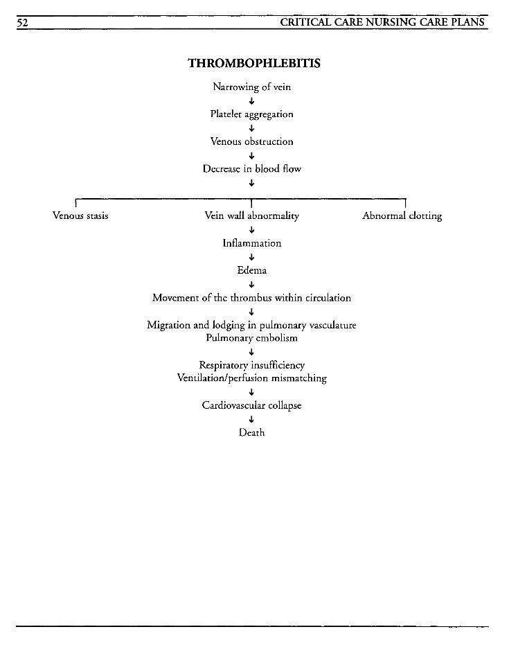
52 CRITICAL CARE NURSING CARE PLANS
THROMBOPHLEBITIS
Narrowing of vein s
Platelet aggregation s
Venous obstruction s
Decrease in blood flow s
I I I Venous stasis Vein wall abnormality Abnormal clotting
s Inflammation
JI Edema
3. Movement of the thrombus within circulation
s Migration and lodging in pulmonary vasculature
Pulmonary embolism J
Respiratory insufficiency Ventilation/perfusion mismatching
s Cardiovascular collapse
4 Death
~
CARDIOVASCULAR SYSTEM 53
Intra-Aortic Balloon Pump
The intra-aortic balloon pump (IABP) is an advanced procedure that is used in the manage- ment of cardiovascular problems that are refractory to routine medical therapeutics. An intra-aortic balloon catheter (IAB) is inserted into the descending aorta, most commonly by way of the femoral artery. The IAB is then attached to the IABP which inflates and deflates the balloon in synchronization with the cardiac cycle. The bal- loon inflates during diastole when the aortic valve closes and increases the aortic pressure when the blood distally to the balloon is forced back towards the aortic valve. The coronary arteries are supplied with additional blood to improve coro- nary blood flow and perfusion and to decrease preload. Deflation occurs prior to the onset of sys- tole and decreases the aortic pressure and ventricular resistance and makes it easier for the ventricle to contract and expel its normal volume of blood, thus decreasing afterload. This counter- pulsation and displacement of blood decreases myocardial oxygen demand by decreasing myocar- dial workload and increases coronary perfusion and cardiac output.
Indications for use of IAB counterpulsation include cardiogenic shock, valvular disease, intractable chest pain resistant to medical treatment, prophylactic support during coronary angiography or anesthesia induction, papillary muscle rupture, ventricular septal defects, compli- cations of acute myocardial infarctions, weaning from the cardiopulmonary bypass, septic shock, and as a bridge to cardiac transplantation. Counterpulsation is contraindicated in patients with severe aortic insufficiency, dissecting aneurysms, peripheral vascular disease, organic
brain syndrome, irreversible brain damage, absent femoral pulses, trauma that has resulted in internal bleeding, active bleeding ulcers, blood dyscrasias, or previous aortofemoral or aortoiliac bypass grafts.
Because the potential for complications is high, this procedure should be utilized only by person- nel well-versed and competent in all aspects of the IABP function and troubleshooting complications.
Two of the major complications associated with the use of the IABP are compromise of the left cir- culation and difficulty with weaning the patient from the IABP.
Oxygen: to increase available oxygen supply
Nitrates: (nitroglycerin, isosorbide dinitrate, Nitro-bid, Nitrostat) used to relax vascular smooth muscle to produce vasodilation, decrease preload, decrease afterload, decrease venous return, decrease peripheral vascular resistance, decrease oxygen demand
Beta-blockers: (propranolol, metoprolol, nadolol, atenolol, timolol, pindolol) used to reduce myoca.rdia1 oxygen demand by blocking catecholamine and sympathetic induced increases in heart rate, contractility and blood pressure; slows AV node conduction; decreases sodium and water retention by reduction of renin secretion; decreases platelet aggregation and may reduce vasospasm
Calcium-channel blockers: (verapamil, nifedipine, diltiazem) used for decreasing myocardial oxygen demand and to enhance relaxation in hypertrophic ardiomyopathies, reduces blood pressure and afterload, and help prevent coronary spasm from decreased oxygen supply
54 CRITICAL CARE NURSING CARE PLANS
Sympathomimetic drugs: (doparnine, Intropin) used for treatment of hypotension in normovolemic states and in the treatment of severe heart failure and cardiogenic shock
Placement of LAB: necessary for counterpulsation to begin
Cardiac catheterization: used to define lesions and evaluate their severity, to provide information on ventricular function, and to allow for measurement of heart pressures and cardiac output
Labwork: PT, PTT, and platelets are obtained to monitor anticoagulation status; general chemistry profiles and renal profiles are monitored every day for chemical imbalances and impending hepatic or renal problems; cardiac isoenzymes are used to monitor for heart damage; CBC and differentials are done every day to monitor for infection and changes in hematologic status; cultures of blood, urine and sputum are done for temperature eleva- tions greater than 102 degrees to assess for infection/suspected organisms
Arterial blood gases: used to assess oxygenation status
Chest x-ray: used daily to monitor placement of IAB and watch for migration, to assess enlargement of the heart and/or pulmonary ves- sels, and to assess pulmonary fluid status and atelectasis
Electrocardiography: reveals changes with atrial and ventricular enlargement, rhythm and conduc- tion abnormalities, ischemia, electrolyte abnormalities, drug toxicity, and presence of dys- rhythmias
Pacemakers: either temporary or permanent, used in anticipation of lethal dysrhythmias and/or con- duction problems
NURSING CARE PLANS Altered tissue perfusion: cardiopulmonary, cerebral, gastrointestinal, renal, peripheraL
Related to: cardiac failure, tissue ischemia, vasoconstriction, hypovolemia, shunting, depressed ventricular function, dysrhythmias, conduction defects, hypoxia, reduction or interruption of blood flow
Defining characteristics: visual disturbances, paresthesias, mental changes, change in level of consciousness, confusion, restlessness, pulse and blood pressure changes, changes in cardiac output, changes in peripheral resistance, impaired oxygena- tion of myocardium, chest pain, cardiac dysrhythmias, changes in EKG (S-T segment, T wave, U wave), LV enlargement, dyspnea, short- ness of breath, tachypnea, palpitations, nausea, vomiting, slow digestion, oliguria, anuria, electrolyte imbalance, cold, clammy skin, decreased peripheral pulses, mottling, cyanosis, diaphoresis
Outcome Criteria
Blood flow and perfusion to vital organs will preserved and circulatory function will be maximized.
be
Patient will be free of dysrhythmias and hemody- namic parameters will be within normal limits.
INTERVENTIONS RATIONALES Monitor vital signs every 15 to 30 minutes until stable, then every hour. Notify MD of devi- ations from parameters.
IABP timing is based on heart rate, and when rate changes > 10 beatslminute, adjustments in timing are necessary to ensure optimal counterpulsation. Dysrhythmias hamper optimal oxygenation and function of the IABP.
CARDIOVASCULAR SYSTEM 55
INTERVENTIONS RATIONALES Monitor mean arterial blood pressure every hour.
Obtain pulmonary artery pres- sures every hour.
Measure cardiac outputlcardiac index and perform hemodynamic measurements every 1-4 hours.
Monitor for malfunction of IAB and IABP and correct prob- blems rapidly. Manually flut- ter IAB prn pump failure.
Assesses volume status to help monitor for efficacy of counter- pulsation. MABP can be calcula- ted by adding 113 (systolic BP- diastolic BP) + diastolic Bl? MABP is a function of cardiac output and systemic vascular resistance. Levels < 60 have little, if any, perfusion to brain.
Provides information as to fluid status and heart pres- sures. PA systolic pres- sures represent RV pressures with normals ranging from 20- 30 mmHg. PA diastolic pres- sures reflects the LVEDP and is an indirect measurement of LV function with normals ranging 10-20 mmHg. PCWP reflects the LA pressure and is used to assess LV filling pressures with normals rang- ing from 4- 12 mmHg.
Directly measures the volume of cardiac output in Llmin, and gives calculated informa- tion regarding preload and afterload. Normal CO should range from 4-8 Llmin and CI from 2.5-4 L/min/m2. SVR which represents afterload should range between 900- 1400 dyneslseclcm5.
Improper timing of balloon can promote complications and wor- sen condition. Early inflation leads to regurgitation into the left ventricle or premature closing of the valve, and in- creases afterload. Late infla- tion decreases augmentation and reduces coronary perfusion. Early deflation allows the pres- sure to rise to normal end- diastolic levels preceding sys- tole which does not reduce af-
INTERVENTIONS RATIONALES
Provide adequate amounts of gas (CO2 or helium) in IAB; refill IAB every 2 hours or
more often if fever present.
terload. Late deflation en- croaches on the next systole and increases afterload. IAB cannot be left in patient longer than 30 minutes without movement of balloon due to thrombus formation on the IAB.
Underinflation of IAB can re- sult in subtherapeutic effects from minimizing blood displace- ment. Increased body tempera- ture increases the normal loss of gas from the balloon.
Notify M D if augmentation can- not be maintained, afterload is not reduced, or if reddish- brown fluid noted in tubing of IAB.
Signals problems with catheter pump, or patient requiring imme- diate attention. Discoloration in IAB tubing signifies that a fracture in the catheter has oc- curred and the fluid is actually blood. At this point, prepare for removal of the catheter.
Determine level of conscious- ness, mental changes, neurolo- gical deficits. creases.
Mental changes will result as tissue perfusion to brain de-
Monitor urine output every hour. Notify M D if < 30 cclhr, or > 200 cclhr in the absence of diuretics or fluid challenge.
Low cardiac output will cause decreased tissue perfusion to kidneys and oliguria. Migration of the IAB can partially or to- tally occlude the renal arteries leading to oliguria or anuria. Increased urine may indicate problems with other body systems, such as SIADH.
Monitor presence and equality of peripheral pulses, extremi- ty color, temperature, and sen- sations. Notify M D of problems.
Decreased or absent pulses may indicate migration of IAB and possible occlusion of arteries.
Elevate head of bed no more than 30 degrees. Do not flex invol- ved leg. fracture.
Flexion greater than this may cause catheter to kink and
Assist with ROM to uninvolved leg as needed, and with flexionl extension of involved foot.
Reduces complicarions from im- mobility.
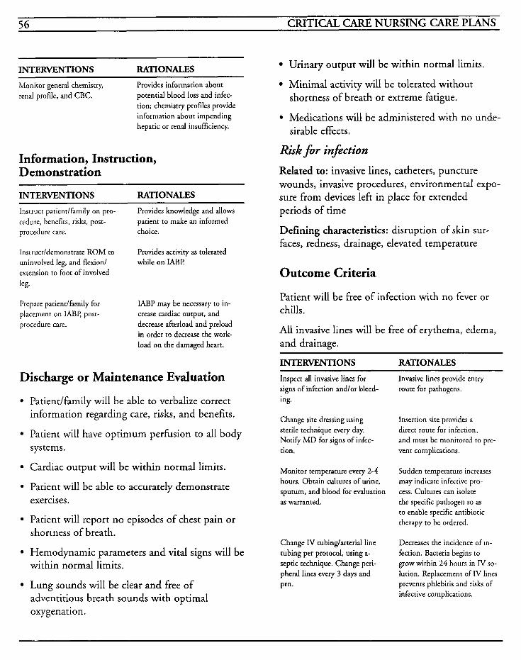
56 CRITICAL CARE NURSING CARE PLANS
INTERVENTIONS RATIONALES
Monitor generd chemistry, renal profile, and CBC.
Provides information about potential blood loss and infec- tion; chemistry profiles provide information about impending hepatic or renal insufficiency.
Information, Instruction, Demonstration
INTERVENTIONS RATIONALES
Instruct patiendfarnily on pro- cedure, benefits, risks, post- procedure care. choice.
Provides knowledge and allows patient to make an informed
Instructldemonstrate ROM to uninvolved leg, and flexionl extension to foot of involved leg.
Provides activity as tolerated while on IABP
Prepare patiendfamily for placement on IABP, post- procedure care.
IABP may be necessary to in- crease cardiac output, and decrease afterload and preload in order to decrease the work- load on the damaged heart.
Discharge or Maintenance Evaluation -
Patiendfamily will be able to verbalize correct information regarding care, risks, and benefits.
Patient will have optimum perhsion to all body systems.
Cardiac output will be within normal limits.
Patient will be able to accurately demonstrate exercises.
Patient will report no episodes of chest pain or shortness of breath.
Hemodynamic parameters and vital signs will be within normal limits.
Lung sounds will be clear and free of adventitious breath sounds with optimal oxygenation.
Urinary output will be within normal limits.
Minimal activity will be tolerated without shortness of breath or extreme fatigue. . Medications will be administered with no unde- sirable effects.
Risk for injktion
Related to: invasive lines, catheters, puncture wounds, invasive procedures, environmental expo- sure from devices left in place for extended periods of time
Defining characteristics: disruption of skin sur- faces, redness, drainage, elevated temperature
Outcome Criteria
Patient will be free of infection with no fever or chills.
All invasive lines will be free of erythema, edema, and drainage.
INTERVENTIONS RATIONALES Inspect all invasive lines for Invasive lines provide entry signs of infection andlor bleed- ing.
Change site dressing using sterile technique every day. Notify M D for signs of infec- tion.
Monitor temperature every 2-4 hours. Obtain cultures of urine, sputum, and blood for evaluation as warranted.
Change IV tubinglarterial line tubing per protocol, using a- septic technique. Change peri- pheral lines every 3 days and pm.
route for pathogens.
Insertion site provides a direct route for infection, and must be monitored to pre- vent complications.
Sudden temperature increases may indicate infective pro- cess. Cultures can isolate the specific pathogen so as to enable specific antibiotic therapy to be ordered.
Decreases the incidence of In- fection. Bacteria begins to
grow within 24 hours in IV so- lution. Replacement of IV lines prevents phlebiris and risks of infective complications.
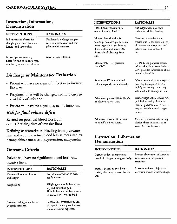
CARDIOVASCULAR SYSTEM 57
Instruction, Information, Demonstration
INTERVENTIONS RATIONALES
Inform patient of need for changing peripheral lines, so-
lutions, and care to sites.
Facilitates knowledge and pa- tient comprehension and com- pliance with treatment.
Instruct patient to notify nurse for pain to invasive sites, or other symptoms of infection.
May indicate infection.
Discharge or Maintenance Evaluation
Patient will have no signs of infection to invasive line sites.
Peripheral lines will be changed within 3 days to avoid risk of infection.
Patient will have no signs of systemic infection.
Riskfirflz4id volume &@it Related to: potential blood loss from oozing/draining sites of invasive lines
Defining characteristics: bleeding from puncture sites and wounds, actual blood loss as measured by hemoglobin/hematocrit, hypotension, tachycardia
Outcome Criteria
Patient will have no significant blood loss from invasive lines.
INTERVENTIONS RATIONALES
Measure all sources of intake and output. ate fluid status.
Provides information to evalu-
Weigh daily. Weight gain over 24 hours usu- ally indicates fluid gain. Fluid imbalance can be approxi- mated as 1 Ib = 500 cc fluid.
INTERVENTIONS RATIONALES Test all body fluids for pre- sence of occult blood.
Monitor insertion site for bleeding, hemorrhage, or hema- toma. Apply pressure dressing if warranted, and notify M D for sustained bleeding from site.
Monitor PT, PTT, platelets, and CBC.
Administer IV solutions and volume expanders as indicated.
Administer packed RBG, blood, or platelets as warranted.
Administer vitamin K or prota- mine sulfate if warranted.
Anticoagulation may place patient at risk for bleeding.
Bleeding tendencies are in- creased due to concomitent use of systemic anticoagulants and patient is at risk for bleed- ing.
PT, PTT, and platelets provide information about coagulation; CBC provides information about potential blood loss.
IV solutions and volume expan- ders may be required to treat rapidly decreasing circulating volume due to exsanguination.
Hemorrhagic volume losses may be life-threatening. Replace- ment of platelets may be neces- sary to provide normal coagu- lation.
May be required to return coag- ulation times to normal or re- verse effects of heparin.
Instruction, Information, Demonstration
INTERVENTIONS RATIONALES
Instruct patient to report any noted bleeding or oozing on body.
Prompt observation of complica- tions can result in prompt treatment.
Instruct patient to avoid any activity that may promote bleed- ing.
Prevents accidental injury and decreases chance of hemorrhage.
Monitor vital signs and hemo- dynamic pressures.
Tachycardia, hypotension, and changes in hernodynamics may indicate volume depletion.
58 CRITICAL CARE NURSING CARE PLANS
Discharge or Maintenance Evaluation
Patient will be able to verbalize signs/symptoms of bleeding to report.
Patient will be compliant in avoidance of safety concerns.
Patient will have stable hemodynamic status with no over hemorrhage from any site.
CARDIOVASCULAR SYSTEM 59
Pacemakers Artificial cardiac pacemakers are used to provide an electrical stimulus to depolarize the heart and cause a contraction to occur at a controlled rate. The function of the pacemaker, or pacer, is to maintain the heart rate when the patient's own intrinsic system is unable to do so. The stimulus is produced by a pulse generator and delivered via electrodes/leads that are implanted in the epicardium or endocardium. The electrodes may be unipolar or bipolar and the proximal end attaches to the pulse generator. In the unipolar electrode, one wire, positioned in the heart, senses and stimulates the electrical heart activity and is connected with the negative terminal on the pulse generator. The other electrode, or ground, is attached to the positive terminal on the pulse gen- erator. This type of lead usually requires a lower threshold of stimulation. The bipolar electrode has both the sensing and ground electrode in the catheter, and provides better contact with the heart muscle. In the event that one of the bipolar wires malfunctions, it can still be used as a unipo- lar lead. The pacemaker will produce a pacer spike on the EKG prior to the depolarized wave- form and this indicates pacemaker capture. Continuous observation for problems with the pacemaker should be performed to ensure that failure to pace, failure to capture, and failure to sense are treated promptly.
The pacemaker rate is set depending on the patient's requirements. The optimal setting is one in which the lowest rate that controls the particu- lar dysrhythmia and provides for adequate cardiac output. The stimulation threshold is the minimal amount of electrical energy required to stimulate the heart to produce a 1 : 1 capture, and is measured in milliamperes (mA). The sensitivity control reflects the size of the wave that is sensed
by the pacemaker and is measured in millivolts (mV), with the smaller number relating to the most s'ensitivity. Pacemakers are used for varying degrees of heart block, sick sinus syndrome, sinus node dysfunction, overriding of some cardiac dys- rhythniias, prophylactically during diagnostic testing, myocardial infarctions, congestive heart failure due to rhythm disturbances, after open heart surgery or in congenital anomalies of the heart.
Temporary pacers are used when the duration of need is short and permanent pacers are placed for life-long use. Temporary pacemakers can be placed via a transthoracic approach during open heart surgery, transvenous approach into the right atrium or right ventricle, or transcutaneously (exteraal pacer) with skin electrodes while await- ing placement of an internal pacemaker.
Placement of the temporary pacemaker can be performed at the bedside in cases of emergency, but use of fluoroscopy is recommended when fea- sible to ensure proper placement. External pacemaker electrodes can either be placed on the chest, or one to the anterior and one posterior to the chest.
Synchronous pacing, known as demand pacing, is commonly used because the pacer is able to sense the patient's heart impulse. If the patient's rate falls below the rate set on the pacer, the pacer is able to sense this and send an impulse to the desired chamber of the heart and cause the rate to remain at the preset level. Dual chamber synchro- nous pacing, or AV sequential, is the closest to normal physiologic function and facilitates the atrial kick.
Asynchronous, or fEed-rate, pacing provides impulses to the atrium, ventricle, or both regard- less of the patient's intrinsic rate. This should be used solely for those occasions when no electrical activity is present to avoid potential lethal compet- itive dysrhythmias.
60 CRITICAL CARE NURSING CARE PLANS
Pacemakers are classified by a 5-letter code devel- oped by the Inter-Society Commission for Heart Disease in which letters are used to denote the chamber paced, the chamber sensed, response to sensing, programmable functions and antitachy- dysrhythmia functions.
Several complications may occur as a result of pacemakers-pneumothorax, hemothorax, myocardial perforation, hematoma, bleeding, dys- rhythmias, pulmonary embolism, electrical microshock, cardiac tamponade, coronary artery laceration, failure to pace, failure to sense, and failure to capture.
MEDICAL CARE Chest x-ray: used to evaluate placement of lead wires
Electrocardiography: used to monitor for heart rhythm problems, dysrhythmias, and for function/malfunction of pacemakers
Surgery: for placement of permanent pacemakers
NURSING CARE PLANS Alteration in tissue perfision: curdiopulmona y, cerebral
Related to: cardiac dysrhythmias, heart blocks, tachydysrhythmias, decreased blood pressure, decreased cardiac output
Defining characteristics: decreased blood pressure, decreased heart rate, decreased cardiac output, changes in level of consciousness, mental changes, cold clammy skin, cardiopulmonary arrest
Outcome Criteria
Patient will be free of dysrhythmias with adequate cardiac output to perfuse all body organs.
INTERVENTIONS RATIONALE Monitor EKG for changes in rhythm, rate, and presence of dysrhythmias. Treat as india- ted.
Observation for pacemaker mal- function promotes prompt treat- ment. Pacer electrodes may ir- ritate ventricle and promote ventricular ectopy.
Keep monitor alarms on at all times, with rate limits set 2-5 beats above and below set rate.
Obtain and observe rhythm strip every 4 hours and prn. Notify MD for abnormalities.
Monitor vital signs every 15 minutes until stable, then every 2 hours.
Monitor for signs of failure to capture and correct problem.
Monitor for signs of failure to sense patient’s own rhythm and correct problem.
Monitor for signs of failure to pace and correct problem.
Ensure that all electrical equipment is grounded. Avoid touching equipment and patient at same time. Patients should not use radios, shavers, etc.
Provides for immediate detec- tion of pacemaker failure or malfunction.
Identifies proper functioning of pacemaker, with appropriate capture and sensing.
Assures adequate perfusion and cardiac output.
Potential causes are low volt- age, battery failure, faulty connections, catheter or wire fracture, improper placement of catheter, or fibrosis at tip of catheter.
Potential causes are lead dis- lodgment, battery failure, low sensitivity, catheter wire frac- ture, or improper placement of catheter.
Potential causes are battery failure, lead dislodgment, dis- connection, or catheter lead fracture.
Prevents potential for micro- shock and accidental electrocu- tion. Electric current seeks the path of least resistance, and the potential for stray cur- rent to travel through the elec- trode into the patient’s heart may precipitate ventricular fib- rillation.
~
CARDIOVASCULAR SYSTEM 61
INTERVENTIONS RATIONALES INTERTENTIONS RATIONALES
Place a dry rubber glove over exposed terminals or leads. Wear rubber gloves when hand- ling the electrodes, terminals, etc.
Provides insulation to prevent stray current contact. Static electricity may pass from person to person through the leads.
Protect patient from microwave ovens, radar, diathermy, elec- trocautery, TENS units, etc.
Environmental electromagnetic interference may impair demand pacemaker function by disrupting the electrical stimulus.
If the patient experiences cardiopulmonary arrest, the pacemaker should be turned off and disconnected from the pa- tient for ventricular fibrilla- tion. After defibrillation, the pacemaker should be recon- nected, turned on, and output should be raised to 20 mA, rate above GO.
Disconnection prior to D C coun- tershock prevents pacer damage and potential of diversion of electrical current.
Pacemaker batteries should not be changed while the pacer is in use. In cases of hardship, batteries should be changed as quickly as possible, wearing rubber gloves, and using utmost caution to avoid touching the battery terminals.
Patients may be totally depen- dent on the pacemaker for their rhythm and cardiac output and loss of time incurred to change the battery may result in life- threatening consequences.
May indicate lead has dislodged and migrated to chest wall or diaphragm after perforation of heart.
Monitor for musde witching or hiccoughs.
Information, Instruction, Demonstration
Monitor for sudden complaints of chest pain, and auscultate for pericardial friction rub or muffled heart tones. Observe for JVD and pulsus paradoxus.
May indicate perforation of the pericardial sac, and impending cardiac tamponade.
~~~
INTERVENTIONS RATIONALES
Instruct on need for pacemaker, procedures involved, expected outcomes, etc.
Provides knowledge, decrezses fear and anxiery, and provides baseline for further instruc- tion.
Monitor for dizziness, syncope, weakness, pronounced fatigue, edema, chest pain, palpitations, pulsations in neck veins, or dyspnea.
During ventricular pacing, AV synchrony may cease and cause a sudden decrease in cardiac output. May indicate “pace- maker syndrome” or failure of the pacer to function which results in decreased perfusion.
Instruct in checking pulse rate every day for 1 month, then every week, and to notify M D if rate varies more than 5 beats/ minute.
Provides patient with some control over situation. As- sists in promoting a sense of security. Allows for prompt recognition of devi- ations from preset rate and potential pacemaker failure.
Prevents accidental disconnec- tion and dislodgment of lead wires.
Limit movement of the extremi- ty involved near insertion site. Instruct on activity limita-
tions: avoid excessive bend- ing, stretching, lifting more than 5 pounds, strenuous acti- vities, or contact sports.
Full range of motion can be recovered in approximately 2 months after fibrosis stabilizes the pacemaker lead. Excessive activity may cause lead dislodgment.
Inflation of pulmonary artery catheter balloon for capillary wedge pressures may dislodge pacer lead wires and cause pacemaker malfunction.
If pacemaker is used concurrent- ly with pulmonary artery catheter obtain wedge pressure only as M D orders.
Instruct to avoid shoulder- strap purses, suspenders, or firing rifle resting over gen- erator site.
May promote irritation over implanted generator site.
Monitor patient for low blood sugar levels, use of glucocor- ticoids or sympathomimetics, mineralocorticoids, or anesthe- tics.
May impair the pacemaker stim- ulation thresholds.
Provides information about the patient, his condition, and pacemaker should he be incapa- citated and cannot speak for himself.
Instruct to wear a medic-alert bracelet with information about the type of pacemaker and rate.
62 CRITICAL CARE NURSING CARE PLANS
INTERVENTIONS RATIONALES Outcome Criteria Instruct to notify MD if radi- ation therapy is needed and to wear a lead shield.
Therapy can cause failure of the silicone chip in the pacer with repeated radiation.
Patient will have healed wound sites without signs/symptoms of infection.
Instruct to avoid electromag- netic fields, magnetic resonance imaging, radio transmitters, arc welding equipment, large running motors, or large ungrounded power tools. If patient notices dizziness or palpitations, he should try to move away from the area, and if symptoms per- sist, to seek medical attention. Late model microwave ovens are no longer thought to be a threat due to tighter seals preventing leakage of energy.
May affect the function of the pacemaker and alter the pro- grammed settings. Sometimes these magnetic fields will affect the pacemaker function only if direct contact is made and once distance is placed between the patient and the equipment, normal function of the pacemaker resumes. If programmed settings are altered the pacer will require repro- gramming. Hyperbaric oxygen chambers may also affect pacer function.
Discharge or Maintenance Evaluation
Patient will be free of dysrhythmias and able to maintain cardiac output within normal limits.
Patient will be able to recall accurately all instructions given.
Patient will be able to recall and adhere to all activity restrictions.
Permanent pacemaker function will be without complication, with no lead dislodgment or com- petitive rhythms noted.
Alteration in skin interity
Related to: insertion of temporary or permanent pacemaker, alteration in activity
Defining characteristics: disruption of skin tissue, insertion sites
INTERVENTIONS RATIONALES
Inspect pacemaker insertion site for erythema, edema, warmth, drainage, or tenderness.
Prompt detection of problems promotes prompt treatment.
Change dressing daily, or per Allows for observation of site hospital protocol, using sterile and detection of inflammation technique. or infection. Sterile technique
is recommended due to the close proximity of the portal to the heart increasing the potential for systemic infection.
Pacemaker lead wires should be coiled and taped securely to patient; pulse generator should be secured to avoid pulling.
Avoids potential for accidental- ly disconnecting pacemaker from generator, or dislodging leads from heart.
Information, Instruction, Demonstration
INTERVENTIONS RATIONALES Instruct on wound care to pacer site; to avoid taking showers for 2 weeks after pacer inser- tion.
lnstruct to observe for and re- port to MD the following symp- toms: redness, drainage, remper- ature greater than 100 degrees, pain or tenderness to site, or swelling at site.
Instruct to avoid constrictive dothing until site has healed.
Instruct on need for pacemaker removal/replacement.
Promotes compliance with care to decrease potential for infec- tion. Moisture can promote bac- terial growth.
Provides for prompt recognition of complications and facilitates prompt treatment.
May cause discomfort at incision site from pressure and rubbing against skin.
Pulse generators may require re- moval for battery replacement, fracture of lead wires, pace- maker failure, etc.
CARDIOVASCULAR SYSTEM 63
Discharge or Maintenance Evaluation
Patient will have well-healed incision with no signs/symptoms of infection.
Patient will be able to recall accurately all instructions given.
Patient will be able to demonstrate appropriate
Potential for injury
Related to: pacemaker failure, hemothorax or pneumothorax after insertion, bleeding, lead migration, heart perforation
Defining characteristics: decreased cardiac output, hemorrhage, diaphoresis, hypotension, restlessness, dyspnea, cyanosis, chest pain, muscle twitching, hiccoughs, muffled heart sounds, jugular vein dis- tention, pulsus paradoxus
wound care prior to discharge.
Outcome Criteria
Patient will be free of any complications that may be associated with pacemaker insertion.
INTERVENTIONS RATIONALES ~
Monitor for bleeding at pacer site. Apply pressure dressings as warranted.
Monitor for pulse presence at site distal to pacer insertion.
Monitor for hypotension, dia- phoresis, dyspnea, and restless- ness.
Bleeding at incisional site may occur based on the patient’s coagulation status. Pressure dressings or manual pressure may be required to control bleeding.
Hemorrhage may promote tissue edema and compression to ar- terial blood flow resulting in diminished or absent pulses.
May indicate puncture of the subclavian vasculature and po- tential hemothorax.
INTERVENTIONS RATIONALES Monitor for dyspnea, chest pain, pallor, cyanosis, absent or dim- inished breath sounds, tracheal deviation, and feeling of impen- ding doom.
May indicate puncture of the lung and pneumothorax.
Monitor for muscle twitching and hiccoughs. Notify MD.
May indicate perforation of the heart with pacing to the chest wall or diaphragm.
Observe for signslsymptoms of cardiac tamponade-pericardial friction rub, pulsus paradoxus, muffled heart tones, JVD.
May indicate perforation of the pericardial sac and impending cardiac tamponade.
Discharge or Maintenance Evaluation
Patient will have no complications associated with pacemaker insertion.
Patient will have clear breath sounds, with no inadequacy of oxygenation.
Patient will be free of infection or hemorrhage.
Alteration in comfort [See MI] Related to: pacemaker insertion or transcutaneous pacing
Defining characteristics: communication of pain, facial grimacing, restlessness, changes in pulse and blood pressure
Anxiety [See MI] Related to: change in health status, fear of death, threat to body image, threat to role functioning, pain
Defining characteristics: restlessness, insomnia, anorexia, increased respirations, increased heart rate, increased blood pressure, difficulty concen- trating, dry mouth, poor eye contact, decreased energy, irritability, crying, feelings of helplessness
64 CRITICAL CARE NURSING CARE PUNS
Impaired physical mobility
Related to: pain, limb immobilization
Defining characteristics: inability to move as desired, imposed restrictions on activity, decreased muscle strength and coordination, limited range of motion
Outcome Criteria
Patient will regain optimal mobility within limita- tions of disease process, and will have increased strength and function of limbs.
INTERVENTIONS RATIONALES Evaluate patient$ perception of degree of immobility.
Psychological and physical im- mobility are interrelated. Psy- chological immobility is used as a defense mechanism when they have no control over their body, and this can lead to dispropor- tionate fear and concern. Changes in body image promote psychological immobility and may result in emotional handi- caps.
Maintain bedrest for 24-48 hours after permanent pacer inserted.
Immobilize extremity proximal to pacer insertion site with arm board, sling, etc.
Resume range of motion exercises 5 days after permanent pacer in- sertion to affected extremity. Provide ROM to unaffected extre- mity as warranted.
Encourage extension/dorsiflexion exercises to feet every 1-2 hours.
Provides time for stabilization of leads and decreases potential for dislodgment.
Prevents potential for dislodg- ment of lead due to movement.
Promotes gradual increase of activity. Stretching should be avoided until lead wire has been secured in heart by fibrotic changes. ROM prevents stiffness of should- ers and joint immobility.
Promotes venous return, pre- vents venous stasis, and de- creases potential for thrombo- phlebitis.
INTERVENTIONS RATIONALES Monitor for progression and im- provement in stiffnedpain.
Physical therapy may be re- quired if immobility results are severe.
Apply trapeze bar to bed. Allows for easier movement by allowing patient to assist with movement in bed.
Reposition every 2 hours and prn.
Prevents potential for immobili- ty hazards such as pressure areas and atelectasis.
Information, Instruction, Demonstration
INTERVENTIONS RATIONALES Encourage deep breathing exer- cises every 1-2 hours; avoid forceful coughing.
Facilitates lung expansion and decreases potential for atelec- tasis. Coughing may dislodge pacemaker lead.
Discharge or Maintenance Evaluation
Patient will regain optimal mobility of all joints with no signs or symptoms of complications.
Patient will be able to demonstrate and recall instructions regarding deep breathing and range of motion exercises.
Disturbance of body image
Related to: presence of pulse generator, loss of control of heart function, disease process
Defining characteristics: fear of rejection, fear of reaction from others, negative feelings about body, refusal to participate in care, refusal to look at wound
CARDIOVASCULAR SYSTEM 65
Outcome Criteria
Patient will accept change in body image and deal constructively with situation.
INTERVENTIONS RATIONALES Evaluate level of patient’s knowledge about disease process, treatment, and anxiety. required.
May identify extent of problem and interventions that will be
Evaluate the extent of loss to the patiendfamily, and what i t
means to them.
Depending on the time frame for patient teaching prior to the insertion of the pacemaker, the patient may not have received adequate information, and may have difficulty dealing with changes in his body appearance as well as generalized health condition and loss of control.
Evaluate stage of grieving. Provides recognition of appro- priate versus inappropriate behavior. Prolonged grief may require further care.
Observe for withdrawal, manipu- lation, noninvolvement with care, or increased dependency. Set limits on dysfunctional behav- ior and help patient to seek positive behaviors that will assist with recovery.
May suggest problems with ad- justment to health condition, grief response to the loss of function, or worry about others accepting patient’s new body status. Patients may deal with crises in the same manner as previously dealt and may need redirection in behaviors to fa- cilitate recovery and accep- tance.
Provide positive reinforcement during care and with instruc- tion and setting goals. Do not give false reassurance.
Promotes trust and establishes rapport with patient as well as provides an opportunity to plan for rhe future based on reality of situation.
Provide opportunity for patient to take active role in wound care. body and health.
Provide reassurance that pace- maker will not alter sexual ac- tivity.
Promotes self-esteem and facili- tates feelings of control of
Promotes knowledge and decreases fear.
Information, Instruction, Demonstration
INTERVENTIONS RATIONALES Discuss potential for mood changes, anger, grief, etc. af- ter discharge, and to seek help if persisting for lengthy time.
Facilitates identification that feelings are not unusual and must be recognized in order t o
effectively deal with them.
Identify support groups for pa- tient/family to contact.
Provides ongoing support for patient and family and allows for ventilation of feelings.
Consult counselor/therapist as warranted.
May require further interven- tions to resolve emotional or psychological problems.
Discharge or Maintenance Evaluation
Patient will be able to effectively deal with body image disturbances in present situation.
Patient will be able to talk with family, therapist, or others about emotional or psycho- logical problems.
Patient will be able to problem-solve and iden- tify short- and long-term goals within reasonable expectations of clinical situation.
Knowledge deficit [See MI] Related to: lack of understanding, lack of under- standing of medical condition, lack of recall, new health crisis
Defining characteristics: questions regarding problems, inadequate follow-up on instructions given, misconceptions, lack of improvement of previ0u.s regimen, development of preventable complications
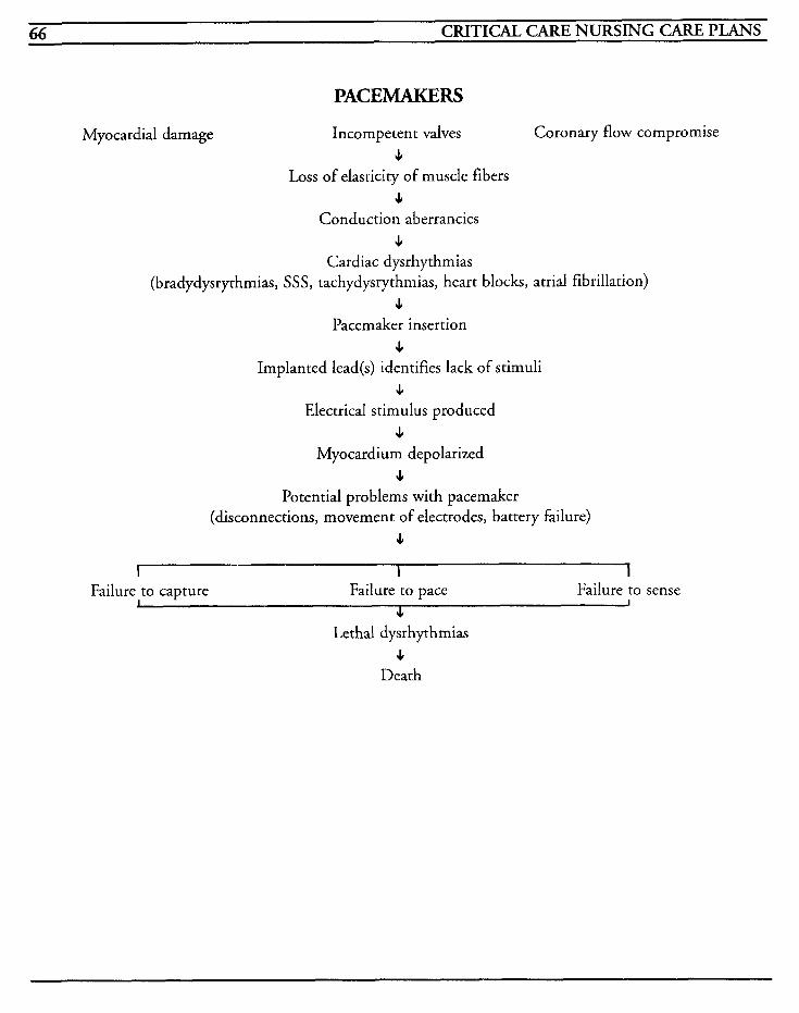
66 CRITICAL CARE NURSING CARE PLANS
PACEMAKERS
Myocardial damage Incompetent valves Coronary flow compromise s
Loss of elasticity of muscle fibers s
Conduction aberrancies I
Cardiac dysrhythmias (bradydysrythmias, SSS, tachydysrythmias, heart blocks, atrial fibrillation)
I Pacemaker insertion
s Implanted lead(s) identifies lack of stimuli
4 Electrical stimulus produced
I Myocardium depolarized
4 Potential problems with pacemaker
(disconnections, movement of electrodes, battery failure) s
I I I
J J Failure to capture Failure to pace Failure to sense
s Lethal dysrhythmias
s Death
CARDIOVASCULAR SYSTEM 67
Coronary artery disease treatment requires the maximization of cardiac output and this can be accomplished by improvement in heart muscle function and increase of blood flow through coro- nary artery bypass grafting and/or valvular replacements. Open heart surgery is commonly performed for three-vessel disease, valve dysfunc- tion and congenital heart defects and requires blood to be diverted from the heart and lungs to facilitate a bloodless operative field.
In coronary artery bypass graft (CABG) surgery, a graft from the arms or legs is anastomosed to the aorta with the distal portion to the involved coro- nary artery to bypass the diseased obstruction and supply adequate blood flow to the heart. The internal mammary artery is also being utilized for CABG surgery because the patency rate is 90-95% over a 5-10 year time period, and there are less problems with differences in lumen size since an artery is then anastomosed to an artery without the need for routing from the aorta. In valvular surgery, incompetent or leaking valves are replaced with prosthetic ones.
Not all patients with coronary artery disease are candidates for CABG surgery. It is usually recom- mended for those patients with intractable angina, signs of ischemia, or an increased risk of coronary ischemia/infarction as a result of angiographical studies. Complications may occur in almost every body system and may be a result of the disease process or defect, the surgery, or the use of cardiopulmonary bypass, and so the decision for surgery is a multi-faceted one.
One of the most important factors in the decision of candidacy for CABG surgery is the ejection
fraction. This is the ratio of stroke volume com- pared to the end-diastolic volume and an ejection fraction greater than 55 reflects a good operative risk. Ejection fraction less than 25% is usually considered inoperable because of the high mortal- ity associated with it.
The surgery is performed via a median sternotomy incision which provides exposure of the heart and avoids the pleural spaces. A cannula is placed in a vein and an artery and then attached to the cardiopulmonary bypass machine whereby the diverted blood is mechanically oxygenated and cir- culated to the other parts of the body. The machine, which is operated by a trained perfusion- ist, substitutes for left ventricular pumping and creates a blood-gas exchange. After the patient’s body temperature has been cooled to around 86 degrees, the aorta is cross-clamped and a cold car- dioplegic solution, usually containing dextrose, potassium, magnesium and inderal, is placed around the heart and injected into the coronary arteries. This causes an electromechanical arrest and provides an inert operative site. Cross-clamp durations longer than 3 hours usually result in severe complications for the patient. After the grafts have been completed or valves replaced, per- fusion is slowly discontinued and cannulas are removed when arterial blood pressure and cardiac functioning are adequate. Two atrial and ventricu- lar pacing wires are placed, as well as arterial lines, pulmonary artery catheter, left atrial line, and mediastinal or pleural chest tubes.
Common complications associated with CABG surgery include perioperative MI, vein graft clo- sure, hemorrhage, blood trauma, complement activation, coagulation abnormalities, fluid shifts, increased catecholamine levels, fat emboli, microemboli, dysrhythmias, pericarditis, postperi-
68 CRITICAL CARE NURSING CARE PLANS
cardiotomy syndrome, embolism, pneumonia, atelectasis, hemothorax, pneumothorax, and post- cardiotomy delirium. Other complications that are seen less often include stress ulcer, renal failure, respiratory failure, cardiac tamponade, cardiogenic shock, endocarditis, gastrointestinal bleeding, mediastinitis, and paralytic ileus.
MEDICAL CARE Pulmonary function studies: used to ascertain baseline pulmonary function
Laboratory: hemoglobin/hematocrit used to mon- itor oxygen-carrying capability, need for blood replacement, and to monitor for dehydration status; electrolytes used to monitor for imbalances which can affect cardiac function; BUN and crea- tinine used to monitor renal function; liver profile used to monitor liver function and perfusion; glu- cose used to monitor for presence of diabetes, nutritional alterations, or organ dysfunction; car- diac enzymes and isoenzymes used to monitor for presence of acute or perioperative myocardial infarction; coagulation profiles used to determine baseline and monitor for coagulation problems; antibody or complement levels used to monitor for postpericardiotomy syndrome or Dressler’s syndrome; type and crossmatch for blood to have available blood products on hand in case of hem- orrhage; ACT used to monitor heparinization
Arterial blood gases: used to monitor oxygenation and assess acid-base balance and ability to wean off mechanical ventilation
Electrocardiography: used to observe for changes in cardiac function, presence of conduction prob- lems, dysrhythmias, or ischemic changes
Echocardiography: used to evaluate wall motion of the heart
Chest x-ray: used to identify heart size and posi- tion, pulmonary vasculature, pulmonary changes, verifies position of endotracheal tube, pacing wires, and hemodynamic catheters; monitors for bar0 trauma
Cardiac catheterization: used to evaluate abnor- mal pressures preop, to assess for pressure gradients across the valves, and to locate and mea- sure coronary lesions
NURSING CARE PLANS Risk for decreased cardiac output
Related to: myocardial depression, dysrhythmias, electrolyte imbalances, hypovolemia, hypervolemia, myocardial infarction, coronary artery spasm, vasoconstriction, impaired contrac- tility, alteration in preload, alteration in afterload, hypo perfusion, microemboli, hypoxia, damaged myocardium, use of PEEP while on ventilatory support
Defining characteristics: elevated blood pressure, elevated mean arterial pressure greater than 120 mmHg, elevated systemic vascular resistance greater than 1400 dyne-seconds/cm5, cardiac output less than 5 L/min or cardiac index less than 2.7 L/min/m2, tachycardia greater than 110, cold, pale extremities, absent or decreased periph- eral pulses, EKG changes, hypotension, S3 or S4 gallops, decreased urinary output, diaphoresis, orthopnea, dyspnea, crackles (rales), jugular vein distention, edema, chest pain
Outcome Criteria
Vital signs and hernodynamic parameters will be within normal limits for patient, with no dysrhythmias noted.
CARDIOVASCULAR SYSTEM 69
INTERVENTIONS RATIONALES INTERVENTIONS RATIONALES Measure left atrial pressure Determines the left ventricular Tachycardia may occur as a res- Monitor vital signs, especially
heart rate and blood pressure. Notify MD of abnormalities. Blood pressure should be taken/ monitored every 15 minutes until stable, or every 5 minutes dur- ing active titration.
Evaluate hypotension that is not responsive to fluid bolus, tachycardia, and distant heart sounds.
Monitor hemodynamic pressures every 1 hour and prn. Maintain pressures with titration of va- soactive drugs per M D ordered parameters.
Measure cardiac outpudcardiac index every 1-2 hours immedi- ately post-op.
ponse to pain, anxiety, blood and fluid deficit, and stress, but rates over 130 increases myocardial oxygen consumption and workload on the heart, de- creasing cardiac output. In- creased blood pressure may pro- mote alterations in heart pres- sures and increase the risk of complications, as well as pla- cing pressure on suture lines of new grafts. Hypotension may result from fluid deficit, dys- rhythmias, and cardiac failure, as well as predispose peripheral vein grafts to close.
May indicate cardiac tamponade in a heart that is unable to fill adequately to maintain cardiac output. Tamponade usu- ally occurs immediately post-op but may occur later during re- covery period.
Assists with recognition of complications and allows for manipulation of cardiac pres- sures by use of fluids and medications. Vasoconstriction is the cause of elevated SVR, and with increases in SVR, may indicate left ventricular dys- function. Cardiac output then becomes dependent on outflow resistance.
Cardiac output is a measurement that is equal to the product of the stroke volume and the heart rate. Cardiac indexes above 3.0 Ymin/m2 are usually adequate except in cases of septic shock. Adequate cardiac output relates to the adequacy of function of other body organs. After CABG surgery, most patients require an increase in CO to meet the stress imposed by the operation and the accompanying increase in oxygen consumption.
and pulmonary artery wedge pres- sures.
Monitor urine output hourly and notify M D if less than 30 cclhr.
Observe for decreased peripheral pulses, cool or cold moist skin, or cyanosis.
Monitor for changes in level of consciousness, mental status changes, restlessness, or con- fusion.
Monitor for JVD, peripheral ede- ma, and pulmonary congestion. Auscultate for crackles (rales).
Observe for shortness of breath, decreases in oximetry, or dyspnea.
Monitor EKG for cardiac conduc- tion disturbances, dysrhythmias, or changes in ratelrhythm.
Monitor for complaints of se- vere chest pain.
Provide for uninterrupted rest periods and assist with care as needed.
end-diastolic volume; increases in pressure may indicate conges- tive heart failure or pulmonary edema, and decreases may indi- cate low blood volume. Trends and changes in values are of more importance than single readings. Left ventricular dys- function can elevate left heart filling pressures without a rise in right heart pressures.
Urine output is an indication of adequate cardiac output and renal perfusion.
May indicate low cardiac output.
Cerebral perfusion is dependent on adequate cardiac output. Hy- poperfusion or microemboli may result in CNS deficits.
May indicate present or impend- ing congestive heart failure.
May indicate hypoxia and de- creased cardiac output.
Lethal dysrhythmias may occur as a result of electrolyte im- balances, myocardial ischemia or infarction, or problems with electrical conduction, with an associated drop in cardiac out- put.
May indicate a perioperative or postoperative myocardial infarc- tion.
Prevcnts fatigue and increased workload on the heart leading to decrease in cardiac output and perfusion.
70 CRITICAL CARE NURSING CARE PLANS
INTERVENTIONS RATIONALES Outcome Criteria Administer IV fluids as ordered. Maintains fluid status and hy-
dration, as well as provides ac- cess for emergency medications.
Administer blood products as or- dered. required to maintain adequate
Blood or packed red cells may be
oxygen-carrying capability, and adequate circulating volume for cellular activity. Platelet func- tion and count is decreased with use of cardiopulmonary by- pass and proportional to the duration of bypass and depth of hypothermia during surgery.
Information, Instruction, Demonstration
INTERVENTIONS RATIONALES Attempt to reverse any contrib- uting factor such as untreated DKA or endocrine dysfunction.
These may precipitate a low CO state.
Prepare patient for placement on IABP. decreases fear.
Promotes knowledge and
Discharge or Maintenance Evaluation
Patient will have maximal cardiac output and stable hemodynamic pressures.
Patient will have adequate perfusion of all body sys tems.
Patient will be able to recall instructions correctly.
Alteration in comfort
Related to: mediastinal, leg, or arm incisions, myocardial infarction, angina, inflammation, tissue damage
Defining characteristics: communication of dis- comfort or pain, restlessness, irritability, increased heart rate, increased blood pressure
Patient will be free of pain or pain will be controlled to patient’s satisfaction.
INTERVENTIONS RATIONALES
Evaluate complaints of pain- type, location, intensity based on 0- 10 scale. Compare preoper- ative pain perceptions with post- operative pain.
Monitor vital signs.
Evaluate complaints of pain in legs or abdomen, or vague non- specific complaints, especially if associated with changes in mental status or vital signs.
Monitor for complaints of pain and/or paresthesia to ulnar area of the hand, and possibly pain to shoulders and arms.
Observe for anxiety, irritabili- ty, crying, restlessness, or in- somnia.
Administer analgesics as soon as discomfort is noticed, or pro- phylactically prior to painful procedures.
Pain may be perceived in differ- ent ways by each individual and is important to differentiate incisional pain from other types of chest pain. CABG patients usually do not have severe dis- comfort to the chest incision but may have increased discom- fort with donor sire pain. Severe pain should be investiga- ted for possibility of compli- cations.
Heart rates usually increase with pain but bradycardia may occur especially in severely damaged myocardium. Blood pres- sure may be increased with in- cisional pain, but can also be labile or decreased when chest pain is severe or if myocardial ischemidnecrosis occurs.
May be indicative of develop- ment of thrombophlebitis, infec- tion or GI dysfunction.
May result from stretching of the brachial plexus during posi- tioning of the arms during sur- gery and generally resolves over time without specific treatment.
Nonverbal cues may indicate the presence of pain.
Pain results in muscle tension, which can decrease circulation and intensify pain perception. Medication given prior to pro- cedures known to cause pain may facilitate cooperation with pro-
CARDIOVASCULAR SYSTEM 71
INTERVENTIONS RATIONALES
cedures and allow for easier chest movement with respiratory therapy.
Promotes relaxation and helps to redirect attention away from discomfort, thereby reducing the amount of analgesic required.
Provide back rubs, position chan- ges, and diversionary activities.
Encourage deep breathing, visu- alization, or guided imagery.
Promotes decrease in stress and may reduce analgesic need.
Information, Instruction, Demonstration
INTERVENTIONS RATIONALES
Instruct on methods to reduce strain on muscles when position- ing.
Supporting extremities and the maintenance of good body align- ment reduce muscle tension and provide comfort.
Discharge or Maintenance Evaluation
Patient will be comfortable, pain-free, and be able to recall methods for stress reduction and pain coiitrol accurately.
Patient will be able to identify differences between postoperative and preoperative chest pain.
Patient will be able to maintain optimal body alignment and minimize muscle tension. 1
Risk for inefective breathing patternhmpaired gas exchange Related to: inadequate ventilation, ventilatiodperfusion mismatching, abnormal ABGs, pain, blood loss, atelectasis, pneumothorax, hemothorax, increased pulmonary vascular resis- tance, increased capillary permeability, chemical mediators, decrease in surfactant
Defining characteristics: dyspnea, tachypnea, apnea, ventilation/perfusion mismatching, abnor- mal ABGs, pain, increased hemodynamic pressures, oxygen saturation less than 90%, adven- titious breath sounds, hypoxia, hypoxemia
Outcome Criteria
Patient will be eupneic with clear breath sounds, and have no evidence of hypoxia/hypoxemia.
INTERVENTIONS RATIONALES
Monitor respiratory rate and depth, presence of dyspnea, use of accessory muscles, nasal flar- ing, and increasing respiratory work effort.
Respiratory rates may be in- creased by pain, fever, blood loss, fluid loss, anxiety, hy- poxia, or gastric distention. Decreases in rate may occur with use of narcotic analgesics. Prompt recognition of potential complications can promote prompt treatment.
Auscultate lung fields for di- minished or absent breath sounds or for adventitious sounds.
Breath sounds are frequently di- minished immediately post-op as a result of atelectasis. Loss of breath sounds in a previously ventilated lung may indicate a partid or total lung collapse, especially when chest tubes have recently been discontinued. Ad- ventitious breath sounds may in- dicate fluid or secretions have accumulated in the interstitial spaces or airways resulting in a partial occlusion of the air- way.
Evaluate chest expansion for symmetry.
Unilateral incomplete chest ex- pansion may indicate that air or fluid is preventing complete expansion of the pleural space, possibly a pneumothorax.
Administer oxygen by cannula or mask as warranted.
Provides supplementd oxygen to decrease the workload on the heart and to maximize oxygen de- livery to under-perfused tis- sues.
72 CRITICAL CARE NURSING CARE PLANS
INTERVENTIONS RATIONALES INTERVENTIONS RATIONALES Observe for pallor or cya- nosis, especially to mucous membranes.
Observe for presence of cough and sputum character.
Encourage deep breathing ex- ercises, inspiratory spirometer, or coughing exercises.
Observe for signs of respiratory distress, tachycardia, extreme restlessness and feeling of im- pending doom.
Monitor respiratory status and ventilatory settings every 1-2 hours while on ventilator.
Assist with weaning from ven- tilatory support. Monitor for hemodynamic instability and de- creasing oxygen saturation. Monitor ABGs as ordered.
Cyanosis of lips, nailbeds, or earlobes, or generalized duski- ness may indicate hypoxia as a result of heart failure or pul- monary dysfunction. Pallor is frequently noted immediately postoperatively due to blood loss or insufficient blood re- placement.
Endotracheal tube intubation may promote throat irritation which can result in coughing, but cough may also indicate impending pulmonary congestion or infection. Purulent sputum may reflect pneumonia.
Promotes expansionlre-expansion of airways. Adventitious breath sounds may indicate presence of secretions or fluid in lungs.
May indicate impending pneumo- thorax or hemothorax, expecially after chest tube removal. May require reinsertion of chest tubes.
CABG patients are placed on me- chanical ventilation support until awake from anesthesia. F I 0 2 is initially 100% and then gradually decreased, while main- raining an adequate PaO2 above 90. FIO2 should be decreased to .50 as rapidly as possible to prevent actual pulmonary changes that occur with high levels of oxygen. Tidal volumes are usu- ally maintained between 10-15 cclKg of ideal body weight to allow for less interference with venous return.
Weaning is usually performed by reducing the rate and then a trial on a T-bar or CPAP mode. Patients who have a history of smoking or C O P D often have prolonged need for ventilatory sup-
port post-CABG due CO their me- chanism of breathing. Ventila- tors provide controlled amounts of oxygen and tidal volumes, and C O P D patients have their inert drive to breathe removed by the use of ventilation. Occasion- ally with use of the cold car- dioplegic solution, the phrenic nerve is injured resulting in a loss of function of the dia- phragm which is necessary for 60% of the spontaneous tidal volume for the patient.
Suction patient every 2-4 hours and prn. Use pulmonary toilette and hyperoxygenate prior to and after suaioning.
Removes mucous thar may occlude airways. Saline instillation helps to liqueg secretions to facilitate easier removal. Oxy- gen concentration drops drastic- ally with suaioning procedures and leaves the patient compro- mised with an increased oxygen consumption.
Auscultate breath sounds pre- and post-suctioning.
Provides for comparison of breath sounds to evaluate for improvement. Occasionally, suctioning will move secretions up the bronchial tree and may cause a partial or total occlu- sion of an airway. Decreases in previously venrilated lung fields may indicate this pheno- menon has occurred.
Monitor use of amiodarone and protamine sulfate. Observe for respiratory impingement. of action.
Some drugs can exacerbate pul- monary problems by their method
CARDIOVASCULAR SYSTEM 73
Information, Instruction, Demonstration
INTERVENTIONS RATIONALES Prepare patient for placement on mechanical ventilation if warranted.
Prepare patient for insertion of tracheostomy after 10 days of ETT intubation/ventilation.
Instruct patient on need for ambulation, movement, change in position.
Instruct on need for respira- tory treatments, coughing, deep breathing.
Prepare patient for reinsertion of chest tubes as warranted.
Lengthy instruction may not be prudent or possible depending on the severity of the situa- tion. If oxygenation cannot be maintained with the use of sup- plemental oxygen, the only al- ternative is intubationlventi- lation.
Prolonged endotracheal intuba- tion may result in tracheal or nasal necrosis or rupture of cuff. Tracheostomy is con- sidered to prevent ulceration into arteries or other vital tissues, but may need to be avoided due to potential for contamination of sternotomy wound from secretions.
Promotes lung expansion and prevents pulmonary congestion.
Reassures patient that com- plying with aggressive pul- monary regimen will not Cause injury to surgical sites.
Promotes re-expansion of lung by removing accumulated fluid, blood, or air, and restores normal negative pressure in the pleural cavity.
Discharge or Maintenance Evaluation
Patient will be free of dyspnea with adequate ABGs and oxygenation, and without evidence of cyanosis or pallor.
Patient will have clear breath sounds to all lung fields with no lung collapse.
Patient will be compliant with respiratory regi- men, and will be able to recall all instructions accurately.
Alteration in skin integrity
Re1ate:d to: insertion of temporary or permanent pacemaker, alteration in activity, surgical incisions, puncture wounds, drains
Defining characteristics: disruption of skin tissue, insertion sites
Outcome Criteria
Patient will have healed wound sites without signs/symptoms of infection.
INTERVENTIONS RATIONALES Inspect pacemaker insertion site for erythema, edema, warmth, drainage, or tenderness.
Observe all incisions for heal- ing and progress. Notify MD for incision4 areas that are not healing, areas that have reopened or dehisced, edema- tous and erythematous tissues, bloody or purulent drainage, or hot painful areas.
Culture drainage from wound as warranted.
Change dressings daily, or per hospital protocol, using sterile technique.
Prompt detection of problems promotes prompt treatmenr.
Chest incisions usually heal first due to minimal amounts of muscle tissue involved. Do- nor sites have more muscle tis- sue, usually are more lengthy incisions and have poorer cir- culation thereby requiring a longer healing process. Signs may indicate a failure to heal, or the development of complica- tions that require further in- tervention.
Identifies causative organism that may result in local or systemic infection, and allows for identification of suitable antimicrobial therapy.
Allows for observation of site and detection of inflammation or infection. Sterile technique is recommended due to the close proximity of the portal to the heart increasing the potential for systemic infection.
74
INTERVENTIONS RATIONALFB Utilize steri-strips to sup- port incisions when sutures are removed. healing of skin tissues.
Maintains approximation of heal- ing wound edges to facilitate
Provide adequate nutritional and fluid intake.
Maintains adequate circulating volume, assists to meet energy requirements to facilitate tissue healing and perfusion.
Information, Instruction, Demonstration
INTERVENTIONS RATIONALES Instruct on wound care to wound sites.
Promotes compliance with care to decrease potential for infcc- tion. Moisture can promote bac- terial growth.
Instruct to observe for and re- port to M D the following symp- toms: redness, drainage, temper- ature greater than 100 degrees, pain or tenderness to site, or swelling at site.
Provides for prompt recognition of complications and facilitates prompt treatment.
Instruct to avoid constrictive clothing until site has healed.
May cause discomfort at incision site from pressure and rubbing against skin.
Effort needed to get in and out of tub requires use of pectoral and arm muscles which may con- tribute to placing undue stress on suture lines of sternotomy.
Instruct to avoid tub baths until allowed by MD.
Discharge or Maintenance Evaluation
Patient will have well-healed incision with no signs/symptoms of infection.
Patient will be able to recall accurately all instructions given.
Patient will be able to demonstrate appropriate wound care prior to discharge.
CRITICAL CARE NURSING CARE PLANS
Potential for injury [See Pacemakers]
Related to: pacemaker failure, hemothorax or pneumothorax after insertion, bleeding, lead migration, heart perforation
Defining characteristics: decreased cardiac output, hemorrhage, diaphoresis, hypotension, restlessness, dyspnea, cyanosis, chest pain, muscle twitching, hiccoughs, muffled heart sounds, jugular vein dis- tention, pulsus paradoxus
Anxiety [See MI] Related to: change in health status, fear of death, threat to body image, threat to role functioning, pain
Defining characteristics: restlessness, insomnia, anorexia, increased respirations, increased heart rate, increased blood pressure, difficulty concen- trating, dry mouth, poor eye contact, decreased energy, irritability, crying, feelings of helplessness
Knowledge &+it [See MI] Related to: lack of understanding, lack of under- standing of medical condition, lack of recall
Defining characteristics: questions regarding prob- lems, inadequate follow-up on instructions given, misconceptions, lack of improvement of previous regimen, development of preventable compli- cations
Impairedphysical mobility [See Pacemakers]
Related to: pain, limb immobilization
Defining characteristics: inability to move as desired, imposed restrictions on activity, decreased muscle strength and coordination, limited range of motion
CARDIOVASCULAR SYSTEM 75
Disturbance of body image [See Pacemakers]
Related to: presence of pulse generator, loss of control of heart function, disease process, presence of scarslwounds
Defining characteristics: fear of rejection, fear of reaction from others, negative feelings about body, refusal to participate in care, refusal to look at wound

This Page Intentionally Left Blank
CARDIOVASCULAR SYSTEM 77
Aortic Aneurysm An aneurysm is a localized dilation of an artery that may occur as a congenital anomaly or as a result of arteriosclerosis and high blood pressure. There are three types of aneurysms found-saccu- lar in which the vessel distention protrudes from one side; fusiform in which the distention involves the entire circumference of the vessel; and dissect- ing in which a tear occurs in the intimal layer of the artery and with pressure, blood splits the wall producing a hematoma that separates the medial layers of the aortic wall. In dissecting aneurysms, generally the separation of the layers does not completely encircle the lumen but may run the entire length of the vessel.
Factors that may precipitate aneurysm formation include atherosclerosis, hypertension, syphilis, Marfan’s syndrome, cystic medial necrosis, trauma, congenital abnormalities, and pregnancy.
Aneurysms that result from Marfan’s syndrome usually involve the first portion of the aorta, and result in aortic insufficiency. Syphilitic aneurysms usually occur in the ascending thoracic aorta.
Abdominal aortic aneurysms ( M A ) usually involve that part of the aorta between the renal and iliac arteries, and thoracic aortic aneurysms (TAA) occur mainly in the ascending, transverse or descending aorta with a prevalence toward men between 60 and 70 years of age. Mycotic aneurysms occur as a result of weakness in the vessel from an infective process, such as endocardi- tis, and usually involve the peripheral arteries, but have been known to affect the aorta.
AAA as a result of arteriosclerosis may be asympto- matic until they become large enough to palpate, large enough to cause pressure and pain, or until leaking or rupture occurs. Frequently, rupture of the AAA leads to vascular collapse and shock, and ultimately, death if not treated.
The goal for treatment is to remove or repair the aneurysm and restore vascular circulation. Aneurysms are generally monitored until their size reaches 6 cm or greater, and then surgical intervention is indicated to prevent complications such as rupture, stroke, or organ ischemia. Dacron grafts are used to help establish blood flow.
MEDICAL CARE Oxygen: to increase available oxygen supply
Electrocardiography: used to monitor heart rhythm and rate for changes associated with decreases in perfusion, dysrhythmias, and for signs of left ventricular hypertrophy
Chest x-ray: used to observe for increase in aortic diameter, right tracheal deviation, and pleural effu- sions
Abdomiinal x-ray: used to visualize aneurysm
CT scans: used to visualize vessel wall thickness, lumen size, length of the aneurysm, and any mural thrombi
Angio-aortography: used to visualize lumen, extent of disease, extent of collateral circulation, arteriovenous fistulas, extent of dissection, and double I umens
Ultrasound: used to visualize the vessels and aneurysm non-invasively, amount of blood flow, and velocity of blood flow
Laboratory: CBC used to monitor for decreases in hemoglobin and hematocrit and for increases in leukocytes; BUN and creatinine used to monitor for renal dysfunction; urinalysis used to monitor hematuria and proteinuria to detect renal compro- mise
Surgery: necessary to replace aneurysm with dacron graft andlor repair the aneurysm
70 CRITICAL CARE NURSING CARE PLANS
NURSING CARE PLANS Alteration in tissue pe&ion: cardio ulrnonary, cerebral, gastrointestinal,
Related to: arterial occlusion, aneurysm, dissecting aneurysm, or operative complications
Defining characteristics: pulsating mass, bruits, thrills, abdominal pain, low back pain, nauseahorn- iting, syncope, chest pain, cough, hoarseness, dysphagia, dyspnea, shortness of breath, pallor, loss of pulses, paresthesias, paralysis
per+ R erad renal
Outcome Criteria
Patient will achieve and maintain hernodynamic stability, with all body systems adequately perfused, and in the absence of pain,
INTERVENTIONS RATIONALES Monitor blood pressure in upper and lower extremities.
Normally systolic BP in thigh is greater than in the arm, but is reversed much of the time with abdominal aneurysms.
Monitor other vital signs and hemodynamic parameters.
Hypertension may exacerbate cardiac and peripheral perfusion instabiliry.
Monitor pulses in both wrists as well as in both legs.
Pulse differences may be noted between wrists and between legs if the aneurysm interferes with circulation to that particular extremity.
Observe for the 5 P’s-pain to extremity, pallor, pulse- lessness, paresthesia, and paralysis. Notify MD.
These may be associated with thrombosis of the AAA.
INTERVENTIONS RATIONALES Monitor for pain especially onsei of sudden sharp pain, and notify MD.
Observe for dysphagia.
Observe for voice weakness, hoarseness, paroxysmal cough, or dyspnea.
Auscultate for bruits over ar- teries; observe and palpate gent- ly for thrill over abdomen. Auscultate for cardiac murmurs.
Administer antihypertensives as ordered to maintain BP within acceptible parameters.
Prepare patient for surgery as indicated.
Abrupt severe tearing pain in chest radiating to shoulders, neck, back, and abdomen is in- dicative of aortic dissection and requires prompt interven- tion. Low back pain may in- dicate impending rupture.
Aneurysm may exert pressure on esophagus.
Aneurysm may exert pressure on laryngeal nerve or on the trachea.
Indicates diminished blood flow indicative of aneurysm. A large aneurysm will have a palpable mass and thrill. An aortic murmur will be present if the aneurysm in- volves the aortic ring.
Hypertension may exacerbate decreased tissue perfusion and compromise cardiovascular status.
Surgical intervention may be mandatory if circulation is compromised.
Information, Instruction, Demonstration
INTERVENTIONS RATIONALES Instruct on disease process, need for surgery, postoperative care.
Reduces anxiety and promotes knowledge and compliance.
Monitor vital signs and hemo- dynamic parameters. Maintain blood pressure at MD-ordered parameters.
Hypertension may exacerbate bleeding due to pressure on suture lines, and hypotension may not provide enough blood flow to keep graft open. Hypotension, tachycardia, and decreased hemodynamic pressures may indicate hypo- volemia or hemorrhage.
CARDIOVASCULAR SYSTEM 79
Discharge or Maintenance Evaluation ~ ~~ ~~~
INTERVENTIONS RATIONALES
Patient will have stable vital signs and hemody- namic pressures.
Patient will be pain free.
Patient will be alert, oriented, and able to verbal- ize instructions accurately.
Patient will have adequate perfusion to all body systems.
Lung fields will be clear and patient eupneic.
Auscultate lung fields for ad- ventitious breath sounds. As- sist patient with cough, deep breathing exercises, incentive spirometry.
Bedrest promotes atelectasis and decreased lung expansion which may lead to pneumonia.
Maintenance of adequate oxygenation necessary for ade- quate tissue perfusion.
Monitor oxygen saturation by oximetry. Administer oxygen as ordered.
Monitor peripheral pulses every hour for 24 hours, then every 4 hours, for color, temperature, capillary refill, and presence of pulses. Notify MD if absent.
Measure circumference of abdo- men or legs and notify M D of significant changes.
Pulselessness indicates de- creased or no blood flow. Occlusion of peripheral ar- teries leads to ischemia and necrosis. Alteration in comfort
Related to: pressure exerted on various structures by aneurysm, infringement on nerves, surgical pro- cedures
Defining characteristics: pain to abdomen, lower back, hips, scrotum, chest, shoulders, neck, and back; nauseahomiting, increases in blood pressure, increased heart rate, facial grimacing, moaning, shortriess of breath
Significant differences be- tween extremities or from day to day may indicate hemorrhage.
Monitor EKG for changes and dys- rhythmias.
Decreases in tissue perfusion may cause cardiac decompensa- tion, MIs, and dysrhythmias.
Monitor I&O every hour and notify MD if < 30 cdhr.
Surgical procedures may result in decreased renal blood flow due to length of cross damp time during aneurysm repair.
Outcome Criteria Do not elevate head of bed > 30-45 degrees.
Higher flexion may cause flex- ion at femoral artery site and may impede blood flow. Patient will be free of pain, with no associated
deviations of vital signs. Auscultate abdomen for bowel sounds. Monitor NG aspirate for amount and characteristics.
Most major thoracoabdominal surgical patients develop an ileus and require decompression of bowel with nasogastric tube.
INTERVENTIONS RATIONALES Monitor vital signs, and notify MD for unstable vital signs that do not change with analgesia.
Pain may increase heart rate, increase blood pressure or de. crease blood pressure, but instability may occur from a variety of other causes.
May indicate bowel ischemia due to length of surgical procedure and decreased perfusion to gut.
Monitor patient for diarrheal stools and notify MD.
Assess for dull abdominal pain, lower backache, lower badc pain.
May indicate impending rupture of abdominal aortic aneurysm. Observe for mental changes,
confusion, restlessness, and headache.
May be due to repair of ascending and thoracic aortic aneurysms.
Assess fOr sudden severe pain to abdomen that may radiate to back, hips, or scrotum, and is associ- ated with nausea, vomiting, and hypotension.
May indicate aortic dissection or rupture of AAA and requires immediate surgical intervention. MI should also be ruled out.

80 CRITICAL CARE NURSING CARE PLANS
INTERVENTIONS RATIONALES Assess for sudden tearing-type of pain to chest that may ra- diate to shoulders, neck, and back.
Observe for difficulty in swal- lowing or talking. Assess for voice hoarseness or cough. Observe for shortness of breath.
Assess for pain to extremities, with mottling/cyanosis/pallor, pulselessness, or paralysis.
Assess for complaints of pain that are vague or involve unre- lated areas of body.
Administer analgesics as ordered. Medicate prior to painful proce- dures as warranted.
Maintain bedrest with position of comfort.
Maintain relaxing environment to promote calmness.
Provide back rubs, reposition- ing every 2 hours and prn, and encourage diversionary activity.
May indicate thoracic aortic aneurysm.
May indicate that aneurysm is placing pressure against esophagus, laryngeal nerve, or trachea.
May indicate claudication of peripheral arteries as a result of enlarged aneurysm placing pressure on vasculature. Para- lysis may indicate acute throm- bosis of the AAA.
May be an early sign of impen- ding complications, such as thrombophlebitis or ulcer.
Provides pain relief/reduction, decreases anxiety, and reduces the workload on the heart and vasculature. Comfort and coop- eration with painful procedures may be enhanced by prior medi- cation administration.
Reduces oxygen consumption and demand.
Reduces competing stimuli which reduces anxiety and assists with pain relief.
Promotes relaxation and may re- direct attention from pain. Analgesics may be reduced in dosage and frequency by mini- mizing pain level.
Information, Instruction, Demonstration
INTERVENTIONS RATIONALES
Instruct in relaxation tech- niques, deep breathing, guided imagery, visualization, etc.
Helps to decrease pain and anxiety and provides dis- traction from pain.
Instruct in activity alterations and limitations. demand and workload.
Decreases myocardial oxygen
Instruct in medication effects, side effects, contraindications, and symptoms to report.
Promotes knowledge and com- pliance with therapeutic regimen. Alleviates fear.
Instruct patient to request pain medication when pain be- comes noticeable and not to wait until pain is severe.
Pain promotes muscle tension, and may impair circulatory status and impair healing process.
Instruct in methods of splinting abdomen when coughing or deep breathing. lungs to prevent atelectasis,
Supports surgical incision to allow patient to expand
and minimizes pain level.
Instruct in using pillows to maintain body alignment and support extremities.
Promotes comfort and reduces muscle tension and strain.
Discharge or Maintenance Evaluation
Patient will report pain being absent or relieved with medication administration.
Medication will be administered prior to pain becoming severe.
Patient will be able to recall instructions on medications accurately.
Activity will be modified in such a way as to pre- vent increased pain.
CARDIOVASCULAR SYSTEM 81
Risk for impaired skin integrity [See Thrombophlebitis] Related to: edema, bedrest, surgery, pressure, altered circulation and blood flow, altered metabolic states
Defining characteristics: skin surhce disruptions, incisions, ulcerations, wounds that do not heal
Knowledge &ficit [See MI] Related to: lack of understanding, lack of under- standing of medical condition, lack of recall
Defining characteristics: questions regarding prob- lems, inadequate follow-up on instructions given, misconceptions, lack of improvement of previous regimen, development of preventable compli- cations
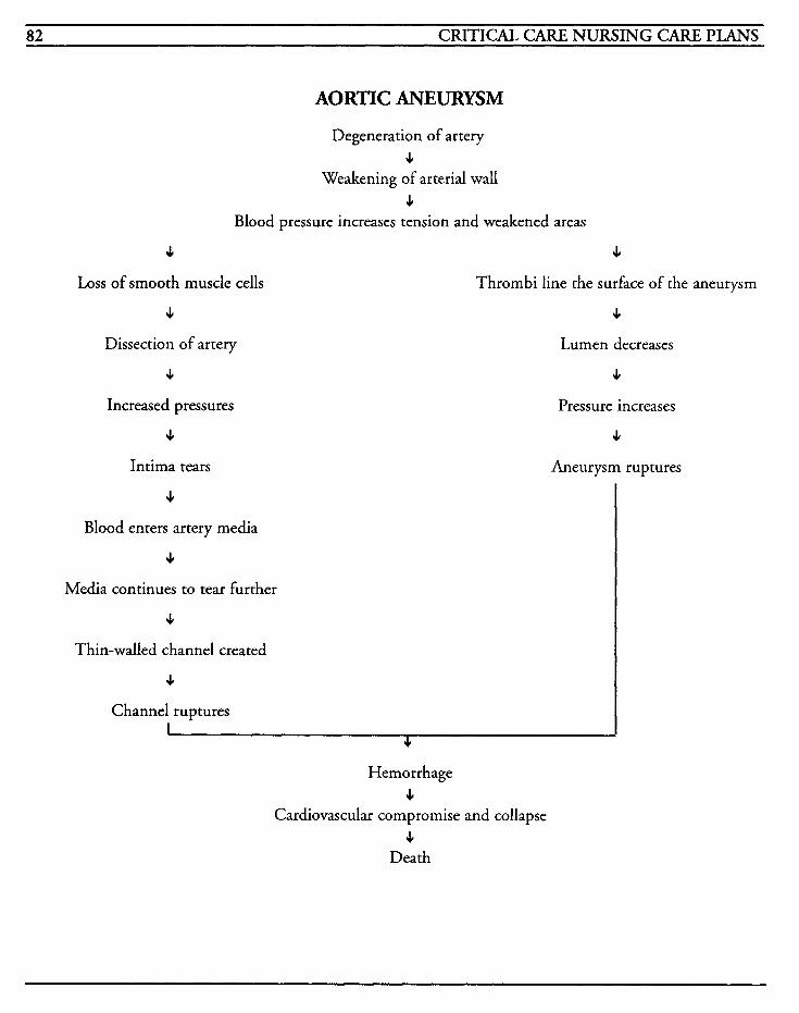
82 CRITICAL CARE NURSING CARE PLANS
AORTIC ANEURYSM
Degeneration of artery J,
Weakening of arterial wall 3
Blood pressure increases tension and weakened areas
3
Loss of smooth muscle cells
3
Dissection of artery
3
Increased pressures
3
Intima tears
3
Thrombi line the surface of the aneurysm
J,
Lumen decreases
J,
Pressure increases
4
Aneurysm ruptures
3
Blood enters artery media
3
Media continues to tear further
3
Thin-walled channel created
3
Channel ruptures I
3
Hemorrhage 3
Cardiovascular compromise and collapse J,
Death
RESPIRATORY SYSTEM 83
Adult Respiratory Distress Syndrome (ARDS)
Chronic Obstructive Pulmonary Disease (COPD)
Pulmonary Embolism
Pneumonia
Pneumo thorax
Status Asthmaticus
Mechanical Ventilation

This Page Intentionally Left Blank
RESPIRATORY SYSTEM 85
Adult Respiratory Distress Syndrome CARDS) Adult respiratory distress syndrome (ARDS) is also known as shock lung, wet lung, white lung, or acute respiratory distress syndrome, and occurs frequently after an acute or traumatic injury or ill- ness involving the respiratory system. The body responds to the injury with life-threatening respi- ratory failure and hypoxemia.
ARDS is usually noted 12-24 hours after the ini- tial insult or 5- 10 days after sepsis occurs. Dyspnea with hyperventilation and hypoxemia are usually the first clinical symptoms. Adventitious breath sounds frequently are not present initially.
Some of the most common precipitating factors are trauma, aspiration, pneumonia, near-drown- ing, toxic gas inhalation, sepsis, shock, DIC, oxygen toxicity, coronary artery bypass, pancreati- tis, fat or amniotic embolism, radiation, head injury, heroin use, massive hemorrhage, smoke inhalation, drug overdose, or uremia. Mortality is high (60-70%) despite treatment and often, patients who do survive, may have chronic resid- ual lung disease. In some cases, patients may have normal pulmonary function after recovery.
The latent phase of ARDS begins when the pul- monary capillary and alveolar endothelium become injured. The insult causes complement to be activated, as well as granulocytes, platelets, and the coagulation cascade. Free oxygen radicals, arachidonic acid metabolites and proteases are released into the system. Humoral substances, such as serotonin, histamine and bradykinin, are released. This results in red blood cell and high plasma protein leakage into the interstitial spaces, due to increased capillary permeability and increased pulmonary hydrostatic pressure. Initially, there may be little evidence of respiratory
problems, and chest x-rays may be normal or show minimal diffuse haziness. The fluid leakage increases and lymphatic flow increases with the acute phases with widespread damage to pulmonary capillary membranes and inflammation. Increases in intra-alveolar edema leads to capillary congestion and collagen forma- tion. Surfactant production and activity decreases, which causes decreased functional residual capac- ity, increased pulmonary shunting with widening A-a gradients, decreased pulmonary compliance, and ventilation/perfusion mismatching results. Chest x-rays will then show the ground glass appearance and finally a complete white-out of the lung.
The chronic phases occurs when the endothelium thickens; Type I cells, which are the gas-exchange pneumocytes, are replaced by Type I1 cells, which are responsible for producing surfactant, and along with fibrin, fluid and other cellular material form a hyaline membrane in place of the normal alveoli.
The goals of treatment are to improve ventilation and perfusion, to treat the underlying disease process that caused the lung injury, and to prevent progression of potentially fatal complications. Oxygen therapy with high levels of oxygen, mechanical ventilatory support with PEEE and fluid and drug management are required.
MEDICAL CARE Laboratory: cultures to identify causative organ- isms when bacterial infection is present and to identify proper antimicrobial agent; C5A levels increase with disease process; fibrin split products increase; platelets decrease; lactic acid levels increase
Chest x-ray: used to evaluate lung fields; early x- rays may be normal or have diffuse infiltrates; later x-rays will show bilateral ground glass
86 CRITICAL CARE NURSING CARE PLANS
appearance or complete whiting-out of lung fields; assists with differentiation between ARDS and cardiogenic pulmonary edema since heart size is normal in ARDS
Oxygen: to correct hypoxia and hypoxemia
Arterial blood gases: to identify acid-base prob- lems, hypocapnia, hypercapnia, and hypoxemia, and to evaluate progress of disease process and effectiveness of oxygen therapy
Ventilation: to provide adequate oxygenation and ventilation in patients who are unable to maintain even minimal levels
Pulmonary function studies: used to evaluate lung compliance and volumes which are normally decreased; physiologic dead space is increased and alveolar ventilation is compromised
Ineffective breathing pattern [See Mechanical Ventilation]
Related to: decreased lung compliance, pulmonary edema, increased lung density, decreased surfactant
Defining characteristics: use of accessory muscles, dyspnea, tachypnea, bradypnea, altered ABGs
Impaired gas excbange [See Mechanical Ventilation]
Related to: intra-alveolar edema, ateleccasis, venti- lation/perfusion mismatching, decreased arterial PO,, decreased amount and activity of surfactant, alveolar hypoventilation, formation of hyaline membranes, alveolar collapse, decreased diffusing capacity, shunting
Defining characteristics: tachypnea, cyanosis, use of accessory muscles, tachycardia, restlessness, mental changes, abnormal arterial blood gases, intrapulmonary shunting increased, A-a gradient changes, hypoxemia, increased dead space
Inefective airway clearance [See Mechanical Ventilation]
Related to: interstitial edema, increased airway resistance, decreased lung compliance, pulmonary secretions
Defining Characteristics: dyspnea, tachypnea, cyanosis, use of accessory muscles, cough with or without production, anxiety, restlessness, feelings of impending doom
Anxiety [See Mechanical Ventilation]
Related to: health crisis, effects of hypoxemia, fear of death, change in health status, change in envi- ronment
Defining characteristics: apprehension, restless- ness, fear, verbalized concern
Knowledge deficit [See Mechanical Ventilation]
Related to: lack of information, inability to process information, lack of recall
Defining characteristics: verbalized concerns and questions
Decreased cardiac output
Related to: increased positive airway pressures, sepsis, dysrhythmias, increased intrapulmonary edema, left ventricular failure
Defining characteristics: tachycardia, cardiac output less than 4 L/min, cardiac index less than 2.5 Llminlm2, cold clammy skin, decreased blood pressure
Outcome Criteria
Patient will be hemodynamically stable.
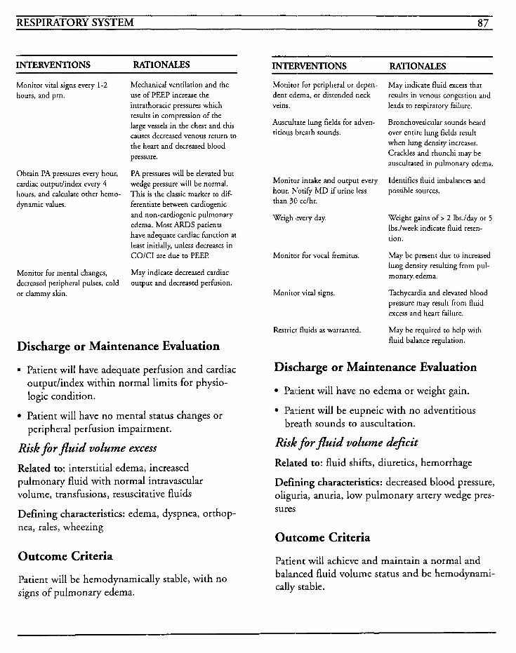
RESPIRATORY SYSTEM 87
INTERVENTIONS RATIONALES INTERVENTIONS RATIONALES
Monitor vital signs every 1-2 hours, and prn.
Obtain PA pressures every hour, cardiac output/index every 4 hours, and calculate other hemo- dynamic values.
Monitor for mental changes, decreased peripheral pulses, cold or clammy skin.
Mechanical ventilation and the use of PEEP increase the intrathoracic pressures which results in compression of the large vessels in the chest and this causes decreased venous return to the heart and decreased blood pressure.
PA pressures will be elevated but wedge pressure will be normal. This is the classic marker to dif- ferentiate between cardiogenic and non-cardiogenic pulmonary edema. Most ARDS patients have adequate cardiac function at least initially, unless decreases in COlCI are due to PEEP.
May indicate decreased cardiac output and decreased perfusion.
Discharge or Maintenance Evaluation
. Patient will have adequate perfusion and cardiac output/index within normal limits for physio- logic condition.
Patient will have no mental status changes or
Risk for fluid volume excess
Related to: interstitial edema, increased pulmonary fluid with normal intravascular volume, transfusions, resuscitative fluids
Defining characteristics: edema, dyspnea, orthop- nea, rales, wheezing
peripheral perfusion impairment.
Outcome Criteria
Patient will be hemodynamically stable, with no signs of pulmonary edema.
Monitor for peripheral or depen- dent edema, or distended neck veins.
Auscultate lung fields for adven- titious breath sounds.
Monitor intake and output every hour. Notify MD if urine less than 30 cclhr.
Weigh every day.
Monitor for vocal fremitus.
Monitor vital signs.
Restrict fluids as warranted.
May indicate fluid excess that results in venous congestion and leads to respiratory failure.
Bronchovesicular sounds heard over entire lung fields result when lung density increases. Crackles and rhonchi may be auscultated in pulmonary edema.
Identifies fluid imbalances and possible sources.
Weight gains of > 2 Ibs./day or 5 Ibs.lweek indicate fluid reten- tion.
May be present due to increased lung density resulting from pul- monary. edema.
Tachycardia and elevated blood pressure may result from fluid excess and heart failure.
May be required to help with fluid balance regulation.
Discharge or Maintenance Evaluation
Paiient will have no edema or weight gain.
Patient will be eupneic with no adventitious breath sounds to auscultation.
Risk for fluid volume &+kit Related to: fluid shifts, diuretics, hemorrhage
Defining characteristics: decreased blood pressure, oliguria, anuria, low pulmonary artery wedge pres- sures
Outcome Criteria
Patient will achieve and maintain a normal and balanced fluid volume status and be hemodynami- cally stable.
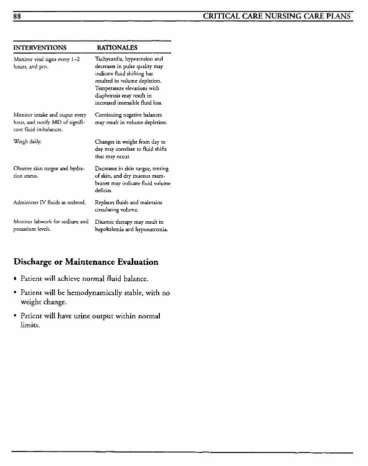
88 CRITICAL CARE NURSING CARE PLANS
INTERVENTIONS RATIONALES
Monitor vital signs every 1-2 hours, and prn.
Monitor intake and ouput every hour, and notify MD of signifi- cant fluid imbalances.
Weigh daily.
Observe skin turgor and hydra- tion status.
Administer IV fluids as ordered.
Monitor labwork for sodium and potassium levels.
Tachycardia, hypotension and decreases in pulse quality may indicate fluid shifting has resulted in volume depletion. Temperature elevations with diaphoresis may result in increased insensible fluid loss.
Continuing negative balances may result in volume depletion.
Changes in weight from day to day may correlate to fluid shifts that may occur.
Decreases in skin turgor, tenting of skin, and dry mucous mem- branes may indicate fluid volume deficits.
Replaces fluids and maintains circulating volume.
Diuretic therapy may result in hypokalemia and hyponatremia.
Discharge or Maintenance Evaluation
Patient will achieve normal fluid balance.
Patient will be hemodynamically stable, with no weight change.
Patient will have urine output within normal limits.
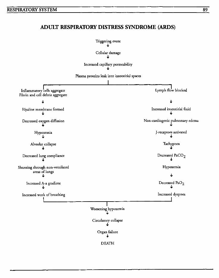
~
RESPIRATORY SYSTEM 89
ADULT RESPIRATORY DISTRESS SYNDROME (ARDS)
Triggering event c
Cellular damage 3
Increased capillary permeability 3
Plasma proteins leak into interstitial spaces
I Inflammatory !ells aggregate
Fibrin and cell debris aggregate Lymph flb blocked
3 c Hyaline membrane formed
c Decreased oxygen diffusion
4 Hypoxemia
c Alveolar collapse
c Decreased lung compliance
3
Shunting through non-ventilated areas of lungs
3
Increased A-a gradient 3
Increased interstitial fluid c
Non-cardiogenic pulmonary edema c
J-receptors activated 3
Tachypnea 3
Decreased PaC02 3
Hypoxemia
c Decreased PaO2
3
Increased work of breathing Increased dyspnea I I Worsening hypoxemia
c Circulatory collapse
3
Organ failure c
DEATH

This Page Intentionally Left Blank
RESPIRATORY SYSTEM 91
Chronic Obstructive Pulmonary Disease (I: 0 P D) Chronic obstructive pulmonary disease (COPD) is irreversible condition in which airways become obstructed and resistance to air flow is increased during expiration when airways collapse. COPD is usually further subdivided into other diseases such as bronchitis and emphysema, and actually COPD refers to these simultaneous disease entities.
Emphysematous changes include enlarging of the air spaces distally to the terminal bronchioles, and concurrent changes in alveolar walls. Capillary numbers decrease in the remaining walls and may sclerose. Gas exchange is decreased due to the reduction in available alveolar surfaces as well as decreased perfusion to non-ventilated areas. Ventilation/perfusion mismatching occurs and functional residual capacity is increased. The anteroposterior diameter of the chest is often enlarged due to the loss of elasticity and increased air trapping in the airway supportive structures. These type A patients are often called “pink puffers” because of the increased response to hypoxemia. Symptoms include dyspnea and increase in breathing effort, which result in a well-oxygenated, or pink, patient who displays overt dyspnea, or puffing.
Bronchitis is usually associated with prolonged exposure to lung irritants, which results in inflam- matory changes and thickening of bronchial walls, and increases in mucous production. The patient exhibits a chronic productive cough due in part to the increase in size of mucous glands and decrease in cilia. These type B patients are often called “blue bloaters” because their response to hypoxemia is reduced, with increasing PaC02 levels and cyanosis. These patients frequently have
bouts of cor pulmonale, or right-sided heart fail- ure, resulting in peripheral edema.
The most common precipitating factors for COPD include cigarette smoking, air or environ- mental pollution, allergic response, autoimmunity, and genetic predisposition. Treatment is aimed at avoidance of respiratory allergens and irritants, controlling bronchospasms, and improving airway clearance.
Laboratory: cultures used to identify causative organisms and determine appropriate antimicro- bial therapy; CBC used to identify presence of infection with elevated white blood cell count, and to monitor for increases in RBCs and hemat- ocrit as the body tries to compensate for oxygen transport requirements; alphal-antitrypsin levels used to identify deficiency that may be present if patient has heredity predisposition; theophylline levels used to monitor for therapeutic levels and/or toxicity
Pulmonary function studies: used to evaluate pul- monary status and function, and to identify airway obstruction, increased residual volume, total lung capacity, compliance, decreased vital capacity, diffusing capacity, and expiratory volumes with emphysema patients; increased residual volume, decreased vital capacity and forced expiratory volumes with normal static com- pliance and diffusion capacity with bronchitis patients
Chest x-ray: used to identify hyperinflation of lungs, flattened diaphragm, or pulmonary hyper- tension; used to identify barotrauma that may occur, increased antero-posterior chest diameter, large retrosternal air spaces, or secondary cardio- vascular complications with right-sided heart failure
92 CRITICAL CARE NURSING CARE PLANS
Electrocardiography: used to identify dysrhythmias associated with this disease; tall p waves in inferior leads, vertical QRS axis, atrial dysrhythmias, right ventricular hypertrophy, sinus tachycardia, and right axis deviation
Defining characteristics: dyspnea, tachypnea, bradypnea, bronchospasms, increased work of breathing, use of accessory muscles, increased mucous production, cough with or without pro- ductivity, adventitious breath sounds
Oxygen: used to improve hypoxemia; liter flow should be low in order to maintain the patient's respiratory drive; PaO, may be acceptable at 55- 60 mmHg to avoid hypoventilation and maintain function
IV fluids: used to maintain hydration and for administration of medical therapeutics
Bronchodilators: xanthines and sympathomimet- ics are used to relieve bronchospasms and help to promote clearance of mucoid secretions
Antibiotics: used to treat respiratory infections
Arterial blood gases: used to identify acid-base disturbances, presence of hypoxemia and hyper- capnia, and to evaluate responses to therapies
Chest physiotherapy: percussion and postural drainage are used to facilitate mobilization of secretions and promote clearance of airways
Corticosteroids: used to decrease secretions and reduce inflammation in the lungs; use of steroids is controversial
Psychological treatment: use of anti-anxiety agents to decrease fear and anxiety related to dysp- nea, without sedation to depress the respiratory drive; psychotherapy may be required to enable patients to cope with their ongoing disease process
NURSING CARE PLANS Ingective airway clearance [See Mechanical Ventilation]
Related to: bronchospasm, fatigue, increased work of breathing, increased mucous production, thick secretions, infection
Inefective breathing pattern [See Mechanical Ventilation]
Related to: pain, increased lung compliance, decreased lung expansion, fear, obstruction, decreased elasticity/recoil
Defining characteristics: dyspnea, tachypnea, use of accessory muscles, cough with or without pro- ductivity, adventitious breath sounds, prolongation of expiratory time, increased mucous production, abnormal arterial blood gases
Impaired gas exchange [See Mechanical Ventilation]
Related to: obstruction of airways, bronchospasm, air-trapping, right-to-left shunting, ventilation/perfusion mismatching, inability to move secretions, hypoventilation
Defining characteristics: hypoxemia, hypercapnia, mental changes, confusion, restlessness, dyspnea, vital sign changes, inability to tolerate activity, res- piratory acidosis
Anxiev [See Mechanical Ventilation]
Related to: threat of death, change in health status, life-threatening crises
Defining characteristics: fear, restlessness, muscle tension, helplessness, communication of uncertainty and apprehension, feeling of suffocation
Activity intolerance
Related to: fatigue, weakness, increased effort and work of breathing, inadequate rest, hypoxia, hypoxemia
RESPIRATORY SYSTEM 93
Defining characteristics: dyspnea, decreased oxygen saturation levels with movement or activ- ity, increased heart rate and blood pressure with movement or activity, feelings of tiredness and weakness
Outcome Criteria
Patient will be able to tolerate minimal activity without respiratory compromise.
~~~ ~
INTERVENTIONS RATIONALES
Monitor for patient’s response to activity changes. ,
Monitor vital signs before, during, and after increased activ- ity levels.
Plan activities to ensure patient obtains adequate amounts of rest and sleep.
Assist patient with activities as warranted.
Increase activity gradually and encourage patient participation.
Administer inhalers as ordered prior to activities.
Identifies patient’s ability to compensate for increases in activ- ity and provides baseline date from which to plan care.
Increases in heart rate greater than lolminute or respiratory rate greater than 32 may indicate that patient has reached his maximal activity limit and further activity may result in circulatorylrespiratory dyshnction.
Decreases potential for dyspnea and provides rest to prevent excessive fatigue.
Conserves energy and decreases oxygen consumption and dysp- nea.
Gradual increases facilitate increased tolerance to activity by balancing oxygen supply and demand, and patient cooperation may facilitate feelings of self- worth and adequacy.
Helps prevent dyspnea by per- forming activities at peak time of medication effects.
Information, Instruction, Demonstration
INTERVENTIONS RATIONALES
Instruct on techniques to save energy expenditure: shower stools, arm and leg rests, gather- ing required articles and placement within reach, etc.
Provide patient with exercise regimen protocol.
Instruct on breathing exercises to be performed with activicy.
Helps CO decrease energy expen- diture and fatigue, which may result in increased dyspnea.
Promotes independence and self- worth; increases tolerance to exercises.
Promotes effective respiratory patterns during exertion.
Discharge or Maintenance Evaluation
Patient will be able to tolerate activity without excessive dyspnea or hemodynamic instability,
Patient will be able to perform ADLs within limits of disease process.
Patient will be able to recall information accu- rately, and will be able to utilize relaxation and breathing techniques effectively.
Patient will be compliant with prescribed exer- cise regimens.
Ineffective individua Ufamily coping [See Mechanical Ventilation]
Related to: changes in lifestyle and health status, sensory overload, fear of death, physical Gmitations, inadequate support system, inadequate coping mechanisms, continual dyspnea
Defining characteristics: inability to meet role expectations, inability to meet basic needs, constant worry, apprehension, fear, inability to problern-solve, anger, hostility, aggression, inap- propriate defense mechanisms, low self-esteem, insomnia, depression, destructive behaviors, vacil- lation when choices are required, delayed decision-making, muscle tension, fatigue
94 CRITICAL CARE NURSING CARE PLANS
Risk for infiction
Related to: disease process, inability to move secretions, decreased cilia function, immunosup- pression, poor nutrition
Defining characteristics: increased temperature, chills, elevated white blood cell count, inability to move secretions
INTERVENTIONS RATIONALES
Monitor for increased dyspnea, sputum color and character changes, cough, and temperature elevation.
Obtain sputum specimen for culture and sensitivity as ordered.
Administer antibiotics as ordered.
Monicor for abrupt changes in other body systems; cardiac abnormalities and alteration in heart sounds, increasing pain, changes in mental status, recur- ring temperature elevations.
Provide adequace rest time for patient.
Yellow or green sputum, with increased viscosity usually indicates infection. Prompt recognition facilitates prompt treatment.
Identifies the causative organism and provides information regard- ing appropriate antimicrobial agent required.
Controls and clears the infection and any secondary infections in che bronchial tree. Improvement should be noted within 24-48 hours after antimicrobial agent has begun.
May indicate presence of secondary infeccion or resistance to ordered antibiotics. Superinfections, systemic bac- teremia, inflammatory cardiac conditions, meningitis or encephalitis may occur.
Helps to facilitate healing and natural immunity.
Discharge or Maintenance Evaluation
Patient will exhibit no signs/symptoms of sec-
Altered nutrition: less tban body requirements [See Mechanical Ventilation]
Related to: dyspnea, inability to take in sufficient
ondary infection.
food, increased metabolism due to disease process, decreased level of consciousness, fatigue, increased sputum, medication side effects
Defining characteristics: actual inadequate food intake, altered taste, altered smell sensation, weight loss, anorexia, absent bowel sounds, decreased peristalsis, muscle mass loss, changes in bowel habits, abdominal distention, nausea, vom- iting
Knowledge &+it
Related to: lack of information, lack of recall of information, cognitive limitations
Defining characteristics: request for information, statement of misconception, statement of concerns, development of preventable complica- tions, inaccurate follow-through with instructions
Outcome Criteria
Patient will be able to recall information accurately and will follow through with all instruc- tions.
INTERVENTIONS RATIONALES
Assess knowledge of COPD dis- ease process, medicacions, and treatments.
Instruct on medication effects, side effects, contraindications, and signslsymptoms to report.
Instruct in proper technique for using and cleaning inhalers.
Instruct on need to avoid smok- ing and other respiratory irritants.
Instruct on effective coughing
Identifies level of knowledge and provides baseline from which CO
plan teaching.
Promotes knowledge and com- pliance with treatment regimen.
Proper technique, including appropriate time intervals between puffs, facilitates effective delivery and therapeutic effect.
May initiate and exacerbate bronchial irritation which can resulr in increased mucous pro- duction and airway obstruction.
Effective coughing reduces
RESPIRATORY SYSTEM 95
INTERVENTIONS RA'IIONALES techniques; postural drainage, chest physiotherapy, etc.
Instruct to drink 10-12 glasses of water per day.
Instruct on use of supplemental oxygen at low flow rates, and reasons to avoid increasing flow indiscriminately.
Instruct on oxygen safery: avoid- ing flammable objects, use of vaseline or other petroleum products, and ambulation with tubing.
Instruct on avoiding sedative or antianxiety drugs as warranted.
Instruct on avoiding people with infections; encourage patient to obtain influenza and pneumonia vaccinations as warranted.
Instruct on activity limitations, methods to conserve energy and promote rest, pursed-lip breath- ing, etc.
Instruct on signslsymptoms to notify MD: increased tempera- ture, change in sputum color or character, increasing dyspnea.
Instruct to continue with follow- up medical care.
Provide patient/family with information regarding support groups, such as the American Lung Association, etc.
fatigue and facilitates removal of secretions. Percussion and pos- tural drainage help to mobilize tenacious secretions.
Maintains hydration and pro- motes easier mobilization of secretions.
COPD patients will rarely require more than 2-3 Llmin to maintain their optimum oxy- genation levels. Increasing flow rates will increase their PaOz but may decrease their respiratory drive and may result in drowsi- ness and confusion.
Promotes physical and environ- mental safety.
Sedative may result in respiratory depression and impair cough reflexes.
Prevents exposure to other infec- tions, and decreases potential for incidence of upper respiratory infections.
Helps decrease fatigue, optimizes activity level within range of disease process, and reduces dys- pnea and oxygen consumption.
Provides for prompt recognition of infection to facilitate prompt intervention prior to respiratory failure.
Provides for monitoring of pro- gression of disease, presence of complications, or exacerbations, and facilitates changes in medical regimen to concur with current medical condition.
Support groups may be required to provide emotional assistance and respite for caregiver(s).
INTERVENTIONS RATIONALES Assist patientlfamily to set redis- tic goals for long- and short-term.
Provides a plan for patient and facilitates self-involvement with realistic goals and methods to meet them. Fosters independence and reduces anxiety.
Discharge or Maintenance Evaluation
Patient will be able to recall information regard- ing disease process and treatment regimen.
Patient will be able to recall accurately the signs/symptoms for which to notify MD, the effects and side effects of medications, and proper procedure for using inhalers.
Patient will be able to demonstrate accurately proper cough techniques, pursed-lip breathing, and proper positioning to facilitate breathing.
Patiendfamily will be able to access support sys- tems effectively.
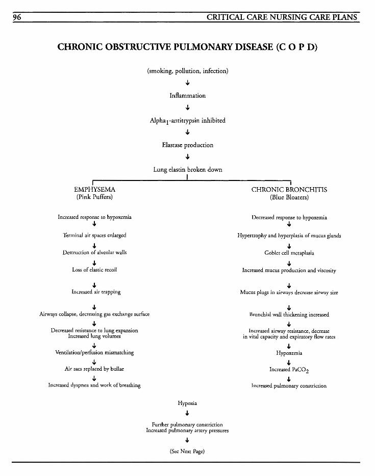
96 CRITICAL CARE NURSING CARE PLANS
CHRONIC OBSTRUCTIVE PULMONARY DISEASE (C 0 P D)
(smoking, pollution, infection)
4 Inff ammation
4 Alphal-antitrypsin inhibited
s Elastase production
4 Lung elastin broken down
1 I
I EMPHYSEMA (Pink Puffers)
Increased response to hypoxemia c
Terminal air spaces enlarged
4 Destruction of alveolar walls
s Loss of elastic recoil
J, Increased air trapping
s Airways collapse, decreasing gas exchange surface
s Decreased resistance to lung expansion
Increased lung volumes
4 Ventilationlperfusion mismatching
4 Air sacs replaced by bullae
4 Increased dyspnea and work of breathing
Hypoxia
4 Further pulmonary constriction
Increased pulmonary artery pressures
4 (See Next Page)
CHRONIC BRONCHITIS (Blue Bloaters)
Decreased response to hypoxemia c
Hypertrophy and hyperplasia of mucus glands
s Goblet cell metaplasia
s Increased mucus production and viscosity
c Mucus plugs in airways decrease airway size
4 Bronchial wall thickening increased
s Increased airway resistance, decrease
in vital capacity and expiratory flow rates
4 Hypoxemia
c Increased PaCO2
s Increased pulmonary constriction
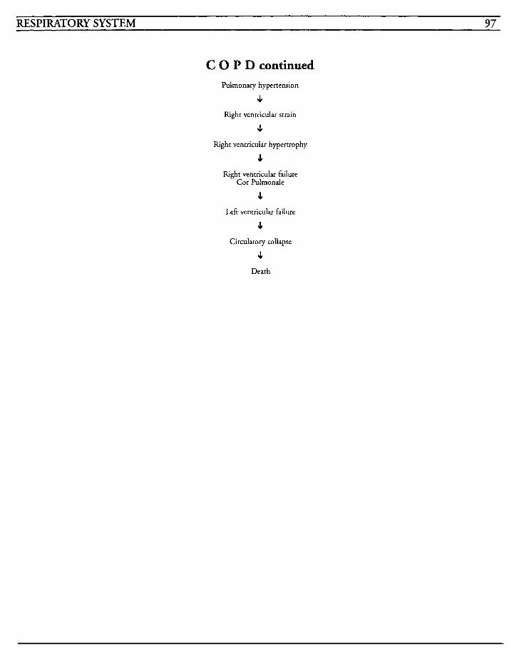
RESPIRATORY SYSTEM 97
C 0 P D continued Pulmonary hypertension
4 Right ventricular strain
4 Right ventricular hypertrophy
4 Right ventricular failure
Cor Pulmonde
4 Left ventricular failure
4 Circulatory collapse
J Death

This Page Intentionally Left Blank
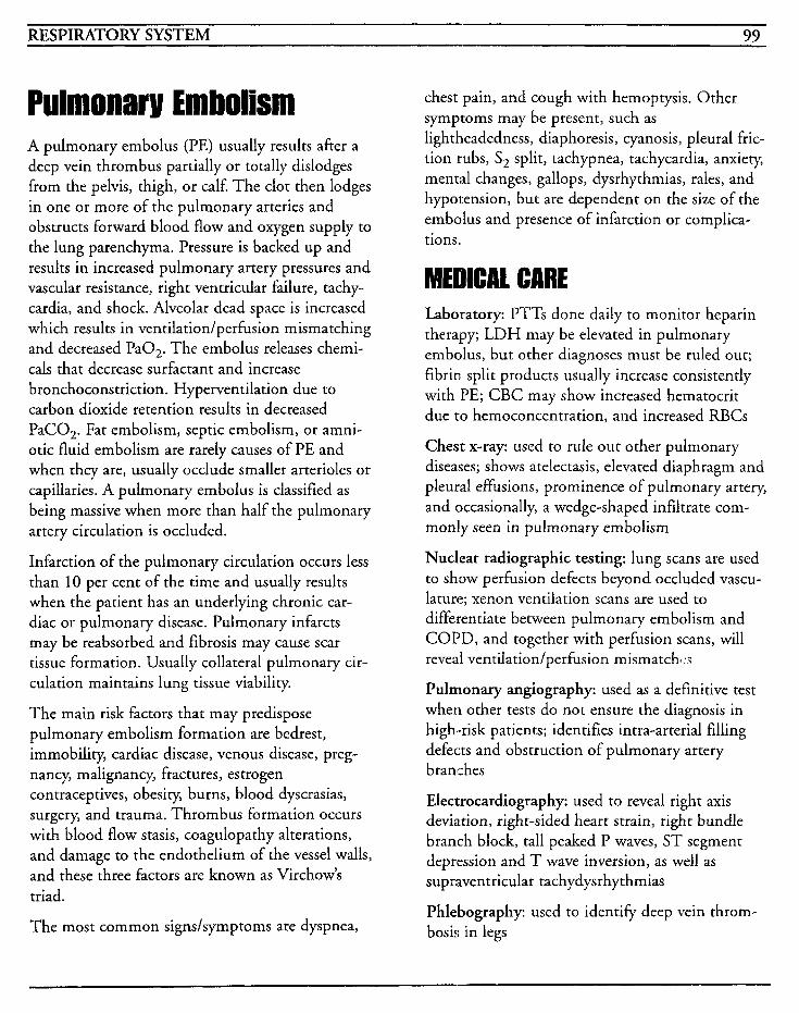
RESPIRATORY SYSTEM 99
A pulmonary embolus (PE) usually results after a deep vein thrombus partially or totally dislodges from the pelvis, thigh, or calf. The clot then lodges in one or more of the pulmonary arteries and obstructs forward blood flow and oxygen supply to the lung parenchyma. Pressure is backed up and results in increased pulmonary artery pressures and vascular resistance, right ventricular failure, tachy- cardia, and shock. Alveolar dead space is increased which results in ventilation/perfusion mismatching and decreased PaO,. The embolus releases chemi- cals that decrease surfactant and increase bronchoconstriction. Hyperventilation due to carbon dioxide retention results in decreased PaCO,. Fat embolism, septic embolism, or amni- otic fluid embolism are rarely causes of PE and when they are, usually occlude smaller arterioles or capillaries. A pulmonary embolus is classified as being massive when more than half the pulmonary artery circulation is occluded.
Infarction of the pulmonary circulation occurs less than 10 per cent of the time and usually results when the patient has an underlying chronic car- diac or pulmonary disease. Pulmonary infarcts may be reabsorbed and fibrosis may cause scar tissue formation. Usually collateral pulmonary cir- culation maintains lung tissue viability.
The main risk factors that may predispose pulmonary embolism formation are bedrest, immobility, cardiac disease, venous disease, preg- nancy, malignancy, fractures, estrogen contraceptives, obesity, burns, blood dyscrasias, surgery, and trauma. Thrombus formation occurs with blood flow stasis, coagulopathy alterations, and damage to the endothelium of the vessel walls, and these three factors are known as Virchow’s triad.
The most common signs/symptoms are dyspnea,
chest pain, and cough with hemoptysis. Other symptoms may be present, such as lightheadedness, diaphoresis, cyanosis, pleural fric- tion rubs, S, split, tachypnea, tachycardia, anxiety, mental changes, gallops, dysrhythmias, rales, and hypot:ension, but are dependent on the size of the embolus and presence of infarction or complica- tions.
MEDICAL CARE Laboratory: PTTs done daily to monitor heparin therapy; LDH may be elevated in pulmonary embolus, but other diagnoses must be ruled out; fibrin split products usually increase consistently with PE; CBC may show increased hematocrit due to hemoconcentration, and increased RBCs
Chest x-ray: used to rule out other pulmonary diseases; shows atelectasis, elevated diaphragm and pleural effusions, prominence of pulmonary artery, and occasionally, a wedge-shaped infiltrate com- monly seen in pulmonary embolism
Nuclear radiographic testing: lung scans are used to show perfusion defects beyond occluded vascu- lature; xenon ventilation scans are used to differentiate between pulmonary embolism and COPD, and together with perfusion scans, will reveal ventilation/perfusion mismatchl:;
Pulmonary angiography: used as a definitive test when other tests do not ensure the diagnosis in high-risk patients; identifies intra-arterial filling defects and obstruction of pulmonary artery branches
Electrocardiography: used to reveal right axis deviation, right-sided heart strain, right bundle branch block, tall peaked P waves, ST segment depression and T wave inversion, as well as supraventricular tachydysrhythmias
Phlebography: used to identifj. deep vein throm- bosis in legs
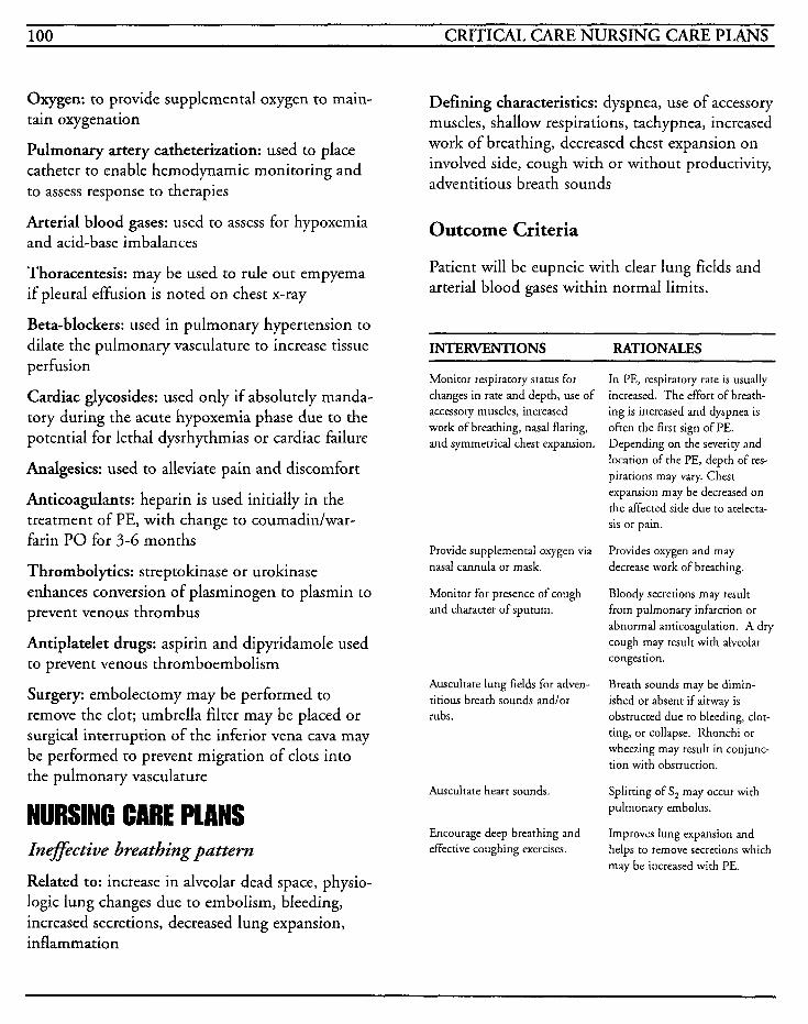
100 CRITICAL CARE NURSING CARE PLANS
Oxygen: to provide supplemental oxygen to main- tain oxygenation
Pulmonary artery catheterization: used to place catheter to enable hemodynamic monitoring and to assess response to therapies
Arterial blood gases: used to assess for hypoxemia and acid-base imbalances
Thoracentesis: may be used to rule out empyema if pleural effusion is noted on chest x-ray
Beta-blockers: used in pulmonary hypertension to dilate the pulmonary vasculature to increase tissue per fusion
Cardiac glycosides: used only if absolutely manda- tory during the acute hypoxemia phase due to the potential for lethal dysrhythmias or cardiac failure
Analgesics: used to alleviate pain and discomfort
Anticoagulants: heparin is used initially in the treatment of PE, with change to coumadidwar- farin PO for 3-6 months
Thrombolytics: streptokinase or urokinase enhances conversion of plasminogen to plasmin to prevent venous thrombus
Antiplatelet drugs: aspirin and dipyridamole used to prevent venous thromboembolism
Surgery: embolectomy may be performed to remove the clot; umbrella filter may be placed or surgical interruption of the inferior vena cava may be performed to prevent migration of clots into the pulmonary vasculature
NURSING CARE PLANS Inefective breathing pattern
Related to: increase in alveolar dead space, physio- logic lung changes due to embolism, bleeding, increased secretions, decreased lung expansion, inflammation
Defining characteristics: dyspnea, use of accessory muscles, shallow respirations, tachypnea, increased work of breathing, decreased chest expansion on involved side, cough with or without productivity) adventitious breath sounds
Outcome Criteria
Patient will be eupneic with clear lung fields and arterial blood gases within normal limits.
INTERVENTIONS RATIONALES
Monitor respiratory status for changes in rate and depth, use of accessory muscles, increased work of breathing, nasal flaring, and symmetrical chest expansion.
Provide supplemental oxygen via nasal cannula or mask.
Monitor for presence of cough and character of sputum.
Auscultate lung fields for adven- titious breath sounds andlor rubs.
Auscultate heart sounds.
Encourage deep breathing and effective coughing exercises.
In PE, respiratory rate is usually increased. The effort of breath- ing is increased and dyspnea is ofien the first sign of PE. Depending on the severity and location of the PE, depth of res- pirations may vary. Chest expansion may be decreased on the affected side due to atelecta- sis or pain.
Provides oxygen and may decrease work of breathing.
Bloody secretions may result from pulmonary infarction or abnormal anticoagulation. A dry cough may result with alveolar congestion.
Breath sounds may be dimin- ished or absent if airway is obstructed due to bleeding, clot- ting, or collapse. Rhonchi or wheezing may result in conjunc- tion with obstruction.
Splitting of S, may occur with pulmonary embolus.
Improves lung expansion and helps to remove secretions which may be increased with PE.
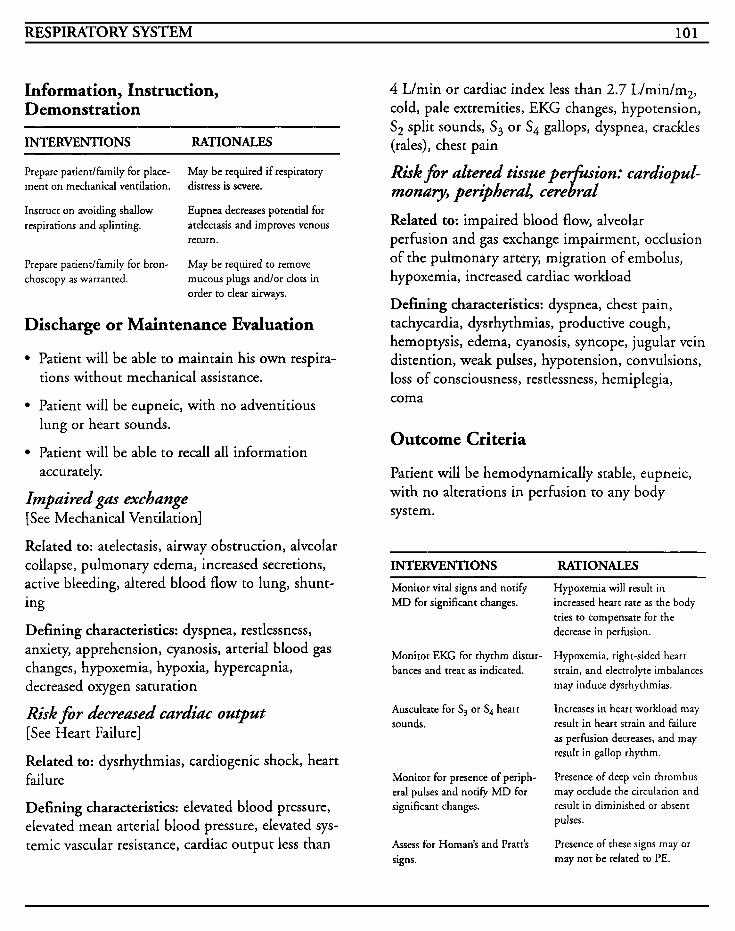
RESPIRATORY SYSTEM 101
Information, Instruction, Demonstration
INTERVENTIONS RA'TIONALES
Prepare patientlfamily for place- ment on mechanical ventilation.
Instruct on avoiding shallow respirations and splinting.
May be required if respiratory distress is severe.
Eupnea decreases potential for atelectasis and improves venous return.
May be required to remove mucous plugs andlor clots in order to clear airways.
Prepare patiendfamily for bron- choscopy as warranted.
Discharge or Maintenance Evaluation
Patient will be able to maintain his own respira- tions without mechanical assistance.
Patient will be eupneic, with no adventitious lung or heart sounds.
Patient will be able to recall all information accurately.
Impaired gas exchange [See Mechanical Ventilation]
Related to: atelectasis, airway obstruction, alveolar collapse, pulmonary edema, increased secretions, active bleeding, altered blood flow to lung, shunt- ing
Defining characteristics: dyspnea, restlessness, anxiety, apprehension, cyanosis, arterial blood gas changes, hypoxemia, hypoxia, hypercapnia, decreased oxygen saturation
Risk for decreased cardiac output [See Heart Failure]
Related to: dysrhythmias, cardiogenic shock, heart failure
Defining characteristics: elevated blood pressure, elevated mean arterial blood pressure, elevated sys- temic vascular resistance, cardiac output less than
4 L/min or cardiac index less than 2.7 L/min/m2, cold, pale extremities, EKG changes, hypotension, S, split sounds, S3 or S4 gallops, dyspnea, crackles (rales), chest pain
Risk f i r altered tissue p&ion: cardiopuL- monaly, peripheral, cerebral
Related to: impaired blood flow, alveolar perfusion and gas exchange impairment, occlusion of the pulmonary artery, migration of embolus, hypoxemia, increased cardiac workload
Defining characteristics: dyspnea, chest pain, tachycardia, dysrhythmias, productive cough, hemoptysis, edema, cyanosis, syncope, jugular vein distention, weak pulses, hypotension, convulsions, loss of consciousness, restlessness, hemiplegia, coma
Outcome Criteria
Patient will be hemodynamically stable, eupneic, with no alterations in perfusion to any body system.
INTERVENTIONS RA'TIONALES Monitor vital signs and notify MD for significant changes.
Hypoxemia will result in increased heart rate as the body tries to compensate for the decrease in perfusion.
Hypoxemia, right-sided heart strain, and electrolyte imbalances may induce dysrhythmias.
Increases in heart workload may result in heart strain and failure as perfusion decreases, and may result in gallop rhythm.
Presence of deep vein thrombus may occlude the circulation and result in diminished or absent pulses.
Presence of these signs may or may not be related to PE.
Monitor EKG for rhythm distur- bances and treat as indicated.
Auscultate for S, or S4 heart sounds.
Monitor for presence of periph- eral pulses and notify MD for significant changes.
Assess for Homan's and Pratt's signs.
102 CRITICAL CARE NURSING CARE PLANS
INTERVENTIONS RATIONALES
Assess skin color, temperature and capillary refill.
Impairment of blood flow may induce pallor or cyanosis to the slun or mucous membranes. Cool clammy skin or mottling may indicate peripheral vasocon- strictiodshock.
Monitor for restlessness or changes in mental status or level of consciousness. development of stroke.
May indicate occlusion, impaired cerebral blood flow, hypoxia, or
Information, Instruction, Demonstration
INTERVENTIONS RATIONALES
Prepare patient for insertion of pulmonary artery catheter.
May be required to monitor hemodynamic status and assess response to therapy.
Surgical intervention may be
recurrent emboli in spite of treat- ment, or if anticoagulant therapy cannot be given. Ligation of the vena cava or insertion of an umbrella filter may be necessary.
Streptokinase, urokinase, or alteplase (t-PA) may be required if the pulmonary embolus is massive and compromises hemo- dynamic stability.
Prepare patient for surgery as warranted. required if patient develops
Instruct on thrombolytic agents as warranted.
Discharge or Maintenance Evaluation
Patient will have adequate tissue perfusion to all body systems.
Patient will have stable hemodynamic parame- ters and vital signs will be within normal limits.
Oxygenation will be optimal as evidenced by pulse oximetry greater than 90% and adequate ABGs.
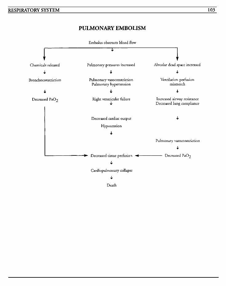
RESPIRATORY SYSTEM 103
PULMONARY EMBOLISM
Embolus obstructs blood flow
Chemicals released Pulmonary pressures increased Alveolar dead space increased
4 4 c Bronchoconstriction
4 Decreased Pa02
Pulmonary vasoconstriction Pulmonary hypertension
J,
Ventilation perfusion mismatch
J,
Right ventricular failure Increased airway resistance 4 Decreased lung compliance
Decreased cardiac output 4
Hypotension
4 Pulmonary vasoconstriction
c W Decreased tissue perfusion - Decreased PaOZ
c Cardiopulmonary collapse
c Death

This Page Intentionally Left Blank
RESPIRATORY SYSTEM 105
Pneumonia is an acute infection of the lung's ter- minal alveolar spaces and/or the interstitial tissues which results in gas exchange problems. The major challenge is identification of the source of the infection. Pneumonia ranks as the sixth most common cause of death in the United States.
When the infection is limited to a portion of the lung, it is known as segmental or lobular pneumo- nia; when the alveoli adjacent to the bronchioles are involved, it is known as bronchopneumonia, and when the entire lobe of the lung is involved, it is known as lobar pneumonia.
Pneumonia may be caused by bacteria, viruses, mycoplasma, rickettsias, or fungi. The causative organism gains entry by aspiration of oropharyn- geal or gastric contents, inhalation of respiratory droplets, from others who are infected, by way of the blood stream, or directly with surgery or trauma.
Viral types are more common in some areas, but identification of causative organisms may be diffi- cult with limited technology.
Patients who develop bacterial pneumonia usually are immunosuppressed or compromised by a chronic disease, or have had a recent viral illness. The most common type of bacterial pneumonia is pneumococcal pneumonia, in which the organism reaches the lungs via the respiratory passageways and result in the collapse of alveoli. The inflam- matory response that this generates causes protein-rich fluid to migrate into the alveolar spaces and provides culture media for the organ- ism to proliferate and spread.
Frequently pneumonia is predisposed by upper respiratory infections, chronic illness, cancer, surgery, atelectasis, chronic obstructive pulmonary disease, asthma, cystic fibrosis, bronchiectasis,
influenza, malnutrition, smoking, alcoholism, immuriosuppressive therapy, aspiration, sickle cell disease, head injury or coma.
Aspiration pneumonia occurs after aspiration of gastric or oropharyngeal contents, or other chemi- cal irritants into the trachea and lungs. Stomach acid damages the respiratory endothelium and may result in non-cardiogenic pulmonary edema, hemorrhage, destruction of surfactant-producing cells, and hypoxemia. The p H of the aspirated material determines the severity of the injury with pH less than 2.5 causing severe damage. Morbidity is high even with treatment.
In pneumonia's early stages, pulmonary vessels dilate and erythrocytes spread into the alveoli and cause a reddish, liver-like appearance, or red hepa- tization, in the lung consolidation area. Polymorphonuclear cells then enter the alveolar spaces and the consolidation increases to a grey hepatization. The leukocytes trap bacteria against the alveolar walls or other leukocytes so that more organisms are found in the increasing margins of the consolidation. The macrophage reaction occurs when mononuclear cells advance into the alveoli and phagocytize the exudate debris.
Diagnosis may be assisted with the observation of sputum characteristics, with bacterial pneumonia having mucopurulent sputum, viral and mycoplas- mic pneumonias having more watery secretions, pneurnococcal pneumonia having rust-colored sputum, and Klebsiella noting dark red mucoid secretions.
The initial signs/symptoms are sudden onset of shaking chills, fever, purulent sputum, pleuritic chest pain that is worsened with respiration or coughing, tachycardia, tachypnea, and use of accessory muscles.
Staphylococcal pneumonia is frequently noted after influenza or in hospitalized patients with a
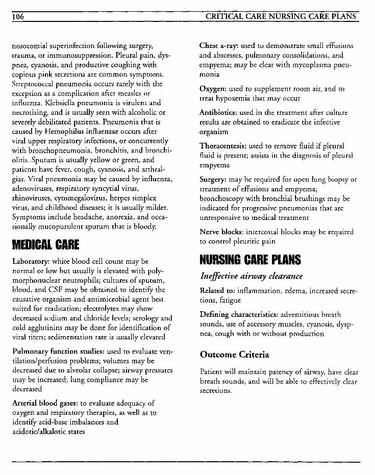
106 CRITICAL CARE NURSING CARE PLANS
nosocomial superinfection following surgery, trauma, or immunosuppression. Pleural pain, dys- pnea, cyanosis, and productive coughing with copious pink secretions are common symptoms. Streptococcal pneumonia occurs rarely with the exception as a complication after measles or influenza. Klebsiella pneumonia is virulent and necrotizing, and is usually seen with alcoholic or severely debilitated patients. Pneumonia that is caused by Hemophilus influenzae occurs after viral upper respiratory infections, or concurrently with bronchopneumonia, bronchitis, and bronchi- olitis. Sputum is usually yellow or green, and patients have fever, cough, cyanosis, and arthral- gias. Viral pneumonia may be caused by influenza, adenoviruses, respiratory syncytial virus, rhinoviruses, cytomegalovirus, herpes simplex virus, and childhood diseases; it is usually milder. Symptoms include headache, anorexia, and occa- sionally mucopurulent sputum that is bloody.
MEDICAL CARE Laboratory: white blood cell count may be normal or low but usually is elevated with poly- morphonuclear neutrophils; cultures of sputum, blood, and CSF may be obtained to identify the causative organism and antimicrobial agent best suited for eradication; electrolytes may show decreased sudium and chloride levels; serology and cold agglutinins may be done for identification of viral titers; sedimentation rate is usually elevated
Pulmonary function studies: used to evaluate ven- tilation/perfusion problems; volumes may be decreased due to alveolar collapse; airway pressures may be increased; lung compliance may be decreased
Arterial blood gases: to evaluate adequacy of oxygen and respiratory therapies, as well as to identify acid-base imbalances and acidotic/alkalotic states
Chest x-ray: used to demonstrate small effusions and abscesses, pulmonary consolidations, and empyema; may be clear with mycoplasma pneu- monia
Oxygen: used to supplement room air, and to treat hypoxemia that may occur
Antibiotics: used in the treatment after culture results are obtained to eradicate the infective organ ism
Thoracentesis: used to remove fluid if pleural fluid is present; assists in the diagnosis of pleural empyema
Surgery: may be required for open lung biopsy or treatment of effusions and empyema; bronchoscopy with bronchial brushing may be indicated for progressive pneumonias that are unresponsive to medical treatment
Nerve blocks: intercostal blocks may be required to control pleuritic pain
NURSING CARE PLANS Inefective airway clearance
Related to: inflammation, edema, increased secre- tions, fatigue
Defining characteristics: adventitious breath sounds, use of accessory muscles, cyanosis, dysp- nea, cough with or without production
Outcome Criteria
Patient will maintain patency of airway, have clear breath sounds, and will be able to effectively clear secretions.
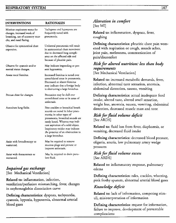
RESPIRATORY SYSTEM 107
INTERVENTIONS RATIONALES
Monitor respiratory status for changes, increased work of breathing, use of accessory mus- cles, and nasal flaring.
Observe for symmetrical chest Unilateral pneumonia will result expansion. in asymmetrical chest movement
due to decreased lung compli- ance on the affected side and because of pleuritic pain.
Tachypnea and hyperpnea are frequently noted with pneumonia.
Observe for cyanosis andlor mental status changes.
Assess vocal fremitus.
Percuss chest for changes.
Auscultate lung fields.
May indicate impending or pre- sent hypoxemia.
Increased fremitus is noted over consolidated areas in pneumonia. Decreased or absent fremitus may indicate that a foreign body is obstructing a large bronchus.
Percussion may be dull over consolidated areas or in areas of atelectasis.
Fine crackles or bronchial breath sounds are noted in lobar pneu- monia; in other types of pneumonia, bronchial sounds are rarely heard. Wheezes may indi- cate aspiration of a solid object. Inspiratory stridor may indicate the presence of an obstruction to a large bronchus.
Assist with bronchoscopy as warranted.
May be required to remove mucous plugs and prevent or improve atelectasis.
May be required to drain puru- Assist with thoracentesis as warranted. lent fluid.
Impaired gas exchange [See Mechanical Ventilation]
Related to: inflammation, infection, ventilation/perfusion mismatching, fever, changes in oxyhemoglobin dissociation curve
Defining characteristics: dyspnea, tachycardia, cyanosis, hypoxia, hypoxemia, abnormal arterial blood gases
Alteration in comfort [See MI]
Related to: inflammation, dyspnea, fever, coughing
Defining characteristics: pleuritic chest pain wors- ened with respiration or cough, muscle aches, joint pain, restlessness, communication of pain/discomfort
Risk for altered nutrition: less than body requirements [See Mechanical Ventilation]
Related to: increased metabolic demands, fever, infection, abnormal taste sensation, anorexia, abdominal distention, nausea, vomiting
Defining characteristics: actual inadequate food intake, altered taste, altered smell sensation, weight loss, anorexia, nausea, vomiting, abdominal distention, decreased muscle mass and tone
Risk f i r fluid volume d$cit [See ARDS]
Related to: fluid loss from fever, diaphoresis, or vomiting, decreased fluid intake
Defining characteristics: decreased blood pressure, oliguria, anuria, low pulmonary artery wedge pressures
Risk for fluid volume excess [See ARDS]
Related to: inflammatory response, pulmonary edema
Defining characteristics: rales, crackles, wheezing, pink ftothy sputum, abnormal arterial blood gases
Knowledge de$cit
Related to: lack of information, competing stim- uli, misinterpretation of information
Defining characteristics: request for information, failure to improve, development of preventable complications
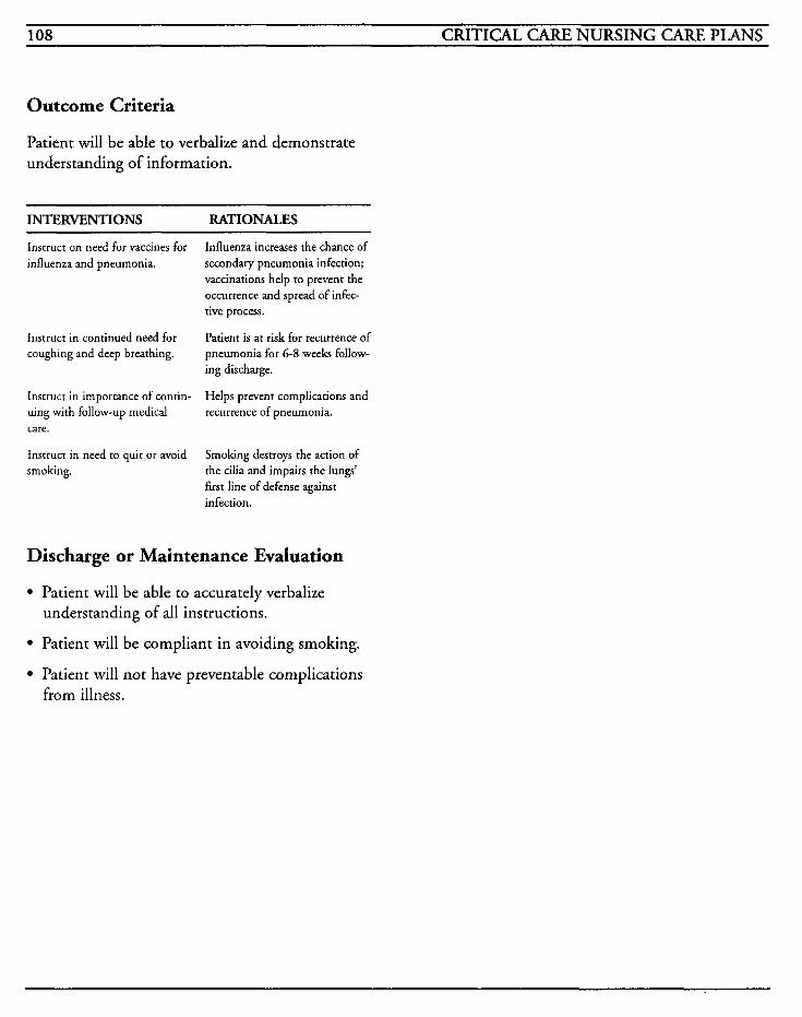
108 CRITICAL CARE NURSING CARE PLANS
Outcome Criteria
Patient will be able to verbalize and demonstrate understanding of information.
INTERVENTIONS RATIONALES
Instruct on need for vaccines for influenza and pneumonia.
Influenza increases the chance of secondary pneumonia infection; vaccinations help to prevent the occurrence and spread of infec- tive process.
Patient is at risk for recurrence of pneumonia for 6-8 weeks follow- ing discharge.
Helps prevent complications and recurrence of pneumonia.
Instruct in continued need for coughing and deep breathing.
Instruct in importance of contin- uing with follow-up medical care.
Instruct in need to quit or avoid smoking.
Smoking destroys the action of the cilia and impairs the lungs’ first line of defense against infection.
Discharge or Maintenance Evaluation
Patient will be able to accurately verbalize understanding of all instructions.
Patient will be compliant in avoiding smoking.
Patient will not have preventable complications from illness.
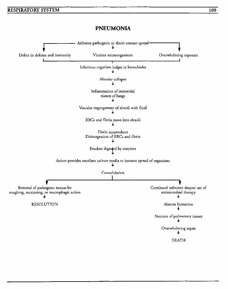
RESPIRATORY SYSTEM 109
PNEUMONIA
1 Airborne pathogenic or direct contact spread t c Defect in defense and immunity Virulent microorganisms Overwhelming exposure
I I I
Infectious organism lodges in bronchioles c
Alveolar collapse 4
Inflammation of interstitial tissues of lungs
c Vascular engorgement of alveoli with fluid
c RBCs and fibrin move into alveoli
c Fibrin accumulates
Disintegration of RBCs and fibrin c
Exudate digesjed by enzymes 4
Action provides excellent culture media to increase spread of organisms c
Consolidation
Removal of pathogenic mucus by coughing, suctioning, or macrophagic action
4 RESOLUTION
Continued infection despite use of antimicrobial therapy
JI
Abscess formation c
Necrosis of pulmonary tissues c
Overwhelming sepsis c
DEATH

This Page Intentionally Left Blank
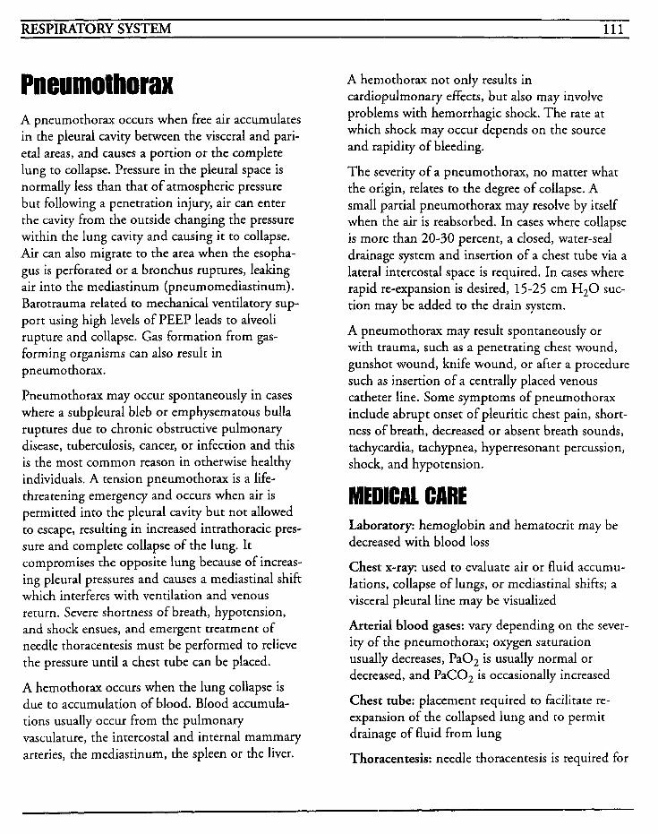
RESPIRATORY SYSTEM 111
A pneumothorax occurs when free air accumulates in the pleural cavity benveen the visceral and pari- etal areas, and causes a portion or the complete lung to collapse. Pressure in the pleural space is normally less than that of atmospheric pressure but following a penetration injury, air can enter the cavity from the outside changing the pressure within the lung cavity and causing it to collapse. Air can also migrate to the area when the esopha- gus is perforated or a bronchus ruptures, leaking air into the mediastinum (pneumomediastinum). Barotrauma related to mechanical ventilatory sup- port using high levels of PEEP leads to alveoli rupture and collapse. Gas formation from gas- forming organisms can also result in pneumothorax.
Pneumothorax may occur spontaneously in cases where a subpleural bleb or emphysematous bulla ruptures due to chronic obstructive pulmonary disease, tuberculosis, cancer, or infection and this is the most common reason in otherwise healthy individuals. A tension pneumothorax is a life- threatening emergency and occurs when air is permitted into the pleural cavity but not allowed to escape, resulting in increased intrathoracic pres- sure and complete collapse of the lung. It compromises the opposite lung because of increas- ing pleural pressures and causes a mediastinal shift which interferes with ventilation and venous return. Severe shortness of breath, hypotension, and shock ensues, and emergent treatment of needle thoracentesis must be performed to relieve the pressure until a chest tube can be placed.
A hemothorax occurs when the lung collapse is due to accumulation of blood. Blood accumula- tions usually occur from the pulmonary vasculature, the intercostal and internal mammary arteries, the mediastinum, the spleen or the liver.
A hemothorax not only results in cardiopulmonary effects, but also may involve problems with hemorrhagic shock. The rate at which shock may occur depends on the source and rapidity of bleeding.
The severity of a pneumothorax, no matter what the origin, relates to the degree of collapse. A small partial pneumothorax may resolve by itself when the air is reabsorbed. In cases where collapse is more than 20-30 percent, a closed, water-seal drainage system and insertion of a chest tube via a lateral intercostal space is required. In cases where rapid re-expansion is desired, 15-25 cm H20 suc- tion may be added to the drain system.
A pneumothorax may result spontaneously or with trauma, such as a penetrating chest wound, gunshot wound, knife wound, or after a procedure such as insertion of a centrally placed venous catheter line. Some symptoms of pneumothorax include abrupt onset of pleuritic chest pain, short- ness of breath, decreased or absent breath sounds, tachycardia, tachypnea, hyperresonant percussion, shock, and hypotension.
MEDICAL CARE Laboratory: hemoglobin and hematocrit may be decreased with blood loss
Chest x-ray: used to evaluate air or fluid accumu- lations, collapse of lungs, or mediastinal shifts; a visceral pleural line may be visualized
Arterial blood gases: vary depending on the sever- ity of the pneumothorax; oxygen saturation usually decreases, Pa02 is usually normal or decreased, and PaCOz is occasionally increased
Chest tube: placement required to facilitate re- expansion of the collapsed lung and to permit drainage of fluid from lung
Thoracentesis: needle thoracentesis is required for
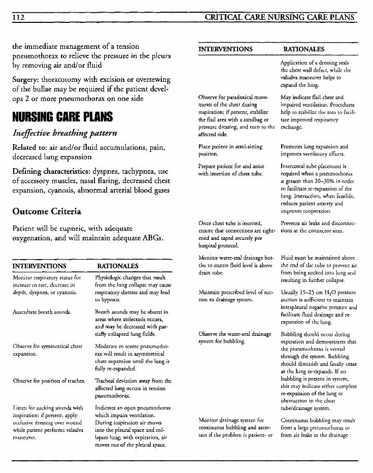
112 CRITICAL CARE NURSING CARE PLANS
the immediate management of a tension pneumothorax to relieve the pressure in the pleura by removing air and/or fluid
Surgery: thoracotomy with excision or oversewing of the bullae may be required if the patient devel- ops 2 or more pneumothorax on one side
NURSINQ CARE PLANS lnefective breathing pattern
Related to: air andlor fluid accumulations, pain, decreased lung expansion
Defining characteristics: dyspnea, tachypnea, use of accessory muscles, nasal flaring, decreased chest expansion, cyanosis, abnormal arterial blood gases
Outcome Criteria
Patient will be eupneic, with adequate oxygenation, and will maintain adequate ABGs.
INTERVENTIONS RATIONALES
Monitor respiratory status for increase in rate, decrease in depth, dyspnea, or cyanosis.
Auscultate breath sounds.
Observe for symmetrical chest expansion.
Observe for position of trachea.
Listen for sucking sounds with inspiration; if present, apply occlusive dressing over wound while patient performs valsalva maneuver.
Physiologic changes that result from the lung collapse may cause respiratory distress and may lead to hypoxia.
Breath sounds may be absent in areas where atelectasis occurs, and may be decreased with par- tially collapsed lung fields.
Moderate to severe pneumotho- rax will result in asymmetrical chest expansion until the lung is fully re-expanded.
Tracheal deviation away from the afTected lung occurs in tension pneumothorax.
Indicates an open pneumothorax which impairs ventilation. During inspiration air moves into the pleural space and col- lapses lung; with expiration, air moves out of the pleural space.
INTERVENTIONS RATIONALES
Observe for paradoxical move- ments of the chest during respiration; if present, stabilize the flail area with a sandbag or pressure dressing, and turn to the affected side.
Place patient in semi-sitting position.
Prepare patient for and assist with insertion of chest tube.
Once chest tube is inserted, ensure that connections are tight- ened and taped securely per hospital protocol.
Monitor water-seal drainage bot- tles to ensure fluid level is above drain tube.
Maintain prescribed level of suc- tion to drainage system.
Observe the water-seal drainage system for bubbling.
Monitor drainage system for continuous bubbling and ascer- tain if the problem is patient- or
Application of a dressing seals the chest wall defect, while the valsalva maneuver helps to expand the lung.
May indicate flail chest and impaired ventilation. Procedures help to stabilize the area to facili- tate improved respiratory exchange.
Promotes lung expansion and improves ventilatory efforts.
Intercostal tube placement is required when a pneurnothorax is greater than 20-30% in order to facilitate re-expansion of the lung. Instruction, when feasible, reduces patient anxiety and improves cooperation.
Prevents air leaks and disconnec- [ions at the connector sites.
Fluid must be maintained above the end of the tube to prevent air from being sucked into lung and resulting in further collapse.
Usually 15-25 cm H,O pressure suction is sufficient to maintain intrapleural negative pressure and facilitate fluid drainage and re- expansion of the lung.
Bubbling should occur during expiration and demonstrates that the pneumothorax is vented through the system. Bubbling should diminish and finally cease as the lung re-expands. If no bubbling is present in system, this may indicate either complete re-expansion of the lung or obstruction in the chest tubeldrainage system.
Continuous bubbling may result from a large pneumothorax or from air leaks in the drainage
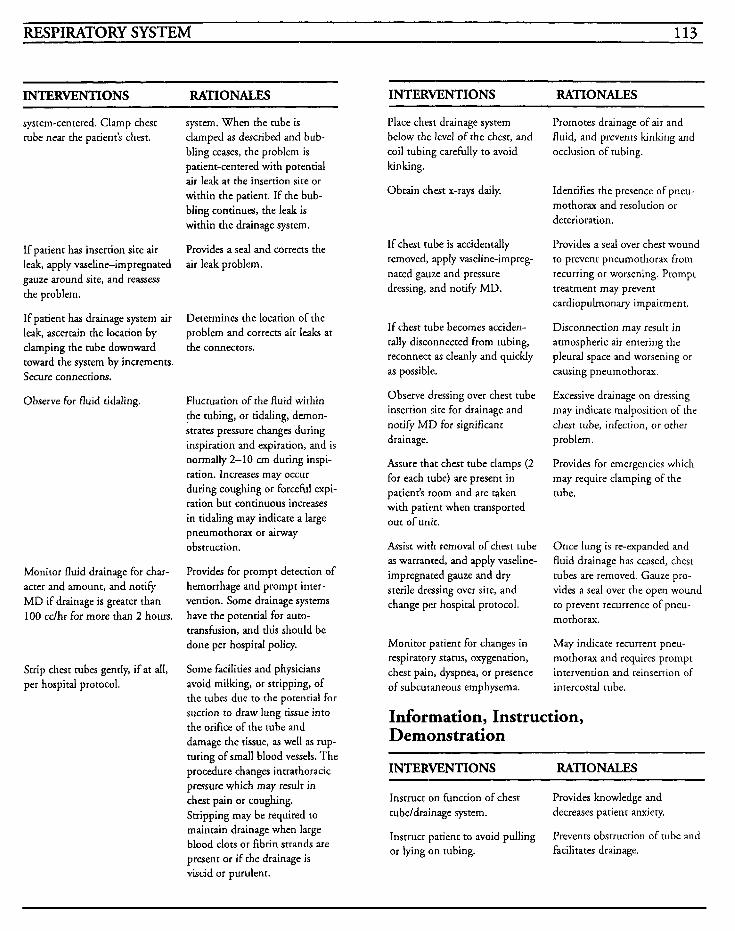
RESPIRATORY SYSTEM 113
~~
INTERVENTIONS RATIONALES
system-centered. Clamp chest tube near the patient's chest.
If patient has insertion site air leak, apply vaseline-impregnated gauze around site, and reassess the problem.
If patient has drainage system air leak, ascertain the location by clamping the tube downward toward the system by increments. Secure connections.
Observe for fluid tidaling.
Monitor fluid drainage for char- acter and amount, and notify MD if drainage is greater than 100 cc/hr for more than 2 hours.
Strip chest tubes gently, if at all, per hospital protocol.
system. When the tube is clamped as described and bub- bling ceases, the problem is patient-centered with potential air leak at the insertion site or within the patient. If the bub- bling continues, the le& is within the drainage system.
Provides a seal and corrects the air leak problem.
Determines the location of the problem and corrects air leaks at the connectors.
Fluctuation of the fluid within !he tubing, or tidaling, demon- strates pressure changes during inspiration and expiration, and is normally 2-10 cm during inspi- ration. Increases may occur during coughing or forceful expi- ration but continuous increases in tidaling may indicate a large pneumothorax or airway obstruction.
Provides for prompt detection of hemorrhage and prompt inter- vention. Some drainage systems have the potential for auto- transfusion, and this should be done per hospital policy.
Some facilities and physicians avoid milking, or stripping, of the tubes due to the potential for suction to draw lung tissue into the orifice of the tube and damage the tissue, as well as rup- turing of small blood vessels. The procedure changes intrathoracic pressure which may result in chest pain or coughing. Stripping may be required to maintain drainage when large blood clots or fibrin strands are
present or if the drainage is viscid or purulent.
INTERVENTIONS RATIONALES
Place chest drainage system below the level of the chest, and coil tubing carefully to avoid kinking.
Obtain chest x-rays daily.
If chest tube is accidentally removed, apply vaseline-impreg- nated gauze and pressure dressing, and notify MD.
If chest tube becomes acciden- tally disconnected from tubing, reconnect as cleanly and quickly as possible.
Observe dressing over chest tube insertion site for drainage and notify MD for significant drainage.
Assure that chest tube clamps (2 for each tube) are present in patient's room and are taken with patient when transported out of unit.
Assist with removal of chest tube as warranted, and apply vaseline- impregnated gauze and dry sterile dressing over site, and change per hospital protocol.
Monitor patient for changes in respiratory status, oxygenation, chest pain, dyspnea, or presence of subcutaneous emphysema.
Promotes drainage of air and fluid, and prevents kinking and occlusion of tubing.
Identifies the presence of pneu- mothorax and resolution or deterioration.
Provides a seal over chest wound to prevent pneumothorax from recurring or worsening. Prompt treatment may prevent cardiopulmonary impairment.
Disconnection may result in atmospheric air entering the pleural space and worsening or causing pneumothorax.
Excessive drainage on dressing may indicate malposition of the chest tube, infection, or other problem.
Provides for emergencies which may require clamping of the tube.
Once lung is re-expanded and fluid drainage has ceased, chest tubes are removed. Gauze pro- vides a seal over the open wound to prevent recurrence of pneu- mothorax.
May indicate recurrent pneu- mothorax and requires prompt intervention and reinsertion of intercostal tube.
Information, Instruction, Demonstration
INTERVENTIONS RATIONALES
Instruct on function of chest Provides knowledge and tubeldrainage system. decreases patient anxiety
Instruct patient to avoid pulling or lying on tubing.
Prevents obstruction of tube and facilitates drainage.
114 CRITICAL CARE NURSING CARE PLANS
INTERVENTIONS RATIONALES
Instruct on signslsymptoms to report to nurse: dyspnea, chest pain, changes in sounds of bub- bling from drainage system.
Promotes prompt recognition of problems that may require prompt intervention.
Discharge or Maintenance Evaluation
Patient will be eupneic with no adventitious breath sounds.
Patient will have symmetrical chest expansion and midline tracheal placement with no episodes of dyspnea.
Patient will achieve and maintain re-expansion of lung with no recurrence or complications.
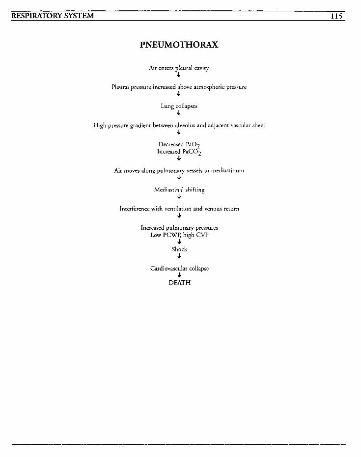
RESPIRATORY SYSTEM 115
PNEUMOTHORAX
Air enters pleural cavity J,
Pleural pressure increased above atmospheric pressure J,
Lung collapses J,
High pressure gradient between alveolus and adjacent vascular sheet 4
Decreased Pa02 Increased PaC02
4 Air moves along pulmonary vessels to mediastinum
4 4
Mediastinal shifting 4
Interference with ventilation and venous return J,
Increased pulmonary pressures Low P C W , high CVP
4 Shock
J,
Cardiovascular collapse 4
DEATH

This Page Intentionally Left Blank
RESPIRATORY SYSTEM 117
Status Asthmatieus Status asthmaticus is a critical emergency that requires prompt intervention to avoid acute and possibly fatal, respiratory failure. In this condition, the asthmatic attacks are unresponsive to medical therapeutics, with severe bronchospasms creating decreased oxygenation and perfusion.
During an acute asthmatic attack, the individual may demonstrate varying degrees of respiratory distress depending on the duration of the attack, and the severity of spasm. The underlying cause of asthma is still as yet unknown, but is thought to be due to imbalances in adrenergic and cholinergic control of the airways, and their response to the allergens, infections, or emotional factors with which they come in contact. Intrinsic asthma occurs when the triggering factors are irritation, infection, or emotions, and extrinsic asthma occurs when precipitated by allergic or complement-mediated factors. Asthma may be drug-induced by aspirin, indomethacin, tartrazine, propranolol, and timolol.
In asthma, the airways are narrowed due to the bronchial muscle spasms, edema, inflammation of the bronchioles, and thick, tenacious mucous pro- duction. The narrowing leads to areas of obstruction and these become hypoventilated and hypoperfused. Eventually a ventilation/perfusion mismatch occurs and may lead to hypoxemia and an increasing A-a gradient. When PaCO, rises to the point of respiratory acidosis, the patient is then considered to be in respiratory failure.
The three most common causes of status asthmati- cus are allergen exposure, noncompliance with medication regime, and respiratory infection expo- sure. Environmental factors, such as excessively hot, cold, or dusty areas, may initiate status asth- maticus because of the effect they have on the air that is breathed.
Wheezing may occur not only with asthma, but with chronic obstructive pulmonary disease, con- gestive heart failure, pulmonary embolism, and tuberculosis, and these diagnoses should be ruled out.
Patients who have status asthmaticus suffer pro- nounced fatigue due to the continuous efforts of breathing, and they easily become dehydrated due to the hyperpnea. The patient usually has dysp- nea, tachypnea, wheezing, tachycardia, pulsus paradoxus, and severe anxiety. The goals of treat- ment include ventilatory support and maintenance of adequate airways, and the preven- tion of respiratory failure or barotrauma.
Laboratory: CBC and sputum specimens usually show eosinophilia
Chest x-ray: used to observe for infiltrates or hyperinflation to the lungs; may be used to visual- ize pneumothorax, hemothorax, or pneumomedias tinum
Arterial blood gases: to identify problems with oxygenation and acid-base balance
Spirometry: to provide information about severity of an attack, and to assess for improvement with therapy; FEV, is the forced expiratory volume for 1 second and is usually < 1500 cc during an asth- matic attack and will increase 500 cc or more if treatment is successful
Oxygen: to provide supplemental available oxygen
Bronchodilators: used to relax bronchial smooth muscle to dilate bronchial tree to facilitate air exchange
Beta-adrenergic agents: ephedrine, epinephrine, isoproterenol, metaproterenol, terbutaline; used to relax bronchial smooth muscle
118 CRITICAL CARE NURSING CARE PLANS
Corticosteroids: used to decrease inflammatory response and decrease edema
Antibiotics: used when infective process is docu- mented; usually bacterial infection is not a common precipitating factor
Mechanical ventilation: necessary when respira- tory failure is present and hypoxemia persists despite medical therapy
INTERVENTIONS RATIONALES
and maintenance of therapeutic levels. Observe patient for anorexia, nausea, vomiting, abdominal pain, nervousness, restlessness, and tachycardia.
Administer sympathomimetics as ordered.
IPPB: used to assist the patient with deep inspira- tion to facilitate more productive coughing of thick mucous and to deliver medication by an aerosol route
Assist/administer inhalation ther- apy as ordered.
Inefective airway clearance
Related to: airway obstruction, edema of bronchi- oles, inability to cough or to cough effectively, excessive mucous production
Defining characteristics: adventitious breath sounds, dyspnea, tachypnea, shallow respirations, cough with or without productivity, cyanosis, anx- iety, restlessness
Outcome Criteria
Patient will maintain patency of airway and will be able to effectively clear secretions.
INTERVENTIONS RATIONALES
Administer bronchodilators as ordered. line treatment for asthma.
Nebulizers are usually the first
Aminophylline is frequently pre- scribed to relax bronchial smooth muscle and mediates histamine release and cAMD degradation, which facilitates improved air flow.
Monitor lab levels for attainment Therapeutic levels range between
10-20 mcg/ml. Symptoms may indicate theophylline toxicity, which will require titration of the drug dosage.
Epinephrine is usually given S Q every 20-30 minutes for 3 doses as needed to relieve broncho- constriction. Terbutaline is usually not the first drug of choice in acute situations due to the delayed onset of action, but is frequently used after the patient shows improvement.
Nebulizers and intermittent posi- tive pressure breathing treatments may be used in mild to moderate episodes but should not be used during acute attacks because of the potential for bron- chospasm in response to the aerosol agent.
Information, Instruction, Demonstration
INTERVENTIONS RATIONALES
Monitor for side effects, such as tachycardias, tremors, nausea, vomiting, or bronchospasm.
May occur as adverse reactions from medications. May require change in specific drug used.
Discharge or Maintenance Evaluation
Patient will maintain patent airway and be able to cough and clear own secretions.
Patient will have clear breath sounds with no adventitious sounds or airway compromise.
Patient will have adequate oxygenation.
Impaired gm excbange [See Mechanical Ventilation]
Related to: bronchospasm, inflammation to bronchi, hypoxemia, fatigue
RESPIRATORY SYSTEM 119
Defining characteristics: dyspnea, tachypnea, hypoxia, hypoxemia, hypercapnia, restlessness, anxiety, abnormal ABGs, dysrhythmias, decreased oxygen saturation
Anxiety [See Mechanical Ventilation]
Related to: dyspnea, change in health status, threat of death
Defining characteristics: fear, restlessness, muscle tension, apprehension, helplessness, sense of impending doom
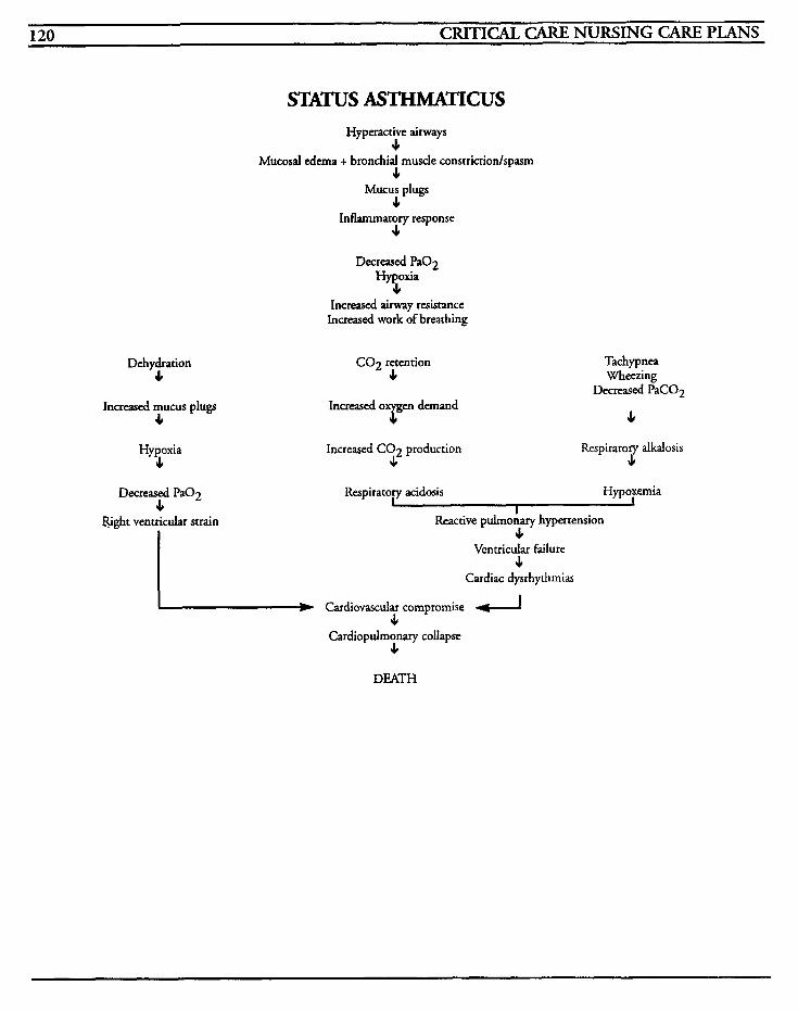
120 CRITICAL CARE NURSING CARE PLANS
STATUS ASTHMATICUS Hyperactive airways
11 Mucosal edema + bronchial musde constrictionhpasm
4 Mucus plugs
4 Inflammatory response
J,
Decreased Pa02 Hypoxia 4
Increased airway resistance Increased work of breathing
Dehydration 4
Increased mucus plugs 4
Hypoxia J,
Decreased PaO2 J,
Bght ventricular strain
CO2 retention 11
Increased CO2 production J,
Tachypnea Wheezing
4 Decreased PaCO2
Respiratory alkalosis s
Respiratory acidosis Hypoxemia I
Reactive pulmonary hypertension 11
Ventricular failure 4
Cardiac dysrhythrnias - Cardiovascular compromise d c Cardiopulmonary collapse
4
DEATH
RESPIRATORY SYSTEM 121
Mechanical Ventilation Mechanical ventilation is used as an artificial adjunct to maintain and optimize ventilation and oxygenation in those patients that are unable to do so on their own for whatever the reason. It is utilized when other adjuncts are ineffective to reg- ulate oxygen and carbon dioxide levels and provide for an adequate acid-base balance.
Major types of ventilators include negative exter- nal pressure and positive pressure ventilators. The external type is very rarely seen today, such as the “iron lung” used for the treatment of polio and the chest ventilator used for home treatment of neuromuscular diseases. These ventilators apply pressure against the thorax that is less than room air, in order to accomplish ventilation by changes in lung pressures. There are no requirements for artificial airways and are fairly easy to use. The patient must remain in or under the unit and, as such, activity is limited, and the negative pressure exerted may result in venous pooling and decreased cardiac output.
Positive pressure ventilators are further subclassi- fied according to the factor that initiates the inspiratory phase, and what factor causes the inspiratory phase to cease. Pressure-cycled ventila- tors use oxygen or compressed air valves to deliver a gas volume until a preset pressure limit is achieved. As the lung compliance and airway resis- tance changes, inspired tidal volumes, alveolar ventilation and FIO, changes also. The alarm sys- tems for this ventilator are sometimes inadequate and the ventilator cannot compensate for leaks that may occur in the system.
Volume-cycled ventilators, currently the most common found in intensive care settings, deliver a preset gas volume to the patient regardless of airway resistance or compliance. Most have safety features to limit excessive airway pressures, and
FIO, and exhaled tidal volumes are more accurate.
High-frequency ventilation is used when other methods have not been successful in oxygenation and ventilation of the patient. It uses lower tidal volumes and increased respiratory rates to decrease the incidence of barotrauma and cardiac decom- pensation. Frequencies range from 60-200 timedmin, and in high-frequency oscillation, movement of air to and from the airway is performed at 600-3000 cycledmin.
PEEC or positive end-expiratory pressure, is used to improve oxygen exchange in persistent hypox- emia when increases in FIO, have not improved the situation. PEEP produces an increased func- tional residual capacity (FRC) which increases the available lung alveoli surface for oxygenation by maintaining the alveoli in an open position. High levels of PEEP may contribute to the incidence of barotrauma and hemodynamic compromise, and is most effective when maintained for lengthy periods of time. To this end, a special PEEP ambu bag must be used to maintain the pressure in order to maintain the beneficial effects.
MEDICAL CARE Laboratory: CBC, transferrin, albumin, prealbu- min, electrolytes used to monitor infection, imbalance, and nutritional status; cultures done to identify infective organism and specify antimicro- bial agent required for eradication
Intubation: artificial airway is required for mechanical ventilation
Arterial blood gases: used to determine levels of oxygen, carbon dioxide, and p H to identify acid- base disturbances, hypoxemia, and to monitor for changes in respiratory status
Respiratory treatments: used to instill varied agents into the lungs to reduce spasm, increase
122 CRITICAL CARE NURSING CARE PLANS
hydration and liquification of secretions, and to facilitate removal of secretions
Ventilatory management: ventilator settings are changed periodically based on patient condition and arterial blood gas analysis to ensure optimum ventilation and oxygenation
Tracheostomy performed when nasal or oral intu- bation is impossible, or after significant time of nasal/oral intubation on a prolonged ventilator patient
NURSING CARE PLANS Inefectiue airway clearance
Related to: thick tenacious secretions, airway obstruction, edema of bronchioles, inability to cough or to cough effectively, presence of artificial airway
Defining characteristics: adventitious breath sounds, dyspnea, tachypnea, shallow respirations, cough with or without productivity, cyanosis, fever, anxiety, restlessness
Outcome Criteria
Patient will maintain patency of airway, have clear breath sounds, and will be able to effectively clear secretions.
INTERVENTIONS RATIONALES
Monitor tube placement for migration; place marking on tube and note length and posi- tion at least every 8 hours; tube should be adequately secured to maintain placement.
Prepare for placement on mechanical ventilation as war- ranted.
INTERVENTIONS RATIONALES
Monitor airway for patency and provide artificial airways as war- ranred. Prepare for mechanical ventilation.
Artificial airways will be required if patient cannot maintain patency. Oropharyngeal airways hold tongue anteriorly but may precipitate vomiting if lengrh is nor accurately measured. Nasopharyngeal airways are more easily tolerated i n conscious patients but may cause nosebleeds and may easily become occluded. Esophageal
Auscultate lung fields for pres- ence of breath sounds, changes in character, and presence to all lobes; observe for symmetrical chest expansion.
obrurator airways are useful only in emergency situations and must be replaced as quickly as possible. These are easier to insert than endotracheal tubes, but stimulate vomiting and cannot be used in conscious patients. The trachea may acci- dentally be intubated and the esophagus may be perforated. Endotracheal intubation requires advanced training and skill, and may be accidentally placed in rhe esophagus, develop leaks that may decrease oxygenation, and over time, may necrotize tissues. Artificial airways may become occluded by mucous, blood, or other secretions; endotracheal tubes may become twisted or compressed, or severe spasms may occlude airway.
Tube migration may occur with coughing, re-taping, or acciden- tally, with the potential for improper placement resulting in hypoxia. Comp’arison of previous placement guidelines will provide prompt recognition of differ- ences and changes, and facilitate prompt intervention.
If routine medical therapeutics are not effective in controlling the spasms, hypoxemia, and hypoxia, respiratory failure will ensue, and mechanical ventila- tion will be required to assure adequate oxygenation and perfusion.
Proper tube placement will result in equal bilateral breath sounds and symmetrical chest expansion. Adventitious breath sounds, such as rhonchi and wheezes, may indicate airflow has been obstructed by occlusion of the tube or migration into an inap- propriate position. Absence of breath sounds to left lung fields
RESPIRATORY SYSTEM 1 3 1
INTERVENTIONS RATIONALES
may indicate intubation of the right main stem bronchus.
Patients who are intubated fre- quently have ineffective cough reflexes or are sedated and have some muscular involvement that may impair coughing, and suc- tioning is required to remove their secretions. Suctioning time should be minimized and hyper- oxygenation performed to reduce the potential for hypoxia.
Promotes maximal lung expan-
Suction patient every 2-4 hours and prn, being sure to hyperoxy- genate patient prior to, during, and after procedure; limit active suctioning to 15 seconds or less at a time; use pulmonary toilette instillation as needed.
Position patient in high-Fowler’s or semi-Fowler’s position. sion.
Turn patient every 2 hours and prn. of pulmonary secretions and
Repositioning promotes drainage
enhances ventilation to decrease potential for atelectasis.
Promotes relaxation of bronchial
spasm, dilates airways to improve ventilation, and maximizes air exchange.
Administer bronchodilators as ordered. smooth muscle to decrease
Information, Instruction, Demonstration
INTERVENTIONS RATIONALES
Patient will have clear breath sounds with no adventitious sounds or airway compromise.
complications. Patient will not have any aspiration
Patient will be able to adequately perform
Impaired gas exchange Related to: bronchospasm, mucous production, edema, inflammation to bronchial tree, hypoxemia, fatigue
Defining characteristics: dyspnea, tachypnea, hypoxia, hypoxemia, hypercapnia, confusion, rest- lessness, cyanosis, inability to move secretions, tachycardia, dysrhythmias, abnormal ABGs, decreased oxygen saturation by oximetry
coughing.
Outcome Criteria
Patient will have arterial blood gases within normal range for patient, with no signs of ventila- tion/perfusion mismatching.
INTERVENTIONS RATIONALES
Instruct on splinting abdomen with pillow during cough efforts.
Promotes increased expiratory pressure and helps to decrease discomfort.
Minimizes fatigue by assisting patient to increase expiratory pressure and facilitates cough.
Instruct on alternative types of coughing exercises, such as quad thrusts, if patient has difficulty during coughing.
Instruct on deep breathing exercises. and decreases anxiety.
Perform chest percussion and postural drainage as warranted.
Promotes full lung expansion
Mobilizes secretions and facili- tates ventilation of all lung fields.
Discharge or Maintenance Evaluation
Patient will maintain patent airway and be able to cough and clear own secretions.
Monitor pulse oximetry for oxygen saturation and notify MD if .c 90.
Oximetry readings of 90 corre- late with PaO, of 60. Levels below 60 do not allow for ade- quate perfusion to tissues and vital organs. Oximetry uses light waves to identify differ- ences between saturation and reduced hemoglobin of the tissues and may be inaccurate in low blood flow states.
Measures the oxygen concentra- tion of the skin, but may cause burns if monitor site 1s nor rotated frequently. Slun, blood flow and temperature may affect these readings.
Monitor transcutaneous oxygen tension if available.
124 CRITICAL CARE NURSING CARE PLANS
INTERVENTIONS RATIONALES INTERSENTIONS RATIONALES
Provide oxygen as ordered.
Monitor for changes in mental status, restlessness, anxiety, headache, confusion, dysrhyth- mias, hypotension, tachycardia, and cyanosis.
Monitor ABGs for changes andlor trends.
Administer oxygen as ordered.
Monitor for signs/symptoms of oxygen toxicity (nausea, vomit- ing, dyspnea, coughing, retrosternal pain, extremity paresthesias, pronounced fatigue, or restlessness).
Limit PEEP (positive end-expira- tory pressures) to 5-20 cm H,O.
Provides supplemental oxygen to benefit patient. Low flow oxy- gen delivery systems use some room air and may be inadequate for patient's needs if their tidal volume is low, respiratory rate is high, or if ventilation status is unstable. Low flow systems should be used in patients with COPD so as to not depress their respiratory drive. High levels of oxygen may cause severe damage to tissues, oxygen toxicity, increases in A-a gradients, microatelectasis, and ARDS.
May indicate impending or pre- sent hypoxia and hypoxemia.
Use PEEP ambu bag when SUC-
tioning patient. despite increasing levels of oxygen by producing an increased functional residual capacity which then increases the available lung alveoli surface for oxygenation. PEEP may predis- pose the patient to barotrauma with elevated levels. Ambu bags that are capable of maintaining PEEP levels are required because short intervals minimize the ben- eficial effects of PEEP.
Information, Instruction, Demonstration
INTERVENTIONS RATIONALES
Provides information on mea- sured levels of oxygen and carbon dioxide as well as acid- base balance. Promotes prompt intervention for deteriorating airway status. PaO, alone does not reflect tissue oxygenation; ventilation must be adequate to provide gas exchange.
Oxygen by itself may not always correct hypoxia of tissues and restore perfusion.
Oxygen toxicity may result when oxygen concentrations are greater than 40% for lengthy durations of time, usually 8 to 24 hours, and may cause actual physiologic changes in the lungs. Progressive respiratory distress, cyanosis, and asphyxia are late signs of toxicity. Oxygen concentrations should be maintained as low as possible in order to maintain adequate PaO,.
PEEP is used to improve oxygen exchange in persistent hypoxemia
Prepare patient for placement on mechanical ventilation as war- ranted. base balance.
Assist with respiratory therapists measurements of oxygen analyzing, lung compli- ance, viral capacity, and A-a gradients.
May be necessary to maintain adequate oxygenation and acid-
Provides information to
facilitate early detection of oxygen toxicity.
Discharge or Maintenance Evaluation
Patient will have arterial blood gases within normal limits for patient.
Patient will be eupneic with adequate oxygena- tion and no signslsymptoms of oxygen toxicity.
Inefective breathing pattern
Related to: fatigue, dyspnea, secretions, inadequate oxygenation, respiratory muscle weak- ness, respiratory center depression, decreased lung expansion, placement on mechanical ventilation
Defining characteristics: dyspnea, tachypnea, bradypnea, apnea, cough, nasal flaring, cyanosis, shallow respirations, pursed-lip breathing, changes in inspiratorylexpiratory ratio, use of accessory muscles, diminished chest expansion, barrel chest, abnormal arterial blood gases, fremitus, anxiety, decreased oxygen saturation
RESPIRATORY SYSTEM 125
Outcome Criteria INTERWNTIONS RATIONALES
Patient will be eupneic, with adequate oxygenation, and will maintain adequate ABGs within normal limits.
INTERVENTIONS RATIONALES Prepare patient for placement on mechanical ventilation and intu- barion procedures.
Assist with intubation of patient; auscultate all lung fields for breath sounds.
Hyperoxygenate patient and aus- cultate for bilateral breath sounds and observe for bilateral symmet- rical chest expansion.
Utilize low pressure endotracheal tubes for intubation.
Maintain airway; secure tube with tape or other securing device.
Obtain chest x-ray afrer ETT is inserted.
Promotes knowledge and reduces fear. May promote cooperation.
Placement of an artificial airway (endotracheal tube [ETT] or tracheostomy) is required for mechanical ventilation support. Nasotracheal intubation may be preferred to prevent oral discom- fort and necrosis, but is associated with a high incidence of sinus disease.
Prolonged difficulty in place- ment of the tube may result in hypoxia. If symmetrical chest expansion is not observed, or if breath sounds cannot be heard bilaterally, this may indicate improper placement of the tube into the right main bronchus or esophagus, and correction of this problem must be addressed promptly.
High pressure cuffed tubes may promote tracheal necrosis or result in a tracheal fistula.
Artificial airways may become occluded by mucous or other secretory fluids, may develop a cuff leak resulting in inability to maintain pressures suficient for ventilation, or may migrate to a position whereby adequate oxy- genation is impaired. Tubes should be adequately secured to prevent movement, loss of airway, and tracheal damage.
Radiographic confirmation of tube placement is mandatory;
If E T T is placed orally, daily changes from side to side of mouth should be routinely performed.
Suction patient as needed, making sure to hyperoxygenate before, during, and after proce- dure. Utilize sterile normal saline for pulmonary toilette instilla- tion prior to suctioning procedures as warrantedlordered.
Restrain patient as warranted and as per hospital protocol.
Monitor ventilator settings at least every 2-4 hours and prn; FIO, should be analyzed periodi- cally to ensure correct amount is being maintained; tidal volume should ideally be 10-15 cc/Kg body weight; airway pressures (peak inspiratory pressure and plateau pressure) should be noted for identification of trends; inspi- ratory and expiratory ratio; sigh volume and rate.
the tube should be 2-3 cm above the carina.
Prevents tissue necrosis from pressure of tube against teeth, lips, and other tissues. Oral tubes promote saliva formation, cause nausea and vomiting if rnove- ment of tube stimulates retching, and prevents the patient from closing his mouth without biting down on the tube.
Suctioning is required to remove secretions because the patient is unable to do so on his own. Effective coughing is decreased because of the inability to increase intrathoracic pressure when the glortis is restricted from air. Suctioning places patient at risk for inadequate oxygenation and decreased perfu- sion. Hyper-oxygenation helps to limit this sudden decrease in available oxygen. Mucous pro- duction is usually increased with placement of E T T due to ciliary movement being impaired and the body's response to the foreign tube. Pulmonary toilette is con- troversial but may be helpful to liquefy secretions to facilitate easier removal.
Prevents accidental extubation in sedated or confused patients.
Ventilator settings are adjusted based on the disease process and patient's condition to maintain optimal oxygenation and ventila- tion while the patient is unable to do so on his own. Oxygen percentages may not be corn- pletely accurate and analysis must be performed to ensure proper amounts arc being deliv- ered. Exhaled tidal volumes should be monitored and changes may indicate changes in lung compliance or problems with delivering specific volumes.
126 CRITICAL CARE NURSING CARE PLANS
INTERVENTIONS RATIONALES INTERVENTIONS RATIONALES
Increases in airway pressures may indicate bronchospasm, presence of mucoid and other secretions, obstruction of the airway, pneu- mothorax, or ARDS with high pressure levels and disconnection of tubing, inadequate cuff pres- sure or non-synchronous breathing with low pressure levels. I:E ratio should be 1:2 but may be altered to improve gas exchange. Sighs, when used, are commonly 1 112 times the volume of a normal breath, 2-6 times per hour to facilitate expansion of alveoli to reduce the potential for atelectasis. Ventilator settings may be inad- vertently changed, or due to forgetfulness, increased oxygena- tion used for suctioning procedure may not be turned down to ordered amounts. This may result in oxygen toxicity or inadequate ventilation.
Observe for temperature of ven- Intubation bypasses the body’s tilator circuitry; drain tubing natural warminglhumidifying away from the patient as action, and requires increased warranted. temperature and moisturizing
of the delivered oxygen. The temperature of the ventilator circuitry (and the delivered oxygen) should be maintained at approximately body temperature to avoid hyperthermic reactions. Temperature increases and humidification promote conden- sation of water in tubing which may restrict adequate volume delivery. Drainage of fluid toward the patient or toward the reservoir may promote bacterial infestations.
Proper cuff inflation is done with the least amount of air to ensure a minimal leak with maintenance of adequate ventilatory pressures and tidal volumes. Cuff pressures should be less than 25 cm H,O to prevent tracheal necrosis.
Monitor airway cuff for leakage, noting amount of air volume in cuff and cuff pressures at least every 4-8 hours and prn.
Auscultate for adventitious breath sounds, subcutaneous emphysema, or localized whea- ing.
Monitor ABGs for trends, and change ventilator settings as ordered.
Observe breathing patterns and note if patient has spontaneous breaths in addition to ventilatory breaths.
Observe patient for non-synchro- nous respirations with ventilator (“fighting the ventilator”). Administer sedation or sedationlneuromuscular block- ade, as ordered.
Increasing volumes of air required to maintain ventilatory pressures, or increasing cuff pres- sures may indicate cuff leak and will require replacement of airway to maintain oxygenation.
May indicate migration of airway tube. Movement from trachea into tissue may cause mediastinal or subcutaneous emphysema and/or pneumotho- rax. Intubation of the bronchus may result in decreased unilateral chest expansion with decreased breath sounds, and localized wheezing. Movement of the tube to the level of the carina may result in excessive coughing, diminished breath sounds, and inability to insert suction catheter.
Maintains adequate oxygenation and acid-base balance.
Increased or decreased ventila- tion may be experienced by ventilator patients who may try to compensate by competing wirh ventilatory breaths. Tachypnea may result in respira- tory alkalosis; bradypnea may result in acidosis with increased PaC02.
Asynchrony with the ventilator decreases alveolar ventilation, increases intrathoracic pressures, and decreases venous return and cardiac output. Pavulon paralyzes all muscles in body to facilitate synchrony with ventilation sup- port. Patients may be completely alert when paralyzed, so sedation is MANDATORY prior to administration of Pavulon. Often, a sedation cocktail of narcotics andlor benzodiazepines may be titrated wirh better results to achieve adequate seda- tion.
RESPIRATORY SYSTEM - 127
Information, Instruction, Demonstration
~
INTERVENTIONS RATIONALES
Prepare patient for placement of rracheostomy as warranted.
Observe for pulsarion of tracheostomy with neck vein pulsation and notify MD.
Assess for cuff l e h g e and changelnorify MD for change of airway.
Obtain chest x-rays every day and prn while patient is intubated.
Insure that neostigmine bromide or edrophonium chloride is available.
Prolonged ventilatory support via nasal or oral endotracheal tube may lead to necrosis of tissues due to pressure exerted by the tube. Tracheostomy is more com- fortable for the patient, decreases the airway resistance, and may reduce the amount of dead space.
May indicate close proximity to innominate vessels that may lead to necrosis and erosion into ves- sels and result in hemorrhage.
Cuffs which have leaks that enable a patient to have the ability to speak, in which air may be felt at the nose andlor mouth, changing pressures with ventila- tion, andfor decreased exhaled volumes require change in order to maintain adequate oxygena- tion and ventilation.
Facilitates recognition of tube migration, atelectatic changes, presence of pneumothorax, or other significant changes.
These reverse effects of pancuroniurn.
Discharge or Maintenance Evaluation
Patient will be able to maintain own airway and expectorate sputum.
Patient will have arterial blood gases within normal limits of patient disease process.
Patient will be eupneic with no adventitious breath sounds.
Patient will have artificial airway intact with no signs/symptoms of complications.
Impaired verbal communication
Related to: intubation, artificial airway, muscular paralysis
Defining characteristics: inability to speak, inabil- ity to communicate needs, inability to make sounds
Outcome Criteria
Patient will achieve a method to communicate his needs.
INTERVENTIONS RATIONALES
Evaluate patient’s ability to speak or communicate by other means.
Ensure that call lighr is placed within easy reach of patient at all times, and that the light system is flagged to denote patient‘s impairment.
Make eye contact with patient at all times; ask questions that may be answered by nodding of the head; provide paper and writing utensils, magic slate, or communication board for communication.
Patient may be fluent in sign language, or able to communi- cate in wriring to make needs known.
Provides patient with concrete evidence that he may call for assistance and that the nurse will be available to meet his needs. Flagging system ensures that personnel not familiar with the patient will be alerted ro his inability to speak.
Communication may be possible if patient is able to nod head yes or no, or blink eyes in sequence. Writing may be illegible due to disease process or sedation, and may frustrate and farigue patient.
Information, Instruction, Demonstration
INTERVENTIONS RATIONALES ~ ~~
Instruct patient in using tongue to make clicking noise, or in tapping table or side rails to gain nurse’s attention as a secondary means of calling for assistance.
Provides alternate merhod to communicate with nurse and helps to allay fear of abandon- menr.
128 CRITICAL CARE NURSING CARE PLANS
INTERVENTIONS RATIONALES INTERVENTIONS RATIONALES ~~~ ~~ ~~
Instruct family members in talk- ing with patient to provide information about issues of con- cern to patient, and help them to deal with the awkwardness of a one-sided conversation.
Promotes understanding for the family and assists in incorpor- ating family into the patient's care to mainrain contact with reality.
Discharge or Maintenance Evaluation
Patient will be able to speak or make needs known.
Patient will develop an adequate alternative means of communication and be able to utilize communication to make needs known.
Patient's family will be able to recognize their own contribution to the patient's recovery.
Anxiety
Related to: ventilatory support, threat of death, change in health status, change in environment, life-threatening crises
Defining characteristics: fear, restlessness, muscle tension, apprehension, helplessness, communica- tion of uncertainty, sense of impending doom, worry
Outcome Criteria
Patient will have decreased anxiety and be able to function at acceptable levels with anxiety-produc- ing stimuli.
INTERVENTIONS EWTIONALES
Evaluate patient's perception of crisis or threat to self.
Monitor for changes in vital signs, restlessness, or facial tension.
Encourage patient to express fears and concerns and provide information pertinent to those
~ ~~
Identifies problem base and facil- itates plan for intervention.
May indicate patient's level of response to stressors and level of anxiety.
Promotes verbalization of con- cerns, and allows time for identification of fears to progres-
concerns. Do not give false reassurance.
sively begin work on emotional barriers. False reassurance tends to minimize patient's feelings resulting in impaired trust and increased anxiety.
Provide support and encourage- menr to family members and assist them in dealing with their own fearslconcerns.
Discuss safety precautions involved with ventilatory sup- port; emergency power source, emergency oxygen and equip- ment, alarm systems, etc.
Ensure that patient's call light is placed within easy reach at all times, and that alternative meth- ods of summoning assistance have been discussed.
~
Information, Instruction, Demonstration
Administer antianxiery medica- tions as ordered.
Family's anxiety may be commu- nicated to the patient and result in increased anxiery levels.
Provides concrete answers to help decrease anxiety and fear of the unknown, and to relay emer- gency plans for patient.
Provides reassurance that nurses will be available to assist with patient's needs, and decreases anxiety.
Helps to reduce anxiety to a manageable level when other techniques have failed.
INTERVENTIONS RATIONALES
Instruct in use of relaxation tech- niques and guided imagery.
Consult psychiatrist, psycholo- gist, or counselor as warranted.
Promotes reduction in stress and anxiety, and provides opportu- nity for patient to control his situation.
Patient may require further inter- vention for dealing with emotional problems.
Discharge or Maintenance Evaluation
Patient will be able to verbalize concerns and fears and be able to rationally deal with them appropriate ways.
Patient will be able to function with anxiety reduced at a manageable level.
in
RESPIRATORY SYSTEM 129
Patient will be able to utilize methods to reduce
Inefective individ~ul...amiiy coping
Related to: change in health status, change in abil- ity to communicate, sensory overload, change in environment, fear of death, physical limitations, inadequate support system, inadequate coping mechanisms, threat to self, pain
Defining characteristics: inability to meet role expectations, inability to meet basic needs, worry, apprehension, fear, inability to problem solve, hos- tility, aggression, inappropriate defense mechanisms, low self-esteem, insomnia, depression, destructive behaviors, vacillation when choices are required, delayed decision making, muscle tension, headaches, pain
anxiety.
Outcome Criteria
Patient will be able to recognize problems with coping and be able to problem-solve adequately.
INTERVENTIONS RATIONALES
Evaluate patient’dfamily’s coping Provides baseline information to skills and ability to verbalize establish interventions best suited problems. to the patient/family/situation.
Coping abilities that the patient has utilized previously may be used in the current crisis to pro- vide a sense of control.
Discuss concerns and fears of loss Identifies needs for intervention of control with patient, and pro- and helps to establish a trusting vide feedback. relationship.
Monitor for dependence on others, inability to make deci- sions, inability to involve self in care, or inability to express con- cerndquestions.
May indicate patient’s need to depend on others to allow time to regain ability for coping with crises, and promotes feeling of safety. Patient may be afraid to make any decision in which his tenuous condition could be com- promised.
~~
INTERVENTIONS RATIONALES ~~
Provide opportunities for patient to make decisions regarding his care, when feasible.
Provides opportunity to gain some sense of control of his life, decreasing anxiety, and assisting in coping skills.
Identifies actual problems and assists patiendfamily ro find real solutions to facilitate increasing self-control and self-esteem.
Blaming oneself or others pro- longs inability to cope and increases feelings of hopelessness.
Anger and hostile feelings may promote resolution of stages of grief and loss, and should be regarded as an important step in that process. Limits must be set to prevent destructive behavior that will impair patienr’s self- esteem.
Spiritual beliefs are questioned when threats of death occur, and may affect patient’s ability to cope with and problem-solve during crises.
Discuss current problems and assist with problem-solving to find solutions.
Discuss feelings of blame, either on self, or on others.
Remain non-judgmental of choices patiendfamily may make. Adopt a non-threatened attitude when anger and hostility are expressed. Set limits on unac- ceptable behaviors.
Discuss feelings of anger at God, religious alienation, lack of meaning to life, etc.
Information, Instruction, Demonstration
INTERVENTIONS RATIONALES
Assess rapport of family members with patient. Involve the family members in the care of the patient when feasible.
Actions of the family may be helpful, but the patienr may per- ceive these as being over-prorective or smothering. Helping with patient’s care may enhance the family’s feelings of importance and control of the situation.
Identifies opportunities for other resources that may be available, and provides means of control over situation.
Provide information to the patient and family regarding other agencies and personnel who may assist them with their crisis.
130 CRITICAL CARE NURSING CARE PLANS
Discharge or Maintenance Evaluation INTERVENTIONS RATIONALES
Patient/family will be able to recognize ineffec- tive coping behavior and regain emotional equilibrium.
Patient/family will be able to adequately prob- lem-solve during crises.
Patiendfamily will be able to recognize options and resources for use post-hospitalization.
Patient/family will be able to make appropriate, informed decisions and be satisfied with choices.
PO ten t i d fo r infection
Related to: intubation, disease process, immuno- suppression, compromised defense mechanisms
Defining characteristics: increased temperature, chills, elevated white blood cell count, purulent sputum
Outcome Criteria
Patient will have no evidence of infective process.
INTERVENTIONS RATIONALES
Evaluate risk factors that would predispose patient to infection.
Monitor sputum for changes in characteristics and color; culture sputum as warranted.
Monitor trachcostomy site for redness, foul odor, or purulent drainage; culture site as warranted.
Intubation and prolonged mechanical ventilation predis- pose patient to nosocomial infection. Age, nutritional status, chronic disease progression and invasive procedures and lines also predispose patient to infection.
Purulent, malodorous sputum indicates infection. Cultures may be required to identify causative organism and to prescribe appro- priate antibiotics.
Purulenr drainage indicates infec- tion. Cultures may be required to identiFy causative organism and to prescribe appropriate antibi- otics.
Maintain good handwashing technique and isolation precau- tions when warranted.
Handwashing is the most important step in preventing nosocomial infection. Patients may require isolation based on their diagnosis to prevenr trans- mission of infection to or from the patient.
Patients are already immuno- compromised and at risk for development of infection.
Reduces spread of infection.
Screen visitors who are ill themselves.
Maintain sterile technique for all dressing changes and suctioning.
Administer antibiotics as ordered. ism.
Required to treat infective organ-
Information, Instruction, Demonstration
INTERVENTIONS RATIONALES
Instruct patientlfamily in proper handwashing and disposal of contaminated secretions, tissues, etc.
Instruct family to avoid visiting if they have upper respiratory infections. infection.
Reduces risk of transmission of infection to others.
Patient is already immuno-com- promised and is at risk for
Instruct patientlfamily on antibi- otics: effects, side effects, contraindications, and treatment. foodsldrugs to avoid.
Provides knowledge and enhances cooperation with
Discharge or Maintenance Evaluation
Patient will be free of fever, chills, purulent drainage, or other indicators of infective process.
Patient will be able to recall information accu- rately regarding antibiotics and infection control procedures.
Patiendfamily will be able to recognize risk fac- tors and avoid further compromise of patient.
RESPIRATORY SYSTEM 131
Altered oral mucous membrane
Related to: oral intubation, increased or decreased saliva, inability to swallow, antibiotic-induced fungal infection
Defining characteristics: oral pain or discomfort, stomatitis, oral lesions, thrush
Outcome Criteria
Patient will be free of oral pain and mucous mem- branes will remain intact.
INTERVENTIONS RATIONALES
0 bserve mouth for missing, loose, or chipped teeth; bleeding, sores, lesions, necrotic areas, or reddened areas.
Move oral endotrached tube to other side of mouth at least daily and prn.
Provide oral care at least every 8 hours and prn.
Swab mouth with mouthwash every 4-8 hours and prn.
Apply lip balm every 2-4 hours and pm.
Suction patient’s ord cavity fre- quently if patient is unable to handle secretions.
Observe for white patches on tongue and mucous membranes, and notify MD.
Teeth may be chipped or knocked out during intubation process and loose teeth may pose a potential for aspiration. Identification of lesions or other problems may facilitate prompt intervention.
Decreases potentid for pressure and ultimately, ulceration of lips or mucous membranes.
Promotes cleanliness, reduces odor, and reduces potential envi- ronment for bacterial invasion.
Removes transient bacteria, reduces odor, and helps to stimulate circulation to oral membranes.
Prevents drying and cracking of lips.
Removes excessive saliva and mucous which may facilitate bacterial growth.
May indicate presence of fungal infection (thrush) which will require anti-fungal solution, such as Nystatin.
Information, Instruction, Demonstration
INTERVENTIONS RATIONALES
Instruct on antifungals as warranted.
Instruct patient in utilizing oral suction equipment when feasible, if patient has copious oral secretions.
Provides knowledge.
Provides a sense of control CO the patient, and facilitates removal of excessive secretions.
Discharge or Maintenance Evaluation
Patient will have intact oral mucous membranes, with no evidence of infection.
Patient will be able to recall instructions accu- rately.
Patient will be able to adequately remove secre- tions by use of suction equipment.
Patient will be compliant with performance of oral care.
Altered nutrition: less than body requirements
Related to: intubation, inability to swallow, inabil- ity to take in food, increased metabolism due to disease process, surgery, decreased level of consciousness
Defining characteristics: actual inadequate food intake, altered taste, altered smell sensation, weight loss, anorexia, absent bowel sounds, decreased peristalsis, muscle mass loss, decreased muscle tone, changes in bowel habits, nausea, vomiting, abdominal distention
Outcome Criteria
Patient will have adequate nutritional intake with no weight or muscle mass loss.
132 CRITICAL CARE NURSING CARE PLANS
INTERVENTIONS RATIONALES
Evaluate ability to eat. Some patients with tracheostomies are able to eat, while those patients who are endouacheally intubated must be kept NPO due to the posi- tioning of the epiglottis, and will require enteral or parenteral alimentation.
Weigh every day.
Observe for muscle wasting.
Continued weight loss will result in catabolic metabolism and impaired respiratory function.
May indicate muscle stores depletion which can impair res- piratory muscle function.
Observe for nausea, vomiting, abdominal distention and palpa- bility, and stool characteristics.
Ventilator patients may develop GI dysfunction from analgesicslsedatives, bedrest, trapped air, and stress, which may result in ileus formation.
Stressors of ventilation and pres- ence in ICU may predispose patient to the formation of a stress ulcer resulting in GI bleed- ing.
Establishes imbalances between actual nutritional intake and metabolic needs.
Evaluates need for andlor ade- quacy of nutritional support.
Test stools and gastric contents for guaiac.
Obtain calorie count and assess- ment of metabolic demands based on disease process.
Monitor lab work as warranted; electrolytes, BUN, creatinine, albumin and prealbumin, glucose levels.
Administer enteral solutions at continual rate by infusion pump as warranted.
Bolus feedings may result in dumping syndrome. Continuous infusion feedings are generally better tolerated and have better absorption. Enteral feeding for- mulas vary depending on the nutritional needs of the patient. The use of enteral formulas require a functioning GI system.
Oral or nasal tubes may migrate with coughing, resulting in improper placement and poten- tial for aspiration. Flushing of
Determine patency of enteral feeding tubes at least every 8 hours. Flush with 20-30 cc of
INTERVENTIONS RATIONALES
water every 8 hours, before and after medication administration via the tube, and prn.
Aspirate gastric residuals every 4- 8 hours, and decrease or hold feedings per hospital protocol.
tube maintains patency.
Increasing residuals may indicate decreased or absent peristalsis and lack of absorption of required nutrients which may require another form of nutri- tional support.
Helps to identifj aspiration of feedings when suctioned. Be aware that the food coloring may cause false readings on occult blood tests on stools. Red color- ing should be avoided due to similarity of blood color and this may impair ability to differenti- ate bleeding problems.
Helps to dissolve clogged partic- date matter to maintain patency
Use food coloring to tint feed- ings. Do not use red coloring.
Instill warm cranberry juice, carbonated cola, or mixture of monosodium glutamate and of tube. water in enteral tube for signs of occlusion.
Administer antidiarrheal medica- tions as warranted.
Osmolality imbalances may result in diarrhea requiring antidiarrheds for control. Changing strengths or types of feedings may be helpful.
Medication helps to stimulate gastric motility and may be help- ful to increase absorption.
Provides complete nutritional support without dependence on GI function for absorption. Additives are based upon lab work and patient requirements. Increases in protein and nitrogen may be prescribed for increased metabolic demands of the patient.
Provides additional caloric bene- fits as well as a source of essential fatty acids. Lipids may be uti- lized for respiratory failure to help decrease CO, retention.
Administer metodopramide as ordered.
Administer parenteral alimenta- tion fluids as warranted via infusion pump.
Administer intralipids as ordered, if not admixed with TPN soh- tion.
~
RESPIRATORY SYSTEM 133
INTERVENTIONS RATIONALES
Change solution at least every 24 hours, as well as tubing.
Monitor lab work per hospital protocol; general chemistry, renal profile, CBC, urine or blood glucose levels.
Do not stop TPN abruptly; taper over several dayslhours per protocol.
Some additives may be unstable after 24 hours, and prolonged infusion with same solution may promote bacterial growth.
Requirements for electrolyte replacement or alteration in for- mula may be changed based on this information. High dextrose content in TPN solutions may require additions of insulin to meet metabolic demands if pan- creatic disease, hepatic disease, or diabetes are present.
Rebound hypoglycemia may result if dextrose concentrations are abruptly changed.
Information, Instruction, Demonstration
INTERVENTIONS RATIONALES
Insert nasogastric feeding tube as warranted, utilizing small weighted tube. Obtain chest x-ray or KUB post procedure.
Maintain elevation of the head of the bed at least 30 degrees at all times.
Assist with placement of central venous catheter for TPN admin- istration. Obtain chest x-ray post procedure.
Instruct in need for supplemental nutritional support, procedures to be performed, and tests that will be required.
Smaller lumen is less irritating to nasal mucosa, and decreases the incidence of gastroesophageal reflux. Radiographic confirmation of placement is necessary due to the potential for aspiration when patients may have impaired gag reflex.
Helps prevent potential aspiration.
Centrally-placed intravenous lines may enable higher concentrations of amino acids to be utilized. Radiographic confir- mation of placement, as well as ruling out hemo- or pneumotho- rax post procedure, is mandatory.
Promotes knowledge, decreases fear of the unknown, and facili- tates cooperation with procedures. Provides opportunity for patient to make informed choices.
Discharge or Maintenance Evaluation
Patient will maintain baseline weight with no loss of muscle mass.
Patient will maintain adequate nutritional status with use of nutritional support, and will experi- ence no complications from support.
Patient will show no signs of malnutritional status.
Patient will be able to recall information accu- rately.
Patient will maintain a normal nitrogen balance
Dysfinctionud ventihtory wean response
Related to: fever, pain, muscle fatigue, sedation, anemia, electrolyte imbalance, sleep deprivation, poor nutrition, cardiovascular lability, psychologi- cal instability
Defining characteristics: inability to wean, lack or inadequacy of spontaneous respirations, negative inspiratory force or pressure < -20 cm H,O, PaO, < 60 mmHg on FIO, > 50%, PaCO, > 40 mmHg, tidal volume < 5 cc/Kg, vital capacity < 10 cc/Kg, minute ventilation > 10 L/min
and immunity will not be compromised.
Outcome Criteria
Patient will be able to be weaned from ventilatory support successfully with arterial blood gases within normal limits.
INTERVENTIONS RATIONALES
Monitor vital signs. Temperature elevations increase metabolism and oxygen demand. Unstable heart rate and rhythm results in increased workload on the heart, increased oxygen con- sumption and demand. Process of weaning will increase work-
134 CRITICAL CARE NURSING CARE PLANS
INTERVENTIONS RATIONALES
load and may compromise an already-stressed body and should not be attempted until these factors have been corrected. Once weaning process has begun, significant changes in heart rate and rhythm, respira- tory rare, and blood pressure may indicate a need to slow or discontinue weaning due to res- piratory compromise.
Monitor EKG for dysrhythmias and treat as warranted.
Ventilatory support decreases venous return to the heart, increases PVR and SVR. Hypoxemia and pH imbalances may result in dysrhythmias from cardiac compromise.
Monitor nutritional status. Evaluate labwork: CBC, transfer- rin, albumin, prealbumin, electrolytes, etc.
Protein, carbohydrate and fat concentrations can alter the abil- ity to maintain oxygenation. Increased fat concentration prior to weaning may assist in decreas- ing potential for CO, retention and decrease in respiratory drive. Labwork may be used to verify adequacy of nutritive state. Calcium imbalances can decrease the function of the diaphragm, and phosphorus may affect 2, 3- DPG and ATP function, affect respiratory muscle function and red cell membrane stability.
Stay with patient until stable, once weaning process has begun. Observe for use of accessory muscles, non-synchronous respi- ratory pattern, or skin color changes.
Respiratory deterioration may occur rapidly and physical pres- ence is required to observe patient to facilitate prompt inter- vention. May indicate deterioration in respiratory status, resulting in inability to wean.
Monitor oxygen saturation per oximetry and notify MD if read- ing less than 90% per pulse oximetry, or sustained reading less than 60% per mixed venous oxygen oximetry; obtain ABGs per protocol.
Oximetry provides identification of tissue oxygen desaturation which usually coincides with decreases in arterial blood gases. Oximetry does not give indica- tion of increased CO, levels and these must be verified with ABGs.
INTERVENTIONS RATIONALES
Attempt to wean only during the day and after the patient has had a resthl sleep period. Avoid activity during weaning.
Crises that may occur with respiratory deterioration and failure to wean may be handled more efficiently when sufficient medical personnel are available, usually during the day. Fatigue may predispose patient to failure due to the need for stamina to withstand the effort of sponta- neous breathing. Activity increases oxygen demand and consumption.
Evaluate patient’s emotional status and ability to cope with weaning.
Prior to attempt, assess weaning parameters to ensure patient meets requirements for successful weaning: NIF > -20 cm H,O, vital capacity > 10-15 cclKg, PaO, > 60 mmHg on FIO, < 40%, resting minute ventilation < 10 L/min, PaCO, c 40 mmHg, tidal volume > 5 cdKg.
Assess patient for resolution of disease process, absence of inspi- ratory muscle fatigue, absence of fever, absence of hemodynamic instability, absence of sedative agents or respiratory suppres- sanrs, presence of spontaneous respirations, pulmonary shunt < 20%, and adequate hemoglobin and hematocrit.
Suction patient and perform chest physiotherapy, percussion and postural drainage as war- ranted prior to disconnection from ventilator.
Utilize T-bar/T-piece adaptor as ordered. (Usually on T-bar for 10-30 minutes per hour initially.)
Utilize SIMV/IMV mode on ventilator as ordered. (Usually rate decreased by 1-2 breathslminute every 15-30 minutes.)
Weaning process may result in anxiety due to fear of failure to wean andlor ability to breathe spontaneously.
Attainment of parameters facili- tate best chance for successful weaning and ensures that neuro- muscular control is adequate for maintenance of spontaneous ventilation. If carbon dioxide retention is chronic, pH is more indicative of weaning readiness.
Factors may promote respiratory insufficiency and compromise which may result in unsuccessful weaning.
Removes secretions that may compromise weaning process and promotes improved pulmonary conditions.
Provides oxygen via endotracheal tube or tracheostomy with patient spontaneously breathing.
Provides ventilatory support to patient with gradually decreasing ventilator breaths and increase of spontaneous breaths. Facilitates gradually increasing respiratory
RESPIRATORY SYSTEM 135
INTERVENTIONS RATIONALES INTERVENTIONS RATIONALES
Utilize PS (pressure support) as ordered. (Usually 3-5 initially and may increase to 20, with gradual lowering as IMV/SIMV rate lowered.)
Utilize CPAP (continuous posi- tive airway pressure) as ordered. (Usually 2-5 cm H,O.)
Monitor for MD-set parameters or respiratory rate > 30, increas- ing PA pressures, heart rate > 110 with new or increased ectopic activity, blood pressure > 20 mmHg from baseline, SaO, < 90%, tidal volumes c
250 cc; if significant changes occur, place back on ventilator as per protocol.
Gradually increase time off venti- lator with each successful attempt. Once patient is able to tolerate 1-2 hours off ofventila- tor at a time, weaning may be advanced more rapidly.
Determine patient's emotional sratus and ability to cope with weaning process.
Extubate patient when he is able to maintain an airway and his spontaneous respirations are able to maintain oxygenation and ventilatory status per protocol. Intubation equipment should remain at the bedside post-extu- bation for 4-24 hours or per protocol.
To extubate, increase oxygen and suction secretions from trachea, nose and mouth.
Deflate cuff and remove tube at
workload. If weaning is not tolerated, may increase PaCO, and decrease pH.
Assists patient to overcome airway resistance and support spontaneous breathing by increasing respiratory muscle function.
Patient exhales against continu- ous positive pressure to prevent atelectasis and improve arterial oxygen tension.
Alterations in vital signs and hemodynamic may result from insufficient ventilation and respi- ratory compromise and indicates intolerance of attempts to wean.
The patient's progress will increase as fatigue decreases, respiratory muscle function improves, and patient is emo- tionally ready to wean.
Weaning may result in excessive anxiety due to fear of failure and/or the ability to breathe spontaneously.
Emergency equipment should be easily available in case reintubation is required due to bronchospasm, laryngospasm, or respiratory deterioration.
Removes secretions that may potentially be aspirated upon removal of tube.
Promotes full inflation of the
the peak of the inspiratory effort.
Administer humidified oxygen at prescribed amount.
Monitor for dyspnea, bronchospasm, laryngospasm, or stridor. Encourage deep breaths and coughing.
Monitor for persistent hoarseness and sore throat.
lungs so that patient will exhale or cough as tube is removed ro prevent aspiration of any secre- tions that may be remaining after suctioning.
Provides moisture and oxygen to increase available oxygen, helps to reduce swelling, and facilitates liquification of secretions for easier removal.
May indicate partial obstruction of airway. Deep breathing helps to expand lungs and facilitates movemenr of secretions.
Transient hoarseness and sore throat is normal post-extubarion but persistent symptoms may indicate vocal cord paralysis or glottis edema.
Information, Instruction, Demonstration - INTERVENTIONS RATIONALES
Instruct on weaning process Decreases fear and anxiety, pro- and procedures based on M D motes cooperation, and increases protocol. potential for successful weaning
attempt.
Knowledge deficit
Related to: change in health status, situational crisis, lack of information, misinterpretation of information, stress, inability to recall information, lack of understanding
Defining characteristics: verbalized questions regarding care, inadequate follow-up on instruc- tions given, misconceptions, lack of improvement, development of preventable complications
136 CRITICAL CARE NURSING CARE PLANS
Outcome Criteria
Patient will be able to verbalize and demonstrate understanding of information given regarding condition, treatment regimen, and medications.
Information, Instruction, Demonstration
INTERVENTIONS RATIONALES
Determine patient’s baseline of knowledge regarding disease process, normal physiology and function of body systems, and medical treatment regimens.
Monitor patient’s readiness to learn and determine best meth- ods to use for teaching. Attempt to incorporate family members in learning process. Reinstrucd reinforce information as needed.
Provide time for individual inter- action with patient.
Instruct on specific disease process that has required ventila- tory support, procedures that may be required, diagnostic tests to be performed, and plans for weaning off ventilator.
Instruct on medications perti- nent to patient‘s care.
Discuss potential for ventilator dependence and alterations that may be required in lifestyle. Encourage setting of short- and long-term goals.
Provides information regarding patient’s understanding of condi- tion as well as a baseline from which to plan teaching.
Patient’s physical condition may not facilitate participation in learning, with cognition af€‘ected by high stress levels or disease process. Family members may be fearful of equipment and envi- ronment which may hamper their ability to learn. Instructions may require repetitive teaching due to competition with other stimuli.
Promotes relationship between patient and nurse, and establishes trust.
Provides knowledge to enable patient to make informed choices, and provides knowledge base on which to build for fur- ther teaching.
Provides knowledge and facili- tates compliance with regimen.
Unsuccessful weaning attempts may foster depression and atti- tude of “giving up.” Practical solutions and trouble-shooting problems that may arise, as well as participation in setting of real- istic goals may enhance self-worth and self-control.
INTERVENTIONS RATIONALES
Instruct family on ventilatory support procedures-function of all equipment, how to trouble- shoot problems, and personnel to contact in case of an emergency.
Reduces fear, enables the family to have sense of security about problems that may arise, and assures them that medical assis- tance can be easily obtained in an emergency.
Promotes knowledge, enhances proper technique for care, and decreases fear.
Instruct family on procedures for suctioning, tracheostomy care, and administration of breathing treatments as ordered.
Instruct family on infection con- trol techniques.
Decreases potential for infection and/or spread of biohazardous materials.
Promotes prompt recognition of potentially dangerous problems to facilitate prompt intervention.
Provides assurance that care is able to be performed with proper technique, and allows for correc- tion of erroneous methods.
Reduces anxiety with discharge.
Instruct patientlfamily on signslsymptoms to notify MD or medical personnel.
Have patientlfamily perform return demonstration of all tasks instructed.
Ensure that prior to discharge, all equipment required will be set up in home.
Instruct on all safety concerns; back-up power and equipment.
Promotes sense of security that emergency situations can be handled.
Discharge or Maintenance Evaluation
Patiendfamily will be able to accurately recall instructions.
Patient/family will be able to demonstrate all tasks with appropriate proper methods.
Patiendfamily will be able to recall emergency numbers, and signs/symptoms for which to notify medical personnel, and can accurately demonstrate back-up power and equipment.
Patiendfamily will be able to follow infection control procedures.
Patiendfamily will be able to problem-solve and set realistic goals.
NEUROLOGICAL SYSTEM 137
NEUROLOGICAL SYSTEM CYA
Head Injuries
Spinal Cord Injuries
Guillain-Bard Syndrome
Status Epilepticus
Meningitis
VentriculostomylICP Monitoring
Endarterectomy

This Page Intentionally Left Blank
NEUROLOGICAL SYSTEM 139
C U A A cerebrovascular accident, or stroke, occurs when a sudden decrease in cerebral blood circulation as a result of thrombosis, embolus, or hemorrhage leads to hypoxia of brain tissues, causing swelling and death. When circulation is impaired or inter- rupted the small area of the brain becomes infarcted and this changes membrane permeability resulting in increased edema and intracranial pres- sure (ICP). The clinical symptoms may vary depending on the area and extent of the injury.
Thrombosis of small arteries in the white matter of the brain account for the most common cause of strokes. A history of hypertension, diabetes mellitus, cardiac disease, vascular disease, or ather- osclerosis may lead to thrombosis, which causes ischemia to the brain supplied by the vessel involved.
Embolism is the second most common cause of CVA, and happens when a blood vessel is suddenly occluded with blood, air, tumor, fat, or septic particulate. The embolus migrates to the cerebral arteries and obstructs circulation causing edema and necrosis.
When hemorrhage occurs, it is usually the sudden result of ruptured aneurysms, tumors, or AV mal- formations, or involves problems with hypertension or bleeding dyscrasias. The cerebral bleeding decreases the blood supply and compresses neuronal tissue.
Patients who have strokes frequently have had prior events, such as TIAs (transient ischemic attacks) with reversible focal neurological deficits lasting less than 24 hours or RINDS (reversible ischemic neurological deficits) lasting greater than 24 hours but leaving little, if any, residual neuro- logical impairment.
In addition to the disease processes discussed ear- lier, cardiac dysrhythmias, alcohol use, cocaine or other recreational drug use, smoking, and the use of oral contraceptives may predispose patients to strokes.
Strokes may cause temporary or permanent losses of motor function, thought processes, memory, speech,, or sensory function. Difficulty with swal- lowing and speaking, hemiplegia, and visual field defects are stations of this disease. Treatment is aimed at supporting vital functions and ensuring adequate cerebral perfusion, and prevention of major complications or permanent disability.
MEDICAL CARE CT scans: used to identify thrombosis or hemor- rhagic stroke, tumors, or hydrocephalus; may not reveal changes immediately
Skull x-rays: may show calcifications of the carotids in the presence of cerebral thrombosis, or partial calcification of an aneurysm in subarach- noid hemorrhage; pineal gland may shift to the opposite side if mass is expanding
Brain scans: used to identify ischemic areas due to CVA but usually are not discernible until up to 2 weeks after injury
Angiography used to identify site and degree of occlusion or rupture of vessel, assess collateral blood circulation and presence of AV malforma- tions
MRI: used to identify areas of infarction, hemor- rhage, and AV malformations
Ultrasound: may be used to gather information regarding flow velocity in the major circulation
Lumbar puncture: performed to evaluate ICP and to identify infection; bloody CSF may indicate a hemorrhagic stroke, and clear fluid with normal
140 CRITICAL CARE NURSING CARE PLANS
pressure may be noted in cerebral thrombosis, embolism, and with TIAs; protein may be elevated if thrombosis results from inflammation
EEG: may be used to help localize area of injury based on brain waves
Laboratory: CBC used to identify blood loss or infection; serum osmolality used to evaluate oncotic pressures and permeability; electrolytes, glucose levels, and urinalysis performed to identify problems and imbalances that may be responsible
Surgery: endarterectomy may be required to remove the occlusion, or microvascular bypass may be performed to bypass the occluded area, such as the carotid artery, aneurysm, or AV malformation
Corticosteroids: used to decrease cerebral edema
Anticonvulsants: used in the treatment and pro- phylaxis of seizure activity
Analgesics: used for discomfort and pain; aspirin and aspirin-containing products are contraindicated with hemorrhage
TPA: use is controversial because of risks of uncontrolled bleeding
NURSING CARE PLANS Alteration in tissue perfusion: cerebral
Related to: occlusion, hemorrhage, interruption of '\\cerebral blood flow, vasospasm, edema
Oefining characteristics: changes in level of con- sciousness, mental changes, personality changes, memory loss, restlessness, combativeness, vital sign changes, motor function impairment, sensory impairment
1
Outcome Criteria
Patient will have improved or normal cerebral per- fusion with no mental status changes or complications.
INTERVENTIONS RATIONALES
Measure blood pressure in both arms.
Maintain head of bed in elevated position with head in a neutral position.
Provide calm, quiet environment with adequate rest periods between activities.
Administer anticoagulants as ordered.
Administer antihypertensives as ordered.
Administer vasodilators as ordered.
Cerebral injury may cause variations in blood pressure readings. Hypotension may result from circulatory collapse, and increased ICP may result from edema or clot formation. Differences in readings between arms may indicate a subclavian artery blockage.
Helps to improve venous drainage, reduces arterial pres- sure, and may improve cerebral perfusion.
Bedrest may be required to prevent rebleeding after initial hemorrhage. Activity may increase ICI?
May be warranted to improve blood flow to cerebral tissues and to prevent further clotring and embolus formation. These are contraindicated in hypertension due to the potential for hemor- rhage.
Hypertension may be transient when occurring during the CVA, but chronic hypertension will require judicious trearmenr to prevent further tissue ischemia and damage.
Helps to improve collateral circu- lation and to reduce the incidence of vasospasm.
NEUROLOGICAL SYSTEM 141
Information, Instruction, Demonstration
INTERVENTIONS RATIONALES
Instruct on use of stool softeners and avoidance of straining at stool.
Valsalva maneuvers increase ICP and may result in rebleeding. Stool softeners help to prevent straining.
May be required to treat problem
tions.
Prepare patient for surgery as warranted. and prevent further complica-
Impaired verbal com munication
Related to: weakness, loss of muscle control, cere- bral circulation impairment, neuromuscular impairment
Defining characteristics: inability to speak, inabil- ity to identify objects, inability to comprehend language, inability to write, inability to choose and use appropriate words, dysarthria
Outcome Criteria
Patient will be able to communicate normally or will be able to make needs known by some form of communication.
INTERVENTIONS RATIONALES
Evaluate patient’s ability to speak or understand language.
Provides a baseline from which to begin planning intervention. Determination of specific areas of brain injury involvement will- preclude what type of assistance will be required.
Aphasic patients have dificulty using and interpreting language, comprehending words, and inability to speak or make signs. Dysarthric patients can under- stand language, but have problems forming or pronounc- ing words as a result of weakness of paralysis of the oral muscles.
Assess whether patient suffers from aphasia or dysarthria.
INTERVENTIONS RATIONALES
Evaluate patient‘s response to simple commands.
Inability to follow simple com- mands may indicate receptive aphasia.
Inability to do so indicates Evaluate patient‘s ability to name objects. expressive aphasia.
Evaluate patient’s ability to write simple sentences or his name.
May indicate patient’s disability with receptive and expressive aphasia.
Intellect frequently remains unimpaired after injury.
Provides for method of commu- nication without necessity of response to large volumes of information. As patient progresses, the intricacy of ques- tions may increase.
Allows for communication of needs and allays anxiety.
Avoid talking down to patient or making patronizing comments.
When asking questions, use yes or no type questions initially, and progress as patient is able.
Provide a method of communi- cation for patient, such as a writing board, or communication board to which patient may point.
Information, Instruction, Demonstration
INTERVENTIONS RATIONALES
Consult with speech therapy. May be required to identify cog- nition, function, and plan interventions for recovery.
Provides method for patient to communicate his needs.
Assist patientlfamily to identify and use methods for communication.
Discharge or Maintenance Evaluation
Patient will be able to communicate effectively.
Patient will be able to understand communica- tion problem and access resources to meet needs.
Impaired physical mobility [See Head Injuries]
142 CRITICAL CARE NURSING CARE PLANS
Related to: weakness, paralysis, paresthesias, impaired cognition
Defining characteristics: inability to move at will, muscle incoordination, decreased range of motion, decreased muscle strength
Senso y -perceptual alterations: visual, kinesthetic, gustatory, tactile, oyactory
Related to: neurological trauma/deficit, stress, altered reception of stimuli
Defining characteristics: behavior changes, disori- entation to time, place, self, and situation, diminished concentration, inability to focus, alter- ation in thought processes, decreased sensation, paresthesias, paralysis, altered ability to taste and smell, inability to recognize objects, muscle inco- ordination, muscle weakness, inappropriate communication
Outcome Criteria
Patient will achieve and maintain alertness and orientation with acceptable behavior and motor/sensory function.
INTERVENTIONS RATIONALES
Assess patient’s perceptions and reorient as necessary.
Assess for visual field defects, visual disturbances, or problems with depth perception.
Assist patienr by placing objects in his field of vision.
May help decrease distortions of thought and identify reality.
Visual distortion may prevent patient from having realistic perception of his environment.
Allows for recognition of people and objects, and decreases confusion.
May create sensory overload and Limit amount of stimuli. Avoid excess noise or equipment. confusion.
Observe patient for non-use of extremities. Test for sensatiod awareness and ability to discern position of body.
May create self-care deficiencies. Loss of sensation or inability to recognize objects may impair
INTERVENTIONS RATIONALES
return to function level. Sensory impairment affects balance and positioning.
Promotes safety and decreases potential for injury.
Evaluate environmene for safety hazards, such as temperature extremes.
Information, Instruction, Demonstration
~~ ~
INTERVENTIONS RATIONALES
Instruct patient to observe feet when standing or ambulating, and to make a conscious effort to reposition body parts. Assist with sensory stimulation to non-use side.
Visual and tactile stimulation helps to retrain movement and to experience sensations.
Discharge or Maintenance Evaluation
* Patient will be alert and oriented to all phases.
Patient will be able to understand changes in functional ability and residual neurological deficits.
Patient will be able to compensate for dysfunc-
Risk for impaired swallowing
Related to: neuromuscular impairment
Defining characteristics: inability to swallow effectively, choking, aspiration
tional abilities.
Outcome Criteria
Patient will be able to swallow effectively with no incidence of aspiration.
NEUROLOGICAL SYSTEM 143
INTERVENTIONS RATIONALES
Evaluate patient’s ability to swal- low, extent of any paralysis, ability to maintain airway.
Maintain head position and sup- port, head of bed elevated at least 30 degrees or more during and after feeding.
Place food in the unaffected side of mouth.
Provide foods that are soft and require little, if any, chewing, or provide thickened liquids.
Assist with stimulation of tongue, cheeks, or lips as war- ranted.
Monitor intake and output, and caloric intake.
Administer tube feedings/TPN as warranted/ordered.
Provides baseline information from which to plan interventions for care.
Helps to prevent aspiration and facilitates ability to swallow.
Allows for sensory stimulation and taste, and may assist to trig- ger swallowing reflexes.
These types of foods are easier to control and decrease potential for choking or aspiration.
May help to retrain oral muscles and facilitate adequate tongue movement and swallowing.
Insufficient nutrient intake orally may result in the need for alter- nate types of nutritional support.
May be required if oral intake is insufficient.
Information, Instruction, Demonstration
INTERVENTIONS RATIONALES
Instruct to use straw for drinking liquids. Maintain swallowing precautions identified by speech therapists.
Encourage family to bring patient’s favorite foods. intake.
Helps to strengthen facial and oral muscles to decrease potential for choking.
Familiar foods may increase oral
Discharge or Maintenance Evaluation
Patient will be able to eat and swallow normally.
Patient will be able to ingest an adequate amount of nutrients without danger of aspiration.
Patient will be able to follow instructions and strengthen muscles used for eating/swallowing.
Self-care deficit: bdthing, dressing, feeding, toileting
Related to: weakness, decreased muscle strength, muscle incoordination, paralysis, paresthesia, pain, functional impairment
Defining characteristics: inability to perform ADLs, inability to feed self, inability to maintain personal hygiene, inability to dresdundress self, inability to take care of toileting needs
Outcome Criteria
Patient will be able to meet self-care needs within own ability level.
INTERVENTIONS RATIONALES -
Evaluate level of neurological impairment and patient’s abilities to perform ADLs.
Assist patient with ADLs as needed and encourage patient to perform tasks he may be capable of doing.
Alter plans of care keeping in mind patient’s visual, motor, or sensory deficits.
Utilize self-help devices and instruct patient in their use.
Establish a bowel regime, using stool softeners, suppositories, etc. Offer bedpan or bedside com- mode ar regular intervals.
Provides baseline from which to plan care for patient needs.
Assistance may reduce levels of frustration but patient will have more self-esteem with tasks he may complete.
Assists patient with safety con- cerns and allows for some degree of independence.
Allows patient to perform task and improves his self-esteem.
Medications may be helpful when establishing a bowel regime and to regulate function. Retraining will allow the patient to gain independence and fosters self-esteem.
Information, Instruction, Demonstration ~~~ ~~
INTERVENTIONS RATIONALES
Consult physicaVoccupationa1 therapist.
May be required to assist wlth development of therapy plan and to identify methods for patient to compensate for neurological defi ci ts.
144 CRITICAL CARE NURSING CARE PLANS
Discharge or Maintenance Evaluation
Patient will be able to perform self-care activi- ties by himself or with the assistance of a caregiver.
Patient will be able to understand and identify methods to facilitate meeting self-care needs.
Patient will be able to access community resources to meet continuing needs.
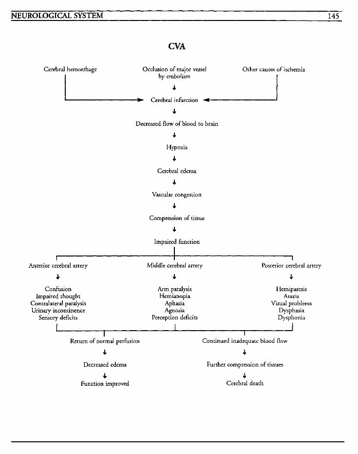
NEUROLOGICAL SYSTEM 145
CVA
Cerebral hemorrhage Occlusion of major vessel Other causes of ischemia by embolism
c I Cerebral infarction t I e Decreased flow of blood to brain
J,
Hypoxia
c Cerebral edema
c Vascular congestion
c Compression of tissue
c Impaired hnction
I I I
Anterior cerebral artery
4 Confusion
Impaired thought Contralateral paralysis Urinary incontinence
Sensory deficits
Middle cerebral artery
e Arm paralysis Hemianopia
Aphasia Agnosia
Perception deficits
Posterior cerebral artery
c Hemiparesis
Ataxia Visual problems
Dysphasia Dysphonia
Return of normal perfusion
J,
Decreased edema
4 Function improved
Continued inadequate blood flow
c Further compression of tissues
c Cerebral death

This Page Intentionally Left Blank
NEUROLOGICAL SYSTEM 1 h7
Head injuries, both open and closed, are usually the result of some type of trauma, and include skull fractures, concussions, lacerations, contusions, andor cerebral hemorrhages. The injury can be the result of a direct blow to the head, or may involve acceleration/deceleration injuries. Acceleration, or coup, injuries occur when the brain is forced against the cranium. Deceleration, or contrecoup, injuries occur after the initial impact when the brain is rotated or thrown in the opposite direction of the force.
Closed head injuries (CHI) result when a blunt trauma to the head causes a neurological deficit or loss of consciousness from bruising, hemorrhage, or laceration of brain tissues. This type of injury may be further categorized into mild concussion, classic concussion, diffuse injury with loss of con- sciousness greater than 24 hours, and diffuse shearing and disruption of brain structures.
A mild concussion occurs when forces on the brain stretch nerve fibers and result in impaired conduction of nerve responses. Neurological dys- function is temporary with no residual effects. In a classic concussion, the loss of consciousness is usu- ally less than 24 hours in length and the patient experiences disorientation and a degree of retro- grade amnesia when consciousness is regained. Some patients may experience residual personality changes or impairment in memory recall. Patients may exhibit a focal deficit caused by an injury that occurs to a specific area.
With diffuse closed head injuries, the loss of con- sciousness is greater than 24 hours and the coma may last up to weeks. The patient can exhibit rest- lessness, withdrawal from painful or noxious stimuli, or purposeful movement. Disorientation and amnesia occur with the return of conscious- ness, and personality changes are permanent due to the widespread cerebrum disruption.
When injury to the axons and neurons in the hemispheres, brain stem, and diencephalon occur and result in diffuse shearing of white matter with concurrent cerebral edema, dysfunction results in coma. More than half of these patients die, and those who do survive, have severe residual dysfunction. Contusions of the brain stem result in coma, as well as cranial nerve dysfunction and cardiopulmonary instability.
Skull fractures are normally classified as linear, basilar, or depressed. If a linear skull fracture does not puncture the dura mater, the fracture will heal without treatment. If the dura is torn, there is an increased chance that the middle meningeal artery is also punctured, and this will cause an epidural hematoma.
A basilar skull fracture can occur in the anterior or posterior fossa, and classic symptoms include cere- brospinal fluid leakage from the nose or ears, or ecchymoses over the mastoid projection or around the eyes. With basilar fractures, there exists a high risk for cranial nerve injury and dysfunction, infection, and residual neurological impairment.
Depressed skull fractures that are not depressed more than the thickness of the skull are usually not treated. A depression more than 5 millimeters or more in depth will require surgery in order to relieve the compression on structures. If the dura mater is punctured, the possibility of bone frag- ments entering the brain tissue is increased, as well as the potential for infection.
Lacerations of the scalp may occur with head injury or skull fractures, and will potentiate the danger of infection.
Intracranial hematomas result after trauma to the head, and frequently occur in conjunction with scalp lacerations, skull fractures, contusions, or penetrating wounds to the head. Subdural hematomas (SDH) usually are caused by venous
148 CRITICAL CARE NURSING CARE PLANS
bleeding, most often from the superior sagittal sinus, and involves the area between the dura mater and the arachnoid space. It may be acute, happening within 24-48 hours of injury, subacute, within 3-20 days of injury, or chronic, greater than 20 days from injury, depending on the time elapsed from injury to the onset of symptoms. SDH may occur spontaneously if the patient has a blood dyscrasia or clotting problem.
Epidural hematomas (EDH) are usually caused by arterial bleeding, generally from the middle meningeal artery, and involve the area above the outer dura mater and below the skull. These occur frequently when skull fractures cross the middle meningeal artery, or transverse or superior sagittal sinus, and the bleeding causes the dura to be pulled away from the skull. A posterior fossa EDH is usually caused by a venous bleed and may result in delayed symptoms due to the slow oozing. With EDH, the patient may have a brief episode of unconsciousness, followed by a varying length of lucid behavior prior to neurological deterioration and increased intracranial pressure.
Intracerebral hemorrhage (ICH) into the brain may occur hours or days after a closed head injury, and many result after rupture of an aneurysm, AV malformation, tumor, or vessel that has been weakened from hypertension. If the hemorrhage occurs in the internal capsule of the brain, paraly- sis will ensue. Symptoms vary depending on site, size, cerebral edema, and blood accumulation rate.
Head injuries .can result in varying severity from absence of neurological dysfunction to death, and each injury must be considered potentially critical. Cervical spine injury evaluation may be required depending on the mechanisms of the closed head injury.
MEDICAL CARE CT scans: used to identify cerebral edema, lesions, hemorrhage, ventricle size, tissue shifts, or infarctions
X-rays: skull x-rays may be used to identify frac- tures or midline shifts, or presence of bone fragments, and to evaluate healing or resolution
MRI: used to reveal disruption of axonal pathways and white matter shearing
Angiography: cerebral angiograms may be used to identify circulatory anomalies, shifting of struc- tures, hemorrhage, or edema
Lumbar puncture: may be used in diagnosis of subarachnoid hemorrhage; LP may be contraindi- cated in some cases
Laboratory: electrolyte imbalances may increase ICP or alter mental status; CBC to evaluate blood loss and hydration status; drug toxicology studies to identify drugs that may be responsible for con- sciousness level changes; anticonvulsant drug levels to monitor therapeutic maintenance levels
Arterial blood gases: used to evaluate hypoxemia and acid-base imbalances that can increase ICP; intracranial hematomas may result in respiratory alkalosis, or metabolic acidosis if patient is also in shock
Diuretics: may be used to draw water from brain cells in order to decrease cerebral edema and ICP
Steroids: may be used to decrease inflammation and edema
Anticonvulsants: may be required to treat and/or prevent seizure activity

NEUROLOGICAL SYSTEM 149
NURSlN6 CARE PLANS Alteration in tissue perfision: cerebral
Related to: hemorrhage, hematoma, lesions, cere- bral edema, metabolic changes, hypoxia, hypovolemia, cardiac dysrhythmias
Defining characteristics: disorientation, confusion, changes in mental status, combative- ness, inability to focus on topic, amnesia, memory loss, restlessness, inability to follow commands, increased intracranial pressure, vital sign changes, impaired motor function, impaired sensory func- tion
Outcome Criteria
Patient will achieve and maintain consciousness, and will have normal cognition and motor func- tion.
INTERVENTIONS RATIONALES
Assess patient for cause of impairment, problem with perfu- sion, and potential for increased ICP
Evaluate neurological status every hour initially, then every 1-2 hours, and notify M D for perti- nent changes. See Glasgow Coma Scale below.
Assess patient’s arousal or lack of arousal to verbal and noxious stimuli.
Establishes plan of care and iden- tifies appropriate choices for intervention. Depending on patient’s conditiodproblem, surgical intervention may be required.
Establishes a baseline from which to gauge changes or trends. Alterations in level of conscious- ness and behavior, as well as other symptoms may be helpful to determine area of damage.
Establishes level of consciousness which is the single most impor- tant measure of the patient’s status. Extensive damage involv- ing the cerebral cortex may result in delayed responses to commands, drowsiness and
INTERVENTIONS RATIONALES
Assess patient’s best verbal response to questions and whether wordslsentences are appropriate.
Assess ability to follow simple commands, noting purposeful and non-purposeful movements bilaterally.
Observe pupils bilaterally, noting equality, size, and reaction to light. Notify M D of significant changes.
Observe position of eyes, noting any deviation laterally or verti- cally. Observe for presence of doll’s eyes.
Observe for presence of blink reflex.
inability to stay awake unless stimulared, or disorienration. Lack of response to stimuli may indicate that damage has occurred to the midbrain, pons, and/or medulla. If a minimal amount of damage has occurred in the cerebral cortex, the patient may be uncooperative or drowsy.
Identifies speech ability and ori- entation levels.
Identifies ability to respond to stimuli when patient is unable to open eyes or cannot speak. Purposeful movement, such as holding up two fingers or squeezing and releasing hands when instructed to do so, can help identify awareness and the ability to respond appropriately!. Abnormal posturing may indi- cate diffuse cortical damage, and the absence of any movement to one side of the body usually indi- cates damage has been done to the motor tracts of the opposite side of the cerebral hemisph’ re
Compression of the brain stem and impairment of the second and third cranial nerves will alter pupillary response.
Loss of doll’s eyes, or the oculo- cephalic reflex, indicates impairment in the function of the brain stem. Positions and movement of the eyes may indi- cate which area of rhe brain has been involved. Problems with abduction of the eyes may be an early indication of increased intracranial pressure.
Loss of blinking reflex may indi- cate injury to the pons and medulla.
f .
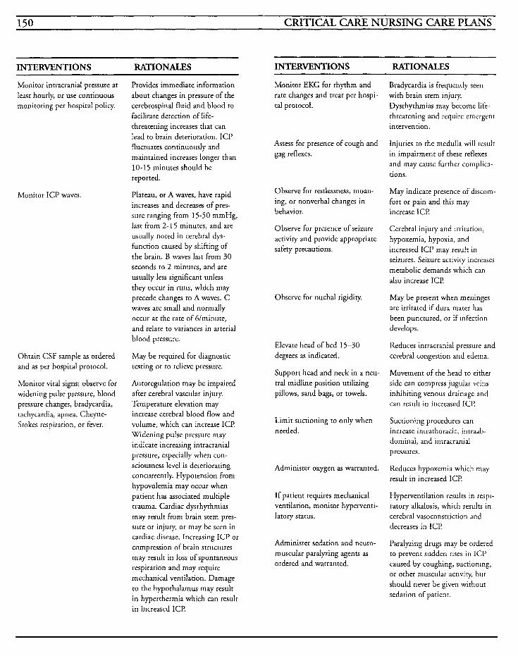
150 CRITICAL CARE NURSING CARE PLANS
INTERVENTIONS RATIONALES INTERVENTIONS RATIONALES
Monitor intracranial pressure at leasr hourly, or use continuous monitoring per hospital policy.
Monitor ICP waves.
Obtain CSF sample as ordered and as per hospital protocol.
Monitor vital signs; observe for widening pulse pressure, blood pressure changes, bradycardia, tachycardia, apnea, Cheyne- Stokes respiration, or fever.
Provides immediate information about changes in pressure of the cerebrospinal fluid and blood to facilitate detection of life- threatening increases that can lead to brain deterioration. ICP fluctuates continuously and maintained increases longer than 10-15 minutes should be reported.
Plateau, or A waves, have rapid increases and decreases of pres- sure ranging from 15-50 mmHg, last from 2-1 5 minutes, and are usually noted in cerebral dys- function caused by shifting of the brain. B waves last from 30 seconds to 2 minutes, and are usually less significant unless they occur in runs, which may precede changes to A waves. C waves are small and normally occur at the rate of G/minute, and relate to variances in arterial blood pressure.
May be required for diagnostic testing or to relieve pressure.
Autoregulation may be impaired after cerebral vascular injury. Temperature elevation may increase cerebral blood flow and volume, which can increase ICI? Widening pulse pressure may indicare increasing intracranial pressure, especially when con- sciousness level is deteriorating concurrently. Hypotension from hypovolemia may occur when patient has associated multiple trauma. Cardiac dysrhythmias may result from brain stem pres- sure or injury, or may be seen in cardiac disease. Increasing ICP or compression of brain structures may result in loss of spontaneous respiration and may require mechanical ventilation. Damage to the hypothalamus may resulr in hyperthermia which can result in increased ICE!
Monitor EKG for rhyrhm and rate changes and treat per hospi- tal protocol.
Bradycardia is frequently seen with brain stem injury. Dysrhyrhmias may become life- threatening and require emergent intervention.
Assess for presence of cough and gag reflexes.
Observe for restlessness, moan- ing, or nonverbal changes in behavior.
Observe for presence of seizure activity and provide appropriate safety precautions.
Observe for nuchal rigidity.
Elevate head of bed 15-30 degrees as indicated.
Support head and neck in a neu- tral midline position utilizing pillows, sand bags, or towels.
Limit suctioning to only when needed .
Administer oxygen as warranted.
If patient requires mechanical ventilation, monitor hyperventi- larory status.
Administer sedation and neuro- muscular paralyzing agents as ordered and warranted.
Injuries ro the medulla will resulr in impairment of these reflexes and may cause further complica- tions.
May indicate presence of discom- fort or pain and this may increase ICP
Cerebral injury and irritation, hypoxemia, hypoxia, and increased ICP may result in seizures. Seizure activity increases metabolic demands which can also increase ICI?
May be present when meninges are irritated if dura mater has been punctured, or if infection develops.
Reduces inrracranial pressure and cerebral congestion and edema.
Movement of the head to either side can compress jugular veins inhibiting venous drainage and can result in increased ICI?
Suctioning procedures can increase intrathoracic, intraab- dominal, and intracranial pressures.
Reduces hypoxemia which may resulr in increased ICP
Hyperventilation results in respi- ratory alkalosis, which results in cerebral vasoconstriction and decreases in ICP
Paralping drugs may be ordered to prevent sudden rises in ICP caused by coughing, suctioning, or other muscular acriviry, but should never be given without sedation of patient.
NEUROLOGICAL SYSTEM 151
INTERVENTIONS RATIONALES Discharge or Maintenance Evaluation
Moniror pulse oximetry and notify MD if levels remain below 90%.
Monitor ABGs as warranted.
Monitor intake and output hourly.
Provide calm, quier environment without extraneous stimuli, and provide rest periods between care activities. Use restraints only when absolutely necessary.
Administer osmotic diuretics as ordered.
Indicates respiratory insufficiency and impendinglpresent hypoxia.
Identifies acid-base imbalances and presence of hypoxemia. Elevations in PaCO, will cause vasodilation in the cerebral vas- culature with a resultant increase in I C P
Reflects amounts of total body water which influences tissue perfusion. Cerebral injury may result in inappropriate A D H or diabetes insipidus, and may lead to hypovolemia.
Helps to reduce ICE Use of restraints may be required to ensure the patient’s safety, but may cause irritation and fighting against the restraints which can increase ICE
Drugs remove water from areas in the brain that maintain an intact blood-brain barrier, and helps to reduce ICE
Information, Instruction Demonstration
Patient will be alert, oriented in all phases, with no speech or motor impairment.
Patient will have no sensory impairment.
Patient will have stable vital signs and no increase in ICI?
Risk f i r inefective breathing pattern
Related to: respiratory center injury, obstruction, structural shifting, surgical intervention
Defining characteristics: dyspnea, Cheyne-Stokes respirations, bradypnea, apnea, hypoxia, hypoxemia, abnormal arterial blood gases
Outcome Criteria
Patient will maintain a patent airway with no evi- dence of respiratory insufficiency.
INTERVENTIONS RATIONALES - Observe respiratory status for rate, depth, rhythm, irregularity, chest expansion and symmetry, and absence.
INTERVENTIONS RATIONALES
Instruct patient to avoid cough- ing, straining, or any valsalva-like maneuvers.
Prepare patiendfamily for place- ment on mechanical ventilation as warranted.
Prepare parient/family for surgi- cal procedures.
Activities increase ICP by increasing intrathoracic and intra-abdominal pressures.
Injury to certain areas of the brain may result in insufficient respiratory status and may require intubation and mechani- cal ventilation to maintain life support.
Craniotomy or burr holes may be necessary to remove bone fragmenrs, remove a hematoma, stop hemorrhage, remove necrotic tissue, or elevate a depressed skull fracture.
Mainrain patency of airway.
Auscultate breath sounds for changes and presence of adventi- tious lung sounds.
Observe for presence of gag, cough, and swallow reflexes.
Changes from patient’s baseline may indicate pulmonary compli- cations or involvement of brain injured areas. Respiratory insuffi- ciency may require mechanical ventilation.
Depending on location of injury, patient may not be able to main- tain his own airway or ventilation and may require arti- ficial means of doing so.
May indicate hypoventilation, obstruction, atelectasis, or infec- tion which may impair cerebral oxygenation.
Lack of these reflexes may impair the parient’s ability to handle his secretions and may require an artificial airway. Nasopharyngeal airways are preferred to avoid stimulation of the gag reflex which can increase ICP
152 CRITICAL CARE NURSING CARE PLANS
INTERVENTIONS RATIONALES
Administer oxygen as warranted. Provides supplemental oxygen to reduce hypoxia and prevent desaturation.
Elevate head of bed as warranted. Promotes chest expansion and ventilation.
Avoid suctioning unless manda- Suctioning may cause hypoxia tory, and observe for changes in and decreases cerebral perfusion, sputum color, consistency, or while increasing ICI? Changes in odor. sputum characteristics may indi-
cate impending or presence of infection.
Information, Instruction, Demonstration
INTERVENTIONS RATIONALES
Instruct patient in deep breath- ing exercises. and/or pneumonia.
Avoid chest physiotherapy during C P T is concraindicated with acute phases. patients with increased ICP
because this potentiates the increase.
Reduces potential for atelectasis
Alteration in thought processes
Related to: injury, psychological problems, med- ications
Defining characteristics: memory deficit, dimin- ished attention span, inability to focus, disorientation to time, place, person, or situation, poor recall, distractibility, personality changes, inappropriate behavior, inability to problem-solve
Outcome Criteria
Patient will be oriented in all phases and will be able to recall data.
INTERVENTIONS RATIONALES Evaluate orientation status with regard to time, place, person, circumstance, and recent events.
Observe patient for abiliry to concentrate and attention span.
Provides a baseline on which to begin and plan interventions.
Abiliry to concentrate may be diminished due to injury and this further potentiates anxiety for the patient.
Assist family members to under- stand patient’s aberrant behavior, personality changes, and other responses.
Prepare patient/family for intu- bation/mechanical ventilation as warranted. Provides knowledge and
As time and condition permits, instruction may be given.
decreases fear in patients who are awake.
Discharge or Maintenance Evaluation
Patient will maintain his own airway and be able to sustain spontaneous respiration.
Patient will be able to handle secretions and dis- pose of them adequately.
Arterial blood gases will be within normal limits for the patient.
Patient will be able to recall information accu- rately and be able to demonstrate appropriate deep breathing.
Encourage family to discuss news and family occurrences with patient.
Explain all procedures with clear concise explanations.
Reduce competing stimuli when conversing with the patient.
Be consistent with srafFassign- ments as much as possible.
Head injury recovery includes agitation and hostility, anger, and disorganized thought sequences. Family members may have diff- culty dealing with the patient’s changed personaliry and behav- ior.
Helps to maintain contact with normal events and assists with orientation.
Patient may have lost the abiliry to reason or conceptualize, and may require repeated reinforce- ment. Retention of information may be decreased and result in further anxiety.
Brain injured patients may be overly excitable and become vio- lent with excess stimulation.
Provides atmosphere of stability and allows patient some control in situation.
NEUROLOGICAL SYSTEM 153
~~~~ ~ ~ ~ ~
INTERVENTIONS RATIONALES
Remain with patient during episodes of fright or agitation.
Offers support and helps to calm patient to reduce anxiety to pre- vent loss of control and panic.
Helps to maintain a sense of hope for improvement and to
Assist patient/family to set realis- tic goals and instruct in ways to control behavior. facilitate rehabilitation.
Information, Instruction, Demonstration
INTERVENTIONS RATIONALES
Consult rehabilitation counselor for assistance with cognitive training as warranted.
Assists patient with methods to compensate for problems with concentration, memory, judg- ment, and problem-solving.
Additional help may be needed to help with recovery.
Make appropriate referrals TO
support groups or counseling as warranted.
Discharge or Maintenance Evaluation
Patient will regain normal mental skills and be oriented in all phases.
Patient will be able to recognize aberrant behav- ior and control negative reactions.
Patient will participate in rehabilitation/counsel- ing for retraining.
Risk for infiction
Related to: trauma, lacerations, broken skin, open wounds, invasive procedures, surgery, use of steroids, cerebrospinal fluid leakage, nutritional deficiency
Defining characteristics: fever, tachycardia, elevated white blood cell count, shift to the left on differential, redness to wounds, purulent drainage or sputum, nuchal rigidity, bloody or purulent CSF
Outcome Criteria
Patient will be free of signs/symptoms of infection.
INTERVENTIONS RATIONALES
Monitor temperature every 2-4 hours.
Observe wounds, incision lines, invasive line sites, or other skin bre& for drainage, redness, or edema.
Observe for CSF leakage from ears and nose, and report to MD.
Use aseptic or sterile technique when changing dressings or pro- viding wound care.
Utilize good handwashing practices.
Monitor urine output for ade- quacy of amount, color, clarity, and presence of foul odor.
Obtain cultures of wound, urine, blood, stool, sputum, or other body fluidslsurfaces as warranted, and as per hospital protocol.
Administer antibiotics as ordered.
Elevation may indicate develop- ment of infection.
Prompt identification of develop- ing problems may result in prompt intervention to prevent systemic sepsis.
Indicates a serious complication from head injury and may result in meningitis.
Prevents spread of infection.
Prevents nosocomial infections,
May identify presence of bacrer- id infection.
Identifies the presence of infec- tion and the causative agent, as well as identification of appropri- ate antimicrobial agent to treat infection.
May be given prophylactically when trauma, surgery, or CSF leakage occurs. Appropriate antibiotic may be ordered after results of culture and sensitivity are received.
154 CRITICAL CARE NURSING CARE PLANS
Information, Instruction, Demonstration
INTERVENTIONS RATIONALES
Instruct on isolation procedures Isolation may be required based as warranted. Instruct visitors on on type of organism grown. avoiding patient if they have Restriction of ill visitors may upper respiratory or other type of reduce exposure of an already infection. susceptible patient.
Instruct on deep breathing and Promotes lung expansion and pulmonary exercises as reduces potential for atelectasis warranted. and pneumonia. Postural
drainage is contraindicated if patient has increased ICE
Discharge or Maintenance Evaluation
Patient will be normothermic with normal white blood cell count.
Patient will exhibit no signs/symptoms of infec- tion.
Wounds will heal without complications.
Impaired physical mobility
Related to: trauma, immobilization, mental impairment, decreased strength, paralysis
Defining characteristics: inability to move at will, inability to transfer or ambulate, decreased range of motion, decreased muscle strength, muscle incoordination, footdrop, contractures, decreased reflexes
Outcome Criteria
Patient will achieve and maintain an optimal level of motor function.
INTERVENTIONS RATIONALES
Evaluate patient’s ability and function and injury.
Assess patient for degree of immobility.
Observe skin for redness, warmth, or tenderness.
Provide kinetic bed or alternating pressure mattress for patient.
Maintain good body alignment and use pillows/rolls to support body. Use high-top tennis shoes and removelreapply every 4-8 hours.
Perform range of motion exer- cises every 4 hours.
Provide skin care every 8 hours and prn. Change wet clothing and linens prn.
Instill artificial tears or lubrica- tion ointment to eyes every 4 hours and prn as ordered.
Identifies impairments and allows for identification of appropriate interventions.
Provides a baseline on which to base interventions. Patient may only require minimal assistance or be completely dependent on caregivers for all body needs.
May indicate pressure is being concentrated in one area and may predispose patient to decu- bitus formation.
Helps to promote circulation and reduces venous stasis and tissue pressure to prevent formation of pressure sores.
Prevents further complications and contractures. Use of tennis shoes helps prevent footdrop.
Helps to maintain mobility and function of joints.
Helps to promote circulation and reduces potential for skin break- down.
Prevents eye tissues from drying out. If patient is unable to main- tain closed eyes, eye patches or tape may be required.
Information, Instruction, Demonstration
INTERVENTIONS RATIONALES
Instruct patient/family in range Helps patient to regain some of motion exercises and mobility control and allows family some aids. involvement in reconditioning
program.
Promotes understanding and compliance with treatment regi- men.
Instruct patiendfamily in reasons for impairment and realistic goals for changes in patient’s lifestyle as warranted.
NEUROLOGICAL SYSTEM 155
INTERVENTIONS RATIONALES INTERVENTIONS RATIONALES
Consult physical andlor occupa- tional therapy, as warranted.
Assists patient with identihing methods to compensate for impairments and provides for post-discharge care.
Encourage family to assist with feeding a s warranted.
Discharge or Maintenance Evaluation
Patient will be able to maintain skin integrity with no complications.
Patient will be able to increase muscle strength and tone and achieve a functional level of muscle function.
Patient will be able to demonstrate exercise pro- gram.
Patient and family will become involved in recovery programs.
Risk for alteration in nutrition: less tban body requirements
*Related to: inability to take in sufficient nutrients, inability to chew or swallow, decreased level of consciousness, intubation, increased metabolism
Defining characteristics: weight loss, muscle wast- ing, catabolism
Outcome Criteria
Patient will be able to ingest sufficient nutrients to meet metabolic demands, and will experience no weight loss.
INTERVENTIONS RATIONALES
Evaluate patient’s ability to eat, swallow, chew, etc.
Identifies problems and estab- lishes data for choices of interventions.
Weigh every day. Establishes trends and helps to evaluate effectiveness of interventions.
Provide small, frequent meals. Improves patient compliance and facilitates digestion.
Administer tube feedings as required.
Elevate the head of the bed at least 30 degrees while eating or giving tube feedings.
Auscultate bowel sounds every 4 hours.
Consult dietitian as warranted.
Monitor serum albumin, preal- bumin, transferrin, iron, renal profiles, and glucose levels.
Involves family in parient’s plan of care and provides opportunity for socialization that may improve intake.
Tube feedings may be required during the initial phase after injury until the patient is able to swallow without danger of aspi- ration.
Helps to prevent aspiration
Quality of bowel sounds may indicate response to feedings or development of an ileus.
Provides additional resources to establish nutrient needs based on many factors including metabolic deman ds .
Assists in identification of nutri- tional problems, body function, and response to nutritional sup- port.
Information, Instruction, Demonstration
~
INTERVENTIONS RATIONALES
Consult speech or occupational therapy For mechanical problems.
May be required ro establish a functional method of eating for the patient.
Bleeding may occur from srresses resulting from injury or from mechanical erosion.
Check gastric contents, vomitus, and stools for occult blood.
Discharge or Maintenance Evaluation
Patient will maintain optimal weight.
Patient will be in a positive nitrogen balance, with laboratory values within normal limits.
Patient will be able to ingest food in sufficient amounts to meet and maintain metabolic demands.
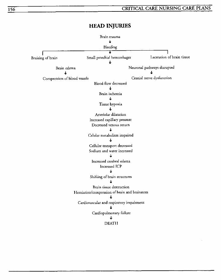
156 CRITICAL CARE NURSING CARE PLANS
HEAD INJURIES
Brain trauma c
Bleeding JI I
Small petechial hemorrhages c
c s
Laceration of brain tissue 1-
Bruising of brain
Brain edema Neuronal pathways disrupted
Compression of blood vessels Cranial nerve dysfunction Blood flow decreased
c Brain ischemia
c Tissue hypoxia
c Arteriolar dilatation
Increased capillary pressure Decreased venous return
c Celular metabolism impaired
c Cellular transport decreased Sodium and water increased
c Increased cerebral edema
Increased ICP c
Shifting of brain structures c
Brain tissue destruction Herniation/cornpression of brain and brainstem
J. Cardiovascular and respiratory impairment
4 Cardiopulmonary failure
c DEATH
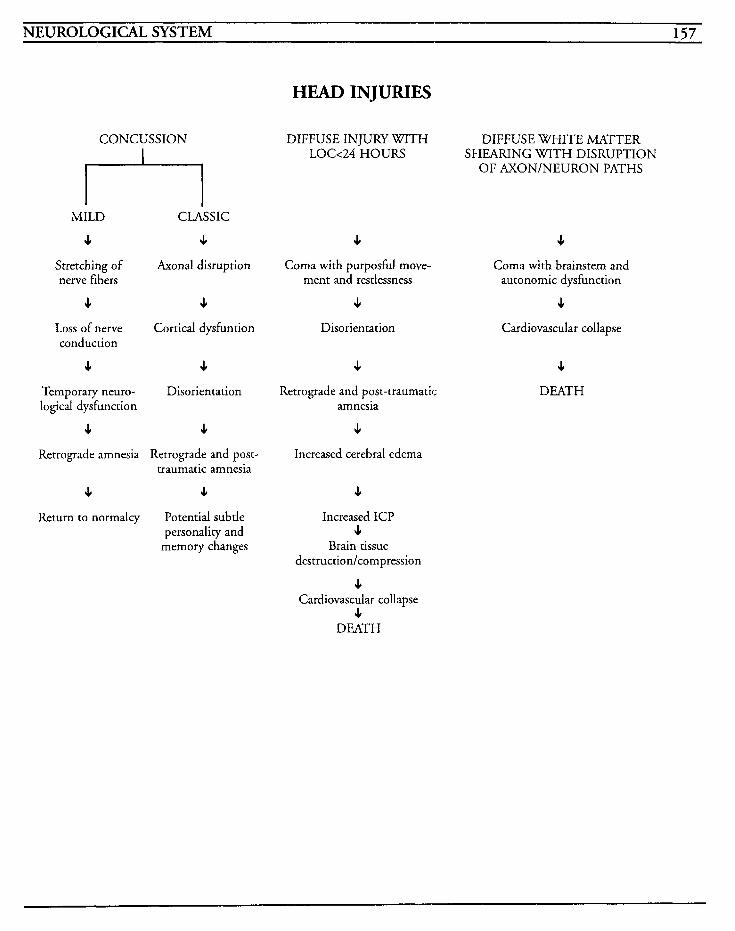
NEUROLOGICAL SYSTEM 157
HEAD INJURIES
CONCUSSION
MILD CLASSIC
c c Stretching of Axonal disruption nerve fibers
c c Loss of nerve Cortical dysfuntion conduction
c c Temporary neuro- Disorientation logical dysfunction
J, J,
Retrograde amnesia Retrograde and post-
J, JI
Return to normalcy Potential subtle personality and
memory changes
traumatic amnesia
DIFFUSE INJURY WITH DIFFUSE WHITE MATTER
OF AXON/NEURON PATHS LOG24 HOURS SHEARING WITH DISRUPTION
c Coma with purposhl move-
ment and restlessness
c Disorientation
Retrograde and post-traumatic amnesia
J,
Increased cerebral edema
e Increased ICP
c Brain tissue
destruction/compression
J, Cardiovascular collapse
J, DEATH
c Coma with brainstem and
autonomic dysfunction
c Cardiovascular collapse
c DEATH

This Page Intentionally Left Blank

NEUROLOGICAL SYSTEM 159
Spinal Cord Injuries Spinal cord injuries are traumatic injuries to the spinal cord caused by contusion, compression, or transection of the cord as a result of dislocation of bones, rupture of ligaments, vessels, or vertebral discs, stretching of neuron tissue, or impairment in blood supply. These lesions are classified as being complete or incomplete. Complete lesions involve the total loss of sensation as well as volun- tary motor function, and incomplete lesions involve mixed losses of sensation and voluntary motor function.
The flexion, hyperextension and/or rotational types of injury that result in spinal cord injury are usually caused by trauma, motor vehicle accidents, falls, gun shot wounds, stab wounds, and diving injuries, The severity of the injury can vary depending on the amount of pathologic changes that are produced. Injury without intervention results in ischemia, edema, hemorrhage, and pro- gressive destruction. After the initial cord compression, small hemorrhages occur in the cen- tral gray matter. The expansion and increase in number of hemorrhagic areas cause even more compression, edema, and finally, necrosis of the cord. The cervical area is the most vulnerable part of the spine because of the mobility of the head and poor support by the muscles, but cervical fractures do not necessarily cause neurological problems.
The level of the injury relates to how much func- tional ability is retained. At the C1 to C8 levels, the patient is a quadriplegic with variances in muscle function from complete paralysis of respi- ratory function to limited use of the fingers. At TI to L1 levels, paraplegia is noted with intact arm movement. At L1 and below, there may be mixed dysfunction with bowel and bladder dysfunction.
In central cord syndrome, the central gray matter of the cord is contused, compressed, or hemorrhaged. This results in varying degrees of sensory loss and bowel/bladder dysfunction, and there is more motor loss in the arms than in the legs. In anterior cord syndrome, the injury has occurred to the anterior horn and spinothalamic areas resulting in a loss of motor function and painltemperature below the lesion. Sensations of touch, position, pressure and vibration may be maintained. In Brown-Sequard syndrome, as a result of a transverse hemi-transection of the cord, motor loss, touch, vibration, pressure, and posi- tion are involved ipsilaterally, with a contralateral loss of pain/temperature sensation. Posterior cord syndrome is exceedingly rare and results in the loss of light touch below the level of injury, with motor function and sensation of pain and temper- ature maintained intact.
When spinal lesions at or above T6 level block sensory impulses from reaching the brain, an excessive and critical autonomic response to a stimulus occurs, and this is known as autonomic dysreflexia. It may be precipitated by bowel or bladder distention or by stimulation of the skin or pain receptors. Symptoms may include severe blood pressure increases, pounding headache, pro- fuse sweating above the lesion, blurred vision, goosebumps, and bradycardia. Treatment is aimed at removing the stimulus that causes the problem, and treating the hypertensive episode.
Spinal shock occurs when there is an abrupt loss of continuity between the spinal cord and the higher nerve centers, with a complete loss of all reflexes and a flaccid paralysis below the level of injury. Normally, this spinal shock lasts 7-1 0 days and when it begins resolution, the flaccidity changes to a spastic type of paralysis.
1 c;n CRITICAL CARE NURSING CARE PLANS
MEDICAL CARE Arterial blood gases: used to identify hypoxemia and acid-base imbalances
Radiography: chest x-rays used to identify diaphragmatic changes or respiratory complications; spinal x-rays used to identify frac- ture or dislocation and identifies level of injury
CT scans: used to identify structural aberrancies and localize injury site
Magnetic resonance imaging: used to identify cord lesions, compression, or edema
Surgery: may be required to align or stabilize frac- ture, or repair other traumatic injuries that may be concurrent
Traction: may be required to align and stabilize fracture or dislocation of the vertebral column
NURSING CARE PLANS Risk j ib decreased cardiac output
Related to: neurogenic shock, sympathetic block- ade, spinal shock
Defining characteristics: hypotension, bradycar- dia, vasovagal reflex, hypoxia, decreased venous return, decreased hemodynamic pressures
Outcome Criteria
Patient will be able to maintain systolic blood pressure above 90 mmHg and have stable vital signs and heart rhythm.
~~
INTERVENTIONS RATIONALES
Monitor vital signs, especially blood pressure and heart rate.
Transection of the spinal cord above the T5 levels may result in vasodilation, decreased venous return, and hypotension. Sympathetic blockade may cause bradycardia.
INTERVENTIONS RATIONALES
Monitor EKG for changes in rhythm and conduction, and treat according to hospital protocol.
Sympathetic blockade may cause conduction problems such as escape rhythms, and vasovagal reflexes may provoke cardiac arrest.
Monitor hemodynamic parame- ters if feasible.
Administer oxygen as warranted, ensuring pre-oxygenation prior to suctioning or prolonged coughing exercises.
Administer vasopressors as warranted.
Fluid shifts, hypotension, and hemorrhage may be reflected in lowered pressures and lower car- diac output/index.
Assists in preventing hypoxia which can result in vasovagal reflex and cardiac arrest.
May be indicated if fluid resusci- tation is not successful in maintaining systolic blood pres- sure above 90 mmHg.
Information, Instruction, Demonstration
INTERVENTIONS RATIONALES
Instruct patient in avoidance of valsalva-type maneuvers. facilitare vasovagal response.
May lower blood pressure and
Discharge or Maintenance Evaluation
Patient will exhibit no episodes of cardiac rhythm disturbances.
Patient will have normotensive blood pressure with stable hemodynamic pressures.
Patient will have optimal cardiac output and index.
Patient will exhibit no hypoxic episodes and avoid desaturation with procedures.
Inneflective breathing pattern
Related to: trauma, spinal cord lesions at high levels, paralysis of respiratory musculature, ineffec- tive coughing, pneumonia, pulmonary edema, pulmonary embolism
NEUROLOGICAL SYSTEM lG1
Defining characteristics: dyspnea, use of accessory muscles, diaphragmatic breathing, decreased tidal volumes, sputum, abdominal distention, abnormal arterial blood gases, apnea, oxygen desaturation
Outcome Criteria
Patient will maintain adequate oxygenation and ventilation without evidence of respiratory com- plications.
INTERVENTIONS RATIONALES
Assess respiratory status for ade- quacy of airway and ventilation, rate, character, depth, increased work of breathing, or use of accessory muscles.
Auscultate lung fields for pres- ence of adventitious sounds and other changes.
Assist withlmeasure pulmonary parameters, such as spontaneous tidal volume, vital capacity, and negative inspiratory force. Obtain arterial blood gases as warranted.
Evaluate patient’s ability to cough and assist with abdominal thrusting technique, or quad coughing, as warranted.
Monitor oxygen saturation con- tinually and notify physician if levels stay below 90%.
Suction patient only when required. Provide humidification of oxygen and utilize pulmonary toilette as warranted.
Spinal cord lesions below C4 level induces diaphragmatic breathing and hypoventilation.
May reflect the presence of infil- trates, pneumonia, atelectasis, or fluid overload.
Measurement of pulmonary parameters may facilitate prompt identification of deterioration in respiratory status. ABGs are drawn to identify acidlbase dis- turbances and hypoxemia that may result from restriction of lung expansion and ineffective cough mechanisms.
Paralysis of respiratory muscula- ture may prevent sufficient pleural pressure to be produced to maintain effective cough. External technique can assist patient to cough effectively.
Oximetry assists in identification of deterioration in ventilatory status, allowing for prompt inter- vention.
Suctioning may precipitate vaso- vagal reflexes, bradycardia, and cardiac arrest. Liquification of environmental air and secretions may prevent mucous plugs and thick mucoid secretions.
Information, Instruction, Demonstration
INTERVENTIONS RATIONALES
Prepare patientlfamily for place- ment on mechanical ventilation as warranted.
Prepare patient for bronchoscopy as warranted.
Monitor for signslsymptoms of pulmonary embolism, pneumo- nia, or pulmonary edema.
Discharge or Maintenance Evaluation
Instruct family member in tech- niques to assist patient with coughing, repositioning frequently, and suctioning tech- niques as warranted.
Hypoxemia that cannot be cor- rected with addition of supplemental oxygen may require intubation and ventila- tion to maintain airway and oxygenation.
May be required to remove obstructive secretions.
Edema may result from fluid resuscitation efforts, and pneu- monia may develop from immobility and ineffective cough ability. Pulmonary emboli may result from venous thrombosis as a complication of immobility or hemorrhagic causes.
Provides information that will be used when patient is discharged and facilitates feelings of control over situation and self-esteem.
Patient will maintain adequate airway and venti- lation.
Patient will exhibit no signs/symptoms of respi- ratory complications.
Patiendfamily will be able to verbalize understanding of instructions and give adequate return demonstration.
Alteration in temperature regulation Related to: poikilothermism, injury to hypothala- mic center or sensory pathways
Defining characteristics: elevated body tempera- ture, decreased body temperature, change of temperature based on environmental temperature
162 CRITICAL CARE NURSING CARE PLANS
Outcome Criteria
Patient will achieve and maintain body tempera- ture above 95 degrees.
INTERVENTIONS RATIONALES
Monitor temperature every 2 hours until stabilized, then every 4 hours and prn.
Interruption of the sympathetic nervous system pathways to the temperature control center in the hypothalamus causes body tem- perature swings in an effort to match environmental tempera- tures.
Hyperthermia may occur during periods of spinal shock because the sympathetic activity is blocked and the patient does not perspire on paralyzed areas of body.
Maintain a slightly cool environ- mental temperature. If patient is hypothermic, apply warm blanket.
Information, Instruction, Demonstration
INTERVENTIONS RATIONALES
Instruct patienrlfamily regarding variable body temperatures and methods to maintain comfort.
Provides knowledge and facili- tates compliance.
Discharge or Maintenance Evaluation
Patient will exhibit normal temperature and be able to maintain core body temperature using methods discussed.
Impaired physical mobility Related to: spinal cord lesion, trauma, paralysis, spasticity, physical restraint, traction
Defining characteristics: contractures, inability to move as desired, spastic movements, muscle atro- phy, muscle wasting, skin breakdown, redness, pressure areas
Outcome Criteria
Patient will be able to achieve maximum mobility within limitations of paralysis and will avoid skin breakdown and contractures.
INTERVENTIONS RATIONALES
Assess motor strength and func- tion at least every 4-8 hours, and prn. Identify level of tactile sen- sation, ability to move parts of body, spasticity, etc.
Observe for muscle atrophy and wasting.
Encourage independent activity as able.
Assist withlprovide range of motion exercises to all joints.
Reposition every 2 hours and prn. Utilize kinetic bed therapy as warranted.
Identifies level of sensory-motor impairment and evahares resolu- tion of spinal shock. Specific injury level may have partially mixed or occult sensorimotor impairment.
May be noted during flaccid paralysis stage of spinal shock.
C1-4 lesions result in quadriple- gia with complete loss of respiratory function; C4-5 lesions result in quadriplegia with potential for phrenic nerve involvement that may result In loss of respiratory function; C5-6 lesions result in quadriplegia with some gross arm movement ability and some sparing of diaphragmatic muscle involve- ment; C6-7 lesions result In quadriplegia with intact biceps; C7-8 lesions result in quadriple- gia with intact biceps and triceps but no intrinsic hand muscula- ture intact; T1-L2 lesions result in paraplegia with variable amounts of involvement to inter- costal and abdominal muscle groups; below L2 lesions result in mixed motor-sensory loss with bowel and bladder impairment.
Improves muscle tone and joint mobility, decreases risk for con- tractures, and prevents muscle atrophy.
Decreases pressure on bony prominences and improves peripheral circulation. Kinetic beds can immobilize the unstable vertebral column and decrease potential for complications from immobility.
NEUROLOGICAL SYSTEM 163
INTERVENTIONS RATIONALES Discharge or Maintenance Evaluation
Ensure proper alignment with each position.
Utilize footboards or high-top tennis shoes.
Observe for changes in skin status and provide frequent skin care.
Assist with/consult physical ther- apists or occupational therapists to develop plan of care for patient.
Maintain cervical traction appa- ratus as warranted.
Observe for redness and swelling to calf muscles. Measure circum- ference daily if problem is noted.
Correct anatomic alignment pre- vents contractures and deformities.
Prevents footdrop.
Loss of sensation, paralysis, and decreased venous return predis- pose the patient for pressure wounds.
Exercises help stimulation circu- lation and preserves joint mobility.
Cervical traction provides for stabilization of vertebral column, reduction, and immobilization to maintain proper alignment. Halo bracddevices provide immobi- lization but can facilitate with active participation with rehabili- tation processes.
Thrombus formation may occur as a result of immobilization and flaccid paralysis.
Information, Instruction, Demonstration
INTERVENTIONS RATIONALES
Instruct family in rehabilitative therapy, exercises, and reposition- ing, and involve them with patient’s care.
Avoid improper placement of footrests, headrests, or padding when repositioning patient.
Instruct in methods for shifting weight.
Administer muscle relaxants as warranted.
Facilitates adaptation to patient’s health status and allows for family members to contribute to patient’s welfare.
May create pressure resulting in pressure sores or necrotic injury.
Improves circulation by reducing pressure to body surfaces.
May be required to reduce pain and spasriciry.
Patient will maintain appropriate body alignment and maximal function within limit of injury.
Patient will avoid complications of immobility.
Patient will be able to verbalize understanding and demonstrate effective therapeutic modali ties.
Patient will exhibit suppleness of joints and muscles.
Alteration in comfort [See Guillain-Barrt]
Related to: trauma, surgery, cervical traction
Defining characteristics: burning pain below lesion, muscle spasms, phantom pain, hyperesthe- sia above lesion level, headaches, communication of pain, facial grimacing, irritability, restlessness
Risk for impaired skin intepity [See Fractures]
Related to: immobility, surgery, traction appara- tus, changes in metabolism, decreased circulation, impaired sensation
Defining characteristics: wounds, drainage, red- ness, pressure sores, abrasions, lacerations
Sensory-perceptual alteration [See CVA]
Related to: traumatic injury, sensory receptor and tract impairment, damaged sensory transmission
Defining characteristics: decreases sensory acuity, impairment of position relation, proprioception, motor incoordination, mood swings, disorienta- tion, agitation, anxiety, abnormal emotional responses, changes in stimulation response
164 CRITICAL, CARE NURSING CARE PLANS
Bowel incontinence
Related to: trauma, impairment of bowel innerva- tion, impairment of perception, modifications of dietary intake, immobility
Defining characteristics: inability to evacuate bowel voluntarily, ileus, gastric distention, hypoac- tive bowel sounds, absent bowel sounds, nausea, vomiting, abdominal pain, constipation
Outcome Criteria
Patient will be able to establish and maintain bowel elimination patterns.
INTERVENTIONS RATIONALES
Observe for presence of abdomi- nal distention.
Auscultate for presence of bowel sounds, noting changes in character.
Evaluate bowel habits, such as frequencp character, and amount of stools.
Establish bowel pattern by use of stool softeners, suppositories, or digital stimulation.
Increase dietary bulk and fiber.
Provide frequent skin care.
Innervation may be impaired as a result of the injury with resultant decrease or loss of peristalsis, and potential for development of ileus. Bowel distention may pre- cipitate autonomic dysreflexia after spinal shock recedes.
High-pitched tinkling bowel sounds may be heard when patient has an ileus, and bowel sounds may be absent during spinal shock phase.
Establishes pattern and facilitates treatment options.
Effectively evacuates bowel.
Information, Instruction, Demonstration
INTERVENTIONS RATIONALES
Instruct patientlfamily regarding method for daily bowel program.
Promotes independence and self-esteem.
Discharge or Maintenance Evaluation
Patient will establish and maintain daily bowel pattern.
Patient will be able to verbalize understanding and demonstrate appropriate methods to accomplish bowel care.
Patient will be able to avoid complications that may be caused by gastric distention or ileus.
Urinary retention
Related to: traumatic loss of bladder innervation, bladder atony
Defining characteristics: urinary retention, incon- tinence, bladder distention, urinary tract infections, kidney dysfunction, stone formation, overflow syndrome
Outcome Criteria
Patient will be able to achieve and maintain bal- anced intake and output with no signs/symptoms of complications.
Promotes peristaltic movement through bowel and improves consistency of stool.
Incontinence of stool increases potential for skin breakdown.
INTERVENTIONS RATIONALES
Monitor intake and output every shift, noting significant differ- ences in amounts.
Observe for ability to void and palpate for bladder distention. Insert Foley catheter as warranted.
May identify urinary retention from an areflexic bladder.
Spinal shock is exhibited in the bladder when there is a loss of sensory perception and the bladder is unable CO contract and empty itself. Bladder distention may precipitate autonomic dysreflexia.
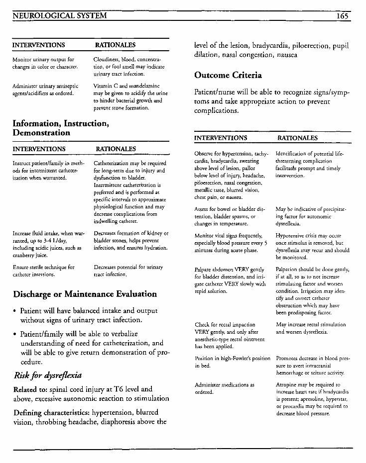
NEUROLOGICAL SYSTEM 165
INTERVENTIONS RATIONALES _ _ _ _ _ _ ~ ~~~
Monitor urinary output for changes in color or character.
Cloudiness, blood, concentra- tion, or foul smell may indicate urinary tract infection.
Vitamin C and mandelamine may be given to acid$ the urine to hinder bacterial growth and prevent stone formation.
Administer urinary antiseptic agentdacidifiers as ordered.
Information, Instruction, Demonstration
INTERVENTIONS RATIONALES
Instruct patiendfamily in meth- ods for intermittent catheter- ization when warranted.
Increase fluid intake, when war- ranted, up to 3-4 Llday, including acidic juices, such as cranberry juice.
Ensure sterile technique for catheter insertions.
Catheterization may be required for long-term due to injury and dysfunction to bladder. Intermittent catheterization is preferred and is performed at specific intervals to approximate physiological function and may decrease complications from indwelling catheter.
Decreases formation of kidney or bladder stones, helps prevent infection, and ensures hydration.
Decreases potential for urinary tract infection.
Discharge or Maintenance Evaluation
Patient will have balanced intake and output without signs of urinary tract infection.
understanding of need for catheterization, and will be able to give return demonstration of pro- cedure.
Patient/family will be able to verbalize
Risk f i r dysreflexiu Related to: spinal cord injury at TG level and above, excessive autonomic reaction to stimulation
Defining characteristics: hypertension, blurred vision, throbbing headache, diaphoresis above the
level of the lesion, bradycardia, piloerection, pupil dilation, nasal congestion, nausea
Outcome Criteria
Patient/nurse will be able to recognize signs/symp- toms and take appropriate action to prevent complications.
INTERVENTIONS RATIONALES
Observe for hypertension, tachy- cardia, bradycardia, sweating above level of lesion, pallor below level of injury, headache, piloerection, nasal congestion, metallic tare, blurred vision, chest pain, or nausea.
Assess for bowel or bladder dis- tention, bladder spasms, or changes in temperature.
Monitor vital signs frequently, especially blood pressure every 5 minutes during acute phase.
Palpate abdomen VERY gently for bladder distention, and irri- gate catheter VERY slowly with tepid solution.
Check for rectal impaction VERY gently, and only after anesthetic-rype rectal ointment has been applied.
Position in high-Fowler's position in bed.
Administer medications as ordered.
Identification of potential Iife- threatening complication facilitates prompt and timely intervention.
May be indicative of precipitat- ing factor for autonomic dysreflexia.
Hypotensive crisis may occur once stimulus is removed, but dysreflexia may recur and should be monitored.
Palpation should be done gently, if at all, so as to not increase stimulating factor and worsen condition. Irrigation may iden- tify and correct catheter obstruction which may have been predisposing factor.
May increase rectal stimulation and worsen dysreflexia.
Promotes decrease in blood pres- sure to avert intracranial hemorrhage or seizure activity.
Atropine may be required to increase heart rate if bradycardia is present; apresoline, hyperstar, or procardia may be required ro decrease blood pressure.
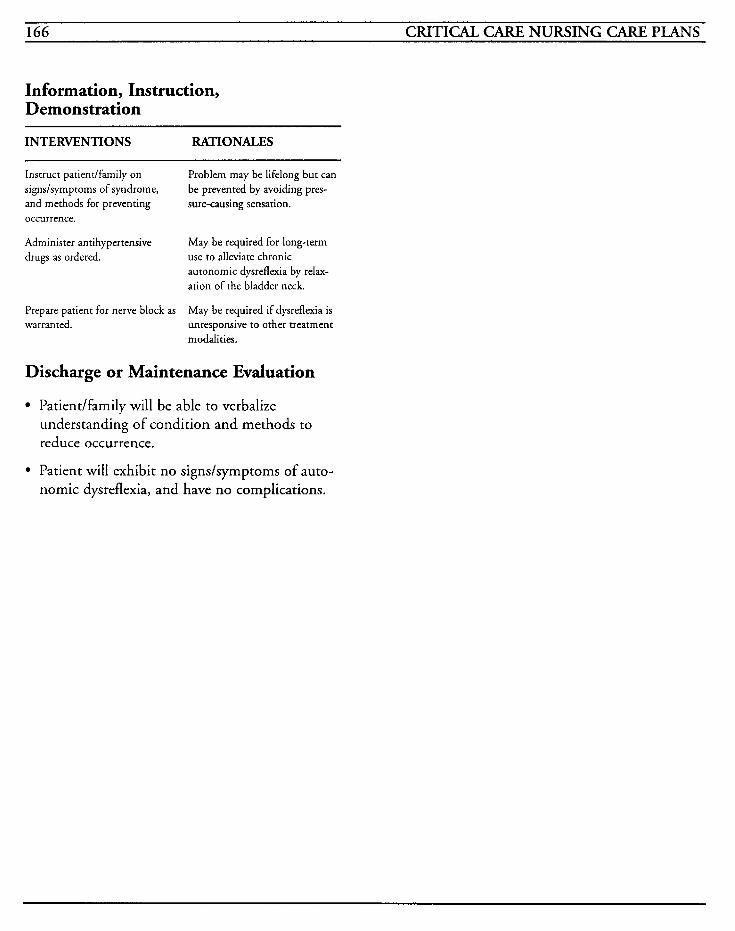
166 CRITICAL CARE NURSING CARE PLANS
Information, Instruction, Demonstration
INTERVENTIONS RATIONALES
Instruct patient/farnily on signslsymptoms of syndrome, and methods for preventing occurrence.
Administer antihypertensive drugs as ordered.
Problem may be lifelong but can be prevented by avoiding pres- sure-causing sensation.
May be required for long-term use to alleviate chronic autonomic dysreflexia by relax- ation of the bladder neck.
Prepare patient for nerve block as May be required if dysreflexia is warranted. unresponsive to other treatment
modalities.
Discharge or Maintenance Evaluation
Patiendfamily will be able to verbalize understanding of condition and methods to reduce occurrence.
Patient will exhibit no signs/symptoms of auto- nomic dysreflexia, and have no complications.
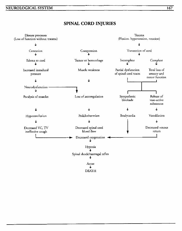
NEUROLOGICAL SYSTEM 167
SPINAL CORD INJURIES
Disease processes (Loss of function without trauma)
c Contusion
c Edema to cord
c Increased intradural
pressure
c
Compression c
Trauma (Flexion, hypertension, rotation)
c Transection of cord
c Tumor or hernorrhage Incomplete Complete
c c c Muscle weakness Partial dysfunction Total loss of
sensory and motor function
c I
of spinal cord tracts
I I
Neurodysfunction 4 1 I
Paralysis of muscles Loss of autoregulation Sympathetic Release of blockade vaso-active
substances
c c 4 J,
Hypoventilation
c Decreased VC, TV ineffective cough
Poikilothermism Bradycardia Vasodilation
c Decreased spinal cord
blood flow
JI
Decreased venous return
Decreased oxygenation c
Hypoxia c
Spinal shock/vasovagal reflex c
Arrest 4
DEATH

This Page Intentionally Left Blank
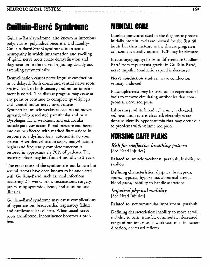
NEUROLOGICAL SYSTEM 169
6uillain-Barre Syndrome Guillain-Barrt syndrome, also known as infectious polyneuritis, polyradiculoneuritis, and Landry- Guillain-Barrt-Strohl syndrome, is an acute neuropathy in which inflammation and swelling of spinal nerve roots create demyelination and degeneration to the nerves beginning distally and ascending symmetrically.
Demyelination causes nerve impulse conduction to be delayed. Both dorsal and ventral nerve roots are involved, so both sensory and motor impair- ment is noted. The disease progress may cease at any point or continue to complete quadriplegia with cranial motor nerve involvement. Symmetrical muscle weakness occurs and moves upward, with associated paresthesias and pain. Dysphagia, facial weakness, and extraocular muscle paralysis occur. Blood pressure and heart rate can be affected with marked fluctuations in response to a dysfunctional autonomic nervous system. After demyelination stops, remyelination begins and frequently complete function is restored in approximately 70% of patients. The recovery phase may last from 4 months to 2 years.
The exact cause of the syndrome is not known but several factors have been known to be associated with Guillain-Barrt, such as, viral infections occurring 2-3 weeks prior, vaccinations, surgery, pre-existing systemic disease, and autoimmune d' iseases.
Guillain-Barrt syndrome may cause complications of hypertension, bradycardia, respiratory failure, and cardiovascular collapse. When sacral nerve roots are affected, incontinence becomes a prob- lem.
MEDICAL CARE Lumbar puncture: used in the diagnostic process; initially protein levels are normal for the first 48 hours but then increase as the disease progresses; cell count is usually normal; ICP may be elevated
Electromyography: helps to differentiate Guillain- BarrC from myasthenia gravis; in Guillain-Barrt, nerve impulse conduction speed is decreased
Nerve conduction studies: nerve conduction velocity is slowed
Plasmapheresis: may be used on an experimental basis to remove circulating antibodies that com- promise nerve receptors
Laboratory: white blood cell count is elevated; sedimentation rate is elevated; electrolytes are done to identifjr hyponatremia that may occur due to problems with volume receptors
NURSING CARE PUNS Risk f i r inefective breathing pattern [See Head Injuries]
Related to: muscle weakness, paralysis, inability to swallow
Defining characteristics: dyspnea, bradypnea, apnea, hypoxia, hypoxemia, abnormal arterial blood gases, inability to handle secretions
Impaired physical mobility [See Head Injuries]
Related to: neuromuscular impairment, paralysis
Defining characteristics: inability to move at will, inability to turn, transfer, or ambulate, decreased range of motion, muscle weakness, muscle incoor- dination, decreased reflexes
170 CRITICAL CARE NURSING CARE PLANS
Risk for alteration in nutrition: less than body requirements [See Mechanical Ventilation]
Related to: neuromuscular impairment, intuba- tion
Defining characteristics: weight loss, muscle wast- ing, catabolism, inability to take in sufficient nutrients, impaired cough/gag/swallow reflexes
Impaired verba 2 com mun ication [See CVA]
Related to: neuromuscular impairment, loss of muscle control, weakness
Defining characteristics: inability to speak, inabil- ity to write
Sensory-perceptual alterations: visualj kinesthetic, gustatory, tactile [See CVA]
Related to: neuromuscular deficits, altered recep- tion of stimuli, altered sensation, inability to communicate, hypoxia
Defining characteristics: paresthesias, hypersensi- tivity to stimuli, muscle incoordination, inability to communicate, anxiety, restlessness
Alteration in comfort
Related to: neuromuscular impairment
Defining characteristics: communication of pain or discomfort with minimal stimuli, muscle aches, tenderness, joint pain, flaccidity, spasticity
Outcome Criteria
Patient will have no complaints of pain, or pain will be controlled to patient's satisfaction.
INTERVENTIONS RATIONALES
Monitor for complaints of painldiscornfort and for non- verbal indications that patient m q be in discomfort.
Administer medication as ordered.
Apply hot or cold packs as warranted.
Use therapeutic touch, massage, imagery, visualization, or relax- ation therapies as warranted.
Patient may be unable to verbal- ize complaints.
Reduces or alleviates pain. Narcotics may cause respirarory depression.
Helps to alleviate discomfort and improves muscle and joinr stiff- ness.
Helps to refocus attention away from pain and provides for active participation in relieving pain.
Discharge or Maintenance Evaluation
Patient will have no complaints of pain or paresthesias.
Patient will be able to communication pain and requests for analgesics.
Patient will have pain controlled effectively to his satisfaction.
Risk for alteration in tissue per-sion: car- diopulmonary, peripheral, renal
Related to: autonomic nervous system impairment, hypovolemia, electrolyte imbalance, hypoxemia, thrombosis
Defining characteristics: hypotension, hyperten- sion, blood pressure lability, bradycardia, tachycardia, dysrhythmias, altered temperature regulation, decreased urine output, anuria, skin breakdown
Outcome Criteria
Patient will achieve and maintain normal perfu- sion of all body systems.
NEUROLOGICAL SYSTEM 171
INTERVENTIONS RATIONALES INTERVENTIONS RATIONALES
Monitor vital signs at rest and with turning. Notify M D of significant changes.
Monitor EKG for changes, and treat dysrhythmias per protocol.
Monitor temperature of skin and core body. Observe for inability to perspire.
Measure hemodynamics if pul- monary artery catheter in place, and notify MD for significant changes.
Observe skin surfaces for redness or breakdown. Place patient on kinetic bed, egg crate mattress, alternating pressure mattress, etc., if warranted
Observe calves for redness, edema, or positive Homan’s or Pratt’s signs.
Provide anti-embolic hose or sequential compression devices to both leg and remove at least once every 8 hours.
Monitor hourly intake and output. Notify MD if urine output is less than 30 cdhr for 2
Severe changes with blood pres- sure may occur as a result of autonomic dysfunction because of the loss of sympathetic out- flow to maintain peripheral vascular tone. Postural hypoten- sion may occur as a result of impaired reflexes which normally readjust pressure during changes in position.
Rate changes may occur as a result of vagal stimulation and impairment of the sympathetic innervation of the heart. Hypoxemia or electrolyte imbal- ances may alter vascular tone and impair venous return.
Vasomotor tone changes can impair the ability to perspire and cause temperature regulation problems. The patient’s impaired sensation may further promote difficulty with warming and cooling the body.
Impairment in vascular tone and venous return can decrease car- diac output.
Decreases in sensation as well as circulatory changes may result in impaired perfusion and facilitate skin breakdown or ischemia. Special bedslmattresses help to reduce hazards of immobility.
Venous stasis may increase potential for deep vein thrombo- sis formation, and patient may be unaware of discomfort due to paresthesias.
Helps to decrease venous stasis and promotes venous return.
Circulating volume may be decreased by patient’s inability to take in adequate hydration, fluid
hours, or if significant imbalance in I&O occurs. tone.
Administer IV fluids as ordered.
shifting, and decreases in vascular
Fluids help to prevent or correct hypovolemia but impaired vascu- lar tone may result in severe hemodynamic lability based on small increases in circulating volumes.
Administer heparin SQlIV as ordered, if no contraindications.
May be given prophylactically due to immobilization.
Discharge or Maintenance Evaluation
Patient will be able to maintain adequate perfu- sion.
Patient will have stable vital signs and hemody- namic parameters.
Patient will have regular cardiac rhythm with no
Risk for urinary retention
Related to: neuromuscular impairment, immobil-
ity
dysrhythmias.
Defining characteristics: inability to void, inabil- ity to completely empty bladder
Outcome Criteria
Patient will be able to empty bladder with no signs/symptoms of infection or retention.
INTERVENTIONS RATIONALES
Monitor for ability to void. Measure output carefully. neuromuscular progression.
Provides information regarding
Progression of disease may predispose patient to retention which may lead to urinary tract infection or other complications.
172 CRITICAL CARE NURSING CARE PLANS
~~~
INTERVENTIONS RATIONALES Outcome Criteria
Observe and palpate for bladder distention.
Bladder may become distended as sphincter reflex is involved in neuromuscular progression.
May be required to facilitate
process has resolved and bladder control has been achieved.
May indicate presence of urinary infection.
Insert indwelling catheter as warranted. urinary emptying until disease
Observe for concentrated urine, presence of blood or pus, changes in clarity or odor.
Information, Instruction, Demonstration
INTERVENTIONS RATIONALB
Instruct on need and procedure for catheter placement.
Promotes understanding and facilitates patient compliance.
Discharge or Maintenance Evaluation
Patient will be able to void suficient amounts without presence of retention or infection.
Patient will be able to accurately recall informa- tion regarding need and procedure for catheter placement.
Patient will be able to achieve bladder control once disease process has resolved.
Risk for constipation
Related to: neuromuscular impairment, bedrest, immobility, changes in dietary habits, changes in environment, analgesics
Defining characteristics: inability to expel all or part of stool, passage of hard stool, frequency less than normal pattern, rectal fullness, abdominal paidpressure, decreased bowel sounds, decreased peristalsis, weakness, fatigue, appetite impairment
Patient will be able to eliminate soft formed stool on a normal basis.
~~~~~ ~~
INTERVENTIONS RATIONALES
Evaluate elimination pattern, normal habits, ability to sense urge to defecate, presence of nausedvomiting, presence of painful hemorrhoids, and history of constipation problems.
Observe for abdominal disten- tion, tenderness, or guarding, nausea, vomiting, and absence of stool.
Palpate rectum for presence of stoollimpaction.
Auscultate bowel sounds for presence, pitch, and changes.
Administer stool softeners, laxa- tives, suppositories, or enemas as warrantedlordered.
Insert nasogastric tube as ordered. Connect with intermit- tent suction per hospital policy.
Increase fiber in dietltube feed- ings as warranted.
Provides baseline information to facilitate appropriate intervention for the patient’s plan of care.
May indicate present or impend- ing ileus or impaction.
Manual removal of stool may be required, and should be performed gently to avoid vagal stimulation. Other interventions may be necessary to allow for bowel elimination.
Diminished or absent bowel sounds, or presence of high- pitched tinkling sounds may indicate that an ileus has devel- oped.
May be required to stimulate bowel evacuation and to establish a bowel regime until patient is able to regain normal muscula- ture control.
Decompresses abdominal disten- tion that occurs with ileus formation, and helps prevent nausea and vomiting.
Helps to promote elimination by adding bulk and helps to regulate fecal consistency.
NEUROLOGICAL SYSTEM 173
Information, Instruction, Demonstration
INTERVENTIONS RATIONALES
Instruct on increases in fluid intake, dietary requirements, use of fruits and juices to improve bowel elimination.
Instruct on needlprocedure for nasogastric tube insertion.
Promotes knowledge and can help facilitate improvement in bowel regime.
Helps to promote understanding of complications that may occur with the loss of peristalsis due to the disease process.
Discharge or Maintenance Evaluation
Patient will achieve normal bowel elimination.
Patient will require no bowel aids to facilitate his normal routine.
Patient will regain muscle control and be able to evacuate stool.
Patient will be able to utilize dietary modifica- tion to maintain bowel regime.
Patient will be able to recall information correctly.
Anxz'ev, fear
Related to: disease process, change in health status, paralysis, respiratory failure, change in environment, threat of death
Defining characteristics: restlessness, apprehension, tension, fearfulness, sympathetic stimulation, changes in vital signs, inability to concentrate or focus, poor attention span, uncer- tainty of treatment and outcome, insomnia
Outcome Criteria
Patient will be able to reduce and/or relieve anxi- ety with appropriate methods.
INTERVENTIONS RATIONALES ~ _ _ _
Evaluate anxiety level frequently. Determination of severity of Stay with patient during acute patient's anxiety/fear can help to episodes. determine appropriate interven-
tion. Nurse's presence during acute anxiety may foster feelings of reassurance and concern for the patient's well-being.
Helps to decrease anxiety and Maintain consistency with nurse assignmenes. builds trust in relationships.
Patient should be placed near nurse's station and within visual con tact.
Provide method for patient to summon assistance. abandonment.
Involve patienc and family in Helps to foster understanding plan of care. Allow patient to and facilitates feelings of control make as many decisions as and improved self-esteem. warranted. Improves cooperation with pro-
cedures and care.
Discussion of fears provides opportunity for clarification of misperceptions and for realistic methods of dealing with prob- lems.
Patient$ anxiety may result in alterations in hemodynamic sta- bility and may require medication to initially deal with situational crises. Patient may require medication to facilitare improved ventilation should mechanical ventilation be war ranted.
Reassures patient that assistance will be nearby should he be unable to use call bell.
Reduces anxiety and fear of
Provide time for patientlfamily to discuss fears and concerns. Offer realistic options and do not give false reassurance.
Administer anti-anxiety medica- tions or sedation as warran ted/ordered.
Discharge or Maintenance Evaluation
Patient will be able to deal with changes in health status effectively.
Patient will be able to control anxiety and reduce fear to a manageable level.
Patient will have decreased anxiety and fear.
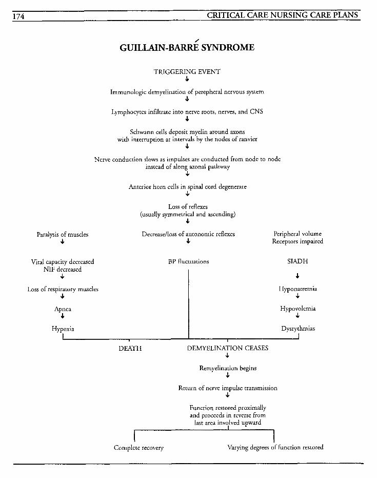
174 CRITICAL CARE NURSING CARE PLANS
Hypoxia
GUILLAIN-B-’ SYNDROME
D ysrythmias
Paralysis of muscles 4
TRIGGERING EVENT s
Immunologic demyelination of perepheral nervous system s
Lymphocytes infiltrate into nerve roots, nerves, and CNS J,
Schwann cells deposit myelin around axons with interruption at intervals by the nodes of ranvier
J,
Nerve conduction slows as impulses are conducred from node to node instead of along zonal pathway
J,
Anterior horn cells in spinal cord degenerate J,
Loss of reflexes (usually symmetrical and ascending)
J,
Decreaselloss of autonomic reflexes Peripheral volume J, Receptors impaired
Vital capacity decreased NIF decreased
J,
Loss of respiratory muscles J,
Apnea J,
BP fluctuations SIADH
J,
Hyponatremia J,
Hypovolemia 4
Remyelination begins s
Return of nerve impulse transmission J,
Function restored proximally and proceeds in reverse from
last area involved upward
I I Complete recovery Varying degrees of function restored
NEUROLOGICAL SYSTEM 175
Status Epilepticus Seizures occur when uncontrolled electrical impulses from the nerve cells in the cerebral cortex discharge and result in autonomic, sensory, and motor dysfunction. Status epilepticus is a series of repeated seizures, a prolonged seizure, or sequen- tial seizures longer than 30 minutes in which the patient does not regain consciousness. This seizure activity has a high mortality rate of up to 30% as a result of neurological and brain damage.
There are three types of status epilepticus: convul- sive, nonconvulsive, and partial status epilepticus. In the convulsive type, seizure activity may have a focal onset, but has tonic-clonic, grand mal type seizures without experiencing alertness between motor attacks. Nonconvulsive seizures are noted with a prolonged twilight state and are usually not motor activity. Partial status epilepticus occurs when continuous or repetitive focal seizures occur but consciousness is not altered.
Status epilepticus usually occurs in patients with pre-existing seizure disorders who have a precipi- tating factor occurance. These factors can include withdrawal from anticonvulsant medication, alco- hol withdrawal, sedative or antidepressant withdrawal, sleep deprivation, meningitis, encephalitis, brain abscesses or tumors, pregnancy, hypoglycemia, uremia, cerebrovascular disease, cerebral edema, or cerebral trauma.
The initial stage causes sympathetic activity increases with a decrease in the cerebral vascular resistance. After 30 minutes, hypotension occurs with a decrease in cerebral blood flow because of loss of autoregulation. The continuing massive autonomic discharges can cause bronchial secre- tions and restriction, with increased capillary permeability and pulmonary edema. Dysrhythmias can occur and patients may develop
rhabdomyolysis and renal failure. Other complica- tions may occur as a result of the significantly elevated metabolic state.
MEDICAL CARE Laboratory: glucose levels decreased; electrolytes to identie imbalances that may be precipitating factor or result from prolonged seizure activity; enzymes, especially creatine phosphokinase elevated after seizure activity; drug screen done to identify potential factor for drug withdrawal; CBC used to identify hemorrhage or infection with shift to the left on differential; drug levels for medications being given for seizures to evaluate therapeutic response and discern toxicity; renal profiles to evaluate renal function; urinalysis to identify hematuria or myoglobinuria
CT scans: may be done to identify lesions or pre- cipitating factors
Electroencephalogram: used to identify presence of seizure activity
Arterial blood gases: used to identify hypoxia and acid-base imbalances; usually acidosis seen
NURSING CARE PLANS Risk for impaired gas exchange [See Mechanical Ventilation]
Relat'ed to: altered oxygen. supply from repetitive seizures, cognitive impairment, neuromuscular impairment
Defining characteristics: restlessness, cyanosis, inability to move secretions, tachycardia, dysrhythmias, abnormal ABGs, decreased oxygen saturation
Risk for inefective airway clearance [See :Mechanical Ventilation]
176 CRITICAL CARE NURSING CARE PLANS
Related to: neuromuscular impairment, cognitive impairment, tracheobronchial obstruction
Defining characteristics: adventitious breath sounds, dyspnea, tachypnea, shallow respirations, cough with or without productivity, cyanosis, anx- iety, restlessness
Hyperthemia [See Pheochromocytoma]
Related to: continued seizure activity, increased metabolic state
Defining characteristics: fever, persistent tonic- clonic seizure activity, persistent focal seizures, persistent generalized seizures, tachycardia
Risk for fluid volume deficit [See ARDS]
Related to: excessive loss of fluid, decreased intake
Defining characteristics: hypo tension, tachycar- dia, fever, weight loss, oliguria, abnormal electrolytes, low filling pressures, decreased mental status, decreased specific gravity, increased serum osmolality
Risk fir injury Related to: seizure activity, increased metabolic demands
Defining characteristics: respiratory acidosis, metabolic acidosis, hypoxemia, hyperthermia, hypoglycemia, electrolyte imbalances, renal failure, rhabdomyolysis, exhaustion, death
Outcome Criteria
INTERVENTIONS RATIONALES
Maintain patent airway and ade- quate ventilation.
Monitor oxygen saturation by oximeter.
Provide supplemental oxygen as warranted.
Monitor ABGs for imbalances and treat per protocol.
Administer medications as ordered.
Maintain patient in seizure-free status.
Monitor EKG for dysrhythmias and treat per hospital protocol.
Patient will achieve and maintain seizure-free status with optimal oxygenation and ventilation without complications.
Monitor intake and output every 2 hours and prn.
Intubation and placement on mechanical Ventilation may be required if seizures cannot be controlled.
Decreases in saturation that cannot be improved with supple- mental oxygen may require mechanical ventilation. Seizure activity increases oxygen con- sumption and demand.
May be required to maintain desired levels of oxygen.
Metabolic increases may lead to lactate formation and acidosis.
Valium may be given IV at 5 mg/min rate to control seizure activity by enhancing neurotrans- mitter GABA. Ativan 2-4 mg IV may be given and repeated every 15 minutes as needed for seizure control. Caution should be exer- cised because respiratory and cardiovascular depression can occur. Phenobarbital IV at 60 mg/min may be given to depress excitation, decrease calcium uptake by nerves, and to strengthen repression of synapses. Dilantin IV at 50 mglmin rate may be given to decrease cellular influx of sodium and calcium and blocking neurotransmission release.
Once seizures have stopped, anti- conwlsant drugs must be given to prevent recurrence of seizure activity.
Electrolyte imbalances, too-rapid administration of medications, and hypoxia may contribute to appearance of cardiac dysrhyth- mias that may require interventional care.
Identifies imbalances with fluid status and fluid shifting.
NEUROLOGICAL SYSTEM 177
INTERVENTIONS RATIONALES Discharge or Maintenance Evaluation
Monitor labwork for changes and trends.
Electrolytes may fluctuate because of cellular movement of ions. Myoglobin may be present in urine as a result of prolonged seizure activity and can lead to renal failure. Drug levels may rise to toxic levels and should be evaluated for therapeutic effec- tiveness.
Identification may lead to timely intervention and treatment.
Identify and treat underlying cause of seizures.
Information, Instruction, Demonstration
INTERVENTIONS RATIONALES
Instruct patient/family in disease process and methods for reduc- tion of seizures.
Instruct in drug regimen, effects, side effects, contraindications, and precautions.
Instruct in oral care.
Instruct on use of medical alert bracelet.
Instruct on methods to promote safety with activities, such as, driving, using mechanical equip- ment, swimming, or hobbies.
Instruct on contact people, com- munity resource groups, counselors, as warranted.
Promotes knowledge and facili- tates compliance.
Promotes knowledge and helps prevent lack of cooperation with medication regime with resultant seizure breakthrough activity. Presence of side effects may indi- cate the need for changes in doses or medication type. Interactions with other drugs may produce adverse reactions, such as potentiated anticoagula- tion effect when dilantin and coumadin are concurrently taken,
Prevents gingival hypertrophy that may occur while taking dilantin.
May hasten emergency treatment in critical situations.
May facilitate prevention of injury to self or other if seizures occur without warning.
Patiedfamily will be able to verbalize understanding of all instructions and comply with medical regimen.
Patient will remain free of seizures and injury.
Patient' will be able to effectively access commu- nity resources for help and support.
Patient will exhibit no signs of complications.
Disturbance in self-esteem
Related to: perception of loss of control, ashamed of medical condition
Defining characteristics: fear of rejection, concerns about changes in lifestyle, negative feel- ings about self, change in perception of role, changes in responsibilities, lack of participation in therapy or care, passiveness, inability to accept positive reinforcement, little eye contact, brief responses to questions
Outcome Criteria
Patient will be able to participate in own care and have positive perceptions of self.
INTERVENTIONS RATIONALES
Encourage patient to initiate self-care or request assistance.
Discuss patient's perceptions of illness and potential reactions of others to his disease.
Discuss previous success episodes and patient's strengths.
May provide opportunities for long-term support and sharing ideas with others who have simi- lar problems.
Participation in care facilitates feelings of normalcy.
Provides opportunities to estab- lish patient's knowledge base, clear up any misconceptions, and opportunity to problem-solve responses to future seizures.
Concentrating on the positive experiences may help to reduce self-consciousness and allow patient to begin to accept condi- tion.
178 CRITICAL CARE NURSING CARE PLANS
INTERVENTIONS RATIONALES ~~ ~~
Discuss concerns with family members, allowing ample time for members to discuss problems and attitudes.
Consult with counselors, minis- terial support, or resource groups.
Negative feelings from patient’s family may afFect his sense of self-esteem.
Provides opportunity for parient to deal with stigma of disease and overcome feelings of inferi- ority.
Discharge or Maintenance Evaluation
Patient will be able to identify ways to cope with negative feelings. . Patiendfamily will be able to discuss concerns and effect realistic problem-solving plans.
Patient will become more accepting of self, with increased self-esteem.
Patient will be able to effectively access commu- nity resources to gain help and support.
Grieving [See Amputation]
Related to: traumatic injury, loss of physical well- being
Defining characteristics: communications of dis- tress, denial, guilt, fear, sadness, changes in affect, changes in ability and desire for communication, crying, insomnia, lethargy
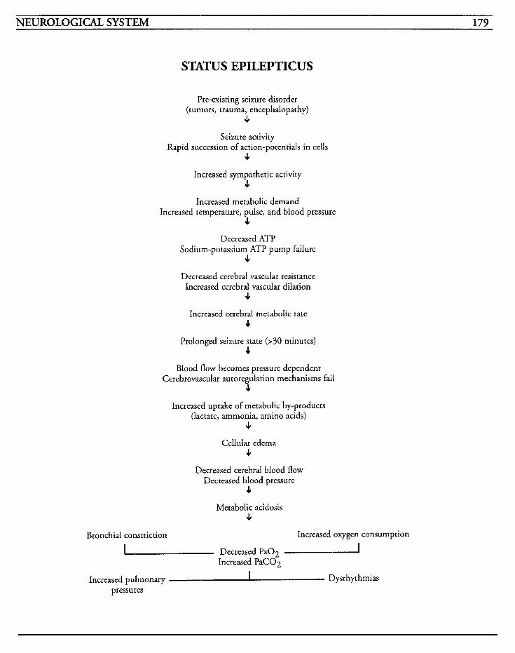
NEUROLOGICAL SYSTEM 179
STATUS EPILEPTICUS
Pre-existing seizure disorder (tumors, trauma, encephalopathy)
c Seizure activity
Rapid succession of action-potentials in cells c
Increased sympathetic activity c
Increased metabolic demand Increased temperature, pulse, and blood pressure
c Decreased ATP
Sodium-potassium ATP pump failure e
Decreased cerebral vascular resistance Increased cerebral vascular dilation
c Increased cerebral metabolic rate
c Prolonged seizure state (>30 minutes)
c Blood flow becomes pressure dependent
Cerebrovascular autoregulation mechanisms fail 4
Increased uptake of metabolic by-products (lactate, ammonia, amino acids)
JI
Cellular edema 4
Decreased cerebral blood flow Decreased blood pressure
4
Metabolic acidosis c
Bronchial constriction Increased oxygen consumption
Decreased PaO2 -A Increased PaCO2
Increased pulmonary a- Dysrhythmias pressures

This Page Intentionally Left Blank
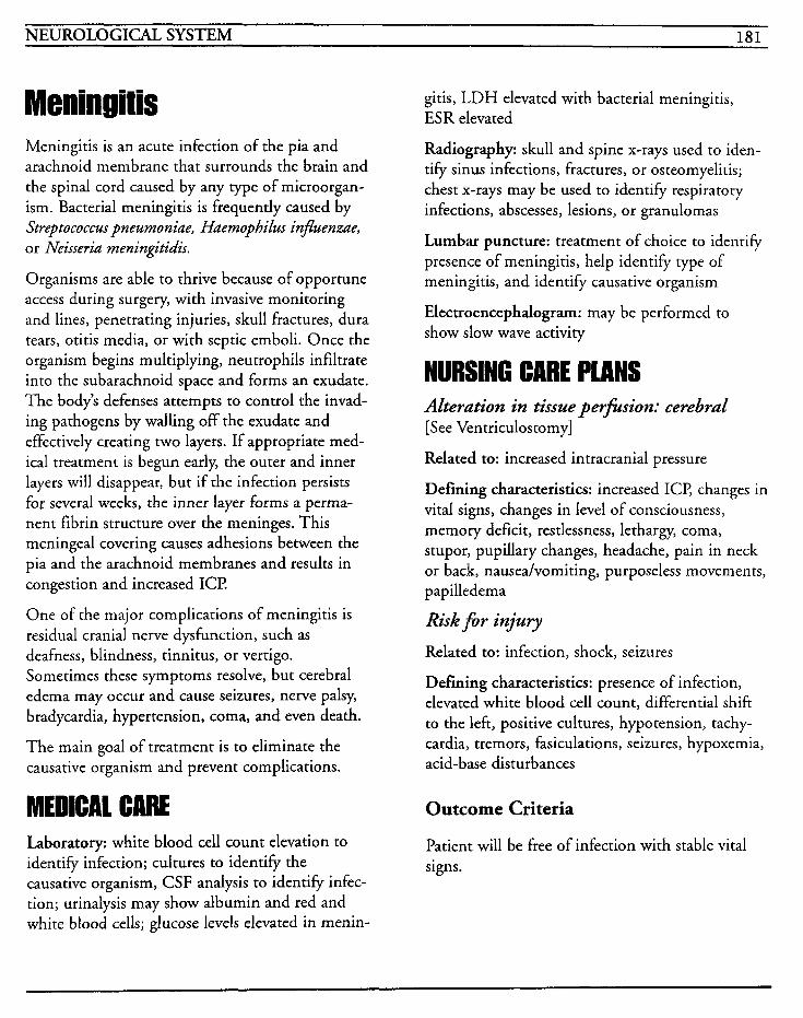
NEUROLOGICAL SYSTEM 181
Meningitis is an acute infection of the pia and arachnoid membrane that surrounds the brain and the spinal cord caused by any type of microorgan- ism. Bacterial meningitis is frequently caused by Streptococcus pneumoniae, Huemophilus injuenzae, or Neisseriu meningitidis.
Organisms are able to thrive because of opportune access during surgery, with invasive monitoring and lines, penetrating injuries, skull fractures, dura tears, otitis media, or with septic emboli. Once the organism begins multiplying, neutrophils infiltrate into the subarachnoid space and forms an exudate. The body’s defenses attempts to control the invad- ing pathogens by walling off the exudate and effectively creating two layers. If appropriate med- ical treatment is begun early, the outer and inner layers will disappear, but if the infection persists for several weeks, the inner layer forms a perma- nent fibrin structure over the meninges. This meningeal covering causes adhesions between the pia and the arachnoid membranes and results in congestion and increased ICP
One of the major complications of meningitis is residual cranial nerve dysfunction, such as deafness, blindness, tinnitus, or vertigo. Sometimes these symptoms resolve, but cerebral edema may occur and cause seizures, nerve palsy, bradycardia, hypertension, coma, and even death.
The main goal of treatment is to eliminate the causative organism and prevent complications.
MEDICAL CARE Laboratory: white blood cell count elevation to identify infection; cultures to identify the causative organism, CSF analysis to identify infec- tion; urinalysis may show albumin and red and white blood cells; glucose levels elevated in menin-
gitis, LDH elevated with bacterial meningitis, ESR elevated
Radiography: skull and spine x-rays used to iden- tify sinus infections, fractures, or osteomyelitis; chest x-rays may be used to identify respiratory infections, abscesses, lesions, or granulomas
Lumbar puncture: treatment of choice to identify presence of meningitis, help identify type of meningitis, and identify causative organism
Electroencephalogram: may be performed to show slow wave activity
NURSING CARE PLANS Alteration in tissue perfusion: cerebral [See Ventriculostomy]
Related to: increased intracranial pressure
Defining characteristics: increased ICE changes in vital signs, changes in level of consciousness, memory deficit, restlessness, lethargy, coma, stupor, pupillary changes, headache, pain in neck or back, nauseahomiting, purposeless movements, papilledema
Risk Jor injury Related to: infection, shock, seizures
Defining characteristics: presence of infection, elevated white blood cell count, differential shift to the left, positive cultures, hypotension, tachy- cardia, tremors, fasiculations, seizures, hypoxemia, acid-base disturbances
Outcome Criteria
Patient will be free of infection with stable vital signs.
182 CRITICAL CARE NURSING CARE PLANS
Defining characteristics: headache, muscle spasms, backache, photophobia, crying, moaning, INTERVENTIONS RATIONALES
restlessness, communication of pain, muscle ten- Assist with lumbar puncture. Identifies prezence of infection
types of meningitis. CSF with
sion, facial grimacing, pallor, changes in vital signs
Hyperthemia [See Pheochromocytoma]
and can differentiate between
low white cell counts, less pro- tein elevation, and glucose levels "
Related to: infection process approximately half that of the blood glucose level may be indicative of viral meningitis. CSF that has an elevated initial pressure, high protein, low glu-
Defining characteristics: fever, tachycardia, tachypnea, warm, flushed skin, seizures
Administer antimicrobials as ordered, as soon as possible.
cose, cloudy color, and high white cell count indicates baccer- id meningitis.
Aqueous penicillin G is usually the drug of choice, but culture results may indicate a different agent needed to eradicate the organism. Antibiotics are usually given in larger doses at closer intervals in order to facilitate penetration across the blood- brain barrier.
Observe appropriate isolation techniques up to 48 hours after antibiotic regimen has begun.
Prevents spread of infection. After antimicrobial therapy has been instituted for 2 days, the patient is not considered infec- tious.
May be required for control of new seizure activity due to rneningeal irritation.
Administer anticonvulsants as ordered.
Discharge or Maintenance Evaluation
Patient will be free of infection with no compli- cations from antimicrobial agents.
Patient will be free of seizure activity.
Patient will comply with isolation restrictions.
Alteration in comfort [See Guillain-Bard]
Related to: infectious organisms, circulating toxins, invasive lines, bedrest
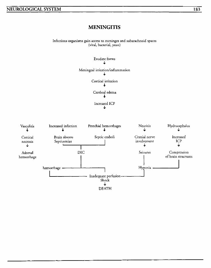
NEUROLOGICAL SYSTEM 183
MENINGITIS
Infectious organisms gain access to meninges and subarachnoid spaces (viral, bacterial, yeast)
Exudate forms e
Meningeal irritation/inflammation c
Cortical irritation 4
Cerebral edema c
Increased ICP c
Vasculitis c
Cortical necrosis
c Adrenal
hemorrhage
Increased infection Petechial hernorrhages Neuritis Hydrocephalus c 4 c c
Brain abscess Septic emboli Cranial nerve Increased Septicemias involvement ICP
4 J,
Seizures Compression - DIC
of brain structures I
hemorrhage -1 Hypoxia
I- Inadequate perfusion - Shock
c DEATH

This Page Intentionally Left Blank
NEUROLOGICAL SYSTEM 185
UentriculostomyhCP Monitoring The brain is housed in a nondistensible cavity that is filled to capacity with CSF, interstitial fluid, and intravascular blood, all of which possess very mini- mal ability for adjustment for increasing intracranial pressure. If the volume of any one of these constituents increases, there is a reciprocal decrease in the volume of one or more of the others, or else intracranial pressure becomes ele- vated. Intracranial pressure is normally beween 2-15 mmHg or 50-200 cm H,O, and fluctuates depending on positioning, vital signs changes, increased intra-abdominal pressure, and stimuli.
There are compensatory mechanisms that assist in decreasing intracranial hypertension. The most easily changed element is intravascular volume which results from compression of the venous system and decreases fluid level. The CSF is another element that can be used to compensate for increasing pressures. CSF can be displaced from the cranial vault to the spinal canal, which increases absorption of CSF by the arachnoid villi, slows production of CSF by the choroid plexus, and decreases ICl? Other compensatory mechanisms may be seen, such as skull expansion in infants whose sutures have not closed, as well as reduction of cerebral blood flow to a small extent, but these are not desirable.
Although auto-regulatory mechanisms can control small increases in ICE rapid or sustained increases suppress these compensatory efforts, and decom- pensation occurs. As the ICP increases, the cerebral blood flow decreases because of pressure exerted on vessels. This causes brain ischemia and accumulation of lactic acid and carbon dioxide, resulting in hypoxemia and hypercapnia. Cerebral vasodilation ensues which increases blood volume and cerebral edema, which further increases ICE
until a vicious cycle is established. When ischemia increases to a certain level, the medulla causes blood pressure to rise in an effort to compensate for the increasing ICE but eventually the ICP will equal the MAP and precipitate curtailing of cere- bral blood flow, resulting in vascular collapse and brain death.
ICP is increased when brain volume is enlarged by mass lesions, tumors, abscesses, hematomas or cerebral edema. Vasodilation and venous outflow obstructions cause changes in cerebrovascular status due to hypoventilation, hypercapnia, improper position of the head, or maneuvers that increase intrathoracic pressure. CSF volumes may increase from decreased reabsorption from an obstruction, such as with hydrocephalus.
Monitoring of ICP can be done from several sites. The lumbar or cervical subarachnoid area is simple to access, but potential for herniation exists. The lateral cerebral ventricles [per ventricu- lostomy] is highly accurate and allows for withdrawal of CSF and measurement of compli- ance, but infection to this area is catastrophic. Subdural sites are most easily inserted but carry serious infection risks, and an epidural site has less potential for infection, but lacks accuracy.
The three types of ICP monitoring are epidural sensor monitoring through a burr hole, subarach- noid screw or bolt monitoring through a twist drill burr hole, and ventricular catheter monitor- ing. Insertion of these may be performed in surgery or in the intensive care setting, but requires sterile field maintenance.
ICP monitoring may be performed on patients with head trauma, ruptured aneurysms, Reye’s syndrome, intracranial bleeds, hydrocephalus, or tumors. A ventriculostomy is a cannula placed in the lateral ventricle and connected with a transducer for measurement of pressures of CSF directly, for periodic drainage of CSF, and for withdrawal of fluid for analysis.

186 CRITICAL CARE NURSING CARE PLANS
Cerebral perfusion pressure (CPP) is the difference between the mean arterial pressure (MAP) and the mean ICE and indicates the pressure in the cere- bral vascular system and approximates the cerebral blood flow. A CPP of 60 mmHg is the minimum value for perfusion to occur, with normal ranges from 80-100 mmHg.
Increases in ICP can be manifested by signs such as systolic blood pressure elevations, widening pulse pressure, bradycardia, headache, nausea with projectile vomiting, papilledema, changes in level of consciousness, pupillary changes, respiratory changes, and cerebral posturing.
MEDICAL CIIRE Surgery: may be required for traumatic injuries and/or placement of ICP monitoring device
Arterial blood gases: may be used to identify acid- base imbalances, hypoxemia, and hypercapnia; frequently patients are hyperventilated to keep PaC02 between 25-28
Osmotics: mannitol used to create osmotic diure- sis in an attempt to decrease ICP
Barbiturate therapy: pentothal or nembutal used to place patient in coma to produce burst-suppres- sion on the EEG and to reduce metabolic activity
Paralyzing drug therapy: pancuronium may be used to decrease metabolic requirements but must be used in conjunction with sedatives since drug only paralyzes muscles and does not change level of awareness
Adrenocorticosteroids: decadron has less sodium- retaining properties and is used to assist with decreasing edema
CT scans: used to identify lesions, hemorrhage, ventricular size, structural shifting, ischemic event (may be several days prior to visibility on scan)
Laboratory: electrolytes drawn to evaluate imbal- ances that may contribute to ICP increases; toxicology screens to identify other drugs that may be responsible for changes in mentation and level of consciousness; serum levels of drugs to assess therapeutic response versus toxicity
NURSING CARE PLANS Alteration in tissue per-s ion: cerebral
Related to: cerebral edema, space-occupying lesions, hemorrhage, substance overdose, hypoxia, hypovolemia, trauma
Defining characteristics: increased ICE changes in vital signs, changes in level of consciousness, memory deficit, restlessness, lethargy, coma, stupor, pupillary changes, headache, nausea/vomit- ing, purposeless movements, papilledema
Outcome Criteria
Patient will have stable vital signs and mentation with no signs or symptoms of increased ICP
INTERVENTIONS RATIONALES
Monitor for changes in level of consciousness or mentation, speech, or response to commandslquestions. tate prompt intervention.
Alterations in levels of conscious- ness are among the earliest signs of increasing ICP and can facili-
Progressive deterioration may require emergent care.
As ICP increases, blood pressure elevates, pulse pressure widens, bradycardia may occur changing to tachycardia as ICP progres- sively worsens. Tachypnea is seen as an early sign but slows with increasingly longer periods of apnea. Fever may indicate hypo- thalarnic damage or infection which can increase metabolic demands and further increase ICE
Monitor vital signs at least every hour, and prn.
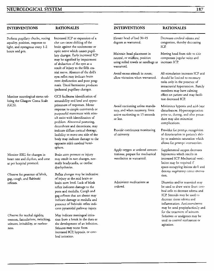
NEUROLOGICAL SYSTEM 187
INTERVENTIONS RATIONALES INTERVENTIONS RATIONALES
Perform pupillary checks, noting equality, position, response to light, and nystagmus every 1-2 hours and prn.
Monitor neurological status uti- lizing the Glasgow Coma Scale (GCS).
Monitor EKG for changes in hearr rate and rhythm, and treat as per hospital protocol.
Observe for presence of blink, gag, cough, and Babinski reflexes.
Observe for nuchal rigidity, tremors, fasiculations, twitching, seizures, irritabiliv, or restless- ness.
Increased ICP or expansion of a clot can cause shifting of the brain against rhe oculomotor or optic nerve which causes pupil- lary changes. Early increased ICP may be signified by impairment of abduction of the eyes as a result of injury to the fifth cra- nial nerve. Absence of the doll’s eyes reflex may indicate brain stem dysfunction and poor prog- nosis. Uncal herniation produces ipsilateral pupillary changes.
GCS facilitates identification of arousabilig and level and appro- priateness of responses. Motor response to simple commands or purposeful movement with srim- uli assist with identification of problem. Abnormal posturing, decerebrare and decorticate, may indicate diffuse cortical damage. Inability ro move one side of the body may indicate damage to the opposite side’s cerebra hemi- sphere.
Brain srem pressure or injury may result in rate changes, nor- mally bradycardia, or cardiac dysrhythmias.
Reflex changes may be indicative of injury at the mid brain or brain stem level. Lack of blink reflex indicates damage to the pons and medulla. Cough and gag reflexes that are absent may indicate damage at medulla and presence of Babinski reflex indi- cates pyramidal pathway injury.
May indicate meningeal irrita- tion from a break in the dura or the development of an infection. Seizures may occur from increased ICP, hypoxia, or cere- bral irritation.
Elevate head of bed 30-45 degrees as warranted.
Maintain head placement in neutral, or midline, position using rolled towels or sandbags as warranted.
Avoid excess stimuli in room; allow visitation when warranted.
Avoid suctioning unless manda- tory, and when necessary, limit active suctioning to 15 seconds or less.
Provide continuous monitoring of oximetry.
Apply oxygen at ordered concen- trations; prepare for mechanical ventilation as warranted.
Administer medications as ordered.
Decreases cerebral edema and congesrion, thereby decreasing ICI?
Moving head from side to side compresses jugular veins and increases ICE!
All stimulation increases ICP and should be limited to necessary tasks only in the presence of intracranial hypertension. Family members may have calming effect on patient and may facili- tate decreased ICl?
Minimizes hypoxia and acid-base disturbances. Hyperoxygenation prior to, during, and after proce- dure may also minimize complications.
Provides for prompt recognition of deterioration in patient‘s abil- ity to maintain saturation which allows for prompt intervention.
Supplemental oxygen decreases hypoxemia which results in increased ICI? Mechanical venri- larion may be required if space-occupying lesions shift and destroy respirarory cenrer enerva- tion.
Diuretics andlor mannirol may be used to draw water from cere- bral cells ro decrease edema and ICP. Steroids may be used to decrease rissue edema and inflammation. Anticonvulsanrs may be used prophylactically and for the rreatment of seizures. Sedatives or analgesics may be used ro control restlessness or agitation.
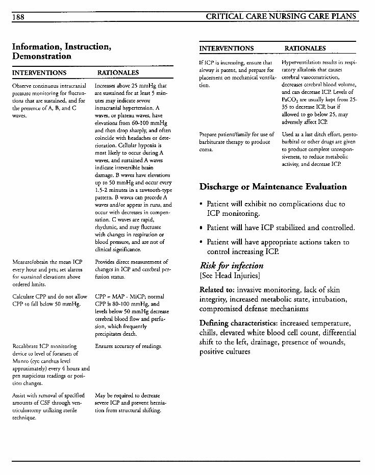
188 CRITICAL CARE NURSING CARE PLANS
Information, Instruction, INTERVENTIONS RATIONALES Demonstration
If ICP is increasing, ensure that airway is patent, and prepare for placement on mechanical ventila-
Hyperventilation results in respi- ratory alkalosis that causes cerebral vasoconstriction,
INTERVENTIONS RATIONALES
Observe continuous intracranial pressure monitoring for fluctua- tions that are sustained, and for the presence of A, B, and C waves.
Measurelobtain the mean ICP every hour and prn; set alarms for sustained elevations above ordered limits.
Calculate CPP and do not allow CPP to fall below 50 mmHg.
Recalibrate ICP monitoring device to level of foramen of Munro (eye canthus level approximately) every 4 hours and prn suspicious readings or posi- tion changes.
Assist with removal of specified amounts of CSF through ven- triculostomy utilizing sterile technique.
Increases above 25 mmHg that are sustained for at least 5 min- utes may indicate severe intracranial hypertension. A waves, or plateau waves, have elevations from 60-100 mmHg
tion.
and then drop sharply, and often coincide with headaches or dete- rioration. Cellular hypoxia is most likely to occur during A waves, and sustained A waves indicate irreversible brain damage. B waves have elevations up to 50 mmHg and occur every 1.5-2 minutes in a sawtooth-type pattern. B waves can precede A waves andlor appear in runs, and occur with decreases in compen- sation. C waves are rapid, rhythmic, and may fluctuate with changes in respiration or blood pressure, and are not of clinical significance.
Provides direct measurement of changes in ICP and cerebral per- fusion status.
CPP = MAP - MiCP; normal CPP is 80-100 mmHg, and levels below 50 mmHg decrease cerebral blood flow and perfu- sion, which frequently precipitates death.
Ensures accuracy of readings.
May be required to decrease severe ICP and prevent hernia- tion from structural shifting.
decreases cerebral blood volume, and can decrease ICP Levels of PaCO, are usually kept from 25- 35 to decrease ICE but if allowed to go below 25, may adversely affect ICP.
Prepare patientlfamily for use of Used as a last ditch effort, pento- barbiturate therapy to produce barbiral or other drugs are given coma. to produce complete unrespon-
siveness, to reduce metabolic activity, and decrease ICP
Discharge or Maintenance Evaluation
Patient will exhibit no complications due to ICP monitoring.
Patient will have ICP stabilized and controlled.
Patient will have appropriate actions taken to
Risk f i r infiction [See Head Injuries]
Related to: invasive monitoring, lack of skin integrity, increased metabolic state, intubation, compromised defense mechanisms
Defining characteristics: increased temperature, chills, elevated white blood cell count, differential shift to the left, drainage, presence of wounds, positive cultures
control increasing ICP
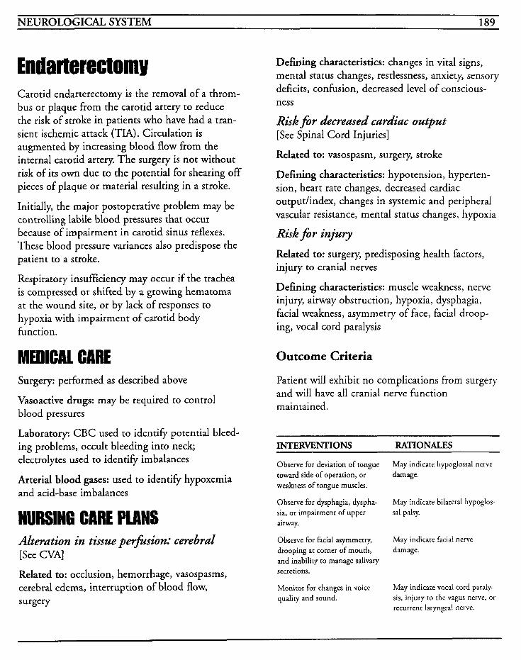
NEUROLOGICAL SYSTEM 189
Carotid endarterectomy is the removal of a throm- bus or plaque from the carotid artery to reduce the risk of stroke in patients who have had a tran- sient ischemic attack (TIA). Circulation is augmented by increasing blood flow from the internal carotid artery. The surgery is not without risk of its own due to the potential for shearing off pieces of plaque or material resulting in a stroke.
Initially, the major postoperative problem may be controlling labile blood pressures that occur because of impairment in carotid sinus reflexes. These blood pressure variances also predispose the patient to a stroke.
Respiratory insufficiency may occur if the trachea is compressed or shifted by a growing hematoma at the wound site, or by lack of responses to hypoxia with impairment of carotid body function.
Surgery: performed as described above
Vasoactive drugs: may be required to control blood pressures
Laboratory: CBC used to identify potential bleed- ing problems, occult bleeding into neck; electrolytes used to identify imbalances
Arterial blood gases: used to identify hypoxemia and acid-base imbalances
NURSING CARE PLANS Alteration in tissue perfision: cerebral [See CVA]
Related to: occlusion, hemorrhage, vasospasms, cerebral edema, interruption of blood flow, surgery
Defining characteristics: changes in vital signs, mental status changes, restlessness, anxiety, sensory deficits, confusion, decreased level of conscious- ness
Rid for decreased cardiac output [See Spinal Cord Injuries]
Related to: vasospasm, surgery, stroke
Defining characteristics: hypotension, hyperten- sion, heart rate changes, decreased cardiac outputhndex, changes in systemic and peripheral vascular resistance, mental status changes, hypoxia
Risk for injury
Related to: surgery, predisposing health factors, injury to cranial nerves
Defining characteristics: muscle weakness, nerve injury, airway obstruction, hypoxia, dysphagia, facial weakness, asymmetry of face, facial droop- ing, vocal cord paralysis
Outcome Criteria
Patient will exhibit no complications from surgery and will have all cranial nerve function maintained.
INTERVlENTIONS RATIONALES
Observe for deviation of tongue toward side of operation, or weakness of tongue muscles.
Observe for dysphagia, dyspha- sia, or impairment of upper airway.
Observe for facial asymmetry, drooping at corner of mouth, and inability to manage salivary secretions.
Monitor for changes in voice quality and sound.
May indicate hypoglossal nerve damage.
May indicate bilateral hypoglos sal palsy.
May indicate facial nerve damage.
May indicate vocal cord paraly- sis, injury to the vagus nerve, or recurrent laryngeal nerve.
190 CRITICAL CARE NURSING CARE PLANS
Discharge or Maintenance Evaluation
Patient will have facial symmetry and normal voice modulation.
Patient will exhibit no signs/symptoms of cra- nial nerve injury.
Patient will be free of any airway compromise and have stable vital signs.
Alteration in skin integrity [See Cardiac Surgery]
Related to: surgical wounds, invasive lines, immo- bility
Defining characteristics: presence of wounds, drainage, redness, swelling, abrasions, pressure, lacerations, bruises, open skin
GASTROINTESTINAL/HEPATIC SYSTEMS 191
Gastrointestinal Bleeding
Esophageal Varices
Hepatitis
Pancreatitis
Acute AbdomedAbdorninal Trauma
Liver Failure

This Page Intentionally Left Blank
GASTROINTESTINAL/HEPATIC SYSTEMS 193
Gastroint estinal Bleeding - Gastrointestinal bleeding may be massive and acute or occult and chronic in nature. GI bleeding results when irritation of the mucosal lining results in erosion through to the submucosal layer. Upper GI hemorrhage is considered to be a bleed from any site proximally to the cecum, and all ulcerative bleeding is arterial with the exception of a tear that cuts across all vessels, malignant tumors, and in patients with esophagitis.
When erosion into an artery occurs, it usually pro- duces two bleeding sites because of arterio-arterial anastomoses. When the bleeding occurs at the ulcer base artery, it may be a life-threatening emer- gency.
Bleeding may occur from the lower gastrointesti- nal tract as well. Causes of lower GI bleeding include hemorrhoids, diverticulosis, inflammatory bowel disease, rectal perforation, or intussuscep- tion.
Acute upper GI bleeding may result from many causes, such as gastritis, peptic ulcer, stress, drugs, hormones, trauma, head injuries, burns, and esophageal varices.
Differential diagnosis between gastric and duode- nal ulcers must be obtained. Duodenal ulcers usually account for approximately 80% of all ulcers noted and rarely become cancerous. Gastric ulcers, on the other hand, may become cancerous and are more likely to bleed.
Initial presenting symptoms of a GI bleed are either hematemesis, melena, or hematochezia. An acute bleed will have more than 60 cc/day of black tarry stool and usually greater than 500 cc, whereas occult bleeding is normally 15-30 cc/day. Stools can be positive for occult blood up to 12 days after an acute bleed. Of all GI hemorrhages, 80% usually stop spontaneously.
The goal of treatment is initially prevention and treatment of shock, with fluid volume replacement. Maintenance of circulating blood volume is imperative to prevent myocardial infarc- tion, sepsis, and death. Endoscopic examination is the primary diagnostic procedure utilized. Once the lesion has been identified, treatment with Pitressin infusion may be used to control bleeding.
MEDICAL CARE Laboratory: CBC to identify changes in blood volume and concentration, but may be normal during rapid loss because of the lapsed time required for equilibration of intravascular with extravascular spaces; MCV is useful to identify prolonged chronic loss with iron deficiency; B,, and folic acid levels used to identify anemia type; reticulocyte count may identify new RBC forma- tion which occurs with an old bleed; platelet count, PT, PTT, and bleeding times to evaluate clotting status and platelet dysfunction; BUN and creatinine to evaluate effect on renal status; elec- trolytes to evaluate imbalances and treatment; ammonia levels may be used to identify liver dys- function; gastric analysis to determine presence of blood and assess secretory activity of gastric mucosa; amylase elevated if duodenal ulcer has posterior penetration; pepsinogen level to help identify type of bleeding, with elevation seen in duodenal ulcer, and decreased levels seen in gastri- tis; stool specimens for guaiac
Arterid blood gases: may be used to show acid-base imbalances, compensation for decreased blood flow; initially respiratory alkalosis changing to metabolic acidosis as metabolic wastes accumu- late
Esophagogastroduodenoscopy (EGD): primary diagnostic tool utilized for upper GI bleeding to visually identify lesion; can be performed as soon as lavage controls bleeding
194 CRITICAL CAFE NURSING CARE PLANS
Angiography: used when bleeding cannot be cleared for endoscopy; can identify bleeding site and allow for injection of vasopressin for active mucosal bleeding
Radiography: chest x-rays may be done to evalu- ate for free aidperforation; upper GI series may be done after endoscopy, but is never done before since the contrast media will adhere to mucosa and prevent further examination; may be done to identify other diagnosis; barium enema may be done once lower GI bleeding is stopped; radionu- clide scanning, such as Red Cell Tags, identify source of bleeding, but may take an extended time for results to show
Electrocardiogram: used to identi+ changes in heart rate and rhythm and identify conduction problems or dysrhythmias that may occur with fluid shifting or electrolyte imbalances
Blood products: blood, plasma, and platelets may be required for replacement based on severity of bleed
Nasogastric tubes: large bore NG tube or Ewald tube is usually inserted to allow for iced/saline lavage, confirmation of bleeding, and for decom- pression of stomach
Levophed: may be used in solution with saline for lavage when plain saline is not effective in stopping bleeding due to its vasoconstrictor effects
Vasopressin: may be used for direct infusion into the gastric artery to control bleeding, or via intra- venous route for specified length of time
Antacids: used to alter pH so that platelets can aggregate and stop bleeding, and to prevent diges- tion of raw mucosal surfaces
Histamine antagonists: used to inhibit gastric acid secretion; commonly used are cimetidine, ranitidine, pepcid, and a i d
Sucralfate: used to help heal ulcer by forming protective barrier at site
Surgery: required in less than 10% of patients; may be necessary for control of hemorrhage
NURSING CARE PLANS Fluid volume deficit
Related to: gastrointestinal bleeding
Defining characteristics: hypotension, tachycar- dia, decreased skin turgor, weakness, decreased urinary output, pallor, diaphoresis, decreased cap- illary refill, mental changes, restlessness, decreased filling pressures
Outcome Criteria
Patient will have no further bleeding and vital signs will be stable.
INTERVENTIONS RATIONALES
Monitor vital signs, including orthostatic changes when feasi- ble.
Monitor hemodynamic parame- ters when possible.
Insert nasogastric tube for acute bleeding episodes, and monitor drainage for changes in bleeding character.
Patients with major GI blood losses will present with supine hypotension and resting tachy- cardia greater than 1 lolmin, orthostatic DBP decreases of at least 10 mmHg, and orthostatic pulse increases of at least 1 Yrnin. Changes in vital signs may help approximate amount of blood loss and reflect decreasing circu- lating blood volume.
Facilitates early identification of fluid shifts. CVP values between 4-18 cm H,O are considered adequate circulating volume.
Facilitates removal of gastric con- tents, blood, and clots, relieves gastric distention, decreases nausea and vomiting, and pro- vides for lavaging of stomach. Blood that is left in stomach can be metabolized into ammonia and can result in neurologic encephalopathy.
GASTROINTESTINAL/HEPATIC SYSTEMS 195
INTERVENTIONS RATIONALES INTERYENTIONS RATIONALES
Actively lavage stomach via NG tube per hospital protocol with cold or room temperature saline until return is light pink or clear.
Notify physician if bleeding clears and then becomes bright red again.
Monitor intake and output, including amounts of lavage solution, bloody aspirate, blood products, and vomitus.
Administer IV fluids through large bore catheters as ordered. Many facilities recommend at least two lines for active bleed- ing.
Administer blood transfusions, fresh frozen plasma, platelets, or whole blood as ordered.
Saline solution is utilized to reduce wash-out of electrolytes that may occur with use of water. Flushing facilitates removal of clots to assist with visualization of bleeding site, and may assist with control of bleeding through vasoconstrictive effect. The cur- rent consensus of opinion is that differences between using cold versus room temperature solu- tions is negligible, and in fact, iced solution may actually inhibit platelet function by lowering core body temperature.
May indicate further bleeding or renewed bleeding.
Helps facilitate estimation of fluid replacement required. Lavage amounts facilitate estimation of the magnitude of bleeding based on the volume of solution needed to clear the gas- tric return, and how long lavage is required before the aspirate clears.
Facilitates rapid replacement of circulating volume prior to availability of blood products. Solutions of choice are normal saline or Lactated Ringer’s, and should be run wide open until blood pressure is stabilized, and titrated to match volume require- ments after that.
Fresh whole blood may be ordered when bleeding is acute and patient is in shock so as to ensure that clotting factors are not deficient. Packed red blood cells are utilized most often for replacement, especially when fluid shifting may create over- load. Frequently, fresh frozen plasma (FFP) will be concur- rently administered to replace clotting factors and facilitate cessation of an acute bleed. For each unit of blood that is trans-
Administer albumin as ordered.
Administer vasopressin as ordered.
Administer histamine blockers andlor omeprazole as ordered.
Administer sucralfate as ordered.
Administer antacids as ordered.
Monitor labwork for changes andlor trends.
fused, a 3 point increase in the hematocrit may be noted. If this elevation is not noted, continued bleeding should be suspected.
May be used for volume expan- sion until blood products are available.
Intra-arterial infusion may be required for severe active bleed- ing and patient must be monitored closely for develop- ment of complications from the infusion. Rates are usually 0.1 - 0.5 Unitslmin into the artery supplying blood or peripherally at 0.3-1.5 Unidmin.
Histamine blockers decrease acid production, increase pH, and decrease gastric mucosal irrita- tion. Omeprazole can completely inhibit acid secretion.
Decreases gastric acid secretion and provides a protective layer over the ulcer site. May decrease or inhibit absorption of other medications.
Facilitates maintenance of pH level to decrease chance of rebleeding.
Hemoglobin and hematocrit help to identify blood replace- ment needs, but may nor initially change as a result of loss of plasma and RBCs. BUN levels greater than 40 in the presence of normal creatinine may signify major bleeding, and BUN should normalize within 12 hours after bleeding has ceased.
196 CRITICAL CARE NURSING CARE PLANS
Information, Instruction, Demonstration
INTERVENTIONS RATIONALES
Administer antibiotics when ordered.
May be indicated when infection is thought to be the cause of the gastritis or ulcer.
EGD provides direct visualiza- tion of an upper GI bleeding site, and a sclerosing substance may be injected at site to stop bleeding or prevent a recurrence.
May be required to control gas- tric hemorrhage. Vagotorny, pyloroplasty, oversewing of the ulcer, and total or subtotal gas- trectomy may be procedure of choice based on severity of bleeding.
Assist with and prepare patient for EGDlsclerotherapy.
Prepare patient for surgery.
Discharge or Maintenance Evaluation
Patient will have stable fluid balance with normal vital signs and hemodynamic parameters.
Patient will have adequate urine output.
Patient will have no complications from fluid or blood replacement therapy.
Patient will have labwork within normal limits.
Patient will have no active bleeding or occult
Altered nutrition: less tban body requirements [See DKA]
blood in stools.
Alteration in comfort [See MI]
Related to: muscle spasms, ulceration, gastric mucosal irritation, presence of invasive lines
Defining characteristics: verbalization of pain, facial grimacing, changes in vital signs, abdominal guarding
Anxiety [See MI]
Related to: change in environment, change in health status, fear of the unknown, life-threatening crisis
Defining characteristics: tension, irritability, rest- lessness, anxiousness, fearfulness, tremors, tachycardia, tachypnea, diaphoresis
Risk for altered tissue perfksion: gastroin- testinal, cerebral, cardiopulmonary, renal, peripheral
Related to: hypovolemia, hypoxia, vasoconstrictive therapy
Defining characteristics: decreased blood pressure, tachycardia, decreased peripheral pulses, decreased hemodynamic pressures, abnormal ABGs, abdorn- inal pain, decreased urine output, confusion, mental status changes, dyspnea, headache
Outcome Criteria
Patient will have adequate tissue perfusion to all body systems.
~~ ~ ~~
Related to: nausea, vomiting, nasogastric tube INTERVENTIONS RATIONALES ~~ ~ _ _ _ _ _ _ ~~~
Defining characteristics: inability to ingest
weight loss
Perform neurological checks every 4 hours and prn. Notify physician of changes in menta- tion or level of consciousness.
Decreases in blood pressure may result in decreased cerebral perfu- sion that may cause confusion. Increases in ammonia levels from
adequate amounts of food, weakness, fatigue,
GASTROINTESTINAL/HEPIC SYSTEMS 197
INTERVENTIONS RATIONALES ~~ ~~
residual blood may result in cere- bral encephalopathy.
May indicate ischemia and necrosis from vasoconstrictive medication which may result when catheter is displaced, or may indicate peritonitis or fur- ther bleeding.
Decreased blood pressure, elec- trolyte imbalances, hypoxemia, or response to cold injectate solution may cause cardiac dys- rhythmias or changes with perfusion loss.
Decreased circulating blood volume may result in peripheral vasoconstriction and shunting to
Monitor for complaints of increasing severity of abdominal pain, as well as pain radiating to shoulders.
Monitor EKG for changes and treat according to hospital proto- cols.
Palpate peripheral pulses for presence and character of pulses. Monitor for changes in color and temperature of extremities. core.
Monitor urine output for decreases or changes in color or specific gravity. Notify physician for abnormalities.
Monitor for complaints of chest pain.
Renal perfusion may be affected by hypovolemia.
Myocardial ischemia and infarc- tion may result if hypovolemic state decreases perfusion to crisis state.
Facilitates early identification of hypoxia and allows for timely
Provide continuous pulse oxirne- try and notify physician for level below 90%. intervention.
Discharge or Maintenance Evaluation
Patient will have stable vital signs and hemody- namic parameters.
Patient will have adequate and stable intake and output.
Patient will have ABGs within normal limits, with no respiratory insufficiency or distress noted.
Patient will have equally palpable pulses with equal color and temperature bilaterally to
Knowledge deficit [See MI]
Related to: lack of information, lack of understanding of medical condition, lack of recall
Defining characteristics: verbalized questions regarding disease, care or instructions, inadequate follow-up on instructions given, misconceptions, development of preventable complications
. . extremities.
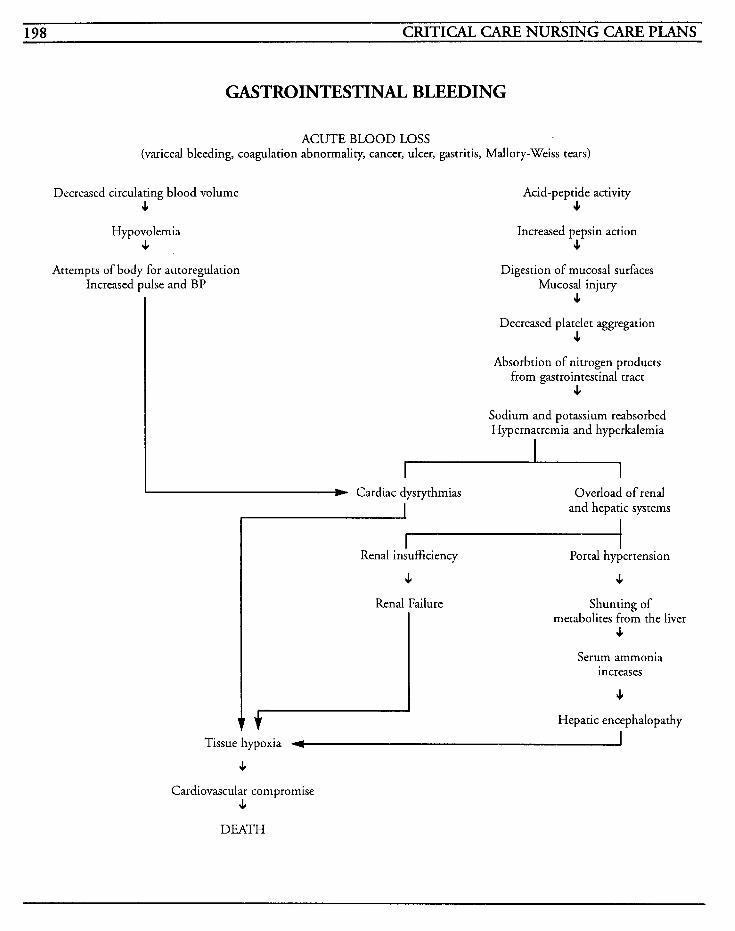
198 CRITICAL CARE NURSING CARE PLANS
GASTROINTESTINAL BLEEDING
ACUTE BLOOD LOSS (variceal bleeding, coagulation abnormality, cancer, ulcer, gastritis, Mdlory-Weiss tears)
Decreased circulating blood volume 4
Hypovolemia J
Attempts of body for autoregulation Increased pulse and BP
Acid-peptide activity J
Increased pepsin action J
Digestion of mucosal surfaces Mucosal injury
J
Decreased platelet aggregation 4
Absorbtion of nitrogen products from gastrointestinal tract
4 Sodium and potassium reabsorbed Hypernatremia and hyperkalemia
d Cardiac dysrythmias Overload of renal and hepatic systems I
Renal insufficiency - Portal hypertension
4 4 Renal Failure Shunting of
metabolites from the liver 4
Serum ammonia increases
4 Hepatic encephalopathy
Tissue hypoxia 4 I J
Cardiovascular compromise J
DEATH
GASTROINTESTINAUHEPATIC SYSTEMS 199
Esophageal varices are twisting, dilated veins that are found in the gastrointestinal tract, but most frequently develop in the submucosal areas of the lower esophagus. Most esophageal varices occur as a result from liver disease and portal hypertension and the development of collateral esophageal veins. When these veins become eroded, the ensu- ing rupture causes extensive vigorous bleeding that is difficult to control.
Normally, the patient does not exhibit symptoms until coughing, vomiting, alcohol, c:' gastritis causes the varices to bleed. Mortality rates are high (above 60%) due to other complications of liver dysfunction, sepsis, or renal failure. Blood loss may be sudden, massive, and life-threatening, with shock and hypovolemia occurring.
Nearly all patients with esophageal varices have at least one of the precipitating factors of cirrhosis, portal vein thrombosis, hepatic fibrosis, schistoso- miasis, hepatic venous outflow obstruction, or splenic vein or superior vena caval abnormalities.
The initial goal of treatment is to replace blood loss and prevent shock from hypovolemia. Balloon tamponade, utilizing the Sengstaken-Blakemore or Minnesota tube, may be required to produce hemostasis.
Complications that occur in conjunction with bleeding may become irreversible and lethal, such as hepatic coma, renal failure, myocardial infarc- tion, or congestive heart failure.
MEDICAL CARE Laboratory: hemoglo bin and hematocrit decreased, BUN increased, liver function tests may be abnormal due to liver involvement and disease, sodium may be elevated, clotting studies may be abnormal due to liver involvement, ammonia may
be elevated, stools for guaiac, bilirubin may be ele- vated if cirrhosis is a factor
Esophlagogastroduodenoscopy (EGD): used to identify and sometimes treat variceal bleeding with sclerotherapy
Radiography: arteriogram used to identify tortu- ous portovenous vessels; chest x-ray used to identify other complicating problems with respira- tory system
Arterial blood gases: may be used to identify acid- base imbalances; may show metabolic acidosis with bleeding
Nasogastric tube: used to keep stomach clear of blood and for lavage, but must be inserted cautiously so as to refrain from increasing bleeding
Balloon tamponade: Sengstaken-Blakemore (SB) or Minnesota tube is a multi-lumen tube that exerts pressure on part of the stomach and against bleeding varices to help control bleeding, and allows for removal of stomach contents; caution must be exercised since placement of this tube can create complications such as airway occlusion or esophageal rupture
Visopressin: may be used as infusion through superior mesenteric artery or a peripheral vein to decrease splanchnic blood flow and promote hemostasis; may induce water intoxication or accentuate cardiac disease by increasing systemic vascular resistance
Nitroglycerin: may be used in conjunction with vasopressin to balance systemic vasoconstriction
Vitamin K: may be used to counteract increased prothrom bin time
Cathartics: magnesium citrate or sorbitol may be used to decrease risk of ammonia-induced neuroencephalo pathy
200 CRITICAL CARE NURSING CARE PLANS
Surgery: may require distal splenorenal shunt, mesocaval and portocaval anastomoses, or devas- cularization of the varices all in the effort to lower pressure in the portal system
NURSING CARE PLANS [Care plans in GI bleeding section also apply to this diagnosis]
Risk for alteration in tissue p e r - i o n : cere- bral, cardiopulm ona? gastroin testinal, renal, and periphera [See Infective Endocarditis]
Related to: variceal bleeding
Defining characteristics: decreased peripheral pulses, hypotension, tachycardia initially, brady- cardia, cold and clammy skin, diaphoresis, mental status changes, lethargy, pallor, abnormal ABGs, decreased oxygen saturation, decreased urine output
Risk for decreased cardiac output [See Cardiogenic Shock]
Related to: variceal bleeding, hemorrhage, exsan- guination
Defining characteristics: decreased peripheral pulses, hypotension, tachycardia, cold and clammy skin, decreased urinary output, mental status changes, pallor
Risk for inflective individual coping [See Mechanical Ventilation]
Related to: bleeding disorder, alcohol abuse, hepatic disease
Defining characteristics: history of excessive alco- hol usage, anxiety, fear, hostility, manipulative behavior, guilt, rationalization, blaming behavior
Risk for injury
Related to: utilization of balloon tamponade to control esophageal bleeding
Defining characteristics: increased bleeding, exsanguination, tube migration, air leakage, esophageal necrosis, encephalopathy, airway occlusion, asphyxia
Outcome Criteria
Patient will be free of complications and injury to self.
~
INTERVENTIONS RATIONALES
Examine Sengstaken-Blakemore (or other type tube) balloons by testing inflation of balloons with air while tube is underwater.
Refrigerate tube prior to inser- tion, and assist physician with insertion of tube into patient’s noselmouth by encouraging swallowing small sips of water.
Ensure that tube is patent in stomach by auscultating stomach for injected air bolus.
Obtain KUB x-ray after place- ment and securing of tube.
When placement is verified, inflate the gastric balloon with air and gently pull the tube back against the gastroesophageal junction. Secure tube, marking location at the nares, and clamp the gastric balloon.
Balloon tubes should be adequately secured with some device [frequently used is a foot- ball helmet with face guard] with slight traction to the balloon tube.
Facilitates easier detection of leaks by escaping air bubbling, and ensures balloons are patent prior to insertion of tube into patient.
Chilling firms the tube to facili- tate easier placement.
Proper positioning is crucial to ensure that the gastric tube is not inflated in the esophagus.
Verifies correct anatomical place- ment.
Applies pressure against the cardia to attempt to control bleeding. Marking the tube facil- itates prompt detection of accidental migration.
Facilitates stable position of tube and prevents migration due to peristalsis or coughing, while exerting appropriate pull/pressure on anatomical sites.
GASTROINTESTINAL/HEPATIC SYSTEMS 20 1
INTERVENTIONS RATIONALES Information, Instruction, Attach a y-connector to the esophageal balloon opening, with a syringe on one side, and a manometer to the other. Fill balloon with air until manometer reading is between 25-35 mmHg and clamp balloon.
Connect gastric port to intermit- tent suction and irrigate every hour.
Insert nasogastric tube above the level of the esophageal balloon and connect to intermittent suc- tion. If tamponade tube has an esophageal suction port, attach it to intermittent suction.
Clearly identify and label each port, checking connections frequently, and have scissors and resuscitative equipment at bedside.
Monitor for complaints of chest pain.
Monitor respiratory status for any changes, decrease in oxygen saturations, or changes in mental status.
Keep head of bed elevated at least 30 degrees at all times.
Compare character and amounts of drainage coming from each lumen.
Deflate esophageal balloon for 30 minutes every 12 hours, or as indicated per hospital protocol.
Maintains sufficient pressure to tamponade bleeding with pres- sure lower than level that may result in esophageal ischemia and necrosis.
Facilitates removal of old blood from stomach, allows observation of changes in bleeding, and relieves gastric distention.
Facilitates removal of salivary secretions and monitors for bleeding above the esophageal balloon, and reduces aspiration risk.
Proper identification may pre- vent accidental deflation or improper irrigation. If the esophageal balloon migrates to the hypopharynx, the esophageal balloon must be cut immediately and removed to prevent airway obstruction.
May indicate complication or esophageal rupture.
May result from tube migration and asphyxia.
Prevents regurgitation and decreases nausea.
Facilitates identification of cessa- tion of bleeding, as well as potentially identifying level of bleeding site.
Decreases risk for esophageal mucosal ischemia and damage.
Demonstration
INTERVENTIONS RATIONALE5
Instruct patientlfamily regarding need for balloon tamponade, procedure of insertion, what to expect, etc.
Observation of patient should be constant.
Promotes knowledge and facili- tates compliance. Decreases fear of the unknown.
Deterioration in patientk status can occur rapidly and continuous observation facilitates prompt intervention to prevent injury.
Discharge or Maintenance Evaluation
Patient will have bleeding from varices controlled with no injury or complication from treatment modalities.
Patient will be able to comply with treatment.
Patitent will have stable vital signs and oxygenation.
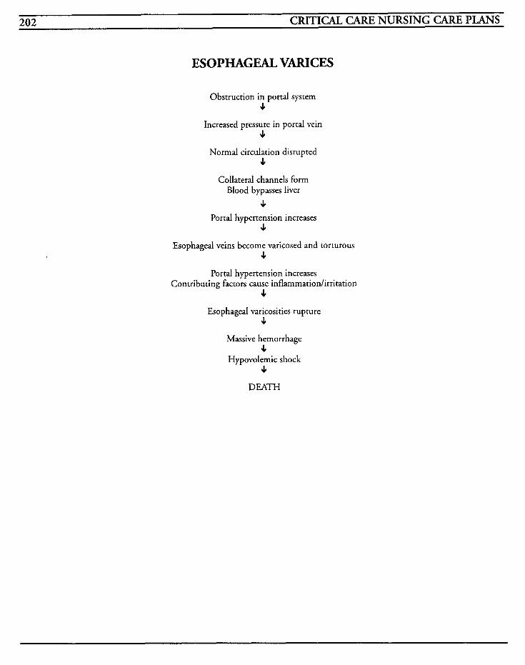
202 CRITICAL CARE NURSING CARE PLANS
ESOPHAGEAL VARICES
Obstruction in portal system 9
Increased pressure in portal vein 9
Normal circulation disrupted 9
Collateral channels form Blood bypasses liver
9 Portal hypertension increases
9
Esophageal veins become varicosed and torturous 9
Portal hypertension increases Contributing factors cause inflammatiodirritation
9 Esophageal varicosities rupture
9
Massive hemorrhage 9
Hypovolemic shock 9
DEATH
GASTROINTESTINAL/HEPATIC SYSTEMS 203
Acute hepatitis is an infection of the liver that usually is viral in origin but may be induced by drugs or toxins. There are currently five types of hepatitis, denoted HAV, HBV, NANB or hepatitis C, HDV, and HEV, with HAV being the most common type. Hepatitis B, or HBV, is more severe and because it can be acquired from expo- sure to individuals who are asymptomatic, the potential for transmission is increased many-fold.
Hepatitis A, or HAV, is transmitted via fecal-oral route, with poor sanitation practices, with conta- mination of food, water, milk, and shellfish, or oral-anal sexual practices. HAV patients may exhibit no acute symptoms or have symptoms that are related to other causes.
Hepatitis B, or HBV, is transmitted via blood and blood products, breaks in the skin or mucous membranes, or from an asymptomatic carrier with Hepatitis B surface antigen (HBsAg).
Hepatitis C, or Non-A, Non-B hepatitis, is trans- mitted via intravenous drug use, sexual contact, blood or blood products, and from asymptomatic carriers.
Hepatitis D, or HDV, is transmitted through the same routes as HBV but must have hepatitis B surface antigen to replicate.
Hepatitis E, or HEV, is seen in developing coun- tries and not encountered in the United States. It is transmitted through food or water contamina- tion.
Once the disease has been contracted, treatment is symptomatic. Prophylactic therapy may assist in prevention of hepatitis from developing after being exposed to the virus. Immune globulin (IG) is generally given to provide temporary passive immunity. Hepatitis B vaccine provides active
immunity and offers protection to people who are at high risk.
Laboratory: CBC shows decreased RBCs as a result of decreased lifespan from enzyme alterations or from hemorrhage; white blood cell count usually shows leukocytosis, atypical lymphocytes, and plasma cells; liver function stud- ies are abnormal, up to 10 times normal values in some cases, albumin decreased, blood glucose may be decreased or elevated transiently due to liver dysfunction; Anti-HAV IgM presence shows either currenc infection or after 6 weeks may indicate immunity; hepatitis B surface antigen and hepati- tis Be antigen show presence of HBV; Anti-HBc in serum indicates carrier status; Anti-HBsAg indicates HBV immunity; antidelta antibodies present without HBsAg indicates HDV; urine bilirubin elevated; prothrombin time may be ele- vated with liver dysfunction
Liver biopsy: may be used to delineate type of hepatitis and degree of liver necrosis
Liver scans: may be performed to identify level of parenchyma1 damage
NURSING CARE PLANS Activity intolerance
Related to: infective process, decreased endurance
Defining characteristics: easy fatiguability, lethargy, malaise, decreased muscle strength, reluc- tance to perform activity
Outcome Criteria Patient will achieve and maintain ability to perform normal activities without intolerance and fatigue.
204 CRITICAL CARE NURSING CARE PLANS
INTERVENTIONS RATIONALES
Maintain bed rest and quiet environment, allowing rest peri- ods in between activities.
Reposition every 2 hours and provide good skin care.
Increase activities as patient is able to tolerate.
Monitor labwork for liver hnc- tion studies.
Administer medications as war- ranted.
Decreases energy expenditure that is needed for healing. Activity can decrease hepatic blood flow and prevent circula- tion and healing to liver cells.
Decreases potential for skin breakdown.
Assists with return to optimal activity levels while enabling patient to have some measure of control over the situation.
May assist with identification of appropriate levels of activity.
Sedatives and antianxiety drugs may be required to effect needed rest. Caution should be taken to ensure drugs used are not hepatotoxic.
Information, Instruction, Demonstration
INTERVENTIONS RATIONALES
Administer antidotesltherapeutic treatment modalities to remove causative agent with toxic damage. hepatitis.
Instruct patient/fmily on disease process and need for extended rest.
Removal of substance may restrict amounts of tissue
Promotes knowledge and facili- tates compliance with treatment.
Discharge or Maintenance Evaluation
Patient will be able to verbalize understanding of disease process and treatment program.
Patient will be able to perform usual activities without fatigue.
Patient will be able to gradually increase level of activities performed.
Related to: changes in health status, changes in physical status, imposed physical isolation, inade- quate support system
Defining characteristics: feelings of loneliness, feelings of rejection, absence of family memberslfriends, sad, dull affect, inappropriate behaviors
Alteration in nutrition: less than body requirements [See Liver Failure]
Related to: metabolism changes, anorexia
Defining characteristics: nausea, vomiting, anorexia, abdominal pressure, malabsorption of fats, altered metabolism of protein, carbohydrates, and fat, weight loss, fatigue, edema
Risk for infiction [See Transplants]
Related to: leukopenia, immunosuppression, mal- nutrition, exposure to causative organisms
Defining characteristics: increased white blood cells, differential with a shift to the left, fever, chills, hypotension, tachycardia, positive cultures
Risk for impaired skin intepity [See Liver Failure]
Related to: bile salt accumulations on skin
Defining characteristics: jaundice, pruritus, itch- ing, scratching
Social isolation [See Transplants]
GASTROINTESTINAL/HEPATIC SYSTEMS 205
Knowledge &fieit
Related to: lack of information, lack of recall, unfamiliarity of resources, misinterpretation of information received
Defining characteristics: questions, requests for information, statements of misperceptions, devel- opment of preventable complications
Outcome Criteria
Patient will be able to verbalize understanding of disease, treatment, and causative behaviors.
INTERVENTIONS RATIONALES
Discuss patient’s perceptions of disease process.
Instruct on disease process, pre- vention and transmission of disease, and isolation require- ments.
Instruct in appropriate home sanitation.
Instruct on activity limitations.
Instruct on all medications, side effects, effects, contraindications, and dangers of administration of over-the-counter drugs without physician approval.
Instruct to refrain from blood donation.
Identifies knowledge base and misconceptions to facilitate appropriate teaching plan.
Types of isolation will vary according to type of hepatitis and personal situation. Family members may require treatment depending on type of hepatitis.
Dirty environment and poor sanitation methods may be responsible for transmission of the disease.
Complete resumption of normal activity may not take place until liver returns to its normal size and patient begins to feel better and this may take up to several months.
Promotes knowledge and facili- tates compliance. Some medications are hepatoroxic or are metabolized by the liver, increasing its workload.
Most states do not allow anyone who has a history of any type of hepatitis to donate blood or blood products to prevent possi- ble spread of the infection.
INTERVENTIONS RATIONALES
Instruct on avoidance of recre- ational drugs or alcohol.
May jeopardize recovery from infection and increases liver dys- function.
May be required for assistance with substance withdrawal and for long-term support once dis- charged.
Consult with counselors, minis- ters, drug or alcohol treatment facilities as warranted.
Discharge or Maintenance Evaluation
Patient will be able to accurately verbalize understanding of all instructions given.
Patiedfamily will be able to modify environ- ment to control spread of disease.
Patient will be able to effectively access commu- nity resources for treatment programs and discharge follow-up care.
Patient will be able to effectively manage med- ical regimen with follow-up from physician.
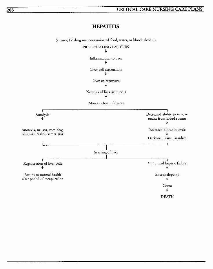
206 CRITICAL CARE NURSING CARE PLANS
HEPATITIS
(viruses; IV drug use; contaminated food, water, or blood; alcohol)
PRECIPITATING FACTORS 4
Inflammation to liver 4
Liver cell destruction 6
Liver enlargement 6
Necrosis of liver acini cells 4
Mononuclear infiltrates I
I 1 Autolysis
6
Anorexia, nausea, vomiting, urticaria, rashes, arthralgias
Decreased ability to remove toxins from blood stream
J
Increased bilirubin levels 4
Darkened urine, jaundice
I I I
Scarring of liver I
I Regeneration of liver cells
4 Return to normal health
after period of recuperation
I Continued hepatic failure
4 Encephalopathy
J
Coma 4
DEATH
GASTROINTESTINAL/HEPATIC SYSTEMS 207
Pancreati tis Acute pancreatitis is a life-threatening inflamma- tory response to an injury, in which pancreatic enzymes are abnormally activated and these enzymes destroy tissues in and surrounding the pancreas by autodigestion. Precipitating factors for the abnormal activation may be caused by effects of ethanol and its metabolite, acetaldehyde, diseases of the biliary tract, obstruction of the common bile duct, bile reflux into the pancreatic duct, ischemia, trauma, infections, surgical or invasive procedures, neoplasms, metabolic aberra- tions, use of oral contraceptives, corticosteroids, thiazide diuretics, or antihypertensives, or stimula- tion of vasoactive substances. Obstruction may result in widespread edema to the pancreas, which increases pressure in the pancreatic system. This increase in pressure results in the rupture of the ducts which allows the enzymes to spill into the cells, and begin the autodigestion process.
Trypsin activates the pancreatic enzymes, phospholipase A, elastase, and kallikrein. Trypsin may cause edema, necrosis, and hemorrhage in the pancreas. Elastase may attack the walls of smaller blood vessels and facilitate hemorrhage. Phospholipase A allows damage to the acinar cell membrane to occur, and may alter coagulation. Vasomotor changes and increases in vascular per- meability may be caused by kallikrein, and this may also be the cause of the pain experienced with pancreatitis. If the disease is allowed to progress, the inflammation leads to massive hemorrhage, destruction of the pancreas, diabetes mellitus, aci- dosis, shock, coma, and death.
One of the predominant symptoms of this disease is the unrelenting abdominal pain located in the epigastric and/or periumbilical areas that may radiate to the chest and back. Nausea, continuous vomiting, low-grade fever, anorexia, diarrhea,
weight loss, jaundice, diaphoresis, dehydration, and poorly defined abdominal mass may also be encountered.
Pseudocysts and abscesses in and around the pan- creas may occur as a result of localized necrosis, and may exert pressure on the stomach or colon. They may develop slowly and may result in fistula formation.
The goal of therapy is to maintain adequate circu- latory fluid volume with electrolyte replacement, pain relief, treatment of infection and treatment of hyperglycemia.
MEDICAL CARE Laboratory: serum amylase is elevated up to 40 times the normal limit in the early stages and then decreases over 2-3 days; urine am ylase elevated and lasts longer than serum amylase; elevated glu- cose, bilirubin, alkaline phosphatase, lactic dehydrogenase, aspartate transferase, potassium, triglycerides, cholesterol, and lipase; decreased albumin, calcium, sodium, and magnesium; white blood cell counts from 8,000-20,000 with increased polymorphonuclear cells; hematocrit may exceed 50%; prothrombin time may be increased; fat content in the stool increased; amy- lase-creatinine clearance ratio may indicate pancreatic disease; renal profiles used to evaluate renal function and hypovolemia
CT scans: used to identify size, shape, density, masses, or infiltrates in the pancreas
Ultrasonography: used to identify neoplasms, edema, inflammation, cysts, abscesses, or infiltrates in the pancreas, but cannot confirm the diagnosis of pancreatitis
Angiography: helps to visualize early pancreatic tumors or problems with vasculature
208 CRITICAL CARE NURSING CARE PLANS
Endoscopic retrograde cholangiopancreatography (ERCP): used to directly visualize the pancreatic duct system by use of endoscopy and radiography; used to identify cysts, calculi, stenosis, pancreatic and biliary duct disease when other diagnostic tools are not conclusive
Surgery may be necessary to drain abscesses or pseudocysts, or to anastomose the pseudocysts to an adjacent structure to provide internal drainage; chronic pancreatitis may require a pancreaticoje- junostomy to relieve obstruction of the duct to relieve pain; experimental surgery for transplanta- tion of the pancreas or islet cells may be performed
NURSING CARE PWNS Alteration in comfort [See MI]
Related to: pancreatic obstruction, autodigestion of pancreas, leakage of pancreatic enzymes, inflammation
Defining characteristics: unrelenting epigastric pain, patient curled up with both arms over abdomen, nausea, vomiting, tenderness, facial gri- macing, groaning
INTERVENTIONS RATIONALES
Administer Demerol IV as Demerol is the drug of choice warrantediordered. for pancreatitis. DO NOT
GIVE Morphine because most opiate-type narcotics cause spasms of the Sphincter of Oddi, increasing patient's pain.
Alteration in nutrition: less than body requirements [See DKA]
Related to: nausea, vomiting, anorexia, digestive enzyme leakage, increased metabolic needs, sepsis
decreased bowel sounds, anorexia, increased metabolism, lack of adequate food ingested
Fluid volume dtlficit [See DKA]
Related to: nausea, vomiting, fever, diaphoresis, nasogastric drainage, fluid shifting, diarrhea
Defining characteristics: nausea, vomiting, ascites, nasogastric suctioning, hypotension, tachycardia, decreased urinary output
Risk for impaired gas exchange [See Mechanical Ventilation]
Related to: complications from disease
Defining characteristics: altered arterial blood gases, dyspnea, use of accessory muscles, tachyp- nea, bradypnea, cough, sputum
Potential for injury
Related to: sepsis, pseudocysts, fistula formation, abscess formation, complications from disease
Defining characteristics: fever, abdominal pain, drainage, increased white blood cell count, shift to the left, systemic infection symptoms, DIC, elec- trolyte imbalances
Outcome Criteria
Patient will be afebrile and have no complications from disease.
INTERVENTIONS RATIONALES
Monitor vital signs at least every 2 hours, and note changes.
Allows for prompt identification of early signs of infection to facilitate timely treatment. Third spacing, bleeding, and secretion of vasodilating substances may result in hypotension.
Defining characteristics: increases in nausea and vomiting, retching, absent bowel sounds,
GASTROINTESTINAL/HEPATIC SYSTEMS 209
INTERVENTIONS RATIONALES INTERVENTIONS RATIONALES
Allows for actual measurement of Prepare patientlfamily members Surgical drainage of abscesses or Monitor hemodynamic pressures if possible.
Monitor EKG for cardiac rhythm, rate, and changes, and treat dysrhythmias per hospital policy.
Auscultate heart sounds for changes, gallops, or murmurs.
Observe for changes in respira- tory status, especially when occurring concurrently with fever and jaundice.
Observe for increasing complaints of abdominal pain or tenderness, chills, fever, or hypotension.
Observe for presence of petechiae, continued bleeding, or hematoma formation.
Measure and monitor abdominal girth changes.
Monitor intake and output every 2 hours, noting hematuria, or significant imbalance.
cardiac output and other para- meters to identify fluid shifts and hemodynamic alterations which may precede systemic complica- tions.
Hypovolemia and electrolyte imbalances may precipitate car- diac dysrhythmias.
JVD in conjunction with a new S, gallop may indicate heart fail- ure or pulmonary edema.
Gram negative sepsis may be seen symptomatically with cholestatic jaundice and decreases in pulmonary function.
May indicate formation of abscess, especially if symptoms occur while patient is receiving vigorous medical treatment. Abdominal rigidity or rebound tenderness may indicate peritoni- tis.
May indicate impending DIC as a result of circulating pancreatic enzymes.
Identifies increases in fluid reten- tion and ascites.
Oliguria may occur as a result of renal involvement due to increases in vascular resistance or decreased renal blood flow. Hematuria may occur as a result from circulating pancreatic enzymes.
Information, Instruction, Demonstration
INTERVENTIONS RATIONALES
Strict aseptic technique should be maintained when dealing with may result in sepsis, which is invasive lines or dressings.
Failure to maintain technique
responsible for over 80% of deaths associated with pancreatitis.
for surgical procedures as warranted.
Instruct in usage of pancreatic enzyme supplements/bile salts.
pseudocyscs may be required.
Long-term replacement may be required for exocrine deficiencies from permanent pancreatic damage.
Discharge or Maintenance Evaluation
Patient will be free of complications from pan- creatitis, and will exhibit timely healing of all wounds.
Patient will be able to accurately verbalize all instructed information.
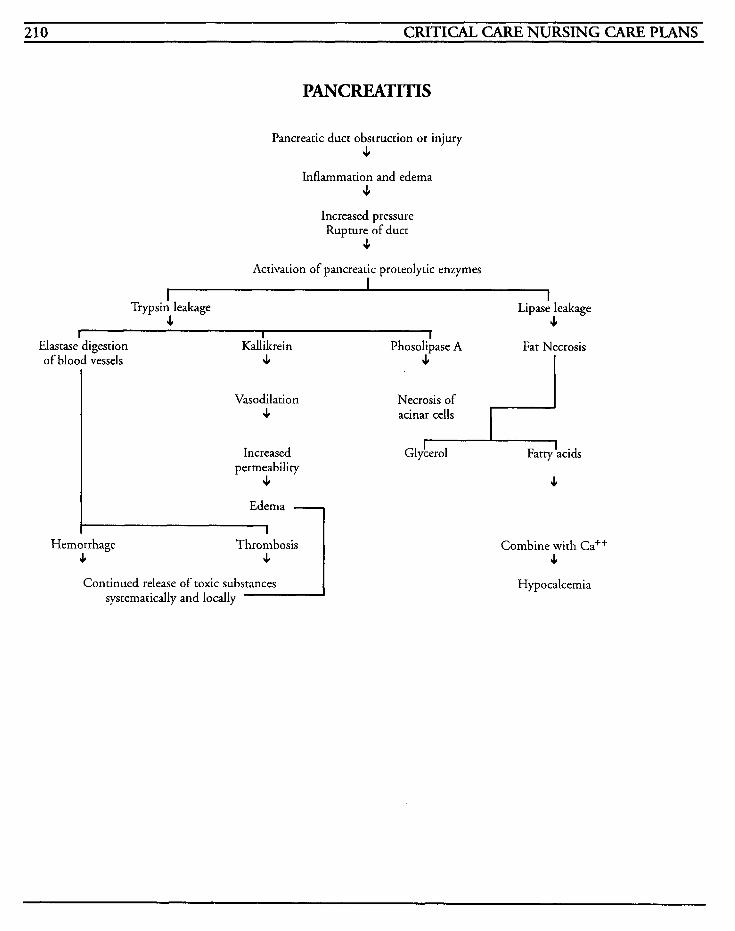
210 CRITICAL CARE NURSING CAFE PLANS
PANCREATITIS
Pancreatic duct obstruction or injury 4
Inflammation and edema 4
Increased pressure Rupture of duct
4 Activation of pancreatic proteolytic enzymes
I I
I
I Trypsi; leakage
4 Lipase leakage
4 I 1 -
Elastase digestion Kallikrein of blood vessels 4
Vasodilation 4
Increased permeability
4
Ed:ma 1 Hemorrhage
s Thrombosis
4 1 Continued release of toxic substances
systematically and locally I
-7 Phosolipase A Fat Necrosis
Necrosis of acinar cells
4
Combine with Ca++ 4
Hypocalcemia
GASTROINTESTINAL/HEPATIC SYSTEMS 211
Acute Abdomen/ Abdominal Trauma When someone is said to have an acute abdomen, it generally indicates that they have a sudden onset of severe abdominal pain that typically requires surgery to prevent peritonitis from contaminated materials spilling into the peritoneal cavity. There are numerous situations that could be responsible for this diagnosis, such as perforation of the appendix, peptic ulcer, bowel, gallbladder, diverti- culi, or abdominal aortic aneurysm, ruptured ectopic pregnancy, or an abdominal injury.
Abdominal injuries may be caused from either blunt trauma or penetrating damage. Blunt trauma, with compression of abdominal structures against the vertebral column, can result from sports injuries, accidents, or falls, and can be caused as a result of a direct impact, rotary or shearing forces, or rapid deceleration. Any of these mechanisms can cause tearing of body structures that may involve substantial bleeding into the peritoneal cavity.
Penetrating injuries can cause perforation of the bowel or hemorrhage from lacerations to major vessels. These types of trauma can either be low- velocity, which damages tissues at the injury site, or high-velocity, in which tissues and organs sur- rounding the penetration path are damaged.
All of the types of injuries discussed have signifi- cant potential for critical emergencies, based on the severity of the wound, and how much damage it has caused. Mortality is approximately 10% from abdominal trauma due in part to the pres- ence of structures involving many body systems being located in the abdomen. The goals of imme- diate treatment involve maintaining the hemodynamic status, control of hemorrhage, and preparation for surgical procedures.
MEDICAL CARE Surgery: usually the treatment of choice due to potential or presence of peritonitis from injury; procedure is dependent on source of bleeding or contamination
Laboratory: urinalysis to identify bleeding or uri- nary tract injuries; CBC to identify sepsis and changes in hematological status; WBC is normally elevated in trauma; differential used to identify shifts to the left; amylase elevated with pancreatic injury or gastrointestinal perforations; renal and liver profiles to discern damage to the particular system; clotting profiles to monitor for coagula- tion status; myoglobin levels elevated with crush injuries, peritoneal fluid analysis for bleeding or infection
Radiagaphy: chest and abdominal x-rays used to identify pneumothorax, free air below the diaphragm, foreign body that may have caused injury, or other complications; loss of psoas muscle outline indicates retroperitoneal bleeding
CT scans: may be used to identify abdominal and retroyeritoneal injuries that may not be overt with regular x-rays; can identify cysts or abscesses that may require surgical intervention
Intravenous pyelogram: used to detect hematuria and trauma to renal structures
Retrograde urethrography/cystography: used to identify urethral or bladder injury
Ultrasound: use is limited; may be useful to dis- tinguish between splenic hematoma from peritoneal blood or ascites
Paracentesis: may be used to identify presence of pus, blood, or other substance, and may be used for peritoneal lavage to identify effects of abdomi- nal trauma and prevent unnecessary surgical intervention
212 CRITICAL CARE NURSING CARE PLANS
NURSING CARE PLANS Risk for infection
Related to: perforation of abdominal structures, laceration of vasculature, open wounds, peritoneal cavity contamination
Defining characteristics: fever, trauma, elevated white blood cell count, sepsis
Outcome Criteria
Patient will be free of infection, with stable vital signs and labwork within normal parameters.
INTERVENTIONS RATIONALES
Monitor vital signs, especially temperature.
Observe skin color, temperature, and monitor for changes.
Obtain blood, urine, sputum, drainage, or other cultures as ordered.
Monitor intake and output every 2 hours.
Administer antibiotics as ordered.
Ensure that universal precautions are utilized, and that sterile or aseptic technique is used when caring for wounds or inserting invasive lines or catheters.
Administer tetanus toxoid as ordered.
May indicate presence of or impending infection and sepsis. Decreasing pulse pressure, hypotension and tachycardia may signifj impending septic shock from endotoxic vasodilation.
Patient may have warm, flushed, dry skin in shock's warm phase, changing to cold and clammy pale skin as shock progresses.
Identifies causative organism and facilitates appropriate selection of antimicrobial agents.
Sepsis may impair renal perfu- sion and result in oliguria or anuria.
Cephalosporins and aminoglyco- sides are frequently used to fight these types of infections.
Assists in preventing spread of infection by cross-contamination, as well as preventing other bacte- rial growth from invasion of skinlbody.
Decreases risk of development of tetanus.
Information, Instruction, Demonstration
INTERVENTIONS RATIONALES
Prepare for surgery as warranted. Surgical intervention may be the treatment of choice to drain abscesses or remove or repair perforated structures.
May be performed to remove fluid and identify causative organism.
Dressings protect wound and
Assist with peritoneal aspiration as warranted.
Change wound dressings as ordered. prevent spread of infection.
Monitor CBC, especially WBC count.
Facilitates assessment of effective- ness of antimicrobial therapy, as well as identifies blood loss or changes in infection.
Decreases potential for cross- Limit visitors as indicated, utiliz- ing appropriate isolation contamination. precautions as warranted.
Discharge or Maintenance Evaluation
Patient will have stable vital signs and hemody- namic status.
Patient will have white blood cell count within normal limits.
Patient will have negative cultures.
Patient will not exhibit further signs/symptoms of infection.
Patient will not develop secondary infection.
Family members will adhere to isolation regula- tions.
Risk for injury
Related to: trauma
Defining characteristics: hemorrhage, peritonitis, altered arterial blood gases, mental status changes, hypotension, tachycardia, bradycardia, arterial injuries, fractures, electrolyte imbalances
GASTROINTESTINAL/HEPATIC SYSTEMS . . . 213
Outcome Criteria
Patient will be free of injury to self, and free of complications that may ensue from trauma.
INTERVENTIONS RATIONALES
Monitor vital signs every 1-2 hours, and prn. Check blood pressure readings in both arms and legs.
Monitor respiratory status, noting changes in breath sounds.
Observe chest for symmetry, paradoxical movement, anatomi- cal deformity, swelling, bruising, or crepitus.
Auscultate heart sounds for changes or abnormalities.
Observe abdomen for wounds, masses, swelling, pulsations, hemaromas, protrusion of organs or viscera, lacerations, and abra- sions; auscultate for bowel sounds.
Decreases in blood pressure or changes with orthostatic readings may indicate impending hypov- olemia. Pulse pressures may increase during the latent effects of shock or with head injuries, and may decrease in early stages of shock. Differences benveen right and left sides greater than 20 mmHg may indicate aortic injury.
Injury to lungs or diaphragm may result in tachypnea and dys- pnea. Breath sounds that are distant or absent may indicate pneumothorax or hemothorax.
Splinting by patient or obvious deformity or swelling may be seen if ribs are fractured. Paradoxical movement may indi- cate flail chest. Palpable crepitus may be present if lung or medi- astinum has been punctured.
Extra heart sounds or murmurs may indicate injury to valves or heart, and distant, muffled heart tones may signal cardiac tarnpon- ade.
Bluish discolorations around the umbilicus may indicate retroperi- toneal bleeding accumulating in abdomen. An odd number of bullet holes may indicate the remaining presence of a foreign objectlbullet in the body. Decreased or absent bowel sounds may indicate ileus or peritonitis. Abdominal bruits may result when a vessel is par- tially occluded, venous hums auscultated over the upper
INTERVENTIONS RATIONALES
Percuss abdomen for changes, dullness or tympany.
Cover protruding abdominal viscera with saline-soaked sterile gauze or sterile towels, and posi- tion patient with knees flexed.
Palpate peripheral pulses for presence, quality, and character. Notify physician for significant changes.
Observe for Grey Turner's and Coopernail's signs.
Monitor for complaints of pain at the tip of the left shoulder or right shoulder.
abdomen or liver may indicate hepatic or splenic vein thrombo- sis, and friction rubs heard over the spleen may indicare infarc- tion or inflammation of spleen.
Dullness that is decreased over liver may indicate presence of free air below the diaphragm. Upper abdominal distention and increased tympany over the stomach may indicate gastric dilation. Flank area dullness may indicate retroperitoneal hemor- rhage.
Protects viscera from drying, and positioning prevents additional protrusion/evisceration.
Changes in pulse characteristics may indicate arterial or venous impairment which may require immediate treatment.
Grey Turner's sign is a bluish discoloration on flank that indi- cates retroperitoneal bleeding accumulation in abdomen. Coopernail's sign is ecchymoses on scrotum or labia and may indicate pelvic fracture.
May indicate rupture of spleen or irritation of the diaphragm from blood or other substance with left shoulder pain, and pos- sible liver laceration with right shoulder pain.
Information, Instruction, Demonstration
~ ~~ ~~ ~
INTERVENTIONS RATIONALES ~~ ~~~
Assist with peritoneal tap and lavage. bleeding which is diagnosed
Done to identify intraperitoneal
when fluid is analyzed.
Promotes knowledge and decreases anxiety which facilitates
men.
Instruct on all procedures and testing; prepare for surgery as warranted. compliance with medical regi-
214 CRITICAL CARE NURSING CARE PLANS
INTERVENTIONS RATIONALES weight loss, nitrogen and electrolyte imbalance,
Instruct patientlfmily to notify Abdominal injury signs and deficiencies decreased albumin and protein levels, vitamin
physician for fever or abdominal pain. several hours to days.
symptoms may not appear for
Discharge or Maintenance Evaluation
Patient will have no evidence of abdominal injury complication.
Patient will have no intraperitoneal bleeding or structural damage to organs.
Patient will be compliant with regimen.
Patient will have successful surgical intervention
Risk fir fluid uolmve &ficz't [See GI Bleeding]
Related to: fluid shifts, hemorrhage, nasogastric suctioning
Defining Characteristics: hypotension, tachycar- dia, decreased urinary output, decreased hemoglobin and hematocrit, decreased filling pres- sures, electrolyte imbalances, presence of peritonitis
Alteration in comfirt [See MI]
Related to: trauma, surgery, edema
Defining characteristics: grimacing, complaints of pain, restlessness, splinting, shallow respirations, abdominal rigidity
Alteration in nutrition: less than body requirements [See DKA]
Related to: trauma, surgery, nasogastric suctioning
Defining characteristics: abdominal pain, ordered nutritional status of NPO, increased metabolism,
with no postoperative complications.
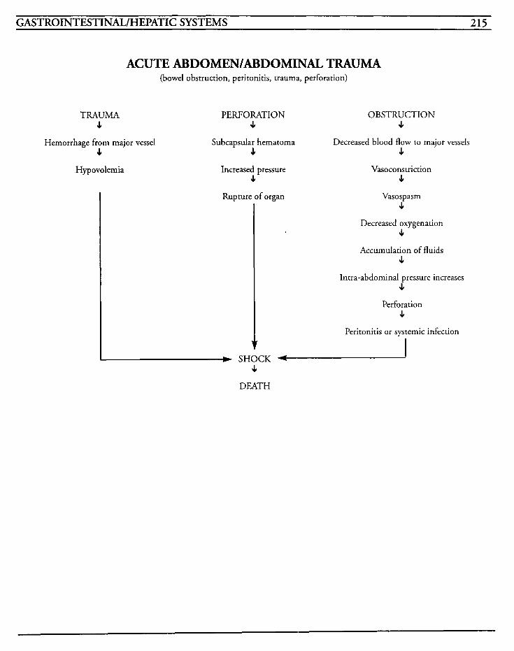
GASTROINTESTINAL/HEPATIC SYSTEMS 215
ACUTE ABDOMEN/ABDOMTNAL TRAUMA (bowel obstruction, peritonitis, trauma, perforation)
TRAUMA PERFORATION OBSTRUCTION J, 4 J,
Subcapsular hematoma 4 4 J,
Hemorrhage from major vessel Decreased blood flow to major vessels
Vasoconstriction Hypovolemia Increased pressure 4
Rupture of organ
J,
Vasospasm 4
Decreased oxygenation J,
Accumulation of fluids J,
,xra-abdominal pressure increases J,
Perforation J,
Peritonitis or systemic infection
I w SHOCK f- J,
DEATH

This Page Intentionally Left Blank
GASTROINTESTINAL/HEPIC SYSTEMS 217
The liver plays a vital role by providing multiple functions, such as, metabolism of carbohydrates, proteins, and fats, storing fat-soluble vitamins, vit- amin B,,, copper, and iron, synthesis of blood clotting factors, amino acids, albumin, and globu- lins, detoxification of toxic substances, phagocytosis of microorganisms, and plays a role in glycolysis and gluconeogenesis. Liver function- ing can be preserved until up to 75% of the hepatocytes become damaged or necrotic, at which time the liver can no longer perform its normal operation.
Early hepatic failure presents as a type of cirrhosis of the liver. Liver cells become inflamed and obstructed, which results in damage to the cells around the central portal vein. When the inflam- mation decreases, the lobule regenerates, and this cycle is repeated until the lobule is irreversibly damaged and fibrotic tissue replaces liver tissue.
Advanced hepatic failure develops when all com- pensatory mechanisms fail, causing the serum ammonia level to rise. The already-damaged liver is unable to synthesize normal products, so acido- sis, hypoglycemia, or blood dyscrasias develop, and the patient becomes comatose.
Acute liver failure, also known as fulminant hepatic failure, may be precipitated by a stress factor that aggravates a preexisting chronic liver disease. Some stress factors include alcohol intake, ingestion of Amanita mushrooms, large amounts of dietary protein, gastrointestinal bleeding, and portacaval shunt surgery. An acute type of liver failure may occur as a result of viral or toxic hepatitis, biliary obstruction, cancer, acute infec- tive processes, drugs, such as acetaminophen, isoniazid, and rifampin, severe dehydration, Reye’s syndrome, or shock states.
Fulminant hepatic failure may begin as stage I hepatic encephalopathy, progressing to drowsiness and asterixis, stupor and incoherent communica- tion, finally to stage IV with deep coma. The stages may progress over at little as two months. Distinguishing attributes between acute and chronic failure are the presence of cerebral edema and intracranial pressure increases.
The goal of treatment is to halt progression of the encephalopathy that occurs with increasing ammonia levels, and is accomplished with use of cathartics, decreasing dietary protein, and electrolyte replacement. Even with treatment, mortality rates are as high as 90%, depending on the age of the patient and severity of disease.
MEDICAL CARE Laboratory. elevated ammonia levels, liver func- tion studies elevated, elevated BUN; electrolytes tested to identify imbalances; serum bilirubin ele- vated; urine bilirubin may be present if direct, serum bilirubin is elevated; albumin decreases and globulin increases in liver failure; cholesterol is ele- vated; PT prolonged; toxicology screens for ingestion of alcohol or other drugs that may have precipitated failure; magnesium level may be low with alcoholic cirrhosis and toxic if magnesium replacement is used
Medication: Neomycin or Kanamycin frequently used to prevent intestinal bacteria from converting proteidamino acids to ammonia; lactulose or sor- bitol used to induce catharsis to empty intestines to decrease conversion to ammonia; thiazide diuretics may be given to decrease fluid retention
Hyperalimentation: may be used as diet of choice due to ability to control concentration of nutrients, electrolytes, and vitamins
218 CRITICAL CARE NURSING CARE PLANS
Hemodialysis: may be used as a temporary mea- sure for severe hepatic encephalopathy
Liver biopsy: may be done to establish diagnosis by study of biopsied tissue
Liver scans: may be used to detect degenerative cirrhosis changes or identifj. focal liver disease
NURSING CARE PUNS Alteration in thought processes
Related to: serum ammonia levels, encephalopathy
Defining characteristics: increased ammonia, increased BUN, mental status changes, decreasing level of consciousness, changes in personality, handwriting changes, tremors, coma
Outcome Criteria Patient will be conscious and stable, with ammo- nia levels within normal ranges. INTERVENTIONS RATIONALES
Monitor neurological status every 1-2 hours, and prn. Notify physician for abnormalities.
If possible, have patient write name each day and do simple mathematic calculation.
Administer cathartic agents as ordered.
Idenrifies onset of problem and potential trend.
As hepatic failure progresses, the ability to write becomes more difficulr, and writing becomes illegible at pre-coma stage. Inability to perform mental cal- culations may indicate worsening failure.
Lactulose minimizes formation of ammonia and other nitroge- nous by-products by altering intestinal pH. Neomycin or Kanamycin help prevent conver- sion of amino acids into ammonia. Sorbitol-type cathar- tics cause an osmotic diarrhea to empty the intestines to decrease ammonia production.
~
INTERWNTIONS RATIONALES ~~~ ~
Observe for asterixis or other tremors.
Rapid wrist flapping when arms are raised in front of body with hands dorsiflexed may indicate presence of encephalopathy.
Decreases risk of injury due to Provide safe environment for patient. altered consciousness levels.
Provide low protein diet. Decreased dietary protein may lessen serum ammonia levels.
May worsen decreasing level of consciousness and make identifi- cation of cause of decreased sensorium more difficult.
Avoid sedatives and narcotics if at all possible.
Information, Instruction, Demonstration
~
INTERVENTIONS RATIONALES
Instruct patientlfamily in poten- tial for altered sensorium and encephalopathy signs. Reorient patient as needed.
Provides knowledge and facili- tates family involvement with maintaining optimal orientation level. Provides support with real- istic expectations of disease process since outcome is poor.
Diarrhea will occur, and lactulose should be titrated to where patient has 3 stools per day.
Instruct in side effects of drugs used to facilitate decrease in ammonia levels.
Discharge or Maintenance Evaluation
Patient will be awake, alert, and oriented.
Patient will have serum ammonia levels within acceptable ranges.
Patient andlor family will be able to verbalize understanding of instructions and be able to communicate concerns.
Alteration in nutrition: less than body requirements
Related to: metabolism changes, increased ammo- nia level
Defining characteristics: anorexia, nausea, vomit- ing, malabsorption of fats, malabsorption of
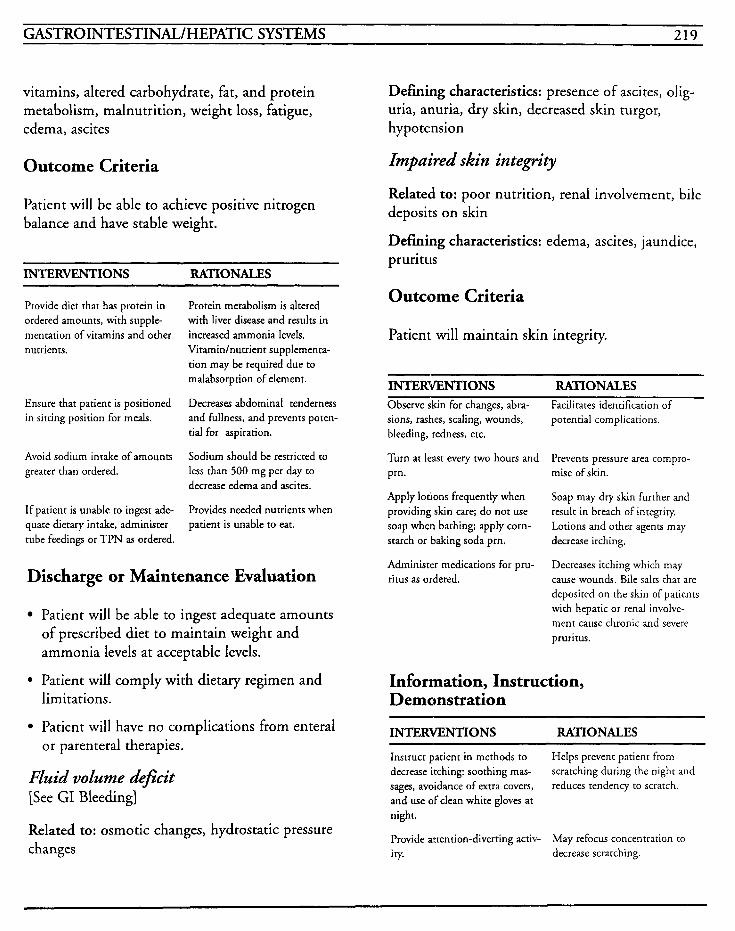
GASTROINTESTINAL/HEPATIC SYSTEMS 219
vitamins, altered carbohydrate, fat, and protein metabolism, malnutrition, weight loss, fatigue, edema, ascites
Outcome Criteria
Patient will be able to achieve positive nitrogen balance and have stable weight.
INTERVENTIONS RATIONALES
Provide diet that has protein in ordered amounts, with supple- mentation of vitamins and other nurrients.
Ensure that patient is positioned in sitting position for meals.
Avoid sodium intake of amounts greater than ordered.
If patient is unable to ingest ade- quate dietary intake, administer rube feedings or TPN as ordered.
Protein metabolism is altered with liver disease and results in increased ammonia levels. Vitamin/nutrient supplementa- tion may be required due to malabsorption of element.
Decreases abdominal tenderness and fullness, and prevents poten- tial for aspiration.
Sodium should be restricted to less than 500 mg per day to decrease edema and ascites.
Provides needed nutrients when patient is unable to eat.
Discharge or Maintenance Evaluation
Patient will be able to ingest adequate amounts of prescribed diet to maintain weighr and ammonia levels at acceptable levels.
6 Patient will comply with dietary regimen and limitations.
Patient will have no complications from enteral or parenteral therapies.
Fluid volume deficit [See GI Bleeding]
Related to: osmotic changes, hydrostatic pressure changes
Defining characteristics: presence of ascites, olig- uria, anuria, dry skin, decreased skin turgor, hypotension
Impaired skin integrity
Relateld to: poor nutrition, renal involvement, bile deposits on skin
Defining characteristics: edema, ascites, jaundice, pruritus
Outcome Criteria
Patient will maintain skin integrity.
INTERVENTIONS RATIONALES Observe skin for changes, abra- sions, rashes, scaling, wounds, bleeding, redness, etc.
Turn at least every two hours and prn. mise of skin.
Apply lotions frequently when providing skin care; do nor use soap when bathing; apply corn- starch or baking soda pm.
Administer medications for pru- ritus as ordered.
Facilitates identification of potential complications.
Prevents pressure area compro-
Soap may dry skin furrher and result in breach of integrity. Lotions and other agents may decrease itching.
Decreases itching which may cause wounds. Bile salrs rhat are deposited on the skin of parienrs with hepatic or renal involve- ment cause chronic and severe pruritus.
Information, Instruction, Demonstration
INTERVENTIONS RATIONALES
Instruct patient in merhods to decrease itching: soothing mas- sages, avoidance of extra covers, and use of clean white gloves at night.
Provide attention-diverting activ- ity. decrease scratching.
Helps prevent patienr from scratching during the night and reduces tendency ro scrarch.
May refocus concentration to
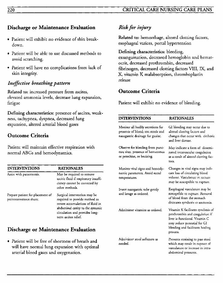
220 CRITICAL CARE NURSING CARE PLANS
Discharge or Maintenance Evaluation
Patient will exhibit no evidence of skin break- down.
Patient will be able to use discussed methods to avoid scratching.
Patient will have no complications from lack of skin integrity.
Inefective breathing pattern
Related to: increased pressure from ascites, elevated ammonia levels, decrease lung expansion, fatigue
Defining characteristics: presence of ascites, weak- ness, tachypnea, dyspnea, decreased lung expansion, altered arterial blood gases
Outcome Criteria
Patient will maintain effective respiration with normal ABGs and hemodynamics.
INTERVENTIONS RATIONALES Assist with paracentesis. May be required to remove
ascitic fluid if respiratory insuffi- ciency cannot be corrected by other methods.
Surgical intervention may be required to provide method to return accumulations of fluid in abdominal cavity to the systemic circulation and provides long- term ascites relief.
Prepare patient for placement of peritoneovenous shunt.
Discharge or Maintenance Evaluation
Patient will be free of shortness of breath and will have normal lung expansion with optimal arterial blood gases and oxygenation.
Risk f i r injury
Related to: hemorrhage, altered clotting factors, esophageal varices, portal hypertension
Defining characteristics: bleeding, exsanguination, decreased hemoglobin and hemat- ocrit, decreased prothrombin, decreased fibrinogen, decreased clotting factors VIII, IX, and X, vitamin K malabsorption, thromboplastin release
Outcome Criteria
Patient will exhibit no evidence of bleeding.
INTERVENTIONS RATIONALES
Monitor all bodily secretions for presence of blood; test stools and nasogastric drainage for guaiac.
GI bleeding may occur due to altered clotting factors and changes that occur with cirrhosis and liver disease.
May indicate a form of dissemi- nated intravascular coagulation as a result of altered clotting fac- tors.
Observe for bleeding from punc- ture sites, presence of hematomas or petechiae, or bruising.
Monitor vital signs and hemody- Changes in vital signs may indi- namic parameters. Avoid rectal cate loss of circulating blood temperatures. volume. Vasculature in rectum
may be susceptible to rupture.
Esophageal vasculature may be susceptible to rupture. Removal of blood from the stomach decreases synthesis to ammonia.
Insert nasogastric tube gently and lavage as ordered.
Administer vitamins as ordered. Vitamin K facilitates synthesis of prothrombin and coagulation if liver is functional. Vitamin C may reduce potential for GI bleeding and facilitates healing process.
Prevents straining to pass stool which may result in rupture of vasculature or increase in intra- abdominal pressures.
Administer stool softeners as needed.
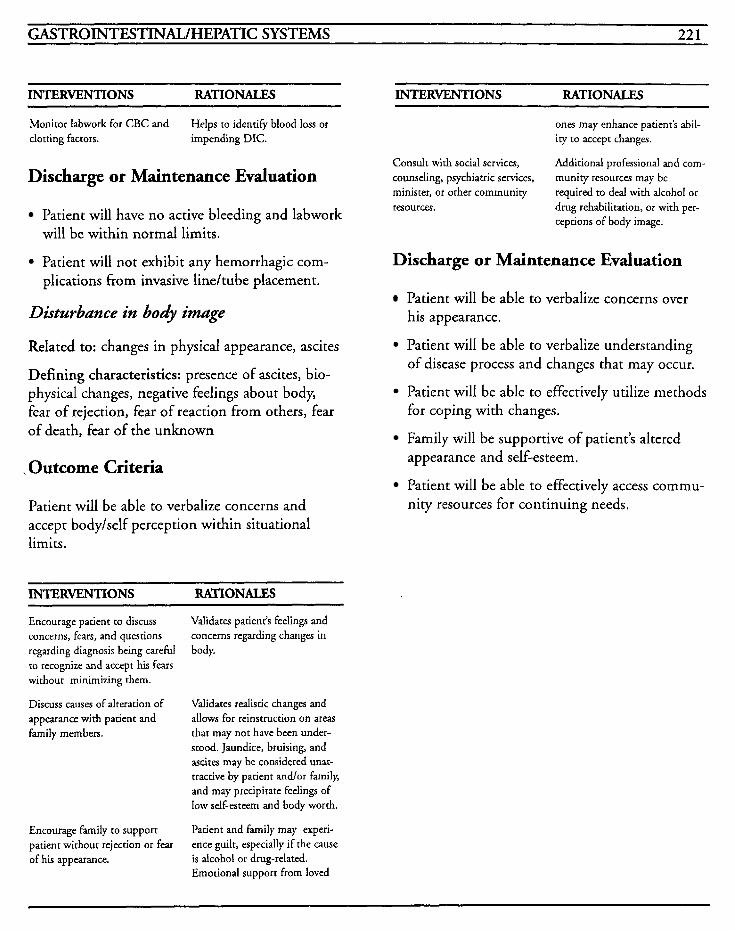
GASTROINTESTINAL/HEPATIC SYSTEMS 22 1
INTERVENTIONS RATIONALES INTERVENTIONS RATIONALES
Monitor labwork for CBC and clotting factors. impending DIC.
Helps to identify blood loss or
Discharge or Maintenance Evaluation
Patient will have no active bleeding and labwork will be within normal limits.
Patient will not exhibit any hemorrhagic com- plications from invasive lineltube placement.
Disturbance in body image
Related to: changes in physical appearance, ascites
Defining characteristics: presence of ascites, bio- physical changes, negative feelings about body, fear of rejection, fear of reaction from others, fear of death, fear of the unknown
I Outcome Criteria
Patient will be able to verbalize concerns and accept bodylself perception within situational limits.
INTERVENTIONS RATIONALES
Encourage patient to discuss concerns, fears, and questions regarding diagnosis being careful to recognize and accept his fears without minimizing them.
Discuss causes of alteration of appearance with patient and family members.
Encourage family to support patient without rejection or fear of his appearance.
Validates patient’s feelings and concerns regarding changes in body.
Validates realistic changes and allows for reinstruction on areas that may not have been under- stood. Jaundice, bruising, and ascites may be considered unat- tractive by patient andlor family, and may precipitate feelings of low self-esteem and body worth.
Patient and family may experi- ence guilt, especially if the cause is alcohol or drug-related. Emotional support from loved
ones may enhance patient’s abil- ity to accept changes.
Additional professional and corn- munity resources may be required to deal with alcohol or drug rehabilitation, or with per- ceptions of body image.
Consult with social services, counseling, psychiatric services, minister, or other community resources.
Discharge or Maintenance Evaluation
Patient will be able to verbalize concerns over his appearance.
Patient will be able to verbalize understanding of disease process and changes that may occur.
Patient will be able to effectively utilize methods for coping with changes.
Family will be supportive of patient‘s altered appearance and self-esteem.
nity resources for continuing needs. Patient will be able to effectively access commu-
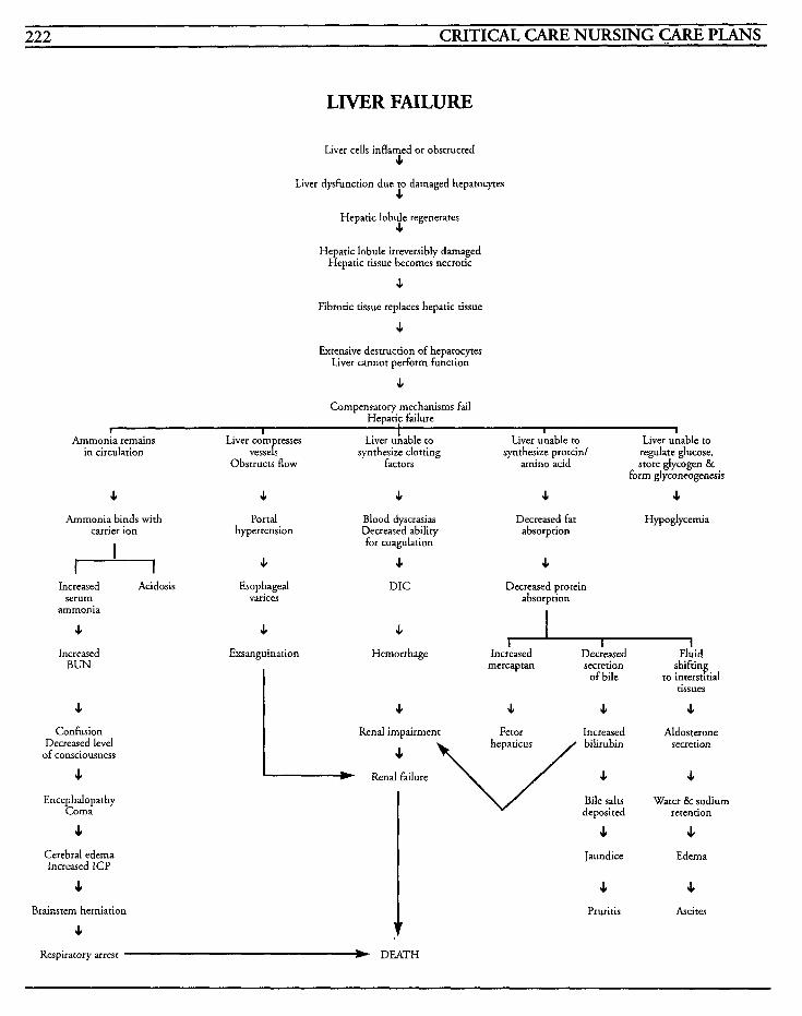
222 CRITICAT., CARE NURSING CARE PLANS
LIVER FAILURE
Liver cells inflamed or obstructed I
Liver dysfunction due to damaged hepatoves I
Hepatic lobule regenerates I
Hepatic lobule irreversibly damaged Hepatic tissue becomes necrotic
I
Fibrotic tissue replaces hepatic tissue
4 Extensive destruction of hepatocyces
Liver cannot perform function
I
Compensatory mechanisms fail Hepatic failure
I - - - ~ I I I I
Ammonia remains Liver com resses Liver unable to Liver unable to Liver unable to in circulation vesseP, synthesize clotting synthesize protein/ regulate glucose,
Obstructs flow factors amino acid store glycogen & form glyconeogenesis
I I I I I Ammonia binds with
carrier ion I
Portal Blood dyscrasias hypertension Decreased ability
for coagulation
I I 4 I I
Increased Acidosis Esophageal serum varices
ammonia
I I
Increased BUN
I Confusion
Decreased level of consciousness
J,
Encephalopathy Coma
s Cerebral edema Increased ICP
s
s Brainstem herniation
hanguinat ion
DIC
Decreased fat H ypoglycemia absorption
c Decreased protein
absorption
Hemorrhage Increased Decreased Fluid mercap tan secretion shifting
tissues of bile to interstitial
I I I I
I Renal impairment Fetor Increased Aldosterone secretion
I
Bile salts Water & sodium deposited rerention
s J, I
+ Jaundice Edema
4 s Pruritis Ascites
Respiratory arrest b DEATH
HEMATOLOGIC SYSTEM 223
Disseminated Intravascular Coagulation (DIC)
HELLP Syndrome
Anemia

This Page Intentionally Left Blank
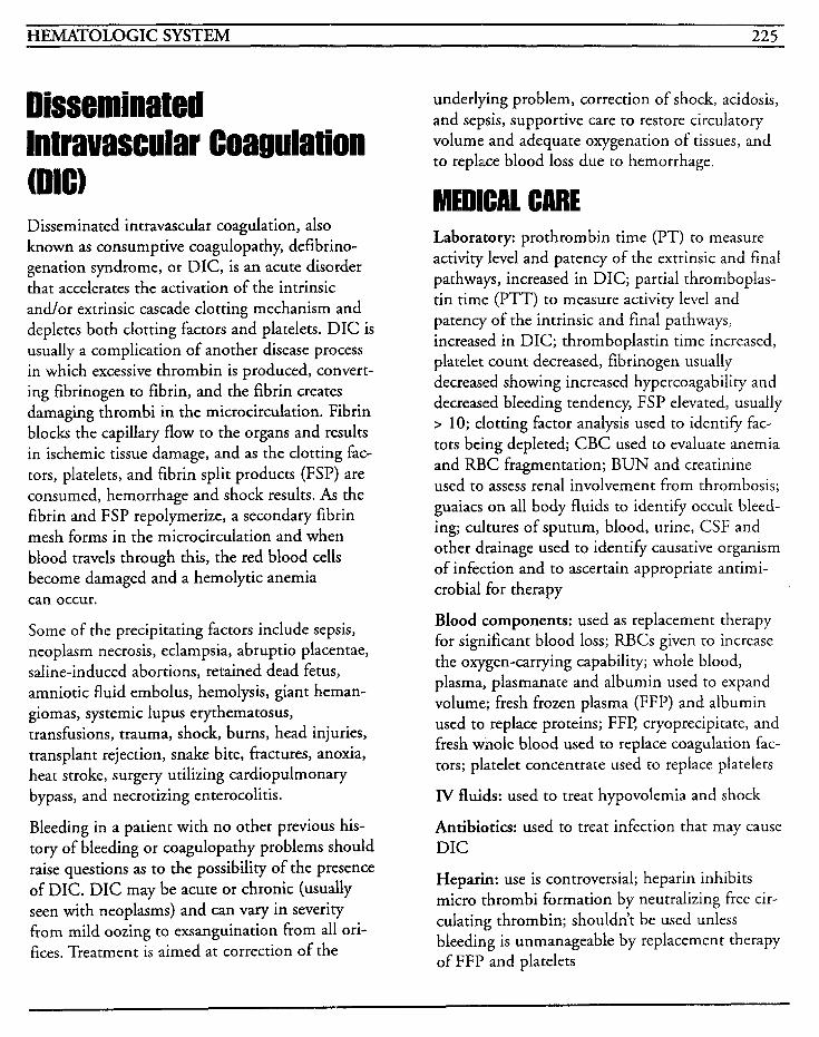
HEMATOLOGIC SYSTEM 225
lntravascular Coagulation underlying problem, correction of shock, acidosis, and sepsis, supportive care to restore circulatory volume and adequate oxygenation of tissues, and to replace blood loss due to hemorrhage.
Disseminated intravascular coagulation, also known as consumptive coagulopathy, defibrino- genation syndrome, or DIC, is an acute disorder that accelerates the activation of the intrinsic and/or extrinsic cascade clotting mechanism and depletes both clotting factors and platelets. DIC is usually a complication of another disease process in which excessive thrombin is produced, convert- ing fibrinogen to fibrin, and the fibrin creates damaging thrombi in the microcirculation. Fibrin blocks the capillary flow to the organs and results in ischemic tissue damage, and as the clotting fac- tors, platelets, and fibrin split products (FSP) are consumed, hemorrhage and shock results. As the fibrin and FSP repolymerize, a secondary fibrin mesh forms in the microcirculation and when blood travels through this, the red blood cells become damaged and a hemolytic anemia can occur.
Some of the precipitating factors include sepsis, neoplasm necrosis, eclampsia, abruptio placentae, saline-induced abortions, retained dead fetus, amniotic fluid embolus, hemolysis, giant heman- giomas, systemic lupus erythematosus, transfusions, trauma, shock, burns, head injuries, transplant rejection, snake bite, fractures, anoxia, heat stroke, surgery utilizing cardiopulmonary bypass, and necrotizing enterocolitis.
Bleeding in a patient with no other previous his- tory of bleeding or coagulopathy problems should raise questions as to the possibility of the presence of DIC. DIC may be acute or chronic (usually seen with neoplasms) and can vary in severity from mild oozing to exsanguination from all ori- fices. Treatment is aimed at correction of the
Laboratory: prothrombin time (PT) to measure activity level and patency of the extrinsic and final pathways, increased in DIC; partial thromboplas- tin time (PTT) to measure activity level and patency of the intrinsic and final pathways, increased in DIC; thromboplastin time increased, platelet count decreased, fibrinogen usually decreased showing increased hypercoagability and decreased bleeding tendency, FSP elevated, usually > 10; clotting factor analysis used to identify fac- tors being depleted; CBC used to evaluate anemia and RBC fragmentation; BUN and creatinine used to assess renal involvement from thrombosis; guaiacs on all body fluids to identify occult bleed- ing; cultures of sputum, blood, urine, CSF and other drainage used to identify causative organism of infection and to ascertain appropriate antimi- crobial for therapy
Blood components: used as replacement therapy for significant blood loss; RBCs given to increase the oxygen-carrying capability; whole blood, plasma, plasmanate and albumin used to expand volume; fresh frozen plasma (FFP) and albumin used to replace proteins; FFP, cryoprecipitate, and fresh whole blood used to replace coagulation fac- tors; platelet concentrate used to replace platelets
lV fluids: used to treat hypovolemia and shock
Antibiotics: used to treat infection that may cause DIC
Heparin: use is controversial; heparin inhibits micro thrombi formation by neutralizing free cir- culating thrombin; shouldn’t be used unless bleeding is unmanageable by replacement therapy of FFP and platelets
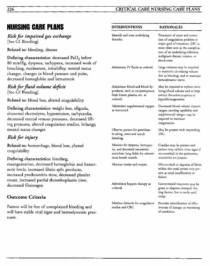
226 CRITICAL CARE NURSING CARE PLANS
NURSING CARE PLANS Risk fir impaired gas exchange [See GI Bleeding]
Related to: bleeding, disease
Defining characteristics: decreased Pa02 below 80 mmHg, dyspnea, tachypnea, increased work of breathing, restlessness, irritability, mental status changes, changes in blood pressure and pulse, decreased hemoglobin and hematocrit
Risk fir fluid volume &fieit [See GI Bleeding]
Related to: blood loss, altered coagulability
Defining characteristics: weight loss, oliguria, abnormal electrolytes, hypotension, tachycardia, decreased central venous pressures, decreased fill- ing pressures, altered coagulation studies, lethargy, mental status changes
Riskfor injury Related to: hemorrhage, blood loss, altered coagulability
Defining characteristics: bleeding, exsanguination, decreased hemoglobin and hemat- ocrit levels, increased fibrin split products, increased prothrombin time, decreased platelet count, increased partial thromboplastin time, decreased fibrinogen
Outcome Criteria
Patient will be free of unexplained bleeding and will have stable vital signs and hemodynamic pres- sures.
INTERVENTIONS RATIONALES
Identify and treat underlying disorder.
Administer IV fluids as ordered.
Administer blood and blood by- products, such as cryoprecipitate, fresh frozen plasma, etc. as ordered.
Administer supplemental oxygen as warranted.
Observe patient for petechiae, bruising, overt and occult bleeding.
Monitor for dyspnea, hemopty- sis, and decreased saturation; auscultate lung fields for adventi- tious breath sounds.
Monitor intake and output.
Administer heparin therapy as ordered.
Monitor labwork for coagulation studies and CBC.
Treatment of cause and correc- tion of coagulation problem is major goal of treatment. DIC is most often seen as the complica- tion of an underlying infection, malignant disease, trauma, or shock state.
Large volumes may be required to mainrain circulating volume due to bleeding, and to maintain hemodynamic status.
May be required to replace circu- lating blood volume and to help correct thrombocytopenia or hypofibrinogenemia.
Decreased blood volume impairs oxygen carrying capability and supplemental oxygen may be required ro maintain oxygenation.
May be present with impending DIC.
Crackles may be present and patient may exhibit these signs if microemboli in the pulmonary circulation are present.
Microemboli or deposits of fibrin within the renal system may pre- sent as renal insufficiency or failure.
Controversial treatment may be given to disperse clumped clot- ting factors, but is rarely used today.
Provides identification of effec- tiveness of therapy or worsening of condition.

HEMATOLOGIC SYSTEM 227
Discharge or Maintenance Evaluation
Patient will have stable vital signs and hemody- namic pressures.
Patient will exhibit no bleeding tendencies or active hemorrhage.
Patient will exhibit no complications from other disease processes.
Patient will achieve and maintain adequate blood volume.
Patient will have underlying disease process corrected.
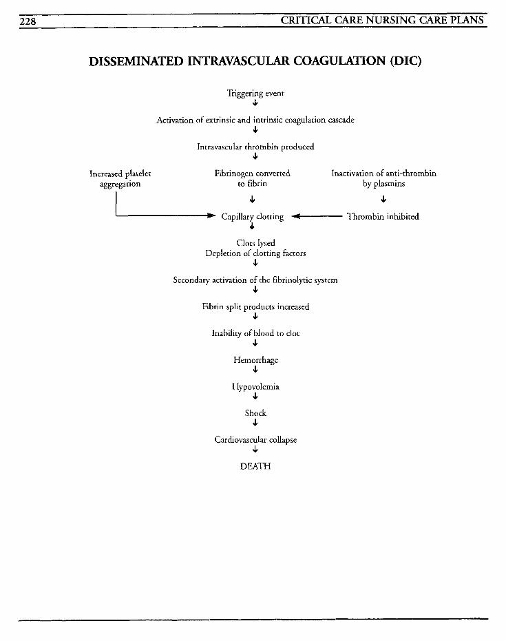
228 CRITICAL CARE NURSING CARE PLANS
DISSEMINATED INTUVASCULAR COAGULATION (DIC)
Triggering event 4
Activation of extrinsic and intrinsic coagulation cascade 4
Intravascular thrombin produced 4
Increased platelet aggregation
Fibrinogen converted Inactivation of anti-thrombin to fibrin by plasmins
4 4 Capillary clotting - Thrombin inhibited
J,
Clots lysed Depletion of clotting factors
P
Secondary activation of the fibrinolytic system P
Fibrin split products increased J,
Inability of blood to clot 4
Hemorrhage 4
Hypovolemia L
Shock J,
Cardiovascular collapse 4
DEATH
HEMATOLOGIC SYSTEM 229
H E 11 P Syndrome HELLP syndrome is an acute and severe compli- cation that presents as a multi-organic disease process occurring concurrently with pregnancy- induced hypertension (PIH). The initials are compiled from the symptoms that comprise the syndrome: hemolysis, elevated liver enzymes and low platelets. These same findings may also be associated with DIC and frequently is diagnosed as such.
PIH usually occurs after the twentieth week of gestation in approximately 5% of all pregnancies, and most often in the primagravida patient. PIH results in increased edema, proteinuria, and hyper- tension. Although the cause is unknown, theories often involve immunologic, endocrine, and chori- onic villi exposure.
HELLP may represent an acute autoimmune state in which the red blood cells lyse, liver enzymes are elevated as a result of fibrin thrombi blocking blood flow to the liver, and platelets decrease due to vasospasm and platelet aggregation. Vasospasm results in increases in systemic and peripheral vas- cular resistance, which increase blood pressure further. Sensitivity to angiotensin I1 is increased, and vasoconstriction may result in increases in vas- cular permeability and hemoconcentration.
The pathological changes in the liver may develop due to generalized activation of the intravascular coagulation process. Fibrin deposits and hemor- rhagic necrosis develops in periportal areas and may lead to subcapsular hematomas or liver rup- ture. A decrease in antithrombin I11 and an increase in thrombin-antithrombin I11 complex (TAT) and the appearance of fibrin monomers and D-dimers is found in almost all cases of HELLP, but decompensated intravascular coagula- tion with increased PT and PTT and decreased
fibrinogen levels is found only in severe forms. Decompensated coagulation occurs with other complications such as liver hematoma, abruptio placenrae, renal failure, and pulmonary edema.
There is usually a low recurrence rate (50/0 or less), and the HELLP syndrome usually resolves with delivery of the baby. Treatment involves prophy- laxis against postpartum worsening, curettage of the uterus, and treatment with calcium antagonists and decadron, as well as intense monitoring for a decline in liver function and for potential for bleeding.
MEDICAL CARE Laboratory: hematocrit used to assess intravascular fluid status; protime and partial thromboplastin time used to evaluate clotting; magnesium levels used to evaluate therapeutic levels for treatment; urine collection for protein used to diagnose com- plications
Magnesium: used to prevent and treat convulsions by decreasing the neuromuscular irritability and depressing the central nervous system
Antihypertensives: apresoline is the drug of choice; used to relax arterioles and stimulate car- diac output and is utilized with diastolic blood pressures greater than 110 mmHg
Beta-blockers: occasionally used to control acute hypertensive crises
Vdium: used to control seizure activity
NURSING CARE PLANS Risk for impairedgas excbaage [See GI Bleeding]
Related, to: bleeding, disease
Defining characteristics: decreased Pa02 below 80 mmHg, dyspnea, tachypnea, increased work of
w n CRITICAL CARE NURSING CARE PLANS
breathing, restlessness, irritability, mental status changes, changes in blood pressure and pulse, decreased hemoglobin and hematocrit
Risk for fluid volume deficit [See GI Bleeding]
Related to: blood loss, altered coagulability
Defining characteristics: weight loss, oliguria, abnormal electrolytes, hypotension, tachycardia, decreased central venous pressures, altered coagu- lation studies, lethargy, mental status changes
Risk for injury Related to: administration of magnesium
Defining characteristics: CNS depression, venous irritation, dyspnea, shallow respirations, decreased oxygen saturation, oliguria, absence of deep tendon reflexes, changes in vital signs
Outcome Criteria
Patient will receive medication without experienc- ing side effects.
INTERVENTIONS RATIONALES
Monitor for convulsions or tremors. problem.
Administer magnesium sulfate as ordered.
Identifies precipitation of
Magnesium is used to prevent and treat convulsions by decreas- ing the neuromuscular irritability and depression of the central nervous system. Normally, MgSO, is given W, with a load- ing dose of 3-4 Grams, followed by an infusion of 1-4 Gramslhr. It may be given IM with dosage of 5 Grams in each hip every 4 hours using the 2-tract method. Some facilities add xylocaine to the medication to decrease the pain of IM injections.
~~
INTERVENTIONS RATIONALE
Monitor vital signs every 1-2 hours, and prn, especially respi- ratory status.
Depression of CNS can result in respiratory insufficiency or paral- ysis. Hypothermia may occur with toxicity of drug. MgS04 should he held for respirations less than 16 per minute.
Dysrhythmias may occur with administration of magnesium or with its antidote, calcium.
Magnesium sulfate may cause toxicity with large doses and result in renal insufficiency and oliguria.
Fetal heart rate may decrease with use of magnesium sulfate.
Absence of DTRs may indicate hyperrnagnesemia and toxicity. Decreased DTRs may occur with therapeutic ranges.
Calcium gluconate is the anti- dote for magnesium sulfate.
Monitor EKG for changes and dysrhythmias, and treat per hos- pital protocol.
Monitor intake and output every 2 hours.
Monitor fetal heart tones every hour.
Assess deep tendon reflexes.
Have calcium gluconate at bedside and give as warranted/ ordered.
Monitor labwork for magnesium levels.
Normal levels are 4-7.5 mEq/L, with toxic levels above that.
Information, Instruction, Demonstration
INTERVENTIONS RATIONALES
Instruct on signs and symptoms to report to nurselphysician.
Facilitates prompt identification of problem to allow for timely intervention.
Observe IM injection sites for redness, firm areas, warmth, and pain.
May indicate presence of sterile abscess from injections which have a variable rate of absorption given in this manner.
HEMATOLOGIC SYSTEM 23 1
Discharge or Maintenance Evaluation
Patient will have stable vital signs.
Patient will be free of convulsions.
Fetal heart rates will remain unaffected and activity will be within normal range.
Patient will exhibit no signs of magnesium toxi- city or complications from therapy.
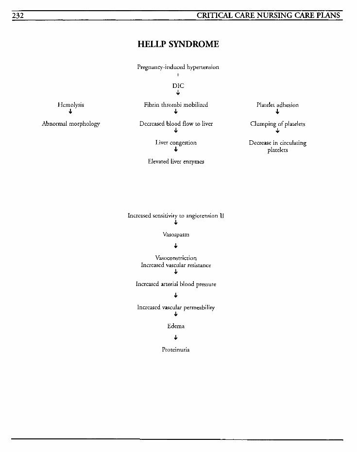
232 CRITICAL CARE NURSING CARE PLANS
HELLP SYNDROME
Hem01 ysis J,
Abnormal morphology
Pregnancy-induced hypertension
DIC J
Fibrin thrombi mobilized J,
Decreased blood flow to liver 4
Liver congestion J,
Elevated liver enzymes
+
Increased sensitivity to angiotension 11 4
Vasospasm
J
Vasoconstriction Increased vascular resistance
J,
Increased arterial blood pressure
J
Increased vascular permeability J,
Edema
J
Pro teinuria
Platelet adhesion J,
Clumping of platelets J,
Decrease in circulating platelets
HEMATOLOGIC SYSTEM 233
Anemia is a condition in which the red blood cell count, hemoglobin, and hematocrit are decreased. This decrease results in a decrease in the oxygen- carrying capability and causes tissue hypoxia. As the body tries to compensate, blood is shifted from areas that have a plentiful amount in tissues that have low oxygen requirements to those areas that require higher oxygen concentrations, such as the heart and brain.
There are several types of anemias; those that are due to decreased red blood cell production, those that are due to blood loss, and the hemolytic ane- mias caused from G6PD deficiency, autoimmunity, or physical causes. Microcytic, or iron deficiency anemia, develops when the trans- portation of iron by transferrin is insufficient to meet requirements of the erythropoietic cells. Macrocytic, or megaloblastic anemia, occurs because of a deficiency in vitamin B,, or folic acid. Pernicious anemia is a type of megaloblastic anemia in which the absence of vitamin B,, as well as lack of the intrinsic factor is noted. Normocytic, or aplastic anemia, is caused from the failure of the bone marrow or destruction of bone marrow by either chemical or physical means. Autoimmune anemia is an acquired condition that involves premature erythrocyte destruction from the person's own immune system. Hemolytic anemia results when erythrocyte destruction is increased and cells have a shortened life span. Sickle cell anemia is an inherited condition in which hemoglobin S is present in the blood result- ing in sickling and abnormal hemolyzation that obstructs capillary flow. Thalassemia is a group of inherited anemias that result from faulty produc- tion of alpha or beta hemoglobin polypeptides.
Anemia can occur as the direct result of prosthetic
heart valves or extracorporeal circulation and the destruction of red blood cells. Anemias can also be precipitated by toxic substance exposure or chronic disease processes, such as uremia or chronic liver disease.
MEDICAL CARE Laboratory: CBC to help differentiate type of anemia-RBCs reduced; hemoglobin decreased with mild considered 10-14 G/dl, moderate 6-10 G/dl, and severe below 6 G/dl; hematocrit decreased; MCH, MCHC variable dependent on type of anemia; MCV 80-100 fl with normocytic, greater than 100 fl with macrocytic, and less than 80 fl with microcytic; platelet count usually decreased, but may be elevated after hemorrhage; RDW increased in iron depletion anemia; B,, level decreased, folate decreased; serum iron and TIBC may be decreased; stool guaiac may be posi- tive if blood loss is from GI tract
Radiography: chest x-ray to discern pulmonary or cardiac complications; upper and lower gastroin- testinal series may be done to identify active or current bleeding
Bone marrow aspiration: may be performed to determine type of anemia
Bone marrow transplants: may be required for severe aplastic anemia
Blood transfusions: may be required to replace blood volume with hemorrhage
NURSINC CARE PLANS Alteration in tissue perfksion: cardiopulmonary, renal, cerebral, gastroin- testinah peripheral
Related to: altered oxygen-carrying capability, blood loss
234 CRITICAL CARE NURSING CARE PLANS
Defining characteristics: decreased hematocrit and hemoglobin, chest pain, palpitations, pallor, dry mucous membranes, cold intolerance, oliguria, nausea, vomiting, abdominal pain, abdominal dis- tention, increased capillary refill time, confusion, lethargy, changes in pulse rate and blood pressure
Outcome Criteria
Patient will have adequate perfusion to all body systems with stable vital signs and hernodynamics
~~ ~~~ ~~
INTERVENTIONS RATIONALES
Monitor vital signs every 1-2 hours and prn.
Monitor neurological status for mental confusion or level of con- sciousness changes.
Auscultate lung fields for adven- titious breath sounds. Auscultate for abnormal heart tones.
Administer supplemental oxygen as warranted.
Monitor EKG for changes in cardiac rhythm or conduction.
Facilitates identification of changes that may require intervention.
May be indicative of impaired cerebral perfusion.
Crackles andlor new presence of cardiac gallops may indicate impending or present congestive failure that may have resulted from the body’s compensatory mechanism of increasing cardiac ourput. Mild anemia can cause exertional dyspnea and palpita- tions; moderate anemia can cause increased palpitations and dysp- nea at rest; severe anemia causes tachycardia, increased pulse pres- sure, systolic murmurs, intermittent claudication, angina, congestive heart failure, orthop- nea, and tachypnea.
Decreases in red blood cells decreases oxygen carrying capa- bility since oxygen is bound to the hemoglobin for transport, and may require supplementa- tion to maintain oxygenarion.
Changes may occur with imbal- ances of electrolytes, with fluid shifts, or with hypoxia.
~~
INTERVENTIONS RATIONALES
Monitor for complaints of chest pain, pressure, palpitations, or dyspnea. ischemia.
Administer blood andlor blood products as warranted.
May indicate decreased cardiac perfusion from hypoxia or
Blood replacement may facilitate improved oxygen-carrying ability due to increased number of red blood cells and correct volume deficiency.
May facilitate identification of deficiencies and allow for assess- ment of effectiveness of treatment.
Monitor labwork for changes.
Information, Instruction, Demonstration
INTERVENTIONS RATIONALES ~~~ ~
Maintain environment tempera- ture within normal ranges.
Reduction of peripheral perfu- sion may result in cold intolerance to vasoconstriction. Excessive heat may cause vasodi- lation and further reduce organ perfusion.
May require transplantation of bone marrow, or surgical repair for site of bleeding.
Prepare patientlfamily for surgi- cal procedures as warranted.
Discharge or Maintenance Evaluation
Patient will achieve and maintain adequate per- fusion to all body systems.
Patient will have stable vital signs and hemody- namic pressures.
Patient will exhibit no evidence of GI bleeding.
Patient will exhibit no signs of complications of disease or therapy.
Risk for fluid volume deficit [See GI Bleeding]
Related to: bleeding
Defining characteristics: hypotension, tachycar-
HEMATOLOGIC SYSTEM 235
dia, decreased skin turgor, weakness, decreased uri- nary output, pallor, diaphoresis, decreased capillary refill, mental changes, restlessness, decreased filling pressures
Activity intolerance [See COPD]
Related to: decreased oxygen-carrying capability
Defining characteristics: weakness, lethargy, fatigue, dyspnea, activity intolerance, chest pain, palpitations, tachycardia, decreased oxygen satura- tion, increased respiratory rate with exertion, hypertension
Alteration in nutrition: less than body requirements [See DKA]
Related to: inability to absorb required nutrients for red blood cell production
Defining characteristics: weight loss, activity intolerance, dyspnea, fatigue, weakness, loss of muscle tone, anorexia
Knowledge deficit
Related to: lack of information, unfamiliarity with information, lack of recall, misinterpretation of information
Defining characteristics: questions, communica- tion of misconceptions, development of preventable complications, incorrect follow-up with instructions
Outcome Criteria
Patient will be able to verbalize understanding of disease process, treatment regimen, and procedures, and comply with therapy.
INTERVENTIONS RATIONALES
Instruct on particular rype of anemia that patient has developed.
Instruct on labwork and other procedures. unknown.
Instruct on dietary requirements.
Provides knowledge and facili- tates compliance.
Decreases anxiety and fear of the
Increasing iron sources from red meat, egg yolks, dried fruits and green leafy vegetables may facili- tate correction of anemia. Folic acid and vitamin C which aug- ments iron absorption may be found in green vegetables, whole grains, citrus fruits, and liver.
Decreased leukocyte count may potentiate the risk of infection and patient should seek medical assistance for timely intervention.
Instruct on signs and symptoms of which to notify physician.
Instruct on medications, effects, side effects, contraindications, and avoidance of over-the- counter medications without physician approval.
Iron or vitamin B,, replacement may be necessary for life, and knowledge regarding therapeutic management will increase com- pliance with treatment and allow for prompt identification of complications that may require changes in dosages, types of' medication, or schedule of administration.
Discharge or Maintenance Evaluation
Patient will be able to verbalize and demonstrate understanding of all instructed information.
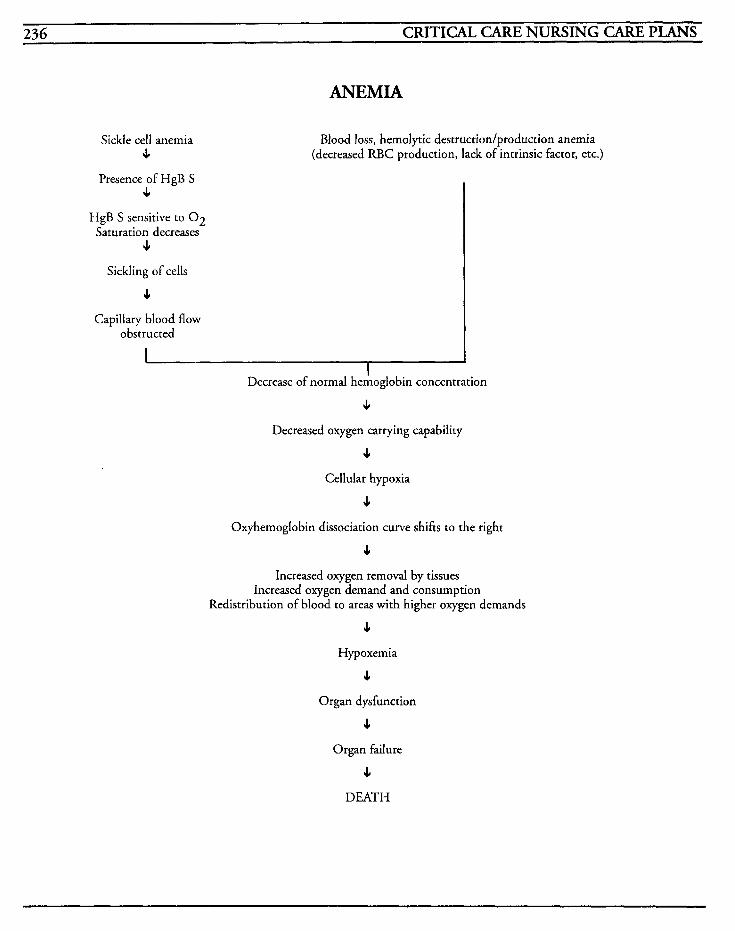
236 CRITICAL CARE NURSING CARE PLANS
ANEMIA
Sickle cell anemia s
Blood loss, hemolytic destruction/production anemia (decreased RBC production, lack of intrinsic factor, etc.)
Presence of HgB S J
HgB S sensitive to 0 2 Saturation decreases
4
Sickling of cells
4 Capillary blood flow
obstructed
1 I
Decrease of normal hemoglobin concentration
4
Decreased oxygen carrying capability
J
Cellular hypoxia
J
Oxyhemoglobin dissociation curve shifts to the right
4 Increased oxygen removal by tissues
Increased oxygen demand and consumption Redistribution of blood to areas with higher oxygen demands
J
Hypoxemia
4
Organ dysfunction
4 Organ failure
J
DEATH
RENAL/ENDOCRINE SYSTEMS 237
Acute Renal Failure (ARF)
Diabetic Ketoacidosis (DKA)
Hyperglycemic Hyperosmolar Nonketotic Coma (HHNK)
Syndrome of Inappropriate ADH Secretion (SIADH)
Diabetes Insipidus (DI)
Pheochromocytoma
Thyrotoxicosis (Thyroid Storm)

This Page Intentionally Left Blank
RENAL/ENDOCRINE SYSTEMS 239
Acute Renal Failure (ARF) Acute renal failure (ARF) is noted when there is a sudden deterioration in function of the renal system that may be caused by renal circulation failure or glomerular or tubular dysfunction. The build-up of waste materials that accumulates affects multiple organ systems.
ARF can be subclassified according to the etiology of condition, such as prerenal, intrarenal, and postrenal. Prerenal conditions occur when blood perfusion is inadequate, such as with hypotension, hemorrhage, myocardial infarction, congestive heart failure, pulmonary embolism, burns, third spacing, septic shock, diuretic abuse, or volume depletion. This dysfunction causes glomerular fil- tration rates to decrease, and decreased reabsorption of sodium in the tubules.
Intrarenal renal failure occurs either from damage to the tubular epithelium, known as acute tubular necrosis (ATN), or from damage to glomeruli and the small vessels. This condition causes renal capil- lary swelling that decreases the glomerular filtration rate (GFR), or decreased GFR is secondary to the obstruction of the glomeruli by edema and cellular debris. ATN is the most common type of ARF and is the result of nephro- toxins or ischemia. Intrarenal failure may take many weeks to repair damage and is usually seen with trauma, sepsis, DIC, transfusion reactions, renal vasculature blockages, heavy metal poison- ing, and with use of aminoglycosides, penicillins, tetracylines, dilantin, and amphotericin. Glomeruli damage is seen with acute glomerulonephritis, polyarteritis nodosa, lupus erythematosus, Goodpasture’s syndrome, endocarditis, abruptio placentae, abortion, serum sickness, malignant hypertension, or hemolytic uremic syndromes.
Postrenal failure may occur as a result of an
obstruction anpvhere in the system from the kidney to the urethra. Some clinical conditions in which this type of failure is seen includes urethral obstruction, prostatic hypertrophy, bladder carci- noma, bladder infection, neurogenic bladder, renal calculi, and abdominal tumors.
There are three phases in ARF-an oliguric phase, a diuretic phase, and a recovery phase. Oliguria occurs when the tubule obstruction and damage makes absorption unstable, and BUN, creatinine, and potassium levels increase. During the diuretic phase, tubular function begins to return but the patient must be monitored for excessive diuresis with loss of electrolytes. When diuresis is no longer excessive, the recovery phase begins with gradual improvement in kidney function for up to one year. There may be a permanent decrease in renal function that, depending on severity, may require dialysis.
MEDICAL CARE Laboratory: CBC- hemoglobin decreased with anemia, RBCs decreased due to fragility, white blood cell count elevated if sepsis or trauma is pre- cipitating event; BUN and creatinine elevated with ratio of 10: 1 ; serum osmolality increased above 285 mOsm/kg; electrolytes used to show imbalances, with elevated potassium due to reten- tion, hemolysis, or acidosis; sodium usually increased, but may be normal; bicarbonate, pH, and calcium decreased; magnesium, phosphorus, and chloride increased; complement studies may be used to identify lupus nephritis; serum electrophoresis may be used to identify abnormal proteins that may damage kidneys permanently; A S 0 titer may be used to diagnose recent strepto- coccal infection that could cause poststreptococcal glomerulonephritis; UA: Urine color is dirty, tea- colored brown, volume is less than 400 cc/day, specific gravity less than 1.020 indicates renal dis- ease and fixed at 1 .O 10 indicates severe renal
240 CRITICAL CARE NURSING CARE PLANS
damage; pH greater than 7.0 seen with UTI, ATN, and chronic renal failure; osmolality less than 350 mOsmlkg indicates tubular damage; cre- atinine clearance decreased; sodium decreased but may be greater than 40 mEq/L if kidney does not reabsorb sodium; RBCs may be present if infec- tion, renal stones, trauma, or tumor is cause; protein of 3 or 4+ indicates glomerular damage, 1+ or 2+ may indicate infection or interstitial nephritis; casts indicate renal disease or infection, brownish casts and numerous epithelia1 cells indi- cate ATN, and red casts indicate acute glomerular nephritis
Electrocardiogram: used to identify dysrhythmias and cardiac changes that may occur with acid-base imbalances or electrolyte imbalance
Radiography: KUB to identify size of structures, cysts, tumors, stones, or abnormal kidney location; chest x-ray to identify fluid overload that may occur with fluid shifts
Radionuclide imaging: may be used to identify hydronephrosis, calicectasis, or delayed filling or emptying, or other causes of AW
Retrograde pyelogram: may be used to identify abnormalities of ureters or renal pelvis
Renal arteriogram: may be used to identify extravascular irregularities or masses, and provides visualization of renal circulation
Magnetic resonance imaging: may be used to evaluate soft tissue
CT scans: may be used to detect presence of renal d’ isease
Dialysis: emergency and chronic dialysis may be required for ARF; ultrafiltration and CAVH may also be utilized
Surgery: may be required for renal calculi removal, resection of the prostate, or placement of fistula for long-term dialysis
NURSING CARE PUINS Fluid volume excess
Related to: impairment of renal system regulation, retention of water
Defining characteristics: oliguria, anuria, changes in urine specific gravity, intake greater than output, weight gain, elevated blood pressure, edema, ascites, increased central venous pressure, neck vein distention, dyspnea, orthopnea, crackles, muffled heart tones, decreased hemoglobin and hematocrit, altered electrolytes, increased filling pressures, restlessness, anxiety, water intoxication
Outcome Criteria
Patient will have balanced intake and output, stable weight, stable vital signs and hemodynamic parameters, and have effective dialysis when required.
INTERVENTIONS RATIONALES
Monitor vital signs and hemody- namic parameters every 1-2 hours.
Monitor intake and output every 2 hours and prn, noting balance or imbalance per 24 hour period. Estimate insensible losses through lungs, skin, and bowel.
Weigh daily.
Hypertension with increases in heart rate may occur when kid- neys fail to excrete urine, changes occur within the renin- angiotensin cascade, or with fluid resuscitation. Hemodynamic pressures can facilitate identification of changes with intravascular volume.
Facilitates identification of fluid requirements based on renal function. Insensible losses can add up to 800- 1000 cc/day and metabolism of carbohydrates can liberate up to 350 cdday of fluid from ingested foods.
Changes in body weight help to identify fluid status. Gains over 1 poundlday indicate fluid reten-
RENWENDOCRINE SYSTEMS 24 1
INTERVENTIONS RATIONALES
Auscultate lungs for adventitious breath sounds.
Measure urine specific gravity, and note changes in character of urine output.
Administer fluids as warranted with restrictions per physician orders.
Administer diuretics as ordered.
'Ihsert Foky catheter as warranted.
Observe for presence and charac- ter of edema.
Monitor for mental status changes.
Monitor arterial blood gases.
Monitor labwork for alterations.
tion. Fluid amounts of 500 cc are equivalent to 1 pound.
Adventitious breath sounds, such as crackles, will be heard with development of pulmonary edema or congestive heart failure.
Specific gravig is less than 1.010 in intrarenal failure and signifies inability to appropriately concen- trate the urine.
Prerenal failure is treated with fluid replacement, occasionally with use of vasopcessors. Management of fluids is based on replacement of output from all sources.
May be given to convert oliguric phase to nonoliguric phase, to flush debris from tubules, decreased hyperkalemia, or foster improved urine output.
Catheterization eliminates poten- tial lower GU tract obstruction and provides for accuracy of measurement of urine output, but may not be treatment of choice due to potential for infec- tion.
Dependent edema may be pre- sent, but pitting edema may not be discernable until the patient has more than 10 pounds of fluid in body. Periorbital edema may be the first clinical evidence of edema and indication of fluid shifting.
May indicate impending hypoxic state, electrolyte imbalances, acidosis, or sepsis.
May indicate presence of acidosis and facilitate intervention for hypoxemia.
Electrolyte imbalances may occur from impaired sodium reabsorp- tion, fluid overload, or lack of excretion of potassium.
INTERVENTIONS RATIONALES
Hyperkalemia may occur as body attempts to correct acidosis, hypernatremia may indicate total body water deficit, and hypona- tremia may result from fluid overloading or inability to con- serve sodium. BUN/crearinine ratio, which is normally 10: 1 is greater than 20:l with prerenal failure.
Urine sodium less than 20 mEq/L, osmolality above 450 mOsm/kg, and urine creatinine above 40 indicates prerenal fail- ure. Urine sodium above 40 mEqlL, osmolality below 350 mOsm/kg, and urine creatinine below 20 indicates ATN.
Monitor urine specimen labwork for changes.
Information, Instruction, Demonstration
~ _ _ _ _ _ _ _
INTERVENTIONS RATIONALES
Identify and correct any reversible reason for ARE
Improvement of perfusion, enhancing cardiac output and hemodynamics, or removal of obstruction may facilitate recov- ery from ARF and limit residud effects.
May be used to identify increas- ing cardiac silhouette, effusions, infiltrates, pulmonary edema, or other complications that may occur with fluid overload.
May be required to treat hyper-
decreased renal perfusion or fluid overload.
Promotes understanding and facilitates compliance.
Dialysis may be required to remove toxic wastes and to cor- rect electrolyte, acid-base, and fluid imbalances.
Obtain chest x-rays and compare with previous films.
Administer antihypertensives as warrantedlordered. tension that occurs from
Instruct patient/family on neces- sity for fluid restriction.
Prepare patientlfamily for dialysis treatment as warranted.
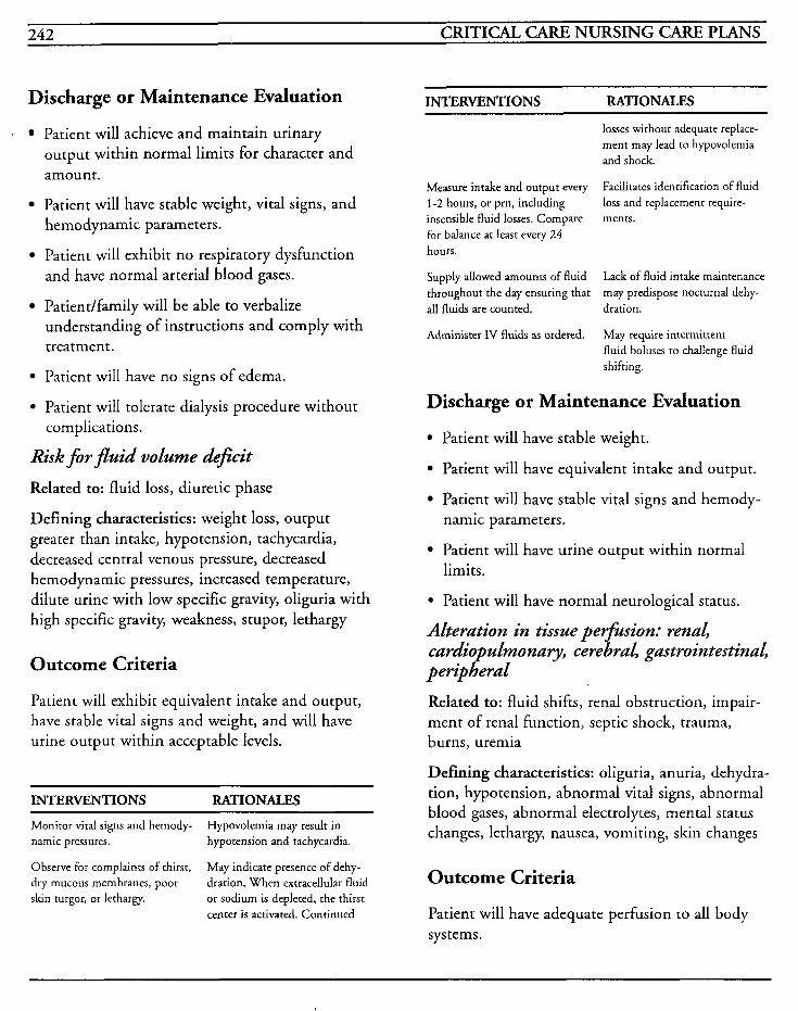
242 CRITICAL CARE NURSING CARE PLANS
Discharge or Maintenance Evaluation INTERVENTIONS RATIONALES
. Patient will achieve and maintain urinary output within normal limits for character and amount.
Patient will have stable weight, vital signs, and hemodynamic parameters.
Patient will exhibit no respiratory dysfunction and have normal arterial blood gases.
Patient/family will be able to verbalize understanding of instructions and comply with treatment.
Patient will have no signs of edema.
* Patient will tolerate dialysis procedure without
Risk f i r fluid volume deficit
Related to: fluid loss, diuretic phase
Defining characteristics: weight loss, output greater than intake, hypotension, tachycardia, decreased central venous pressure, decreased hemodynamic pressures, increased temperature, dilute urine with low specific gravity, oliguria with high specific gravity, weakness, stupor, lethargy
complications.
Outcome Criteria
Patient will exhibit equivalent intake and output, have stable vital signs and weight, and will have urine output within acceptable levels.
INTERVENTIONS RATIONALES
Monitor vital signs and hemody- namic pressures. hypotension and tachycardia.
Hypovolemia may result in
losses without adequate replace- ment may lead to hypovolemia and shock.
Facilitates identification of fluid loss and replacement require-
Measure intake and output every 1-2 hours, or prn, including insensible fluid losses. Compare ments. for balance at least every 24 hours.
Supply allowed amounts of fluid throughout the day ensuring that all fluids are counted. dration.
Administer IV fluids as ordered.
Lack of fluid intake maintenance may predispose nocturnal dehy-
May requite intermittent fluid boluses to challenge fluid shifting.
Discharge or Maintenance Evaluation
Patient will have stable weight.
Patient will have equivalent intake and output.
Patient will have stable vital signs and hemody- namic parameters.
Patient will have urine output within normal limits.
* Patient will have normal neurological status.
Alteration in tissue perfision: renal, cardio ulmonary, cerebral, gastrointestinal,
Related to: fluid shifts, renal obstruction, impair- ment of renal function, septic shock, trauma, burns, uremia
Defining characteristics: oliguria, anuria, dehydra- tion, hypotension, abnormal vital signs, abnormal blood gases, abnormal electrolytes, mental status changes, lethargy, nausea, vomiting, skin changes
perip R eral
Observe for complaints of thirst,
skin turgor, or lethargy.
May indicate presence of dehy-
or sodium is depleted, the thirst dry mucous membranes, poor dration. When extracellular fluid Outcome Criteria
center is activated. Continued Patient will have adequate perfusion to all body systems.
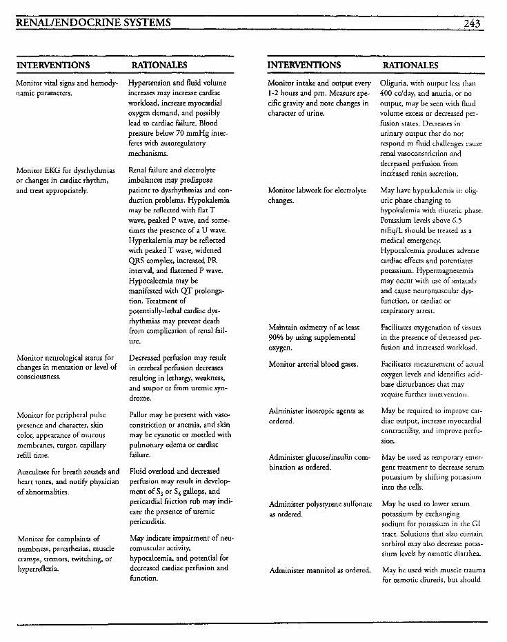
RENAL/ENDOCRINE SYSTEMS ~ , 243
INTERVENTIONS RATIONALES
Monitor vital signs and hemody- narnic parameters.
Monitor EKG for dysrhythmias or changes in cardiac rhythm, and treat appropriately.
Monitor neurological status for changes in mentation or level of consciousness.
Monitor for peripheral pulse presence and character, skin color, appearance of mucous membranes, turgor, capillary refill time.
Auscultate for breath sounds and heart tones, and notify physician of abnormalities.
Monitor for complaints of numbness, paresthesias, muscle cramps, tremors, twitching, or hyperreflexia.
Hypertension and fluid volume increases may increase cardiac workload, increase myocardial oxygen demand, and possibly lead to cardiac failure. Blood pressure below 70 mmHg inter- feres with autoregulatory mechanisms.
Renal failure and electrolyte imbalances may predispose patient to dysrhythmias and con- duction problems. Hypokalemia may be reflected with flat T wave, peaked P wave, and some- times the presence of a U wave. Hyperkalemia may be reflected with peaked T wave, widened QRS complex, increased PR interval, and flattened P wave. Hypocalcemia may be manifested with QT prolonga- tion. Treatment of potentially-lethal cardiac dys- rhythmias may prevent death from complication of renal fail- ure.
Decreased perfusion may result in cerebral perfusion decreases resulting in lethargy, weakness, and stupor or from uremic syn- drome.
Pallor may be present with vaso- constriction or anemia, and skin may be cyanotic or mottled with pulmonary edema or cardiac failure.
Fluid overload and decreased pe rh ion may result in develop- ment of S, or S4 gallops, and pericardial friction rub may indi- cate the presence of uremic pericarditis.
May indicate impairment of neu- romuscular activity, hypocalcemia, and potential for decreased cardiac perfusion and function.
INTERWNTIONS RATIONALES
Monitor intake and output every 1-2 hours and prn. Measure spe- cific gravity and note changes in character of urine.
Monitor labwork for electrolyte changes.
Maintain oximetry of at least 90% by using supplemental oxygen.
Monitor arterial blood gases.
Administer inotropic agents as ordered.
Administer glucoselinsulin com- bination as ordered.
Administer polystyrene sulfonate as ordered.
Administer mannitol as ordered.
Oliguria, with output less than 400 cclday, and anuria, or no output, may be seen with fluid volume excess or decreased per- fusion states. Decreases in urinary output that do not respond to fluid challenges cause renal vasoconstriction and decryased perfusion from increased renin secretion.
May have hyperkalemia in olig- uric phase changing to hypokalemia with diuretic phase. Potassium levels above 6.5 rnEqlL should be treated as a medical emergency. Hypocalcemia produces adverse cardiac effects and potentiates potassium. Hypermagnesemia may occur with use of antacids and cause neuromuscular dys- function, or cardiac or respiratory arrest.
Facilitates oxygenation of tissues in the presence of decreased per- fusion and increased workload.
Facilitates measurement of actual oxygen levels and identifies acid- base disturbances that may require further intervention.
May be required to improve car- diac output, increase myocardial contractility, and improve perfu- sion.
May be used as temporary emer- gent treatment to decrease serum potassium by shifting potassium into the cells.
May be used to lower serum potassium by exchanging sodium for potassium in the GI tract. Solutions that also contain sorbitol may also decrease potas- sium levels by osmotic diarrhea.
May be used with muscle trauma for osmotic diuresis, but should
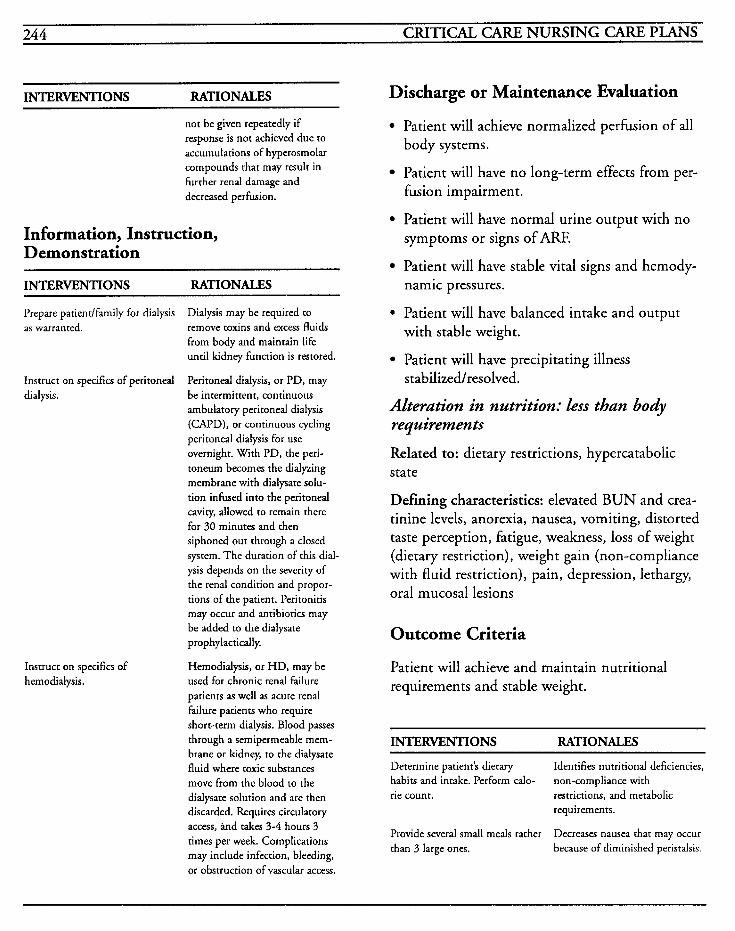
244 CRITICAL CARE NURSING CARE PLANS
INTERVENTIONS RATIONALES
not be given repeatedly if response is not achieved due to accumulations of hyperosmolar compounds that may result in further renal damage and decreased perfusion.
Information, Instruction, Demonstration
INTERVENTIONS RATIONALES
Prepare patiendfamily for dialysis as warranted.
Instruct on specifics of peritoneal dialysis.
Instruct on specifics of hemodialysis.
Dialysis may be required to remove toxins and excess fluids from body and maintain life until kidney function is restored.
Peritonea1 dialysis, or PD, may be intermittent, continuous ambulatory peritonea dialysis (CAPD), or continuous cycling peritoneal dialysis for use overnight. With PD, the peri- toneum becomes the dialyzing membrane with dialysate solu- tion infused into the peritonea cavity, allowed to remain there for 30 minutes and then siphoned out through a closed system. The duration of this dial- ysis depends on the severity of the renal condition and propor- tions of the patient. Peritonitis may occur and antibiotics may be added to the dialysate prophylactically.
Hemodialysis, or HD, may be used for chronic renal failure patients as well as acute renal failure patients who require short-term dialysis. Blood passes through a semipermeable mem- brane or kidney, to the dialysate fluid where toxic substances move from the blood to the dialysate solution and are then discarded. Requires circulatory access, ind takes 3-4 hours 3 times per week. Complications may include infection, bleeding, or obstruction of vascular access.
Discharge or Maintenance Evaluation
Patient will achieve normalized perfusion of all body systems.
Patient will have no long-term effects from per- fusion impairment.
Patient will have normal urine output with no symptoms or signs of ARF.
Patient will have stable vital signs and hemody- namic pressures.
. Patient will have balanced intake and output with stable weight.
Patient will have precipitating illness
Alteration in nutrition: less than body requirements
Related to: dietary restrictions, hypercatabolic state
Defining characteristics: elevated BUN and crea- tinine levels, anorexia, nausea, vomiting, distorted taste perception, fatigue, weakness, loss of weight (dietary restriction), weight gain (non-compliance with fluid restriction), pain, depression, lethargy, oral mucosal lesions
s tabilized/resolved.
Outcome Criteria
Patient will achieve and maintain nutritional requirements and stable weight.
INTERVENTIONS RATIONALES
Determine patient’s dietary habits and intake. Perform calo- rie count. restrictions, and metabolic
Identifies nutritional deficiencies, non-compliance with
requirements.
Decreases nausea that may occur because of diminished peristalsis.
Provide several small meals rather than 3 large ones.
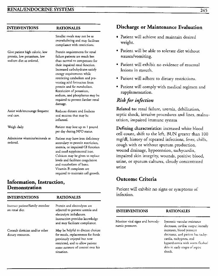
RENAL/ENDOCRINE SYSTEMS 245
INTERVENTIONS RATIONALES
Smaller meals may not be as overwhelming and may facilitate compliance with restrictions.
Protein requirements for renal failure patients are much less than normal to compensate for their impaired renal function. Increased carbohydrates satisfy energy requirements while restricting catabolism and pre- venting acid formation from protein and fat metabolism. Restriction of potassium, sodium, and phosphorus may be required to prevent further renal damage.
Reduces distaste and freshens oral mucosa that may be inflamed.
Give patient high caloric, low protein, low potassium, low sodium diet as ordered.
Assist withlencourage frequent oral care.
Weigh daily. Patient may lose up to 1 pound per day during NPO status.
Administer vitaminslminerals as Patient may have iron deficiency ordered. secondary to protein restriction,
anemia, or impaired GI function and need supplemental iron. Calcium may be given to replace levels and facilitate coagulation and metabolism of bone. Vitamin B complexes are required to maintain cell growth.
Information, Instruction, Demonstration
INTERVENTIONS RATIONALES
Instruct patientlfamily member on renal diet.
Protein and electrolytes are adjusted to prevent uremia and electrolyte imbalances. Instruction provides knowledge and may facilitate compliance.
May be helphl to discuss choices for meals, replacements for foods previously enjoyed but now restricted, and to allow patient some measure of control over his situation.
Consult dietician andor other dietary resources.
Discharge or Maintenance Evaluation
Patient will achieve and maintain desired weight.
Patient will be able to tolerate diet without nausea/vomiting.
Patient will exhibit no evidence of mucosal lesions in mouth.
Patient will adhere to dietary restrictions.
Patient will comply with medical regimen and supplementation.
Risk f i r infiction
Related to: renal failure, uremia, debilitation, septic shock, invasive procedures and lines, malnu- trition, impaired immune system
Defining characteristics: increased white blood cell count, shift to the left, BUN greater than 100 mg/dl, history of repeated infections, fever, chills, cough with or without sputum production, wound drainage, hypotension, tachycardia, impaired skin integrity, wounds, positive blood, urine, or sputum cultures, cloudy concentrated urine
Outcome Criteria
Patient will exhibit no signs or symptoms of infection.
INTERVENTIONS RATIONALES
Monitor vital signs and hernody- namic pressures.
Systemic vascular resistance decreases, cardiac output inirially increases, blood pressure decreases, and patient has tachy- cardia, tachypnea, and hypertherrnia with warm flushed skin in early stages of septic shock.
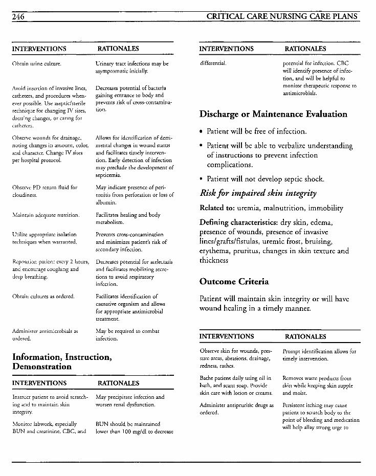
246 CRITICAL CARE NURSING CARE PLANS
INTERVENTIONS RATIONALES INTERVENTIONS RATIONALES
Obtain urine culture.
Avoid insertion of invasive lines, catheters, and procedures when- ever possible. Use aseptidsterile technique for changing IV sites, dressing changes, or caring for catheters.
Observe wounds for drainage, noting changes in amount, color, and character. Change IV sites per hospital protocol.
Observe P D return fluid for cloudiness.
Maintain adequate nutrition.
Utilize appropriate isolation techniques when warranted.
Reposition patient every 2 hours, and encourage coughing and deep breathing.
Obtain cultures as ordered.
Administer antimicrobials as ordered.
Urinary tract infections may be asymptomatic initially.
Decreases potential of bacteria gaining entrance to body and prevents risk of cross-contamina- tion.
Allows for identification of detri- mental changes in wound status and facilitates timely interven- tion. Early detection of infection may preclude the development of septicemia.
May indicate presence of peri- tonitis from perforation or loss of albumin.
Facilitates healing and body metabolism.
Prevents cross-contamination and minimizes patient’s risk of secondary infection.
Decreases potential for atelectasis and facilitates mobilizing secre- tions to avoid respiratory infection.
Facilitates identification of causative organism and allows for appropriate antimicrobial treatment.
May be required to combat infection.
Information, Instruction, Demonstration
INTERVENTIONS RATIONALES
Instruct patient to avoid scratch- ing and to maintain skin integrity.
Monitor labwork, especially BUN and creatinine, CBC, and
May precipitate infection and worsen renal dysfunction.
BUN should be maintained lower than 100 mgldl to decrease
differential. potential for infection. CBC will identify presence of infec- tion, and will be helpful to monitor therapeutic response to antimicrobials.
Discharge or Maintenance Evaluation
Patient will be free of infection.
Patient will be able to verbalize understanding of instructions to prevent infection complications.
Patient will not develop septic shock.
Risk for impaired skin integrity Related to: uremia, malnutrition, immobility
Defining characteristics: dry skin, edema, presence of wounds, presence of invasive lines/grafts/fistulas, uremic frost, bruising, erythema, pruritus, changes in skin texture and thickness
Outcome Criteria
Patient will maintain skin integrity or will have wound healing in a timely manner.
INTERVENTIONS RATIONALES
Observe skin for wounds, pres- sure areas, abrasions, drainage, redness, rashes.
Bathe patient daily using oil in bath, and scant soap. Provide skin care with lotion or creams.
Administer antipruritic drugs as ordered.
Prompt identification allows for timely intervention.
Removes waste products from skin while keeping skin supple and moist.
Persistent itching may cause patient to scratch body to the point of bleeding and medication will help allay strong urge to
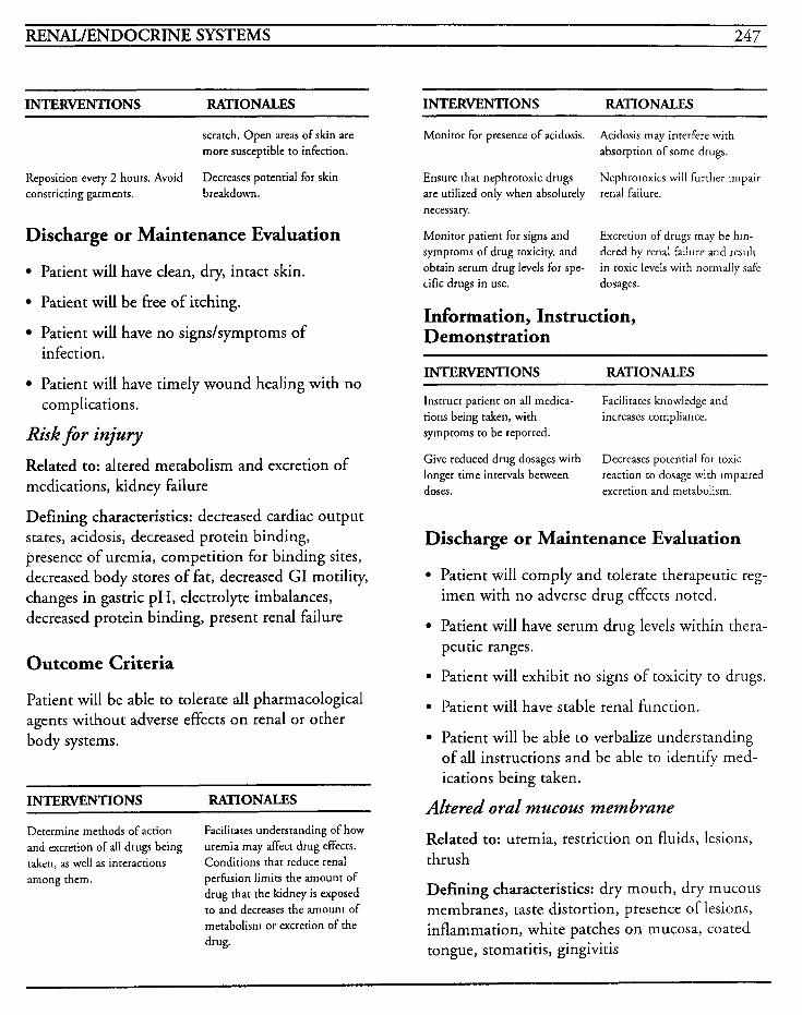
RENAL/ENDOCRINE SYSTEMS 247
INTERVENTIONS RATIONALES INTERVENTIONS RATIONALES
scratch. Open areas of skin are more susceptible to infection.
Decreases potential for skin Reposition every 2 hours, Avoid constricting garments. breakdown.
Discharge or Maintenance Evaluation
Patient will have clean, dry, intact skin.
Patient will be free of itching.
Patient will have no signs/symptoms of infection.
Patient will have timely wound healing with no complications.
Risk for injury Related to: altered metabolism and excretion of medications, kidney failure
Defining characteristics: decreased cardiac output states, acidosis, decreased protein binding, presence of uremia, competition for binding sites, decreased body stores of fat, decreased GI motility, changes in gastric pH, electrolyte imbalances, decreased protein binding, present renal failure
Outcome Criteria
Patient will be able to tolerate all pharmacological agents without adverse effects on renal or other body systems.
INTERVENTIONS RATIONALES
Determine methods of action and excretion of all drugs being taken, as well as interactions among them.
Facilitates understanding of how uremia may affect drug effects. Conditions that reduce renal perfusion limits the amount of drug that the kidney is exposed to and decreases the amount of metabolism or excretion of the drug.
Monitor for presence of acidosis.
Ensure that nephrotoxic drugs are utilized only when absolutely necessary.
Monitor patient for signs and symptoms of drug toxicity, and obtain serum drug levels for spe- cific drugs in use.
Acidosis may interfere with absorption of some drugs.
Nephrotoxics will further impair renal failure.
Excretion of drugs may be hin- dered by renal failure and result in toxic levels with normally safe dosages.
Information, Instruction, Demonstration
INTERVENTIONS RATIONALES
Instruct patient on all medica- tions being taken, with symptoms to be reported.
Give reduced drug dosages with longer time intervals between doses. excretion and metabolism.
Facilitates knowledge and increases compliance.
Decreases potential for toxic reaction to dosage with impaired
Discharge or Maintenance Evaluation
Patient will comply and tolerate therapeutic reg- imen with no adverse drug effects noted.
Patient will have serum drug levels within thera- peutic ranges.
Patient will exhibit no signs of toxicity to drugs.
Patient will have stable renal function.
Patient will be able to verbalize understanding of all instructions and be able to identify med- ications being taken.
Altered oral mucous membrane
Related to: uremia, restriction on fluids, lesions, thrush
Defining characteristics: dry mouth, dry mucous membranes, taste distortion, presence of lesions, inflammation, white patches on mucosa, coated tongue, stomatitis, gingivitis
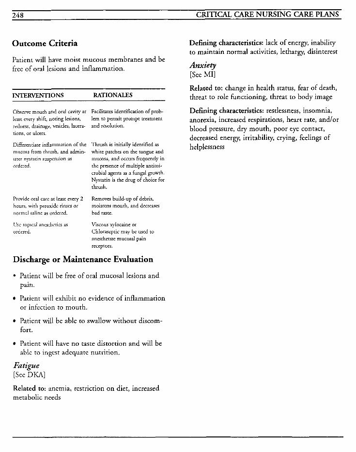
248 CRITICAL CARE NURSING CARE PLANS
Outcome Criteria
Patient will have moist mucous membranes and be free of oral lesions and inflammation.
INTERVENTIONS RATIONALES
Observe mouth and oral cavity a t least every shift, noting lesions, redness, drainage, vesicles, lacera- tions, or ulcers.
Differentiate inflammation of the Thrush is initially identified as white patches on the tongue and
Facilitates identification of prob- lem to permit prompt treatment and resolution.
mucosa from thrush, and admin- rsrer nystarin suspension as ordered.
Provide oral care at least every 2 hours, with peroxide rinses or normal saline as ordered.
Use ropical anesthetics as ordered.
mucosa, and occurs frequently in the presence of multiple antimi- crobial agents as a fungal growth. Nystatin is the drug of choice for thrush.
Removes build-up of debris, moistens mouth, and decreases bad taste.
Viscous xylocaine or Chloraseptic may be used to
anesthetize mucosal pain receptors.
Discharge or Maintenance Evaluation
0 Patient will be free of oral mucosal lesions and pain.
Patient will exhibit no evidence of inflammation or infection to mouth.
Patient will be able to swallow without discom- fort.
Patient will have no taste distortion and will be able to ingest adequate nutrition.
Fatigue [See DKA]
Defining characteristics: lack of energy, inability to maintain normal activities, lethargy, disinterest
Anxiety [See MII
Related to: change in health status, fear of death, threat to role functioning, threat to body image
Defining characteristics: restlessness, insomnia, anorexia, increased respirations, heart rate, and/or blood pressure, dry mouth, poor eye contact, decreased energy, irritability, crying, feelings of helplessness
Related to: anemia, restriction on diet, increased metabolic needs
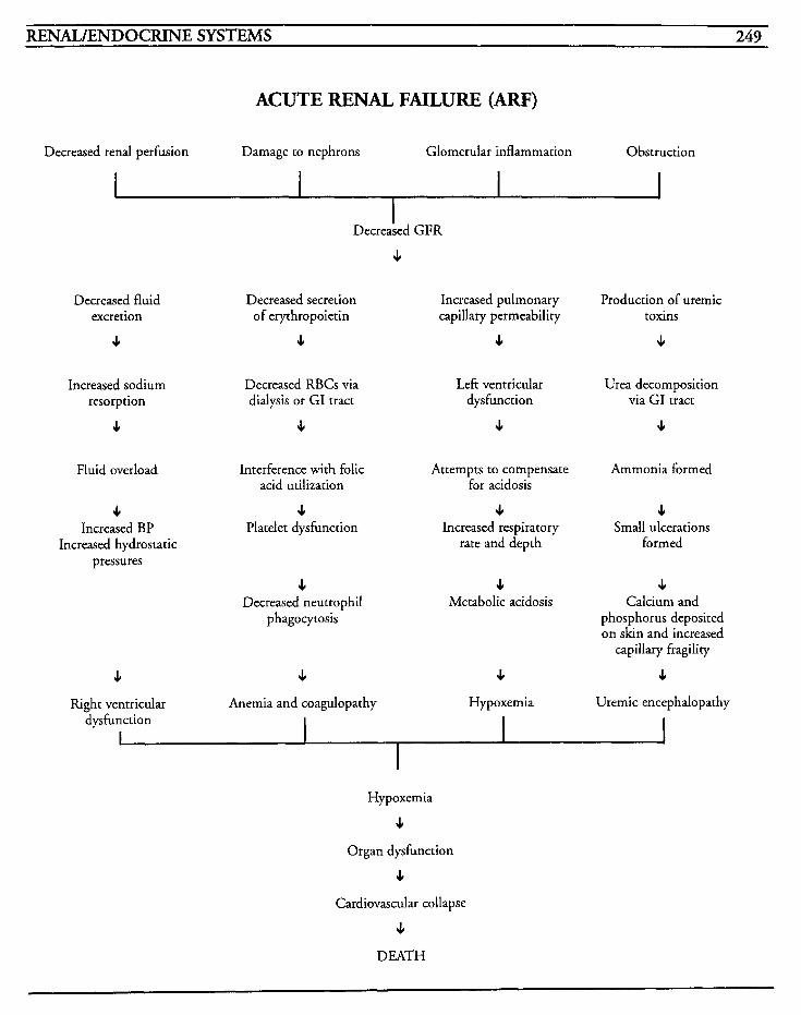
RENAL/ENDOCRINE SYSTEMS 249
ACUTE RENAL FAILURE (ARF)
Decreased renal perfusion Damage to nephrons Glomerular inflammation Obstruction
Decreased GFR
4
Decreased fluid excretion
4
Increased sodium resorption
4
Fluid overload
4 Increased BP
Increased hydrostatic pressures
4
Decreased secretion Increased pulmonary of erythropoietin capillary permeability
4 4
Decreased RBCs via dialysis or GI tract
4
Left ventricular dysfunction
4
Interference with folic Attempts to compensate acid utilization for acidosis
4 4 Platelet dysfunction Increased respiratory
rate and depth
4 4 Decreased neutrophil Metabolic acidosis
phagocytosis
4 4 Right ventricular Anemia and coagulopathy Hypoxemia
Production of uremic toxins
4
Urea decomposition via GI tract
4
Ammonia formed
4 Small ulcerations
formed
4 Calcium and
phosphorus deposited on skin and increased
capillary fragility
4 Uremic encephalopathy -
dysfunction I
I I
Hypoxemia
4 Organ dysfunction
4 Cardiovascular collapse
4 DEATH

This Page Intentionally Left Blank
RENAL,/ENDOCRINE SYSTEMS 25 1
Diabetic Ketoacidosis (D K I U Diabetic ketoacidosis, or DKA, is a critical emer- gency state that is caused by a deficiency of insulin in patients with insulin-dependent diabetes melli- tus. This deficiency can be caused by physiological causes, or by failure to take an adequate amount of insulin, and results in hyperglycemia, ketonuria, metabolic acidosis, and dehydration. Precipitating causes include failure to take an adequare amount of insulin on a daily basis or failure to increase and compensate for acute infection processes, surgery, trauma, pregnancy, or other acute stress events. Early symptoms include polyuria, polydipsia, fatigue, drowsiness, headache, muscle cramps and nausea/vomiting. Later symptoms, such as Kussmaul breathing, sweet, fruity breath odor, hypotension, and weak and thready pulses will precede stupor and coma.
Treatment is aimed at correction of the acidotic state, hyperglycemia, hyperosmolality, hypovolemia, and potassium deficits, in conjunc- tion with treatment of the underlying cause of the problem.
MEDICAL CARE Laboratory: serum glucose level is increased above 300 mg/dl and may be greater than 1000 mg/dl; serum acetone positive; lipids and cholesterol levels elevated; osmolality increased but normally less than 330 mOsm/L; potassium initially normal or elevated due to cellular shifting, then later decreased; sodium may be decreased, normal, or elevated; phosphorus is often decreased; amylase can be elevated if pancreatitis is precipitating cause; serum insulin may be decreased in type I DM or normal to high in type I1 suggesting that there is improper utilization of insulin or that insulin resistance may have developed secondary to antibody formation; BUN may be elevated if
dehydration is severe and renal perfusion is decreased; urinalysis will show positive for glucose and acetone, specific gravity and osmolality may be elevated; hemoglobin A1 c helps to differenti- ate whether episode is due to poor control of DM over previous few months or whether episode is incident-related; hematocrit may be elevated with dehydration; elevation of WBCs may occur in response to hemoconcentration or to stress; cul- tures may be helpful to discern potential cause of infection which may be precipitating factor
Arterial blood gases: pH will be less than 7.3, bicarbonate levels will be decreased, usually less than 15 mEq/L
Electrocardiogram: may show changes associated with electrolyte imbalances, especially hyperkalemia, with peaked T waves
NURSING CARE PLANS Fluid volume deficit Related to: hyperglycemic-induced osmotic diure- sis, vomiting, inadequate oral intake
Defining characteristics: dry mucous membranes, decreased skin turgor, thirst, hypotension, ortho- static changes, tachycardia, weak and thready pulse, weight loss, intake less than output, increased urinary output, dilute urine
Outcome Criteria
Patient will have stable vital signs, adequate skin turgor, intake and output equivalent, and electrolyte levels within acceptable ranges.
INTERVENTIONS RATIONALES
Monitor vital signs, especially noting respiratory status changes or alterations in blood pressure.
Tachycardia and hypotension are classic symptoms of hypovolemia. When systolic BP drops more than
252 CRITICAL CARE NURSING CARE PLANS
INTERVENTIONS RATIONALES INTERVENTIONS RATIONALES
Monitor intake and output every 2-4 hours.
Administer IV fluids per hospital protocol, usually at least 2-3 L/day, and usually initially, 3+ L.
Weigh every day.
Observe for complaints of nausedvomiting, abdominal bloating, or distention.
Auscultate lungs for crackles, and assess patient for presence of edema, or bounding pulses.
Insert catheter per hospital policy
Monitor laboratory tests for BUN and creatinine, serum osmolality, hematocrit, and electrolytes.
10 rnrnHg when position is changed may indicate severity of hypovolemic state. Kussmauli respirations may be present depending on degree of hyper- glycemia, and respiratory changes may occur as the lungs attempt to remove acids by creating a compensatory respiratory alkalo- sis. Fever, in conjunction with flushed, dry skin, may indicate dehydration.
Facilitates measurement and effectiveness of volume replace- ment and adequate circulating fluid volume.
Amounts and solution types may vary based upon the degree of dehydration and patient status. Usual solutions of normal or half-normal saline with or with- out dextrose are used, as well as occasional use of plasma expanders depending on unsuc- cessful fluid rehydration.
Assesses fluid status,
Gastric motility may be affected by fluid deficits, and vomiting or other gastric losses may potenti- ate fluid and electrolyte imbalances.
Congestive heart failure or circu- latory overload may occur with rapid rehydration.
Provides for more accurate assess- ment of output, especially if urinary retention or incontinence is present.
Hematocrit may be elevated because of hemoconcentration following osmotic diuresis. Dehydration may result in cellu- lar destruction and may result in renal insufficiency. Dehydration will result in elevated osmolality. Potassium levels are usually ele-
vated initially in response to the acidosis, but with diuresis, a hypokalemic state will ensue. Sodium may be decreased with shifting of fluids, and high sodium levels may indicate either a severe fluid loss or sodium reabsorption in response to aldosterone secretion.
Phosphate replacement may help with plasma buffering capacity, but excessive replacement can cause hypocalcemia. Potassium supplementation is usually done as soon as urinary output is ade- quate to prevent hypokalemic states. As insulin replacement occurs and acidosis is corrected, hypokalemia usually occurs.
Assess patient’s mental status and Mental status changes can occur observe for significant changes in with exceedingly high or low status. glucose levels, electrolyte imbal-
ances, acidotic states, hypoxia, or with decreases in cerebral perfu- sion pressure.
Administer electrolyte replace- ments as per hospital policy/ doctor’s orders.
Information, Instruction, Demonstration
~~
INTERVENTIONS RATIONALES
Instruct patient/family members regarding signs/symptoms of hyperglycemia. of complications.
Instruct in seeking medical atten- tion for infective processes or illness that may deplete circulat- ing volume. volume depletion.
Provides information and pro- motes more timely identification
Infection may predispose the patient to fever and a hypermeta- bolic state which may increase
Discharge or Maintenance Evaluation
Patient will have vital signs and hernodynamic parameters within acceptable ranges.
Patient will have normal skin turgor with ade- quate output.
RENAUENDOCRINE SYSTEMS 253-
9 Patient will have electrolytes and glucose levels
Alteration in nutrition: less than body requirements
Related to: insulin deficiency, excessive amounts of epinephrine, growth hormone, and cortisol, increased protein-fat metabolism, decreased oral intake, nausea, vomiting, altered mental status, infection
Defining characteristics: weakness, fatigue, increased levels of glucose and ketones, weight loss in spite of polyphagia, lack of adequate food intake, glycosuria
within normal ranges.
Outcome Criteria
Patient will be able to have intake of appropriate amounts and types of calories and nutrients, and have glucose levels within acceptable range for patient.
__ ~~~
INTERVENTIONS RATIONALES
Obtain weight every day.
Provide high-nutrient liquids as soon as patient is able to tolerate oral intake, with progression to solid food as tolerated.
Auscultate bowel sounds every 4-8 hours, and observe for abdominal distention or pain.
Monitor for changes in level of consciousness, cool or clammy skin, tachycardia, extreme hunger, anxiety, headache, light- headedness, tremors, or irritability.
Facilitates assessment of nutri- tional utilization and fluid shifts.
Provides nutrition and helps restore bowel function.
Elevated glucose levels can cause altered electrolyte levels and both may decrease gastric function. DKA may also mimic an acute surgical abdomen.
When carbohydrate metabolism begins and blood glucose level decreases, hypoglycemia can occur. Comatose patients may not exhibit any noticeable change in mentation status and should be monitored closely. Long-standing diabetic patients
INTERVENTIONS RATIONALES
Administer regular insulin, either by continuous infusion after an IV bolus dose has been given, by intramuscular injections every 1- 2 hours, or by subcutaneous injection.
Monitor serum glucose every hour while on insulin IV infusion, and notify MD per parameters of when blood glu- cose has dropped to 250 mgldl.
Administer subcutaneous insulin 1-2 hours before stopping the continuous insulin infusion.
Administer IV solutions contain- ing dextrose as ordered.
Administer Reglan IV or PO as ordered by physician.
may not show normal signs of hypoglycemia due in part to their diminished response to IOW glucose levels.
Subcutaneous route may be an option if the patient’s peripherd perfusion is adequate but the response will not be as rapid as with IV administration. Regular insulin is rapid acting and will assist in movement of glucose into cells. The continuous IV method is normally preferred because it oprimizes transition to carbohydrate metabolism, and helps to reduce hypoglycemia. Normally, the infusion rate is 5-10 Unidhr until glucose levels decrease within a stated parameter. Another goal of IV administration of insulin is to decrease the acidosis.
Blood glucose levels will decrease with insulin therapy usually in increments of 75 to 100 mg/dl/hr. Once the blood sugar has dropped to 250 mg/dl, and depending upon the degree of acidosis that is present, dextrose is added to the IV infusion, and the insulin infusion should be stopped to prevent hypoglycemic episodes.
Prevents recurrence of ketosis and rebound hyperglycemia.
Dextrose solutions are usually added after the blood glucose levels have decreased to 250 mg/dl in order to avoid hypo- glycemia.
May be used to treat symptoms related to neuropathies that affect the GI tract, and facllitate oral intake and nutrient absorption.
~
CRITICAL CARE NURSING CARE PLANS
Information, Instruction, Demonstration
INTERVENTIONS RATIONALES
Instruct patientlfamily member Complex carbohydrates decrease in dietary management, with the amounts of insulin needs, ideal amounts ofGO% carbohy- reduce serum cholesterol, and drates, 20% fats, and 20% help to satiate patient. Food proteins to be divided in desig- should be scheduled for peak nated number of meals and effects with insulin as well as snacks. patient preference. Snacks are
important to prevent Somogyi responses and hypoglycemia during sleep.
Assists in facilitating adjustments to diet for patient's special needs, and can facilitate development of workable meal plans.
Monitoring blood glucose levels is more accurate than urine glu- cose testing, and can facilitate identification of alterations in levels of glucose to promote tighter control of varying glucose levelslinsulin usage.
Promotes saturation of binding sites on plastic tubing to decrease incidence of insulin adhering to tubing rather than staying in
Obtain consult with dietician.
Instrucr in correct procedure for fingerstick glucose testing, with return demonstration as needed.
Ensure that at least 50 cc of soh- 'tion is flushed through the tubing prior to connection to patient when intravenous insulin drips are utilized. solution.
Discharge or Maintenance Evaluation
Patient will have normalized blood glucose levels within their own special parameters.
Patient will be able to ingest oral food of suffi- cient amounts and nutrients to maintain and stabilize weight.
Patient will be free of ketosis.
Patiendfamily member will be able to verbalize understanding of instructions and able to pro- vide acceptable return demonstration of procedure.
Impaired gas exchange Related to: accumulation of ketones and acids sec- ondary to insulin deficiencies and excessive production of stress hormones
Defining characteristics: acid-base imbalances, acetone breath, tachypnea, Kussmaul respirations, serum and urine ketones present, decreased pH, decreased bicarbonate levels, hyperkalernia, decreased level of consciousness, confusion
Outcome Criteria
Patient will have normalized acid-base balance with stable vital signs and mentation level.
INTERVENTIONS RATIONALES
Monitor respiratory status for changes in rate, rhythm and depth, and for presence of ace- tone smell on breath.
Acetone breath is due co break- down of aceroacetic acids. The lungs remove carbonic acid through respiration process, and may produce a cornpensacory respiratory alkalosis for ketoaa- dosis. Increased work of breathing may indicate that the patient is losing the ability to compensate for the severe acido- sis or respiratory fatigue.
Acidosis, hypoxia, or decreased cerebral perfusion may cause changes in mentation. Impairment in consciousness may predispose the patient to aspiration and its complications.
Promote correction of acidosis
Monitor for changes in neurolog- ical status.
Administer IV fluids and insulin as ordered. with DKA.
Administer sodium bicarbonate, if ordered, for severe acidosis only.
Current recommendations are for use only where pH is 7.1 or below because excessive use of sodium bicarbonate may induce hypokalernia as well as alcer che oxygen dissociation curve causing prolongation of the comatose state.
~
RENAL/ENDOCRINE SYSTEMS 255
~~ ~~ _ _ _ ~
INTERVENTIONS RATIONALES INTERVENTIONS RATIONALES
Monitor labwork for hypokalemia.
Administer supplemental oxygen as necessary per hospital proto- col.
May occur as acidosis and volume deficits are corrected.
Provides needed oxygen espe- cially in patients that may not be able to obtain adequate oxygena- tion with room air, and helps to improve acidosis.
bronchitis or pneumonia, either of which may be the precipitat- ing cause of the DKA. Crackles may indicate fluid overload or congestive failure as a result from rapid fluid replacement.
Elderly female diabetics are Provide perineal or catheter care
Discharge or Maintenance Evaluation
Patient will have pH, bicarbonate, potassium, serum and urine ketones within normal limits.
Patient will have stable vital signs with respira- tory rate within normal limits.
Patient will be free of acetone on breath.
Risk for infiction
Related to: elevated glucose levels, alterations in circulation, pre-existing infection, especially URI or UTI, decreased leukocyte function
Defining characteristics: increased serum and urine glucose levels, temperature elevation, chills, fever, elevated white blood cell count, differential with shift to the left
Outcome Criteria
Patient will be free of infection and able to verbal- ize methods to prevent or reduce risk of infection.
INTERVENTIONS RATIONALES
frequently. prone to the development of urinary tract and vaginal infec- tions.
Reposition patient and provide skin care every 2 hours.
Facilitates lung expansion, decreases risk of skin irritation and breakdown, and improves peripheral circulation.
Assists with identification of causative organism and appropri- ate antimicrobial therapy.
Early intervention may reduce the risk of sepsis or multi-system involvemen t.
Obtain culture specimens as ordered or as per hospital policy.
Administer antibiotics as ordered.
Information, Instruction, Demonstration
INTERVENTIONS RATIONALES
Ensure proper handwashing Prevenrs cross-contamination techniques are used by staff and patient. infection.
Maintain aseptic technique with administration of IV medica- tions, insertion of catheters and invasive lines, and maintenance care. Restart IVs per hospital protocol.
Instruct patients in perineal care, disposal of secretions and infected materials. cross-contamination.
and decreases risk of spread of
Elevated glucose levels provide an excellent culture medium for bacterial growth.
Promotes compliance, minimizes risk of spread of infection, and
Monitor for fever, facial flushing, drainage from wounds, urine cloudiness, changes in sputum, tachycardia. infection.
Auscultate for changes in breath sounds.
Patient may have been admitted with undiagnosed infection or have developed a nosocomial
Accumulation of bronchial secre- tions may be heard as rhonchi and may indicate the presence of
Instruct in importance of oral care. disease.
Reduces risk of oral or gum
Discharge or Maintenance Evaluation
Patient will be able to identify actions to reduce or prevent infection and cross-contamination.
256 CRITICAL CARE NURSING CARE PLANS
Patient will be free of infective process.
Patient will be able to adequately demonstrate techniques to prevent or reduce infection risk.
Potential for injury: bypoglycemia Related to: insulin therapy, decreased insulin- antagonist hormones circulating in body, rebound action
Defining characteristics: blood glucose levels below 70 mg/dl, altered mental state, decreased level of consciousness, cool and clammy skin, pallor, tremors, tachycardia, irritability, visual dis- turbances, paresthesias, dizziness, hunger, nausea, fatigue, diaphoresis
Outcome Criteria
Patient will have stable blood glucose levels and be able to identify methods of treatment and identifi- cation of hypoglycemic episodes.
INTERVENTIONS RATIONALES
Monitor for signs/symptoms of hypoglycemia.
Change IV fluid to solution con- taining glucose when blood glucose level reached 250 mgldl, a< well as change inhsion rate on insulin drip.
If hypoglycemia occurs, give the patient oral (if able to tolerate fluids and awake) or parenteral glucose solutions, as per policy.
Prompt identification of problem will facilitate prompt treatment and help prevent further compli- cations.
Prevents excessive drop in blood glucose level and allows time for blood chemistry to normalize.
Glucagon, 10-50% solutions, may be given IV, or 15 grams of a rapid-acting carbohydrate will be effective in elevating the blood sugar level. Milk and crackers will assist in protecting patient from recurrences of hypoglycemic episode.
Information, Instruction, Demonstration
INTERVENTIONS RATIONALB
Instruct patientlfamily in signs of hypoglycemia and treatment for this condition.
Promotes knowledge and facili- tates compliance. Assists patient and family to feel in control.
Discharge or Maintenance Evaluation
Patient will have stable blood glucose level above 80 mg/dl.
Patient/family member will be able to identify signs and symptoms of hypoglycemia and inter- ventions for treatment.
Patient will have no hypoglycemic symptoms.
Fatigue
Related to: insufficient insulin, increased metabolic demands, decreased metabolic energy production, infection
Defining characteristics: lack of energy, inability to perform normal routine, decreased performance, accident prone, lethargy, tiredness, alterations in consciousness
Outcome Criteria
Patient will have increased energy and be able to participate adequately in normal activities.
INTERVENTIONS RATIONALES
Observe patient for activity toler- ance. that identification of problem
Provides baseline information so
and interventions may be planned. Elevations in pulse, blood pressure and respiratory rate may indicate physiologic intolerance of activity.
Prevents excessive fatigue. Provide period of rest or sleep
RENAL/ENDOCRINE SYSTEMS 257
INTERVENTIONS RATIONALES
alternated with periods of activ- ity as patient can tolerate.
Increase activity and patient par- ticipation gradually.
Provides time to build up toler- ance, and increases self-esteem.
Information, Instruction, Demonstration
INTERVENTIONS RATIONALES
Discuss with patient/family member the importance of activ- ity, planning schedules with alternating rest and activity, and methods of conserving energy.
Information may facilitate moti- vation to increase activity level knowing that decreased energy will be expended and he will be able to accomplish more activity.
Discharge or Maintenance Evaluation
0 Patient will be able to tolerate increased activity with stable vital signs.
Patient/family will be able to verbalize and/or demonstrate techniques to conserve energy while performing activities.
Knowledge &$kit Related to: lack of information, lack of recall, mis- interpreted information, unfamiliarity with resources
Defining characteristics: requests for information, questions, misrepresentation of facts, inaccurate follow-through of instructions, development of preventable complications
Outcome Criteria
Patient will be able to verbalize understanding of diabetes disease process, identify signs and symp- toms of complications, correctly demonstrate all procedures, and access community resources adequately.
INTERVENTIONS RATIONALES Instruct patiendfamily member about disease process, normal ranges for blood glucose, glu- cometer use, relationship between insulin and glucose levels, type of diabetes the patient has, etc.
Instruct patient/family member in glucometer use and urine test- ing, with return demonstration by patient.
Instruct in dietary plan, allowances, caloric intake, meals outside the home, etc.
Instruct in medication regime, with actions, side effects, and contraindications noted.
Instruct in activity and other factors that determine diabetic control.
Instruct in avoidance of smoking.
Instruct in examination and care of feet.
Instruct in protocols for sick days-to take medications, to notify MD, to monitor blood sugar every 2-4 hours, to check
Provides knowledge base on which further instruction can be performed.
Promotes tighter control of dia- betes with self-monitoring at least four times per day, and may help prevent or delay long-term complications.
Dietary control with assist with maintenance of decreased blood glucose levels. Fiber may slow glucose absorption and decrease fluctuations in serum levels.
Promotes understanding of drug use and facilitates compliance with regimen. Proper techniques with administration of insulin assist with understanding and identification of potential prob- lems so that interventions may be found.
Promotes control of diabetes and may help reduce incidence of ketoacidosis. Aerobic exercises promote effective utilization of insulin and strengthens the car- diovascular system. Illness management and management of other stress-type factors facilitates equilibrium with disease process during these episodes.
Nicotine causes constriction of blood vessels which restricts insulin absorption up to 30%.
Identifies potential complications that may occur because of peripheral neuropathy or circula- tory impairment, and allows for early intervention.
Provides plan for complications that occur, and gives the patient the knowledge to enable him to adequately care for himself
258 CRITICAL CARE NURSING CARE PLANS
INTERVENTIONS RATIONALES
urine ketones if blood sugar is >240 mg/dl, and to replace car- bohydrates with liquids
Instruct in maintaining medical maintenance, including vision checks, and follow-up care.
during times of illness.
Vision changes may be gradual and may be more pronounced in poorly-controlled diabetics. Visual acuity may deteriorate to retinopathy and eventual blind- ness. Follow-up care can assist in preventing exacerbations of dia- betic complications and delay development of systemic prob- lems.
Impotence may occur as an ini- tial symptom of diabetes mellirus. Penile prosthesis and/or counseling may be of help.
May contain increased sugar content and may interact with other medications being taken.
Provides continued support post discharge, and assists to support
Discuss sexual function and questions patientlfamily member may ask.
Instruct in avoidance of use of over-the-counter medications without physician approval.
Instruct in available community resources, support groups like the American Diabetic Association, lifestyle changes. smoking and weight loss clinics, etc.
Discharge or Maintenance Evaluation
Patient/family member will be able to accurately verbalize knowledge base regarding diabetic dis- ease process.
Patientlfamily member will be able to accurately verbalize all information given.
Patient/family member will be able to accurately return demonstration for all necessary procedures.
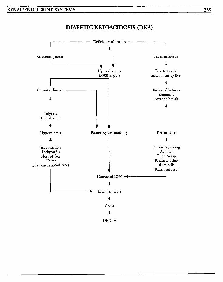
~~ ~
RENAL/ENDOCRINE SYSTEMS 259
Osmotic diuresis
DIABETIC KETOACIDOSIS (DKA)
I
I Deficiency of insulin
s I
Gluconeogenesis Fat metabolism . Hyperglycemia
d m g ' d ' ) I Osmotic diuresis
c
Polyuria Dehydration
c 1 c
Polyuria Dehydration
c
Free fatty acid metabolism by liver
c Increased ketones
Ketonuria Acetone breath
c
Hypovolernia Plasma hyperosmolality Ketoacidosis
4 Hypotension Tachycardia Flushed face
Thirst Dry mucus membranes
c Nauseahomiting
Acidosis High A-gap
Potassium shift from cells
Kussmaul resp. I
Decreased CNS I c
I W Brain ischernia
4
Coma
c DEATH

This Page Intentionally Left Blank
RENAIJENDOCRINE SYSTEMS 26 1
Hyperglycemic
(HHNK) HHNK, or hyperglycemic hyperosmolar nonke- totic coma, may also be known as hyperglycemic nonacidotic diabetic coma, and presents a life- threatening emergency. Glucose transportation across the cell membrane is impaired by enough of an insulin deficiency that causes hyperglycemia without inhibiting lipolysis or ketogenesis in the liver. The hyperosmolality occurs from the hyper- natremia and hyperglycemia, and may further impair the secretion of insulin and prevent fatty acid release from adipose tissues. Extracellular fluid volume deficits occur as a result of osmotic diuresis in the body's attempt to offset increasing
increase, glomerular filtration rates decrease and reduces the ability of the kidneys to excrete the glucose.
HHNK occurs when insulin action or secretion is inadequate, and may occur in patients who have no previous history of diabetes mellitus. The elderly are especially prone to this because of the lower body water content and dehydration, and this may alter their buffering ability to respond to changes in osmolality. Illnesses, and other stress- provoking episodes, may either cause or hasten the development of HHNK by increasing glucose pro- duction in response to excessive stress hormone production.
HHNK has almost the same pathophysiologic pattern as DKA, but the difference is that with HHNK, a sufficient amount of insulin is being released to prevent the development of ketosis.
' plasma osmolality. As fluid volume deficits
HHNK has also been associated with usage of thi- azide diuretics, glucocorticoids, phenytoin, sympathomimetics, diazoxide, chlorpromazine, sedatives, cimetidine, calcium channel blockers, and immunosuppressive agents because of their effects with glucogenesis and/or insulin.
Mortality is approximately 50% due in part to common complications that occur, such as shock, coma, acute tubular necrosis, and vascular throm- bosis. Correction of the problem is the main goal of treatment, with fluid balance the initial concern. The lack of insulin may be corrected by supplemental insulin administration and usually requires 100 Units or less in the first 24 hour period. Electrolyte imbalances are corrected and may require large amounts of potassium supple- mentation.
MEDICAL CARE Laboratory: blood sugar level elevated, frequently over 1000 mg/dl; plasma osmolality elevated, fre- quently as high as 450 mOsm/kg; hematocrit elevated due to hemoconcentration; urine and serum acetone levels negative; BUN and creatinine elevated; marked leukocytosis; electrolytes to eval- uate deficiency; hypernatremia usually present
Arterial blood gases: used to identify acidosis; pH is usually greater than 7.30, bicarbonate is usually greater than 15 mEq/L; acidosis is mainly due to lactic acid or renal dysfunction
Electrocardiogram: used to identify dysrhythmias that may result as a consequence of electrolyte and fluid disturbances
NURSING CARE PLANS Fluid volume deficit [See DKA]
262 CRITICAL CARE NURSING CARE PLANS
Alterution in nutrition: less thun body requirements [See DKA]
Potentiul for injury: hypoglycemiu [See DKA]
Potentiul for ulterution in tissue perjksion: per+herul
Related to: dehydration, increased platelet aggre- gation, increased viscosity of blood
Defining characteristics: cool extremities, decreased peripheral pulses, extremity pallor or cyanosis, unequal extremity temperatures
Outcome Criteria
Patient will have bilaterally equal peripheral pulses, color and temperature to extremities, with no complications.
INTERVENTIONS RATIONALES
Monitor and assess lower extrem- ities for color, temperature, presence of pulses, and equality.
Test for positive Homan’s sign, redness, warmth, tenderness, or swelling to legs.
Remove TED hose at least every 8 hours for 30 minutes to 1 hour.
Assist with passive range of motion/encourage active range of motion exercises.
Identifies the status of circulation in the extremities and assists with prompt identification of complications.
May indicate thrombus forma- tion, but is not always present with thrombus formation.
Provides opportunity for thor- ough assessment and identification of changes, as well as for comfort of pztient.
Prevents venous stasis.
Information, Instruction, Demonstration
INTERVENTIONS RATIONALES
Instruct patiendfamily member to avoid constricting apparel, crossing legs or ankles, or any other activity that impedes circulation.
Notify physician for any evidence of thrombus formation.
Prevents circulatory impairment and risk of complications.
Prompt identification can lead to timely intervention.
Discharge or Maintenance Evaluation
Patient will have equal pulses, color, and tem- perature to lower extremities bilaterally. . Patient will have no evidence of thrombus formation.
Patient/family will be compliant with methods to reduce risk of thrombus formation.
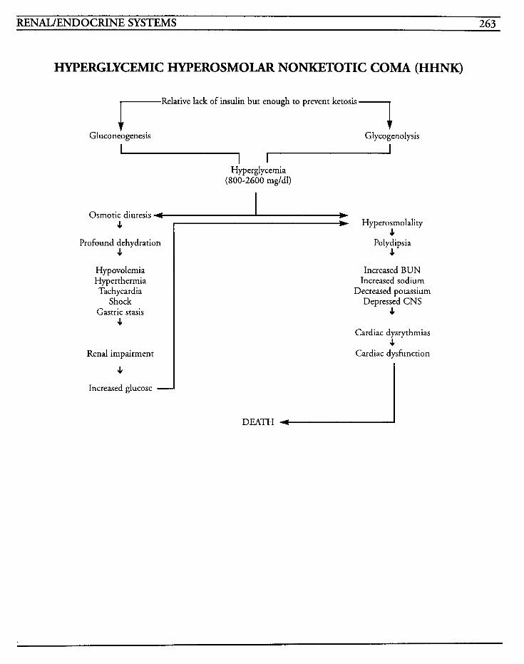
RENAIJENDOCRINE SYSTEMS 263
HYPERGLYCEMIC HYPEROSMOLAR NONKETOTIC COMA (HHNK)
1 Relative lack of insulin but enough to prevent ketosis
Gluconeogenesis r Glycogenolysis
Hyperglycemia (800-2600 mgldl)
Osmotic diuresis 4
Profound dehydration 4
Hypovolemia Hyperthermia Tachycardia
Shock Gastric stasis
4
Renal impairment
4
# k-J'perosmc 4 Polydipsia
J,
lity
Increased BUN Increased sodium
Decreased potassium Depressed CNS
4
Cardiac dysrythmias 4
Cardiac dysfunction
Increased glucose 1
DEATH +

This Page Intentionally Left Blank
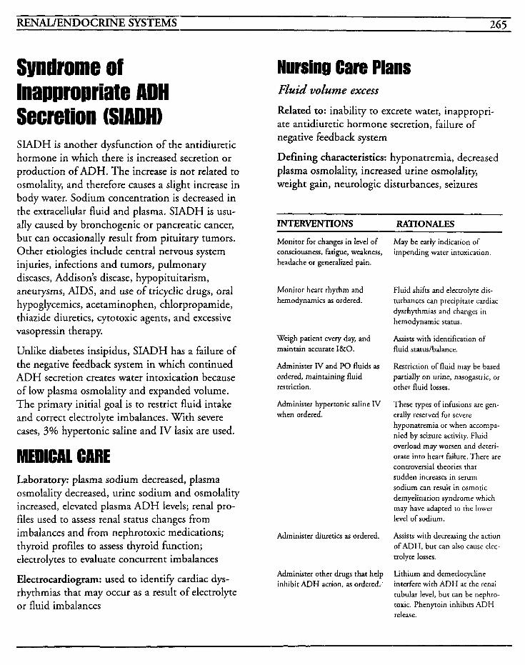
RENAL/ENDOCRINE SYSTEMS 265
Syndrome of Inappropriate ADH Secretion (SIADH) SIADH is another dysfunction of the antidiuretic hormone in which there is increased secretion or production of ADH. The increase is not related to osmolality, and therefore causes a slight increase in body water. Sodium concentration is decreased in the extracellular fluid and plasma. SIADH is usu- ally caused by bronchogenic or pancreatic cancer, but can occasionally result from pituitary tumors. Other etiologies include central nervous system injuries, infections and tumors, pulmonary diseases, Addison's disease, hypopituitarism, aneurysms, AIDS, and use of tricyclic drugs, oral hypoglycemics, acetaminophen, chlorpropamide, thiazide diuretics, cytotoxic agents, and excessive vasopressin therapy.
Unlike diabetes insipidus, SIADH has a failure of the negative feedback system in which continued ADH secretion creates water intoxication because of low plasma osmolality and expanded volume. The primary initial goal is to restrict fluid intake and correct electrolyte imbalances. With severe cases, 3% hypertonic saline and IV lasix are used.
MEDICAL CARE Laboratory: plasma sodium decreased, plasma osmolality decreased, urine sodium and osmolality increased, elevated plasma ADH levels; renal pro- files used to assess renal status changes from imbalances and from nephrotoxic medications; thyroid profiles to assess thyroid function; electrolytes to evaluate concurrent imbalances
Electrocardiogram: used to identify cardiac dys- rhythmias that may occur as a result of electrolyte or fluid imbalances
Nursing Care Plans Fluid volume excess
Related to: inability to excrete water, inappropri- ate antidiuretic hormone secretion, failure of negative feedback system
Defining characteristics: hyponatremia, decreased plasma osmolality, increased urine osmolality, weight gain, neurologic disturbances, seizures
INTERVENTIONS RATIONALES
Monitor for changes in level of consciousness, fatigue, weakness, headache or generalized pain.
Monitor heart rhythm and hemodynamics as ordered.
Weigh patient every day, and maintain accurate I&O.
Administer IV and PO fluids as ordered, maintaining fluid restriction.
Administer hypertonic saline IV when ordered.
Administer diuretics as ordered.
Administer other drugs that help inhibit ADH action, as ordered,'
May be early indication of impending water intoxication.
Fluid shifts and electrolyte dis- turbances can precipitate cardiac dysrhythmias and changes in hemodynamic status.
Assists with identification of fluid statudbalance.
Restriction of fluid may be based partially on urine, nasogastric, or other fluid losses.
These types of infusions are gen- erally reserved for severe hyponatremia or when accornpa- nied by seizure activity. Fluid overload may worsen and deteri- orate into heart failure. There are controversial theories that sudden increases in serum sodium can result in osmotic demyelination syndrome which may have adapted to the lower level of sodium.
Assists with decreasing the action of ADH, but can also cause elec- trolyte losses.
Lithium and demeclocycline interfere with ADH ac the renal tubular level, but can be nephro- toxic. Phenytoin inhibits ADH release.
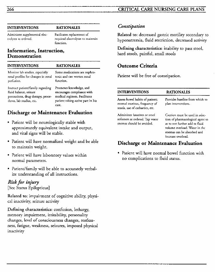
266 CRITICAL CARE NURSING CARE PLANS
INTERVENTIONS RATIONALES
Administer supplemental elec- trolyres as ordered.
Facilitates replacement of required electrolytes to maintain function.
Information, Instruction, Demonstration
INTERVENTIONS RATIONALES
Monitor lab studies, especially renal profiles for changes in renal perfusion. function.
Instruct patient/fmily regarding fluid balance, seizure precautions, drug therapy, proce- dures, lab studies, etc.
Some medications are nephro- toxic and can worsen renal
Promotes knowledge, and encourages compliance with medical regimen. Facilitates patient taking active part in his care.
Discharge or Maintenance Evaluation
Patient will be neurologically stable with approximately equivalent intake and output, and vital signs will be stable.
Patient will have normalized weight and be able to maintain weight.
Patient will have laboratory values within normal parameters.
Patiendfamily will be able to accurately verbal- ize understanding of all instructions.
Risk for injury [See Status Epilepticus]
Related to: impairment of cognitive ability, physi- cal inactivity, seizure activity
Constipation
Related to: decreased gastric motility secondary to hyponatremia, fluid restriction, decreased activity
Defining characteristics: inability to pass stool, hard stools, painful, small stools
Outcome Criteria
Patient will be free of constipation.
INTERVENTIONS RATIONALES
Assess bowel habits of patient; normal routines, frequency of stools, use of cathartics, etc.
Administer laxatives or stool softeners as ordered. Tap water enemas should be avoided.
Provides baseline from which to plan interventions.
Caution must be used in selec- tion of pharmacological agent so as to not further add to fluid volume overload. Water in the enemas can be absorbed and increase overload.
Discharge or Maintenance Evaluation
Patient will have normal bowel function with no complications to fluid status.
Defining characteristics: confusion, lethargy, memory impairment, irritability, personality changes, level of consciousness changes, restless- ness, fatigue, weakness, seizures, imposed physical inactivity
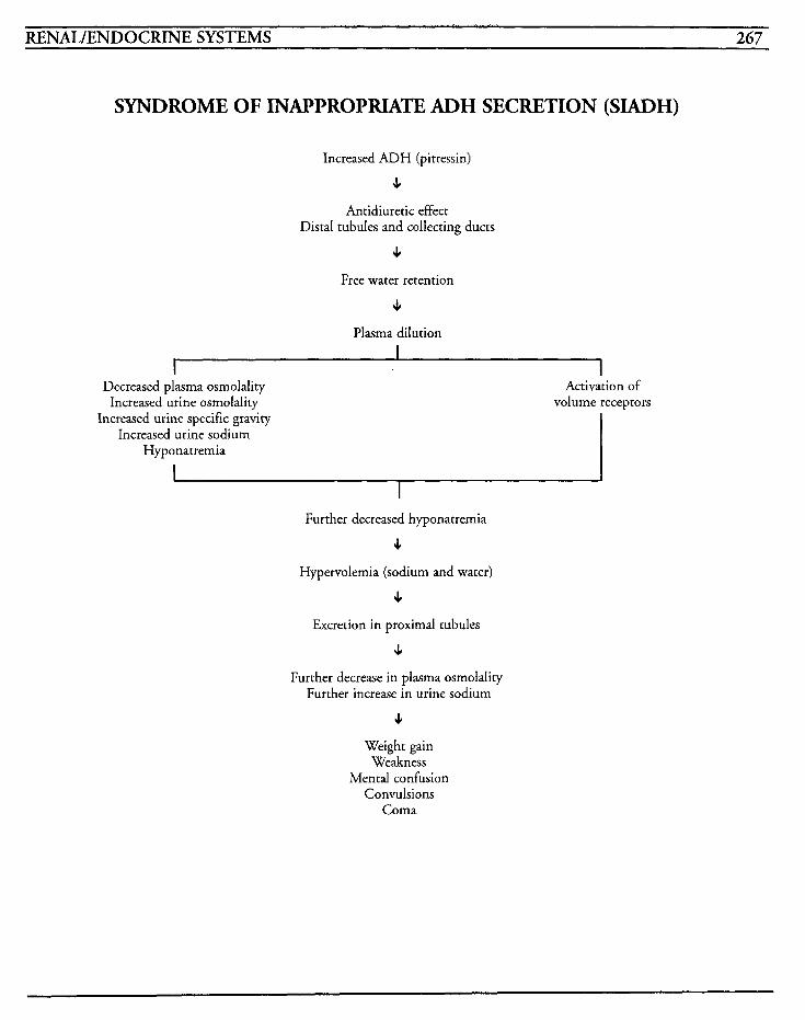
RENAL/ENDOCRINE SYSTEMS 267
SYNDROME OF INAPPROPRIATE ADH SECRETION (SIADH)
Increased ADH (pitressin)
c Antidiuretic effect
Distal tubules and collecting ducts
c Free water retention
J,
Plasma dilution I
I I Decreased plasma osmolality Activation of
Increased urine osmolality volume receptors Increased urine specific gravity
Increased urine sodium Hyponatremia
I Further decreased hyponatremia
e Hypervolemia (sodium and water)
c Excretion in proximal tubules
J,
Further decrease in plasma osmolality Further increase in urine sodium
c Weight gain
Weakness Mental confusion
Convulsions Coma

This Page Intentionally Left Blank
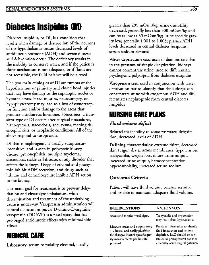
RENAL/ENDOCFUNE SYSTEMS 269
Diabetes Insipidus (DD Diabetes insipidus, or DIY is a condition that results when damage or destruction of the neurons of the hypothalamus causes decreased levels of antidiuretic hormone (ADH) and severe diuresis and dehydration occur. The deficiency results in the inability to conserve water, and if the patient's thirst mechanism is not adequate, or if fluids are not accessible, the fluid balance will be altered.
The two main etiologies of DI are tumors of the hypothalamus or pituitary and closed head injuries that may have damage to the supraoptic nuclei or hypothalamus. Head injuries, neurosurgery, or hypophysectomy may lead to a loss of osmorecep- tor function and/or damage to the areas that produce antidiuretic hormone. Sometimes, a tran- sient type of DI occurs after surgical procedures, histiocyctosis, sarcoidosis, aneurysms, meningitis, encephalititis, or neoplastic conditions. All of the above respond to vasopressin.
DI that is nephrogenic is usually vasopressin- insensitive, and is seen in polycystic kidney disease, pyelonephritis, multiple myeloma, sarcoidosis, sickle cell disease, or any disorder that affects the kidneys. Usage of ethanol and pheny- toin inhibit ADH secretion, and drugs such as lithium and demeclocycline inhibit ADH action in the kidney.
The main goal for treatment is to prevent dehy- dration and electrolyte imbalances, while determination and treatment of the underlying cause is underway. Vasopressin administration will control diabetes insipidus; D-amino-D-arginine vasopressin (DDAW) is a nasal spray that has prolonged antidiuretic effects with minimal side effects.
Laboratory: serum osmolality elevated, usually
greater than 295 mOsm/kg; urine osmolality decreased, generally less than 500 mOsm/kg and can be as low as 30 mOsm/kg; urine specific grav- ity low, generally 1.001 to 1.005; plasma ADH levels decreased in central diabetes insipidus; serum sodium elevated
Water deprivation test: used to demonstrate that in the presence of simple dehydration, kidneys cannot concentrate urine; used to differentiate psychogenic polydipsia from diabetes insipidus
Vasopressin test: used in conjunction with water deprivation test to identify that the kidneys can concentrate urine with exogenous ADH and dif- ferentiates nephrogenic from central diabetes insipidus
NURSING CARE PLANS Fluid volume
Related to: inability to conserve water, dehydra- tion, decreased levels of ADH
Defining characteristics: extreme thirst, decreased skin turgor, dry mucous membranes, hypotension, tachycardia, weight loss, dilute urine output, increased urine output, hemoconcentration, hyperosmolality, increased serum sodium
Outcome Criteria
Patient will have fluid volume balance restored and be able to maintain adequate fluid volume.
INTERVENTIONS RATIONALES
Assess and monitor vital signs. Tachycardia and hypocension may result from hypovolemia.
Measure intake and output every Provides information to identiFy 1-2 hours, and notify physician fluid imbalances and volume for changes. Record specific grav- depletion. I&O should be con- ity measurements per hospital tinued in postoperative patients, protocol. especially neurosurgical pacieno,
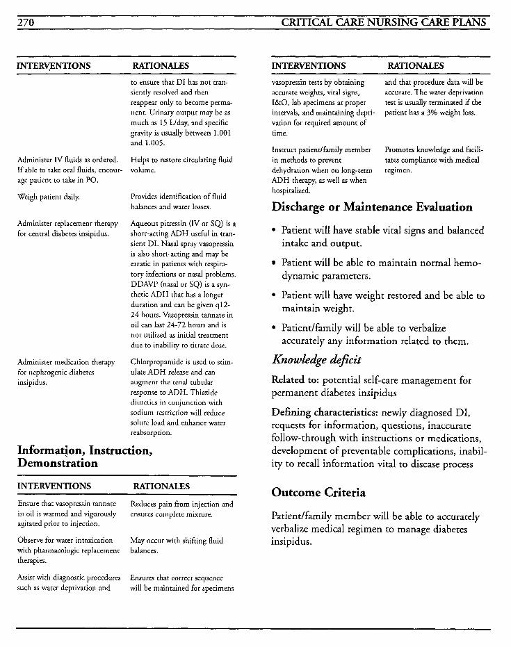
270 CRITICAL CARE NURSING CARE PLANS
INTERWNTIONS RATIONALES INTERVENTIONS RATIONALES
Administer IV fluids as ordered. If able to take oral fluids, encour- age patient to take in PO.
Weigh patient daily.
Administer replacement therapy for central diabetes insipidus.
Administer medication therapy for nephrogenic diabetes insipidus.
to ensure that DI has not tran- siently resolved and then reappear only to become perma- nent. Urinary output may be as much as 15 Llday, and specific gravity is usually between 1.001 and 1.005.
Helps to restore circulating fluid volume.
Provides identification of fluid balances and water losses.
Aqueous pitressin (IV or SQ) is a short-acting ADH useful in tran- sient DI. Nasal spray vasopressin is also short-acting and may be erratic in patients with respira- tory infections or nasal problems. DDAVP (nasal or SQ) is a syn- thetic ADH that has a longer duration and can be given q12- 24 hours. Vasopressin tannate in oil can last 24-72 hours and is not utilized as initial treatment due to inability to titrate dose.
Chlorpropamide is used to stim- ulate ADH release and can augment the renal tubular response to ADH. Thiazide diuretics in conjunction with sodium restriction will reduce solute load and enhance water reabsorption.
Information, Instruction, Demonstration
~
INTERVENTIONS RATIONALES
Ensure that vasopressin tannate in oil is warmed and vigorously agitated prior to injection.
Observe for water intoxication with pharmacologic replacement therapies.
Assist with diagnostic procedures such as water deprivation and
Reduces pain from injection and ensures complete mixture.
May occur with shifting fluid balances.
Ensures that correct sequence will be maintained for specimens
vasopressin tests by obtaining accurate weights, vital signs, I&O, lab specimens at proper intervals, and maintaining depri- vation for required amount of time.
Instruct patient/family member in methods to prevent dehydration when on long-term regimen. ADH therapy, as well as when hospitalized.
and that procedure data will be accurate. The water deprivation test is usually terminated if the patient has a 3% weight loss.
Promotes knowledge and facili- tates compliance with medical
Discharge or Maintenance Evaluation
Patient will have stable vital signs and balanced intake and output.
Patient will be able to maintain normal hemo- dynamic parameters.
Patient will have weight restored and be able to maintain weight.
Patiendfamily will be able to verbalize
Knowledge deficit
Related to: potential self-care management for permanent diabetes insipidus
Defining characteristics: newly diagnosed DI, requests for information, questions, inaccurate follow-through with instructions or medications, development of preventable complications, inabil- ity to recall information vital to disease process
accurately any information related to them.
Outcome Criteria
Patient/family member will be able to accurately verbalize medical regimen to manage diabetes insipidus.
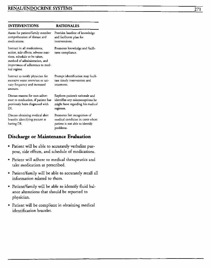
RENAL/ENDOCRINE SYSTEMS 27 1
INTERVENTIONS RATIONALES ~~ ~ ~
Assess for patientlfamily member comprehension of disease and medications. interventions.
Instruct in all medications, action, side effects, adverse reac- tions, schedule to be taken, method of administration, and importance of adherence to med- ical regime.
Instruct to notie physician for excessive water retention or uri- nary frequency and increased treatment. amount.
Discuss reasons for non-adher- ence to medication, if patient has previously been diagnosed with DI. regimen.
Discuss obtaining medical alert bracelet identifying patient as having DI.
Provides baseline of knowledge and facilitates plan for
Promotes knowledge and facili- tares compliance.
Prompt identification may facili- tate timely intervention and
Explores patient's rationale and identifies any misconceptions he might have regarding his medical
Promotes fast recognition of medical condition in cases where patient is not able to identify problems.
Discharge or Maintenance Evaluation
Patient will be able to accurately verbalize pur- pose, side effects, and schedule of medications.
Patient will adhere to medical therapeutics and take medication as prescribed.
Patiendfamily will be able to accurately recall all information related to them.
Patient/family will be able to identify fluid bal- ance alterations that should be reported to physician.
Patient will be compliant in obtaining medical identification bracelet.
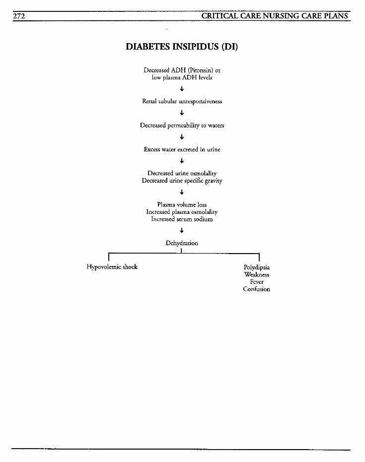
272 CRITICAL CARE NURSING CARE PLANS
DIABETES INSIPIDUS (DI)
Decreased ADH (Pitressin) or low plasma ADH levels
4 Renal tubular unresponsiveness
JI
Decreased permeability to waters
4 Excess water excreted in urine
4 Decreased urine osmolality
Decreased urine specific gravity
4 Plasma volume loss
Increased plasma osmolality Increased serum sodium
4 Dehydration
Hypovolemic shock Polydipsia Weakness
Fever Confusion
RENAIJENDOCRINE SYSTEMS 273
Pheochromocytoma is a vascular tumor, composed of chromafin cells that secrete catecholamines or their precursors (epinephrine, norephinephrine, or dopamine). This, in turn, causes severe persistent or intermittent hypertension due to the severe vasoconstriction in response to the catecholamine excess.
Usually the tumor is encapsulated and located within the medulla of the adrenal glands, but can occur in the sympathetic paraganglionic areas of the abdomen, chest, brain, or cervical areas. These tumors are usually benign, but can be malignant in up to ten percent of patients. Frequently occur- ring between the ages of 30 and 50, attacks may occur paroxysmally if the tumor releases catecholamines on an intermittent basis. These episodes may range from once per year to several times per day. Attacks may be spontaneous, or be caused by palpation of the tumor, emotional stress or trauma, exposure to cold, beta-blockers, postural changes, abdominal compression, anes- thesia induction, urination, defecation, or heavy lifting.
The tumor’s hallmark symptom is high blood pressure with fluctuations up to 220/150 or higher. The catecholamine secretion causes symp- toms of “flight or fight” reactions, typically beginning with palpitations, headache, pallor, cool, moist hands and feet, flushing, profuse sweating, and extreme anxiety.
Pheochromocytoma is also a part of the Multiple Endocrine Neoplasia (MEN) Syndromes and may be found in conjunction with neurofibromatoses, hemangiomas, and medullary thyroid cancers. Other diagnoses, such as angina, essential hyper- tension, hyperthyroidism, acute anxiety reactions, transient ischemic attacks, and menopause, must
be ruled out. Pheochromocytoma always leads to death if untreated.
MEDICAL CARE Medications: use of alpha- and beta-blockers (phenoxybenzamine and propranolol, or phenoxylbenzamine and metyrosine) to control catecholamine excess symptoms; IV infusions of trimethaphan camsylate or sodium nitroprusside to control vasopressor effects
Laboratory: fasting serum glucose elevated, increased hematocrit; 24-hour urine for catecholamines, vanillymandelic acid and metanephrines to identify elevated levels
Electrocardiogram: used to identify tachycardia, bradycardia, LV enlargement and strain from ele- vated blood pressure, cardiac dysrhythmias
Radiography: chest and abdominal x-rays used to localize and identify tumor; CT scans, IVP, radionuclide imaging and selective venographic angiography also used to localize tumors; caution must be used due to potential for test to exacer- bate hypertensive crisis
Surgery: surgical removal of the pheochromocy- toma may be required
NURSING CARE PLANS Altered tissue pe&sion: ca rdiop ulm onary, cerebral, gastrointestinal, peripheral, and renal
Related to: excessive catecholamine secretion
Defining characteristics: pulse and blood pressure changes, changes in cardiac output, changes in peripheral resistance, impaired myocardial oxygenation, chest pain, cardiac dysrhythmias, EKG changes, dyspnea, tachypnea, palpitations, nausea, vomiting, epigastric pain, constipation,
274 CRITICAL CARE NURSING CARE PLANS
slow digestion, weight loss, headaches, visual dis- turbances, paresthesias, oliguria, anuria, electrolyte imbalances, cold and clammy skin, decreased peripheral pulses, flushing, diaphoresis
Outcome Criteria
Patient will maintain adequate perfusion to all vital organs and will have adequate peripheral and systemic circulation.
INTERVENTIONS RATIONALES
Monitor vital signs, including lying, sitting, and standing BP.
Monitor functional abilities in relation to the affected system.
Assess for presence and character of pulses, capillary refill time, skin color and temperature, urine output, mentation, gastric dis- tention, presence of bowel sounds, and appetite.
Position patient in Fowler’s position.
Avoid any non-essential activi- ties, especially pressure-causing movement. Avoid straining with bowel movements or urination.
Provides information about heart rate and perfusion pressure which will affect blood flow and tissue perfusion. Chronic exces- sive secretion of catecholamines will affect the reflexes that are responsible for maintaining upright blood pressure and may result in orthostatic hypotension.
Interrelationships of the body systems can cause overlapping signs and symptoms associated with tissue perfusion and can cause changes in oxygenation, cardiac output, metabolic demands, neurologic function, renal function, and nutrition.
May indicate decreased perfusion related to the particular body system.
Helps to decrease the blood volume returning to the heart by pooling blood i,n dependent parts of the body. Decreases BP by use of orthostatic changes associated with the chronic catecholamine secretion.
Ambulation, exercise, and val- salva-type efforts may provoke an attack, increasing blood pressure and decreasing tissue perfusion.
INTERVENTIONS RATIONALES
Administer medications as ordered.
Titrate IV meds as needed to keep systolic blood pressure less than 170 mmHg, and diastolic pressure less than 100 mmHg.
Weigh every day.
Monitor intake and output, and notify physician far urine output less than 30 cc/hr.
Avoid palpation of abdomen; post sign near bed to refrain from palpation during assessments.
Monitor labwork, especially FBS, hematocrit and renal function levels.
Alpha- and beta-blockers may stabilize the condition prior to surgical intervention. Metyrosine interrupts the catecholamine synthesis, decreases levels of nor- epinephrine production, decreases levels of VMA, and decreases BI?
Reduces risk of complications from severely elevated pressure.
Weight loss may occur due to increased metabolism, decreased appetite, nausea, or vomiting.
Decreased renal perfusion may lead to decreased urinary output, renal impairment, and failure.
Prevents possible palpation of cumor and triggering of acute crisis.
Catecholarnine release can increase glycolysis and inhibit insulin release. Excess catecholamines can also increase erythropoierin stimulation and can elevate hematocrit, as well as decrease blood flow to the kidney resulting in renal impair- ment.
Information, Instruction, Demonstration
INTERVENTIONS RATIONALES
Assist with obtaining 24-hour urine specimen for diagnosis.
Elevated levels may be diagnostic for pheochromocytorna, but coma and increased stress states must be ruled out. Normal values for VMA are < 10 mgl24 hrs, metanephrines < 1.3 mg/24 hrs, free epinephrine and norepi- nephrine < 100 mcg124 hrs.
Avoid use of rauwolfia alkaloids, tetracycline, quinine, methyl-
These substances may interfere with the results and hamper
RENAL/ENDOCRINE SYSTEMS 275
INTERVENTIONS RATIONALES Organ function will be within patient’s normal
dopa, catecholamines, large quantities of vanilla, coffee, chocolate, nuts, bananas, guaife- nesin, and salicylates for at least 2 days prior to 24-hour test, if possible.
Instruct parientlfamily in causes of exacerbations or attacks, and methods to reduce frequency of occurrence.
Instruct to avoid exposure to cold temperatures.
Instruct in medications, effects, side effects, adverse reactions, complications, and symptoms to report to physician.
Instruct in methods to decrease emotional stress, such as relax- ation techniques.
Instruct in having frequent blood pressure checks, keeping log of trends, ranges to report to physi- cian. etc.
Instruct in avoiding rapid changes in position.
Instruct to avoid wearing any clothing that may be tight or constrictive.
determination of diagnosis.
Promotes understanding of the condition and risk of decreased perfusion to vital organs.
Cold may cause vasoconstriction, decreases circulation, and perfu- sion, as well as precipitate an attack.
Promotes knowledge and facili- tates compliance with medical regimen.
Reduces stress and lessens precip- itating factors with intermittent attacks by facilitating vasodila- tion.
Primary indicator of the tumor activity is blood pressure increases, which cause decreased perfusion to tissues and organs. Increased knowledge will decrease fear and increase com- pliance with treatment, and provide opportunity for prompt treatment to prevent serious complications.
Facilitates body’s attempt to cope with orthostatic hypotension by allowing time for body and cir- culatory system to adjust to changes.
May result in an attack by com- pression of abdomen or tumor region.
Extremities will be warm, with normal color and sensation, and have equally palpable pulses.
equivalent intake and output.
Patient will be free of abdominal or epigastric pain, and able to ingest adequate nutritional intake to maintain weight.
Patient will have adequate urinary output with
Anxiety Related to: excessive catecholamine release, threat to health status, changes in health status, life- threatening crisis, possibility of surgical intervention
Defining characteristics: apprehension, sense of impending doom, fear of death, restlessness, fear, fear of death, fear of surgery, fear of the unknown, feelings of helplessness, anxiousness, worry, com- munication of uncertainty, voiced concern over changes in life events
Outcome Criteria
Patient will have less anxiety or anxiety will be within an acceptable and manageable level.
INTERVENTIONS RATIONALES ~
Assess anxiety level, noting ver- Catecholamine increases can balizations of fear or sense of produce marked anxiety which doom. then increases oxygen demand
on tissues.
Provides an opportunity to vent feelings and to obtain informa- tion. Decreases anxiety and
Provide calm environment for patient to express fears, concerns, and feelings. Allow time for patient to ask questions. promotes a caring and trusting
atmosphere. Discharge or Maintenance Evaluation
Patient will have normalized vital signs. Encourage visits from family and friends who do not increase or
Provides emotional support and relieves anxiety when familiar
276 CRITICAL CARE NURSING CARE PLANS
INTERVENTIONS RATIONALES
provide patient with emotional s t ress .
Decrease stimuli in environment.
Administer medications as ordered.
people are available.
Prevents further stressors.
Assists to allay fear and anxiery
Information, Instruction, Demonstration
INTERVENTIONS RATIONALES
Instruct patientlfamily members about disease process, what to expect with procedures, pre- and postoperative care.
Instruct on emotional stress and other precipitating triggers for attacks, and methods to reduce stress and anxiety.
Instruct on medications, effects, side effects, contraindications, and symptoms to report to physician. regimen.
Decreases anxiety caused by fear of the unknown, and promotes knowledge and understanding.
Reduces anxiety and provides patient with some measure of control over the situation.
Promotes knowledge and understanding which facilitates compliance with medical
Discharge or Maintenance Evaluation
Patient will have reduced anxiety and be able to vent feelings and concerns.
Patiendfamily will be able to verbalize understanding of disease process, medications, and treatments, and will be compliant with reg- imen.
Patient will be able to avoid stressful visitors, situations, or other provoking events, and will be able to perform relaxation exercises when stressed.
Hypertherm ia
Defining characteristics: increase in body temper- ature greater than normal range, flushed warm skin, increased heart rate, increased respiratory rate, diaphoresis, delirium
Outcome Criteria
Patient will have temperature within normal range.
INTERVENTIONS RATIONALES
Monitor temperature every 1-2 Fluctuations in temperature can hours or use continuous moni- occur rapidly and temperature coring. elevations can increase metabo-
lism needs.
Assists patient with comfort and decreases temperature.
Adjust room temperature for patient comfort and maintain at or below 72 degrees.
Administer antipyretics as Decreases fever. ordered.
Provide frequent tepid sponge baths and change linens if patient is diaphoretic. evaporation.
Promotes patient comfort and reduces temperature by
Avoid chilling or shivering of patient.
Place covered ice packs to groin, axillae, andlor behind neck, if warranted.
Use cooling blanket for tempera- tures greater than 103 degrees if warranted. Cool body slowly- no faster than 1 degreell5 minutes. Blanket should be cov- eted and continuous monitoring of temperature should be performed.
Shivering may increase metabolic requirements and actually increase temperature,
Decreases temperature by means of conduction.
Assists in lowering temperatures by conduction. Blankets should be covered ro prevent burns and tissue injury. Cooling that is done too rapidly can produce ventricular ectopy.
ordered. Related to: increased metabolic rate in response to catecholarnines, decreased heat loss due to vaso-
Administer thorazine IM/IV as Thorazine is an alpha-adrenergic - blocking agent chat causes peripheral vasodilation which helps heat to dissipate and also
constriction can assist in decreasing shivering.
RENAL/ENDOCRINE SYSTEMS 277
Information, Instruction, Demonstration
INTERVENTIONS RATIONALES
Instruct patiendfamily in proce- dures, what to expect with cooling blanket application, etc.
Promotes knowledge and reduces anxiety.
Discharge or Maintenance Evaluation
Patient will achieve and maintain normal body temperature.
Patient will be compliant with medical regimen.
Sensor -perception alteration (visualj
[See CVA]
Related to: altered sensory reception, chemical alterations due to hypoxia, chemical alterations due to glucose/insulin and electrolyte imbalances, restrict environment, psychologic stress, vasocon- striction
thoug d t processes, kinesthetic)
Defining characteristics: confusion, anxiety, fear, disorientation, change in behavior patterns, hyper- esthesia, restlessness, irritability, impaired decision-making
Alteration in nutrition: less than body requirements [See Mechanical Ventilation]
Related to: hypermetabolic state, nausea, vomit- ing, anorexia, malabsorption
Defining characteristics: inadequate food intake, weight loss, muscle weakness, fatigue
Impaired gas exchange [See Mechanical Ventilation]
Related to: increased respiratory workload, impaired oxygen to heart, hypoventilation, altered oxygen supply, altered blood flow, change in vas-
cular resistance, altered oxygen-carrying capacity of blood, shift of the oxyhemoglobin dissociation curve, hypermetabolic state
Defining characteristics: confusion, restlessness, hypercapnia, hypoxia, cyanosis, dyspnea, tachyp- nea, changes in ABG values, changes in A-a gradient, changes in vital signs, activity intolerance, changes in mental status
Constipation
Related to: inadequate dietary/fluid intake, GI distress, changes in level of activity, decreased blood flow slowing digestion, malabsorption
Defining characteristics: nausea, vomiting, decreased appetite, epigastric pain, hard-formed stool, absence of stool, abdominal pain
Outcome Criteria
Patient will have normal elimination pattern reestablished and maintained.
INTERVENTIONS RATIONALES
Determine patient’s bowel habits, lifestyle, ability to sense urge to defecate, painful hemorrhoids, and history of constipation.
Auscultate bowel sounds for presence and quality.
Monitor diet and fluid intake.
Monitor for abdominal pain and distention.
Assists with identification of an effective bowel regime andlor impairment and need for assis- tance. GI function may be decreased as a result of decreased digestion.
Presence of abnormal sounds, such as high-pitched tinkles, suggest complications like ileus.
Adequate amounts of fiber and roughage provides bulk and ade- quate fluid intake (greater than 2 Llday) is importanr in determin- ing stool consistency.
Gas, abdominal distention, or ileus could be a factor. Lack of peristalsis from impaired diges- tion can create bowel distention and worsen to the point of ileus.
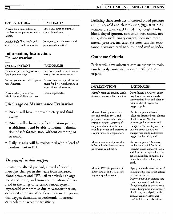
278 CRITICAL CARE NURSING CARE PLANS
INTERVENTIONS RATIONALES
Provide bulk, stool softeners, laxatives, or suppositories as war- ranted.
Provide high-fiber, whole grain cereals, breads and fresh fruits.
May be required to stimulate evacuation of stool.
Improves stool consistency and promotes elimination.
Information, Instruction, Demonstration
Defining characteristics: increased blood pressure and pulse, cold and clammy skin, jugular vein dis- tention, dyspnea, crackles, edema, cough, frothy blood-tinged sputum, confusion, restlessness, noc- turia, decreased urinary output, increased mean arterial pressure, increased systemic vascular resis- tance, decreased cardiac output and cardiac index
Outcome Criteria
INTERVENTIONS RATIONALES Patient will have adequate cardiac output to main- tain hemodynamic stability and perfusion to all
Determine preexisting habits of Laxative dependence can predis- laxativelenema usage. pose patient to constipation. organs.
Instruct patient to avoid frequent use of enemas.
Promotes enema dependence and causes fluid loss which results in more difficult elimination. INTERVENTIONS RATIONALES
Provide activity or exercise within limits of disease process.
Promotes peristalsis.
Discharge or Maintenance Evaluation
Patient will have improved dietary and fluid intake.
Patient will achieve bowel elimination pattern establishment and be able to maintain elimina- tion of soft-formed stool without cramping or straining.
Daily exercise will be maintained within level of confinement in ICU.
Decreased cardiac output
Related to: altered preload, altered afterload, inotropic changes in the heart from increased blood pressure and TPR, left ventricular enlarge- ment and strain, and from accumulation of extra fluid in the lungs or systemic venous system, myocardial compromise due to vasoconstriction, decreased coronary blood flow, increased myocar- dial oxygen demands, hyperthermia, increased catecholamine receptor sensitivity
Identify other pre-existing condi- tions and assess cardiac function.
Monitor blood pressure, heart rate and rhythm, apical and peripheral pulses, pulse deficits, respiratory status, presence of cough or adventitious breath sounds, presence and character of any sputum, and oxygenation.
Measure cardiac outpurlcardiac index and other hemodynamic parameters as indicated.
Monitor EKG for presence of dysrhythmias, and treat accord- ing to hospital protocol.
Other factors and disease states may further stress an already compromised heart and place an extra burden of myocardial oxygen supply.
Cardiac output and blood volume is decreased with elevated blood pressure. Afterload increases, pulse increases, and changes in contracriliry and con- duction occur. Respiratory changes may result in decreased oxygen intake and hypoxia.
Cardiac output < 5 L/min or cardiac index < 2.5 Llminlm’ indicates severe vasoconstriction and decrease in myocardial oxy- genation, leading to myocardial ischemia, cardiac failure, and death.
Dysrhythmias decrease the heart’s pumping efficiency which affects the cardiac output. Dysrhythmias may indicate inad- equate myocardial perfusion. Tachydysrhythmias decrease ven- tricular filling time and coronary blood flow; bradydysrhythmias decrease cardiac output and result in left ventricular failure.
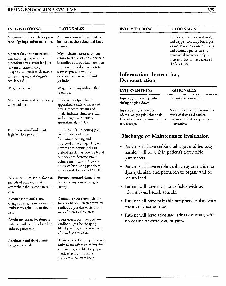
RENAL/ENDOCRINE SYSTEMS 279
INTERVENTIONS RATIONALES
Auscultate heart sounds for pres- ence of gallops andlor murmurs.
Monitor for edema to extremi- ties, sacral region, or other dependent areas; assess for jugu- lar vein distention, cold peripheral extremities, decreased urinary output, and sluggish capillary refill.
Weigh every day.
Monitor intake and output every 2 hrs and prn.
Position in semi-Fowler’s or high-Fowler’s position.
Balance rest with short, planned periods of activity; provide atmosphere that is conducive to rest.
Monitor for mental status changes, decreases in orientation, restlessness, agitation, or dizzi- ness.
Administer vasoactive drugs as ordered, with titration based on ordered parameters.
Administer anti-dysrhythmic drugs as ordered.
Accumulations of extra fluid can be heard as these abnormal heart sounds.
May indicate decreased venous return to the heart and a decrease in cardiac output. Fluid retention may result in a decrease in uri- nary output as a result of decreased venous return and perfusion.
Weight gain may indicate fluid retention.
Intake and output should approximate each other. A fluid deficit between output and intake indicates fluid retention and a weight gain (500 cc approximately = 1 Ib).
Semi-Fowler‘s positioning pre- vents blood pooling and facilitates breathing and improved air exchange. High- Fowler’s positioning reduces preload quickly by pooling blood but does not decrease stroke volume significantly. Afterload decreases by dilating peripheral arteries and decreasing LVEDl?
Prevents increased demand on heart and myocardial oxygen supply.
Central nervous system distur- bances can occur with decreased cardiac output due to decreases in perfusion to these areas.
These agents promote optimum cardiac output by changing blood pressure, and can reduce afterload and preload.
These agents decrease pacemaker activity, modig areas of impaired conduction, and blocks sympa- thetic effects of the heart: myocardial contractility is
INTERVENTIONS RATIONALES
decreased, heart rate is slowed, and oxygen consumption is pre- served. Blood pressure decreases and coronary perfusion and myocardial oxygen supply is increased due to the decrease in the heart rate.
Information, Instruction, Demonstration
INTERVENTIONS RATIONALES
Instruct to elevate legs when sitting or lying down.
Instruct in signs to report: May indicate complications as a edema, weight gain, chest pain, result of decreased cardiac headache, blood pressure or pulse output and facilitate prompt rate changes. intervention.
Promotes venous return.
Discharge or Maintenance Evaluation
Patient will have stable vital signs and hemody- namics will be within patient’s acceptable parameteirs.
Patient will have stable cardiac rhythm with no dysrhythmias, and perfusion to organs will be maintained.
Patient will have clear lung fields with no adventitious breath sounds.
Patient will have palpable peripheral pulses with warm, dry extremities.
Patient will have adequate urinary output, with no edema or extra weight gain.
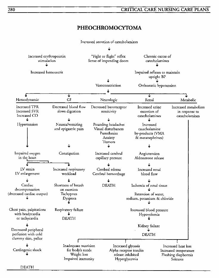
280 CRITICAL CARE NURSING CARE PLANS
Increased erythropoietin stimulation
c
PHEOCHROMOCYTOMA
Increased secretion of catecholamines
c “Fight or flight” reflex Chronic excess of
4 Sense of impending doom catecholamines
Increased hemotocrit Impaired reflexes to maintain upright BP
4 4
c Vasoconstriction Orthostatic hypotension
.I c c J, Hemodynamic GI Neurologic Renal Metabolic
Increased TPR Decreased blood flow Increased SVR slows digestion Increased CO
3. c Hypertension Nauseahomi ting
and epigastric pain
c Constipation
I Impaired oxygen
in the heart I JI
LV strain LV enlargement
J Cardiac
decompensation (decreased cardiac output)
e
Chest pain, palpitations with bradycardia
or tachycardia
J Decreased peripheral perfusion with cold clammy skin, pallor
e Cardiogenic shock
e
JI Increased respiratory
workload
c Shortness of breath
on exertion Tach ypnea
D yspnea c
Respiratory failure c
DEATH
Decreased baroreceptor sensitivity
c Pounding headaches Visual disturbances
Paresthesias Anxiety Tremors
c Increased cerebral capillary pressure
4 Cerebral edema
Cerebral hemorrhage
c DEATH
Increased urine excretion of
catecholamines c
Increased catecholamine
by-products (VMA & metanephrines)
c Angio tension
Aldosterone release
c Increased renal
blood flow
c Ischemia of renal tissue
c Retention of water.
sodium, potassium & chloride c
Increased blood pressure Hypovolemia
c Kidney failure
4 DEATH
Increased metabolism in response to
catecholamines
Inadequate nutrition Increased glycosis Increased heat loss for body’s needs Alpha receptor insulin Increased temperature
Weight loss release inhibited Flushing diaphoresis Impaired immunity Hyperglycemia Seizures
DEATH
RENAL/ENDOCRINE SYSTEMS 28 1
Thyrot oxicosis
Hyperthyroid crisis, also known as thyroid storm and thyrotoxicosis, is a life-threatening emergency characterized by greatly exaggerated signs of hyperthyroidism. Mortality is high, and symptoms appear rapidly when triggered by infection, trauma, surgery, diabetes, or abrupt withdrawal of thyroid medication. Thyroid storm may be diffi- cult to diagnose because the precipitating illness may mask its detection.
Hyperthyroid patients are more susceptible to cat- echolamines because of the increased number of catecholamine receptors they possess. A triggering illness creates an outpouring of catecholamines, and so the elevated levels of thyroid and increased number of receptors create the crisis. A hyperme- tabolic state then ensues causing increased oxygen and nutrient consumption, fluid and electrolyte imbalances, and a catabolic state.
Patients in crisis typically have hyperthermia, tachydysrhythmias, dehydration, nausea, vomit- ing, weight loss, and neurologic changes. Treatment is usually begun without waiting for confirmation of lab tests and is aimed at support- ing vital functions. Reversal of excessive thyroid hormone decreases the hypermetabolic state, and reduction of the circulating thyroid hormones fur- ther decreases the crisis. Once vital functions are preserved, treatment of the precipitating cause is begun. If the crisis is untreated, heart failure, exhaustion, and death will ensue.
MEDICAL CARE Laboratory: serum and serum free T4 and T, are increased; TSH levels are decreased; thyroglobulin is increased; electrolytes are used to identify imbal-
ances; thyroid antibodies positive in Graves’ dis- ease; glucose levels elevated from insulin resistance, increased glycogenolysis, or impaired insulin secretion; serum cortisol decreased due to lower adrenal reserve; alkaline phosphatase increased; serum calcium increased; liver function abnormal, decreased serum catecholamines; urine creatinine increased
TRH test: used in some cases to identify TSH suppression with administration of TRH hormones
Electrocardiogram: used to identifjr elevated thy- roid levels or electrolyte imbalances; atrial fibrillation may be present; cardiomegaly in elderly with masked hyperthyroidism
Oxygen: used to provide supplemental oxygen due to increased oxygen consumption and increased metabolic demands
Radiography. chest x-rays used to identify cardiac enlargement that may occur in response to increased circulatory demands, to identify presence of cardiac overload and congestion, respi- ratory infiltrates or other precipitating causes
Radioactive iodine uptake test: used to differenti- ate types of thyroid problems; usually high in Graves’ disease and toxic goiter, but low in thyroiditis
Thyroid scan: may be used to aid diagnosis when thyrotoxicosis is caused from cancer or a multinodular goiter
Iodine solutions: used to slow the release of thy- roid hormones; common solutions are Lugol’s solution and sodium iodide
Beta-adrenergic blockers: used to reverse periph- eral effects of excessive thyroid hormones and to decrease the hypermetabolic state; commonly used is propranolol; reserpine IN also helps to reduce peripheral effects and may help decrease the tachy- cardias
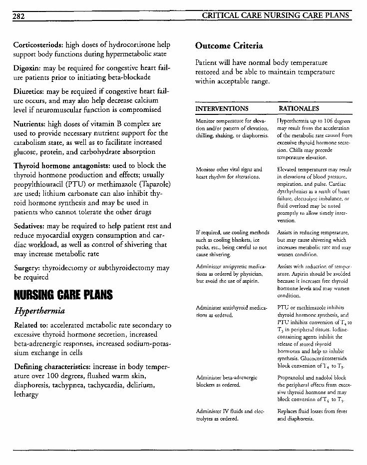
282 CRITICAL CARE NURSING CARE PLANS
Corticosteriods: high doses of hydrocortisone help support body functions during hypermetabolic state
Digoxin: may be required for congestive heart fail- ure patients prior to initiating beta-blockade
Diuretics: may be required if congestive heart fail- ure occurs, and may also help decrease calcium level if neuromuscular function is compromised
Nutrients: high doses of vitamin B complex are used to provide necessary nutrient support for the catabolism state, as well as to facilitate increased glucose, protein, and carbohydrate absorption
Thyroid hormone antagonists: used to block the thyroid hormone production and effects; usually propylthiouracil (PTU) or methimazole (Tapazole) are used; lithium carbonate can also inhibit thy- roid hormone synthesis and may be used in patients who cannot tolerate the other drugs
Sedatives: may be required to help patient rest and reduce myocardial oxygen consumption and car- diac workload, as well as control of shivering that may increase metabolic rate
Surgery: thyroidectomy or subthyroidectomy may be required
NURSING CME PLANS Hyperthemia Related to: accelerated metabolic rate secondary to excessive thyroid hormone secretion, increased beta-adrenergic responses, increased sodium-potas- sium exchange in cells
Defining characteristics: increase in body temper- ature over 100 degrees, flushed warm skin, diaphoresis, tachypnea, tachycardia, delirium, lethargy
Outcome Criteria
Patient will have normal body temperature restored and be able to maintain temperature within acceptable range.
INTERVENTIONS RATIONALES
Monitor temperature for eleva- tion andlor pattern of elevation, chilling, shaking, or diaphoresis.
Monitor other vital signs and heart rhythm for alterations.
If required, use cooling methods such as cooling blankets, ice packs, etc., being careful to not cause shivering.
Administer antipyretic medica- tions as ordered by physician, but avoid the use of aspirin.
Administer antithyroid medica- tions as ordered.
Administer beta-adrenergic blockers as ordered.
Administer IV fluids and elec- trolytes as ordered.
Hyperthermia up to 106 degrees may result from the acceleration of the metabolic rate caused from excessive thyroid hormone secre- tion. Chills may precede temperature elevation.
Elevated temperatures may result in elevations of blood pressure, respiration, and pulse. Cardiac dysrhythmias as a result of heart failure, electrolyte imbalance, or fluid overload may be noted promptly to allow timely inter- vention.
Assists in reducing temperature, but may cause shivering which increases metabolic rate and may worsen condition.
Assists with reduction of remper- ature. Aspirin should be avoided because it increases free thyroid hormone levels and may worsen condition.
PTU or rnethimazole inhibits thyroid hormone synthesis, and PTU inhibits conversion ofT, to T, in peripheral tissues. Iodine- containing agents inhibit the release of stored thyroid hormones and help to inhibit synthesis. Glucocorticosteroids block conversion of T4 to T3.
Propranolol and nadolol block the peripheral effects from exces- sive thyroid hormone and may block conversion ofT4 to T,.
Replaces fluid losses from fever and diaphoresis.
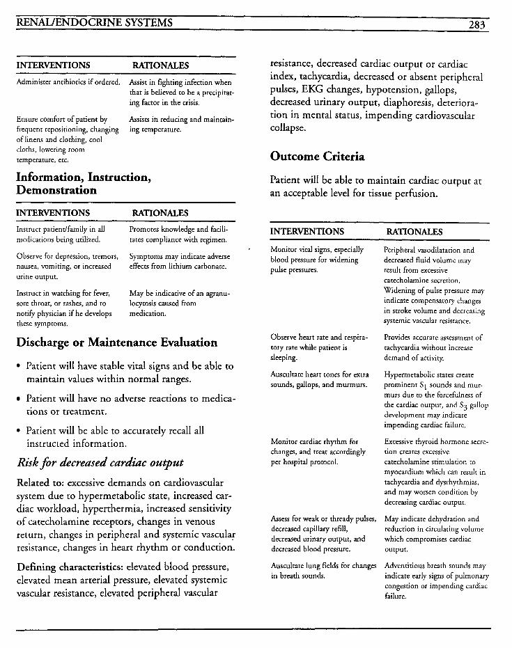
RENAWENDOCRINE SYSTEMS 283
INTERVENTIONS RATIONALES
Administer antibiotics if ordered. Assist in fighting infection when that is believed to be a precipitat- ing factor in the crisis.
Assists in reducing and maintain- Ensure comfort of patient by frequent repositioning, changing ing temperature. of linens and clothing, cool clorhs, lowering room temperature, etc.
Information, Instruction, Demonstration
INTERVENTIONS RATIONALES
resistance, decreased cardiac output or cardiac index, tachycardia, decreased or absent peripheral pulses, EKG changes, hypotension, gallops, decreased urinary output, diaphoresis, deteriora- tion in mental status, impending cardiovascular collapse.
Outcome Criteria
Patient will be able to maintain cardiac output at an acceptable level for tissue perfusion.
Instruct patiendfamily in all medications being utilized.
Observe for depression, tremors, nausea, vomiting, or increased urine output.
Instruct in watching for fever, sore throat, or rashes, and to notify physician if he develops rhese symptoms.
Promotes knowledge and facili- tates compliance with regimen.
Symptoms may indicate adverse effects from lithium carbonate.
May be indicative of an agranu- locytosis caused from medication.
Discharge or Maintenance Evaluation
Patient will have stable vital signs and be able to maintain values within normal ranges.
Patient will have no adverse reactions to medica- tions or treatment.
Patient will be able to accurately recall all
Risk for decreased cardiac output
Related to: excessive demands on cardiovascular system due to hypermetabolic state, increased car- diac workload, hyperthermia, increased sensitivity of catecholamine receptors, changes in venous return, changes in peripheral and systemic vascular resistance, changes in heart rhythm or conduction.
Defining characteristics: elevated blood pressure, elevated mean arterial pressure, elevated systemic vascular resistance, elevated peripheral vascular
instructed information.
INTERVENTIONS RATIONALES
Monitor vital signs, especially blood pressure for widening pulse pressures.
Observe heart rate and respira- tory rate while patient is sleeping.
Auscultate heart tones for extra sounds, gallops, and murmurs.
Monitor cardiac rhythm for changes, and treat accordingly per hospital protocol.
Assess for weak or thready pulses, decreased capillary refill, decreased urinary output, and deueased blood pressure.
Auscultate lung fields for changes in breath sounds.
Peripheral vasodilatation and decreased fluid volume may result from excessive catecholamine secretion. Widening of pulse pressure may indicate compensatory changes in stroke volume and decreasing systemic vascular resistance.
Provides accurate assessment of tachycardia without increase demand of activity.
Hypermetabolic states create prominent S1 sounds and mur- murs due to the forcefulness of the cardiac ourput, and S3 gallop development may indicate impending cardiac failure.
Excessive thyroid hormone secre- tion creates excessive catecholamine srimulation to myocardium which can result in tachycardia and dysrhythmias, and may worsen condition by decreasing cardiac output.
May indicate dehydration and reduction in circulating volume which compromises cardiac output.
Adventitious breath sounds may indicate early signs of pulmonary congestion or impending cardiac failure.
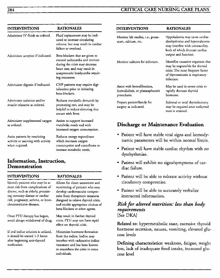
284 CRITICAL CARE NURSING CARE PLANS
INTERVENTIONS RATIONALES INTERVENTIONS RATIONALES
Administer IV fluids as ordered.
Administer atropine if indicated.
Administer digoxin if indicated.
Administer sedatives andlor muscle relaxants as ordered.
Administer supplemental oxygen as ordered.
Assist patients by restricting activity or assisting with activity when required.
Fluid replacement may be'indi- cated to increase circulating volume, but may result in cardiac failure or overload.
Beta-blockers that are given to control tachycardia and tremors during the crisis may decrease heart rate, and may result in symptomatic bradycardia requir- ing treatment.
CHF patients may require digi- talization prior to initiating beta-blockers.
Reduces metabolic demands by promoting rest, and may be helpful to reduce shivering that occurs with fever.
Assists to support increased metabolic needs and with increased oxygen consumption.
Reduces energy expenditure which increases oxygen consumption and contributes to increase metabolic needs.
Information, Instruction, Demonstration
INTERVENTIONS RATIONALES Identify patients who may be at most risk from complications of disease, such as elderly, preexist- ing coronary disease or cardiac risk, pregnancy, asthma, or bron- choconstrictive diseases.
Once PTU therapy has begun, avoid abrupt withdrawal of drug.
If oral iodine solution is utilized, it should be started 1-3 hours after beginning anti-thyroid medication.
Allows for closer assessment and monitoring of patients who may develop cardiovascular compro- mise from therapeutic measures designed to relieve thyroid crisis, and enable appropriate choices of beta-blockers or other agents.
May result in further thyroid crisis. PTU may not have rapid effect on thyroid crisis.
Minimizes hormone formation from the iodine. Iodine may interfere with radioactive iodine treatment and has been known to exacerbate the crisis in some individuals.
Monitor lab studies, i.e., potas- sium, calcium, etc.
Hypokalemia may cause cardiac dysrhythmias and hypercalcemia may interfere with contractility, both of which decrease cardiac output and function.
Identifies causative organism that may be responsible for thyroid crisis. The most frequent factor of thyrotoxicosis is respiratory infection.
May be used in severe crisis to rapidly decrease thyroid
Monitor cultures for infection.
Assist with hemofiltration, hemodialysis, or plasmapheresis procedures. hormone.
Prepare patientlfamily for surgery as indicated.
Subtotal or total thyroidectomy may be required once ruthyroid state is attained.
Discharge or Maintenance Evaluation
Patient will have stable vital signs and hemody- namic parameters will be within normal limits.
Patient will have stable cardiac rhythm with no dysr h yt hmias . Patient will exhibit no signs/symptoms of car- diac failure.
Patient will be able to tolerate activity without circulatory compromise.
Patient will be able to accurately verbalize instructed information.
Risk for altered nutrition: less than body requirements [See DKA]
Related to: hypermetabolic state, excessive thyroid hormone secretion, nausea, vomiting, elevated glu- cose levels
Defining characteristics: weakness, fatigue, weight loss, lack of inadequate food intake, increased glu- cose level
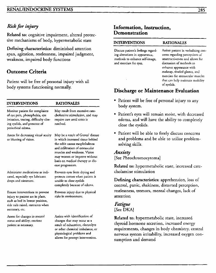
RENAL/ENDOCRINE SYSTEMS 285
Risk fir inju y Related to: cognitive impairment, altered protec- tive mechanisms of body, hypermetabolic state
Defining characteristics: diminished attention span, agitation, restlessness, impaired judgment, weakness, impaired body functions
Outcome Criteria
Patient will be free of personal injury with all body systems functioning normally.
INTERVENTIONS RATIONALES
Monitor patient for complaints of eye pain, photophobia, eye irritation, tearing, dificulty clos- ing eyelids, and presence of periorbital edema.
Assess for decreasing visual acuity or blurring of vision.
Administer medications as indi- cated, especidly eye lubricant drops and ointment.
Ensure interventions to prevent injury to patient are in place, such as bed in lowest position, side rails raised, restraints when necessary, etc.
Assess for changes in mental status and ability; reorient patient as necessary.
May result from excessive cate- cholamine stimulation, and may require care until crisis is resolved.
May be a result of Graves’ disease in which increased tissue behind the orbit causes exophthalmos and infiltration of extraocular muscles and weakness. Vision may worsen or improve without basis on medical therapy or dis- ease progression.
Prevents eyes from drying and protects cornea when patient is unable to close eyelids completely because of edema.
Prevents injury due to physical risks in environment.
Assists with identification of changes that may occur as a result of exhaustion, electrolyte or other chemical imbalance, or physiological problems and allows for prompt intervention.
Information, Instruction, Demonstration
INTERVENTIONS RATIONALES ~ ~~~~
Discuss patient’s feelings regard- ing alterations in appearance, methods to enhance self-image, and exercises for eyes.
Assists patient in verbalizing con- cerns regarding perceptions of unattractiveness and allows for discussion of methods to enhance appearance with makeup, shaded glasses, and exercises for extraocular muscles that can help maintain mobility of eyelids.
Discharge or Maintenance Evaluation
Patient will be free of personal injury to any body system.
Patient‘s eyes will remain moist, with decreased edema, and will have the ability to completely close the eyelids.
Patient will be able to freely discuss concerns and problems and be able to utilize problem- solving skills.
Anxiety [See Pheochromocytoma]
Related to: hypermetabolic state, increased cate- cholamine stimulation
Defining characteristics: apprehension, loss of control, panic, shakiness, distorted perception, restlessness, tremors, mental changes, lack of attention
Fatipe [See DKA]
Related to: hypermetabolic state, increased thyroid hormone secretion, increased energy requirements, changes in body chemistry, central nervous system irritability, increased oxygen con- sumption and demand
286 CRITICAL CARE NURSING CARE PLANS
Defining characteristics: lack of energy, inability to perform normal activities, inability to concen- trate, lethargy, irritability, nervousness, tension, apathy, depression
Knowledge &fieit Related to: lack of information, unfamiliarity with resources, misinterpretation of information, lack of recall
Defining characteristics: requests for information, questions, misrepresentation of facts, inaccurate follow-through of instructions, development of preventable complications
Outcome Criteria
Patient will be able to accurately recall measures for managing hyperthyroidism and be able to decrease risk of complications.
INTERVENTIONS RATIONALES
Discuss patient's perceptions and knowledge of disease.
Ensure that family members are included in discussions and allowed to verbalize their con- cerns and questions.
Instruct in all medications, effects, side effects, complica- tions. and symptoms to report to physician.
Instruct to notify physician for fever, sore throar, or rashes.
Establishes knowledge base of patient and helps identify inter- ventions and appropriate plan of care.
Patient's physical condition may interfere with his ability to con- centrate which can hinder the learning process. Instruction to the family can assist with rein- struction when needed.
Provides knowledge and facili- tates compliance with regimen. Antithyroid therapy will require long-term use in order to inhibit hormone production. Alternate drugs may be chosen if the patient develops symptoms of agranulocytosis from his therapy.
May be indicative of adverse reactions to thiourea rherapy and facilitates prompt treatment.
INTERVENTIONS RATIONALES
Instruct to avoid taking over-the- counter medications unless advised to do so by physician.
Instruct in diet needs, avoidance of caffeine, artificial preservatives and dyes.
Instruct in needhationale for continued medical follow-up.
Antithyroid medicines can affect andlor be affected by several OTC drugs and may cause dan- gerous interactions.
Hypermetabolic states require increased nutrients to maintain well-being and meet demand. Stimulants and additives may result in systemic problems.
Compliance with monitoring medical regimen and identifca- tion of potential complications can be assessed for timely inter- vention.
Discharge or Maintenance Evaluation
Patient/family members will be able to accurately recall all instructional information provided to them.
Patient will be free of preventable complications.
Patient will be able to correctly recall all med- ications and effects.
Patient will be able to manage hyperthyroidism without crisis.
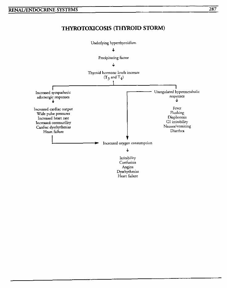
RENAL/ENDOCRINE SYSTEMS 287
THYROTOXICOSIS (THYROID STORM)
Underlying hyperthyroidism
c Precipitating factor
c Thyroid hormone levels increase
(T3 and T4)
1
Increased sympathetic adrenergic responses
c 7 Unregulated hypermetabolic
responses JI
Fever Flushing
Diaphoresis GI irritability
Nauseahomiting Diarrhea
Increased cardiac output Wide pulse pressures Increased heart rate
Increased contractility Cardiac dysrhythmias
Heart failure
Increased oxygen consumption
4 Irritability Confision
Angina D ysrhythmias Heart failure

This Page Intentionally Left Blank
MUSCULOS KELETM SYSTEM 289
SYSTEM Fractures
Amputation
Fat Embolism

This Page Intentionally Left Blank
.WC"' '
MUSCULOSKELETAL SYSTEM 29 1
A fracture is a break in a bone that occurs when direct or indirect pressure is placed on the bone in a force sufficient to exceed the bone's normal elas- ticity and causes deformation. There are many types of fractures, but the major classifications include open or compound, closed or simple, complete, incomplete, and pathologic fractures.
In closed fractures, there is no contact of the bone with the environment. In open fractures, the skin surrounding the area of the break is open and the bone is exposed to the environment. The major goal in these types of fractures involves the preven- tion of infection in conjunction with achieving proper alignment. Many patients have severe bleeding associated with this type of fracture. A complete fracture is one that involves the complete cross-section of the bone and it is visibly misaligned. In an incomplete fracture, the actual break may only involve a part of the cross-section of the bone in which one side of the bone is broken and the other part is merely bent. Pathologic fractures occur without or with mini- mal trauma and are usually seen in diseases such as osteoporosis and cancer.
Fractures not only cause damage to the bone involved, but to the soft tissues, nerves, tendons, and vascular system as well. These structures are in close proximity to the bones and help to support skeletal weight and to facilitate joint movement. When the fracture occurs, this stability is lost, and in turn, results in pain, swelling and splinting. The surrounding muscles are usually flaccid ini- tially after the injury, but within an hour or less, may commence to spasm and this may impair venous circulation and displace the fracture further.
Another complication that frequently occurs is called compartmental syndrome. After a fracture
occurs to the arm or leg, the fascia surrounding the muscles form compartments with small open- ings for major arteries, nerves, and tendons, Edema can compress these structures and cause ischemia to muscle tissues. The initial ischemic changes result in a histamine release that causes dilation of the capillary bed and edema to the area. The edema further compresses the larger arteries, which in turn creates further ischemia, further histamine release, and a vicious cycle is formed. The nerves, veins, arteries, and muscles may receive irreversible damage within 6 hours, and contractures, paralysis, and paresthesia may occur within 24-48 hours without intervention.
Healing begins when the blood around the end of the bone forms a clot and is related to the revascu- larization process. An inflammatory response occurs with blood vessel dilatation, then the increased permeability of the capillaries allow pro- tein and granulocytes to leak into the tissue. Fibrinogen converts to fibrin that collects proteins and other types of cells, and the granulation tissue allows for debris removal. When the pH of the fluid surrounding the bone fragments decreases, calcium goes into the solution and this begins the process that helps to form new bone. After a couple of weeks, the pH of the tissues rises, and calcium precipitates into the meshwork and a callus is formed as a bridge within the fragments of bone.
Frequently, if open fractures are present, fat partic- ulate may embolize, and the patient must be monitored for this complication. [See Fat Embolus]
MEDICAL CARE X-rays: used to identify type, location, and sever- ity of fractures or traumatic injuries and to evaluate healing process stage
Bone scans, CT scans, MRI scans: used to iden- ti$ fractures and/or soft tissue damage
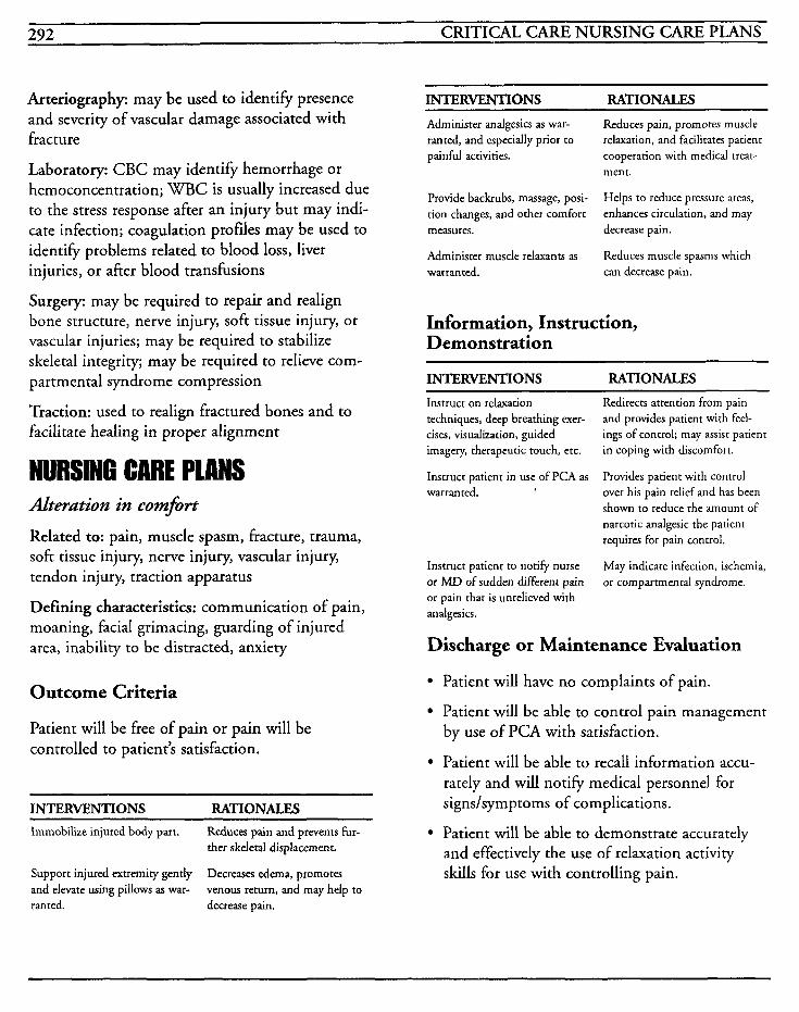
292 CRITICAL CARE NURSING CARE PLANS
Arteriography: may be used to identify presence and severity of vascular damage associated with fracture
Laboratory: CBC may identify hemorrhage or hemoconcentration; WBC is usually increased due to the stress response after an injury but may indi- cate infection; coagulation profiles may be used to identify problems related to blood loss, liver injuries, or after blood transfusions
Surgery: may be required to repair and realign bone structure, nerve injury, soft tissue injury, or vascular injuries; may be required to stabilize skeletal integrity; may be required to relieve com- partmental syndrome compression
Traction: used to realign fractured bones and to facilitate healing in proper alignment
NURSING CARE PLANS Alteration in comfort
Related to: pain, muscle spasm, fracture, trauma, soft tissue injury, nerve injury, vascular injury, tendon injury, traction apparatus
Defining characteristics: communication of pain, moaning, facial grimacing, guarding of injured area, inability to be distracted, anxiety
Outcome Criteria
Patient will be free of pain or pain will be controlled to patient’s satisfaction.
INTERVENTIONS RATIONALES Immobilize injured body part. Reduces pain and prevents fur-
ther skeletal displacement.
Decreases edema, promotes venous return, and may help to
Support injured extremity gently and elevate using pillows as war- ranted. decrease pain.
INTERVENTIONS RATIONALES
Administer analgesics as war- ranted, and especially prior to painful activities.
Provide backrubs, massage, posi- tion changes, and other comfort measures.
Administer muscle relaxants as warranted.
Reduces pain, promotes muscle relaxation, and facilitates patient cooperation with medical treat- ment.
Helps to reduce pressure areas, enhances circulation, and may decrease pain.
Reduces muscle spasms which can decrease pain.
Information, Instruction, Demonstration
~ _ _ _ _ ~
INTERVENTIONS RATIONALES
Instruct on relaxation techniques, deep breathing exer- cises, visualization, guided imagery, therapeutic touch, etc.
Instruct patient in use of PCA as warranted.
Instruct patient to notify nurse or MD of sudden different pain or pain that is unrelieved with analgesics.
Redirects attention from pain and provides patient with feel- ings of control; may assist patient in coping with discomfort.
Provides patient with control over his pain relief and has been shown to reduce the amount of narcotic analgesic the patient requires for pain control.
May indicate infection, ischemia, or compartmental syndrome.
Discharge or Maintenance Evaluation
Patient will have no complaints of pain.
Patient will be able to control pain management by use of PCA with satisfaction.
Patient will be able to recall information accu- rately and will notify medical personnel for signs/symptoms of complications.
Patient will be able to demonstrate accurately and effectively the use of relaxation activity skills for use with controlling pain.
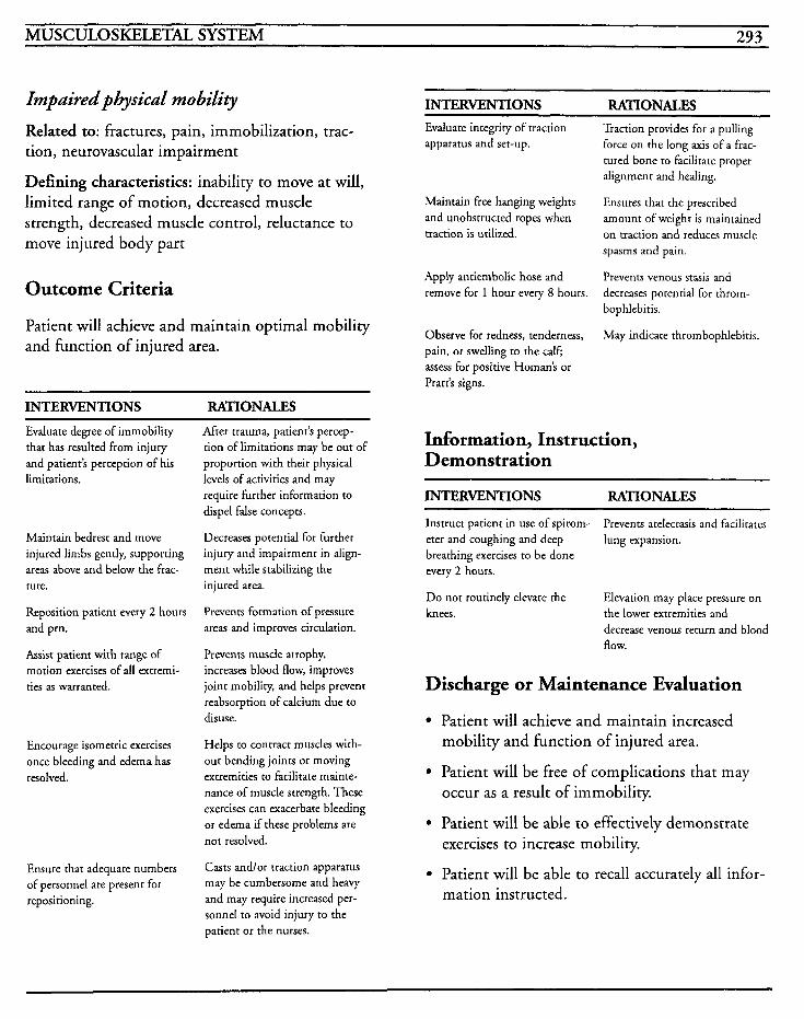
MUSCULOSKELETAL SYSTEM 293
Impaired physical mobility Related to: fractures, pain, immobilization, trac- tion, neurovascular impairment
Defining characteristics: inability to move at will, limited range of motion, decreased muscle strength, decreased muscle control, reluctance to move injured body part
Outcome Criteria
Patient will achieve and maintain optimal mobility and function of injured area.
INTERVENTIONS RATIONALES Evaluate degree of immobility that has resulted from injury and patient's perception of his limitations.
Maintain bedrest and move injured limbs gently, supporting areas above and below the frac- ture.
Reposition patient every 2 hours and prn.
Assist patient with range of motion exercises of all extremi- ties as warranted.
Encourage isometric exercises once bleeding and edema has resolved.
Ensure that adequate numbers of personnel are present for repositioning.
After trauma, patient's percep- tion of limitations may be out of proportion with their physical levels of activities and may require further information to dispel false concepts.
Decreases potential for further injury and impairment in align- ment while stabilizing the injured area.
Prevents formation of pressure areas and improves circulation.
Prevents muscle atrophy, increases blood flow, improves joint mobility, and helps prevent reabsorption of calcium due to disuse.
Helps to contract muscles with- out bending joints or moving extremities to facilitate mainte- nance of muscle strength. These exercises can exacerbate bleeding or edema if these problems are not resolved.
Casts andlor traction apparatus may be cumbersome and heavy and may require increased per- sonnel to avoid injury to the patient or the nurses.
INTERVENTIONS RATIONALES Evaluate integrity of traction apparatus and set-up.
Maintain free hanging weights and unobstructed ropes when traction is utilized.
Apply antiembolic hose and remove for 1 hour every 8 hours.
Observe for redness, tenderness, pain, or swelling to the calf; assess for positive Homan's or Pratt's signs.
Traction provides for a pulling force on the long axis of a frac- tured bone to facilitate proper alignment and healing.
Ensures that the prescribed amount of weight is maintained on traction and reduces muscle spasms and pain.
Prevents venous stasis and decreases potential for throm- bophlebitis.
May indicate thrombophlebitis.
Information, Instruction, Demonstration
INTERVENTIONS RATIONALES
Instruct patient in use of spirom- eter and coughing and deep breathing exercises to be done every 2 hours.
Do not routinely elevate the knees. the lower extremities and
Prevents atelectasis and facilitates lung expansion.
Elevation may place pressure on
decrease venous return and blood flow.
Discharge or Maintenance Evaluation
Patient will achieve and maintain increased mobility and function of injured area.
Patient will be free of complications that may occur as a result of immobility.
Patient will be able to effectively demonstrate exercises to increase mobility.
Patient will be able to recall accurately all infor- mation instructed.
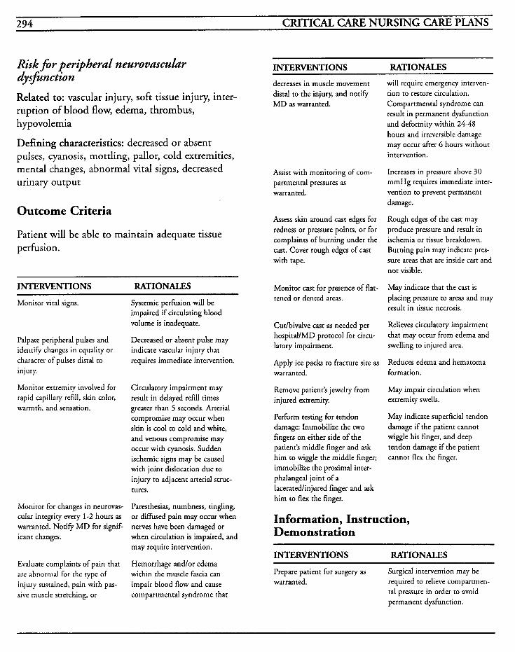
294 CRITICAL CARE NURSING CARE PLANS
Risk f i r peripheral neurovascuhr dysfinction
Related to: vascular injury, soft tissue injury, inter- ruption of blood flow, edema, thrombus, hypovolemia
Defining characteristics: decreased or absent pulses, cyanosis, mottling, pallor, cold extremities, mental changes, abnormal vital signs, decreased urinary output
Outcome Criteria
Patient will be able to maintain adequate tissue perfusion.
INTERVENTIONS RATIONALES
Monitor vital signs.
Palpate peripheral pulses and identify changes in equality or character of pulses distal to injury.
Monitor extremity involved for rapid capillary refill, skin color, warmth, and sensation.
Monitor for changes in neurovas- cular integrity every 1-2 hours as warranted. Notify M D for signif- icant changes.
Evaluate complaints of pain that are abnormal for the type of injury sustained, pain with pas- sive muscle stretching, or
Systemic perfusion will be impaired if circulating blood volume is inadequate.
Decreased or absent pulse may indicate vascular injury that requires immediate intervention.
Circulatory impairment may result in delayed refill times greater than 5 seconds. Arterial compromise may occur when skin is cool to cold and white, and venous compromise may occur with cyanosis. Sudden ischemic signs may be caused with joint dislocation due to injury to adjacent arterial struc- tures.
Paresthesias, numbness, tingling, or diffused pain may occur when nerves have been damaged or when circulation is impaired, and may require intervention.
Hemorrhage andlor edema within the muscle fascia can impair blood flow and cause compartmental syndrome that
INTERVENTIONS RATIONALES
decreases in muscle movement distal to the injury, and notify M D as warranted.
Assist with monitoring of com- partmental pressures as warranted.
Assess skin around cast edges for redness or pressure points, or for complaints of burning under the cast. Cover rough edges of cast with tape.
Monitor cast for presence of flat- tened or dented areas.
Cutlbivalve cast as needed per hospital/MD protocol for circu- latory impairment.
Apply ice packs to fracture site as warranted.
Remove patient’s jewelry from injured extremity.
Perform testing for tendon damage: Immobilize the two fingers on either side of the patient’s middle finger and ask him to wiggle the middle finger; immobilize the proximal inter- phalangeal joint of a laceratedlinjured finger and ask him to flex the finger.
will require emergency interven- tion to restore circulation. Compartmental syndrome can result in permanent dysfunction and deformity within 24-48 hours and irreversible damage may occur after 6 hours without intervention.
Increases in pressure above 30 mmHg requires immediate inter- vention to prevent permanent damage.
Rough edges of the cast may produce pressure and result in ischemia or tissue breakdown. Burning pain may indicate pres- sure areas that are inside cast and not visible.
May indicate that the cast is placing pressure to areas and may result in tissue necrosis.
Relieves circulatory impairment that may occur from edema and swelling to injured area.
Reduces edema and hematoma formation.
May impair circulation when extremity swells.
May indicate superficial tendon damage if the patient cannot wiggle his finger, and deep tendon damage if the patient cannot flex the finger.
Information, Instruction, Demonstration
INTERVENTIONS RATIONALES
Prepare patient for surgery as warranted. required to relieve compartmen-
Surgical intervention may be
tal pressure in order to avoid permanent dysfunction.
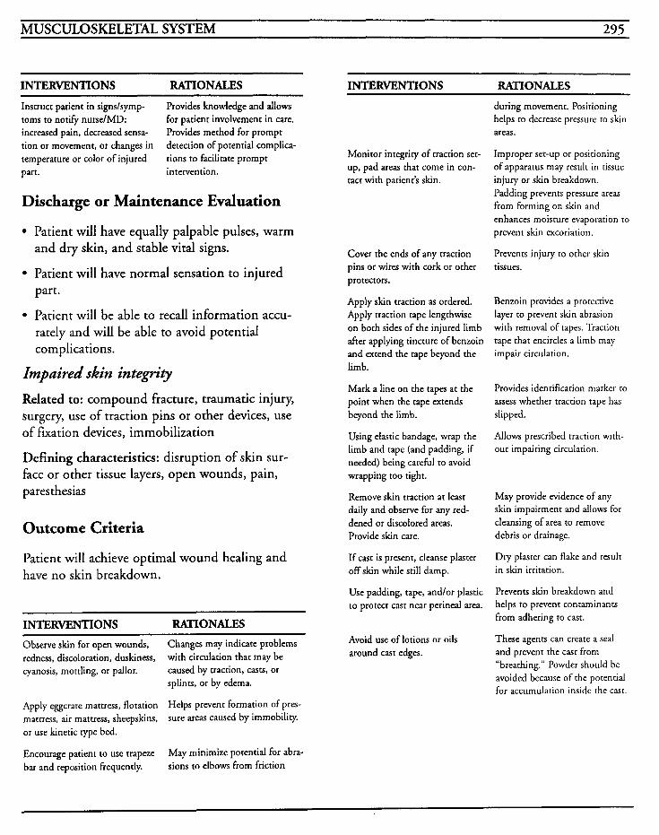
MUSCULOSKELETAL SYSTEM 295
INTERVENTIONS RATIONALES INTERVENTIONS RATIONALES Instruct patient in signslsymp- toms to notify nurse/MD: increased pain, decreased sensa- tion or movement, or changes in temperature or color of injured part. intervention.
Provides knowledge and allows for patient involvement in care. Provides method for prompt detection of potential complica- tions to facilitate prompt
Discharge or Maintenance Evaluation
Patient will have equally palpable pulses, warm and dry skin, and stable vital signs.
Patient will have normal sensation to injured part.
Patient will be able to recall information accu- rately and will be able to avoid potential complications.
Impaired skin integrz.9 Related to: compound fracture, traumatic injury, surgery, use of traction pins or other devices, use of fixation devices, immobilization
Defining characteristics: disruption of skin sur- face or other tissue layers, open wounds, pain, pares thesias
Outcome Criteria
Patient will achieve optimal wound healing and have no skin breakdown.
INTERVENTIONS RATIONACES
Observe skin for open wounds, redness, discoloration, duskiness, cyanosis, mottling, or pallor.
Apply eggcrate mattress, flotation mattress, air mattress, sheepskins, or use kinetic rype bed.
Encourage patient to use trapeze bar and reposition frequently.
Changes may indicate problems with circulation that may be caused by traction, casts, or splints, or by edema.
Helps prevent formation of pres- sure areas caused by immobility.
May minimize potential for abra- sions to elbows from friction
Monitor integrity of traction set- up, pad areas that come in con- tact with patient’s skin.
Cover the ends of any traction pins or wires with cork or other protectors.
Apply skin traction as ordered. Apply traction tape lengthwise on both sides of the injured limb after applying tincture of benzoin and extend the tape beyond the limb.
Mark a line on the tapes at the point when the tape extends beyond the limb.
Using elastic bandage, wrap the limb and tape (and padding, if needed) being careful to avoid wrapping too tight.
Remove skin traction at least daily and observe for any red- dened or discolored areas. Provide skin care.
If cast is present, cleanse plaster off skin while still damp.
Use padding, tape, and/or plastic to protect cast near perinea area.
Avoid use of lotions or oils around cast edges.
during movement. Positioning helps ro decrease pressure to skin areas.
Improper set-up or positioning of apparatus may result in tissue injury or skin breakdown. Padding prevents pressure areas from forming on skin and enhances moisture evaporation to prevent skin excoriation.
Prevents injury to other skin tissues.
Benzoin provides a protective layer to prevent skin abrasion with removal of tapes. Traction tape that encircles a limb may impair circulation.
Provides identification marker to assess whether traction tape has slipped.
Allows prescribed traction with- out impairing circulation.
May provide evidence of any skin impairment and allows for cleansing of area to remove debris or drainage.
Dry plaster can flake and result in skin irritation.
Prevents skin breakdown and helps to prevent contaminants from adhering to cast.
These agents can creare a seal and prevent the casr from “breathing.” Powder should be avoided because of the potential for accumulation inside the cast.
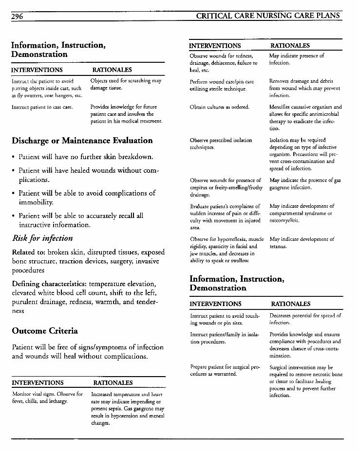
296 CRITICAL CARE NURSING CARE PLANS
Information, Instruction, Demonstration
INTERVENTIONS RATIONALES
Instruct the patient to avoid purring objects inside cast, such as fly swarters, coat hangers, etc.
Instruct patient in cast care.
Objects used for scratching may damage tissue.
Provides knowledge for future patient care and involves the patient in his medical treatment.
Discharge or Maintenance Evaluation
Patient will have no further skin breakdown.
Patient will have healed wounds without com- plications.
Patient will be able to avoid complications of immobility.
Patient will be able to accurately recall all instructive information.
Risk for infection
Related to: broken skin, disrupted tissues, exposed bone structure, traction devices, surgery, invasive procedures
Defining characteristics: temperature elevation, elevated white blood cell count, shift to the left, purulent drainage, redness, warmth, and tender- ness
Outcome Criteria
Patient will be free of signs/symptoms of infection and wounds will heal without complications.
INTERVENTIONS RATIONALES
Monitor vital signs. Observe for fever, chills, and lethargy.
Increased temperature and heart rate may indicate impending or present sepsis. Gas gangrene may result in hypotension and mental changes.
INTERVENTIONS RATIONALES Observe wounds for redness, drainage, dehiscence, failure to heal, etc.
Perform wound carelpin care utilizing sterile technique.
Obtain cultures as ordered.
Observe prescribed isolation techniques.
Observe wounds for presence of crepitus or fruity-smellinglfrothy drainage.
Evaluate patient’s complaints of sudden increase of pain or diffi- culty with movement in injured area.
Observe for hyperreflexia, muscle rigidity, spasticity in facial and jaw muscles, and decreases in ability to speak or swallow.
May indicate presence of infection.
Removes drainage and debris from wound which may prevent infection.
Identifies causative organism and allows for specific antimicrobial therapy to eradicate the infec- tion.
Isolation may be required depending on type of infective organism. Precautions will pre- vent cross-contamination and spread of infection.
May indicate the presence of gas gangrene infection.
May indicate development of comparrmenral syndrome or osteomyelitis.
May indicate development of tetanus.
Information, Instruction, Demonstration
INTERVENTIONS RATIONALES
Instruct patient to avoid touch- ing wounds or pin sites.
Instruct patientlfarnily in kola- tion procedures.
Decreases potential for spread of infection.
Provides knowledge and ensures compliance with procedures and decreases chance of cross-conta- mination.
Surgical intervention may be required to remove necrotic bone or tissue to facilitate healing process and to prevent further infection.
Prepare patient for surgical pro- cedures as warranted.
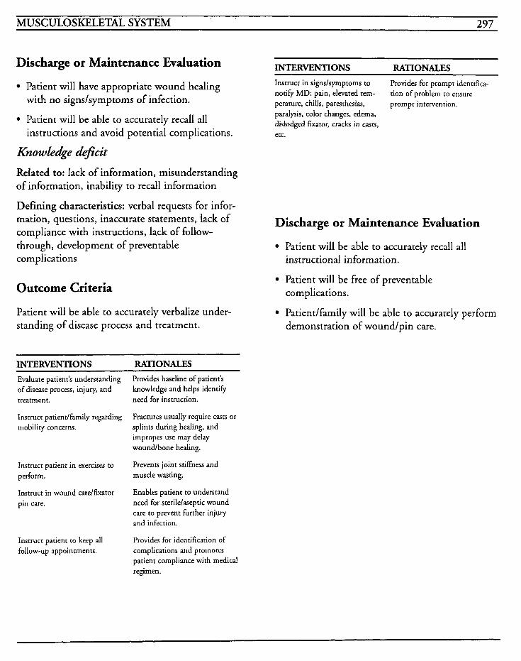
MUSCULOSKELETAL SYSTEM 297
INTERVENTIONS RATIONACES Discharge or Maintenance Evaluation
Patient will have appropriate wound healing with no signdsymptoms of infection.
Instruct in signdsymptoms to notify MD: pain, elevated tern- perature, chills, paresrhesias, prompt intervention.
Provides for prompt identifica- tion of problem to ensure
paralysis, color changes, edema, dislodged fixator, cracks in casts, etc.
Patient will be able to accurately recall all instructions and avoid potential complications.
Knowledge deficit
Related to: lack of information, misunderstanding of information, inability to recall information
Defining characteristics: verbal requests for infor- mation, questions, inaccurate statements, lack of compliance with instructions, lack of follow- through, development of preventable complications
Outcome Criteria
Patient will be able to accurately verbalize under- standing of disease process and treatment.
Discharge or Maintenance Evaluation
Patient will be able to accurately recall all instructional information.
Patient will be free of preventable complications.
Patientlfamily will be able to accurately perform demonstration of wound/pin care.
~~ ~
INTERVENTIONS RATIONALES ~~ ~~
Evaluate patient’s understanding of disease process, injury, and treatment.
Instruct patientlfamily regarding mobility concerns.
Instruct patient in exercises to perform.
Instruct in wound care/fLuator pin care.
Instruct patient to keep all follow-up appointments.
Provides baseline of patient’s knowledge and helps identiFy need for instruction.
Fractures usually require casts or splints during healing, and improper use may delay woundlbone healing.
Prevents joint stiffness and muscle wasting.
Enables patient to understand need for sterile/aseptic wound care to prevent further injury and infection.
Provides for identification of complications and promotes patient compliance with medical regimen.
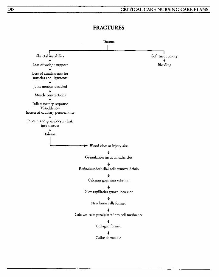
298 CRITICAL CARE NURSING CARE PLANS
FRACTURES
Trauma
Skeletal I instability Soft tissue injury 3 I
Loss of weight support Bleeding I
Loss of attachments for muscles and ligaments
3 Joint motion disabled
I Muscle contractions
4 Inflammatory response
Vasodilation Increased capillary permeability
I Protein and granulocytes leak
into tisssues 3
Edema
Blood clots at injury site
I Granulation tissue invades clot
4 Reticuloendothelial cells remove debris
I Calcium goes into solution
I New capillaries grown into clot
I New bone cells formed
I Calcium salts precipitate into cell meshwork
I Collagen formed
3 Callus formation
L
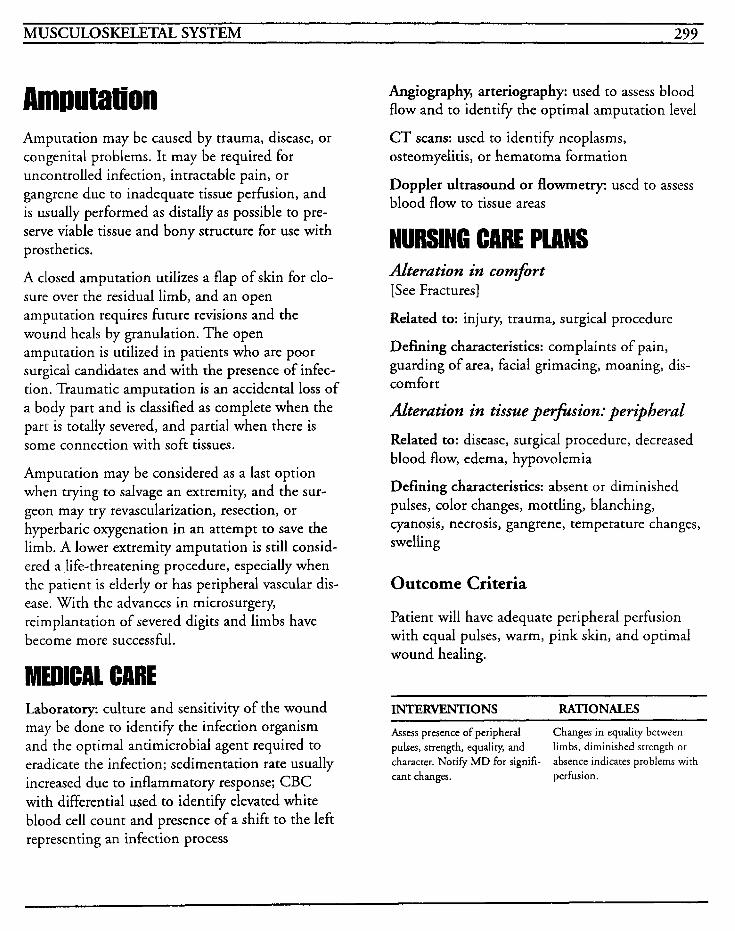
MUSCULOSKELETAL SYSTEM 299
Amputation Amputation may be caused by trauma, disease, or congenital problems. It may be required for uncontrolled infection, intractable pain, or gangrene due to inadequate tissue perfusion, and is usually performed as distally as possible to pre- serve viable tissue and bony structure for use with prosthetics.
A closed amputation utilizes a flap of skin for clo- sure over the residual limb, and an open amputation requires future revisions and the wound heals by granulation. The open amputation is utilized in patients who are poor surgical candidates and with the presence of infec- tion. Traumatic amputation is an accidental loss of a body part and is classified as complete when the part is totally severed, and partial when there is some connection with soft tissues.
Amputation may be considered as a last option when trying to salvage an extremity, and the sur- geon may try revascularization, resection, or hyperbaric oxygenation in an attempt to save the limb. A lower extremity amputation is still consid- ered a life-threatening procedure, especially when the patient is elderly or has peripheral vascular dis- ease. With the advances in microsurgery, reimplantation of severed digits and limbs have become more successful.
MEDICAL CARE Laboratory. culture and sensitivity of the wound may be done to identify the infection organism and the optimal antimicrobial agent required to eradicate the infection; sedimentation rate usually increased due to inflammatory response; CBC with differential used to identify elevated white blood cell count and presence of a shift to the left representing an infection process
Angiography, arteriography: used to assess blood flow and to identify the optimal amputation level
CT scans: used to identify neoplasms, osteomyelitis, or hematoma formation
Doppler ultrasound or flowmetry: used to assess blood flow to tissue areas
NURSING CARE PWNS Alteration in comfort [See Fractures]
Related to: injury, trauma, surgical procedure
Defining characteristics: complaints of pain, guarding of area, facial grimacing, moaning, dis- comfort
Alteration in tissue petfksion: peripheral
Related to: disease, surgical procedure, decreased blood flow, edema, hypovolemia
Defining characteristics: absent or diminished pulses, color changes, mottling, blanching, cyanosis, necrosis, gangrene, temperature changes, swelling
Outcome Criteria
Patient will have adequate peripheral perfusion with equal pulses, warm, pink skin, and optimal wound healing.
INTERVENTIONS RATIONALES ~ ~~
Assess presence of peripheral pulses, strength, equality, and character. Notify MD for signifi- cant changes. perfusion.
Changes in equality between limbs, diminished strength or absence indicates problems with
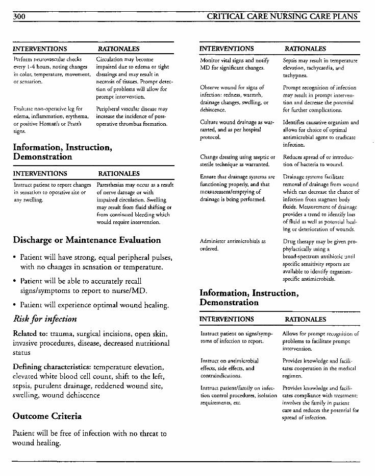
300 CRITICAL CARE NURSING CARE PLANS
INTERVENTIONS RATIONALES INTERVENTIONS RATIONALES Perform neurovascular checks every 1-4 hours, noting changes in color, temperature, movement, or sensation.
Circulation may become impaired due to edema or tight dressings and may result in necrosis of tissues. Prompt detec- tion of problems will allow for prompt intervention.
Peripheral vascular disease may increase the incidence of post- operative thrombus formation.
Evaluate non-operative leg for edema, inflammation, erythema, or positive Homan’s or Pratt’s signs.
Information, Instruction, Demonstration
INTERVENTIONS RATIONALES Instrucr patient to report changes in sensarion to operative site or any swelling. impaired circulation. Swelling
Paresthesias may occur as a result of nerve damage or with
may result from fluid shifting or from continued bleeding which would require intervention.
Discharge or Maintenance Evaluation
Patient will have strong, equal peripheral pulses, with no changes in sensation or temperature.
+ Patient will be able to accurately recall signs/symptoms to report to nurse/MD.
Patient will experience optimal wound healing.
Risk for infection
Related to: trauma, surgical incisions, open skin, invasive procedures, disease, decreased nutritional status
Defining characteristics: temperature elevation, elevated white blood cell count, shift to the left, sepsis, purulent drainage, reddened wound site, swelling, wound dehiscence
Outcome Criteria
Monitor vital signs and notify MD for significant changes.
Sepsis may result in temperature elevation, tachycardia, and tachypnea.
Prompt recognition of infection may result in prompt interven- tion and decrease the porential for further complications.
Identifies causative organism and allows for choice of optimal
infection.
Reduces spread of or introduc- tion of bacteria to wound.
Observe wound for signs of infection: redness, warmth, drainage changes, swelling, or d e h i s c en c e ,
Culture wound drainage as war- ranted, and as per hospital protocol. antimicrobial agent to eradicate
Change dressing using aseptic or sterile technique as warranted.
Ensure that drainage systems are functioning properly, and that measurementlemptying of drainage is being performed.
Drainage systems facilitate removal of drainage from wound which can decrease the chance of infection from stagnant body fluids. Measurement of drainage provides a trend to identiG loss of fluid as well as potential heal- ing or deterioration of wounds.
Drug rherapy may be given pro- phylactically using a broad-spectrum antibiotic until specific sensitivity reports are available to identify organism- specific antimicrobials.
Administer antimicrobials as ordered.
Information, Instruction, Demonstration
INTERVENTIONS RATIONALES
Instruct patient on signslsymp- toms of infection to report,
Instruct on antimicrobial effects, side effects, and contraindications.
Instruct patient/family on infec- tion control procedures, isolation requirements, etc.
Allows for prompt recognition of problems to facilitate prompt intervention.
Provides knowledge and facili- tates cooperation in the medical regimen.
Provides knowledge and facili- tates compliance with treatment; involves rhe family in parienr care and reduces the potential for spread of infection.
Patient will be free of infection with no threat to wound healing.
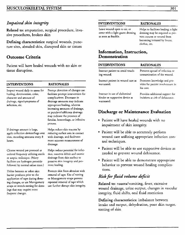
MUSCULOSKELETAL SYSTEM 301
Impaired skin integrity
Related to: amputation, surgical procedure, inva- sive procedures, broken skin
Defining characteristics: surgical wounds, punc- ture sites, abraded skin, disrupted skin or tissues
Outcome Criteria
Patient will have healed wounds with no skin or tissue disruption.
INTERVENTIONS RATIONALES
Inspect wound daily to assess for healing, deterioration, color, character and amount of drainage, signslsyrnptoms of infection, etc.
If drainage amount is large, apply collection deviceslbags over sites, recording amounts every 8 hours.
Cleanse wound per protocol at ordered frequency utilizing sterile or aseptic technique. (Many facilities use hydrogen peroxide followed by normal saline rinse.)
Utilize benzoin or other slun barrier products prior to the application of tape during dress- ing changes, or use Montgomery straps or stretch netting for dress- ings that may require more frequent changes.
Prompt detection of changes can facilitate prompt intervention for complications. Decreases in drainage amounts may indicate appropriate healing, whereas increasing amounts of drainage, or purulentlodiferous drainage may indicate the presence of fistulas, hemorrhage, or infective process.
Helps reduce skin trauma by reducing surface area in contact with drainage, and facilitates more accurate measurement of drainage.
Helps reduce potential for infec- tion: removes debris and caustic drainage from skin surface to preserve skin integrity and pro- mote healing.
Protects skin from abrasion with removal of tape. Use of netting or Montgomery straps prevent repeated removal of tape which can further disrupt skin integrity.
~~
INTERVENTIONS RATIONALES ~~~ ~
Leave wound open to air, or cover with a light gauze dressing as soon as feasible.
Helps to facilitate healing; a light dressing may be required to pre- vent sutures or wound from becoming irritated by linens, clothes, etc.
Information, Instruction, Demonstration
INTERVENTIONS RATIONALES
Instruct patient to avoid touch- ing wound.
Instruct patient in wound care as warranted.
Prevents spread of infection or contamination of the wound.
Promotes knowledge and pro- vides for patient involvement in his care.
Provides additional support for incisions at risk of dehiscence.
Instruct in use of abdominal binder or supportive device as warranted.
Discharge or Maintenance Evaluation
Patient will have healed wounds with no impairment of skin integrity.
Patient will be able to accurately perform wound care utilizing appropriate infection con- trol techniques.
Patient will be able to use supportive devices as needed to prevent wound dehiscence.
Patient will be able to demonstrate appropriate behavior to prevent wound healing complica- tions.
Risk for fluid volume deficit
Related to: nauseahomiting, fever, excessive wound drainage, urine output, changes in vascular integrity, fluid shifts, oral fluid restriction
Defining characteristics: imbalance between intake and output, dehydration, poor skin turgor, tenting of skin
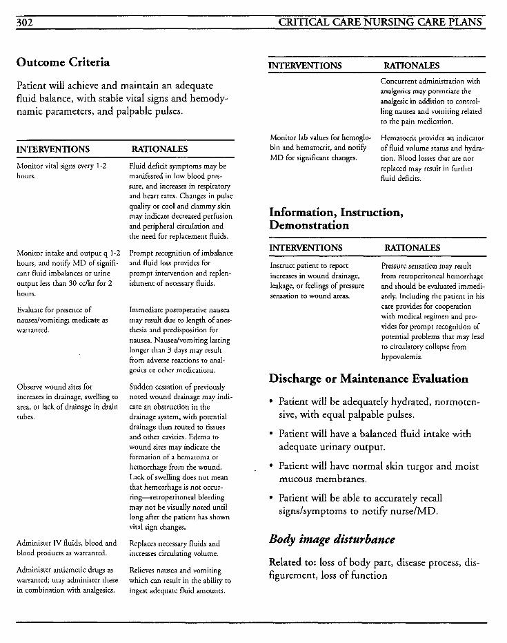
702 CRITICAL CARE NURSING CARE PLANS
Outcome Criteria
Patient will achieve and maintain an adequate fluid balance, with stable vital signs and hemody- namic parameters, and palpable pulses.
~~ ~
INTERVENTIONS RATIONALES
Monitor viral signs every 1-2 hours.
Monitor intake and output q 1-2 hours, and notify MD of signifi- cant fluid imbalances or urine output less than 30 cclhr for 2 hours.
Evaluate for presence of nausedvomiting; medicate as warranted.
Observe wound sites for increases in drainage, swelling to area, or lack of drainage in drain tubes.
Administer IV fluids, blood and blood products as warranted.
Administer antiemetic drugs as warranted; may administer these in combination with analgesics.
Fluid deficit symptoms may be manifested in low blood pres- sure, and increases in respiratory and heart rates. Changes in pulse quality or cool and clammy skin may indicate decreased perfusion and peripheral circularion and the need for replacement fluids.
Prompt recognition of imbalance and fluid loss provides for prompt intervention and replen- ishment of necessary fluids.
Immediate postoperative nausea may result due to length of anes- thesia and predisposition for nausea. Nausedvomiting lasting longer than 3 days may result from adverse reactions to anal- gesics or other medications.
Sudden cessation of previously noted wound drainage may indi- cate an obstruction in the drainage system, with potential drainage then routed to tissues and other cavities. Edema to wound sites may indicate the formation of a hematoma or hemorrhage from the wound. Lack of swelling does not mean that hemorrhage is not occur- ring-retroperitoneal bleeding may not be visually noted until long after the patient has shown vital sign changes.
Replaces necessary fluids and increases circulating volume.
Relieves nausea and vomiting which can result in the ability to ingest adequate fluid amounts.
INTERVENTIONS RATIONALES
Monitor lab values for hemoglo- bin and hernatocrit, and notify MD for significant changes.
Concurrent administration with analgesics may potentiare the analgesic in addition to control- ling nausea and vomiting related to the pain medication.
Hematocrit provides an indicator of fluid volume status and hydra- tion. Blood losses that are not replaced may result in further fluid deficits.
Information, Instruction, Demonstration
~~ ~~~
INTERVENTIONS RATIONALES
Instruct patient to report increases in wound drainage, leakage, or feelings of pressure sensation to wound areas.
Pressure sensation may result from retroperitoneal hemorrhage and should be evaluated immedi- ately. Including the patient in his care provides for cooperation with medical regimen and pro- vides for prompt recognition of potential problems that may lead to circulatory collapse from hypovolemia.
Discharge or Maintenance Evaluation
Patient will be adequately hydrated, normoten- sive, with equal palpable pulses.
Patient will have a balanced fluid intake with adequate urinary output.
Patient will have normal skin turgor and moist mucous membranes.
Patient will be able to accurately recall signs/symptoms to notify nurse/MD.
Body image disturbance
Related to: loss of body part, disease process, dis- figurement, loss of function
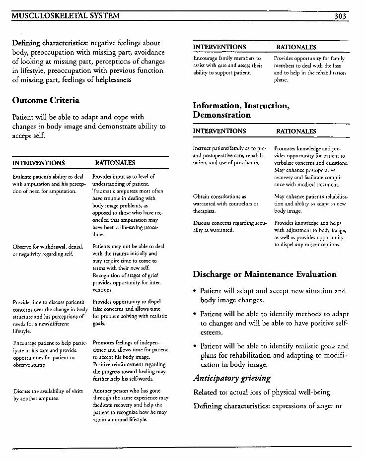
MUSCULOSKELETAL SYSTEM 303
Defining characteristics: negative feelings about body, preoccupation with missing part, avoidance of looking at missing part, perceptions of changes in lifestyle, preoccupation with previous function of missing part, feelings of helplessness
Outcome Criteria
Patient will be able to adapt and cope with changes in body image and demonstrate ability to accept self.
~~
INTERVENTIONS RATIONALES
Evaluate patient’s ability to deal with amputation and his percep- tion of need for amputation.
Observe for withdrawal, denial, or negativity regarding self.
Provide time to discuss patient’s concerns over the change in body structure and his perceptions of needs for a new/different lifestyle.
Encourage patient to help partic- ipate in his care and provide opportunities for patient to observe stump.
Discuss the availability of visits by another amputee.
Provides input as to level of understanding of patient. Traumatic amputees most often have trouble in dealing with body image problems, as opposed to those who have rec- onciled that amputation may have been a life-saving proce- dure.
Patients may not be able to deal with the trauma initially and may require time to come to terms with their new self. Recognition of stages of grief provides opportunity for inter- ventions.
Provides opportunity to dispel false concerns and allows time for problem solving with realistic goals.
Promotes feelings of indepen- dence and allows time for patient to accept his body image. Positive reinforcement regarding the progress toward healing may hrther help his self-worth.
Another person who has gone through the same experience may facilitate recovery and help the patient to recognize how he may attain a normal lifestyle.
INTERVENTIONS RATIONALES Encourage family members to assist with care and assess their ability to support patient.
Provides opportunity for family members to deal with the loss and to help in the rehabilitation phase.
Information, Instruction, Demonstration
INTERVENTIONS RATIONALES
Instruct patientlfamily as to pre- and postoperative care, rehabili- tation, and use of prosthetics.
Promotes knowledge and pro- vides opportuniry for patient to verbalize concerns and questions, May enhance postoperative recovery and facilitate compli- ance with medical treatment.
May enhance patient’s rehabilita- tion and ability to adapt to new
Obtain consultations as warranted with counselors or therapists. body image.
Discuss concerns regarding sexu- ality as warranted.
Provides knowledge and helps with adjustment to body image, as well as provides opportunity to dispel any misconceptions.
Discharge or Maintenance Evaluation
Patient will adapt and accept new situation and body image changes.
Patient will be able to identify methods to adapt to changes and will be able to have positive self- esteem.
Patient will be able to identify realistic goals and plans for rehabilitation and adapting to modifi- cation in body image.
Anticipatory grieving
Related to: actual loss of physical well-being
Defining characteristics: expressions of anger or
304 CRITICAL CARE NURSING CARE PLANS
distress at loss, crying, sadness, guilt, alterations in sleep patterns, activity, eating or libido
Outcome Criteria
Patient will be able to express feelings appropriately and work through the stages of grief and grieving.
INTERVENTIONS RATIONALES
Evaluate emotional status.
Identify patient‘s stage in the grieving process.
Provide factual information to patientlfamily in regard to the diagnosis/prognosis. Do not give false reassurance.
Anxiety, depression, and anger are normal reactions to loss of body parts. The patient may progress through the various stages of grief at their own rate and changes may be related to their physical condition as well.
Shock may be the initial response associated with the amputation, especially if it was traumatic. The patient may be so acutely ill that he is unable to express his feel- ings and concerns. Denial may initially be useful for patient’s ability to cope with the injury, but continued denial may impair the patient’s ability to effectively deal with the problem. Anger may be expressed either verbally, non-verbally, or physically, and the patient may displace his anger by placing blame. Depression may last from weeks to years and acceptance and sup- port for these feelings will facilitate recovery.
Family may be where the initial instruction is directed if the patient’s awareness is diminished due to his injury. The final out- come of a patient’s injuries may not be initially known and so information should be kept simple.
INTERVENTIONS RATIONALES
Assist patient to focus on needs he has now before changing focus to long-term goals. Encourage patient to take con- trol in decisions regarding his care whenever possible.
Provide acceptance of anger, hopelessness, and depression, but set limits on unacceptable behav- ior when warranted.
Reduces frustration of facing an uncertain future, and allows the patient some control in dealing with current problems.
Acceptance of the patient‘s feel- ings acknowledges him as being worthwhile and a non-judgmen- ral attitude is important in establishing trust and care. Limits may be needed to protecr the patient and others from vio- lent behavior while allowing the patient to express his negative feelings.
Physical and spiritual distress will be faced by the patient and hls family and they will require long- term assistance and counseling in order to cope with the changes required by this injury.
Provide consultation with thera- pists, social workers, or minister as warranted.
Discharge or Maintenance Evaluation
Patient will be able to progress through the vari- ous stages of grief and grieving effectively.
Patient will be able to express his feelings and concerns appropriately without unacceptable violent be havior.
Patient will be able to access community resources for long-term counseling and assistance to deal with his injury.
Patient and family will be able to gain adequate support throughout the grieving process.
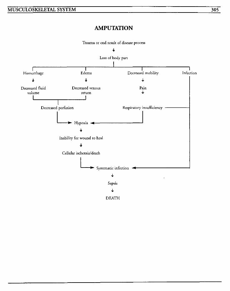
MUSCULOSKELETAL SYSTEM 305
AMPUTATION
Trauma or end result of disease process
4 Loss of body part
I I I I I
Hemorrhage Edema Decreased mobility Infection
J 4 Decreased fluid Decreased venous
volume return
4 Pain 4
Decreased perfusion Respiratory insufficiency
L Hypoxia I 4
Inability for wound to heal
4 Cellular ischemia/death
Systematic infection 4
4 Sepsis
4 DEATH

This Page Intentionally Left Blank
~
MUSCULOSKELETAL SYSTEM 307
Fat Embolism A fat embolism usually occurs in patients with multiple fractures or fractures that involve the long bones or pelvis, when particles of bone marrow, tissue fat droplets, or combinations of platelets and free fatty acids are released and migrate to the lungs or brain. Embolization can occur within the first 24 hours up to 72 hours after injury.
The first signs/symptoms are usually changes in the mental status, with apprehension, confusion and restlessness noted. Petechiae to the chest, anterior axillae, shoulders, conjunctiva and buccal membranes occur due to capillary occlusion and are usually seen later. Respiratory distress with hypoxemia and hypoxia, pulmonary edema, and interstitial pneumonitis occur. The pulse rate increases, temperature elevates above 1 00 degrees and PaO, decreases.
MEDICAL CARE Laboratory: serum lipase is tion rate is increased; urine presence of free fat
elevated , sedimenta- tests used to evaluate
Arterial blood gases: used to evaluate acid-base balance, presence of adequate oxygenation, and response to oxygen therapy
Electrocardiogram: used to evaluate changes in heart rate as well as cardiac changes, such as inver- sion of T waves and prominence of S wave in lead I showing myocardial and right ventricular failure
Corticosteroids: use is controversial, but may decrease inflammation and swelling
Heparin: use is controversial, but low dose heparin may be used to clear lipemic plasma and stimulate lipase activity
Dextran: low molecular weight dextran may be used to alter platelets and decrease intimal adhe- sions
X-rays: serial chest x-rays are used to evaluate pul- monary improvement or deterioration; x-rays of the bones involved in injury are used to evaluate healing process or alignment problems
NURSING CARE PLANS Impaired gas exchange
Related to: altered blood flow due to embolism, shunting
Defining characteristics: abnormal acid-base bal- ance, hypoxemia, hypoxia, tachypnea, tachycardia, air hunger, dyspnea, cyanosis, decreased oxygen saturation
Outcome Criteria
Patient will be able to achieve and maintain ade- quate respiratory function with arterial blood gases within normal ranges and with no evidence of res- piratory distress.
INTERVENTIONS RATIONALES
Monitor vital signs, especially respiratory status; assess for dysp- nea, use of accessory muscles, retractions. nasal flaring, or stri- dor.
Observe for changes in mental status, irritability, apprehension, or confusion.
Monitor pulse oxirnetry for oxygen saturation and notify MD for levels below 90%.
~~
Dyspnea and tachypnea may be early signs of respiratory insuffi- ciency. Other signs usually result from advanced respiratory dis- tress, and all require prompt intervention.
Changes in mental status often are the very first signs in respira- tory insufficiency. As hypoxernia and acidosis worsen, the level of consciousness may deteriorate to the point of lethargy or stupor.
Oximetry may provide early warning of decreasing oxygena- tion and allow for prompt and timely intervention. In patients
308 CRITICAL CARE NURSING CARE PLANS
INTERVENTIONS RATIONALES INTERVENTIONS RATIONALES
Administer oxygen via nasal can- nula or mask as warranted.
Obtain ABGs as warranted.
Auscultate breath sounds for changes in equality and for pres- ence of crackles (rales), rhonchi, wheezing, inspiratory stridor or crowing, or hyperresonant sounds.
Observe for presence of blood in spurum.
Observe for petechiae CO chest, axillae, buccal mucosa, and con- junctiva.
Encourage coughing, deep breathing exercises, and use of incentive spirometer.
who have decreased peripheral circulation however, the accuracy of pulse oximetry will be com- promised and cannot be relied on totally.
Provides supplemental oxygcn and increases available supply of oxygen to ensure optimal tissue oxygenation.
Decreased PaO, and increased PaCO, indicate impending respi- ratory failure and impaired gas exchange.
Adventitious breath sounds may indicate progression of respira- tory insufficiency. Inspiratory crowing may indicate upper airway edema frequently seen with fat emboli.
May indicate hemoptysis that occurs with pulmonary embolism.
Petechiae to these areas are fre- quently seen with fat emboli, and may occur 2-5 days after injury.
Improves alveolar ventilation/ oxygenation and helps to mini- mize atelectasis.
Information, Instruction, Demonstration
INTERVENTIONS RATIONALES
Prepare for placement on ventila- tor as warranted.
Use great care in repositioning patient especially dxing the first days post-injury.
Deteriorating respiratory status may require mechanical ventila- tion to facilitate oxygenation. [See Mechanical Ventilation Care Plan.]
Gentle handling of injured bones and tissues may prevent the development of a fat embolism.
Monitor lab studies. Patients with far emboli frequenrly have anemia, elevated sedimentation rates, elevated lipase levels, fat in body fluids, hypocalcemia, and thrombocy- topenia.
Some physicians use steroids to prevent and treat fat emboli.
Administer corticosteroids as ordered.
Discharge or Maintenance Evaluation Patient will have no respiratory dysfunction or distress.
Patient will have arterial blood gases within his normal range.
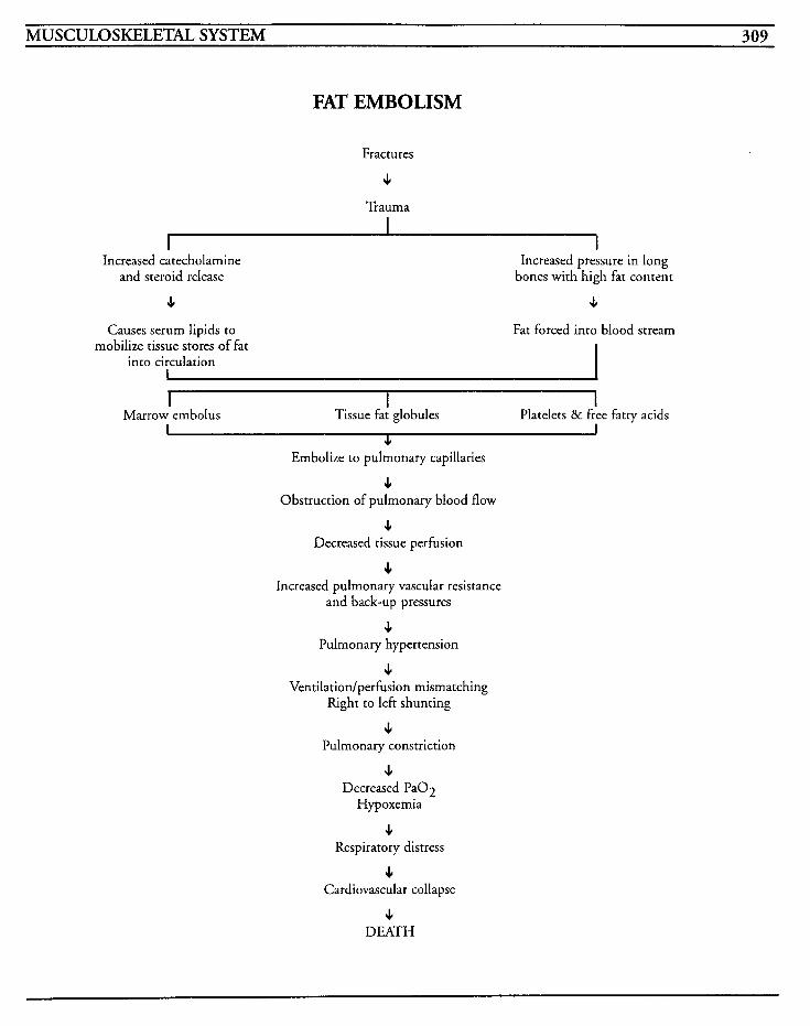
MUSCULOS KELETm SYSTEM 309
FAT EMBOLISM
Fractures
4 Trauma
Increased catecholamine and steroid release
Increased pressure in long bones with high fat content
4 4
Causes serum lipids to mobilize tissue stores of fat
into circulation I
Fat forced into blood stream
I I I Platelets & free fatty acids Marrow embolus Tissue fat globules
I I c Embolize to pulmonary capillaries
4 Obstruction of pulmonary blood flow
4 Decreased tissue perfusion
4 Increased pulmonary vascular resistance
and back-up pressures
4 Pulmonary hypertension
4 Ventilation/perfusion mismatching
Right to left shunting
4 Pulmonary constriction
4 Decreased Pa02
Hypoxemia
4 Respiratory distress
4 Cardiovascular collapse
4 DEATH

This Page Intentionally Left Blank
INTEGUMENTARY SYSTEM 31 1
Frost bite/Hypothermia
Malignant Hyperthermia
BurndTherrnal Injuries

This Page Intentionally Left Blank
INTEGUMENTARY SYSTEM 313
FrostbiteAiyuotherrnia Injuries from overexposure to cold, either air or water, occur in two types-localized injuries, such as frostbite, and systemic injuries, such as hypothermia. Untreated, both may be fatal.
Frostbite occurs after exposure to cold temperatures, usually below freezing. The severity of the injury is dependent on the,amount of body heat lost, age and exacerbating factors such as wind chill, presence of wet clothing, and impair- ment of the circulatory status.
In frostbite, ice crystals form in the tissue fluids in and between the cells, causing injury to the red blood cells, which then develop sludging, and vas- cular damage. Blood is shunted to the heart and the brain. Skin is cold, hard, ashen white and numb, and with rewarming, becomes splotchy red or grayish in color, edematous, and very painful.
Frostbite can be either superficial, affecting skin and subcutaneous tissues, or deep, extending below subcutaneous tissues. With deep frostbite, the skin becomes white until thawed and then it becomes purplish-blue, with painful skin blisters, tissue necrosis, and development of gangrene when the tissue dies. At this point, amputation of the extremity may be required.
The most frequently seen sites that are involved with frostbite are the nose, ears, hands, and lower extremities. The goal of treatment is to restore body temperature to normal and prevent vascular damage to tissues. Supportive care is also impor- tant in restoring electrolyte imbalances and preventing hypovolemia.
Hypothermia occurs when the body's core temper- ature drops below 95" Farenheit and is noted by lethargy, mental dullness, decreasing level of con- sciousness, visual and auditory hallucinations, decreases in respirations and heart rate, and coma.
The core temperature may be as low as 80" Fahrenheit and below 900, the body loses its self- warming mechanisms.
Hypothermia may also preclude successful resusci- tation. Cardiac arrest is difficult to overcome if the core temperature is less than 85" Fahrenheit due to the increased ventricular fibrillation threshold.
Treatment is aimed at rewarming the body to increase the core temperature to adequate ranges, and to preserve organ and tissue viability.
MEDICAL CARE Laboratory: CBC may indicate infection with shift to left; electrolytes will be required to restore balance from fluid shifts
IV fluids: used to restore circulating volume and prevent dehydration, and may be used to assist with rewarming
Dextran: low molecular weight dextran may be used to improve microcirculation to tissues
Reserpine: may be used to decrease sludging from injured cells and tissues
Antibiotics: may be necessary to treat infection if patient has open wounds or systemic infection
Analgesics: morphine and other drugs may be used to relieve severe pain from cold injuries; aspirin may be used to decrease platelet aggrega- tion and sludging
Surgery: fasciotomy may be required to reduce tissue pressure caused from edema; amputation may be necessary for gangrenous injuries, or debridement may be required for necrotic tissues
Dialysis: peritonea1 or hemodialysis may be used depending on severity of injury, in order to rewarm body
314 CRITICAL CARE NURSING CARE PLANS
Rewarming techniques: warming blankets, warmed solutions for chest lavage or bladder irri- gation, and warmed IV solutions may be utilized to increase temperature
NURSING CARE PLANS Inefective thermorepltion
Related to: exposure to cold, suppressed shivering response
Defining characteristics: temperature below 95" Fahrenheit, cold skin, mottling, cyanosis, pallor, poor judgment, apathy, decreased mental ability, level of consciousness changes, coma, lack of shiv- ering, cardiopulmonary arrest, anuria, oliguria, decreased peripheral perfusion
Outcome Criteria
Patient will achieve and maintain an acceptable temperature with no complications.
INTERVENTIONS RATIONALES
Obtain baseline temperature, and monitor every 15 minutes until stable.
Rewarm patient per hospital protocol. (Whole body or partial immersion into water that is 99- 105 degrees, hypothermia blankets, gastric lavage with warmed solutions, peritonea1 or hemodialysis, and IV infusions that are warmed are some meth- ods currently used.)
Temperatures below 90 degrees result in suppression of normal body mechanisms to self-warm- ing. Rewarming that is done too rapidly may cause peripheral vasodilation and may actually impede rewarming efforts.
Early rewarming decreases tissue damage from ice crystal forma- tion, and helps to decrease cardiac instability and predisposi- tion to ventricular fibrillation.
INTERVENTIONS RATIONALES
Observe for mental changes and return of shivering response.
Shivering is suppressed at tem- peratures below 90 degrees F and is che body's normal response to facilitate self-warming. Patients have decreased mental abilities and levels of consciousness dependent on severity of hypothermidinjury, with hypoxia and hypoxemia occur- ring due to decreased perfusion.
Information, Instruction, Demonstration
INTERVENTIONS RATIONALES ~
Instruct patient/family on appro- priate procedures for rewarming. anxiety.
Provides knowledge and reduces
Discharge or Maintenance Evaluation
Patient will be normothermic, with stable vital signs.
Patient will be awake, alert, and oriented, with no alterations in abilities.
Patient will be able to maintain thermoregula- tion.
Patient will exhibit no complications from hypothermia.
Alteration in tissue pe@sion: peripheral, cerebral, curdiopulmonuy, renal, gastroin- testinul
Related to: exposure to cold temperatures, frozen body parts, hypothermia, tissue necrosis, sludging of red blood cells, tissue ischemia
Defining characteristics: skin mottling, grayish skin color, purplish-blue color, cold skin, burning, tingling, numbness, pain, skin blisters, gangrene, diminished or absent pulses, decreased capillary
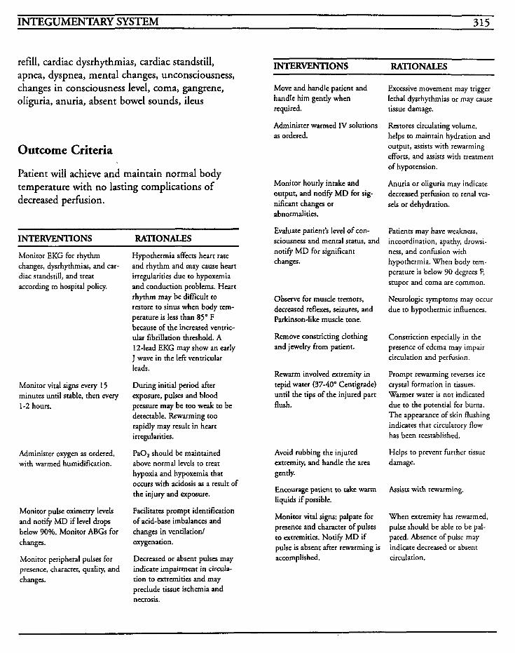
INTEGUMENTARY SYSTEM 315
refill, cardiac dysrhythmias, cardiac standstill, apnea, dyspnea, mental changes, unconsciousness, changes in consciousness level, coma, gangrene, oliguria, anuria, absent bowel sounds, ileus
Outcome Criteria
Patient will achieve and maintain normal body temperature with no lasting complications of decreased perfusion.
INTERVENTIONS RATIONALES
Monitor EKG for rhythm changes, dysrhythmias, and car- diac standstill, and treat according to hospital policy.
Monitor vital signs every 15 minutes until stable, then every 1-2 hours.
Administer oxygen as ordered, with warmed humidification.
Monitor pulse oximetry levels and notif) MD if level drops below 90%. Monitor ABGs for changes.
Monitor peripheral pulses for presence, character, quality, and changes.
Hypothermia affects heart rate and rhythm and may cause heart irregularities due to hypoxemia and conduction problems. Heart rhythm may be difficult to restore to sinus when body tem- perature is less than 85" F because of the increased ventric- ular fibrillation threshold. A 12-lead EKG may show an early J wave in the left ventricular leads.
During initial period after exposure, pulses and blood pressure may be too weak to be detectable. Rewarming too rapidly may result in heart irregularities.
PaOz should be maintained above normal levels to treat hypoxia and hypoxemia that occurs with acidosis as a result of the injury and exposure.
Facilitates prompt identification of acid-base imbalances and changes in ventilation/ oxygenation.
Decreased or absent pulses may indicate impairment in circula- tion to extremities and may preclude tissue ischemia and necrosis.
INTERVENTIONS RATIONALES
Move and handle patient and handle him gently when required.
Administer warmed IV solutions as ordered.
Monitor hourly intake and output, and notify M D for sig- nificant changes or abnormalities.
Evaluate patient's level of con- sciousness and mental status, and notify M D for significant changes.
Observe for muscle tremors, decreased reflexes, seizures, and Parkinson-like muscle tone.
Remove constricting clothing and jewelry from patient.
Rewarm involved extremity in tepid water (37-40" Centigrade) until the tips of the injured part flush.
Avoid rubbing the injured extremity, and handle the area gently.
Encourage patient to take warm liquids if possible.
Monitor vital signs; palpate for presence and character of pulses to extremities. Notify MD if pulse is absent after rewarming is accomplished.
Excessive movement may trigger lethal dysrhythmias or may cause tissue damage.
Restores circulating volume, helps to maintain hydration and output, assists with rewarming efforts, and assists with treatment of hypotension.
Anuria or oliguria may indicate decreased perfusion CO renal ves- sels or dehydration.
Patients may have weakness, incoordination, apathy, drowsi- ness, and confusion with hypothermia. When body tem- perature is below 90 degrees F, stupor and coma are common.
Neurologic symptoms may occur due to hypothermic influences.
Constriction especially in the presence of edema may impair circulation and perfusion.
Prompt rewarming teverses ice crystal formation in tissues. Warmer water is not indicated due to the potential for burns. The appearance of skin flushing indicates that circulatory flow has been reestablished.
Helps to prevent further tissue damage.
Assists with rewarming.
When extremity has rewarmed, pulse should be able to be pal- pated. Absence of pulse may indicate decreased or absent circulation.
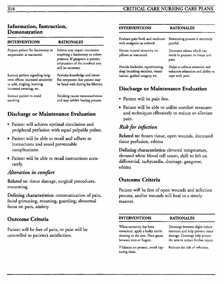
316 CRITICAL CARE NURSING CARE PLANS
Information, Instruction, Demonstration
INTERVENTIONS RATIONALES
Prepare patient for fasciotorny or amputation as warranted.
Instruct patient regarding long- term effects: increased sensitivity to cold, tingling, burning, increased sweating, etc.
Instruct patient to avoid smoking.
Ederna may impair circulation requiring a fasciotomy to relieve pressure. If gangrene is present, amputation of the involved area will be necessary.
Provides knowledge and identi- fies symptoms that patient may be faced with during his lifetime.
Smoking causes vasoconstriction and may inhibit healing process.
Discharge or Maintenance Evaluation
Patient will achieve optimal circulation and peripheral perfusion with equal palpable pulses.
Patient will be able to recall and adhere to ’instructions and avoid preventable complications.
Patient will be able to recall instructions accu- rately.
Alteration in comfirt Related to: tissue damage, surgical procedures, rewarming
Defining characteristics: communication of pain, facial grimacing, moaning, guarding, abnormal focus on pain, anxiety
Outcome Criteria
INTERVENTIONS RATIONALES
Evaluate pain level, and medicate with analgesics as ordered.
Elevate injured extremity on pillows as warranted.
Provide backrubs, repositioning, deep breathing exercises, visual- ization, guided imagery, etc.
Rewarming process is extremely painful.
Decreases edema which can result in pressure to tissues and pain.
Helps to refocus attention and enhances relaxation and ability to cope with pain.
Discharge or Maintenance Evaluation
Patient will be. pain free.
Patient will be able to utilize comfort measures and techniques effectively to reduce or alleviate pain.
Risk for infection
Related to: frozen tissue, open wounds, decreased tissue perfusion, edema
Defining characteristics: elevated temperature, elevated white blood cell count, shift to left on differential, tachycardia, drainage, gangrene, edema
Outcome Criteria
Patient will be free of open wounds and infection process, and/or wounds will heal in a timely manner.
INTERVENTIONS RATIONALES
Patient will be free of pain, or pain will be controlled to patient’s satisfaction.
When extremity has been rewarmed, apply a bulky sterile dressing to the area. Place gauze between toes or fingers.
If blisters are present, avoid rup- turing them.
Dressings between digits reduce moisture and help prevent tissue damage. Dressings help protect the area to reduce further injury.
Reduces the risk of infection.
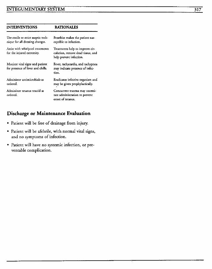
INTEGUMENTARY SYSTEM 317
INTERVENTIONS RATIONALES
Use sterile or strict aseptic tech- nique for all dressing changes.
Assist with whirlpool treatments for the injured extremity.
Frostbite makes the patient sus- ceptible to infection.
Treatments help CO improve cir- culation, remove dead tissue, and help prevent infection.
Fever, tachycardia, and tachypnea may indicate presence of infec- tion.
Eradicates infective organism and
Monitor vital signs and patient for presence of fever and chills.
Administer antimicrobials as ordered. may be given prophyiacticdly.
Administer tetanus toxoid as ordered. tate administration to prevent
Concurrent trauma may necessi-
onset of tetanus.
Discharge or Maintenance Evaluation
Patient will be free of drainage from injury.
Patient will be afebrile, with normal vital signs, and no symptoms of infection.
Patient will have no systemic infection, or pre- ventable complication.
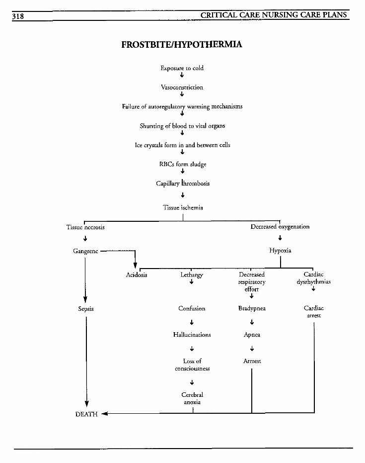
318 CRITICAL CARE NURSING CARE PLANS
FROSTBITE/HYPOTHERMIA
Exposure to cold JI
Vasoconstriction 4
Failure of autoregulatory warming mechanisms 4
Shunting of blood to vital organs 4
Ice crystals form in and between cells c
RBCs form sludge c
Capillary ihrombosis
4 Tissue ischemia
I
4 4
I Decreased Axygenation Tissue necrosis
Gangrene '7 Hypoxia
1 Sepsis
1 I I 1 I
Acidosis Lethargy Decreased Cardiac c respiratory dysrhythmias
effort 4 4
Confusion Bradypnea Cardiac arrest
4 Hallucinations
4 Apnea
4 4
DEATH
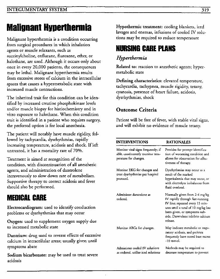
INTEGUMENTARY SYSTEM 319
Malignant Hmerthermia Malignant hyperthermia is a condition occurring from surgical procedures in which inhalation agents or muscle relaxants, such as succinylcholine, enflurane, fluroxene, ether, or halothane, are used. Although it occurs only about once in every 20,000 patients, the consequences may be lethal. Malignant hyperthermia results from excessive stores of calcium in the intracellular spaces that causes a hypermetabolic state with increased muscle contractions.
The inherited trait for this condition can be iden- tified by increased creatine phosphokinase levels and/or muscle biopsy for histiochemistry and in vitro exposure to halothane. When this condition trait is identified in a patient who requires surgery, the preferred option is for local anesthesia.
The patient will notably have muscle rigidity, fol- lowed by tachycardia, dysrhythmias, rapidly increasing temperature, acidosis and shock. If left untreated, it has a mortality rate of 70%.
Treatment is aimed at recognition of the condition, with discontinuation of all anesthetic agents, and administration of dantrolene intravenously to slow down rate of metabolism. Supportive therapy to correct acidosis and fever should also be performed.
MEDICAL CARE Electrocardiogram: used to identify conduction problems or dysrhythmias that may occur
Oxygen: used to supplement oxygen supply due to increased metabolic state
Dantrium: drug used to reverse effects of excessive calcium in intracellular areas; usually given until symptoms abate
Sodium bicarbonate: may be used to treat severe acidosis
Hypothermic treatment: cooling blankets, iced lavages and enemas, infusions of cooled IV solu- tions may be required to reduce temperature
NURSING CARE PLANS Hypertbermiu Related to: reaction to anesthetic agents; hyper- metabolic state
Defining characteristics: elevated temperature, tachycardia, tachypnea, muscle rigidity, tetany, cyanosis, presence of heart failure, acidosis, dysrhythmias, shock
Outcome Criteria
Patient will be free of fever, with stable vital signs, and will exhibit no evidence of muscle tetany.
INTERVENTIONS RATIONALES
Monitor vital signs frequently; if able, continuously monitor tem- perature for changes.
Monitor EKG for changes and treat dysrhythmias per hospital protocol.
Administer dantrolene as ordered.
Monitor ABGs for changes.
Administer cooled IV solutions as ordered, utilize iced solutions
Provides for prompt identifica- tion of worsening condition and allows for observation for effec- tiveness of therapy.
Dysrhythmias may occur as a result of the marked hyperkalemia that may occur, or with electrolyte imbalances from fluid overload.
Normally given from 2-4 mg/kg IV rapidly through fast-running IV line; repeated every 15 min- utes until a total of 10 mg/kg has been given, or symptoms sub- side. Dantrolene inhibits calcium release.
May indicate metabolic or respi- ratory acidosis, and patients frequently have noted base excess -10 mmol.
Methods may be required to decrease remperature to prevenr
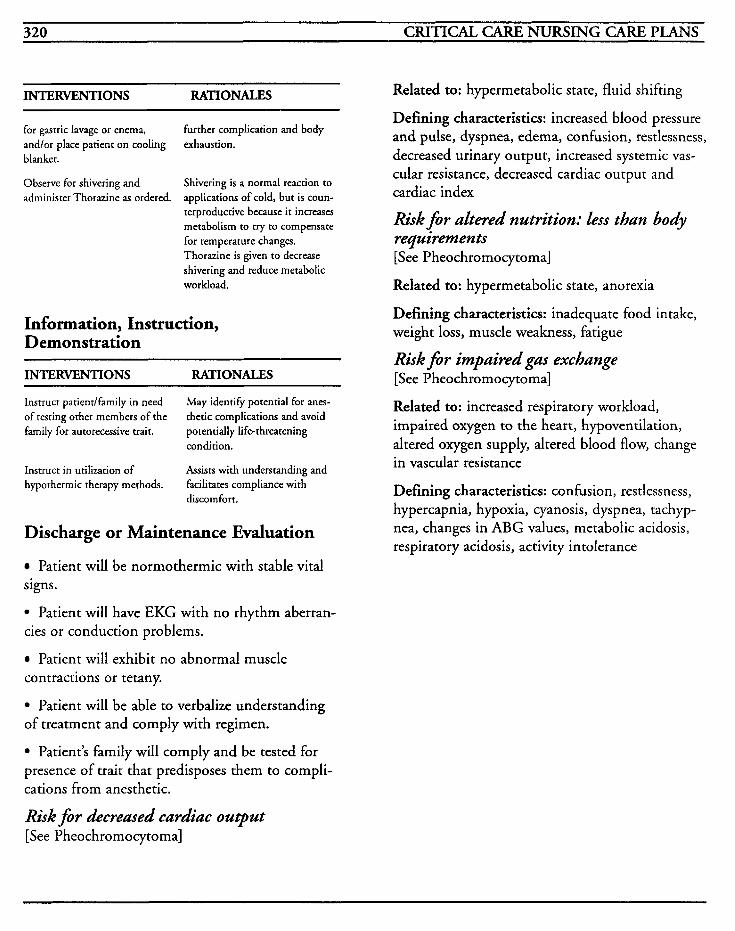
320 CRITICAL CARE NURSING CARE PLANS
INTERVENTIONS RATIONALES
for gastric lavage or enema, and/or place patient on cooling blanket.
Observe for shivering and administer Thorazine as ordered.
further complication and body exhaustion.
Shivering is a normal reaction to applications of cold, but is coun- terproductive because it increases metabolism to try to compensate for temperature changes. Thorazine is given to decrease shivering and reduce metabolic workload.
Information, Instruction, Demonstration
INTERVENTIONS RATIONALES
Instruct patientlfamily in need of testing other members of the family for autorecessive trait.
May identify potential for anes- thetic complications and avoid potentially life-threatening condition.
Instruct in utilization of hypothermic therapy methods.
Assists with understanding and facilitates compliance with discomfort.
Discharge or Maintenance Evaluation
Patient will be normothermic with stable vital signs.
Patient will have EKG with no rhythm aberran- cies or conduction problems.
Patient will exhibit no abnormal muscle contractions or tetany.
Patient will be able to verbalize understanding of treatment and comply with regimen.
Patient’s family will comply and be tested for presence of trait that predisposes them to compli- cations from anesthetic.
Risk for decreased cardiac output [See Pheochromocytoma]
Related to: hypermetabolic state, fluid shifting
Defining characteristics: increased blood pressure and pulse, dyspnea, edema, confusion, restlessness, decreased urinary output, increased systemic vas- cular resistance, decreased cardiac output and cardiac index
Risk for altered nutrition: less than body requirements [See Pheochromocytoma]
Related to: hypermetabolic state, anorexia
Defining characteristics: inadequate food intake, weight loss, muscle weakness, fatigue
Risk for impaired gas exchange [See Pheochromocytoma]
Related to: increased respiratory workload, impaired oxygen to the heart, hypoventilation, altered oxygen supply, altered blood flow, change in vascular resistance
Defining characteristics: confusion, restlessness, hypercapnia, hypoxia, cyanosis, dyspnea, tachyp- nea, changes in ABG values, metabolic acidosis, respiratory acidosis, activity intolerance
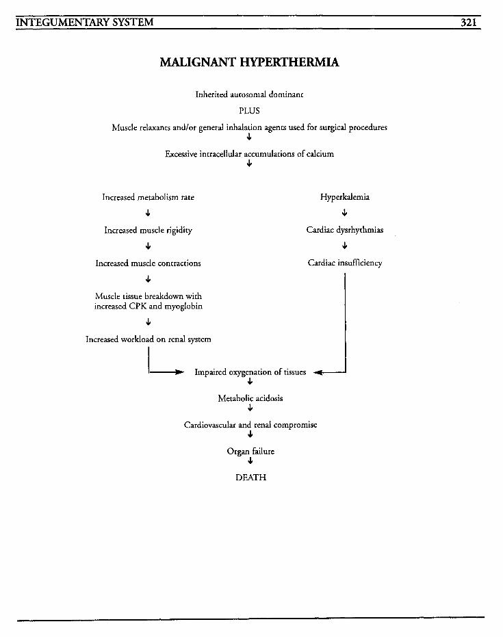
~
INTEGUMENTARY SYSTEM 321
MALIGNANT HYPERTHERMIA
Inherited autosomal dominant
PLUS
Muscle relaxants and/or general inhalation agents used for surgical procedures 4
Excessive intracelldar accumulations of calcium 4
Increased metabolism rate
4 Increased muscle rigidity
4 Increased muscle contractions
Hyperkalemia
4 Cardiac dysrhythmias
4 Cardiac insufficiency
4 Muscle tissue breakdown with increased CPK and myoglobin
c Increased workload on renal system - Impaired oxygenation of tissues -
4 Metabolic acidosis
4 Cardiovascular and renal compromise
J,
Organ failure J,
DEATH

This Page Intentionally Left Blank
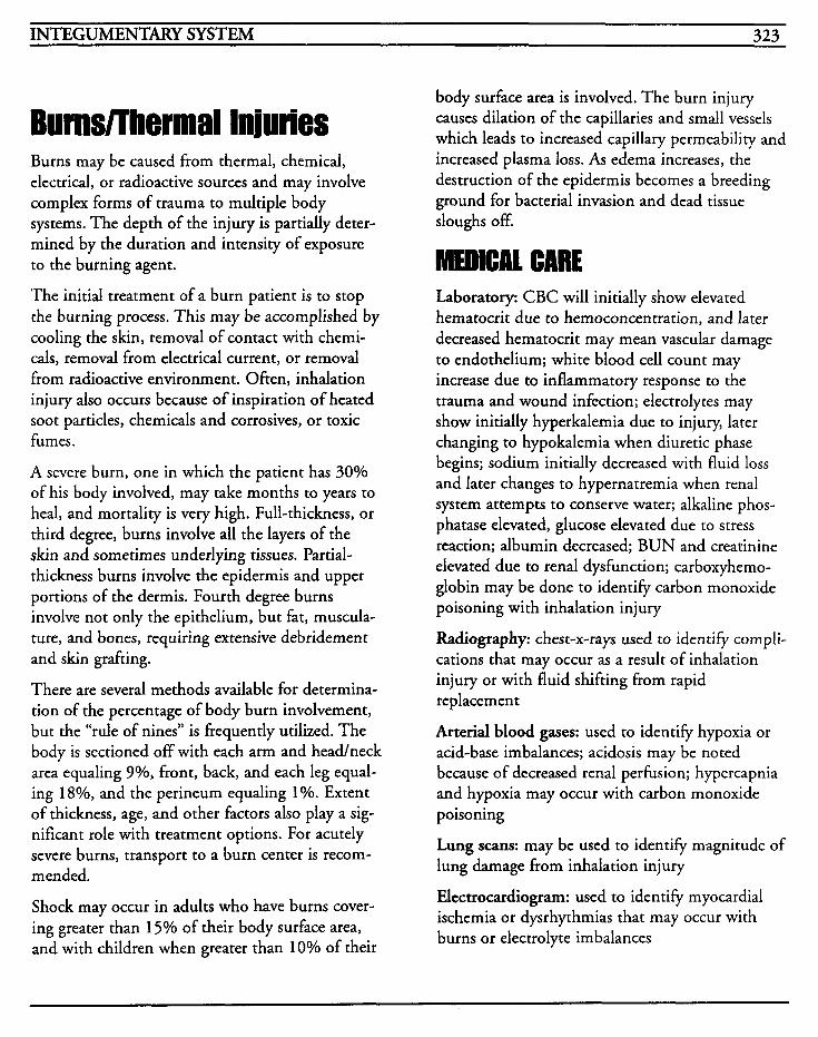
INTEGUMENTARY SYSTEM 323
Burns/Thermal Injuries Burns may be caused from thermal, chemical, electrical, or radioactive sources and may involve complex forms of trauma to multiple body systems. The depth of the injury is partially deter- mined by the duration and intensity of exposure to the burning agent.
The initial treatment of a burn patient is to stop the burning process. This may be accomplished by cooling the skin, removal of contact with chemi- cals, removal from electrical current, or removal from radioactive environment. Often, inhalation injury also occurs because of inspiration of heated soot particles, chemicals and corrosives, or toxic fumes.
A severe burn, one in which the patient has 30% of his body involved, may take months to years to heal, and mortality is very high. Full-thickness, or third degree, burns involve all the layers of the skin and sometimes underlying tissues. Partial- thickness burns involve the epidermis and upper portions of the dermis. Fourth degree burns involve not only the epithelium, but fat, muscula- ture, and bones, requiring extensive debridement and skin grafting.
There are several methods available for determina- tion of the percentage of body burn involvement, but the “rule of nines” is frequently utilized. The body is sectioned off with each arm and head/neck area equaling 3%, front, back, and each leg equal- ing 18%, and the perineum equaling 1%. Extent of thickness, age, and other factors also play a sig- nificant role with treatment options. For acutely severe burns, transport to a burn center is recom- mended.
Shock may occur in adults who have burns cover- ing greater than 15% of their body surface area, and with children when greater than 10% of their
body surface area is involved. The burn injury causes dilation of the capillaries and small vessels which leads to increased capillary permeability and increased plasma loss. As edema increases, the destruction of the epidermis becomes a breeding ground for bacterial invasion and dead tissue sloughs off.
Laboratory: CBC will initially show elevated hematocrit due to hernoconcentration, and later decreased hematocrit may mean vascular damage to endothelium; white blood cell count may increase due to inflammatory response to the trauma and wound infection; electrolytes may show initially hyperkalemia due to injury, later changing to hypokalemia when diuretic phase begins; sodium initially decreased with fluid loss and later changes to hypernatremia when renal system attempts to conserve water; alkaline phos- phatase elevated, glucose elevated due to stress reaction; albumin decreased; BUN and creatinine elevated due to renal dysfunction; carboxyhemo- globin may be done to identify carbon monoxide poisoning with inhalation injury
Radiography: chest-x-rays used to identify compli- cations that may occur as a result of inhalation injury or with fluid shifting from rapid replacement
Arterial blood gases: used to identify hypoxia or acid-base imbalances; acidosis may be noted because of decreased renal perhsion; hypercapnia and hypoxia may occur with carbon monoxide poisoning
Lung scans: may be used to identify magnitude of lung damage from inhalation injury
Electrocardiogram: used to identify myocardial ischemia or dysrhythmias that may occur with burns or electrolyte imbalances

324 CRITICAL CARE NURSING CARE PLANS
Analgesics: required to reduce pain associated with tissue damage and nerve injury
Tetanus toxoid: required to provide immunity against infective organism
Antimicrobials: may be required to treat infection
Surgery: may be required for skin grafting, fasciotomy, debridement, or repair of other injuries
lV fluids: massive amounts of IV fluids may be required for fluid resuscitation immediately post- burn, and will be required for maintenance of fluid balance as shifting occurs
NURSING CARE PLIINS Risk for fluid volume &ficit
Related to: burn injury, loss of fluid through injured surfaces, hemorrhage, increased metabolic state, fluid shifts, third spacing, shock, increased cellular membrane permeability
Defining characteristics: tachycardia, hypotension, changes in mental status, restlessness, decreased urine output, prolonged capillary refill, pallor, mottling, diaphoresis, poor turgor
Outcome Criteria
Patient will achieve and maintain fluid balance with adequate urinary output.
INTERVENTIONS RATIONALES
Monitor vital signs, and notify MD of significant changes or trends.
Hypotension may indicate that the circulating fluid volume is decreased. Changes in vital signs may indicate the amount of blood loss but may not change until loss is greater than 1000 cc. Hypovolemic shock may occur due to hemorrhage, third spac- ing, or coagulopathy.
INTERVENTIONS RATIONALES
Measure hemodynamics if pul- monary artery catheter has been placed. Notify MD for abnormal parameters.
Observe for restlessness, anxiery, mental changes, changes in level of consciousness, or weakness.
Observe for bleeding from all orifices and puncture sites, and for presenceldevelopment of ecchymoses, hematomas, or petechiae.
Monitor intake and output hourly and notify M D for signif- icant imbalances.
Administer IV fluids as ordered. Two IV sites should be main- tained.
CW, or right atrial pressure, gives estimate of fluid volume status. Dehydration may be reflected by CVP of less than 5 , while overhydration may be reflected at levels over 18 cm H 2 0 . Hemodynamic values may help to evaluate the body’s response to the circulating vol- umes.
Changes may reflect the severity of fluid loss.
May indicate impaired coagula- tion, impending or present DIC, or inadequate replacement of clotting factors.
May indicate fluid volume deficit, and establishes a guide for fluid and blood product replacement. Fluid replacement is titrated to ensure urinary output of at least 30-40 cdhr. Myoglobin may discolor the urine red to black, and if present, urinary output should be at leas 75-100 cc/hr to reduce potential for renal tubular necrosis.
Replaces fluid loss, allows for administration of vasoactive drugs, plasma extenders, and emergency medications, as well as the administration. Two sites are recommended to facilitate simultaneous fluid and blood resuscitation in critical settings. Crystalloids, such as Ringer’s lactate, are used during the first 24 hours, then colloids are used because colloids help to mobilize extravascular fluids. Dextrose is usually not given during the first 24 hours after injury because dextrose does not remain in the vascular space where it is needed.
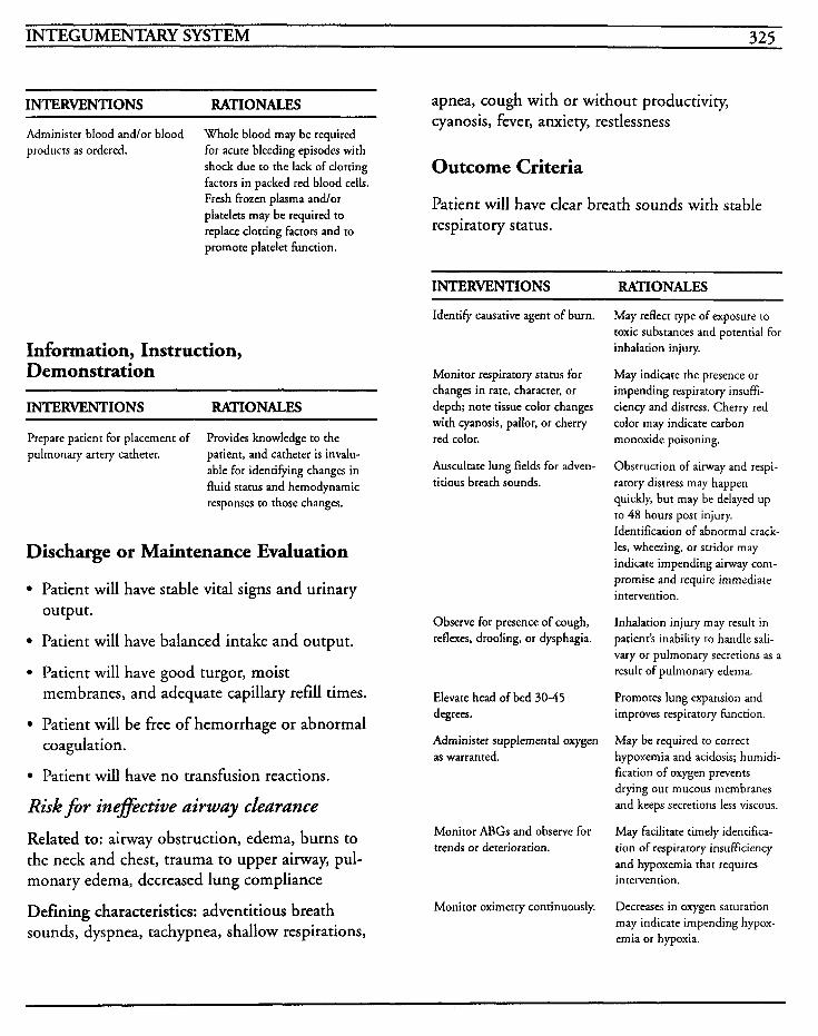
INTEGUMENTARY SYSTEM 325
INTERVENTIONS RATIONALES
Administer blood andlor blood products as ordered.
Whole blood may be required for acute bleeding episodes with shock due to the lack of clotting factors in packed red blood cells. Fresh frozen plasma andlor platelets may be required to replace clotting factors and to promote platelet function.
Information, Instruction, Demonstration
INTERVENTIONS RATIONALES ~~ _ _ _ _ ~ ~~ ~~ ~
Prepare patient for placement of pulmonary artery catheter.
Provides knowledge to the patient, and catheter is invalu- able for identifying changes in fluid status and hemodynamic responses to those changes.
Discharge or Maintenance Evaluation
Patient will have stable vital signs and urinary output.
Patient will have balanced intake and output.
Patient will have good turgor, moist membranes, and adequate capillary refill times.
Patient will be free of hemorrhage or abnormal coagulation.
Patient will have no transfusion reactions.
Risk for ineffective airway clearance
Related to: airway obstruction, edema, burns to the neck and chest, trauma to upper airway, pul- monary edema, decreased lung compliance
Defining characteristics: adventitious breath sounds, dyspnea, tachypnea, shallow respirations,
apnea, cough with or without productivity, cyanosis, fever, anxiety] restlessness
Outcome Criteria
Patient will have clear breath sounds with stable respiratory status.
INTERVENTIONS RATIONALES
Identify causative agent of burn.
Monitor respiratory status for changes in rate, character, or depth; note tissue color changes with cyanosis, pallor, or cherry red color.
Auscultate lung fields for adven- titious breath sounds.
Observe for presence of cough, reflexes, drooling, or dysphagia.
Elevate head of bed 30-45 degrees.
Administer supplemental oxygen as warranted.
Monitor ABGs and observe for trends or deterioration.
Monitor oximetry continuously.
May reflect type of exposure to toxic substances and potential for inhalation injury.
May indicate the presence or impending respiratory insuffi- ciency and distress. Cherry red color may indicate carbon monoxide poisoning.
Obstruction of aimay and respi- ratory distress may happen quickly, but may be delayed up to 48 hours post injury. Identification of abnormal crack- les, wheezing, or stridor may indicate impending airway com- promise and require immediate intervention.
Inhalation injury may result in patient’s inability to handle sali- vary or pulmonary secretions as a result of pulmonary edema.
Promotes lung expansion and improves respiratory function.
May be required to correct hypoxemia and acidosis; humidi- fication of oxygen prevents drying out mucous membranes and keeps secretions less viscous.
May facilitate timely identifica- tion of respiratory insufticiency and hypoxemia that requires intervention.
Decreases in oxygen saturation may indicate impending hypox- emia or hypoxia.
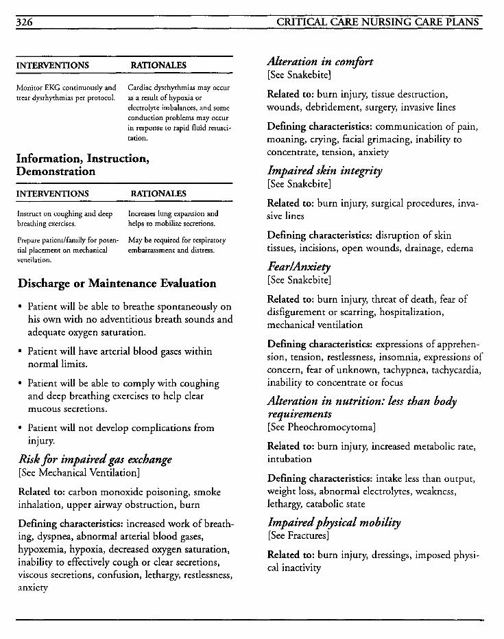
326 CRITICAL CARE NURSING CARE PLANS
INTERVENTIONS RATIONALES
Monitor EKG continuously and treat dysrhythmias per protocol.
Cardiac dysrhythmias may occur as a result of hypoxia or electrolyte imbalances, and some conduction problems may occur in response to rapid fluid resusci- tation.
Information, Instruction, Demonstration
INTERVENTIONS RATIONALES
Instruct on coughing and deep breathing exercises.
Prepare patient/family for poten- tial placement on mechanical ventilation.
Increases lung expansion and helps to mobilize secretions.
May be required for respiratory embarrassment and distress.
Discharge or Maintenance Evaluation
Patient will be able to breathe spontaneously on his own with no adventitious breath sounds and adequate oxygen saturation.
Patient will have arterial blood gases within normal limits.
Patient will be able to comply with coughing and deep breathing exercises to help clear mucous secretions.
Patient will not develop complications from injury.
Risk for impairedgm exchange [See Mechanical Ventilation]
Related to: carbon monoxide poisoning, smoke inhalation, upper airway obstruction, burn
Defining characteristics: increased work of breath- ing, dyspnea, abnormal arterial blood gases, hypoxemia, hypoxia, decreased oxygen saturation, inability to effectively cough or clear secretions, viscous secretions, confusion, lethargy, restlessness, anxiety
Alteration in comfort [See Snakebite]
Related to: burn injury, tissue destruction, wounds, debridement, surgery, invasive lines
Defining characteristics: communication of pain, moaning, crying, facial grimacing, inability to concentrate, tension, anxiety
Impaired skin integn'ty [See Snakebite]
Related to: burn injury, surgical procedures, inva- sive lines
Defining characteristics: disruption of skin tissues, incisions, open wounds, drainage, edema
FeadAnxiety [See Snakebite]
Related to: burn injury, threat of death, fear of disfigurement or scarring, hospitalization, mechanical ventilation
Defining characteristics: expressions of apprehen- sion, tension, restlessness, insomnia, expressions of concern, fear of unknown, tachypnea, tachycardia, inability to concentrate or focus
Alteration in nutrition: less than body requirements [See Pheochromocytoma]
Related to: burn injury, increased metabolic rate, intubation
Defining characteristics: intake less than output, weight loss, abnormal electrolytes, weakness, lethargy, catabolic state
Impaired physical mobility [See Fractures]
Related to: burn injury, dressings, imposed physi- cal inactivity
INTEGUMENTARY SYSTEM 327
Defining characteristics: inability to move at will, imposed inactivity, contractures, wounds, pain
Risk for infiction [See Frostbite]
Related to: burn injury, tissue destruction, open wounds, impaired skin integrity, ARDS
Defining characteristics: elevated white blood cell count, differential shift to the left, fever, tachycar- dia, tachypnea, wound drainage, necrosis, presence of systemic infection
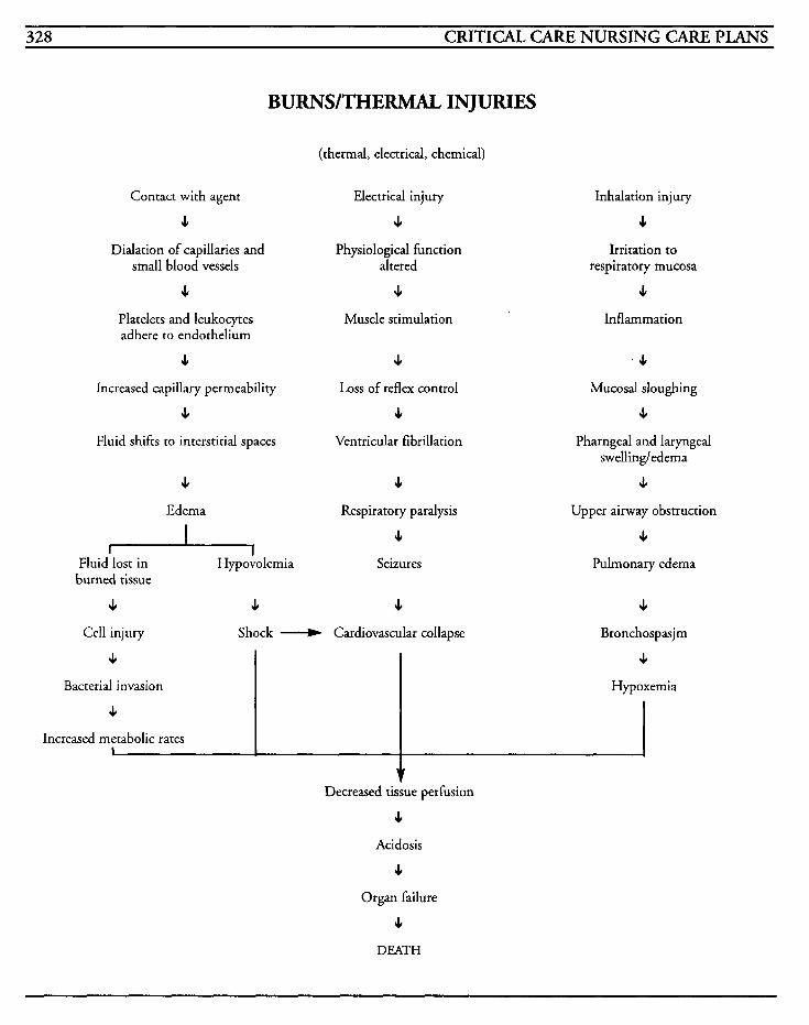
328 CRITICAL CARE NURSING CARE PLANS
4
Bacterial invasion
J,
Increased metabolic rates I
BURNS/THERMAL INJURIES
s Hypoxemia
(thermal, electrical, chemical)
Contact with agent
s Dialation of capillaries and
small blood vessels
s Platelets and leukocytes adhere to endothelium
s Increased capillary permeability
s Fluid shifts to interstitial spaces
s Edema
I Fluid lost in Hypovolemia burned tissue
s s
Electrical injury
s Physiological function
altered
s Muscle stimulation
s Loss of reflex control
s Ventricular fibrillation
s Respiratory paralysis
s Seizures
s
Inhalation injury
s Irritation to
respiratory mucosa
s Inflammation
. s Mucosal sloughing
s Pharngeal and laryngeal
swelling/edema
J,
Upper airway obstruction
s Pulmonary edema
s
Multiple Organ Dysfunction Syndrome (MODS)
Acute Poisoning/Drug Overdose
Snakebite
Transplants
Cardiogenic Shock

This Page Intentionally Left Blank
OTHER 331
Multiole Orflan Dysfunction Syndrome (MODS) Sepsis denotes the presence of microorganisms or their by-products in the bloodstream that create a fulminating infection with resultant systemic involvement and shock. The hemodynamic changes that occur during septic shock may result in inadequate perfusion and the development of multiple organ dysfunction syndrome (MODS). Another syndrome which may lead to MODS is systemic inflammatory response syndrome (SIRS). Both sepsis and SIRS utilize the same inflamma- tory cascade with differing sources of infectious versus non-infectious causes, and can both poten- tially lead to MODS.
As the bacterial infection progresses, the immune system attempts to destroy the causative microor- ganism, and the endotoxins within the cell membrane are released into the vascular system. The endotoxins then trigger systemic inflammation, activation of the complement cas- cade, and histamine release. This results in vasodilation, increased capillary permeability, and leakage of the protein-rich plasma into the intersti- tial tissues.
As the plasma seeps into the alveoli, and platelets and white blood cells embolize in the microcircula- tion, resulting in release of more vasoactive materials, the lung's compliance decreases and ARDS develops. The liver is unable to detoxify the circulating endotoxins because of microemboliza- tion in the liver itself as well as sludge in the hepatic system. As fluid volume decreases, the heart rate increases and cardiac output is raised. As the abdominal organs are constricted from emboli in the microcirculation, myocardial toxic factor (MTF) is released and blocks the calcium ion action and contractility decreases. As more and more endotoxins are circulating, more and more
body systems are affected with decreased perfusion, hypoxia, and anaerobic mechanisms that the body tries to use to maintain metabolic function.
The goal of treatment is to support cardiopulmonary function and to identify and eradicate the organisms responsible for the infec- tion in the first place. With two organ systems involved, mortality is 50-60% despite treatment, with the percentage increasing to 90-100% mor- tality with four or more systems involved.
The most frequent precipitating factor is usually a temporary episode of a shock state that results in body cell ischemia. The typical pattern of MODS includes a hypotensive episode that is apparently successfully resuscitated, with elevation of heart rate and progressive respiratory failure. The patient is then intubated and appears to be doing better, but is in a hypermetabolic and hyperdynamic state that produces progressive changes in labwork. Inotropic support is required, then pseudomonas, yeast, or viral organisms progress, causing renal failure and involvement of all systems, with death ensuing approximately one month after the initial event.
MEDICAL CARE Laboratory: CBC used to identi5 hemorrhage, platelet dysfunction, infection, shifts to the left on differential; electrolytes with sodium decreased; renal profiles used to evaluate renal dysfunction and therapeutic response to treatment; hepatic profiles to evaluate hepatic dysfunction; coagula- tion profiles to identifjr clotting dysfunction and DIC; fibrinogen elevated with DIC; cultures done to identify causative organism and determine appropriate antimicrobial therapy; glucose elevated due to metabolic state; lactate level increased with metabolic acidosis, shock, or hepatic dysfunction
332 CRITICAL CARE NURSING CARE PLANS
Electrocardiogram: used to identify conduction disturbances or cardiac dysrhythmias; may have ST and T wave changes mimicking MI
Arterial b1,ood gases: used to identify hypoxia, hypoxemia, acid-base imbalances and evaluate effectiveness of therapy; initially may have respira- tory alkalosis and hypoxemia, and in later stages, metabolic and respiratory acidosis with compen- satory mechanism failure
Radiography: chest x-rays used to identify pulmonary or cardiac changes in vasculature, edema, complications; abdominal x-rays used to identifi potential sources of infection, i.e., free air in abdomen
Antibiotics: may be used to treat infectious cause of sepsis
Narcan: has been used to counteract some of the endotoxins that are circulating in system
Corticosteroids: have been used to decrease inflammatory response to toxins
NURSING CARE PLANS Risk for in$iction [See Renal Failure]
Related to: progression of sepsis to septic shock, secondary infections, compromised immune system, invasive lines, malnutrition, debilitation
Defining characteristics: increased white blood cell count, shift to the left, fever, chills, cough with or without sputum production, wound drainage, hypo tension, tachycardia, impaired skin integrity, wounds, positive blood, urine or sputum cultures, cloudy concentrated urine
Hypertbermia [See Pheochromocytoma]
Related to: circulating endotoxins, dehydration, hypermetabolic state
Defining characteristics: increased temperature, fever, flushed, warm skin, tachypnea, tachycardia
Risk for alteration in tissue p e f i i o n : cere- bral, gaseointestinal, cardiopulmonary, renal, and perip heral
Related to: vasoconstriction, microembolism, vas- cular occlusion, hypovolemia, increased oxygen consumption, inadequate oxygen delivery, alter- ation in utilization of oxygen by tissues
Defining characteristics: decreased peripheral pulses, prolonged capillary refill time, pallor, cyanosis, erythema, paresthesias, pain, tissue edema, lethargy, confusion, oliguria, anuria, abnormal ABGs
Outcome Criteria
Patient will have adequate perfusion to all body systems.
INTERVENTIONS RATIONALES
Moniror vital signs, noting trends.
Monitor hernodynamic pressures if available, at least every 1-2 hours and pm.
Hypotension occurs when microorganisms enter the blood- stream and activate chemical substances that result in vasodila- tion, decreased systemic vascular resistance, and hypovolemia. Tachypnea may be the first symptom of sepsis as the body responds to endotoxins and developing hypoxia.
When shock progresses to cold stage, cardiac output decreases in response to decreased contractil- ity and alterations in afterload and preload. Fluid shifting may cause third spacing and fluid overload, and monitoring hemo- dynamics can facilitate early identification of changes in trends.
OTHER 333
INTERVENTIONS RATIONALES
Monitor EKG for changes and treat according to hospital proto- col.
Monitor mental status and level of consciousness for changes.
Auscultate lung fields for adven- titious breath sounds.
Observe for changes in periph- eral skin color and temperature.
Monitor intake and output every hour.
Palpate abdomen and auscultate for bowel sounds.
Administer IV fluids as ordered.
Administer oxygen as ordered.
Administer vasoactive drugs as ordered.
Tachycardia occurs in response to hypovolemia and circulating endotoxins. Dysrhythrnias may occur from hypoxia, acid-base imbalances, electrolyte imbal- ances, or shock.
May indicate impending or pre- sent hypoxia or acidosis leading to decreased cerebral perhsion.
May indicate fluid overload in response to fluid resuscitation or presence of congestive failure.
Vasodilation may occur in the early phase of shock with warm, pink, dry skin, but as shock pro- gresses, vasoconstriction occurs and reduces peripheral blood flow resulting in mottling, or pale to dusky skin that is cold and clammy.
As renal perfusion is compro- mised by vasoconstriction or microemboli, oliguria or anuria may develop.
Absence of bowel sounds may indicate decreased perfusion to the mesentery from vasoconstric- tion that may result in paralytic ileus.
Large volumes may be required to maintain circulating volume from hypovolemic state, but must be monitored to identify and treat fluid overloading.
Provides supplemental oxygen necessary for cellular perfusion and to relieve hypoxia.
May be required to maintain pressure and hemodynamics at adequate levels to maintain per- h i o n to body systems.
Information, Instruction, Demonstration
INTERVENTIONS RATIONALES
Observe for oozing at puncture sites, petechiae, ecchymoses, or bleeding from any area. problem.
Monitor for drug toxicicy signs and symptoms.
May indicate presence or impending DIC or coagulation
Decreased perfusion may increase half-life and decrease metabolism of therapeutic drugs and cause toxic reactions.
Discharge or Maintenance Evaluation
Patient will have stable vital signs and hemody- namic parameters.
Patient will have warm skin, with palpable peripheral pulses that are equal bilaterally.
Patient will be neurologically stable, and have adequate perfusion to all body systems.
Risk for impziredgas excbunge [See Mechanical Ventilation]
Related to: endotoxins in circulation, hyperventi- lation, hypoventilation, respiratory alkalosis, increased capillary permeability, alterations in blood flow due to microembolism, capillary damage
Defining characteristics: dyspnea, tachypnea, hypoxia, hypoxemia, hypercapnia, confusion, rest- lessness, cyanosis, inability to move secretions, tachycardia, dysrhythmias, abnormal ABGs, decreased oxygen saturation
Risk for fluid volume &$kit [See GI Bleeding]
Related to: vasodilation, third spacing, fluid shift- ing, increased capillary permeability
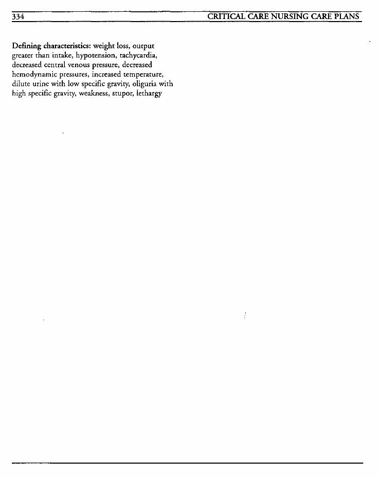
334 CRITICAL CARE NURSING CARE PLANS
Defining characteristics: weight loss, output greater than intake, hypotension, tachycardia, decreased central venous pressure, decreased hernodynamic pressures, increased temperature, dilute urine with low specific gravity, oliguria with high specific gravity, weakness, stupor, lethargy
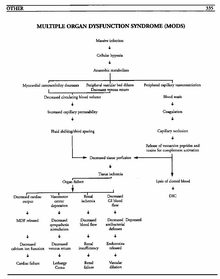
OTHER 335
I
MULTIPLE ORGAN DYSFUNCTION SYNDROME (MODS)
I
Increased capillary permeability
J,
Coagulation
4
Fluid shiftinglthird spacing Capillary occlusion
Release of vasoactive peptides and toxins for complement activation
I Decreased tissue perfusion
J,
Tissue ischemia
L Organ' failure
I Decreadd cardiac Vasobotor Rkal Decreased
output center ischemia GI blood
J, J, 4 4 depression flow
MDF released Decreased Decreased Decreased Depressed sympathetic blood flow antibacterial stimulation defenses
J, J, J, 4 Decreased Decreased Relld Endotoxins
J, J, J, J,
calcium ion function venous return insufficiency released
Cardiac Failure Lethargy Renal Vascular Coma failure dilation
Lysis of clotted blood
J,
DIC

This Page Intentionally Left Blank
Acute PoisoninaDrug Overdose Attempts to end one's life by use of excessive amounts of medication may be executed for many reasons. Active self-destructive behavior usually results from the patient's perception of an overwhelming catastrophic event in his life, in conjunction with the lack of appropriate coping strategies, and is visualized as a means of escape from the sensed threat to himself.
Suicidal patients are frequently ambivalent about wanting to die, and may have visions of last- minute rescue. The suicidal person may feel despair, guilt, shame, hopelessness, boredom, depression, weariness, or dependency, and when the point is reached when the person perceives that life no longer has meaning and despair is overwhelming, the patient acts on those emotions. Suicide may be considered the last logical step when the person perceives that others do not want them around or that the problem can never be reconciled.
Usually, an attempt at causing death is the culmi- nation of a process in which the person had ideations about killing himself, verbal or nonver- bal threats of his intention, and gestures in which attempts of causing self-injury without actual intentions to cause death.
Suicide is the eighth leading cause of death in this country today, and the second leading cause of death in young people. Drug ingestion is the most frequent method utilized with suicide attempts, partially because of the availability of medications, and partially to avoid more violent means of death, such as with weapons or by hanging.
MEDICdL CARE Laboratory: drug screens may be used to identify agent used for suicide attempt; alcohol level to assess concurrent use or toxicity; electrolytes may be abnormal due to trauma or interaction with medication; hematocrit may be decreased with hypovolemia; drug levels, such as phenobarbital or acetaminophen, may be elevated due to toxicity; renal profile may show renal insufficiency; liver profile may show hepatic dysfunction, especially with acetaminophen overdose; coagulation profiles may be abnormal; urinalysis may show low specific gravity, increased protein, hematuria, oxalate crystals, or metabolic by-products from drug overdose
Radiography: chest x-rays may show aspiration pneumonia or pulmonary edema complications
Electrocardiogram: used to identify conduction problems or dysrhythmias that may occur from drug overdosage, electrolyte disturbances, or with congestive failure
Dialysis: hemodialysis or hemoperfusion may be performed to remove some drugs when levels are severely elevated
Diuretics: may be required to force osmotic diure- sis with agents such as mannitol, to manage certain forms of overdose
Acetylcysteine: Mucomyst is treatment of choice with acetaminophen overdose
Charcoal: used to bind poisons, toxins, or other irritants, increases absorption in the GI tract, and helps to inactivate toxins until excreted
NURSING CARE PWNS Risk for inefective breathing pattern [See Mechanical Ventilation]
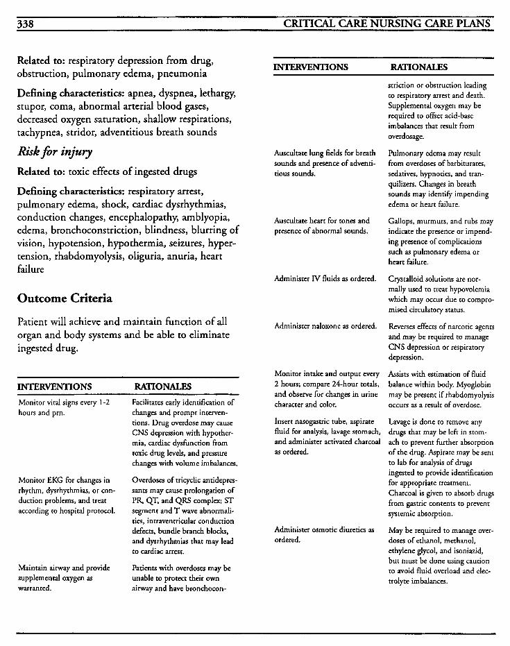
338 CRITICAL CARE NURSING CARE PLANS
Related to: respiratory depression from drug, obstruction, pulmonary edema, pneumonia
Defining characteristics: apnea, dyspnea, lethargy, stupor, coma, abnormal arterial blood gases, decreased oxygen saturation, shallow respirations, tachypnea, stridor, adventitious breath sounds
Risk for injury Related to: toxic effects of ingested drugs
Defining characteristics: respiratory arrest, pulmonary edema, shock, cardiac dysrhythmias, conduction changes, encephalopathy, amblyopia, edema, bronchoconstriction, blindness, blurring of vision, hypotension, hypothermia, seizures, hyper- tension, rhabdomyolysis, oliguria, anuria, heart failure
INTERVENTIONS RATIONALES
striction or obstruction leading to respiratory arrest and death. Supplemental oxygen may be required to offset acid-base imbalances that result from overdosage.
Auscultate lung fields for breath sounds and presence of adventi- tious sounds.
Auscultate heart for tones and presence of abnormal sounds.
Administer IV fluids as ordered.
Outcome Criteria
Patient will achieve and maintain function of all organ and body systems and be able to eliminate ingested drug.
INTERWNTIONS RATIONALES Monitor vital signs every 1-2 hours and prn.
Monitor EKG for changes in rhythm, dysrhythmias, or con- duction problems, and treat according to hospital protocol.
Maintain airway and provide supplemental oxygen as warranted.
Facilitates early identification of changes and prompt interven- tions. Drug overdose may cause CNS depression with hypother- mia, cardiac dysfunction from toxic drug levels, and pressure changes with volume imbalances.
Overdoses of tricyclic antidepres- sants may cause prolongation of PR, QT, and QRS complex; ST segment and T wave abnormali- ties, intraventricular conduction defects, bundle branch blocks, and dysrhythrnias that may lead to cardiac arrest.
Patients with overdoses may be unable to protect their own airway and have bronchocon-
Administer naloxone as ordered.
Monitor intake and output every 2 hours; compare 24-hour totals, and observe for changes in urine character and color.
Insert nasogastric tube, aspirate fluid for analysis, lavage stomach, and administer activated charcoal as ordered.
Administer osmotic diuretics as ordered.
Pulmonary edema may result from overdoses of barbiturates, sedatives, hypnotics, and tran- quilizers. Changes in breath sounds may identify impending edema or heart failure.
Gallops, murmurs, and rubs may indicate the presence or impend- ing presence of complications such as pulmonary edema or heart failure.
Crystalloid solutions are nor- mally used to treat hypovolemia which may occur due to compro- mised circulatory status.
Reverses effects of narcotic agents and may be required to manage CNS depression or respiratory depression.
Assists with estimation of fluid balance within body. Myoglobin may be present if rhabdomyolysis occurs as a result of overdose.
Lavage is done to remove any drugs that may be left in stom- ach to p.revent further absorption of the drug. Aspirate may be sent to lab for analysis of drugs ingested to provide identification for appropriate treatment. Charcoal is given to absorb drugs from gastric contents to prevent systemic absorption.
May be required to manage over- doses of ethanol, methanol, ethylene glycol, and isoniazid, but must be done using caution to avoid fluid overload and elec- trolyte imbalances.
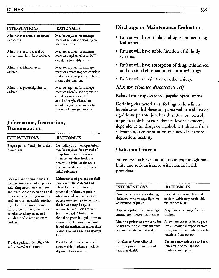
OTHER 339
INTERVENTIONS RATIONALES
Administer sodium bicarbonate as ordered.
May be required for manage- ment of salicylate poisoning to alkalinize urine.
May be required for manage- ment of amphetamine or PCP overdoses to acidify urine.
Administer ascorbic acid or ammonium chloride as ordered.
Administer physostigmine as ordered.
Administer Mucomyst as ordered. ment of acetaminophen overdose
May be required for manage-
to decrease absorption and limit hepatic dysfunction.
May be required for manage- ment of tricyclic antidepressant overdoses to reverse the anticholinergic effects, but should be given cautiously to prevent cholinergic toxicity.
Information, Instruction, Demonstration
INTERVENTIONS RATIONALES
Prepare patientlfamily for dialysis procedures.
Ensure suicide precautions are exercised-removal of all poten- tially dangerous items from room and reach, close observation at all times, keeping exiting windows and doors impenetrable, provid- ing all medications in liquid form, accompanying the patient to other ancillary areas, and avoidance of secret pacts with patient.
Provide padded side rails, with rails elevated at all times.
Hemodialysis or hemoperfusion may be required for removal of drugs from system in severe intoxication when levels are potentially lethal or the toxin may be metabolized to a more lethal substance.
Maintenance of precautions facil- itate a safe environment and allows for identification of potential problems. A patient who has made one attempt at suicide may attempt to complete the job and may be quite resourceful with items to per- form the deed. Medications should be given in liquid form to ensure that the patient has swal- lowed the medication rather than saving it to use as suicide attempt later.
Provides safe environment and reduces risk of injury, especially if patient has a seizure.
Discharge or Maintenance Evaluation
Patient will have stable vital signs and neurolog- ical status.
Patient will have stable function of all body
Patient will have absorption of drugs minimized and maximal elimination of absorbed drugs.
sys terns.
Patient will remain free of other injury.
Risk f o r violence directed at self
Related to: drug overdose, psychological status
Defining characteristics: feelings of loneliness, hopelessness, helplessness, perceived or real loss of significant person, job, health status, or control, unpredictable behavior, threats, low self-esteem, dependence on drugs or alcohol, withdrawal from substances, communication of suicidal ideations, depression, hostility
Outcome Criteria
Patient will achieve and maintain psychologic sta- bility and seek assistance with mental health providers.
INTERVENTIONS RATIONALES
Ensure environment is calming, darkened, with enough light for observation of patient.
Approach patient in a nonjudg- mental, nonthreatening manner.
Listen to patient and what he has to say about his current situation without reacting emotionally.
Confirm understanding of patient’s problem, but do not reinforce denial.
Facilitates decreased fear and anxiety which may result with violent behavior.
May have a calming effect on patient.
Allows patient to verbalize prob- lems. Emotional responses from caregivers may exacerbate hostile reactions from patient.
Fosters communication and facil- itates realistic feelings and methods for coping.
340 CRITICAL CARE NURSING CARE PLANS
INTERVENTIONS RATIONALES
Assist patient to verbalize emo- tions, anger, and other stressors, and to develop a plan for dealing with them. solving problems.
Provides safe outlets for patient to express feelings and helps to work out realistic solutions for
Information, Instruction, Demonstration
INTERVENTIONS RATIONALES
Instruct patiendfamily on com- munity resources, hot lines, crisis centers, ministerial counselors, etc.
Consult mental health provider/ professional as warranted.
Provides knowledge and assis- tance of resources available once patient is discharged.
Allows for effective therapeutic psychological treatment to dis- cern appropriate methods of coping with crisis.
Validates their feelings and responses and may assist them in finding more appropriate meth- ods to cope with crisis.
Patient may be more likely to try suicidal attempt again if situa- tions or coping strategies are not changed. Understanding that if the patient has a definite plan for suicide, the more likely it is that he will be successful at ending his life, and that immediate intervention will be required.
Encourage family members to discuss their feelings and meth- ods of coping.
Discuss actions to take if patient expresses suicidal ideations or attempts.
Discharge or Maintenance Evaluation
Patient will achieve psychological equilibrium and have no further suicidal attemptdgestures.
Patient will be able to cope with crises in an appropriate manner, and will be able to effec- tively search out community resources for assistance.
Patient and family will be able to verbalize feel- ings and effectively achieve therapeutic communication.
Patient and family will be able to access avail-
Risk for ineffective individual coping [See Mechanical Ventilation]
Related to: crisis, drug overdose, loss of control, depression
Defining characteristics: verbal manipulation, inability to meet basic needs, inability to effectively deal with crisis, ineffective defense mechanisms, irritability, hostility
able resources.
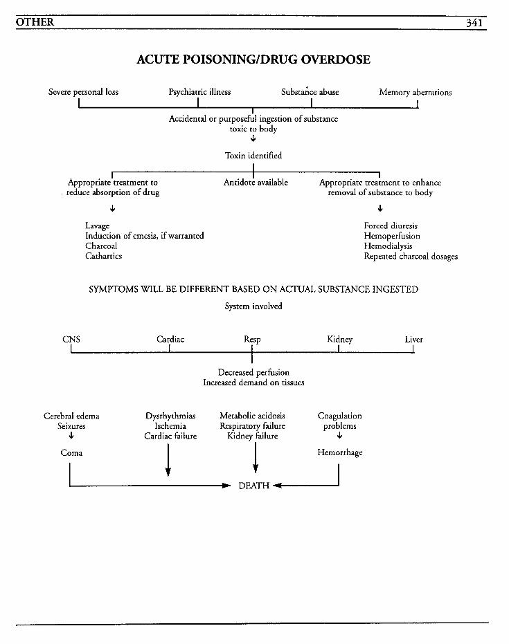
OTHER 34 1
ACUTE POISONING/DRUG OVERDOSE
Severe personal loss Psychiatric illness Substaice abuse Memory aberrations
I Accidental or purposeful ingestion of substance
toxic to body J,
Toxin identified
I I I 1
I Appropriate treatment to
. reduce absorption of drug
I I Antidote available Appropriate treatment to enhance
removal of substance to body
c J,
Lavage Induction of emesis, if warranted Charcoal Cathartics
Forced diuresis Hemoperfusion Hemodialysis Repeated charcoal dosages
SYMPTOMS WILL BE DIFFERENT BASED ON ACTUAL SUBSTANCE INGESTED
System involved
CNS Cardiac Resp Kidney Liver
Decreased perfusion Increased demand on tissues
Cerebral edema Dysrhythmias Metabolic acidosis Coagulation Seizures Ischemia Respiratory failure problems 4 Cardiac failure Kidney failure J,
Coma Hemorrhage
I W DEATH-

This Page Intentionally Left Blank
OTHER 343
In the United States, there are actually two types of poisonous snakes-coral snakes and pit vipers, which include rattlesnakes, water moccasins, and copperheads. Coral snakes are usually nocturnal creatures and less active than pit vipers, but tend to bite with a chewing motion and cause signifi- cant tissue damage.
Snakebites may occur on any portion of the body, but usually are noted on the extremities. Pit viper bites with envenomation result in immediate pain and edema within 10-20 minutes. Other symptoms include fever, ecchymoses, blisters, and local necrosis, as well as nausea, vomiting, diarrhea, metallic or rubbery taste, tachycardia, hypotension, and shock. Neurotoxins may cause numbness, tingling, fasciculations, twitching, con- vulsions, dysphasia, occasional paralysis, respiratory distress, coma, and death. Pit viper bites may also impair coagulation and cause inter- nal bleeding.
Coral snake bites usually have a delayed reaction up to several hours, and may result in very little or no tissue pain, edema, or necrosis. The neurotoxic venom produces paresthesias, weakness, nausea, vomiting, dysphagia, excessive salivation, blurred vision, respiratory distress and failure, loss of muscle coordination, paralysis, abnormal reflexes, shock, cardiovascular collapse, and death. Coral snake bites may also result in coagulopathy prob- lems.
The snake venom is a mixture of several proteins, enzymes, and polypeptides, and may produce sev- eral toxic reactions in patients who have been bitten. Correct diagnosis is imperative to treat the specific envenomation accurately and in a timely manner. Snakebites are critical emergencies and require precise identification of the snake as well
as presence of envenomation. Designation of severity of the bite is commonly rated as minor, moderate, or severe, and depends on the presence of symptoms, depth of envenomation, and labora- tory findings.
Treatment of snakebite involves administration of antivenin after a test dose for horse serum sensitiv- ity is performed. If this sensitivity is present, diphenhydramine may be given prior to the antivenin. Swelling may necessitate surgical inter- vention to relieve the pressure and to prevent further vascular damage, and ensuing complications are usually related to secondary infection, renal failure, disseminated intravascular coagulation, or gangrene.
MEDICAL CARE Laboratory: CBC used to identify blood loss and hemoconcentration; fibrinogen level, platelets, PT, PTT, and APTT to evaluate clotting; blood type and cross-matching to provide blood products as warranted; renal and liver studies to identify dys- function, elevated BUN, creatinine, bilirubin, or creatine kinase
Electrocardiogram: used to establish a baseline for identification of problems that may occur with hernodynamic changes and to identify dysrhyth- mias and conduction problems
Surgery: fasciotomy may be required to relieve pressure caused from swelling or compartmental syndrome; amputation may be required for gan- grene or necrosis
Analgesics: used to alleviate and/or control the pain related to envenomation and swelling; mor- phine is usually not given due to its vasodilator action
Antivenin: required as the antidote for snakebite; amount of antivenin is dependent on the severity of the reaction rather than patient weight, and ranges from 3 to 15 or more vials; children usually
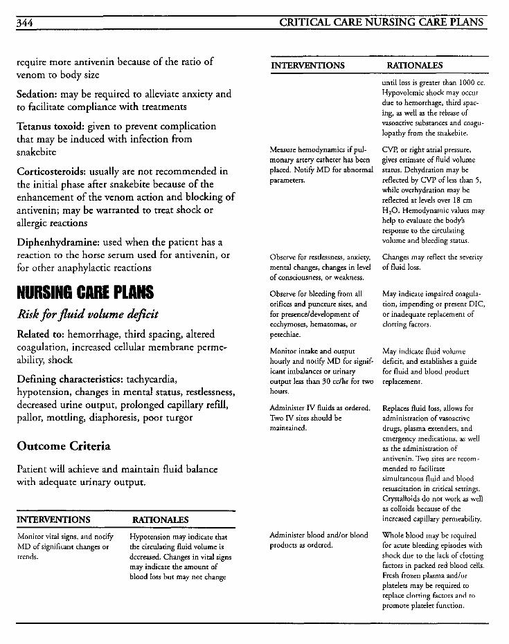
344 CRITICAL CARE NURSING CARE PLANS
require more antivenin because of the ratio of venom to body size
Sedation: may be required to alleviate anxiety and to facilitate compliance with treatments
Tetanus toxoid: given to prevent complication that may be induced with infection from snakebite
Corticosteroids: usually are not recommended in the initial phase after snakebite because of the enhancement of the venom action and blocking of antivenin; may be warranted to treat shock or allergic reactions
Diphenhydramine: used when the patient has a reaction to the horse serum used for antivenin, or for other anaphylactic reactions
NURSING CARE PLANS Risk for fluid volume deficit Related to: hemorrhage, third spacing, altered coagulation, increased cellular membrane perme- ability, shock
Defining characteristics: tachycardia, hypotension, changes in mental status, restlessness, decreased urine output, prolonged capillary refill, pallor, mottling, diaphoresis, poor turgor
Outcome Criteria
Patient will achieve and maintain fluid balance with adequate urinary output.
~~
INTERVENTIONS RATIONALES
Monitor vital signs, and notify MD of significant changes or
Hypotension may indicate that the circulating fluid volume is
trends. decreased. Changes in vital signs may indicate the amount of blood loss but may not change
INTERVENTIONS RATIONALES
Measure hernodynamics if pul- monary artery catheter has been placed. Notify M D for abnormal parameters.
Observe for restlessness, anxiety, mental changes, changes in level of consciousness, or weakness.
Observe for bleeding from all orifices and puncture sites, and for presenceldevelopment of ecchymoses, hematomas, or petechiae.
Monitor intake and output hourly and notify MD for signif- icant imbalances or urinary output less than 30 cclhr for two hours.
Administer IV fluids as ordered. Two IV sites should be maintained.
Administer blood andlor blood products as ordered.
until loss is greater than 1000 cc. Hypovolemic shock may occur due to hemorrhage, third spac- ing, as well as the release of vasoactive substances and coagu- lopathy from the snakebite.
CW, or right atrial pressure, gives estimate of fluid volume status. Dehydration may be reflected by CVP of less than 5 , while overhydration may be reflected at levels over 18 cm H,O. Hemodynamic values may help to evaluate the body’s response to the circulating volume and bleeding status.
Changes may reflect the severity of fluid loss.
May indicate impaired coagula- tion, impending or present DIC, or inadequate replacement of clotting factors.
May indicate fluid volume deficit, and establishes a guide for fluid and blood product replacemenr.
Replaces fluid loss, allows for administration of vasoacrive drugs, plasma extenders, and emergency medications, as well as the administration of antivenin. Two sites are recom- mended to facilitate simultaneous fluid and blood resuscitation in critical settings. Crystalloids do not work as well as colloids because of the increased capillary permeability.
Whole blood may be required for acute bleeding episodes with shock due to the lack of clotting factors in packed red blood cells. Fresh frozen plasma andlor platelets may be required to replace clotting factors and CO
promote platelet function.
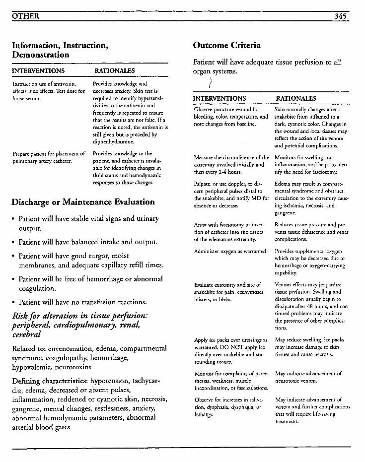
OTHER 345
Information, Instruction, Demonstration
INTERVENTIONS RATIONALES
Instruct on use of antivenin, effects, side effects. Test dose for horse serum.
Provides knowledge and decreases anxiety. Skin test is required to identify hypersensi- tivities to the antivenin and frequently is repeated to ensure that the results are not false. If a reaction is noted, the antivenin is still given but is preceded by diphenhydramine.
Provides knowledge to the patient, and catheter is invalu- able for identifying changes in fluid status and hemodynamic responses to those changes.
Prepare patient for placement of pulmonary artery catheter.
Discharge or Maintenance Evaluation
Patient will have stable vital signs and urinary output.
Patient will have balanced intake and output.
Patient will have good turgor, moist membranes, and adequate capillary refill times.
Patient will be free of hemorrhage or abnormal coagulation.
Patient will have no transfusion reactions.
Risk for alteration in tissue petfhsion: peripheral, cardiopulmonary, renal, cerebral
Related to: envenomation, edema, compartmental syndrome, coagulopathy, hemorrhage, hypovolemia, neurotoxins
Defining characteristics: hypotension, tachycar- dia, edema, decreased or absent pulses, inflammation, reddened or cyanotic skin, necrosis, gangrene, mental changes, restlessness, anxiety, abnormal hemodynamic parameters, abnormal arterial blood gases
Outcome Criteria
Patient will have adequate tissue perfusion to all organ systems.
INTERVENTIONS RATIONALES Observe puncture wound for bleeding, color, temperature, and note changes from baseline.
Measure the circumference of the extremity involved initially and then every 2-4 hours.
Palpate, or use doppler, to dis- cern peripheral pulses distal to the snakebite, and notify M D for absence or decrease.
Assist with fasciotomy or inser- tion of catheter into the tissues of the edematous extremity.
Administer oxygen as warranted.
Evaluate extremity and site of snakebite for pain, ecchymoses, blisters, or blebs.
Apply ice packs over dressings as warranted. DO NOT apply ice directly over snakebite and sur- rounding tissues.
Monitor for complaints of pares- thesias, weakness, muscle incoordination, or fasciculations.
Observe for increases in saliva- tion, dysphasia, dysphagia, or lethargy.
Skin normally changes after a snakebite from inflamed to a dark, cyanotic color. Changes in the wound and local tissues may reflect the action of the venom and potential complications.
Monitors for swelling and inflammation, and helps to iden- tify the need for fasciotomy.
Edema may result in compart- mental syndrome and obstruct circulation to the extremity caus- ing ischemia, necrosis, and gangrene.
Reduces tissue pressure and pre- vents tissue dehiscence and other complications.
Provides supplemental oxygen which may be decreased due to hemorrhage or oxygen-carrying capabiliry.
Venom effects may jeopardize tissue perfusion. Swelling and discoloration usually begin to dissipate after 48 hours, and con- tinued problems may indicate the presence of other complica- tions.
May reduce swelling. Ice packs may increase damage to skin tissues and cause necrosis.
May indicate advancement of neurotoxic venom.
May indicate advancement of venom and further complications that will require life-saving treatment.
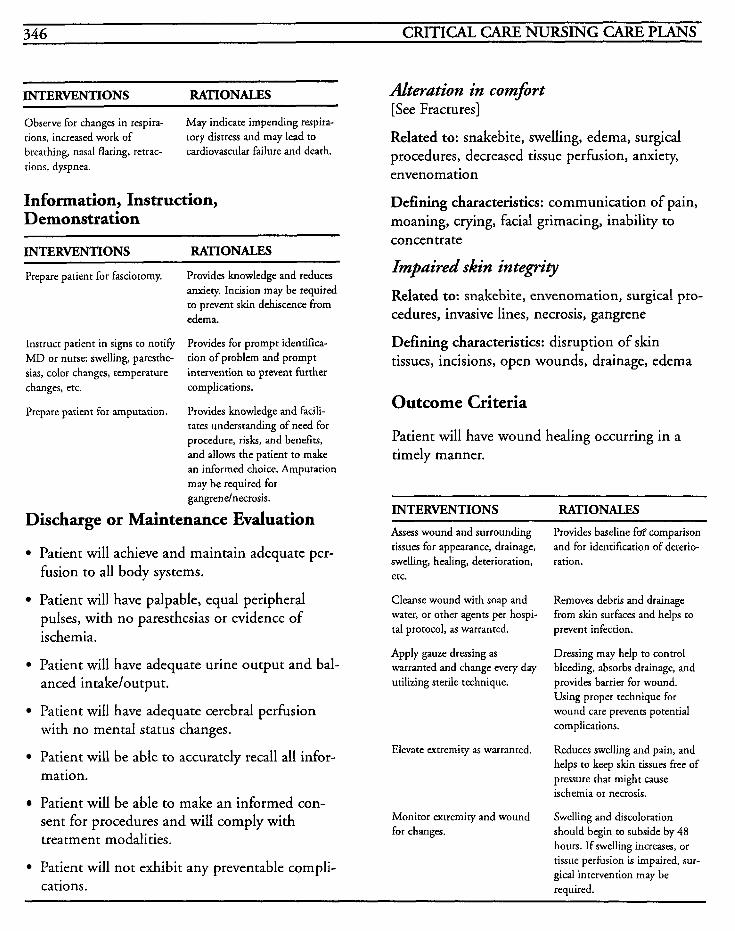
INTERVENTIONS RATIONALES Alteration in comfort
tions, increased work of breathing, nasal flaring, retrac-
[See Fractures]
Related to: snakebite, swelling, edema, surgical procedures, decreased tissue perfusion, anxiety, envenomation
Observe for changes in respira- May indicate impending respira- tory distress and may lead to cardiovascular failure and death.
tions, dyspnea.
Information, Instruction, Demonstration
INTERVENTIONS RATIONALES
Prepare patient for fasciotomy. Provides knowledge and reduces anxiety. Incision may be required to prevent skin dehiscence from edema.
Provides for prompt identifica- tion of problem and prompt intervention to prevent further
Instruct patient in signs to notify MD or nurse: swelling, paresthe- sias, color changes, temperature changes, etc. complications.
Prepare patient for amputation. Provides knowledge and facili- tates understanding of need for procedure, risks, and benefits, and allows the patient to make an informed choice. Amputation may be required for gangrendnecrosis.
Discharge or Maintenance Evaluation
Patient will achieve and maintain adequate per- fusion to all body systems.
Patient will have palpable, equal peripheral pulses, with no paresthesias or evidence of ischemia.
Patient will have adequate urine output and bal- anced intake/output.
Patient will have adequate cerebral perfusion with no mental status changes.
Patient will be able to accurately recall all infor- mation.
Patient will be able to make an informed con- sent for procedures and will comply with treatment modalities.
Patient will not exhibit any preventable compli- cations.
Defining characteristics: communication of pain, moaning, crying, facial grimacing, inability to concentrate
Impaired skin integr.9 Related to: snakebite, envenomation, surgical pro- cedures, invasive lines, necrosis, gangrene
Defining characteristics: disruption of skin tissues, incisions, open wounds, drainage, edema
Outcome Criteria
Patient will have wound healing occurring in a timely manner.
INTERVENTIONS RATIONALES Assess wound and surrounding tissues for appearance, drainage, swelling, healing, deterioration, etc.
Cleanse wound with soap and water, or other agents per hospi- tal protocol, as warranted.
Apply gauze dressing as warranted and change every day utilizing sterile technique.
Elevate extremity as warranted.
Monitor extremity and wound for changes.
Provides baseline fof comparison and for identification of deterio- ration.
Removes debris and drainage from skin surfaces and helps to prevent infection.
Dressing may help to control bleeding, absorbs drainage, and provides barrier for wound. Using proper technique for wound care prevents potential complications.
Reduces swelling and pain, and helps to keep skin tissues free of pressure that might cause ischemia or necrosis.
Swelling and discoloration should begin to subside by 48 hours. If swelling increases, or tissue perfusion is impaired, sur- gical intervention may be required.
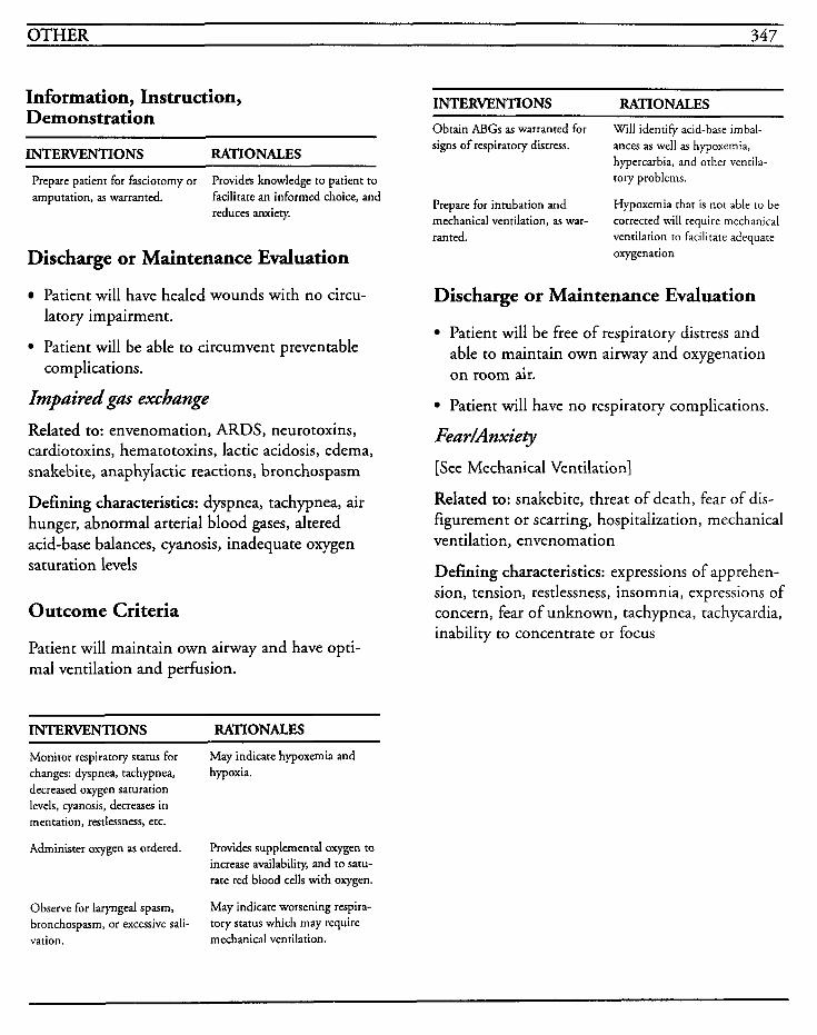
OTHER 347
Information, Instruction, Demonstration
INTERVENTIONS RATIONALES
Prepare patient for fasciotomy or amputation, as warranted.
Provides knowledge to patient to facilitate an informed choice, and reduces anxiety.
Discharge or Maintenance Evaluation
Patient will have healed wounds with no circu- latory impairment.
Patient will be able to circumvent preventable
Impaired gas exchange
Related to: envenomation, ARDS, neurotoxins, cardiotoxins, hematotoxins, lactic acidosis, edema, snakebite, anaphylactic reactions, bronchospasm
Defining characteristics: dyspnea, tachypnea, air hunger, abnormal arterial blood gases, altered acid-base balances, cyanosis, inadequate oxygen saturation levels
complications.
Outcome Criteria
Patient will maintain own airway and have opti- mal ventilation and perfusion.
INTERVENTIONS RATIONALES
Monitor respiratory status for changes: dyspnea, tachypnea, hypoxia. decreased oxygen saturation levels, cyanosis, decreases in mentation, restlessness, etc.
Administer oxygen as ordered.
May indicate hypoxemia and
Provides supplementd oxygen to increase availability, and to satu- rate red blood cells with oxygen.
May indicate worsening respira- tory status which may require
Observe for laryngeal spasm, bronchospasm, or excessive sali- vation. mechanical ventilation.
INTERVENTIONS RATIONALES Obtain ABGs as warranted for signs of respiratory distress.
Will identify acid-base imbal- ances as well as hypoxemia, hypercarbia, and orher ventila- tory problems.
Hypoxemia that is not able to be corrected will require mechanical
oxygenation
Prepare for intubation and mechanical ventilation, as war- ranted. ventilation to facilitate adequate
Discharge or Maintenance Evaluation
Patient will be free of respiratory distress and able to maintain own airway and oxygenation on room air.
Patient will have no respiratory complications.
Fea r/Anxiety
[See Mechanical Ventilation]
Related to: snakebite, threat of death, fear of dis- figurement or scarring, hospitalization, mechanical ventilation, envenomation
Defining characteristics: expressions of apprehen- sion, tension, restlessness, insomnia, expressions of concern, fear of unknown, tachypnea, tachycardia, inability to concentrate or focus
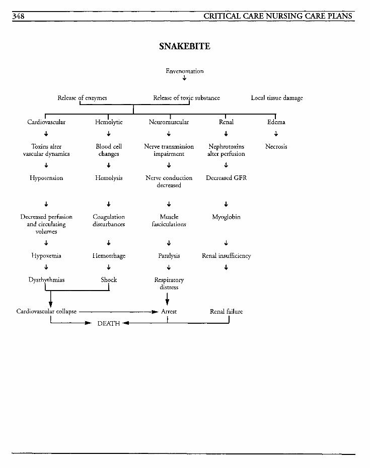
348 CRITICAL CARE NURSING CARE PLANS
SNAKEBITE
Envenomation e
Release of enzymes Release of toxic substance Local tissue damage
I I I I I I
Cardiovascular Hemhytic
4 c Toxins alter Blood cell
4 c Hypotension Hemolysis
vascular dynamics changes
4 c Decreased perfusion Coagulation
and circulating disturbances
4 4 Hypoxemia Hemorrhage
c c
volumes
Shock
Neuron-hscular
c Nerve transmission
impairment
c Nerve conduction
decreased
e Muscle
fasciculations
c Paralysis
c Respiratory
distress + Cardiovascular collapse Arrest
Renal EdLma
c 4
Nephrotoxins Necrosis alter perfusion
c Decreased GFR
c Myoglobin
J,
Renal insufficiency
e
Renal failure
DEATH 1
OTHER 349
Transplants Transplantation of living tissues, cells, or organs from one individual to another is one method of treatment for several end-stage organ diseases. Often, transplantation is the last resort for a vari- ety of disorders after conventional medical or surgical therapies have failed to provide adequate functioning. Recent advances in technique and treatment have improved the rate of success, and transplantation has improved the quality of life for many patients who otherwise would either die or be resigned to lives of dialysis or suffering.
Transplants are categorized by the relationship between the donor and the recipient. An autograft relates to the transplantation of tissue from one location to another in the same person. An isograft is a graft between identical twins, and an allograft, or homograft, is a graft between mem- bers of the same species. A xenograft, or heterografi, is a graft between members of differ- ent species.
Bone marrow transplants are performed in order to restore immunologic and hematologic function to patients who have aplastic anemia, leukemia, or severe combined immunodeficiency disorder. Multiple aspirations of bone marrow are obtained and then infused intravenously with red blood cells.
Heart transplants are performed to attempt to restore function in end-stage cardiac failure that has been unresponsive to other medical therapeu- tics, and usually involve patients who have cardiomyopathy, rheumatic heart disease, congeni- tal heart disease, or coronary artery disease. After the patient is placed on cardiopulmonary bypass and the diseased heart is removed, the donor allo-
graft heart is implanted. Frequently, a combined heart-lung transplant is performed due to the increased success rate as a result of fewer vascular anastomoses being required.
Renal transplants are performed to restore kidney function in end-stage renal disease. Allografts are usually obtained from living relatives or cadavers. The kidney is usually implanted in a retroperitoneal position against the psoas muscle in the iliac fossa. When cadaver kidneys are used approximately half of the recipients may require dialysis because of the presence of acute tubular necrosis.
Liver transplants are performed to restore function in patients with chronic active hepatitis, hepatitis B antigen-negative postnecrotic cirrhosis, primary hepatocellular tumor, or congenital anomalies of the bile duct or inborn errors of metabolism in children. The liver is implanted into the right upper abdominal quadrant and the vasculature is anastomosed. Biliary drainage anastomosis prob- lems often result in bacteremia.
Pancreas transplants are performed on patients with insulin-dependent diabetes mellitus to pro- vide insulin-producing tissue. The pancreas, either total with a small amount of duodenum, or partial segment of the distal pancreas, is transplanted. This type of transplant is performed as a life- enhancing procedure and is most successful prior to the development of severe secondary diabetic complications.
The goal in transplantation is to maintain optimal functioning of the organ and to prevent rejection. This goal is facilitated by antigen matching, tissue typing for histocompatibility, tests for prior sensi- tization, transfusions of whole blood, and immunosuppressive therapy.
Despite a small increase in the available donor
350 CRITICAL CARE NURSING CARE PLANS
organs, the number of candidates for transplant far exceeds the organs available, and many patients die prior to undergoing transplantation. Complications of infection, rejection, and immunosuppressive drugs are a very real part of the process.
Transplantation of almost any tissue is feasible but rejection is the most frequent complication when the body tries to destroy the graft tissue. Rejection occurs when the immune system recognizes the graft as being foreign to the body and begins a responsive action to the antigens of the graft. Thus begins a cell-mediated immune response in the lymph tissues. Antibody-mediated immune responses, inflammatory responses, and comple- ment activation also play a significant role in the rejection process.
Rejection may occur immediately after transplan- tation or up to years later, and most transplant patients experience at least one rejection episode during their lives. Signs/symptoms of rejection vary depending on the type of graft. Cornea1 transplant rejection is evidenced by corneal cloud- ing, corneal edema, or conjunctival hyperemia. Cardiac transplant rejection is evidenced by decreased QRS, right axis shift, atrial dysrhythmias, conduction defects, S, gallop, jugu- lar vein distention, decreased exercise tolerance, low grade fever, malaise, weight gain, dyspnea, right ventricular failure, and peripheral edema. Liver transplant rejection may be manifested with changes in urine or stool color, jaundice, hepatomegaly, ascites, pain in the center of the back, right flank, or right upper quadrant of the abdomen, low grade fever, malaise, or anorexia. Renal transplant rejection may involve low grade fever, decreased urine output, pain, swelling and/or tenderness in the kidney, increased blood pressure, malaise, weight gain, or peripheral edema. Pancreas transplant rejection may show
symptoms of increased glucose levels, polyuria, polydipsia, polyphagia, weight loss, low grade fever, and tender or enlarged pancreas. Bone marrow transplantation rejection is usually evidenced by severe diarrhea, jaundice and skin changes.
Rejection can be classified as being acute, hypera- cute, or chronic depending on the mechanisms of rejection and the duration of time prior to the appearance of symptoms. Acute reactions may occur anywhere from 7 days to several weeks after transplant. A cell-mediated acute reaction occurs when the graft develops interstitial edema, ischemia, and necrosis, but high dose steroid ther- apy may reverse the reaction. Antibody-mediated acute reactions occur when fibrin, platelets, and polymorphonuclear cells adhere to the graft cells, resulting from recipient antibody-donor antigen responses. This aggregation produces ischemia and eventually necrosis. Hyperacute reactions develop immediately after the transplant up to a few days after. Immediate hyperacute reactions happen when the recipient has preformed antibodies against the donor antigens and is usually caused by previous blood transfusions, previous transplants, or from pregnancy. An accelerated hyperacute reaction happens when the recipient lymphocytes and neutrophils infiltrate the graft and may be prevented with the use of antisera to T lymphocytes. Chronic reactions occur over many months and eventually leads to loss of graft function. This occurs when the vascular endothe- hum becomes inflamed, and the arterial lumen decreases. Fibrin and platelets aggregate and over time, result in decreased blood flow to the organ and ischemia and dysfunction prevail.
The principal mechanism of rejection is GVH (graft versus host) disease. This occurs when an immunocompetent donor graft is transplanted into an immune-impaired recipient. If the donor
OTHER 35 1
and the recipient are not histocompatible, foreign cells will initiate an attack against the host cells, which are then unable to reject them. This usually occurs with bone marrow or liver transplants.
MEDICAL CARE Laboratory: renal profiles used to assess kidney function; hepatic profiles used to assess liver func- tion; CBC used to evaluate anemia, infection, and blood loss; glucose levels used to monitor pancre- atic function; A B 0 blood grouping; Lewis antigens used to evaluate compatibility for kidney transplants; microtoxicity assays for evaluation of bone marrow; tissue typing for histocompatibility; lymphocyte antibody screen to evaluate preformed antibodies; lymphocyte cross-matching used after a suitable donor is found; serology, HIV, hepatitis screens to evaluate suitability for transplantation
Surgery: required for transplantation of tissueslorgans
Biopsy: tissue biopsies used as the most accurate diagnostic tool to determine the extent of lympho- cyte infiltration and potential tissue damage; serial biopsies can be used to monitor course of treatment
Immunosuppressive drugs: used to decrease or eliminate the body's ability to reject new transplanted tissues; can increase the risk for opportunistic organisms; usually a combination of drugs are used rather than just one
Blood transfusions: used to improve graft survival of certain organisms
Radiation therapy: used in some instances for pre- transplantation immunosuppression
Thoracic duct drainage: used in some instances for pretransplantation imrnunosuppression
NURSING CARE PLANS Risk f i r infection
Related to: immunosuppression, effects of trans- plantation, invasive procedures, invasive linedcatheters, trauma, surgery
Defining characteristics: increased immature white blood cells, differential with a shift to the left, fever, chills, cough, hypotension, tachycardia, presence of wounds, positive blood, urine, or sputum cultures, cloudy urine, purulent drainage
Outcome Criteria
Patient will have no signs/symptoms of infection after transplant surgery.
~ ~~
INTERVENTIONS RATIONALES
Patient should be in private room, with appropriate isolation techniques in use. Visitors with illness must be restricted from visiting.
Observe for signslsymptoms of infection to all body systems.
Provide diet with appropriate nutrients and fluids. Restrict fresh fruits and vegetables.
Monitor CBC, especially WBC count for abrupt changes in neu- trophils.
Use sterile/aseptic technique with
Decreases potential of infection when patient is already immuno- compromised.
Provides for prompt identifica- tion of complication and facilitates timely intervention.
Proper nutrition facilitates anti- body formation and prevents dehydration. Fresh fruidvegeta- bles may harbor parasitic spores or bacteria that may result in an infection.
Sudden decreases in mature WBCs may result from chemotherapy and further com- promise the immune response.
Immunosuppressive drugs or dressing changes, IV site changes, or other invasive care.
effects of the patient's disease process may slow wound healing. Drainage is a potential medium for bacterial growth.
Steroid and antibiotic adminis- tration may result in an overgrowth of fungal coloniza- tion resulting in candidiasis.
Observe mouth and oral cavity for presence of lesions or thrush. Use nystatin as warranted.
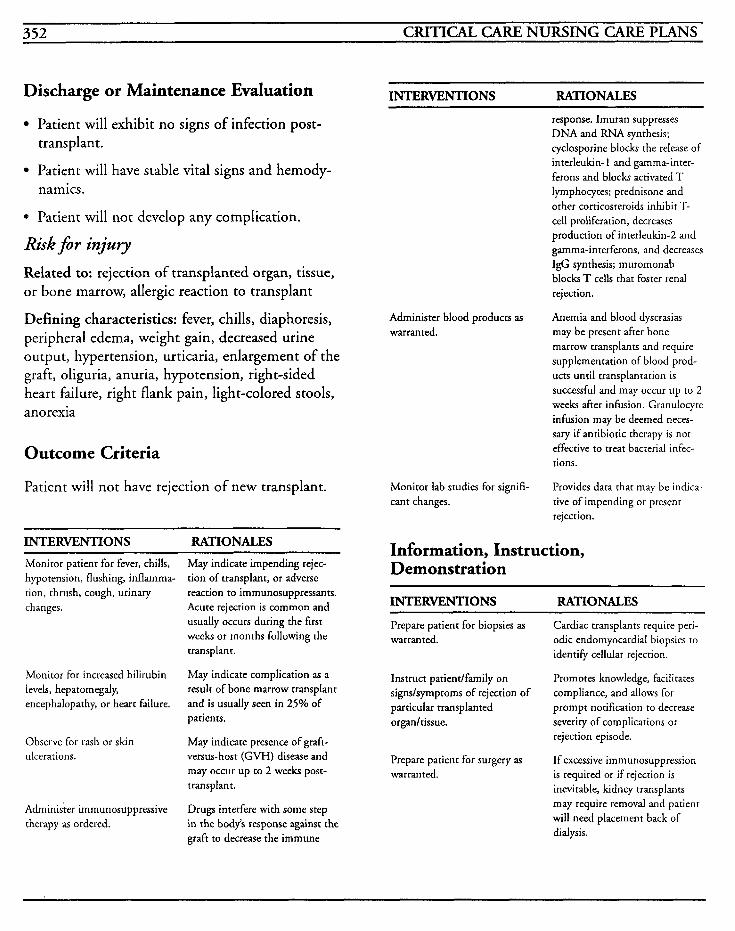
352 CRITICAL CARE NURSING CARE PLANS
Discharge or Maintenance Evaluation INTERVENTIONS RATIONALES
Patient will exhibit no signs of infection post- transplant.
Patient will have stable vital signs and hemody- namics.
0 Patient will not develop any complication.
Risk for injury Related to: rejection of transplanted organ, tissue, or bone marrow, allergic reaction to transplant
Defining characteristics: fever, chills, diaphoresis, peripheral edema, weight gain, decreased urine output, hypertension, urticaria, enlargement of the graft, oliguria, anuria, hypotension, right-sided heart failure, right flank pain, light-colored stools, anorexia
Outcome Criteria
Patient will not have rejection of new transplant.
INTERVENTIONS RATIONALES Monitor patient for fever, chills, hypotension, flushing, inflamma- tion, thrush, cough, urinary changes.
Monitor for increased bilirubin levels, hepatomegaly, encephalopathy, or heart failure.
Observe for rash or skin ulcerations.
Administer immunosuppressive therapy as ordered.
May indicate impending rejec- tion of transplant, or adverse reaction to immunosuppressants. Acute rejection is common and usually occurs during the first weeks or months following the transplant.
May indicate complication as a result of bone marrow transplant and is usually seen in 25% of patients.
May indicate presence of grafi- versus-host ( G W ) disease and may occur up to 2 weeks post- transplant.
Drugs interfere with some step in the body’s response against the graft to decrease the immune
Administer blood products as warranted.
Monitor lab studies for signifi- cant changes.
response. Imuran suppresses DNA and RNA synthesis; cyclosporine block the release of interleukin- 1 and gamma-inter- ferons and block activated T lymphocytes; prednisone and other corticosteroids inhibit T- cell proliferation, decreases production of interleukin-2 and gamma-interferons, and decreases IgG synthesis; muromonab blocks T cells that foster renal rejection.
Anemia and blood dyscrasias may be present after bone marrow transplants and require supplementation of blood prod- ucts until transplantation is successful and may occur up to 2 week after infusion. Granulocyte infusion may be deemed neces- sary if antibiotic therapy is not effective to treat bacterial infec- tions.
Provides data that may be indica- tive of impending or present rejection.
Information, Instruction, Demonstration
INTERVENTIONS RATIONALES
Prepare patient for biopsies as warranted.
Instruct patient/family on signslsymptoms of rejection of particular transplanted organ/tissue.
Prepare patient for surgery as warranted.
Cardiac transplants require peri- odic endomyocardial biopsies to identify cellular rejection.
Promotes knowledge, facilitates compliance, and allows for prompt notification to decrease severity of complications or rejection episode.
If excessive immunosuppression is required or if rejection is inevitable, kidney transplants may require removal and patient will need placement back of dialysis.
OTHER 353
INTERVENTIONS RATIONALES ~~
Instruct patient on all medica- tions taken, side effeccs, adverse effects, contraindications, and potential drug interactions.
Decreases risk of self-medication, and provides for prompt notifi- cation of adverse reactions that may require further intervention.
Discharge or Maintenance Evaluation
Patient will have minimal rejection of transplanted organ/tissue.
Patient will be able to comply with drug regi- men to prevent rejection.
Patient will be able to verbalize understanding of signslsymptoms to report to physician, and will be able to seek prompt medical care.
Patient will be cognizant of all medications being taken, purposes, and potential side effects, and will have no adverse reactions.
Patient will avoid further surgery.
Alteration in tissue perfision: curdiopulmonary, cerebra4 renai gastrointestinal, peripheral [See Renal Failure]
Related to: transplant rejection, allergic reactions, infection, pulmonary edema, DIC
Defining characteristics: oliguria, anuria, polyuria, fever, chills, increased white blood cell count, differential shift to the left, bleeding, ecchymoses, hematuria, guaiac positive stools, DIC, blood dyscrasias, decreased platelet count, headache, mental status changes, adventitious breath sounds, gallops, abnormal heart tones, dys- rhythmias, rashes, ulcerations, nausea, vomiting
Social isolation
Related to: changes in health status, changes in physical status, imposed physical isolation, inade- quate support system
Defining characteristics: feelings of loneliness, feelings of rejection, absence of family membedfriends, sad, dull affect, inappropriate behaviors
Outcome Criteria
Patient will be able to participate in activities as tolerated and be able to have effective interaction with people within confines of medical disease process.
INTERVENTIONS RATIONALES
Determine patient’s comprehen- sion of medical situation and rationales.
Utilize appropriate isolation techniques based on patient’s condition, and when possible, limit use of protective equipment.
Encourage visitation of family as much as possible. Provide a tele- phone so that patient may contact family and friends.
Identify significant family mem- bers or friends who are important to patient and involve them in care.
Assist patient to develop strate- gies for coping with isolation.
Contact social services, coun- selors, organizations, ministers, or other resources.
Identifies potential misconcep- tions and allows for realistic input to facilitate understanding.
Facilitates providing safe environ- ment for patient yet providing social interaction to decrease feelings of social isolation. Appropriate use of gcwns, mask;, and gloves may be required due to patient’s suppressed immune system.
Transplantation costs are high and done in major hospital set- tings, so that family members may not be able to travel great distances for the length of time the patient may be hospitalized. Methods of communication are important to promote feelings of inclusion in family matters.
Support systems decrease sense of isolation and loneliness and helps to reestablish communication.
Promotes feelings of self-control while developing goals for achievement.
May be helpful to continue care once patient is discharged, and may be able to facilitate support- ive encounters.
354 CRITICAL CARE NURSING CARE PLANS
Discharge or Maintenance Evaluation
Patient will be able to verbalize understanding of necessity for isolation procedures and will comply.
Patient will have fewer feelings of loneliness and isolation.
Patient will be able to meet sensory demands by family and friends.
Patient will be able to effectively access commu-
Alteration in comfort [See Cardiac Surgery]
Related to: transplant operation, invasive lines and catheters, immobility
Defining characteristics: communication of pain, facial grimacing, increased blood pressure, increased heart rate, diaphoresis, moaning, splinting
Alteration in skin integity [See Cardiac Surgery]
Related to: transplant operation, invasive lines and catheters, biopsies, wounds
Defining characteristics: surgical incisions, disrup- tion of skin surfaces, abrasions, redness, warmth, drainage
Knowledge deficit [See Renal Failure]
Related to: transplant operation, changes in health status, anxiety
Defining characteristics: lack of knowledge, pres- ence of preventable complications, verbalized questions
nity resources for referrals.
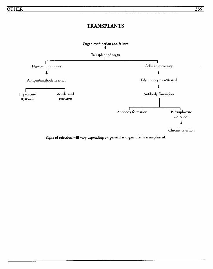
OTHER 355
TRANSPLANTS
Organ dysfunction and failure 4
Transplant of organ I
I I Humoral immunity Cellular immunity
4 4 Antigdantibody reaction T-lymphocytes activated
I + Hyperacute
rejection Accelerated
rejection
4 Antibody formation
I I Antibody formation B-lymphocyte
activation
J,
Chronic rejection
Signs of rejection will vary depending on particular organ that is transplanted.

This Page Intentionally Left Blank
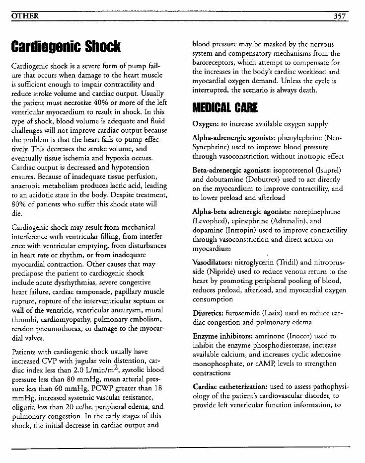
OTHER 357
Cardiogenic Shock Cardiogenic shock is a severe form of pump fail- ure that occurs when damage to the heart muscle is sufficient enough to impair contractility and reduce stroke volume and cardiac output. Usually the patient must necrotize 40% or more of the left ventricular myocardium to result in shock. In this type of shock, blood volume is adequate and fluid challenges will not improve cardiac output because the problem is that the heart fails to pump effec- tively. This decreases the stroke volume, and eventually tissue ischemia and hypoxia occurs. Cardiac output is decreased and hypotension ensures. Because of inadequate tissue perfusion, anaerobic metabolism produces lactic acid, leading to an acidotic state in the body. Despite treatment, 80% of patients who suffer this shock state will die.
Cardiogenic shock may result from mechanical interference with ventricular filling, from interfer- ence with ventricular emptying, from disturbances in heart rate or rhythm, or from inadequate myocardial contraction. Other causes that may predispose the patient to cardiogenic shock include acute dysrhythmias, severe congestive heart failure, cardiac tamponade, papillary muscle rupture, rupture of the interventricular septum or wall of the ventricle, ventricular aneurysm, mural throm bi, cardiom yopathy, pulmonary embolism, tension pneumothorax, or damage to the myocar- dial valves.
Patients with cardiogenic shock usually have increased CVP with jugular vein distention, car- diac index less than 2.0 Llmin/m2, systolic blood pressure less than 80 mmHg, mean arterial pres- sure less than 60 mmHg, PCWP greater than 18 mmHg, increased systemic vascular resistance, oliguria less than 20 cc/hr, peripheral edema, and pulmonary congestion. In the early stages of this shock, the initial decrease in cardiac output and
blood pressure may be masked by the nervous system and compensatory mechanisms from the baroreceptors, which attempt to compensate for the increases in the body’s cardiac workload and myocardial oxygen demand. Unless the cycle is interrupted, the scenario is always death.
Oxygen: to increase available oxygen supply
Alpha-adrenergic agonists: p henylep hrine (Neo- Synephrine) used to improve blood pressure through vasoconstriction without inotropic effect
Beta-adrenergic agonists: isoproterenol (Isuprel) and dobutamine (Dobutrex) used to act directly on the myocardium to improve contractility, and to lower preload and afterload
Alpha-beta adrenergic agonists: norepinephrine (Levophed), epinephrine (Adrenalin), and dopamine (Intropin) used to improve contractility through vasoconstriction and direct action on myocardium
Vasodilators: nitroglycerin (Tridil) and nitroprus- side (Nipride) used to reduce venous return to the heart by promoting peripheral pooling of blood, reduces preload, afterload, and myocardial oxygen consumption
Diuretics: furosemide (Lasix) used to reduce car- diac congestion and pulmonary edema
Enzyme inhibitors: amrinone (Inocor) used to inhibit the enzyme phosphodiesterase, increase available calcium, and increases cyclic adenosine monophosphate, or CAME levels to strengthen con tractions
Cardiac catheterization: used to assess pathophysi- ology of the patient’s cardiovascular disorder, to provide left ventricular function information, to
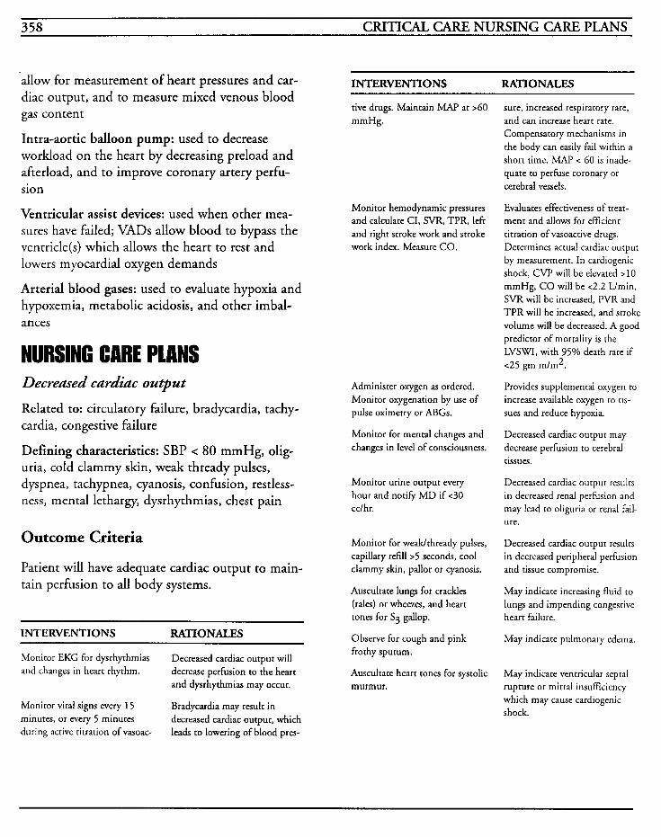
358 CRITICAL CARE NURSING CARE PLANS
allow for measurement of heart pressures and car- diac output, and to measure mixed venous blood gas content
Intra-aortic balloon pump: used to decrease workload on the heart by decreasing preload and afterload, and to improve coronary artery perfu- sion
Ventricular assist devices: used when other mea- sures have failed; VADs allow blood to bypass the ventricle(s) which allows the heart to rest and lowers myocardial oxygen demands
Arterial blood gases: used to evaluate hypoxia and hypoxemia, metabolic acidosis, and other imbal- ances
NURSING CARE PLANS Decreased cardiac output
Related to: circulatory failure, bradycardia, tachy- cardia, congestive failure
Defining characteristics: SBP < 80 mmHg, olig- uria, cold clammy skin, weak thready pulses, dyspnea, tachypnea, cyanosis, confusion, restless- ness, mental lethargy, dysrhythmias, chest pain
Outcome Criteria
Patient will have adequate cardiac output to main- tain perfusion to all body systems.
INTERVENTIONS RATIONALES
Monitor EKG for dysrhythmias and changes in heart rhythm.
Decreased cardiac output will decrease perfusion to the heart and dysrhythmias may occur.
Bradycardia may result in decreased cardiac output, which leads to lowering of blood pres-
Monitor vital signs every 15 minutes, or every 5 minutes during active titration of vasoac-
INTERVENTIONS RATIONALES
tive drugs. Maintain MAP at >GO mmHg.
Monitor hemodynamic pressures and calculate CI, SVR, TPR, left and right stroke work and stroke work index. Measure CO.
Administer oxygen as ordered. Monitor oxygenation by use of pulse oximetry or ABGs.
Monitor for mental changes and changes in level of consciousness.
Monitor urine output every hour and notify MD if <30 cclhr.
Monitor for weadthready pulses, capillary refill > 5 seconds, cool clammy skin, pallor or cyanosis.
Auscultate lungs for crackles (rales) or wheezes, and heart tones for S3 gallop.
Observe for cough and pink frothy sputum.
Auscultate heart tones for systolic murmur.
sure, increased respiratory rate, and can increase heart rate. Compensatory mechanisms in the body can easily fail within a short time. MAP < 60 is inade- quate to perfuse coronary or cerebral vessels.
Evaluates effectiveness of treat- ment and allows for efficient titration of vasoactive drugs. Determines actual cardiac output by measurement. In cardiogenic shock, CVP will be elevated >10 mmHg, CO will be <2.2 L/min, SVR will be increased, PVR and TPR will be increased, and stroke volume will be decreased. A good predictor of mortality is the LVSWI, with 95% death rate if <25 gm m/m2.
Provides supplemental oxygen to increase available oxygen to us- sues and reduce hypoxia.
Decreased cardiac output may decrease perfusion to cerebral tissues.
Decreased cardiac output results in decreased renal perfusion and may lead to oliguria or renal fail- ure.
Decreased cardiac output results in decreased peripheral perfusion and tissue compromise.
May indicate increasing fluid to lungs and impending congestive heart failure.
May indicate pulmonary edema.
May indicate ventricular septal rupture or mitral insuGciency which may cause cardiogenic shock.
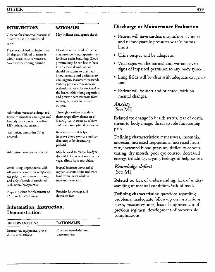
OTHER 359
INTERVENTIONS RATIONALES Discharge or Maintenance Evaluation Observe for abnormal precordial movement at 3-5 intercostal space.
Place head of bed no higher than 30 degrees if blood pressure is within acceptable parameters. Avoid trendelenburg position.
Administer vasoactive drugs and titrate to maintain vital signs and hemodynamic pressures within MD ordered parameters.
Administer morphine IV as ordered.
Administer atropine as ordered.
Avoid using isoproterenol with MI patients except for temporary use prior to transvenous pacing, and only if shock is associated with severe bradycardia.
Prepare patient for placement on IABP or for VAD usage.
May indicate cardiogenic shock.
Elevation of the head of the bed may promote lung expansion and facilitate easier breathing. Blood pressure may be too low to have HOB elevated and patient should be supine to maintain blood pressure and perfusion to vital organs. Placement in trende- lenburg position may increase preload, increase the workload on the heart, inhibit lung expansion, and prevent baroreceptors from sensing decreases in cardiac output.
Through a variety of actions, these drugs allow alteration of hemodynamic status to achieve and maintain optimal perfusion.
Relieves pain and helps to improve blood pressure and car- diac output by decreasing preload.
May be used to reverse bradycar- dia and help prevent some of the vagal effects from morphine.
Isuprel increases myocardial oxygen consumption and work- load of the heart while it increases heart rate.
Provides knowledge and decreases fear.
Information, Instruction, Demonstration
INTERVENTIONS RATIONALES
Instruct on equipment, proce- dures, medications. decreases fear.
Provides knowledge and
Patient will have cardiac output/cardiac index and hemodynamic pressures within normal limits.
Urine output will be adequate.
Vital signs will be normal and without overt signs of impaired perfusion to any body system.
Lung fields will be clear with adequate oxygena- tion.
Patient will be alert and oriented, with no mental changes.
Anxiety [See MI] Related to: change in health status, fear of death, threat to body image, threat to role functioning, pain
Defining characteristics: restlessness, insomnia, anorexia, increased respirations, increased heart rate, increased blood pressure, difficulty concen- trating, dry mouth, poor eye contact, decreased energy, irritability, crying, feelings of helplessness
KnowZedge &$kit [See MI] Related to: lack of understanding, lack of under- standing of medical condition, lack of recall
Defining characteristics: questions regarding problems, inadequate follow-up on instructions given, misconceptions, lack of improvement of previous regimen, development of preventable complications
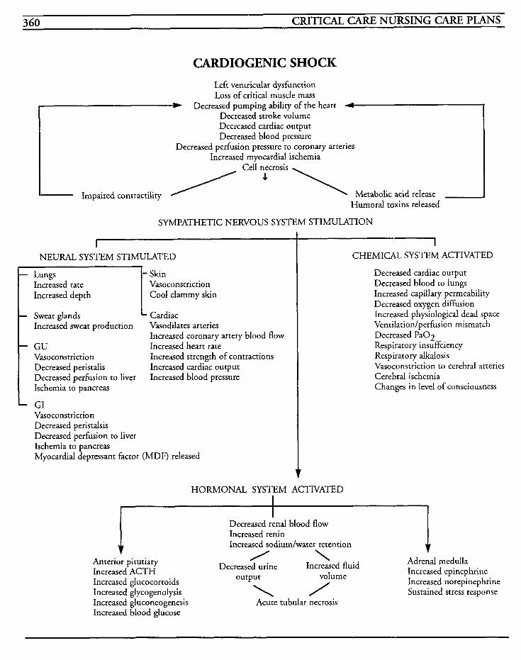
360 CRITICAL CARE NURSING CARE PLANS
CARDIOGENIC SHOCK Left ventricular dysfunction Loss of critical muscle mass * Decreased pumping ability of the heart 4
Decreased stroke volume Decreased cardiac output Decreased blood pressure
Decreased perfusion pressure to coronary arteries Increased myocardial ischemia
Cell necrosis
Metabolic acid release Humoral toxins released
Impaired contractility / + \
SYMPATHETIC NERVOUS SYSTEM STIMULATION
I NEURAL SYSTEM STIMULATED
- Skin Increased rate Vasoconstriction Increased depth Cool clammy skin
Sweat glands Cardiac Increased sweat production Vasodilates arteries
GU Increased heart rate Vasoconstriction Increased strength of contractions Decreased peristalis Increased cardiac output Decreased perfusion to liver Increased blood pressure Ischemia to pancreas
GI Vasoconstriction Decreased peristalsis Decreased perfusion to liver Ischemia to pancreas Myocardial depressant factor (MDF) released
Increased coronary artery blood flow
HORMONAL SYSTEM
CHEMICAL SYSTEM ACTIVATED
Decreased cardiac output Decreased blood to lungs Increased capillary permeability Decreased oxygen diffusion Increased physiological dead space VentiIation/perfusion mismatch Decreased Pa02 Respiratory insuffciency Respiratory alkalosis Vasoconstriction to cerebral arteries Cerebral ischemia Changes in level of consciousness
ACTIVATED
Decreased renal blood flow Increased renin Increased sodiumlwater retention
Adrenal medulla Increased epinephrine Increased norepinephrine Sustained stress response
Increased gluconeogenesis Increased blood glucose
Acute tubular necrosis

~ ~~
INDEX OF NURSING DIAGNOSES 361

This Page Intentionally Left Blank
INDEX OF NURSING DIAGNOSES 363
Activity intolerance 92, 203, 235
Alteration in comfort 15, 26, 50, 63, 70, 79, 107, 163, 170, 182, 196,208,214,292,299,316,326,346, 354
Alteration in nutrition: less than body requirements 204, 208,214,218,235,244,253,326
Alteration in skin integrity 62, 73, 190, 354
Alteration in temperature regulation 161
Alteration in thought processes 152, 218
Alteration in tissue perfusion: cardiopulmonary, cerebral GO
Alteration in tissue perfusion: cardiopulmonary, cerebral, gastrointestinal, peripheral, renal 78
Alteration in tissue perfusion: cardiopulmonary, cerebral, renal, gastrointestinal, peripheral 353
Alteration in tissue perfusion: cardiopulmonary, renal, cere- bral, gastrointestinal, peripheral 233
Alteration in tissue perfusion: cerebral 140, 149, 181, 186, 189
Alteration in tissue perfusion: peripheral 47, 299
Alteration in tissue perfusion: peripheral, cerebral, cardiopul- monary, renal, gastrointestinal 314
Alteration in tissue perfusion: renal, cardiopulmonary, cere- bral, gastrointestinal, peripheral 242
Alteration in nutrition: less than body requirements 262, 277
Altered nutrition: less than body requirements 94, 131, 196
Altered oral mucous membrane 13 1, 247
Altered tissue perfusion: cardiopulmonary, cerebral, gastroin- testinal, peripheral, and renal 273
Altered tissue perfusion: cardiopulmonary) cerebral, gastroin- testinal, renal, peripheral 54
Altered tissue perfusion: cardiopulmonary, cerebral, periph- eral 17
Altered tissue perfusion: cardiopulmonary, renal, peripheral,
cerebral 26
Anticipatory grieving 303
Anxiety 10, 27, 36, 42, 63, 74, 86, 92, 128, 196, 248, 275, 285,359
Anxiety, fear 173
Body image disturbance 302
Bowel incontinence 164
Constipation 277
Decreased cardiac output 6, 18, 27, 34, 86, 278, 358
Disturbance in body image 221
Disturbance in self-esteem 177
Disturbance of body image 64,75
Dyshnctional ventilatory wean response 133
Fatigue 256
FearJAnxiety 326, 347
Fluid volume deficit 194, 208, 219, 251, 261,269
Fluid volume excess 4, 240, 265
Grieving 178
Hyperthermia 34, 176, 182, 282, 319, 332
Impaired gas exchange 8, 86, 92, 101, 107, 123,254, 277, 307,347
Impaired physical mobility 64,74, 141, 154, 162, 169, 293, 326
Impaired skin integrity 219, 295, 301, 326, 346
Impaired verbal communication 127, 141, 170
Ineffective airway clearance 86, 92, 106, 118, 122
Ineffective breathing pattern 8692, 100, 112, 124, 160, 220
Ineffective individual/family coping 93, 129
Ineffective thermoregulation 3 14
Inneffective in breathing pattern 1 GO
Knowledge deficit 10,23,28,36,43, 50,65,74, 81,86, 94, 107, 135, 197,205,235,257,270,286,297,354,359
364 CRITICAL CARE NURSING CARE PLANS
Knowledge deficit 10, 23, 28, 36, 43, 50, 65, 74, 81, 86, 94, 107, 135, 197,205,235,257,270,286,297,354,359
Potential for alteration in tissue perfusion: peripheral 262
Potential for infection 130
Potential for injury 63, 74, 208
Potential for injury: hypoglycemia 256, 262
Risk for alteration in nutrition: less than body requirements 155, 170
Risk for alteration in tissue perfusion: cardiopulmonary, peripheral, renal 170
Risk for alteration in tissue perfusion: cerebral, cardiopul- monary, gastrointestinal, renal, and peripheral 200
Risk for alteration in tissue perfusion: cerebral, gastrointesti- nal, cardiopulmonary, renal, and peripheral 332
Risk for alteration in tissue perfusion: peripheral, cardiopul- monary, renal, cerebral 345
Risk for altered nutrition: less than body requirements 284, 320
Risk for altered tissue perfusion: cardiopulmonary, cerebral, renal, gastrointestinal, and peripheral 32, 41
Risk for altered tissue perfusion: cardiopulmonary, periph- eral, cerebral 101
Risk for altered tissue perfusion: gastrointestinal, cerebral, cardiopulmonary, renal, peripheral 196
Risk for altered nutrition: less than body requirements 107
Risk for constipation 172
Risk for decreased cardiac output 40, 68, 101, 160, 189, 200,283, 320
Risk for dysreflexia 165
Risk for fluid volume deficit 57, 87,107, 176, 214, 226, 230,234,242, 301,324,333,344
Risk for fluid volume excess 20, 87,107
Risk for impaired gas exchange 175, 208, 226, 229, 320, 326,333
Risk for impaired skin integrity 9,49, 81 , 163, 204, 246
Risk for impaired swallowing 142
Risk for ineffective airway clearance 175, 325
Risk for ineffective breathing pattern 15 1 , 169, 337
Risk for ineffective breathing pattedimpaired gas exchange 71
Risk for ineffective individual coping 200, 340
Risk for infection 35, 56, 94, 153, 188, 204, 212, 245, 255, 296,300,316, 327,332,351
Risk for injury 176, 181, 189, 200, 212, 220, 226, 230, 247,266,284,338,352
Risk for peripheral neurovascular dysfunction 294
Risk for urinary retention 171
Risk for violence directed at self 339
Self-care deficit: bathing, dressing, feeding, toileting 143
Sensory-perception alteration (visual, thought processes, kinesthetic) 277
Sensory-perceptual alteration 163
Sensory-perceptual alterations: visual, kinesthetic, gustatory, tactile 170
Sensory-perceptual alterations: visual, kinesthetic, gustatory, tactile, olfactory 142
Social isolation 353
Urinary retention 164

REFERENCES 365
REFERENCES

This Page Intentionally Left Blank
REFERENCES 367
Books
Berkow, Robert, Editor. The Merck Manual, 16th Ed., Merck, Sharp, & Dohme Inc., Rahway, NJ, 1993.
Guyton, Arthur. Human Physiolow and Mechanisms of Disease, 6th Ed., W. B. Saunders Co., Philadelphia, PA, 1997.
Guyton, Arthur. Textbook of Medical Physioloc, 9th Ed., W. B. Saunders Co., Philadelphia, PA, 1995.
Minssen, Beth. Critical Care Core Curriculum, 6th Ed., Panhandle Education For Nurses, Lubbock, TX, 1995.
Minssen, Beth. Multi& Orvan Failure Syndrome, 2nd ed., Panhandle Education For Nurses, Lubbock, TX, 1995.
Skidmore-Roth, Linda. Mosbv’s 1997 Nursinc Druc Reference, Mosby Year-Book Inc., St. Louis, MO, 1997.
Suddarth, Doris Smith. The Lippincott Manual of Nursin? Practice, 6th Ed., J. B. Lippincott, Philadelphia, PA, 1996.
Swearington, Pamela and Keen, Janet. Manual of Critical Care, 3rd Ed., Mosby, St. Louis, MO, 1995.
Tierney, Lawrence M., et al., Current Medical Diamosis and Treatment, 35th ed., Appleton and Lange, Stamford, CT, 1996.
Periodicals
Ahrens, Susan G. “Managing Heart Failure: A Blueprint for Success,” Nursing 95, 25 (12): 26-31, 1995.
Blanford, Nickie. “Renal Transplantation: A Case Study of the Ideal,” Critical Care Nurse, 13 (1): 46-55, 1993.
Bright, Linda D. “Deep Vein Thrombosis,” American Journal of Nursing, 95 (6): 48-49, 1995.
368 CRITICAL CARE NURSING CARE PLANS
Huston, C. J. and Boelman, R. “Autonomic Dysreflexia,” American Journal of Nursing, 95 (6): 55, 1995.
Mee, Cheryl L. “Ventilator Alarms-How to Respond with Confidence,” Nursing 95, 25 (7): 61-64, 1995.
Meissner, Judith E. “Caring for Patients with Meningitis,” Nursing 95, 25 (7): 50-5 1, 1995.
Merkley, Kathleen. “Assessing Chest Pain,” RN, 57 (6): 58-62, 1994.
Teplitz, Linda. “Hypertensive Crisis: Review and Update,” Critical Care Nurse, 13 (6): 20-35, 1993.
Weinman, Steven A. “Emergency Management of Drug Overdose,” Critical Care Nurse, 13 (6): 45-5 1 , 1993.
Career CollegesKomrnunity Colleges and Post Secondary Vo-Techs For desk or review copies call: 1-800-477-3692 or fax 1-518-464-0301 For orders call: 1-800-347-7707 or fax 1-606-647-5023 Mail to: ITP Career Education
Attn: Order Fulfillment P.O. Box 6904 Florence, KY 41022 Email: [email protected]
Four-Year CollegeKJniversity For desk or review copies call: 1-800-423-0563 or fax 1-606-647-5020 For orders call: 1-800-354-9706 or fax 1-800-487-8488 Mail to: ITP Higher Education
Attn: Order Fulfillment P.O. Box 6904 Florence, KY 41022
Business, Industry and Government For orders call: 1-800-347-7707, ext. 4 or fax 1-606-647-5963 Mail to: ITP Business, Industry, Government
Attn: Order Fulfillment P.O. Box 6904 Florence, KY 41022 All other orders and inquiries 1-800-347-7707, ext. 4
Retail Mail to: International Thomson Publishing
Attn: Professional/Technical Order Fulfillment P.O. Box 6904 Florence, KY 41022 Phone: 1-800-842-3636 or fax 1-606-647-5963
High Schools and Secondary Vo-techs For desk or review copies call: 1-800-824-5179 or fax 1-800-453-7882 For orders call: 1-800-354-9706 or fax 1-800-487-8488 Mail to: ITP School
Attn: Order Fulfillment P.O. Box 6904 Florence, KY 41022
Canada Mail to: Nelson ITP Canada
1120 Birchmount Road Scarborough, Ontario M1K 5G4 Canada
Telephone Number: 1-416-752-9448 or 1-800-268-2222 Fax Number: 1-416-752-8101 or 1-800-430-4445 E-mail: [email protected]
International Ordering Mail to: International Thomson Learning
P.O. Box 6904 Florence, KY 41022 Phone: 1-606-282-5786 Fax: 1-606-282-5700