nuclear factor-i and activator protein-2 bind in a mutually exclusive way to overlapping promoter...
TRANSCRIPT

Nucleic Acids Research, Vol. 18, No. I
Nuclear factor-I and activator protein-2 bind in a mutuallyexclusive way to overlapping promoter sequences andtrans-activate the human growth hormone gene
Stephane J.Courtois, Dominique A.Lafontaine, Frederic P.Lemaigre, Serge M.Durviaux and GuyG.Rousseau*Hormone and Metabolic Research Unit, Louvain University Medical School and International Instituteof Cellular and Molecular Pathology, 75 avenue Hippocrate, B-1200 Brussels, Belgium
Received October 24, 1989; Accepted November 30, 1989
ABSTRACT
Transcription of the human growth hormone (hGH)gene and its regulation are controlled by trans-actingfactors that bind to hGH gene promoter sequences.Several DNase I footprints have been described within500 bp of this promoter, one of which (- 289 to - 267)has not yet been ascribed to a defined factor. By DNaseI footprinting, gel mobility shift, and methylationinterference assays with extracts from HeLa cells andGH-producing pituitary tumor (GC) cells, we show thatthis factor belongs to the NF-I family. When NF-I wascompeted out of the cell extracts, the trans-actingfactor AP-2 bound to the same site as NF-1. AP-2 waspresent not only in HeLa cells, but also in GC cells albeitat a much lower concentration. Consistent with themutually exclusive binding of NF-I and AP-2, theirmethylation interference patterns included four guanineresidues that were crucial for binding of both NF-I andAP-2. Cell-free transcription from the hGH genepromoter showed that these two factors can trans-activate this gene.
INTRODUCTION
The growth hormone (GH) gene is an interesting model forstudying the control of gene expression by hormones and trans-acting factors. The human hGH-N (hGH-1) gene is expressedexclusively in the somatotrophs of the anterior pituitary. Itstranscription is stimulated by GHRH via cAMP and byglucocorticoids (1). The mechanism of these hormonal effects,which probably involve cis-acting hGH gene promoter sequences,is ill-understood. The tissue-specificity of expression of the hGHgene is ascribed, at least in part, to a pituitary-specific factorcalled GHF-1/Pit-I (2). This homeobox-containing protein (3)binds to two contiguous sites on the promoter, thereby protectingthese sites against cleavage by deoxyribonuclease I (DNase I)(footprints -92 to -65 and -130 to -105).Other factors, which also occur in nonpituitary cells such as
HeLa cells, bind to the hGH gene promoter and may contributeto controlling its basal and (or) hormone-dependent transcription.
The interaction of some of these factors is revealed by the DNaseI footprints observed on the hGH gene promoter when it isincubated with extracts from pituitary tumor (GC) cells or HeLacells. One of these footprints, called growth hormone footprint2 (GHF2), extends from -139 to -115 and is due to the bindingof a factor indistinguishable from Spl (4). Another footprintcalled GHF3 extends from -289 to -253. This footprint is dueto factor(s) that appears to contribute to the control ofhGH gene
transcription (5). While the proximal part of GHF3 (GHF3p,-266 to -253) reflects the binding of upstream stimulatingfactor/major late transcription factor (USF/MLTF) (4), the distalpart of this footprint (GHF3d) has not yet been ascribed to a
particular factor. Although activator protein 2 (AP-2) purifiedfrom HeLa cells had been reported (6) to bind on the hGH gene
promoter from -287 to -265, we had shown (4) that GHF3dis probably due to another factor. We have now investigated thisquestion by DNase I footprinting, gel mobility shift, andmethylation interference assays. We have also evaluated thefunctional significance of factor(s) binding to GHF3d by cell-free transcription experiments.
MATERIALS AND METHODSMaterialsEnzymes were purchased from Promega Biotec or Boehringer.Radiolabeled compounds and reverse transcriptase (AMV) werefrom Amersham International plc. Poly(dI-dC) was from Sigma,dimethylsulfate from Aldrich, and piperidine from Merck. Tissueculture components were from Flow Laboratories or from GibcoBRL.
OligonucleotidesThe following double-stranded oligodeoxyribonucleotides (oligo)were from Eurogentec (Liege, Belgium) except oligo AP-2 whichwas a gift from Dr. D. Christophe (Free University of Brussels).Oligo NF-I corresponds to the high-affinity binding site (+21to +44) in the adenovirus origin of replication, modified in sucha way (underlined) that a putative AP-2 consensus sequence iseliminated, 5' ATTTlTGGCTACAAGCCAATATGAT 3'. Oligo
* To whom correspondence should be addressed
57
.=) 1990 Oxford University Press

58 Nucleic Acids Research
GHF3d corresponds to the distal moiety (-289 to -267) ofGHF3, 5' ACCATGGCCTGCGGCCAGAGGGC 3', plus a 5'TCGA end. Oligo AP-2 corresponds to the high-affinity AP-2binding site on the human metallothionein-IIA gene from -188to - 166, 5' GAACTGACCGCCCGCGGCCCGTG 3', plus a5' AT end and a 3' CATG end on the coding strand only. OligoGHF-1 corresponds to the proximal GHF-1/Pit-1 binding siteon the hGH gene promoter (-88 to -69) plus BamHI compatibleends, 5' GATCCCATGCATAAATGTACACAG 3'.
Plasmid constructionsPlasmid phGH494 A -260/ - 188 A - 149/-57 contains a 497-bpEcoRI- BamHI (-494 to +3) insert of the hGH-1 gene inpBR322 with deletion of nucleotides -260 to - 188 and - 149to - 57. This construction is convenient to visualize the footprintsdescribed. Plasmid phGH37-L, used as an internal control forcell-free transcription, contains a 40 bp HaeIl-BamHI (-37 to+ 3) insert of the hGH- 1 gene in pBR322 with insertion of aBamHI-SmaI linker downstream from + 3. The deletion mutantp13-13 (7) contains 237 bp of the long terminal repeat (LTR)of the mouse mammary tumor virus (MMTV) upstream of thetranscription initiation (cap) site.
Cells and cell extractsHuman cervix carcinoma (HeLa S3) and rat pituitary tumor (GC)cells were grown in modified Eagle's medium for suspensioncultures, supplemented with 10% newborn calf serum (HeLacells) or with 5% foetal calf serum plus 12.5% horse serum (GCcells). Whole cell extracts (10-20 mg protein/ml) were preparedas described (8), except that 0.75 instead of one volume of saturedammonium sulfate solution was added for the first precipitationstep of GC cell extracts.
Binding assaysDNase I footprinting and gel mobility shift assays were performedas described (4). For the methylation interference assay, theprocedure of Chodosh et al. (9) was used with minormodifications. Single stranded oligodeoxyribonucleotides werelabeled with y-[32P]-ATP in the presence of T4 polynucleotidekinase and hybridized with the unlabeled complementary strandas described (4). Probes were partially methylated withdimethylsulfate and used in a binding reaction, with themodification that each incubation was scaled up to 30 y1 andcontained 50,000 cpm of the appropriate probe, 8 yg of poly(dI-dC) and 120 yg of cell extract protein. For NF-I contact pointsanalysis, all incubations also contained 50 ng (GC extracts) or150 ng (HeLa extracts) of oligo AP-2 to prevent binding of thisfactor to the radioactive probe. For AP-2 contact points analysis,the incubation contained 100 ng of oligo NF-I to prevent bindingof NF-I to the radioactive probe. After incubation at roomtemperature for 30 min, samples were loaded on a low ionicstrength 5% polyacrylamide gel and electrophorezed accordingto Carthew et al. (10). Overnight autoradiography at 40 Cidentified free DNA (F) and bound protein-DNA complexes (B)which were isolated from the gel matrix by horizontal migrationonto NA-45 DEAE membranes (Schleicher and Schuell). DNAwas eluted as described (11), phenol/chloroform extracted, andpurified by anion exchange chromatography (Qiagen-tip 5). Aftertwo ethanol precipitations, the DNA was piperidine-cleaved,lyophilized, electrophorezed on a 18 % polyacrylamide-8 M ureagel, and visualized by autoradiography.
Cell-free transcription and primer extensionFor in vitro transcription experiments, each incubation contained100 Itg of cell extract protein, 200 ng of circular templatephGH494 A-260/-188 A-149/-57 and 150-200 ng ofcircular phGH37-L template used as internal control, in 10 y1of 20 mM Tris.Cl (pH 7.9), 50 mM KCI, 6.25 mM MgCl2,0.05 mM EDTA, 1 mM dithiothreitol and 8.5% glycerol.Incubations lasted for 90 min at 30 'C. After DNase I treatmentRNA's were hybridized with 30,000 cpm of labeled primer in15 Al of 50 mM Tris.Cl (pH 8.0), 50 mM KCI, 8 mM MgCl2,4 mM dithiothreitol and were heated for 2 h at 600 C. The primer,complementary to nucleotides 436 to 418 of pBR322, was labeledwith 'y-[32P]-ATP in presence of T4 polynucleotide kinase. Theduplexes were incubated at 420 C for 30 min in the same bufferplus 1 mM dNTP, 80 ag/ml of actinomycin Cl and 11.5 unitsof AMV reverse transcriptase. Nucleic acids were analyzed onan 8 M urea, 8% polyacrylamide gel, autoradiographed andquantified by densitometry (Joyce Loebl Chromoscan 6).Transcripts from the two templates were distinguishable becausethose from phGH37-L were 12 bp longer, due to the insertionin the latter of the BamHI-SmaI linker.
RESULTSNF-I interacts with the hGH gene promoterExamination of the sequence corresponding to GHF3d (Table1) shows that it contains two overlapping putative binding sites,one for AP-2 and one for nuclear factor I (NF-I). Each of thesesequences contains one mismatch with the consensus. The AP-2mismatch does not prevent binding of purified AP-2 (6). Wetherefore investigated whether NF-I can also bind to this regionof the hGH gene promoter. NF-I actually refers to a family ofubiquitous nuclear proteins which are differently expresseddepending on the tissue and species (12). The footprints shownin Fig. 1 demonstrate that our cell extracts contained NF-I andthat the latter can bind to the GHF3d sequence. The promoterof the LTR of MMTV known to contain a high affinity bindingsite for NF-I (13) was incubated with HeLa (lanes 2-5) or GC(lanes 6-9) cell extracts. A typical NF-I footprint was observed(lanes 2 and 6) at the expected coordinates, i.e. from -81 to-58 relative to the cap site. This NF-I binding was preventedby competing oligo GHF3d (lanes 3 and 7), but not by additionof two unrelated oligos such as AP-2 (lanes 4 and 8) or USF(lanes 5 and 9). We conclude that NF-I can recognize the GHF3dsequence despite the mismatch with the consensus.Gel mobility shift assays provided additional evidence for the
binding of NF-I to the hGH gene promoter from -289 to -267.When labeled oligo GHF3d was incubated with HeLa or GC cellextracts a wide retarded band was observed, corresponding toprotein-GHF3d interactions (Fig. 2, lanes 2 and 5). A 50-foldmolar excess of unlabeled oligo GHF3d inhibited the formationof this complex (lanes 3 and 6), which was therefore consideredas specific. Indeed, an unrelated oligonucleotide (oligo USF) wasunable to prevent the formation of this oligo GHF3d-proteincomplex (lanes 4 and 7). The width of this specific complex couldreflect the polypeptidic heterogeneity of NF-I which generatesclosely migrating bands in gel mobility shift experiments (14).The fact that this interaction persisted under high salt conditions(150 mM KCI) is also in favor of complexes involving NF-I (14,15). A qualitative difference was seen between the data obtainedwith HeLa and GC cell extracts. The specific complex producedin HeLa cell extracts migrated more slowly than the one produced

Nucleic Acids Research 59
Table 1. Corrparison of the hGH gene promoter sequence (GHF3d) protectedagainst DNase I cleavage by GC and HeLa cell extracts, with the consensusbinding sites for AP-2 and NF-I.
AP-2 consensus 5' GCCTGGGG 3'
11111 11GHF3d 5' (-289) ACCATGGCCTGCGGCCAGAGGGC (-267) 3'
NF-I consensus 5' TGG NNNNNGCCAA 3'A
Identities between sequences are indicated by vertical bars. N refers to anynucleotide. The consensus for AP-2 and NF-I are taken from refs. 20 and 15,respectively.
in GC cell extracts (compare lanes 2 and 5 in Fig. 2). This isconsistent with the different tissue and species expression of thepolypeptides of the NF-I family, but does not rule out theinvolvement of unrelated proteins, since different factors can bindto a same DNA sequence (16). We investigated this question bymethylation interference experiments.
Despite their heterogeneity, the members of the NF-I familyshare the same DNA binding domain (17, 18) and are thereforeexpected to produce the same methylation interference patternon their specific DNA target. Double-stranded oligo GHF3dlabeled at one 5' end was partially methylated withdimethylsulfate and used in a binding reaction with HeLa or GCcell extracts. Despite the difference seen in band shift betweenHeLa and GC cell extracts, the methylation interference patternwas identical with these two extracts (Fig. 3). Data with thecoding (Fig. 3A) and the noncoding (Fig. 3B) strands show thatthe guanines that are essential for the interaction are locatedexclusively in the two boxes of the palindromic NF-I consensussequence. On the coding strand and in the 5'half palindrome ofthe noncoding strand, these guanines correspond (Table 2) tothose described to be crucial for the interaction between NF-Iand its binding site on the adenovirus replication origin (19). Weconclude that polypeptides of the NF-I family present in HeLaand GC cell extracts interact with the same nucleotides of thesequence from -289 to -267 of the hGH gene promoter.
Mutually exclusive binding of NF-I and AP-2To investigate the possible involvement of AP-2 in the GHF3dseen with GC cell extracts, we first determined whether thisprotein was present in such extracts. Indeed, AP-2 is not strictlytissue-specific (20) and has not been described in pituitary cells.Gel mobility shift experiments were performed with labeled oligoGHF3d incubated with GC cell extracts in the presence ofcompeting oligo NF-I to prevent binding of NF-I to GHF3d (Fig.4A). Three retarded bands were seen (lane 5), one of which(arrow) disappeared when the incubation was conducted inpresence of oligo AP-2 (lane 6). This band was thereforeconsidered as revealing a specific interaction between AP-2 andthe GHF3d sequence. The retarded bands which did not disappearunder competition conditions were considered as nonspecific. Thevery weak intensity of the AP-2 specific band as compared tothat with HeLa cell extracts (see Fig. 4B below) probably reflects
Helea GC Cell extract
L6 W- CompetitorM L&. <. ;I ;
U-I1i1111lx.z4II * Of^
* -~~58i
40 S~~~~~~~~~S
Fig. 1 DNase I footprinting of NF-I on the LTR of MMTV. The DNA probeconsists of a 438 bp-long BamHI-RsaI fragment of p13-13 labeled at the BamHIend of the coding strand. The probe was incubated without (lane 1) or with 45 jigHeLa (lanes 2-5) or 60 tg GC (lanes 6-9) cell extract protein. A 200-fold ex-cess (30 ng) of unlabeled oligo GHF3d or oligo AP-2 or (unrelated) oligo USFwas present in the incubations as indicated above the lanes. The radioactive sizemarker lane (M) is pBR322 digested by HpaII.
a low relative concentration of AP-2 in GC cell extracts. TheAP-2 complex observed with GC and Hela extracts migrated withthe same mobility.The coexistence of AP-2 and NF-I in our cell extracts raised
the question of their simultaneous or mutually exclusive bindingon the target DNA sequence, the more so as most of thenucleotides involved in the AP-2 consensus lie between those ofthe NF-I consensus (Table 1). To approach this question, we firstperformed gel mobility shift assays with labeled oligo GHF3d

60 Nucleic Acids Research
incubated with GC (Fig. 4A) or HeLa (Fig. 4B) cell extracts inthe absence or presence of competing unlabeled oligonucleotides.With HeLa cell extracts, a specific oligo GHF3d-protein complexwas seen (Fig. 4B, lane 2), which disappeared with competingoligo GHF3d (lane 3), but not with an unrelated (USF) oligo(lane 7), as expected. Addition of competing oligo AP-2 did notmodify this specific complex (lane 4). In contrast, incubation withcompeting oligo NF-I led to the disappearance of the GHF3d-protein complex and the appearance of a narrower band in thesame region of the gel (lane 5).This new complex was AP-2specific since it disappeared when oligo AP-2 was added to the
'IIWWtIa w(I ,l, |o
Bom...d
Fig. 2 Detection of protein-oligo GHF3d complexes by gel mobility shift assay.Labeled oligo GHF3d was incubated without (lane 1) or with 20 Ag HeLa (lanes2-4) or 20 itg GC (lanes 5-7) cell extract protein. A 50-fold excess (15 ng)of unlabeled oligo GHF3d or (unrelated) oligo (USF) was added as indicatedabove each lane. The specific protein-DNA complexes are indicated in the margin.All incubations contained 150 mM KCI.
incubation containing oligo NF-I (lane 6). The same pattern ofspecific complexes was obtained with these competingoligonucleotides in GC cell extracts (Fig. 4A). These experimentssuggest that the specific GHF3d-protein complex seen in our gelmobility shift assays results from an interaction with NF-I. Thebinding of AP-2 on the GHF3d sequence can be detected onlywhen binding of NF-I is prevented. Thus, the binding of NF-Iand AP-2 appears to be mutually exclusive in our experimentalconditions.
If the GHF3d footprint does correspond to the binding of eitherNF-I or AP-2, then the methylation interference pattern of thebinding observed in absence of NF-I should involve the AP-2consensus. The methylation pattern observed when NF-I wastitrated out of the incubation (Fig. 5 and Table 2) was indeedclearly different from that detected by NF-I contact point analysis.Six of the seven guanines that were important for the interactionof the protein with the two DNA strands are part of the AP-2consensus. The seventh guanine (noncoding strand) is just nextto the 5' end of this consensus.
Gel mobility shifts with the isolated oligo GHF3d sequencedo not necessarily reflect the situation with the intact promoter.Indeed, DNA-protein interactions may be influenced by flankingpromoter sequences. To evaluate the actual contribution of NF-I and AP-2 to the GHF3d, we performed the DNase I footprintingexperiments shown in Figure 6. The expected GHF3 footprintwas seen on the hGH gene promoter with the two cell extracts(lanes 2 and 6). To visualize only GHF3d, competing oligo USFwas included (lanes 3-5, 7-9). When competing oligo AP-2was added (lanes 3,7) the GHF3d footprint was not modified,as described earlier (4). This ruled out the direct involvementof AP-2 in GHF3d under these conditions. With Hela cellextracts, addition of a 200-fold excess of competing oligo NF-Iinstead of oligo AP-2 (lane 4) did not completely prevent thefootprint either, as if another protein were now interacting withthe promoter. This protein was AP-2 since concomitant additionof competing oligo NF-I and oligo AP-2 totally prevented this
A
9..
..~~~~~~~~~~.8,.W:
I$* 9-. 01V
"w
.W.: ob .*.'
:v3,;: .4
*~~~ ~ ~ ~ ~ ~ ~ ~ ~ ~ i* Q
Fig. 3 Methylation interference analysis of NF-I/DNA interactions at the GHF3d site. Oligo GHF3d labeled either on the coding strand (A) or on the noncodingstrand (B) was incubated with HeLa or GC cell extracts as described in Materials and Methods. Coding and noncoding sequences are shown along the gel. Theconsensus sequence for NF-I is boxed, with the mismatch indicated by the dashed line. Methylated residues that interfere with the binding to the recognition sequenceare indicated by arrowheads. F refers to free DNA and B refers to bound DNA.
*0 40 WS3

Nucleic Acids Research 61
Table 2. Methylation interference patterns for NF-I and AP-2 binding.
vv V(+20) TATTTTGGATTGAAGCCAATATGATAATGA (+49)
ATAAAACCTAACTTCGGTTATACTATTACTAA AA
VV V0 00
(-289) ACCATGGCCTGCGGCCAGAGGGCTGGTACCGGACGCCGGTCTCCCG
00 * 0A
(-267)
AA
Triangles refer to NF-I and circles to AP-2. The filled circle corresponds topartial interference. Data for the adenovirus are taken from de Vries et al.(19). The NF-I consensus sequence is underlined, and the AP-2 consensus is inbold characters.
BA
Competitor
Bound
N "-4
Q
L zL
C'4
+.
ClL;
Competitor
N
N -. -*I tXL U. LL<Z
tI)
Bound
Free
A 1 2 3 4 5 6
Free
B 1 2 3 4 5 6 7
Fig. 4 Mutually exclusive binding of NF-I and AP-2 to their target sequence demonstrated by gel mobility shift assay. Labeled oligo GHF3d was incubated without(lane 1, A and B) or with 20 Ag GC (A, lanes 2 to 6) or 20 /tg HeLa (B, lanes 2 to 7) cell extract protein. 15 ng of unlabeled competing oligonucleotide wereadded as indicated. The arrow in the margin (A) points to the specific complex between AP-2 and DNA (lane 5).
footprint (lane 5). With GC cell extracts, the mere addition ofoligo NF-I sufficed to prevent GHF3d (lane 8). Because of itsmuch lower abundance in these extracts than in HeLa cellextracts, AP-2 was unable to produce a footprint even when NF-Iwas titrated out (compare lane 8 with lane 4). Therefore, the sameDNase I pattern was seen with a combination of oligo AP-2 andoligo NF-I (lane 9) as with oligo NF-I alone (lane 8). The same
patterns of GHF3d in such competition experiments were
observed also in absence of oligo USF (not shown), suggestingthat the interaction of NF-I and AP-2 with their respective bindingsites does not depend on binding of USF to the neighboringsequence -266 to -253. These results confirm the data obtained
by gel mobility shift assay, namely that AP-2 and NF-I can bindindependently to the hGH gene promoter. In our experimentalconditions, when AP-2 and NF-I coexist, NF-I interactspreferentially with this promoter.
Functional studiesCell-free transcription experiments were performed to determinehow binding of NF-I or AP-2 affected the activity of the hGHgene promoter (Fig. 7). The template used contained thephGH494 A -260/- 188 A -149/-57 promoter. The deletionA -149/-57 eliminates the binding site for the pituitary-specifictrans-acting factor GHF-1/Pit-1 and for the ubiquitous factor Spi,
Adeno-virus
hGH
.A-
Abl,
p
60 4060
= ::-
62 Nucleic Acids Research
:::.....40 to 0
~~
_, Q
- _,_ -_.
-
Fig. 5 Methylation interference analysis of AP-2/DNA interactions at the GHF3dsite. The consensus sequence for AP-2 is boxed. The open arrowhead correspondsto partial interference. See Fig. 3 for other details.
ext:rail
itJrllpeltzllo: Nt-ii,1S
*.dif3 ci
2b6(JI'-3-tit)
253 _
Fig. 6 Mutually exclusive binding of NF-I and AP-2 on the hGH gene promoterdemonstrated by DNase I footprinting. The DNA probe consists of a 330 bp-long EcoRI-BamHI fragment of phGH494 A -260/-188 A - 149/-57 labeledat the BamHI end of the coding strand. The probe was incubated without (lane1) or with 40 4tg HeLa (lanes 2 -5) or 55 yg GC (lanes 6-9) cell extract protein.A 200-fold excess (30 ng) of unlabeled oligonucleotides was added in theincubations as indicated above the lanes.
so that effects of other factors can be more conveniently analyzed.The deletion Ai-260/-188 affects none of the footprints seenon the hGH gene promoter when incubated with GC or HeLacell extracts. When transcribed in a GC cell extract, the construct
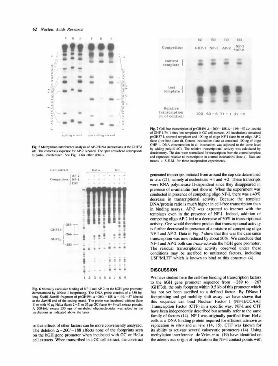
Fig. 7 Cell-free transcription of phGH494 A -260/- 188 A- 149/-57 i.e. devoidof GHF- 1/Pit- I sites (test template) in GC cell extracts. All incubations containedphGH37-L (control template) and 100 ng of oligo NF-I (lane b) or oligo AP-2(lane c) or both (lane d). Control incubations (lane a) contained 100 ng of oligoGHF-1. DNA concentration in all incubations was adjusted to the same levelby adding poly(dI-dC). The relative transcriptional activity was calculated bydensitometry. The data were normalized for transcription from the control templateand expressed relative to transcription in control incubations (lane a). Data aremeans ± S.E.M. for three independent experiments.
generated transcripts initiated from around the cap site determinedin vivo (21), namely at nucleotides + 1 and +2. These transcriptswere RNA polymerase II-dependent since they disappeared inpresence of ct-amanitin (not shown). When the experiment wasconducted in presence of competing oligo NF-I, there was a 40%decrease in transcriptional activity. Because the templateDNA/protein ratio is much higher in cell-free transcription thanin binding assays, AP-2 was expected to interact with thetemplates even in the presence of NF-I. Indeed, addition ofcompeting oligo AP-2 led to a decrease of 30% in transcriptionalactivity. One would therefore predict that transcriptional activityis further decreased in presence of a mixture of competing oligoNF-I and AP-2. Data in Fig. 7 show that this was the case sincetranscription was now reduced by about 50%. We conclude thatNF-I and AP-2 both can trans-activate the hGH gene promoter.The residual transcriptional activity observed under theseconditions may be ascribed to untitrated factors, includingUSF/MLTF which is known to bind to this construct (4).
DISCUSSIONWe have studied here the cell-free binding of transcription factorsto the hGH gene promoter sequence from -289 to -267(GHF3d), the only footprint within 0.5 kb of this promoter whichhas not yet been ascribed to a defined factor. By DNase Ifootprinting and gel mobility shift assay, we have shown thatthis sequence can bind Nuclear Factor I (NF-I)/CCAATTranscription Factor (CTF) in a specific way. NF-I and CTFhave been independently described but actually refer to the samefamily of factors (14). NF-I was originally purified from HeLacells as a DNA-binding protein required for efficient adenovirusreplication in vitro and in vivo (14, 15). CTF was known forits ability to activate several eukaryotic promoters (14). Usingmethylation interference, de Vries et al. (19) have identified onthe adenovirus origin of replication the NF-I contact points with
..} Ir
.1
........A
:.. .- .,.
?. s' .4, '!J,

Nucleic Acids Research 63
the DNA bases shown in Table 2. This raised the question ofthe actual contact points of NF-I with an eukaryotic promotersuch as that of the hGH gene. The five guanines that are involvedin the interaction of NF-I with the adenovirus sequence (19) werefound here as being important also in the hGH gene sequence(Table 2). Methylation of a guanine on the hGH gene noncodingstrand (-282) prevented NF-I binding. This guanine correspondsto a thymine (+28) on the noncoding strand of the adenovirussequence. Thus, replacement of this adenovirus pyrimidine bya purine does not prevent NF-I binding, and this confirms BrdU-substitution data (19). In contrast, methylation of the guanine atposition -281 on the noncoding strand did not interfere withNF-I binding to the hGH gene sequence, despite the fact thatmethylation of the corresponding adenine at position +29 of theadenovirus did (Table 2). This is consistent with the fact thatdimethylsulfate methylates guanines on the N7 atom which is inthe major groove and adenines on the N3 atom which is in theminor groove of the DNA helix. This confirms that NF-I facesthe minor groove at coordinate +29 of the adenovirus andsuggests that it is also the case at coordinate -281 of the hGHgene promoter. The guanines at -272 and -277 on the codingstrand of the hGH gene sequence replace the adenines at +38and +33 of the adenovirus sequence. Methylation of theseguanines did not interfere with NF-I binding. The electrophoreticmobilities of the complexes between NF-I and its target sequence
differed depending on the origin of this factor i.e. HeLa or GCcells (Fig. 2), and yet the methylation pattern was identical (Fig.3). This is compatible with the finding (12, 17, 18) that differenttissues may express different isoforms of NF-I all sharing thesame DNA binding domain. We conclude from these experimentsthat the molecular mechanisms ofDNA recognition by NF-I are
similar when this factor binds to the origin of replication ofadenovirus and to the hGH gene promoter. This is consistent withthe recent finding that the same domain of NF-I mediates DNAbinding on the adenovirus origin of replication and on an
eukaryotic promoter (22).AP-2 purified from HeLa cells protects two regions of the hGH
gene promoter against cleavage by DNase I (6). The first region(-167 to -145) binds AP-2 with low affinity and the other(-287 to -265), which corresponds approximately to GHF3d(-289 to -267), binds AP-2 with much higher affinity.However, with crude HeLa cell extracts, no footprint was seen
at the low affinity site (5) and we show here with such extractsthat the footprint located at the high affinity site is in fact dueto NF-I. When the latter was titrated out of these cell extracts,AP-2 did produce an identical footprint, consistent with themutually exclusive binding of AP-2 and NF-I to this site. By gelmobility shift assay, we have demonstrated that AP-2 is alsopresent in GC cell extracts, but at a much lower concentrationthan in HeLa cell extracts. Indeed, no AP-2 footprint was seenfrom -289 to -267 when NF-I was titrated out of GC cellextracts. Methylation interference experiments showed that sixguanines that were important for the interaction of AP-2 withthe two DNA strands are part of the AP-2 consensus. Ouridentification of a crucial guanine residue just next to the AP-2consensus is not surprising since the length of this consensus
varies from 8 to 10 bp (20, 6). Our data also show thatmethylation of four guanine residues, namely at -276 and -283on the coding strand and -275 and -282 on the noncodingstrand, prevented binding of both NF-I and AP-2. This supportsthe mutually exclusive character of the binding of these factorson the hGH gene promoter. There are other cases where AP-2or a CCAAT-transcription factor is displaced by another protein.
A CCAAT-binding transcription factor that interacts with a seaurchin histone 2B-1 gene promoter is competed for by a sperm-specific CCAAT displacement protein which might act as arepressor of gene transcription (23). Another example is thedisplacement of AP-2 by AP-3 from its binding site on the SV40core sequence (24).As to the functional significance of NF-I binding, our cell-free
transcription data suggest that this factor stimulates transcriptionfrom the hGH gene promoter. This interpretation is in agreementwith transfection data from Lefevre et al. (5). One of their hGHgene promoter mutants called ANco2-CAT that lacked a regionencompassing the GHF3d sequence was less active in terms ofCAT activity than the intact promoter. Moreover, two other hGHgene promoter mutants (hGH-NK and hGH ANcol) that had analtered GHF3d sequence, and yet still showed a footprint, wereas active as the intact promoter in the CAT assay. This behaviorcan now be explained in light of our results since these mutantsstill contain a NF-I consensus. NF-I is also known to be involvedin the glucocorticoid-dependent stimulation of transcription fromthe MMTV promoter (25,26). Schule et al. (27) and Strahle etal. (28) showed that NF-I can interact synergistically with theglucocorticoid receptor to stimulate MMTV transcription. Sincethis receptor also binds to the hGH gene (29,30), NF-I couldplay a similar role in controlling glucocorticoid-dependenttranscription of this gene. Concerning AP-2, this relatively cell-specific factor is involved in the basal control of promoters suchas that of the metallothionein-IIA gene where it mediatestranscriptional activation by phorbol esters and cAMP (6). A roleof AP-2 in the cAMP-dependent transcriptional activation of thehGH gene by GHRH was therefore an interesting possibility.However, this hypothesis has been recently discounted byexperiments with hGH gene promoter deletion constructs (31).Still, our cell-free transcription experiments suggest that AP-2can trans-activate the hGH gene, which is therefore a candidatefor regulation by this factor in vivo. Transcription of the rat GHgene is stimulated by retinoic acid through 5' flanking cis-actingsequences (32). Since retinoic acid increases the concentrationof AP-2 mRNA (33), a role of AP-2 in this control mechanismcannot be excluded.The cloning of NF-I and AP-2 has shown that their DNA-
binding domains share no structural features with each other orwith other trans-acting factors (22). However, NF-I and AP-2have in common a proline-rich domain which appears to beindispensable for their transcriptional activity (22). Since NF-Iand AP-2 can trans-activate the hGH gene by binding to the samepromoter region, the interaction of this proline-rich domain witha specific component of the transcriptional machinery mightinvolve a trans-activating mechanism that is common to NF-Iand AP-2.
ACKNOWLEDGEMENTS
We thank Dr. C. Egan for helpful discussion and T. Lambertand V. Henry for secretarial assistance. We are grateful to Dr.J. Martial (University of Liege) for the hGH-1 clone, to Dr.B. Groner (Friedrich Miescher Institut, Basel) for providing theplasmid p13-13 and to Dr. D. Christophe (Free University ofBrussels) for providing oligo AP-2. S.J.C. holds a Fellowshipfrom the Institut pour l'Encouragement de la Recherche Scien-tifique dans l'Industrie et l'Agriculture (Belgium). F.P.L. isResearch Assistant of the Fonds National de la Recherche scien-tifique (Belgium). This work was supported by the Fonds de laRecherche Scientifique Medicale (Belgium) and the Belgian

64 Nucleic Acids Research
State-Prime Minister's Office-Science Policy Programming(Incentive Program in Life Sciences Grant no. 20).
REFERENCES1. Brent, G.A., Harney, J.W., Moore, D.D. and Larsen, P.R. (1988) Mol.
Endocrinol., 2, 792-798.2. Bodner, M. and Karin, M. (1987) Cell, 50, 267-275.3. Castrillo, J.L., Bodner, M. and Karin, M. (1989) Science, 243, 814-817.4. Lemaigre, F.P., Courtois, S.J., Lafontaine, D.A. and Rousseau, G.G. (1989)
Eur. J. Biochem., 181, 555-561.5. Lefevre, C., Imagawa, M., Dana, S., Grindlay, J. Bodner, M. and Karin,
M. (1987) EMBO J., 6, 971-981.6. Imagawa, M., Chiu, R. and Karin, M. (1987) Cell, 51, 251-260.7. Hynes, N. Van Ooyen, A.J.J., Kennedy, N., Herrlich, P., Ponta, H. and
Groner, B. (1983) Proc. Natl. Acad. Sci. USA, 80, 3637-3641.8. Manley, J.L., (1984) In Hames, B.D. and Higgins, S.J. (eds), Transcription
and translation-A practical approach. IRL Press, Oxford, pp. 71-88.9. Chodosh, L.A., Baldwin, A.S., Carthew, R.W. and Sharp P.A. (1988) Cell,
53, 11-24.10. Carthew, R.W., Chodosh, L.A. and Sharp, P.A. (1985) Cell, 43, 439-448.11. Lizardi, P.M., Binder, R. and Short, S.A. (1984) Gene Anal. Techn., 1,
33-39.12. Paonessa, G., Gounari, F., Frank, R. and Cortese, R. (1988) EMBO J.,
7, 3115-3123.13. Nowock, J., Borgmeyer, U., Piischel, A.W., Rupp, R.A.W. and Sippel,
A.E. (1985) Nucl. Acids Res., 13, 2045-2061.14. Jones, K.A., Kadonaga, J.T., Rosenfeld, P.J., Kelly, T.J. and Tjian R. (1987)
Cell, 48, 79-89.15. Rosenfeld, P.J. and Kelly, T.J. (1986) J. Biol. Chem., 261, 1398-1408.16. Mc Knight, S. and Tjian, R. (1986) Cell, 46, 795-805.17. Santoro, C., Mermod, N. Andrews, P.C. and Tjian, R. (1988) Nature, 334,
218-224.18. Gil, G., Smith, J.R., Goldstein, J.L., Slaughter, C.A., Orth, K. Brown,
M.S. and Osborne, T.F. (1988) Proc. Natl. Acad. Sci. USA, 85, 8963-8967.19. de Vries, E., van Driel, W., van den Heuvel, S.J.L. and van der Vliet, P.C.
(1987) EMBO J., 6, 161-168.20. Roesler, W.J., Vandenbark, G.R. and Hanson, R.W. (1988) J. Biol. Chem.,
263, 9063-9066.21. Miller, W.L. and Eberhardt N.L. (1983) Endocrine Rev., 4, 97-130.22. Mermod, N., O'Neill, E.A., Kelly, T.J. and Tjian, R. (1989) Cell, 58,
741-753.23. Barberis, A., Superti-Furga, G. and Busslinger, M. (1987) Cell, 50,
347-359.24. Mercurio, F. and Karin, M. (1989) EMBO J., 8, 1455-1460.25. Cordingley, M.G., Tate-Riegel, A. and Hager, G.L. (1987) Cell, 48,
261 -270.26. Buetti, E., Kiihnel, B. and Diggelmann, H. (1989) Nucl. Acids. Res.,
17,3065-3078.27. Schule, R., Muller, M., Kaltschmidt, C. and Renkawitz, R. (1988) Science,
242, 1418-1420.28. Strahle, U., Schmid, W. and Schutz, G. (1988) EMBO J., 7, 3389-3395.29. Moore, D.D., Marks, A.R., Buckley, D.I., Kapler, G. Payvar, F. and
Goodman, H.M. (1985) Proc. Natl. Acad. Sci. USA, 82, 699-702.30. Eliard, P.H., Marchand, M.J., Rousseau, G.G., Formstecher, P., Mathy-
Hartert, M., Belayew, A. and Martial, J.A. (1985) DNA, 4, 409-417.31. Dana, S. and Karin, M. (1989) Mol. Endocrinol., 3, 815-821.32. Bedo, G., Santisteban, P., and Aranda, A. (1989) Nature, 339, 231-234.33. Williams, T., Adman, A., Luscher, B. and Tjian R. (1988) Genes Dev.,
2, 1557-1569.