nuclear bcl10 expression characterizes a group of ocular adnexa malt lymphomas with shorter...
TRANSCRIPT

Nuclear bcl10 expression characterizes agroup of ocular adnexa MALT lymphomaswith shorter failure-free survival
Renato Franco1, Francisca I Camacho2, Alessia Caleo3, Stefania Staibano3, Delfina Bifano3,Amalia De Renzo4, Fausto Tranfa5, Anna De Chiara1, Gerardo Botti1, Roberta Merola6,Ana Diez7, Giulio Bonavolonta5, Gaetano De Rosa3 and Miguel A Piris2
1Pathology Unit, National Cancer Institute ‘Giovanni Pascale’, Naples, Italy; 2Lymphoma Group, MolecularPathology Programme, Centro Nacional de Investigaciones Oncologicas, Madrid, Spain; 3Biomorphologicaland Functional Sciences Department, ‘Federico II’ University, Naples, Italy; 4Hematology Institute, ‘FedericoII’ University, Naples, Italy; 5Ophthalmology Department, ‘Federico II’ University, Naples, Italy; 6ClinicalPathology and Cytogenetics Unit, ‘Regina Elena’ Cancer Institute, Rome, Italy and 7Immunohistochemistryand Histology Unit, Biotechnology Programme, Centro Nacional de Investigaciones Oncologicas, Madrid, Spain
Ocular adnexa B-cell lymphomas are a relatively rare group of extranodal lymphomas, marginal-zone B-celllymphomas of mucosa-associated lymphoid tissue (MALT lymphomas) being the most frequent type at thislocation. As with other nongastrointestinal MALT lymphomas, ocular adnexa MALT lymphomas have distinctcharacteristics from those of the gastric MALT model, implying specific pathogenic events, which could be ofinterest in the prediction of clinical behavior and the choice between therapeutic options. In a series of 39 casesof ocular adnexa MALT lymphomas, studied using a tissue microarray, we observed that the most frequentalteration was related to apoptosis regulation. Thus, caspase 3 activity was completely abolished, andphosphorylated IjBa, a marker of NF-jB activation, showed increased expression, while cases with anincreased number of large cells displayed increased expression of survivin and other cell-cycle-relatedproteins, such as cyclin A, cyclin E and Ki67, and p16 expression was reduced. There were no occurrences oft(11;18)(q21,q21), while 5/37 cases exhibited t(14;18)(q32;q21). Aberrant nuclear expression of bcl10 wasobserved in 11 cases, independently of the presence of translocations, and was significantly associated withphosphorylated IjBa expression and a reduced TdT-mediated biotin-dUTP nicked-end labeling apoptotic index.Moreover, patients with tumoral bcl10 nuclear expression showed shorter failure-free survival.Modern Pathology (2006) 19, 1055–1067. doi:10.1038/modpathol.3800597; published online 28 April 2006
Keywords: bcl10; ocular adnexa MALT lymphomas; t(11;18)(q21;q21); t(14;18)(q21;q32)
Ocular adnexa B cell lymphomas (OABLs) feature avariety of different histological types, the mostfrequently diagnosed being extranodal marginal-zone lymphoma of mucosa-associated lymphoidtissue (MALT lymphoma).1–6
Lymphomagenesis is driven by multiple accumu-lated genetic abnormalities that determine thesurvival of neoplastic cells and involve mainlycell-cycle and apoptotic pathways.7 MALT lympho-mas in particular are a paradigm of alternative
genetic events, involving different chromosomalaberration and/or apoptosis control protein expres-sion, leading to the deregulation of apoptotic path-ways through the activation of the NF-kB pathway.7,8
In general, MALT lymphomas at different extra-nodal locations share some common morphological,phenotypic and molecular features, which has led tothe establishment of gastric MALT lymphoma as theprototype for the definition of the features of MALTlymphoma at different sites.9,10 Nevertheless, thereis increasing evidence to suggest that cases diag-nosed with MALT lymphomas arising at differentsites differ in the presence of infectious specificantigens—Helicobacter, Campylobacter, Borrelia,Chlamydia—promoting lymphoid cell proliferation,and in the frequency of chromosomal translocationssuch as t(11;18)(q21;q21), or the newly describedt(14;18)(q32;q21) and t(3;14) (p14.1;q32).10–12
Received 24 October 2005; revised 24 February 2006; accepted 27February 2006; published online 28 April 2006
Correspondence: Dr FI Camacho, MD, PhD, Lymphoma Group,Molecular Pathology Programme, Centro Nacional de Investiga-ciones Oncologicas, C/Melchor Fernandez Almagro, 3, Madrid28029, Spain.E-mail: [email protected]
Modern Pathology (2006) 19, 1055–1067& 2006 USCAP, Inc All rights reserved 0893-3952/06 $30.00
www.modernpathology.org

As in the case of other extranodal lymphomas,most MALT lymphomas arising in the ocular adnexahave an indolent course with a favorable prog-nosis;1,2 previous reports have demonstrated lowlocal aggressivity with a reasonably good possibilityof relapse control by radiotherapy.13 This is inkeeping with the findings from a large series ofMALT lymphomas, which showed that the efficacyof treatment varied accordingly with the site, stage,and clinical status of patients.14 Nevertheless, asubset of these tumors shows a higher incidence oflocal or distant relapses, or even of systemicprogression.14–16 Whether this can be explained interms of the site of origin or the biological features ofthe tumoral cells is a matter of debate16 whoseresolution requires further research.
Recent studies concur in that it is possible toassociate the clinical outcome of lymphoma patientswith multiple biological features defined usingcDNA, oligonucleotide or tissue microarrays.17–20
The use of tissue microarrays could be particularlyuseful in this context, since tumoral specimens inocular adnexa lymphomas are frequently small,fully embedded in paraffin, and require micro-dissection or tissue-core selection to guarantee thatthe analysis reports on the features of the tumoralcells.
In order to elucidate the biology of ocular adnexaMALT lymphomas we have analyzed a tissuemicroarray including a large series of 39 cases witha panel of antibodies and probes directed againstmolecules known to be involved in B-cell differ-entiation, cell cycle and apoptosis control,17–20 and,for control purposes, compared them with somenon-MALT lymphomas from the same anatomicalregion. Moreover, we have included in the analysis atissue-microarray in situ hybridization study of theapoptotic index (TdT-mediated biotin-dUTP nicked-end labeling (TUNEL)), and of t(11;18)(q21;q21) andt(14;18)(q32;q21), which are characteristic translo-cations of MALT lymphomas. We have comparedour results with the clinical data and the follow-upof the series, in order to establish associationsbetween these biological characteristics and theoutcome.
Materials and methods
Selection of Cases
Thirty-nine cases of MALT OABLs were collectedfrom the Department of Pathology of ‘Federico II’University. The cases, diagnosed between 1980 and2003, were included in this study on the basis of theavailability of diagnostic paraffin blocks that werethick enough to provide a minimum of 50 sections,and of clinical information for more than 6 monthsfollowing diagnosis.
All cases were reviewed according to WHOclassification criteria, using standard tissue sectionsand immunohistochemical slides.
A smaller group of non-MALT lymphomas, whichare very infrequent in this anatomical region wasalso included in the study. It was comprised of fourdiffuse large B-cell lymphoma cases, three cases offollicular lymphoma (one grade 2, and two grade 3)and one case of Burkitt’s lymphoma.
Each case of MALT OABL was subgroupedaccording to the cell type prevalent in the margin-al-zone type (monocytoid or centrocyte type) andplasmacytoid. The prevalence of plasmacytoid cellswas recorded if they represented more than 50% ofneoplastic cells. Moreover, as with the histologicalprogression in the gastric model, the presence oflarge cells was recorded for each case as absent/rare(o5%), numerous (5–10% of the neoplastic popula-tion), or with clusters of large cells (420 cells).21
Tissue-Microarray Design
Following conventional protocols we used a tissue-microarray device (Beecher Instruments, Sun Prairie,WI, USA) to construct a single-tissue-microarrayblock of 96 cores. All cases of the MALT and non-MALT lymphoma groups were histologically re-viewed. The tumor-cell-rich areas, which were alsorepresentative of the presence of large cells, weremarked in the paraffin blocks. Two selected 1-mmcores from different areas were included, alongwith two separate reactive tonsil tissues as a controlto ensure the quality, reproducibility and homo-geneous staining of slides.17–20
Immunohistochemical Study
Immunohistochemical staining was performed onselected proteins involved in B-cell differentiationand in cell cycle and apoptosis regulation inlymphomagenesis.
Stained tissue-microarray sections were evaluatedby four different pathologists (RF, AC, DB, SS) usinguniform criteria. Discrepancies were resolvedthrough simultaneous inspection and discussion ofthe results. Single-marker expression was recordedas negative/positive and high/low-level, after con-sideration of the expression in reactive comparedwith tumoral cells and the specific cut-off of eachmarker. Details of cutoffs used for each marker arepresented in Table 1.
As proposed, cytoplasmic bcl10 expression wasscored as strong when it was similar to tonsilcentroblast positivity, moderate when similar tocentrocyte positivity, and weak/absent when similarto tonsil mantle-zone positivity.22 Nuclear positivitywas also recorded. Phosphorylated IkBa (p-IkBa),considered to be an indirect expression of NF-kBactivity, was considered positive when expressed inthe cytoplasm and the nucleus.
Discrepancies between two cores from the samecase were resolved in a joint analysis of the twocores.
Nuclear bcl10 in ocular adnexa MALT lymphomasR Franco et al
1056
Modern Pathology (2006) 19, 1055–1067

In Situ Detection of Epstein–Barr Virus and Apoptosis
EBER in situ hybridization was performed usingstandard procedures and with the appropriatecontrol tissue. Epstein–Barr virus (EBV) was de-tected by ISH with fluorescein-conjugated Epstein–Barr virus (EBER) PNA probe (DAKO, Glostrup,Denmark). Cases were considered EBV positivewhen nuclear positivity was present in a majorityof neoplastic cells.
Apoptosis was detected using the ApopTag Per-oxidase In Situ Apoptosis Detection Kit (Intergen,Oxford, UK), based on the TUNEL method. Briefly,tissue-microarray sections were pretreated withproteinase K for 15 min, incubated with TdT enzymefor 1 h at 371C and then with antidigoxigeninperoxidase conjugate for 1 h. Color was developedwith 3,30 diaminobenzidine tetrahydrochloride(10 min) and counterstained with hematoxylin.
The reproducibility of the results was confirmedby comparing restricted lymphoid cell differentia-tion (CD20, CD3, CD138, CD79a, bcl6, and CD5) and
EBER-ISH marker results with those obtained fromthe original whole section for ten selected cases.
Fluorescent In Situ Hybridization Study
Tissue array sections from paraffin-embedded tissuewere heated for 4 h at 621C and immediatelydeparaffinized in two rinses of 100% xylene for10 min each. The slides were then treated with 0.3 Msodium chloride and 0.03 M sodium citrate for20 min at 801C, and with 0.05 mg/ml proteinase for10 min at 371C. For t(11;18)(q21;q21) detection, weused LSI API2/MALT1 t(11;18)(q21;q21) dual-color,dual-fusion translocation probe, and for t(14;18)(q32;q21) we used LSI IGH/MALT1 t(14;18)(q32;q21)dual-color, dual-fusion translocation probe.
The cutoff value for the diagnosis of rearrange-ment involving IGH and MALT1 was 5.3%, which isabove the mean percentage of cells with a false-positive signal plus three s.d., as assessed in tissuefrom reactive tonsils present in TMA. Moreover, IGH
Table 1 Characteristics of antibodies
Antigen Clone Source Dilution Reactivity Threshold Internal control
Bcl2 124 DAKO 1:25 High/low 450% neoplastic cell Small lymphocyteBax Polyclonal Santa Cruz 1:1000 Positive/negative 410% neoplastic cell Benign small lymphocyteBcl-XL 2H12 Zymed 1:10 High/low 410% neoplastic cells TMA controlsSurvivin Polyclonal RD System 1:1500 High/low 410% neoplastic cells TMA controlsCaspase-3 C92-605 PharMingen 1:25 Positive/negative 410% positive neoplastic
cellsTMA controls
Bcl10 331.3 Santa Cruz 1:1000 Positive/negative 410% neoplastic cells Reactive lymphocyteMUM1 Polyclonal Santa Cruz 1:200 High/low 480% positive cells Plasma cellsBcl6 PG-B6p DAKO 1:10 Positive/negative 410% neoplastic cells GC (germinal center) B cellsCD38 Vs38 DAKO 1:25 High/low 480% positive cells Plasma cellsCD5 4C7 Novocastra 1:50 Positive/negative 410% positive cells Reactive lymphocyteCD10 56C6 Novocastra 1:10 Positive/negative Any tumoral cell positive GC B cellsCD20 L-26 DAKO 1:100 Positive/negative Any positive neoplastic
cellsReactive lymphocyte
Cyclin A 6E6 Novocastra 1:100 Positive/negative 410% neoplastic cells Proliferating cells (G2/M)Cyclin B1 7A9 Novocastra 1:25 Positive/negative 410% positive cells Proliferating cells (G2/M)Cyclin D1 DCS-6 DAKO 1:100 Positive/negative Any positive neoplastic
cellMacrophage and endothelialcells
Cyclin D3 DCS-22 Novocastra 1:10 Positive/negative 410% positive cells Proliferating cellsCyclin E 1313 Novocastra 1:10 Positive/negative 410% positive cells TMA controls, proliferating
cellsCDK1 1 Transduction 1:1500 Positive/negative 410% positive cells TMA controls, proliferating
cellsCDK2 8D4 NeoMarkers 1:500 Positive/negative 410% positive cells TMA controls, proliferating
cellsCDK4 35.1 Chemicon 1:10 Positive/negative 410% positive cells TMA controlsCDK6 K6.83 Chemicon 1:10 Positive/negative 410% positive cells TMA controlsP21 EA10 Oncogene 1:50 Positive/negative 410% neoplastic cells Scattered GCP16 Polyclonal Santa Cruz 1:50 High/low 410% positive cells Normal cellsP27 57 Transduction 1:1000 High/low 410% positive cells Resting lymphoid cellsKi67 MIB 1 DAKO 1:100 High/low 420% positive cells Proliferating cellsP53 DO-7 Novocastra 1:50 Positive/negative 450%positive cells Scattered GC cellsHdm2 IF2(mdm2) Oncogene 1:10 High/low 410% positive cells MacrophagesCD3 F7.2.38 DAKO 1:25 Positive/negative Any tumoral cell Reactive lymphocyteP18 B9 Santa Cruz 1:150 High/low 410% positive cells Normal cellsp-IkBa 1118,
ssc9965Santa Cruz 1:150 Positive/negative 410% (nucleus and
cytoplasm)Normal cells
GC¼ germinal center.
Nuclear bcl10 in ocular adnexa MALT lymphomasR Franco et al
1057
Modern Pathology (2006) 19, 1055–1067

dual-color break-apart rearrangement probes (VysisInc., Downers Grove, IL, USA) were applied to cellsof all t(14;18)(q32;q21)-positive lymphomas to con-firm the translocation. The Spectrum Green-labeledLSI IGVH probe covers the entire IGH variableregion, while the Spectrum Orange-labeled probelies completely 30 to the IGH locus. As a result of thisprobe design, any translocation with a breakpoint atthe J segments, or within switch sequences, shouldproduce separate orange and green signals. Addi-tionally, FISH with centromere-specific probes forchromosome 18 (Vysis) was performed in all cases.The appropriate probe mix (10 ml) was applied to thetissue sections and covered with a coverslip. Bothprobe and target DNA were simultaneously dena-tured at 751C for 5 min and incubated overnight at371C using the Hybrite System. Posthybridizationwashes were performed according to the ‘rapid-wash protocol’ provided by Vysis. Slides werecounterstained with 406-diamidino-2-phenylindole2HCl (DAPI). FISH was performed according to themanufacturer’s instructions (Vysis). FISH data werecollected using an Olympus BX 61 fluorescencemicroscope equipped with a cooled black-and-whitecamera controlled by the associated software(Olympus, Italy).23,24
Statistical Analysis
The Pearson’s w2 test was used, where appropriate,to establish whether there were any relationshipsbetween the frequencies of different markers in-cluded in this study. Differences were considered tobe significant for values of Po0.05.
Overall survival (OS) and failure-free survival(FFS) curves were calculated using the Kaplan–Meier method. Statistical significance of associa-tions between individual variables and OS and FFSwere determined using the log-rank test. All statis-tical analyses were performed using the SPSS 98v.12 program.
OS was defined as the time from diagnosis (firstbiopsy) to death by any cause, or until the mostrecent follow-up. Moreover, taking into account thelong-term survival observed in many of these casesand the existence of disease-unrelated deaths,disease-specific OS was also recorded. FFS wasmeasured as the time from diagnosis to the occur-rence of progression, relapse after complete remis-sion, or death from any cause. FFS had a value ofzero for patients who did not achieve completeremission.
Results
Clinical Findings
The main clinical characteristics of MALT and non-MALT OABL cases are set out in Table 2. The MALTlymphoma group was comprised of 39 cases (21
females and 18 males) with a mean age of 62 years(ranging from 27 to 84 years), and with 26 patientsolder than 59 years. The orbital region was involvedin 35 cases (in particular, the lachrymal gland wasinfiltrated in 9/35 cases), the conjunctival mucosa inthree cases, and the eyelid in one case. Bone marrowinvolvement was observed in four cases. Standardimaging studies, including ultrasonography and CTscan revealed no other locations in any of the cases.
Twelve of the 39 cases were studied serologicallyfor the presence of infection by hepatitis C virus(HCV) or hepatitis B virus. Six cases were found tobe serum positive for HCV.
Therapy was heterogeneous, the patients beingtreated in accordance with their clinical stage,disease aggressiveness, age, and the status of know-ledge over the last 20 years. Radiotherapy wasstandard, either alone (16/39 cases) or in conjunc-tion with chemotherapy (6/39 cases). Statisticalanalysis of our series revealed no significantdifferences between these therapeutic approaches.
Relapse or persistence of local disease wasobserved in 13 cases. Relapse was recorded at thesame site of origin in seven cases, and in thecontralateral orbital region in two cases. Diseasepersisted after therapy in two cases. Disseminationwas recorded in three cases: to loco-regional lymphnodes in two cases, and to the ovary in the third.
The mean OS in the series was 58 months (rangefrom 6 to 215 months). The mean disease-specificOS, recorded in two cases, was 33 months, and themean FFS was 28 months (range from 0 to 74months). At the end of follow-up 24 patients werealive without disease, eight were alive with disease,two had died from causes attributable to the diseaseand five had died from causes unrelated to thedisease.
Main Morphological and ImmunohistochemicalFeatures of MALT OABLs
Tumoral biopsy revealed a diffuse or vaguelynodular pattern of growth, with rare residualfollicles in 10/39 cases, clearly highlighted byfollicular dendritic cell staining (CD23 antibody).The neoplasia was mostly constituted by marginal-zone cells (monocytoid- and centrocyte types) withinterspersed lymphoplasmacytoid cells; lympho-plasmacytoid cell prevalence was recorded in twocases only. A low proportion (5–10%) of scatteredcentroblast- and immunoblast-like cells was presentin 24 cases (61%). No cases with clusters of morethan 20 large cells were observed (Figure 1).
Standard immunohistochemical staining showedpan-B markers in all cases (CD20 and CD79a), andnegativity for germinal-center markers (CD10 orbcl6). Expression of CD43 (12/36) was variable. Nopositivity of neoplastic cells was observed for CD5and CD23. Light-chain restriction was revealed in 33cases; in particular, 25/34 showed kappa-chainrestriction and 9/34 lambda-chain restriction.
Nuclear bcl10 in ocular adnexa MALT lymphomasR Franco et al
1058
Modern Pathology (2006) 19, 1055–1067

Table 2 Main clinicopathological features of patients
Case no. Sex Age Site Histology/cytotypeprevalence
Bonemarrow
HCV EBER Therapy Relapse(monthssincediagnosis)
Systemicprogression(months fromdiagnosis)
Follow-up time(months since
diagnosis)
LC CD38 MUM1 CD43 Light-chainrestriction
Status
1 M 68 O MALT, Mz � ND � WW NO NO 173 No � � � L A2 M 58 O MALT, Mz � ND � CT+RT Yes (69) NO 213 No � � � L DO3 M 76 O MALT, Mz � ND � CT NO NO 215 No � � � No DO4 F 27 O MALT, Mz � ND � CT Yes (54) Y 6 Yes � � + k D5 F 44 O MALT, Mz � ND � RT NO NO 138 No � � � No A6 M 75 O MALT, Mz � ND � WW NO NO 20 No � � + k DO7 F 65 O MALT, Mz � ND � RT Yes (7) NO 163 No � � + L AWD8 M 66 C MALT, Mz � ND � WW NO NO 53 No � � � k DO9 F 83 O MALT, Mz � ND � WW NO NO 24 Yes � � + k DO
10 F 61 O MALT, Mz ND ND � WW Yes (17) Y 147 No � � � k A11 F 37 O MALT, Mz � ND � WW Yes (74) NO 138 No � � + k A12 F 66 O MALT, Mz � + � RT NO NO 81 No � � + k A13 F 54 O MALT, Mz � � � CT+RT NO NO 104 Yes + + � k A14 M 67 C MALT, Mz � ND � RT NO NO 105 No � � � L A15 M 49 O MALT, Mz � ND � RT NO NO 94 Yes � � + L A16 M 64 O MALT, Mz � ND � WW Yes (13) NO 78 No � � + k AWD17 M 65 O MALT, Mz � + � RT NO NO 61 No � � � k A18 F 67 O MALT, Mz ND ND � CT+RT NO NO 6 Yes � � � k A19 M 79 O MALT, Mz � � � WW Yes (12) Y 47 No � � ND k AWD20 F 74 O MALT, Mz � + � WW Yes (50) Y 51 Yes + � � k AWD21 F 76 O MALT, Mz � ND � WW Yes (35) Y 35 No � � � k AWD22 F 78 O MALT, Mz ND ND � RT NO NO 6 Yes � � � No A23 F 55 O MALT, Mz + � � CT+RT NO NO 6 No � � ND L A24 M 62 C MALT, Mz + � � CT+RT NO NO 32 Yes � � � L A25 M 46 O MALT, Mz ND ND � RT NO NO 6 No � � � No A26 F 50 O MALT, Mz ND ND � RT NO NO 6 Yes � � + k A27 F 78 O MALT, PL ND ND � RT NO NO 6 No + + + k A28 F 30 O MALT, Mz � � � WW NO NO 27 Yes � � � k A29 M 77 O MALT, Mz ND + � RT NO NO 8 No + + � k A30 F 55 P MALT, Mz � ND � RT Yes (12) NO 25 No + � � L A31 M 70 O MALT, Mz � ND � WW NO NO 22 Yes + � � L A32 F 69 O MALT, Mz + ND � CT+RT NO NO 11 No � � ND k AWD33 M 78 O MALT, Mz � ND � RT NO NO 21 No + � + k A34 M 69 O MALT, Mz � ND � CT Yes (5) Y 7 Yes � � � k D35 F 84 O MALT, PL � + � RT Yes (16) NO 18 Yes + + � k AWD36 F 60 O MALT, Mz + + � CT NO NO 18 Yes � � + k A37 M 65 O MALT, Mz � ND � CT NO Y(0) 15 Yes � � � ND AWD38 F 37 O MALT, Mz � � � RT NO NO 12 No � � � k A39 M 42 O MALT, Mz � ND � RT NO NO 6 No + � � k A40 F 60 P FL ND ND � WW Yes (12) NO 12 � � � L D41 F 85 O FL ND ND � RT NO Y 6 � � � No A42 M 67 O FL � � � RT Yes (36) NO 178 � � ND No A43 F 82 O DBCL ND + � CT+RT NO NO 13 + + � L A44 F 82 P DBCL � ND � WW NO NO 15 � + + k DO45 F 69 O DBCL ND ND � ND NO NO 6 + � � k A46 M 44 O DBCL + ND � RT NO NO 38 � � � L A47 M 20 O Burkitt � � + CT NO NO 115 � � � k A
M¼male; F¼ female; O¼orbit; C¼ conjunctiva; L¼ eyelid; Mz¼marginal-zone type; P¼plasmocytoid; ND¼not determined; WW¼watch and wait; CT¼chemotherapy; RT¼radiotherapy;LC¼ large cells; A¼ alive; DO¼dead from other causes; D¼dead; AWD¼ alive with disease.
Nuclear
bcl10in
ocularadnexa
MA
LTlym
phomas
RFranco
etal
1059
Mod
ern
Path
olo
gy
(2006)
19,
1055
–1067

Figure 1 (a) Classical ocular adnexa MALT lymphoma; (1) prevalence of monocytoid cells (H&E) (�63); (2) prevalence of plasmocytoidcells (H&E) (�63); (3) prevalence of centrocytic cells (H&E) (�63); (4) lymphoepithelial complex (�63); (5) strong cytoplasmaticexpression of Bcl10 (� 63); (6) aberrant nuclear expression of Bcl10 (� 100); (7) high nuclear expression of p-IkBa (�100); (8) absentexpression of caspase 3 (�63); (9) low expression of ki67 (�63); (10) low expression of cyclin E (� 63), (11) low expression of cyclin A(�63); (12) low expression of cyclin B1 (�63); (13) normal expression of p16 (� 63); (14) low expression of Bcl-XL (�63); (15) lowexpression of survivin (�63); (16) reduced TUNEL apoptotic index (�63). (b) Ocular adnexa MALT lymphoma with large cell (� 63): (1)increased number of large cells (H&E); (2) increased expression of ki67; (3) increased expression of cyclin E; (4) increased expression ofcyclin A; (5) increased expression of cyclin B1; (6) reduced expression of p16; (7) increased expression of Bcl-XL; (8) increased expressionof survivin.
Nuclear bcl10 in ocular adnexa MALT lymphomasR Franco et al
1060
Modern Pathology (2006) 19, 1055–1067

In all, 9/39 cases showed CD38 positivity, whichwas associated with distinct plasma cell differentia-tion in two cases.
Tissue-Microarrays Analysis
The results of the TMA-immunohistochemicalevaluations are reported in Tables 3–6.
In particular, and as expected for a low-gradelymphoma, in MALT OABLS, neoplastic cells ofour series strongly expressed some markers in-volved in apoptosis regulation, such as bcl2 (39/39cases), and diminished caspase 3 activity (38/39cases). The group was divided considering the
expression of cytoplasmic and nuclear p-IkBa (19/39), nuclear bcl10 (14/37), survivin (6/38) andBcl-XL (6/39).
Profiling of cell-cycle markers revealed a rela-tively low level of expression of protein relatedto G2/M transition, that is, cyclin A (4/39 cases),cyclin B1 (4/39 cases), and CDK1 (11/39 cases).There were similar findings for proteins related toG1/S transition, that is, cyclin D1 (0/39), cyclin D3(6/39), cyclin E (7/39), CDK2 (0/39), CDK4 (12/39),and CDK6 (14/39). Some abnormalities in theexpression of CDK inhibitors were observed in theseries. Thus, while p18 was uniformly expressed byneoplastic cells; p21 was strongly expressed bytumoral cells in four cases; loss of nuclear p27 was
Table 3 Expression of proteins involved in cell cycle
Case Histotype Ki67 CDK1 CDK2 CDK4 CDK6 CycD1 CycD3 CycE CicA P16 P18 P21 P27 P53 Hdm2
1 MALT L � � � � � � � � H L L H � L2 MALT L � � � � � � � � H ND L H � L3 MALT L + � + � � � � � H L L H � L4 MALT L � � � � � � � � L H L L � L5 MALT L � � � + � � � � H ND L H � L6 MALT L � � � � � � � � L H L L � L7 MALT L + � + + � + � � H H L H � H8 MALT L � � � + � � � � L L L H � L9 MALT L � � � � � + + � H L L L � L
10 MALT H � � � + � � � + H H L L � H11 MALT L + � + � � � � � L L L H � L12 MALT H + � � + � + � � H H L L � L13 MALT H � � + + � � � � L H L L � L14 MALT L + � � � � � � � L H L H � L15 MALT L � � � � � � � � L H L H � L16 MALT H + � � � � � � � L L L H � L17 MALT L � � � � � � � � H L L H � L18 MALT L � � � � � � � � L ND L L � L19 MALT L � � � � � � � � L ND L L � L20 MALT L � � � � � � � � L ND L L � L21 MALT L � � + + � � � � H H L H � L22 MALT L � � + + � � � � H H H H � H23 MALT L � � � � � + + � H L L H � L24 MALT L � � � � � � + � L H H H � H25 MALT L � � � � � � � � L H L H � L26 MALT L � � + + � � � � H H L L � L27 MALT H + � + + � � + � H H L H � H28 MALT L � � � � � � � � L ND L H � L29 MALT H + � � � � � + � L H L H � H30 MALT L � � � � � � � � H H L H � H31 MALT H + � + + � + + + L H L H � H32 MALT L � � � � � � � � L ND L L � L33 MALT L � � � � � � � � L ND L L � L34 MALT L � � � � � + � � H H L H � H35 MALT H + � + + � � + + L H H H � H36 MALT L � � + + � � � + L H H L � L37 MALT H + � + + � � � � L H L H � H38 MALT L � � � � � � � � H L L L � L39 MALT L � � � � � � � � L H L H � H40 FL L � � + + � � + + L H L L � H41 FL L � � � � � � � � H H L H � L42 FL H + � � � � � � � H L L H � H43 DBCL L � � + + � � + + H L L L + H44 DBCL H + � + + � + � + L H H H � H45 DBCL L � � + + � � � � L H L L � H46 DBCL L � � � � � � ND � H L L L � L47 Burkitt H + � + + � + + + L H L L + H
L¼ low level; H¼high level; ND¼not determined.
Nuclear bcl10 in ocular adnexa MALT lymphomasR Franco et al
1061
Modern Pathology (2006) 19, 1055–1067
observed in 14 cases and loss of p16 in 22 cases(Figure 1).
TUNEL revealed a reduced apoptotic index withrespect to normal nongerminal center lymphoidtissue in 8/36 cases (Figure 1). EBER expressionwas negative in 39/39 cases.
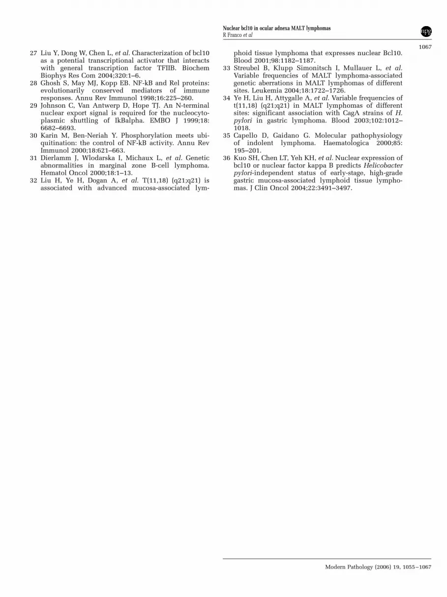
The FISH study of t(11;18) yielded negative resultsin all the cases examined, although t(14;18)(q32;q21)was present in 5/37 cases (Figure 2). The presence oft(3;14)(p14.1;q32) was not assayed.11
Relationship between Variables
Pearson’s exact test showed numerous significantassociations between the variables examined.
Cytological Type
Among MALT lymphomas, lymphoplasmacytoiddifferentiation was significantly associated with ahigh level of expression of CD38 (P¼ 0.03), MUM1(PE0.000), and cyclin E (P¼ 0.006), when comparedwith the cases with prevalent marginal-zone differ-entiation.
Histological Progression
Cases with a greater presence of large B-cells (morethan 5% of centroblasts or immunoblasts) showedan increased expression of proteins related tocell cycle or apoptosis regulation, such as
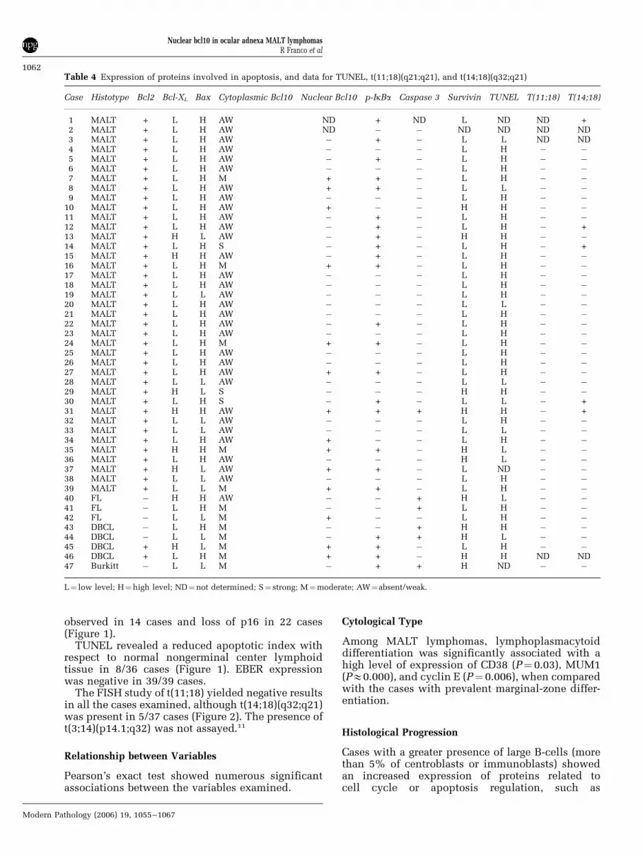
Table 4 Expression of proteins involved in apoptosis, and data for TUNEL, t(11;18)(q21;q21), and t(14;18)(q32;q21)
Case Histotype Bcl2 Bcl-XL Bax Cytoplasmic Bcl10 Nuclear Bcl10 p-IkBa Caspase 3 Survivin TUNEL T(11;18) T(14;18)
1 MALT + L H AW ND + ND L ND ND +2 MALT + L H AW ND � � ND ND ND ND3 MALT + L H AW � + � L L ND ND4 MALT + L H AW � � � L H � �5 MALT + L H AW � + � L H � �6 MALT + L H AW � � � L H � �7 MALT + L H M + + � L H � �8 MALT + L H AW + + � L L � �9 MALT + L H AW � � � L H � �
10 MALT + L H AW + � � H H � �11 MALT + L H AW � + � L H � �12 MALT + L H AW � + � L H � +13 MALT + H L AW � + � H H � �14 MALT + L H S � + � L H � +15 MALT + H H AW � + � L H � �16 MALT + L H M + + � L H � �17 MALT + L H AW � � � L H � �18 MALT + L H AW � � � L H � �19 MALT + L L AW � � � L H � �20 MALT + L H AW � � � L L � �21 MALT + L H AW � � � L H � �22 MALT + L H AW � + � L H � �23 MALT + L H AW � � � L H � �24 MALT + L H M + + � L H � �25 MALT + L H AW � � � L H � �26 MALT + L H AW � � � L H � �27 MALT + L H AW + + � L H � �28 MALT + L L AW � � � L L � �29 MALT + H L S � � � H H � �30 MALT + L H S � + � L L � +31 MALT + H H AW + + + H H � +32 MALT + L L AW � � � L H � �33 MALT + L L AW � � � L L � �34 MALT + L H AW + � � L H � �35 MALT + H H M + + � H L � �36 MALT + L H AW � � � H L � �37 MALT + H L AW + + � L ND � �38 MALT + L L AW � � � L H � �39 MALT + L L M + + � L H � �40 FL � H H AW � � + H L � �41 FL � L H M � � + L H � �42 FL � L L M + � � L H � �43 DBCL � L H M � � + H H � �44 DBCL � L L M � + + H L � �45 DBCL + H L M + + � L H � �46 DBCL + L H M + + � H H ND ND47 Burkitt � L L M � + + H ND � �
L¼ low level; H¼high level; ND¼not determined; S¼ strong; M¼moderate; AW¼absent/weak.
Nuclear bcl10 in ocular adnexa MALT lymphomasR Franco et al
1062
Modern Pathology (2006) 19, 1055–1067

Table 5 Summary of expression of proteins
Diagnosis Cases no. Large cells CD38 MUM1 CD43 Light-chain restriction Ki67 CDK1 CDK2 CDK4 CDK6 CycD1 CycD3 CycE CycA P16 P18
MALT 39 15/39 9/39 4/39 12/36 25k 9H 11/39 0/39 12/39 14/39 0/39 6/39 7/39 4/39 17H 22H9l 30L 22L 9L
8NDLow non-MALT 3 0/3 0/3 0/2 1l 1H 1/3 0/3 1/3 1/3 0/3 0/3 1/3 1/3 2H 2H
2L 1L 1LHigh non-MALT 5 2/5 2/5 1/5 3k 2H 2/5 0/5 4/5 4/5 0/5 2/5 2/4 3/5 2H 3H
2l 3L 3L 2L
L¼ low level; H¼high level.
Table 6 Summary of expression of proteins, and data for TUNEL, t(11;18)(q21;q21), and t(14;18)(q32;q21)
Diagnosis Cases no. P21 P27 P53 Hdm2 Bcl2 Bcl-XL Bax Cytoplasmatic bcl10 Nuclear bcl10 p-IkBa Caspase 3 Survivin TUNEL T(11;18) T(14;18)
MALT 39 4H 25H 0/39 12H 39/39 6H 30H 31AW 11/37 19/39 1/38 6H 28H 0/36 5/3735L 14L 27L 33L 9L 5M 32L 8L
3SLow non-MALT 3 3L 2H 0/3 2H 0/3 1H 2H 1AW 1/3 0/3 2/3 1H 2H 0/3 0/3
1L 1L 2L 1L 2M 2L 1LHigh non-MALT 5 1H 1H 2/5 4H 2/5 1H 2H 5M 2/5 4/5 3/5 4H 3H 0/4 0/4
4L 4L 1L 4L 3L 1L 1L
L¼ low level; H¼high level; S¼ strong; M¼moderate; AW¼absent/weak.
Nuclear
bcl10in
ocularadnexa
MA
LTlym
phomas
RFranco
etal
1063
Mod
ern
Path
olo
gy
(2006)
19,
1055
–1067

ki67 (P¼ 0.082), cyclin A (P¼ 0.077), cyclinB (P¼ 0.132), cyclin E (P¼ 0.075), survivin(P¼ 0.041), and Bcl-XL (P¼ 0.041), and reducedexpression of p16 (P¼ 0.042). All the cell-cyclemarkers also showed coregulated expression, asexpected (Figure 1b).
Apoptosis Regulation
Nuclear bcl10 was associated with the expression ofsome proteins involved in cell-cycle progression, inparticular Ki67 (Po0.05), CDK1 (Po0.05), CDK4(Po0.05), CDK6 (Po0.05), cyclin A (P¼ 0.054), andcyclin D3 (P¼ 0.05). Moreover, it was associatedwith the expression of p-IkBa (Po0.05), Hdm2(Po0.001), p21 (P¼ 0.05), and the TUNEL index(Po0.001).
The presence of p-IkBa was significantly asso-ciated with a high level of expression of CDK4(Po0.05), CDK6 (Po0.001), p27 (Po0.01), andwith the presence of t(14;18) (Po0.05). Therewas also a nearly significant trend relating p-IkBawith reduced TUNEL-apoptotic index (P¼ 0.059).
MALT vs Non-MALT Lymphomas
Comparing MALT with non-MALT lymphomas weobserved in the latter group significantly greaterexpression of cyclin A (Po0.05), cyclin E (Po0.05),and survivin (Po0.01), and a trend towards greaterexpression of Hdm2 that just failed to reachsignificance (P¼ 0.051).
Advanced-Stage Disease
Finally, the advanced stage of the disease, whenthere is neoplastic infiltration of the bone marrow,
was associated with a high degree of p21 expression(Po0.01).
HCV Serum Positivity
No relation of HCV presence was observed with anyof the clinicopathological parameters examined.
Relationship with FFS and OS
Survival analysis of MALT lymphomas using theKaplan–Meier method revealed a shorter FFS forcases with nuclear expression of bcl10 (Po0.05)(Figure 3).
Multivariate analysis revealed no significant asso-ciation with FFS.
None of the variables examined showed associa-tions with OS.
Discussion
MALT lymphomas in extranodal sites have mainlybeen characterized either by grouping all thedifferent locations, or by following the featuresobserved in the most characteristic locations, suchas the gastrointestinal tract. Nevertheless, MALTlymphomas arising at sites other than the gastro-intestinal tract display some specific morphologicaland molecular features.6,10 This has prompted us tocharacterize a large series of MALT lymphomasarising in the ocular adnexa with reference to aselected number of molecules that could provideinformation about the pathogenesis and biologicalpredictors of this disease.
Historically, lymphomas arising in conjunctiva,orbit and eyelids have been grouped in a singlecategory with similar therapeutic strategies andsuperimposable prognostic markers.1,2 MALTOABLs account for 24% of all MALT nongastriclymphomas, and are the third most frequent group
months1209060300
Fai
lure
-Fre
e S
urvi
val P
roba
bilit
y
Nuclear bcl10
No nuclear expression of bcl10
100
80
90
50
70
60
40
30
20
10
Figure 3 Kaplan–Meier curve of failure-free survival in relation toaberrant bcl10 nuclear expression (Po0.0367).
Figure 2 Two-color FISH to paraffin-embedded tissue for thedetection of the translocation t(14;18) (q32;q21). By using probesfor IGH (green) and MALT1 (red), the translocation of MALT1 isdemonstrated in this image, featuring fusion signals (yellow) inthe interphase nuclei (detail in inset).
Nuclear bcl10 in ocular adnexa MALT lymphomasR Franco et al
1064
Modern Pathology (2006) 19, 1055–1067

of MALT lymphomas after the gastrointestinal andcutaneous groups.6
Our data confirm a slight prevalence of femalepatients, with preferential involvement of orbitlocalization with respect to other sites.2 Medianage in this series was similar to that observed inother published series of MALT lymphomas.25
Most of the patients in this series have beentreated with approaches aimed at eradicating localdisease, either by surgery or radiotherapy. Therelatively low proportion of patients treated withchemotherapy does not allow us to conclude whichis the most successful approach. One striking featurewas the relatively favorable clinical outcome, withonly two cases of death attributable to the diseaseand 13 cases (30%) showing local relapses, persis-tence, or systemic progression, with a mean time of22 months from diagnosis.
HCV serum positivity was recorded in 5/10MALT-type lymphoma cases. HCV is known to be ahepatotropic and lymphotropic virus, and it hasbeen suggested that it may have a role in clonalB-cell proliferation.26 The relatively high prevalence(50%) of HCV infection in this series supports thehypothesis that it may play a pathogenic role in thistumor.
Our series shows that the histological features ofthe tumor have no effect on clinical prognosis. Inparticular, large-cell presence is not significantlyrelated to relapse and/or progression, in contrast tothe findings in gastric MALT lymphoma.21
Deregulation of apoptosis seems to be a character-istic feature of this group of neoplasms, showing adiminished presence of active caspase 3, which is acommon end for different apoptotic pathways, andan increased expression of cytoplasmic and nuclearp-IkBa, presumably related to NF-kB activity.
Alterations in the cell-cycle regulators wereobserved only in cases with histological progres-sion, as indicated by an increased proportion oflarge B cells. These cases had a higher proliferationrate and level of expression of some moleculesinvolved in cell-cycle regulation, such as cyclins Aand E. These are associated with an increase in theamount of several proteins, such as survivin andBcl-XL, that are involved in apoptosis control,making this group of lymphomas more similar tolarge B-cell lymphomas in our series.
In MALT lymphomas arising at gastrointestinallocations, it is claimed that MALT1, with or withoutbcl10 cooperation, activates the phosphorylationcascade, leading to IkB-a phosphorylation. IkB-a isa physiological ligand of NF-kB in the cytoplasm,whose phosphorylation permits the liberation andmigration of NF-kB into the nucleus, where NF-kBplays its transcriptional role, upregulating theexpression of antiapoptotic proteins.7,27–30 In MALTlymphoma, deregulation of the expression ofMALT1 gene due to chromosomal translocationst(11;18)(q21; q21) and t(14;18)(q32;q21) seems to beone of the main pathogenic mechanisms leading to
reduced apoptotic activity.12,15,22,31–33 In contrast towhat has been described in gastric MALT lympho-mas, our results did not indicate the presence oft(11;18). This translocation has been extensivelyexamined in gastric MALT lymphomas and isassociated with cases of a more advanced stage orthose lacking responses to Helicobacter pylorieradication.15,34 It occurs in 20–50% of gastricMALT lymphomas but the true incidence at othersites, with the exception of lung, is largely un-known.22,24,31,33,34 In particular, the t(11;18) isreported as occurring in MALT lymphomas arisingin the ocular adnexa at frequencies between 2.7and 20%.10,33,34 The different incidence of thisand other translocations in MALT lymphomasfrom distinct anatomical sites is probably linkedto specific prelymphomatous conditions that leadto neoplastic transformation. Another translocationinvolving MALT1 and IgH genes, t(14;18) (q32;q21)has recently been described as occurring morefrequently at sites considered to have a lowincidence of t(11;18), such as skin and orbit.12,33 Inour series t(14;18)(q32;q21) was relatively morefrequent and associated with p-IkBa, thus confirm-ing the link with NF-kB activity. In this study, wehave not assayed the presence of the recentlydescribed t(3;14)(p14.1;q32).11
Another critical point in the pathogenesis ofMALT lymphomas is the potential role of aberrantnuclear expression of bcl10. Under physiologicalconditions bcl10 plays a dual pro- and antiapoptoticrole.12,15,31–33,35 The nuclear expression of bcl10 hasbeen proposed as a surrogate of NF-kB activationafter (11;18)(q21;q21), t(1;14)(p22;q32), while strongcytoplasmic expression has been linked witht(14;18)(q32;q21).7,12,22,35,36 Strikingly, our data showa clear association between nuclear bcl10, increasedexpression of p-IkBa, and shorter time to failure.This, in addition to providing predictor parameters,suggests tentative therapeutic targets. Additionally,this series featured absent/weak cytoplasmic bcl10expression compared with tonsil control in mostcases, moderate bcl10 cytoplasmic expression infive cases, and strong cytoplasmic expression inthree cases. A clear relation between this strongcytoplasmic expression and t(14;18)(q32;q21) wasnot observed, since only two of these three casesshowed t(14;18)(q32;q21) and three cases witht(14;18) (q32;q21) showed only weak cytoplasmicbcl10 expression. The lack of association of aberrantnuclear and strong cytoplasmic bcl10 expressionwith any described translocation suggests theexistence of additional chromosomal abnormalitiesthat are yet to be characterized, such as thet(3;14)(p14.1;q32).
In conclusion, tissue-microarray analysis showsthat MALT OABLs represent a group mostly char-acterized by the deregulation of apoptosis. Altera-tion of apoptosis appears to be partially mediated bybcl10 shuttling to the nucleus, irrespective oft(11;18) presence, in contrast to what is observed
Nuclear bcl10 in ocular adnexa MALT lymphomasR Franco et al
1065
Modern Pathology (2006) 19, 1055–1067

in other MALT lymphomas. On the other hand,t(14;18) (q32;q21), observed in a restricted numberof cases, seems to affect apoptosis regulation byitself, because of its association with NFkB activity,as determined by p-IkBa expression.
Acknowledgements
This study was supported by grants from theMinisterio de Sanidad y Consumo (G03/179,PI051623) and the Ministerio de Ciencia y Tecno-logia (HI2003-0035), Spain. We thank Marıa JesusAcuna and Raquel Pajares (Immunohistochemistryand Histology Unit, CNIO) for their expertise andexcellent technical assistance with immunohisto-chemical assays.
References
1 Knowles DM, Jakobiec FA, McNally L, et al. Lymphoidhyperplasia and malignant lymphoma occurring inocular adnexa (orbit, conjunctiva and eyelids) aprospective multiparametric analysis of 108 casesduring 1977–1987. Hum Pathol 1990;21:959–973.
2 Knowles DM, Jakobiec FA. Ocular adnexa lymphoidneoplasms: clinical, histopathologic, electron micro-scopic and immunologic characteristics. Hum Pathol1982;13:148–162.
3 Ferreri AJM, Guidoboni M, Ponzoni M, et al. Evidencefor association between Chlamydia pittaci and ocularanexal lymphomas. J Natl Cancer Inst 2004;96:586–594.
4 Mannami T, Yoshino T, Oshima K, et al. Clinical,histopathological and immunogenetic analysis ofocular adnexal lymphoproliferative disorders: charac-terization of MALT lymphoma and reactive lymphoidhyperplasia. Mod Pathol 2001;14:641–649.
5 McKelvie PA, McNab A, Francis IC, et al. Ocularadnexal lymphoproliferative disease: a series of 73cases. Clin Exp Ophthalmol 2001;29:387–393.
6 Thieblemont C, Berger F, Dumontet C, et al. Mucosaassociated lymphoid tissue lymphoma is a dissemi-nated disease in one third of 158 patients analyzed.Blood 2000;95:802–806.
7 Sanchez-Beato M, Sanchez-Aguilera A, Piris MA. Cellcycle deregulation in B-cell lymphomas. Blood2003;101:1220–1235.
8 Jaffe ES. Common threads of mucosa-associated lym-phoid tissue lymphoma pathogenesis: from infectionto translocation. J Natl Cancer Inst 2004;96:571–573.
9 Nakamura S, Yao T, Aoyagi K, et al. Helicobacter pyloriand primary gastric lymphoma. A histopathologic andimmunohistochemical analysis of 237 patients. Cancer1997;79:3–11.
10 Takada S, Yoshino T, Taniwaki M, et al. Involvementof the chromosomal translocation t(11;18) in somemucosa-associated lymphoid tissue lymphomas anddiffuse large B-cell lymphomas of the ocular adnexa.Evidence from multiplex reverse transcriptase-poly-merase chain reaction and fluorescence in situ hybri-dization on using formalin-fixed, paraffin-embeddedspecimens. Mod Pathol 2003;16:445–452.
11 Streubel B, Vinatzer U, Lamprecht A, et al.T(3;14)(p14.1;q32) involving IgH and FOXP1 is a novelrecurrent chromosomal aberration in MALT lym-phoma. Leukemia 2005;19:652–658.
12 Streubel B, Lamprecht A, Dierlamm J, et al.T(14;18)(q32;q21) involving IGH and MALT1 is afrequent chromosomal aberration in MALT lymphoma.Blood 2003;101:2335–2339.
13 Uno T, Isobe K, Shikama N, et al. Radiotherapy forextranodal, marginal zone, B cell lymphoma ofmucosa-associated lymphoid tissue originating inocular adnexa. A multiinstitutional, retrospectivereview of 50 patients. Cancer 2003;98:865–871.
14 Conconi A, Martinelli A, Thieblemont C, et al. Clinicalactivity of rituximab in extranodal marginal zoneB-cell lymphoma of MALT type. Blood 2003;102:2741–2745.
15 Ye H, Liu H, Raderer M, et al. High incidence oft(11;18) (q21;q21) in Helicobacter pylori-negativegastric MALT Lymphoma. Blood 2003;101:2547–2550.
16 Zucca E, Cavalli F. Extranodal lymphoma. Ann Oncol2000;11(Suppl 3):219–222.
17 Saez AI, Saez AJ, Artiga MJ, et al. Building an outcomepredictor model for diffuse large B-cell lymphoma. AmJ Pathol 2004;164:613–622.
18 Ruiz-Ballesteros E, Mollejo M, Rodriguez A, et al.Splenic marginal zone lymphoma. Proposal of newdiagnostic and prognostic markers identified aftertissue and cDNA microarray analysis. Blood 2005;106:1831–1838.
19 Zinzani P, Dirnhofer S, Sabattini E, et al. Identificationof outcome predictors in diffuse large B-cell lymphoma.Immunohistochemical profiling of homogeneouslytreated de novo tumors with nodal presentation ontissue micro-arrays. Haematologica 2005;90:341–347.
20 Sundram U, Kim Y, Mraz-Gernhard S, et al. Expressionof the bcl-6 and MUM1/IRF4 proteins correlate withoverall and disease-specific survival in patientswith primary cutaneous large B-cell lymphoma: atissue microarray study. J Cutaneous Pathol 2005;32:227–234.
21 De Jong D, Boot H, Van Heerde P, et al. Histologicalgrading and gastric lymphoma: pre-treatment criteriaand clinical relevance. Gastroenterology 1997;112:1466–1474.
22 Ye H, Gong L, Liu H, et al. MALT lymphoma witht(14;18) (q32;q21)/iGH-MALT1 is characterized bystrong cytoplasmatic MALT1 and bcl10 expression.J Pathol 2005;205:293–301.
23 Murga Penas EM, Hinz K, Roser K, et al. Transloca-tions t(11;18)(q21;q21) and t(14;18)(q32;q21) are themain chromosomal abnormalities involving MLT/MALT1 in MALT lymphomas. Leukemia 2003;17:2225–2229.
24 Remstein ED, Kurtin PJ, Einerson RR, et al. Primarypulmonary MALT lymphomas show frequent andheterogeneous cytogenetic abnormalities, includinganeuploidy and translocations involving API2 andMALT1 and IGH and MALT1. Leukemia 2004;18:156–160.
25 Thieblemont C, Bastion Y, Berger F, et al. Mucosaassociated lymphoid tissue gastrointestinal and non-gastrointestinal lymphoma behaviour: analysis of 108patients. J Clin Oncol 1997;15:1624–1630.
26 Silvestri F, Pipan C, Barillari G, et al. prevalence ofhepatitis C virus infection in patients with lympho-proliferative disorders. Leukemia 1996;10:351–355.
Nuclear bcl10 in ocular adnexa MALT lymphomasR Franco et al
1066
Modern Pathology (2006) 19, 1055–1067
27 Liu Y, Dong W, Chen L, et al. Characterization of bcl10as a potential transcriptional activator that interactswith general transcription factor TFIIB. BiochemBiophys Res Com 2004;320:1–6.
28 Ghosh S, May MJ, Kopp EB. NF-kB and Rel proteins:evolutionarily conserved mediators of immuneresponses. Annu Rev Immunol 1998;16:225–260.
29 Johnson C, Van Antwerp D, Hope TJ. An N-terminalnuclear export signal is required for the nucleocyto-plasmic shuttling of IkBalpha. EMBO J 1999;18:6682–6693.
30 Karin M, Ben-Neriah Y. Phosphorylation meets ubi-quitination: the control of NF-kB activity. Annu RevImmunol 2000;18:621–663.
31 Dierlamm J, Wlodarska I, Michaux L, et al. Geneticabnormalities in marginal zone B-cell lymphoma.Hematol Oncol 2000;18:1–13.
32 Liu H, Ye H, Dogan A, et al. T(11,18) (q21;q21) isassociated with advanced mucosa-associated lym-
phoid tissue lymphoma that expresses nuclear Bcl10.Blood 2001;98:1182–1187.
33 Streubel B, Klupp Simonitsch I, Mullauer L, et al.Variable frequencies of MALT lymphoma-associatedgenetic aberrations in MALT lymphomas of differentsites. Leukemia 2004;18:1722–1726.
34 Ye H, Liu H, Attygalle A, et al. Variable frequencies oft(11,18) (q21;q21) in MALT lymphomas of differentsites: significant association with CagA strains of H.pylori in gastric lymphoma. Blood 2003;102:1012–1018.
35 Capello D, Gaidano G. Molecular pathophysiologyof indolent lymphoma. Haematologica 2000;85:195–201.
36 Kuo SH, Chen LT, Yeh KH, et al. Nuclear expression ofbcl10 or nuclear factor kappa B predicts Helicobacterpylori-independent status of early-stage, high-gradegastric mucosa-associated lymphoid tissue lympho-mas. J Clin Oncol 2004;22:3491–3497.
Nuclear bcl10 in ocular adnexa MALT lymphomasR Franco et al
1067
Modern Pathology (2006) 19, 1055–1067