novel polymer 2006
TRANSCRIPT

A Novel Polymer for Potential Use in a Trileaflet Heart Valve
Siobhain L. Gallocher,1 Andres F. Aguirre,1 Vladimir Kasyanov,2 Leonard Pinchuk,3
Richard T. Schoephoerster1
1 Department of Biomedical Engineering, Cardiovascular Engineering Center, Florida International University, Miami, Florida
2 Laboratory of Biomedical Engineering, Institute of Anatomy and Anthropology, Riga Stradins University, Riga, Latvia
3 Innovia LLC, Miami, Florida
Received 16 August 2005; revised 16 November 2005; accepted 27 December 2005Published online 28 April 2006 in Wiley InterScience (www.interscience.wiley.com). DOI: 10.1002/jbm.b.30546
Abstract: A novel polyolefin, poly(styrene-b-isobutylene-b-styrene) (Quatromer™), is beingproposed as a viable polymer for use in trileaflet heart valves because of its oxidative stability.The current study was designed to assess the polymer’s hemocompatibility and mechanicaldurability. Mechanical characterization included static tensile tests and dynamic tension–tension and bending fatigue tests, where the properties of isotropic and composite (polypro-pylene (PP) embedded) Quatromer specimens were compared with those of a polyurethane(PUR) approved for cardiovascular applications. It was found that by embedding PP fibersinto the Quatromer matrix, the tensile and fatigue properties of the polymer could beimproved, making them comparable, if not better than the PUR. The thrombotic potential ofQuatromer was compared with the PUR, glutaraldehyde-fixed porcine valve material, and apositive and negative control by measuring platelet deposition with radiolabeled platelets in aparallel plate flow configuration. The porcine valve material was found to have significantlyhigher platelet deposition under all flow regimes, while no significant difference existedbetween Quatromer and PUR. In conclusion, Quatromer is shown to have suitable hemocom-patibility and mechanical durability for use in polymer trileaflet heart valves, and fiberreinforcement can effectively be used to tailor the mechanical properties. © 2006 WileyPeriodicals, Inc. J Biomed Mater Res Part B: Appl Biomater 79B: 325–334, 2006
Keywords: fatigue; poly(styrene-b-isobutylene-b-styrene); platelet deposition; mechanicalproperties; heart valve
INTRODUCTION
Since the 1960s, heart valve prostheses have been efficientlyused in helping patients with heart valve disease improvetheir overall quality of life; nevertheless, the NIH’s WorkingGroup on Heart Valves reports that 10-year mortality ratesstill range from 30–55%, indicating that advancements invalve design are still necessary.1
Heart valves can be classified as mechanical, biopros-thetic, or flexible membrane trileaflet valves. Mechanicalvalves show excellent durability, but their recipients re-quire permanent anticoagulant therapy because of throm-botic reactions.2 Bioprosthetic valves exhibit advantagesin hemodynamic properties as they produce the centralflow characteristics of natural valves3; however, they have
a shorter fatigue life than their mechanical counterparts.4
Synthetic trileaflet valves are being developed to providevalves that are both durable and non-thrombogenic. Theyhave been fabricated from supposedly inert synthetic ma-terials, including silicone rubber, Teflon®, polyetherure-thane urea, and most commonly, segmented polyurethanes.These valves present natural hemodynamics while havingthe potential for long-term durability. Unfortunately, theyhave not been successful to date because of calcification,thrombogenicity, and long-term material degradation as aresult of oxidative reactions and high dynamic stressesborne by the material.5–7
Polyurethane has been the primary material of choice forthe development of trileaflet valves, but only few of thesevalves have been successful.6 Of the various kinds of elas-tomers, it is one of the better alternatives because of its hightensile strength, lubricity, good abrasion resistance, and easeof handling.8 The mechanical and biological properties of thedifferent formulations of polyurethanes have been found to besuitable for use in blood-contacting devices, but their long-term biostability has been a persistent problem.9
Correspondence to: R. T. Schoephoerster (e-mail: [email protected])Contract grant sponsor: American Heart Association, Florida; contract grant num-
ber: 0151061BContract grant sponsor: National Heart, Lung, and Blood Institute; contract grant
number: HL070401
© 2006 Wiley Periodicals, Inc.
325
It is apparent that a novel polymer is needed for the futuresuccess of flexible trileaflet valves, but the environment inwhich the valve must function limits the possible materialchoices.6,9–12 Degradation of materials in the body is as aresult of oxidation, acid hydrolysis, and enzymatic pathways,and this leads to cracking and loss of tensile strength.8,13,14
Overall biodegradation is due to a cooperative interactionwhere the extent of enzymatic hydrolysis is directly propor-tional to the amount of oxidation. Materials that are moreresistant to oxidation are less vulnerable to enzymatic degra-dation; therefore, overall biodegradation is reduced.13 Chem-ical degradation in conjunction with material fatigue canenhance the overall physical degeneration of the material,thereby accelerating its ultimate failure.15 A more biostablematerial would be one that does not undergo these modes ofchemical degradation.
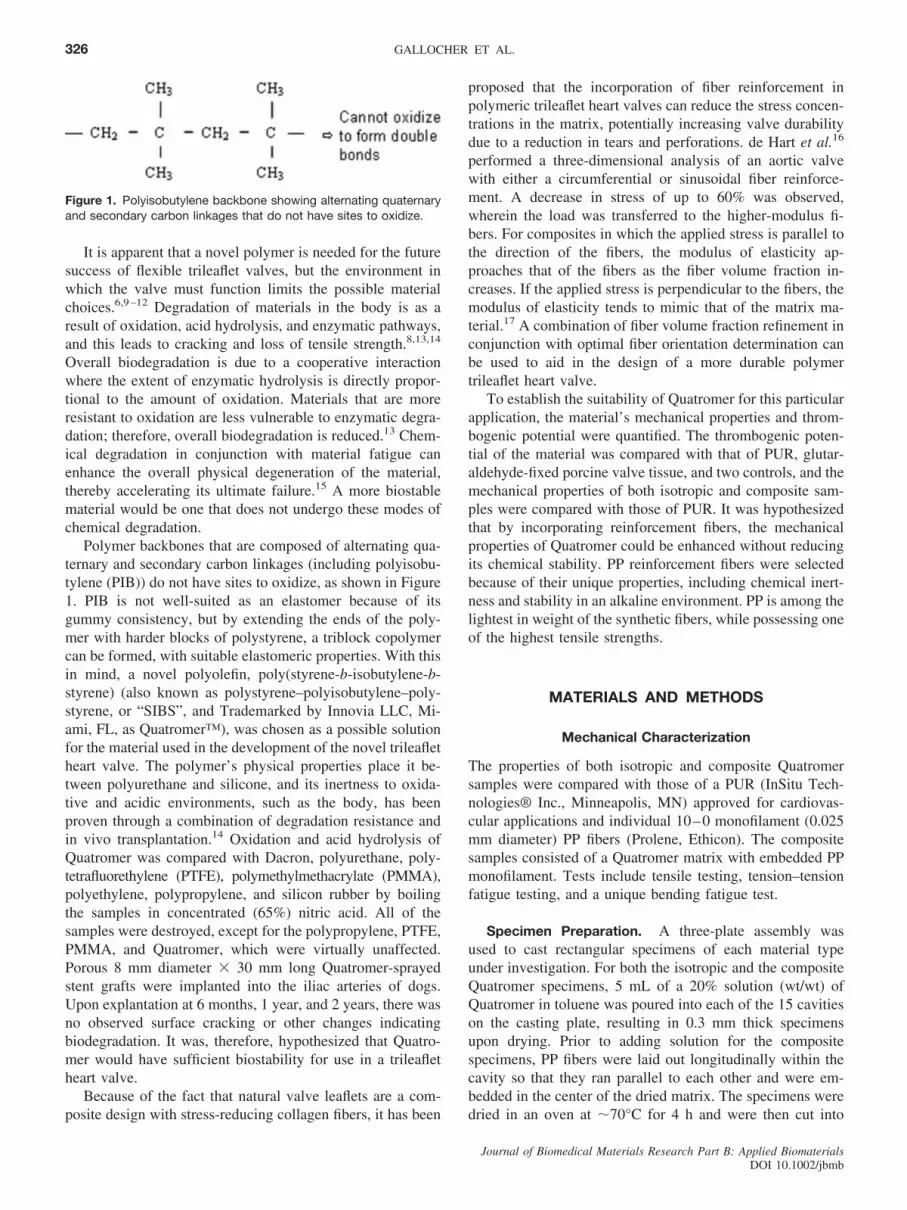
Polymer backbones that are composed of alternating qua-ternary and secondary carbon linkages (including polyisobu-tylene (PIB)) do not have sites to oxidize, as shown in Figure1. PIB is not well-suited as an elastomer because of itsgummy consistency, but by extending the ends of the poly-mer with harder blocks of polystyrene, a triblock copolymercan be formed, with suitable elastomeric properties. With thisin mind, a novel polyolefin, poly(styrene-b-isobutylene-b-styrene) (also known as polystyrene–polyisobutylene–poly-styrene, or “SIBS”, and Trademarked by Innovia LLC, Mi-ami, FL, as Quatromer™), was chosen as a possible solutionfor the material used in the development of the novel trileafletheart valve. The polymer’s physical properties place it be-tween polyurethane and silicone, and its inertness to oxida-tive and acidic environments, such as the body, has beenproven through a combination of degradation resistance andin vivo transplantation.14 Oxidation and acid hydrolysis ofQuatromer was compared with Dacron, polyurethane, poly-tetrafluorethylene (PTFE), polymethylmethacrylate (PMMA),polyethylene, polypropylene, and silicon rubber by boilingthe samples in concentrated (65%) nitric acid. All of thesamples were destroyed, except for the polypropylene, PTFE,PMMA, and Quatromer, which were virtually unaffected.Porous 8 mm diameter � 30 mm long Quatromer-sprayedstent grafts were implanted into the iliac arteries of dogs.Upon explantation at 6 months, 1 year, and 2 years, there wasno observed surface cracking or other changes indicatingbiodegradation. It was, therefore, hypothesized that Quatro-mer would have sufficient biostability for use in a trileafletheart valve.
Because of the fact that natural valve leaflets are a com-posite design with stress-reducing collagen fibers, it has been
proposed that the incorporation of fiber reinforcement inpolymeric trileaflet heart valves can reduce the stress concen-trations in the matrix, potentially increasing valve durabilitydue to a reduction in tears and perforations. de Hart et al.16
performed a three-dimensional analysis of an aortic valvewith either a circumferential or sinusoidal fiber reinforce-ment. A decrease in stress of up to 60% was observed,wherein the load was transferred to the higher-modulus fi-bers. For composites in which the applied stress is parallel tothe direction of the fibers, the modulus of elasticity ap-proaches that of the fibers as the fiber volume fraction in-creases. If the applied stress is perpendicular to the fibers, themodulus of elasticity tends to mimic that of the matrix ma-terial.17 A combination of fiber volume fraction refinement inconjunction with optimal fiber orientation determination canbe used to aid in the design of a more durable polymertrileaflet heart valve.
To establish the suitability of Quatromer for this particularapplication, the material’s mechanical properties and throm-bogenic potential were quantified. The thrombogenic poten-tial of the material was compared with that of PUR, glutar-aldehyde-fixed porcine valve tissue, and two controls, and themechanical properties of both isotropic and composite sam-ples were compared with those of PUR. It was hypothesizedthat by incorporating reinforcement fibers, the mechanicalproperties of Quatromer could be enhanced without reducingits chemical stability. PP reinforcement fibers were selectedbecause of their unique properties, including chemical inert-ness and stability in an alkaline environment. PP is among thelightest in weight of the synthetic fibers, while possessing oneof the highest tensile strengths.
MATERIALS AND METHODS
Mechanical Characterization
The properties of both isotropic and composite Quatromersamples were compared with those of a PUR (InSitu Tech-nologies® Inc., Minneapolis, MN) approved for cardiovas-cular applications and individual 10–0 monofilament (0.025mm diameter) PP fibers (Prolene, Ethicon). The compositesamples consisted of a Quatromer matrix with embedded PPmonofilament. Tests include tensile testing, tension–tensionfatigue testing, and a unique bending fatigue test.
Specimen Preparation. A three-plate assembly wasused to cast rectangular specimens of each material typeunder investigation. For both the isotropic and the compositeQuatromer specimens, 5 mL of a 20% solution (wt/wt) ofQuatromer in toluene was poured into each of the 15 cavitieson the casting plate, resulting in 0.3 mm thick specimensupon drying. Prior to adding solution for the compositespecimens, PP fibers were laid out longitudinally within thecavity so that they ran parallel to each other and were em-bedded in the center of the dried matrix. The specimens weredried in an oven at �70°C for 4 h and were then cut into
Figure 1. Polyisobutylene backbone showing alternating quaternaryand secondary carbon linkages that do not have sites to oxidize.
Journal of Biomedical Materials Research Part B: Applied BiomaterialsDOI 10.1002/jbmb
326 GALLOCHER ET AL.

dog-bone samples, following ASTM standard D 638–89,Type M-III. The same procedure was adopted for the 0.3-mm-thick PUR specimens, except that drying was performedunder vacuum. A caliper (Mitutoyo, Boca Raton, FL) wasused to measure specimen thickness at three different loca-tions, and those that varied more than 10% were discarded.
Tensile Test. The tensile properties of Quatromer, PUR,10–0 monofilament PP fibers, and a Quatromer/PP (Q/PP)composite with both 3 and 12 embedded fibers were com-pared. Tensile tests were performed using the ElectroforceTM
(ELF) 3200 materials tester (Enduratec Systems Corp.,Minnetonka, MN), following ASTM standards D 638M–89(plastics), D 882–88 (thin plastic sheets), and D 3039–89(composites). A crosshead speed of 5 mm/min was used inaccordance with these standards. Outcome measures includ-ed: Young’s Modulus (E), ultimate tensile stress (UTS), andultimate strain (US). A minimum of five specimens weretested for each standard.
Tension–Tension Fatigue Test. The tension fatigue testswere performed according to ASTM standard D 3479M–96,using the ELF materials tester. The materials’ properties wereassessed in air and under load control, where loading ampli-tude was set at �10% of the mean load. Load amounts usedfor each specimen were progressively lowered from the staticvalue until a sufficient amount of data points existed to createan S/N curve. A load frequency of 100 Hz was used, and thetemperature of the specimens was monitored through thermalinfrared imaging techniques to ensure that it did not exceed100°C (glass transition temperature, Tg � 165°C). No markedheating of the specimen occurred, and at no time did thetemperature even approach 100°C. Cycling was continueduntil failure, which was defined as a strain of 0.5. If aspecimen reached 350 million cycles (equivalent to 10 years)without failure, the test was stopped and it was consideredwith infinite life for that stress, as it appeared to be below theendurance limit.
Bending Fatigue Test. An ASTM or ISO standard testfor bending fatigue of an elastomeric material was not avail-able; nevertheless, it is well-known that the valve leaflets aresubjected to pure bending in normal flow conditions.18 Toassess the effects of cyclic bending, the specimens wereloaded on an MTS 858 Mini Bionix® system (MTS SystemsCorp., Eden Prairie, MN) in buckling mode.
By image analysis, it was determined that the deflection ofthe specimen produced a major curvature of 0.296 mm�1
(inverse radius of curvature) (Figure 2), which is in agree-ment with curvatures that have been measured for prostheticvalve leaflets in vitro. Iyengar et al.19 quantified the curvatureof bioprosthetic heart valves using structured light projection,and it was determined that for cylinder diameters rangingfrom 4.76 to 12.70 mm, the range of actual curvatures wasfrom 0.158 to 0.420 mm�1. A polymer with zero residualstrain after cyclic loading is normally thought to be durableand have good fatigue tolerance properties; therefore, a stiff-
ness reduction analysis was used for material characteriza-tion. There were five groups of specimens being tested, eachcontaining five specimens of both isotropic Quatromer andcomposite Q � 12 PP fibers. These five groups were cycledat 12 Hz for 7, 16, 57.5, 79, and 89 million cycles, respec-tively. After cycling, each specimen was tested (within a onehour lapse) according to the tensile test protocols discussedearlier. A change in the material’s properties indicated im-pairment due to fatigue.
Platelet Deposition
Whole blood containing radiolabeled platelets was circulatedonce through a flow loop containing a parallel plate flowchamber. Test specimens of either Quatromer, PUR, glutar-aldehyde-fixed porcine valve tissue (St. Jude porcine biopros-thetic Toronto SPV stentless valve, St. Jude Medical, Inc., St.Paul, MN), glass (positive control), or Sylgard® (Sylgard184, Dow Corning Corporation, Midland, MI) (negative con-trol) were embedded in a Sylgard sheet. The glass/polymersamples (5 � 5 mm2) were positioned on a flat glass surface,and the Sylgard was cast around them to obtain a totalspecimen thickness of 0.5 mm. The surface of the specimenin contact with the glass was the one ultimately exposed toblood flow, therefore, the method of manufacturing ensuredthat a smooth transition existed between the test specimenand the surrounding Sylgard. For the tissue specimen, aspacer was used during casting, and the tissue was transferredinto position prior to testing. Once cured, the samples wereremoved from the glass, trimmed to size, and evaluated forspecimen thickness and cross-sectional quality. A caliper(Mitutoyo, Boca Raton, FL) was used to measure total spec-imen thickness, and those that varied more than 10% werediscarded. To ensure that a smooth transition existed betweenthe Sylgard and the test material, samples were selected at
Figure 2. Measurement of the curvatures produced in the bendingfatigue test. The true measure of each block shown in the image is5 � 5 mm; therefore, a correction factor of 0.674 has to be applied.This results in a radius of 3.37 mm and a curvature of 0.296 mm�1.[Color figure can be viewed in the online issue, which is available atwww.interscience.wiley.com.]
Journal of Biomedical Materials Research Part B: Applied BiomaterialsDOI 10.1002/jbmb
327NOVEL POLYMER FOR HEART VALVE

random for cross-sectional analysis with a Mitutoyo Measur-ing Mircroscope (Mitutoyo, Boca Raton, FL).
The inside of the chamber through which blood flowedhad dimensions of 0.5 � 5 � 50 mm3, and the test sampleswere located 35 mm downstream of the entrance in order toensure fully developed laminar flow over them. The shearrates used in this study included steady shear rates of 10(low), 100 (medium), and 1000 (high) s�1 and average pul-satile shear rates of 100 (medium) and 1000 (high) s�1. Afrequency of 1 Hz was selected for the pulsatile flow, simu-lating a resting heart rate. The flow conditions chosen ex-posed the platelets to shear rates covering the range experi-enced by heart valves under both rest and exercise conditions.
Blood Collection and 111In Labeling. The Office of Hu-man Research Protections (OHRP) guidelines were adheredto for blood collection by venipuncture (150 mL) from adultvolunteers. Two different anticoagulants were used: 50 mL ofthe blood was anticoagulated with acid citrate dextrose(ACD) and the remaining blood was anticoagulated withheparin. Blood, anticoagulated with ACD, was used for 111In-(tropolone)3 labeling, following the procedure of Schoepho-erster et al.20 and Robaina et al.21 111In(tropolone)3 labelingefficiency is maximized in ACD, but this anticoagulant alsoresults in changes to blood pH.22 It has been reported thatplatelet-mediated coagulation is highly sensitive to pH23;consequently, the labeled blood was added to the heparin-anticoagulated blood, allowed to mix and heat to 37°C, andthen divided into four equal aliquots (37.5 mL), each forimmediate testing of an individual specimen. By mixing theACD-anticoagulated blood with the heparin-anticoagulatedblood (in a 1:2 ratio), the pH is normalized, and platelet
activity is assumed to return to normal. A total of 4 runs wereperformed each day, and the chambers and specimen typeswere randomized so as not to result in any bias.
Flow Loop. The flow loop consisted of a parallel plateflow chamber connected by �1.5 ft of silicone rubber tubing(Masterflex® L/S 14®, Cole-Parmer, Vernon Hills, IL; Fig-ure 3). Blood was pumped by a peristaltic pump (Master-flex®, Cole-Parmer Instrument Company, Vernon Hills, IL),and during the steady flow investigations, a damping chamberwas inserted into the flow loop to remove any pulsations dueto the pump rollers. The chamber was removed during pul-satile experiments, as it led to the damping of even the 1 Hzpulsations.
For each experimental run, the content of one of the fourvials was emptied into a blood reservoir maintained at 37°C.The flow loop was primed, and bubbles were removed fromthe chamber and tubing by gentle tapping. The entire contentsof a vial was pumped through the loop, and upon completion,an equivalent volume of physiologic saline was pumpedthrough the system at 5 mL/min to remove any traces ofstanding blood. The flow chamber and tubing were removedfrom the loop and replaced with the next chamber and tubing.This process was repeated until all four chambers and vials ofradiolabeled blood were completed.
Data Collection and Analysis. The Sylgard sheet withthe embedded test specimen was removed, gently rinsed in anisotonic saline solution to remove any additional blood, andsectioned. Samples were counted in an automatic gamma-well counter (Cobra II, Packard Instrument Corp., Meriden,CT).
A minimum of eight tests were run for each test materialat each flow condition, and the results were used to provide a
Figure 3. Schematic of the flow loop consisting of the interchange-able Lexan flow chamber, a blood reservoir, peristaltic pump, waterbath adjusted to maintain blood at 37°C, and silicone rubber tubing.[Color figure can be viewed in the online issue, which is available atwww.interscience.wiley.com.]
Figure 4. Stress–strain relationship for the different polymer materi-als. The error bars represent one standard deviation. Note that theQuatromer and PUR specimens had not failed at 50% strain. [Colorfigure can be viewed in the online issue, which is available atwww.interscience.wiley.com.]
Journal of Biomedical Materials Research Part B: Applied BiomaterialsDOI 10.1002/jbmb
328 GALLOCHER ET AL.

comparison of platelet deposition due to varying materialsand flow conditions. The radioactive counts were used tocalculate the platelet density (number of platelets per unitsurface area) according to the methods outlined by Schoepho-erster et al.20:
No. Platelets
Unit Area (cm2)�
111In cpmSection Area (cm2) �
No. Platelets
mL Whole Blood
�mL Whole Blood
Whole Blood111In cpm � (100-%Free111In)%(1)
where the 111In cpm per section area refers to the countsrecorded from each section removed from the flow chamber,the No. platelets per mL Whole Blood refers to a manualplatelet count, the Whole Blood 111In cpm refers to theamount of 111In in whole blood, and the % Free 111In is thepercentage of 111In free in the plasma.
Normalization of platelet density (NPD) was carried out toeradicate the effects of variability between subject plateletcount and 111In labeling efficiency, in a manner consistentwith that of Schoephoerster et al.20 and Robaina et al.21
Platelet density of the test section was normalized with re-spect to the inlet group by dividing the platelet densitymeasurements of the test specimen by the average plateletdensity over the inlet Sylgard region:
NPD �
No. Platelets (test)
Unit Area
Avg. No. Platelets (control)
Unit Area
(2)
Statistics
A statistical analysis for both the mechanical and the plateletdeposition studies was conducted using the computer packageSPSS for Windows. ANOVA and subsequent post hoc testswere carried out, and any differences were considered to beof significance if p � 0.05.
RESULTS
Mechanical Characterization
As indicated in Figure 4 and Table I, PUR was significantly
stronger than the isotropic Quatromer specimen, with aYoung’s modulus almost five times greater than the formu-lation of Quatromer used in this study. The maximum strainattained for both isotropic specimens was 0.5, and at thisstrain, neither of them failed. By incorporating PP fibers, theYoung’s modulus of the Quatromer specimen was increased,wherein an increase in the number of fibers resulted in aprogressive decrease in elasticity. The stiffness of the Q � 3PP fibers was in the range of that of the PUR, but thiscomposite failed at a strain of 0.35. Q � 12 PP fibers showeda much higher stiffness than PUR, in which the Young’smodulus was more than two times greater. This composite’sstrain at failure was 0.3.
The properties of the composite can be thought of as anaverage of the individual components, and the load contribu-tion of the fibers can be calculated according to the followingequation22:
Pf
Pc�
Ef
EcVf (3)
where Pf and Pc are the fiber and composite loads, respec-tively; Ef and EC are the longitudinal Young’s moduli of thefiber and composite, respectively; and Vf is the ratio betweenthe area of the fiber (Af) and the area of the composite (Ac).For Q � 3 PP fibers, Pf/Pc � 0.73, and so almost 3/4 of theentire uniaxial load was carried by 0.16 vol % of highmodulus fibers. For the Q � 12 PP fiber composite, Pf/Pc �0.95, and so nearly the entire uniaxial load was carried by0.65 vol % of high modulus fibers.
The results from the tension–tension fatigue tests revealedthat the isotropic Quatromer specimen had the lowest endurancelimit, followed by the Q � 3 PP, the PUR, and finally the Q �12 PP (Figure 5). The isotropic Quatromer specimen failed after130 million cycles (equivalent to 3.5 years at a physiological rateof 70 bpm) at an applied stress lower than 0.33 MPa. Byincorporating 3 PP fibers, the endurance limit of the material wasincreased slightly, but a more noticeable difference was seen in
TABLE I. Young’s Modulus (E), Ultimate Strain (US), UltimateTensile Stress (UTS), and Number of Specimens Tested (N) foreach material
Material E (MPa) US UTS (MPa) N
Quatromer (Q) 3.88 � 0.40 �0.5 – 22PP fiber 6633 � 492 0.43 � 0.05 1543 � 124 14Q � 3 PP fibers 14.9 � 2.67 0.34 � 0.05 3.40 � 0.55 8PUR 18.5 � 1.23 �0.5 – 13Q � 12 fibers 45.4 � 2.85 0.30 � 0.03 7.92 � 0.87 10
Figure 5. Stress vs. number of cycles for the different polymer ma-terials. Arrows indicate that the material did not fail after 350 millioncycles. [Color figure can be viewed in the online issue, which isavailable at www.interscience.wiley.com.]
Journal of Biomedical Materials Research Part B: Applied BiomaterialsDOI 10.1002/jbmb
329NOVEL POLYMER FOR HEART VALVE

the PUR specimens, in which an endurance limit larger than 1.5MPa was achieved. At this applied stress, the PUR did not fail(number of cycles � 350 million) as denoted by the arrows. TheQ � 12 PP fiber composite was considered to have the highestendurance limit as it attained over 350 million cycles at anapplied stress of 2.5 MPa.
For the bending fatigue tests, each of the groups cycled 7,16, 57.5, 79, and 89 million cycles (each group consisting of10 specimens: 5 Quatromer and 5 Q � 12 PP fibers) weresubjected to a tensile test, where the outcome measures weretrue Young’s modulus (Etrue), engineering Young’s modulus(Eeng), true ultimate tensile stress (UTStrue), and engineeringultimate tensile stress (UTSeng). The composite specimensincluded an additional measure of ultimate strain (US). Theseresults were compared with control specimens of both typesthat had not been cycled.
The one-way ANOVA performed on the isotropic Quatro-mer specimens determined that a significant difference ex-isted between the groups for the measured values of Etrue
(p � 0.011), Eeng (p � 0.010), UTStrue (p � 0.00), andUTSeng (p � 0.00). A Tukey post hoc test was performed foreach outcome measure, and a pair wise comparison revealedthat for both Etrue and Eeng, the control group that was notcycled was found to be significantly different from the grouptested for 89 million cycles, but those groups were not sig-nificantly different from any of the others. For the UTStrue
and UTSeng, Tukey’s post hoc test revealed that the controlgroup that had not been cycled was significantly different forall of the specimens that had been cycled, but the cycledspecimens were not significantly different from each other.The one-way ANOVA performed on the composite Q � 12PP fibers revealed that no significant differences existed forany of the test parameters. A summary of the numericalvalues is presented in Table II.
Platelet Deposition
On qualitatively comparing NPD data (Table III), it is quiteapparent that the glass and especially the tissue samples havehigher platelet deposition than any of the polymer samples.An ANOVA and subsequent post hoc test was performed onthe complete data set, and it was confirmed that both thetissue and the glass samples were significantly different underall flow conditions. Because of this, subsequent analyses wereperformed comparing only the polymer samples.
The affects of material type on platelet deposition werecompared by grouping the data according to shear rate (Fig-ure 6). The qualitative results revealed in the figure wereconfirmed in subsequent ANOVAs, where significant differ-ence among shear rates was established for the mediumpulsatile (p � 0.039) and low steady (p � 0.005) shear rates.Post hoc tests were performed, and at the medium pulsatileshear rate, a pair-wise comparison revealed that the PURsample displayed significantly greater platelet deposition thanSylgard, but when compared with the Quatromer, no signif-icant difference existed (p � 0.194). At the low steady shearrate, deposition onto both the Quatromer and the PUR weresignificantly different from that on Sylgard, but they were notdifferent from each other (p � 0.373).
The affects of shear rate were also of interest, and so theresults were grouped according to the material type andcompared. No distinct trends existed for any of the materials,
TABLE II. Comparison of the Average Mechanical Propertiesof Isotropic Quatromer and Composite Q � 12 PP FibersSubjected to Bending Fatigue for Either 0 or 89 Million Cycles.
Quatromer (Q) Q � 12 Fibers
0 89 million 0 89 million
US – – 0.3 0.34Etrue (MPa) 3.88 4.41 42.8 45.8Eeng (MPa) 3.61 4.11 39.8 42.6UTStrue (MPa) 1.43 1.76 9.74 10.7UTSeng (MPa) 0.95 1.18 7.47 8.02
TABLE III. Average and Standard Deviation of the Normalized Platelet Density (NPD) for all Samples at all Flow Rates*
Glass (G) Sylgard (S) Quatromer (Q) PUR (P) Tissue (T)
High Pulsatile (HP) 4.88 � 1.57 0.93 � 0.54 1.18 � 0.58 1.33 � 0.42 33.77 � 12.46Medium Pulsatile (MP) 3.84 � 4.03 0.96 � 0.78 1.40 � 0.81 2.29 � 0.92 92.59 � 91.25High Steady (HS) 3.22 � 2.94 1.03 � 0.68 1.34 � 0.73 1.07 � 0.40 21.30 � 11.71Medium Steady (MS) 2.03 � 1.68 0.87 � 0.11 1.74 � 0.83 1.51 � 0.88 120.51 � 69.08Low Steady (LS) 3.22 � 1.37 1.09 � 0.71 2.74 � 0.53 2.20 � 0.53 61.33 � 29.59
* Values in italics show significant difference.
Figure 6. Normalized platelet density (NPD) for the Sylgard (S), Qua-tromer (Q), and PUR (P) test samples at high pulsatile (HP), mediumpulsatile (MP), high steady (HS), medium steady (MS), and low steady(LS) shear, where high, medium, and low refer to shear rates of 1000,100, and 10 s�1, respectively. The data is grouped according to flowrates.
Journal of Biomedical Materials Research Part B: Applied BiomaterialsDOI 10.1002/jbmb
330 GALLOCHER ET AL.

but in general, for both the Quatromer and the PUR, the lowerflow rates resulted in greater deposition than the higher flowrates, i.e. high pulsatile � medium pulsatile and highsteady � medium steady � low steady. An ANOVA revealedthat there was no significant difference because of flow forthe Sylgard samples (p � 0.987), but significance was estab-lished for both the Quatromer (p � 0.000) and the PUR (p �0.002) specimens. Tukey’s test for the Quatromer samplesdetermined that NPD at the low steady shear rate was signif-icantly different from that at every flow rate except themedium steady, whereas the PUR results were grouped intotwo subsets in which NPD for the high steady, high pulsatile,and medium steady flow rates were not significantly different(p � 0.681), and NPD for the medium steady, low steady,and medium pulsatile flow rates were not significantly dif-ferent (p � 0.163). This revealed that the platelet depositionincreased as shear rate decreased.
DISCUSSION
Polymer trileaflet valves have been investigated since the1960s, but their success has not been realized because ofunsuitable material and design choices. Designs incorporat-ing the natural valve trileaflet geometry and isotropic leafletshave been favored because of their physiological hemody-namics. It is believed that physiological hemodynamics leadto a reduction in turbulence and blood damage; however,long-term analyses have revealed that these valves fail owingto calcification, thrombogenicity, high tensile stresses borneby the material, and biodegradation, which manifests as anoverall lack of durability. Polyurethanes have been the pri-mary material of choice, but their biostability is a prevalentproblem.9 Materials in the body degrade due to oxidativeattack, acid hydrolysis, and enzymatic degradation, whereininitial oxidation is assumed to be the primer for degradation.
This realization has prompted the use of fiber-reinforcedQuatromer in a trileaflet heart valve. Quatromer is known tobe resistant in oxidative and acidic environments, and itsmaterial properties are thought to be suitable for use in avalve. Two important issues that need to be quantified beforefurther development of the valve include material hemocom-patibility and durability, and it was the intention of thisresearch endeavor to provide conclusive evidence as to thesuitability of this material. It was hypothesized that throughthe incorporation of reinforcement fibers, a matrix stress-reduction could be achieved similar to that of the collagenfibers in the natural heart valve. One other group, Cacciola etal.,24 attempted this with limited success. The reinforcementfibers did provide a stress reduction, but the hydrodynamicfunctioning of the valve was not optimal because of valveregurgitation.
Mechanical Characterization
Comparative tensile tests of isotropic and composite Quatro-mer samples revealed that it was possible to enhance the
tensile properties of the polymer by embedding PP fibers inthe matrix. The Young’s modulus of isotropic Quatromer waslower than that of PUR, but by incorporating 3 PP fibers, theirproperties became comparable. By incorporating 12 PP fi-bers, the Young’s modulus of Quatromer became twice thatof PUR. Therefore, by varying fiber volume fraction, it ispossible to tailor the mechanical properties of the composite.The high modulus and strength of the reinforcement fibers areeffectively transmitted to the composite as a whole, and theircontribution to the load is high even though they account forless than 1% of the total composite volume. When 3 PP fiberswere incorporated, the fibers carried 73% of the total load,while for 12 PP fibers, they accounted for 95% of the load.
Cyclic tensile and bending stresses acting on the valveleaflet over long-term use have been proven to result infailure of both bioprosthetic and synthetic valves.18,25–28 Itwas because of this that a combination of tension–tension andbending fatigue tests was used to characterize the durabilityof Quatromer. The tension–tension fatigue tests indicated thatby incorporating PP fibers, the fatigue life of Quatromercould be increased to above that of PUR. Isotropic Quatromersamples had a lower fatigue limit than PUR at the same stresslevels, and when 3 PP fibers were incorporated, the fatiguelimit was increased, but it was still lower than PUR. After theincorporation of 12 PP fibers, the fatigue limit of the com-posite increased to above that of PUR, hence, by varying thenumber of embedded fibers, the desired dynamic mechanicalproperties can be obtained. The peak stress acting on a leafletin vivo has been modeled to be in the range of 1 MPa,16,29
where isotropic leaflets result in a higher peak stress thananisotropic, reinforced leaflets. This typical in vivo stress isless than the apparent endurance limit of the Q � 12 PPspecimens, which attained over 350 million cycles at anapplied stress of 2.5 MPa.
It is worth noting that in vitro durability studies of thereinforced Quatromer valve are only in the early stages,therefore, conclusive evidence as to the valve’s durability isnot yet available. Accelerated fatigue testing is being carriedout in a Vivitro Systems (Vancouver, British Columbia)Hi-Cycle accelerated fatigue tester, in which a backpressureof 90 �20/�0 mmHg is maintained at a cycling rate of1200/min. Quatromer valves subjected to accelerated fatiguetesting have reached 400 million cycles (�10 years) to date;this test is still ongoing. Prior studies have been performed onpolyurethane heart valves,18,24,27,28 and one group, Jansen etal.,28 achieved 400 million cycles with their valve. The com-parative study performed here revealed that the Quatromersamples incorporating 12 PP fibers had a higher fatigue lifethan PUR, in which the composite achieved 350 millioncycles at a higher applied stress than that for the PUR. It can,therefore, be proposed that a heart valve leaflet containing thefiber volume fraction, oriented along the principle stressdirections, will be more durable than a polyurethane valve ofthe same design.
The bending fatigue test revealed that by incorporating 12PP fibers, the bending fatigue properties of the polymer canbe optimized. The test did not significantly affect the material
Journal of Biomedical Materials Research Part B: Applied BiomaterialsDOI 10.1002/jbmb
331NOVEL POLYMER FOR HEART VALVE

properties of the composite samples as witnessed by the lackof statistical significance between all groups for measures ofUS, Etrue, Eeng, UTStrue, and UTSeng. This was not the casefor the isotropic Quatromer samples, in which the controlgroup was found to be significantly different from the groupthat was cycled 89 million cycles for measures of Etrue andEeng and significantly different from all of the cycled groupsfor measures of UTStrue, and UTSeng, indicating that thesetests resulted in a weakening of the material. The bendingfatigue tests attained a maximum of 89 million cycles, rep-resenting only 2.5 years for a physiological heart rate of 70bpm; nevertheless, one can conclude that the reinforcementfibers improve both the static and dynamic properties ofQuatromer, and the results support the conclusion that theQuatromer/PP composite is a viable candidate material foruse as a heart valve, based on its mechanical properties.
Platelet Deposition
Measurement of platelet deposition provided a means toquantify the thrombogenic potential of Quatromer. The ma-terial’s affect on platelet deposition was the primary featureunder investigation, while the affects of varying shear weresecondary. A single-pass flow loop was used to ensure plate-let deposition was a result of the material’s reactivity, and notbecause re-circulated platelets were highly activated due tothe tortuosity of the flow loop and the damaging effects of theperistaltic pump. A flow loop was designed to minimize aplatelet’s exposure to excessive shear, thereby, increasing thelikelihood that effects were concentrated in the parallel plateflow chamber, where conditions of shear and material typewere regulated.
The one-way ANOVA and subsequent post-hoc tests onthe complete data set revealed that both the tissue and theglass samples were significantly different from the polymersamples under all flow conditions. It was expected that dep-osition onto glass would be high, hence it can be used as apositive control material, but the considerable increase inplatelet deposition onto the porcine valve material was notanticipated. The tissue specimens were made by sectioning aSt. Jude porcine bioprosthetic Toronto SPV stentless valveinto 5 mm squares, and the sample was oriented so that bloodflowed across the inflow portion (ventricular side) of thespecimen. Upon further analysis of the surface of the tissuesamples, it was determined that the corrugated nature of thematerial could have contributed to the increased reactivity.All other test specimens had a smooth surface over whichblood flowed, but the natural valve had an uneven structure.It was concluded that these surface properties contributed toflow disturbances and possible increased residence time forplatelets; all known to be contributing factors to plateletactivation and adhesion.20,30–33 Studies have shown that tis-sues fixed with glutaraldehyde are less thrombogenic andresult in less platelet deposition than native tissue under staticexposure to both whole blood and platelet rich plasma.34,35
These studies indicated that collagen looses its ability toinduce platelet activation and adhesion once it has been fixed
with glutaraldehyde, therefore, material affects are less likelyto contribute to increased platelet deposition when comparedwith affects due to flow disturbances.
Material affects on platelet deposition were examined bygroup-wise comparisons under each respective shear condi-tion. Deposition onto the Sylgard was consistently lower thanthat onto any of the other materials (Figure 6), and statisticalanalysis revealed that the differences were only of signifi-cance at the lower shear rates, i.e., at medium pulsatile shearand at low steady shear. Under those same flow conditions, acomparison of platelet deposition onto the Quatromer andPUR samples proved that they were not significantly different(p �� 0.05). It can, therefore, be stated that in terms ofthrombogenic potential, Quatromer was as thromboresistantas the leading choice for polymer trileaflet heart valves,polyurethane. At the higher shear rates, platelet depositiononto all samples was not significantly different. Ross et al.32
intimated that at low shear rates, surfaces were more reactiveto platelet deposition although transport of platelets to thesurface was reduced. It was believed that under high shearconditions, although platelet transport to the surface is high,the surface is incapable of maintaining deposited platelets,and the high shear conditions result in platelets being depos-ited and subsequently sheared off. It would then make sensethat a surface whose characteristics make it more thromboticwould show pronounced differences in platelet deposition atthe lower flow rates. As the Quatromer surface was no lessthromboresistant than the polyurethane surface, it can beconcluded that it is also a viable candidate for a polymer heartvalve in terms of its hemocompatibility.
Shear affects on platelet deposition were compared bysystematically grouping results according to material type. Itis apparent that no trends existed, and the Sylgard sampleshad similar results under all flow conditions.
For all flow conditions, the volume of blood the testsamples were exposed to remained constant. This meant thatthe time of exposure for the lower flow rates was longer thanthat for the higher flow rates. Both Ross et al.32 and Hubbelland McIntire36,37 presented results showing that when theexposure time was increased for the same shear rate, plateletdeposition increased. Because the shear rates in this investi-gation varied, it was determined that by exposing the testmaterials to the same volume of blood, a better comparisonbetween flow rates would be achieved as each sample wouldcome in contact with an equivalent volume of platelets. Forthe low steady flow rate, a comparison was made betweenplatelet deposition after 30 and 90 min, and results wereequivalent. It was concluded that platelet deposition did reacha maximum and would not continue to increase indefinitely.If the time of exposure was kept constant, at the lower flowrates the test samples would be exposed to fewer platelets,therefore, deposition at these flow rates would most likely beless, simply because opportunity for platelet contact with thesurface would be minimized.
Bluestein et al.31 suggested that pulsatility increased thepotential for platelet deposition, but this phenomenon was notwitnessed in these experiments. Pulsatile flow was estab-
Journal of Biomedical Materials Research Part B: Applied BiomaterialsDOI 10.1002/jbmb
332 GALLOCHER ET AL.

lished where the mean flow rates were equivalent to thesteady flow rates. Under these circumstances, throughout theflow cycle there were conditions where the shear was bothgreater and less than that during steady flow, and it wasassumed that the high shear resulted in greater platelet acti-vation while the low shear allowed greater residence time forplatelet deposition. As these conditions were cyclical, plate-lets adhered on the surface could have been sheared off at thehigh shear conditions, hence the consequence that pulsatilitydid not result in greater platelet deposition. Schoephoerster etal.20 examined these conditions in both a stenosis and aneu-rysm model, and regions of flow recirculation after initialexposure to increased shear were primary locations for plate-let deposition. Bluestein et al.31 examined these conditions ina mechanical heart valve, and the flow disturbances wereconcluded to result in increased platelet deposition and freeemboli formation. As blood passes through a heart valve, it issubjected to varying degrees of shear, including regions offlow recirculation, and the only way to examine the influenceof these conditions would be to determine platelet reactionsdue to flow past an actual valve. Although pulsatile flowconditions were established in the parallel plate flow cham-ber, at no time was flow recirculation present. The pulsatileflow varied the flow rate around a mean value, but at no timewas negative flow present. Flow recirculation, rather thanplain pulsatility is more likely to lead to increased plateletdeposition, which may be the reason why increased plateletdeposition was not witnessed in this study.20,31
CONCLUSION
The feasibility of Quatromer for use in a polymer trileafletheart valve was investigated through a combination of me-chanical and hemocompatibility testing. Polyurethanes havebeen the most widely investigated materials for use in poly-mer valves, and due to this a polyurethane approved forcardiovascular applications was used as a comparative mate-rial to gage the effectiveness of Quatromer.
The mechanical evaluation of the isotropic Quatromerformulation that was used in this study revealed that it did nothave the strength or structural integrity that the polyurethanehad under both static and dynamic testing conditions, butwhen PP fibers were incorporated into the matrix, theYoung’s modulus increased and both the tension–tension andbending fatigue properties of the material were improved.With 12 PP fibers, no mechanical degradation was evidentafter 89 million cycles of bending fatigue, and for the ten-sion–tension fatigue, the endurance limit at 350 million cy-cles for the composite polymer was higher than that for thepolyurethane. The platelet deposition studies proved thatQuatromer was no more thrombogenic than the medical gradepolyurethane.
In conclusion, both the biocompatibility and mechanicalcharacterization of Quatromer have proven it to be a viablecandidate for use in a trileaflet heart valve. Fiber reinforce-ment can effectively be used to tailor the mechanical prop-
erties, and current characterization of the valve’s durabilityhas shown its promise, where a valve has attained 400 millioncycles without failure. The unexpectedly high platelet depo-sition on the tissue samples prompted a further in vitro studyclassifying the platelet activation in valves mounted in a leftventricular assist device.38 Flow effects were the major con-sideration, and platelet activation due to the polymer valvewas not found to be significantly different from either a St.Jude bileaflet valve or a St. Jude Tissue valve. Chronic (20weeks) in vivo studies are being performed in an ovine model,where safety and hemodynamic performance of valves im-planted in the aortic position are of interest. In an animal thatsurvived 10 weeks, no sign of thrombus or embolus forma-tion was found, further supporting the valve’s biocompatibil-ity. Further surface modification to increase the hemocom-patibility of Quatromer has been performed, and these resultswill be presented in subsequent publications. Many of thesestudies are ongoing, but preliminary results continue to con-firm the viability of a Quatromer-based, fiber-reinforcedtrileaflet heart valve for human use.
Bioprosthetic heart valves were provided by St. Jude Medical,Inc.
REFERENCES
1. Edmunds LH, McKinlay S, Anderson JM, Callahan TH, Chese-bro JH, Geiser EA, Makanani DM, McIntire LV, Meeker WQ,Naughton GK, Panza JA, Schoen FJ, Didisheim P. Directionsfor improvement of substitute heart valves: National Heart,Lung, and Blood Institute’s Working Group report on heartvalves. J Biomed Mater Res 1997;38:263–266.
2. Cannegieter SC, Rosendaal FR, Wintzen AR, van der Meer FJ,Vandenbrouke JP, Briet E. Optimal oral anticoagulant therapyin patients with mechanical heart valves. N Engl J Med 1995;333:11–17.
3. Chandran KB, Fatemi R, Schoephoerster RT, Wurzel D, HansenD, Pantalos G, Yu LS, Kolff WJ. In vitro comparison of velocityprofiles and turbulent shear distal to polyurethane trileaflet andpericardial prosthetic valves. Artif Organs 1989;13:148–154.
4. Senthilnathan V, Treasure T, Grunkemeier G, Starr A. Heartvalves: Which is the best choice? Cardiovasc Surg 1999;7:393–397.
5. Imamura E, Kaye MP. Function of expanded-polytetrafluoro-ethylene laminated trileaflet valves in animals. Mayo Clin Proc1977;52:770–775.
6. Hyde JAJ, Chinn JA, Phillips RE. Polymer heart valves. J HeartValve Dis 1999;8:331–339.
7. Reul H. In-vitro evaluation of artificial heart valves. Adv Car-diol Phy 1983;5:16–30.
8. Pinchuk L. A review of the biostability and carcinogenicity ofpolyurethanes in medicine and the new generation of ‘biostable’polyurethanes. J Biomater Sci Polym Ed 1994;6:225–267.
9. Duguay DG, Labow RS, Santerre JP, McLean DD. Develop-ment of a mathematical model describing the enzymatic degra-dation of biomedical polyurethanes. I. Background, rationale,and model formulation. Polym Degrad Stab 1995;47:229–249.
10. Mackay TG, Wheatley DJ, Bernacca GM, Fisher AC, HindleCS. New polyurethane heart valve prosthesis: Design, manu-facture and evaluation. Biomaterials 1996;17:1857–1863.
11. Mackay TG, Bernacca GM, Fisher AC, Hindle CS, WheatleyDJ. In vitro function and durability assessment of a novel
Journal of Biomedical Materials Research Part B: Applied BiomaterialsDOI 10.1002/jbmb
333NOVEL POLYMER FOR HEART VALVE

polyurethane heart valve prosthesis. Artif Organs 1996;20:1017–1025.
12. Wheatley DJ, Raco L, Bernacca GM, Sim I, Belcher PR, BoydJS. Polyurethane: Material for the next generation of heart valveprostheses? Eur J Cardiothorac Surg 2000;17:440–448.
13. Hsu S, Huang T. The susceptibility of poly(ether)urethanes toenzymatic degradation after oxidative pretreatment. Polym De-grad Stab 2000;67:171–178.
14. Pinchuk L, Khan IJ, Martin JB, Wilson GJ. Polyisobutylene-based thermoplastic elastomers for ultra long-term implant ap-plications. Sixth World Biomaterials Congress Transactions(Trans Soc Biomaterials). 1999;1452.
15. Wiggins MJ, Anderson JM, Hiltner A. Biodegradation of poly-urethane under fatigue loading. J Biomed Mater Res A 2003;65:524–535.
16. de Hart J, Cacciola G, Schreurs PJG, Peters GWM. A three-dimensional analysis of a fibre-reinforced aortic valve prosthe-sis. J Biomech 1998;31:629–638.
17. Skinner HB. Composite technology for total hip arthroplasty.Clin Orthop Relat Res 1988;235:224–236.
18. Bernacca GM, Mackay TG, Wilkinson R, Wheatley DJ. Calci-fication and fatigue failure in a polyurethane heart value. Bio-materials 1995;16:279–285.
19. Iyengar AK, Sugimoto H, Smith DB, Sacks MS. Dynamic invitro quantification of bioprosthetic heart valve leaflet motionusing structured light projection. Ann Biomed Eng 2001;29:963–964.
20. Schoephoerster RT, Oynes F, Nunez G, Kapadvanjwala M,Dewanjee MK. Effects of local geometry and fluid dynamics onregional platelet deposition on artificial surfaces. ArteriosclerThromb 1993;13:1806–1813.
21. Robaina S, Jayachandran B, He Y, Frank A, Moreno MR,Schoephoerster RT, Moore JE. Platelet adhesion to simulatedstented sufaces. J Endovasc Ther 2003;10:978–986.
22. Dewanjee MK, Rao SA, Didisheim P. Indium-111 tropolone, anew high-affinity platelet label: Preparation and evaluation oflabeling parameters. J Nucl Med 1981;22:981–987.
23. Green FW, Kaplan MM, Curtis LE, Levine PH. Effect of acidand pepsin on blood coagulation and platelet aggregation. Apossible contributor prolonged gastroduodenal mucosal hemor-rhage. Gastroenterology 1978;74:38–43.
24. Cacciola G, Peters GWM, Baaijens FPT. A synthetic fiber-reinforced stentless heart valve. J Biomech 2000;33:653–658.
25. Chetta GE, Lloyd JR. The design, fabrication and evaluation ofa trileaflet prosthetic heart valve. J Biomech Eng 1980;102:34–41.
26. Fann JI, Miller DC. Porcine valves: Hancock and Carpentier-Edwards aortic prostheses. Semin Thorac Cardiovasc Surg1996;8:259–268.
27. Bernacca GM, O’Connor B, Williams DF, Wheatley DJ. Hy-drodynamic function of polyurethane prosthetic heart valves:Influences of Young’s modulus and leaflet thickness. Biomate-rials 2002;23:45–50.
28. Jansen J, Reul H. A synthetic three-leaflet valve. J Med EngTechnol 1992;16:27–33.
29. Li J, Luo XY, Kuang ZB. A nonlinear anisotropic model forporcine aortic valves. J Biomech 2001;34:1279–1289.
30. Sefton MV, Gemmell CH, Gorbet MB. What really is bloodcompatibility? J Biomater Sci Polym Ed 2000;11:1165–1182.
31. Bluestein D, Rambod E, Gharib M. Vortex shedding as amechanism for free emboli formation in mechanical heartvalves. J Biomech Eng 2000;122:125–134.
32. Ross JM, McIntire LV, Moake JL, Rand JH. Platelet adhe-sion and aggregation on human type VI collagen surfacesunder physiological flow conditions. Blood 1995;85:1826 –1835.
33. Alevriadou BR, Moake JL, Turner NA, Ruggeri ZM, Folie BJ,Phillips MD, Schreiber AB, Hrinda ME, McIntire LV. Real-time analysis of shear dependent thrombus formation and itsblockade by inhibitors of von Willebrand factor binding toplatelets. Blood 1993;81:1263–1276.
34. Sung HW, Chen CN, Huang RN, Hsu JC, Chang WH. In vitrosurface characterization of a biological patch fixed witha naturally occurring crosslinking agent. Biomaterials 2000;21:1353–1362.
35. Hum OS, Ghista DN, Brash JL. Thrombogenic studies of bio-prosthetic heart valve surfaces. I. In vitro platelet adhesion in astatic system. Thromb Res 1985;38:163–172.
36. Hubbell JA, McIntire LV. Visualization and analysis of muralthrombogenesis on collagen, polyurethane, and nylon. Bioma-terials 1986;7:354–364.
37. Hubbell JA, McIntire LV. Technique for visualization and anal-ysis of mural thrombogenesis. Rev Sci Instrum 1986;57:892–897.
38. Yin W, Gallocher S, Pinchuk L, Schoephoerster RT, Jesty J,Bluestein D. Flow induced platelet activation in a St. Judemechanical heart valve, a trileaflet polymeric heart valve, and aSt. Jude tissue valve. Artif Organs 2004;29:826–831.
Journal of Biomedical Materials Research Part B: Applied BiomaterialsDOI 10.1002/jbmb
334 GALLOCHER ET AL.