novel classes of non-coding rnas and cancer
TRANSCRIPT
Sana et al. Journal of Translational Medicine 2012, 10:103http://www.translational-medicine.com/content/10/1/103
REVIEW Open Access
Novel classes of non-coding RNAs and cancerJiri Sana1,2, Petra Faltejskova1,2, Marek Svoboda1 and Ondrej Slaby1,2,3*
Abstract
For the many years, the central dogma of molecular biology has been that RNA functions mainly as aninformational intermediate between a DNA sequence and its encoded protein. But one of the great surprises ofmodern biology was the discovery that protein-coding genes represent less than 2% of the total genomesequence, and subsequently the fact that at least 90% of the human genome is actively transcribed. Thus, thehuman transcriptome was found to be more complex than a collection of protein-coding genes and their splicevariants. Although initially argued to be spurious transcriptional noise or accumulated evolutionary debris arisingfrom the early assembly of genes and/or the insertion of mobile genetic elements, recent evidence suggests thatthe non-coding RNAs (ncRNAs) may play major biological roles in cellular development, physiology andpathologies. NcRNAs could be grouped into two major classes based on the transcript size; small ncRNAs and longncRNAs. Each of these classes can be further divided, whereas novel subclasses are still being discovered andcharacterized. Although, in the last years, small ncRNAs called microRNAs were studied most frequently with morethan ten thousand hits at PubMed database, recently, evidence has begun to accumulate describing the molecularmechanisms by which a wide range of novel RNA species function, providing insight into their functional roles incellular biology and in human disease. In this review, we summarize newly discovered classes of ncRNAs, andhighlight their functioning in cancer biology and potential usage as biomarkers or therapeutic targets.
Keywords: Non-coding RNAs, microRNAs, siRNAs, piRNAs, lncRNAs, Cancer
IntroductionThe abundance of non-translated functional RNAs inthe cell has been a textbook truth for decades. Most ofthese non-coding RNAs (ncRNAs) fulfil essential func-tions, such as ribosomal RNAs (rRNAs) and transferRNAs (tRNAs) involved in mRNA translation, small nu-clear RNAs (snRNAs) involved in splicing and small nu-cleolar RNAs (snoRNAs) involved in the modification ofrRNAs. The central dogma of molecular biology, devel-oped from the study of simple organisms like Escheri-chia coli, has been that RNA functions mainly as aninformational intermediate between a DNA sequence(‘gene’) and its encoded protein. The presumption wasthat most genetic information that specifies biologicalform and phenotype is expressed as proteins, which havenot only diverse catalytic and structural functions, butalso regulate the activity of the system in various ways.
* Correspondence: [email protected] Memorial Cancer Institute, Department of Comprehensive CancerCare, Zluty kopec 7, Brno, Czech Republic, Europe2Central European Institute of Technology, Masaryk University, Brno, CzechRepublic, EuropeFull list of author information is available at the end of the article
© 2012 Sana et al.; licensee BioMed Central LtCommons Attribution License (http://creativecreproduction in any medium, provided the or
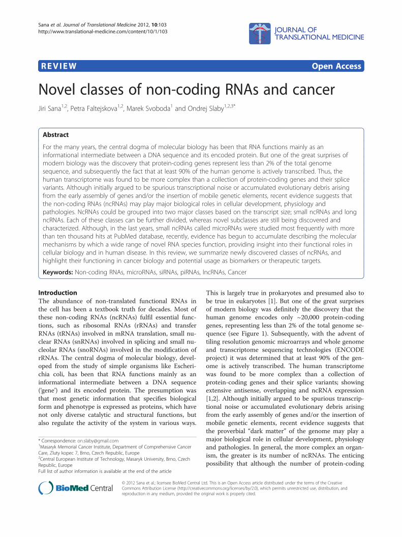
This is largely true in prokaryotes and presumed also tobe true in eukaryotes [1]. But one of the great surprisesof modern biology was definitely the discovery that thehuman genome encodes only ~20,000 protein-codinggenes, representing less than 2% of the total genome se-quence (see Figure 1). Subsequently, with the advent oftiling resolution genomic microarrays and whole genomeand transcriptome sequencing technologies (ENCODEproject) it was determined that at least 90% of the gen-ome is actively transcribed. The human transcriptomewas found to be more complex than a collection ofprotein-coding genes and their splice variants; showingextensive antisense, overlapping and ncRNA expression[1,2]. Although initially argued to be spurious transcrip-tional noise or accumulated evolutionary debris arisingfrom the early assembly of genes and/or the insertion ofmobile genetic elements, recent evidence suggests thatthe proverbial “dark matter” of the genome may play amajor biological role in cellular development, physiologyand pathologies. In general, the more complex an organ-ism, the greater is its number of ncRNAs. The enticingpossibility that although the number of protein-coding
d. This is an Open Access article distributed under the terms of the Creativeommons.org/licenses/by/2.0), which permits unrestricted use, distribution, andiginal work is properly cited.
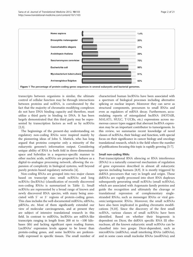
Figure 1 The percentage of protein-coding genes sequences in several eukaryotic and bacterial genomes.
Sana et al. Journal of Translational Medicine 2012, 10:103 Page 2 of 21http://www.translational-medicine.com/content/10/1/103
transcripts between organisms is similar, the ultimatecontrol of cellular function may be through interactionsbetween proteins and ncRNA, is corroborated by thefact that the majority of chromatin-modifying complexesdo not have DNA binding capacity and therefore, mustutilize a third party in binding to DNA. It has beenlargely demonstrated that this third party may be repre-sented by transcription factors as well as by ncRNAs[2,3].The beginnings of the present-day understanding on
regulatory non-coding RNAs were inspired mainly bythe pioneering ideas of John S. Mattick, who has longargued that proteins comprise only a minority of theeukaryotic genome’s information output. Consideringunique ability of RNA to both fold in three-dimensionalspace and hybridize in a sequence-specific manner toother nucleic acids, ncRNAs are proposed to behave as adigital-to-analogue processing network, allowing the ex-pansion of complexity in biological systems, well beyondpurely protein-based regulatory networks [4].Non-coding RNAs are grouped into two major classes
based on transcript size; small ncRNAs and longncRNAs (lncRNAs) (classification of recently discoverednon-coding RNAs is summarized in Table 1). SmallncRNAs are represented by a broad range of known andnewly discovered RNA species, with many being asso-ciated with 5′ or 3′ regions of protein-coding genes.This class includes the well-documented miRNAs, siRNAs,piRNAs, etc. Most of them significantly extended ourview of molecular carcinogenesis, and at present theyare subject of intensive translational research in thisfield. In contrast to miRNAs, lncRNAs are mRNA-liketranscripts ranging in length from 200 nt to ~100 kilo-bases (kb) and lacking significant open reading frames.LncRNAs’ expression levels appear to be lower thanprotein-coding genes, and some lncRNAs are preferen-tially expressed in specific tissues. The small number of
characterized human lncRNAs have been associated witha spectrum of biological processes including alternativesplicing or nuclear import. Moreover they can serve asstructural components, precursors to small RNAs andeven as regulators of mRNA decay. Furthermore, accu-mulating reports of misregulated lncRNA (HOTAIR,MALAT1, HULC, T-UCRs, etc.) expressions across nu-merous cancer types suggest that aberrant lncRNA expres-sion may be an important contributor to tumorigenesis. Inthis review, we summarize recent knowledge of novelclasses of ncRNAs, their biology and function, with specialfocus on their significance in cancer biology and oncologytranslational research, which is the field where the numberof publications focusing this topic is rapidly growing [5-7].
Small non-coding RNAsPost-transcriptional RNA silencing or RNA interference(RNAi) is a naturally conserved mechanism of regulationof gene expression described in almost all eukaryoticspecies including humans [8,9]. It is mostly triggered bydsRNA precursors that vary in length and origin. ThesedsRNAs are rapidly processed into short RNA duplexessubsequently generating small ncRNAs (small ncRNAs),which are associated with Argonaute family proteins andguide the recognition and ultimately the cleavage ortranslational repression of complementary single-stranded RNAs, such as messenger RNAs or viral gen-omic/antigenomic RNAs. Moreover, the small ncRNAshave also been implicated in guiding chromatin modifi-cations [9,10]. Since the discovery of the first smallncRNA, various classes of small ncRNAs have beenidentified. Based on whether their biogenesis isdependent on Dicer, the dsRNA specific RNA III ribo-nuclease, all the known eukaryotic small ncRNAs can beclassified into two goups: Dicer-dependent, such asmicroRNAs (miRNAs), small interfering RNAs (siRNAs),and in some cases small nucleolar RNAs (snoRNAs); and

Table 1 Types of recently discovered human non-coding RNAs
Class Symbol Characteristic Disease / biological function associations
Smallnon-codingRNAs
MicroRNAs miRNAs 18–25 nt; account 1–2% of the human genome;control the 50% of protein-coding genes; guidesuppression of translation; Drosha and Dicerdependent small ncRNAs
initiation of various disorders including many,if not all, cancers / regulation of proliferation,differentiation, and apoptosis involved in humandevelopment
Small interferingRNAs
siRNAs 19–23 nt; made by Dicer processing; guidesequence specific degradation of target mRNA
great potential in diseases treatment /posttranscriptional gene silencing mainlythrough RISC degradation mechanism; defenceagainst pathogenic nucleic acids
Piwi-interactingRNAs
piRNAs 26–30 nt; bind Piwi proteins; Dicer independent;exist in genome clusters; principally restricted tothe germline and somatic cells bordering thegermline
relationship between piRNAs and diseases hasnot yet been discovered / involved in germ celldevelopment, stem self-renewal, andretrotransposon silencing
Small nucleolarRNAs
snoRNAs 60–300 nt; enriched in the nucleolus; invertebrate are excised from pre-mRNA introns;bind snoRNP proteins
association with development of some cancers /important function in the maturation of othernon-coding RNAs, above all, rRNAs and snRNAs;miRNA-like snoRNAs regulate mRNAs
Promoter-associated smallRNAs
PASRs 20–200 nt; modified 5′ (capped) ends; coincidewith the transcriptional start sites of protein- andnon-coding genes; made from transcription ofshort capped transcripts
relationship with diseases has not yet beendiscovered / involved in the regulation of thetranscription of protein-coding genes bytargeting epigenetic silencing complexes
Transcriptioninitiation RNAs
tiRNAs ~ 18 nt ; have the highest density justdownstream of transcriptional start sites; showpatterns of positional conservation; preferentiallylocated in GC-rich promoters
Centromere repeatassociated smallinteracting RNAs
crasiRNAs 34–42 nt; processed from long dsRNAs relationship between crasiRNAs and diseases hasnot yet been discovered / involved in therecruitment of heterochromatin and/orcentromeric proteins
Telomere-specificsmall RNAs
tel-sRNAs ~ 24 nt; Dicer independent; 2′-O-methylated atthe 3′ terminus; evolutionarily conserved fromprotozoa to mammals; have not been describedin human up to now
relationship between tel-sRNAs and diseases hasnot yet been discovered / epigenetic regulation
Pyknons subset of patterns of variable length; formmosaics in untranslated and protein-codingregions; more frequently in 3′ UTR
expected association with cancer biology /possible link with posttranscriptional silencingof genes, mainly involved in cell communication,regulation of transcription, signaling, transport,etc.
Longnon-codingRNAs
Long intergenicnon-coding RNAs
lincRNAs ranging from several hundreds to tens ofthousands nts; lie within the genomic intervalsbetween two genes; transcriptional cis-regulationof neighbouring genes
involved in tumorigenesis and cancer metastasis/ involved in diverse biological processes such asdosage compensation and/or imprinting
Long intronic non-coding RNAs
lie within the introns; evolutionary conserved;tissue and subcellular expression specified
aberrantly expressed in human cancers / possiblelink with posttranscriptional gene silencing
Telomere-associatedncRNAs
TERRAs 100 bp - >9 kb; conserved among eukaryotes;synthesized from C-rich strand; polyadenylated;form inter-molecular G-quadruplex structure withsingle-stranded telomeric DNA
possible impact on telomere-associated diseasesincluding many cancers / negative regulation oftelomere length and activity through inhibitionof telomerase
Long non-codingRNAs with dualfunctions
both protein-coding and functionally regulatoryRNA capacity
deregulation has been described in breast andovarian tumors / modulate gene expressionthrough diverse mechanisms
Pseudogene RNAs gene copies that have lost the ability to code fora protein; potential to regulate their protein-coding cousin; made through retrotrans-position;tissue specific
often deregulated during tumorigenesis andcancer progression / regulation of tumorsuppressors and oncogenes by acting asmicroRNA decoys
Transcribed-ultraconservedregions
T-UCRs longer than 200 bp; absolutely conservedbetween orthologous regions of human, rat, andmouse; located in both intra- and intergenicregions
expression is often altered in some cancers;possible involvement in tumorigenesis /antisense inhibitors for protein-coding genesor other ncRNAs
Sana et al. Journal of Translational Medicine 2012, 10:103 Page 3 of 21http://www.translational-medicine.com/content/10/1/103

Sana et al. Journal of Translational Medicine 2012, 10:103 Page 4 of 21http://www.translational-medicine.com/content/10/1/103
Dicer-independent small ncRNAs, such as PIWI-interacting RNAs (piRNAs) [11] (Figure 2). Moreover,phylogenetic analysis indicates that known Argonautefamily proteins can be divided into two subgroups namelyAGO based on AGO1 and PIWI based on PIWI. Interest-ingly, Ago proteins interact with miRNAs and siRNAswhile Piwi subgroup is characterized by interaction withpiRNAs [12]. Biogenesis of other small non-coding RNAsis less or completely undescribed yet. These RNAs aregenerally classified according to their genome and func-tion localization. Among them belong promoter-associated small RNAs (PASRs), transcription initiationRNAs (tiRNAs), centromere repeat associated small inter-acting RNAs (crasiRNAs), and telomere-specific smallRNAs (tel-sRNAs). To the class of small non-codingRNAs also belong the recently discovered pyknons that, assuggested by current findings, are involved in many bio-logical functions. It was many times described that someof above mentioned small non-coding RNAs play import-ant roles in pathogenesis of various diseases includingtumors. In this respect, the most studied ncRNAs aremiRNAs, which have been described in many, if not all,cancers [13-16].
MicroRNAsThe most frequently studied subclass of small ncRNAsare microRNAs (miRNAs), originally discovered by Vic-tor Ambros in Caenorhabditis elegans. They are 18–25nucleotides long, evolutionary conserved, single-stranded
Figure 2 Short ncRNAs biogenesis pathways.
RNA molecules involved in specific regulation of gene ex-pression in eukaryotes [17]. It is predicted that miRNAgenes account for 1–2% of the human genome and con-trol the activity of ~50% of all protein-coding genes[18,19]. Early annotation for the genomic position of miR-NAs indicated that most miRNAs are located in intergenicregions (>1 kb away from annotated or predicted genes),although a sizeable minority was found in the intronicregions of known genes in the sense or antisense orienta-tion. This led to the postulation that most miRNA genesare transcribed as autonomous transcription units [19]. Adetailed analysis of miRNA gene expression showed thatmiRNA genes can be transcribed from their own promo-ters and that miRNAs are generated by RNA polymeraseII (RNAPII) as primary transcripts (pri-miRNAs). Theseare processed to short 70-nucleotide stem–loop structuresknown as pre-miRNAs by the ribonuclease called Droshaand the double-stranded-RNA-binding protein known asPasha (or DGCR8 – DiGeorge critical region 8), whichtogether compose a multiprotein complex termed amicroprocessor. The pre-miRNAs are transported tocytoplasm by the RAN GTP-dependent transporterexportin 5 (XPO5). In the cytoplasm, the pre-miRNAsare processed to mature miRNA duplexes by their inter-action with the endonuclease enzyme Dicer in complexwith dsRNA binding protein TRBP [19,20]. One strand(“guide strand”) of the resulting 18–25-nucleotide ma-ture miRNA duplex ultimately gets integrated into themiRNA-induced silencing complex (miRISC) with the
Sana et al. Journal of Translational Medicine 2012, 10:103 Page 5 of 21http://www.translational-medicine.com/content/10/1/103
central part formed by proteins of the Argonaute family,whereas the other strand (passenger or miRNA*) isreleased and degraded. The retained (“guide”) strand isthe one that has the less stably base-paired 5′ end in themiRNA/miRNA* duplex. Generally, most miRNA genesproduce one dominant miRNA species. However, theratio of miRNA to miRNA* can vary in different tissuesor developmental stages, which probably depends onspecific properties of the pre-miRNA or miRNA duplex,or on the activity of different accessory processing fac-tors [19]. Moreover, the ratio might be modulated bythe availability of mRNA targets as a result of enhanceddestabilization of either miRNA or miRNA* occurringin the absence of respective complementary mRNAs[20]. Mature miRNAs in miRISC exert their regulatoryeffects by binding to imperfect complementary sites. MiR-NAs repress target-gene expression post-transcriptionally,apparently at the level of translation, through a miRISCcomplex that is similar to, or possibly identical with, thatused for the RNAi pathway discussed later. Perfect com-plementarity of mRNA-miRNA allows Ago-catalyzedcleavage of the mRNA strand, whereas central mis-matches exclude cleavage and promote repression ofmRNA translation. Consistent with translational con-trol, miRNAs that use this mechanism reduce the pro-tein levels of their target genes, but the mRNA levels ofthese genes are barely affected [21-23]. Current studiesindicate that miRNA targeting in mammalian cellsoccurs predominantly through binding to sequenceswithin 3′UTRs [24,25], however inhibition of gene ex-pression through targeting the 5′UTR has been alsodemonstrated [26]. Nevertheless, statistical analyses ofconserved miRNA target sequences proved that mam-malian miRNA target sites rarely occur within 5′UTRs[24,25,27]. Moreover, it was found out that miR-10ainduces, rather than inhibits, protein expressionthrough binding to 5′UTRs of cellular transcripts [23].It is therefore supposed that binding to 5′UTR resultsin mechanistic effects divergent from 3′UTR binding.Most of the miRNAs described to date regulate crucial
cell processes such as proliferation, differentiation, andapoptosis. Therefore, these RNAs are involved in humandevelopment as well as in initiation of various disordersincluding many, if not all, cancers where miRNAs havebeen found to be also significant prognostic and predict-ive markers [13,28-35]. Examples of miRNAs with sig-nificant functional effects in cancer are mentionedbelow.Bloomston et al. [36] identified 6 miRNAs linked to
long-term survival in pancreatic adenocarcinoma.They found also that expression level of miR-196a-2was able to predict patients’ survival, since highermiRNA levels marked the poor survivors group. InHCC, up-regulation of miR-221 and down-regulation of
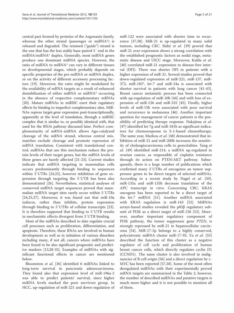
miR-122 were associated with shorter time to recur-rence [37,38]. MiR-21 is up-regulated in many solidtumors, including CRC. Slabý et al. [39] proved thatmiR-21 over-expression shows a strong correlation withthe established prognostic factors as nodal stage, meta-static disease and UICC stage. Moreover, Kulda et al.[40] correlated miR-21 expression to disease-free inter-val (DFI). There was shorter DFI in patients with ahigher expression of miR-21. Several studies proved thatdown-regulated expression of miR-221, miR-137, miR-372, miR-182*, let-7 and miR-34a is associated withshorter survival in patients with lung cancer [41-43].Breast cancer metastatic process has been connectedwith up-regulation of miR-10b [44] and with loss of ex-pression of miR-126 and miR-335 [45]. Finally, higherlevels of miR-15b were associated with poor survivaland recurrence in melanoma [46]. Another importantquestion for management of cancer patients is the pos-sibility of predicting therapy response. Nakajima et al.[47] identified let-7g and miR-181b as significant indica-tors for chemoresponse to S-1-based chemotherapy.The same year, Markou et al. [48] demonstrated that in-hibition of miR-21 and miR-200b increases the sensitiv-ity of cholangiocarcinoma cells to gemcitabine. Yang etal. [49] identified miR-214, a miRNA up-regulated inovarian cancer, as responsible of cisplatin resistancethrough its action on PTEN/AKT pathway. Subse-quently, there is a large number of publications whichconfirmed many 3′UTRs of oncogenes an tumor sup-pressor genes to be direct targets of selected miRNAs.According to a recent study by Nagel et al. [50],miR-135a and miR-135b decrease translation of theAPC transcript in vitro. Concerning CRC, KRASoncogene has been reported to be a direct target ofthe let-7 miRNA [51]. Another miRNA associatedwith KRAS regulation is miR-143 [52]. MiRNAsarrays-based studies revealed the p85β regulatory sub-unit of PI3K as a direct target of miR-126 [53]. More-over, another important regulatory component ofPI3K pathway, the tumor suppressor gene PTEN, isstrongly repressed by miR-21 in hepatocellular carcin-oma [54]. MiR-17-5p belongs to a highly conserved,polycistronic miRNA cluster miR-17-92. Yu et al. [55]described the function of this cluster as a negativeregulator of cell cycle and proliferation of humanbreast cancer cells, which directly regulates cyclin D1(CCND1). The same cluster is also involved in malig-nancies of B cell origin [56] and a direct regulation by c-MYC has been reported [57,58]. Some of the most oftenderegulated miRNAs with their experimentally provedmRNA targets are summarized in the Table 2, however,the number of described miRNAs and putative targets ismuch more higher and it is not possible to mention allof them.
Table 2 Gene targets of the most common describedhuman cancer-associated miRNAs
MiRNA Associated cancers In vitro confirmed genetargets
MiR-21 CRC, PC, RCC, GBM, BrC,NSCLC, BCL, PTC, HCC,HNSCC, ESCC, GC, CML,CCC, MM, OC, M, LC, PDA
PDCD4, TIMP3, RhoB, Spry1,PTEN, TM1, CDK2AP1,ANP32A, SMARCA4,ANKRD46, THRB, Cdc25A,BMPRRII, LRRFIP1, BTG2,MARCKS, TPM1
MiR-155 NSCLC, SCLC, HCC, BrC,M, CCC, HL, PDA, RCC,GBM, PTC, CML, CRC,SPA, AML, NPC, CLL
FOXO3A, SOX6, SATB1, SKI,Wee1, SOCS1, SHIP1, S/EBPβ,IFN-γRα, AGTR1, FGF7,ZNF537, ZIC3, IKBKE, RhoA,BACH1, ZIC3, HIVEP2, CEBPB,ZNF652, ARID2, SMAD5,TP53INP1
MiR-145 BrC, CRC, ESCC, NSCLC, PC,BCL, OC, GC, BlC, NPC, HCC
c-Myc, ERK5, FSCN1, SMAD2/3,IGF-1R, FLI1, DFF45, mucin 1,MYO6, CBFB, PPP3CA, CLINT1,ICP4, RTKN
MiR-221 BrC, PC, CRC, M, GBM, ALL,HCC, PTC, PDA, GC, CML,NSCLC, AML, OC
DVL2, KIT, CDKN1B, Bmf, p27,HOXB5, CDKN1C/p57,CDKN1B/p27, MMP1, SOD2,TIMP3, Dicer1, ERα, ARHI,PUMA, p27Kip1, p57
MiR-222
Let-7a M, HL, nHL, CRC, SLC, NSCLC,GC, HNSCC, ESCC, OC, CLL,HCC
PRDM-1, STAT3, Caspase-3,Integrin β3, PRDM1/blimp-1
MiR-16 LC, OC, NPC, GC, PC, BrC,HCC, MM, CLL, HL
VEGFR2, FGFR1, Zyxin, CyclinE1, Bmi-1, BRCA-1, BCL2
MiR-200 BrC, PDA, GC, HNSCC, M,OC, PC
FN1, MSN, NTRK2, LEPR,ARHGAP19, ZEB1/2, Flt1/VEGFR1, FAP-1, FOG2, ERRFI-1
MiR-205 M, BrC, PC, ESCC, HNSCC Runx2, E2F1, ErbB3, Zeb1
MiR-31 PTC, CRC, BrC, LC, GC, HCC LATS2, WAVE3, SATB2, ITGA5,RDX, RhoA, FIH
MiR-126 CRC, GC, BrC, SCLC, AML,NSCLC, HCC
SLC7A5, SOX2, PLAC1, VEGFA,PIK3R2, Crk, EGFL7, p85beta
MiR-210 PDA, RCC, BrC, PC, GBM,NSCLC, OC, GC, HNSCC
FGFRL1, SDHD, MNT
MiR-9 GBM, PC, nHL, EC, OC CAMTA1, PDGFR-β, CDX2,PRDM-1, E-cadherin, NF-kappaB1
MiR-141 PC, EC, CRC, HNSCC, LC,BrC, ESCC, OC, RCC
SIP1, YAP1
MiR-122 HCC, RCC Bcl-w, ADAM17
CRC colorectal cancer, PC prostate cancer, RCC renal cell carcinoma, GBMglioblastoma multiforme, BrC breast cancer, LC lung cancer, NSCLC non-smallcell lung cancer, SCLC small cell lung cancer, BCL B-cell lymphoma, PTCpapillary thyroid carcinoma, HCC hepatocellular carcinoma, HNSCC head andneck squamous cell carcinoma, ESCC esophagus squamous cell carcinoma, GCgastric cancer, CLL chronic lymphocytic leukemia, CML chronic myelogenousleukemia, ALL acute lymphocytic leukemia, AML acute myeloid leukemia, CCCcervical cell carcinoma, MM multiple myeloma, OC ovarian cancer, Mmelanoma, LC laryngeal carcinoma, PDA pancreatic ductal adenocarcinoma,HL Hodgkin lymphoma, nHL Non-Hodgkin lymphoma, SPA sporadic pituitaryadenomas, NPC nasopharyngeal carcinoma, BlC bladder cancer, ECendometrial cancer.
Sana et al. Journal of Translational Medicine 2012, 10:103 Page 6 of 21http://www.translational-medicine.com/content/10/1/103
Small interfering RNAsAnother class of small ncRNAs involved in post-transcriptional RNA silencing are so-called small inter-fering RNAs (siRNAs). They are produced from longdsRNAs of exogenous or endogenous origin [59]. Theseshort helical RNA molecules are formed by two at leastpartially complementary RNA single strands, namely thepassenger strand and the guide strand. Typical strandlengths of these dsRNAs are 19–23 nucleotides and theyare made by Dicer processing as miRNAs [60]. One ofthe arisen single strands is subsequently incorporatedinto RISC (RNA-induced silencing complex) whereguides sequence-specific degradation of complementarytarget mRNAs unlike miRNA that rather suppressestranslation and does not lead to degradation of themRNA target [9,61,62]. SiRNAs are worldwide used ingene silencing experiments and have become a specificand powerful tool to turn off the expression of targetgenes, and also turned into a promising experimentaltool in molecular oncology. SiRNAs could be used incancer therapy by several strategies. These include thesuppression of overexpressed oncogenes, retarding celldivision by interfering with cyclins and related genes orenhancing apoptosis by inhibiting anti-apoptotic genes.For example, Vassilev et al. [63] developed new siRNA-based inhibitors of the p53-MDM2 protein interaction.A year later, Wu et al. [30] demonstrated that down-regulation of RPL6 (ribosomal protein L6) in gastric can-cer SGC7901 and AGS cell lines by siRNA reduced col-ony forming ability and cell growth. Moreover, the cellcycle of these cells was suppressed in G1 phase. Simi-larly, CDK8 specific siRNA transfection down-regulatedthe expression of CDK8 in colon cancer cells, which wasalso associated with a decrease in the expression of β-catenin, inhibition of proliferation, increased apoptosisand G0/G1 cell cycle arrest [64]. Dufort et al. [65]described that cell transfection of IGF-IR siRNAsdecreased proliferation, diminished phosphorylation ofdownstream signaling pathway proteins, AKT and ERK,and caused a G0/G1 cell cycle block in two murinebreast cancer cell lines, EMT6 and C4HD. The IGF-IRsilencing also induced secretion of two proinflammatorycytokines, TNF-α and IFN-γ. Another study showed thatmTOR-siRNA transfection significantly inhibits cell pro-liferation, increases the level of apoptosis and decreasesmigration of NSCLC cells, and could be used as an alter-native therapy targeting mTOR with fewer side effects[66]. RNAi against multidrug resistance genes or che-moradioresistance and angiogenesis targets may alsoprovide beneficial cancer treatments. He et al. [67]proved that silencing of MDR1 by siRNA led todecreased P-glycoprotein activity and lower drug resist-ance of L2-RAC cells, which could be used as a novelapproach of combined gene and chemotherapy for yolk
Sana et al. Journal of Translational Medicine 2012, 10:103 Page 7 of 21http://www.translational-medicine.com/content/10/1/103
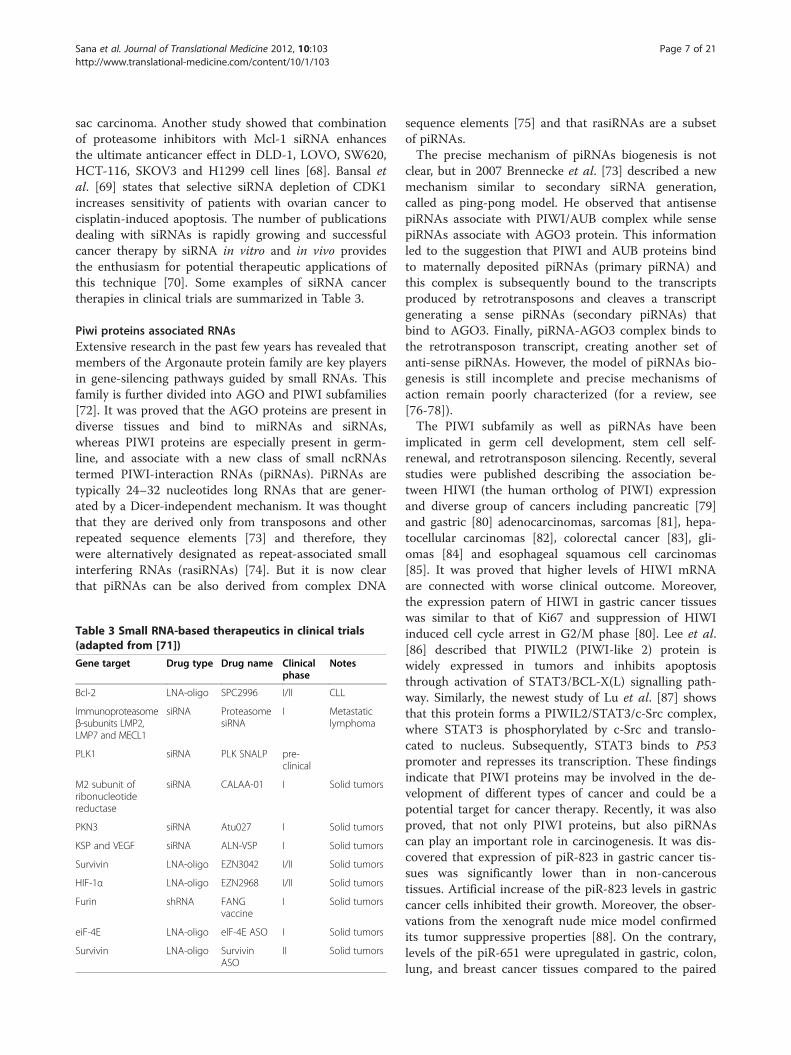
sac carcinoma. Another study showed that combinationof proteasome inhibitors with Mcl-1 siRNA enhancesthe ultimate anticancer effect in DLD-1, LOVO, SW620,HCT-116, SKOV3 and H1299 cell lines [68]. Bansal etal. [69] states that selective siRNA depletion of CDK1increases sensitivity of patients with ovarian cancer tocisplatin-induced apoptosis. The number of publicationsdealing with siRNAs is rapidly growing and successfulcancer therapy by siRNA in vitro and in vivo providesthe enthusiasm for potential therapeutic applications ofthis technique [70]. Some examples of siRNA cancertherapies in clinical trials are summarized in Table 3.
Piwi proteins associated RNAsExtensive research in the past few years has revealed thatmembers of the Argonaute protein family are key playersin gene-silencing pathways guided by small RNAs. Thisfamily is further divided into AGO and PIWI subfamilies[72]. It was proved that the AGO proteins are present indiverse tissues and bind to miRNAs and siRNAs,whereas PIWI proteins are especially present in germ-line, and associate with a new class of small ncRNAstermed PIWI-interaction RNAs (piRNAs). PiRNAs aretypically 24–32 nucleotides long RNAs that are gener-ated by a Dicer-independent mechanism. It was thoughtthat they are derived only from transposons and otherrepeated sequence elements [73] and therefore, theywere alternatively designated as repeat-associated smallinterfering RNAs (rasiRNAs) [74]. But it is now clearthat piRNAs can be also derived from complex DNA
Table 3 Small RNA-based therapeutics in clinical trials(adapted from [71])
Gene target Drug type Drug name Clinicalphase
Notes
Bcl-2 LNA-oligo SPC2996 I/II CLL
Immunoproteasomeβ-subunits LMP2,LMP7 and MECL1
siRNA ProteasomesiRNA
I Metastaticlymphoma
PLK1 siRNA PLK SNALP pre-clinical
M2 subunit ofribonucleotidereductase
siRNA CALAA-01 I Solid tumors
PKN3 siRNA Atu027 I Solid tumors
KSP and VEGF siRNA ALN-VSP I Solid tumors
Survivin LNA-oligo EZN3042 I/II Solid tumors
HIF-1α LNA-oligo EZN2968 I/II Solid tumors
Furin shRNA FANGvaccine
I Solid tumors
eiF-4E LNA-oligo elF-4E ASO I Solid tumors
Survivin LNA-oligo SurvivinASO
II Solid tumors
sequence elements [75] and that rasiRNAs are a subsetof piRNAs.The precise mechanism of piRNAs biogenesis is not
clear, but in 2007 Brennecke et al. [73] described a newmechanism similar to secondary siRNA generation,called as ping-pong model. He observed that antisensepiRNAs associate with PIWI/AUB complex while sensepiRNAs associate with AGO3 protein. This informationled to the suggestion that PIWI and AUB proteins bindto maternally deposited piRNAs (primary piRNA) andthis complex is subsequently bound to the transcriptsproduced by retrotransposons and cleaves a transcriptgenerating a sense piRNAs (secondary piRNAs) thatbind to AGO3. Finally, piRNA-AGO3 complex binds tothe retrotransposon transcript, creating another set ofanti-sense piRNAs. However, the model of piRNAs bio-genesis is still incomplete and precise mechanisms ofaction remain poorly characterized (for a review, see[76-78]).The PIWI subfamily as well as piRNAs have been
implicated in germ cell development, stem cell self-renewal, and retrotransposon silencing. Recently, severalstudies were published describing the association be-tween HIWI (the human ortholog of PIWI) expressionand diverse group of cancers including pancreatic [79]and gastric [80] adenocarcinomas, sarcomas [81], hepa-tocellular carcinomas [82], colorectal cancer [83], gli-omas [84] and esophageal squamous cell carcinomas[85]. It was proved that higher levels of HIWI mRNAare connected with worse clinical outcome. Moreover,the expression patern of HIWI in gastric cancer tissueswas similar to that of Ki67 and suppression of HIWIinduced cell cycle arrest in G2/M phase [80]. Lee et al.[86] described that PIWIL2 (PIWI-like 2) protein iswidely expressed in tumors and inhibits apoptosisthrough activation of STAT3/BCL-X(L) signalling path-way. Similarly, the newest study of Lu et al. [87] showsthat this protein forms a PIWIL2/STAT3/c-Src complex,where STAT3 is phosphorylated by c-Src and translo-cated to nucleus. Subsequently, STAT3 binds to P53promoter and represses its transcription. These findingsindicate that PIWI proteins may be involved in the de-velopment of different types of cancer and could be apotential target for cancer therapy. Recently, it was alsoproved, that not only PIWI proteins, but also piRNAscan play an important role in carcinogenesis. It was dis-covered that expression of piR-823 in gastric cancer tis-sues was significantly lower than in non-canceroustissues. Artificial increase of the piR-823 levels in gastriccancer cells inhibited their growth. Moreover, the obser-vations from the xenograft nude mice model confirmedits tumor suppressive properties [88]. On the contrary,levels of the piR-651 were upregulated in gastric, colon,lung, and breast cancer tissues compared to the paired

Sana et al. Journal of Translational Medicine 2012, 10:103 Page 8 of 21http://www.translational-medicine.com/content/10/1/103
non-cancerous tissues. The growth of gastric cancer cellswas efficiently inhibited by a piR-651 inhibitor and thecells were arrested at the G2/M phase [89]. Interestingly,the peripheral blood levels of piR-651 and piR-823 in thepatients with gastric cancer were significantly lower thanthose from controls. Thus, piRNAs may be valuable bio-markers for detecting circulating gastric cancer cells[90]. Resolving the function of PIWI proteins and piR-NAs has broad implications not only in understandingtheir essential role in fertility, germline, stem cell devel-opment, and basic control and evolution of animal gen-omes, but also in the biology of cancers [12].
Small nucleolar RNAsSmall nucleolar RNAs (snoRNAs), 60 – 300 nucleotideslong, represent one of the abundant groups of smallncRNAs characterized in eukaryotes. SnoRNAs areenriched in the nucleolus, which is the most prominentorganelle in the interphase nucleus providing the cellularlocale for the synthesis and processing of cytoplasmicribosomal RNAs (rRNAs) [91]. Most of the snoRNAsare located within introns of protein-coding genes andare transcribed by RNA polymerase II, however, theycan also be processed from introns of longer ncRNAprecursors [92]. Nevertheless, while vertebrate snoRNAsare prevalently excised from pre-mRNA introns, in plantand yeast these RNAs are mainly generated from inde-pendent transcription units, as either monocistronic or(especially in plants) polycistronic snoRNA transcripts[93].All snoRNAs fall into two major classes based on the
presence of short consensus sequence motifs. First groupcontains the box C (RUGAUGA) and D (CUGA) motifs,whereas members of the second group are characterizedby the box H (ANANNA) and ACA elements [94]. Inboth classes of snoRNAs, short stems bring the con-served boxes close to one another to constitute thestructural core motifs of the snoRNAs, which coordinatethe binding of specific proteins to form small nucleolarRNPs (snoRNPs) distinct for both groups [91,95]. SnoR-NAs have important functions in the maturation ofother non-coding RNAs. Above all, they manage post-transcriptional modification of rRNA and snRNA by 2′-O-methylation and pseudouridylation (for a review, see[91]). Interestingly, it was identified number of humansnoRNAs with miRNA-like function. These snoRNAsare processed to small 20–25 nucleotides long RNAsthat stably associate with Ago proteins. Processing is in-dependent of the Drosha, but requires Dicer. Moreover,cellular target mRNA, whose activity is regulated bysnoRNA, was identified [96].Several studies have indicated that alterations of snoR-
NAs play important functions in cancer developmentand progression. The first report linking snoRNAs to
cancer was published in 2002 by Chang et al. [97]. Heproved that h5sn2, a box H/ACA snoRNA, was signifi-cantly downregulated in human meningiomas comparedwith normal brain tissues. Subsequently, Dong et al.[98] identified snoRNA U50 as a reasonable candidatefor the 6q tumor-suppressor gene in prostate cancerand this statement was confirmed in another study de-scribing involvement of snoRNAs U50 in the develop-ment and/or progression of breast cancer [99].Interestingly, chromosome 6q14-15 is a breakpoint ofchromosomal translocation t(3;6)(q27;q15) for humanB-cell lymphoma [100]. The same year, the GAS5(growth arrest-specific transcript 5) was identified tocontrol mammalian apoptosis and cell growth. GAS5transcript levels were found to be significantly lower inbreast cancer samples relative to adjacent unaffectednormal breast epithelial tissues and despite the fact thatthis gene has no significant protein-coding potential, itwas proved that several snoRNAs are encoded in itsintrons [101]. By profiling ncRNAs signatures inNSCLC tissues and matched noncancerous lung tissues,four snoRNAs (snoRD33, snoRD66, snoRD76 [102] andsnoRA42 [103]) were found to be overexpressed in lungtumor tissues and it is supposed that they could be usedas potential markers for early detection of non-smal celllung cancer [102]. Moreover, snoRD33 is located atchromosome 19q13.3 that contains oncogenes involvedin different malignances including lung cancer, whereassnoRD66 and snoRD76 are located at chromosomalregions 3q27.1 and 1q25.1, respectively. These twochromosomal segments are the most frequently ampli-fied in human solid tumors [28,104,105]. Recently, lowlevels of four snoRNAs (RNU44, RNU48, RNU43,RNU6B), commonly used for normalization of miRNAexpression, were associated with a poor prognosis ofthe cancer patients [106]. Martens-Uzunova et al. [107]analyzed the composition of the entire small transcrip-tome by Illumina/Solexa deep sequencing and herevealed several snoRNAs with deregulated expressionin samples of patients with prostate cancer. The newestpublication concerning snoRNAs proved that snoRD112-114 located at the DLK1-DIO3 locus are ectopicallyexpressed in acute promyelotic leukemia (APL), whichshows that a relationship exists between a chromosomaltranslocation and expression of snoRNA loci. Moreover,in vitro experiments revealed that the snoRD114-1 [14q(II-1)] variant promotes cell growth through G0/G1 toS phase transition mediated by the Rb/p16 pathways[108]. Finally, it was also published that snoRNAs arepresent in stable form in plasma and serum samples[102,106] and therefore could be used as fluid-based bio-markers for cancers. These facts indicate that snoRNAsare critically associated with the development and pro-gression of cancer, however further research for
Sana et al. Journal of Translational Medicine 2012, 10:103 Page 9 of 21http://www.translational-medicine.com/content/10/1/103
comprehensive understanding their role in carcinogen-esis is required.
Promoter-associated RNAsRecently, a new class of ncRNAs known as promoter-associated RNAs (paRNAs) (sometimes termed aspromoter-upstream transcripts – PROMPTs [109], tran-scription start site-associated RNAs [110] or promoter-proximal transcription start site RNAs [111]), were discov-ered. These ncRNAs are derived from eukaryotic promo-ters and have the potential to regulate the transcription ofprotein-coding genes by targeting epigenetic silencingcomplexes [71,112,113]. Their size ranged from 18 to 200nucleotides and they include long, small and tiny RNAs.The short paRNAs (PASRs) were identified in 2007
[114] using RNA maps. They are located near the pro-moter or transcription start site (TSS), but they are notassociated with a known protein-coding genes. Thesetranscripts are 20–90 nt long and it was proved thatthey are not Dicer product [110]. Human PASRs areexpressed at low levels and their number per gene ispositively correlated with promoter activity and mRNAlevel [109]. The tiny paRNAs or transcription iniciationRNAs (tiRNAs) are shorter than 23 nt and they aretranscribed in both sense and antisense directionsaround the promoter [115]. Furthermore, they areclosely associated with highly expressed promoters andare preferentially located in GC-rich promoters [71,115].It is still unclear how these two classes of small RNAs arerelated to one another, or if they share common biogen-esis pathways [115]. Recently, a long paRNAs (PALRs,100–200 nt) has been identified at a single-gene level andthey were associated with regulatory functions (for a re-view, see [112,113,116,117]), especially with modificationof DNA methylation [118].It is supposed, that because of potential of paRNAs to
regulate transcription, their deregulation could be asso-ciated with different types of diseases, including cancer.It was proved, that transfection of mimetic paRNAs intoHeLa and HepG2 cells resulted in the transcriptional re-pression of human C-MYC and connective tissue growthfactor (CTGF) [119]. Hawkins et al. [120] described thattargeting of the human ubiquitin C gene (UbC) with asmall paRNA led to long-term silencing which corre-lated with an early increase in histone methylation and alater increase in DNA methylation at the targeted locus.Furthermore, it was shown that PASRs play an import-ant role in maintaining accessible chromatin architecturefor transcription and releasing negative supercoils duringtranscription [110]. Concerning tiRNAs, they may havesimilar functions like PASRs, moreover they are usuallyfound at CTCF-binding sites. Taft et al. [121] proved,that overexpression of tiRNAs decreased CTCF bindingand associated gene expression, whereas inhibition of
tiRNAs resulted in increased CTCF localization andassociated gene expression. Wang et al. [122] described,that an RNA-binding protein TLS (for translocated inliposarcoma) can specifically bind to CREB-binding pro-tein (CBP) and p300 histone acetyltransferase dependingon its allosteric modulation by PALRs, and so repressgene target CCND1 in human cell lines. Finally, it wasshown that paRNAs have the potential to form double-stranded RNAs and to be processed into endogenoussiRNAs [123]. These facts indicate, that this novel classof ncRNAs has a great potential to regulate expressionof various tumor suppressors and oncogenes on tran-scriptional level and therefore be involved in humancancerogenesis.
Centromere repeat associated small interacting RNAsCell stresses can induce incorrect centromere functionmanifesting in loss of sister chromatid cohesion, abnor-mal chromosome segregation, and aneuploidy, whichhave been observed in many human diseases includingcancers [124]. These defects are often correlated withthe aberrant accumulation of centromere satellite tran-scripts [125]. Morover, it was observed that human cellsunder stress accumulate large transcripts of SatIII satel-lites [126]. The accumulation of similar transcripts invertebrate cells is thought to result from defective RNAprocessing of larger transcripts that leads to a reductionof the small RNAs that participate in the recruitmentof specific histones critical for centromere function[125,127]. The research on mammalian model uncov-ered the strong bidirectional promoter capability of thekangaroo endogenous retrovirus (KERV-1) LTR to pro-duce long double-stranded RNAs for both KERV-1 andsurrounding sequences, including sat23. These longdsRNAs are then processed into centromere repeatassociated small interacting RNAs (crasiRNAs), 34 - 42nucleotides in length. Unfortunately, the mechanism bywhich full-length KERV-1 and sat23 transcripts are pro-cessed into crasiRNAs remains unknown. The crasiRNAsare involved in the recruitment of heterochromatin and/or centromeric proteins. These findings have profoundimplications for understanding of centromere functionand epigenetic identity by suggesting that a retrovirus,KERV-1, may participate in the organization of centro-mere chromatin structures indispensable to chromosomesegregation in vertebrates [124]. These small centromere-associated ncRNAs occur conserved among eukaryotessuggesting their impact also in human.
Telomere-specific small RNAsAnother group of recently described short ncRNAs aretelomere-specific small RNAs (tel-sRNAs). Tel-sRNAsare ~ 24 nt long, Dicer-independent, and 2′-O-methylatedat the 3′ terminus. They are asymmetric with specificity

Sana et al. Journal of Translational Medicine 2012, 10:103 Page 10 of 21http://www.translational-medicine.com/content/10/1/103
toward telomere G-rich strand, and evolutionarily con-served from protozoan to mammalian cells. Interestingly,tel-sRNAs are up-regulated in cells that carry null muta-tion of H3K4 methyltransferase MLL and down-regulatedin cells that carry null mutations of histone H3K9 methyl-transferase SUV39H, suggesting that they are subject toepigenetic regulation. These results support that tel-sRNAs are heterochromatin associated pi-like small RNAs[128]. Recently, it was also reported that an 18-mer RNAoligo of (UUAGGG)3 has potential to inhibit telomeraseTERT activity in vitro by RNA duplex formation in thetemplate region of the telomerase RNA component [129].Therefore, it is supposed that tel-sRNAs containingUUAGGG repeats could act as sensors of chromatin sta-tus and create a feedback loop between the telomeric het-erochromatic regulation and telomere length control.Although tel-sRNAs have not been described in humanuntil to date, they could play an important role in carcino-genesis and contribute to unlimited replicative potencialof cancer cells.
PyknonsPyknons are a subset of 127998 patterns of variablelength, which form mosaics in untranslated as well asprotein-coding regions of human genes. Nevertheless,they are found more frequently in the 3′UTR of genesthan in other regions of the human genome [130,131].Pyknons are present in statistically significant manner ingenes that are involved in specific processes such as cellcommunication, transcription, regulation of transcrip-tion, signaling, transport, etc. Pyknons involve ~ 40% ofthe known miRNA sequences, thus suggesting possiblelink with posttranscriptional gene silencing and RNAinterference [131]. Different sets of pyknons are con-nected to allele-specific sequence variations of disease-associated SNPs and miRNAs, suggesting that increasedsusceptibility to multiple common human disorders isassociated with global alterations in genome-wide regu-latory templates affecting the biogenesis and functions ofnon-coding RNAs [132].In the time since their discovery, evidence has been
slowly accumulating that these pyknon motifs marktranscribed, non-coding RNA sequences with potentialfunctional relevance in human disease. Tsirigos et al.[133] described two GO terms (GO:0006281/DNA re-pair, GO:0006298/mismatch repair) that were signifi-cantly enriched in pyknons-containing regions of thehuman introns. He pointed out that these two terms areuniquely associated with pyknons and a search of theENSEMBL database [134] for human genes labeled withthese two GO terms identified a MLH1 gene, that hasbeen associated with hereditary non-polyposis colorectalcancer and other types of carcinomas and microsatelliteinstabilities. The human MLH1 transcript has 17 introns
and the authors proved that these introns contain morethan 10 different pyknons. Nevertheless, further researchfor comprehensive understanding their role in carcino-genesis is necessary.
Long non-coding RNAsLong non-coding RNAs (lncRNAs) are the broadest classencompassed all non-protein-coding RNA species withlength more than 200 nucleotides, however, frequentlyranging up to 100 kb. Many identified lncRNA are tran-scribed by RNA polymerase II (RNAPII), spliced, andusually contain canonical polyadenylation signals, butthis is not a fast rule [2]. On the other hand, Pagano etal. [135] found out that some of these lncRNAs are dueto their promoter structure likely to be transcribed bypolymerase III (RNAPIII) and he marked them ascogenes since they could specifically coact with aprotein-coding pol II gene. There is substantial evidenceto suggest that lncRNAs mirror protein coding genes.Additionally, lncRNAs’ promoters are bound and regu-lated by transcriptional factors and epigenetically markedwith specific histone modifications [136]. LncRNAs aredevelopmentally and tissue specific, and have been asso-ciated with a spectrum of biological processes, for ex-ample, alternative splicing, modulation of proteinactivity, alternation of protein localization, and epigeneticregulation. LncRNAs can be also precursors of smallRNAs and even tools for miRNAs silencing [71,137-141].However, one of their primary tasks appears to be regula-tors of protein-coding gene expression (Figure 3) [142].Recently, Wang et al. [143] described four differentmechanisms of lncRNAs action. He supposes that thesemolecules can function as signals, decoys, guides or asscaffolds (Figure 4). It is not surprising, then, that dysre-gulation of lncRNAs seems to be an important featureof many complex human diseases, including cancer(Table 4), ischaemic heart disease [144] and Alzheimer’sdisease [145]. Also dysregulation of lncRNAs that func-tion as regulators of the expression of tumor suppres-sors or oncogenes, and not the protein-coding sequenceitself, may be one of the ‘hits’ that leads to oncogenesis[2]. That is why they might be suitable as potential bio-markers and targets for novel therapeutic approaches inthe future.
Long intergenic non-coding RNAsLong intergenic non-coding RNAs (lincRNAs) are newlydiscovered ncRNAs belonging to lncRNAs. RNAs of thissubclass ranging in length from several hundred to tensof thousands of bases and they lie within the genomicintervals between two genes. More than 3000 humanlincRNAs have been identified, but less than 1% hasbeen characterized [136,186]. It was shown that distinctlincRNAs are involved in diverse biological processes

Figure 3 Schematic illustration of lncRNAs functioning. LncRNA transcribed from an upstream non-coding promoter can negatively (1) orpositively (2) affect expression of the downstream gene by inhibiting RNA polymerase II recruitment and/or inducing chromatin remodeling,respectively. LncRNA is able to hybridize to the pre-mRNA and block recognition of the splice sites by the spliceosome, thus resulting in analternatively spliced transcript (3). Alternatively, hybridization of the sense and antisense transcripts can allow Dicer to generate endogenoussiRNAs (4). The binding of lncRNA to the miRNA results in the miRNA function silencing (5). The complex of lncRNA and specific protein partnerscan modulate the activity of the protein (6), is involved in structural and organization roles of the cell (7), alters the protein localizes in the cell(8), and affects epigenetic processes (9). Finally, long ncRNAs can be processed to the small RNAs (10)
Sana et al. Journal of Translational Medicine 2012, 10:103 Page 11 of 21http://www.translational-medicine.com/content/10/1/103
such as imprinting or cancer metastasis [7,140,186].Moreover, recent studies proved that lincRNAs are ex-quisitely regulated during development and in responseto diverse signaling cues, and exhibit distinct gene ex-pression patterns in primary tumors and metastases[136]. Therefore, these lncRNAs could be utilized forcancer diagnosis, prognosis, and serve as potential thera-peutic targets.Recently it has been demonstrated that lncRNAs can
act as natural ‘miRNA sponges’ to reduce miRNA levels[155]. The most highly upregulated transcript found in amicroarray-based study of gene expression in hepatocel-lular carcinoma was determined to be the ncRNAHULC, or Highly Upregulated in Liver Cancer. Tran-scribed from chromosome 6p24.3, this lncRNA demon-strates the hallmarks of a typical mRNA molecule,including a single spliced GT-AG intron, canonical poly-adenylation signals upstream of the poly(A) tail and nu-clear export demonstrating strong localization to thecytoplasm. Although HULC was found to co-purify withribosomes, no translation product for this lncRNA hasbeen detected, supporting its classification as a non-coding transcript [156]. In addition to liver cancer,HULC was found to be highly upregulated in hepaticcolorectal cancer metastasis and in hepatocellular car-cinoma cell lines (HCC) producing hepatitis B virus(HBV) [157]. HULC exists as part of an intricate auto-regulatory network, which when perturbed, resulted inincreased HULC expression (Figure 5a). The HULC
RNA appeared to function as a ‘molecular decoy’ or‘miRNA sponge’ sequestering miR-372, of which onefunction is the translational repression of PRKACB, akinase targeting cAMP response element binding protein(CREB). Once activated, the CREB protein was able topromote HULC transcription by maintaining an openchromatin structure at the HULC promoter resulting inincreased HULC transcription [158].Another well known RNA that belongs to lncRNA
subclass described in previous paragraph is HOX anti-sense intergenic RNA (HOTAIR) (see Figure 5b).HOTAIR is 2.2 kb gene localized within the humanHOXC gene cluster on the long arm of chromosome 2.It has been shown that this lincRNA has a potential toregulate HOXD genes in trans via the recruitment ofpolycomb repressive complex 2 (PRC2), followed by thetrimethylation of lysine 27 of histone H3 [7]. In general,the 5′ region of the RNA binds the PRC2 complex re-sponsible for H3K27 methylation, while the 3′ region ofHOTAIR binds LSD1 (flavin-dependent monoamine oxi-dase), a histone lysine demethylase that mediates enzym-atic demethylation of H3K4Me2. HOTAIR exists inmammals, has poorly conserved sequences and consid-erably conserved structures, and has evolved faster thannearby HOXC genes [187]. HOTAIR was one of the firstmetastasis-associated lncRNAs, described to have a fun-damental role in cancer. This lncRNA was found to behighly upregulated in both primary and metastatic breasttumors, showing up to 2000-fold increased transcription

Figure 4 Schematic diagram of the four mechanisms oflncRNAs functioning. A, lncRNAs can function as signals andregulate gene expression. B, lncRNAs can titrate transcription factorsand other proteins away from chromatin or they can function asdecoy for miRNA target sites. C, lncRNAs can recruit chromatin-modifying enzymes to target genes and therefore function asguides. D, lncRNAs can bring together multiple proteins to formribonucleoprotein complexes (modified according to [143])
Sana et al. Journal of Translational Medicine 2012, 10:103 Page 12 of 21http://www.translational-medicine.com/content/10/1/103
over normal breast tissue. This phenotype seems to beclosely linked with PRC2-dependent gene repressioninduced by HOTAIR. High levels of HOTAIR expressioncorrelate with both metastasis and poor survival rate,connecting lncRNAs with tumor invasiveness and pa-tient prognosis [140]. In addition, it was observed thatthe high expression level of HOTAIR in hepatocellularcarcinoma could be a candidate biomarker for predictingtumor recurrence in hepatocellular carcinoma patientswho have undergone liver transplant therapy and mightbe a potential therapeutic target [188]. Huarte et al.[189] identified several lincRNAs that are regulated by
p53. Furthermore, he proved that lincRNAs-p21 servesas a repressor in p53-dependent transcriptionalresponses, since inhibition of this lincRNA affected theexpression of hundreds of gene targets enriched forgenes normally repressed by p53.While targeting cancer-specific miRNAs has proven to
be successful, it will be necessary to design molecules withpotential to inhibit lincRNAs. Gupta et al. [140] provedthat these molecules can be depleted by siRNAs, but thispossibility is quite complicated because of extensive sec-ondary structures in lincRNAs [187]. Nevertheless, it isevident that cancer-associated lincRNAs may provide newapproaches to the diagnosis and treatment of cancer.
Long intronic non-coding RNAsThe biogenesis of long intronic ncRNAs is poorly under-stood at this time. Nevertheless, there are some indirectevidences that indicate an involvement of RNA polymer-ase II (RNAPII). Among such evidences belong a con-cordant and co-regulated expression profiles of manyintronic ncRNAs and their corresponding protein-coding genes, the broad contribution of RNAPII asso-ciated transcription factors and physiological stimuli inthe transcription of intronic ncRNAs as well the pres-ence of poly(A+) tail [190-194]. Nonetheless, it isdescribed that over 10% of long intronic poly(A+)ncRNAs are up-regulatated compared to only 4% ofprotein-coding transcripts after treatment with theRNAPII specific inhibitor α-amanitin [190,193,195].These findings suggest that some intronic ncRNA andpeculiar protein-coding RNAs could be transcribed byanother RNA polymerase such as the recently describedspRNAP-IV, whose transcriptional output seems to beenhanced by α-amanitin, or also could be transcribed byRNAP III [190,195-199].Similarly to lincRNAs, there are also described evolu-
tionary conserved long intronic ncRNAs sequences frommouse and human [200,201]. When the introns of a lar-ger selection of vertebrates were aligned, the length ofthe conserved region became only 100 bp, while in thealignment of a smaller group of closely related species(human–mouse–cow–dog) the evolutionary conserva-tion of the region extended to as much as 750 bp [201].The widespread occurrence, tissue and subcellular ex-
pression specificity, evolutionary conservation, environ-ment alteration responsiveness and aberrant expression inhuman cancers are features that accredit intronic ncRNAsto be mediators of gene expression regulation. A few setsof intronic ncRNAs have the same tissue expression pat-tern as the corresponding protein-coding genes, whereasothers are inversely correlated. These findings point tocomplex regulatory relationships between intronic ncRNAsand their host loci [190,193,202,203]. Some small ncRNAsare encoded within intronic regions; moreover, intronic

Table 4 Human cancer associated lncRNAs (adapted from [4])
LncRNA Size Cytoband Cancer types References
HOTAIR 2158 nt 12q13.13 breast [7,140]
MALAT1/α/NEAT2 7.5 kb 11q13.1 breast, lung, uterus, pancreas, colon, prostate,liver, osteosarcoma, neuroblastoma, cervix
[146-151]
HULC 500 nt 6p24.3 liver [152,153]
BC200 200 nt 2p21 breast, cervix, esophagus, lung, ovary, parotid, tongue [154,155]
H19 2.3 kb 11p15.5 bladder, lung, liver, breast, endometrial, cervix esophagus,ovary, prostate, colorectal
[156-159]
BIC/MIRHG155/MIRHG2 1.6 kb 21q11.2 B-cell lymphoma [160]
PRNCR1 13 kb 8q24.2 prostate [161]
LOC285194 2105 nt 3q13.31 osteosarcoma [162]
PCGEM1 1643 nt 2 g32.2 prostate [163-165]
UCA1/CUDR 1.4–2.7 kb 19p13.12 bladder, colon, cervix, lung, thyroid, liver,breast, esophagus, stomach
[166]
DD3/PCA3 0.6–4 kb 9q21.22 prostate [167,168]
anti-NOS2A 1.9 kb 17q23.2 brain [169]
uc.73A 201 nt 2q22.3 colon [170]
uc.338 590 nt 12q13.13 liver [171]
ANRIL/p15AS/CDK2BAS 34.8 kb 9p21.3 prostate, leukemia [172-175]
MEG3 1.6 kb 14q32.2 brain [176-178]
GAS5/SNHG2 isoforms 1q25.1 breast [101]
SRA-1/SRA 1965 nt 5q31.3 breast, uterus, ovary [179,180]
PTENP1 3.9 kb 9p13.3 prostate [181,182]
ncRAN 2186 nt 2087 nt 17q25.1 bladder, neuroblastoma [183,184]
LSINCT5 2.6 kb 5p15.33 breast, ovary [185]
Sana et al. Journal of Translational Medicine 2012, 10:103 Page 13 of 21http://www.translational-medicine.com/content/10/1/103
miRNAs tend to be present in large introns with 5′-biasedposition distribution, what correlates with the previous ob-servation that most long intronic transcripts are expressedwithin first introns of the host genes. Thus, it is expectedthat a number of long intronic ncRNAs are processed intosmaller ncRNAs [68,190,204,205]. Similar to lincRNAsHOTAIR, Heo et al. [206] described a long intronic non-coding RNA termed as cold assisted intronic non-codingRNA – COLDAIR, which is required for the vernalization-mediated epigenetic repression of FLC mediated by PRC2.Interestingly, the newest study of Tahira et al. [207] showsthat long intronic non-coding RNAs are differentiallyexpressed in primary and metastatic pancreatic cancer.Moreover, loci harbouring intronic lncRNAs differentiallyexpressed in pancreatic ductal carcinoma metastases wereenriched in genes associated to the MAPK pathway. Thesefindings indicate potential relevance of this class of tran-scripts in biological processes related to malignant trans-formation and metastasis.
Telomere-associated ncRNAsTelomeres protect linear chromosome ends from beingrecognized and processed as double-strand breaks by
DNA repair activities. This protective function of telo-meres is essential for chromosome stability. Until re-cently, the heavily methylated state of subtelomericregions, the gene-less nature of telomeres, and theobserved telomere position effect led to the notion thattelomeres are transcriptionally silent [208]. This hypoth-esis was recently challenged when several groups inde-pendently demonstrated that subtelomeric and telomericregions, although devoid of genes, have the potential tobe transcribed into telomeric UUAGGG-repeat contain-ing ncRNAs (TERRA) [209-211]. TERRA molecules areconserved among eukaryotes and have been identifiedalso in human. TERRA transcripts are synthesized fromthe C-rich strand and polyadenylated, and their synthesisis α-amanitin-sensitive, suggesting that they are tran-scripts of RNAPII [208,212]. TERRA molecules rangebetween 100 bp and >9 kb in length and were reportedto form intermolecular G-quadruplex structure withsingle-stranded telomeric DNA, but can also fold into acompact repeated structure containing G-quartets [211].TERRA transcripts can be found throughout the differ-ent stages of the cell cycle, and their levels are affectedby several factors that include telomere length, tumor

Figure 5 Proposed mechanism of HULC up-regulation in hepatocellular carcinoma (a) and HOTAIR mediated gene silencing of 40 kb ofthe HOXD locus (b).
Sana et al. Journal of Translational Medicine 2012, 10:103 Page 14 of 21http://www.translational-medicine.com/content/10/1/103
stage, cellular stress, developmental stage, and telomericchromatin structure [208].TERRA most likely negatively regulates telomere length
[211]. Increased TERRA levels by interfering with TERRAdecay, such as the impairment of non-sense-mediatedRNA decay in human cells or by deletion of the 5′–3′exo-nuclease Rat1p in Saccharomyces cerevisiae, are associatedwith a loss of telomere reserve [209,212]. Current modelspropose a role for TERRA in controlling telomerase activ-ity. In yeast, the formation of a DNA/RNA hybrid betweenTERRA and telomeres is thought to inhibit elongation bytelomerase, whereas in mammals, TERRA was shown toefficiently inhibit telomerase activity in vitro, presumablyby base pairing with the template region of the RNA com-ponent of telomerase [208,210,212]. Caslini et al. [213]described that telomere uncapping through either TRF2shelterin protein knockdown or exposure to telomere G-strand DNA oligonucleotides significantly increases thetranscription of TERRA, an effect mediated by the func-tional cooperation between transcriptional regulator MLLand the tumor suppressor p53. Sampl et al. [214] foundout that the expression of TERRA in patients with glio-blastoma multiforme negatively correlates with the grade.Moreover, this finding of a diagnostic value of TERRAlevels in astrocytoma WHO grade 2 to 4 correspondedwith preliminary data in advances stages of human tumorsof larynx, colon, and lymph node [210]. Unfortunately, itis largely unclear how the expression of TERRA and theamount of TERRA transcripts are regulated in the cell[208]. Nevertheless, TERRA opens new avenues for telo-mere research that will impact on telomere-associated dis-eases including many cancers [215].
Long ncRNAs with dual functionsUntil not long ago, ncRNAs were strictly considered asRNA molecules with regulatory functions but not
associated with the protein coding capacity typical ofmessenger RNAs. However, the recent identification andcharacterization of bifunctional RNAs, i.e. RNAs forwhich coding capacity and activity as functional regula-tory RNAs have been reported, suggests that a definitecategorization of some RNA molecules is far from beingstraightforward [216]. The steroid receptor RNA activa-tor (SRA) is a unique co-regulator that functions as anon-coding RNA, although incorporation of an additional5′ region can result in translation of an SRA protein(SRAP) that also has co-activator activity [180,217,218].SRA was initially shown to enhance gene expressionthrough a ribonucleoprotein complex with steroid recep-tors and SRC-1 [217]. Currently, SRA is known as anRNA co-activator for many other nuclear receptors. Inaddition, SRA may act as an RNA scaffold for co-repressor complexes [216,219]. SRA transcripts have beenidentified in normal human tissues, with a higher expres-sion in liver, skeletal muscle, adrenal and pituitary glands,whereas intermediate expression levels were observed inthe placenta, lung, kidney and pancreas [217]. In somepathological cases, increased RNA levels of SRA werereported like in breast and ovarian tumors [179,220,221].Interestingly, levels of SRA expression could be character-istic of tumor grade or particular subtypes of lesionsamong different tumors. Indeed, serous ovarian tumorsshowed higher levels of SRA than granulosa tumor cells[216,220].
Pseudogene RNAsPseudogenes are gene copies that have lost the abilityto code for a protein; they are typically identifiedthrough annotation of disabled, decayed or incompleteprotein-coding sequences. These molecules have longbeen labeled as “junk” DNA, failed copies of genesthat arise during the evolution of genomes. However,

Sana et al. Journal of Translational Medicine 2012, 10:103 Page 15 of 21http://www.translational-medicine.com/content/10/1/103
recent results showed that some pseudogenes appear toharbor the potential to regulate their protein-coding cou-sins [222,223]. Processed pseudogenes are made throughretrotransposition of mRNAs, especially as a possible by-product of LINE-1 (Long INterspersed Elements) retro-transposition. Thus, these mRNAs are reverse tran-scribed and re-integrated into the genomic DNA[224,225]. The parent gene of the mRNA need not to beon the same chromosome as the retrotransposed copy.Retrotransposed mRNAs have three possible fates in thegenome: formation of processed genes, formation ofnon-transcribed pseudogenes, or formation of pseudo-genes transcribed into RNAs [222]. Interestingly, someof these RNAs exhibit a tissue-specific pattern of acti-vation. Pseudogene transcripts can be processed intoshort interfering RNAs that regulate coding genesthrough the RNAi pathway. In another remarkable dis-covery, it has been shown that pseudogene RNAs arecapable of regulating tumor suppressors and oncogenesby acting as microRNA decoys [223,225]. Moreover,Devor et al. [226] found out that primate-specific miR-NAs, miR-220 and miR-492, each lie within a pro-cessed pseudogene. Several studies also showderegulated expression of these molecules during can-cer progression, which provides evidence for the func-tional involvement of pseudogene RNAs incarcinogenesis and suggests these molecules as a po-tential novel diagnostic or therapeutic target in humancancers. One of these pseudogenes is myosin lightchain kinase pseudogene (MYLK). MYLKP1 is partiallyduplicated from the original MYLK gene that encodesnonmuscle and smooth muscle myosin light chain kin-ase (smMLCK) isoforms and regulates cell contractilityand cytokinesis. Despite strong homology with thesmMLCK promoter (∼ 90%), the MYLKP1 promoter isminimally active in normal bronchial epithelial cells,but highly active in lung adenocarcinoma cells. More-over, MYLKP1 and smMLCK exhibit negatively corre-lated transcriptional patterns in normal and cancercells with MYLKP1 strongly expressed in cancer cellsand smMLCK highly expressed in non-neoplasticcells. For instance, expression of smMLCK decreasedin colon carcinoma tissues compared to normal colontissues. Mechanistically, MYLKP1 overexpression inhi-bits smMLCK expression in cancer cells by decreasingRNA stability, leading to increased cell proliferation.These findings provide strong evidence for the func-tional involvement of pseudogenes in carcinogenesisand suggest MYLKP1 as a potential novel diagnosticor therapeutic target in human cancers [227]. Usingmassively parallel signature sequencing (MPSS) tech-nology, RT-PCR, and 5′ rapid amplification of cDNAends (RACE) a novel androgen regulated and tran-scribed pseudogene of kallikreins termed as KLK31P
was discovered. It was further proved that thispseudogene may play an important role in prostatecarcinogenesis [228]. He et al. [229] found out thatpseudogene RNAs are also able to regulate a dosageof PTEN tumor suppressor during tumor develop-ment. Pseudogene RNAs however, warrant further in-vestigation into the true extent of their function[223,227].
Transcribed-ultraconserved regionsUltraconcerved regions (UCRs) are a subset of con-served sequences that are located in both intra- andintergenic regions. They are 481 sequences, longer than200 bp that are absolutely conserved between ortholo-gous regions of human, rat, and mouse genomes [230].Calin et al. [170] have proved in cancer systems that dif-ferentially expressed UCR could alter the functionalcharacteristics of malignant cells. The link between gen-omic location of UCRs and analyzed cancer-related gen-omic elements is highly statistically significant andcomparable to that reported for miRNAs. UCRs are fre-quently located at fragile sites and genomic regionsinvolved in cancers. Using northern blot, qRT-PCR andmicroarray analysis, it was revealed that UCRs have dis-tinct signatures in human leukemias and carcinomas[170].Majority of UCRs are transcribed (T-UCRs) in normal
human tissues, both ubiquitously and tissue specifically.From the molecular point of view, untranscribed UCRsmight have regulatory functions as enhancers [231],while many functions can be assigned for T-UCRs, suchas antisense inhibitors for protein-coding genes or otherncRNAs, including miRNAs. On the other hand, insteadof T-UCRs interacting with protein-coding genes andmiRNAs, it is possible that miRNAs control T-UCRs.Evidence supporting this predication is that many T-UCRs have significant antisense complementarity withparticular miRNAs and negative correlation between ex-pression of specific T-UCRs and predicted interactormiRNAs [170,232].The expression of many T-UCRs is significantly altered
in cancer, especially in adult chronic lymphocytic leuke-mias, colorectal and hepatocellular carcinomas and neu-roblastomas [170]. Their aberrant transcription profilescan be used to distinguish types of human cancers andhave been linked to patient outcome [233]. Especially inneuroblastoma, functional T-UCR annotations, inferredthrough a functional genomics approach and validatedusing cellular models, reveal associations with severalcancer-related cellular processes such as apoptosis anddifferentiation [234]. Further, DNA hypomethylationinduces release of T-UCR silencing in cancer cells. Stud-ies of primary human tumors have shown that hyper-methylation of T-UCR CpG islands is common event

Sana et al. Journal of Translational Medicine 2012, 10:103 Page 16 of 21http://www.translational-medicine.com/content/10/1/103
among the various tumor types. Thus in addition tomiRNAs, another class of ncRNAs (T-UCRs) undergoesDNA methylation-associated inactivation in transformedcells, and so supports model that both epigenetic andgenetic alterations in coding and noncoding sequencescooperate in human tumorigenesis. Most importantly,restoration of T-UCR expression was observed upontreatment with the DNA-demethylating agent [232]. An-other study proved, that SNPs (single nucleotide poly-morphisms) rs9572903 and rs2056116 in ultraconservedregions were associated with increased familial breastcancer risk [235]. Because of increasing number of stud-ies concerning T-UCRs is published, it is supposed thatthe more specific roles of these molecules in cancer willbe known in a short time.
Conclusions and future perspectivesFor a long time, the central dogma of molecular biologyproposed RNA molecules primarily to be informational“messenger” between DNA and protein. But, surprisingly,only 2% of the human genome sequence encodes proteins,while a large part of it is devoted to the expression ofncRNAs, which are divided into two main groups accord-ing to their nucleotide length – small and long ncRNAs.These molecules are suggested to be important regulatorsof gene expression. Nevertheless, the two groups ofncRNAs are distinct in their biological functions andmechanisms of gene regulations. Small ncRNAs areinvolved mainly in the post-transcriptional gene regulationusing translational repression or RNAi pathway, whilelong ncRNAs are much more involved in epigenetic regu-lation. In many cases, differential expression of ncRNAs isbecoming recognized as a one of the hallmarks of cancercell, indicating their potential usage as the novel diagnos-tic, prognostic, or predictive biomarkers. Growing evi-dence also suggests that ncRNAs have the promisingpotential in targeted regulation of gene expression and,therefore, in cancer targeted therapy. However, the func-tion of many ncRNAs remains unknown and it will be ne-cessary to discover the precise mechanisms by which arethese molecules involved in carcinogenesis.
Competing interestThe authors declare that they have no competing interests.
AcknowledgementsThis work was supported by grant IGA 10361-3/2009, NS/9814-4/2008, NS10352-3/2009, NS/11214-4/2010 of the Czech Ministry of Health, Project No.MZ0MOU2005 of the Czech Ministry of Health and by the project “CEITEC –Central European Institute of Technology” (CZ.1.05/1.1.00/02.0068).
Author details1Masaryk Memorial Cancer Institute, Department of Comprehensive CancerCare, Zluty kopec 7, Brno, Czech Republic, Europe. 2Central EuropeanInstitute of Technology, Masaryk University, Brno, Czech Republic, Europe.3Masaryk Memorial Cancer Institute, Department of Comprehensive CancerCare, Zluty kopec 7, 656 53, Brno, Czech Republic, Europe.
Authors’ contributionsSJ and FP drafted the manuscript, SM and SO revised the manuscriptcritically for important and intellectual content. All authors read andapproved the final manuscript. All authors read and approved the finalmanuscript.
Received: 2 January 2012 Accepted: 21 May 2012Published: 21 May 2012
References1. Stein LD: Human genome: end of the beginning. Nature 2004,
431:915–916.2. Taft RJ, Pang KC, Mercer TR, Dinger M, Mattick JS: Non-coding RNAs:
regulators of disease. J Pathol 2010, 220:126–139.3. Knowling S, Morris KV: Non-coding RNA and antisense RNA. Nature’s trash
or treasure? Biochimie 2011, 93:1922–1927.4. Mattick JS: Non-coding RNAs: the architects of eukaryotic complexity.
EMBO Rep 2001, 2:986–991.5. Costa FF: Non-coding RNAs: new players in eukaryotic biology. Gene
2005, 357:83–94.6. Okamura K, Chung W-J, Ruby JG, Guo H, Bartel DP, Lai EC: The Drosophila
hairpin RNA pathway generates endogenous short interfering RNAs.Nature 2008, 453:803–806.
7. Rinn JL, Kertesz M, Wang JK, Squazzo SL, Xu X, Brugmann SA, GoodnoughLH, Helms JA, Farnham PJ, Segal E, Chang HY: Functional demarcation ofactive and silent chromatin domains in human HOX loci by noncodingRNAs. Cell 2007, 129:1311–1323.
8. Elbashir SM, Harborth J, Lendeckel W, Yalcin A, Weber K, Tuschl T: Duplexesof 21-nucleotide RNAs mediate RNA interference in cultured mammaliancells. Nature 2001, 411:494–498.
9. Meister G, Tuschl T: Mechanisms of gene silencing by double-strandedRNA. Nature 2004, 431:343–349.
10. Lippman Z, Martienssen R: The role of RNA interference inheterochromatic silencing. Nature 2004, 431:364–370.
11. Houwing S, Kamminga LM, Berezikov E, Cronembold D, Girard A, van denElst H, Filippov DV, Blaser H, Raz E, Moens CB, Plasterk RHA, Hannon GJ,Draper BW, Ketting RF: A role for Piwi and piRNAs in germ cellmaintenance and transposon silencing in Zebrafish. Cell 2007, 129:69–82.
12. Seto AG, Kingston RE, Lau NC: The coming of age for Piwi proteins. MolCell 2007, 26:603–609.
13. Sana J, Hajduch M, Michalek J, Vyzula R, Slaby O: MicroRNAs andglioblastoma: roles in core signalling pathways and potential clinicalimplications. J Cell Mol Med 2011, 15:1636–1644.
14. Slaby O, Bienertova-Vasku J, Svoboda M, Vyzula R: Genetic polymorphismsand microRNAs: new direction in molecular epidemiology of solidcancer. J Cell Mol Med 2012, 16:8–21.
15. Slaby O, Svoboda M, Michalek J, Vyzula R: MicroRNAs in colorectal cancer:translation of molecular biology into clinical application. Mol Cancer 2009,8:102.
16. Redova M, Svoboda M, Slaby O: MicroRNAs and their target genenetworks in renal cell carcinoma. Biochem Biophys Res Commun 2011,405:153–156.
17. Lee RC, Feinbaum RL, Ambros V: The C. elegans heterochronic gene lin-4encodes small RNAs with antisense complementarity to lin-14. Cell 1993,75:843–854.
18. Griffiths-Jones S: miRBase: the microRNA sequence database. Methods MolBiol 2006, 342:129–138.
19. Krol J, Loedige I, Filipowicz W: The widespread regulation of microRNAbiogenesis, function and decay. Nat Rev Genet 2010, 11:597–610.
20. Roberts APE, Lewis AP, Jopling CL: miR-122 activates hepatitis C virustranslation by a specialized mechanism requiring particular RNAcomponents. Nucleic Acids Res 2011, 39:7716–7729.
21. Grey F, Tirabassi R, Meyers H, Wu G, McWeeney S, Hook L, Nelson JA: A viralmicroRNA down-regulates multiple cell cycle genes through mRNA 5′UTRs. PLoS Pathog 2010, 6:e1000967.
22. Tsai N-P, Lin Y-L, Wei L-N: MicroRNA mir-346 targets the 5′-untranslatedregion of receptor-interacting protein 140 (RIP140) mRNA and up-regulates its protein expression. Biochem J 2009, 424:411–418.
23. �rom UA, Nielsen FC, Lund AH: MicroRNA-10a binds the 5′UTR ofribosomal protein mRNAs and enhances their translation. Mol Cell 2008,30:460–471.

Sana et al. Journal of Translational Medicine 2012, 10:103 Page 17 of 21http://www.translational-medicine.com/content/10/1/103
24. Farh KK-H, Grimson A, Jan C, Lewis BP, Johnston WK, Lim LP, Burge CB,Bartel DP: The widespread impact of mammalian MicroRNAs on mRNArepression and evolution. Science 2005, 310:1817–1821.
25. Lim LP, Lau NC, Garrett-Engele P, Grimson A, Schelter JM, Castle J, Bartel DP,Linsley PS, Johnson JM: Microarray analysis shows that some microRNAsdownregulate large numbers of target mRNAs. Nature 2005, 433:769–773.
26. Lee I, Ajay SS, Yook JI, Kim HS, Hong SH, Kim NH, Dhanasekaran SM,Chinnaiyan AM, Athey BD: New class of microRNA targets containingsimultaneous 5′-UTR and 3′-UTR interaction sites. Genome Res 2009,19:1175–1183.
27. Chi SW, Zang JB, Mele A, Darnell RB: Argonaute HITS-CLIP decodesmicroRNA-mRNA interaction maps. Nature 2009, 460:479–486.
28. Li M, Li J, Ding X, He M, Cheng S-Y: microRNA and cancer. AAPS J 2010,12:309–317.
29. Kwak PB, Iwasaki S, Tomari Y: The microRNA pathway and cancer. CancerSci 2010, 101:2309–2315.
30. Wu WKK, Law PTY, Lee CW, Cho CH, Fan D, Wu K, Yu J, Sung JJY: MicroRNAin colorectal cancer: from benchtop to bedside. Carcinogenesis 2011,32:247–253.
31. Lin P-Y, Yu S-L, Yang P-C: MicroRNA in lung cancer. Br J Cancer 2010,103:1144–1148.
32. Lakomy R, Sana J, Hankeova S, Fadrus P, Kren L, Lzicarova E, Svoboda M,Dolezelova H, Smrcka M, Vyzula R, Michalek J, Hajduch M, Slaby O: MiR-195,miR-196b, miR-181c, miR-21 expression levels and O-6-methylguanine-DNA methyltransferase methylation status are associated with clinicaloutcome in glioblastoma patients. Cancer Sci 2011, 102:2186–2190.
33. Satoh J: MicroRNAs and their therapeutic potential for human diseases:aberrant microRNA expression in Alzheimer’s disease brains. J PharmacolSci 2010, 114:269–275.
34. Slaby O, Jancovicova J, Lakomy R, Svoboda M, Poprach A, Fabian P, Kren L,Michalek J, Vyzula R: Expression of miRNA-106b in conventional renal cellcarcinoma is a potential marker for prediction of early metastasis afternephrectomy. J Exp Clin Cancer Res 2010, 29:90.
35. Slaby O, Lakomy R, Fadrus P, Hrstka R, Kren L, Lzicarova E, Smrcka M,Svoboda M, Dolezalova H, Novakova J, Valik D, Vyzula R, Michalek J:MicroRNA-181 family predicts response to concomitantchemoradiotherapy with temozolomide in glioblastoma patients.Neoplasma 2010, 57:264–269.
36. Bloomston M, Frankel WL, Petrocca F, Volinia S, Alder H, Hagan JP, Liu C-G,Bhatt D, Taccioli C, Croce CM: MicroRNA expression patterns todifferentiate pancreatic adenocarcinoma from normal pancreas andchronic pancreatitis. JAMA 2007, 297:1901–1908.
37. Fornari F, Gramantieri L, Giovannini C, Veronese A, Ferracin M, Sabbioni S,Calin GA, Grazi GL, Croce CM, Tavolari S, Chieco P, Negrini M, Bolondi L:MiR-122/cyclin G1 interaction modulates p53 activity and affectsdoxorubicin sensitivity of human hepatocarcinoma cells. Cancer Res 2009,69:5761–5767.
38. Gramantieri L, Fornari F, Ferracin M, Veronese A, Sabbioni S, Calin GA, GraziGL, Croce CM, Bolondi L, Negrini M: MicroRNA-221 targets Bmf inhepatocellular carcinoma and correlates with tumor multifocality. ClinCancer Res 2009, 15:5073–5081.
39. Slaby O, Svoboda M, Fabian P, Smerdova T, Knoflickova D, Bednarikova M,Nenutil R, Vyzula R: Altered expression of miR-21, miR-31, miR-143 andmiR-145 is related to clinicopathologic features of colorectal cancer.Oncology 2007, 72:397–402.
40. Kulda V, Pesta M, Topolcan O, Liska V, Treska V, Sutnar A, Rupert K,Ludvikova M, Babuska V, Holubec L Jr, Cerny R: Relevance of miR-21and miR-143 expression in tissue samples of colorectalcarcinoma and its liver metastases. Cancer Genet Cytogenet 2010,200:154–160.
41. Takamizawa J, Konishi H, Yanagisawa K, Tomida S, Osada H, Endoh H,Harano T, Yatabe Y, Nagino M, Nimura Y, Mitsudomi T, Takahashi T:Reduced expression of the let-7 microRNAs in human lung cancers inassociation with shortened postoperative survival. Cancer Res 2004,64:3753–3756.
42. Gallardo E, Navarro A, Viñolas N, Marrades RM, Diaz T, Gel B, Quera A,Bandres E, Garcia-Foncillas J, Ramirez J, Monzo M: miR-34a as a prognosticmarker of relapse in surgically resected non-small-cell lung cancer.Carcinogenesis 2009, 30:1903–1909.
43. Yu S-L, Chen H-Y, Chang G-C, Chen C-Y, Chen H-W, Singh S, Cheng C-L, YuC-J, Lee Y-C, Chen H-S, Su T-J, Chiang C-C, Li H-N, Hong Q-S, Su H-Y, Chen
C-C, Chen W-J, Liu C-C, Chan W-K, Chen WJ, Li K-C, Chen JJW, Yang P-C:MicroRNA signature predicts survival and relapse in lung cancer. CancerCell 2008, 13:48–57.
44. Ma L, Teruya-Feldstein J, Weinberg RA: Tumour invasion and metastasisinitiated by microRNA-10b in breast cancer. Nature 2007, 449:682–688.
45. Tavazoie SF, Alarcón C, Oskarsson T, Padua D, Wang Q, Bos PD, Gerald WL,Massagué J: Endogenous human microRNAs that suppress breast cancermetastasis. Nature 2008, 451:147–152.
46. Satzger I, Mattern A, Kuettler U, Weinspach D, Voelker B, Kapp A, Gutzmer R:MicroRNA-15b represents an independent prognostic parameter and iscorrelated with tumor cell proliferation and apoptosis in malignantmelanoma. Int J Cancer 2010, 126:2553–2562.
47. Mohri T, Nakajima M, Fukami T, Takamiya M, Aoki Y, Yokoi T: Human CYP2E1 isregulated by miR-378. Biochem Pharmacol 2010, 79:1045–1052.
48. Markou A, Tsaroucha EG, Kaklamanis L, Fotinou M, Georgoulias V, LianidouES: Prognostic value of mature microRNA-21 and microRNA-205overexpression in non-small cell lung cancer by quantitative real-timeRT-PCR. Clin Chem 2008, 54:1696–1704.
49. Yang H, Kong W, He L, Zhao J-J, O’Donnell JD, Wang J, Wenham RM,Coppola D, Kruk PA, Nicosia SV, Cheng JQ: MicroRNA expression profilingin human ovarian cancer: miR-214 induces cell survival and cisplatinresistance by targeting PTEN. Cancer Res 2008, 68:425–433.
50. Nagel R, le Sage C, Diosdado B, van der Waal M, Oude Vrielink JAF, Bolijn A,Meijer GA, Agami R: Regulation of the adenomatous polyposis coli geneby the miR-135 family in colorectal cancer. Cancer Res 2008, 68:5795–5802.
51. Johnson SM, Grosshans H, Shingara J, Byrom M, Jarvis R, Cheng A, LabourierE, Reinert KL, Brown D, Slack FJ: RAS is regulated by the let-7 microRNAfamily. Cell 2005, 120:635–647.
52. Chen X, Guo X, Zhang H, Xiang Y, Chen J, Yin Y, Cai X, Wang K, Wang G, BaY, Zhu L, Wang J, Yang R, Zhang Y, Ren Z, Zen K, Zhang J, Zhang C-Y: Roleof miR-143 targeting KRAS in colorectal tumorigenesis. Oncogene 2009,28:1385–1392.
53. Guo C, Sah JF, Beard L, Willson JKV, Markowitz SD, Guda K: The noncodingRNA, miR-126, suppresses the growth of neoplastic cells by targetingphosphatidylinositol 3-kinase signaling and is frequently lost in coloncancers. Genes Chromosomes Cancer 2008, 47:939–946.
54. Meng F, Henson R, Wehbe-Janek H, Ghoshal K, Jacob ST, Patel T: MicroRNA-21 regulates expression of the PTEN tumor suppressor gene in humanhepatocellular cancer. Gastroenterology 2007, 133:647–658.
55. Yu Z, Wang C, Wang M, Li Z, Casimiro MC, Liu M, Wu K, Whittle J, Ju X,Hyslop T, McCue P, Pestell RG: A cyclin D1/microRNA 17/20 regulatoryfeedback loop in control of breast cancer cell proliferation. J Cell Biol2008, 182:509–517.
56. Ota A, Tagawa H, Karnan S, Tsuzuki S, Karpas A, Kira S, Yoshida Y, Seto M:Identification and characterization of a novel gene, C13orf25, as a targetfor 13q31-q32 amplification in malignant lymphoma. Cancer Res 2004,64:3087–3095.
57. O’Donnell KA, Wentzel EA, Zeller KI, Dang CV, Mendell JT: c-Myc-regulatedmicroRNAs modulate E2F1 expression. Nature 2005, 435:839–843.
58. He L, Thomson JM, Hemann MT, Hernando-Monge E, Mu D, Goodson S,Powers S, Cordon-Cardo C, Lowe SW, Hannon GJ, Hammond SM: AmicroRNA polycistron as a potential human oncogene. Nature 2005,435:828–833.
59. Sakamoto KM: Knocking Down Human Disease: Potential Uses of RNAInterference in Research and Gene Therapy. Pediatric Research 2004,55:912–913.
60. Noll B, Seiffert S, Vornlocher H-P, Roehl I: Characterization of smallinterfering RNA by non-denaturing ion-pair reversed-phase liquidchromatography. J Chromatogr A 2011, 1218:5609–5617.
61. Martinez J, Patkaniowska A, Urlaub H, Lührmann R, Tuschl T: Single-stranded antisense siRNAs guide target RNA cleavage in RNAi. Cell 2002,110:563–574.
62. Martinez J, Tuschl T: RISC is a 5′ phosphomonoester-producing RNAendonuclease. Genes Dev 2004, 18:975–980.
63. Vassilev LT: Small-molecule antagonists of p53-MDM2 binding: researchtools and potential therapeutics. Cell Cycle 2004, 3:419–421.
64. He S-B, Yuan Y, Wang L, Yu M-J, Zhu Y-B, Zhu X-G: Effects of cyclin-dependent kinase 8 specific siRNA on the proliferation and apoptosis ofcolon cancer cells. J Exp Clin Cancer Res 2011, 30:109.
65. Durfort T, Tkach M, Meschaninova MI, Rivas MA, Elizalde PV, VenyaminovaAG, Schillaci R, François J-C: Small interfering RNA targeted to IGF-IR

Sana et al. Journal of Translational Medicine 2012, 10:103 Page 18 of 21http://www.translational-medicine.com/content/10/1/103
delays tumor growth and induces proinflammatory cytokines in amouse breast cancer model. PLoS ONE 2012, 7:e29213.
66. Matsubara H, Sakakibara K, Kunimitsu T, Matsuoka H, Kato K, Oyachi N,Dobashi Y, Matsumoto M: Non-small cell lung carcinoma therapy usingmTOR-siRNA. Int J Clin Exp Pathol 2012, 5:119–125.
67. He Y, Bi Y, Hua Y, Liu D, Wen S, Wang Q, Li M, Zhu J, Lin T, He D, Li X, WangZ, Wei G: Ultrasound microbubble-mediated delivery of the siRNAstargeting MDR1 reduces drug resistance of yolk sac carcinoma L2 cells.J Exp Clin Cancer Res 2011, 30:104.
68. Zhou W, Hu J, Tang H, Wang D, Huang X, He C, Zhu H: Small interferingRNA targeting mcl-1 enhances proteasome inhibitor-induced apoptosisin various solid malignant tumors. BMC Cancer 2011, 11:485.
69. Bansal N, Marchion DC, Bicaku E, Xiong Y, Chen N, Stickles XB, Sawah EA,Wenham RM, Apte SM, Gonzalez-Bosquet J, Judson PL, Hakam A, LancasterJM: BCL2 antagonist of cell death kinases, phosphatases, and ovariancancer sensitivity to cisplatin. J Gynecol Oncol 2012, 23:35–42.
70. Wang X, Chen Y, Ren J, Qu X: Small interfering RNA for effective cancertherapies. Mini Rev Med Chem 2011, 11:114–124.
71. Costa FF: Non-coding RNAs: Meet thy masters. Bioessays 2010, 32:599–608.72. Carmell MA, Xuan Z, Zhang MQ, Hannon GJ: The Argonaute family:
tentacles that reach into RNAi, developmental control, stem cellmaintenance, and tumorigenesis. Genes Dev 2002, 16:2733–2742.
73. Brennecke J, Aravin AA, Stark A, Dus M, Kellis M, Sachidanandam R, HannonGJ: Discrete small RNA-generating loci as master regulators oftransposon activity in Drosophila. Cell 2007, 128:1089–1103.
74. Aravin AA, Lagos-Quintana M, Yalcin A, Zavolan M, Marks D, Snyder B,Gaasterland T, Meyer J, Tuschl T: The small RNA profile during Drosophilamelanogaster development. Dev Cell 2003, 5:337–350.
75. Aravin AA, Sachidanandam R, Girard A, Fejes-Toth K, Hannon GJ:Developmentally regulated piRNA clusters implicate MILI in transposoncontrol. Science 2007, 316:744–747.
76. Samji T: PIWI, piRNAs, and Germline Stem Cells: What’s the link? Yale JBiol Med 2009, 82:121–124.
77. Thomson T, Lin H: The biogenesis and function of PIWI proteins andpiRNAs: progress and prospect. Annu Rev Cell Dev Biol 2009, 25:355–376.
78. Klattenhoff C, Theurkauf W: Biogenesis and germline functions of piRNAs.Development 2008, 135:3–9.
79. Grochola LF, Greither T, Taubert H, Möller P, Knippschild U, Udelnow A,Henne-Bruns D, Würl P: The stem cell-associated Hiwi gene in humanadenocarcinoma of the pancreas: expression and risk of tumour-relateddeath. Br J Cancer 2008, 99:1083–1088.
80. Liu X, Sun Y, Guo J, Ma H, Li J, Dong B, Jin G, Zhang J, Wu J, Meng L, ShouC: Expression of hiwi gene in human gastric cancer was associated withproliferation of cancer cells. Int J Cancer 2006, 118:1922–1929.
81. Taubert H, Greither T, Kaushal D, Würl P, Bache M, Bartel F, Kehlen A,Lautenschläger C, Harris L, Kraemer K, Meye A, Kappler M, Schmidt H,Holzhausen H-J, Hauptmann S: Expression of the stem cell self-renewalgene Hiwi and risk of tumour-related death in patients with soft-tissuesarcoma. Oncogene 2007, 26:1098–1100.
82. Zhao Y-M, Zhou J-M, Wang L-R, He H-W, Wang X-L, Tao Z-H, Sun H-C, WuW-Z, Fan J, Tang Z-Y, Wang L: HIWI is associated with prognosis inpatients with hepatocellular carcinoma after curative resection. Cancer2012, 118(10):2708–2717.
83. Zeng Y, Qu L, Meng L, Liu C, Dong B, Xing X, Wu J, Shou C: HIWIexpression profile in cancer cells and its prognostic value for patientswith colorectal cancer. Chin Med J 2011, 124:2144–2149.
84. Sun G, Wang Y, Sun L, Luo H, Liu N, Fu Z, You Y: Clinical significance ofHiwi gene expression in gliomas. Brain Res 2011, 1373:183–188.
85. He W, Wang Z, Wang Q, Fan Q, Shou C, Wang J, Giercksky K-E, Nesland JM,Suo Z: Expression of HIWI in human esophageal squamous cellcarcinoma is significantly associated with poorer prognosis. BMC Cancer2009, 9:426.
86. Lee JH, Schütte D, Wulf G, Füzesi L, Radzun H-J, Schweyer S, Engel W,Nayernia K: Stem-cell protein Piwil2 is widely expressed in tumors andinhibits apoptosis through activation of Stat3/Bcl-XL pathway. Hum MolGenet 2006, 15:201–211.
87. Lu Y, Zhang K, Li C, Yao Y, Tao D, Liu Y, Zhang S, Ma Y: Piwil2 suppressesp53 by inducing phosphorylation of signal transducer and activator oftranscription 3 in tumor cells. PLoS ONE 2012, 7:e30999.
88. Cheng J, Deng H, Xiao B, Zhou H, Zhou F, Shen Z, Guo J: piR-823, a novelnon-coding small RNA, demonstrates in vitro and in vivo tumor
suppressive activity in human gastric cancer cells. Cancer Lett 2012,315:12–17.
89. Cheng J, Guo J-M, Xiao B-X, Miao Y, Jiang Z, Zhou H, Li Q-N: piRNA, thenew non-coding RNA, is aberrantly expressed in human cancer cells. ClinChim Acta 2011, 412:1621–1625.
90. Cui L, Lou Y, Zhang X, Zhou H, Deng H, Song H, Yu X, Xiao B, Wang W, GuoJ: Detection of circulating tumor cells in peripheral blood from patientswith gastric cancer using piRNAs as markers. Clin Biochem 2011, 44:1050–1057.
91. Kiss T: Small nucleolar RNAs: an abundant group of noncoding RNAswith diverse cellular functions. Cell 2002, 109:145–148.
92. Bortolin M-L, Kiss T: Human U19 intron-encoded snoRNA is processedfrom a long primary transcript that possesses little potential for proteincoding. RNA 1998, 4:445–454.
93. Weinstein LB, Steitz JA: Guided tours: from precursor snoRNA tofunctional snoRNP. Curr Opin Cell Biol 1999, 11:378–384.
94. Ganot P, Caizergues-Ferrer M, Kiss T: The Family of Box ACA SmallNucleolar RNAs Is Defined by an Evolutionarily Conserved SecondaryStructure and Ubiquitous Sequence Elements Essential for RNAAccumulation. Genes Dev 1997, 11:941–956.
95. Vidovic I, Nottrott S, Hartmuth K, Lührmann R, Ficner R: Crystal structure ofthe spliceosomal 15.5kD protein bound to a U4 snRNA fragment. MolCell 2000, 6:1331–1342.
96. Ender C, Krek A, Friedländer MR, Beitzinger M, Weinmann L, Chen W, PfefferS, Rajewsky N, Meister G: A human snoRNA with microRNA-like functions.Mol Cell 2008, 32:519–528.
97. Chang L-S, Lin S-Y, Lieu A-S, Wu T-L: Differential expression of human 5 SsnoRNA genes. Biochemical and Biophysical Research Communications 2002,299:196–200.
98. Dong X-Y, Rodriguez C, Guo P, Sun X, Talbot JT, Zhou W, Petros J, Li Q,Vessella RL, Kibel AS, Stevens VL, Calle EE, Dong J-T: SnoRNA U50 is acandidate tumor-suppressor gene at 6q14.3 with a mutation associatedwith clinically significant prostate cancer. Hum Mol Genet 2008, 17:1031–1042.
99. Dong X-Y, Guo P, Boyd J, Sun X, Li Q, Zhou W, Dong J-T: Implication ofsnoRNA U50 in human breast cancer. J Genet Genomics 2009, 36:447–454.
100. Tanaka R, Satoh H, Moriyama M, Satoh K, Morishita Y, Yoshida S, WatanabeT, Nakamura Y, Mori S: Intronic U50 small-nucleolar-RNA (snoRNA) hostgene of no protein-coding potential is mapped at the chromosomebreakpoint t(3;6)(q27;q15) of human B- cell lymphoma. Genes to Cells2000, 5:277–287.
101. Mourtada-Maarabouni M, Pickard MR, Hedge VL, Farzaneh F, Williams GT:GAS5, a non-protein-coding RNA, controls apoptosis and isdownregulated in breast cancer. Oncogene 2009, 28:195–208.
102. Liao J, Yu L, Mei Y, Guarnera M, Shen J, Li R, Liu Z, Jiang F: Small nucleolarRNA signatures as biomarkers for non-small-cell lung cancer. Mol Cancer2010, 9:198.
103. Mei Y-P, Liao J-P, Shen J-P, Yu L, Liu B-L, Liu L, Li R-Y, Ji L, Dorsey SG, Jiang Z-R,Katz RL, Wang J-Y, Jiang F: Small nucleolar RNA 42 acts as an oncogene inlung tumorigenesis. Oncogene 2012, 2012, 31(22):2794–2804.
104. Jiang F, Yin Z, Caraway NP, Li R, Katz RL: Genomic profiles in stage Iprimary non small cell lung cancer using comparative genomichybridization analysis of cDNA microarrays. Neoplasia 2004, 6:623–635.
105. Gebhart E: Double minutes, cytogenetic equivalents of geneamplification, in human neoplasia - a review. Clinical & translationaloncology: official publication of the Federation of Spanish Oncology Societiesand of the National Cancer Institute of Mexico 2005, 7:477–485.
106. Gee HE, Buffa FM, Camps C, Ramachandran A, Leek R, Taylor M, Patil M,Sheldon H, Betts G, Homer J, West C, Ragoussis J, Harris AL: The small-nucleolar RNAs commonly used for microRNA normalisation correlatewith tumour pathology and prognosis. Br J Cancer 2011, 104:1168–1177.
107. Martens-Uzunova ES, Jalava SE, Dits NF, van Leenders GJLH, Møller S,Trapman J, Bangma CH, Litman T, Visakorpi T, Jenster G: Diagnostic andprognostic signatures from the small non-coding RNA transcriptome inprostate cancer. Oncogene 2012, 31:978–991.
108. Valleron W, Laprevotte E, Gautier E-F, Quelen C, Demur C, Delabesse E,Agirre X, Prósper F, Kiss T, Brousset P: Specific small nucleolar RNA expressionprofiles in acute leukemia. U.K: Leukemia: Official Journal of the LeukemiaSociety of America, Leukemia Research Fund; 2012.
109. Preker P, Nielsen J, Kammler S, Lykke-Andersen S, Christensen MS,Mapendano CK, Schierup MH, Jensen TH: RNA exosome depletion reveals
Sana et al. Journal of Translational Medicine 2012, 10:103 Page 19 of 21http://www.translational-medicine.com/content/10/1/103
transcription upstream of active human promoters. Science 2008,322:1851–1854.
110. Seila AC, Calabrese JM, Levine SS, Yeo GW, Rahl PB, Flynn RA, Young RA,Sharp PA: Divergent transcription from active promoters. Science 2008,322:1849–1851.
111. Core LJ, Lis JT: Transcription regulation through promoter-proximalpausing of RNA polymerase II. Science 2008, 319:1791–1792.
112. Morris KV, Santoso S, Turner A-M, Pastori C, Hawkins PG: Bidirectionaltranscription directs both transcriptional gene activation andsuppression in human cells. PLoS Genet 2008, 4:e1000258.
113. Schwartz JC, Younger ST, Nguyen N-B, Hardy DB, Monia BP, Corey DR,Janowski BA: Antisense transcripts are targets for activating small RNAs.Nat Struct Mol Biol 2008, 15:842–848.
114. Kapranov P, Cheng J, Dike S, Nix DA, Duttagupta R, Willingham AT, StadlerPF, Hertel J, Hackermüller J, Hofacker IL, Bell I, Cheung E, Drenkow J, DumaisE, Patel S, Helt G, Ganesh M, Ghosh S, Piccolboni A, Sementchenko V,Tammana H, Gingeras TR: RNA maps reveal new RNA classes and apossible function for pervasive transcription. Science 2007, 316:1484–1488.
115. Taft RJ, Kaplan CD, Simons C, Mattick JS: Evolution, biogenesis andfunction of promoter-associated RNAs. Cell Cycle 2009, 8:2332–2338.
116. Martianov I, Ramadass A, Serra Barros A, Chow N, Akoulitchev A: Repressionof the human dihydrofolate reductase gene by a non-coding interferingtranscript. Nature 2007, 445:666–670.
117. Han J, Kim D, Morris KV: Promoter-associated RNA is required for RNA-directed transcriptional gene silencing in human cells. Proc Natl Acad SciUSA 2007, 104:12422–12427.
118. Imamura T, Yamamoto S, Ohgane J, Hattori N, Tanaka S, Shiota K: Non-coding RNA directed DNA demethylation of Sphk1 CpG island. BiochemBiophys Res Commun 2004, 322:593–600.
119. : Post-transcriptional processing generates a diversity of 5′-modified longand short RNAs. Nature 2009, 457:1028–1032.
120. Hawkins PG, Santoso S, Adams C, Anest V, Morris KV: Promoter targetedsmall RNAs induce long-term transcriptional gene silencing in humancells. Nucleic Acids Res 2009, 37:2984–2995.
121. Taft RJ, Hawkins PG, Mattick JS, Morris KV: The relationship betweentranscription initiation RNAs and CCCTC-binding factor (CTCF)localization. Epigenetics Chromatin 2011, 4:13.
122. Wang X, Arai S, Song X, Reichart D, Du K, Pascual G, Tempst P, RosenfeldMG, Glass CK, Kurokawa R: Induced ncRNAs allosterically modify RNA-binding proteins in cis to inhibit transcription. Nature 2008, 454:126–130.
123. Watanabe T, Totoki Y, Toyoda A, Kaneda M, Kuramochi-Miyagawa S, ObataY, Chiba H, Kohara Y, Kono T, Nakano T, Surani MA, Sakaki Y, Sasaki H:Endogenous siRNAs from naturally formed dsRNAs regulate transcriptsin mouse oocytes. Nature 2008, 453:539–543.
124. Carone DM, Longo MS, Ferreri GC, Hall L, Harris M, Shook N, Bulazel KV,Carone BR, Obergfell C, O’Neill MJ, O’Neill RJ: A new class of retroviral andsatellite encoded small RNAs emanates from mammalian centromeres.Chromosoma 2009, 118:113–125.
125. Bouzinba-Segard H, Guais A, Francastel C: Accumulation of small murineminor satellite transcripts leads to impaired centromeric architectureand function. Proc Natl Acad Sci USA 2006, 103:8709–8714.
126. Valgardsdottir R, Chiodi I, Giordano M, Cobianchi F, Riva S, Biamonti G:Structural and functional characterization of noncoding repetitive RNAstranscribed in stressed human cells. Mol BiolCell 2005, 16:2597–2604.
127. Fukagawa T, Nogami M, Yoshikawa M, Ikeno M, Okazaki T, Takami Y,Nakayama T, Oshimura M: Dicer is essential for formation of theheterochromatin structure in vertebrate cells. Nat Cell Biol 2004, 6:784–791.
128. Cao F, Li X, Hiew S, Brady H, Liu Y, Dou Y: Dicer independent small RNAsassociate with telomeric heterochromatin. RNA 2009, 15:1274–1281.
129. Horard B, Gilson E: Telomeric RNA enters the game. Nat Cell Biol 2008,10:113–115.
130. Meynert A, Birney E: Picking pyknons out of the human genome. Cell2006, 125:836–838.
131. Rigoutsos I, Huynh T, Miranda K, Tsirigos A, McHardy A, Platt D: Shortblocks from the noncoding parts of the human genome have instanceswithin nearly all known genes and relate to biological processes. ProcNatl Acad Sci USA 2006, 103:6605–6610.
132. Glinsky GV: Human genome connectivity code links disease-associatedSNPs, microRNAs and pyknons. Cell Cycle 2009, 8:925–930.
133. Tsirigos A, Rigoutsos I: Human and mouse introns are linked to the sameprocesses and functions through each genome’s most frequent non-conserved motifs. Nucleic Acids Res 2008, 36:3484–3493.
134. Stabenau A, McVicker G, Melsopp C, Proctor G, Clamp M, Birney E: TheEnsembl core software libraries. Genome Res 2004, 14:929–933.
135. Pagano A, Castelnuovo M, Tortelli F, Ferrari R, Dieci G, Cancedda R: NewSmall Nuclear RNA Gene-Like Transcriptional Units as Sources ofRegulatory Transcripts. PLoS Genet 2007, 3:174–184.
136. Guttman M, Amit I, Garber M, French C, Lin MF, Feldser D, Huarte M, Zuk O,Carey BW, Cassady JP, Cabili MN, Jaenisch R, Mikkelsen TS, Jacks T, HacohenN, Bernstein BE, Kellis M, Regev A, Rinn JL, Lander ES: Chromatin signaturereveals over a thousand highly conserved large non-coding RNAs inmammals. Nature 2009, 458:223–227.
137. Chen Y, Song Y, Wang Z, Yue Z, Xu H, Xing C, Liu Z: Altered expression ofMiR-148a and MiR-152 in gastrointestinal cancers and its clinicalsignificance. J Gastrointest Surg 2010, 14:1170–1179.
138. Lipovich L, Johnson R, Lin C-Y: MacroRNA underdogs in a microRNAworld: evolutionary, regulatory, and biomedical significance ofmammalian long non-protein-coding RNA. Biochim Biophys Acta 2010,1799:597–615.
139. Tripathi V, Ellis JD, Shen Z, Song DY, Pan Q, Watt AT, Freier SM, Bennett CF,Sharma A, Bubulya PA, Blencowe BJ, Prasanth SG, Prasanth KV: The nuclear-retained noncoding RNA MALAT1 regulates alternative splicing bymodulating SR splicing factor phosphorylation. Mol Cell 2010, 39:925–938.
140. Gupta RA, Shah N, Wang KC, Kim J, Horlings HM, Wong DJ, Tsai M-C, Hung T,Argani P, Rinn JL, Wang Y, Brzoska P, Kong B, Li R, West RB, van de Vijver MJ,Sukumar S, Chang HY: Long non-coding RNA HOTAIR reprogramschromatin state to promote cancer metastasis. Nature 2010, 464:1071–1076.
141. �rom UA, Derrien T, Beringer M, Gumireddy K, Gardini A, Bussotti G, Lai F,Zytnicki M, Notredame C, Huang Q, Guigo R, Shiekhattar R: Longnoncoding RNAs with enhancer-like function in human cells. Cell 2010,143:46–58.
142. Mattick JS, Amaral PP, Dinger ME, Mercer TR, Mehler MF: RNA regulation ofepigenetic processes. Bioessays 2009, 31:51–59.
143. Wang KC, Chang HY: Molecular mechanisms of long noncoding RNAs.Mol Cell 2011, 43:904–914.
144. Ishii N, Ozaki K, Sato H, Mizuno H, Saito S, Takahashi A, Miyamoto Y,Ikegawa S, Kamatani N, Hori M, Saito S, Nakamura Y, Tanaka T: Identificationof a novel non-coding RNA, MIAT, that confers risk of myocardialinfarction. J Hum Genet 2006, 51:1087–1099.
145. Faghihi MA, Modarresi F, Khalil AM, Wood DE, Sahagan BG, Morgan TE,Finch CE, St Laurent G 3rd, Kenny PJ, Wahlestedt C: Expression of anoncoding RNA is elevated in Alzheimer’s disease and drives rapid feed-forward regulation of beta-secretase. Nat Med 2008, 14:723–730.
146. Ji P, Diederichs S, Wang W, Böing S, Metzger R, Schneider PM, Tidow N,Brandt B, Buerger H, Bulk E, Thomas M, Berdel WE, Serve H, Müller-Tidow C:MALAT-1, a novel noncoding RNA, and thymosin beta4 predictmetastasis and survival in early-stage non-small cell lung cancer.Oncogene 2003, 22:8031–8041.
147. Lin R, Maeda S, Liu C, Karin M, Edgington TS: A large noncoding RNA is amarker for murine hepatocellular carcinomas and a spectrum of humancarcinomas. Oncogene 2007, 26:851–858.
148. Davis IJ, Hsi B-L, Arroyo JD, Vargas SO, Yeh YA, Motyckova G, Valencia P,Perez-Atayde AR, Argani P, Ladanyi M, Fletcher JA, Fisher DE: Cloning of anAlpha-TFEB fusion in renal tumors harboring the t(6;11)(p21;q13)chromosome translocation. Proc Natl Acad Sci USA 2003, 100:6051–6056.
149. Guo F, Li Y, Liu Y, Wang J, Li Y, Li G: Inhibition of metastasis-associatedlung adenocarcinoma transcript 1 in CaSki human cervical cancer cellssuppresses cell proliferation and invasion. Acta Biochim Biophys Sin(Shanghai) 2010, 42:224–229.
150. Fellenberg J, Bernd L, Delling G, Witte D, Zahlten-Hinguranage A:Prognostic significance of drug-regulated genes in high-gradeosteosarcoma. Mod Pathol 2007, 20:1085–1094.
151. Koshimizu T, Fujiwara Y, Sakai N, Shibata K, Tsuchiya H: Oxytocin stimulatesexpression of a noncoding RNA tumor marker in a humanneuroblastoma cell line. Life Sci 2010, 86:455–460.
152. Panzitt K, Tschernatsch MMO, Guelly C, Moustafa T, Stradner M, StrohmaierHM, Buck CR, Denk H, Schroeder R, Trauner M, Zatloukal K: Characterizationof HULC, a novel gene with striking up-regulation in hepatocellularcarcinoma, as noncoding RNA. Gastroenterology 2007, 132:330–342.

Sana et al. Journal of Translational Medicine 2012, 10:103 Page 20 of 21http://www.translational-medicine.com/content/10/1/103
153. Matouk IJ, Abbasi I, Hochberg A, Galun E, Dweik H, Akkawi M: Highlyupregulated in liver cancer noncoding RNA is overexpressed in hepaticcolorectal metastasis. Eur J Gastroenterol Hepatol 2009, 21:688–692.
154. Chen W, Böcker W, Brosius J, Tiedge H: Expression of neural BC200 RNA inhuman tumours. J Pathol 1997, 183:345–351.
155. Iacoangeli A, Lin Y, Morley EJ, Muslimov IA, Bianchi R, Reilly J, Weedon J,Diallo R, Böcker W, Tiedge H: BC200 RNA in invasive and preinvasivebreast cancer. Carcinogenesis 2004, 25:2125–2133.
156. Brannan CI, Dees EC, Ingram RS, Tilghman SM: The product of the H19gene may function as an RNA. Mol Cell Biol 1990, 10:28–36.
157. Gabory A, Jammes H, Dandolo L: The H19 locus: role of an imprinted non-coding RNA in growth and development. Bioessays 2010, 32:473–480.
158. Hibi K, Nakamura H, Hirai A, Fujikake Y, Kasai Y, Akiyama S, Ito K, Takagi H:Loss of H19 imprinting in esophageal cancer. Cancer Res 1996, 56:480–482.
159. Berteaux N, Lottin S, Adriaenssens E, Van Coppenolle F, Van Coppennolle F,Leroy X, Coll J, Dugimont T, Curgy J-J: Hormonal regulation of H19 geneexpression in prostate epithelial cells. J Endocrinol 2004, 183:69–78.
160. Eis PS, Tam W, Sun L, Chadburn A, Li Z, Gomez MF, Lund E, Dahlberg JE:Accumulation of miR-155 and BIC RNA in human B cell lymphomas. ProcNatl Acad Sci USA 2005, 102:3627–3632.
161. Chung S, Nakagawa H, Uemura M, Piao L, Ashikawa K, Hosono N, Takata R,Akamatsu S, Kawaguchi T, Morizono T, Tsunoda T, Daigo Y, Matsuda K,Kamatani N, Nakamura Y, Kubo M: Association of a novel long non-codingRNA in 8q24 with prostate cancer susceptibility. Cancer Sci 2011,102:245–252.
162. Pasic I, Shlien A, Durbin AD, Stavropoulos DJ, Baskin B, Ray PN, Novokmet A,Malkin D: Recurrent focal copy-number changes and loss ofheterozygosity implicate two noncoding RNAs and one tumorsuppressor gene at chromosome 3q13.31 in osteosarcoma. Cancer Res2010, 70:160–171.
163. Petrovics G, Zhang W, Makarem M, Street JP, Connelly R, Sun L, SesterhennIA, Srikantan V, Moul JW, Srivastava S: Elevated expression of PCGEM1, aprostate-specific gene with cell growth-promoting function, isassociated with high-risk prostate cancer patients. Oncogene 2004,23:605–611.
164. Srikantan V, Zou Z, Petrovics G, Xu L, Augustus M, Davis L, Livezey JR,Connell T, Sesterhenn IA, Yoshino K, Buzard GS, Mostofi FK, McLeod DG,Moul JW, Srivastava S: PCGEM1, a prostate-specific gene, is overexpressedin prostate cancer. Proc Natl Acad Sci USA 2000, 97:12216–12221.
165. Fu X, Ravindranath L, Tran N, Petrovics G, Srivastava S: Regulation ofapoptosis by a prostate-specific and prostate cancer-associatednoncoding gene, PCGEM1. DNA Cell Biol 2006, 25:135–141.
166. Wang X-S, Zhang Z, Wang H-C, Cai J-L, Xu Q-W, Li M-Q, Chen Y-C, Qian X-P,Lu T-J, Yu L-Z, Zhang Y, Xin D-Q, Na Y-Q, Chen W-F: Rapid identification ofUCA1 as a very sensitive and specific unique marker for human bladdercarcinoma. Clin Cancer Res 2006, 12:4851–4858.
167. Bussemakers MJ, van Bokhoven A, Verhaegh GW, Smit FP, Karthaus HF,Schalken JA, Debruyne FM, Ru N, Isaacs WB: DD3: a new prostate-specificgene, highly overexpressed in prostate cancer. Cancer Res 1999, 59:5975–5979.
168. de Kok JB, Verhaegh GW, Roelofs RW, Hessels D, Kiemeney LA, Aalders TW,Swinkels DW, Schalken JA: DD3(PCA3), a very sensitive and specificmarker to detect prostate tumors. Cancer Res 2002, 62:2695–2698.
169. Korneev SA, Korneeva EI, Lagarkova MA, Kiselev SL, Critchley G, O’Shea M:Novel noncoding antisense RNA transcribed from human anti-NOS2Alocus is differentially regulated during neuronal differentiation ofembryonic stem cells. RNA 2008, 14:2030–2037.
170. Calin GA, Liu C, Ferracin M, Hyslop T, Spizzo R, Sevignani C, Fabbri M,Cimmino A, Lee EJ, Wojcik SE, Shimizu M, Tili E, Rossi S, Taccioli C, PichiorriF, Liu X, Zupo S, Herlea V, Gramantieri L, Lanza G, Alder H, Rassenti L, VoliniaS, Schmittgen TD, Kipps TJ, Negrini M, Croce CM: Ultraconserved regionsencoding ncRNAs are altered in human leukemias and carcinomas.Cancer Cell 2007, 12:215–229.
171. Braconi C, Valeri N, Kogure T, Gasparini P, Huang N, Nuovo GJ, TerraccianoL, Croce CM, Patel T: Expression and functional role of a transcribednoncoding RNA with an ultraconserved element in hepatocellularcarcinoma. Proc Natl Acad Sci USA 2011, 108:786–791.
172. Yu W, Gius D, Onyango P, Muldoon-Jacobs K, Karp J, Feinberg AP, Cui H:Epigenetic silencing of tumour suppressor gene p15 by its antisenseRNA. Nature 2008, 451:202–206.
173. Folkersen L, Kyriakou T, Goel A, Peden J, Mälarstig A, Paulsson-Berne G,Hamsten A, Hugh Watkins, Franco-Cereceda A, Gabrielsen A, Eriksson P:Relationship between CAD risk genotype in the chromosome 9p21 locusand gene expression. Identification of eight new ANRIL splice variants.PLoS ONE 2009, 4:e7677.
174. Yap KL, Li S, Muñoz-Cabello AM, Raguz S, Zeng L, Mujtaba S, Gil J, Walsh MJ,Zhou M-M: Molecular interplay of the noncoding RNA ANRIL andmethylated histone H3 lysine 27 by polycomb CBX7 in transcriptionalsilencing of INK4a. Mol Cell 2010, 38:662–674.
175. Pasmant E, Laurendeau I, Héron D, Vidaud M, Vidaud D, Bièche I:Characterization of a germ-line deletion, including the entire INK4/ARFlocus, in a melanoma-neural system tumor family: identification ofANRIL, an antisense noncoding RNA whose expression coclusters withARF. Cancer Res 2007, 67:3963–3969.
176. Miyoshi N, Wagatsuma H, Wakana S, Shiroishi T, Nomura M, Aisaka K, Kohda T,Surani MA, Kaneko-Ishino T, Ishino F: Identification of an imprinted gene,Meg3/Gtl2 and its human homologue MEG3, first mapped on mouse distalchromosome 12 and human chromosome 14q. Genes Cells 2000, 5:211–220.
177. Zhang X, Zhou Y, Mehta KR, Danila DC, Scolavino S, Johnson SR, Klibanski A:A pituitary-derived MEG3 isoform functions as a growth suppressor intumor cells. J Clin Endocrinol Metab 2003, 88:5119–5126.
178. Zhang X, Rice K, Wang Y, Chen W, Zhong Y, Nakayama Y, Zhou Y, Klibanski A:Maternally expressed gene 3 (MEG3) noncoding ribonucleic acid: isoformstructure, expression, and functions. Endocrinology 2010, 151:939–947.
179. Leygue E, Dotzlaw H, Watson PH, Murphy LC: Expression of the steroid receptorRNA activator in human breast tumors. Cancer Res 1999, 59:4190–4193.
180. Chooniedass-Kothari S, Emberley E, Hamedani MK, Troup S, Wang X,Czosnek A, Hube F, Mutawe M, Watson PH, Leygue E: The steroid receptorRNA activator is the first functional RNA encoding a protein. FEBS Lett2004, 566:43–47.
181. Poliseno L, Salmena L, Zhang J, Carver B, Haveman WJ, Pandolfi PP: Acoding-independent function of gene and pseudogene mRNAsregulates tumour biology. Nature 2010, 465:1033–1038.
182. Alimonti A, Carracedo A, Clohessy JG, Trotman LC, Nardella C, Egia A,Salmena L, Sampieri K, Haveman WJ, Brogi E, Richardson AL, Zhang J,Pandolfi PP: Subtle variations in Pten dose determine cancersusceptibility. Nat Genet 2010, 42:454–458.
183. Yu M, Ohira M, Li Y, Niizuma H, Oo ML, Zhu Y, Ozaki T, Isogai E, Nakamura Y,Koda T, Oba S, Yu B, Nakagawara A: High expression of ncRAN, a novelnon-coding RNA mapped to chromosome 17q25.1, is associated withpoor prognosis in neuroblastoma. Int J Oncol 2009, 34:931–938.
184. Zhu Y, Yu M, Li Z, Kong C, Bi J, Li J, Gao Z, Li Z: ncRAN, a newly identifiedlong noncoding RNA, enhances human bladder tumor growth, invasion,and survival. Urology 2011, 77:510. e1–5.
185. Silva JM, Boczek NJ, Berres MW, Ma X, Smith DI: LSINCT5 is over expressedin breast and ovarian cancer and affects cellular proliferation. RNA Biol2011, 8:496–505.
186. Khalil AM, Guttman M, Huarte M, Garber M, Raj A, Rivea Morales D, ThomasK, Presser A, Bernstein BE, van Oudenaarden A, Regev A, Lander ES, Rinn JL:Many human large intergenic noncoding RNAs associate withchromatin-modifying complexes and affect gene expression. Proc NatlAcad Sci USA 2009, 106:11667–11672.
187. Tsai M-C, Manor O, Wan Y, Mosammaparast N, Wang JK, Lan F, Shi Y, SegalE, Chang HY: Long noncoding RNA as modular scaffold of histonemodification complexes. Science 2010, 329:689–693.
188. Yang Z, Zhou L, Wu L-M, Lai M-C, Xie H-Y, Zhang F, Zheng S-S:Overexpression of long non-coding RNA HOTAIR predicts tumorrecurrence in hepatocellular carcinoma patients following livertransplantation. Ann Surg Oncol 2011, 18:1243–1250.
189. Huarte M, Guttman M, Feldser D, Garber M, Koziol MJ, Kenzelmann-Broz D,Khalil AM, Zuk O, Amit I, Rabani M, Attardi LD, Regev A, Lander ES, Jacks T,Rinn JL: A large intergenic noncoding RNA induced by p53 mediatesglobal gene repression in the p53 response. Cell 2010, 142:409–419.
190. Louro R, Smirnova AS, Verjovski-Almeida S: Long intronic noncoding RNAtranscription: expression noise or expression choice? Genomics 2009,93:291–298.
191. Louro R, Nakaya HI, Amaral PP, Festa F, Sogayar MC, da Silva AM, Verjovski-Almeida S, Reis EM: Androgen responsive intronic non-coding RNAs. BMCBiol 2007, 5:4.
192. Cawley S, Bekiranov S, Ng HH, Kapranov P, Sekinger EA, Kampa D,Piccolboni A, Sementchenko V, Cheng J, Williams AJ, Wheeler R, Wong B,

Sana et al. Journal of Translational Medicine 2012, 10:103 Page 21 of 21http://www.translational-medicine.com/content/10/1/103
Drenkow J, Yamanaka M, Patel S, Brubaker S, Tammana H, Helt G, Struhl K,Gingeras TR: Unbiased mapping of transcription factor binding sitesalong human chromosomes 21 and 22 points to widespread regulationof noncoding RNAs. Cell 2004, 116:499–509.
193. Nakaya HI, Amaral PP, Louro R, Lopes A, Fachel AA, Moreira YB, El-Jundi TA,da Silva AM, Reis EM, Verjovski-Almeida S: Genome mapping andexpression analyses of human intronic noncoding RNAs reveal tissue-specific patterns and enrichment in genes related to regulation oftranscription. Genome Biol 2007, 8:R43.
194. Dinger ME, Amaral PP, Mercer TR, Pang KC, Bruce SJ, Gardiner BB, Askarian-Amiri ME, Ru K, Soldà G, Simons C, Sunkin SM, Crowe ML, Grimmond SM,Perkins AC, Mattick JS: Long noncoding RNAs in mouse embryonic stemcell pluripotency and differentiation. Genome Res 2008, 18:1433–1445.
195. Kravchenko JE, Rogozin IB, Koonin EV, Chumakov PM: Transcription ofmammalian messenger RNAs by a nuclear RNA polymerase ofmitochondrial origin. Nature 2005, 436:735–739.
196. Li S-C, Tang P, Lin W-C: Intronic microRNA: discovery and biologicalimplications. DNA Cell Biol 2007, 26:195–207.
197. Borchert GM, Lanier W, Davidson BL: RNA polymerase III transcribeshuman microRNAs. Nat Struct Mol Biol 2006, 13:1097–1101.
198. Massone S, Vassallo I, Castelnuovo M, Fiorino G, Gatta E, Robello M, BorghiR, Tabaton M, Russo C, Dieci G, Cancedda R, Pagano A: RNA polymerase IIIdrives alternative splicing of the potassium channel-interacting proteincontributing to brain complexity and neurodegeneration. J Cell Biol 2011,193:851–866.
199. Massone S, Vassallo I, Fiorino G, Castelnuovo M, Barbieri F, Borghi R, TabatonM, Robello M, Gatta E, Russo C, Florio T, Dieci G, Cancedda R, Pagano A:17A, a novel non-coding RNA, regulates GABA B alternative splicing andsignaling in response to inflammatory stimuli and in Alzheimer disease.Neurobiol Dis 2011, 41:308–317.
200. Louro R, El-Jundi T, Nakaya HI, Reis EM, Verjovski-Almeida S: Conservedtissue expression signatures of intronic noncoding RNAs transcribedfrom human and mouse loci. Genomics 2008, 92:18–25.
201. Rearick D, Prakash A, McSweeny A, Shepard SS, Fedorova L, Fedorov A: Criticalassociation of ncRNA with introns. Nucleic Acids Res 2011, 39:2357–2366.
202. Mercer TR, Dinger ME, Sunkin SM, Mehler MF, Mattick JS: Specificexpression of long noncoding RNAs in the mouse brain. Proc Natl AcadSci USA 2008, 105:716–721.
203. Katayama S, Tomaru Y, Kasukawa T, Waki K, Nakanishi M, Nakamura M, NishidaH, Yap CC, Suzuki M, Kawai J, Suzuki H, Carninci P, Hayashizaki Y, Wells C, FrithM, Ravasi T, Pang KC, Hallinan J, Mattick J, Hume DA, Lipovich L, Batalov S,Engström PG, Mizuno Y, Faghihi MA, Sandelin A, Chalk AM, Mottagui-Tabar S,Liang Z, Lenhard B, Wahlestedt C: Antisense transcription in the mammaliantranscriptome. Science 2005, 309:1564–1566.
204. Hirose T, Ideue T, Nagai M, Hagiwara M, Shu M-D, Steitz JA: A spliceosomalintron binding protein, IBP160, links position-dependent assembly of intron-encoded box C/D snoRNP to pre-mRNA splicing. Mol Cell 2006, 23:673–684.
205. Filipowicz W, Pogacić V: Biogenesis of small nucleolar ribonucleoproteins.Curr Opin Cell Biol 2002, 14:319–327.
206. Heo JB, Sung S: Vernalization-mediated epigenetic silencing by a longintronic noncoding RNA. Science 2011, 331:76–79.
207. Tahira AC, Kubrusly MS, Faria MF, Dazzani B, Fonseca RS, Maracaja-CoutinhoV, Verjovski-Almeida S, Machado MCC, Reis EM: Long noncoding intronicRNAs are differentially expressed in primary and metastatic pancreaticcancer. Mol Cancer 2011, 10:141.
208. Isken O, Maquat LE: Telomeric RNAs as a novel player in telomericintegrity. F1000 Biol Rep 2009, 1:90.
209. Azzalin CM, Reichenbach P, Khoriauli L, Giulotto E, Lingner J: Telomericrepeat containing RNA and RNA surveillance factors at mammalianchromosome ends. Science 2007, 318:798–801.
210. Schoeftner S, Blasco MA: Developmentally regulated transcription ofmammalian telomeres by DNA-dependent RNA polymerase II. Nat CellBiol 2008, 10:228–236.
211. Schoeftner S, Blasco MA: A “higher order” of telomere regulation: telomereheterochromatin and telomeric RNAs. EMBO J 2009, 28:2323–2336.
212. Luke B, Panza A, Redon S, Iglesias N, Li Z, Lingner J: The Rat1p 5′ to 3′exonuclease degrades telomeric repeat-containing RNA and promotestelomere elongation in Saccharomyces cerevisiae. Mol Cell 2008, 32:465–477.
213. Caslini C, Connelly JA, Serna A, Broccoli D, Hess JL: MLL associates withtelomeres and regulates telomeric repeat-containing RNA transcription.Mol Cell Biol 2009, 29:4519–4526.
214. Sampl S, Pramhas S, Stern C, Preusser M, Marosi C, Holzmann K: Expressionof Telomeres in Astrocytoma WHO Grade 2 to 4: TERRA Level Correlateswith Telomere Length, Telomerase Activity, and Advanced ClinicalGrade. Transl Oncol 2012, 5:56–65.
215. Schoeftner S, Blasco MA: Chromatin regulation and non-coding RNAs atmammalian telomeres. Semin Cell Dev Biol 2010, 21:186–193.
216. Ulveling D, Francastel C, Hubé F: When one is better than two: RNA withdual functions. Biochimie 2011, 93:633–644.
217. Lanz RB, McKenna NJ, Onate SA, Albrecht U, Wong J, Tsai SY, Tsai MJ,O’Malley BW: A steroid receptor coactivator, SRA, functions as an RNAand is present in an SRC-1 complex. Cell 1999, 97:17–27.
218. Kawashima H, Takano H, Sugita S, Takahara Y, Sugimura K, Nakatani T: A novelsteroid receptor co-activator protein (SRAP) as an alternative form of steroidreceptor RNA-activator gene: expression in prostate cancer cells andenhancement of androgen receptor activity. Biochem J 2003, 369:163–171.
219. Xu K, Liang X, Cui D, Wu Y, Shi W, Liu J: miR-1915 inhibits Bcl-2 tomodulate multidrug resistance by increasing drug-sensitivity in humancolorectal carcinoma cells. Mol Carcinog 2011, doi:10.1002/mc.21832.
220. Hussein-Fikret S, Fuller PJ: Expression of nuclear receptor coregulators inovarian stromal and epithelial tumours. Mol Cell Endocrinol 2005, 229:149–160.
221. Lanz RB, Chua SS, Barron N, Söder BM, DeMayo F, O’Malley BW: Steroidreceptor RNA activator stimulates proliferation as well as apoptosisin vivo. Mol Cell Biol 2003, 23:7163–7176.
222. Harrison PM, Zheng D, Zhang Z, Carriero N, Gerstein M: Transcribedprocessed pseudogenes in the human genome: an intermediate form ofexpressed retrosequence lacking protein-coding ability. Nucleic Acids Res2005, 33:2374–2383.
223. Pink RC, Wicks K, Caley DP, Punch EK, Jacobs L, Carter DRF: Pseudogenes:pseudo-functional or key regulators in health and disease? RNA 2011,17:792–798.
224. Esnault C, Maestre J, Heidmann T: Human LINE retrotransposons generateprocessed pseudogenes. Nat Genet 2000, 24:363–367.
225. Terai G, Yoshizawa A, Okida H, Asai K, Mituyama T: Discovery of shortpseudogenes derived from messenger RNAs. Nucleic Acids Res 2010,38:1163–1171.
226. Devor EJ: Primate microRNAs miR-220 and miR-492 lie within processedpseudogenes. J Hered 2006, 97:186–190.
227. Han YJ, Ma SF, Yourek G, Park Y-D, Garcia JGN: A transcribed pseudogeneof MYLK promotes cell proliferation. FASEB J 2011, 25:2305–2312.
228. Lu W, Zhou D, Glusman G, Utleg AG, White JT, Nelson PS, Vasicek TJ, HoodL, Lin B: KLK31P is a novel androgen regulated and transcribedpseudogene of kallikreins that is expressed at lower levels in prostatecancer cells than in normal prostate cells. Prostate 2006, 66:936–944.
229. He L: Posttranscriptional regulation of PTEN dosage by noncoding RNAs.Sci Signal 2010, 3:pe39.
230. Bejerano G, Pheasant M, Makunin I, Stephen S, Kent WJ, Mattick JS, Haussler D:Ultraconserved elements in the human genome. Science 2004, 304:1321–1325.
231. Nobrega MA, Ovcharenko I, Afzal V, Rubin EM: Scanning human genedeserts for long-range enhancers. Science 2003, 302:413.
232. Lujambio A, Portela A, Liz J, Melo SA, Rossi S, Spizzo R, Croce CM, Calin GA,Esteller M: CpG island hypermethylation-associated silencing of non-coding RNAs transcribed from ultraconserved regions in human cancer.Oncogene 2010, 29:6390–6401.
233. Scaruffi P, Stigliani S, Moretti S, Coco S, De Vecchi C, Valdora F, Garaventa A,Bonassi S, Tonini GP: Transcribed-Ultra Conserved Region expression isassociated with outcome in high-risk neuroblastoma. BMC Cancer 2009, 9:441.
234. Mestdagh P, Fredlund E, Pattyn F, Rihani A, Van Maerken T, Vermeulen J,Kumps C, Menten B, De Preter K, Schramm A, Schulte J, Noguera R,Schleiermacher G, Janoueix-Lerosey I, Laureys G, Powel R, Nittner D, MarineJ-C, Ringnér M, Speleman F, Vandesompele J: An integrative genomicsscreen uncovers ncRNA T-UCR functions in neuroblastoma tumours.Oncogene 2010, 29:3583–3592.
235. Yang R, Frank B, Hemminki K, Bartram CR, Wappenschmidt B, Sutter C,Kiechle M, Bugert P, Schmutzler RK, Arnold N, Weber BHF, Niederacher D,Meindl A, Burwinkel B: SNPs in ultraconserved elements and familialbreast cancer risk. Carcinogenesis 2008, 29:351–355.
doi:10.1186/1479-5876-10-103Cite this article as: Sana et al.: Novel classes of non-coding RNAs andcancer. Journal of Translational Medicine 2012 10:103.