nonlinear optical properties of meso-tetra(fluorenyl
TRANSCRIPT
HAL Id: hal-03157994https://hal.archives-ouvertes.fr/hal-03157994
Submitted on 8 Mar 2021
HAL is a multi-disciplinary open accessarchive for the deposit and dissemination of sci-entific research documents, whether they are pub-lished or not. The documents may come fromteaching and research institutions in France orabroad, or from public or private research centers.
L’archive ouverte pluridisciplinaire HAL, estdestinée au dépôt et à la diffusion de documentsscientifiques de niveau recherche, publiés ou non,émanant des établissements d’enseignement et derecherche français ou étrangers, des laboratoirespublics ou privés.
Nonlinear optical properties ofmeso-Tetra(fluorenyl)porphyrins peripherally
functionalized with one to four ruthenium alkynylsubstituents
X. Zhang, L. Shi, M.A. Fox, A. Barlow, M. Morshedi, M.P. Cifuentes, M.G.Humphrey, Olivier Mongin, Frédéric Paul, Christine Paul-Roth
To cite this version:X. Zhang, L. Shi, M.A. Fox, A. Barlow, M. Morshedi, et al.. Nonlinear optical properties ofmeso-Tetra(fluorenyl)porphyrins peripherally functionalized with one to four ruthenium alkynyl sub-stituents. Dyes and Pigments, Elsevier, 2021, 188, pp.109155. �10.1016/j.dyepig.2021.109155�. �hal-03157994�
Nonlinear Optical Properties of meso-Tetra(fluorenyl)porphyrins Peripherally
Functionalized with One to Four Ruthenium Alkynyl Substituents
Xu Zhang,a Limiao Shi,a Mark A. Fox,b Adam Barlow,c Mahbod Morshedi,c Marie P. Cifuentes,c Mark G. Humphrey,c,* Olivier Mongin,a Frédéric Paul, a,* and Christine O. Paul-Rotha,*
a. Univ Rennes, INSA Rennes, CNRS, ISCR (Institut des Sciences Chimiques de Rennes) – UMR 6226, F-35000 Rennes, France. b. Department of Chemistry, Durham University, Durham, DH1 3LE, U.K. c.
Research School of Chemistry, Australian National University, Canberra ACT 2601, Australia.
Electronic Supplementary Information (ESI) available: Synthesis of the aldehyde 11, NMR spectra, cyclic voltammograms, emission spectra and Z-scan data for selected compounds.
ABSTRACT
The synthesis of a series of four porphyrin derivatives based on a meso-tetrafluorenylporphyrin core functionalized with one to four trans-chlorobis(dppe)ruthenium alkynyl units (dppe = 1,2-bis(diphenylphosphino)ethane) at the periphery, together with cyclic voltammetry (CV) and UV-vis absorption and emission spectroscopy studies, are reported. In these multipolar assemblies, the organoruthenium endgroups are potential electron-donors and the central porphyrin core is a potential electron-acceptor. The third-order nonlinear optical (NLO) responses have been assessed by Z-scan, revealing that these extended π-networks incorporating polarizable organometallic units behave as nonlinear absorbers in the near-IR range. The role of the peripheral transition metal centers on the third-order NLO properties is discussed.
Keywords
Porphyrin • Fluorenyl • Ruthenium • UV-Vis Absorption • Nonlinear absorption Journ
al Pre-
proof
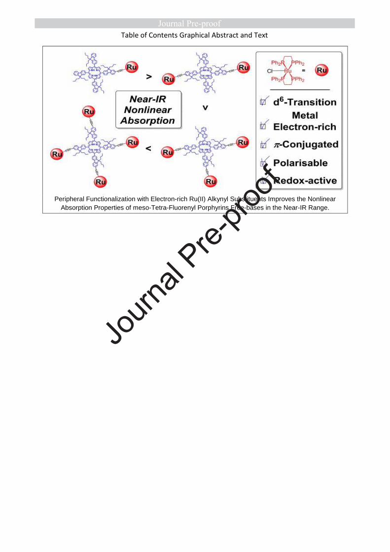
Table of Contents Graphical Abstract and Text
Peripheral Functionalization with Electron-rich Ru(II) Alkynyl Substituents Improves the Nonlinear Absorption Properties of meso-Tetra-Fluorenyl Porphyrins Free-bases in the Near-IR Range.
Journ
al Pre-
proof

Dr. Christine O. PAUL-ROTH is the corresponding author
Journ
al Pre-
proof
2
1. Introduction
Recently, there has been an increasing interest in multi-photon
absorbing molecules and materials, driven by the upsurge in
key societal applications that can exploit these substances,1
namely photonic devices for laser beam control,2 optical data
storage,3 microfabrication,4 fluorescence imaging, and
photodynamic therapy.5
Among the panoply of possibilities, certain organometallic
compounds6, 7 such as group 8 transition metal alkynyl
complexes stand out as attractive building blocks because their
nonlinear optical (NLO) properties are often significantly larger
than those of purely organic analogues.8 Thus, many examples
of formally octahedral d6 complexes featuring an equatorial
Ru(dppe)2 core [dppe = 1,2-bis(diphenylphosphino)ethane]
have been explored.9 When incorporated into extended π
networks, a remarkable enhancement of the NLO responses is
often observed.10, 11 In the pursuit of optimized systems, these
organometallic units permit great structural control, as the
ligand trans to the alkynyl ligand can be varied at will,
affording the possibility of fine-tuning their NLO performance.
In parallel work, large metallated π-compounds such as
porphyrins or phthalocyanines have also been identified as
promising cubic NLO-phores.12-14 For instance, Rao et al.
showed that various metallated meso-tetra(p-tolyl)porphyrins
(TTP) exhibit high cubic optical nonlinearities at 532 and 600
nm.15 Thus, depending on the overall symmetry of the
tetrapyrrolic core, on the nature of the central metal ion (if
any), and on the nature of the peripheral substituents
appended to the macrocyclic core, widely different
nonlinearities can result. To achieve high hyperpolarizabilities,
the presence of a metal inside the porphyrin cavity is usually
recommended, since, besides electronic effects, it facilitates
planarization of the macrocyclic π-manifold.13, 14
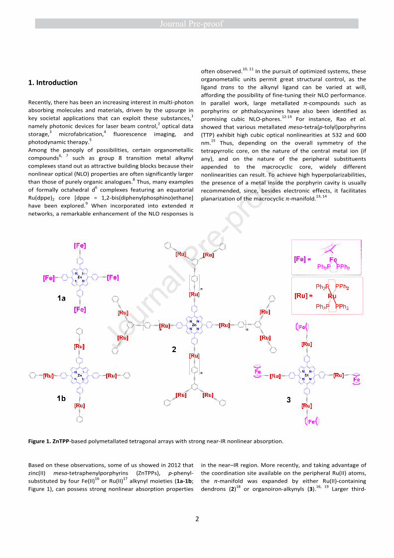
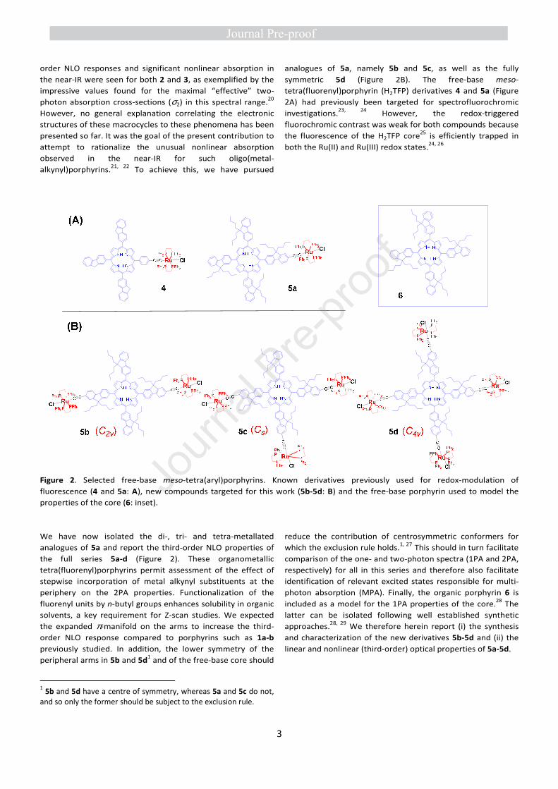
Figure 1. ZnTPP-based polymetallated tetragonal arrays with strong near-IR nonlinear absorption.
Based on these observations, some of us showed in 2012 that
zinc(II) meso-tetraphenylporphyrins (ZnTPPs), p-phenyl-
substituted by four Fe(II)16 or Ru(II)17 alkynyl moieties (1a-1b;
Figure 1), can possess strong nonlinear absorption properties
in the near–IR region. More recently, and taking advantage of
the coordination site available on the peripheral Ru(II) atoms,
the π-manifold was expanded by either Ru(II)-containing
dendrons (2)18 or organoiron-alkynyls (3).16, 19 Larger third-
Journ
al Pre-
proof
3
order NLO responses and significant nonlinear absorption in
the near-IR were seen for both 2 and 3, as exemplified by the
impressive values found for the maximal “effective” two-
photon absorption cross-sections (σ2) in this spectral range.20
However, no general explanation correlating the electronic
structures of these macrocycles to these phenomena has been
presented so far. It was the goal of the present contribution to
attempt to rationalize the unusual nonlinear absorption
observed in the near-IR for such oligo(metal-
alkynyl)porphyrins.21, 22 To achieve this, we have pursued
analogues of 5a, namely 5b and 5c, as well as the fully
symmetric 5d (Figure 2B). The free-base meso-
tetra(fluorenyl)porphyrin (H2TFP) derivatives 4 and 5a (Figure
2A) had previously been targeted for spectrofluorochromic
investigations.23, 24 However, the redox-triggered
fluorochromic contrast was weak for both compounds because
the fluorescence of the H2TFP core25 is efficiently trapped in
both the Ru(II) and Ru(III) redox states.24, 26
Figure 2. Selected free-base meso-tetra(aryl)porphyrins. Known derivatives previously used for redox-modulation of
fluorescence (4 and 5a: A), new compounds targeted for this work (5b-5d: B) and the free-base porphyrin used to model the
properties of the core (6: inset).
We have now isolated the di-, tri- and tetra-metallated
analogues of 5a and report the third-order NLO properties of
the full series 5a-d (Figure 2). These organometallic
tetra(fluorenyl)porphyrins permit assessment of the effect of
stepwise incorporation of metal alkynyl substituents at the
periphery on the 2PA properties. Functionalization of the
fluorenyl units by n-butyl groups enhances solubility in organic
solvents, a key requirement for Z-scan studies. We expected
the expanded π-manifold on the arms to increase the third-
order NLO response compared to porphyrins such as 1a-b
previously studied. In addition, the lower symmetry of the
peripheral arms in 5b and 5d1 and of the free-base core should
1 5b and 5d have a centre of symmetry, whereas 5a and 5c do not, and so only the former should be subject to the exclusion rule.
reduce the contribution of centrosymmetric conformers for
which the exclusion rule holds.1, 27 This should in turn facilitate
comparison of the one- and two-photon spectra (1PA and 2PA,
respectively) for all in this series and therefore also facilitate
identification of relevant excited states responsible for multi-
photon absorption (MPA). Finally, the organic porphyrin 6 is
included as a model for the 1PA properties of the core.28 The
latter can be isolated following well established synthetic
approaches.28, 29 We therefore herein report (i) the synthesis
and characterization of the new derivatives 5b-5d and (ii) the
linear and nonlinear (third-order) optical properties of 5a-5d.
Journ
al Pre-
proof
4
2. Results and discussion
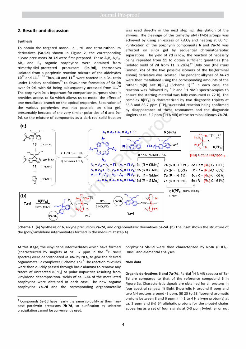
Synthesis
To obtain the targeted mono-, di-, tri- and tetra-ruthenium
derivatives (5a-5d) shown in Figure 2, the corresponding
alkyne precursors 7a-7d were first prepared. These A3B, A2B2,
AB3 and B4 organic porphyrins were obtained from
trimethylsilyl–protected precursors (9a-9d), themselves
isolated from a porphyrin-reaction mixture of the aldehydes
1030 and 11.31, 32 Thus, 10 and 11
32 were reacted in a 3:1 ratio
under Lindsey conditions33 to favour the formation of 9a-9b
over 9c-9d, with 9d being subsequently accessed from 11.34
The porphyrin 9a is important for comparison purposes since it
provides access to 5a which allows us to model the effect of
one metallated branch on the optical properties. Separation of
the various porphyrins was not possible on silica gel,
presumably because of the very similar polarities of 6 and 9a-
9d, so the mixture of compounds as a dark red solid fraction
was used directly in the next step viz. desilylation of the
alkynes. The cleavage of the trimethylsilyl (TMS) groups was
achieved by using an excess of K2CO3 and heating at 60 °C.
Purification of the porphyrin components 6 and 7a-7d was
effected on silica gel by sequential chromatographic
separations. The yield of 7d is low, the reaction of necessity
being repeated from 11 to obtain sufficient quantities (the
isolated yield of 7d from 11 is 28%).34 Only one (the trans
isomer, 7b) of the two possible isomers of the bis(terminal
alkyne) derivative was isolated. The pendant alkynes of 7a-7d
were then metallated using the corresponding amounts of the
ruthenium(II) salt 8[PF6] (Scheme 1).35 In each case, the
reaction was followed by 31P and 1H NMR spectroscopies to
ensure the starting material was fully consumed (> 72 h). The
complex 8[PF6] is characterized by two diagnostic triplets at
55.8 and 83.7 ppm (31P), successful reaction being confirmed
by disappearance of these resonances and the diagnostic
singlets at ca. 3.2 ppm (1H NMR) of the terminal alkynes 7b-7d.
Scheme 1. (a) Synthesis of 6, alkyne precursors 7a-7d, and organometallic derivatives 5a-5d. (b) The inset shows the structure of
the (poly)vinylidene intermediates formed in the medium at step 4).
At this stage, the vinylidene intermediates which have formed
(characterized by singlets at ca. 37 ppm in the 31P NMR
spectra) were deprotonated in situ by NEt3 to give the desired
organometallic complexes (Scheme 1b).2 The reaction mixtures
were then quickly passed through basic alumina to remove any
traces of unreacted 8[PF6] or polar impurities resulting from
vinylidene decomposition. Yields of ca. 60% of the metallated
porphyrins were obtained in each case. The new organic
porphyrins 7b-7d and the corresponding organometallic
2 Compounds 5a-5d have nearly the same solubility as their free-base porphyrin precursors 7b-7d, so purification by selective precipitation cannot be conveniently used.
porphyrins 5b-5d were then characterized by NMR (CDCl3),
HRMS and elemental analyses.
NMR data
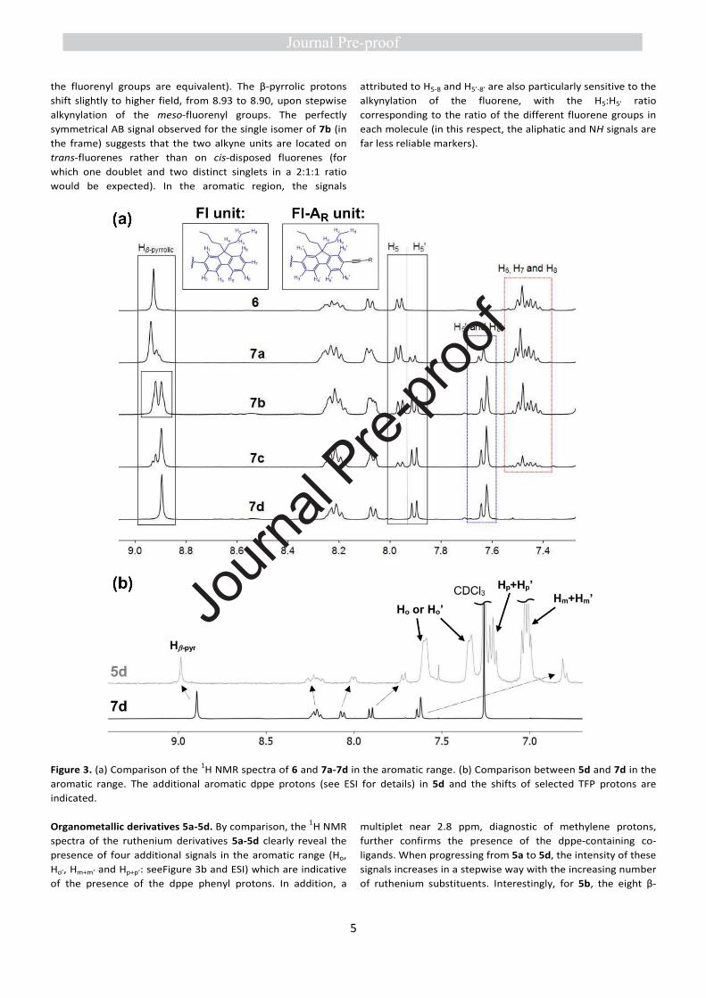
Organic derivatives 6 and 7a-7d. Partial 1H NMR spectra of 7a-
7d are compared to that of the reference compound 6 in
Figure 3a. Characteristic signals are obtained for all protons in
four spectral ranges: (i) Eight β-pyrrolic H around 9 ppm and
two NH protons around -3 ppm, (ii) 25 to 28 fluorenyl aromatic
protons between 8 and 6 ppm, (iii) 1 to 4 H alkyne proton(s) at
ca. 3 ppm and (iv) 64 aliphatic protons for the n-butyl chains
appearing as a set of four signals at 0-3 ppm (whether or not
Journ
al Pre-
proof
5
the fluorenyl groups are equivalent). The β-pyrrolic protons
shift slightly to higher field, from 8.93 to 8.90, upon stepwise
alkynylation of the meso-fluorenyl groups. The perfectly
symmetrical AB signal observed for the single isomer of 7b (in
the frame) suggests that the two alkyne units are located on
trans-fluorenes rather than on cis-disposed fluorenes (for
which one doublet and two distinct singlets in a 2:1:1 ratio
would be expected). In the aromatic region, the signals
attributed to H5-8 and H5’-8’ are also particularly sensitive to the
alkynylation of the fluorene, with the H5:H5’ ratio
corresponding to the ratio of the different fluorene groups in
each molecule (in this respect, the aliphatic and NH signals are
far less reliable markers).
Figure 3. (a) Comparison of the 1H NMR spectra of 6 and 7a-7d in the aromatic range. (b) Comparison between 5d and 7d in the
aromatic range. The additional aromatic dppe protons (see ESI for details) in 5d and the shifts of selected TFP protons are
indicated.
Organometallic derivatives 5a-5d. By comparison, the 1H NMR
spectra of the ruthenium derivatives 5a-5d clearly reveal the
presence of four additional signals in the aromatic range (Ho,
Ho’, Hm+m’ and Hp+p’: seeFigure 3b and ESI) which are indicative
of the presence of the dppe phenyl protons. In addition, a
multiplet near 2.8 ppm, diagnostic of methylene protons,
further confirms the presence of the dppe-containing co-
ligands. When progressing from 5a to 5d, the intensity of these
signals increases in a stepwise way with the increasing number
of ruthenium substituents. Interestingly, for 5b, the eight β-
Journ
al Pre-
proof
6
pyrrolic protons now appear as two overlapped doublets,
around 9.0 ppm, confirming the trans-assignment of the
isolated isomer, consistent with the corresponding symmetry
assumed for 7b. In addition, the presence of the
organometallic end-groups in 5a-5d is confirmed by diagnostic
singlets in the 31P NMR (Table 1), around 49 ppm, which
correspond to the four, eight, twelve and sixteen equivalent
phosphorus nuclei of the dppe ligands. Similar NMR signatures
have previously been observed for the related organometallic
compounds 1217
and 1336 (Figure 4).
Table 1. Characteristic 31P NMR and cyclic voltammetric data
for organometallic porphyrins 5a-5d and reference compounds
6, 12, and 13.
Cmpd 31P{1H} NMR E° (V vs SCE) b
(ppm) a [Ru(III/II)]
E° [Porphyrin]
E°Ox E°Red
5a c 49.4 0.42 0.99, 1.33 -1.21
5b 49.4 0.42 0.99, 1.33 -1.20 5c 49.5 0.43 1.00, 1.35 -1.21 5d 49.5 0.43 1.03, 1.36 -1.25
6 / / 0.99, 1.38, 1.70
-1.14 -1.50
12 d 51.0 0.49 0.87, 1.16 /
13 e 50.2 0.41 / /
a CDCl3. b CH2Cl2, 20 °C, 0.1 M [NBu4][PF6], scan rate 0.1 V.s-
1 with ferrocene or decamethylferrocene used as internal
calibrants. Potentials are expressed relative to that of the
saturated calomel electrode (SCE), with the
Cp*2Fe+/Cp*2Fe couple at −0.08 V (see Exp. Part).37 c Data
from ref. 24. d Data from ref. 17. e Data from ref. 36.
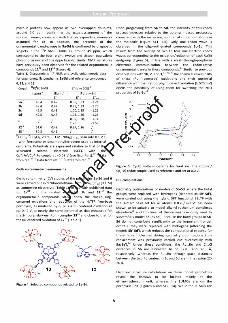
Cyclic voltammetry measurements
Cyclic voltammetry (CV) studies of the porphyrins 5a-5d and 6
were carried out in dichloromethane, with [NBu4][PF6] (0.1 M)
as supporting electrolyte (Table 1). In line with published data
for 5a24 and the related molecules 1b and 12,17 the
organometallic compounds 5b-5d show the classic ring-
centered oxidations and reduction of the H2TFP free-base
porphyrin, as modelled by 6, plus a Ru-centered oxidation at
ca. 0.42 V, at nearly the same potential as that measured for
the 2-fluorenylalkynyl Ru(II) complex 1336 and close to that for
the Ru-centered oxidation of 1224 (Table 1).
Figure 4. Selected compounds related to 5a-5d.
Upon progressing from 5a to 5d, the intensity of this redox
process increases relative to the porphyrin-based processes,
consistent with the increasing number of ruthenium atoms in
the molecule (Figure S11, ESI). Only one redox wave is
observed in the oligo-ruthenated compounds 5b-5d. This
results from the overlap of two to four one-electron redox
waves corresponding to the oxidation/reduction of each Ru(II)
endgroup (Figure 5), in line with a weak through-porphyrin
electronic communication between the redox-active
organometallic units in these compounds.21 Similar to previous
observations with 1b, 2, and 3,17, 19, 20 the chemical reversibility
of these (Ru(II)-centered) oxidations and their potential
difference with the first porphyrin-based oxidation (≥ 570 mV)
opens the possibility of using them for switching the NLO
properties of 5a-5d.8
Figure 5. Cyclic voltammograms for 5a-d (vs. the (Cp2Fe+/
Cp2Fe) redox couple used as reference and set at 0.0 V.
DFT computations
Geometry optimizations of models of 5b-5d, where the butyl
groups were replaced with hydrogens (denoted as 5b′-5d′),
were carried out using the hybrid DFT functional B3LYP with
the 3-21G* basis set for all atoms. B3LYP/3-21G* has been
shown to be suitable to model alkynyl ruthenium complexes
elsewhere38 and this level of theory was previously used to
successfully model 5a (as 5a′). Because the butyl groups in 5b-
5d do not contribute significantly to the important frontier
orbitals, they were replaced with hydrogens (affording the
models 5b′-5d′), which reduces the computational expense for
these large molecules during geometry optimizations (this
replacement was previously carried out successfully with
5a/5a′).24 Under these conditions, the Ru…Ru and Cl…Cl
distances in 5b are estimated to be 32.8 and 37.8 Å,
respectively, whereas the Ru…Ru through-space distances
between the two Ru centers in 5c and 5d are in the region 22-
26 Å.
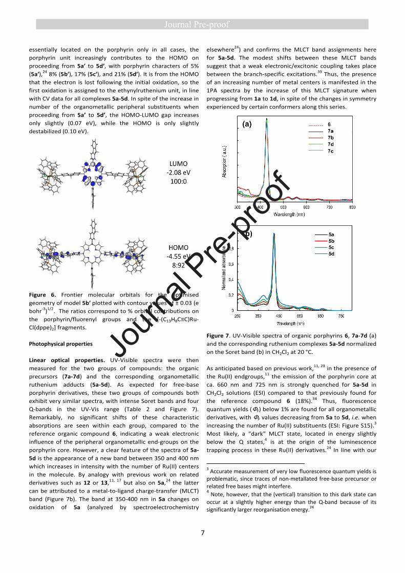
Electronic structure calculations on these model geometries
reveal the HOMOs to be located mainly at the
ethynylruthenium unit, whereas the LUMOs are on the
porphyrin unit (Figures 6 and S12-S14). While the LUMOs are
Journ
al Pre-
proof
7
essentially located on the porphyrin only in all cases, the
porphyrin unit increasingly contributes to the HOMO on
proceeding from 5a′ to 5d′, with porphyrin characters of 5%
(5a′),24 8% (5b′), 17% (5c′), and 21% (5d′). It is from the HOMO
that the electron is lost following the initial oxidation, so the
first oxidation is assigned to the ethynylruthenium unit, in line
with CV data for all complexes 5a-5d. In spite of the increase in
number of the organometallic peripheral substituents when
proceeding from 5a’ to 5d’, the HOMO-LUMO gap increases
only slightly (0.07 eV), while the HOMO is only slightly
destabilized (0.10 eV).
Figure 6. Frontier molecular orbitals for the optimised
geometry of model 5b′ plotted with contour values of ± 0.03 (e
bohr-3)1/2. The ratios correspond to % orbital contributions on
the porphyrin/fluorenyl groups and the [-(C13H8C≡C)Ru-
Cl(dppe)2] fragments.
Photophysical properties
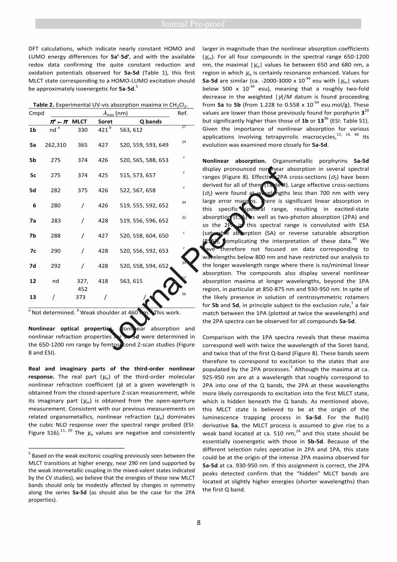
Linear optical properties. UV-Visible spectra were then
measured for the two groups of compounds: the organic
precursors (7a-7d) and the corresponding organometallic
ruthenium adducts (5a-5d). As expected for free-base
porphyrin derivatives, these two groups of compounds both
exhibit very similar spectra, with intense Soret bands and four
Q-bands in the UV-Vis range (Table 2 and Figure 7).
Remarkably, no significant shifts of these characteristic
absorptions are seen within each group, compared to the
reference organic compound 6, indicating a weak electronic
influence of the peripheral organometallic end-groups on the
porphyrin core. However, a clear feature of the spectra of 5a-
5d is the appearance of a new band between 350 and 400 nm
which increases in intensity with the number of Ru(II) centers
in the molecule. By analogy with previous work on related
derivatives such as 12 or 13,11, 17 but also on 5a,24 the latter
can be attributed to a metal-to-ligand charge-transfer (MLCT)
band (Figure 7b). The band at 350-400 nm in 5a changes on
oxidation of 5a (analyzed by spectroelectrochemistry
elsewhere24) and confirms the MLCT band assignments here
for 5a-5d. The modest shifts between these MLCT bands
suggest that a weak electronic/excitonic coupling takes place
between the branch-specific excitations.39 Thus, the presence
of an increasing number of metal centers is manifested in the
1PA spectra by the increase of this MLCT signature when
progressing from 1a to 1d, in spite of the changes in symmetry
experienced by certain conformers along this series.
Figure 7. UV-Visible spectra of organic porphyrins 6, 7a-7d (a)
and the corresponding ruthenium complexes 5a-5d normalized
on the Soret band (b) in CH2Cl2 at 20 °C.
As anticipated based on previous work,11, 23 in the presence of
the Ru(II) endgroups,11 the emission of the porphyrin core at
ca. 660 nm and 725 nm is strongly quenched for 5a-5d in
CH2Cl2 solutions (ESI) compared to that previously found for
the reference compound 6 (18%).34 Thus, fluorescence
quantum yields (ΦF) below 1% are found for all organometallic
derivatives, with ΦF values decreasing from 5a to 5d, i.e. when
increasing the number of Ru(II) substituents (ESI: Figure S15).3
Most likely, a “dark” MLCT state, located in energy slightly
below the Q states,4 is at the origin of the luminescence
trapping process in these Ru(II) derivatives.24 In line with our
3 Accurate measurement of very low fluorescence quantum yields is problematic, since traces of non-metallated free-base precursor or related free bases might interfere. 4 Note, however, that the (vertical) transition to this dark state can occur at a slightly higher energy than the Q-band because of its significantly larger reorganisation energy.24
Journ
al Pre-
proof
8
DFT calculations, which indicate nearly constant HOMO and
LUMO energy differences for 5a’-5d′, and with the available
redox data confirming the quite constant reduction and
oxidation potentials observed for 5a-5d (Table 1), this first
MLCT state corresponding to a HOMO-LUMO excitation should
be approximately isoenergetic for 5a-5d.5
Table 2. Experimental UV-vis absorption maxima in CH2Cl2.
Cmpd λmax (nm) Ref.
ππππ*←←←←ππππ MLCT Soret Q bands
1b nd a 330 421 b 563, 612 17
5a 262,310 365 427 520, 559, 593, 649 24
5b 275 374 426 520, 565, 588, 653 c
5c 275 374 425 515, 573, 657 c
5d 282 375 426 522, 567, 658 c
6 280 / 426 519, 555, 592, 652 34
7a 283 / 428 519, 556, 596, 652 32
7b 288 / 427 520, 558, 604, 650 c
7c 290 / 428 520, 556, 592, 653 c
7d 292 / 428 520, 558, 594, 652 34
12 nd 327,
452
418 563, 615 17
13 / 373 / / 36
a Not determined. b Weak shoulder at 460 nm. c This work.
Nonlinear optical properties. Nonlinear absorption and
nonlinear refraction properties for 5a-5d were determined in
the 650-1200 nm range by femtosecond Z-scan studies (Figure
8 and ESI).
Real and imaginary parts of the third-order nonlinear
response. The real part (γre) of the third-order molecular
nonlinear refraction coefficient (γ) at a given wavelength is
obtained from the closed-aperture Z-scan measurement, while
its imaginary part (γim) is obtained from the open-aperture
measurement. Consistent with our previous measurements on
related organometallics, nonlinear refraction (γre) dominates
the cubic NLO response over the spectral range probed (ESI:
Figure S16).11, 20 The γre values are negative and consistently
5 Based on the weak excitonic coupling previously seen between the MLCT transitions at higher energy, near 290 nm (and supported by the weak intermetallic coupling in the mixed-valent states indicated by the CV studies), we believe that the energies of these new MLCT bands should only be modestly affected by changes in symmetry along the series 5a-5d (as should also be the case for the 2PA properties).
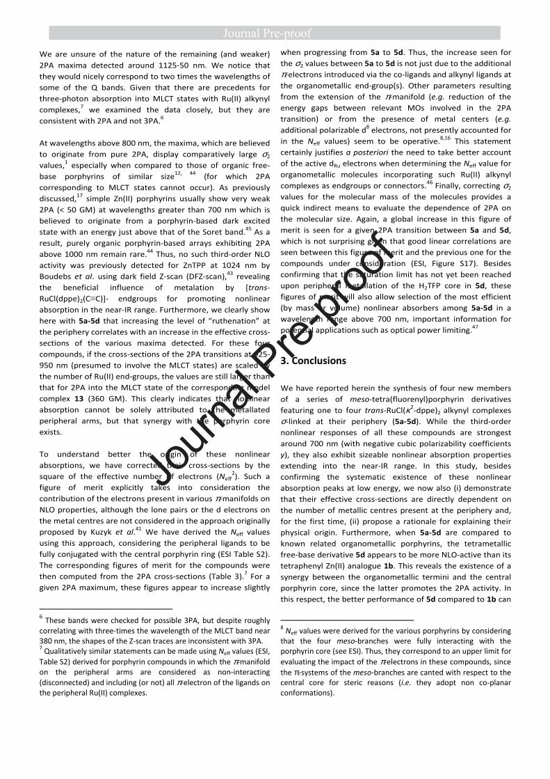
larger in magnitude than the nonlinear absorption coefficients
(γim). For all four compounds in the spectral range 650-1200
nm, the maximal |γre| values lie between 650 and 680 nm, a
region in which γre is certainly resonance enhanced. Values for
5a-5d are similar (ca. -2000-3000 x 10-34 esu with |γim| values
below 500 x 10-34 esu), meaning that a roughly two-fold
decrease in the weighted |γ|/M datum is found proceeding
from 5a to 5b (from 1.228 to 0.558 x 10⁻³⁴ esu.mol/g). These
values are lower than those previously found for porphyrin 320
but significantly higher than those of 1b or 1336 (ESI: Table S1).
Given the importance of nonlinear absorption for various
applications involving tetrapyrrolic macrocycles,12, 14, 40 its
evolution was examined more closely for 5a-5d.
Nonlinear absorption. Organometallic porphyrins 5a-5d
display pronounced nonlinear absorption in several spectral
ranges (Figure 8). Effective 2PA cross-sections (σ2) have been
derived for all of them (Table 3). Large effective cross-sections
(σ2) were found at wavelengths less than 700 nm with very
large error margins. There is significant linear absorption in
this specific spectral range, resulting in excited-state
absorption (ESA) as well as two-photon absorption (2PA) and
so the 2PA in this spectral range is convoluted with ESA
(saturable absorption (SA) or reverse saturable absorption
(RSA)), complicating the interpretation of these data.43 We
have therefore not focused on data corresponding to
wavelengths below 800 nm and have restricted our analysis to
the longer wavelength range where there is no/minimal linear
absorption. The compounds also display several nonlinear
absorption maxima at longer wavelengths, beyond the 1PA
region, in particular at 850-875 nm and 930-950 nm. In spite of
the likely presence in solution of centrosymmetric rotamers
for 5b and 5d, in principle subject to the exclusion rule,1 a fair
match between the 1PA (plotted at twice the wavelength) and
the 2PA spectra can be observed for all compounds 5a-5d.
Comparison with the 1PA spectra reveals that these maxima
correspond well with twice the wavelength of the Soret band,
and twice that of the first Q-band (Figure 8). These bands seem
therefore to correspond to excitation to the states that are
populated by the 2PA processes.7 Although the maxima at ca.
925-950 nm are at a wavelength that roughly correspond to
2PA into one of the Q bands, the 2PA at these wavelengths
more likely corresponds to excitation into the first MLCT state,
which is hidden beneath the Q bands. As mentioned above,
this MLCT state is believed to be at the origin of the
luminescence trapping process in 5a-5d. For the Ru(II)
derivative 5a, the MLCT process is assumed to give rise to a
weak band located at ca. 510 nm,24 and this state should be
essentially isoenergetic with those in 5b-5d. Because of the
different selection rules operative in 2PA and 1PA, this state
could be at the origin of the intense 2PA maxima observed for
5a-5d at ca. 930-950 nm. If this assignment is correct, the 2PA
peaks detected confirm that the “hidden” MLCT bands are
located at slightly higher energies (shorter wavelengths) than
the first Q band.
Journ
al Pre-
proof
Table 3. Experimental γre, γim, |γ|, and σ2 values in CH2Cl2 at selected wavelengthsa corresponding to extrema of
the nonlinear absorption spectra of 5a-5d, and comparison to data previously obtained for 1b20 and 13.36
a Conditions: measurements were carried out in CH2Cl2; γ values are referenced to the nonlinear refractive index of silica n2
= 2.92 × 10⁻¹⁶ cm2 W-1. b Wavelength of the laser in nm. c 10-34 esu. The SI units for γ are C m4 V-3, while those in the cgs
system (used almost exclusively in the literature, and so given here) are cm5 statV-2 or esu. To convert between the two
systems, γSI = (1/3)4 × 10-23 γcgs. d Effective (apparent) 2PA cross-section in Göppert-Mayer units (1 GM = 1 × 10-50 cm4 s
photon-1). eσ2 corrected for the squared effective number of electrons (Neff)2.41, 42 f|σ2| corrected for the molecular mass (M
in g mol-1) ). g No error estimated.
==
Figure 8. Two-photon absorption cross-section plots (red) for 5a (a), 5b (b), 5c (c) and 5d (d) overlaid on the one-photon absorption (1PA)
spectra (blue) and the same 1PA spectra plotted at twice the wavelength (black).
Cmpd λλλλb γγγγre
c γγγγιιιιm
c |γγγγ|
c σσσσ2
d [σσσσ2/(Neff)
2]
e [σσσσ2/M]
f
1b 900 Data not measured17
1100 ± 50 0.28 ± 0.01 0.23 ± 0.01
5a 850 -1400 ± 350 350± 130 1500 ± 350 7500 ± 2800 1.5 ± 0.6 3.2 ± 1.2
935 -1150 ± 150 610 ± 150 1300 ± 200 11000 ± 2900 2.2 ± 0.6 4.7 ± 1.2
1125 -550 ± 80 150 ± 30 600 ± 90 1900 ± 400 0.4 ± 0.1 0.8 ± 0.2
5b 850 -1000 ± 350 450 ± 200 1100 ± 400 10000 ± 4500 1.8 ± 0.8 3.0 ± 1.4
930 -1150 ± 150 610 ± 160 1300 ± 200 11000 ± 3000 2.0 ± 0.5 3.3 ± 0.9
1150 -1700 ± 1500 290 ± 300 1700 ± 1600 3400 ± 3500 0.6 ± 0.6 1.0 ± 1.0
5c 875 -1600 ± 200 570 ± 140 1650 ± 250 14000 ± 3500 2.3 ± 0.6 3.3 ± 0.8
950 -2100 ± 550 950 ± 350 2300 ± 650 16200 ± 5700 2.7 ± 0.9 3.8 ± 1.3
1150 -530 ± 90 170 ± 40 550 ± 100 2000 ± 450 0.3 ± 0.1 0.5 ± 0.1
5d 875 -1400 ± 200 800 ± 200 1600 ± 250 16000 ± 4000 2.4 ± 0.6 3.0 ± 0.8
950 -1600 ± 450 1120 ± 500 2000 ± 700 21000 ± 9000 3.1 ± 1.3 3.9 ± 1.7
1150 -500 ± 150 450 ± 150 650 ± 200 5500 ± 1800 0.8 ± 0.3 1.0 ± 0.3
Journ
al Pre-
proof
We are unsure of the nature of the remaining (and weaker)
2PA maxima detected around 1125-50 nm. We notice that
they would nicely correspond to two times the wavelengths of
some of the Q bands. Given that there are precedents for
three-photon absorption into MLCT states with Ru(II) alkynyl
complexes,7 we examined the data closely, but they are
consistent with 2PA and not 3PA.6
At wavelengths above 800 nm, the maxima, which are believed
to originate from pure 2PA, display comparatively large σ2
values,1 especially when compared to those of organic free-
base porphyrins of similar size12, 44 (for which 2PA
corresponding to MLCT states cannot occur). As previously
discussed,17 simple Zn(II) porphyrins usually show very weak
2PA (< 50 GM) at wavelengths greater than 700 nm which is
believed to originate from a porphyrin-based dark excited
state with an energy just above that of the Soret band.45 As a
result, purely organic porphyrin-based arrays exhibiting 2PA
above 1000 nm remain rare.44 Thus, no such third-order NLO
activity was previously detected for ZnTPP at 1024 nm by
Boudebs et al. using dark field Z-scan (DFZ-scan),43 revealing
the beneficial influence of metalation by [trans-
RuCl(dppe)2(C≡C)]- endgroups for promoting nonlinear
absorption in the near-IR range. Furthermore, we clearly show
here with 5a-5d that increasing the level of “ruthenation” at
the periphery correlates with an increase in the effective cross-
sections of the various maxima detected. For these four
compounds, if the cross-sections of the 2PA transitions at 925-
950 nm (presumed to involve the MLCT states) are scaled by
the number of Ru(II) end-groups, the values are still larger than
that for 2PA into the MLCT state of the corresponding model
complex 13 (360 GM). This clearly indicates that nonlinear
absorption cannot be solely attributed to the metallated
peripheral arms, but that synergy with the porphyrin core
exists.
To understand better the origin of these nonlinear
absorptions, we have corrected their cross-sections by the
square of the effective number of electrons (Neff2). Such a
figure of merit explicitly takes into consideration the
contribution of the electrons present in various π-manifolds on
NLO properties, although the lone pairs or the d electrons on
the metal centres are not considered in the approach originally
proposed by Kuzyk et al.41 We have derived the Neff values
using this approach, considering the peripheral ligands to be
fully conjugated with the central porphyrin ring (ESI Table S2).
The corresponding figures of merit for the compounds were
then computed from the 2PA cross-sections (Table 3).7 For a
given 2PA maximum, these figures appear to increase slightly
6 These bands were checked for possible 3PA, but despite roughly correlating with three-times the wavelength of the MLCT band near 380 nm, the shapes of the Z-scan traces are inconsistent with 3PA. 7 Qualitatively similar statements can be made using Neff values (ESI,
Table S2) derived for porphyrin compounds in which the π-manifold on the peripheral arms are considered as non-interacting (disconnected) and including (or not) all π-electron of the ligands on the peripheral Ru(II) complexes.
when progressing from 5a to 5d. Thus, the increase seen for
the σ2 values between 5a to 5d is not just due to the additional
π-electrons introduced via the co-ligands and alkynyl ligands at
the organometallic end-group(s). Other parameters resulting
from the extension of the π-manifold (e.g. reduction of the
energy gaps between relevant MOs involved in the 2PA
transition) or from the presence of metal centers (e.g.
additional polarizable d8 electrons, not presently accounted for
in the Neff values) seem to be operative.8,16 This statement
certainly justifies a posteriori the need to take better account
of the active dRu electrons when determining the Neff value for
organometallic molecules incorporating such Ru(II) alkynyl
complexes as endgroups or connectors.46 Finally, correcting σ2
values for the molecular mass of the molecules provides a
quick indirect means to evaluate the dependence of 2PA on
the molecular size. Again, a global increase in this figure of
merit is seen for a given 2PA transition between 5a and 5d,
which is not surprising given that good linear correlations are
seen between this figure of merit and the previous one for the
compounds under consideration (ESI, Figure S17). Besides
confirming that the saturation limit has not yet been reached
upon peripheral metallation of the H2TFP core in 5d, these
figures of merit will also allow selection of the most efficient
(by mass or volume) nonlinear absorbers among 5a-5d in a
wavelength range above 700 nm, important information for
potential applications such as optical power limiting.47
3. Conclusions
We have reported herein the synthesis of four new members
of a series of meso-tetra(fluorenyl)porphyrin derivatives
featuring one to four trans-RuCl(κ2-dppe)2 alkynyl complexes
σ-linked at their periphery (5a-5d). While the third-order
nonlinear responses of all these compounds are strongest
around 700 nm (with negative cubic polarizability coefficients
y), they also exhibit sizeable nonlinear absorption properties
extending into the near-IR range. In this study, besides
confirming the systematic existence of these nonlinear
absorption peaks at low energy, we now also (i) demonstrate
that their effective cross-sections are directly dependent on
the number of metallic centres present at the periphery and,
for the first time, (ii) propose a rationale for explaining their
physical origin. Furthermore, when 5a-5d are compared to
known related organometallic porphyrins, the tetrametallic
free-base derivative 5d appears to be more NLO-active than its
tetraphenyl Zn(II) analogue 1b. This reveals the existence of a
synergy between the organometallic termini and the central
porphyrin core, since the latter promotes the 2PA activity. In
this respect, the better performance of 5d compared to 1b can
8 Neff values were derived for the various porphyrins by considering that the four meso-branches were fully interacting with the porphyrin core (see ESI). Thus, they correspond to an upper limit for
evaluating the impact of the π-electrons in these compounds, since the π-systems of the meso-branches are canted with respect to the central core for steric reasons (i.e. they adopt non co-planar conformations).
Journ
al Pre-
proof

11
be related to the extension of the central π-manifold. In
addition, with the help of relevant figures of merit, we also
show that all d6-transition metal complexes contribute to
enhancement of the near-IR 2PA (MPA) properties in 5a-5d.
As a result, and in line with previous contributions of our
groups, this study confirms that peripheral metallation by
electron-rich Ru(II) alkynyl complexes at a (central)
tetra(aryl)porphyrin ring constitutes a general and efficient
method to enhance 2PA in the near-IR range.9 Given the
strategic importance of the 1000-1500 nm spectral range (also
known as the “telecommunications window”) for various
applications, we hope that such approaches will prove helpful
in the design of new nonlinear absorbers based on
tetrapyrrolic macrocycles.
4. Experimental
Synthetic procedures
General. Unless otherwise stated, all solvents used in reactions
were distilled using common purification protocols,48 except
for DMF and iPr2NH which were dried over molecular sieves (3
Å). Compounds were purified by chromatography on silica gel
using different mixtures of eluents as specified. 1H and 13C
NMR spectra were recorded on Bruker Ascend 400 and 500
MHz spectrometers at 298 K (for labelling of nuclei, see Figure
3). The chemical shifts are given in ppm and referenced to
internal tetramethylsilane. Cyclic voltammograms were
recorded with an Autolab PG-STAT 30 potentiostat at 20 °C
from solutions of ca. 10−4 M analyte in dry dichloromethane
containing 0.1 M [Bu4N][PF6] at a scan rate ν = 100 m s−1 under
a dry nitrogen atmosphere. The single compartment three-
electrode cell was equipped with platinum wire counter and
reference electrodes and a glassy carbon working electrode.
Redox potentials were measured using the decamethyl-
ferrocene/decamethylferrocenium (Cp*2Fe+/Cp*2Fe) redox
couple as an internal reference system at −0.53 V49 vs. the
usual ferrocene/ferrocenium (Cp2Fe+/Cp2Fe) redox couple in
CH2Cl2 set at 0.46 V.37 Solutions were purged and maintained
under a nitrogen atmosphere. High-resolution mass spectra
(HRMS) were recorded on different spectrometers: a Bruker
MicroTOF-Q II, a Thermo Fisher Scientific Q-Exactive in ESI
positive mode and a Bruker Ultraflex III MALDI Spectrometer
at CRMPO (centre regional de mesures physiques de l’Ouest)
in Rennes. Reagents were purchased from commercial
suppliers and used as received. The [RuCl(dppe)2][PF6] salt
(8[PF6]),35, 50 9,9-dibutyl-9H-fluorene-2-carbaldehyde (10)30
9 Note that the impact of these organometallic endgroups on the NLO properties is not always directly related to their electron-releasing capability in the ground state,8 but rather to their involvement in the low-energy excited states (MLCT states here), which determine the nonlinear polarization and absorption properties of a given molecule.
and 7-((trimethylsilyl)ethynyl)-9,9-dibutyl-9H-fluorene-2-carb-
aldehyde (11)32 were prepared as described earlier.
Synthesis of organic porphyrins 6 and 7a-7d. In a two-necked
flask, a mixture of 9,9-dibutyl-fluorene-2-carbaldehyde (2.1 g,
6.7 mmol, 3 equiv), 9,9-dibutyl-7-((trimethylsilyl)ethynyl)-
fluorene-2-carbaldehyde (900 mg, 2.2 mmol, 1 equiv) and
pyrrole (0.6 mL, 8.9 mmol, 4 equiv.) was dissolved in dry
chloroform (550 mL) under argon. After deoxygenating the
mixture via argon bubbling for 30 min, BF3•OEt2 (0.2 mL) was
injected and the reaction was stirred in the dark for 3 h under
argon at room temperature. p-Chloranil (1.5 g, 6.1 mmol) was
then added as oxidant and the reaction was heated at 60 °C for
another 2 h in air. After cooling the reaction to room
temperature, NEt3 (2 mL) was injected, and the medium was
kept stirring for 10 min. After evaporation of the volatiles,
purification was carried out by silica chromatography using a
CH2Cl2/heptane (1:3) mixture as eluent. The various porphyrin
isomers 9a-9d could not be separated and were thus collected
together and isolated as a red solid. This mixture (3.6 g, 2.5
mmol) was then dissolved in a CH2Cl2/THF/MeOH (3:1:1)
solvent mixture and K2CO3 (2.0 g, 14.5 mmol) was added with
stirring at 60 °C for 10 h. After stirring overnight, the volatiles
were removed in vacuo and the resulting mixture of
desilylated porphyrins 7a-7d was purified by chromatography
on silica gel, using heptane/THF (4:1) and then heptane/THF
(10:1) mixtures as eluents.
Porphyrin 6. Yield: 44%.28 1H NMR (400 MHz, CDCl3): δ = 8.92
(s, 8H, Hβ-pyr), 8.26-8.18 (m, 8H, H1,3), 8.07 (d, 4H, J = 7.5 Hz,
H4), 7.96 (d, 4H, J = 7.3 Hz, H4), 7.52-7.41 (m, 12H, H6,7,8), 2.14
(t, J = 7.2 Hz, 16H, Ha), 1.21-1.14 (m, 16H, Hc), 1.02-0.87 (m,
16H, Hb), 0.78-0.72 (m, 24H, Hd), -2.57 (s, 2H, NH).
Porphyrin 7a. Yield: 17%.32
Porphyrin 7b. Yield: 8%. 1H NMR (400 MHz, CDCl3): δ = 8.91 (d,
8H, J = 9.1 Hz, Hβ-pyr), 8.23-8.18 (m, 8H, H1,3, H1’, H3’), 8.08-8.06
(m, 4H, H4, H4’), 7.96 (d, 2H, J = 7.2 Hz, H5), 7.91 (d, 2H, J = 7.7
Hz, H5’), 7.63 (d, 4H, J = 8.0 Hz, H6’, H8’), 7.52-7.41 (m, 6H,
H6,7,8), 3.21 (s, 2H, H7’), 2.14 (s, 16H, Ha), 1.21-1.14 (m, 16H, Hc),
0.99-0.85 (m, 16H, Hb), 0.78-0.73 (m, 24H, Hd), -2.58 (s, 2H,
NH). 13C{
1H} NMR (100 MHz, CDCl3): δ = 151.2, 149.6, 149.2,
141.8, 141.6, 140.9, 140.8, 140.7, 139.9, 133.8, 133.6, 131.5,
129.4, 129.3, 127.4, 127.0, 126.7, 123.1, 120.9, 120.9, 120.6,
120.5, 120.5, 120.1, 120.0, 118.3, 117.8, 84.7, 55.4, 55.5, 40.3,
40.2, 26.3, 23.1, 14.0, 13.9. HRMS-MALDI (DCTB): m/z =
1462.895 [M]+• (calcd for [C108H110N4]+•: 1462.8725). Anal.
Calcd. (%) for C108H110N4•EtOH: C, 87.49; H, 7.74; N, 3.71.
Found: C, 87.58; H, 7.76; N, 3.48.
Porphyrin 7c. Yield: 2%. 1H NMR (400 MHz, CDCl3): δ = 8.93-
8.90 (m, 8H, Hβ-pyr), 8.25-8.18 (m, 8H, H1,3, H1’, H3’), 8.07 (d, 4H,
J = 7.2 Hz, H4, H4’), 7.96 (d, 1H, J = 7.2 Hz, H5), 7.91 (d, 3H, J =
7.6 Hz, H5’), 7.63 (d, 3H, J = 8.0 Hz, H6’, H8’), 7.51-7.41 (m, 3H,
H6,7,8), 3.21 (s, 3H, H7’), 2.13 (s, 16H, Ha), 1.22-1.14 (m, 16H, Hc),
0.91-0.84 (m, 16H, Hb), 0.79-0.73 (m, 24H, Hd), -2.59 (s, 2H,
Journ
al Pre-
proof
12
NH). 13C{
1H} NMR (100 MHz, CDCl3): δ = 151.2, 149.6, 149.2,
141.8, 141.6, 140.9, 140.8, 140.8, 139.9, 133.8, 133.6, 131.5,
129.4, 129.3, 127.4, 127.0, 126.7, 123.1, 121.0, 120.9, 120.6,
120.6, 120.5, 120.1, 120.0, 118.3, 117.8, 84.7, 55.4, 55.3, 40.3,
40.2, 26.3, 23.1, 14.0, 13.9. Anal. Calcd. (%) for
C110H110N4•EtOH: C, 87.68; H, 7.62; N, 3.65. Found: C, 87.58; H,
7.76; N, 3.48.
Porphyrin 7d. Yield: 1%.34
Synthesis of organoruthenium porphyrins 5a-5d. In a Schlenk
tube, a mixture of the desired porphyrin precursor (5b-5d) (40
mg, 0.03 mmol, 1 equiv.), n equiv. (n corresponding to the
number of terminal alkynes in 5b-d) of the [RuCl(dppe)2][PF6]
salt (n x 32 mg, n x 0.03 mmol) and NaPF6 (n x 5 mg, n x 0.1
mmol, n equiv.) were stirred in distilled CH2Cl2 under argon at
20 °C. The reaction medium was deoxygenated by argon
bubbling for 10 min and the reaction was kept stirring for 96 h
at room temperature, after which NEt3 was injected to
complete the reaction and stirring maintained 2 h more under
argon. After evaporation of the volatiles, the residue was
purified by chromatography on basic Al2O3 using CH2Cl2/NEt3
(100:1) as eluent, providing fractions of the various title
porphyrin derivatives in a pure state.
Porphyrin 5a. Yield: 63%.24
Porphyrin 5b. Yield: 60%. 1H NMR (400 MHz, CDCl3): δ = 8.97-
8.93 (m, 8H, Hβ-pyr), 8.27-8.16 (m, 8H, Hflu), 8.10-8.09 (m, 2H,
Hflu), 7.99-7.97 (m, 4H, Hflu), 7.72-7.71 (m, 3H, HPh-dppe), 7.59-
7.58 (m, 14H Hflu, HPh-dppe), 7.52-7.42 (m, 9H, Hflu, HPh-dppe),
7.35-7.33 (m, 15H, Hflu, HPh-dppe), 7.24-7.19 (m, 15H, HPh-dppe),
7.04-6.98 (m, 28H, HPh-dppe), 6.79 (d, 4H, J = 7.8 Hz, HPh-dppe),
5.59 (s, 4H, HPh-dppe), 2.76 (s, 12H, CH2-dppe), 2.58-2.52 (m, 4H,
CH2-dppe), 2.23-2.03 (m, 16H, Ha), 1.06-0.96 (m, 16H, Ha), 0.90-
0.71 (m, 40H, Hb, Hd), -2.49~2.61 (m, 2H, NH). 31P{
1H} NMR
(100 MHz, CDCl3): δ = 49.4 (s, 8P, P(dppe)2). HRMS-ESI
(CHCl3/HCO2H): m/z = 1663.5735 [M]2+• (calcd for
[C212H204N4P8Ru2Cl2]2+•: 1663.5720), 3327.1438 [M]+• (calcd for
[C212H204N4P8Ru2Cl2]+•: 3327.1446).
Porphyrin 5c. Yield: 60%. 1H NMR (400 MHz, CDCl3): δ = 8.99-
8.93 (m, 8H, Hβ-pyr), 8.29-8.16 (m, 8H, Hflu), 8.11-8.07 (m, 1H,
Hflu), 8.01-7.96 (m, 4H, Hflu), 7.82-7.78 (m, 1H, Hflu), 7.72 (d, 3H,
J = 7.5 Hz, HPh-dppe), 7.67-7.55 (m, 21H Hflu, HPh-dppe), 7.52-7.38
(m, 10H, Hflu, HPh-dppe), 7.37-7.30 (m, 24H, Hflu, HPh-dppe), 7.25-
7.20 (m, 21H, HPh-dppe), 7.05-6.99 (m, 46H, HPh-dppe), 6.80 (d, 5H,
J = 8.3 Hz, HPh-dppe), 5.85 (t, 1H, J = 8.3 Hz, HPh-dppe), 3.55-3.51
(m, 2H, CH2-dppe), 2.76 (s, 22H, CH2-dppe), 2.29-2.06 (m, 16H, Ha),
1.08-1.05 (m, 16H, Hc), 0.91-0.72 (m, 40H, Hb, Hd), -2.49~-2.57
(m, 2H, NH). 31P{
1H} NMR (100 MHz, CDCl3): δ = 49.5 (s, 12P,
P(dppe)2). HRMS-ESI (CHCl3/HCO2H): m/z = 1428.4280 [M]3+•
(calcd for [C266H251N4P12Ru3Cl3]3+•; 1428.4296), 2142.6377
[M]2+• (calcd for [C266H251N4P12Ru3Cl3]2+•: 2142.6447).
Porphyrin 5d. Yield: 61%. 1H NMR (400 MHz, CDCl3): δ = 8.99
(s, 8H, Hβ-pyr), 8.26-8.18 (m, 8H, Hflu), 8.02-7.99 (m, 4H, Hflu),
7.72 (d, 4H, J = 7.6 Hz, Hflu), 7.61-7.59 (m, 31H, Hflu, HPh-dppe),
7.35-7.33 (m, 31H, Hflu, HPh-dppe), 7.24-7.19 (m, 32H, HPh-dppe),
7.04-7.00 (m, 70H, HPh-dppe), 6.80 (d, 8H, J = 8.5 Hz, HPh-dppe),
2.76 (s, 32H, CH2-dppe), 2.20-2.09 (m, 16H, Ha), 1.08-0.99 (m,
16H, Hc), 0.89-0.83 (m, 40H, Hb, Hd), -2.47 (m, 2H, NH). 31P{
1H}
NMR (100 MHz, CDCl3): δ = 49.5 (s, 16P, P(dppe)2). HRMS-ESI
(CHCl3/HCO2H): m/z = 1310.6095 [M]4+• (calcd for
[C320H298N4P16Ru4Cl4]4+•: 1310.6066), 1747.4765 [M]3+• (calcd
for [C320H298N4P16Ru4Cl4]3+•: 1747.4757), 2621.2119 [M]2+•.
Computations. All computations were carried out with the
Gaussian 09 package.51 The S0 model geometries of 5b′, 5c′
and 5d′ with no symmetry constraints were optimized with the
B3LYP functional52 using the 3-21G* basis set53 for all atoms.
The MO diagrams in Figures 6 and S10-12 were generated with
the Gabedit package54 and the %MO contributions were
determined using the GaussSum software.55
Absorption and emission studies. All photophysical
measurements were performed with freshly-prepared air-
equilibrated solutions at room temperature (298 K). UV-Vis
absorption spectra were recorded on a BIO-TEK instrument
UVIKON XL spectrometer or on a Jasco V-570
spectrophotometer. PL emission was recorded on a Photon
Technology International (PTI) apparatus coupled to an 814
Photomultiplier Detection System, Lamp Power Supply 220B
and MD-5020. Steady-state fluorescence measurements were
performed on dilute solutions (ca. 10-6 M, optical density < 0.1)
contained in standard 1 cm quartz cuvettes using an Edinburgh
Instruments (FLS920) spectrometer in photon-counting mode,
equipped with a calibrated quantum counter for excitation
correction. Fully corrected emission spectra were obtained, for
each compound, after excitation at the wavelength of the
absorption maximum, with Aλex < 0.1 to minimise internal
absorption. Fluorescence quantum yields were measured
using standard methods; TPP in CH2Cl2 (Φlum = 0.12 at λex = 417
nm) was used as a reference.
Z-scan studies. Wavelength-dependent Z-scan experiments
were obtained using a light source consisting of a Quantronix
Integra-C3.5F laser pumping a Quantronix Palitra-FS optical
parametric amplifier, tuneable over a wavelength range from
500 nm to 2000 nm. The output was confirmed by use of an
Ocean Optics USB2000+ spectrometer (500-1000 nm) or an
Ocean Optics NIR-Quest spectrometer (1000-1800 nm). The
output delivered 130 fs pulses with a 1 kHz repetition rate.
Colored glass filters and a Thorlabs polarizing filter were used
to remove unwanted wavelengths. The power was adjusted by
use of neutral density filters to obtain nonlinear phase shifts
between 0.2 to 1.3 rad. The focal length of the beam at the
experiment was 100 mm for wavelengths between 500 and
950 nm, and 75 mm from 1000-1800 nm, which gave 35-50 μm
beam waists resulting in Rayleigh lengths longer than that of
the sample thickness. Samples travelled down the Z axis on a
Thorlabs motorized stage between 0-40 mm or 50-100 mm,
depending on the focal length. Data were collected by two
Thorlabs photodiodes, 500-900 nm with Si based detectors
Journ
al Pre-
proof

13
and 900-1800 nm with InGaAs detectors. Data from the
detectors were collected by a Tektronix oscilloscope feeding a
custom LabVIEW program permitting fitting of a theoretical
trace. A sample of CH2Cl2 was run at each wavelength as an
aid in referencing to the response from a 3 mm fused silica
plate (also run at each wavelength). Solutions of the
chromophores were analyzed in deoxygenated and distilled
CH2Cl2 at concentrations 0.09, 0.15, 0.14 and 0.13 wt% for 5a-
5d, respectively, placed in 1 mm glass cells. The real and
imaginary components of the second hyperpolarizability (γ) of
the materials were calculated assuming additivity to these
reference samples, and γim was used to calculate the two-
photon absorption cross sections (σ2).
Conflicts of interest
There are no conflicts to declare.
Acknowledgements
This research was supported by grants from MEAE and MESRI
(PHC FASIC Chercheurs 2019, Project N° 43406ZE), from the
Australian Research Council (M.G.H., DP170100408), from
Durham University, and from the CNRS (LEA Rennes-Durham,
PICS program 7106 and LIA Redochrom). The China Scholarship
Council (CSC) is also acknowledged for PhD funding (CSC) (X.Z.
and L.S.). J. A. G. Williams (Durham) is kindly acknowledged for
discussion and fluorescence measurements.
References
1. G. S. He, L.-S. Tan, Q. Zheng and P. N. Prasad, Chem. Rev., 2008, 108, 1245-1330.
2. G. S. He, J. D. Bhawalkar, C. F. Zhao, C. K. Park and P. N. Prasad, Opt. Lett., 1995, 20, 2393-2395.
3. D. A. Parthenopoulos and P. M. Rentzepis, Science, 1989, 245, 843-845.
4. a) S. Kawata, H.-B. Sun, T. Tanaka and K. Takada, Nature, 2001, 412, 697-698; b) C. R. Mendonca, D. S. Correa, F. Marlow, T. Voss, P. Tayalia and E. Mazur, Appl. Phys. Lett., 2009, 95, 113309 (1-3).
5. a) T. J. Dougherty and S. L. Marcus, Eur. J. Cancer, 1992, 28A, 1734-1742; b) H. A. Collins, M. Khurana, E. H. Moriyama, A. Mariampillai, E. Dahlstedt, M. Balaz, M. K. Kuimova, M. Drobizhev, V. X. D. Yang, D. Phillips, A. Rebane, B. C. Wilson and H. L. Anderson, Nat. Photonics, 2008, 2, 420-424; c) J. R. Starkey, A. K. Rebane, M. A. Drobizhev, F. Meng, A. Gong, A. Elliott, K. McInnerney and C. W. Spangler, Clin. Cancer Res., 2008, 14, 6564-6573; d) M. Khurana, E. H. Moriyama, A. Mariampillai, K. Samkoe, D. Cramb and B. C. Wilson, J.
Biomed. Opt., 2009, 14, 064006(1-14). 6. a) W. Nie, Adv. Mater., 1993, 5, 520-545; b) H. S. Nalwa, Adv.
Mater., 1993, 5, 341-358; c) M. G. Humphrey, T. Schwich, P. J. West, M. P. Cifuentes and M. Samoc, in Comprehensive
Inorganic Chemistry II - from Element to Applications, eds. J. Reedijk and K. Poeppelmeier, Oxford: Elsevier, 2013, vol. 8, pp. 781-835.
7. P. V. Simpson, L. A. Watson, A. Barlow, G. Wang, M. P. Cifuentes and M. G. Humphrey, Angew. Chem. Int. Ed. , 2016, 55, 2387-2391
8. G. Grelaud, M. P. Cifuentes, F. Paul and M. G. Humphrey, J.
Organomet. Chem., 2014, 751, 181-200. 9. K. A. Green, M. P. Cifuentes, M. Samoc and M. G. Humphrey,
Coord. Chem. Rev., 2011, 255, 2530-2541; K. A. Green, M. P. Cifuentes, M. Samoc and M. G. Humphrey, Coord. Chem. Rev., 2011, 255, 2025-2038.
10. a) T. Schwich, M. P. Cifuentes, P. A. Gugger, M. Samoc and M. G. Humphrey, Adv. Mater., 2011, 23, 1433–1435; b) M. G. Humphrey, M. P. Cifuentes and M. Samoc, Top. Organomet.
Chem., 2011, 28, 57–73; c) A. Trujillo, R. Veillard, G. Argouarch, T. Roisnel, A. Singh, I. Ledoux and F. Paul, Dalton
Trans., 2012, 41, 7454-7456. 11. A. Triadon, G. Grelaud, N. Richy, O. Mongin, G. J. Moxey, I. M.
Dixon, X. Yang, G. Wang, A. Barlow, J. Rault-Berthelot, M. P. Cifuentes, M. G. Humphrey and F. Paul, Organometallics, 2018, 35, 2245-2262.
12. M. Pawlicki, H. A. Collins, R. G. Denning and H. L. Anderson, Angew. Chem. Int. Ed., 2009, 48, 3244-3266.
13. M. Ravikanth and K. G. Ravindra, Curr. Sci., 1995, 68, 1010-1017.
14. G. de la Torre, P. Vazquez, F. Agullo-Lopez and T. Torres, Chem. Rev., 2004, 104, 3723-3750.
15. S. V. Rao, N. K. M. N. Srinivas, D. N. Rao, L. Giribabu, B. G. Maiya, R. Philip and G. R. Kumar, Optics Commun., 2000, 182, 255-264.
16. S. Drouet, A. Merhi, G. Grelaud, M. P. Cifuentes, M. G. Humphrey, K. Matczyszyn, M. Samoc, L. Toupet, C. O. Paul-Roth and F. Paul, New J. Chem., 2012, 36, 2192-2195.
17. S. Drouet, A. Merhi, D. Yao, M. P. Cifuentes, M. G. Humphrey, M. Wielgus, J. Olesiak-Banska, K. Matczyszyn, M. Samoc, F. Paul and C. O. Paul-Roth, Tetrahedron, 2012, 68, 10351-10359.
18. A. Merhi, G. Grelaud, K. A. Green, H. M. Ngo, M. Reynolds, I. Ledoux, A. Barlow, G. Wang, M. P. Cifuentes, M. G. Humphrey, F. Paul and C. O. Paul-Roth, Dalton Trans., 2015, 44, 7748-7751.
19. A. Merhi, G. Grelaud, N. Ripoche, A. Barlow, M. P. Cifuentes, M. G. Humphrey, F. Paul and C. O. Paul-Roth, Polyhedron, 2015, 86, 64-70.
20. A. Merhi, G. Grelaud, M. Morshedi, S. Abid, K. A. Green, A. Barlow, T. Groizard, S. Kahlal, J.-F. Halet, H. M. Ngo, I. Ledoux-Rak, M. P. Cifuentes, M. G. Humphrey, F. Paul and C. O. Paul-Roth, Dalton Trans., 2018, 47, 11123-11135.
21. Y. Tanaka, M. Ono and M. Akita, J. Porphyrins
Phthalocyanines, 2015, 19, 442-450. 22. a) K. Mishiba, M. Ono, Y. Tanaka and M. Akita, Chem. Eur. J. ,
2017, 23, 2067-2076; b) K. Onitsuka, H. Kitajima, M. Fujimoto, A. Iuchi, F. Takei and S. Takahashi, Chem. Commun., 2002, 2576-2577; c) D. Bellows, S. M. Ali, C. P. Gros, M. E. Ojaimi, J.-M. Barbe, R. Guillard and P. D. Harvey, Inorg. Chem., 2009, 48, 7613-7629; d) C. Bucher, C. H. Devillers, J.-C. Moutet, G. Royal and E. Saint-Aman, Coord. Chem. Rev. , 2009, 253, 21–36; e) V. N. Nemykin, G. T. Rohde, C. D. Barrett, R. G. Hadt, C. Bizzarri, P. Galloni, B. Floris, I. Nowik, R. H. Herber, A. G. Marrani, R. Zanoni and N. M. Loim, J. Am. Chem. Soc., 2009, 131, 14969-14978; f) G. T. Rohde, J. R. Sabin, C. D. Barrett and V. N. Nemykin, New J. Chem., 2011, 35, 1440-1448; g) P. Gautam, B. Dhokale, V. Shukla, C. P. Singh, K. S. Bindra and R. Misra, J. Photochem. Photobiol. A, 2012, 239, 24-27; h) S. J.
Journ
al Pre-
proof
14
Dammer, P. V. Solntsev, J. R. Sabin and V. N. Nemykin, Inorg.
Chem., 2013, 52, 9496-9510. 23. A. Merhi, X. Zhang, D. Yao, S. Drouet, O. Mongin, F. Paul, J. A.
G. Williams, M. A. Fox and C. O. Paul-Roth, Dalton Trans., 2015, 44, 9470-9485.
24. X. Zhang, S. Abid, L. Shi, J. A. G. Williams, M. A. Fox, F. Miomandre, C. Tourbillon, J.-F. Audibert, O. Mongin, F. Paul and C. O. Paul-Roth, Dalton Trans., 2019, 48, 11897-11911.
25. a) C. O. Paul-Roth and G. Simonneaux, Tetrahedron Lett., 2006, 47, 3275–3278; C. O. Paul-Roth and G. Simonneaux, C.
R. Acad. Sci., Ser. IIb; Chim., 2006, 9, 1277-1286; b) C. O. Paul-Roth, J. A. G. Williams, J. Letessier and G. Simonneaux, Tetrahedron Lett., 2007, 48, 4317-4322.
26. M. Murai, M. Sugimoto and M. Akita, Dalton Transactions, 2013, 42, 16108-16120.
27. K. D. Bonin and T. J. McIlrath, J. Opt. Soc. Am. B 1984, 1, 52-55.
28. D. Yao, X. Zhang, A. Triadon, N. Richy, O. Mongin, M. Blanchard-Desce, F. Paul and C. O. Paul-Roth, Chem. Eur. J., 2017, 23, 2635-2647.
29. B. Li, J. Li, Y. Fu and Z. Bo, J. Am. Chem. Soc., 2004, 126, 3430-3431; B. Li, K. Xu, M. Sun, Y. Fu, G. Yu, Y. Liu and Z. Bo, Macromolecules, 2006, 39, 456-461.
30. a) T. Pei, K. Peng, X.-y. Cai, L.-j. Yuan and J.-b. Xia, Wuli
Huaxue Xuebao, 2017, 33, 2550-2558; b) H. Wang, F. Liu, Y. Yang, M. Zhang, C. Peng, S. Bo, X. Liu, L. Qiu and Z. Zhen, New
J. Chem., 2015, 39, 1038-1044; c) D. Zhang, V. Martin, I. Garcia-Moreno, A. Costela, M. E. Perez-Ojeda and Y. Xiao, Phys. Chem. Chem. Phys., 2011, 13, 13026-13033.
31. H. Su, S. Zhu, M. Qu, R. Liu, G. Song and H. Zhu, J. Phys. Chem.
C, 2019, 123, 15685-15692. 32. D. Yao, X. Zhang, S. Abid and C. O. Paul-Roth, J. Photochem.
Photobiol. A, 2017, 338, 96-103. 33. a) J. S. Lindsey, K. A. Maccrum, J. S. Tyhonas and Y. Y. Chuang,
J. Org. Chem., 1994, 59, 579-587; b) F. R. Li, K. X. Yang, J. S. Tyhonas, K. A. Maccrum and J. S. Lindsey, Tetrahedron, 1997, 53, 12339-12360.
34. X. Zhang, S. Ben Hassine, N. Richy, O. Mongin, M. Blanchard-Desce, F. Paul and C. O. Paul-Roth, New J. Chem., 2020, 44, 4144-4157.
35. N. Gauthier, C. Olivier, S. Rigaut, D. Touchard, T. Roisnel, M. G. Humphrey and F. Paul, Organometallics, 2008, 27, 1063-1072.
36. F. Malvolti, C. Rouxel, G. Grelaud, L. Toupet, T. Roisnel, X. Yang, G. Wang, A. Barlow, F. I. Abdul Razak, R. Stranger, M. P. Cifuentes, M. G. Humphrey, O. Mongin, M. Blanchard-Desce, C. O. Paul-Roth and F. Paul, Eur. J. Inorg. Chem., 2016, 3868-3882.
37. N. G. Connelly and W. E. Geiger, Chem. Rev., 1996, 96, 877-910.
38. a) M. I. Bruce, A. Burgun, M. A. Fox, M. Jevric, P. J. Low, B. K. Nicholson, C. R. Parker, B. W. Skelton, A. H. White and N. N. Zaitseva, Organometallics, 2013, 32, 3286-3299; b) M. I. Bruce, M. A. Fox, P. J. Low, B. K. Nicholson, C. R. Parker, W. C. Patalinghug, B. W. Skelton and A. H. White, Organometallics, 2012, 31, 2639-2657; c) M. A. Fox, R. L. Roberts, T. E. Baines, B. Le Guennic, J.-F. Halet, F. Hartl, D. S. Yufit, D. Albesa-Jove, J. A. K. Howard and P. J. Low, J. Am. Chem. Soc., 2008, 130, 3566-3578; d) M. A. Fox, R. L. Roberts, W. M. Khairul, F. Hartl and P. J. Low, J. Organomet. Chem., 2007, 692, 3277-3290; e) M. A. Fox, B. Le Guennic, R. L. Roberts, D. A. Brue, D. S. Yufit, J. A. K. Howard, G. Manca, J.-F. Halet, F. Hartl and P. J. Low, J.
Am Chem. Soc., 2011, 133, 18433-18446; f) M. C. Walkey, L. T. Byrne, M. J. Piggott, P. J. Low and G. A. Koutsantonis, Dalton
Trans., 2015, 44, , 8812-8815. 39. M. Kasha, H. R. Rawls and A. El-Bayoumi, Pure & Appl. Chem.,
1965, 11, 371-392. 40. a) M. Calvete, G. Y. Yang and M. Hanack, Synth. Met., 2004,
141, 231–243; b) F. Z. Henari, J. Opt. A: Pure Appl. Opt., 2001, 3, 188-190; c) K. J. McEwan, G. Bourhill, J. M. Robertson and H. L. Anderson, J. Nonlinear Opt. Phys. & Mat., 2000, 9, 451-468.
41. M. G. Kuzyk, J. Chem. Phys., 2003, 119, 8327-8334. 42. M. G. Kuzyk, J. Mater. Chem., 2009, 19, 7444-7465. 43. G. Boudebs, C. Cassagne, H. Wang, J.-L. Godet and C. B. de
Araújo, J. Luminescence, 2018, 199, 319–322. 44. F. Bolze, S. Jenni, A. Sour and V. Heitz, Chem. Commun., 2017,
53, 12857-12877. 45. X. Zhou, A.-M. Ren, J.-K. Feng, X.-J. Liu and Y.-D. Zhang, Chem.
Phys. Chem., 2004, 4, 991-997. 46. R. L. Roberts, T. Schwich, T. C. Corkery, M. P. Cifuentes, K. A.
Green, J. D. Farmer, P. J. Low, T. B. Marder, M. Samoc and M. G. Humphrey, Adv. Mater., 2009, 21, 2318-2322.
47. C. W. Spangler, J. Mater. Chem., 1999, 9, 2013-2020. 48. D. D. Perrin and W. L. F. Armarego, Purification of Laboratory
Chemicals, Pergamon Press, Oxford, 3rd ed., 1988. 49. M. A. Fox, J. E. Harris, S. Heider, V. Pérez-Gregorio, M. E.
Zakrzewska, J. D. Farmer, D. S. Yufit, J. A. K. Howard and P. J. Low, J. Organomet. Chem., 2009, 694, 2350-2358.
50. J. R. Polam and L. C. Porter, J. Coord. Chem., 1993, 29, 109-119.
51. M. J. Frisch, G. W. Trucks, H. B. Schlegel, G. E. Scuseria, M. A. Robb, J. R. Cheeseman, G. Scalmani, V. Barone, B. Mennucci, G. A. Petersson, H. Nakatsuji, M. Caricato, X. Li, H. P. Hratchian, A. F. Izmaylov, J. Bloino, G. Zheng, J. L. Sonnenberg, M. Hada, M. Ehara, K. Toyota, R. Fukuda, J. Hasegawa, M. Ishida, T. Nakajima, Y. Honda, O. Kitao, H. Nakai, J. T. Vreven, J. A. Montgomery, J. E. Peralta, F. Ogliaro, M. Bearpark, J. J. Heyd, E. Brothers, K. N. Kudin, V. N. Staroverov, R. Kobayashi, J. Normand, K. Raghavachari, A. Rendell, J. C. Burant, S. S. Iyengar, J. Tomasi, M. Cossi, N. Rega, J. M. Millam, M. Klene, J. E. Knox, J. B. Cross, V. Bakken, C. Adamo, J. Jaramillo, R. Gomperts, R. E. Stratmann, O. Yazyev, A. J. Austin, R. Cammi, C. Pomelli, J. W. Ochterski, R. L. Martin, K. Morokuma, V. G. Zakrzewski, G. A. Voth, P. Salvador, J. J. Dannenberg, S. Dapprich, A. D. Daniels, O. Farkas, J. B. Foresman, J. V. Ortiz, J. Cioslowski and D. J. Fox, Journal, 2009.
52. a) A. D. Becke, J. Chem. Phys., 1993, 98, 5648-5652; b) C. Lee, W. Yang and R. G. Parr, Phys. Rev. B, 1988, 37, 785-789.
53. a) G. A. Petersson, A. Bennett, T. G. Tensfeldt, M. A. Al-Laham, W. A. Shirley and J. Mantzaris, J. Chem. Phys. , 1988, 89, 2193-2218; b) G. A. Petersson and M. A. Al-Laham, J.
Chem. Phys. , 1991, 94, 6081-6090. 54. A.-R. Allouche, J. Comput. Chem. , 2011, 32, 174-182. 55. N. M. O'Boyle, A. L. Tenderholt and K. M. Langner, J. Comp.
Chem., 2008, 29, 839-845.
Journ
al Pre-
proof

Journ
al Pre-
proof
Highlights
• A new series of meso-tetra(fluorenyl)porphyrin derivatives featuring one to four
trans-RuCl(κ2-dppe)2 alkynyl complexes on peripheral para-phenyl positions has been
synthesized.
• We evidence by cyclic voltammetry and by electronic absorption that the peripheral
organometallic redox-active endgroups are only weakly electronically coupled in the
ground state.
• The complete series exhibits sizeable nonlinear absorption properties extending into
the near-IR range.
• We demonstrate that the effective cross-sections of this series are directly
dependent on the number of metallic centres present at the periphery.
• For the first time with such derivatives, we propose a rationale for explaining the
physical origin of third-order nonlinear absorptions in the near-IR range.
Journ
al Pre-
proof

Conflicts of interest
There are no conflicts to declare
Journ
al Pre-
proof