noninvasive, transient and selective blood-brain barrier opening in non-human primates in vivo
TRANSCRIPT

Noninvasive, Transient and Selective Blood-Brain BarrierOpening in Non-Human Primates In VivoFabrice Marquet1, Yao-Sheng Tung1, Tobias Teichert2, Vincent P. Ferrera2,3, Elisa E. Konofagou1,4*
1 Department of Biomedical Engineering, Columbia University, New York, New York, United States of America, 2 Department of Neuroscience, Columbia University, New
York, New York, United States of America, 3 Department of Psychiatry, Columbia University, New York, New York, United States of America, 4 Department of Radiology,
Columbia University, New York, New York, United States of America
Abstract
The blood-brain barrier (BBB) is a specialized vascular system that impedes entry of all large and the vast majority of smallmolecules including the most potent central nervous system (CNS) disease therapeutic agents from entering from thelumen into the brain parenchyma. Microbubble-enhanced, focused ultrasound (ME-FUS) has been previously shown todisrupt noninvasively, selectively, and transiently the BBB in small animals in vivo. For the first time, the feasibility oftranscranial ME-FUS BBB opening in non-human primates is demonstrated with subsequent BBB recovery. Sonications werecombined with two different types of microbubbles (customized 4–5 mm and DefinityH). 3T MRI was used to confirm theBBB disruption and to assess brain damage.
Citation: Marquet F, Tung Y-S, Teichert T, Ferrera VP, Konofagou EE (2011) Noninvasive, Transient and Selective Blood-Brain Barrier Opening in Non-HumanPrimates In Vivo. PLoS ONE 6(7): e22598. doi:10.1371/journal.pone.0022598
Editor: Martin W. Brechbiel, National Institute of Health, United States of America
Received March 4, 2011; Accepted June 30, 2011; Published July 22, 2011
Copyright: � 2011 Marquet et al. This is an open-access article distributed under the terms of the Creative Commons Attribution License, which permitsunrestricted use, distribution, and reproduction in any medium, provided the original author and source are credited.
Funding: This study was supported in part by the National Institutes of Health (NIH) R01EB009041 (E.E.K.) and National Science Foundation (NSF) CAREER0644713 (E.E.K.), NIH R01MH059244 (V.P.F.) and the Kavli Foundation (E.E.K. and V.P.F.). Additional support came from DFG 819/1-1 to T.T. The funders had no rolein study design, data collection and analysis, decision to publish, or preparation of the manuscript.
Competing Interests: The authors have declared that no competing interests exist.
* E-mail: [email protected]
Introduction
The main limiting factor towards the development of novel
treatments of neurological and neurodegenerative diseases is the
blood-brain barrier (BBB): more than 98% of small-molecule
drugs and nearly all large-molecule drugs do not cross this
anatomic barrier [1,2]. Several techniques exist to circumvent the
BBB, such as intracranial injections, mixing or attaching agents to
BBB-modifying chemicals, and the chemical alteration of agents to
be delivered through endogenous transport systems [2,3].
However, these techniques are either invasive, drug-specific or
are plagued by very poor spatial specificity. Even the latest
advances in brain gene therapy [4] provide cell specific drug
delivery but are not region specific. Global breaching of the BBB
can be a risky process, as it increases influx of all molecules and
therapeutic agents in untargeted areas of the brain [5] even if this
approach has been proven to be successful for some applications
such as metastatic lung cancer [6]. An ideal method would ensure
drug-independent, reversible, localized and noninvasive delivery
through the BBB to minimize potential hazards. Previously, our
group has shown, along with others, that microbubble-enhanced,
focused ultrasound (ME-FUS) is capable of disrupting the BBB
noninvasively, transiently and selectively in small animals [7,8,9]
typically at frequencies above 1 MHz. Although the exact
mechanism of BBB opening remains to be determined, the
interaction of the ultrasound beam with microbubbles results in
mechanical perturbation of the microvasculature and thereby
changes in the integrity of the BBB components. Previous studies
suggest that both paracellular and transcellular barriers are
affected during and after ME-FUS exposure [10].
Until now, however, the method has mainly been confined to
research laboratories without specific prospects for clinical
translation [1] because feasibility in large mammals has not been
shown. Demonstration in large animals can open new avenues in
targeted delivery of BBB-impermeable therapeutic agents (e.g.,
nerve growth factor, gene therapy), which have been shown
effective in the treatment of neurodegenerative diseases such as
Alzheimer’s and Parkinson’s [11,12,13] and have been shown
feasible in small animals [14], especially since early detection has
been shown lately to be feasible using PET imaging [15],
warranting focal and noninvasive treatment methodologies. This
technique could also prove useful for basic neuroscience research,
replacing invasive techniques such as intracranial microinjections.
The BBB is a complex regulatory system within the neurovas-
cular unit, which controls the flow of nutrients and chemicals into
and out of the brain parenchyma maintaining the brain
homeostasis necessary for proper neuronal firing [16]. The BBB
hinders the effective systemic delivery of neurological agents and
biomarkers to the brain through a combination of passive,
transport and metabolic barriers. Determining factors for the
passage of molecules across the BBB are lipid solubility, charge
and molecular size (threshold range spans between 50 Da and
400 Da) [17]. Therefore, potential therapeutic agents, such as
inhibitors to enzymes (100–1,000 Da) and proteins (30–
3,000 kDa), do not efficiently cross the BBB when administered
systemically. Such delivery and efficacy are critical in inducing
therapeutic effects and triggering biological pathways.
Applying ME-FUS BBB opening in large animals is very
challenging as focusing inside the brain is impeded by the presence
of the skull along the beam path. The big difference between the
PLoS ONE | www.plosone.org 1 July 2011 | Volume 6 | Issue 7 | e22598
high speed of sound through the skull and the low speed through
the underlying brain tissue, combined with a severe attenuation of
ultrasound waves through the skull bone, strongly distorts the
beam shape especially at higher frequencies [18]. High intensity
focused ultrasound (HIFU), a promising technique used in
noninvasive tumor ablation, has led the way of transcranial
focusing since the early 1950s [19]. The interest of HIFU on CNS
diseases treatment was initiated in the 1950s [20], but for both of
these applications total craniotomy was performed to circumvent
the skull attenuation and aberration effects. The introduction in
the 1990s of piezoceramic and piezocomposite transducers
capable of being driven at high voltages and the improvement of
multichannel electronics [19] have allowed to overcome these
effects and provide accurate focusing through the skull in the late
1990s [21,22,23]. Transcranial HIFU research has recently paved
the way for the development of complex, high cost and efficient
methods requiring prior knowledge of the skull topology to
perform accurate focusing [24,25] with transcranial feasibility
shown for ablation shown in humans [26,27,28]. While HIFU
therapy use continuous wave (CW) and relies on thermal effects in
order to induce a thermal necrosis, ME-FUS BBB opening uses
short pulsed-wave (PW) and relies mostly on mechanical effects
such as cavitation (be it stable or inertial) [29,30].
An alternative to correcting for the aberrations induced by the
skull is to operate at lower frequencies, but the focus can become
very wide due to diffraction effects, thus decreasing the spatial
resolution. Sonothrombolysis studies use transcranial CW ultra-
sound to dissolve clots in the brain, at typically lower frequencies
(around 200 kHz), which are less prone to phase aberrations and
absorption but may enhance cavitational effects [31,32,33]. The
beam is generally loosely focused to cover a large volume of the
brain in each application. However, one of these studies showed
large, secondary hemorrhage [34], which has been hypothesized
to be linked to unexpected enhanced cavitation effects caused by
standing waves generated within the skull [35,36]. Standing waves
are known to be capable of trapping microbubbles in the antinodes
and decreasing their inertial cavitation threshold [36,37]. ME-
FUS BBB opening also relies on mechanical effects to transiently
and locally increase the trans-BBB permeability but uses PW
sequences with very short duty cycles (from 0.1% to 2%).
Therefore, the safety should be easier to ensure despite the use
of low frequencies (around 200 kHz).
Our group has thus selected a middle solution to the
aforementioned tradeoff, i.e., operate at intermediate frequencies
(500 kHz) that allows transcranial propagation and sufficiently
high spatial resolution with a single-element transducer, warrant-
ing a sufficiently wide safety window. Until now, feasibility with
this system has been shown in simulations and in vitro [38,39].
Results and Discussion
In this study, transcranial ME-FUS is shown for the first time to
induce BBB disruption in non-human primates (NHP). A total of
four locations were disrupted in two animals (see Methods). Two
neighboring regions in the visual cortex (V3), the caudate and the
hippocampus were targeted. Pressures ranging from 0.3 MPa to
0.6 MPa were investigated. Previous studies have shown that a
pressure increase results in a larger BBB opening extent and higher
BBB permeability [41] while a safety window exists within the
pressure range of 0.30 MPa (threshold of opening) and 0.60 MPa
[42]. For all experiments, T1-weighted MRI at 3.0T was used to
confirm the BBB disruption, tracking the diffusion of intravenous
(IV) injected gadodiamide in the brain.
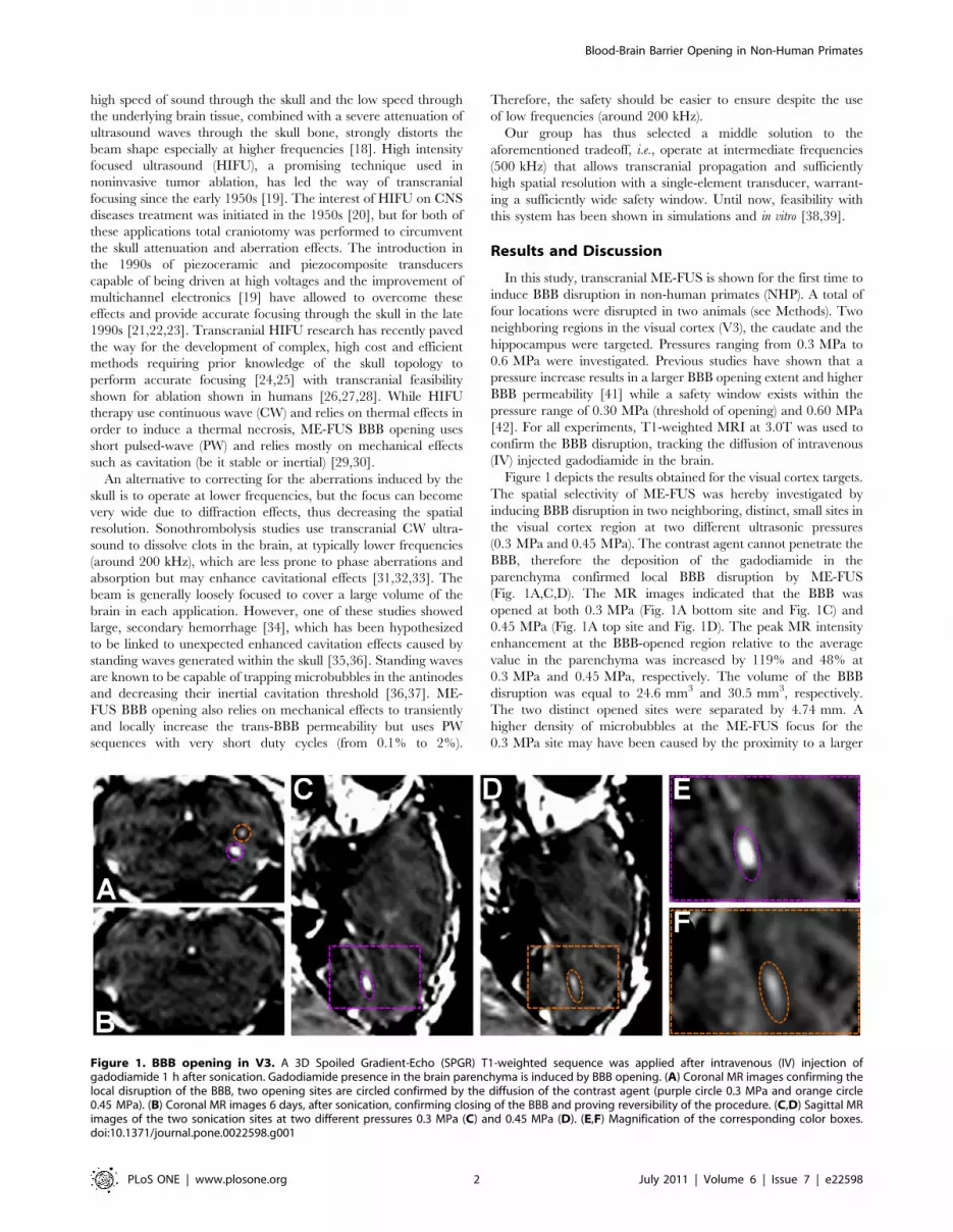
Figure 1 depicts the results obtained for the visual cortex targets.
The spatial selectivity of ME-FUS was hereby investigated by
inducing BBB disruption in two neighboring, distinct, small sites in
the visual cortex region at two different ultrasonic pressures
(0.3 MPa and 0.45 MPa). The contrast agent cannot penetrate the
BBB, therefore the deposition of the gadodiamide in the
parenchyma confirmed local BBB disruption by ME-FUS
(Fig. 1A,C,D). The MR images indicated that the BBB was
opened at both 0.3 MPa (Fig. 1A bottom site and Fig. 1C) and
0.45 MPa (Fig. 1A top site and Fig. 1D). The peak MR intensity
enhancement at the BBB-opened region relative to the average
value in the parenchyma was increased by 119% and 48% at
0.3 MPa and 0.45 MPa, respectively. The volume of the BBB
disruption was equal to 24.6 mm3 and 30.5 mm3, respectively.
The two distinct opened sites were separated by 4.74 mm. A
higher density of microbubbles at the ME-FUS focus for the
0.3 MPa site may have been caused by the proximity to a larger
Figure 1. BBB opening in V3. A 3D Spoiled Gradient-Echo (SPGR) T1-weighted sequence was applied after intravenous (IV) injection ofgadodiamide 1 h after sonication. Gadodiamide presence in the brain parenchyma is induced by BBB opening. (A) Coronal MR images confirming thelocal disruption of the BBB, two opening sites are circled confirmed by the diffusion of the contrast agent (purple circle 0.3 MPa and orange circle0.45 MPa). (B) Coronal MR images 6 days, after sonication, confirming closing of the BBB and proving reversibility of the procedure. (C,D) Sagittal MRimages of the two sonication sites at two different pressures 0.3 MPa (C) and 0.45 MPa (D). (E,F) Magnification of the corresponding color boxes.doi:10.1371/journal.pone.0022598.g001
Blood-Brain Barrier Opening in Non-Human Primates
PLoS ONE | www.plosone.org 2 July 2011 | Volume 6 | Issue 7 | e22598
vessel, explaining the higher MRI contrast enhancement. The
location of the induced BBB disruption areas were shifted from the
expected location of respectively 0.8 mm and 0.7 mm laterally
and 8.1 mm and 7.9 mm axially towards the transducer. The
same MRI sequence and IV contrast agent injection were
repeated six days after BBB opening (Fig. 1B). No intensity
enhancement was observed indicating that the BBB was closed or
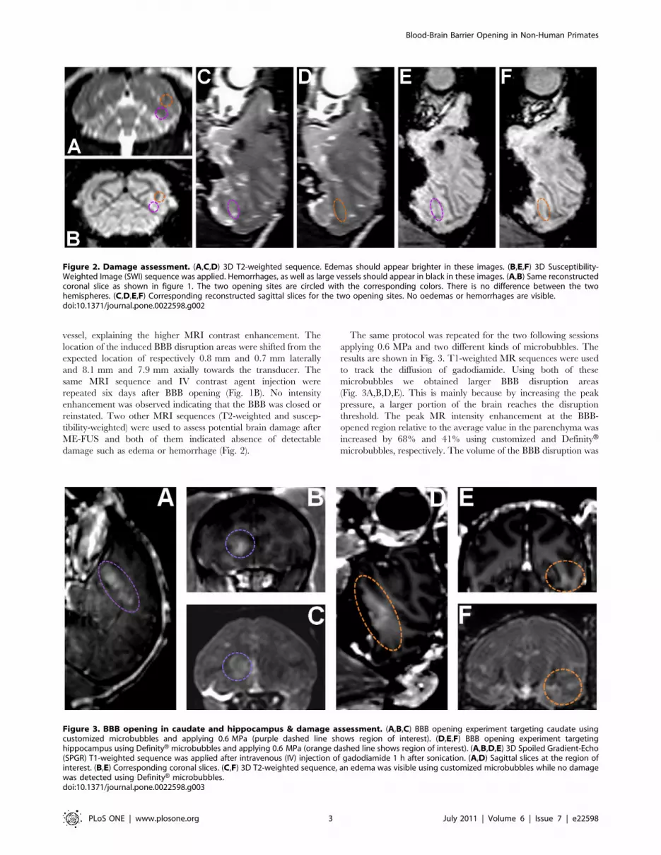
reinstated. Two other MRI sequences (T2-weighted and suscep-
tibility-weighted) were used to assess potential brain damage after
ME-FUS and both of them indicated absence of detectable
damage such as edema or hemorrhage (Fig. 2).
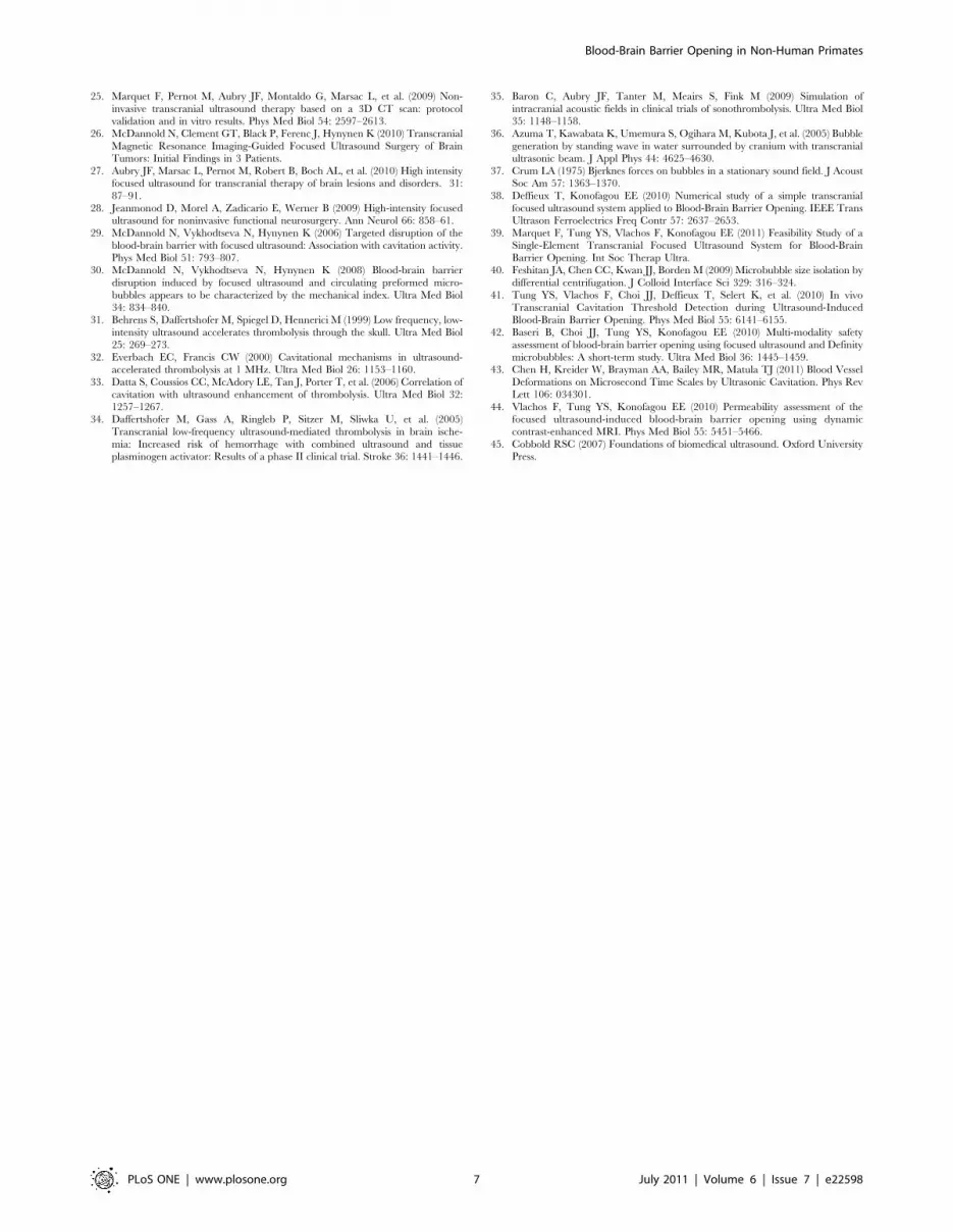
The same protocol was repeated for the two following sessions
applying 0.6 MPa and two different kinds of microbubbles. The
results are shown in Fig. 3. T1-weighted MR sequences were used
to track the diffusion of gadodiamide. Using both of these
microbubbles we obtained larger BBB disruption areas
(Fig. 3A,B,D,E). This is mainly because by increasing the peak
pressure, a larger portion of the brain reaches the disruption
threshold. The peak MR intensity enhancement at the BBB-
opened region relative to the average value in the parenchyma was
increased by 68% and 41% using customized and DefinityHmicrobubbles, respectively. The volume of the BBB disruption was
Figure 2. Damage assessment. (A,C,D) 3D T2-weighted sequence. Edemas should appear brighter in these images. (B,E,F) 3D Susceptibility-Weighted Image (SWI) sequence was applied. Hemorrhages, as well as large vessels should appear in black in these images. (A,B) Same reconstructedcoronal slice as shown in figure 1. The two opening sites are circled with the corresponding colors. There is no difference between the twohemispheres. (C,D,E,F) Corresponding reconstructed sagittal slices for the two opening sites. No oedemas or hemorrhages are visible.doi:10.1371/journal.pone.0022598.g002
Figure 3. BBB opening in caudate and hippocampus & damage assessment. (A,B,C) BBB opening experiment targeting caudate usingcustomized microbubbles and applying 0.6 MPa (purple dashed line shows region of interest). (D,E,F) BBB opening experiment targetinghippocampus using DefinityH microbubbles and applying 0.6 MPa (orange dashed line shows region of interest). (A,B,D,E) 3D Spoiled Gradient-Echo(SPGR) T1-weighted sequence was applied after intravenous (IV) injection of gadodiamide 1 h after sonication. (A,D) Sagittal slices at the region ofinterest. (B,E) Corresponding coronal slices. (C,F) 3D T2-weighted sequence, an edema was visible using customized microbubbles while no damagewas detected using DefinityH microbubbles.doi:10.1371/journal.pone.0022598.g003
Blood-Brain Barrier Opening in Non-Human Primates
PLoS ONE | www.plosone.org 3 July 2011 | Volume 6 | Issue 7 | e22598

equal to 285.5 mm3 and 116.3 mm3, respectively. The BBB
opening regions at the caudate and the hippocampus were shifted
from the targeted location by respectively 0.6 mm and 0.9 mm
laterally and 6.5 mm and 7.2 mm axially. T2-weighted MR
sequences were also used to assess potential damages in the brain
(Fig. 3C,F). An edematous region was detected using custom made
microbubbles while no damage was detected using DefinityH. All
the animals have been survived and therefore histological findings
are not available at this time. Even though no in-depth cognitive
tests have been performed thus far, qualitative assessment of the
animal basic behavior has been monitored. Normal cognitive
behavior has been noted following ME-FUS procedures at
moderate pressures and using DefinityH. In the case of 0.6 MPa
and customized microbubbles, the animal with the edema
exhibited a weakness in the contra-lateral arm over four days
after treatment, most likely due to the induced edema, but then
fully recovered after that four-day period.
Passive cavitation detector (PCD) recordings were performed
during all experiments and are depicted in Fig. 4. Spectrograms
depicted the frequency content of the bubble response during ME-
FUS application and helped classify the cavitation behavior. Using
moderate pressures (Fig. 4A,B), the PCD recordings showed the
nonlinear modes due to the bubble oscillations induced by the
acoustic excitation (stable cavitation). Very little broadband
response was detected. This noise is induced by bubble collapse
and jet, more generally described as inertial cavitation. In those
two cases, the cavitation behavior was mainly dominated by stable
cavitation. While increasing the pressure to 0.6 MPa, a large
broadband signal was recorded for both customized (Fig. 4C) and
DefinityH (Fig. 4D) microbubbles. Previous work from our group
has shown that inertial cavitation is one of the main causes of
induced damage during treatment [41]. We believe that the
discrepancy observed in the last two experiments is mainly due to
the bubble size dependence. A recent study [43] showed that
contrary to current belief, liquid jets are directed away from the
nearest vessel wall. Therefore, using smaller microbubbles, those
jets might not actually be able to puncture and damage the vessels
while using larger microbubbles trapped between vessel walls, jets
are more likely to induce damage. Customized and DefinityHmicrobubbles do not only differ in size. The gas is also different
(perfluorobutane for customized and perfluoropropane for Defi-
nityH) but since the solubility and diffusivity of those two gases are
similar, it is not expected to significantly affect the bubble
oscillation during ME-FUS exposure. The different carbon chains
(DSPC and DPPC, respectively), however, may change the shell
property. The effect of shell property is part of ongoing work in
our lab in order to assess the role of microbubbles in BBB opening.
Achieving drug-independent, localized, reversible and noninva-
sive BBB disruption in non-human primates can pave the way
towards novel brain drug delivery and gene therapy techniques.
The volume of the BBB opening was shown to be small enough
using moderate pressures (24.6 mm3 and 30.5 mm3) to ensure
potential therapeutic agents in untargeted regions. This technique
was also proven to be highly selective with two distinct BBB
opening sites in the visual cortex separated by a distance on the
order of a few millimeters. Increasing the pressure resulted in
larger opening regions (285.5 mm3 and 116.3 mm3). Those
preliminary results have also enlightened the brain site and
microbubbles type dependences. The targeting quality was also
assessed. These results were found to be in good agreement with
previous in silico and in vitro findings [38,39]. These previous studies
showed that as long as we choose a good targeting vector (with an
incidence angle close to normal), the distortion and lateral shift are
very low, the attenuation and axial shift are also reproducible in
vitro (with less than 1 dB and 1 mm of standard deviation
respectively) at the same angle of incidence. The initial in vivo
results suggest that the shift induced by the skull interface may also
be reproducible in vivo under the same angle of incidence
condition. Other targeting vectors might require a more complex
way of determining the position of the focus and the global
attenuation (e.g., simulations based on prior 3D CT skull scans).
Ongoing work will be achieved in order to assess those
dependences in a statistically significant way. At this point, no
quantification of the contrast agent diffusion was performed. In
future work, a permeability quantification technique reported in
mice by our group [44] will be applied as part of the primate
study.
In conclusion, initial feasibility of noninvasive, highly selective,
drug-independent and reversible BBB opening was demonstrated
in non-human primates in vivo for the first time. High spatial
selectivity of this technique was also shown. This study is a major
step toward clinical translation of this emerging technology that
can be combined with any type of pharmacological treatment to
the brain. Ongoing investigations entail optimization of the
Figure 4. PCD recordings during ME-FUS exposure. The corresponding spectrograms of the first pulse with microbubbles administrationshows that very few broadband acoustic emissions are detected at 0.3 MPa (A) and 0.45 MPa (B) using customized microbubbles. This suggests thatin those cases, the mechanism is mainly dominated by stable cavitation. Increasing the pressure to 0.6 MPa, using both customized (C) and DefinityH(D) microbubbles, a large amplitude broadband signal (4–8 MHz) is detected which is the signature of inertial cavitation.doi:10.1371/journal.pone.0022598.g004
Blood-Brain Barrier Opening in Non-Human Primates
PLoS ONE | www.plosone.org 4 July 2011 | Volume 6 | Issue 7 | e22598

procedure including safety and efficacy of the trans-BBB drug
delivery.
Materials and Methods
Initial feasibility studies were performed on two male rhesus
macaques over the course of three sessions. In the first two
sessions, the monodispersed 4–5 mm microbubbles were manu-
factured in-house and size-isolated using differential centrifugation
[40] while in the last session DefinityH microbubbles were used
(Lantheus Medical Imaging, MA, polydispersed, mean diameter
1.1–3.3 mm, 98% below 10 mm, maximum diameter 20 mm). In
the first session, a region of the visual cortex (ventral V3), i.e., the
medio-ventral wall of the occipital cortex, was targeted. In the
second animal (second session), the caudate was targeted and the
pressure was increased to 0.6 MPa to investigate potential damage
induced using customized microbubbles. The third and final
session was performed in the first animal targeted the hippocam-
pus using 0.6 MPa and DefinityH microbubbles.
A 500-kHz center frequency focused ultrasound transducer was
used for this experiment (Riverside Research Institute, NY, USA).
The acoustic parameters used for the three protocols were the
following: focal maximum estimated pressure of 0.3 MPa and
0.45 MPa, pulse length of 10 ms, pulse repetition frequency of
2 Hz, total sonication duration of 2 min. The single-element
transducer was mounted on a standard monkey stereotactic frame
for accurate positioning (Fig. 5). In vitro pressure measurements
were realized in another study [39]. This study determined the
global attenuation (absorption, reflexion and scattering) due to the
presence of the skull (around -5.7 dB at 500 kHz). The attenuation
in the skin [45] was assumed to be around 20.9 dB.cm21 and its
thickness was estimated to be equal to 0.5 cm. The attenuation in
the monkey brain tissue [45] was assumed to be around
20.5 dB.cm21 and the thickness of this layer was estimated to
be equal to 2 cm. Therefore, the emission amplitude has to be
raised by 7.15 dB (approximately a factor 2.28) compared to the
calibration measurements in water to compensate for the energy
loss along the path.
The first animal had previously participated in several
electrophysiological and fMRI experiments. It had a surgically
implanted head post to restrain head movements. The head post
was embedded within a dental-acrylic implant which was held in
place by ceramic screws. The screws penetrated the skull plate but
did not protrude more than a millimeter into the skull-cavity. A
previously implanted scleral search coil had been removed prior to
the experiments reported here. During the electrophysiological
experiments, single unit activity was recorded from the frontal eye
fields of both hemispheres. No recording from the animal’s brain
targeted regions had been previously performed. The dental-
acrylic implant covered a large portion of the skull. However, the
occipital pole of the skull was not covered and enabled a sufficient
acoustic window to the visual cortex without interference from the
implant.
For the application of the FUS, all animals were anesthetized
with 2% isoflurane (carrier gas: oxygen). The heart rate was held
at approximately 120 beats per minute and the respiratory rate at
around 60 breaths per minute. Prior to sonication, the scalp hair
was removed with a depilatory cream to ensure maximal acoustic
transmission. The animal’s head was then placed in a stereotactic
frame to enable careful targeting of the ultrasound. The sonication
was performed immediately after intravenous (IV) injection of
500 mL microbubbles for all experiments (56109 numbers/mL for
customized microbubbles and 1.261010 numbers/mL for Defini-
tyH). Targeting was ensured using a manipulator and a positioning
rod indicating the position of the focus relatively to the stereotactic
coordinates (Fig. 6). Targeted regions of visual cortex (V3) were
determined using a monkey brain atlas.
MRI was used to confirm BBB opening using gadiodiamide
contrast agent. 3D Spoiled Gradient-Echo (SPGR) T1-weighted
sequences (TR/TE = 20/1.4 ms; flip angle: 30u; NEX = 2; spatial
resolution: 5006500 mm2; slice thickness: 1 mm with no interslice
gap) were applied after intravenous (IV) injection of gadodiamide
(OmniscanH, molecular weight 573.66 Da, GE Healthcare,
Princeton, NJ, USA) 1 h after sonication. The dose applied was
0.2 mL/kg and the IV injection was performed 2 minutes before
the SPGR T1-weigthed scan (scan duration: 18 minutes).
Gadodiamide presence in the brain parenchyma was induced by
BBB opening. 3D T2-weighted sequence (TR/TE = 3000/80; flip
angle: 90u; NEX = 3; spatial resolution: 4006400 mm2; slice
thickness: 2 mm with no interslice gap) and 3D Susceptibility-
Weighted Image (SWI) sequence were applied (TR/TE = 19/
27 ms; flip angle: 15u; NEX = 1; spatial resolution: 4006400 mm2;
slice thickness: 1 mm with no interslice gap) and were used to
assess brain damage.
A single-element PCD (center frequency: 7.5 MHz, focal
length: 60 mm, Olympus NDT, Waltham, MA, USA) was
positioned through the center hole of the FUS transducer. The
two transducers were aligned so that their focal regions fully
Figure 5. Experimental setup for in vivo FUS-induced BBB opening in the operating room. (A) A single-element, circular focusedultrasound transducer with a hole in the center was driven by a function generator (Agilent Technologies, Palo Alto, CA, USA) through a 50-dB poweramplifier (ENI Inc., Rochester, NY, USA). The center frequency, focal depth, outer radius and inner radius of FUS were 500 kHz, 90 mm, 30 mm and11.2 mm, respectively. (B) Closer view of the transducer mounted on the stereotactic frame with a manipulator allowing precise positioning of thetransducer in the stereotactic referential. (C) Monkey placed in the stereotactic frame. The monkey head is shaved and a degassed echographic gelcontainer is placed on the top of its head to insure maximal acoustic transmission.doi:10.1371/journal.pone.0022598.g005
Blood-Brain Barrier Opening in Non-Human Primates
PLoS ONE | www.plosone.org 5 July 2011 | Volume 6 | Issue 7 | e22598

overlapped within the confocal volume. The PCD transducer,
which was connected to a digitizer (Gage Applied Technologies,
Inc., Lachine, QC, Canada) through a 20-dB amplification (5800,
Olympus NDT, Waltham, MA, USA), was used to passively
acquire acoustic emissions from microbubbles. A time-frequency
map of the acoustic emission was generated using a customized
spectrogram function (8-cycles, i.e., 16 ms, Chebyshev window;
98% overlap; 4096-point FFT) in MATLABH (2010a, Mathworks,
Natick, MA). The spectrogram can then clearly indicate how the
frequency content of the signal changes over time. Therefore, the
presence of broadband response can help classify cavitation
behavior.
Ethical statementAll animal studies were approved by the Institutional Animal
Care and Use Committee at Columbia University and the New
York State Psychatric Institute (protocol ID: CU1066, NYSPI279).
All animals, housed and handled in strict accordance with good
animal practice under supervision of veterinarians, received
environmental enrichment and were monitored for evidence of
disease and changes in attitude, appetite, or behavior suggestive of
illness. Every effort was made to alleviate animal discomfort and
pain by appropriate and routine use of anesthetic and/or analgesic
agents.
Acknowledgments
The authors especially wish to thank the Riverside Research Institute for
providing the transducer that was used in this study; Mark Borden, Ph.D.,
and Jameel A. Feshitan, for the microbubble manufacture; and Fotios
Vlachos, Ph.D., and Stephen M. Dashnaw, Department of Biomedical
Engineering, Columbia University, for their help in MRI scanning. The
authors appreciate Girma Asfaw, Department of Neuroscience, Columbia
University, for his surgical assistance.
Author Contributions
Conceived and designed the experiments: FM YST TT. Performed the
experiments: FM YST TT. Analyzed the data: FM EEK. Wrote the paper:
FM VPF EEK. Overall project supervision: EEK VPF.
References
1. Dove A (2008) Breaching the barrier. Nat Biotechnol 26: 1213–1215.
2. Pardridge WM (2005) The blood-brain barrier: bottleneck in brain drug
development. NeuroRX 2: 3–14.
3. Pardridge WM (2007) Blood-brain barrier delivery. Drug Discov Today 12:
54–61.
4. Foust KD, Nurre E, Montgomery C L, Hernandez A, Chan CM, et al. (2008)
Intravascular AAV9 preferentially targets neonatal neurons and adult astrocytes.Nat Biotechnol 27: 59–65.
5. Doolittle ND, Miner ME, Hall WA, Siegal T, Hanson Jm, et al. (2000) Safetyand efficacy of a multicenter study using intraarterial chemotherapy in
conjunction with osmotic opening of the blood-brain barrier for the treatment
of patients with malignant brain tumors. Cancer 88: 637–647.
6. Fortin D, Gendron C, Boudrias M, Garant MP (2007) Enhanced chemotherapy
delivery by intraarterial infusion and blood-brain barrier disruption in thetreatment of cerebral metastasis. Cancer 109: 751–760.
7. Mychaskiw G, Badr AE, Tibbs R, Clower BR, Zhang JH (2000) Optison(FS069) Disrupts the Blood-Brain Barrier in Rats. Anesth Analg 91: 798–803.
8. Hynynen K, McDannold N, Vykhodtseva N, Jolesz FA (2001) Noninvasive MRImaging–guided Focal Opening of the Blood-Brain Barrier in Rabbits.
Radiology 220: 640–646.
9. Choi JJ, Pernot M, Small SA, Konofagou EE (2007) Non-invasive,Transcranial,
and Localized Opening of the Blood-Brain Barrier in Mice using FocusedUltrasound - A Feasibility Study. Ultra Med Biol 33: 95–104.
10. Sheikov N, McDannold N, Vykhodtseva N, Jolesz F, Hynynen K (2004) Cellularmechanisms of the blood-brain barrier opening induced by ultrasound in
presence of microbubbles. Ultrasound Med Biol 2004; 30: 979–989.
11. Mandel RJ (2010) CERE-110, an adeno-associated virus-based gene delivery
vector expressing human nerve growth factor for the treatment of Alzheimer’s
disease. Curr Opin Mol Ther 12: 240–247.
12. Fiske BK, Frasier MA, Sherer TB (2008) Special focus section: gene therapy forParkinson’s disease. Exp Neurol 209: 28–29.
13. Tuszynski MH (2007) Nerve growth factor gene therapy in Alzheimer diseaseAlz Dis Assoc. Disord 2: 179–189.
14. Jordao JF, Ayala-Grosso CA, Markham K, Huang Y, Chopra R, et al. (2010)PLoS One 5: e10549.
15. Berti V, Osorio RS, Mosconi L, Li Y, De Santi S, et al. (2010) Early Detection of
Alzheimer’s Disease with PET Imaging. Neurodegenerative Dis 7: 131–135.
16. Abbott NJ, Ronnback L, Hansson E (2006) Astrocyte–endothelial interactions at
the blood–brain barrier. Nat Rev Neuro 7: 41–53.
17. Habgood MD, Begley DJ, Abbott NJ (2000) Determinants of passive drug entry
into the central nervous system. Cell Mol Neurobiol 20: 231–253.
18. Fry FJ (1977) Transkull transmission of an intense focused ultrasonic beam.
Ultra Med Biol 3: 179–181.
19. Fry WJ, Mosberg WH, Jr., Barnard JW, Fry FJ (1954) Production of focaldestructive lesions in the central nervous system with ultrasound. J Neurosurg
11: 471–478.
20. Haar GT, Coussios C (2007) High intensity focused ultrasound: past, present
and future. Int J Hyperthermia 23: 85–87.
21. Tanter M, Thomas JL, Fink M (1998) Focusing and steering through absorbing
and aberrating layers: Application to ultrasonic propagation through the skull.J Acoust Soc Am 103: 2403–2410.
22. Hynynen K, Jolesz FA (1998) Demonstration of potential noninvasiveultrasound brain therapy through an intact skull. Ultra Med Biol 24: 275–283.
23. Clement GT, Sun J, Giesecke T, Hynynen K (2000) A hemisphere array fornon-invasive ultrasound brain therapy and surgery. Phys Med Biol 45:
3707–3719.
24. Clement GT, Hynynen K (2002) A non-invasive method for focusing ultrasound
through the human skull. Phys Med Biol 47: 1219–1236.
Figure 6. Targeting procedure for in vivo FUS-induced BBB opening. (A) A positioning rod (black arrow), indicating the position of the focus(5 cm away from the edge of the transducer), was used to target. (B) This positioning rod was mounted on the manipulator in order to locate theorigin of the stereotactic coordinates. (C) The origin of the stereotactic coordinates indicated by the engraved cross on the metal piece between theear-bars is targeted with the tip of the positioning rod.doi:10.1371/journal.pone.0022598.g006
Blood-Brain Barrier Opening in Non-Human Primates
PLoS ONE | www.plosone.org 6 July 2011 | Volume 6 | Issue 7 | e22598
25. Marquet F, Pernot M, Aubry JF, Montaldo G, Marsac L, et al. (2009) Non-
invasive transcranial ultrasound therapy based on a 3D CT scan: protocolvalidation and in vitro results. Phys Med Biol 54: 2597–2613.
26. McDannold N, Clement GT, Black P, Ferenc J, Hynynen K (2010) Transcranial
Magnetic Resonance Imaging-Guided Focused Ultrasound Surgery of BrainTumors: Initial Findings in 3 Patients.
27. Aubry JF, Marsac L, Pernot M, Robert B, Boch AL, et al. (2010) High intensityfocused ultrasound for transcranial therapy of brain lesions and disorders. 31:
87–91.
28. Jeanmonod D, Morel A, Zadicario E, Werner B (2009) High-intensity focusedultrasound for noninvasive functional neurosurgery. Ann Neurol 66: 858–61.
29. McDannold N, Vykhodtseva N, Hynynen K (2006) Targeted disruption of theblood-brain barrier with focused ultrasound: Association with cavitation activity.
Phys Med Biol 51: 793–807.30. McDannold N, Vykhodtseva N, Hynynen K (2008) Blood-brain barrier
disruption induced by focused ultrasound and circulating preformed micro-
bubbles appears to be characterized by the mechanical index. Ultra Med Biol34: 834–840.
31. Behrens S, Daffertshofer M, Spiegel D, Hennerici M (1999) Low frequency, low-intensity ultrasound accelerates thrombolysis through the skull. Ultra Med Biol
25: 269–273.
32. Everbach EC, Francis CW (2000) Cavitational mechanisms in ultrasound-accelerated thrombolysis at 1 MHz. Ultra Med Biol 26: 1153–1160.
33. Datta S, Coussios CC, McAdory LE, Tan J, Porter T, et al. (2006) Correlation ofcavitation with ultrasound enhancement of thrombolysis. Ultra Med Biol 32:
1257–1267.34. Daffertshofer M, Gass A, Ringleb P, Sitzer M, Sliwka U, et al. (2005)
Transcranial low-frequency ultrasound-mediated thrombolysis in brain ische-
mia: Increased risk of hemorrhage with combined ultrasound and tissueplasminogen activator: Results of a phase II clinical trial. Stroke 36: 1441–1446.
35. Baron C, Aubry JF, Tanter M, Meairs S, Fink M (2009) Simulation of
intracranial acoustic fields in clinical trials of sonothrombolysis. Ultra Med Biol35: 1148–1158.
36. Azuma T, Kawabata K, Umemura S, Ogihara M, Kubota J, et al. (2005) Bubble
generation by standing wave in water surrounded by cranium with transcranialultrasonic beam. J Appl Phys 44: 4625–4630.
37. Crum LA (1975) Bjerknes forces on bubbles in a stationary sound field. J AcoustSoc Am 57: 1363–1370.
38. Deffieux T, Konofagou EE (2010) Numerical study of a simple transcranial
focused ultrasound system applied to Blood-Brain Barrier Opening. IEEE TransUltrason Ferroelectrics Freq Contr 57: 2637–2653.
39. Marquet F, Tung YS, Vlachos F, Konofagou EE (2011) Feasibility Study of aSingle-Element Transcranial Focused Ultrasound System for Blood-Brain
Barrier Opening. Int Soc Therap Ultra.40. Feshitan JA, Chen CC, Kwan JJ, Borden M (2009) Microbubble size isolation by
differential centrifugation. J Colloid Interface Sci 329: 316–324.
41. Tung YS, Vlachos F, Choi JJ, Deffieux T, Selert K, et al. (2010) In vivoTranscranial Cavitation Threshold Detection during Ultrasound-Induced
Blood-Brain Barrier Opening. Phys Med Biol 55: 6141–6155.42. Baseri B, Choi JJ, Tung YS, Konofagou EE (2010) Multi-modality safety
assessment of blood-brain barrier opening using focused ultrasound and Definity
microbubbles: A short-term study. Ultra Med Biol 36: 1445–1459.43. Chen H, Kreider W, Brayman AA, Bailey MR, Matula TJ (2011) Blood Vessel
Deformations on Microsecond Time Scales by Ultrasonic Cavitation. Phys RevLett 106: 034301.
44. Vlachos F, Tung YS, Konofagou EE (2010) Permeability assessment of thefocused ultrasound-induced blood-brain barrier opening using dynamic
contrast-enhanced MRI. Phys Med Biol 55: 5451–5466.
45. Cobbold RSC (2007) Foundations of biomedical ultrasound. Oxford UniversityPress.
Blood-Brain Barrier Opening in Non-Human Primates
PLoS ONE | www.plosone.org 7 July 2011 | Volume 6 | Issue 7 | e22598