nonclassical pathway of pseudomonas aeruginosa dna-induced interleukin8 secretion in cystic fibrosis...
TRANSCRIPT
10.1128/IAI.74.5.2975-2984.2006.
2006, 74(5):2975. DOI:Infect. Immun. Mónica A. Delgado, Jens F. Poschet and Vojo Deretic Epithelial CellsSecretion in Cystic Fibrosis Airway
DNA-Induced Interleukin-8aeruginosaPseudomonasNonclassical Pathway of
http://iai.asm.org/content/74/5/2975Updated information and services can be found at:
These include:
REFERENCEShttp://iai.asm.org/content/74/5/2975#ref-list-1at:
This article cites 70 articles, 36 of which can be accessed free
CONTENT ALERTS more»articles cite this article),
Receive: RSS Feeds, eTOCs, free email alerts (when new
http://journals.asm.org/site/misc/reprints.xhtmlInformation about commercial reprint orders: http://journals.asm.org/site/subscriptions/To subscribe to to another ASM Journal go to:
on Novem
ber 4, 2013 by guesthttp://iai.asm
.org/D
ownloaded from
on N
ovember 4, 2013 by guest
http://iai.asm.org/
Dow
nloaded from
on Novem
ber 4, 2013 by guesthttp://iai.asm
.org/D
ownloaded from
on N
ovember 4, 2013 by guest
http://iai.asm.org/
Dow
nloaded from
on Novem
ber 4, 2013 by guesthttp://iai.asm
.org/D
ownloaded from
on N
ovember 4, 2013 by guest
http://iai.asm.org/
Dow
nloaded from
on Novem
ber 4, 2013 by guesthttp://iai.asm
.org/D
ownloaded from
on N
ovember 4, 2013 by guest
http://iai.asm.org/
Dow
nloaded from
on Novem
ber 4, 2013 by guesthttp://iai.asm
.org/D
ownloaded from
on N
ovember 4, 2013 by guest
http://iai.asm.org/
Dow
nloaded from
on Novem
ber 4, 2013 by guesthttp://iai.asm
.org/D
ownloaded from
INFECTION AND IMMUNITY, May 2006, p. 2975–2984 Vol. 74, No. 50019-9567/06/$08.00�0 doi:10.1128/IAI.74.5.2975–2984.2006Copyright © 2006, American Society for Microbiology. All Rights Reserved.
Nonclassical Pathway of Pseudomonas aeruginosa DNA-InducedInterleukin-8 Secretion in Cystic Fibrosis Airway Epithelial Cells
Monica A. Delgado, Jens F. Poschet, and Vojo Deretic*Department of Molecular Genetics and Microbiology, University of New Mexico Health Sciences Center,
915 Camino de Salud NE, Albuquerque, New Mexico 87131
Received 19 December 2005/Accepted 30 January 2006
Pseudomonas aeruginosa is a critical colonizer of the respiratory tract in cystic fibrosis. The chronic infections withthis microorganism contribute to excessive inflammation and progressive lung damage in cystic fibrosis patients.The full repertoire of Pseudomonas products that promote inflammation in the cystic fibrosis lung is not known.Here we show that P. aeruginosa DNA released from the bacterium, but not human DNA from epithelial cells orEscherichia coli DNA, displays proinflammatory properties and induces human respiratory epithelial cells to secreteinterleukin-8 (IL-8), a key chemokine causing excessive neutrophil infiltration in the cystic fibrosis lung. IL-8secretion was not due to an increase in NF-�B- or activator protein-1-dependent IL-8 promoter transcription, butinstead depended on p38 and Erk mitogen-activated protein kinases. No secretion of IL-8 was observed usingconventional Toll-like receptor 9 ligands (CpG oligonucleotides), although it could be demonstrated that parts ofthe Toll-like receptor 9-signaling pathway were functional, since class B and C CpG oligonucleotide ligandsstimulated production of RANTES chemokine. The IL-8 secretion in response to P. aeruginosa DNA was decreasedby treatments that inhibit acidification of intracellular organelles, using chloroquine, a pH-neutralizing compound,or bafilomycin A1, an inhibitor of vacuolar H�-ATPase. These data indicate that DNA released from P. aeruginosaduring chronic infections may significantly contribute to the proinflammatory processes in cystic fibrosis. Ourfindings also show that treatments with drugs diminishing organellar acidification may reduce the inflammatoryresponse in cystic fibrosis.
Cystic fibrosis (CF) is the most common autosomal recessivedisease in Caucasians and is caused by mutations in the geneencoding the cystic fibrosis transmembrane conductance regu-lator (CFTR) (57). This complex disease is characterized bychronic bacterial infection and inflammation of the airways,pancreatic exocrine insufficiency, gastrointestinal involvement,infertility in males, and inefficient salt absorption from sweatfluids (57). The most common cause of death in CF is respi-ratory failure as a result of progressive pulmonary decline (47)due to chronic infections and inflammation (24). It has notbeen resolved whether the excessive inflammation is simply aresponse to persistent infection (6) or whether the primarydefect in CFTR directly contributes to the intrinsically higherexcessive proinflammatory response (36) by CF respiratory epi-thelial cells (41). Regardless of the exact etiology, the chroniclung inflammation remains largely responsible for the life-shortening effects of CF. A hallmark of CF airway inflamma-tion is accumulation of interleukin-8 (IL-8) acting as a chemo-kine, attracting polymorphonuclear cells/neutrophils, which inturn contribute to tissue destruction (8, 36, 38, 45).
The predominant lung pathogen in CF is Pseudomonasaeruginosa, cultured in specimens from more than 80% of CFpatients aged 26 years or older (24). Many P. aeruginosa prod-ucts, including lipoproteins, flagella, exopolysaccharide al-ginate, pili, lipopolysaccharide (LPS), and toxins (1, 14, 17, 20,27), belong in the general group of pathogen-associated mo-lecular patterns (PAMPs). PAMPs reflect conserved microbial
molecular features and are recognized by host cells via patternrecognition receptors (PRRs), which discriminate between selfand pathogen products (34, 48). Toll-like receptors (TLRs)and nucleotide-binding oligomerization domain (Nod) pro-teins are two classes of PRRs involved in innate immune de-tection (4). Signaling from these receptors can result in acti-vation of mitogen-activated protein kinases (MAPKs), NF-�B,or type I interferon pathways (4, 48). Pseudomonas challengeof human tracheobronchial epithelial cell cultures causesMAPK activation, besides NF-�B, and is associated with IL-8secretion (65). MAPK activation by PAMPs has been demon-strated with various ligands, including bacterial lipopeptides,LPS, flagellin, and CpG DNA (2, 3, 23, 29, 58, 67, 69). Thereare different patterns of MAPK activation, depending upon thestimulus and cell type, with p38 being the most commonMAPK activated upon engagement of TLRs.
Signals from various PRRs are essential for innate immunityagainst P. aeruginosa (61). For example, TLR4 plays a protectiverole against cytotoxic P. aeruginosa (19). Although P. aeruginosaLPS is a relatively weak TLR4 agonist (18), LPS modificationsseem to modulate its potency (27). Pseudomonas flagella acti-vate airway epithelia via TLR5 or through TLR2 in coopera-tion with aGM1 (1). Pseudomonas ExoS stimulates both TLR2and TLR4 (17), while alginate rich in mannuronate may too actvia TRL2 and TLR4 (21). However, when innate immunityclearance fails, a continuing stimulation of PRRs can becomea double-edged sword. We have recently shown that uponconversion to the mucoid, alginate-overproducing phenotypetypically encountered in CF, P. aeruginosa upregulates a largenumber of lipoproteins, termed lipotoxins (20), which furtheraugment proinflammatory proteins. A P. aeruginosa lipotoxinhas also been shown to play a role in the allergic response via
* Corresponding author. Mailing address: Department of MolecularGenetics and Microbiology, University of New Mexico Health SciencesCenter, 915 Camino de Salud NE, Albuquerque, NM 87131. Phone:(505) 272-0291. Fax: (505) 272-5309. E-mail: [email protected].
2975
prolonged stimulation of the TLR2 and TLR4 signaling path-ways (55). Interestingly, in the absence of normal recognitionof P. aeruginosa pili by TLR2, there is a strong hyperreactiveresponse modulated by TLR4 (42), attesting to the contribu-tion of PAMPs and PRRs to unproductive and potentiallydamaging inflammatory responses.
PRRs seem to be equally expressed in CF and non-CF res-piratory epithelial cells, and their responses appear mainlyindistinguishable (25, 46), with the exception of aGM1-aug-mented effects on TLR2 signaling in CF cells (1, 46, 62). Ingeneral, it is possible that continuous stimulation with bacterialproducts, instead of helping clear the microbes, stokes therunaway inflammation in CF. Although a number of candidateP. aeruginosa PAMPs have been identified (1, 17, 20, 27, 55,62), the full repertoire remains to be assessed, and it is notknown at present whether even-more-potent stimulators ofinflammation are generated by P. aeruginosa. Due to a largenumber of bacterial cells generally found in the CF lung, it iscertain that the DNA released from the dying bacteria may actin a way similar to CpG oligonucleotides. The CpG oligonu-cleotides have been used to study TLR9 signaling, as theymimic the absence of CpG methylated motifs in bacterial chro-mosomal DNA (30, 40). In this work we explored the possi-bility that P. aeruginosa DNA, due to its high GC content(66.6%) and hence overabundance of unmethylated CpG mo-tifs, may act as a proinflammatory P. aeruginosa product. Wetested whether P. aeruginosa DNA stimulated IL-8 secretionfrom human airway epithelial cells and investigated the signal-ing pathways controlling these processes.
MATERIALS AND METHODS
Bacterial strains and growth conditions. Nonmucoid P. aeruginosa PAO1 wasfrom a stock originally received from B. Holloway. Mucoid P. aeruginosa PA578I(Alg�; mucA22) has the most common mucA mutation found in mucoid CFisolates (9). Bacteria were grown in L broth.
Cell culture. The bronchial epithelial cell lines IB3-1 (�F508/W1282X hetero-zygote) and S9 (created by correcting IB3-1 cells via introduction of a functionalCFTR) (16) were grown on LHC-8 medium (Bio-fluids, Rockville, MD) supple-mented with 10% fetal bovine serum and antibiotics. Primary normal humanbronchial epithelial (NHBE) cells were obtained from a commercial source(Cambrex Bio Science, Baltimore, MD) without identifiers (exempt status fromthe Institutional Review Board and NIH). NHBE cells were cultured in serum-free bronchial epithelial growth medium with BEGM Single Quots supplements(Cambrex Bio Science). CuFi1 cells, derived from a CF patient, were developedby J. Zabner (70) and were cultured in collagen (Sigma, St. Louis, MO)-coatedflasks in serum-free bronchial epithelial growth medium supplemented with BEGMSingle Quots (Cambrex Bio Science). All cells were grown at 37°C in 5% CO2.Submerged cultures of respiratory epithelial cells have been used as reported rou-tinely in studies addressing immunological responses. All control samples containedcarrier solvent added in equivalent amounts to the treatment specimens.
Genomic DNA preparations and CpG oligonucleotides. DNAs were preparedusing a Wizard genomic DNA purification kit (Promega). All DNAs were sus-pended in DNase-free, LPS-free distilled water. DNA preparations were dena-tured before stimulation by heating at 95°C for 5 min, followed by rapid coolingon ice. For some experiments, DNA was methylated with 2 U/�g DNA of CpGmethylase M.SssI (New England Biolabs) for 4 h at 37°C. When indicated, DNAwas digested with DNase I (Novagen) in 20 mM Tris-HCl, pH 7.5, containing 2mM MgCl2 and 1 U DNase I/�g DNA for 1 h at 37°C, and DNase I wasinactivated by heating for 5 min at 95°C. Human DNA was prepared fromrespiratory epithelial cells. CpG oligonucleotides were obtained from IntegratedDNA Technologies (Coralville, IA): CpG A D19, ggTGCATCGATGCAGggggG (63); CpG B 2006, tcgtcgttttgtcgttttgtcgtT (28); and CpG C 2395, tcgtcgttttcggcgcgcgccG (64). (Lowercase letters indicate phosphorothioate linkages; cap-ital letters indicate phosphodiester linkage 3� of the base.)
Transfections and luciferase reporter assays. For monitoring transient NF-�B, pAP-1, or pIL-8 activation, the IB3-1 cell line was seeded at 2 � 104 cells per
well in a 96-well plate, and 18 to 22 h later cells were transfected using Effectenetransfection reagent (QIAGEN) with 48 ng of an NF-�B-responsive luciferasereporter construct (pBVIII-luc, containing three tandem repeats of two NF-�Bsites; from G. Nunez, University of Michigan Medical School), 16 ng of a�-galactosidase construct (pEF1-Bos; generously provided by G. Nunez, Univer-sity of Michigan Medical School) for normalization, and 36 ng of pCDNA3, or 60ng of pAP-1-Luc plasmid (Stratagene), 20 ng of a �-galactosidase construct fornormalization, and 20 ng of pCDNA3, or 75 ng of pIL-8:luc reporter plasmid(containing the 5�-flanking region of the IL-8 gene from �133 to �50; from N.Mukaida, Kanazawa University, Ishikawa, Japan), and 25 ng of a �-galactosidaseconstruct for normalization. The positive control for AP-1-transfected cells wascotransfection with the pFC-MEKK plasmid (Stratagene), transfecting with 60ng of pAP-1-Luc plasmid, 20 ng of a �-galactosidase construct, 12 ng of pCDNA3plasmid, and 8 ng of pFC-MEKK plasmid. Transfection mixtures were placed onthe cells for 6 h, after which the transfection mixture was removed and replacedwith complete growth medium. At 24 h following transfection, cells were incu-bated with growth medium, serum free, for 21 h and then cells were incubatedwith stimulus in serum-free medium for 2, 4, 6, 8, or 24 h. Luciferase activity wasassayed using the luciferase reagent (Promega), and �-galactosidase activity wasassayed using the Galacto-Star luminescence system (Tropix, Bedford, MA).Transfection efficiency was controlled by standardizing luciferase activity to con-stitutive �-galactosidase production. Stimuli were human tumor necrosis factoralpha (TNF-) at 20 ng/ml (Sigma Chemical Co.) or DNA at 25 �g/ml.
Ratiometric fluorescence microscopy, pH measurements, and endosomal pHmanipulation. Cellubrevin-pHluorin (pH-sensitive green fluorescent protein[GFP]) was from J. Rothman (43). Cells were transfected with 1 �g/ml DNAusing Lipofectin for 6 h at 37°C, 5% CO2. Ratiometric fluorescence microscopywas carried out using an Olympus IX-70 microscope and Olympix KAF1400charge-coupled-device camera (LSR). The ratio of emission at 508 nm uponexcitation at 410 nm versus 470 nm and calibration curves were generated aspreviously described (52, 53). For pH normalization, cells were grown in thepresence of 0.1 mM chloroquine, as previously described (52, 53).
NF-�B nuclear translocation assay and confocal microscopy. Respiratory cellswere seeded in a 24-well plate and 18 to 22 h later incubated in serum-freeculture medium for an additional 21 h. Cells were stimulated for the indicatedtimes with medium alone (control), 20 ng/ml TNF-, or 25 �g/ml PAO1 DNA.Cells were washed three times with phosphate-buffered saline (PBS), fixed with2% paraformaldehyde for 20 min, washed three times with PBS, permeabilizedwith 0.2% saponin for 5 min, washed three times with PBS, blocked for 30 min,and incubated with monoclonal antibodies against p65 NF-�B (Santa Cruz)overnight. Slides were washed three times with PBS, incubated with a secondaryanti-mouse fluorescein isothiocyanate-conjugated antibody, washed, incubatedwith 300 nM 4�,6�-diamidino-2-phenylindole (DAPI; Molecular Probes) for 5min, and finally washed and mounted in mounting medium and examined byconfocal microscopy using a Zeiss 510 META microscope. Overlaps betweenDAPI and NF-�B stains were scored as positive nuclear translocation events.
IL-8 and RANTES assays. To analyze IL-8 protein expression, 2 � 104 cells/well (IB3-1, S9, or NHBE cells) or 1.5 � 104 cells/well (CuFi1) were seeded in96-well plates, and 18 to 22 h later cells were incubated with serum-free growthmedium for 21 h (IB3-1 and S9 cells) and then cultured in the presence andabsence of 20 ng/ml human TNF- (Sigma Chemical Co.), 10 ng/ml P. aeruginosaLPS (Sigma Chemical Co.), 5 to 30 �g/ml DNA, or 3 �M CpG oligonucleotidesfor the specific indicated time. CuFi1 and NHBE primary cells were stimulated18 to 22 h after seeding them, using the growth medium with supplements. Afterculture, IL-8 was measured in supernatants using an enzyme-linked immunosor-bent assay (ELISA) kit for IL-8 (R&D Systems, Minneapolis, MN). ELISAswere performed according to the manufacturer’s protocol. When indicated,preincubation (1 h at 37°C) of DNAs or LPS with 10 �g/ml polymyxin B (SigmaChemical Co.) was used to abrogate the effect of LPS. When indicated, 15 �g/mlcycloheximide was added to cells 2 h before stimulation. When indicated, theMAPK inhibitors were added to cells 2 h (SB203580 and PD98059 [Calbiochem])or 30 min (SP600125 [A.G. Scientific, Inc., California]) before stimulation at theindicated concentrations. When indicated, 20 �M chloroquine (Sigma ChemicalCo.) or 30 and 300 nM bafilomycin A1 (Calbiochem) was added to the cells 30min before stimulation. RANTES was assayed using culture supernatants from2 � 104 cells/well seeded in a 96-well plate for 18 to 22 h. Adherent cells wereincubated in serum-free growth medium for 21 h and then cultured in the presenceand absence of 20 ng/ml human TNF-, 25 �g/ml PAO1 DNA, or 3 �M oligonu-cleotides for 24, 48, or 72 h. RANTES was measured in supernatants using a humanRANTES/CCL5 ELISA kit (Biosource International, Camarillo, CA).
Statistical analysis. Experiments were repeated 3 (Fig. 1A and B, 2A, 3A andC, 5, 6, 7A and 8A and B), 6 (Fig. 1C, 3B, and 7B), 9 (Fig. 2B), or 10 times (Fig.8C and D). All the statistical analyses were performed using Fisher’s protected
2976 DELGADO ET AL. INFECT. IMMUN.
least significant difference post hoc test analysis of variance (ANOVA; Super-ANOVA version 1.11; Abacus Concepts, Inc., Berkeley, CA). Data are reported asmeans standard errors. A P value of �0.05 was considered significant relativeto the untreated control.
RESULTS
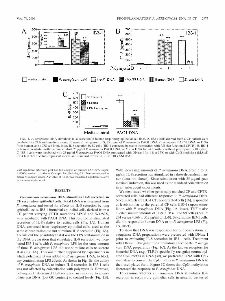
Pseudomonas aeruginosa DNA stimulates IL-8 secretion inCF respiratory epithelial cells. Total DNA was prepared fromP. aeruginosa and tested for effects on IL-8 secretion by lungepithelial cells. IB3-1 bronchial epithelial cells, derived from aCF patient carrying CFTR mutations �F508 and W1282X,were incubated with PAO1 DNA. This resulted in stimulatedsecretion of IL-8 relative to resting cells (Fig. 1A). HumanDNA, extracted from respiratory epithelial cells, used at thesame concentration did not stimulate IL-8 secretion (Fig. 1A).To rule out the possibility that it was the LPS contamination inthe DNA preparation that stimulated IL-8 secretion, we incu-bated IB3-1 cells with P. aeruginosa LPS for the same amountof time. P. aeruginosa LPS did not stimulate cells to secreteIL-8 (Fig. 1A). This was further supported by experiments inwhich polymyxin B was added to P. aeruginosa DNA, to blockany contaminating LPS effects. As shown in Fig. 1B, the abilityof P. aeruginosa DNA to induce IL-8 secretion in IB3-1 cellswas not affected by coincubation with polymyxin B. However,polymyxin B decreased IL-8 secretion in response to Esche-richia coli DNA (low GC content) to control levels (Fig. 1B).
With increasing amounts of P. aeruginosa DNA, from 5 to 30�g/ml, IL-8 secretion was stimulated in a dose-dependent man-ner (data not shown). Since stimulation with 25 �g/ml gavemaximal induction, this was used as the standard concentrationin all subsequent experiments.
We next tested whether genetically matched CF and CFTR-corrected cells had different responses to P. aeruginosa DNA.S9 cells, which are IB3-1 CFTR-corrected cells (16), respondedat levels similar to the parental CF cells (IB3-1) upon stimu-lation with P. aeruginosa DNA (Fig. 1A, inset). TNF- alsoelicited similar amounts of IL-8 in IB3-1 and S9 cells (4,500 254 versus 4,566 512 pg/ml of IL-8). S9 cells, like IB3-1 cells,did not respond to human DNA or to P. aeruginosa LPS (Fig.1A, inset).
To show that DNA was responsible for our observations, P.aeruginosa DNA preparations were pretreated with DNase Iprior to evaluating IL-8 secretion in IB3-1 cells. Treatmentwith DNase I abrogated the stimulatory effect of the P. aerugi-nosa DNA preparation (Fig. 1C). As the known receptors forbacterial DNA (e.g., TLR9) specifically recognize nonmethyl-ated CpG motifs in DNA (30), we pretreated DNA with CpGmethylase to convert the CpG motifs in P. aeruginosa DNA totheir methylated form. Figure 1C shows that CpG methylationdecreased the response to P. aeruginosa DNA.
To examine whether P. aeruginosa DNA stimulates IL-8secretion in respiratory epithelial cells in general, we tested
FIG. 1. P. aeruginosa DNA stimulates IL-8 secretion in human respiratory epithelial cell lines. A. IB3-1 cells derived from a CF patient wereincubated for 24 h with medium alone, 10 ng/ml P. aeruginosa LPS, 25 �g/ml of P. aeruginosa PAO1 DNA, P. aeruginosa PA578I DNA, or DNAfrom human cells (C38 cell line). Inset, IL-8 secretion by S9 cells (IB3-1 corrected by stable transfection with full-size functional CFTR). B. IB3-1cells were incubated with medium control, 25 �g/ml P. aeruginosa PAO1 DNA, or E. coli DNA for 24 h, with or without polymyxin B (10 �g/ml).C. IB3-1 cells were incubated with 25 �g/ml P. aeruginosa PAO1 DNA pretreated with DNase I for 1 h at 37°C or with CpG methylase (M.SssI)for 4 h at 37°C. Values represent means and standard errors. ��, P � 0.01 (ANOVA).
VOL. 74, 2006 PROINFLAMMATORY P. AERUGINOSA DNA IN CF 2977

additional primary cells and cell lines. CuFi1, a cell line derivedfrom a patient homozygous for CFTR �F508 (70), also re-sponded to P. aeruginosa DNA by increasing IL-8 secretion(Fig. 2A). The response to P. aeruginosa DNA preparationswas comparable to the response to TNF-. Just like IB3-1 cells,CuFi1 did not respond to human DNA (Fig. 2A).
We next tested the response in primary, NHBE cells. NHBEcells showed increased IL-8 secretion after P. aeruginosa DNA
stimulation, albeit to a lesser extent than IB3-1 and CuFi1cells, but responded effectively to TNF- stimulation (Fig. 2B).
DNA preparations from mucoid or nonmucoid P. aeruginosadisplay similar IL-8 stimulatory potential. We next comparedDNA preparations from mucoid alginate-overproducing P.aeruginosa (PA578I) and its nonmucoid ancestral strain (PAO1),since conversion to mucoidy is a well-recognized pathogenicdeterminant of P. aeruginosa expressed in CF and associated
FIG. 2. P. aeruginosa DNA stimulates IL-8 secretion in primary human airway epithelial cells. A. CuFi1 cells (derived from a CF patient) wereincubated for 24 h with medium alone, 20 ng/ml TNF-, 25 �g/ml of DNA from P. aeruginosa PAO1, P. aeruginosa PA578I, or human cells (S9).B. Primary NHBE cells. Values represent percent relative to control. Insets, absolute IL-8 values, in nanograms per milliliter. �, P � 0.05; ��, P� 0.01 (ANOVA).
FIG. 3. P. aeruginosa DNA does not activate the IL-8 promoter in airway epithelial cells. IB3-1 cells were cotransfected with �-galactosidaseand NF-�B:luciferase (A), pAP-1:luciferase (B), or pIL-8:luciferase (C) as reporters. As a positive control for pAP-1:luciferase, cells werecotransfected with the MEKK gene (B). Cells were incubated for 8 or 24 h (insets) with medium alone, with 20 ng/ml TNF-, or for 2, 4, 6, 8, or24 h (insets) with 25 �g/ml P. aeruginosa PAO1 DNA. Luciferase activities were normalized to �-galactosidase. ��, P � 0.01 (ANOVA).
2978 DELGADO ET AL. INFECT. IMMUN.
with disease deterioration (24). DNA preparations from mu-coid and nonmucoid P. aeruginosa elicited similar IL-8 levels inall cells tested (Fig. 1 and 2).
P. aeruginosa DNA engages a signaling pathway indepen-dent of NF-�B activation. The conventional mechanism of IL-8increase upon stimulation with CpG DNA is activation of anNF-�B pathway following TLR9 engagement (5, 32). To testwhether NF-�B was involved in stimulation of IL-8 productionin response to P. aeruginosa DNA, IB3-1 cells were transfectedwith an NF-�B-luciferase reporter and NF-�B-dependent pro-moter activation was monitored in response to PseudomonasDNA. Unexpectedly, NF-�B was not activated by P. aeruginosaDNA at 2, 4, 6, 8, or 24 h, while a typical strong NF-�Bresponse was observed upon activation with TNF- (Fig. 3A).NF-�B activation was not detected, although increased levelsof IL-8 were detected in the supernatants (data not shown). AsAP-1 is another important transcriptional activator of the IL-8promoter, we tested whether an AP-1 induction pathway wasresponsible for IL-8 production/secretion. IB3-1 cells weretransfected with a pAP-1-luciferase reporter plasmid and stim-ulated with P. aeruginosa DNA. As is the case with NF-�B, P.aeruginosa DNA did not activate pAP-1 at 2, 4, 6, 8, or 24 h(Fig. 3B). This contrasted with the positive control, consistingof cells cotransfected with MEKK, which showed a strongAP-1:luc activation (Fig. 3B).
Finally, we tested the entire previously characterized IL-8promoter, containing all known regulatory sites that controlIL-8 transcription (including NF-�B and AP-1 sites) (32).IB3-1 cells were transfected with the IL-8 promoter-luciferasereporter plasmid and then stimulated with P. aeruginosa DNAfor 2, 4, 6, 8, or 24 h. As with individually tested NF-�B andAP-1 probes, we did not detect transcriptional activation of theIL-8 promoter upon stimulation with P. aeruginosa DNA,albeit the promoter responded to TNF- as expected (Fig. 3C).
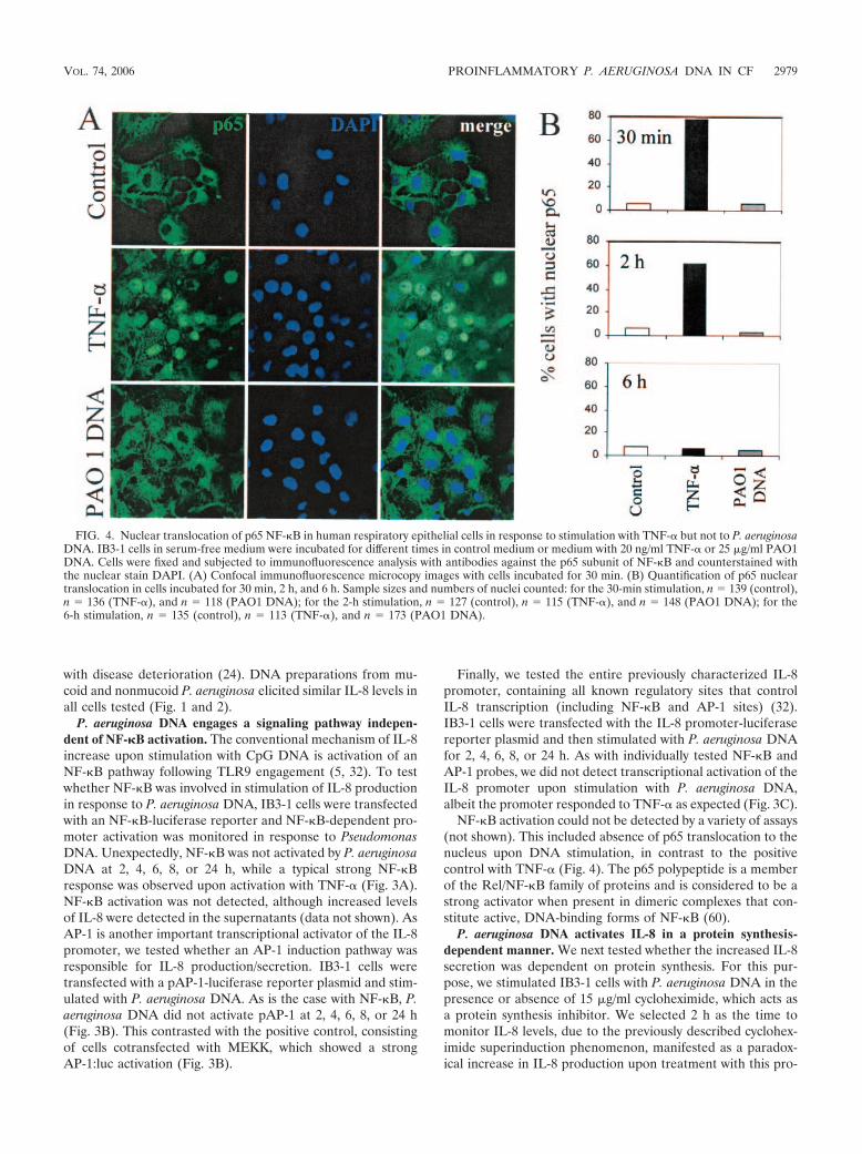
NF-�B activation could not be detected by a variety of assays(not shown). This included absence of p65 translocation to thenucleus upon DNA stimulation, in contrast to the positivecontrol with TNF- (Fig. 4). The p65 polypeptide is a memberof the Rel/NF-�B family of proteins and is considered to be astrong activator when present in dimeric complexes that con-stitute active, DNA-binding forms of NF-�B (60).
P. aeruginosa DNA activates IL-8 in a protein synthesis-dependent manner. We next tested whether the increased IL-8secretion was dependent on protein synthesis. For this pur-pose, we stimulated IB3-1 cells with P. aeruginosa DNA in thepresence or absence of 15 �g/ml cycloheximide, which acts asa protein synthesis inhibitor. We selected 2 h as the time tomonitor IL-8 levels, due to the previously described cyclohex-imide superinduction phenomenon, manifested as a paradox-ical increase in IL-8 production upon treatment with this pro-
FIG. 4. Nuclear translocation of p65 NF-�B in human respiratory epithelial cells in response to stimulation with TNF- but not to P. aeruginosaDNA. IB3-1 cells in serum-free medium were incubated for different times in control medium or medium with 20 ng/ml TNF- or 25 �g/ml PAO1DNA. Cells were fixed and subjected to immunofluorescence analysis with antibodies against the p65 subunit of NF-�B and counterstained withthe nuclear stain DAPI. (A) Confocal immunofluorescence microcopy images with cells incubated for 30 min. (B) Quantification of p65 nucleartranslocation in cells incubated for 30 min, 2 h, and 6 h. Sample sizes and numbers of nuclei counted: for the 30-min stimulation, n � 139 (control),n � 136 (TNF-), and n � 118 (PAO1 DNA); for the 2-h stimulation, n � 127 (control), n � 115 (TNF-), and n � 148 (PAO1 DNA); for the6-h stimulation, n � 135 (control), n � 113 (TNF-), and n � 173 (PAO1 DNA).
VOL. 74, 2006 PROINFLAMMATORY P. AERUGINOSA DNA IN CF 2979
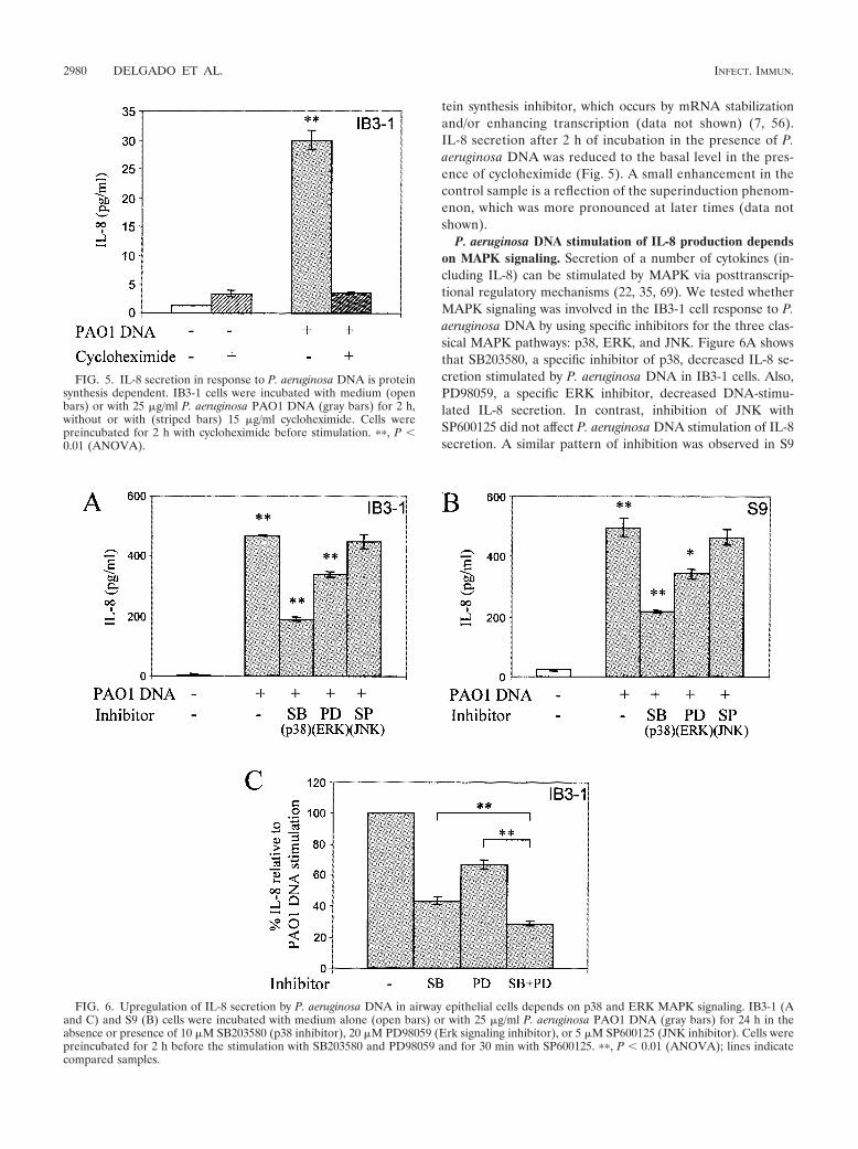
tein synthesis inhibitor, which occurs by mRNA stabilizationand/or enhancing transcription (data not shown) (7, 56).IL-8 secretion after 2 h of incubation in the presence of P.aeruginosa DNA was reduced to the basal level in the pres-ence of cycloheximide (Fig. 5). A small enhancement in thecontrol sample is a reflection of the superinduction phenom-enon, which was more pronounced at later times (data notshown).
P. aeruginosa DNA stimulation of IL-8 production dependson MAPK signaling. Secretion of a number of cytokines (in-cluding IL-8) can be stimulated by MAPK via posttranscrip-tional regulatory mechanisms (22, 35, 69). We tested whetherMAPK signaling was involved in the IB3-1 cell response to P.aeruginosa DNA by using specific inhibitors for the three clas-sical MAPK pathways: p38, ERK, and JNK. Figure 6A showsthat SB203580, a specific inhibitor of p38, decreased IL-8 se-cretion stimulated by P. aeruginosa DNA in IB3-1 cells. Also,PD98059, a specific ERK inhibitor, decreased DNA-stimu-lated IL-8 secretion. In contrast, inhibition of JNK withSP600125 did not affect P. aeruginosa DNA stimulation of IL-8secretion. A similar pattern of inhibition was observed in S9
FIG. 5. IL-8 secretion in response to P. aeruginosa DNA is proteinsynthesis dependent. IB3-1 cells were incubated with medium (openbars) or with 25 �g/ml P. aeruginosa PAO1 DNA (gray bars) for 2 h,without or with (striped bars) 15 �g/ml cycloheximide. Cells werepreincubated for 2 h with cycloheximide before stimulation. ��, P �0.01 (ANOVA).
FIG. 6. Upregulation of IL-8 secretion by P. aeruginosa DNA in airway epithelial cells depends on p38 and ERK MAPK signaling. IB3-1 (Aand C) and S9 (B) cells were incubated with medium alone (open bars) or with 25 �g/ml P. aeruginosa PAO1 DNA (gray bars) for 24 h in theabsence or presence of 10 �M SB203580 (p38 inhibitor), 20 �M PD98059 (Erk signaling inhibitor), or 5 �M SP600125 (JNK inhibitor). Cells werepreincubated for 2 h before the stimulation with SB203580 and PD98059 and for 30 min with SP600125. ��, P � 0.01 (ANOVA); lines indicatecompared samples.
2980 DELGADO ET AL. INFECT. IMMUN.
cells (Fig. 6B). The actions of p38 and ERK inhibitors werepartially additive (Fig. 6C).
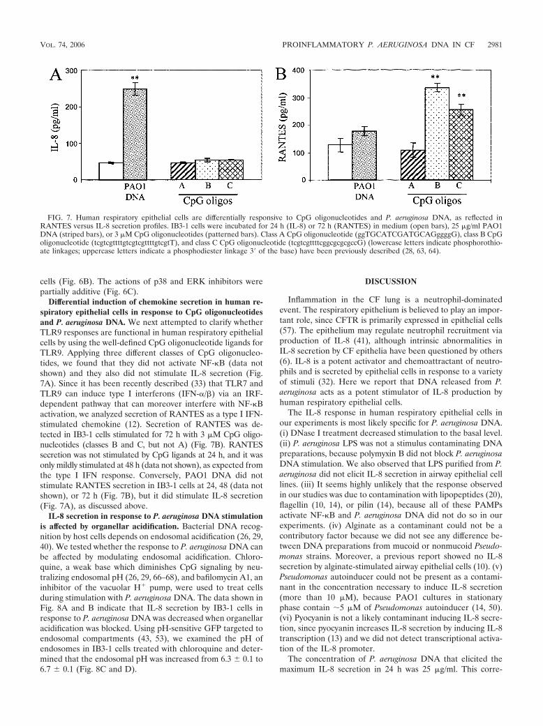
Differential induction of chemokine secretion in human re-spiratory epithelial cells in response to CpG oligonucleotidesand P. aeruginosa DNA. We next attempted to clarify whetherTLR9 responses are functional in human respiratory epithelialcells by using the well-defined CpG oligonucleotide ligands forTLR9. Applying three different classes of CpG oligonucleo-tides, we found that they did not activate NF-�B (data notshown) and they also did not stimulate IL-8 secretion (Fig.7A). Since it has been recently described (33) that TLR7 andTLR9 can induce type I interferons (IFN-/�) via an IRF-dependent pathway that can moreover interfere with NF-�Bactivation, we analyzed secretion of RANTES as a type I IFN-stimulated chemokine (12). Secretion of RANTES was de-tected in IB3-1 cells stimulated for 72 h with 3 �M CpG oligo-nucleotides (classes B and C, but not A) (Fig. 7B). RANTESsecretion was not stimulated by CpG ligands at 24 h, and it wasonly mildly stimulated at 48 h (data not shown), as expected fromthe type I IFN response. Conversely, PAO1 DNA did notstimulate RANTES secretion in IB3-1 cells at 24, 48 (data notshown), or 72 h (Fig. 7B), but it did stimulate IL-8 secretion(Fig. 7A), as discussed above.
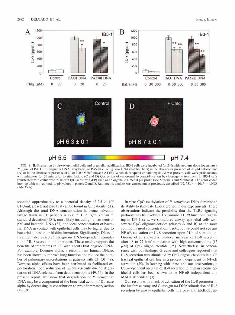
IL-8 secretion in response to P. aeruginosa DNA stimulationis affected by organellar acidification. Bacterial DNA recog-nition by host cells depends on endosomal acidification (26, 29,40). We tested whether the response to P. aeruginosa DNA canbe affected by modulating endosomal acidification. Chloro-quine, a weak base which diminishes CpG signaling by neu-tralizing endosomal pH (26, 29, 66–68), and bafilomycin A1, aninhibitor of the vacuolar H� pump, were used to treat cellsduring stimulation with P. aeruginosa DNA. The data shown inFig. 8A and B indicate that IL-8 secretion by IB3-1 cells inresponse to P. aeruginosa DNA was decreased when organellaracidification was blocked. Using pH-sensitive GFP targeted toendosomal compartments (43, 53), we examined the pH ofendosomes in IB3-1 cells treated with chloroquine and deter-mined that the endosomal pH was increased from 6.3 0.1 to6.7 0.1 (Fig. 8C and D).
DISCUSSION
Inflammation in the CF lung is a neutrophil-dominatedevent. The respiratory epithelium is believed to play an impor-tant role, since CFTR is primarily expressed in epithelial cells(57). The epithelium may regulate neutrophil recruitment viaproduction of IL-8 (41), although intrinsic abnormalities inIL-8 secretion by CF epithelia have been questioned by others(6). IL-8 is a potent activator and chemoattractant of neutro-phils and is secreted by epithelial cells in response to a varietyof stimuli (32). Here we report that DNA released from P.aeruginosa acts as a potent stimulator of IL-8 production byhuman respiratory epithelial cells.
The IL-8 response in human respiratory epithelial cells inour experiments is most likely specific for P. aeruginosa DNA.(i) DNase I treatment decreased stimulation to the basal level.(ii) P. aeruginosa LPS was not a stimulus contaminating DNApreparations, because polymyxin B did not block P. aeruginosaDNA stimulation. We also observed that LPS purified from P.aeruginosa did not elicit IL-8 secretion in airway epithelial celllines. (iii) It seems highly unlikely that the response observedin our studies was due to contamination with lipopeptides (20),flagellin (10, 14), or pilin (14), because all of these PAMPsactivate NF-�B and P. aeruginosa DNA did not do so in ourexperiments. (iv) Alginate as a contaminant could not be acontributory factor because we did not see any difference be-tween DNA preparations from mucoid or nonmucoid Pseudo-monas strains. Moreover, a previous report showed no IL-8secretion by alginate-stimulated airway epithelial cells (10). (v)Pseudomonas autoinducer could not be present as a contami-nant in the concentration necessary to induce IL-8 secretion(more than 10 �M), because PAO1 cultures in stationaryphase contain 5 �M of Pseudomonas autoinducer (14, 50).(vi) Pyocyanin is not a likely contaminant inducing IL-8 secre-tion, since pyocyanin increases IL-8 secretion by inducing IL-8transcription (13) and we did not detect transcriptional activa-tion of the IL-8 promoter.
The concentration of P. aeruginosa DNA that elicited themaximum IL-8 secretion in 24 h was 25 �g/ml. This corre-
FIG. 7. Human respiratory epithelial cells are differentially responsive to CpG oligonucleotides and P. aeruginosa DNA, as reflected inRANTES versus IL-8 secretion profiles. IB3-1 cells were incubated for 24 h (IL-8) or 72 h (RANTES) in medium (open bars), 25 �g/ml PAO1DNA (striped bars), or 3 �M CpG oligonucleotides (patterned bars). Class A CpG oligonucleotide (ggTGCATCGATGCAGggggG), class B CpGoligonucleotide (tcgtcgttttgtcgtcgttttgtcgtT), and class C CpG oligonucleotide (tcgtcgttttcggcgcgcgccG) (lowercase letters indicate phosphorothio-ate linkages; uppercase letters indicate a phosphodiester linkage 3� of the base) have been previously described (28, 63, 64).
VOL. 74, 2006 PROINFLAMMATORY P. AERUGINOSA DNA IN CF 2981
sponded approximately to a bacterial density of 2.5 � 108
CFU/ml, a bacterial load that can be found in CF patients (51).Although the total DNA concentration in bronchoalveolarlavage fluids in CF patients is 17.6 11.2 �g/ml (mean standard deviation) (54), most likely including human neutro-phil and bacterial DNA (37), the local concentration of bacte-rial DNA in contact with epithelial cells may be higher due tobacterial adhesion or biofilm formation. Significantly, DNase Itreatment decreased P. aeruginosa DNA-dependent stimula-tion of IL-8 secretion in our studies. These results support thebenefits of treatments in CF with agents that degrade DNA.For example, Dornase alpha, a recombinant human DNase,has been shown to improve lung function and reduce the num-ber of pulmonary exacerbations in patients with CF (31, 49).Dornase alpha effects have been attributed to facilitated ex-pectoration upon reduction of mucus viscosity due to degra-dation of DNA released from dead neutrophils (49, 54). In thepresent report, we show that degradation of P. aeruginosaDNA may be a component of the beneficial action of Dornasealpha by decreasing its contribution to proinflammatory action(49, 59).
In vitro CpG methylation of P. aeruginosa DNA diminishedits ability to stimulate IL-8 secretion in our experiments. Theseobservations indicate the possibility that the TLR9 signalingpathway may be involved. To examine TLR9 functional signal-ing in IB3-1 cells, we stimulated airway epithelial cells withseveral CpG oligonucleotides (classes A and B) at the mostcommonly used concentration, 1 �M, but we could not see anyNF-�B activation or IL-8 secretion upon 24 h of stimulation.Greene et al. showed a low-level increase of IL-8 secretionafter 48 to 72 h of stimulation with high concentrations (15�M) of CpG oligonucleotide (25). Nevertheless, in concur-rence with our findings, Greene and colleagues reported thatIL-8 secretion was stimulated by CpG oligonucleotides in a CFtracheal epithelial cell line in a process independent of NF-�Bactivation (25). In keeping with these and our observations, aCpG-dependent increase of IL-8 secretion in human colonic ep-ithelial cells has been shown to be NF-�B independent andMAPK dependent (3).
Our results with a lack of activation of the IL-8 promoter inthe luciferase assay and P. aeruginosa DNA stimulation of IL-8secretion by airway epithelial cells in a p38- and ERK-depen-
FIG. 8. IL-8 secretion by airway epithelial cells and organellar acidification. IB3-1 cells were incubated for 24 h with medium alone (open bars),25 �g/ml of PAO1 P. aeruginosa DNA (gray bars), or PA578I P. aeruginosa DNA (hatched bars) in the absence or presence of 20 �M chloroquine(A) or in the absence or presence of 30 or 300 nM bafilomycin A1 (B). When chloroquine or bafilomycin A1 was present, cells were preincubatedwith inhibitors for 30 min prior to stimulation. (C and D) Correction of endosomal hyperacidification by chloroquine treatment in IB3-1 cellstransfected with cellubrevin-pHluorin (pH-sensitive GFP) used as an organelle lumenal pH probe (see Materials and Methods). The color-codedlook-up table corresponds to pH values in panels C and D. Ratiometric analysis was carried out as previously described (52, 53). n � 10; P � 0.0488(ANOVA).
2982 DELGADO ET AL. INFECT. IMMUN.
dent manner are best explained by posttranslational mecha-nisms. The MAPK p38 is known as an mRNA stabilizer ofmany cytokine transcripts containing adenylate- and uridylate-rich elements in the 3� untranslated region, including IL-8transcript (22, 35). The MAPK p38 can also participate inderepression of translation via these elements (39, 69). ERKhas been involved in transport from the nucleus to the cyto-plasm of cytokine mRNA (15). According to our results, itseems possible that IL-8 secretion is being stimulated by P.aeruginosa DNA in airway epithelial cells by inducing posttran-scriptional mechanisms akin to the ones listed above. Further-more, exocytosis induction of the preformed IL-8 protein, viap38 and ERK pathways, may also be taking place (11, 44).
P. aeruginosa DNA stimulation was partially sensitive tochloroquine and bafilomycin A1, compounds that block endo-somal acidification essential in TLR9 signaling (26, 29, 40,66–68). It has been demonstrated that organelles are hyper-acidified in CF respiratory epithelial cells, including endosomalcompartments (52, 53). Taking this into account, along withthe fact that endosomal acidification is requisite to CpG sig-naling via TLR9 (26, 29, 40) and that airway epithelial cellsexpress a variety of TLRs, including TLR9 (3, 25, 29, 46, 51),we anticipated that CF cells might respond more than non-CFcells to the GC-rich P. aeruginosa DNA. However, this turnedout not to be the case. Nevertheless, IL-8 secretion stimulatedby P. aeruginosa DNA was affected by blockers of endosomalacidification.
Collectively, our experiments show that human respiratoryepithelial cells respond to P. aeruginosa DNA by secretingIL-8. Our findings suggest that P. aeruginosa DNA-stimulatedsecretion of IL-8 by human respiratory epithelial cells repre-sents a protein synthesis-dependent process, independent ofNF-�B activation and transcriptional activation of IL-8. Inspite of the ability of human respiratory epithelial cells torespond to typical CpG TLR9 ligands, the pattern of RANTESinduction by CpG oligonucleotides versus IL-8 by P. aeruginosaDNA strongly suggests activation of two separate pathways: (i)type I interferon pathways in the former process and (ii) p38and ERK pathways in the latter. Significantly, the signalinginvolved in P. aeruginosa DNA-elicited IL-8 secretion dependson endosomal acidification, based on a partial inhibitory effectof chloroquine and pharmacological blockers of vacuolar H�-ATPase. Our findings suggest that elimination of bacterialDNA by DNase treatment and use of lysosomotropic drugsmay help decrease inflammation in CF.
ACKNOWLEDGMENTS
This work was supported by NIH grant AI31193 and in part by grantAI050825 from NIH and a grant from Philip Morris USA Inc. andPhilip Morris International.
REFERENCES
1. Adamo, R., S. Sokol, G. Soong, M. I. Gomez, and A. Prince. 2004. Pseudo-monas aeruginosa flagella activate airway epithelial cells through asialo GM1and toll-like receptor 2 as well as toll-like receptor 5. Am. J. Respir. CellMol. Biol. 30:627–634.
2. Agrawal, S., A. Agrawal, B. Doughty, A. Gerwitz, J. Blenis, T. Van Dyke, andB. Pulendran. 2003. Cutting edge: different Toll-like receptor agonists in-struct dendritic cells to induce distinct Th responses via differential modu-lation of extracellular signal-regulated kinase-mitogen-activated protein ki-nase and c-Fos. J. Immunol. 171:4984–4989.
3. Akhtar, M., J. L. Watson, A. Nazli, and D. M. McKay. 2003. Bacterial DNAevokes epithelial IL-8 production by a MAPK-dependent, NF-�B-indepen-dent pathway. FASEB J. 17:1319–1321.
4. Athman, R., and D. Philpott. 2004. Innate immunity via Toll-like receptorsand Nod proteins. Curr. Opin. Microbiol. 7:25–32.
5. Bauer, S., C. J. Kirschning, H. Hacker, V. Redecke, S. Hausmann, S. Akira,H. Wagner, and G. B. Lipford. 2001. Human TLR9 confers responsiveness tobacterial DNA via species-specific CpG motif recognition. Proc. Natl. Acad.Sci. USA 98:9237–9242.
6. Becker, M. N., M. S. Sauer, M. S. Muhlebach, A. J. Hirsh, Q. Wu, M. W.Verghese, and S. H. Randell. 2004. Cytokine secretion by cystic fibrosisairway epithelial cells. Am. J. Respir. Crit. Care Med. 169:645–653.
7. Bischoff, D. S., J. H. Zhu, N. S. Makhijani, and D. T. Yamaguchi. 2005. KCchemokine expression by TGF-beta in C3H10T1/2 cells induced towardsosteoblasts. Biochem. Biophys. Res. Commun. 326:364–370.
8. Bonfield, T. L., J. R. Panuska, M. W. Konstan, K. A. Hilliard, J. B. Hilliard,H. Ghnaim, and M. Berger. 1995. Inflammatory cytokines in cystic fibrosislungs. Am. J. Respir. Crit. Care Med. 152:2111–2118.
9. Boucher, J. C., H. Yu, M. H. Mudd, and V. Deretic. 1997. Mucoid Pseudo-monas aeruginosa in cystic fibrosis: characterization of muc mutations inclinical isolates and analysis of clearance in a mouse model of respiratoryinfection. Infect. Immun. 65:3838–3846.
10. Cobb, L. M., J. C. Mychaleckyj, D. J. Wozniak, and Y. S. Lopez-Boado. 2004.Pseudomonas aeruginosa flagellin and alginate elicit very distinct gene ex-pression patterns in airway epithelial cells: implications for cystic fibrosisdisease. J. Immunol. 173:5659–5670.
11. Coxon, P. Y., M. J. Rane, S. Uriarte, D. W. Powell, S. Singh, W. Butt, Q.Chen, and K. R. McLeish. 2003. MAPK-activated protein kinase-2 partici-pates in p38 MAPK-dependent and ERK-dependent functions in humanneutrophils. Cell Signal. 15:993–1001.
12. Cremer, I., J. Ghysdael, and V. Vieillard. 2002. A non-classical ISRE/ISGF3pathway mediates induction of RANTES gene transcription by type I IFNs.FEBS Lett. 511:41–45.
13. Denning, G. M., L. A. Wollenweber, M. A. Railsback, C. D. Cox, L. L. Stoll,and B. E. Britigan. 1998. Pseudomonas pyocyanin increases interleukin-8expression by human airway epithelial cells. Infect. Immun. 66:5777–5784.
14. DiMango, E., H. J. Zar, R. Bryan, and A. Prince. 1995. Diverse Pseudomonasaeruginosa gene products stimulate respiratory epithelial cells to produceinterleukin-8. J. Clin. Investig. 96:2204–2210.
15. Dumitru, C. D., J. D. Ceci, C. Tsatsanis, D. Kontoyiannis, K. Stamatakis,J. H. Lin, C. Patriotis, N. A. Jenkins, N. G. Copeland, G. Kollias, and P. N.Tsichlis. 2000. TNF-alpha induction by LPS is regulated posttranscription-ally via a Tpl2/ERK-dependent pathway. Cell 103:1071–1083.
16. Egan, M., T. Flotte, S. Afione, R. Solow, P. L. Zeitlin, B. J. Carter, and W. B.Guggino. 1992. Defective regulation of outwardly rectifying Cl- channels byprotein kinase A corrected by insertion of CFTR. Nature 358:581–584.
17. Epelman, S., D. Stack, C. Bell, E. Wong, G. G. Neely, S. Krutzik, K. Miyake,P. Kubes, L. D. Zbytnuik, L. L. Ma, X. Xie, D. E. Woods, and C. H. Mody.2004. Different domains of Pseudomonas aeruginosa exoenzyme S activatedistinct TLRs. J. Immunol. 173:2031–2040.
18. Erridge, C., A. Pridmore, A. Eley, J. Stewart, and I. R. Poxton. 2004. Lipo-polysaccharides of Bacteroides fragilis, Chlamydia trachomatis and Pseudo-monas aeruginosa signal via toll-like receptor 2. J. Med. Microbiol. 53:735–740.
19. Faure, K., T. Sawa, T. Ajayi, J. Fujimoto, K. Moriyama, N. Shime, and J. P.Wiener-Kronish. 2004. TLR4 signaling is essential for survival in acute lunginjury induced by virulent Pseudomonas aeruginosa secreting type III secre-tory toxins. Respir. Res. 5:1.
20. Firoved, A. M., W. Ornatowski, and V. Deretic. 2004. Microarray analysisreveals induction of lipoprotein genes in mucoid Pseudomonas aeruginosa:implications for inflammation in cystic fibrosis. Infect. Immun. 72:5012–5018.
21. Flo, T. H., L. Ryan, E. Latz, O. Takeuchi, B. G. Monks, E. Lien, O. Halaas,S. Akira, G. Skjak-Braek, D. T. Golenbock, and T. Espevik. 2002. Involve-ment of toll-like receptor (TLR) 2 and TLR4 in cell activation by mannu-ronic acid polymers. J. Biol. Chem. 277:35489–35495.
22. Frevel, M. A., T. Bakheet, A. M. Silva, J. G. Hissong, K. S. Khabar, and B. R.Williams. 2003. p38 mitogen-activated protein kinase-dependent and -inde-pendent signaling of mRNA stability of AU-rich element-containing tran-scripts. Mol. Cell. Biol. 23:425–436.
23. Goral, J., and E. J. Kovacs. 2005. In vivo ethanol exposure down-regulatesTLR2-, TLR4-, and TLR9-mediated macrophage inflammatory response bylimiting p38 and ERK1/2 activation. J. Immunol. 174:456–463.
24. Govan, J. R., and V. Deretic. 1996. Microbial pathogenesis in cystic fibrosis:mucoid Pseudomonas aeruginosa and Burkholderia cepacia. Microbiol. Rev.60:539–574.
25. Greene, C. M., T. P. Carroll, S. G. Smith, C. C. Taggart, J. Devaney, S.Griffin, S. J. O’Neill, and N. G. McElvaney. 2005. TLR-induced inflamma-tion in cystic fibrosis and non-cystic fibrosis airway epithelial cells. J. Immu-nol. 174:1638–1646.
26. Hacker, H., H. Mischak, T. Miethke, S. Liptay, R. Schmid, T. Sparwasser, K.Heeg, G. B. Lipford, and H. Wagner. 1998. CpG-DNA-specific activation ofantigen-presenting cells requires stress kinase activity and is preceded bynon-specific endocytosis and endosomal maturation. EMBO J. 17:6230–6240.
27. Hajjar, A. M., R. K. Ernst, J. H. Tsai, C. B. Wilson, and S. I. Miller. 2002.
VOL. 74, 2006 PROINFLAMMATORY P. AERUGINOSA DNA IN CF 2983
Human Toll-like receptor 4 recognizes host-specific LPS modifications. Nat.Immunol. 3:354–359.
28. Hartmann, G., R. D. Weeratna, Z. K. Ballas, P. Payette, S. Blackwell, I.Suparto, W. L. Rasmussen, M. Waldschmidt, D. Sajuthi, R. H. Purcell, H. L.Davis, and A. M. Krieg. 2000. Delineation of a CpG phosphorothioateoligodeoxynucleotide for activating primate immune responses in vitro andin vivo. J. Immunol. 164:1617–1624.
29. Heil, F., P. Ahmad-Nejad, H. Hemmi, H. Hochrein, F. Ampenberger, T.Gellert, H. Dietrich, G. Lipford, K. Takeda, S. Akira, H. Wagner, and S.Bauer. 2003. The Toll-like receptor 7 (TLR7)-specific stimulus loxoribineuncovers a strong relationship within the TLR7, 8 and 9 subfamily. Eur.J. Immunol. 33:2987–2997.
30. Hemmi, H., O. Takeuchi, T. Kawai, T. Kaisho, S. Sato, H. Sanjo, M.Matsumoto, K. Hoshino, H. Wagner, K. Takeda, and S. Akira. 2000. AToll-like receptor recognizes bacterial DNA. Nature 408:740–745.
31. Hodson, M. E., S. McKenzie, H. K. Harms, C. Koch, G. Mastella, J. Navarro,and B. Strandvik. 2003. Dornase alfa in the treatment of cystic fibrosis inEurope: a report from the Epidemiologic Registry of Cystic Fibrosis. Pediatr.Pulmonol. 36:427–432.
32. Hoffmann, E., O. Dittrich-Breiholz, H. Holtmann, and M. Kracht. 2002.Multiple control of interleukin-8 gene expression. J. Leukoc. Biol. 72:847–855.
33. Honda, K., H. Yanai, T. Mizutani, H. Negishi, N. Shimada, N. Suzuki, Y.Ohba, A. Takaoka, W. C. Yeh, and T. Taniguchi. 2004. Role of a transduc-tional-transcriptional processor complex involving MyD88 and IRF-7 inToll-like receptor signaling. Proc. Natl. Acad. Sci. USA 101:15416–15421.
34. Janeway, C. A., Jr., and R. Medzhitov. 2002. Innate immune recognition.Annu. Rev. Immunol. 20:197–216.
35. Jijon, H. B., W. J. Panenka, K. L. Madsen, and H. G. Parsons. 2002. MAPkinases contribute to IL-8 secretion by intestinal epithelial cells via a post-transcriptional mechanism. Am. J. Physiol. Cell Physiol. 283:C31–C41.
36. Khan, T. Z., J. S. Wagener, T. Bost, J. Martinez, F. J. Accurso, and D. W.Riches. 1995. Early pulmonary inflammation in infants with cystic fibrosis.Am. J. Respir. Crit. Care Med. 151:1075–1082.
37. Kirchner, K. K., J. S. Wagener, T. Z. Khan, S. C. Copenhaver, and F. J.Accurso. 1996. Increased DNA levels in bronchoalveolar lavage fluid ob-tained from infants with cystic fibrosis. Am. J. Respir. Crit. Care Med.154:1426–1429.
38. Konstan, M. W., K. A. Hilliard, T. M. Norvell, and M. Berger. 1994. Bron-choalveolar lavage findings in cystic fibrosis patients with stable, clinicallymild lung disease suggest ongoing infection and inflammation. Am. J. Respir.Crit. Care Med. 150:448–454.
39. Kontoyiannis, D., M. Pasparakis, T. T. Pizarro, F. Cominelli, and G. Kollias.1999. Impaired on/off regulation of TNF biosynthesis in mice lacking TNFAU-rich elements: implications for joint and gut-associated immunopathol-ogies. Immunity 10:387–398.
40. Krieg, A. M. 2002. CpG motifs in bacterial DNA and their immune effects.Annu. Rev. Immunol. 20:709–760.
41. Kube, D., U. Sontich, D. Fletcher, and P. B. Davis. 2001. Proinflammatorycytokine responses to P. aeruginosa infection in human airway epithelial celllines. Am. J. Physiol. Lung Cell Mol. Physiol. 280:L493–L502.
42. Lorenz, E., D. C. Chemotti, K. Vandal, and P. A. Tessier. 2004. Toll-likereceptor 2 represses non-pilus adhesin-induced signaling in acute infectionswith the Pseudomonas aeruginosa pilA mutant. Infect. Immun. 72:4561–4569.
43. Miesenbock, G., D. A. De Angelis, and J. E. Rothman. 1998. Visualizingsecretion and synaptic transmission with pH-sensitive green fluorescent pro-teins. Nature 394:192–195.
44. Milella, M., A. Gismondi, P. Roncaioli, L. Bisogno, G. Palmieri, L. Frati,M. G. Cifone, and A. Santoni. 1997. CD16 cross-linking induces both secre-tory and extracellular signal-regulated kinase (ERK)-dependent cytosolicphospholipase A2 (PLA2) activity in human natural killer cells: involvementof ERK, but not PLA2, in CD16-triggered granule exocytosis. J. Immunol.158:3148–3154.
45. Muhlebach, M. S., W. Reed, and T. L. Noah. 2004. Quantitative cytokinegene expression in CF airway. Pediatr. Pulmonol. 37:393–399.
46. Muir, A., G. Soong, S. Sokol, B. Reddy, M. I. Gomez, A. Van Heeckeren, andA. Prince. 2004. Toll-like receptors in normal and cystic fibrosis airwayepithelial cells. Am. J. Respir. Cell Mol. Biol. 30:777–783.
47. Orenstein, D. M., G. B. Winnie, and H. Altman. 2002. Cystic fibrosis: a 2002update. J. Pediatr. 140:156–164.
48. Pasare, C., and R. Medzhitov. 2005. Toll-like receptors: linking innate andadaptive immunity. Adv. Exp. Med. Biol. 560:11–18.
49. Paul, K., E. Rietschel, M. Ballmann, M. Griese, D. Worlitzsch, J. Shute, C.Chen, T. Schink, G. Doring, S. van Koningsbruggen, U. Wahn, and F.
Ratjen. 2004. Effect of treatment with dornase alpha on airway inflammationin patients with cystic fibrosis. Am. J. Respir. Crit. Care Med. 169:719–725.
50. Pearson, J. P., L. Passador, B. H. Iglewski, and E. P. Greenberg. 1995. Asecond N-acylhomoserine lactone signal produced by Pseudomonas aerugi-nosa. Proc. Natl. Acad. Sci. USA 92:1490–1494.
51. Platz, J., C. Beisswenger, A. Dalpke, R. Koczulla, O. Pinkenburg, C.Vogelmeier, and R. Bals. 2004. Microbial DNA induces a host defensereaction of human respiratory epithelial cells. J. Immunol. 173:1219–1223.
52. Poschet, J. F., J. C. Boucher, L. Tatterson, J. Skidmore, R. W. Van Dyke, andV. Deretic. 2001. Molecular basis for defective glycosylation and Pseudomo-nas pathogenesis in cystic fibrosis lung. Proc. Natl. Acad. Sci. USA 98:13972–13977.
53. Poschet, J. F., J. Skidmore, J. C. Boucher, A. M. Firoved, R. W. Van Dyke,and V. Deretic. 2002. Hyperacidification of cellubrevin endocytic compart-ments and defective endosomal recycling in cystic fibrosis respiratory epi-thelial cells. J. Biol. Chem. 277:13959–13965.
54. Ratjen, F., K. Paul, S. van Koningsbruggen, S. Breitenstein, E. Rietschel,and W. Nikolaizik. 2005. DNA concentrations in BAL fluid of cystic fibrosispatients with early lung disease: influence of treatment with dornase alpha.Pediatr. Pulmonol. 39:1–4.
55. Revets, H., G. Pynaert, J. Grooten, and P. De Baetselier. 2005. LipoproteinI, a TLR2/4 ligand modulates Th2-driven allergic immune responses. J. Im-munol. 174:1097–1103.
56. Roger, T., T. Out, N. Mukaida, K. Matsushima, H. Jansen, and R. Lutter.1998. Enhanced AP-1 and NF-�B activities and stability of interleukin 8(IL-8) transcripts are implicated in IL-8 mRNA superinduction in lungepithelial H292 cells. Biochem. J. 330:429–435.
57. Rowe, S. M., S. Miller, and E. J. Sorscher. 2005. Cystic fibrosis. N. Engl.J. Med. 352:1992–2001.
58. Schroder, N. W., D. Pfeil, B. Opitz, K. S. Michelsen, J. Amberger, U. Zahringer,U. B. Gobel, and R. R. Schumann. 2001. Activation of mitogen-activated proteinkinases p42/44, p38, and stress-activated protein kinases in myelo-monocyticcells by Treponema lipoteichoic acid. J. Biol. Chem. 276:9713–9719.
59. Shah, P. L., S. F. Scott, R. A. Knight, and M. E. Hodson. 1996. The effectsof recombinant human DNase on neutrophil elastase activity and interleu-kin-8 levels in the sputum of patients with cystic fibrosis. Eur. Respir. J.9:531–534.
60. Siebenlist, U., G. Franzoso, and K. Brown. 1994. Structure, regulation andfunction of NF-kappa B. Annu. Rev. Cell Biol. 10:405–455.
61. Skerrett, S. J., H. D. Liggitt, A. M. Hajjar, and C. B. Wilson. 2004. Cuttingedge: myeloid differentiation factor 88 is essential for pulmonary host de-fense against Pseudomonas aeruginosa but not Staphylococcus aureus. J. Im-munol. 172:3377–3381.
62. Soong, G., B. Reddy, S. Sokol, R. Adamo, and A. Prince. 2004. TLR2 ismobilized into an apical lipid raft receptor complex to signal infection inairway epithelial cells. J. Clin. Investig. 113:1482–1489.
63. Verthelyi, D., K. J. Ishii, M. Gursel, F. Takeshita, and D. M. Klinman. 2001.Human peripheral blood cells differentially recognize and respond to twodistinct CPG motifs. J. Immunol. 166:2372–2377.
64. Vollmer, J., R. Weeratna, P. Payette, M. Jurk, C. Schetter, M. Laucht, T.Wader, S. Tluk, M. Liu, H. L. Davis, and A. M. Krieg. 2004. Characterizationof three CpG oligodeoxynucleotide classes with distinct immunostimulatoryactivities. Eur. J. Immunol. 34:251–262.
65. Wu, Q., Z. Lu, M. W. Verghese, and S. H. Randell. 2005. Airway epithelialcell tolerance to Pseudomonas aeruginosa. Respir. Res. 6:26.
66. Yeo, S. J., D. Gravis, J. G. Yoon, and A. K. Yi. 2003. Myeloid differentiationfactor 88-dependent transcriptional regulation of cyclooxygenase-2 expres-sion by CpG DNA: role of NF-�B and p38. J. Biol. Chem. 278:22563–22573.
67. Yi, A. K., and A. M. Krieg. 1998. Rapid induction of mitogen-activatedprotein kinases by immune stimulatory CpG DNA. J. Immunol. 161:4493–4497.
68. Yi, A. K., R. Tuetken, T. Redford, M. Waldschmidt, J. Kirsch, and A. M.Krieg. 1998. CpG motifs in bacterial DNA activate leukocytes through thepH-dependent generation of reactive oxygen species. J. Immunol. 160:4755–4761.
69. Yu, Y., H. Zeng, S. Lyons, A. Carlson, D. Merlin, A. S. Neish, and A. T.Gewirtz. 2003. TLR5-mediated activation of p38 MAPK regulates epithelialIL-8 expression via posttranscriptional mechanism. Am. J. Physiol. Gastro-intest. Liver Physiol. 285:G282–G290.
70. Zabner, J., P. Karp, M. Seiler, S. L. Phillips, C. J. Mitchell, M. Saavedra, M.Welsh, and A. J. Klingelhutz. 2003. Development of cystic fibrosis andnoncystic fibrosis airway cell lines. Am. J. Physiol. Lung Cell Mol. Physiol.284:L844–L854.
Editor: J. D. Clements
2984 DELGADO ET AL. INFECT. IMMUN.