nmr structural studies of the supramolecular adducts between a liver cytosolic bile acid binding...
TRANSCRIPT
Articles
NMR Structural Studies of the Supramolecular Adducts between a Liver Cytosolic Bile AcidBinding Protein and Gadolinium(III)-Chelates Bearing Bile Acids Residues: MolecularDeterminants of the Binding of a Hepatospecific Magnetic Resonance Imaging Contrast Agent
Michael Assfalg,† Eliana Gianolio,‡ Serena Zanzoni,† Simona Tomaselli,§ Vito Lo Russo,| Claudia Cabella,⊥ Laura Ragona,§
Silvio Aime,*,‡ and Henriette Molinari*,†
Dipartimento Scientifico e Tecnologico, UniVersita di Verona, Strada Le Grazie 15, 37134 Verona, Italy, Dipartimento di Chimica, UniVersitadi Torino, Via Pietro Giuria 7, Torino, Italy, ISMAC-CNR, Via Bassini 15, 20133 Milano, Italy, Bracco Imaging SpA,Via E. Folli 50, 20134Milano, Italy, and Bracco Imaging Spa,Via Ribes 5, 10010 Colleretto Giacosa (TO), Italy
ReceiVed April 4, 2007
The binding affinities of a selected series of Gd(III) chelates bearing bile acid residues, potential hepatospecificMRI contrast agents, to a liver cytosolic bile acid transporter, have been determined through relaxivitymeasurements. The Ln(III) complexes of compound1 were selected for further NMR structural analysisaimed at assessing the molecular determinants of binding. A number of NMR experiments have been carriedout on the bile acid-like adduct, using both diamagnetic Y(III) and paramagnetic Gd(III) complexes, boundto a liver bile acid binding protein. The identified protein “hot spots” defined a single binding site locatedat the protein portal region. The presented findings will serve in a medicinal chemistry approach for thedesign of hepatocytes-selective gadolinium chelates for liver malignancies detection.
Introduction
Bile acid synthesis from cholesterol is the primary pathwayfor cholesterol catabolism.1 Bile acids are amphipathic moleculesthat contain a sterol scaffold with hydroxyl groups and a sidechain that terminates in a carboxyl group. Their amphipathicnature is essential to solubilize dietary lipids, which subsequentlypromotes their absorption in the digestive tract. Most of thebile acids are present within the enterohepatic organs. They areusually stored in the gallbladder, however, when a meal isingested, they flow into the duodenum and intestine. Bile acidsare efficiently (95%) absorbed again by passive diffusion andactive transport in the ileum and transported back to the livervia the portal vein. In the liver, they are taken up at thebasolateral membrane and exported again at the apical mem-brane of the hepatocytes into the bile canaliculus, thus complet-ing their enterohepatic circulation.2,3 Besides their roles indietary lipid absorption and cholesterol homeostasis, it hasbecome clear that bile acids are also signaling molecules.4
In the bile acids enterohepatic circulation, three key stepsare mediated by (i) a receptor system that binds bile salts onone surface and translocates them into the cell; (ii) a cellularbile salt binding protein that moves them across the cell; and(iii) an exit system, which moves bile salts out of the otherside of the cell.5 Within this framework bile acid bindingproteins (BABPsa) were proposed to act as the putative bileacid carriers in the cytosol.6-8 BABPs, belonging to the fatty
acid-binding protein (FABP) family, are small molecular massproteins (14-15 kDa) exhibiting the typical fold of the familyin which 10 strands of antiparallelâ-sheet surround thehydrophobic ligand binding cavity and two shortR-helices arelocated between the first and the second strands. We haverecently reported the NMR structural characterization of chickenliver bile acid binding protein (cL-BABP) in complex with twoidentical bile salts,9 and the acquired knowledge on bindingmode has prompted us to investigate, in the present paper, anovel type of interaction between a model BABP protein, fromchicken liver, and different conjugates in which a Gd(III)complex is linked to a bile acid through a spacer.10 The rationalefor the design of such conjugates derived from a search for newhepatospecific MRI contrast agents in which a lipophilic residuewas linked to the basic unit of diethylenetriaminepentaaceticacid (DTPA). These “lipophilic complexes” enter hepatocytesby means of active transport mechanism mediated by organicanion transport polypeptides (OATP). Interestingly, it was shownthat OATPs are not expressed in the basolateral membrane ofsome hepatoma cell lines, thus making these systems diagnosticof specific hepatic malignancies.10,11Indeed, it has been recentlyreported that the low molecular weight gadolinium chelate, withlaboratory code B22956/1 and with proposed internationalnonproprietary name gadocoletic acid trisodium salt12 (Gd-2 inthis work), is a new contrast agent showing high biliaryexcretion, which could be potentially advantageous in hepato-biliary imaging. It is in fact a good substrate to the liver specificOATP1B3, reported to be poorly expressed or absent in humanliver tumors.13 Gd-2 is known to enter hepatocytes, from whereit is excreted into the bile to an extent depending on the animalspecies. As preliminary pharmacokinetic studies in healthy
* To whom correspondence should be addressed. Phone:+39 0116707520(S.A.); +39 0458027901 (H.M.). Fax:+39 0116707855 (S.A.);+390458027929 (H.M.). E-mail: [email protected] (S.A.); [email protected](H.M.).
† Universitadi Verona.‡ Universitadi Torino.§ ISMAC-CNR.| Bracco Imaging Spa, Milano, Italy.⊥ Bracco Imaging Spa, Colleretto Giacosa (TO), Italy.
a Abbreviations: DTPA, diethylenetriaminepentaacetic acid; BABP, bileacid binding protein; STD, saturation transfer difference; HSQC, hetero-nuclear single quantum coherence; GCDA, glycochenodeoxycholic acid.
5257J. Med. Chem.2007,50, 5257-5268
10.1021/jm070397i CCC: $37.00 © 2007 American Chemical SocietyPublished on Web 10/04/2007
volunteers have shown that the biliary excretion is the primaryroute of elimination, the study of the mechanism involved inthe transport of bile acid-like diagnostic agents within hepato-cytes is crucial to understand the molecular determinants of suchcirculation.14 In this line, it is clear that the structural featuresof the adduct of Gd(III)/bile acid conjugates with the livercytosolic carrier protein is an important step toward the designof liver-selective contrast agents. Such a design is highly neededas, up to now, the only hepatocyte-selective contrast agent thathas been approved for clinical use is mangafodipir trisodiumMn-DPDP (Manganese(II)N,N′-dipyridoxylethylenediamine-N,N′-diacetate-5,5′-bisphosphate), whereas gadolinium diethyl-enetriaminepentaacetic acid (Gd-DTPA) and gadolinium ethox-ybenzyl diethylenetriaminepentaacetic acid (Gd-EOB-DTPA)are contrast agents that are both hepatocytes-selective and showextracellular distribution.15
In the present paper, a relaxivity study is presented, aimedat the screening of the affinity constants and relaxivity propertiesof a series of Gd(III)/bile acid chelate/cL-BABP adducts,
followed by a detailed NMR study of the “best” complex,namely, Gd-1, exhibiting the highest affinity toward the proteinand belonging to the so-called “second-generation conjugates”.10
At first, a wide variety of NMR experiments have beenperformed on the diamagnetic Y(III) analogue of Gd-1, calledY-1, easily amenable to structural studies. Interaction data havebeen subsequently completed with the NMR analysis of thecorresponding Gd(III) derivative. The ensemble of the structuraldata clearly defines the stoichiometry, the affinity, and theprotein binding site for this novel complex and opens the wayto a detailed analysis of the molecular factors governing theinteractions of these conjugates within the framework ofenterohepatic circulation.
Results and Discussion
Relaxometric Analysis of the Complex between Gd(III)/Bile Acid Conjugates and cL-BABP. A series of Gd(III)-DTPA/bile acid conjugates have been tested for their bindingcapability with cL-BABP. The analyzed Gd complexes, shown
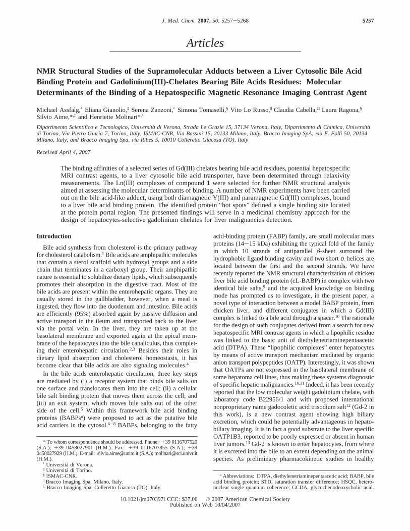
Figure 1. Structure of the Gd(III)-DTPA-bile acid conjugates under investigation in the present paper.
5258 Journal of Medicinal Chemistry, 2007, Vol. 50, No. 22 Assfalg et al.
in Figure 1, belong to a group of conjugates, previously selectedand described,10,16 differing by (i) the nature of bile acid, (ii)the site of conjugation of the Gd(III) complex to the bile acidmoiety, and (iii) the global charge of the conjugate. For eachGd complex, the water relaxivity (defined as the relaxationenhancement of water protons in the presence of the paramag-netic complex at 1 mM concentration) was determined; theobtained values are reported in Table 1. The evaluation of thebinding parameters to cL-BABP was obtained with the relaxo-metric approach called proton relaxation enhancement (PRE),which exploits the increase in relaxation rate of the paramagneticcomplex determined by its binding to a macromolecularsubstrate.17
Two different types of experiments were carried out: in thefirst, the enhancement of the water proton relaxation rate of aGd complex solution was measured in the presence of increasingamounts of cL-BABP, to determine the dissociation constant(Kd) and the relaxivity value (r1p
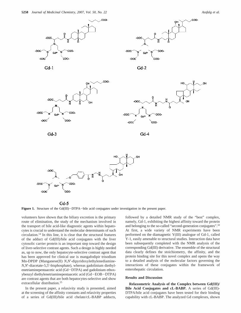
b) of the adduct (Table 1 andSupporting Information). In the second, a fixed concentrationof cL-BABP was titrated with the Gd complex to determinethe number of specific binding sites (n; Figure 2).
In general, it appears that the conjugation of bile acids to theGd(III)-DTPA complex reduces their affinity to the protein,given thatKd values smaller than 10-6 M have been reportedfor the chenodeoxycholic acid.9 For all the selected compounds,a 1:1 protein/ligand stoichiometry was found, differently fromthe 1:2 protein/ligand stoichiometry reported for BABPs fromdifferent species in complex with bile acids.6,8 By inspectionof Table 1, a clear relationship between the affinity constantsand the structural features of the compounds can be inferred.For example, Gd-1, Gd-2, and Gd-3 differ by the number ofhydroxyl groups present on the steroid rings, and the measured
Kd values increase by 1 order of magnitude for each hydroxylgroup added to the steroid (Table 1). It appears that a decreasedhydrophobicity and/or an increase in steric hindrance of the bileacid in the complex determine a lower affinity. Furthermore,when considering the same bile acid, theKd is much lower forthe complexes conjugated at position C24 (Gd-5, Gd-4) than forthose bearing the conjugation at C3 (Gd-3). This observation isin agreement with the recently solved NMR (PDB ID 2JN3)and X-ray (PDB ID 1TW4) structures of cL-BABP in complexwith chenodeoxycholate and cholate molecules, respectively.In these structures, it is clear that the preferred orientation ofthe steroid moieties is the one with the carboxylate tailprotruding toward the solvent and the C3 unit buried within theprotein cavity. Indeed, it is expected that functionalization atC3 forces the bile acid derivative to assume a differentorientation with respect to that observed in the natural bile acid-transporter protein adduct. On the other way it should be keptin mind that many factors enter in the evaluation of the efficacyof potential contrast agents, such as pharmacokinetic evaluations.The analyzed derivatives were previously classified as first-,second-, or third-generation conjugates depending upon the siteof conjugation of the Gd(III) chelate to the bile acid and on thepresence of coupling of the bile acid carboxylic moiety to afunctionalized R-amino acid. It was observed that severalsecond-generation conjugates, where the Gd(III) complex islinked to position C3 of the steroid moiety, showed high biliaryelimination as well as good tolerabilities.10 These observations,together with the higher affinity displayed by Gd-1 toward cL-BABP, made it the compound of choice for further detailedNMR structural analysis.
The determination of the binding stoichiometry of the Gdcomplexes to cL-BABP was obtained through a titrationexperiment monitored by relaxometry analysis, where the proteinconcentration was kept fixed, and that of the Gd complex wasincreased stepwise. The titration data for Gd-1 complex aredisplayed in Figure 2 as an example. In the first part of thetitration, the total concentration of the Gd complex is muchlower than that of the protein, and the Gd complex can beconsidered almost completely bound. Here the increase inrelaxivity depends onn, Kd, andr1p
b (wheren is the number ofbinding sites andr1p
b is the relaxivity of the complex bound tothe protein). In the second part of the titration, the total Gd-complex concentration is much higher than that of the protein,therefore, the protein is almost completely bound and the slopeof the curve depends on the free Gd-complex concentration.The number of binding sites can be estimated from the valueof the protein-ligand molar ratio at which the change in theslope of the titration curve occurs. In the present case, this ratiowas equal to one, because the protein concentration used forthe experiment was 0.13 mM and the change in slope occurredat a ligand concentration of about 0.14 mM. The relaxivityvalues of the supramolecular adducts with cL-BABP are verysimilar for all the investigated complexes, being 21.3( 3 mM-1
s-1 at 20 MHz and 25°C, that is, about three times thecorresponding values for the free complexes.
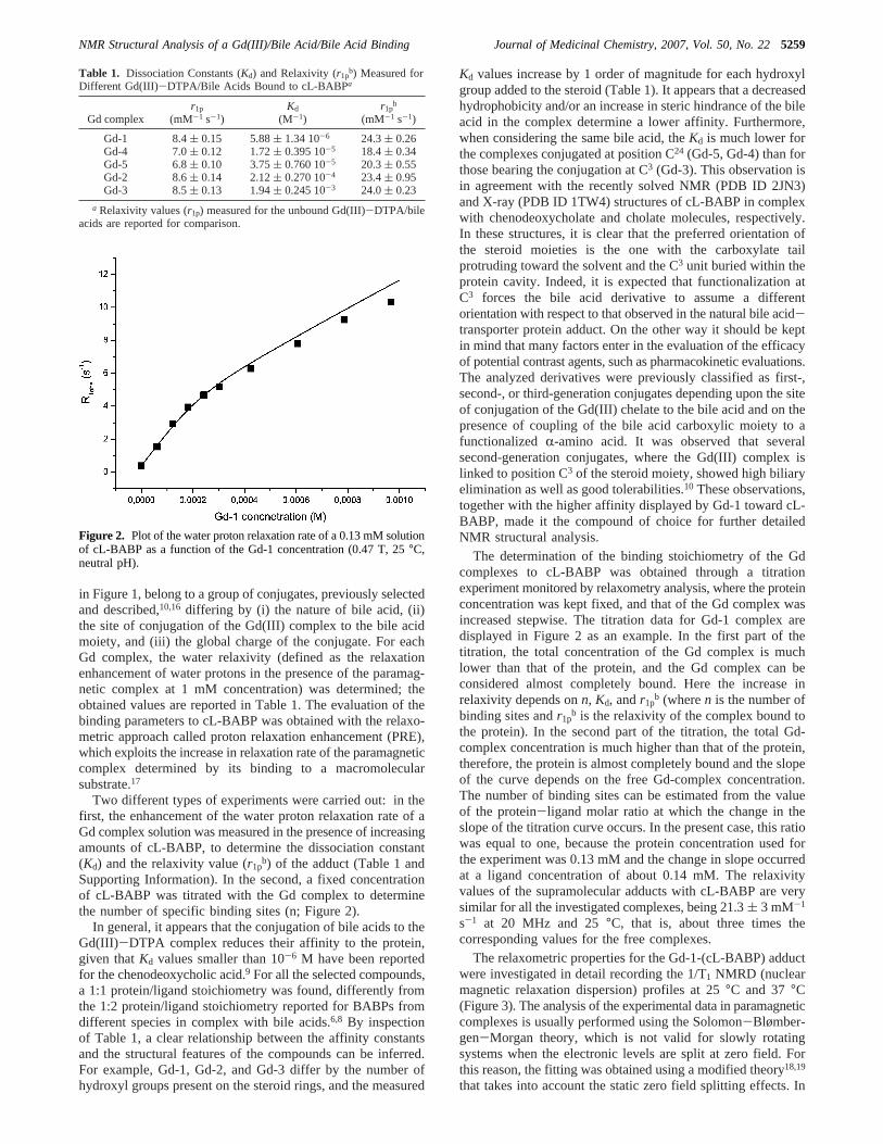
The relaxometric properties for the Gd-1-(cL-BABP) adductwere investigated in detail recording the 1/T1 NMRD (nuclearmagnetic relaxation dispersion) profiles at 25°C and 37°C(Figure 3). The analysis of the experimental data in paramagneticcomplexes is usually performed using the Solomon-Blømber-gen-Morgan theory, which is not valid for slowly rotatingsystems when the electronic levels are split at zero field. Forthis reason, the fitting was obtained using a modified theory18,19
that takes into account the static zero field splitting effects. In
Table 1. Dissociation Constants (Kd) and Relaxivity (r1pb) Measured for
Different Gd(III)-DTPA/Bile Acids Bound to cL-BABPa
Gd complexr1p
(mM-1 s-1)Kd
(M-1)r1p
b
(mM-1 s-1)
Gd-1 8.4( 0.15 5.88( 1.34 10-6 24.3( 0.26Gd-4 7.0( 0.12 1.72( 0.395 10-5 18.4( 0.34Gd-5 6.8( 0.10 3.75( 0.760 10-5 20.3( 0.55Gd-2 8.6( 0.14 2.12( 0.270 10-4 23.4( 0.95Gd-3 8.5( 0.13 1.94( 0.245 10-3 24.0( 0.23
a Relaxivity values (r1p) measured for the unbound Gd(III)-DTPA/bileacids are reported for comparison.
Figure 2. Plot of the water proton relaxation rate of a 0.13 mM solutionof cL-BABP as a function of the Gd-1 concentration (0.47 T, 25°C,neutral pH).
NMR Structural Analysis of a Gd(III)/Bile Acid/Bile Acid Binding Journal of Medicinal Chemistry, 2007, Vol. 50, No. 225259
addition, the equations were modified according to Lipari-Szabo approach. This model allows determining the presenceof local mobility, with an associated short correlation timeτl,on top of a slower global motion with an associated correlationtime τg. The global correlation time associated with the adductGd-1/cL-BABP was assumed to be equal to the proteinτg
measured by high-resolution NMR6 (7.1 ns at 25°C) and wasfixed to that value during the fitting procedure. At 25°C, theobtainedτl value was 504 ps. By increasing the temperature to37 °C, these values decreased to 431 ps and 5.2 ns forτl andτg, respectively. In both cases, the general order parameter (0< S2 < 1) describing the degree of spatial restriction of thelocal motion has been found to be very low (0.15), suggestinga substantial freedom of motion of the Gd complex bound tothe protein. The overall decrease of the molecular reorientationalrates is responsible for the high-field shift of the relaxivity peakobserved in the 1/T1 NMRD profile at 37°C as compared tothe one recorded at 25°C. Interestingly, the relaxivity maximaat the two temperatures is the same (though shifted by ca. 10MHz), suggesting that the attained relaxivities are not quenchedby the occurrence of a not fast enough exchange of thecoordinated water molecule as often reported for supramolecularadducts between albumin and Gd(III) complexes.20,21 In theherein considered system, the detection of a relatively lowrelaxivity value can be ascribed essentially to the occurrenceof a local motion of the Gd(III) chelate around the spacer thatlinks the paramagnetic moiety and the recognition synthon.
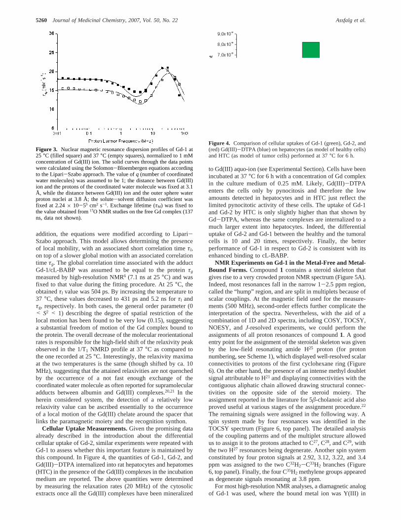
Cellular Uptake Measurements.Given the promising dataalready described in the introduction about the differentialcellular uptake of Gd-2, similar experiments were repeated withGd-1 to assess whether this important feature is maintained bythis compound. In Figure 4, the quantities of Gd-1, Gd-2, andGd(III)-DTPA internalized into rat hepatocytes and hepatomes(HTC) in the presence of the Gd(III) complexes in the incubationmedium are reported. The above quantities were determinedby measuring the relaxation rates (20 MHz) of the cytosolicextracts once all the Gd(III) complexes have been mineralized
to Gd(III) aquo-ion (see Experimental Section). Cells have beenincubated at 37°C for 6 h with a concentration of Gd complexin the culture medium of 0.25 mM. Likely, Gd(III)-DTPAenters the cells only by pynocitosis and therefore the lowamounts detected in hepatocytes and in HTC just reflect thelimited pynocitotic activity of these cells. The uptake of Gd-1and Gd-2 by HTC is only slightly higher than that shown byGd-DTPA, whereas the same complexes are internalized to amuch larger extent into hepatocytes. Indeed, the differentialuptake of Gd-2 and Gd-1 between the healthy and the tumoralcells is 10 and 20 times, respectively. Finally, the betterperformance of Gd-1 in respect to Gd-2 is consistent with itsenhanced binding to cL-BABP.
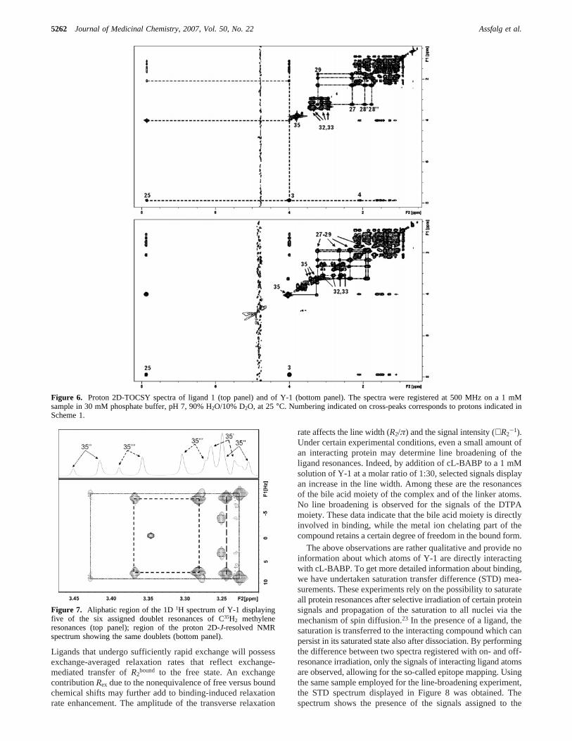
NMR Experiments on Gd-1 in the Metal-Free and Metal-Bound Forms. Compound1 contains a steroid skeleton thatgives rise to a very crowded proton NMR spectrum (Figure 5A).Indeed, most resonances fall in the narrow 1-2.5 ppm region,called the “hump” region, and are split in multiplets because ofscalar couplings. At the magnetic field used for the measure-ments (500 MHz), second-order effects further complicate theinterpretation of the spectra. Nevertheless, with the aid of acombination of 1D and 2D spectra, including COSY, TOCSY,NOESY, andJ-resolved experiments, we could perform theassignments of all proton resonances of compound1. A goodentry point for the assignment of the steroidal skeleton was givenby the low-field resonating amide H25 proton (for protonnumbering, see Scheme 1), which displayed well-resolved scalarconnectivities to protons of the first cyclohexane ring (Figure6). On the other hand, the presence of an intense methyl doubletsignal attributable to H21 and displaying connectivities with thecontiguous aliphatic chain allowed drawing structural connec-tivities on the opposite side of the steroid moiety. Theassignment reported in the literature for 5â-cholanoic acid alsoproved useful at various stages of the assignment procedure.22
The remaining signals were assigned in the following way. Aspin system made by four resonances was identified in theTOCSY spectrum (Figure 6, top panel). The detailed analysisof the coupling patterns and of the multiplet structure allowedus to assign it to the protons attached to C27, C28, and C29, withthe two H27 resonances being degenerate. Another spin systemconstituted by four proton signals at 2.92, 3.12, 3.22, and 3.4ppm was assigned to the two C32H2-C33H2 branches (Figure6, top panel). Finally, the four C35H2 methylene groups appearedas degenerate signals resonating at 3.8 ppm.
For most high-resolution NMR analyses, a diamagnetic analogof Gd-1 was used, where the bound metal ion was Y(III) in
Figure 3. Nuclear magnetic resonance dispersion profiles of Gd-1 at25 °C (filled square) and 37°C (empty squares), normalized to 1 mMconcentration of Gd(III) ion. The solid curves through the data pointswere calculated using the Solomon-Bloembergen equations accordingto the Lipari-Szabo approach. The value ofq (number of coordinatedwater molecules) was assumed to be 1; the distance between Gd(III)ion and the protons of the coordinated water molecule was fixed at 3.1Å, while the distance between Gd(III) ion and the outer sphere waterproton nuclei at 3.8 Å; the solute-solvent diffusion coefficient wasfixed at 2.24× 10-55 cm2 s-1. Exchange lifetime (τM) was fixed tothe value obtained from17O NMR studies on the free Gd complex (137ns, data not shown).
Figure 4. Comparison of cellular uptakes of Gd-1 (green), Gd-2, and(red) Gd(III)-DTPA (blue) on hepatocytes (as model of healthy cells)and HTC (as model of tumor cells) performed at 37°C for 6 h.
5260 Journal of Medicinal Chemistry, 2007, Vol. 50, No. 22 Assfalg et al.
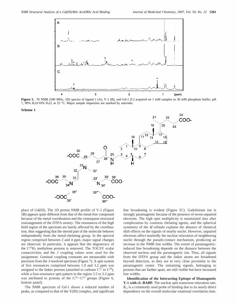
place of Gd(III). The 1D proton NMR profile of Y-1 (Figure5B) appears quite different from that of the metal-free compoundbecause of the metal coordination and the consequent structuralrearrangement of the DTPA moiety. The resonances of the highfield region of the spectrum are barely affected by the coordina-tion, thus suggesting that the steroid part of the molecule behavesindependently from the metal-chelating group. In the spectralregion comprised between 2 and 4 ppm, major signal changesare observed. In particular, it appears that the degeneracy ofthe C35H2 methylene protons is removed. The TOCSY scalarconnectivities and theJ coupling values were used for theassignment. Geminal coupling constants are measurable withprecision from theJ-resolved spectrum (Figure 7). A spin systemof five resonances comprised between 1.9 and 3.2 ppm wasassigned to the linker protons (attached to carbons C27 to C29),while a four-resonance spin pattern in the region 2.5 to 3.2 ppmwas attributed to protons of the C32-C33 groups (Figure 6,bottom panel).
The NMR spectrum of Gd-1 shows a reduced number ofpeaks, as compared to that of the Y(III) complex, and significant
line broadening is evident (Figure 5C). Gadolinium ion isstrongly paramagnetic because of the presence of seven unpairedelectrons. The high spin multiplicity is maintained also aftercomplexation by common chelating agents, and the sphericalsymmetry of the 4f orbitals explains the absence of chemicalshift effects on the signals of nearby nuclei. However, unpairedelectrons affect markedly the nuclear relaxation of neighboringnuclei through the pseudo-contact mechanism, producing anincrease in the NMR line widths. The extent of paramagnetic-induced line broadening depends on the distance between theobserved nucleus and the paramagnetic ion. Thus, all signalsfrom the DTPA group and the linker atoms are broadenedbeyond detection, as they are in very close proximity to theparamagnetic center. The remaining signals, belonging toprotons that are farther apart, are still visible but have increasedline widths.
Identification of the Interacting Epitope of DiamagneticY-1 with cL-BABP. The nuclear spin transverse relaxation rate,R2, is a commonly used probe of binding due to its nearly directdependence on the overall molecular rotational correlation time.
Figure 5. 1H NMR (500 MHz, 1D) spectra of ligand 1 (A), Y-1 (B), and Gd-1 (C) acquired on 1 mM samples in 30 mM phosphate buffer, pH7, 90% H2O/10% D2O, at 25°C. Major sample impurities are marked by asterisks.
Scheme 1
NMR Structural Analysis of a Gd(III)/Bile Acid/Bile Acid Binding Journal of Medicinal Chemistry, 2007, Vol. 50, No. 225261
Ligands that undergo sufficiently rapid exchange will possessexchange-averaged relaxation rates that reflect exchange-mediated transfer ofR2
bound to the free state. An exchangecontributionRex due to the nonequivalence of free versus boundchemical shifts may further add to binding-induced relaxationrate enhancement. The amplitude of the transverse relaxation
rate affects the line width (R2/π) and the signal intensity (∝R2-1).
Under certain experimental conditions, even a small amount ofan interacting protein may determine line broadening of theligand resonances. Indeed, by addition of cL-BABP to a 1 mMsolution of Y-1 at a molar ratio of 1:30, selected signals displayan increase in the line width. Among these are the resonancesof the bile acid moiety of the complex and of the linker atoms.No line broadening is observed for the signals of the DTPAmoiety. These data indicate that the bile acid moiety is directlyinvolved in binding, while the metal ion chelating part of thecompound retains a certain degree of freedom in the bound form.
The above observations are rather qualitative and provide noinformation about which atoms of Y-1 are directly interactingwith cL-BABP. To get more detailed information about binding,we have undertaken saturation transfer difference (STD) mea-surements. These experiments rely on the possibility to saturateall protein resonances after selective irradiation of certain proteinsignals and propagation of the saturation to all nuclei via themechanism of spin diffusion.23 In the presence of a ligand, thesaturation is transferred to the interacting compound which canpersist in its saturated state also after dissociation. By performingthe difference between two spectra registered with on- and off-resonance irradiation, only the signals of interacting ligand atomsare observed, allowing for the so-called epitope mapping. Usingthe same sample employed for the line-broadening experiment,the STD spectrum displayed in Figure 8 was obtained. Thespectrum shows the presence of the signals assigned to the
Figure 6. Proton 2D-TOCSY spectra of ligand 1 (top panel) and of Y-1 (bottom panel). The spectra were registered at 500 MHz on a 1 mMsample in 30 mM phosphate buffer, pH 7, 90% H2O/10% D2O, at 25°C. Numbering indicated on cross-peaks corresponds to protons indicated inScheme 1.
Figure 7. Aliphatic region of the 1D1H spectrum of Y-1 displayingfive of the six assigned doublet resonances of C35H2 methyleneresonances (top panel); region of the proton 2D-J-resolved NMRspectrum showing the same doublets (bottom panel).
5262 Journal of Medicinal Chemistry, 2007, Vol. 50, No. 22 Assfalg et al.
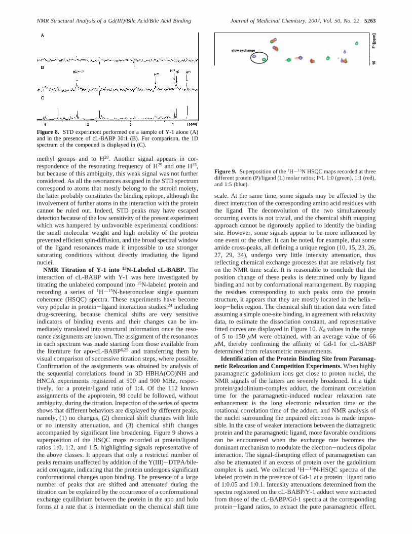
methyl groups and to H20. Another signal appears in cor-respondence of the resonating frequency of H29 and one H35,but because of this ambiguity, this weak signal was not furtherconsidered. As all the resonances assigned in the STD spectrumcorrespond to atoms that mostly belong to the steroid moiety,the latter probably constitutes the binding epitope, although theinvolvement of further atoms in the interaction with the proteincannot be ruled out. Indeed, STD peaks may have escapeddetection because of the low sensitivity of the present experimentwhich was hampered by unfavorable experimental conditions:the small molecular weight and high mobility of the proteinprevented efficient spin-diffusion, and the broad spectral windowof the ligand resonances made it impossible to use strongersaturating conditions without directly irradiating the ligandnuclei.
NMR Titration of Y-1 into 15N-Labeled cL-BABP. Theinteraction of cL-BABP with Y-1 was here investigated bytitrating the unlabeled compound into15N-labeled protein andrecording a series of1H-15N-heteronuclear single quantumcoherence (HSQC) spectra. These experiments have becomevery popular in protein-ligand interaction studies,24 includingdrug-screening, because chemical shifts are very sensitiveindicators of binding events and their changes can be im-mediately translated into structural information once the reso-nance assignments are known. The assignment of the resonancesin each spectrum was made starting from those available fromthe literature for apo-cL-BABP6,25 and transferring them byvisual comparison of successive titration steps, where possible.Confirmation of the assignments was obtained by analysis ofthe sequential correlations found in 3D HBHA(CO)NH andHNCA experiments registered at 500 and 900 MHz, respec-tively, for a protein/ligand ratio of 1:4. Of the 112 knownassignments of the apoprotein, 98 could be followed, withoutambiguity, during the titration. Inspection of the series of spectrashows that different behaviors are displayed by different peaks,namely, (1) no changes, (2) chemical shift changes with littleor no intensity attenuation, and (3) chemical shift changesaccompanied by significant line broadening. Figure 9 shows asuperposition of the HSQC maps recorded at protein/ligandratios 1:0, 1:2, and 1:5, highlighting signals representative ofthe above classes. It appears that only a restricted number ofpeaks remains unaffected by addition of the Y(III)-DTPA/bile-acid conjugate, indicating that the protein undergoes significantconformational changes upon binding. The presence of a largenumber of peaks that are shifted and attenuated during thetitration can be explained by the occurrence of a conformationalexchange equilibrium between the protein in the apo and holoforms at a rate that is intermediate on the chemical shift time
scale. At the same time, some signals may be affected by thedirect interaction of the corresponding amino acid residues withthe ligand. The deconvolution of the two simultaneouslyoccurring events is not trivial, and the chemical shift mappingapproach cannot be rigorously applied to identify the bindingsite. However, some signals appear to be more influenced byone event or the other. It can be noted, for example, that someamide cross-peaks, all defining a unique region (10, 15, 23, 26,27, 29, 34), undergo very little intensity attenuation, thusreflecting chemical exchange processes that are relatively faston the NMR time scale. It is reasonable to conclude that theposition change of these peaks is determined only by ligandbinding and not by conformational rearrangement. By mappingthe residues corresponding to such peaks onto the proteinstructure, it appears that they are mostly located in the helix-loop-helix region. The chemical shift titration data were fittedassuming a simple one-site binding, in agreement with relaxivitydata, to estimate the dissociation constant, and representativefitted curves are displayed in Figure 10.Kd values in the rangeof 5 to 150µM were obtained, with an average value of 66µM, thereby confirming the affinity of Gd-1 for cL-BABPdetermined from relaxometric measurements.
Identification of the Protein Binding Site from Paramag-netic Relaxation and Competition Experiments.When highlyparamagnetic gadolinium ions get close to proton nuclei, theNMR signals of the latters are severely broadened. In a tightprotein/gadolinium-complex adduct, the dominant correlationtime for the paramagnetic-induced nuclear relaxation rateenhancement is the long electronic relaxation time or therotational correlation time of the adduct, and NMR analysis ofthe nuclei surrounding the unpaired electrons is made impos-sible. In the case of weaker interactions between the diamagneticprotein and the paramagnetic ligand, more favorable conditionscan be encountered when the exchange rate becomes thedominant mechanism to modulate the electron-nucleus dipolarinteraction. The signal-disrupting effect of paramagnetism canalso be attenuated if an excess of protein over the gadoliniumcomplex is used. We collected1H-15N-HSQC spectra of thelabeled protein in the presence of Gd-1 at a protein-ligand ratioof 1:0.05 and 1:0.1. Intensity attenuations determined from thespectra registered on the cL-BABP/Y-1 adduct were subtractedfrom those of the cL-BABP/Gd-1 spectra at the correspondingprotein-ligand ratios, to extract the pure paramagnetic effect.
Figure 8. STD experiment performed on a sample of Y-1 alone (A)and in the presence of cL-BABP 30:1 (B). For comparison, the 1Dspectrum of the compound is displayed in (C).
Figure 9. Superposition of the1H-15N HSQC maps recorded at threedifferent protein (P)/ligand (L) molar ratios; P/L 1:0 (green), 1:1 (red),and 1:5 (blue).
NMR Structural Analysis of a Gd(III)/Bile Acid/Bile Acid Binding Journal of Medicinal Chemistry, 2007, Vol. 50, No. 225263
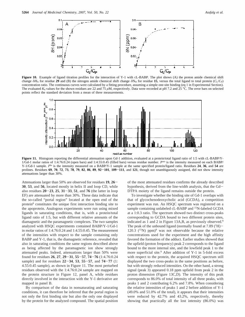
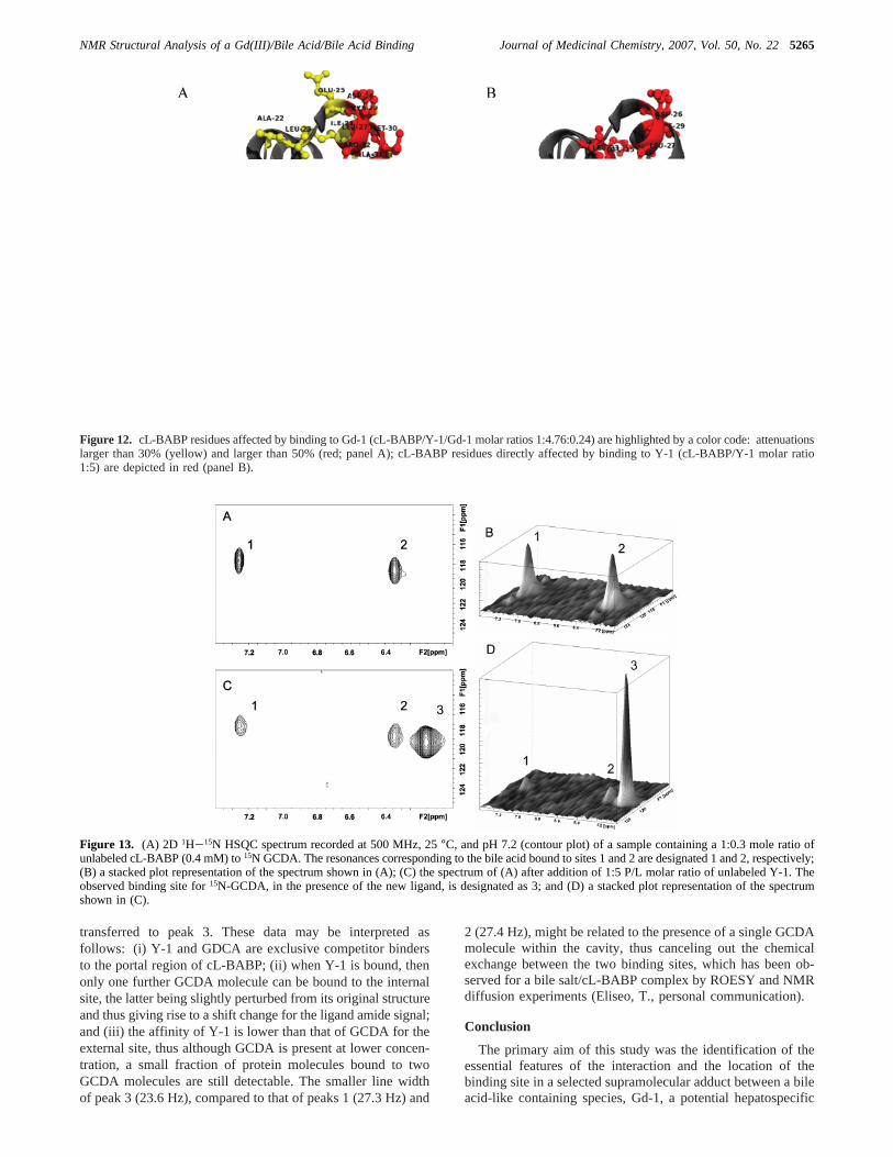
Attenuations larger than 50% are observed for residues19, 26-30, 53, and56, located mostly in helix II and loop CD, whilealso residues20-23, 25, 31-33, 51, and76 (the latter in loopEF) are attenuated by more than 30%. These data indicate thatthe so-called “portal region” located at the open end of theprotein6 constitutes the unique first interaction binding site tothe apoprotein. Analogous experiments were run using mixedligands in saturating conditions, that is, with a protein/totalligand ratio of 1:5, but with different relative amounts of thediamagnetic and the paramagnetic complexes. The two samplesanalyzed with HSQC experiments contained BABP/Y-1/Gd-1in molar ratios of 1:4.76:0.24 and 1:4.55:0.45. The measurementof the intensities with respect to the sample containing onlyBABP and Y-1, that is, the diamagnetic reference, revealed thatalso in saturating conditions the same regions described aboveas being affected by the paramagnetic ion show stronglyattenuated peaks. Indeed, attenuations larger than 50% werefound for residues26, 27, 29-31, 55-57, 74-76 (1:4.76:0.24sample) and for residues22-34, 53, 55-57, and 74-77 (1:4.55:0.45 sample), as shown in Figure 11. The most perturbedresidues observed with the 1:4.76:0.24 sample are mapped onthe protein structure in Figure 12, panel A, while residuesdirectly involved in the interaction with the Y-1 derivative aremapped in panel B.
By comparison of the data in nonsaturating and saturatingconditions, it can therefore be inferred that the portal region isnot only the first binding site but also the only one displayedby the protein for the analyzed compound. The spatial position
of the most attenuated residues confirms the already describedhypothesis, derived from the line-width analysis, that the Gd-DTPA moiety of the ligand remains outside the protein.
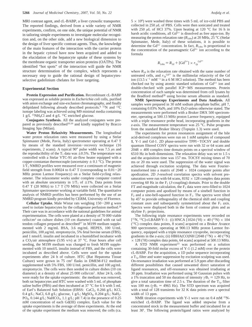
To investigate whether the binding site of Gd-1 overlaps withthat of glycochenodeoxycholic acid (GCDA), a competitionexperiment was run. An HSQC spectrum was registered on asample containing unlabeled cL-BABP and15N-labeled GCDAat a 1:0.3 ratio. The spectrum showed two distinct cross-peakscorresponding to GCDA bound to two different protein sites,indicated as 1 and 2 in Figure 13A,B, as previously observed.9
The peak of the unbound ligand (normally found at 7.89 (1H)-120.3 (15N) ppm)9 was not observable because the relativeconcentrations used for the experiment and the high affinityfavored the formation of the adduct. Earlier studies showed thatthe upfield (proton frequency) peak 2 corresponds to the ligandbound to the more internal site, and the lowfield peak 1 to themore superficial site.9 After addition of Y-1 in 5-fold excesswith respect to the protein, the acquired HSQC spectrum stilldisplayed the two cross-peaks in the same positions as before,but with strongly reduced intensities. On the other hand, a strongsignal (peak 3) appeared 0.18 ppm upfield from peak 2 in theproton dimension (Figure 13C,D). The intensity of this peakcorresponds to 86.0% of total intensity of all three peaks, withpeaks 1 and 2 contributing 6.2% and 7.8%. When consideringthe relative intensities of peaks 1 and 2 before addition of Y-1(49.0% and 51.0% of the total), it appears that their intensitieswere reduced by 42.7% and 43.2%, respectively, therebyshowing that practically all the lost intensity (86.0%) was
Figure 10. Example of ligand titration profiles for the interaction of Y-1 with cL-BABP. The plot shows (A) the proton amide chemical shiftchangeδHN for residue29 and (B) the nitrogen amide chemical shift changeδNH for residue15, versus the total ligand to total protein (CL/CP)concentration ratio. The continuous curves were calculated by a fitting procedure, assuming a simple one-site binding (eq 1 in Experimental Section).The evaluatedKd values for the shown residues are 22 and 75µM, respectively. Data were recorded at pH 7.2 and 25°C. The error bars on selectedpoints reflect the standard deviation from a mean of three measurements.
Figure 11. Histogram reporting the differential attenuation upon Gd-1 addition, evaluated at a protein/total ligand ratio of 1:5 with cL-BABP/Y-1/Gd-1 molar ratios of 1:4.76:0.24 (open bars) and 1:4.55:0.45 (filled bars) versus residue number.Ipara is the intensity measured on each BABP/Y-1/Gd-1 sample.Idia is the intensity measured on a BABP/Y-1 sample at the same specified protein/ligand ratio. Residues24, 36, and54 areprolines. Residues69, 70, 72, 73, 78, 79, 82, 86, 89, 92-101, 109-111, and121, though not unambiguously assigned, did not show intensityattenuations larger than 30%.
5264 Journal of Medicinal Chemistry, 2007, Vol. 50, No. 22 Assfalg et al.
transferred to peak 3. These data may be interpreted asfollows: (i) Y-1 and GDCA are exclusive competitor bindersto the portal region of cL-BABP; (ii) when Y-1 is bound, thenonly one further GCDA molecule can be bound to the internalsite, the latter being slightly perturbed from its original structureand thus giving rise to a shift change for the ligand amide signal;and (iii) the affinity of Y-1 is lower than that of GCDA for theexternal site, thus although GCDA is present at lower concen-tration, a small fraction of protein molecules bound to twoGCDA molecules are still detectable. The smaller line widthof peak 3 (23.6 Hz), compared to that of peaks 1 (27.3 Hz) and
2 (27.4 Hz), might be related to the presence of a single GCDAmolecule within the cavity, thus canceling out the chemicalexchange between the two binding sites, which has been ob-served for a bile salt/cL-BABP complex by ROESY and NMRdiffusion experiments (Eliseo, T., personal communication).
Conclusion
The primary aim of this study was the identification of theessential features of the interaction and the location of thebinding site in a selected supramolecular adduct between a bileacid-like containing species, Gd-1, a potential hepatospecific
Figure 12. cL-BABP residues affected by binding to Gd-1 (cL-BABP/Y-1/Gd-1 molar ratios 1:4.76:0.24) are highlighted by a color code: attenuationslarger than 30% (yellow) and larger than 50% (red; panel A); cL-BABP residues directly affected by binding to Y-1 (cL-BABP/Y-1 molar ratio1:5) are depicted in red (panel B).
Figure 13. (A) 2D 1H-15N HSQC spectrum recorded at 500 MHz, 25°C, and pH 7.2 (contour plot) of a sample containing a 1:0.3 mole ratio ofunlabeled cL-BABP (0.4 mM) to15N GCDA. The resonances corresponding to the bile acid bound to sites 1 and 2 are designated 1 and 2, respectively;(B) a stacked plot representation of the spectrum shown in (A); (C) the spectrum of (A) after addition of 1:5 P/L molar ratio of unlabeled Y-1. Theobserved binding site for15N-GCDA, in the presence of the new ligand, is designated as 3; and (D) a stacked plot representation of the spectrumshown in (C).
NMR Structural Analysis of a Gd(III)/Bile Acid/Bile Acid Binding Journal of Medicinal Chemistry, 2007, Vol. 50, No. 225265
MRI contrast agent, and cL-BABP, a liver cytosolic transporter.The reported findings, derived from a wide variety of NMRexperiments, confirm, on one side, the unique potential of NMRin tailoring simple experiments to investigate molecular recogni-tion and, on the other side, add a new biological dimension tothe design of liver specific contrast agents. Thus, the knowledgeof the main features of the interaction with the carrier proteinin the hepatic cytosol have now been acquired and added tothe elucidation of the hepatocyte uptake of these systems bythe membrane organic transporter anion proteins (OATPs). Theidentified “hot spots” of the interaction will guide the NMRstructure determination of the complex, which represents anecessary step to guide the rational design of hepatocytes-selective gadolinium chelates for liver targeting.
Experimental Section
Protein Expression and Purification. Recombinant cL-BABPwas expressed as soluble protein inEscherichia colicells, purifiedwith anion exchange and size-exclusion chromatography, and finallydelipidated following already described protocols.6 15N and 13Cisotope labeling was achieved using M9 minimal media containing1 g/L 15NH4Cl and 4 g/L13C enriched glucose.
Conjugates Synthesis.All the analyzed conjugates were pre-pared as previously described10,16 and kindly supplied by BraccoImaging Spa (Milan).
Water Proton Relaxivity Measurements. The longitudinalwater proton relaxation rates were measured by using a StelarSpinmaster (Mede, Pavia, Italy) spectrometer operating at 0.47 Tby means of the standard inversion-recovery technique (16experiments, 2 scans). A typical 90° pulse width was 7.5µs andthe reproducibility of the T1 data was(0.5%. The temperature wascontrolled with a Stelar VTC-91 air-flow heater equipped with acopper-constantan thermocouple (uncertainty( 0.1°C). The proton1/T1 NMRD profiles were measured over a continuum of magneticfield strength from 0.00024 to 0.47 T (corresponding to 0.01-20MHz proton Larmor Frequency) on a Stelar field-cycling relax-ometer. The relaxometer works under complete computer controlwith an absolute uncertainty in 1/T1 of (1%. Data points from0.47 T (20 MHz) to 1.7 T (70 MHz) were collected on a StelarSpinmaster spectrometer working at variable field. The quantitativeanalysis of NMRD profiles has been performed by the use of anNMRD program kindly provided by CERM, University of Florence.
Cellular Uptake. Male Wistar rats weighing 150-200 g wereused to isolate hepatocytes by the collagenase perfusion method.26
Their care was in accordance with the national guidelines for animalexperimentation. The cells were plated at a density of 70 000 viablecells/cm2 on culture dishes (10 cm diameter) coated with rat tailtendon collagen prepared as described27 in M199 medium supple-mented with 2 mg/mL BSA, 3.6 mg/mL HEPES, 100 U/mLpenicillin, 100µg/mL streptomycin, 5% fetal bovine serum (FBS),and 1 nmol/L insulin and incubated in a humidified incubator witha CO2/air atmosphere (5:95 v/v) at 37°C. Four hours after cellseeding, the M199 medium was changed to fresh M199 supple-mented with 10 nmol/L insulin instead of FBS, and the cells wereincubated as described above. Cells were used for the uptakeexperiments after 24 h of culture. HTC (Rat Hepatoma TissueCulture) were grown in 75 cm2 flasks in DMEM-F12 mediumsupplemented with 5% FBS, 100 U/mL penicillin, and 100µg/mLstreptomycin. The cells were then seeded in culture dishes (10 cmdiameter) at a density of about 25 000 cells/cm2. After 24 h, cellswere ready for the uptake experiments: the medium was removed,both rat hepatocytes and HTC were washed with 5 mL of phosphatesaline buffer (PBS) and then incubated at 37°C for 6 h with 5 mLof Earl’s Balanced Salt Solution (EBSS: CaCl2, 0.266 g/L; KCl,0.4 g/L; NaCl, 6.8 g/L; glucose, 1 g/L; MgSO4, 0.204 g/L; NaH2-PO4, 0.144 g/L; NaHCO3, 1.1 g/L; pH 7.4) in the presence of 0.25mM concentration of each Gd(III) complex. Each value for theuptake experiments is the average of three experiments. At the endof the uptake experiment the medium was removed; the cells (ca.
5 × 10⁶) were washed three times with 5 mL of ice-cold PBS andcollected in 250µL of PBS. Cells were then sonicated and treatedwith 250 µL of HCl 37%. Upon heating at 120°C for 16 h inharsh acidic conditions, all Gd3+ is dissolved as free aquo-ion. Bymeasuring the proton relaxation rate (R1obs) at 20 MHz, 25°C (StelarSpinmaster, Mede, Italy) of these solutions, it is possible todetermine the Gd3+ concentration. In fact,R1obs is proportional tothe concentration of the paramagnetic Gd3+ ion according to theformula
whereR1d is the relaxation rate obtained with the same number ofuntreated cells, andr1p
Gd3+ is the millimolar relaxivity of the Gdion (13.5 s-1 mM-1 in a 6 M HCl solution). The method has beenchecked out by using atomic standard solutions of Gd3+ ion anddouble-checked with parallel ICP-MS measurements. Proteinconcentration of each sample was determined from cell lysates bythe Bradford method28 using bovine serum albumin as standard.
NMR Spectroscopy Experiments and Data Analysis.Allsamples were prepared in 30 mM sodium phosphate buffer, pH 7,containing 0.03% NaN3 and 10% D2O unless otherwise stated. Themeasurements were performed with a Bruker DRX 500 spectrom-eter, operating at 500.13 MHz proton Larmor frequency, equippedwith a triple resonance probe head, incorporating gradients in thez-axis. The measurement temperature was 25°C. Pulse programsfrom the standard Bruker library (Topspin 1.3) were used.
The experiments for proton resonances assignment of the bileacid-derived complexes were run on 1 mM samples of compound1, Y-1 and Gd-1. 2D phase-sensitive TPPI-TOCSY and doublequantum filtered COSY spectra were run with 32 or 64 scans and2048× 400 complex time domain points on a spectral window of6510 Hz in both dimensions. The relaxation delay was set to 1.5 sand the acquisition time was 157 ms. TOCSY mixing times of 70ms or 20 ms were used. The suppression of the water signal wasachieved through excitation sculpting. The data were Fourier-transformed into a matrix of 2048× 1024 computer points afterapodization. 2DJ-resolved correlation spectra with solvent pre-saturation were run with 64 scans, 8192× 128 complex data pointscovering 60 Hz in the F1 (J-coupling) domain. Prior to the doubleFT and magnitude calculation, the F1 data were zero-filled to 1024computer points and apodized by means of a sinebell function inF2 and a sine-bell-squared function in F1. The spectra were tiltedby 45° to provide orthogonality of the chemical shift and couplingconstant axes and subsequently symmetrized about the F1 axis.Spectra were displayed both in the form of contour plots and asskyline F2 projections.
The following triple resonance experiments were recorded on[15N, 13C]-cLBABP/Y-1: (i) HNCA [1024 (1H) × 40 (15N) × 104(13C) complex data points, 8 scans] acquired on a Bruker Avance900 spectrometer, operating at 900.13 MHz proton Larmor fre-quency, equipped with a triple resonance cryoprobe, incorporatinggradients in thez-axis; (ii) HBHA(CO)NH [2048 (15N) × 40 (15N)× 128 (1H) complex data points, 64 scans] acquired at 500.13 MHz.
A STD NMR experiment23 was performed on a solutioncontaining 30-fold molar excess of Y-1 (1.2 mM) over the proteincL-BABP. For the acquisition, a 1D pulse sequence incorporatinga T1F filter and water suppression by excitation sculpting was used.On-resonance irradiation was performed at 5.9 ppm after discardingdifferent possibilities that caused unwanted direct saturation ofligand resonances, and off-resonance was obtained irradiating at30 ppm. Irradiation was performed using 50 Gaussian pulses witha 1% truncation and 50 ms duration of intensityγB1 ) 129 Hz, togive a total saturation time of 2.5 s. The duration of the T1F filterwas 100 ms (γB1 ) 4965 Hz). The STD spectrum was acquiredwith a total of 128 transients for 32 K data points over a spectralwidth of 13 ppm.
NMR titration experiments with Y-1 were run on 0.4 mM15N-enriched cL-BABP. The ligand was added stepwise from aconcentrated stock in the measurement buffer and incubated for atleast 30′. The following protein/ligand ratios were analyzed by
R1obs) R1d + [Gd3+] × r1pGd3+
5266 Journal of Medicinal Chemistry, 2007, Vol. 50, No. 22 Assfalg et al.

running HSQC correlation spectra (1H-15N-HSQC): 1:0.025,1:0.05, 1:0.25, 1:0.5, 1:0.75, 1:1, 1:1.25, 1:2, 1:3, 1:5. A similarexperimental approach was used to analyze the interaction of Gd-1to labeled cL-BABP, at molar ratios of 1:0.05 and 1:0.1. Intensityattenuations were calculated as (Idia - Ipara)/Idia, where Ipara wasmeasured from the samples containing Gd-1, whileIdia wasmeasured from the samples containing Y-1 in the same protein-ligand molar ratios. To evaluate the paramagnetic effect ofgadolinium in saturating conditions, another set of experiments wasrun by adding to a protein/Y-1(1:5) sample, aliquots of a stockcontaining the same concentrations of protein/Gd-1(1:5). In thisway, the protein and total ligand concentrations were kept fixed,while changing only the Y-1/Gd-1 molar ratio. HSQC measurementswere run on samples containing BABP/Y-1/Gd-1 in molar ratiosof 1:5:0 (used as diamagnetic reference to evaulateIdia), 1:4.76:0.24 and 1:4.55:0.45. Finally, a competition experiment was runusing a 0.4 mM unlabeled-protein sample and15N-labeled GCDAat a molar ratio of 1:0.3. The signals of the bound GCDA and thedisplacement of GCDA after addition of unlabeled Y-1 at a finalprotein/Y-1 molar ratio of 1:5 were observed via HSQC NMRspectroscopy.1H-15N-HSQC spectra were collected as gradient-and sensitivity-enhanced correlation spectra and acquired with a1H spectral width of 6010 Hz and 2048 complex points, zero-filledto a total of 2048 points. In the15N dimension, 128 hypercomplexincrements were collected and zero-filled to a total of 512 points.Typically 8, 16, or 32 transients were collected. In the case of thecompetition experiment, the number of scans was increased to 1024,and the number of collected increments decreased to 48. Spectraanalysis and peak intensity measurements were performed withTopspin 1.3 (Bruker, Karlsruhe) and Sparky 3.11 (UCSF). Thechemical shift changes in the titration experiment were analyzedboth as separate proton and nitrogen frequencies and as weightedaverage displacements. The NMR isotherms were fitted to a single-step one-site binding model to obtainKd values with the MATLABprogram, according to the following equation
whereδobs, δb, andδf are the chemical shifts of the protein signalsin the observed, bound, and free states;CP and CL are the totalconcentrations of the protein and the ligand, respectively.
Protein stock solution concentrations were determined by UV,while the ligand stock solutions were determined by measuring dryweights using a microbalance. Random samples were prepared threetimes, and the error on the measured points reflects the standarddeviation from the mean.
Acknowledgment. This research was supported by FIRB2003 (Grant RBNE03B8KK) and MIUR 2004 from the ItalianMinistry for Education, University and Research. The supportof CIRMMP (Consorzio Interuniversitario di Risonanze Mag-netiche di Metalloproteine Paramagnetiche) is gratefully ac-knowledged. Ivano Bertini, Claudio Luchinat, and GiacomoParigi from CERM, University of Florence, kindly provided theirsoftware for relaxometric analysis. Lucia Zetta (ISMAC, CNR,Milano) critically read the manuscript and Mara Guariento andMassimo Pedo` (University of Verona) helped in proteinexpression.
Supporting Information Available: Relaxometric determina-tion of the binding affinity between Gd-L complexes and cL-BABP(PRE method). This material is available free of charge via theInternet at http://pubs.acs.org.
References(1) Houten, S. M.; Watanabe, M.; Auwerx, J. Endocrine functions of
bile acids.EMBO J.2006, 25, 1419-25.
(2) Chiang, J. Y. Bile acid regulation of gene expression: Roles ofnuclear hormone receptors.Endocr. ReV. 2002, 23, 443-63.
(3) Russell, D. W. The enzymes, regulation, and genetics of bile acidsynthesis.Annu. ReV. Biochem.2003, 72, 137-74.
(4) Qiao, L.; Han, S. I.; Fang, Y.; Park, J. S.; Gupta, S.; Gilfor, D.;Amorino, G.; Valerie, K.; Sealy, L.; Engelhardt, J. F.; Grant, S.;Hylemon, P. B.; Dent, P. Bile acid regulation of C/EBPbeta, CREB,and c-Jun function, via the extracellular signal-regulated kinase andc-Jun NH2-terminal kinase pathways, modulates the apoptoticresponse of hepatocytes.Mol. Cell. Biol. 2003, 23, 3052-66.
(5) Dawson, P. A.; Oelkers, P. Bile acid transporters.Curr. Opin. Lipidol.1995, 6, 109-14.
(6) Ragona, L.; Catalano, M.; Luppi, M.; Cicero, D.; Eliseo, T.; Foote,J.; Fogolari, F.; Zetta, L.; Molinari, H. NMR dynamic studies suggestthat allosteric activation regulates ligand binding in chicken liver bileacid-binding protein.J. Biol. Chem.2006, 281, 9697-709.
(7) Nichesola, D.; Perduca, M.; Capaldi, S.; Carrizo, M. E.; Righetti, P.G.; Monaco, H. L. Crystal structure of chicken liver basic fatty acidbinding protein complexed with cholic acid.Biochemistry2004, 43,14072-9.
(8) Tochtrop, G. P.; Richter, K.; Tang, C.; Toner, J. J.; Covey, D. F.;Cistola, D. P. Energetics by NMR: Site-specific binding in apositively cooperative system.Proc. Natl. Acad. Sci. U.S.A.2002,99, 1847-52.
(9) Tomaselli, S.; Ragona, L.; Zetta, L.; Assfalg, M.; Ferranti, P.; Longhi,R.; Bonvin, A. M. J. J.; Molinari, H. NMR-based modelling andbinding studies of a ternary complex between chicken liver bile acidbinding protein and bile acids.Proteins2007, 69, 177-191.
(10) Anelli, P. L.; Lattuada, L.; Lorusso, V.; Lux, G.; Morisetti, A.;Morosini, P.; Serleti, M.; Uggeri, F. Conjugates of gadoliniumcomplexes to bile acids as hepatocyte-directed contrast agents formagnetic resonance imaging.J. Med. Chem.2004, 47, 3629-41.
(11) von Dippe, P.; Levy, D. Expression of the bile acid transport proteinduring liver development and in hepatoma cells.J. Biol. Chem.1990,265, 5942-5.
(12) Cavagna, F. M.; Lorusso, V.; Anelli, P. L.; Maggioni, F.; de Haen,C. Preclinical profile and clinical potential of gadocoletic acidtrisodium salt (B22956/1), a new intravascular contrast medium forMRI. Acad. Radiol.2002, 9 (Suppl 2), S491-4.
(13) Libra, A.; Fernetti, C.; Lorusso, V.; Visigalli, M.; Anelli, P. L.; Staud,F.; Tiribelli, C.; Pascolo, L. Molecular determinants in the transportof a bile acid-derived diagnostic agent in tumoral and nontumoralcell lines of human liver.J. Pharmacol. Exp. Ther.2006, 319, 809-17.
(14) Lorusso, V.; Pascolo, L.; Fernetti, C.; Visigalli, M.; Anelli, P.;Tiribelli, C. In vitro and in vivo hepatic transport of the magneticresonance imaging contrast agent B22956/1: Role of MRP proteins.Biochem. Biophys. Res. Commun.2002, 293, 100-5.
(15) Karabulut, N.; Elmas, N. Contrast agents used in MR imaging ofthe liver.Diagn. InterV. Radiol.2006, 12, 22-30.
(16) De Haen, C.; Beltrami, A.; Cappelletti, E.; Lattuada, L.; Virtuani,M. Bile acid conjugates with metal ion chelates and the use thereof.U.S. Patent US2003113265.2003, 2003.
(17) Kirsch, J. E. Basic principles of magnetic resonance contrast agents.Top. Magn. Reson. Imaging1991, 3, 1-18.
(18) Bertini, I.; Galas, O.; Luchinat, C.; Parigi, G. Computer Programfor the calculation of paramagnetic enhancements nuclear-relaxationrates in slowly rotating systems.J. Magn. Reson.1995, 113, 151-156.
(19) Bertini, I.; Kowalewski, J.; Luchinat, C.; Parigi, G. Cross correlationbetween the dipole-dipole interaction and the Curie spin relaxation:The effect of anisotropic magnetic susceptibility.J. Magn. Reson.2001, 152, 103-8.
(20) Aime, S.; Chiaussa, M.; Digilio, G.; Gianolio, E.; Terreno, E. Contrastagents for magnetic resonance angiographic applications: H-1 andO-17 NMR relaxometric investigations on two gadolinium(III)DTPA-like chelates endowed with high binding affinity to humanserum albumin.J. Biol. Inorg. Chem.1999, 4, 766-774.
(21) Aime, S.; Gianolio, E.; Longo, D.; Pagliarin, R.; Lovazzano, C.; Sisti,M. New insights for pursuing high relaxivity MRI agents frommodelling the binding interaction of Gd-III chelates to HSA.ChemBioChem2005, 6, 818.
(22) Waterhous, D. V.; Barnes, S.; Muccio, D. D. Nuclear magneticresonance spectroscopy of bile acids. Development of two-dimensional NMR methods for the elucidation of proton resonanceassignments for five common hydroxylated bile acids, and their parentbile acid, 5 beta-cholanoic acid.J. Lipid Res.1985, 26, 1068-78.
(23) Meyer, B.; Peters, T. NMR spectroscopy techniques for screeningand identifying ligand binding to protein receptors.Angew. Chem.,Int. Ed. 2003, 42, 864-90.
(24) Shuker, S. B.; Hajduk, P. J.; Meadows, R. P.; Fesik, S. W.Discovering high-affinity ligands for proteins: SAR by NMR.Science1996, 274, 1531-4.
∆δobs) (δb - δf
2CP)[(CP + CL + Kd) -
x(CP + CL + Kd)2 - 4CPCL] (1)
NMR Structural Analysis of a Gd(III)/Bile Acid/Bile Acid Binding Journal of Medicinal Chemistry, 2007, Vol. 50, No. 225267
(25) Vasile, F.; Ragona, L.; Catalano, M.; Zetta, L.; Perduca, M.; Monaco,H.; Molinari, H. Solution structure of chicken liver basic fatty acidbinding protein.J. Biomol. NMR2003, 25, 157-60.
(26) Probst, I.; Unthan-Fechner, K. Activation of glycolysis by insulinwith a sequential increase of the 6-phosphofructo-2-kinase activity,fructose-2,6-bisphosphate level and pyruvate kinase activity incultured rat hepatocytes.Eur. J. Biochem.1985, 153, 347-353.
(27) Strom, C. S.; Michalopoulos, G. Collagen as substrate for cell growthand differentiation.Methods Enzymol.1982, 82, 544-555.
(28) Bradford, M. M. A rapid and sensitive method for the quantitationof microgram quantities of protein utilizing the principle of protein-dye binding.Anal. Biochem.1986, 72, 248-254.
JM070397I
5268 Journal of Medicinal Chemistry, 2007, Vol. 50, No. 22 Assfalg et al.