nitric oxide-dependent vasorelaxation induced by extractive solutions and fractions …
TRANSCRIPT

Journal of EthnopharmacologyJournal of Ethnopharmacology
Volume 104, Issue 3, Pages 295-434 (6 April 2006)
01 A role for physicians in ethnopharmacology and drug discovery • DISCUSSION Pages 297-301 Mohsin Raza
02 Protective effect of Cissus quadrangularis on neutrophil mediated tissue injury induced by aspirin in rats • ARTICLE Pages 302-305 Mallika Jainu, K. Vijai Mohan and C.S. Shyamala Devi
03Anti-diabetic activity of methanol/methylene chloride stem bark extracts of Terminalia superba and Canarium schweinfurthii on streptozotocin-induced diabetic rats • ARTICLE Pages 306-309 P. Kamtchouing, S.M. Kahpui, P.-D. Djomeni Dzeufiet, L. Tédong, E.A. Asongalem and T. Dimo
04 Anti-inflammatory and antinociceptive activities of Seseli L. species (Apiaceae) growing in Turkey • ARTICLE Pages 310-314 Esra Küpeli, Alev Tosun and Erdem Yesilada
05 Effects of Urginea sanguinea, a traditional asthma remedy, on embryo neuronal development • ARTICLE Pages 315-321 J. Marx, E. Pretorius and M.J. Bester
06 Antioxidant activity of Nelumbo nucifera (sacred lotus) seeds • ARTICLE Pages 322-327 Sujay Rai, Atul Wahile, Kakali Mukherjee, Bishnu Pada Saha and Pulok K. Mukherjee
07Nitric oxide-dependent vasorelaxation induced by extractive solutions and fractions of Maytenus ilicifolia Mart ex Reissek (Celastraceae) leaves • ARTICLE Pages 328-335 Yanna D. Rattmann, Thales R. Cipriani, Guilherme L. Sassaki, Marcello Iacomini, Lia Rieck, Maria C.A. Marques and José E. da Silva-Santos
08 Mechanisms of the vasorelaxant effect of Danshen (Salvia miltiorrhiza) in rat knee joints • ARTICLE Pages 336-344 F.Y. Lam, S.C.W. Ng, J.H.Y. Cheung and J.H.K. Yeung
09Puerariae radix promotes differentiation and mineralization in human osteoblast-like SaOS-2 cells • ARTICLE Pages 345-350 Jeong-Eun Huh, Ha-Ru Yang, Dong-Suk Park, Do-Young Choi, Yong-Hyeon Baek, Eun-Mi Cho, Yoon-Je Cho, Kim Kang-Il, Deog-Yoon Kim and Jae-Dong Lee
10 Antihypertensive and vasodilator effects of methanolic and aqueous extracts of Tribulus terrestris in rats • ARTICLE Pages 351-355 Oludotun A. Phillips, Koyippalli T. Mathew and Mabayoje A. Oriowo
11 Action of Hygrophila auriculata against streptozotocin-induced oxidative stress • ARTICLE Pages 356-361 M. Vijayakumar, R. Govindarajan, G.M.M. Rao, Ch.V. Rao, A. Shirwaikar, S. Mehrotra and P. Pushpangadan
12 Effect of anemonin on NO, ET-1 and ICAM-1 production in rat intestinal microvascular endothelial cells • RTICLE Pages 362-366 Huiqin Duan, Yongdong Zhang, Jianqin Xu, Jian Qiao, Zhanwei Suo, Ge Hu and Xiang Mu
13 Antihyperglycemic effect of the fruit-pulp of Eugenia jambolana in experimental diabetes mellitus • ARTICLE Pages 367-373 Suman Bala Sharma, Afreena Nasir, Krishna Madhava Prabhu and Pothapragada Suryanarayana Murthy
14 Huperzia saururus, activity on synaptic transmission in the hippocampus • ARTICLE Pages 374-378 M.G. Ortega, M.G. Vallejo, J.L. Cabrera, M.F. Pérez, R.S. Almirón, O.A. Ramírez and A.M. Agnese
15Tissue lipid lowering-effect of a traditional Nigerian anti-diabetic infusion of Rauwolfia vomitoria foilage and Citrus aurantium fruit • ARTICLE Pages 379-386 Joan I.A. Campbell, Alicja Mortensen and Per Mølgaard
16 The botanical materia medica of the Iatrosophikon—A collection of prescriptions from a monastery in Cyprus • ARTICLE Pages 387-406 Andreas Lardos
17Chemoprevention and cytotoxic effect of Bauhinia variegata against N-nitrosodiethylamine induced liver tumors and human cancer cell lines • SHORT COMMUNICATION Pages 407-409 B. Rajkapoor, B. Jayakar, N. Murugesh and D. Sakthisekaran
18 Anti-inflammatory activity of Trichodesma indicum root extract in experimental animals • SHORT COMMUNICATION Pages 410-414 James B. Perianayagam, S.K. Sharma and K.K. Pillai
19 Lepidium meyenii (Maca) does not exert direct androgenic activities • SHORT COMMUNICATION Pages 415-417 P. Bogani, F. Simonini, M. Iriti, M. Rossoni, F. Faoro, A. Poletti and F. Visioli
20 Screening of plants used in Danish folk medicine to treat memory dysfunction for acetylcholinesterase inhibitory activity • SHORT COMMUNICATION Pages 418-422 Anne Adsersen, Bente Gauguin, Lene Gudiksen and Anna K. Jäger
21 Free radical scavenging potential of Chlorophytum tuberosum baker • SHORT COMMUNICATION Pages 423-425 Sreevidya Narasimhan, Raghavan Govindarajan, Madhavan Vijayakumar and Shanta Mehrotra
22Protective effect of bioactive fraction of Sphaeranthus indicus Linn. against cyclophosphamide induced suppression of
humoral immunity in mice• SHORT COMMUNICATION Pages 426-429 A.R. Bafna and S.H. Mishra

Journal of Ethnopharmacology 104 (2006) 297–301
Commentary
A role for physicians in ethnopharmacology and drug discovery
Mohsin Raza ∗Department of Physiology, School of Medical Sciences, Tarbiat Modares University, P.O. Box 14115-111, Tehran, Iran
Received 27 October 2005; received in revised form 8 January 2006; accepted 10 January 2006Available online 3 February 2006
Abstract
Ethnopharmacology investigations classically involved traditional healers, botanists, anthropologists, chemists and pharmacologists. The roleof some groups of researchers but not of physician has been highlighted and well defined in ethnopharmacological investigations. Historical datashows that discovery of several important modern drugs of herbal origin owe to the medical knowledge and clinical expertise of physicians. Currenttrends indicate negligible role of physicians in ethnopharmacological studies. Rising cost of modern drug development is attributed to the lack ofclassical ethnopharmacological approach. Physicians can play multiple roles in the ethnopharmacological studies to facilitate drug discovery aswell as to rescue authentic traditional knowledge of use of medicinal plants. These include: (1) Ethnopharmacological field work which involvesinterviewing healers, interpreting traditional terminologies into their modern counterparts, examining patients consuming herbal remedies andidentifying the disease for which an herbal remedy is used. (2) Interpretation of signs and symptoms mentioned in ancient texts and suggestingproper use of old traditional remedies in the light of modern medicine. (3) Clinical studies on herbs and their interaction with modern medicines.(4) Advising pharmacologists to carryout laboratory studies on herbs observed during field studies. (5) Work in collaboration with local healersto strengthen traditional system of medicine in a community. In conclusion, physician’s involvement in ethnopharmacological studies will lead tomore reliable information on traditional use of medicinal plants both from field and ancient texts, more focused and cheaper natural product baseddrug discovery, as well as bridge the gap between traditional and modern medicine.© 2006 Elsevier Ireland Ltd. All rights reserved.
Keywords: Physician; Ethnopharmacology; Traditional medicine; Drug discovery
1. Introduction
Ethnopharmacology provides an opportunity for both mul-tidisciplinary and interdisciplinary scientific collaborationbetween the investigators of botany, pharmacology and toxi-cology, chemistry, anthropology and sociology (Schultes, 1962;Malone, 1983; Sandberg, 1987; Verpoorte, 1989; Etkin, 1993).Ethnopharmacologic exploration, involving both field visits, aswell as experimental research has lead in past to highly valuableinformation about medicinal plants used in different cultures andmany were developed into drugs (Bruhn and Holmstedt, 1981;Holmstedt, 1991; Fabricant and Farnsworth, 2001).
Physicians by their training are exposed to several disci-plines of science relevant to ethnopharmacological investiga-tions. Indeed, the need for physicians as an active member ofethnopharmacological team has been felt in the past and fail-
∗ Tel.: +98 21 88011001x3577; fax: +98 21 88013030.E-mail address: [email protected].
ures and limitations have been attributed due to the lack of theirparticipation (Weniger, 1991; Anonymous, 1993; Farnsworth,1994; Lozoya, 1994; Cordell and Colvard, 2005). However, noclear role or scope of activities is defined for physicians. Thisarticle discusses the role physicians could play in various aspectsof ethnopharmacological research and discovery of drugs basedon traditional knowledge.
2. Historical background
Enormous ethnopharmacological research was carried out byphysicians with expertise or interest in chemistry, pharmacology,botany or anthropology during the early period of medicinalplant research 250 years ago. The classic example is of Dr.William Withering, who in 1775 discovered the use of foxglovein the treatment of ‘dropsy’ (i.e. edema) due to cardiac ailment(now known as congestive heart failure). The plant was used forthe cure of ‘dropsy’ in the form of aqueous tea of 20 or moreherbs by an old woman in Shropshire. Withering combined hismedical expertise and knowledge of botany and discovered that
0378-8741/$ – see front matter © 2006 Elsevier Ireland Ltd. All rights reserved.doi:10.1016/j.jep.2006.01.007

298 M. Raza / Journal of Ethnopharmacology 104 (2006) 297–301
Table 1Selected physicians and their contributions in ethnopharmacological investigations
Willem Pies (1611–1678) Medicinal uses of Pilocarpus jaborandiWilliam Withering (1741–1799) Use of Foxglove in “Dropsy” (congestive heart failure)Robert Christison (1797–1882) Toxicology of Physostigma venenosumJohn Hutton Balfour (1808–1884) Description of Physostigma venenosumClaude Bernard (1813–1878) Pharmacological investigation of curarePaolo Mantegazza (1831–1910) Medicinal uses of cocaJohn Kirk (1832–1922) Effect of African arrow poison (Strophanthus sp.) on CVSSymphronio Olympio Cezar Coutinho (1832–1887) Investigation of medicinal uses and introduction of Pilocarpus jaborandi in medical practiceDouglas Argyll Robertson (1837–1909) Introduction of Physostigma venenosum in ophthalmic medicineThomas Richard Fraser (1841–1920) Pharmacology of Physostigma venenosumNagai Nagayoshi (1844–1929) Chemistry and pharmacology of ephedrineJohn Raleigh Briggs (1851–1907) Investigation of peyoteArthur Heffter (1860–1925) Chemistry and pharmacology of peyote alkaloidsThomas Moreno Y Maiz (1868)a Pharmacological investigation of cocaine
For more details see: Aronson (1987), Holmstedt (1972, 1991), Holmstedt et al. (1979), Holmstedt and Fredga (1981) and Heinrich and Gibbons (2001).a Year of completion of thesis.
foxglove was the active ingredient, and that only dropsy relatedto heart ailment was curable (Aronson, 1987).
Several investigators (Table 1) who played leading role inthe discovery and/or use of physostigmine, cocaine, ephedrine,emetine, pilocarpine, strychnine, etc. from traditional sourceswere physicians (Holmstedt, 1972, 1991; Holmstedt et al., 1979;Holmstedt and Fredga, 1981; Heinrich and Gibbons, 2001).These physicians while working with experts from other disci-plines performed several roles, from field studies during explo-rations, to working both as botanists and anthropologists toconducting lab experiments as chemists and pharmacologists.
With the advancement of synthetic approaches in recentyears, there has been a general lack of classical ethnophar-macological approaches in medicinal plant research both as asource of lead compounds for new drugs as well as in publishedresearch (Etkin, 2001; Cordell and Colvard, 2005). For severalyears there has also been reduced collaboration between differ-ent disciplines and clinicians that has contributed to a decline inthe number of new drugs (Farnsworth et al., 1985; Tyler, 1986;Anonymous, 2002). Due to public reliance on traditional thera-pies and use of herbal products, the curricula in some countrieshave been recently modified (Giordano et al., 2002) and conse-quently physicians are becoming more familiar with conceptsand practices of traditional medicine systems. In the future theymay serve to integrate and ‘translate’ traditional knowledge intomodern medicine.
3. Areas of ethnopharmacological studies where aphysician could contribute
3.1. Ethnopharmacological field work
Field observations of traditional therapies are of pivotalimportance for investigating their pharmacological effects inhumans and isolating their active principles (Holmstedt andBruhn, 1982). A physician can carry out field observation ofpharmacological effects of traditional therapies in humans withprecision (Holmstedt and Bruhn, 1982), which in turn couldguide a pharmacologist working in a lab.
3.1.1. Interviewing traditional healers and interpretation oftraditional concepts
Interviewing traditional healers for accurate informationabout herbal recipes, their component herbs, their medicinal andother uses constitutes an important activity in ethnopharmaco-logical field investigation (Lipp, 1989). A major problem hasbeen the translation of indigenous diseases or concepts of ill-ness into their modern counterparts and vice versa (Cox, 1994).Description of a disease and its diagnostic criteria, signs, symp-toms, treatment, dosage schedule and its progress (Table 2) areall to be noted by the field worker (Lipp, 1989). Commonly,confusion in data interpretation occurs when information aboutan illness is obtained in the field by a non-physician, such asan ethnobotanist, biologist, anthropologist or even a trainedinterviewer (Lewis and Elvin-Lewis, 1994; Lozoya, 1994). Forexample, a mere description of fever, pain at any site, tremor,skin lesion, fainting, or edema can be interpreted as variousillnesses. (I have personally experienced how difficult it is tointerpret heard or written information from a non-physician fieldworker due to crucial missing data which could point to a spe-cific illness.) Up to 35% of diseases remain undiagnosed whena traditional healer is interviewed by a non-physician workerin the field (Cox, 1994) and essential information is lost onlybecause physicians were not involved.
New medical discoveries and advancement of associatedtechnology is further widening the gap between traditional con-cepts of diseases, their treatment by healers and the present dayphysicians. A physician interested in ethnopharmacology couldfill this gap and offer modern explanation of old concepts ofhealing. However, all this requires a modest and collaborativeattitude of physicians with healers, botanists and anthropolo-gists which the key to successful acquisition of the information(Lozoya, 1994).
For various reasons, it may be difficult to get information onmedicinal uses of plants from healers which could later be usedin biomedical research (Malone, 1983). Direct observations byphysician (vide Table 2 for summary) may reduce the need ofcross-checking information obtained from one healer or usingdifferent interviewing techniques.
M. Raza / Journal of Ethnopharmacology 104 (2006) 297–301 299
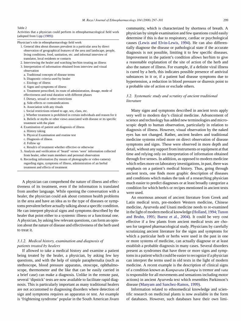
Table 2Activities that a physician could perform in ethnopharmacological field work(adopted from Lipp (1989))
Physician’s role in ethnopharmacology field work1. General idea about diseases prevalent in a particular area by direct
observation of geographical features of the area and landscape, people,living conditions, food, sanitation, etc. and informal interview oftranslator, local residents or contacts
2. Interviewing the healer and watching her/him treating an illness3. Interpretation of information obtained from interview and visual
observationa. Traditional concepts of disease termsb. Diagnostic criteria used by healerc. Etiology of illnessd. Signs and symptoms of illnesse. Treatment prescribed, its route of administration, dosage, mode ofeffectiveness and total duration with different phasesf. Dietary, sexual or other restrictionsg. Side-effects or contraindicationsh. Association with any ritualsi. Social restrictions related to age, sex, class, etc.j. Whether treatment is prohibited in certain individuals and reason for itk. Beliefs or myths or other views associated with disease or its specifictreatment with the plant
4. Examination of patient and diagnosis of illnessa. History takingb. Physical Examination and routine testc. Diagnosis of illnessd. Follow upe. Result/s of treatment whether effective or otherwise
5. Analysis and verification of ‘heard’ verses ‘seen’ information collectedfrom healer, other team members and personal observation
6. Recording information (by means of photographs or video camera)regarding signs, symptoms of illness, administration of an herbaltreatment and effects of treatment
A physician can comprehend the nature of illness and effec-tiveness of its treatment, even if the information is translatedfrom another language. While opening the conversation with ahealer, the physician could ask about common health problemsin the area and have an idea as to the type of diseases or symp-toms prevalent before actually talking about a specific condition.He can interpret physical signs and symptoms described by thehealer that point either to a systemic illness or a functional one.A physician, by asking few relevant questions, can form an opin-ion about the nature of disease and effectiveness of the herb usedto treat it.
3.1.2. Medical history, examination and diagnosis ofpatients treated by healers
If allowed to take a medical history and examine a patientbeing treated by the healer, a physician, by asking few keyquestions, and with the help of simple paraphernalia (such asstethoscope, blood pressure apparatus, otoscope, ophthalmo-scope, thermometer and the like that can be easily carried ina brief case) can make a diagnosis. Unlike in the remote past,several ‘dipstick’ tests are now available to facilitate rapid diag-nosis. This is particularly important as many traditional healersare not accustomed to diagnosing disorders where detection ofsign and symptoms requires an apparatus or test. An exampleis ‘frightening syndrome’ popular in the South American Jivaro
community, which is characterized by shortness of breath. Aphysician by simple examination and few questions could easilydetermine if this is due to respiratory, cardiac or psychologicalcauses (Lewis and Elvin-Lewis, 1994). He can also differen-tially diagnose the disease or pathological state if the accuratediagnosis is not possible, limiting it to few specific diseases.Improvement in the patient’s condition allows her/him to givea reasonable explanation of the site of action of the herb andalso the nature of illness. For example, if a definite viral illnessis cured by a herb, this indicates possible presence of antiviralsubstances in it or, if a patient had disease symptoms due tohypertension, a reduction in blood pressure or diuresis point toa probable site of action or exclude others.
3.2. Systematic study and scrutiny of ancient traditionalliterature
Many signs and symptoms described in ancient texts applyvery well to modern day’s clinical medicine. Advancement ofscience and technology has added new terminologies and micro-scopic depth to human observation, particularly in relation todiagnosis of illness. However, visual observation by the nakedeyes has not changed. Rather, ancient healers and traditionalmedicine systems relied more on direct observation of diseasesymptoms and signs. These were observed in more depth anddetail, without any support from instruments or equipment at thattime and relying only on interpretation of information obtainedthrough five senses. In addition, as opposed to modern medicinewhich relies more on laboratory investigations, in past, there wasemphasis on a patient’s medical history. Thus, going throughancient texts, one finds more graphic description of diseasesand conditions which makes the task of a researching physicianmuch easier to predict diagnoses or at least broadly categorize acondition for which herb/s or recipes mentioned in ancient textswere used.
An enormous amount of ancient literature from Greek andLatin medical texts, pre-modern Western medicine, Chinesemedicine, Ayurveda and Unani medicine needs to re-examinedin the light of modern medical knowledge (Holland, 1994; Tunonand Bruhn, 1995; Buenz et al., 2004). It could be very costeffective if a few plants from ancient medical texts are cho-sen for targeted pharmacological study. Physicians by carefullyscrutinizing ancient literature for the signs and symptoms forwhich a particular herb or herbs were used in the past in oneor more systems of medicine, can actually diagnose or at leastestablish a probable diagnosis in many cases. Several disorderspresent as syndromes that have three or more signs and symp-toms in a patient which could be easier to recognize if a physiciancan interpret the terms used in old texts in the light of modernmedicine. A recent example is the description of clinical signsof a condition known as Kampavata (Kampa is tremor and vatais responsible for all movements and sensations including motoractions) in ancient Ayurveda text which resembles Parkinson’sdisease (Manyam and Sanchez-Ramos, 1999).
Information related to ethnomedical knowledge and scien-tific research on medicinal plants is now available in the formof databases. However, such databases have their own limi-

300 M. Raza / Journal of Ethnopharmacology 104 (2006) 297–301
tations. For example, NAPALERT (Farnsworth, 1994) is thelargest database of natural products, with entries of over 27,000plants with a list of 300 or more associated symptoms or dis-eases. There are 1299 records of uses for reducing ‘fever’, 1879for ‘inflammation’ and 733 for ‘liver’ disorders. However, thedatabase only provides a single word or phrase for the use ofmedicinal plants and no description or background informationis available (Verpoorte, 1989; Farnsworth, 1994). Analyses ofsuch database with accurate precision by a physician could leadto reasonable and specific prediction and link specific plants tocertain disorders. For example, if a plant is reported to be usedfor fever, headache and also for nasal catarrh and skin disorders,the possibility of its use as anti-viral or anti allergic could beconcluded.
Several medical disorders present with varied signs andsymptoms due to cross-cultural variations in the expression ofillness, but are still linked by a common pathology of biologicalsystem (Kleinman, 1987; de Haes and Olschewski, 1998). Thisis especially true for the disorders that have psychological symp-toms specific to a culture (Janca and Isaac, 1997). For example,signs and symptoms resulting from addiction, depressive illness,or somatic anxiety have common elements in different cultures(Ulusahin et al., 1994; Piccinelli and Simon, 1997; Yardley etal., 1999). Thus, description of such disorders in ancient lit-eratures may vary considerably. However, since all traditionalsystems approach the disease in a holistic manner (Vogel, 1991)and have many similarities, their description has some commonsigns which can be interpreted by an experienced physician.
Recent advances in technology to rapidly scan and digi-tize the ancient herbal texts can involve interested physiciansmore easily. In addition, development of software and elec-tronic resources to better interpret old linguistic, botanical andmedical terminologies can further assist physicians to overcomelanguage barriers, saving a lot of time and manpower (Buenz etal., 2004).
3.3. Clinical observations on herbs and herb-druginteractions
A significant number of patients simultaneously use herbaland modern drugs in industrialized countries and under-developed world. In addition, several traditional communitieshave started incorporating modern medicine in their treatmentregimen. One such example is of Hausa-Fulani community ofNigeria where this trend has been clearly documented (Etkin etal., 1990). These situations create an opportunity for physiciansto clinically evaluate the benefits and side effects of herbal drugsin a particular illness, discover new indications for traditionalherbs, as well as note herbal-drug interactions.
Introduction of new single compounds drugs adds to the ther-apeutic armamentarium of physician, leading to new herbal-druginteractions, and possible contraindications. Physicians practic-ing in well-equipped hospitals or clinics or a family physicianin a community have access to patients consuming both herbalsand modern drugs. Thus, a physician in this situation will be ableto clinically observe the benefits or side effects of both herbalsand drugs introduced recently and when used in combination.
Specialist physicians could perform more specific evaluation ofpatients consuming both herbals and modern drugs.
3.4. Advice to pharmacologist/s for laboratoryinvestigation of herb
Information provided by a physician from a field visit or clin-ical observation on the effectiveness of a herb to a well-trainedpharmacologist can lead to proper selection of in vivo or in vitromodels of various diseases for laboratory research. A physiciancan properly guide a pharmacologist in this regard and suggesta primary action of an herbal recipe or of a particular plant inhumans that he observed during his field work. For example,a plant that was found to be curative for a bacterial infectionwill help a researcher to select appropriate antimicrobial tests.A plant effective in seizure or epilepsy will guide one to exploreits effects in relevant animal models. Thus, physicians can playa very important role in enhancing drug discovery. This couldminimize time, save resources and speed up the process for dis-covery of compound/s responsible for a specific pharmacologicactivity observed directly in humans.
3.5. Strengthening the existing traditional medicine systemin a community
In communities where the tools and trades of traditionalmedicine and the use of medicinal plants are kept as secrets,traditional healers often have concerns about the violation ofintellectual property rights, patenting and profiteering by localor foreign drug companies. Local physicians in cooperationwith the elders of the community or traditional healers couldwork in collaboration and actually strengthen the prevalent cul-ture of use of medicinal plants. In this setting, a physician canguide the healers on use of herbs or recipes in the light of mod-ern research, apprise them of toxicity, precautions or new usesand ‘update’ their traditional or oral herbal pharmacopoeia. Forexample, a physician can advise a healer that certain medici-nal plants that lower the platelet aggregation can make blood‘thin’ and should not be given to the patients or their dosesbe reduced if they are already taking aspirin. However, forworking closely with the traditional healer, a physician has tofirst build trust and treat her/him as his colleague and friend.The knowledge and experience of a traditional healer has tobe considered valuable as it comes from thousands of yearsof trial and error and forms the basis of modern medicine andtherapeutics.
4. Conclusion
Globalization and advancement in technology is expectedto promote plant derived medicinal preparations and traditionalmedicine (Verpoorte, 2005). Physicians could ‘come back’ andlike their predecessors, use their expertise of basic sciencesand clinical knowledge to contribute in ethnopharmacologicalstudies and drug discovery. Those associated with academia orindustries should perform field studies with ethnobotanists andget authentic and specific information on the use of medici-
M. Raza / Journal of Ethnopharmacology 104 (2006) 297–301 301
nal plants. Their involvement could lead to a highly targetedapproach in discovery of drugs from industry and significantlyreduce the cost of drug development. A systematic study ofancient texts by physicians could lead to better interpretationof the medicinal uses of plants in different cultures. Physicianspracticing medicine in a community where the use of medici-nal plants is common could contribute by closely working withthe local healers. They could discuss new discoveries aboutmedicinal plants, new drugs and herbal-drug interactions withthe healers and publish their observations. However, this willrequire a change of approach on the part of physicians towardstraditional medicine and adopting proper attitude towards thetraditional healers. Several medical disorders are awaiting ther-apeutic cure and the pharmaceutical industry has yet to comeup with effective drug treatments. Participation of physiciansin ethnopharmacological studies will ultimately benefit patientswith incurable diseases and also fulfill the main goal of the fieldof ethnopharmacology.
Acknowledgements
I thank Dr. Nina L. Etkin for her valuable comments onan earlier version of the manuscript and Dr. K.M. Hedayat forreviewing the English language.
References
Anonymous, 1993. Research Guidelines for Evaluating the Safety and Effi-cacy of Herbal Medicines. WHO, Regional Office for the Western Pacific,Manila.
Anonymous, 2002. Towards more effective drug discovery. Nature 415, 1.Aronson, J.K., 1987. The discovery of the foxglove as a therapeutic agent.
Chemistry in Britain 23, 33–36.Bruhn, J.G., Holmstedt, B., 1981. Ethnopharmacology: objectives, principles
and perspectives. In: Beal, J.L., Reinhard, E. (Eds.), Natural Products asMedicinal Agents. Hippocrates-Verlag, Stuttgart, pp. 405–430.
Buenz, E.J., Schnepple, D.J., Bauer, B.A., Elkin, P.L., Riddle, J.M., Motley,T.J., 2004. Techniques: bioprospecting historical herbal texts by huntingfor new leads in old tomes. Trends in Pharmacological Sciences 25,494–498.
Cordell, G.A., Colvard, M.D., 2005. Some thoughts on the future ofethnopharmacology. Journal of Ethnopharmacology 100, 5–14.
Cox, P.A., 1994. The ethnobotanical approach to drug discovery: strengthsand limitations. Ciba Foundation Symposium 185, 25–36.
de Haes, J.C., Olschewski, M., 1998. Quality of life assessment in a cross-cultural context: use of the Rotterdam Symptom Checklist in a multina-tional randomised trial comparing CMF and Zoladex (Goserlin) treatmentin early breast cancer. Annals of Oncology 9, 745–750.
Etkin, N.L., 1993. Anthropological methods in ethnopharmacology. Journalof Ethnopharmacology 38, 93–104.
Etkin, N.L., 2001. Perspectives in ethnopharmacology: forging a closerlink between bioscience and traditional empirical knowledge. Journal ofEthnopharmacology 76, 177–182.
Etkin, N.L., Ross, P.J., Muazzamu, I., 1990. The indigenization of pharma-ceuticals: therapeutic transitions in rural Hausaland. Social Sciences andMedicine 30, 919–928.
Fabricant, D.S., Farnsworth, N.R., 2001. The value of plants used in tradi-tional medicine for drug discovery. Environment and Health Perspectives109 (Suppl. 1), 69–75.
Farnsworth, N.R., 1994. Ethnopharmacology and drug development. CibaFoundation Symposium 185, 42–51.
Farnsworth, N.R., Akerele, O., Bingel, A.S., Soejarto, D.D., Guo, Z., 1985.Medicinal plants in therapy. Bulletin of the World Health Organisation63, 965–981.
Giordano, J., Boatwright, D., Stapleton, S., Huff, L., 2002. Blending theboundaries: steps toward an integration of complementary and alternativemedicine into mainstream practice. Journal of Alternative and Comple-mentary Medicine 8, 897–906.
Heinrich, M., Gibbons, S., 2001. Ethnopharmacology in drug discovery: ananalysis of its role and potential contribution. Journal of Pharmacy andPharmacology 53, 425–432.
Holland, B.K., 1994. Prospecting for drugs in ancient texts. Nature 369, 702.Holmstedt, B., 1972. The Ordeal bean of Calabar, The pageant of
Physostigma venenosum in medicine. In: Swain, T. (Ed.), Plants in theDevelopment of Modern Medicine. Harvard University Press, Cambridge,MA, pp. 303–360.
Holmstedt, B., 1991. Historical perspective and future of ethnopharmacology.Journal of Ethnopharmacology 32, 7–24.
Holmstedt, B., Bruhn, J., 1982. Is there a place for ethnopharmacology inour time? Trends in Pharmacological Sciences 3, 181–183.
Holmstedt, B., Fredga, A., 1981. Sundry episodes in the history of coca andcocaine. Journal of Ethnopharmacology 3, 113–147.
Holmstedt, B., Wassen, S.H., Schultes, R.E., 1979. Jaborandi: an interdisci-plinary appraisal. Journal of Ethnopharmacology 1, 3–21.
Janca, A., Isaac, M., 1997. ICD-10 and DSM-IV symptoms of somatoformdisorders in different cultures. The Keio Journal of Medicine 46, 128–131.
Kleinman, A., 1987. Anthropology and psychiatry. The role of culture incross-cultural research on illness. The British Journal of Psychiatry 151,447–454.
Lewis, W.H., Elvin-Lewis, M.P., 1994. Basic, quantitative and experimentalresearch phases of future ethnobotany with reference to the medicinalplants of South America. Ciba Foundation Symposium 185, 60–72.
Lipp, F.J., 1989. Methods for ethnopharmacological field work. Journal ofEthnopharmacology 25, 139–150.
Lozoya, X., 1994. Two decades of Mexican ethnobotany and research inplant drugs. Ciba Foundation Symposium 185, 130–140.
Malone, M.H., 1983. The pharmacological evaluation of naturalproducts—general and specific approaches to screening ethnopharmaceu-ticals. Journal of Ethnopharmacology 8, 127–147.
Manyam, B.V., Sanchez-Ramos, J.R., 1999. Traditional and complementarytherapies in Parkinson’s disease. Advances in Neurology 80, 565–574.
Piccinelli, M., Simon, G., 1997. Gender and cross-cultural differences insomatic symptoms associated with emotional distress. An internationalstudy in primary care. Psychologic Medicine 27, 433–444.
Sandberg, F., 1987. The integrated natural products research in the develop-ment of plant derived pharmaceuticals. Fitoterapia 58, 309–313.
Schultes, R.E., 1962. The role of the ethnobotanist in the search for newmedicinal plants. Lloydia 25, 257–266.
Tunon, H., Bruhn, J.G., 1995. Drugs in ancient texts. Nature 376, 546.Tyler, V.E., 1986. Plant drugs in the twenty-first century. Economic Botany
40, 279–288.Ulusahin, A., Basoglu, M., Paykel, E.S., 1994. A cross-cultural comparative
study of depressive symptoms in British and Turkish clinical samples.Social Psychiatry and Psychiatric Epidemiology 29, 31–39.
Verpoorte, R., 1989. Some phytochemical aspects of medicinal plant research.Journal of Ethnopharmacology 25, 43–59.
Verpoorte, R., 2005. Perspectives of ethnopharmacology. Journal ofEthnopharmacology 100, 1–2.
Vogel, H.G., 1991. Similarities between various systems of traditionalmedicine. Considerations for the future of ethnopharmacology. Journalof Ethnopharmacology 35, 179–190.
Weniger, B., 1991. Interest and limitation of a global ethnopharmacologicalsurvey. Journal of Ethnopharmacology 32, 37–41.
Yardley, L., Medina, S.M., Jurado, C.S., Morales, T.P., Martinez, R.A.,Villegas, H.E., 1999. Relationship between physical and psychosocial dys-function in Mexican patients with vertigo: a cross-cultural validation ofthe vertigo symptom scale. Journal of Psychosomatic Research 46, 63–74.

Journal of Ethnopharmacology 104 (2006) 302–305
Protective effect of Cissus quadrangularis on neutrophilmediated tissue injury induced by aspirin in rats
Mallika Jainu, K. Vijai Mohan, C.S. Shyamala Devi ∗Department of Biochemistry, University of Madras, Guindy Campus, Chennai 600025, India
Received 13 July 2004; received in revised form 4 August 2005; accepted 4 August 2005Available online 9 December 2005
Abstract
Cissus quadrangularis (family: Vitaceae) is well known for the treatment of gastric disorders in traditional medicine, owing to its rich sourceof carotenoids, triterpenoids and ascorbic acid, and has received considerable attention regarding its role in human nutrition. In the search of newpotential antiulcer agents, the present study evaluated the ethanol extract of Cissus quadrangularis (CQE) against the gastric toxicity induced byaspirin in rats. The optimum protective dose of 500 mg/kg of extract was selected by the pretreatment of gastric ulcers with different doses ofCQE (250, 500 and 750 mg/kg) for 7 days which showed ulcer protection by 40, 71.2 and 72.6%, respectively, as compared to ranitidine (RTD)(30 mg/kg) by 71.9% in the aspirin model. In addition, results have shown that administration of aspirin increases lipid peroxidation status, xanthineoxidase (XO), myeloperoxidase and decrease in selenium–glutathione peroxidase activities in the gastric mucosa, resulting in mucosal damageat both cellular and subcellular level. Pretreatment with CQE ameliorated the observed effect significantly in the gastric mucosa of ulceratedrats. These findings suggest that the gastroprotective activity of CQE could be mediated possibly through its antioxidant effect as well as by theattenuation of the oxidative mechanism and neutrophil infiltration.© 2005 Published by Elsevier Ireland Ltd.
Keywords: Aspirin; Cissus quadrangularis; Ulcer lesions; Lipid peroxidation
1. Introduction
In recent times, many medicinal plants continue to providevaluable therapeutic agents for the treatment of ulcers both inmodern medicine and by the traditional system throughout theworld. Since chemical compounds are known to have undesir-able side-effects, the present study focused on natural products.
1.1. Plant
Cissus quadrangularis Linn. Wall. Ex. Wight (family:Vitaceae) is an edible plant, commonly known as “bone setter”found in hotter parts of India, Ceylon, East Africa and Malaysiaand Thailand. Cissus quadrangularis is used as a common foodsupplement in southern India. Stem parts of Cissus quadrangu-
∗ Corresponding author. Present address: 66, White House, II Main road,Gandhi Nagar, Adyar, Chennai 600020, Tamil Nadu, India.Tel.: +91 44 24412575; fax: +91 44 22352494.
E-mail address: [email protected] (C.S.S. Devi).
laris were collected from Native Care and Cure Center, Indiaand were duly authenticated by Dr. P. Brindha, PharmacologyDepartment, Captain Srinivasa Murthy Drug Research Institute,Arumbakkam, Chennai 600106. A voucher specimen PP. 502has been deposited in the department.
1.2. Uses in traditional medicine
The stout fleshy quadrangular stem is traditionally used forthe treatment of gastritis, bone fractures, skin infections, con-stipations, eye diseases, piles, anemia, asthma, irregular men-struation, burns and wounds (Asolkar et al., 1992; Kritikarand Basu, 2000). Cissus quadrangularis has potent fracturehealing property, antimicrobial, antiulcer, antioxidative, cholin-ergic activity and beneficial effect on cardiovascular diseases(Udupa and Prasad, 1964; Subbu, 1970; Anoop and Jagdeesan,2002; Murthy et al., 2003; Jainu and Devi, 2003). Previously,we have demonstrated that methanolic extract of Cissus quad-rangularis possesses antiulcer and cytoprotective property inindomethacin-induced gastric mucosal injury (Jainu and Devi,
0378-8741/$ – see front matter © 2005 Published by Elsevier Ireland Ltd.doi:10.1016/j.jep.2005.08.076

M. Jainu et al. / Journal of Ethnopharmacology 104 (2006) 302–305 303
2004). Due to its widespread health use and pharmacologicactions, this study will highlight the health promoting and ther-apeutic effects of Cissus quadrangularis.
1.3. Previously isolated class of constituents
The phytochemical analysis of the plant showed the presenceof Vitamin C, �-carotene, two asymmetric tetracyclic triter-penoids, �-sitosterol, �-amyrin, �-amyrone and three stillbenederivatives, quadrangularins A, B, C, etc. (Chopra et al., 1956;Attawish et al., 2002).
Some experimental studies have demonstrated that oxygen-generated free radicals derived from infiltrated neutrophils andenhanced lipid peroxidation play important roles in the patho-genesis of acute gastric lesions induced by aspirin (Kontureket al., 1994). Substances that are able to hinder their formationor capture the free oxygen radicals formed are thus potentialantiulcerogenic agents.
These facts form the basis for a study of whether the antiox-idant mechanisms are involved in CQE mediated protection inaspirin-induced gastric damage. The objective of the study isto investigate the effect of CQE on neutrophil infiltration tis-sue injury induced by aspirin in order to reveal the mechanismunderlying the antiulcer effect of the plant.
2. Materials and methods
2.1. Preparation of alcoholic extract
Dried parts of Cissus quadrangularis were coarsely pow-dered and 1 kg of this powdered plant material was soaked inethanol for 48 h and extracted by soxhlet extraction. The extractwas vacuum dried and was stored at −4 ◦C until further use.The yield of the extract was 5.2% (w/w) of powdered ethanolicextract. For administration, the extract was dissolved in distilledwater and used for the present study.
2.2. Animals
Male albino rats’ weighing 150–200 g were purchased fromTamil Nadu University of Veterinary and Animal Sciences,India. The animals were housed in polypropylene cages main-tained in controlled temperature and light cycle. The animalswere fed with food pellets and water was given ad libitum.The experiments were initiated only after the approval of theInstitutes of Animal Ethics Committee (No: 360/01/a/CPSEA/2001).
2.3. Acute toxicity
Adult albino rats of either sex were divided into four groups(n = 6) and orally fed with CQE at dose levels of 0.5, 1.5, 3.0and 5.0 g/kg. Animals were watched carefully for 72 h after CQEadministration and then for the next 7 days. At the end of thisexperimental period, the rats were observed for signs of toxic-ity, morphological behaviour and mortality. A separate group ofcontrols received only the vehicle.
2.4. Induction of aspirin-induced gastric lesions
Adult male Wistar rats weighing 120–180 g were used forthe experiment. All the animals were fasted for 24 h prior tothe experiments. Aspirin at a dose of 400 mg/kg (Kamsiah etal., 2002) was administered to the animals and after 6 h, theanimals were sacrificed by cervical dislocation and the stomachwas incised along the greater curvature and the area of mucosaldamage of glandular stomach was calculated in units of squaremillimeter (Szabo et al., 1985).
2.5. Dosage fixation
A group of animals, which served as control, receivedonly distilled water. The CQE at dose levels of 250, 500 and750 mg/kg and ranitidine (RTD), the reference drug, in thedose of 30 mg/kg was administered orally for 7 days. A dose-dependent antiulcer effect of CQE (250, 500 and 750 mg/kg)was seen on gastric ulcers. Therefore, for the test drug (CQE),an optimum dose of 500 mg/kg was selected for further studiesand the percentage of ulcer protection was nearly equipotent toRTD, a standard antiulcer drug.
2.6. Treatment protocol
Animals were divided into four groups of six animals in eachgroup. Group 1 animals served as control. They received onlydistilled water equivalent to the volume of plant extract. Group 2represented the ulcerated group. Ulceration was produced by theadministration of aspirin (400 mg/kg) orally. Group 3 animalswere pretreated with the test drug CQE (500 mg/kg) orally, oncedaily for 7 days and then ulceration was induced by aspirin. After6 h, the animals were sacrificed, stomach was taken out and thescrapped gastric mucosal tissues were used for the estimation ofbiochemical enzymes.
2.6.1. Assay of Se-glutathione peroxidase (Se-GSHPx) andxanthine oxidase (XO)
For the assays of Se-GSHPx and XO enzymes, gastricmucosal tissues were homogenized in nine volumes of ice-cold 0.05 M Tris–HCl buffer (pH 7.4). The homogenate wascentrifuged at 4 ◦C (10,000 × g, 20 min) and the resultant super-natent was dialyzed against 100 volumes of the same buffer at4 ◦C for 24 h.
Gastric mucosal Se-GSHPx enzyme activity was assayed bythe methods of Hochstein and Utley (1968). The enzyme activitywas determined at 37 ◦C by recording the decrease in absorbanceat 340 nm following the oxidation of NADPH in the presenceof H2O2, GSH and yeast glutathione reductase. One unit of thisactivity is defined as the amount of enzyme oxidizing 1 �mol ofNADPH per min.
Gastric mucosal XO was assayed by the method ofHashimoto (1974). Three millilitres of incubation mixture con-tains 150 �M phosphate buffer, 0.2 �M xanthine, 0.3 �M potas-sium oxonate and 0.2 ml of tissue homogenate. The tubes wereincubated at 30 ◦C for 30 min, 0.1 ml of 100% of TCA was addedand mixed well. The content was centrifuged at 10,000 × g for

304 M. Jainu et al. / Journal of Ethnopharmacology 104 (2006) 302–305
15 min and the supernatant was analysed for XO activity by mea-suring the increase in absorbance at 292 nm following formationof uric acid at 30 ◦C. One unit (U) of this enzyme is defined as theamount of enzyme forming 1 �mol uric acid/min. The activityof XO was expressed as mU/g tissue.
2.6.2. Assay of lipid peroxidationThe LPO product malondialdehyde (MDA) was estimated
using 1,1,3,3-tetramethoxypropane as the standard, according tothe method described by Ohkawa et al. (1979). To 0.5 ml of tissuehomogenate, 1.5 ml of 20% acetic acid, 0.2 ml of SDS and 1.5 mlof TBA were added. The mixture was made up to 4.0 ml with dis-tilled water and then heated for 60 min at 95 ◦C using glass ballas condenser. After cooling, 4.0 ml of butanol–pyridine mixturewas added and shaken well. After centrifugation at 4000 rpm for10 min, the organic layer was taken and its absorbance was readat 532 nm and the results were expressed as nmol/mg protein.
2.6.3. Assay of myeloperoxidaseGastric mucosal myeloperoxidase (MPO), a marker enzyme
of neutrophil infiltration, was assayed by the method of Suzukiet al. (1983). A sample of tissue homogenate (0.5 �l) wasadded to a 0.5 ml of reaction volume containing PBS, pH 5.4,hexadecyl–trimethyl ammonium bromide and tetraethylbenzi-dine at 37 ◦C. The reaction was started by the addition of H2O2and terminated by the sequential addition of catalase and sodiumacetate. One unit of this enzyme activity was defined as theamount of enzyme causing a change in absorbance at 655 nmand the results were expressed as U/mg protein. Protein contentwas assessed according to the method of Lowry et al. (1951).
2.7. Statistical analysis
The data were expressed as mean ± S.E.M. One-way anal-ysis of variance followed by Dunnett’s multiple comparisontests were used to assess statistical significance of differencesbetween groups.
3. Results
Rats that received oral doses of 0.5, 1.5, 3.0 and 5.0 g/kg,did not manifest any clinical signs of toxicity. None of thedoses tested could produce mortality in rats during the treat-ment period. In tests on rats, we found that doses of CQE up to5 g/kg were non-toxic and we were unable to establish its oralLD50 value.
Aspirin administered rats showed multiple gastric mucosallesions, most often 1–2 mm in size or petechial, bleeding atthe moment of the observation. The CQE showed significantantiulcer effect against ulcers induced in the aspirin model in adose-dependent manner. In ulcerated rats, CQE at dose levels of250, 500 and 750 mg/kg showed protection indexes of 40.0, 71.2and 72.6%, respectively, whereas RTD showed 71.9% protec-tion at a dose of 30 mg/kg. The percentage of ulcer protectionof CQE at dose of 500 mg/kg was nearly the same as that of750 mg/kg. There was no significant difference in the ulcer pro-tection indexes of CQE at a dose of 500 mg/kg as compared with
Fig. 1. Effect of Cissus quadrangularis extract (CQE) on Se-glutathione perox-idase (Se-GSHPx) and xanthine oxidase (XO) activities in the aspirin-inducedgastric ulcer model. Results are expressed as mean ± S.E.M. of six animals ineach group. aP < 0.001 vs. control; bP < 0.001 vs. aspirin group.
750 mg/kg given orally for 7 days. Therefore, an optimal doseof CQE (500 mg/kg) given for 7 days significantly protected theanimals against gastric ulcer. Hence, further studies were carriedout with the same dose of CQE.
As shown in Fig. 1 the increase in XO activity upon aspirinadministration was attenuated by pretreatment with CQE atthe dose of 500 mg/kg. Our data show that according to gas-tric mucosal damage, Se-GSHPx activity decreased significantlyafter aspirin administration when compared to control. CQE pre-treatment replenish the Se-GSHPx activity to the levels seen incontrol rats.
Fig. 2 depicts the activity of MPO, as an index of neutrophilinfiltration and LPO level in the control and experimental ani-mals. Aspirin induction strongly increased the activity of MPOand LPO levels in comparison with control. While pretreatmentwith CQE restored these alterations to near-normal levels inulcerated rats, it was effective against aspirin-induced gastriculcer.
Fig. 2. Effect of Cissus quadrangularis extract (CQE) on gastric mucosal lipidperoxide content (LPO) and myeloperoxidase (MPO) in aspirin administeredrats. Results are expressed as mean ± S.E.M. of six animals in each group.aP < 0.001 vs. control; bP < 0.001 vs. aspirin group.
M. Jainu et al. / Journal of Ethnopharmacology 104 (2006) 302–305 305
4. Discussion
The results of this study demonstrate that the CQE possessesantiulcer property as evidenced by its significant inhibition inthe formation of gastric lesions induced by aspirin. The acutetoxicity tests on rats showed that doses of CQE up to 5 g/kgwere non-toxic and the oral LD50 value might be higher thanthis dose.
Aspirin caused rise in the lipid peroxidation status possiblydue to the activation of neutrophils, which play an important rolein the damaging activity (Yoshikawa et al., 1992). The measure-ment of local enzymatic activity of MPO has been used to quan-tify neutrophil sequestration in tissues and the severity of inflam-mation (Nishizawa et al., 1996). The activity of Se-GSHPx,an index of tissue neutrophil infiltration increased, with lesiondevelopment in the aspirin administered rats (Krawisz et al.,1984). However, inhibition of MPO and increase in Se-GSHPxactivity by CQE may be simple by a reflection of decreasedinflammation independent of any direct effect on neutrophils.The gastric ulcer induced by aspirin was almost completelyprevented, with attenuation of increased gastric mucosal XOand MPO activities and LPO content, by treatment with CQE,thereby decreasing the neutrophil sequestration at the woundsite.
Triterpenoids and �-sitosterols present in Cissus quadrangu-laris possess antilipidperoxidative effect (Somova et al., 2003)and thus prevent gastric damage. The plant constituent such as�-sitosterol has the ability to reduce the enzyme MPO, indi-cating a reduction of neutrophil influx in the inflamed tissues(DelaPureta et al., 2000). Murthy et al. (2003) have reported thatCissus quadrangularis possesses antioxidant activity in both thein vitro and in vivo models due to the presence of �-carotene inthis plant. Previous reports suggested that the healing action ofCissus quadrangularis is due to its antioxidative property (Jainuand Devi, 2003). Thus, the protective role of CQE against neu-trophil mediated tissue injury may be due, in part, to a reductionin neutrophil infiltration into the gastric mucosa, via its antiox-idant property.
In conclusion, the antiulcer efficacy of CQE in gastric ulcermodel could be due to inhibition of neutrophil infiltration andantioxidant properties. These properties of CQE merit detailedanalysis relating to active principles and the mechanism ofaction, which might provide new alternatives for the clinicalmanagement of gastric ulcer diseases.
Acknowledgement
The authors would like to thank Dr. P. Brindha for his expertcomments and suggestions in regard to this work.
References
Anoop, A., Jagdeesan, M., 2002. Gastric and duodenal antiulcer and cytopro-tective effect of Cissus quadrangularis Linn. variant II in rats. NigerianJournal of Natural Products and Medicine 6, 1–7.
Asolkar, L.V., Kakkar, K.K., Chakre, O.J., 1992. Second Supplement to Glos-sary of Indian Medicinal Plants with Active Principles. CSIR Publication,New Delhi, p. 61.
Attawish, A., Chavalttumrong, D., Chivapat, S., Chuthaputti, S., Rattarajaras-roj, S., Punyamong, S., 2002. Subchronic toxicity of Cissus quadrangu-laris Linn. Songklanakarin Journal of Science and Technology 24, 39–51.
Chopra, R.N., Nayar, S.L., Chopra, I.C., 1956. Glossary of Indian MedicinalPlants. Publication and Information Directorate, CSIR, New Delhi, p. 66.
DelaPureta, R., Martinez-Dominguez, E., Ruiz-Gutierrez, V., 2000. Effect ofminor components of virgin olive oil on topical anti-inflammatory assays.Zetschrift Fur Naturforschong 55, 814–819.
Hashimoto, S., 1974. New spectrophotometric assay method of xanthine oxi-dase in crude tissue homogenate. Analytical Biochemistry 62, 426–433.
Hochstein, P., Utley, H., 1968. Hydrogen peroxide detoxication by glutathioneperoxidase and catalase in rat liver homogenate. Analytical Biochemistry4, 574–579.
Jainu, M., Devi, C.S.S., 2003. Potent antiulcerogenic activity of methanolicextract of Cissus quadrangularis by antioxidative mechanism. Journal ofClinical Biochemistry and Nutrition 34, 43–47.
Jainu, M., Devi, C.S.S., 2004. Effect of Cissus quadrangularis on gastricmucosal defensive factors in experimentally induced gastric ulcer—a com-parative study with sucralfate. Journal of Medicinal Food 7, 372–376.
Kamsiah, J., Gapor, M.T., Nafeeza, M.I., Fauzee, A.M., 2002. Effect ofvarious doses of palm Vitamin E and tocopherol on aspirin-induced gas-tric lesions in rats. International Journal of Experimental Pathology 83,295–301.
Konturek, S.J., Brozozowski, T., Stachura, J., Majka, J., 1994. Role of neu-trophils and mucosal blood flow in gastric adaptation to aspirin. EuropeanJournal of Pharmacology 253, 107–114.
Krawisz, J.E., Sharon, P., Stenson, W.F., 1984. Quantitative assay for acuteintestinal inflammation based on myeloperoxidase activity. Assessment ofinflammation in rat and hamster models. Gastroenterology 87, 1344–1350.
Kritikar, K.R., Basu, B.D., 2000. In: Basu, L.M., (Ed.), Indian MedicinalPlants, Lalit Mohan Basu Publisher, Allahabad, India, pp. 841–843.
Lowry, O.H., Rosebrough, N.H., Farr, A.L., Randal, R.I., 1951. Protein mea-surement with Folin’s-phenol reagent. Journal of Biological Chemistry193, 265–275.
Murthy, K.N.C., Vanitha, A., Swamy, M.M., Ravishankar, G.A., 2003.Antioxidant and antimicrobial activity of Cissus quadrangularis L. Jour-nal of Medicinal Food 6, 99–105.
Nishizawa, H., Yamamda, H., Miyazaki, H., Ohara, M., Kaneko, K.,Yamakawa, T., Wiener-kronish, J., Kodoh, I., 1996. Soluble complementreceptor type I inhibited the systemic organic injury caused by acid instil-lation into a lung. Anesthiology 85, 1120–1128.
Ohkawa, H., Ohishi, N., Yagi, K., 1979. Assay for lipid peroxides in ani-mal tissues by thiobarbituric acid reaction. Analytical Biochemistry 95,351–358.
Somova, L.I., Shode, F.O., Ramananan, P., Nadar, A., 2003. Antihypertensive,antiatherosclerotic and antioxidant activity of triterpenoids isolated fromOlea europaea, subspecies africana leaves. Journal of Ethnopharmacology84, 299–305.
Subbu, V.S.V., 1970. Mechanism of action of Vitis glucoside on myocardialtissue. Indian Journal of Medicinal Science 25, 400–403.
Suzuki, K., Ota, H., Sasagawa, S., Sakatani, T., Fujikura, T., 1983. Assaymethod of myeloperoxidase in human polymorphonuclear leukocytes.Analytical Biochemistry 132, 345–353.
Szabo, S., Trier, J.S., Brown, A., Schoor, J., 1985. Early vascular injury andincreased permeability in gastric mucosal injury caused by ethanol in rat.Gastroenterology 88, 228–236.
Udupa, K.N., Prasad, G., 1964. Biochemical Ca45 studies on effect of Cissusquadrangularis in fracture repair. Indian Journal of Medicinal Research52, 480–487.
Yoshikawa, T., Naito, Y., Ueda, S., 1992. Ischemia reperfusion injury and freeradical involvement in gastric mucosal disorders. Advanced ExperimentalMedicinal Biology 316, 231–238.

Journal of Ethnopharmacology 104 (2006) 306–309
Anti-diabetic activity of methanol/methylene chloride stem barkextracts of Terminalia superba and Canarium schweinfurthii
on streptozotocin-induced diabetic rats
P. Kamtchouing a,∗, S.M. Kahpui a, P.-D. Djomeni Dzeufiet a,L. Tedong a, E.A. Asongalem b, T. Dimo a
a Department of Animal Physiology, Faculty of Science, University of Yaounde, P.O. Box 812, Yaounde, Cameroonb Department of Physiological Sciences, Faculty of Medicine and Biomedical Science, University of Yaounde I, P.O. Box 812, Yaounde, Cameroon
Received 6 October 2004; received in revised form 4 August 2005; accepted 4 August 2005Available online 4 November 2005
Abstract
Stem bark extracts of Terminalia superba Engl. and Diels and Canarium schweinfurthii Engl. are used in Africa for the treatment of variousailments, including diabetes mellitus. The anti-diabetic effects of the methanol/methylene chloride extracts of the stem barks on streptozotocin(STZ)-induced diabetes were evaluated on male rats. Through the subcutaneous route, diabetes was induced using 60 mg/mL of streptozotocin.After 2 days, the rats received, by gavage, 150 mg/kg and 300 mg/kg of extract daily for 14 days. At 300 mg/kg, the two extracts (Terminaliasuperba and Canarium schweinfurthii), significantly showed at least 67.1% and 69.9% reduction in blood glucose level, respectively, while insulin(three units) given subcutaneously and once daily, had 76.8% reduction compared to diabetic untreated control rats. Similarly, the weight gainswere 6.6% and 4.9%, respectively, and were comparable to the normal rats, whereas, diabetic untreated rats lost 14.1% body weight. Still with thesame dose, there was 68.5% and 58.5% (p < 0.001) significant decrease in food consumption and 79.7% and 64.0% (p < 0.001) in fluid intake bydiabetic rats treated with the respective plant extracts. The insulin-treated rats showed 56.4% and 75.8% decrease in food and fluid intake comparedto an augmentation for diabetic control rats, 43.0% and 383.8%, respectively, at the end of the second week of experimentation. These resultsshowed that the plant extracts can reverse hyperglycemia, polyphagia and polydipsia provoked by streptozotocin, and thus, they have anti-diabeticproperties.© 2005 Elsevier Ireland Ltd. All rights reserved.
Keywords: Terminalia superba; Canarium schweinfurthii; Streptozotocin; Diabetes mellitus
1. Introduction
Diabetes mellitus is a chronic metabolic disease caused by anabsolute or relative lack of insulin and or reduced insulin activity,which results in hyperglycemia and abnormalities in carbohy-drate, protein and fat metabolism. Though different types oforal hypoglycemic agents are available along with insulin forthe treatment of diabetes mellitus, there is a growing interest inherbal remedies due to the side effects associated with these ther-apeutic agents (Kamesawara et al., 2000). The investigation ofanti-diabetic agents of plant origin which are used in traditionalmedicine is thus of great importance.
∗ Corresponding author. Tel.: +237 9931195.E-mail address: [email protected] (P. Kamtchouing).
Terminalia superba Engl. and Diels (Combretaceae) andCanarium schweinfurthii Engl. (Burseraceae) are some of theplants used by traditional healers as a remedy for diabetes mel-litus in Africa. The people of Sotho in Southern Senegal takethe powdered stem bark of Terminalia superba against dia-betes. In tropical Africa, Canarium schweinfurthii is used asa remedy for diabetes. Terminalia superba is a big tree withdeciduous leaves, attaining 50 m of height and 120 cm stemdiameter. It is widely distributed in the dense humid forests,semi-deciduous forests and also in easily flooded and secondaryforests. Canarium schweinfurthii is a big tree with deciduousleaves attaining 45 m of height and 150 cm stem diameter. Itis widely distributed in dense semi-deciduous and secondaryforests (Berhaut, 1974). Chemical constituents of Terminaliasuperba and Canarium schweinfurhii are alkaloids (Burkill,1985).
0378-8741/$ – see front matter © 2005 Elsevier Ireland Ltd. All rights reserved.doi:10.1016/j.jep.2005.08.075

P. Kamtchouing et al. / Journal of Ethnopharmacology 104 (2006) 306–309 307
The present investigation was undertaken to study the anti-diabetic effects of the methanol/methylene chloride extracts ofthe stem bark of Terminalia superba and Canarium schwein-furthii on streptozotocin (STZ)-induced diabetic rats.
2. Materials and methods
2.1. Animals
The experimental animals were male albino Wistar rats(150–250 g body weight) raised in the Animal House of theFaculty of Science, University of Yaounde I. They were fed witha standard laboratory diet (LanavetR, Garoua, Cameroon) andgiven tap water ad libitum. They were fasted overnight prior toblood sugar determination or streptozotocin injection but wereallowed free access to water.
2.2. Preparation of plant extracts
The stem bark of Terminalia superba was harvested in NgoaEkele Yaounde (Centre province, Cameroon). Botanical iden-tification was performed at the National Herbarium, Yaounde,Cameroon, and herbarium voucher specimen No. 19652/HNCcollected by Leeuwenberg (No. 5963) has been deposited atYaounde Herbarium. The stem bark of Canarium schweinfurthiiwas harvested in Mbalmayo (Centre province, Cameroon).Botanical identification was performed at the National Herbar-ium, Yaounde, Cameroon, where herbarium voucher specimenNo. 16929/HNC collected by R. Letouzey (No. 8763) has beendeposited at Yaounde Herbarium. All fresh plant materials weresun-dried and ground into powder. The dried powdered materials(245 g of Terminalia superba and 550 g of Canarium schwein-furthii) were separately macerated in 1:1 (v/v, 500 mL) mixturesof methanol/methylene chloride for 2 days with occasional stir-ring at room temperature. The extracts were filtered and con-centrated using a rotor evaporator and dried in an oven at 50 ◦C.The yields of 55 g of Terminalia superba and 60 g of Canariumschweinfurthii crude extracts were 22.4% and 10.9%, respec-tively.
2.3. Streptozotocin-induced diabetes
Streptozotocin, purchased from Sigma Chemical Co. (SaintLouis, MO, USA) was dissolved in 0.1 M ice-cold citrate buffer,pH 4.5, immediately before use. Five rats per group were admin-istered streptozotocin (60 mg/kg) by subcutaneous injection.After 48 h, fasting blood glucose levels as well as glycosuriawere assessed to confirm the diabetic state. Only rats with afasting blood glucose level of at least 250 mg/dL and positiveurine glucose were considered diabetic and used in the experi-ment.
2.4. Experimental design
Three-month old male albino Wistar rats weighing 150–250 gwere used. The animals were randomly divided into five groupsof five animals each.
Group 1: Normal control (non-diabetic, untreated) rats.Group 2: Diabetic control (diabetic, untreated) rats.Group 3: Diabetic test rats administered three units of insulinby subcutaneous injections.Group 4 (A and B): Diabetic test rats given plant extracts at thedose of 150 mg/kg.Group 5 (A and B): Diabetic test rats given plant extracts at thedose of 300 mg/kg.
Treatment of experimental animals with plant extracts wasinitiated 2 days post streptozotocin injection and was carriedout once daily, by gavage, for 14 days. Food and water weremade freely available.
2.5. Measurement of blood glucose, body weight, food andfluid intakes
Body weight, food and fluid intakes were monitored dailyduring the experimental period (14 days). Blood samples forglucose determination were obtained from the tail tip of 12 hfasted rats on day 0 (before streptozotocin administration), days2 (48 h post streptozotocin injection), 5, 8, 11 and 14 of the exper-iments. Blood glucose level was determined using a glucometerAccutrend GCR (Boerhinger Mannheim, Germany). Urine glu-cose was also assessed in fresh urine using glucose indicatorsticks (Boerhinger Mannheim, Germany) before and 48 h afterstreptozotocin administration, for the confirmation of the dia-betic state of animals.
2.6. Statistical analysis
Mean values were obtained by one-way analysis of variance(ANOVA), using statistical package for social science (SPSS)computer program. The significance of difference between andwithin various groups was determined. The results are expressedas mean ± S.E.M. Values of p < 0.05 were taken to imply statis-tical significance.
3. Results
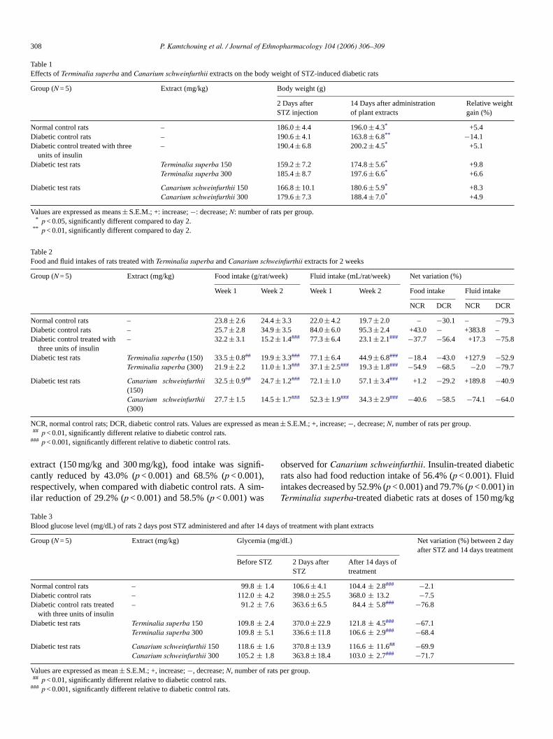
The effects of the Terminalia superba and Canarium schwe-infurthii extracts on the body weight of diabetic rats areshown in Table 1. During the 2 weeks of observation ofthe extract-treated diabetic rats at doses of 150 mg/kg and300 mg/kg, there were significant (p < 0.05) weight gains rel-ative to day 2. These gains were 9.8% and 8.3% (p < 0.05) for150 mg/kg and 6.6% and 4.9% (p < 0.05) for 300 mg/kg, respec-tively. The diabetic rats treated with three units of insulin alsoshowed a significant 5.1% (p < 0.05) weight increase but notuntreated diabetic rats, which lost 14.1% (p < 0.01) of their bodyweight.
Table 2 shows the effects of the extracts on food and fluidintakes by diabetic rats. When compared to the normal control,the untreated diabetic rats had severe polyphagia and poly-dipsia at the end of the second week of the experiment withrespective increase in food and fluid intakes of 43.0% and383.8% rats. However, in the presence of Terminalia superba
308 P. Kamtchouing et al. / Journal of Ethnopharmacology 104 (2006) 306–309
Table 1Effects of Terminalia superba and Canarium schweinfurthii extracts on the body weight of STZ-induced diabetic rats
Group (N = 5) Extract (mg/kg) Body weight (g)
2 Days afterSTZ injection
14 Days after administrationof plant extracts
Relative weightgain (%)
Normal control rats – 186.0 ± 4.4 196.0 ± 4.3* +5.4Diabetic control rats – 190.6 ± 4.1 163.8 ± 6.8** −14.1Diabetic control treated with three
units of insulin– 190.4 ± 6.8 200.2 ± 4.5* +5.1
Diabetic test rats Terminalia superba 150 159.2 ± 7.2 174.8 ± 5.6* +9.8Terminalia superba 300 185.4 ± 8.7 197.6 ± 6.6* +6.6
Diabetic test rats Canarium schweinfurthii 150 166.8 ± 10.1 180.6 ± 5.9* +8.3Canarium schweinfurthii 300 179.6 ± 7.3 188.4 ± 7.0* +4.9
Values are expressed as means ± S.E.M.; +: increase; −: decrease; N: number of rats per group.* p < 0.05, significantly different compared to day 2.
** p < 0.01, significantly different compared to day 2.
Table 2Food and fluid intakes of rats treated with Terminalia superba and Canarium schweinfurthii extracts for 2 weeks
Group (N = 5) Extract (mg/kg) Food intake (g/rat/week) Fluid intake (mL/rat/week) Net variation (%)
Week 1 Week 2 Week 1 Week 2 Food intake Fluid intake
NCR DCR NCR DCR
Normal control rats – 23.8 ± 2.6 24.4 ± 3.3 22.0 ± 4.2 19.7 ± 2.0 – −30.1 – −79.3Diabetic control rats – 25.7 ± 2.8 34.9 ± 3.5 84.0 ± 6.0 95.3 ± 2.4 +43.0 − +383.8 –Diabetic control treated with
three units of insulin– 32.2 ± 3.1 15.2 ± 1.4### 77.3 ± 6.4 23.1 ± 2.1### −37.7 −56.4 +17.3 −75.8
Diabetic test rats Terminalia superba (150) 33.5 ± 0.8## 19.9 ± 3.3### 77.1 ± 6.4 44.9 ± 6.8### −18.4 −43.0 +127.9 −52.9Terminalia superba (300) 21.9 ± 2.2 11.0 ± 1.3### 37.1 ± 2.5### 19.3 ± 1.8### −54.9 −68.5 −2.0 −79.7
Diabetic test rats Canarium schweinfurthii(150)
32.5 ± 0.9## 24.7 ± 1.2### 72.1 ± 1.0 57.1 ± 3.4### +1.2 −29.2 +189.8 −40.9
Canarium schweinfurthii(300)
27.7 ± 1.5 14.5 ± 1.7### 52.3 ± 1.9### 34.3 ± 2.9### −40.6 −58.5 −74.1 −64.0
NCR, normal control rats; DCR, diabetic control rats. Values are expressed as mean ± S.E.M.; +, increase; −, decrease; N, number of rats per group.## p < 0.01, significantly different relative to diabetic control rats.
### p < 0.001, significantly different relative to diabetic control rats.
extract (150 mg/kg and 300 mg/kg), food intake was signifi-cantly reduced by 43.0% (p < 0.001) and 68.5% (p < 0.001),respectively, when compared with diabetic control rats. A sim-ilar reduction of 29.2% (p < 0.001) and 58.5% (p < 0.001) was
observed for Canarium schweinfurthii. Insulin-treated diabeticrats also had food reduction intake of 56.4% (p < 0.001). Fluidintakes decreased by 52.9% (p < 0.001) and 79.7% (p < 0.001) inTerminalia superba-treated diabetic rats at doses of 150 mg/kg
Table 3Blood glucose level (mg/dL) of rats 2 days post STZ administered and after 14 days of treatment with plant extracts
Group (N = 5) Extract (mg/kg) Glycemia (mg/dL) Net variation (%) between 2 dayafter STZ and 14 days treatment
Before STZ 2 Days afterSTZ
After 14 days oftreatment
Normal control rats – 99.8 ± 1.4 106.6 ± 4.1 104.4 ± 2.8### −2.1Diabetic control rats – 112.0 ± 4.2 398.0 ± 25.5 368.0 ± 13.2 −7.5Diabetic control rats treated
with three units of insulin– 91.2 ± 7.6 363.6 ± 6.5 84.4 ± 5.8### −76.8
Diabetic test rats Terminalia superba 150 109.8 ± 2.4 370.0 ± 22.9 121.8 ± 4.5### −67.1Terminalia superba 300 109.8 ± 5.1 336.6 ± 11.8 106.6 ± 2.9### −68.4
Diabetic test rats Canarium schweinfurthii 150 118.6 ± 1.6 370.8 ± 13.9 116.6 ± 11.6## −69.9Canarium schweinfurthii 300 105.2 ± 1.8 363.8 ± 18.4 103.0 ± 2.7### −71.7
Values are expressed as mean ± S.E.M.; +, increase; −, decrease; N, number of rats per group.## p < 0.01, significantly different relative to diabetic control rats.
### p < 0.001, significantly different relative to diabetic control rats.

P. Kamtchouing et al. / Journal of Ethnopharmacology 104 (2006) 306–309 309
and 300 mg/kg, respectively, while Canarium schweinfurthiishowed 40.9% (p < 0.001) and 64.0% (p < 0.001) reductionswhen compared with diabetic control rats. Diabetic rats treatedwith three units of insulin also showed a significantly lowerwater intake of 75.8% (p < 0.001).
Effect of the stem bark extracts on fasting blood glucose levelsof streptozotocin-diabetic rats are shown in Table 3. Follow-ing a 48 h post streptozotocin injection, all diabetic rats exhib-ited hyperglycemia, which ranged between 330 and 400 mg/dLwhile normal control rats showed a normal blood sugar levelof 106 mg/dL. After 2 weeks of treatment with the extracts,the glycemic level of 150 mg/kg Terminalia superba extract-treated diabetic rats dropped significantly from 370.0 ± 22.9 onday 2 to 121.8 ± 11.8 mg/dL (p < 0.001) on day 14 and from336.6 ± 11.8 to 106.6 ± 2.9 mg/dL (p < 0.001) for 300 mg/kgdose, corresponding to 67% and 68% reduction, respectively.Canarium schweinfurthii extract, like insulin, also provokedreduction of the blood glucose levels of diabetic rats after 14days of treatment. These reductions were 69.9% (p < 0.001) and71.7% (p < 0.001) at the doses of 150 mg/kg and 300 mg/kg,respectively.
4. Discussion
Our results suggest that the methanol/methylene chlorideextracts of the stem bark of Terminalia superba and Canariumschweinfurthii have dose-dependent anti-diabetic activities onstreptozotocin-induced diabetes. The metabolic disturbanceswere corrected after the plant extracts were administered for2 weeks, as shown by the normalisation of fasting blood glu-cose levels, reduction in polyphagia and polydipsia and weightgain by diabetic-treated rats. Terminalia superba appeared tohave greater potency than Canarium schweinfurthii in reducingthe body weight, food and water intakes but was equipotent inblood sugar reductions.
The mechanisms by which streptozotocin brings about itsdiabetic state include selective destruction of pancreatic insulin-secreting beta cells, which make cells less active (Junod et al.,1969; Jacot and Assal, 1989) and lead to poor glucose utili-sation by tissues (Marles and Farnsworth, 1995). Terminaliasuperba and Canarium schweinfurthii significantly reduced thehigh fasting glucose levels in streptozotocin-induced diabeticrats. This suggests that the extracts may possess an insulin-like effect on peripheral tissues by either promoting glucoseuptake and metabolism, by inhibiting hepatic gluconeogenesis(Ali et al., 1993; Gray et al., 2000) or absorption of glucose intothe muscles and adipose tissues (Kamanyi et al., 1994), by thestimulation of a regeneration process and revitalisation of the
remaining beta cells (Shanmugasundaram et al., 1990; Rokeyaet al., 1999; Bolkent et al., 2000).
5. Conclusion
Terminalia superba and Canarium schweinfurthii extractspossess anti-diabetic properties.
Acknowledgement
The authors thank Prof. E. Dongo and his team from theDepartment of Organic Chemistry of the Faculty of Sciences ofthe University of Yaounde I for the plant extractions.
References
Ali, L., Azad Khan, A.K., Mamun, M.I.R., Mosihuzzaman, M., Nahar, N.,Nur-E-Alan, M., Rokeya, B., 1993. Studies on the hypoglycaemic effectsof fruits pulp, seed and whole plant of Momordica charantia on normaland diabetic model rats. Planta Medica 59, 408–412.
Berhaut, J., 1974. Flore illustre du Senegal. Dicotyledones. Lome II. Dakar,pp. 131–132, 414–416.
Bolkent, S., Yamardag, R., Tabakogluoguz, A., Ozsoy-Sacon, O., 2000.Effects of Chord (Beta vulgaris L. var. cicla) extract on pancreatic B-cells in streptozotocin-diabetic rats: a morphologic and biochemical study.Journal of Ethnopharmacology 73, 251–259.
Burkill, H.M., 1985. The Useful Plants of West Tropical Africa, vol. 1.,second ed. Royal Botanic Gardens, Kew Great Britain, pp. 425–426.
Gray, A.M., Abdel-Wahab, Y.H.A., Flatt, P.R., 2000. The traditional planttreatment, Sabucus nigra (Elder), exhibits insulin-like and insulin-releasing actions in vitro. Journal of Nutrition 130, 15–20.
Jacot, E., Assal, J.P.H., 1989. Regulation de la glycemie. Dans: Pharma-cologie des concepts Fondamentaux aux Applications Therapeutiques.Schorderet, in: Frison-Roche et Slatkine (Ed.), pp. 481–494.
Junod, A., Lambert, A.E., Stauffacher, W., Renold, A.E., 1969. Diabetogenicaction of streptozotocin of dose to metabolic response. Journal of ClinicalInvestigation 48, 2129–2139.
Kamanyi, A., Djamen, D., Nkeh, B., 1994. Hypoglycaemic properties of theaqueous roots extract of Morinda lucida (Rubiacea) study in the mouse.Phytotherapy Research 8, 369–371.
Kamesawara, B.R., Giri, R., Kesavulu, M.M., Apparao, C.H., 2000. Effect oforal administration of bark extracts of Pterocarpus santalinus L. on bloodglucose level in experimental animals. Journal of Ethnopharmacology 74,69–74.
Marles, R.J., Farnsworth, N.R., 1995. Antidiabetic plants and their activeconstituents. Phytomedicine 22, 123–189.
Rokeya, B., Nahar, N., Ali, L., Hassan, Z., Nure-E-Alam, M., Chowdhury,S.N., Azad Khan, A.K., Mosihuzzaman, M., 1999. Effects of five medic-inal plants on blood glucose levels in non-diabetic and diabetic modelrats. Diabetes Research 34, 219–228.
Shanmugasundaram, E.R.B., Gopinath, K.L., Shanmugasundaram, K.R.,Rajendran, V.M., 1990. Possible regeneration of the islets of Langer-hans in streptozotocin-diabetes rats given Gymnema sylvestre leaf extracts.Journal of Ethnopharmacology 30, 265–279.

Journal of Ethnopharmacology 104 (2006) 310–314
Anti-inflammatory and antinociceptive activities of Seseli L. species(Apiaceae) growing in Turkey
Esra Kupeli a, Alev Tosun b, Erdem Yesilada a,∗a Department of Pharmacognosy, Faculty of Pharmacy, Gazi University, Etiler 06330, Ankara, Turkey
b Department of Pharmacognosy, Faculty of Pharmacy, Ankara University, Tandogan 06100, Ankara, Turkey
Received 15 June 2005; received in revised form 15 August 2005; accepted 14 September 2005Available online 13 October 2005
Abstract
The ethyl acetate and methanol (80%) extracts obtained from 10 Seseli L. species (Apiaceae) growing in Turkey, Seseli andronakii Woron.,Seseli campestre Besser, Seseli gummiferum Pall. ex Sm. subsp. corymbosum (Boiss. and Heldr.) P.H. Davis, Seseli gummiferum Pall. ex Sm. subsp.gummiferum, Seseli hartvigii Parolly and Nordt, Seseli libanotis (L.) W. Koch, Seseli petraeum M. Bieb., Seseli peucedanoides (Bieb.) Koso-Pol.,Seseli resinosum Freyn and Sint., Seseli tortuosum L. were evaluated for their in vivo anti-inflammatory and antinociceptive activities. For thepreliminary screening of anti-inflammatory activity, carrageenan-induced hind paw edema and for the antinociceptive activity, p-benzoquinone-induced abdominal constriction test were used. Among the plant extracts, the ethyl acetate extracts from Seseli andronakii, Seseli campestre,Seseli gummiferum subsp. corymbosum, Seseli petraeum, Seseli resinosum and Seseli tortuosum showed 30.1, 32.3, 36.9, 39.8, 35.1, 37.6%inhibition in p-benzoquinone-induced abdominal constriction test, respectively. The ethyl acetate extracts of Seseli gummiferum subsp. corymbosum,Seseli petraeum, Seseli resinosum and Seseli tortuosum also exhibited notable inhibition, ranging between 24.5–29.7, 28.1–33.3, 17.4–27.5 and27.9–31.3%, respectively, in carrageenan-induced hind paw edema model at 100 mg/kg dose without inducing any gastric damage, quite comparableto indomethacin (41.8–44.8% inhibition) as a reference sample. During the acute toxicity evaluation, neither death nor gastric bleeding was observedfor any of the plant extracts. Results have supported the traditional use of some Seseli species against inflammatory disorders. Further studies arewarranted to define and isolate the active anti-inflammatory and antinociceptive components from the active species which may yield safe andeffective agents to be used in current treatments.© 2005 Elsevier Ireland Ltd. All rights reserved.
Keywords: Anti-inflammatory activity; Antinociceptive activity; Seseli species; Apiaceae
1. Introduction
Seseli is an old Greek name that was called by Hippocrates forcertain members of the Apiaceae family (Hamlyn, 1969). Thegenus Seseli L., belongs to the Apiaceae family which is com-posed of aromatic herbs and economically important speciesthat are used as foods, spices, condiments and ornamentals(Lawrence, 1969; Crowden et al., 1969; Pimenov and Leonov,1993).
Several Seseli species are reported in ancient literature forvarious healing effects. The roots of Seseli mairei Wolff., aplant growing in the Yun-Nan and Si-Chuan areas of China,are known as “Zhu Ye Fang Feng” in Chinese folklore and used
∗ Corresponding author. Fax: +90 216 5780068.E-mail address: [email protected] (E. Yesilada).
as a herbal remedy for human inflammation, swelling, rheuma-tism, pain and common cold (Hu et al., 1990). The seeds of anIndian species, Seseli indicum, have been reported to possessanthelmintic, carminative, stomachic and stimulant properties(Tandan et al., 1990).
In Turkish folk medicine, the fruit of Seseli tortuosum is usedas emmenagogue and anti-flatulence (Baytop, 1999), while theleaves of Seseli libanotis (Kelemkesir or Kelemenkesir in Turk-ish) are consumed as a vegetable in the eastern Turkey (Baytop,1994). However, only a few phytochemical or biological activ-ity studies have been reported on the Seseli species growing inTurkey so far. In a previous study, a simple coumarin and fiveangular type pyranocoumarins were isolated from the aerial partsof Seseli gummiferum ssp. corymbosum (Tosun et al., 2005a).Additionally, the essential oils from several Turkish Seseli L.species were investigated (Baser et al., 2000; Kaya et al., 2003;Tosun et al., 2005b). However, various phytochemical studies
0378-8741/$ – see front matter © 2005 Elsevier Ireland Ltd. All rights reserved.doi:10.1016/j.jep.2005.09.021
E. Kupeli et al. / Journal of Ethnopharmacology 104 (2006) 310–314 311
carried out on the other Seseli species, not grown in Turkey, haverevealed that the plant contains particularly coumarins (Banerjeeet al., 1980, 1987; Lemmich and Shabana, 1984; Coassini Lokarand Delben, 1988; Barrero et al., 1990; Ceccherelli et al., 1990;Glowniak et al., 1991), cinnamic acid derivatives (Banerjee et al.,1987), sesquiterpene lactones and phenylpropanoids (Barrero etal., 1992, 1994) in addition to essential oil (Bader et al., 2003;Habibi et al., 2003).
Several biological activities particularly focused on essen-tial oils of Seseli species, including fungutoxic (Chaturvediand Tripathi, 1989), insect-repellent (Dixit et al., 1992) andantimicrobial activity (Syed et al., 1989) were reported. Tandanet al. (1990) reported a significant and dose-dependent anti-inflammatory activity in carrageenan-induced acute inflamma-tions in rats and analgesic effect was assessed by acetic acid-induced writhing and hot plate tests in mice for seselin, acommon coumarin isolated from Seseli indicum seeds. How-ever, the compound was reported to devoid of anticonvulsantand antipyretic activities and also was found safe in doses up to6 g/kg (oral) in 72 h mortality study in mice.
As a part of our ongoing investigations on Turkish SeseliL. species, we undertook the present screening study in orderto elucidate the potential anti-inflammatory and antinociceptiveactivities of the aerial parts of Turkish Seseli species from theview of scientific points. All Seseli L. species investigated in thepresent study are listed in Table 1 with their localities. Although12 species are recorded in the Flora of Turkey (Hedge and Lam-ond, 1972; Davis et al., 1988; Duman, 2000; Parolly and Nordt,2001), two species were not found in their described localitiesin Turkey. Therefore, 10 species of Seseli L. are examined inthis study. For the preliminary screening, the ethyl acetate andmethanolic (80%) extracts obtained from the listed species weretested in mice for anti-inflammatory activity using carrageenan-induced hind paw edema model and antinociceptive activityusing p-benzoquinone induced abdominal contractions.
2. Experimental
2.1. Plant materials
Plant materials were collected from different localities inTurkey. Voucher specimens were authenticated by Prof. H.Duman of Department of Biology, Faculty of Science and Art,Gazi University, and were deposited at the Herbarium of Facultyof Pharmacy of Ankara University (AEF) and the Herbarium ofGazi University (GAZI), Ankara, Turkey. Collection sites, plantparts and herbarium numbers for each plant material is given inTable 1.
2.2. Preparation of plant extracts
Aerial parts of each plant material was dried under shadowand powdered by using a laboratory scale mill. The extract yields(w/w) are given in Table 1. The extracts were prepared followingthe procedures described below.
The ethyl acetate (EtOAc) extract: dried plant material (10 g)was extracted with ethyl acetate at room temperature by mag-
netic stirrer (×200 ml) for 24 h. The extract was evaporated todryness in vacuo to give crude EtOAc extract.
The methanol (MeOH 80%) extract: after the ethyl acetateextraction, the plant material (10 g) was extracted with methanol(80%) at room temperature by magnetic stirrer (×200 ml) for24 h. The extract was evaporated to dryness in vacuo to givecrude methanolic extract.
2.3. Pharmacological procedures
2.3.1. AnimalsMale Swiss albino mice (20–25 g) were purchased from the
animal breeding laboratories of Refik Saydam Central Instituteof Health (Ankara, Turkey). The animals left for 2 days foracclimatization to animal room conditions were maintained onstandard pellet diet and water ad libitum. The food was with-drawn on the day before the experiment, but allowed free accessof water. A minimum of six animals was used in each group.Throughout the experiments, animals were processed accord-ing to the suggested ethical guidelines for the care of laboratoryanimals.
2.3.2. Preparation of test samples for bioassayAll the plant materials were administered in 100 mg/kg
dose after suspending in 0.5% sodium carboxymethyl cellulose(CMC) suspension in distilled water. The control group ani-mals received the same experimental handling as those of thetest groups except that the drug treatment was replaced withappropriate volumes of the dosing vehicle. Either indomethacin(10 mg/kg) or acetyl salicylic acid (ASA) (100 mg/kg) in 0.5%CMC was used as reference drug.
2.3.3. Antinociceptive activityp-Benzoquinone-induced abdominal constriction test (Okun
et al., 1963) was performed on mice for determination ofantinociceptive activity. According to the method, 60 min afterthe oral administration of test samples, the mice were intraperi-tonally injected with 0.1 ml/10 g body weight of 2.5% (w/v)p-benzoquinone (PBQ; Merck) solution in distilled H2O. Con-trol animals received an appropriate volume of dosing vehicle.The mice were then kept individually for observation and thetotal number of abdominal contractions (writhing movements)was counted for the next 15 min, starting on the fifth min after thePBQ injection. The data represent average of the total number ofwrithes observed. The antinociceptive activity was expressed aspercentage change from writhing controls. Aspirin (ASA) at 100and 200 mg/kg doses was used as the reference drug in this test.
2.3.4. Anti-inflammatory activityCarrageenan-induced hind paw edema model was used
for determination of anti-inflammatory activity (Yesilada andKupeli, 2002). The difference in footpad thickness between theright and left foot was measured with a pair of dial thicknessgauge calipers (Ozaki Co., Tokyo, Japan). Mean values of treatedgroups were compared with mean values of a control group andanalyzed using statistical methods. Sixty minutes after the oraladministration of test sample or dosing vehicle, each mouse was

312 E. Kupeli et al. / Journal of Ethnopharmacology 104 (2006) 310–314
Table 1The Seseli species studied, collection sites, herbarium numbers and percentage yields of EtOAc and MeOH (80%) extracts
Plant name and authors Collection site Herbariumnumber
EtOAc extract(w/w, %)
MeOH extract(w/w, %)
Seseli andronakii Woron Erzurum, Oltu-Sarikayalar, 1450–1750 m ED 1617 41 82Seseli campestre Besser Istanbul, Sultanbeyli, Pasakoy, ca. 500 m ED 1656 43 153Seseli gummiferum Pall. ex Sm. ssp. corymbosum
(Boiss. and Heldr.) P.H. DavisAntalya-Akseki, Pinarbasi Village, 1650–1900 m AEF 21701 107 188
Seseli gummiferum Pall. ex Sm. ssp. gummiferum Ankara-Hasanoglan, Idris Mountain, 1600–1700 m AEF 21999 91 202Seseli hartvigii Parolly and Nordt. Antalya-Saklikent, Bakirlar Mountain, 2300–2500 m AEF 21700 37 117Seseli libanotis (L.) W. Koch Ardahan-Posof, 1900 m ED 1622 31 176Seseli petraeum M. Bieb. Gumushane, The road of Alemdar Village, 1400 m ED 1644 79 296Seseli peucedanoides (Bieb.) Koso-Pol. Ankara-Hasanoglan, Idris Mountain, 1600–1700 m AEF 23158 35 215Seseli resinosum Freyn and Sint. Bartin-Cakraz, 0–5 m AEF 21696 46 210Seseli tortuosum L. Ankara, Beynam Forest, 1400 m ED 1612 61 135
injected with freshly prepared (0.5 mg/25 �l) suspension of car-rageenan (Sigma, St. Louis, MO, USA) in physiological saline(154 nM NaCl) into subplantar tissue of the right hind paw. Asthe control, 25 �l saline solutions were injected into that of theleft hind paw. Paw edema was measured in every 90 min during6 h after induction of inflammation. The difference in footpadthickness was measured by a gauge calipers (Ozaki Co., Tokyo,Japan). Mean values of treated groups were compared with meanvalues of a control group and analyzed using statistical methods.Indomethacin (10 mg/kg) was used as the reference drug.
2.3.5. Acute toxicityAnimals employed in the carrageenan-induced paw edema
experiment were observed during 24 h and morbidity or mor-tality was recorded, if happens, for each group at the end ofobservation period.
2.3.6. Gastric-ulcerogenic effectAfter the antinociceptive activity experiment, mice were
killed under deep ether anesthesia and stomachs were removed.Then the abdomen of each mouse was opened through the greater
Table 2Effect of the extracts against p-benzoquinone-induced writhings in mice
Material Extract type Dose (mg/kg) Number of writhings ± S.E.M. Inhibitory ratio (%) Ratio of ulceration
Control 46.5 ± 4.6 0/6
Seseli andronakii EtOAc 100 32.5 ± 1.9 30.1** 0/6MeOH 100 49.2 ± 7.2 – 0/6
Seseli campestre EtOAc 100 31.5 ± 2.7 32.3* 0/6MeOH 100 47.7 ± 2.9 – 0/6
Seseli gummiferum ssp. corymbosum EtOAc 100 29.3 ± 2.3 36.9** 0/6MeOH 100 51.0 ± 4.9 – 0/6
Seseli gummiferum ssp. gummiferum EtOAc 100 36.3 ± 3.9 21.9MeOH 100 48.3 ± 5.1 – 0/6
Seseli hatrvigii EtOAc 100 33.5 ± 2.6 27.9 0/6MeOH 100 49.2 ± 6.3 – 0/6
Seseli libanotis EtOAc 100 38.5 ± 2.5 17.2 0/6MeOH 100 58.0 ± 2.6 – 0/6
Seseli petraeum EtOAc 100 28.0 ± 2.9 39.8** 0/6MeOH 100 43.2 ± 2.7 7.1 0/6
Seseli peucedanoides EtOAc 100 56.5 ± 2.3 – 0/6MeOH 100 43.3 ± 4.8 6.9 0/6
Seseli resinosum EtOAc 100 30.2 ± 3.7 35.1* 0/6MeOH 100 49.8 ± 4.1 – 0/6
Seseli tortuosum EtOAc 100 29.0 ± 3.5 37.6* 0/6MeOH 100 55.7 ± 2.1 – 0/6
ASA 100 23.3 ± 2.2 49.9*** 4/6
EtOAc: ethylacetate extract; MeOH: methanol extract; S.E.M.: standard error mean.* p < 0.05.
** p < 0.01.*** p < 0.001.
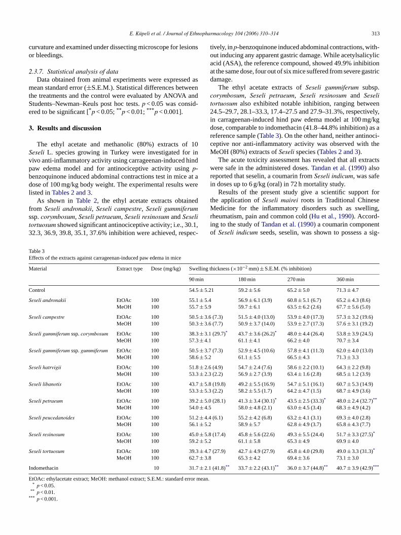
E. Kupeli et al. / Journal of Ethnopharmacology 104 (2006) 310–314 313
curvature and examined under dissecting microscope for lesionsor bleedings.
2.3.7. Statistical analysis of dataData obtained from animal experiments were expressed as
mean standard error (±S.E.M.). Statistical differences betweenthe treatments and the control were evaluated by ANOVA andStudents–Newman–Keuls post hoc tests. p < 0.05 was consid-ered to be significant [*p < 0.05; **p < 0.01; ***p < 0.001].
3. Results and discussion
The ethyl acetate and methanolic (80%) extracts of 10Seseli L. species growing in Turkey were investigated for invivo anti-inflammatory activity using carrageenan-induced hindpaw edema model and for antinociceptive activity using p-benzoquinone induced abdominal contractions test in mice at adose of 100 mg/kg body weight. The experimental results werelisted in Tables 2 and 3.
As shown in Table 2, the ethyl acetate extracts obtainedfrom Seseli andronakii, Seseli campestre, Seseli gummiferumssp. corymbosum, Seseli petraeum, Seseli resinosum and Seselitortuosum showed significant antinociceptive activity; i.e., 30.1,32.3, 36.9, 39.8, 35.1, 37.6% inhibition were achieved, respec-
tively, in p-benzoquinone induced abdominal contractions, with-out inducing any apparent gastric damage. While acetylsalicylicacid (ASA), the reference compound, showed 49.9% inhibitionat the same dose, four out of six mice suffered from severe gastricdamage.
The ethyl acetate extracts of Seseli gummiferum subsp.corymbosum, Seseli petraeum, Seseli resinosum and Seselitortuosum also exhibited notable inhibition, ranging between24.5–29.7, 28.1–33.3, 17.4–27.5 and 27.9–31.3%, respectively,in carrageenan-induced hind paw edema model at 100 mg/kgdose, comparable to indomethacin (41.8–44.8% inhibition) as areference sample (Table 3). On the other hand, neither antinoci-ceptive nor anti-inflammatory activity was observed with theMeOH (80%) extracts of Seseli species (Tables 2 and 3).
The acute toxicity assessment has revealed that all extractswere safe in the administered doses. Tandan et al. (1990) alsoreported that seselin, a coumarin from Seseli indicum, was safein doses up to 6 g/kg (oral) in 72 h mortality study.
Results of the present study give a scientific support forthe application of Seseli mairei roots in Traditional ChineseMedicine for the inflammatory disorders such as swelling,rheumatism, pain and common cold (Hu et al., 1990). Accord-ing to the study of Tandan et al. (1990) a coumarin componentof Seseli indicum seeds, seselin, was shown to possess a sig-
Table 3Effects of the extracts against carrageenan-induced paw edema in mice
Material Extract type Dose (mg/kg) Swelling thickness (×10−2 mm) ± S.E.M. (% inhibition)
90 min 180 min 270 min 360 min
Control 54.5 ± 5.21 59.2 ± 5.6 65.2 ± 5.0 71.3 ± 4.7
Seseli andronakii EtOAc 100 55.1 ± 5.4 56.9 ± 6.1 (3.9) 60.8 ± 5.1 (6.7) 65.2 ± 4.3 (8.6)MeOH 100 55.7 ± 5.9 59.7 ± 6.1 63.5 ± 6.2 (2.6) 67.7 ± 5.6 (5.0)
Seseli campestre EtOAc 100 50.5 ± 3.6 (7.3) 51.5 ± 4.0 (13.0) 53.9 ± 4.0 (17.3) 57.3 ± 3.2 (19.6)MeOH 100 50.3 ± 3.6 (7.7) 50.9 ± 3.7 (14.0) 53.9 ± 2.7 (17.3) 57.6 ± 3.1 (19.2)
Seseli gummiferum ssp. corymbosum EtOAc 100 38.3 ± 3.1 (29.7)* 43.7 ± 3.6 (26.2)* 48.0 ± 4.4 (26.4) 53.8 ± 3.9 (24.5)MeOH 100 57.3 ± 4.1 61.1 ± 4.1 66.2 ± 4.0 70.7 ± 3.4
Seseli gummiferum ssp. gummiferum EtOAc 100 50.5 ± 3.7 (7.3) 52.9 ± 4.5 (10.6) 57.8 ± 4.1 (11.3) 62.0 ± 4.0 (13.0)MeOH 100 58.6 ± 5.2 61.1 ± 5.5 66.5 ± 4.3 71.3 ± 3.3
Seseli hatrvigii EtOAc 100 51.8 ± 2.6 (4.9) 54.7 ± 2.4 (7.6) 58.6 ± 2.2 (10.1) 64.3 ± 2.2 (9.8)MeOH 100 53.3 ± 2.3 (2.2) 56.9 ± 2.7 (3.9) 63.4 ± 1.6 (2.8) 68.5 ± 1.2 (3.9)
Seseli libanotis EtOAc 100 43.7 ± 5.8 (19.8) 49.2 ± 5.5 (16.9) 54.7 ± 5.1 (16.1) 60.7 ± 5.3 (14.9)MeOH 100 53.3 ± 5.3 (2.2) 58.2 ± 5.5 (1.7) 64.2 ± 4.7 (1.5) 68.7 ± 4.9 (3.6)
Seseli petraeum EtOAc 100 39.2 ± 5.0 (28.1) 41.3 ± 3.4 (30.1)* 43.5 ± 2.5 (33.3)* 48.0 ± 2.4 (32.7)**
MeOH 100 54.0 ± 4.5 58.0 ± 4.8 (2.1) 63.0 ± 4.5 (3.4) 68.3 ± 4.9 (4.2)
Seseli peucedanoides EtOAc 100 51.2 ± 4.4 (6.1) 55.2 ± 4.2 (6.8) 63.2 ± 4.1 (3.1) 69.3 ± 4.0 (2.8)MeOH 100 56.1 ± 5.2 58.9 ± 5.7 62.8 ± 4.9 (3.7) 65.8 ± 4.3 (7.7)
Seseli resinosum EtOAc 100 45.0 ± 5.8 (17.4) 45.8 ± 5.6 (22.6) 49.3 ± 5.5 (24.4) 51.7 ± 3.3 (27.5)*
MeOH 100 59.2 ± 5.2 61.1 ± 5.8 65.3 ± 4.9 69.9 ± 4.0
Seseli tortuosum EtOAc 100 39.3 ± 4.7 (27.9) 42.7 ± 4.9 (27.9) 45.8 ± 4.0 (29.8) 49.0 ± 3.3 (31.3)*
MeOH 100 62.7 ± 3.8 65.3 ± 4.2 69.4 ± 3.6 73.1 ± 3.0
Indomethacin 10 31.7 ± 2.1 (41.8)** 33.7 ± 2.2 (43.1)** 36.0 ± 3.7 (44.8)** 40.7 ± 3.9 (42.9)***
EtOAc: ethylacetate extract; MeOH: methanol extract; S.E.M.: standard error mean.* p < 0.05.
** p < 0.01.*** p < 0.001.

314 E. Kupeli et al. / Journal of Ethnopharmacology 104 (2006) 310–314
nificant and dose-dependent anti-inflammatory and analgesicactivity. Seselin is known to be found either in the roots, stems,leaves or inflorescence of several species. Garcia-Argaez et al.(2000) has also proven the anti-inflammatory effect of severalcoumarins, including seselin, using a TPA-induced ear edemamodel in mice and concluded that the anti-inflammatory activityof coumarins depend on their individual substitution on the aro-matic ring rather than the coumarin skeleton itself. They havefurther suggested that the activity might possibly be due to theirability to inhibit lipid peroxidation and to act as a radical scav-enger.
Seseli species contain a large variety of coumarin derivativeswhich are mainly accumulated in nonpolar extracts (Banerjee etal., 1980; Lemmich and Shabana, 1984; Coassini Lokar andDelben, 1988; Barrero et al., 1990; Ceccherelli et al., 1990;Glowniak et al., 1991). Eventually, the effect might be due tothe coumarin components (O’Kennedy and Thornes, 1997) ofthe active Seseli species.
Among the 10 Seseli species studied, only the ethyl acetateextracts of Seseli gummiferum subsp. corymbosum, Seselipetraeum and Seseli resinosum were shown to possess remark-able antinociceptive and anti-inflammatory activity withoutinducing any apparent acute toxicity. As far as our best knowl-edge is that being first activity screening of all Seseli speciesgrowing in Turkey. Since Seseli species are also used as spice orfood in Anatolia, further studies to define and isolate active anti-inflammatory and antinociceptive components from the activespecies may yield safe agents to use in current treatments.
References
Bader, A., Caponi, C., Cioni, P.L., Flamini, G., Morelli, I., 2003. Acorenonein the essential oil of flowering aerial parts of Seseli tortuosum L. Flavorand Fragrance Journal 18, 57–58.
Banerjee, S.K., Gupta, B.D., Kumar, R., Atal, C.K., 1980. New coumarinsfrom the umbels of Seseli sibiricum. Phytochemistry 19, 281–284.
Banerjee, S.K., Mukhopadhyay, S., Gupta, B.D., Singh, K., Raj, S., 1987.Sesebrinic acid, a cinnamic acid derivative from Seseli sibiricum. Phyto-chemistry 26, 1817–1820.
Barrero, A.F., Herrador, M.M., Arteaga, P., 1990. Cumarinas en especies delgenero Seseli (Fam. Umbelliferae). Ars Pharmaceutica 31, 241–256.
Barrero, A.F., Herrador, M.M., Arteaga, P., 1992. Sesquiterpenes and phenyl-propanoids from Seseli vayredanum. Phytochemistry 31, 203–207.
Barrero, A.F., Herrador, M.M., Arteaga, P., 1994. Sesquiterpene lactones andother constituents of Seseli vayredanum. Phytochemistry 37, 1351–1358.
Baser, K.H.C., Ozek, T., Kurkcuoglu, M., Aytac, Z., 2000. Essential oil ofS. campestre Besser. Journal of Essential Oil Research 12, 105–107.
Baytop, T., 1999. Therapy with Plants in Turkey (Past and Present), 2nd ed.Nobel Medical Book House, Istanbul.
Baytop, T., 1994. Turkce Bitki Adları Sozlugu, Ataturk Kultur, Dil ve TarihYuksek Kurumu, TDKY 3578. TTK Basimevi, Ankara.
Chaturvedi, R.V., Tripathi, S.C., 1989. Fungutoxic, physico-chemical andphytotoxic properties of essential oil of Seseli indicum W and A. Journalof Phytopathology 124, 316–322.
Ceccherelli, P., Curini, M., Marcotullio, M.C., Madruzza, G., 1990. Tortuo-side, a new natural coumarin glucoside from Seseli tortuosum. Journal ofNatural Products 53, 536–538.
Coassini Lokar, L.R., Delben, S., 1998. Photoactive furocoumarins in twopopulations of Seseli elatum. Phytochemistry 27, 1073–1077.
Crowden, R.K., Harborne, J.B., Heywood, V.H., 1969. Chemosystematics ofthe Umbelliferae – a general survey. Phytochemistry 8, 1963–1984.
Davis, P.H., Mill, R.R., Tan, K., 1988. Flora of Turkey and the East AegeanIslands, vol. 10. Edinburgh University Press, Edinburgh.
Dixit, V., Chaturvedi, R.V., Tripathi, S.C., 1992. Evaluation of some essentialoils against Pulse Bettle (Callosobruchus chinensis). National AcademyScientific Letters 15, 255–257.
Duman, H., 2000. Seseli L. In: Guner, A., Ozhatay, N., Ekim, T., Baser,K.H.C. (Eds.), Flora of Turkey and the East Aegean Islands, vol. 11.Edinburgh University Press, Edinburgh, p. 141.
Garcia-Argaez, A.N., Ramirez, Apan T.O., Delgado, H.P., Velazquez, G.,Martinez-Vazquez, M., 2000. Anti-inflammatory activity of coumarinsfrom Decatropis bicolor on TPA ear mice model. Planta Medica 66,279–281.
Glowniak, K., Doraczynska-Szopa, A., Erkelens, C., Sluis, W.G., 1991.Isopeucenidin and libanotin from Libanotis intermedia roots: isolationand NMR analysis. Planta Medica 57, A52.
Habibi, Z., Masoudi, S., Rustaiyan, A., 2003. Chemical composition of theessential oil of Seseli tortuosum L. ssp. kiabii Akhani. from Iran. Journalof Essential Oil Research 15, 412–413.
Hamlyn, P., 1969. The Marshall Cavendish, Encyclopedia of Gardening, vol.19. Garrod and Lofthouse International, London, p. 2034.
Hedge, I.C., Lamond, J.M., 1972. Seseli L. In: Davis, P.H. (Ed.), Flora ofTurkey and the East Aegean Islands, vol. 4. Edinburgh University Press,Edinburgh, pp. 367–372.
Hu, C.-Q., Chang, J.-J., Lee, K.-H., 1990. Antitumor agents, 115. Seselidiol,a new cytotoxic polyacetylene from Seseli mairei. Journal of NaturalProducts 53, 932–935.
Kaya, A., Demirci, B., Base, K.H.C., 2003. The essential oil of Seseli tortu-osum L. growing in Turkey. Flavor and Fragrance Journal 18, 159–161.
Lawrence, G.H.M., 1969. Taxonomy of Vascular Plants. The Macmillan Com-pany, New York, pp. 642–646.
Lemmich, J., Shabana, M., 1984. Coumarin sulphates of Seseli libanotis.Phytochemistry 23, 863–865.
O’Kennedy, R., Thornes, R.D., 1997. Coumarins: Biology, Applications andMode of Action. John Wiley and Sons, Chichester.
Okun, R., Liddon, S.C., Lasagnal, L., 1963. The effect of aggregation, elec-tric shock and adrenergic blocking drugs on inhibition of the “writhingsyndrome”. Journal of Pharmacology and Experimental Therapeutics 139,107–109.
Parolly, G., Nordt, B., 2001. Seseli hartvigii (Apiaceae), a new name forSeseli ramosissimum Hartvig and Strid, with carpological and ecologicalnotes on this species. Willdonowia 31, 87–93.
Pimenov, M.G., Leonov, M.V., 1993. The Genera of the Umbelliferae. Whit-stable Litho, Whitstable, Kent.
Syed, M., Chaudhary, F.M., Bhatty, M.K., 1989. Antimicrobial activity ofthe essential oils of Umbelliferae family. Part VIII. Seseli libanotis,Ligusticum stewartii and Pycnocycla aucheriana oils. Pakistan Journalof Scientific and Industrial Research 32, 316–319.
Tandan, S.K., Chandra, S., Tripathi, H.C., Lal, J., 1990. Pharmacologicalactions of seselin, a coumarin from Seseli indicum seeds. Fitoterapia 61,360–363.
Tosun, A., Ozkal, N., Baba, M., Okuyama, T., 2005a. Pyranocoumarins fromSeseli gummiferum subsp. corymbosum growing in Turkey. Turkish Jour-nal of Chemistry 29, 327–334.
Tosun, A., Baba, M., Kodama, T., Nakanishi, H., Okuyama, T., 2005b. Thecomposition of essential oil of Seseli L. species growing in Turkey. Nat-ural Medicines 59, 85–90.
Yesilada, E., Kupeli, E., 2002. Berberis crataegina DC root exhibits potentanti-inflammatory, analgesic and febrifuge effects in mice and rats. Journalof Ethnopharmacology 79, 237–248.
Journal of Ethnopharmacology 104 (2006) 315–321
Effects of Urginea sanguinea, a traditional asthma remedy,on embryo neuronal development
J. Marx, E. Pretorius ∗, M.J. BesterDepartment of Anatomy, School of Health Sciences, Medical Faculty of the University of Pretoria, Pretoria, South Africa
Received 10 June 2005; received in revised form 30 August 2005; accepted 15 September 2005Available online 19 October 2005
Abstract
The Southern African plant, Urginea sanguinea Shinz (Hyacinthaceae) (US), is a well-known traditional herbal medicine and it is used formany different ailments, including asthma. Pregnant women also use this plant and little is known regarding the toxic effects of this plant materialon the developing foetus. US contains the cardiac glycoside (CG) Transvaalin; CGs are known to cross the placenta and blood–brain barrier andtherefore may have a negative effect on the foetal development. To address this, in vitro cytotoxicity of this preparation as well as its effect on chickembryo neural development was investigated. Water extracts of US were shown to be cytotoxic in cell cultures of L929 cell and primary embryonicneural cell cultures. Electron microscopy studies following in ovo exposure revealed altered neuron morphology with patterns of cell damage eitherassociated with apoptosis or necrosis. CGs are known to inhibit membrane bound Na+/K+-ATPase in conducting tissues, causing disruption of thecalcium pathways, mitochondrial calcium overload leading to either apoptosis or necrosis or where both occur, a process of necrapoptosis. The inovo effects observed strongly indicate that US causes necrapoptosis in chick embryonic neurons.© 2005 Elsevier Ireland Ltd. All rights reserved.
Keywords: Urginea sanguinea; Apoptosis; Necrosis; Cardiac glycosides; Electron microscopy; Cytotoxicity
1. Introduction
It is estimated that there are 27 million indigenous medicineconsumers in Southern Africa and a significantly large numberof these patients consult traditional healers for potentially lifethreatening conditions (Puckree et al., 2002; Mulholland andDrewes, 2004). The Southern African plant, Urginea sanguineaShinz (Hyacinthaceae) (US) is a well-known traditional herbalmedicine and the bulb of this plant is used for many differ-ent ailments (Watt and Breyer-Brandwijk, 1962; Hedberg andStuagard, 1989; Foukaridis et al., 1995; Van Wyk and Gericke,2000; Marx et al., 2005). The powdered bulbs are used to treat,amongst other ailments, bronchitis, asthma and influenza (Molland Strebel, 1989), while a tea is prepared for the treatmentof veneral disease, abdominal pain, backache and hypertension.However, many indigenous people taking this plant as medicinehave complained of adverse health effects, and although many
∗ Corresponding author. Present address: BMW Building, P.O. Box 2034,Faculty of Health Sciences, University of Pretoria, Pretoria 0001, South Africa.Tel.: +27 12 319 2533; fax: +27 12 319 2240.
E-mail address: [email protected] (E. Pretorius).
complaints have been noted, little scientific information regard-ing the toxic effects on cellular function, growth and differenti-ation is available. The cardiac glycoside, Transvaalin has beenisolated from US (Louw, 1949, 1952). Cardiac glycosides areused to stimulate the heart muscle and act on conducting tis-sue (including neural tissue) by changing the functioning of thedepolarisation of the membrane-linked Na+/K+ pump and causean increase in Ca2+. Although cardiac glycosides are beneficialin treating cardiac conditions, there is often a possible risk whenused during pregnancy; likewise, US derived herbal prepara-tions are used during pregnancy the foetus is possibly also atrisk. Therefore, the purpose of this study was to determine thecytotoxicity of US in vitro using the L929 cell line and primarycultures of chick embryo neurons before studying the effects ofin ovo exposure on neuron morphology by electron microscopy.
2. Materials and methods
2.1. Preparation of plant extract
Fresh US bulbs were obtained from Louis Trichardt, Limpopoprovince, South Africa. Prof. J.J. Meyer, from the Department of
0378-8741/$ – see front matter © 2005 Elsevier Ireland Ltd. All rights reserved.doi:10.1016/j.jep.2005.09.022
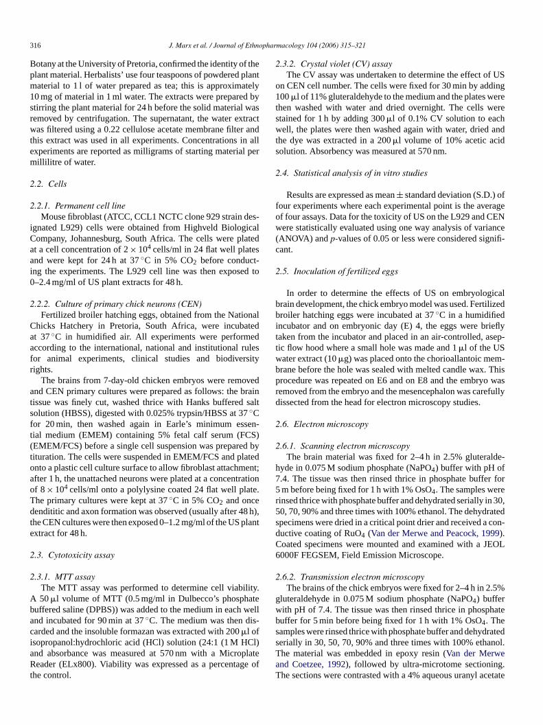
316 J. Marx et al. / Journal of Ethnopharmacology 104 (2006) 315–321
Botany at the University of Pretoria, confirmed the identity of theplant material. Herbalists’ use four teaspoons of powdered plantmaterial to 1 l of water prepared as tea; this is approximately10 mg of material in 1 ml water. The extracts were prepared bystirring the plant material for 24 h before the solid material wasremoved by centrifugation. The supernatant, the water extractwas filtered using a 0.22 cellulose acetate membrane filter andthis extract was used in all experiments. Concentrations in allexperiments are reported as milligrams of starting material permillilitre of water.
2.2. Cells
2.2.1. Permanent cell lineMouse fibroblast (ATCC, CCL1 NCTC clone 929 strain des-
ignated L929) cells were obtained from Highveld BiologicalCompany, Johannesburg, South Africa. The cells were platedat a cell concentration of 2 × 10⁴ cells/ml in 24 flat well platesand were kept for 24 h at 37 ◦C in 5% CO2 before conduct-ing the experiments. The L929 cell line was then exposed to0–2.4 mg/ml of US plant extracts for 48 h.
2.2.2. Culture of primary chick neurons (CEN)Fertilized broiler hatching eggs, obtained from the National
Chicks Hatchery in Pretoria, South Africa, were incubatedat 37 ◦C in humidified air. All experiments were performedaccording to the international, national and institutional rulesfor animal experiments, clinical studies and biodiversityrights.
The brains from 7-day-old chicken embryos were removedand CEN primary cultures were prepared as follows: the braintissue was finely cut, washed thrice with Hanks buffered saltsolution (HBSS), digested with 0.025% trypsin/HBSS at 37 ◦Cfor 20 min, then washed again in Earle’s minimum essen-tial medium (EMEM) containing 5% fetal calf serum (FCS)(EMEM/FCS) before a single cell suspension was prepared bytituration. The cells were suspended in EMEM/FCS and platedonto a plastic cell culture surface to allow fibroblast attachment;after 1 h, the unattached neurons were plated at a concentrationof 8 × 10⁴ cells/ml onto a polylysine coated 24 flat well plate.The primary cultures were kept at 37 ◦C in 5% CO2 and oncedendititic and axon formation was observed (usually after 48 h),the CEN cultures were then exposed 0–1.2 mg/ml of the US plantextract for 48 h.
2.3. Cytotoxicity assay
2.3.1. MTT assayThe MTT assay was performed to determine cell viability.
A 50 �l volume of MTT (0.5 mg/ml in Dulbecco’s phosphatebuffered saline (DPBS)) was added to the medium in each welland incubated for 90 min at 37 ◦C. The medium was then dis-carded and the insoluble formazan was extracted with 200 �l ofisopropanol:hydrochloric acid (HCl) solution (24:1 (1 M HCl)and absorbance was measured at 570 nm with a MicroplateReader (ELx800). Viability was expressed as a percentage ofthe control.
2.3.2. Crystal violet (CV) assayThe CV assay was undertaken to determine the effect of US
on CEN cell number. The cells were fixed for 30 min by adding100 �l of 11% gluteraldehyde to the medium and the plates werethen washed with water and dried overnight. The cells werestained for 1 h by adding 300 �l of 0.1% CV solution to eachwell, the plates were then washed again with water, dried andthe dye was extracted in a 200 �l volume of 10% acetic acidsolution. Absorbency was measured at 570 nm.
2.4. Statistical analysis of in vitro studies
Results are expressed as mean ± standard deviation (S.D.) offour experiments where each experimental point is the averageof four assays. Data for the toxicity of US on the L929 and CENwere statistically evaluated using one way analysis of variance(ANOVA) and p-values of 0.05 or less were considered signifi-cant.
2.5. Inoculation of fertilized eggs
In order to determine the effects of US on embryologicalbrain development, the chick embryo model was used. Fertilizedbroiler hatching eggs were incubated at 37 ◦C in a humidifiedincubator and on embryonic day (E) 4, the eggs were brieflytaken from the incubator and placed in an air-controlled, asep-tic flow hood where a small hole was made and 1 �l of the USwater extract (10 �g) was placed onto the chorioallantoic mem-brane before the hole was sealed with melted candle wax. Thisprocedure was repeated on E6 and on E8 and the embryo wasremoved from the embryo and the mesencephalon was carefullydissected from the head for electron microscopy studies.
2.6. Electron microscopy
2.6.1. Scanning electron microscopyThe brain material was fixed for 2–4 h in 2.5% gluteralde-
hyde in 0.075 M sodium phosphate (NaPO4) buffer with pH of7.4. The tissue was then rinsed thrice in phosphate buffer for5 m before being fixed for 1 h with 1% OsO4. The samples wererinsed thrice with phosphate buffer and dehydrated serially in 30,50, 70, 90% and three times with 100% ethanol. The dehydratedspecimens were dried in a critical point drier and received a con-ductive coating of RuO4 (Van der Merwe and Peacock, 1999).Coated specimens were mounted and examined with a JEOL6000F FEGSEM, Field Emission Microscope.
2.6.2. Transmission electron microscopyThe brains of the chick embryos were fixed for 2–4 h in 2.5%
gluteraldehyde in 0.075 M sodium phosphate (NaPO4) bufferwith pH of 7.4. The tissue was then rinsed thrice in phosphatebuffer for 5 min before being fixed for 1 h with 1% OsO4. Thesamples were rinsed thrice with phosphate buffer and dehydratedserially in 30, 50, 70, 90% and three times with 100% ethanol.The material was embedded in epoxy resin (Van der Merweand Coetzee, 1992), followed by ultra-microtome sectioning.The sections were contrasted with a 4% aqueous uranyl acetate
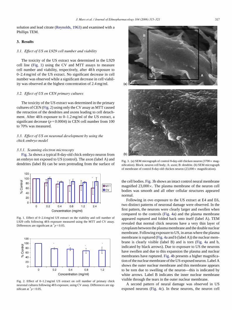
J. Marx et al. / Journal of Ethnopharmacology 104 (2006) 315–321 317
solution and lead citrate (Reynolds, 1963) and examined with aPhillips TEM.
3. Results
3.1. Effect of US on L929 cell number and viability
The toxicity of the US extract was determined in the L929cell line (Fig. 1) using the CV and MTT assays to measurecell number and viability, respectively, after 48 h exposure to0–2.4 mg/ml of the US extract. No significant decrease in cellnumber was observed while a significant decrease in cell viabil-ity was observed at the highest concentration of 2.4 mg/ml.
3.2. Effect of US on CEN primary cultures
The toxicity of the US extract was determined in the primarycultures of CEN (Fig. 2) using only the CV assay as MTT causedthe retraction of the dendrites and axons leading to cell detach-ment. After 48 h exposure to 0–1.2 mg/ml of the US extract, asignificant decrease (p = 0.0004) in CEN cell number from 100to 70% was measured.
3.3. Effect of US on neuronal development by using thechick embryo model
3.3.1. Scanning electron microscopyFig. 3a shows a typical 8-day-old chick embryo neuron from
an embryo not exposed to US (control). The axon (label A) anddendrites (label B) can be seen protruding from the surface of
Fig. 1. Effect of 0–2.4 mg/ml US extract on the viability and cell number ofL929 cells following 48 h exposure measured using the MTT and CV assay.Differences are significant at *p < 0.05.
Fig. 2. Effect of 0–1.2 mg/ml US extract on cell number of primary chickneuronal cultures following 48 h exposure, using CV assay. Differences are sig-nificant at *p < 0.05.
Fig. 3. (a) SEM micrograph of control 8-day-old chicken neuron (3700× mag-nification). Block: neuron cell body; A: axon; B: dendrite. (b) SEM micrographof membrane of control 8-day-old chicken neuron (23,000× magnification).
the cell bodies. Fig. 3b shows an intact control neural membranemagnified 23,000×. The plasma membrane of the neuron cellbodies was smooth and all other cellular structures appearednormal.
Following in ovo exposure to the US extract at E4 and E6,two distinct patterns of neuronal damage were observed. In thefirst pattern, the neurons were clearly larger and swollen whencompared to the controls (Fig. 4a) and the plasma membraneappeared ruptured and folded back onto itself (label A). TEMrevealed that normal chick neurons have a very thin layer ofcytoplasm between the plasma membrane and the double nuclearmembrane. Following exposure to US, in areas where the plasmamembrane is ruptured (Fig. 4a and b (label A)) the nuclear mem-brane is clearly visible (label B) and is torn (Fig. 4a and b,indicated by black arrows). Due to exposure to US the neuronshave swollen and due to this expansion the plasma and nuclearmembranes have ruptured. Fig. 4b presents a higher magnifica-tion of the nuclear membrane of the US-exposed neuron. Label Ashows the outer nuclear membrane and this membrane appearsto be torn due to swelling of the neuron—this is indicated bywhite arrows. Label B indicates the inner nuclear membranevisible through the tears in the outer nuclear membrane.
A second pattern of neural damage was observed in USexposed neurons (Fig. 4c). In these neurons, the neuron cell
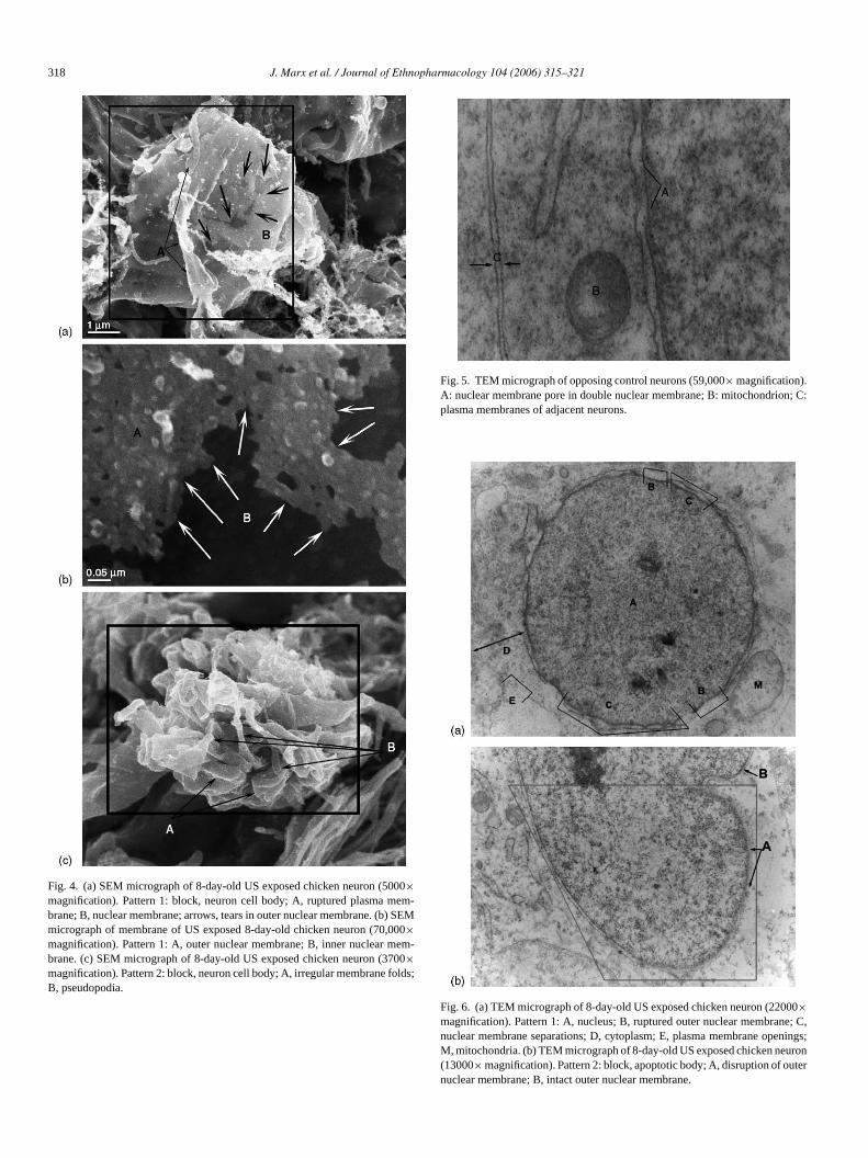
318 J. Marx et al. / Journal of Ethnopharmacology 104 (2006) 315–321
Fig. 4. (a) SEM micrograph of 8-day-old US exposed chicken neuron (5000×magnification). Pattern 1: block, neuron cell body; A, ruptured plasma mem-brane; B, nuclear membrane; arrows, tears in outer nuclear membrane. (b) SEMmicrograph of membrane of US exposed 8-day-old chicken neuron (70,000×magnification). Pattern 1: A, outer nuclear membrane; B, inner nuclear mem-brane. (c) SEM micrograph of 8-day-old US exposed chicken neuron (3700×magnification). Pattern 2: block, neuron cell body; A, irregular membrane folds;B, pseudopodia.
Fig. 5. TEM micrograph of opposing control neurons (59,000× magnification).A: nuclear membrane pore in double nuclear membrane; B: mitochondrion; C:plasma membranes of adjacent neurons.
Fig. 6. (a) TEM micrograph of 8-day-old US exposed chicken neuron (22000×magnification). Pattern 1: A, nucleus; B, ruptured outer nuclear membrane; C,nuclear membrane separations; D, cytoplasm; E, plasma membrane openings;M, mitochondria. (b) TEM micrograph of 8-day-old US exposed chicken neuron(13000× magnification). Pattern 2: block, apoptotic body; A, disruption of outernuclear membrane; B, intact outer nuclear membrane.

J. Marx et al. / Journal of Ethnopharmacology 104 (2006) 315–321 319
body appeared intact (neuron indicated by block), but the cellsurface showed irregular folds (label A) resembling pseudopo-dia (label B).
3.3.2. Transmission electron microscopyFig. 5 shows a TEM micrograph of opposing neurons of the
control. The plasma membranes and the nuclear membranesare intact (Fig. 5, labels A–C) and a thin cytoplasm is present(Fig. 5, label D). The TEM micrograph of the US exposed chickembryos’ neurons also showed the two patterns mentioned inthe above paragraphs. Pattern 1 is shown in Fig. 6a. Label Aindicates the nucleus; label B the ruptured outer nuclear mem-brane where at regular intervals nuclear membrane separation isobserved (Fig. 6, label C). In SEM micrographs the cell bodiesappeared swollen in this pattern (Fig. 4a and b) and in TEMmicrographs the distance between the plasma and nuclear mem-branes is increased when compared to the controls (Fig. 6a, labelD). The plasma membranes of neurons with pattern 1 morphol-ogy are damaged with large gaps (Fig. 6a, label E) occurringand no apoptotic bodies are observed.
In pattern 2, shown in Fig. 6b, no nuclear membrane separa-tion has occurred and apoptotic bodies are present in the nucleusand are indicated by block (Fig. 6b). Disruption of the outernuclear membrane has also occurred in some regions (Fig. 6b,label A); while in other regions (Fig. 6b, label B) the outer mem-brane has remained intact.
4. Discussion and conclusion
US is a livestock poison and is also widely used as a traditionalherbal remedy with the foetus being at great risk if used bypregnant woman. Therefore, the purpose of this study was todetermine the effect of US preparations as prepared by herbalistson neuronal development using the chick embryo as model.
To confirm that the plant material used in this study is toxic,the fibroblast L929 cell line that is an FDA-approved cell linefor in vitro toxicological studies was used. The L929 cells wereexposed to 0–2.4 mg/ml of US water extract for 48 h before cellnumber and cell viability was determined using the CV and MTTassays, respectively. No significant changes in cell number wereobserved while a decrease in cell viability were observed.
Prior to testing the effects in ovo, primary cultures of CENwere established and the toxic effect of 0–1.2 mg/ml US waterextract was determined. After 48 h exposure, a decrease in cellnumber was observed indicating the sensitivity of chick embry-ological neurons to US. In ovo, chick embryos were exposedto 10 �g US extract at E4 and E6, and although US did notcause embryological death, defects or changes in growth, ultra-structurally altered neuron morphology was observed. It is diffi-cult to correlate in ovo and in vitro dosages due to factors such asabsorption, metabolism and excretion that may alter the dosageto which the developing brain is actually exposed.
In each brain studied, two distinct morphological patternsof neuronal damage were observed. Characteristic of the firstpattern of cellular damage was neuron swelling, tearing of theplasma and nuclear membranes and absence of apoptopic bodies(Fig. 4b). Necrosis refers to cellular features after a cell has died
(Park et al., 2000; Van Cruchten and Van Den Broeck, 2002).Oncosis as defined by Majno and Joris (1995) is a subdivision orspecial type of necrosis with specific cellular features such as cel-lular and organellar swelling, membrane disruption and changesin membrane permeability as well as absence of apoptotic bod-ies. The cellular changes observed in ovo following exposureto US are typical of oncosis (Fig. 5a and b). Liu et al. (2004)reported that oncosis often affects many contiguous cells, withearly manifestations consisting of plasma membrane blebbing,dilation of the endoplasmatic reticulum, mitochondrial swellingand clumping of nuclear chromatin. Schmitt and Aebert (2004)identified the characteristic features of oncosis as disruption ofthe plasma membrane and cellular swelling. Muller et al. (2004)studied the morphological effects of transient forebrain ischemiain rat and found that apoptosis-like morphology and/or caspase 3expression (<10%) occurred in only a small fraction of ischemicneurons and the majority of cell deaths occur via a necrosis-likepathway.
The second pattern showed the formation of pseudopodiaand, in 1991, Kerr and Harmon first described the process where“budding” occurs during cell death. Majno and Joris (1995)termed “budding” as the formation of pseudopodia of cellsspecifically during the apoptotic process. These structures maycontain any type of organelle and differ from blebs (zeiosis),which are typically associated with ischemic cell death and arisefrom the cell membrane as blister-like fluid-filled structures thatmay swell and burst (Majno and Joris, 1995; Fadeel, 2004).In apoptosis plasma, membranes are altered due to the struc-tural orientation changes of the lipid in the membranes, possiblyresulting in the folds or pseudopodia that are observed. In thecurrent study, this is seen in Fig. 4c, label B. TEM micrographsindicated apoptotic body formation of the nucleus. No nuclearmembrane separation as seen in pattern 1 (Fig. 6a, label C)was seen where apoptotic bodies were present. Therefore, chickembryo neurons undergo both apoptosis and necrosis (oncosis)following exposure to US similar to that described by Mulleret al. (2004) for the ischemic rat hippocampus.
Marx (2005) suggested that the main reason for these toxiceffects is the cardiac glycoside Transvaalin (Louw, 1949, 1952;Foukaridis et al., 1995) that occurs at a concentration of0.01–0.05% (depending on the source and time of harvest) pergram of dried plant material. Using this information and assum-ing that Transvaalin is completely water-soluble, the L929 celllines were exposed to 0–0.024–0.125 mg/ml, while the CENwere exposed to 0–0.012–0.0625 mg/ml of Transvaalin. CGs,such as Transvaalin present in US are well known for theireffects on cardiac muscle, and also have an effect on other con-ducting tissues such as the brain (Hamlyn et al., 1991; Vaillendet al., 2002) that has specific CG binding sites (Maki et al.,1992). Under physiological conditions, membrane depolarisa-tion occurs through Na+ influx into the cell; this triggers theslower influx of Ca2+. High intracellular concentrations of Ca2+
then results in the efflux of K+, and the reestablishment of theaction potential occurs by the reverse of the Na+/K+ exchange.CGs inhibit the Na+/K+-pump in the membranes of conduct-ing tissue thereby inhibiting membrane-bound Na+/K+-ATPase(Nishio et al., 2004). This leads to an increase in the intracellular

320 J. Marx et al. / Journal of Ethnopharmacology 104 (2006) 315–321
concentration of Na+, and subsequently to an increase in theintracellular cytosol Ca2+ stores (Lee, 1985; Levi et al., 1994).The mitochondria provide a protective mechanism against Ca2+
cellular cytotoxicity, by buffering of elevations from free Ca2+
in neuronal cytoplasm (White and Reynolds, 1995; Budd andNicholls, 1996a,b; Wang and Thayer, 1996). However, increasedinflux of Ca2+ due to CGs-like Transvaalin will result in Ca2+
overload of the mitochondria leading to the opening of thepermeability transition pore (PTP) (Gunter and Pfeiffer, 1990;Pfeiffer et al., 2001), which is located in the inner membraneof the mitochondria. The opening of the PTP initiates mito-chondrial permeability transition (MPT) (Crompton et al., 1988;Gunter and Pfeiffer, 1990; Gunter et al., 2004). Salicylate, whichis also one of the components isolated from the bulb of US, alsoacts as an activator of Ca2+ and phosphate in promoting the open-ing of inner mitochondrial membrane pore (Al-Nasser, 1999).As a consequence of PTP opening, molecules with a molecu-lar mass less than 1500 Da non-selectively diffuse across themitochondrial inner membrane, and this leads to mitochondrialdepolarization, swelling and uncoupling of oxidative phospho-rylation and inhibition of the formation of mitochondrial ATP(Gunter et al., 2004). Increased Ca2+ levels as well as the open-ing of the mitochondrial PTP are associated with both apoptosisand necrosis (Trump and Berezesky, 1995, 1996; Ishida, 2004;Waring, 2005).
Neuronal exposure in ovo to US results in neurons under-going cell death either by apoptosis or oncosis/necrosis. Thisphenomenon, whereby a compound induces both within thesame tissue, is known as necrapoptosis (Lemasters, 1999) andhas also been observed by Muller et al. (2004) as a result ofischemia in the rat hippocampus. The CG, Transvaalin is prob-ably the compound that induces this effect in the brain of thechick embryo where this effect is potentiated by salicylate thatlowers the threshold for onset of MPT (Oh et al., 2003). Furtherresearch will be directed towards the isolation of Transvaalin andother constituents of US in order to identify and standardize theconcentration of the compound/compounds causing these toxiceffects in the brain of the chick embryo.
Acknowledgement
Authors thank the National Research Foundation of SouthAfrica (NRF) for funding the project Indigenous KnowledgeSystems (FA2004033100004) (to E. Pretorius). Thank you toChris van der Merwe and Alan Hall at the Microscopy Unit ofthe University of Pretoria.
References
Al-Nasser, I.A., 1999. Salicylate-induced kidney mitochondrial permeabil-ity transition is prevented by cyclosporin A. Toxicology Letters 105,1–8.
Budd, S.L., Nicholls, D.G., 1996a. A reevaluation of the role of mitochon-dria in neuronal Ca2+ homeostasis. Journal of Neurochemistry 66, 403–411.
Budd, S.L., Nicholls, D.G., 1996b. Mitochondria, calcium regulation andacute glutamate excitotoxicity in cultured cerebellar granule cells. Journalof Neurochemistry 67, 2282–2291.
Crompton, M., Ellinger, H., Costi, A., 1988. Inhibition by cyclosporin Aof a Ca2+-dependent pore in heart mitochondria activated by inorganicphosphate and oxidative stress. The Biochemical Journal 255, 357–360.
Fadeel, B., 2004. Plasma membrane alterations during apoptosis: role incorpse clearance. Antioxidants and redox signalling 6, 269–275.
Foukaridis, G.N., Osuch, E., Mathibe, L., Tsipa, P., 1995. The ethnopharma-cology and toxicology of Urginea sanguinea in the Pretoria area. Journalof Ethnopharmacology 49, 77–79.
Gunter, T.E., Pfeiffer, D.R., 1990. Mechanisms by which mitochondria trans-port calcium. American Journal of Physiology 258, C755–C786.
Gunter, T.E., Yule, D.I., Gunter, K.K., Eliseev, R.A., Salter, J.D., 2004. Cal-cium and mitochondria. Federation of European Biochemical Societies567, 96–102.
Hamlyn, J.M., Blaustein, M.P., Bova, S., DuCharme, D.W., Harris, D.W.,Mandel, F., Mathews, W.R., Ludens, J.H., 1991. Identification and char-acterization of a ouabain-like compound from human plasma. In: Pro-ceedings of the National Academy of Sciences of the United States ofAmerica, vol. 88, pp. 6259–6263.
Hedberg, I., Stuagard, F., 1989. Traditional Medicinal Plants. Ipeleng Pub-lishers, Gaborone.
Ishida, H., 2004. The research method for investigating the role of the mito-chondrial permeability transition pore in cell death. Nippon YakarigakuZasshi 123, 329–334.
Lee, C.O., 1985. 200 years of digitalis: the emerging central role of thesodium ion in the control of cardiac force. American Journal of Physiol-ogy 249, C367–C378.
Lemasters, J.J., 1999. Mechanism of Hepatic Toxicity V. Necrapoptosis andthe mitochondrial permeability transition: shared pathways to necrosisand apoptosis. American Journal of Physiology 276, G1–G6.
Levi, A.J., Boyett, M.R., Lee, C.O., 1994. The cellular actions of digitalisglycosides on the heart. Progress in Biophysics and Molecular Biology62, 1–54.
Liu, X., Van Vleet, T., Schnellmann, R.G., 2004. The role of calpain inoncotic cell death. Annual Review of Pharmacology and Toxicology 44,349–370.
Louw, P.G.J., 1949. Two new cardiac glycosides, Rubellin and Transvaalin,from South African species of Urginea. Nature 163, 30–31.
Louw, P.G.J., 1952. Transvaalin, a cardiac glycoside isolated from Urgineaburkei, BKR. (Transvaal slangkop). The Onderstepoort Journal of Veteri-nary Research 25, 123–131.
Majno, G., Joris, I., 1995. Apoptosis, oncosis and necrosis. An Overview ofcell death. American Journal of Pathology 146, 3–15.
Maki, A.A., Baskin, D.G., Stahl, W.L., 1992. [3H]-Ouabain binding sites inrat brain: Distribution and properties assessed by quantitative autoradiog-raphy. The Journal of Histochemistry and Cytochemistry: Official Journalof the Histochemistry Society 40, 771–779.
Marx, J., 2005. Investigation of the cytotoxic potential of aqueous extractsof Dicerocaryum zanguebarium and Urginea sanguinea, in vitro. M.Sc.Thesis. University of Pretoria.
Marx, J., Pretorius, E., Espag, W.J., Bester, M.J., 2005. Urginea sanguinea:medicinal wonder or death in disguise. Environmental Toxicology andPharmacology 20, 26–34.
Moll, E.J., Strebel, R.C., 1989. Poisonous Plants. Struik publishers, CapeTown.
Mulholland, D.A., Drewes, S.E., 2004. Global phytochemistry: indigenousmedicinal chemistry on track in Southern Africa. Phytochemistry 65,769–782.
Muller, G.J., Stadelmann, C., Bastholm, L., Elling, F., Lassmann, H.,Johansen, F.F., 2004. Ischemia leads to apoptosis- and necrosis-like neu-ron death in the ischemic rat hippocampus. Brain Pathology 14, 415–424.
Nishio, M., Ruch, S.W., Kelly, J.E., Aistrup, G.L., Sheehan, K., Wasserstrom,J.A., 2004. Ouabain increases sarcoplasmic reticulum calcium release incardiac myocytes. The Journal of Pharmacology and Experimental Ther-apeutics 308, 1181–1190.
Oh, K.W., Qian, T., Brenner, D.A., Lemasters, J.J., 2003. Salicylate enhancesnecrosis and apoptosis mediated by the mitochondrial permeability tran-
J. Marx et al. / Journal of Ethnopharmacology 104 (2006) 315–321 321
sition. Toxicological Sciences: An Official Journal of the Society ofToxicology 73, 44–52.
Park, B.S., Kim, G.C., Back, S.J., Kim, N.D., Kim, Y.S., Kim, S.K., Jeong,M.H., Lim, Y.J., Yoo, Y.H., 2000. Murine bone marrow-derived mast cellsexhibit evidence of both apoptosis and oncosis after IL-3. ImmunologicalInvestigations 29, 5160.
Pfeiffer, D.R., Gunter, T.E., Eliseev, R., Broekemeier, K.M., Gunter, K.K.,2001. Release of Ca2+ from mitochondria via the saturable mechanismsand the permeability transition. IUBMB Life 52, 205–212.
Puckree, T., Mkhize, M., Mgobhozi, Z., Lin, J., 2002. African traditionalhealers: what health care professionals need to know. International Journalof Rehabilitation Research 25, 247–251.
Reynolds, E.S., 1963. The use of lead citrate at high pH as an electronmicroscopy. The Journal of Cell Biology 17, 208–212.
Schmitt, J.P., Aebert, H., 2004. Reply. The Annals of Thoracic Surgery 77,382–383.
Trump, B.F., Berezesky, I.K., 1995. Calcium-mediated cell injury and celldeath. The FASEB Journal: Official Publication of the Federation ofAmerican Societies for Experimental Biology 9, 219–228.
Trump, B.F., Berezesky, I.K., 1996. The role of altered [Ca2+]i regulationin apoptosis, oncosis, and necrosis. Biochimica et Biophysica Acta 1313,173–178.
Vaillend, C., Mason, S.E., Cuttle, M.F., Alger, B.E., 2002. Mechanisms ofneuronal hyperexcitability caused by partial inhibition of Na+-K+-ATPases
in the rat CA1 hippocampal region. Journal of Neurophysiology 88,2963–2978.
Van Cruchten, S., Van Den Broeck, W., 2002. Morphological and biochem-ical aspects of apoptosis, oncosis and necrosis. Anatomia, Histologia,Embryologia 31, 214–223.
Van der Merwe, C.F., Coetzee, J., 1992. Quetol 651 for general use: revisedformulation. In: Proceedings of Electron Microscopic Society of SouthAfrica, vol. 22, pp. 31–32.
Van der Merwe, C.F., Peacock, J., 1999. Enhances conductivity of biologicalmaterial for SEM. In: Proceedings of Electron Microscopic Society ofSouth Africa, vol. 29, p. 44.
Van Wyk, B.-E., Gericke, N., 2000. People’s Plants. Briza publications.Wang, G.J., Thayer, S.A., 1996. Sequestration of glutamate-induced Ca2+
loads by mitochondria in cultured rat hippocampal neurons. Journal ofNeurophysiology 76, 1611–1621.
Waring, P., 2005. Redox active calcium ion channels and cell death. Archivesof Biochemistry and Biophysics 434, 33–42.
Watt, J.M., Breyer-Brandwijk, M.G., 1962. The medicinal and poisonousplants of southern and eastern Africa, second ed. Livingstone Ltd., Edin-burgh/London.
White, R.J., Reynolds, I.J., 1995. Mitochondria and Na+/Ca2+ exchange bufferglutamate-induced calcium loads in cultured cortical neurons. The Journalof Neuroscience: The Official Journal of the Society for Neuroscience 15,1318–1328.

Journal of Ethnopharmacology 104 (2006) 322–327
Antioxidant activity of Nelumbo nucifera (sacred lotus) seeds
Sujay Rai, Atul Wahile, Kakali Mukherjee, Bishnu Pada Saha, Pulok K. Mukherjee ∗School of Natural Product Studies, Department of Pharmaceutical Technology, Jadavpur University,
188 Raja S C Mullick Road, Kolkata 700 032, India
Received 15 February 2005; received in revised form 15 September 2005; accepted 16 September 2005Available online 18 October 2005
Abstract
Antioxidant activity of hydro alcoholic extract of Nelumbo nucifera seeds (HANN) was studied using in vitro and in vivo models. Total phenoliccontent in HANN was found to be 7.61 ± 0.04% (w/w). Characteristic HPTLC fingerprints of HANN were also made using different solventsystems. The HANN exhibited strong free radical scavenging activity as evidenced by the low IC50 values in both DPPH (1,1-diphenyl-2-picrylhydrazyl) (6.12 ± 0.41 �g/ml) and nitric oxide (84.86 ± 3.56 �g/ml) methods. The values were found to be less than those of rutin, the standardused. Acute toxicity of HANN was evaluated in Swiss Albino mice, no signs of toxicity were observed up to the oral dose of 1000 mg/kg bodyweight. Administration of HANN to Wistar rats at 100 and 200 mg/kg body weight for 4 days prior to carbon tetrachloride (CCl4) treatment causeda significant dose dependent increase (p < 0.05 to p < 0.001) in the level of superoxide dismutase (SOD) and catalase and a significant decrease(p < 0.05 to p < 0.001) in the level of thiobarbituric acid reactive substances (TBARS), when compared to CCl4 treated control in both liver andkidney. These changes observed at 100 mg/kg body weight treatment were comparable to those observed for standard Vitamin E at 50 mg/kgtreatment. Nelumbo nucifera seeds contain alkaloids, saponins, phenolics and carbohydrates. The results support significant antioxidant nature ofHANN.© 2005 Elsevier Ireland Ltd. All rights reserved.
Keywords: Antioxidant; Nelumbo nucifera; Nymphaeaceae; Seeds; Hydro alcoholic extract
1. Introduction
Nelumbo nucifera Gaertn. (Nymphaeaceae) also known assacred lotus is a large aquatic herb with stout, creeping rhizomefound throughout India. Nelumbo nucifera is a native of China,Japan and possibly India. Almost all parts of the lotus plant areeaten as vegetable and also used in the indigenous system ofmedicine (Anonymous, 1992). Nelumbo nucifera is reported toposses’ antidiarrhoeal (Mukherjee et al., 1995a), psychophar-macological (Mukherjee et al., 1996a), diuretic (Mukherjee et
Abbreviations: DMSO, dimethyl sulfoxide; DPPH, 1,1-diphenyl-2-picrylhydrazyl; EDTA, ethylene diamine tetra acetic acid; FCPR, Folin and Ciocal-teu’s phenol reagent; HANN, hydro alcoholic extract of Nelumbo nucifera seeds;HPTLC, high performance thin layer chromatography; MDA, malondialdehyde;CAT, catalase; Na-CMC, sodium carboxy methylcellulose; OD, optical density;PBS, phosphate buffer saline; ROS, reactive oxygen species; SOD, superox-ide dismutase; TBA, thiobarbituric acid; TBARS, thiobarbituric acid reactivesubstances; TPC, total phenol content
∗ Corresponding author. Tel.: +91 33 2414 6046; fax: +91 33 2414 6046.E-mail address: [email protected] (P.K. Mukherjee).
al., 1996b), antipyretic (Mukherjee et al., 1996c), antimicrobial(Mukherjee et al., 1995b,c; Mukherjee, 2002), hypoglycemic(Mukherjee et al., 1995d). Antioxidant activity of various partsof Nelumbo nucifera is well established, e.g. leaf (Wu et al.,2003), stamens (Jung et al., 2003) and rhizomes (Hu and Skib-sted, 2002; Cho et al., 2003).
Lotus seeds are sold in the Indian market in the nameof kamal gatta, as a vegetable (Anonymous, 1992). Nelumbonucifera seeds are commonly used as folk remedy in the treat-ment of tissue inflammation, cancer, antiemetic, given to chil-dren as diuretic and refrigerant (Chopra et al., 1956; Liu etal., 2004). It is also used as a cooling medicine for skin dis-eases, leprosy and considered as antidote to poison (Chopra etal., 1956). The seeds are reported to possess hepatoprotectiveand free radical scavenging activity (Sohn et al., 2003), antifer-tility activity (Mazumder et al., 1992) and also suppress cellcycle progression, cytokine genes expression, and cell prolif-eration in human peripheral blood mononuclear cells (Liu etal., 2004). The major phytoconstituents present in the seedsare alkaloids like dauricine, lotusine, nuciferine, pronucifer-ine, liensinine, isoliensinine, roemerine, nelumbine and neferine
0378-8741/$ – see front matter © 2005 Elsevier Ireland Ltd. All rights reserved.doi:10.1016/j.jep.2005.09.025

S. Rai et al. / Journal of Ethnopharmacology 104 (2006) 322–327 323
(Tomita et al., 1961; Furukawa et al., 1965; Wang et al., 1991;Anonymous, 1992; Qian, 2002). Both dauricine and neferineisolated from Nelumbo nucifera block the Na+, K+ and Ca2+
transmembrane currents in cardiac cells (Qian, 2002). Nefer-ine has shown to have anti-arrhythmic action and also signif-icantly inhibits rabbit platelet aggregation (Li et al., 1990; Yuand Hu, 1997). Xiao et al. (2005) reported an inhibitory effectof isoliensinine on bleomycin-induced pulmonary fibrosis inmice. Procyanidins were isolated form the seedpods of Nelumbonucifera by Ling and coworkers (Ling et al., 2005), they havealso reported the antioxidant activity of procyanidins. Nelumbonucifera seeds also contain saponins, phenolics and carbohy-drates (Anonymous, 1992; Ling et al., 2005).
Since only in vitro free radical scavenging potential of theseeds using DPPH assay has been reported (Sohn et al., 2003),present study is being undertaken taken to evaluate its in vitroand in vivo antioxidant potential.
2. Materials and methods
2.1. Extract
Standardized 50% hydro alcoholic extract (Saponins 30%)of Nelumbo nucifera seeds (HANN), strength 10:1 (equivalentto 10%, w/w) was procured from Tulsi Amrit Pvt. Ltd., Indore,India.
2.2. Instruments and chemicals used
For HPTLC standardization, CAMAG (Muttenz, Switzer-land) HPTLC system made up of a Linomat IV sample applica-tor, a twin trough plate development chamber, TLC Scanner 3and winCATS integration software was used. Aluminum backedHPTLC plates 20 cm × 20 cm with 0.2 mm layers of silica gel60 F254 (E. Merck, Mumbai, India), previously pre-washed withmethanol was used. For total phenolic content estimation andantioxidant assays spectrophotometer (Jasco - V 530, Japan)was used.
Rutin, 1,1-diphenyl-2-picryl hydrazyl (DPPH) and gallic acidwere obtained from Sigma Chemicals Co., St. Louis, USA.Naphthyl ethylene diamine dihydrochloride was obtained fromRoch-Light Limited, Suffolk, England. Folin and Ciocalteu’sphenol reagent (FCPR) was procured from SRL Pvt. Ltd., Mum-bai. Methanol, dimethyl sulfoxide (DMSO), acetic acid andsodium nitroprusside, sodium carbonate, butanol, carbon tetra-chloride (CCl4), sulfanilic acid, potassium dihydrogen phos-phate, dipotassium hydrogen phosphate, sodium carbonate andsodium bicarbonate were obtained from E-Merck (India) Ltd.,Mumbai, India. Sucrose, sodium carboxy methylcellulose (Na-CMC), sodium lauryl sulfate and Vitamin E were obtained fromS.D. Fine Chem. Ltd., Biosar, India. Ethylene diamine tetraacetic acid (EDTA), sodium chloride, potassium chloride andhydrogen peroxide were obtained from Qualigens Fine Chemi-cals, Mumbai, India. Adrenaline bitartarate, thiobarbituric acidand pyridine were obtained from Loba Chemie, Mumbai, India.Phosphate buffer saline (PBS) was obtained from Hi-Media Lab-oratories, Mumbai, India. Double refined Peanut oil (Arachis
hypogoea Linn. Family: Leguminosea) used was purchased fromthe local market.
2.3. Total phenol content
Total phenol content (TPC) is determined in powder crudedrugs, extracts and beverages by using the Folin–Ciocalteu’smethod. In a test tube 200 �l of the extract (1–0.1 mg/ml) wasmixed with 1 ml of FCPR (diluted 1:10 v/v with water) and800 �l of sodium carbonate (202.5 g/l of distilled water). Aftershaking, it was kept for 2 h and absorbance of green-blue com-plex was measured at 750 nm in a spectrophotometer. Using gal-lic acid, standard curve was prepared and linearity was obtainedin the range of 0.78–25 �g/ml. Using the standard curve the TPCof extracts were obtained (Sadasivam and Manickam, 1992).TPC of HANN was found to be 7.61 ± 0.04% (w/w) (gallic acidequivalent); n = 3.
2.4. HPTLC analysis of extract
HPTLC fingerprints of HANN were made using various sol-vent systems. HANN was dissolved in methanol (1 mg/ml) and10 �l of it was applied on the silica gel 60 F254 HPTLC plates.After development the plates were scanned at 254 nm. In chlo-roform:methanol solvent system (7:1 v/v) HANN gave six spotsat Rf 0.19, 0.36, 0.40, 0.48, 0.61 and 0.74. Similarly in hex-ane:ethyl acetate solvent system (7:3 v/v), showed nine spots atRf 0.10, 0.15, 0.27, 0.39, 0.42, 0.51, 0.61, 0.77 and 0.85.
2.5. Preparation of the extracts and standards
For in vitro experiments, a weighed quantity of the HANNwas dissolved in distilled DMSO and used. Solutions of rutinused as standards for these studies was prepared in distilledDMSO. All these solutions were serially diluted with DMSOto get lower dilutions. For in vivo experiments the suspensionsof HANN was prepared in Na-CMC (0.3%, w/v) using distilledwater. Standard (Vitamin E) was dissolved in Peanut oil andused.
2.6. Animals
Adult male Wistar rats (180–220 g) and Swiss Albino mice(18–22 g) were obtained from local animal suppliers. On arrival,the animals were placed at random and allocated to treatmentgroups in polypropylene cages with paddy husk as bedding. Ani-mals were housed at a temperature of 24 ± 2 ◦C and relativehumidity of 30–70%. A 12:12 h, light:dark cycle was followed.All animals had free access to water and standard pellet labo-ratory animal diet (Hindustan Lever Ltd., Kolkata, India). Allthe experimental procedures and protocols used in this studywere reviewed and approved by the Institutional Animal Careand Use Committee and were in accordance with the guidelinesof the Committee for the Purpose of Control and Supervisionon Experiments on Animals (CPCSEA), Ministry of Social Jus-tice and Environment, Government of India. Experiments werecarried out between 10:00 and 17:00 h.

324 S. Rai et al. / Journal of Ethnopharmacology 104 (2006) 322–327
2.7. In vitro assays
2.7.1. DPPH methodThe antioxidant activity of the plant extract and the standards
were assessed on the basis of the radical scavenging effect of thestable DPPH free radical (Blois, 1958). About 100 �l of eachextract or standard (from 21 mg/ml to 21 �g/ml) was added to2 ml of DPPH in methanol solution (100 �M) in a test tube. Afterincubation at 37 ◦C for 30 min, the absorbance of each solutionwas determined at 517 nm using spectrophotometer. The cor-responding blank readings were also taken and the remainingDPPH was calculated (Hwang et al., 2001). IC50 value is theconcentration of the sample required to scavenge 50% DPPHfree radical.
2.7.2. Nitric oxide radical inhibition assaySodium nitroprusside in aqueous solution at physiological
pH, spontaneously generates nitric oxide, which interacts withoxygen to produce nitrite ions, which can be estimated by theuse of Griess Illosvoy reaction (Garrat, 1964). In this investiga-tion, Griess Illosvoy reagent was modified by using naphthylethylene diamine dihydrochloride (0.1%, w/v) instead of 1-naphthylamine (5%). Scavengers of nitric oxide compete withoxygen leading to reduced production of nitric oxide (Marcocciet al., 1994). The reaction mixture (3 ml) containing sodiumnitroprusside (10 mM, 2 ml), phosphate buffer saline (0.5 ml)and extract or standard solution (0.5 ml) was incubated at 25 ◦Cfor 150 min. After incubation, 0.5 ml of the reaction mixturecontaining nitrite was pipetted and mixed with 1 ml of sulfanilicacid reagent (0.33% in 20% glacial acetic acid) and allowedto stand for 5 min for completing diazotization. Then, 1 ml ofnaphthyl ethylene diamine dihydrochloride was added, mixedand allowed to stand for 30 min at 25 ◦C. A pink colored chro-mophore is formed in diffused light. The absorbance of thesesolutions was measured at 540 nm against the correspondingblank solutions using spectrophotometer. IC50 value is the con-centration of sample required to inhibit 50% of nitric oxideradical.
2.8. Acute toxicity study
Swiss albino mice were divided in to test and control groupscomprising of six animals in each group. The test was per-formed using increasing oral doses of HANN in 0.3% Na-CMC(200, 400, 600, 800, and 1000 mg/kg body weight), in 10 ml/kgvolume to different test groups. Normal group received 0.3%Na-CMC (10 ml/kg). The mice were allowed for food ad libi-tum, kept under regular observation for 24 h, for any mortalityor behavioral changes.
2.9. In vivo antioxidant activity
Male Wistar rats were divided into five groups comprising ofsix animals in each group. Group I served as normal (untreated)and received 1 ml of 0.3% Na-CMC. Group II served as con-trol (CCl4 treated) and received 1 ml of 0.3% Na-CMC. GroupsIII and IV animals received the HANN at 100 and 200 mg/kg
body weight, respectively. Group V received the standard Vita-min E, at 50 mg/kg body weight. On the fifth day except forgroup I, all other group animals received 0.5 ml/kg body weightof CCl4, intraperitoneally. On the seventh day, all the animalswere sacrificed by decapitation. Liver and kidney were removed;weighed and homogenized immediately with Elvenjan homog-enizer fitted with teflon plunger, in ice chilled 10% KCl solution(10 ml/g of tissue). The suspension was centrifuged at 671 × g at4 ◦C for 10 min and clear supernatant was used for the followingestimations. CAT was estimated by following the breakdownof hydrogen peroxide (Beer and Seizer, 1952; Maechlay andChance, 1954). Superoxide dismutase (SOD) was assayed basedon the inhibition of epinephrine auto-oxidation by the enzyme(Misra and Fridovich, 1972; Sun and Zigman, 1978). Lipid per-oxidation was measured in terms of malondialdehyde (MDA)content following the TBARS method (John and Steven, 1978;Yagi and Rastogi, 1979).
2.10. Statistical analysis
The statistical analyses were performed by one-way ANOVA,followed by Dunnett’s t-test. The results were expressed as themean ± S.E.M. to show variations in a group. Differences areconsidered significant when p < 0.05.
3. Results
Acute toxicity of hydro alcoholic extract of Nelumbo nuciferaseeds (HANN) was evaluated in mice up to the dose of1000 mg/kg body weight p.o. for 24 h. HANN did not cause anysignificant behavioral changes and no mortality was observed.
HANN tested for in vitro free radical scavenging potentialusing DPPH and nitric oxide method showed potent activityas evidenced by low IC50 values. In DPPH method HANNshowed IC50 value at 16.12 ± 0.41 �g/ml, which was compara-ble to that of rutin (18.95 ± 0.49 �g/ml). In nitric oxide methodextract showed more activity (IC50 value; 84.86 ± 3.56 �g/ml)compared to that of standard rutin, which showed IC50 value at152.17 ± 5.01 �g/ml.
The administration of CCl4 to the control animals caused asignificant decrease in the level of CAT and SOD, together witha significant increase in the level of thiobarbituric acid reactivesubstances (TBARS) in both liver and kidney (p < 0.001), whencompared to normal rats (Table 1). A significant dose depen-dent reversal of these changes towards the normal group wasobserved by the administration of HANN at 100 and 200 mg/kgbody weight, for 4 days before CCl4 treatment, in both liver andkidney (p < 0.05 to < 0.001), when compared to CCl4 treatedcontrol. These changes at 100 mg/kg body weight treatment werecomparable to that of standard Vitamin E at 50 mg/kg. HANNtreatment at both the dose levels caused a significant decreasein the level of TBARS in the liver and kidney, the values wherelower than the standard Vitamin E (p < 0.05 to < 0.001, whencompared to CCl4 treated control). Similarly at same tested doseof HANN, significant increase in the level of SOD and CAT wasobserved (p < 0.05 to < 0.001) when compared to CCl4 treatedcontrol.
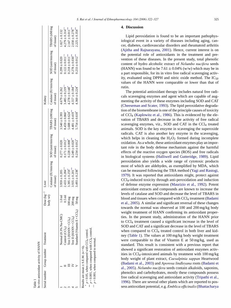
S. Rai et al. / Journal of Ethnopharmacology 104 (2006) 322–327 325
Tabl
e1
Invi
voan
tioxi
dant
activ
ityof
HA
NN
Seri
alnu
mbe
rG
roup
num
ber
Tre
atm
ent
Dos
e(k
g−1
body
wt)
Liv
erK
idne
y
Cat
alas
e(I
U/m
in/m
gof
tissu
e)SO
D(u
nit/m
in/m
gof
tissu
e)T
BA
RS
(nM
/mg
oftis
sue)
Cat
alas
e(I
U/m
in/m
gof
tissu
e)SO
D(u
nit/m
in/m
gof
tissu
e)T
BA
RS
(nM
/mg
oftis
sue)
1I
Nor
mal
(0.3
%N
a-C
MC
)1.
0m
l3.
959
±0.
205
0.27
7±
0.02
03.
540
±0.
393
4.67
9±
0.26
20.
299
±0.
020
1.82
7±
0.13
22
IIC
ontr
ol(C
Cl 4
)0.
5m
l2.
433
±0.
284+
+0.
171
±0.
012+
+8.
459
±0.
860+
+3.
485
±0.
219+
+0.
122
±0.
011+
+4.
279
±0.
314+
+
3II
ITe
stI
(HA
NN
+C
Cl 4
)10
0m
g3.
395
±0.
259*
0.22
8±
0.00
8*5.
688
±0.
624*
4.27
8±
0.14
8*0.
218
±0.
011**
2.14
7±
0.01
1**
4IV
Test
II(H
AN
N+
CC
l 4)
200
mg
3.93
5±
0.20
1**0.
249
±0.
020**
4.48
1±
0.41
9**4.
557
±0.
184**
0.27
8±
0.03
0**2.
030
±0.
278**
5V
Stan
dard
(Vita
min
E+
CC
l 4)
50m
g3.
493
±0.
238*
0.23
4±
0.01
2*5.
754
±0.
618*
4.36
0±
0.22
4*0.
233
±0.
012**
2.22
5±
0.35
4**
Res
ults
are
mea
n±
S.E
.M.(
n=
6).
*p
<0.
05,w
hen
com
pare
dw
ithC
Cl 4
cont
rol.
**p
<0.
001,
whe
nco
mpa
red
with
CC
l 4co
ntro
l.+
+p
<0.
001,
whe
nco
mpa
red
tono
rmal
.
4. Discussion
Lipid peroxidation is found to be an important pathophys-iological event in a variety of diseases including aging, can-cer, diabetes, cardiovascular disorders and rheumatoid arthritis(Ajitha and Rajnarayana, 2001). Hence, current interest is onthe potential role of antioxidants in the treatment and pre-vention of these diseases. In the present study, total phenoliccontent of hydro alcoholic extract of Nelumbo nucifera seeds(HANN) was found to be 7.61 ± 0.04% (w/w) which may be ina part responsible, for its in vitro free radical scavenging activ-ity, evaluated using DPPH and nitric oxide method. The IC50values of the HANN were comparable or lower than that ofrutin.
The potential antioxidant therapy includes natural free radi-cals scavenging enzymes and agent which are capable of aug-menting the activity of these enzymes including SOD and CAT(Cheeseman and Scater, 1993). The lipid peroxidative degrada-tion of the biomembrane is one of the principle causes of toxicityof CCl4 (Kaplowitz et al., 1986). This is evidenced by the ele-vation of TBARS and decrease in the activity of free radicalscavenging enzymes, viz., SOD and CAT in the CCl4 treatedanimals. SOD is the key enzyme in scavenging the superoxideradicals. CAT is also another key enzyme in the scavenging,which helps in cleaning the H2O2 formed during incompleteoxidation. As a whole, these antioxidant enzymes play an impor-tant role in the body defense mechanism against the harmfuleffects of the reactive oxygen species (ROS) and free radicalsin biological systems (Halliwell and Gutteridge, 1989). Lipidperoxidation also yields a wide range of cytotoxic productsmost of which are aldehydes, as exemplified by MDA, whichcan be measured following the TBA method (Yagi and Rastogi,1979). It was reported that antioxidants might, protect againstCCl4-induced toxicity through anti-peroxidation and inductionof defense enzyme expression (Maurizio et al., 1992). Potentantioxidant extracts and compounds are known to increase thelevels of catalase and SOD and decrease the level of TBARS inblood and tissues when compared with CCl4 treatment (Badamiet al., 2005). A similar and significant reversal of these changestowards the normal was observed at 100 and 200 mg/kg bodyweight treatment of HANN confirming its antioxidant proper-ties. In the present study, administration of the HANN priorto CCl4 treatment caused a significant increase in the level ofSOD and CAT and a significant decrease in the level of TBARSwhen compared to CCl4 treated control in both liver and kid-ney (Table 1). The values at 100 mg/kg body weight treatmentwere comparable to that of Vitamin E at 50 mg/kg, used asstandard. This result is consistent with a previous report thatshowed a significant restoration of antioxidant enzymes activ-ities in CCl4-intoxicated animals by treatment with 100 mg/kgbody weight of plant extract, Caesalpinia sappan Heartwood(Badami et al., 2003) and Aporosa lindleyana roots (Badami etal., 2005). Nelumbo nucifera seeds contain alkaloids, saponins,phenolics and carbohydrates, mostly these compounds possessfree radical scavenging and antioxidant activity (Tripathi et al.,1996). There are several other plants which are reported to pos-sess antioxidant potential, e.g. Emblica officinalis (Bhattacharya
326 S. Rai et al. / Journal of Ethnopharmacology 104 (2006) 322–327
et al., 2000), Withania somnifera (Bhattacharya et al., 2001),Bacopa monniera (Rohini et al., 2004), Vitis vinifera (Yilmazand Toledo, 2004) and Mangifera indica (Anila and Vijayalak-shmi, 2003). Antioxidant activity of various parts of Nelumbonucifera is well established, e.g. leaf (Wu et al., 2003), sta-mens (Jung et al., 2003) and rhizomes (Hu and Skibsted, 2002;Cho et al., 2003) except the seeds which has been reported formany therapeutic activities as mentioned earlier. The presentstudy further proves the significant antioxidant potential ofthis therapeutically potent natural product. Nelumbo nuciferaseeds are hepatoprotective (Sohn et al., 2003) and also usedas folk remedy in the treatment of tissue inflammation andcancer (Chopra et al., 1956; Liu et al., 2004), ROS and freeradicals are known play a causative role in these diseases, sofree radical scavenging and antioxidant nature of HANN provedin this study may be, at least in part, can contribute for theseactivities.
It can be concluded that the HANN possess strong antioxi-dant properties as evidenced by the significant dose dependentincrease in the level of CAT, SOD and decrease in the levels ofTBARS. The in vitro studies also confirm the same. Free radicalscavenging and antioxidant activity of HANN may be due tothe presence of phenolics, alkaloids and saponins. However, thedetailed mechanisms are not fully understood and remain to befurther resolved.
Acknowledgements
One of the authors Mr. Sujay Rai is thankful to Indian Councilof Medical Research (ICMR), New Delhi, India for providingfinancial support to carry out this work, through Senior ResearchFellowship.
References
Ajitha, M., Rajnarayana, K., 2001. Role of oxygen free radicals in humandiseases. Indian Drugs 38, 545–554.
Anila, L., Vijayalakshmi, N.R., 2003. Antioxidant action of flavonoids fromMangifera indica and Emblica officinalis in hypercholesterolemic rats.Food Chemistry 83, 569–574.
Anonymous, 1992. The Wealth of India, vol. 3. CSIR, New Delhi, India.Badami, S., Moorkoth, S., Rai, S.R.M.S., Kannan, E., Bhojraj, S., 2003.
Antioxidant activity of Caesalpinia sappan heartwood. Biological andPharmaceutical Bulletin 26, 1534–1537.
Badami, S., Rai, S.R., Suresh, B., 2005. Antioxidant activity of Aporosalindleyana root. Journal of Ethnopharmacology 101, 180–184.
Beer, R.F., Seizer, T.W., 1952. A spectrophotometric method for measuringbreakdown of hydrogen peroxide by catalase. Journal of Biology andChemistry 115, 130–140.
Bhattacharya, A., Ghosal, S., Bhattacharya, S.K., 2000. Antioxidant activ-ity of tannoid principles of Emblica officinalis (amla) in chronic stressinduced changes in rat brain. Indian Journal of Experimental Biology 38,877–880.
Bhattacharya, A., Ghosal, S., Bhattacharya, S.K., 2001. Anti-oxidant effect ofWithania somnifera glycowithanolides in chronic footshock stress-inducedperturbations of oxidative free radical scavenging enzymes and lipid per-oxidation in rat frontal cortex and striatum. Journal of Ethnopharmacology74, 1–6.
Blois, M.S., 1958. Antioxidant determinations by the use of a stable freeradical. Nature 29, 1199–1200.
Cheeseman, K.H., Scater, T.F., 1993. Free radical in medicine. British Med-ical Bulletin 49, 479–724.
Cho, E.J., Yokozawa, T., Rhyu, D.Y., Kim, S.C., Shibahara, N., Park, J.C.,2003. Study on the inhibitory effects of Korean medicinal plants andtheir main compounds on the 1,1-diphenyl-2-picrylhydrazyl radical. Phy-tomedicine 10, 544–551.
Chopra, R.N., Nayar, S.L., Chopra, I.C., 1956. Glossary of Indian MedicinalPlants, 22. CSIR, New Delhi, p. 174.
Furukawa, H., Yang, T.H., Lin, T.J., 1965. Effect of Nelumbo nucifera rhi-zome extract on blood sugar level in rats. Yakugaku Zasshi 85, 472–475.
Garrat, D.C., 1964. The Quantitative Analysis of Drugs, vol. 3. Chapmanand Hall, Japan, pp. 456–458.
Halliwell, B., Gutteridge, J.M.C., 1989. Free Radicals in Biology andMedicine, 2nd ed. Clarendron Press, Oxford.
Hu, M., Skibsted, L.H., 2002. Antioxidative capacity of rhizome extract andrhizome knot extract of edible lotus (Nelumbo nuficera). Food Chemistry76, 327–333.
Hwang, B.Y., Kim, H.S., Lee, J.H., Hong, Y.S., Ro, J.S., Lee, K.S., Lee,J.J., 2001. Antioxidant benzoylated flavan-3-ol glycoside from Celastrusorbiculatus. Journal of Natural Products 64, 82–84.
John, A.B., Steven, A.D., 1978. Microsomal lipid peroxidation. In: Fleischer,S., Packer, L. (Eds.), Methods in Enzymology. Academic Press, NewYork, pp. 302–310.
Jung, H.A., Kim, J.E., Chung, H.Y., Choi, J.S., 2003. Antioxidant princi-ples of Nelumbo nucifera stamens. Archives of Pharmacal Research 26,279–285.
Kaplowitz, N., Aw, T.Y., Simon, F.R., Stolz, A., 1986. Drug induced hepa-totoxicity. Annals of Internal Medicine 104, 826–839.
Li, G.R., Qian, J.Q., Lu, F.H., 1990. Effects of neferine on heart electrome-chanical activity in anaesthetized cats. Zhongguo Yao Li Xue Bao 11,158–161.
Ling, Z.Q., Xie, B.J., Yang, E.L., 2005. Isolation, characterization, and deter-mination of antioxidative activity of oligomeric procyanidins from theseedpod of Nelumbo nucifera Gaertn. Journal of Agricultural and FoodChemistry 53, 2441–2445.
Liu, C.P., Tsai, W.J., Lin, Y.L., Liao, J.F., Chen, C.F., Kuo, Y.C., 2004.The extracts from Nelumbo nucifera suppress cell cycle progression,cytokine genes expression, and cell proliferation in human peripheralblood mononuclear cells. Life Science 75, 699–716.
Maechlay, A.C., Chance, B., 1954. The assay of catalase and peroxidase.In: Glick, D. (Ed.), Methods of Biochemical Analysis. Interscience Inc.,New York, pp. 357–424.
Marcocci, L., Packer, L., Sckaki, A., Albert, G.M., 1994. Antioxidant actionof Ginkgo biloba extracts EGb 761. Methods in Enzymology 234,462–475.
Maurizio, P., Leonarduzzi, G., Biasi, F., Albano, E., Biocca, M.E., Poli,G., Dianzani, M.U., 1992. Vitamin E dietary supplementation protectsagainst carbon tetrachloride-induced chronic liver damage and cirrhosis.Hepatology 16, 1014–1021.
Mazumder, U.K., Gupta, M., Pramanik, G., Mukhopadhyay, R.K., Sarkar, S.,1992. Antifertility activity of seed of Nelumbo nucifera in mice. IndianJournal of Experimental Biology 30, 533–534.
Misra, H.P., Fridovich, I., 1972. The role of superoxide dismutase anion in theanti-oxidation of epinephrine and sample assay in superoxide dismutase.Journal of Biology and Chemistry 241, 3170–3175.
Mukherjee, P.K., 2002. Quality Control of Herbal Drugs—An Approach toEvaluation of Botanicals, 1st ed. Business Horizons, New Delhi, India,pp. 604–608.
Mukherjee, P.K., Das, J., Balasubramanian, R., Saha, K., Pal, M., Saha, B.P.,1995a. Antidiarrhoeal evaluation of Nelumbo nucifera rhizome extract.Indian Journal of Pharmacology 27, 262–364.
Mukherjee, P.K., Giri, S.N., Saha, K., Pal, M., Saha, B.P., 1995b. Antifungalscreening of Nelumbo nucifera (Nymphaeaceae) rhizome extract. IndianJournal of Microbiology 35, 327–330.
Mukherjee, P.K., Balasubramanian, R., Saha, K., Pal, M., Saha, B.P., 1995c.Antibacterial efficiency of Nelumbo nucifera (Nymphaeaceae) rhizomeextract. Indian Drugs 32, 274–276.
S. Rai et al. / Journal of Ethnopharmacology 104 (2006) 322–327 327
Mukherjee, P.K., Pal, S.R., Saha, K., Saha, B.P., 1995d. Hypoglycemic activ-ity of Nelumbo nucifera rhizome (methanolic extract) in streptozotocininduced diabetic rats. Phytotherapy Research 9, 522–524.
Mukherjee, P.K., Saha, K., Balasubramanian, R., Pal, M., Saha, B.P., 1996a.Studies on psychopharmacological effects of Nelumbo nucifera Gaertn.rhizome extract. Journal of Ethnopharmacology 54, 63–67.
Mukherjee, P.K., Das, J., Saha, K., Pal, M., Saha, B.P., 1996b. Diuretic activ-ity of the rhizomes of Nelumbo nucifera Gaertn. (Fam. Nymphaeaceae).Phytotherapy Research 10, 424–425.
Mukherjee, P.K., Saha, K., Das, J., Giri, S.N., Pal, M., Saha, B.P., 1996c.Antipyretic activity of Nelumbo nucifera rhizome extract. Indian Journalof Experimental Biology 82, 274–276.
Qian, J.Q., 2002. Cardiovascular pharmacological effects of bisben-zylisoquinoline alkaloid derivatives. Acta Pharmacologica Sinica 23,1086–1092.
Rohini, G., Sabitha, K.E., Devi, C.S., 2004. Bacopa monniera Linn. extractmodulates antioxidant and marker enzyme status in fibrosarcoma bearingrats. Indian Journal of Experimental Biology 42, 776–780.
Sadasivam, S., Manickam, A., 1992. Biochemical Methods for AgriculturalSciences. Wiley Eastern Ltd., New Delhi, India, pp. 187–188.
Sohn, D.H., Kim, Y.C., Oh, S.H., Park, E.J., Li, X., Lee, B.H., 2003. Hep-atoprotective and free radical scavenging effects of Nelumbo nucifera.Phytomedicine 10, 165–169.
Sun, M., Zigman, S., 1978. An improved spectrophotometric assay for super-oxide dismutase based on epinephrine autoxidation. Analytical Biochem-istry 90, 81–89.
Tomita, M., Furukawa, H., Yang, T.H., 1961. Alkaloids of Nelumbo nucifera.I. Yakugaku Zasshi 81, 469–473.
Tripathi, Y.B., Chaurasia, S., Tripathi, E., Upadhyay, A., Dubey, G.P., 1996.Bacopa monniera Linn. as an antioxidant: mechanism of action. IndianJournal of Experimental Biology 34, 523–526.
Wang, J., Hu, X., Yin, W., Cai, H., 1991. Alkaloids of plumula Nelumbinis.Zhongguo Zhong Yao Za Zhi 16, 673–675.
Wu, M.J., Wang, L., Weng, C.Y., Yen, J.H., 2003. Antioxidant activity ofmethanol extract of the lotus leaf (Nelumbo nucifera Gaertn.). The Amer-ican Journal of Chinese Medicine 31, 687–698.
Xiao, J.H., Zhang, J.H., Chen, H.L., Feng, X.L., Wang, J.L., 2005. Inhibitoryeffect of isoliensinine on bleomycin-induced pulmonary fibrosis in mice.Planta Medica 71, 225–230.
Yagi, K., Rastogi, R., 1979. Assay for lipid peroxides in animal tissue bythiobarbituric acid reaction. Annals of Biochemistry 95, 351.
Yilmaz, Y., Toledo, R.T., 2004. Major flavonoids in grape seeds and skins:antioxidant capacity of catechin, epicatechin, and gallic acid. Journal ofAgricultural and Food Chemistry 52, 255–260.
Yu, J., Hu, W.S., 1997. Effects of neferine on platelet aggregation in rabbits.Yaoxue Xuebao 32, 1–4.
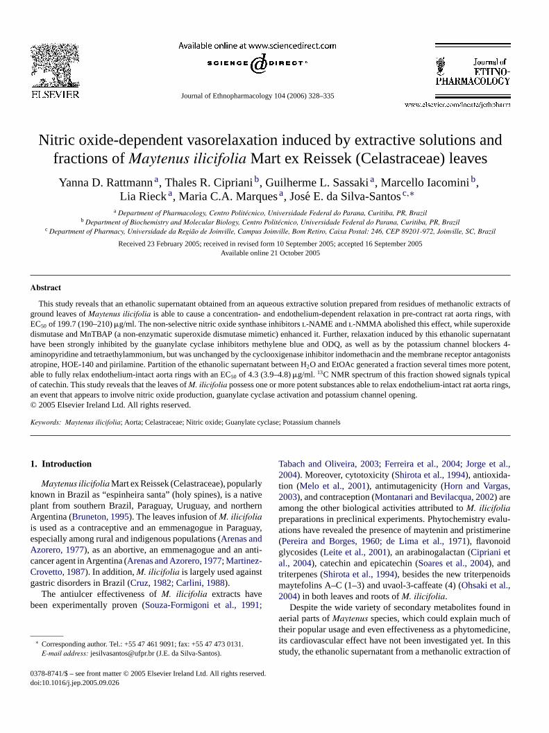
Journal of Ethnopharmacology 104 (2006) 328–335
Nitric oxide-dependent vasorelaxation induced by extractive solutions andfractions of Maytenus ilicifolia Mart ex Reissek (Celastraceae) leaves
Yanna D. Rattmann a, Thales R. Cipriani b, Guilherme L. Sassaki a, Marcello Iacomini b,Lia Rieck a, Maria C.A. Marques a, Jose E. da Silva-Santos c,∗
a Department of Pharmacology, Centro Politecnico, Universidade Federal do Parana, Curitiba, PR, Brazilb Department of Biochemistry and Molecular Biology, Centro Politecnico, Universidade Federal do Parana, Curitiba, PR, Brazil
c Department of Pharmacy, Universidade da Regiao de Joinville, Campus Joinville, Bom Retiro, Caixa Postal: 246, CEP 89201-972, Joinville, SC, Brazil
Received 23 February 2005; received in revised form 10 September 2005; accepted 16 September 2005Available online 21 October 2005
Abstract
This study reveals that an ethanolic supernatant obtained from an aqueous extractive solution prepared from residues of methanolic extracts ofground leaves of Maytenus ilicifolia is able to cause a concentration- and endothelium-dependent relaxation in pre-contract rat aorta rings, withEC50 of 199.7 (190–210) �g/ml. The non-selective nitric oxide synthase inhibitors l-NAME and l-NMMA abolished this effect, while superoxidedismutase and MnTBAP (a non-enzymatic superoxide dismutase mimetic) enhanced it. Further, relaxation induced by this ethanolic supernatanthave been strongly inhibited by the guanylate cyclase inhibitors methylene blue and ODQ, as well as by the potassium channel blockers 4-aminopyridine and tetraethylammonium, but was unchanged by the cyclooxigenase inhibitor indomethacin and the membrane receptor antagonistsatropine, HOE-140 and pirilamine. Partition of the ethanolic supernatant between H2O and EtOAc generated a fraction several times more potent,able to fully relax endothelium-intact aorta rings with an EC50 of 4.3 (3.9–4.8) �g/ml. 13C NMR spectrum of this fraction showed signals typicalof catechin. This study reveals that the leaves of M. ilicifolia possess one or more potent substances able to relax endothelium-intact rat aorta rings,an event that appears to involve nitric oxide production, guanylate cyclase activation and potassium channel opening.© 2005 Elsevier Ireland Ltd. All rights reserved.
Keywords: Maytenus ilicifolia; Aorta; Celastraceae; Nitric oxide; Guanylate cyclase; Potassium channels
1. Introduction
Maytenus ilicifolia Mart ex Reissek (Celastraceae), popularlyknown in Brazil as “espinheira santa” (holy spines), is a nativeplant from southern Brazil, Paraguay, Uruguay, and northernArgentina (Bruneton, 1995). The leaves infusion of M. ilicifoliais used as a contraceptive and an emmenagogue in Paraguay,especially among rural and indigenous populations (Arenas andAzorero, 1977), as an abortive, an emmenagogue and an anti-cancer agent in Argentina (Arenas and Azorero, 1977; Martinez-Crovetto, 1987). In addition, M. ilicifolia is largely used againstgastric disorders in Brazil (Cruz, 1982; Carlini, 1988).
The antiulcer effectiveness of M. ilicifolia extracts havebeen experimentally proven (Souza-Formigoni et al., 1991;
∗ Corresponding author. Tel.: +55 47 461 9091; fax: +55 47 473 0131.E-mail address: [email protected] (J.E. da Silva-Santos).
Tabach and Oliveira, 2003; Ferreira et al., 2004; Jorge et al.,2004). Moreover, cytotoxicity (Shirota et al., 1994), antioxida-tion (Melo et al., 2001), antimutagenicity (Horn and Vargas,2003), and contraception (Montanari and Bevilacqua, 2002) areamong the other biological activities attributed to M. ilicifoliapreparations in preclinical experiments. Phytochemistry evalu-ations have revealed the presence of maytenin and pristimerine(Pereira and Borges, 1960; de Lima et al., 1971), flavonoidglycosides (Leite et al., 2001), an arabinogalactan (Cipriani etal., 2004), catechin and epicatechin (Soares et al., 2004), andtriterpenes (Shirota et al., 1994), besides the new triterpenoidsmaytefolins A–C (1–3) and uvaol-3-caffeate (4) (Ohsaki et al.,2004) in both leaves and roots of M. ilicifolia.
Despite the wide variety of secondary metabolites found inaerial parts of Maytenus species, which could explain much oftheir popular usage and even effectiveness as a phytomedicine,its cardiovascular effect have not been investigated yet. In thisstudy, the ethanolic supernatant from a methanolic extraction of
0378-8741/$ – see front matter © 2005 Elsevier Ireland Ltd. All rights reserved.doi:10.1016/j.jep.2005.09.026

Y.D. Rattmann et al. / Journal of Ethnopharmacology 104 (2006) 328–335 329
M. ilicifolia leaves (ES) was tested with the aim of investigatingits effects on rat aorta rings.
2. Materials and methods
2.1. Plant material, obtainment and fractionation of theethanolic supernatant (ES)
M. ilicifolia leaves collected in the metropolitan region ofCuritiba (a city located in southern Brazil) were a donation fromCentral de Producao e Comercializacao de Plantas Medicinais,Aromaticas e Condimentares do Parana Ltda (PR, Brazil). Theplant was identified by Dr. O. Guimaraes (Department of Botany,Federal University of Parana, PR, Brazil). Voucher specimensare deposited at the Herbarium of this university under number30842.
The acquisition of the ES from M. ilicifolia leaves was pro-cessed according to the schematic representation shown in Fig. 1.Briefly, ground leaves (118 g) were subjected to CHCl3:MeOH(2:1, v/v; 0.5 l) at 60 ◦C for 2 h (3×). Then, the residue I wassubjected to MeOH:H2O (4:1, v/v; 0.5 l) at 80 ◦C for 2 h (2×).Finally, residue II was extracted with H2O (0.5 l) at 100 ◦C for3 h (2×) and the aqueous extract treated with 3 V EtOH, whichgave rise to a precipitate and an ethanolic supernatant (ES) thatwas studied in this work.
The ES (1 g) was partitioned between H2O and EtOAc(60 ml:30 ml), generating the fractions named FH2O and FEtOAc,respectively. The aqueous layer was then shaker with EtOAc(30 ml, 3×). The combined organic layers were evaporated andthe residue suspended in H2O, which was freeze-dried (yield170 mg). When this was treated with a small volume of EtOAc(20 ml), a portion (64 mg) remained insoluble (named FINS)while another was soluble (named FSOL).
2.2. 13C NMR analysis
13C NMR spectra of the FINS (the most potent fraction of ES)were obtained using a 400 MHz Bruker DRX Avance spectrom-
Fig. 1. Fractionation of the crude M. ilicifolia leaves extract.
eter with a 5 mm inverse probe, at 40 ◦C in DMSO-d6. Chemicalshifts are expressed in ppm (δ) relative to DMSO-d6 at δ 39.5.
2.3. Animals
Male Wistar rats (3–4 months old) from the colony of theFederal University of Parana were used. The animals were main-tained under standard laboratory conditions, with a constant12 h light/dark cycle and controlled temperature (22 ± 2 ◦C).Standard pellet food (Nuvital®, Curitiba/PR, Brazil) and waterwere available ad libitum. The Institutional Ethics Committeeof the Federal University of Parana approved all the proceduresadopted in this study (authorization number 052).
2.4. Preparation of rat aorta rings
Isolated aorta rings, with or without functional endothelium,were prepared according to the standard procedures previouslydescribed (Da Silva-Santos et al., 2002), using organ baths con-taining 3 ml of Krebs–Henseleit (pH 7.4; composition in mM:NaCl 115.3, KCl 4.9, CaCl2·2H2O 1.46, KH2PO4 1.2, MgSO41.2, d-glucose 11.1, NaHCO3 25). The tissues were equilibratedfor 60 min at a resting tension of 1 g before the addition ofany drug. Tension was recorded via isometric force transduc-ers (Letica Scientific Instruments, Barcelone, Spain) coupled toa MacLab® recording system (MacLab/8) and an applicationprogram (Chart, v 3.3), both from ADI Instruments (Castle Hill,Australia), working on an Apple Computer®.
2.5. Experimental protocols
2.5.1. Characterization of the vascular effects of theextractive solutions and fractions obtained from M. ilicifolialeaves
Phenylephrine (1 �M) pre-contracted aorta rings with orwithout functional endothelium were exposed to 300, 400, 600and 900 �g/ml of the extract I, extract II or the precipitateobtained during the preparation of the ES (as illustrated inFig. 1). The baths were then washed and after a further 60 mininterval, the contraction and relaxation evoked by phenylephrineand acetylcholine were measured again. This same protocolwas followed using smaller concentrations of the ES (150, 200and 300 �g/ml) and its fractions FH2O, FEtOAc, FINS and FSOL(3–100 �g/ml).
2.5.2. Evaluation of the role of endothelium derived factorson the ES effects
Endothelium-intact aorta rings were incubated with l-NAME(10 �M), or l-NMMA (1 mM), or d-NMMA (300 �M), oraminoguanidine (1 mM), or methylene blue (100 �M), or ODQ(10 �M), or indomethacin (1 �M), or SOD (300 UI/ml), or MnT-BAP (100 �M), 15–30 min prior to the addition of phenyle-phrine (1 �M) into the bath. Then, while under the tonic phaseof the phenylephrine-induced contraction, the effect of the ES(300 �g/ml) was recorded and compared with the data obtainedin the absence of any other drug. The relaxation induced by
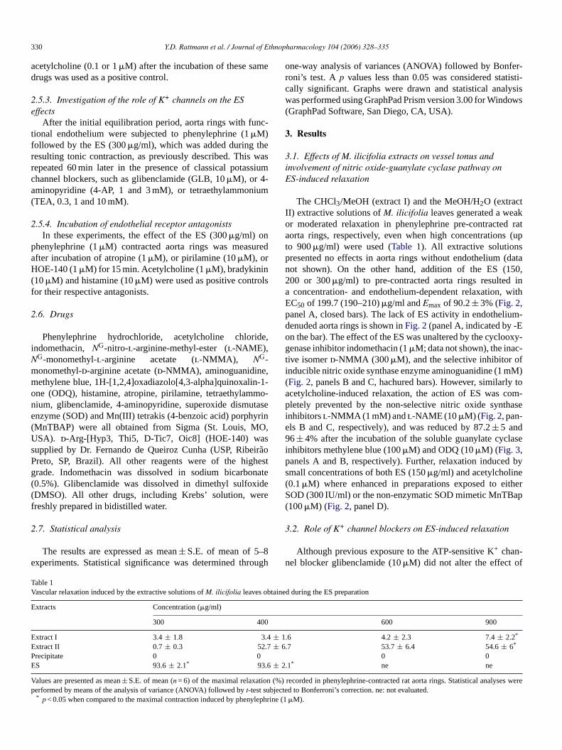
330 Y.D. Rattmann et al. / Journal of Ethnopharmacology 104 (2006) 328–335
acetylcholine (0.1 or 1 �M) after the incubation of these samedrugs was used as a positive control.
2.5.3. Investigation of the role of K+ channels on the ESeffects
After the initial equilibration period, aorta rings with func-tional endothelium were subjected to phenylephrine (1 �M)followed by the ES (300 �g/ml), which was added during theresulting tonic contraction, as previously described. This wasrepeated 60 min later in the presence of classical potassiumchannel blockers, such as glibenclamide (GLB, 10 �M), or 4-aminopyridine (4-AP, 1 and 3 mM), or tetraethylammonium(TEA, 0.3, 1 and 10 mM).
2.5.4. Incubation of endothelial receptor antagonistsIn these experiments, the effect of the ES (300 �g/ml) on
phenylephrine (1 �M) contracted aorta rings was measuredafter incubation of atropine (1 �M), or pirilamine (10 �M), orHOE-140 (1 �M) for 15 min. Acetylcholine (1 �M), bradykinin(10 �M) and histamine (10 �M) were used as positive controlsfor their respective antagonists.
2.6. Drugs
Phenylephrine hydrochloride, acetylcholine chloride,indomethacin, NG-nitro-l-arginine-methyl-ester (l-NAME),NG-monomethyl-l-arginine acetate (l-NMMA), NG-monomethyl-d-arginine acetate (d-NMMA), aminoguanidine,methylene blue, 1H-[1,2,4]oxadiazolo[4,3-alpha]quinoxalin-1-one (ODQ), histamine, atropine, pirilamine, tetraethylammo-nium, glibenclamide, 4-aminopyridine, superoxide dismutaseenzyme (SOD) and Mn(III) tetrakis (4-benzoic acid) porphyrin(MnTBAP) were all obtained from Sigma (St. Louis, MO,USA). d-Arg-[Hyp3, Thi5, D-Tic7, Oic8] (HOE-140) wassupplied by Dr. Fernando de Queiroz Cunha (USP, RibeiraoPreto, SP, Brazil). All other reagents were of the highestgrade. Indomethacin was dissolved in sodium bicarbonate(0.5%). Glibenclamide was dissolved in dimethyl sulfoxide(DMSO). All other drugs, including Krebs’ solution, werefreshly prepared in bidistilled water.
2.7. Statistical analysis
The results are expressed as mean ± S.E. of mean of 5–8experiments. Statistical significance was determined through
one-way analysis of variances (ANOVA) followed by Bonfer-roni’s test. A p values less than 0.05 was considered statisti-cally significant. Graphs were drawn and statistical analysiswas performed using GraphPad Prism version 3.00 for Windows(GraphPad Software, San Diego, CA, USA).
3. Results
3.1. Effects of M. ilicifolia extracts on vessel tonus andinvolvement of nitric oxide-guanylate cyclase pathway onES-induced relaxation
The CHCl3/MeOH (extract I) and the MeOH/H2O (extractII) extractive solutions of M. ilicifolia leaves generated a weakor moderated relaxation in phenylephrine pre-contracted rataorta rings, respectively, even when high concentrations (upto 900 �g/ml) were used (Table 1). All extractive solutionspresented no effects in aorta rings without endothelium (datanot shown). On the other hand, addition of the ES (150,200 or 300 �g/ml) to pre-contracted aorta rings resulted ina concentration- and endothelium-dependent relaxation, withEC50 of 199.7 (190–210) �g/ml and Emax of 90.2 ± 3% (Fig. 2,panel A, closed bars). The lack of ES activity in endothelium-denuded aorta rings is shown in Fig. 2 (panel A, indicated by -Eon the bar). The effect of the ES was unaltered by the cyclooxy-genase inhibitor indomethacin (1 �M; data not shown), the inac-tive isomer d-NMMA (300 �M), and the selective inhibitor ofinducible nitric oxide synthase enzyme aminoguanidine (1 mM)(Fig. 2, panels B and C, hachured bars). However, similarly toacetylcholine-induced relaxation, the action of ES was com-pletely prevented by the non-selective nitric oxide synthaseinhibitors l-NMMA (1 mM) and l-NAME (10 �M) (Fig. 2, pan-els B and C, respectively), and was reduced by 87.2 ± 5 and96 ± 4% after the incubation of the soluble guanylate cyclaseinhibitors methylene blue (100 �M) and ODQ (10 �M) (Fig. 3,panels A and B, respectively). Further, relaxation induced bysmall concentrations of both ES (150 �g/ml) and acetylcholine(0.1 �M) where enhanced in preparations exposed to eitherSOD (300 IU/ml) or the non-enzymatic SOD mimetic MnTBap(100 �M) (Fig. 2, panel D).
3.2. Role of K+ channel blockers on ES-induced relaxation
Although previous exposure to the ATP-sensitive K+ chan-nel blocker glibenclamide (10 �M) did not alter the effect of
Table 1Vascular relaxation induced by the extractive solutions of M. ilicifolia leaves obtained during the ES preparation
Extracts Concentration (�g/ml)
300 400 600 900
Extract I 3.4 ± 1.8 3.4 ± 1.6 4.2 ± 2.3 7.4 ± 2.2*
Extract II 0.7 ± 0.3 52.7 ± 6.7 53.7 ± 6.4 54.6 ± 6*
Precipitate 0 0 0 0ES 93.6 ± 2.1* 93.6 ± 2.1* ne ne
Values are presented as mean ± S.E. of mean (n = 6) of the maximal relaxation (%) recorded in phenylephrine-contracted rat aorta rings. Statistical analyses wereperformed by means of the analysis of variance (ANOVA) followed by t-test subjected to Bonferroni’s correction. ne: not evaluated.
* p < 0.05 when compared to the maximal contraction induced by phenylephrine (1 �M).

Y.D. Rattmann et al. / Journal of Ethnopharmacology 104 (2006) 328–335 331
Fig. 2. Endothelium- and nitric oxide-dependent effects of ES in rat aorta rings. (A) Relaxing activity of ES (150, 200 and 300 �g/ml; closed bars) in endothelium-intact and endothelium-denuded (indicated by -E) aorta. (B) Influence of l-NMMA (1 mM) and its inactive isomer d-NMMA (indicated by INIS, 300 �M; hachuredbar) on the vasodilatory effect of ES (300 �g/ml). (C) Activity of other nitric oxide synthase inhibitors, both l-NAME (10 �M) and aminoguanide (AMG, 1 mM;hachured bar), on ES (300 �g/ml) effect. (D) Enhancement of ES (150 �g/ml) relaxation induced by superoxide anion scavengers, either superoxide dismutase (SOD;300 IU/ml) or MnTBAP (100 �M; hachured bar). The relaxation induced by acetylcholine (ACh; 1 �M in panels A–C; 0.1 �M in panel D) is shown for comparison.The results are the mean ± S.E. of mean of 5–7 experiments. Statistical analyses were performed by means of the analysis of variance (ANOVA) followed by t-testsubjected to Bonferroni’s correction. *p < 0.05 when compared to the absence of relaxation (panel A) or the respective control group (panels B–D).
the ES (Fig. 4, open bar), the selective blocker of voltage-sensitive K+ channels 4-aminopyridine (1 and 3 mM) reducedthe vasodilatory activity of the ES in a concentration-dependentmanner. For instance, the maximal relaxation generated by theES in the presence of 3 mM of 4-aminopyridine was reduced byaround 68% (Fig. 4). Moreover, the non-selective K+ channelblocker tetraethylammoniun (0.3–10 mM) vanished the relax-ing responses evoked by the ES.
3.3. Effects of atropine, HOE-140 and pirilamine on thevasodilator action of the ES
Relaxation induced by the ES in phenylephrine-contractedaorta rings remained unaltered after a 15 min prior incubationof atropine, HOE-140 or pirilamine, at a range of concentration(1–10 �M) that was able to eliminate the effects of acetylcholine,bradykinin or histamine, respectively (Fig. 5).
3.4. Vessel relaxation induced by ES fractions and 13NMRspectrum of the FINS fraction
Incubation of the FH2O has been unable to relaxphenylephrine-contracted rat aorta rings (Fig. 6, panel A,closed circles), even when higher concentrations were tested(up to 300 �g/ml; data not shown). On the other hand, a fulland concentration-dependent loss of vessel tonus was found
when these preparations were exposed for the FEtOAc, withEC50 of 12.8 (9.3–14.7) �g/ml (Fig. 6, panel A, squares).Moreover, although both FSOL and FINS (Fig. 6, panel A,triangles and open circles, respectively) have been able to relaxaorta rings, the FINS was the most potent fraction, with EC50 of4.3 (3.9–4.8) �g/ml. The 13C NMR spectrum of this fraction isshown on Fig. 6 (panel B).
4. Discussion
This study reveals that the ethanolic supernatant (ES)obtained from an aqueous extract of M. ilicifolia leaves inducesan endothelium- and concentration-dependent relaxation of invitro rat aorta rings in a way directly related to nitric oxide pro-duction. Although nitric oxide may not be the only endothelialderived substance which plays an important role in vasodilation,the ability of the constitutive nitric oxide synthase inhibitors(both l-NAME and l-NMMA) in eliminating the relaxationinduce by the ES, associated with the proven failure of thecyclooxygenase inhibitor indomethacin, discards the involve-ment of other endothelial mediators, such as prostanoids andthe endothelium derived hyperpolarizing factor (EDHF), in thevascular effects of the ES.
Its well-known that scavenging free radicals, such as superox-ide anion (O2
−), favors vasodilation in large part by enhancingthe bioavailability of nitric oxide (Gryglewski et al., 1986).

332 Y.D. Rattmann et al. / Journal of Ethnopharmacology 104 (2006) 328–335
Fig. 3. Role of soluble guanylate cyclase on the activity of ES. Effects of theguanylate cyclase inhibitors methylene blue (MB, 100 �M) and ODQ (10 �M)on the vascular relaxation induce by acetylcholine (1 �M) and ES (300 �g/ml) inrat aorta rings (A and B, respectively). The vascular responses to acetylcholineand ES in the absence of any guanylate cyclase inhibitor are shown for compar-ison (open and closed bars, respectively). The results show the mean ± S.E. ofmean of six experiments. Statistical analyses were performed by means of theanalysis of variance (ANOVA) followed by t-test subjected to Bonferroni’s cor-rection. *p < 0.05 when compared to the effects of ES in the absence of solubleguanylate cyclase inhibitors.
Fig. 4. Involvement of potassium channels on ES-induced aorta relaxation.Activity of ES (300 �g/ml) on the absence (closed bar) or the presence of thepotassium channel blockers glibenclamide (GLB, 10 �M), 4-aminopyridine (4-AP; 1 and 3 mM) and tetraethylammonium (0.3 mM). The results show themean ± S.E. of mean of 6–8 experiments. Statistical analyses were performedby means of the analysis of variance (ANOVA) followed by t-test subjected toBonferroni’s correction. *p < 0.05 when compared to the effects of ES in theabsence of potassium channel blockers.
Fig. 5. Inability of membrane receptor antagonists to prevent the effects ofES. Relaxation evoked by the ES (300 �g/ml) before and after exposure toatropine (1 �M), HOE-140 (1 �M) or pirilamine (10 �M) in rat aorta rings (A–C,respectively). The relaxation induced by acetylcholine (ACh, 1 �M; panel A),bradykinin (BK, 10 �M; panel B), and histamine (HIST, 10 �M; panel C) areshown for comparison. The results show the mean ± S.E. of mean of six experi-ments. Statistical analyses were performed by means of the analysis of variance(ANOVA) followed by t-test subjected to Bonferroni’s correction. *p < 0.05when compared to the effects in the absence of the antagonist.
Therefore, an ability to inhibit the production of O2− generated
manly by isoforms of endothelial NADPH oxidase or xantineoxidase, or even act as a O2
− scavenger, could explain the loss ofvessel tonus seen when the ES is added in contracted aorta rings.Nevertheless, similarly to acetylcholine-induced relaxation, theES had its effect enhanced when the availability of either extra-cellular or intracellular O2
− have been reduced by addition of
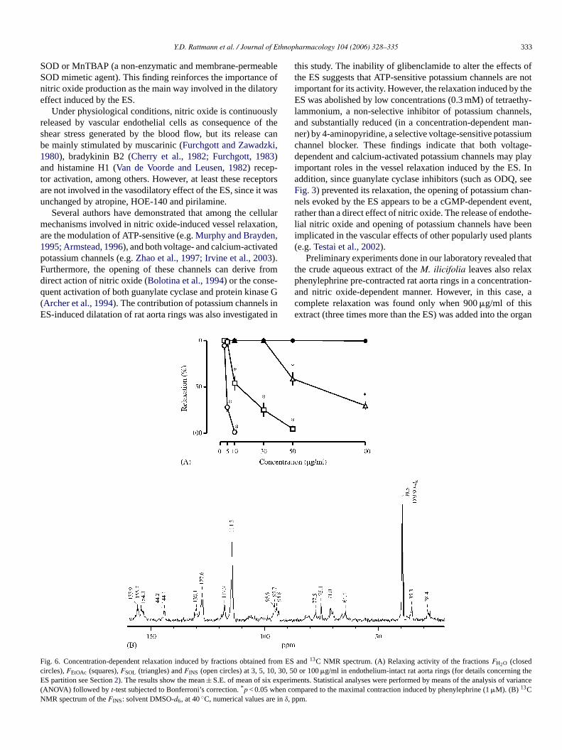
Y.D. Rattmann et al. / Journal of Ethnopharmacology 104 (2006) 328–335 333
SOD or MnTBAP (a non-enzymatic and membrane-permeableSOD mimetic agent). This finding reinforces the importance ofnitric oxide production as the main way involved in the dilatoryeffect induced by the ES.
Under physiological conditions, nitric oxide is continuouslyreleased by vascular endothelial cells as consequence of theshear stress generated by the blood flow, but its release canbe mainly stimulated by muscarinic (Furchgott and Zawadzki,1980), bradykinin B2 (Cherry et al., 1982; Furchgott, 1983)and histamine H1 (Van de Voorde and Leusen, 1982) recep-tor activation, among others. However, at least these receptorsare not involved in the vasodilatory effect of the ES, since it wasunchanged by atropine, HOE-140 and pirilamine.
Several authors have demonstrated that among the cellularmechanisms involved in nitric oxide-induced vessel relaxation,are the modulation of ATP-sensitive (e.g. Murphy and Brayden,1995; Armstead, 1996), and both voltage- and calcium-activatedpotassium channels (e.g. Zhao et al., 1997; Irvine et al., 2003).Furthermore, the opening of these channels can derive fromdirect action of nitric oxide (Bolotina et al., 1994) or the conse-quent activation of both guanylate cyclase and protein kinase G(Archer et al., 1994). The contribution of potassium channels inES-induced dilatation of rat aorta rings was also investigated in
Fig. 6. Concentration-dependent relaxation induced by fractions obtained from ES and 13C NMR spectrum. (A) Relaxing activity of the fractions FH2O (closedcircles), FEtOAc (squares), FSOL (triangles) and FINS (open circles) at 3, 5, 10, 30, 50 or 100 �g/ml in endothelium-intact rat aorta rings (for details concerning theES partition see Section 2). The results show the mean ± S.E. of mean of six experiments. Statistical analyses were performed by means of the analysis of variance(ANOVA) followed by t-test subjected to Bonferroni’s correction. *p < 0.05 when compared to the maximal contraction induced by phenylephrine (1 �M). (B) 13CNMR spectrum of the FINS: solvent DMSO-d6, at 40 ◦C, numerical values are in δ, ppm.
this study. The inability of glibenclamide to alter the effects ofthe ES suggests that ATP-sensitive potassium channels are notimportant for its activity. However, the relaxation induced by theES was abolished by low concentrations (0.3 mM) of tetraethy-lammonium, a non-selective inhibitor of potassium channels,and substantially reduced (in a concentration-dependent man-ner) by 4-aminopyridine, a selective voltage-sensitive potassiumchannel blocker. These findings indicate that both voltage-dependent and calcium-activated potassium channels may playimportant roles in the vessel relaxation induced by the ES. Inaddition, since guanylate cyclase inhibitors (such as ODQ, seeFig. 3) prevented its relaxation, the opening of potassium chan-nels evoked by the ES appears to be a cGMP-dependent event,rather than a direct effect of nitric oxide. The release of endothe-lial nitric oxide and opening of potassium channels have beenimplicated in the vascular effects of other popularly used plants(e.g. Testai et al., 2002).
Preliminary experiments done in our laboratory revealed thatthe crude aqueous extract of the M. ilicifolia leaves also relaxphenylephrine pre-contracted rat aorta rings in a concentration-and nitric oxide-dependent manner. However, in this case, acomplete relaxation was found only when 900 �g/ml of thisextract (three times more than the ES) was added into the organ

334 Y.D. Rattmann et al. / Journal of Ethnopharmacology 104 (2006) 328–335
baths. Nevertheless, this finding allow us to suggest that theleaves infusion of M. ilicifolia, popularly used to treat sev-eral disorders, such as gastric disturbances, contains the samesubstance (or substances) present in the ES, which is able to stim-ulate endogenous nitric oxide production. Previous studies haveattributed the gastroprotective action of plants to an increasedproduction of nitric oxide (e.g. Freitas et al., 2004). We are cur-rently investigating if nitric oxide production is accountable forthe gastroprotective effects of M. ilicifolia preparations, sincecontrolled production of nitric oxide by constitutive isoformsof nitric oxide synthase in stomach microcirculation has beenimplicated in both protective and beneficial events during gas-tric disorders (for review see Wallace and Miller, 2000; Martinet al., 2001).
Although we still unable to identify the active compoundresponsible for the ES activity on vessel tonus, the 13C NMRspectrum (Fig. 6, panel B) of the fraction named FINS, which wasthe most potent between the fractions obtained from proceduresadopted for ES partition showed signals typical of catechin, withthose at δ 144.1 and 144.2 corresponding to vic-hydroxyl groupsdirectly bonded to the phenolic ring and those at 130.1 sug-gesting a carbon substituted in the catechol moiety. Further, thepresence of signals at δ 77.5, 64.4, 155.7 and 155.9 are attributedto characteristic carbons of flavan rings (Breitmeier and Voelter,1990). Endothelial nitric oxide synthase stimulation by a cate-chin derivative has been recently reported (Lorenz et al., 2004).Taken together, these findings allow us to suggest that catechinderivatives presented in the ES and concentrated in the FINS maybe involved in the nitric oxide-dependent relaxation describedin this study. We are attempting to isolate and characterize thissubstance, as well as evidence the molecular events involved inits pharmacological activity.
In conclusion, our results indicate, for the first time, thatan ethanolic supernatant obtained from an aqueous extractivesolution of M. ilicifolia leaves induces a complete vascular relax-ation in rat aorta rings. This effect is mediated by endothelium-dependent nitric oxide production followed by soluble guanylatecyclase activation and potassium channel opening and is notrelated to activation of muscarinic, bradykinin and histamineendothelial receptors. These findings may contribute to ourunderstanding about the wide employment of M. ilicifolia infolk medicine. In addition, disturbances in nitric oxide produc-tion are involved in many disorders, including systemic andpulmonary hypertension and atherosclerosis (for recent reviewssee Barbato and Tzeng, 2004; Ghofrani et al., 2004; Rubio andMorales-Segura, 2004). Finally, the development of innovativetherapies able to maintain or stimulate endogenous nitric oxideproduction, perhaps based on the effects of novel pharmaco-logical agents obtained from medicinal plants, may lead to theimproved management of several pathological conditions.
Acknowledgements
The authors would like to thank Dr. Jamil Assreuy (Depart-ment of Pharmacology, UFSC, Brazil), and Dr. Fernando deQ. Cunha (Department of Pharmacology, USP, Ribeirao Preto,Brazil), and Dr. Adair R.S. dos Santos (Department of Phys-
iology, UFSC, Brazil) for kindly donating drugs, which wereessential to this research.
References
Archer, S.L., Huang, J.M., Hampl, V., Nelson, D.P., Shultz, P.J., Weir, E.K.,1994. Nitric oxide and cGMP cause vasorelaxation by activation of acharybdotoxin-sensitive K+ channel by cGMP-dependent protein kinase.Proceedings of the National Academy of Sciences of the United Statesof America 91, 7583–7587.
Arenas, P., Azorero, R.M., 1977. Plants of common use in Paraguayan folkmedicine for regulating fertility. Economic Botany 31, 298–301.
Armstead, W.M., 1996. Role of ATP-sensitive K+ channels in cGMP-mediated pial artery vasodilation. American Journal of Physiology 270,H423–H426.
Barbato, J.E., Tzeng, E., 2004. iNOS gene transfer for graft disease. Trendsin Cardiovascular Medicine 14, 267–272.
Bolotina, V.M., Najibi, S., Palacino, J.J., Pagano, P.J., Cohen, R.A., 1994.Nitric oxide directly activates calcium-dependent potassium channels invascular smooth muscle. Nature 368, 850–853.
Breitmeier, E., Voelter, W., 1990. 13C NMR Spectroscopy: High ResolutionMethods and Applications in Organic Chemistry and Biochemistry, 3rded. VCH, New York.
Bruneton, J., 1995. Pharmacognosy, Phytochemistry, Medicinal Plants, 1sted. Intercept, UK.
Carlini, E.A.B.S., 1988. Estudo da Acao Antiulcera Gastrica de PlantasBrasileiras: Maytenus ilicifolia (Espinheira Santa) e outras, 1st ed. CEME- Central de Medicamentos, Brasılia.
Cherry, P.D., Furchgott, R.F., Zawadzki, J.V., Jothianandan, D., 1982. Roleof endothelial cells in relaxation of isolated arteries by bradykinin. Pro-ceedings of the National Academy of Sciences of the United States ofAmerica 79, 2106–2110.
Cipriani, T.R., Mellinger, C.G., Gorin, P.A., Iacomini, M., 2004. An arabino-galactan isolated from the medicinal plant Maytenus ilicifolia. Journal ofNatural Products 67, 703–706.
Cruz, G.L., 1982. Dicionario das Plantas Uteis do Brasil, 2nd ed. CivilizacaoBrasileira, Sao Paulo.
Da Silva-Santos, J.E., Terluk, M.R., Assreuy, J., 2002. Differential involve-ment of guanylate cyclase and potassium channels in nitric oxide-inducedhyporesponsiveness to phenylephrine in endotoxemic rats. Shock 17,70–76.
de Lima, O.G., Coelho, J.S., Weigert, E., d’Albuquerque, I.L., Lima Dde, A.,Moraes e Souza, M.A., 1971. On the presence of maytenin and pristimer-ine in the cortical part of the roots of Maytenus ilicifolia from the Southof Brazil. Revista do Instituto de Antibioticos, Universidade Federal dePernambuco 11, 35–38.
Ferreira, P.M., de Oliveira, C.N., de Oliveira, A.B., Lopes, M.J., Alzamora, F.,Vieira, M.A., 2004. A lyophilized aqueous extract of Maytenus ilicifolialeaves inhibits histamine-mediated acid secretion in isolated frog gastricmucosa. Planta 219, 319–324.
Freitas, C.S., Baggio, C.H., Da Silva-Santos, J.E., Rieck, L., de Moraes San-tos, C.A., Junior, C.C., Ming, L.C., Garcia Cortez, D.A., Marques, M.C.,2004. Involvement of nitric oxide in the gastroprotective effects of anaqueous extract of Pfaffia glomerata (Spreng) Pedersen, Amaranthaceae,in rats. Life Sciences 74, 1167–1179.
Furchgott, R.F., 1983. Role of endothelium in responses of vascular smoothmuscle. Circulation Research 53, 557–573.
Furchgott, R.F., Zawadzki, J.V., 1980. The obligatory role of endothelial cellsin the relaxation of arterial smooth muscle by acetylcholine. Nature 288,373–376.
Ghofrani, H.A., Pepke-Zaba, J., Barbera, J.A., Channick, R., Keogh, A.M.,Gomez-Sanchez, M.A., Kneussl, M., Grimminger, F., 2004. Nitric oxidepathway and phosphodiesterase inhibitors in pulmonary arterial hyperten-sion. Journal of the American College of Cardiology 43, 68S–72S.
Gryglewski, R.J., Palmer, R.M.J., Moncada, S., 1986. Superoxide anion isinvolved in the breakdown of endothelium-derived vascular relaxing fac-tor. Nature 15, 170–179.
Y.D. Rattmann et al. / Journal of Ethnopharmacology 104 (2006) 328–335 335
Horn, R.C., Vargas, V.M.F., 2003. Antimutagenic activity of extracts ofnatural substances in the salmonella/microsome assay. Mutagenesis 18,113–118.
Irvine, J.C., Favaloro, J.L., Kemp-Harper, B.K., 2003. NO-activates solu-ble guanylate cyclase and Kv channels to vasodilate resistance arteries.Hypertension 41, 1301–1307.
Jorge, R.M., Leite, J.P., Oliveira, A.B., Tagliati, C.A., 2004. Evaluationof antinociceptive, anti-inflammatory and antiulcerogenic activities ofMaytenus ilicifolia. Journal of Ethnopharmacology 94, 93–100.
Leite, J.P., Rastrelli, L., Romussi, G., Oliveira, A.B., Vilegas, J.H., Vile-gas, W., Pizza, C., 2001. Isolation and HPLC quantitative analysis offlavonoid glycosides from Brazilian beverages (Maytenus ilicifolia and M.aquifolium). Journal of Agricultural and Food Chemistry 49, 3796–3801.
Lorenz, M., Wessler, S., Follmann, E., Michaelis, W., Dusterhoft, T., Bau-mann, G., Stangl, K., Stangl, V., 2004. A constituent of green tea,epigallocatechin-3-gallate, activates endothelial nitric oxide synthase by aphosphatidylinositol-3-OH-kinase-, cAMP-dependent protein kinase-, andakt-dependent pathway and leads to endothelial-dependent vasorelaxation.The Journal of Biological Chemistry 279, 6190–6195.
Martin, M.J., Jimenez, M.D., Motilva, V., 2001. New issues about nitric oxideand its effects on the gastrointestinal tract. Current Pharmaceutical Design7, 881–908.
Martinez-Crovetto, 1987. Plantas reguladoras de la fecundidad utilizadas enla medicina popular del nordeste argentino. America Indigena 472, 79–93.
Melo, S.F., Soares, S.F., da Costa, R.F., da Silva, C.R., de Oliveira, M.B.,Bezerra, R.J., Caldeira-de-Araujo, A., Bernardo-Filho, M., 2001. Effect ofthe Cymbopogon citrates, Maytenus ilicifolia and Baccharis genistelloidesextracts against the stannous chloride oxidative damage in Escherichiacoli. Mutation Research 496, 33–38.
Montanari, T., Bevilacqua, E., 2002. Effect of Maytenus ilicifolia Mart. onpregnant mice. Contraception 65, 171–175.
Murphy, M.E., Brayden, J.E., 1995. Nitric oxide hyperpolarizes rabbit mesen-teric arteries via ATP-sensitive potassium channels. The Journal of Phys-iology 486, 47–58.
Ohsaki, A., Imai, Y., Naruse, M., Ayabe, S., Komiyama, K., Takashima, J.,2004. Four new triterpenoids from Maytenus ilicifolia. Journal of NaturalProducts 67, 469–471.
Pereira, M.V., Borges, G.P., 1960. Maiteine, calcium and magnesium inMaytenus ilicifolia. Arquivos Brasileiros de Medicina 21, 51–53.
Rubio, A.R., Morales-Segura, M.A., 2004. Nitric oxide, an iceberg in cardio-vascular physiology: far beyond vessel tone control. Archives of MedicalResearch 35, 1–11.
Shirota, O., Morita, H., Takeya, K., Itokawa, H., 1994. Cytotoxic aromatictriterpenes from Maytenus ilicifolia and Maytenus chuchuhuasca. Journalof Natural Products 57, 1675–1681.
Soares, L.A., Oliveira, A.L., Ortega, G.G., Petrovick, P.R., 2004. Develop-ment and validation of a LC-method for determination of catechin andepicatechin in aqueous extractives from leaves of Maytenus ilicifolia.Journal of Pharmaceutical and Biomedical Analysis 36, 787–790.
Souza-Formigoni, M.L., Oliveira, M.G., Monteiro, M.G., da Silveira-Filho,N.G., Braz, S., Carlini, E.A., 1991. Antiulcerogenic effects of twoMaytenus species in laboratory animals. Journal of Ethnopharmacology34, 21–27.
Tabach, R., Oliveira, W.P., 2003. Evaluation of the anti-ulcerogenic activityof a dry extract of Maytenus ilicifolia Martius ex. Reiss produced by ajet spouted bed dryer. Pharmazie 58, 573–576.
Testai, L., Chericoni, S., Calderone, V., Nencioni, G., Nieri, P., Morelli,I., Martinotti, E., 2002. Cardiovascular effects of Urtica dioica L.(Urticaceae) roots extracts: in vitro and in vivo pharmacological stud-ies. Journal of Ethnopharmacology 81, 105–109.
Van de Voorde, J., Leusen, I., 1982. Vascular endothelium and the relax-ation effect of histamine on aorta of the rat. Archives Internationales dePharmacodynamie et de Therapie 256, 329–330.
Wallace, J.L., Miller, M.J., 2000. Nitric oxide in mucosal defense: a littlegoes a long way. Gastroenterology 119, 512–520.
Zhao, Y.J., Wang, J., Rubin, L.J., Yuan, X.J., 1997. Inhibition of K(V) andK(Ca) channels antagonizes NO-induced relaxation in pulmonary artery.American Journal of Physiology 272, H904–H912.

Journal of Ethnopharmacology 104 (2006) 336–344
Mechanisms of the vasorelaxant effect of Danshen(Salvia miltiorrhiza) in rat knee joints
F.Y. Lam ∗, S.C.W. Ng, J.H.Y. Cheung, J.H.K. YeungDepartment of Pharmacology, Faculty of Medicine, The Chinese University of Hong Kong, Shatin, New Territories, Hong Kong SAR, China
Received 31 March 2004; received in revised form 4 August 2005; accepted 19 September 2005Available online 2 November 2005
Abstract
The aim of this study is to investigate the mechanisms of the vasorelaxant effect of the crude extract of Salvia miltiorrhiza (family: Labiatae),also known as “Danshen”, in rat knee joints. Changes in blood flow of rat knee joints were measured in vivo by a laser Doppler perfusion imager.Topical administration of Danshen onto the exposed knee joint blood vessels produced dose-dependent increases in blood flow. Treatment of therat knee joint with 2× 1 nmol of atropine, 2× 0.1 nmol of propranolol, or 2× 0.1 nmol of a mixture of pyrilamine plus cimitedine produced nochange on the vasodilator response to Danshen. However, significant inhibition of the Danshen-induced vasodilator response was observed inknee joints treated with 2× 100 nmol of NG-nitro-l-arginine methyl ester (l-NAME), 2× 100 nmol of flurbiprofen, 2 × 10 nmol of the calcitnoningene-related peptide (CGRP) receptor antagonist CGRP8–37, and also in knee joints but had been denervated by capsaicin treatment or by surgery.Intravenous administration of low doses of Danshen (2.5 and 6 mg/kg) did not affect the systemic blood pressure but significantly increased kneejoint blood flow, whereas, high doses of Danshen (167 and 381 mg/kg) produced hypotension with concurrent decreases in knee joint blood flow.These findings indicate that the knee joint blood vessels are more sensitive to the relaxant effect of Danshen compared to blood vessels in the generalcirculation. The vasorelaxant effect of Danshen was found to be partly mediated by CGRP released from sensory nerves, and nitric oxide andprostaglandins also played a part. However, there is no evidence to support a role for muscarinic receptors, adrenoceptors, or histamine receptors.© 2005 Elsevier Ireland Ltd. All rights reserved.
Keywords: Salvia miltiorrhiza; Danshen; CGRP; Nitric oxide; Prostaglandins; Rat knee joint
1. Introduction
The root of Salvia miltiorrhiza Bunge (family: Labiatae), alsoknown as “Danshen” in Chinese, has been used as a medicinein China for centuries. It was commonly used for the treatmentof cardiovascular diseases such as angina pectoris, myocardialinfarction and stroke (Ji et al., 2000). A number of pharmaco-logical studies had confirmed the cardio-protective efficacy ofthis herbal medicine (Fung et al., 1993; Wu et al., 1993; Zhouand Ruigrok, 1990; Ji et al., 2003; Chen, 1981).
Some discrepancies have appeared among previous studiesregarding the effects of Danshen on blood pressure. Danshenhad been shown to produce dose-related hypotension in the ratthat was inhibited by atropine, propranolol, and a mixture of
∗ Corresponding author at: Department of Pharmacology, Room 404A, BasicMedical Sciences Building, The Chinese University of Hong Kong, Shatin, NewTerritories, Hong Kong, China. Tel.: +852 2609 6790; fax: +852 2603 5139.
E-mail address: [email protected] (F.Y. Lam).
chlorpheniramine plus cimetidine (Lei and Chiou, 1986a), sug-gesting the roles of muscarinic receptors, �-adrenoceptors, andhistamine receptors in mediating the effect. However, anotherstudy showed that the hypotensive effect of Danshen was notblocked by propranolol, or pyrilamine plus cimetidine, but wasinhibited by atropine and potentiated by captopril, indicatingthe involvement of muscarinic receptors and angiotensin con-verting enzyme inhibition but not �-adrenoceptors or histaminereceptors (Li et al., 1990). Danshen was also effective in relax-ing the noradrenaline-precontracted rat aorta (Kamata et al.,1993), an effect which was blocked by the nitric oxide synthaseinhibitor NG-monomethyl-l-arginine (l-NMMA), suggestingan endothelium-dependent mechanism.
Sensory neurones contain vasodilator neuropeptides, one ofwhich is calcitonin gene-related peptide (CGRP) (Gulbenkianet al., 1986; Lam and Ferrell, 1993). Whether this neural sourceof vasodilators contributes to the vasorelaxant effect of Danshenis unknown. This possibility was tested in the present study byinvestigating the effect of denervation and the effect of a CGRP
0378-8741/$ – see front matter © 2005 Elsevier Ireland Ltd. All rights reserved.doi:10.1016/j.jep.2005.09.033

F.Y. Lam et al. / Journal of Ethnopharmacology 104 (2006) 336–344 337
receptor antagonist on the Danshen-induced response. In addi-tion, several receptor antagonists and enzyme inhibitors werealso tested to elucidate the effect of Danshen, the mechanismsand mediators involved.
2. Materials and methods
2.1. Animals
Experiments were performed on Sprague–Dawley rats(250–300 g) bred and kept by the Laboratory Animal ServicesCentre of the Chinese University of Hong Kong. All exper-iments were performed under Animal Licence issued by theHealth Department, the Government of the Hong Kong SARand endorsed by the Animal Experimentation Ethics Commit-tee of the Chinese University of Hong Kong.
2.2. Induction of anaesthesia
For recovery experiments, the rats were injected with40 mg/kg thiopentone intra-peritoneally to produce a short dura-tion of anaesthesia. For terminal experiments, the animals wereinjected with 1.8 g/kg urethane by the same route. Some animalsoccasionally required additional injection of 0.2 g/kg urethaneto maintain deep anaesthesia, which was judged by the absenceof a flexor withdrawal reflex response to a pinch applied to theirforelimbs.
2.3. Knee joint denervation
Denervation of the rat knee joint was accomplished by eitherchemical or surgical means. The rat was anaesthetized withthiopentone as described in Section 2.1. For chemical denerva-tion, one of its knees was injected with 0.2 ml of 1% capsaicin;the contralateral knee was injected with the same volume ofphysiological saline (0.9% NaCl) to act as an internal control.The animal was allowed to recover and left for 14 days beforesubjected to blood flow studies described in Section 2.4. Thisprocedure has been shown to result in approximately 80% den-ervation of unmyelinated fibres (Ferrell et al., 1992).
For surgical denervation, the fur covering the rat’s lefthindlimb was shaved. The left saphenous nerve that arises fromthe abdominal cavity and arcs down towards to the left side ofthe knee was dissected free from its surrounding tissue, and atleast 0.5 cm of the nerve was excised. The wound was stitched,sterilized and sprayed with silicone wound dressing (Smith& Nephew, UK). Amoxycillin (40 mg/kg) was injected intra-peritoneally as a prophylactic antibiotic. The rat was allowedto recover and left for 10 days before subjected to blood flowstudies described in Section 2.4.
2.4. Cannulation and measurement of blood pressure
The external jugular vein of the anaesthetized rat was can-nulated with a polythene tubing (PE-50, Portex Ltd.) filled withheparinised saline for intravenous injection of drugs. The leftcarotid artery was also cannulated, connected to a pressure
transducer (Gould P23ID), and the signals were amplified andrecorded by a PowerLab system (AD Instruments, Australia). Acardiac cycle consists of systolic (SP) and diastolic (DP) pres-sures. Therefore, an average blood pressure was calculated andtermed mean arterial pressure (MAP) by the following equation:DP + 1/3(SP − DP).
2.5. Measurement of knee joint perfusion
Changes in rat knee joint blood flow were measured by alaser Doppler perfusion imager (LDI) as described by Lam andNg (2003). The deeply anaesthetized rat was placed in a supineposition with the knees at rest. The skin covering one of the kneejoints was excised and all underlying fascia removed to exposethe medial aspect of the joint capsule. A LDI (Moor Instruments,Axminster, UK) that generates a helium–neon laser of 630 nmwavelength was placed 28 cm above the exposed knee. This laserbeam was directed on to the exposed knee joint by two motor-controlled mirrors. The backscattered photons from the scannedtissue are captured by a photodetector in the scanner. The LDIthen generates a two-dimensional map of the joint perfusion,which can subsequently be displayed on a computer monitor asa colour-coded image. The actual flux values at each point in theimage are stored on disc and can be utilised for calculation of themean flux values within a given area using an image processingsoftware supplied with the LDI. Responses were expressed aspercentage change in flux values of test and control images, i.e.((test flux − control flux)/control flux) × 100%.
Control scans of the rat knee joint were taken after steadyblood flow was maintained for half an hour. A range of dosesof Danshen (0.1–60 mg) in boluses of 0.1 ml were administeredtopically onto the exposed knee and scans were taken for 60 minfollowing each drug application. For all studies involving topicaladministrations of drugs, both knee joints of the rat were usedbut only one dose of Danshen was tested on each knee. Then-value for each experiment is 8 except where stated.
2.6. Investigations for receptor types and mediatorsinvolved
Several receptor antagonists and enzyme inhibitors weretested on the vasodilator response to Danshen to elucidateits mechanisms of action and mediators involved. Antagoniststested in the present study include the muscarinic receptorantagonist atropine, the �-adrenoceptor antagonist propranolol,the histamine H1 receptor antagonist pyrilamine combinedwith the histamine H2 receptor antagonist cimetidine, and theCGRP receptor antagonist CGRP8–37. The nitric oxide synthaseinhibitor l-NAME was used to test for involvement of nitricoxide (NO), and the cyclo-oxygenase (COX) inhibitor flurbipro-fen was used to test for involvement of prostanoids.
For this series of experiments, rat knee joints were first treatedwith a dose of 0.1 ml of the antagonist/enzyme inhibitor andleft for 15 min to allow for equilibration of the drug with thejoint tissue. Then the same dose of antagonist/enzyme inhibitorwas co-administered with an agonist or Danshen (total volumeadministered remained at 0.1 ml). Knee joint blood flow was
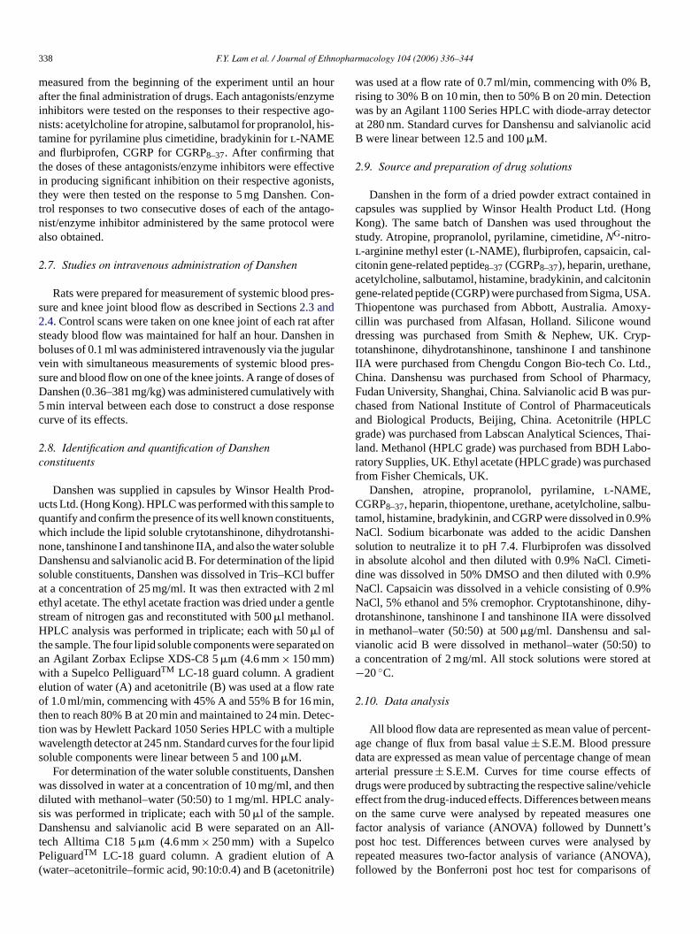
338 F.Y. Lam et al. / Journal of Ethnopharmacology 104 (2006) 336–344
measured from the beginning of the experiment until an hourafter the final administration of drugs. Each antagonists/enzymeinhibitors were tested on the responses to their respective ago-nists: acetylcholine for atropine, salbutamol for propranolol, his-tamine for pyrilamine plus cimetidine, bradykinin for l-NAMEand flurbiprofen, CGRP for CGRP8–37. After confirming thatthe doses of these antagonists/enzyme inhibitors were effectivein producing significant inhibition on their respective agonists,they were then tested on the response to 5 mg Danshen. Con-trol responses to two consecutive doses of each of the antago-nist/enzyme inhibitor administered by the same protocol werealso obtained.
2.7. Studies on intravenous administration of Danshen
Rats were prepared for measurement of systemic blood pres-sure and knee joint blood flow as described in Sections 2.3 and2.4. Control scans were taken on one knee joint of each rat aftersteady blood flow was maintained for half an hour. Danshen inboluses of 0.1 ml was administered intravenously via the jugularvein with simultaneous measurements of systemic blood pres-sure and blood flow on one of the knee joints. A range of doses ofDanshen (0.36–381 mg/kg) was administered cumulatively with5 min interval between each dose to construct a dose responsecurve of its effects.
2.8. Identification and quantification of Danshenconstituents
Danshen was supplied in capsules by Winsor Health Prod-ucts Ltd. (Hong Kong). HPLC was performed with this sample toquantify and confirm the presence of its well known constituents,which include the lipid soluble crytotanshinone, dihydrotanshi-none, tanshinone I and tanshinone IIA, and also the water solubleDanshensu and salvianolic acid B. For determination of the lipidsoluble constituents, Danshen was dissolved in Tris–KCl bufferat a concentration of 25 mg/ml. It was then extracted with 2 mlethyl acetate. The ethyl acetate fraction was dried under a gentlestream of nitrogen gas and reconstituted with 500 �l methanol.HPLC analysis was performed in triplicate; each with 50 �l ofthe sample. The four lipid soluble components were separated onan Agilant Zorbax Eclipse XDS-C8 5 �m (4.6 mm × 150 mm)with a Supelco PelliguardTM LC-18 guard column. A gradientelution of water (A) and acetonitrile (B) was used at a flow rateof 1.0 ml/min, commencing with 45% A and 55% B for 16 min,then to reach 80% B at 20 min and maintained to 24 min. Detec-tion was by Hewlett Packard 1050 Series HPLC with a multiplewavelength detector at 245 nm. Standard curves for the four lipidsoluble components were linear between 5 and 100 �M.
For determination of the water soluble constituents, Danshenwas dissolved in water at a concentration of 10 mg/ml, and thendiluted with methanol–water (50:50) to 1 mg/ml. HPLC analy-sis was performed in triplicate; each with 50 �l of the sample.Danshensu and salvianolic acid B were separated on an All-tech Alltima C18 5 �m (4.6 mm × 250 mm) with a SupelcoPeliguardTM LC-18 guard column. A gradient elution of A(water–acetonitrile–formic acid, 90:10:0.4) and B (acetonitrile)
was used at a flow rate of 0.7 ml/min, commencing with 0% B,rising to 30% B on 10 min, then to 50% B on 20 min. Detectionwas by an Agilant 1100 Series HPLC with diode-array detectorat 280 nm. Standard curves for Danshensu and salvianolic acidB were linear between 12.5 and 100 �M.
2.9. Source and preparation of drug solutions
Danshen in the form of a dried powder extract contained incapsules was supplied by Winsor Health Product Ltd. (HongKong). The same batch of Danshen was used throughout thestudy. Atropine, propranolol, pyrilamine, cimetidine, NG-nitro-l-arginine methyl ester (l-NAME), flurbiprofen, capsaicin, cal-citonin gene-related peptide8–37 (CGRP8–37), heparin, urethane,acetylcholine, salbutamol, histamine, bradykinin, and calcitoningene-related peptide (CGRP) were purchased from Sigma, USA.Thiopentone was purchased from Abbott, Australia. Amoxy-cillin was purchased from Alfasan, Holland. Silicone wounddressing was purchased from Smith & Nephew, UK. Cryp-totanshinone, dihydrotanshinone, tanshinone I and tanshinoneIIA were purchased from Chengdu Congon Bio-tech Co. Ltd.,China. Danshensu was purchased from School of Pharmacy,Fudan University, Shanghai, China. Salvianolic acid B was pur-chased from National Institute of Control of Pharmaceuticalsand Biological Products, Beijing, China. Acetonitrile (HPLCgrade) was purchased from Labscan Analytical Sciences, Thai-land. Methanol (HPLC grade) was purchased from BDH Labo-ratory Supplies, UK. Ethyl acetate (HPLC grade) was purchasedfrom Fisher Chemicals, UK.
Danshen, atropine, propranolol, pyrilamine, l-NAME,CGRP8–37, heparin, thiopentone, urethane, acetylcholine, salbu-tamol, histamine, bradykinin, and CGRP were dissolved in 0.9%NaCl. Sodium bicarbonate was added to the acidic Danshensolution to neutralize it to pH 7.4. Flurbiprofen was dissolvedin absolute alcohol and then diluted with 0.9% NaCl. Cimeti-dine was dissolved in 50% DMSO and then diluted with 0.9%NaCl. Capsaicin was dissolved in a vehicle consisting of 0.9%NaCl, 5% ethanol and 5% cremophor. Cryptotanshinone, dihy-drotanshinone, tanshinone I and tanshinone IIA were dissolvedin methanol–water (50:50) at 500 �g/ml. Danshensu and sal-vianolic acid B were dissolved in methanol–water (50:50) toa concentration of 2 mg/ml. All stock solutions were stored at−20 ◦C.
2.10. Data analysis
All blood flow data are represented as mean value of percent-age change of flux from basal value ± S.E.M. Blood pressuredata are expressed as mean value of percentage change of meanarterial pressure ± S.E.M. Curves for time course effects ofdrugs were produced by subtracting the respective saline/vehicleeffect from the drug-induced effects. Differences between meanson the same curve were analysed by repeated measures onefactor analysis of variance (ANOVA) followed by Dunnett’spost hoc test. Differences between curves were analysed byrepeated measures two-factor analysis of variance (ANOVA),followed by the Bonferroni post hoc test for comparisons of

F.Y. Lam et al. / Journal of Ethnopharmacology 104 (2006) 336–344 339
means. Mean values on histograms were compared by unpairedStudent’s t-test. P-values less than 0.05 were consideredsignificant.
3. Results
3.1. Effects of topical administration of Danshen
Topical applications of single dose of Danshen on the rat kneejoint increased the knee joint blood flow in a dose-dependentmanner (Fig. 1). The lowest dose of Danshen (0.1 mg) produceda 36% increase that subsided in 5 min; the median dose (5 mg)
Fig. 1. Effects of increasing doses of Danshen on rat knee joint blood flow.Danshen was administered topically as a bolus of 0.1 ml on to the exposed ratknee joint at time 0. Data are shown as mean ± S.E.M. (shown by vertical bars)of percentage change of flux from basal blood flow. Danshen produced dose-dependent increases on knee joint blood flow; n = 8 for all.
Fig. 2. Effects of two consecutive doses of antagonists or enzyme inhibitors ontheir respective agonist-induced vasodilator response. The rat knee joint was firstpretreated with a dose of the antagonist or enzyme inhibitor, and 15 min later, thesame dose of the antagonist or enzyme inhibitor was co-administered with theagonist. All antagonists and enzyme inhibitors produced significant inhibitionon the maximum vasodilator response of their respective agonist. H antag-onists = pyrilamine plus cimetidine. Unpaired t-test: *P < 0.05, ***P < 0.001;n = 3–5.
produced a 96% increase and subsided in 20 min, and the highestdose (60 mg) produced a 193% increase and subsided in 45 min.All peak responses were observed around 2 min. Blood pressurewas not affected by any of the doses tested.
Fig. 3. Effects of (A) 2× 1 nmol atropine, (B) 2× 1 nmol propranolol, and (C)2× 10 �mol pyrilamine plus cimetidine on the vasodilator response to 5 mgDanshen. The rat knee joint was first pretreated with a dose of the antagonists(1 nmol or 10 �mol), and 15 min later, the same dose of the antagonists was co-administered with Danshen at time 0. All these antagonists did not significantlyaffect the basal blood flow or the Danshen-induced vasodilator response, exceptfor pyrilamine plus cimetidine which significantly augmented the vasodilatorresponse at 5 min. Bonferroni post hoc test: *P < 0.05; n = 8 for all.

340 F.Y. Lam et al. / Journal of Ethnopharmacology 104 (2006) 336–344
3.2. Effects of receptor antagonists and enzyme inhibitorson Danshen
The knee joint was first pretreated with topical applicationof 0.1 ml of the receptor antagonist or enzyme inhibitor andleft for 15 min. Thereafter, 0.1 ml of the same dose of thereceptor antagonist or enzyme inhibitor was co-administeredwith an agonist or 5 mg Danshen. Such treatment with 2×1 nmol of the muscarinic receptor antagonist atropine inhib-ited the maximum vasodilator response to acetylcholine by63% (Fig. 2), but had no effect on the vasodilator responseto Danshen (Fig. 3A). The two consecutive doses of atropinealso produced no significant change on basal blood flow(Fig. 3A).
Similarly, treatments with 2× 1 nmol of the �-adrenoceptorantagonist propranolol or 2× 10 nmol of a mixture of the H1receptor antagonist pyrilamine plus the H2 receptor antagonistcimetidine produced 45% and 58% inhibition on the maximum
Fig. 4. Effects of (A) 2× 100 nmol l-NAME and (B) 2× 100 nmol flurbiprofenon the vasodilator response to 5 mg Danshen. The rat knee joint was first pre-treated with 100 nmol of the enzyme inhibitors, and 15 min later, the same doseof the enzyme inhibitors was co-administered with Danshen at time 0. l-NAMEand flurbiprofen produced 24% and 46% inhibition on the peak vasodilatorresponse to Danshen, respectively. l-NAME alone produced significant increaseon the basal blood flow at 1 min, but flurbiprofen alone had no effect. Two-factorANOVA: ***P < 0.001; Dunnett’s post hoc test: *P < 0.05; n = 8 for all.
vasodilator responses to propranolol and histamine, respec-tively (Fig. 2). However, these antagonists had no effect onthe vasodilator response to Danshen or on basal blood flow(Fig. 3B and C, respectively), except that the mixture of his-tamine receptor antagonists produced a significant increase ofthe Danshen-induced vasodilator response at the 5 min timepoint (Fig. 3C).
Treatments with 2× 100 nmol of the nitric oxide synthaseinhibitor l-NAME or 2× 100 nmol of the cyclo-oxygenaseenzyme inhibitor flurbiprofen produced 54% and 67% inhi-bition on the maximum vasodilator responses produced bybradykinin (Fig. 2). The two enzyme inhibitors also reducedthe peak vasodilator response to Danshen by 24% and 46%. l-NAME produced a small increase on basal blood flow at 2 minbut flurbiprofen did not significantly alter the basal blood flow(Fig. 4).
Treatment with 2× 10 nmol of the CGRP receptor antag-onist CGRP8–37 produced 67% inhibition on the maximum
Fig. 5. Effects of (A) 2× 10 nmol CGRP8–37 and (B) a combined mixture of 2×100 nmol l-NAME plus 2 × 100 nmol flurbiprofen plus 2× 10 nmol CGRP8–37
on the vasodilator response to 5 mg Danshen. CGRP8–37 reduced the peakvasodilator response to Danshen by 45% and the combined mixture produced44% inhibition. CGRP8–37 alone produced significant increase in basal bloodflow, whereas, the combined mixture alone decreased the basal blood flow at thetime points indicated. Two-factor ANOVA: ***P < 0.001; Dunnett’s post hoc test:*P < 0.05, **P < 0.01; n = 8 for all, except for the control curve of the combinedmixture, for which n = 4.

F.Y. Lam et al. / Journal of Ethnopharmacology 104 (2006) 336–344 341
vasodilator response to CGRP (Fig. 1). As shown in Fig. 5,CGRP8–37 alone or combined with 2× 100 nmol of l-NAMEand 2× 100 nmol of flurbiprofen produced 45% and 44% inhi-bition on the peak vasodilator response to Danshen. CGRP8–37alone caused significant increase on basal blood flow, but thecombined mixture of CGRP8–37, l-NAME and flurbiprofen pro-duced reduction on basal blood flow.
3.3. Effects of denervation on Danshen
The effect of denervation of the knee joint on Danshen-induced vasodilatation was investigated to test for neuralinvolvement. Rats were subjected to chemical denervation withcapsaicin or surgical denervation as described in Section 2.3.On day 14s and 10 after chemical and surgical denervation,respectively, these rats were tested with topical administrationsof 5 mg Danshen on their knees. In the chemically denervatedknee and the surgically denervated knee, the peak vasodilatorresponse to Danshen was reduced by 26% (Fig. 6A) and 34%(Fig. 6B). Control experiments indicated that chemical or surgi-cal denervation had no effect on basal blood flow of the rat kneejoints.
Fig. 6. Effects of (A) chemical denervation by capsaicin and (B) surgical den-ervation on the vasodilator response to 5 mg Danshen. Chemical and surgicaldenervation produced 26% and 34% inhibition on the peak vasodilator responseto Danshen, respectively. Two-factor ANOVA: ***P < 0.001; n = 8 for all.
3.4. Effects of intravenous administration of Danshen
Danshen was administered cumulatively via the intravenousroute, with simultaneous measurements of changes in systemicblood pressure and knee joint blood flow. As shown in Fig. 7, lowdoses of Danshen (2.5 and 6 mg/kg) had no effect on systemicblood pressure but significantly increased blood flow of the kneejoint by 23% and 21%, respectively. Danshen at the two highestdoses (167 and 381 mg/kg) produced significant decreases (9%and 15%) in systemic blood pressure with concurrent significantdecreases in knee joint blood flow of 16% and 17%, respectively.
3.5. HPLC analysis of Danshen
Some of the major constituents known to be present in Dan-shen herbs have been identified in the Danshen sample used inthe present study. This includes dihydrotanshinone, crytotan-shinone, tanshinone I and tanshinone IIA, and the water soluble
Fig. 7. Effects of cumulative intravenous administration of Danshen on (A) sys-temic blood pressure and (B) blood flow in the rat knee joint. Low doses ofDanshen did not affect the systemic blood pressure but significantly increasedknee joint blood flow. High doses of Danshen produced significant decreasein systemic blood pressure with concurrent drop in knee joint blood flow.MAP = mean arterial pressure. Dunnett’s post hoc test (compared with basal):*P < 0.05, **P < 0.01, ***P < 0.001; n = 8 for all.

342 F.Y. Lam et al. / Journal of Ethnopharmacology 104 (2006) 336–344
Danshensu and salvianolic acid B. The quantities of these con-stituents determined in the Danshen sample were 97.6 �g/gdihydrotanshinone, 70.4 �g/g crytotanshinone, 31.8 �g/g tan-shinone I, 21.0 �g/g tanshinone IIA, 30.63 mg/g Danshensu and40.98 mg/g salvianolic acid B.
4. Discussion and conclusion
This study confirmed that the crude extract of Danshen,when applied topically onto the rat knee joint, produced dose-dependent dilation of the blood vessels that peak at about2 min. Denervated rat knee joints exhibited smaller vasodila-tor response to Danshen compared with normal rat knee joints.CGRP8–37, l-NAME and flurbiprofen (administered alone ortogether) were effective in reducing the Danshen-inducedvasodilator response, but atropine, propranolol, or a mixtureof pyrilamine plus cimetidine had no effect. These findingssuggested that the vasorelaxant effect of Danshen was medi-ated by CGRP released from sensory nerves. Nitric oxide andprostaglandins also played a part in the response, but mus-carinic receptors, adrenoceptors, or histamine receptors werenot involved.
The present study showed that intravenous administrations ofDanshen at doses of 167 mg/kg and above produced hypotensionin the rat. The hypotensive effect was accompanied with paral-lel reduction in knee joint blood flow, which was expected inview of the drop in systemic blood pressure. However, the find-ing that intravenous administration of sub-hypotensive doses ofDanshen produced increase in knee joint blood flow was unex-pected. This suggests that blood vessels in the rat knee joint aremore sensitive to the vasorelaxant effect of Danshen comparedwith blood vessels elsewhere in the circulation. Our previousstudy has shown that intraperitoneal injections of 5 g/kg Dan-shen twice per day for 3 days produced no adverse reaction inthe rat (Chan et al., 1995), indicating that Danshen has verylow toxicity. In the present study, intravenous cumulative injec-tions of 0.36–381 mg/kg Danshen were also well tolerated by therats. These doses are less than those used by other researchersfor their investigations on the hypotensive action of intravenousDanshen (150–625 mg/kg) in rats and rabbits (Lei and Chiou,1986a, 1986b).
Drug-induced changes in blood pressure could result fromthe direct effects of drugs on the cardiovascular system, orby indirect effects of drugs on humoral and neural control ofthe cardiovascular system. The present study showed that topi-cal administration of Danshen produced local effects on bloodvessels of the rat knee joint without affecting blood pressure.This is a good platform for the study of Danshen actions onblood vessels without complications of its cardiac effects. Usingsuch experimental protocol, the present study showed that 2×1 nmol atropine did not affect the vasodilator response to Dan-shen but it significantly attenuated the vasodilator response toacetylcholine, which agreed with a previous study that showedatropine, present at half of the dose used in this study, producedsignificant inhibition on the vasodilator response to acetyl-choline in the rat knee joint (McDougall et al., 1998). Atropineon its own produced no change on basal blood flow, indicating
an absence of parasympathetic influence on resting blood flowin the rat knee joint. The present finding also do not support arole for muscarinic receptors in mediating the Danshen-inducedeffect.
Both �1- and �2-adrenoceptors had been identified function-ally in isolated pulmonary vessel rings and in the pulmonaryvascular beds of animals and men; mediating vasodilatation inresponse to circulating catecholamines (Hyman et al., 1981). Insystemic vessels, these receptors are generally of the �2 subtype,although this varies between species. A role for �-adrenoceptorsin mediating Danshen-induced vasodilatation was not evidentsince 2× 1 nmol of the non-selective �-adrenoceptor antagonistpropranolol did not alter the vasodilator response to Danshenbut it significantly suppressed the vasodilator response to salbu-tamol. Basal blood flow of the rat knee joint was not affected bythese two doses of propranolol, which supports the notion thatthe sympathetic neurotransmitter noradrenaline in physiologicalconditions has little or no effect on vascular �-adrenoceptors.
The vasodilator response to histamine is partly due to releaseof endothelium-derived relaxant factors (EDRF) in some vascu-lar beds, and it was suggested that a combination of H1 and H2receptor blocking agents was more effective in preventing theactions of histamine than either blocking agent alone (Lei andChiou, 1986a, 1986b). Therefore, a mixture of pyrilamine pluscimetidine was used in this study to test the involvement of his-tamine receptors in the Danshen-induced vasodilator response.Treatment of the rat knee joint with 2× 10 nmol of this antag-onist mixture produced significant inhibition on the vasodilatorresponse to histamine but did not inhibit the vasodilator responseto Danshen. This suggested a lack of involvement of histaminereceptors in mediating the vasodilator response to Danshen. Infact, the Danshen-induced vasodilator response was augmentedby the two histamine receptor antagonists at the 5 min time point,but this is probably of minor significance because statistical anal-ysis of the entire curves of Danshen alone and Danshen in thepresence of the antagonists showed no difference. The histaminereceptor antagonists alone also had no significant effect on basalblood flow, indicating an absence of tonic release of histaminein the rat knee joint.
The relaxant effect of Danshen on noradrenaline-precon-tracted rat aorta is susceptible to inhibition by the nitric oxidesynthase inhibitor NG-monomethyl-l-arginine (l-NMMA),suggesting an endothelium-dependent mechanism that involvedrelease of nitric oxide (NO) (Kamata et al., 1993). In the rab-bit knee joint, close intra-arterial administration of l-NAMEreduced the basal synovial blood flow, indicating a role for NOin physiological regulation of vascular tone in the knee joint(Najafipour and Ferrell, 1993). In contrast, another study showedthat intravenous administrations of l-NAME and l-NMMA didnot significantly affect the resistance of rat knee joint blood ves-sels (McDougall and Ferrell, 1996). Thus, it was suggested thatNO production in the rat knee joint is minimal and NO is notimportant for maintenance of resting blood flow to the rat kneejoint (McDougall and Ferrell, 1996). This is supported by thepresent finding that showed direct topical administration of 2×100 nmol l-NAME did not reduce basal blood flow, but produceda small and transient vasodilator response. The mechanisms and
F.Y. Lam et al. / Journal of Ethnopharmacology 104 (2006) 336–344 343
significance of this unexpected vasodilator response to l-NAMEhave yet to be determined.
In the present study, l-NAME was found to reduce the maxi-mum vasodilator responses to bradykinin and Danshen by 54%and 24%, suggesting a partial involvement of NO in theseresponses. Endothelium-dependent vasorelaxation has also beendemonstrated for an aqueous extract of Danshen, lithosper-mic acid B, which produced relaxation of noradrenaline pre-contracted rat aorta, and the vasodilator response was abolishedwhen the endothelium was denuded or pretreated with l-NMMA(Kamata et al., 1993). It should be noted that unlike lithospermicacid B, the vasodilator effect of the crude extract of Danshen isnot entirely dependent on NO, but that it contributes to about onequarter of its total effect. Therefore, the vasodilator response toDanshen cannot be ascribed solely to its lithospermic acid Bcontent.
Apart from NO, prostaglandins (PGs) such as prostacyclin(PGI2) constitute another possible group of endothelium-derivedvasodilators that could mediate the vasorelaxant effect of Dan-shen. This possibility was tested with flurbiprofen, a cyclo-oxygenase (COX) enzyme inhibitor that blocks PG synthesis.A substantial 46% inhibition on the peak vasodilator responseto Danshen was produced by treatment of the rat knee joint with2× 100 nmol flurbiprofen. This confirms PGs are involved inthe response. The two doses of flurbiprofen did not significantlyaffect the basal blood flow, which contrasted the earlier findingthat COX-1 enzymes played a part in maintaining blood flow tothe rat knee joint (Egan et al., 2001). The reason for this dis-crepancy is unknown but the earlier study had used a differentCOX inhibitor (indomethacin versus flurbiprofen) and a differ-ent route of administration (intravenous versus topical) to thatof the present study. It should be noted that the two doses offlurbiprofen used in the present study has reduced the maximumvasodilator response to bradykinin by 67%, indicating flurbipro-fen can produce adequate inhibition of the COX enzymes in thepresent study.
Intra-articular administration of 1% capsaicin into the ratknee joint has been shown to produce degeneration of unmyli-nated nerve fibres innervating the joint (Ferrell et al., 1992),suppressed neuropeptide-induced joint inflammation (Lam andFerrell, 1989), and caused depletion of neuropeptide-containingfibres (Mapp et al., 1996). In the present study, denervation ofthe rat knee by pretreatment with capsaicin for 14 days causeda 26% reduction on the peak vasodilator response to Danshencompared with that obtained in the intact rat knee. Similarly, inrat knee joints that had received surgical denervation 10 daysearlier, the peak vasodilator response to Danshen was inhibitedby 34% compared with that in normal rat knees. Therefore, aneural component in the vasodilator response to Danshen wasconfirmed. However, there is no evidence to support a physi-ological role for sensory neuropeptides in maintaining normalblood flow to the rat knee joint since basal blood flow was thesame in chemically denervated, surgically denervated, or normalrat knee joints.
CGRP, as mentioned above, is one of the mediators of neu-rogenic vasodilatation. This 37 amino acid peptide has potentvasodilator action in many species, including man (Brain et
al., 1985; Poyner, 1992), and it is thought to be the princi-pal vasodilator released after efferent stimulation of sensorynerves. In the present study, a role for CGRP in mediating thevasorelaxant effect of Danshen is indirectly supported by thedenervation experiments that demonstrated a neural componentin the Danshen-induced vasodilator response. Direct evidencefor CGRP contribution was provided by the study with theCGRP receptor antagonist CGRP8–37, which showed that 2×10 nmol of the antagonist inhibited the maximum vasodilatorresponses to CGRP and Danshen by 67% and 45%, respectively.It was also observed that co-administrations of CGRP8–37 withl-NAME and flurbiprofen did not produce a further reductionon the Danshen-induced vasodilator response. This suggests thatthere are other mechanisms involved in the Danshen-inducedvasodilator response in addition to CGRP and the endothelium-derived relaxant factors. Further studies would be necessary toelucidate these unknown mechanisms.
Control experiments showed that the two doses of CGRP8–37alone produced a significant increase on basal blood flow, indi-cating that this antagonist has inherent vasodilator action. Itis suspected that this vasodilator action of CGRP8–37 is dueto its agonist action on tachykinin NK1 receptors (Anderssonand Almegard, 1993). The activation of NK1 receptors hasbeen shown to produce vasodilatation involving release ofendothelium-derived relaxant factors (Lam and Ferrell, 1993;Marceau et al., 1989; Ralevic et al., 1995). This idea is sup-ported by the finding that co-administrations of CGRP8–37 withl-NAME and flurbiprofen produced a reduction on basal bloodflow instead of an increase.
The Danshen capsule used in the present study was found tocontain many of the major constituents known to be present inthe Danshen herb. This includes the lipid soluble dihydrotan-shinone, crytotanshinone, tanshinone I and tanshinone IIA, andthe water soluble Danshensu and salvianolic acid B. The presentstudy also showed that Danshensu and salvianolic acid B werepresent in much greater quantities in the Danshen crude extractcompared with the lipid soluble components. However, it is notknown which of these constituents are the most important inmediating the vasorelaxant effect of Danshen in its crude extractform, and experiments are in progress to address this question.
In conclusion, the present study has demonstrated that, bycareful selection of the dosages and routes of administration ofthe Danshen crude extract, it can be tailored to produce tran-sient or prolong effect on local blood flow in the knee jointwith or without lowering of the systemic blood pressure. Thevasorelaxant effect of Danshen was found to be partly mediatedby CGRP released from sensory neurones, and it also involvesthe endothelium-derived relaxant factors NO and PGs, but thereis no contribution by muscarinic receptors, �-adrenoceptors, orhistamine receptors.
Acknowledgements
The authors would like to thank Ms Ethel S.K. Ng and MsPenelope M.Y. Or for their technical assistance. The authorswish to thank Winsor Health Products Limited (Hong Kong)for the supply and authentication of Danshen. Provision of

344 F.Y. Lam et al. / Journal of Ethnopharmacology 104 (2006) 336–344
postgraduate studentships to Mr. S.C.W. Ng, and MissJ.H.Y. Cheung by The Chinese University of Hong Kong isacknowledged.
References
Andersson, S.E., Almegard, B., 1993. CGRP(8–37) and CGRP(32–37) con-tract the iris sphincter in the rabbit eye: antagonism by spantide andGR82334. Regulatory Peptide 49, 73–80.
Brain, S.D., Williams, T.J., Tippins, J.R., Morris, H.R., Macintyre, I., 1985.Calcitonin gene-related peptide is a potent vasodilator. Nature 313,54–56.
Chan, K., Lo, A.C., Yeung, J.H., Woo, K.S., 1995. The effect of Danshen(Salvia miltiorrhiza) on warfarin pharmacodynamics and pharmacoki-netics of warfarin enantiomers in rats. The Journal of Pharmacy andPharmacology 47, 402–406.
Chen, K.J., 1981. Certain progress in the treatment of coronary heart diseasewith traditional medical plants in China. American Journal of ChineseMedicine 9, 193–196.
Egan, C.G., Lockhart, J.C., McLean, J.S., Ferrell, W.R., 2001. Expression ofconstitutive but not inducible cyclooxygenase maintains articular perfu-sion in the rat knee. Experimental Physiology 86, 191–197.
Ferrell, W.R., Lam, F.Y., Montgomery, I., 1992. Differences in the axoncomposition of nerves supplying the rat knee joint following intra-articularinjection of capsaicin. Neuroscience Letter 141, 259–261.
Fung, K.P., Zeng, L.H., Wu, J., Wong, H.N., Lee, C.M., Hon, P.M., Chang,H.M., Wu, T.W., 1993. Demonstration of the myocardial salvage effectof lithospermic acid B isolated from the aqueous extract of Salvia milti-orrhiza. Life Sciences 52, L.239–L.244.
Gulbenkian, S., Merighi, A., Wharton, J., Varndell, I.M., Polak, J.M., 1986.Ultrastructural evidence for the coexistence of cailcitonin gene-relatedpeptide and substance P in secretory vesicles of peripheral nerves in theguinea-pig. Journal of Neurocytology 15, 535–542.
Hyman, A.L., Nandiwada, P., Knight, D.S., Kadowitz, P.J., 1981. Pulmonaryvasodilator responses to catecholamines and sympathetic nerve stimula-tion in the cat. Evidence that vascular beta2 adrenoceptors are innervated.Circulation Research 48, 407–415.
Ji, X.Y., Tan, B.K.H., Zhu, Y.Z., 2000. Salvia miltiorrhiza and ischemicdiseases. Acta Pharmacologica Sinica 21, 1089–1094.
Ji, X.Y., Tan, B.K.H., Zhu, Y.C., Linz, W., Zhu, Y.Z., 2003. Comparison ofcardioprotective effects using ramipril and Danshen for the treatment ofacute myocardial infarction in rats. Life Sciences 72, 1413–1426.
Kamata, K., Iizuka, T., Nagao, M., Kasuya, Y., 1993. Endothelium-dependentvasodilator effects of the extract from Salviae Miltiorrhizae Radix. A
study on the identification of lithospermic acid B in the extracts. GeneralPharmacology 24, 977–981.
Lam, F.Y., Ferrell, W.R., 1989. Capsaicin suppresses substance P-inducedjoint inflammation in the rat. Neuroscience Letters 105, 155–158.
Lam, F.Y., Ferrell, W.R., 1993. Effects of interactions of naturally occurringneuropeptides on blood flow in the rat knee joint. British Journal ofPharmacology 108, 694–699.
Lam, F.F.Y., Ng, E.S.K., 2003. Characterisation of somatostatin actions onknee joint blood vessels of the rat. European Journal of Pharmacology474, 295–301.
Lei, X.L., Chiou, G.C.Y., 1986a. Studies on cardiovascular actions of Salviamiltiorrhiza. American Journal of Chinese Medicine 14, 26–32.
Lei, X.L., Chiou, G.C.Y., 1986b. Cardiovascular pharmacology of Panaxnotoginseng (Burk) F.H. Chen and Salvia miltiorrhiza. American Jour-nal of Chinese Medicine 12, 145–152.
Li, C.P., Yung, K.H., Chiu, K.W., 1990. Hypotensive action of Salvia milti-orrhiza cell culture extract. American Journal of Chinese Medicine 18,157–166.
Mapp, P.I., Kerslake, S., Brain, S.D., Blake, D.R., Cambridge, H., 1996. Theeffect of intra-articular capsaicin on nerve fibres within the synovium ofthe rat knee joint. Journal of Chemical Neuroanatomy 10, 11–18.
Marceau, F., Tremblay, B., Couture, R., Regoli, D., 1989. Prostacyclin releaseinduced by neurokinins in cultured human endothelial cells. CanadianJournal of Physiology and Pharmacology 67, 159–162.
McDougall, J.J., Elenko, R.D.V., Bray, R.C., 1998. Cholinergic vasoregula-tion in normal and adjuvant monoarthritic rat knee joints. Journal of theAutonomic System 72, 55–60.
McDougall, J.J., Ferrell, W.R., 1996. Inhibition of nitric oxide productionduring electrical stimulation of the nerves supplying the rat knee joint.Journal of the Autonomic System 57, 73–77.
Najafipour, H., Ferrell, W.R., 1993. Nitric oxide modulates sympathetic vaso-constriction and basal blood flow in normal and acutely inflamed rabbitknee joints. Experimental Physiology 78, 615–624.
Poyner, D.R., 1992. Calcitonin gene-related peptide: multiple actions, multi-ple receptors. Pharmacology and Therapeutics 268, 59–64.
Ralevic, V., Khalil, Z., Helme, R.D., Dusting, G.J., 1995. Role of nitric oxidein the actions of substance P and other mediators of inflammation in ratskin microvasculature. European Journal of Pharmacology 284, 231–239.
Wu, T.W., Zeng, L.H., Fung, K.P., Wu, J., Pang, H., Gery, A.A., Weisel, R.D.,Wang, J.Y., 1993. Effect of sodium tanshinone IIA sulfonate in the rabbitmyocardium and on human cardiomyocytes and vascular endothelial cells.Biochemical Pharmacology 46, 2327–2332.
Zhou, W., Ruigrok, T.J., 1990. Protective effect of Danshen during myocardialischemia and reperfusion: an isolated rat heart study. American Journalof Chinese Medicine 18, 19–24.
Journal of Ethnopharmacology 104 (2006) 345–350
Puerariae radix promotes differentiation and mineralization inhuman osteoblast-like SaOS-2 cells
Jeong-Eun Huh a, Ha-Ru Yang a, Dong-Suk Park b, Do-Young Choi b, Yong-Hyeon Baek b,Eun-Mi Cho a, Yoon-Je Cho c, Kim Kang-Il c, Deog-Yoon Kim d, Jae-Dong Lee b,∗
a Oriental Medicine Research Center for Bone & Joint Disease, Kyung Hee University, 1 Hoegidong, Dongdaemungu, Seoul 130-701, Republic of Koreab Department of Acupuncture & Moxibustion, College of Oriental Medicine, Kyung Hee University, 1 Hoegidong, Dongdaemungu,
Seoul 130-701, Republic of Koreac Department of Orthopedic Surgery, College of Medicine, Kyung Hee University, 1 Hoegidong, Dongdaemungu, Seoul 130-701, Republic of Koread Department of Nuclear Medicine, College of Medicine, Kyung Hee University, 1 Hoegidong, Dongdaemungu, Seoul 130-701, Republic of Korea
Received 28 May 2005; received in revised form 15 September 2005; accepted 22 September 2005Available online 7 February 2006
Abstract
Puerariae radix (PR) is a traditional medicine herb used for enhancing body resistance against various diseases. The aim of this study was toidentify whether Puerariae radix extract induces osteogenic activity in human osteoblast-like SaOS-2 cells. Puerariae radix had no effect on theviability of osteoblastic cells, and dose-dependently increased alkaline phosphatase (ALP) activity. Puerariae radix markedly increased mRNAexpression for vascular endothelial growth factor (VEGF), osteocalcin (OCN), osteopontin (OPN), and type I collagen (Col I) in SaOS-2 cells.Extracellular accumulation of proteins such as VEGF and Col I was increased in a dose-dependent manner. Also, Puerariae radix significantlyinduced mineralization in the culture of SaOS-2 cells. In conclusion, this study showed that Puerariae radix had no effect on viability, but enhancedALP activity, VEGF, bone matrix proteins such as OCN, OPN, and Col I, and mineralization in SaOS-2 cells. These results propose that Puerariaeradix can play an important role in osteoblastic bone formation, and may possibly lead to the development of bone-forming drugs.© 2005 Elsevier Ireland Ltd. All rights reserved.
Keywords: Puerariae radix; ALP activity; VEGF; OCN; OPN; Col I; Mineralization
1. Introduction
Bone modeling and remodeling are essential for the devel-opment, maturation, maintenance, and repair of bones. Thedifferentiation of osteoblasts is included in these events and iscontrolled by various local growth factors and cytokines pro-duced in bone as well as by systemic hormones.
Osteoblasts, which arise from mesenchymal stem cell pre-cursors, undergo differentiation in response to a number of fac-tors including bone morphogenic proteins (BMPs), transforminggrowth factor (TGF), insulin-like growth factor I (IGF-I), vas-cular endothelial growth factor (VEGF), and glucocorticoids(McCarthy et al., 1989; Midy and Plouet, 1994; Hughes etal., 1995; Goad et al., 1996; Gerber et al., 1999; Spelsberg etal., 1999). These are important for osteoblastic differentiation
∗ Corresponding author. Tel.: +82 2 958 9208; fax: +82 2 958 9043.E-mail address: [email protected] (J.-D. Lee).
and modulate the expression of osteoblast-specific genes. More-over, several different molecules are associated with depositionand maintenance of mineralized skeletal elements. Once matrixsynthesis begins in osteoblast culture models such as primaryosteoblast cultures, the cells differentiate as genes encodingosteoblastic markers such as alkaline phosphatase (ALP), osteo-pontin (OPN), and osteocalcin (OCN) are expressed. Finally,osteoblasts become embedded in the extracellular matrix con-sisting mainly of type I collagen (Col I), and matrix mineraliza-tion begins as mineral deposits extend along and within collagenfibrils (Franceschi and Iyer, 1992; Lian et al., 2003). Recently,Maeda et al. reported that statins stimulate the expression ofbone anabolic factors such as VEGF and BMP-2, and promoteosteoblastic differentiation and mineralization in MC3T3-E1cells (Maeda et al., 2001, 2003).
Puerariae radix (PR), the root of Pueraria lobata Ohwi, awild creeper leguminous plant, is one of the earliest and mostimportant crude herbs used in oriental medicine. Its clinical usefor various diseases in internal medicine, surgery, pediatrics, and
0378-8741/$ – see front matter © 2005 Elsevier Ireland Ltd. All rights reserved.doi:10.1016/j.jep.2005.09.041
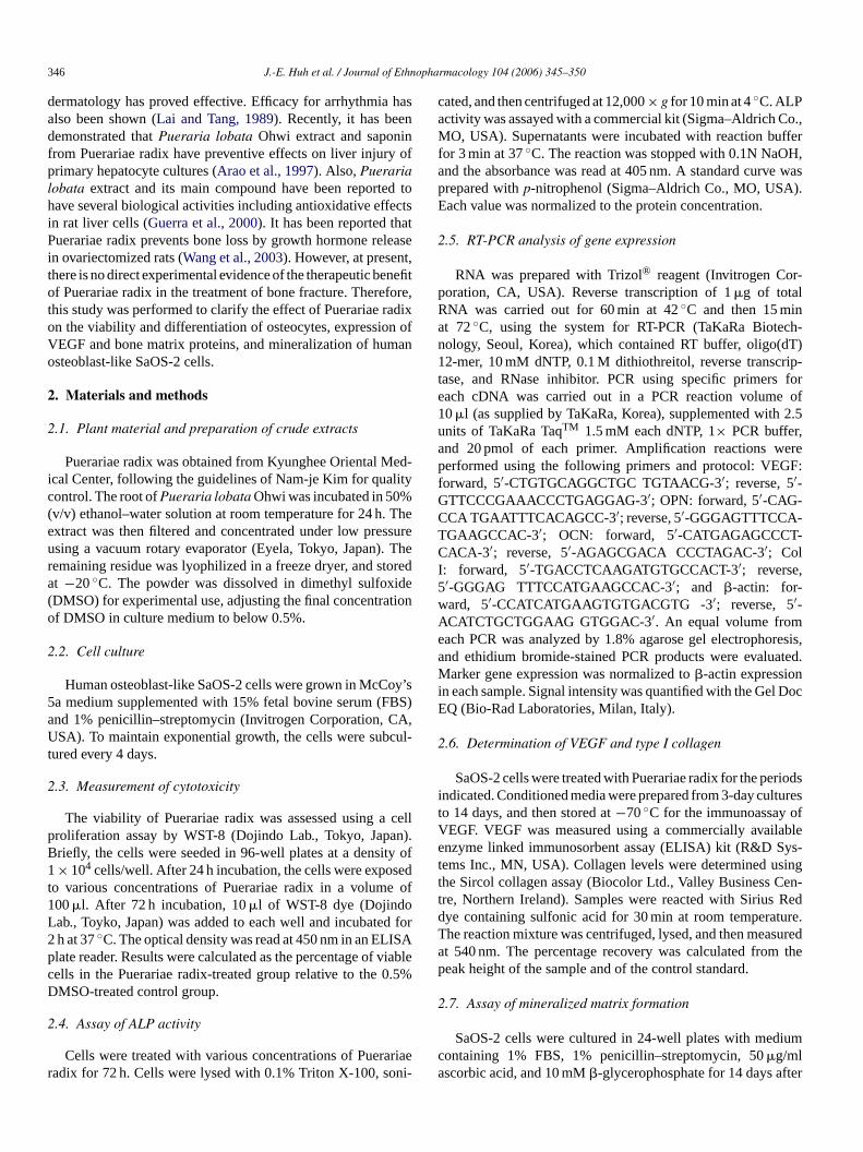
346 J.-E. Huh et al. / Journal of Ethnopharmacology 104 (2006) 345–350
dermatology has proved effective. Efficacy for arrhythmia hasalso been shown (Lai and Tang, 1989). Recently, it has beendemonstrated that Pueraria lobata Ohwi extract and saponinfrom Puerariae radix have preventive effects on liver injury ofprimary hepatocyte cultures (Arao et al., 1997). Also, Puerarialobata extract and its main compound have been reported tohave several biological activities including antioxidative effectsin rat liver cells (Guerra et al., 2000). It has been reported thatPuerariae radix prevents bone loss by growth hormone releasein ovariectomized rats (Wang et al., 2003). However, at present,there is no direct experimental evidence of the therapeutic benefitof Puerariae radix in the treatment of bone fracture. Therefore,this study was performed to clarify the effect of Puerariae radixon the viability and differentiation of osteocytes, expression ofVEGF and bone matrix proteins, and mineralization of humanosteoblast-like SaOS-2 cells.
2. Materials and methods
2.1. Plant material and preparation of crude extracts
Puerariae radix was obtained from Kyunghee Oriental Med-ical Center, following the guidelines of Nam-je Kim for qualitycontrol. The root of Pueraria lobata Ohwi was incubated in 50%(v/v) ethanol–water solution at room temperature for 24 h. Theextract was then filtered and concentrated under low pressureusing a vacuum rotary evaporator (Eyela, Tokyo, Japan). Theremaining residue was lyophilized in a freeze dryer, and storedat −20 ◦C. The powder was dissolved in dimethyl sulfoxide(DMSO) for experimental use, adjusting the final concentrationof DMSO in culture medium to below 0.5%.
2.2. Cell culture
Human osteoblast-like SaOS-2 cells were grown in McCoy’s5a medium supplemented with 15% fetal bovine serum (FBS)and 1% penicillin–streptomycin (Invitrogen Corporation, CA,USA). To maintain exponential growth, the cells were subcul-tured every 4 days.
2.3. Measurement of cytotoxicity
The viability of Puerariae radix was assessed using a cellproliferation assay by WST-8 (Dojindo Lab., Tokyo, Japan).Briefly, the cells were seeded in 96-well plates at a density of1 × 104 cells/well. After 24 h incubation, the cells were exposedto various concentrations of Puerariae radix in a volume of100 �l. After 72 h incubation, 10 �l of WST-8 dye (DojindoLab., Toyko, Japan) was added to each well and incubated for2 h at 37 ◦C. The optical density was read at 450 nm in an ELISAplate reader. Results were calculated as the percentage of viablecells in the Puerariae radix-treated group relative to the 0.5%DMSO-treated control group.
2.4. Assay of ALP activity
Cells were treated with various concentrations of Puerariaeradix for 72 h. Cells were lysed with 0.1% Triton X-100, soni-
cated, and then centrifuged at 12,000 × g for 10 min at 4 ◦C. ALPactivity was assayed with a commercial kit (Sigma–Aldrich Co.,MO, USA). Supernatants were incubated with reaction bufferfor 3 min at 37 ◦C. The reaction was stopped with 0.1N NaOH,and the absorbance was read at 405 nm. A standard curve wasprepared with p-nitrophenol (Sigma–Aldrich Co., MO, USA).Each value was normalized to the protein concentration.
2.5. RT-PCR analysis of gene expression
RNA was prepared with Trizol® reagent (Invitrogen Cor-poration, CA, USA). Reverse transcription of 1 �g of totalRNA was carried out for 60 min at 42 ◦C and then 15 minat 72 ◦C, using the system for RT-PCR (TaKaRa Biotech-nology, Seoul, Korea), which contained RT buffer, oligo(dT)12-mer, 10 mM dNTP, 0.1 M dithiothreitol, reverse transcrip-tase, and RNase inhibitor. PCR using specific primers foreach cDNA was carried out in a PCR reaction volume of10 �l (as supplied by TaKaRa, Korea), supplemented with 2.5units of TaKaRa TaqTM 1.5 mM each dNTP, 1× PCR buffer,and 20 pmol of each primer. Amplification reactions wereperformed using the following primers and protocol: VEGF:forward, 5′-CTGTGCAGGCTGC TGTAACG-3′; reverse, 5′-GTTCCCGAAACCCTGAGGAG-3′; OPN: forward, 5′-CAG-CCA TGAATTTCACAGCC-3′; reverse, 5′-GGGAGTTTCCA-TGAAGCCAC-3′; OCN: forward, 5′-CATGAGAGCCCT-CACA-3′; reverse, 5′-AGAGCGACA CCCTAGAC-3′; ColI: forward, 5′-TGACCTCAAGATGTGCCACT-3′; reverse,5′-GGGAG TTTCCATGAAGCCAC-3′; and �-actin: for-ward, 5′-CCATCATGAAGTGTGACGTG -3′; reverse, 5′-ACATCTGCTGGAAG GTGGAC-3′. An equal volume fromeach PCR was analyzed by 1.8% agarose gel electrophoresis,and ethidium bromide-stained PCR products were evaluated.Marker gene expression was normalized to �-actin expressionin each sample. Signal intensity was quantified with the Gel DocEQ (Bio-Rad Laboratories, Milan, Italy).
2.6. Determination of VEGF and type I collagen
SaOS-2 cells were treated with Puerariae radix for the periodsindicated. Conditioned media were prepared from 3-day culturesto 14 days, and then stored at −70 ◦C for the immunoassay ofVEGF. VEGF was measured using a commercially availableenzyme linked immunosorbent assay (ELISA) kit (R&D Sys-tems Inc., MN, USA). Collagen levels were determined usingthe Sircol collagen assay (Biocolor Ltd., Valley Business Cen-tre, Northern Ireland). Samples were reacted with Sirius Reddye containing sulfonic acid for 30 min at room temperature.The reaction mixture was centrifuged, lysed, and then measuredat 540 nm. The percentage recovery was calculated from thepeak height of the sample and of the control standard.
2.7. Assay of mineralized matrix formation
SaOS-2 cells were cultured in 24-well plates with mediumcontaining 1% FBS, 1% penicillin–streptomycin, 50 �g/mlascorbic acid, and 10 mM �-glycerophosphate for 14 days after

J.-E. Huh et al. / Journal of Ethnopharmacology 104 (2006) 345–350 347
reaching confluence. The cells were fixed with 100% methanoland stained with the Alizarin Red method. Mineralization wasquantified by visual counting with an optical microscope (mag-nification, ×200).
2.8. Statistical analysis
The results are expressed as means ± S.D. calculated fromthe specified number of determinations. Data comparisons wereperformed using the Student’s t-test. Significance was definedas a P-value of <0.05.
3. Results
3.1. Effect of Puerariae radix on viability and ALP activity
To investigate the cytotoxic effect of Puerariae radix,osteoblast-like SaOS-2 cells were first examined. A range of0–100 �g/ml Puerariae radix was applied to the SaOS-2 cells.Puerariae radix showed no cytotoxic effect on SaOS-2 cellsafter 3 days at increasing doses (Fig. 1). To ascertain whetherPuerariae radix is capable of affecting osteoblastic cell differ-entiation, we examined the changes in ALP activity. Puerariaeradix dose-dependently increased ALP activity in osteoblastsover the 3 days, and the maximal effect was reached when cellswere incubated with 10 �g/ml Puerariae radix (Fig. 1).
3.2. Effect of Puerariae radix on VEGF expression
We next tested the effect of Puerariae radix on expressionof the growth factor, VEGF, synthesized by SaOS-2 cells at alater stage of culture. Treatment of the cells with Puerariae radixat 1 �g/ml increased VEGF mRNA synthesis at 3, 7, and 14days of culture (Fig. 2A). In addition, Puerariae radix increasedVEGF mRNA synthesis at 7 days of culture in a dose-dependentmanner (Fig. 2B). Also, VEGF secretion significantly increasedat 3, 7, and 14 days of treatment with 1 �g/ml Puerariae radix.
Fig. 1. Effects of Puerariae radix on viability in human osteoblast-like SaOS-2cells. Puerariae radix at 0.01, 0.1, 1, 10, and 100 �g/ml was added to SaOS-2cells for 3 days. Cell viability was determined by a colorimetric WST-8 assay.ALP activity was measured using an ALP kit from whole cell extracts. Data areexpressed as percentage of control. Results are shown as the mean ± S.D. ofthree experiments. **P < 0.05 and ***P < 0.001 compared with vehicle-treatedcontrol.
Fig. 2. Effects of Puerariae radix on VEGF mRNA expression in SaOS-2cells. (A) Time course of VEGF mRNA expression. Cells were treated withvehicle or Puerariae radix at specified concentration for the time periods indi-cated. Expression of VEGF mRNA was determined by RT-PCR analysis. (B)Dose–response of VEGF mRNA expression. Cells were treated with vehicleor Puerariae radix treated with different concentrations for 7 days culture: (1)control, (2) 0.01 �g/ml PR, (3) 0.1 �g/ml PR, (4) 1 �g/ml PR, (5) 10 �g/ml PR,(6) 100 �g/ml PR, and (7) 50 ng/ml VEGF. Each value is expressed as relativelevel of target gene to �-actin.
The secretion increased markedly at 7 days, with only a slightincrease to the end of culture (Fig. 3A). In addition, Puerariaeradix dose-dependently increased VEGF secretion at 7 days ofculture in SaOS-2 cells (Fig. 3B).
3.3. Effect of Puerariae radix on extracellular matrixproteins expression
As the expression of OCN, OPN, and Col I changes duringthe maturation and differentiation of osteoblasts, we examinedthe effect of Puerariae radix on their expression in SaOS-2 cells.Following treatment of the cells with 1 �g/ml Puerariae radixfor 14 days of culture, the expression of OCN, OPN, and ColI mRNA was not affected by treatment with Puerariae radix at3 and 7 days, but was markedly enhanced at 14 days of culture(Fig. 4A). OCN, OPN, and Col I mRNA expressions showed adose-dependent increase at 14 days of culture following Puer-ariae radix treatment compared to vehicle-treated cells (Fig. 4B).Col I protein production rarely increased by Puerariae radix at7 days, but markedly increased at 14 days in a dose-dependentmanner (Fig. 5).
3.4. Effect of Puerariae radix on mineralization
Finally, we tested the effect of Puerariae radix on osteoblasticdifferentiation as evidenced by mineralization. Calcified tissueformation was clearly observed after 14 days of culture withPuerariae radix (Fig. 6A), and the amount of mineralizationincreased in a dose-dependent manner (Fig. 6B).
4. Discussion
We demonstrated that Puerariae radix potently inducedosteoblastic differentiation markers such as ALP, VEGF, OCN,OPN, Col I, and mineralization in SaOS-2 cells. This is the first
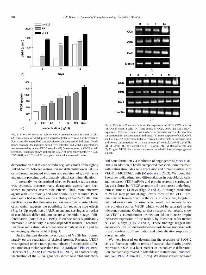
348 J.-E. Huh et al. / Journal of Ethnopharmacology 104 (2006) 345–350
Fig. 3. Effects of Puerariae radix on VEGF protein secretion in SaOS-2 cells.(A) Time course of VEGF protein secretion. Cells were treated with vehicle orPuerariae radix at specified concentration for the time periods indicated. Condi-tioned media for the indicated period were collected, and VEGF concentrationswere measured by human VEGF assay kit. (B) Dose–response of VEGF proteinsecretion. Results are shown as the mean ± S.D. of three experiments. *P < 0.05,**P < 0.01, and ***P < 0.001 compared with vehicle-treated control.
demonstration that Puerariae radix regulates much of the tightlylinked control between maturation and differentiation in SaOS-2cells through increased synthesis and secretion of growth factorand matrix proteins, and ultimately stimulates mineralization.
Importantly, we determined whether Puerariae radix extractwas cytotoxic, because many therapeutic agents have beenshown to possess severe side effects. Thus, more effectiveagents with little toxicity and good solubility are required. Puer-ariae radix had no effect on the viability of SaOS-2 cells. Thisresult indicates that Puerariae radix is non-toxic to osteoblasticcells, which suggests the possibility for reducing side effects(Fig. 1). Up-regulation of ALP, an enzyme serving as a markerof osteoblastic differentiation, occurs at the middle stage of dif-ferentiation (Aubin et al., 1995). Puerariae radix significantlyincreased ALP activity in a dose-dependent manner. Therefore,Puerariae radix stimulates osteoblastic activity at least in part byenhancing synthesis of ALP (Fig. 1).
During the past decade, investigation of VEGF has focusedlargely on the regulation of skeletal growth. Recently, VEGFwas reported to be a more potent inducer of osteoblastic differ-entiation on a molar basis than BMP-2 (Midy and Plouet, 1994;Deckers et al., 2000; Furumatsu et al., 2003). In another study,inactivation of the VEGF gene was shown to inhibit endochon-
Fig. 4. Effects of Puerariae radix on the expression of OCN, OPN, and ColI mRNA in SaOS-2 cells. (A) Time course of OCN, OPN, and Col I mRNAexpression. Cells were treated with vehicle or Puerariae radix at the specifiedconcentration for the time periods indicated. (B) Dose–response of OCN, OPN,and Col I mRNA expression. Cells were treated with vehicle or Puerariae radixat different concentrations for 14 days culture: (1) control, (2) 0.01 �g/ml PR,(3) 0.1 �g/ml PR, (4) 1 �g/ml PR, (5) 10 �g/ml PR, (6) 100 �g/ml PR, and(7) 50 ng/ml VEGF. Each value is expressed as relative level of target gene to�-actin.
dral bone formation via inhibition of angiogenesis (Maes et al.,2003). In addition, it has been reported that short-term treatmentwith statins stimulates gene expression and protein synthesis forVEGF in MC3T3-E1 cells (Maeda et al., 2003). We found thatPuerariae radix stimulated differentiation in osteoblastic cells,and increased VEGF mRNA and protein secretion starting at 3days of culture, but VEGF secretion did not increase under long-term culture at 14 days (Figs. 2 and 3). Although productionof VEGF may persist at high levels, most of the VEGF pro-tein may be broken down in the cells. Furthermore, long-termcultured osteoblasts, or osteocytes, would not secrete bioac-tive proteins such as VEGF, which would be saturated in themicroenvironment. Owing to these reasons, our results showthat VEGF accumulation in the medium did not increase despiteincreased expression of the mRNA by Puerariae radix treatedcells at 14 days (Figs. 2 and 3). These findings suggest thatenhanced VEGF production by osteoblasts has an important rolein the osteoblastic differentiation and mineralization response toPuerariae radix.
We next focused on determining the response of SaOS-2cells to Puerariae radix in terms of extracellular matrix proteinexpression. OCN is a later marker of osteoblastic differentia-tion that is closely related to osteoblastic maturation (Franceschiand Iyer, 1992; Aubin et al., 1995). We demonstrated increased

J.-E. Huh et al. / Journal of Ethnopharmacology 104 (2006) 345–350 349
Fig. 5. Effects of Puerariae radix on type I collagen protein secretion in SaOS-2cells. (A) Time course of type I collagen protein secretion. Cells were treatedwith vehicle or Puerariae radix at specified concentration for the time periodsindicated. Conditioned media for indicated period were collected, and type Icollagen were measured by colorimetric assay kit. (B) Dose–response of typeI collagen protein secretion. Results are shown as the mean ± S.D. of threeexperiments. ***P < 0.001 compared with vehicle-treated control.
OCN mRNA expression in response to Puerariae radix at 14days of SaOS-2 cell culture (Fig. 4). In addition, OPN is abone matrix protein secreted by osteoblasts, and regarded asthe last in a chronological sequence of markers of osteoblas-tic differentiation. OPN expression is enhanced by hormones,cytokines, and regulates mineral growth in vitro and in vivo(Hunter et al., 1996). In our experiments, Puerariae radix mod-erately increased OPN mRNA expression in a dose-dependentmanner at 14 days of culture (Fig. 4). Osteoblasts abundantlysynthesize and secrete Col I, a major bone matrix constituentand extracellular macromolecule in osteoblast cultures. Puer-ariae radix markedly increased Col I mRNA expression, andproteins at 14 days of SaOS-2 cell culture (Figs. 4 and 5). Itis likely that VEGF stimulated by Puerariae radix induces thematrix protein at a later stage of the culture.
Finally, we found that Puerariae radix induced mineralizednodule formation at 14 days of SaOS-2 cell culture (Fig. 6).This result supports the hypothesis that Puerariae radix promotesosteoblastic differentiation in vitro, through increased synthesisand secretion of growth factor and matrix proteins.
In this study, we have investigated the effects of Puerariaeradix on osteoblast-like differentiation, and our findings showthat Puerariae radix has no effect on cell viability and regulates
Fig. 6. Effects of Puerariae radix on mineralization in SaOS-2 cells. (A) Cell lay-ers stained with Alizarin Red were destained, observed under the microscope,and photographed (magnification, ×200). Vehicle (control), cells exposed to1 �g/ml Puerariae radix (PR), or 50 ng/ml VEGF (VEGF), are shown. (B) Min-eralization was quantified per field in at least three fields after each experiment,and the result expressed as the mean intensity. **P < 0.01 and ***P < 0.001compared with vehicle-treated control.
cellular differentiation in SaOS-2 cells. However, the mecha-nisms for gene expression are complex and the results of thisstudy only begin to clarify these mechanisms. Thus, furtherinvestigation is required to isolate the active constituents anddevelop new therapeutics. These results suggest that Puerariaeradix can play an important role in osteoblastic bone formationthrough up-regulation of ALP activity, VEGF, OCN, OPN, ColI expression and mineralization, and possibly lead to the devel-opment of bone healing and osteoporosis drugs.
Acknowledgment
This study was supported by a grant from the OrientalMedicine R&D Project, Ministry of Health and Welfare, Repub-lic of Korea (03-PJ9-PG6-SO01-0002).
References
Arao, T., Udayama, M., Kinjo, J., Nohara, T., Funakoshi, T., Kojima, S.,1997. Preventive effects of saponins from Puerariae radix (the root ofPueraria lobata Ohwi) on in vitro immunological injury of rat primaryhepatocyte cultures. Biological & Pharmaceutical Bulletin 20, 988–991.
Aubin, J.E., Liu, F., Malaval, L., Gupta, A.K., 1995. Osteoblast and chon-droblast differentiation. Bone 17, 77S–83S.

350 J.-E. Huh et al. / Journal of Ethnopharmacology 104 (2006) 345–350
Deckers, M.M., Karperien, M., van der Bent, C., Yamashita, T., Papapoulos,S.E., Lowik, C.W., 2000. Expression of vascular endothelial growth fac-tors and their receptors during osteoblast differentiation. Endocrinology141, 1667–1674.
Franceschi, R.T., Iyer, B.S., 1992. Relationship between collagen synthesisand expression of the osteoblast phenotype in MC3T3-E1 cells. Journalof Bone and Mineral Research 7, 235–246.
Furumatsu, T., Shen, Z.N., Kawai, A., Nishida, K., Manabe, H., Oohashi,T., Inoue, H., Ninomiya, Y., 2003. Vascular endothelial growth factorprincipally acts as the main angiogenic factor in the early stage of humanosteoblastogenesis. Journal of Biochemistry 133, 633–639.
Gerber, H.P., Vu, T.H., Ryan, A.M., Kowalski, J., Werb, Z., Ferrara, N.,1999. VEGF couples hypertropic cartilage remodeling, and angiogen-esis during endochondral bone formation. Nature Medicine 5, 623–628.
Goad, D.L., Rubin, J., Wang, H., Tashjian Jr., A.H., Patterson, C., 1996.Enhanced expression of vascular endothelial growth factor in humanSaOS-2 osteoblast-like cells and murine osteoblasts induced by insulin-like growth factor I. Endocrinology 137, 2262–2268.
Guerra, M.C., Speroni, E., Broccoli, M., Cangini, M., Pasini, P., Minghett,A., Crespi-Perellino, N., Mirasoli, M., Cantelli-Forti, G., Paolini, M.,2000. Comparison between Chinese medical herb Pueraria lobata crudeextract and its main isoflavone puerarin antioxidant properties and effectson rat liver CYP-catalysed drug metabolism. Life Sciences 7, 2997–3006.
Hughes, F.J., Collyer, J., Stanfield, M., Goodman, S.A., 1995. The effects ofbone morphogenic protein-2, -4, and -6 on differentiation of rat osteoblastcells in vitro. Endocrinology 136, 2671–2677.
Hunter, G.K., Hauschka, P.V., Poole, A.R., Rosenberg, L.C., Goldberg, H.A.,1996. Nucleation and inhibition of hydroxyapatite formation by mineral-ized tissue proteins. The Biochemical Journal 317, 59–64.
Lai, X.L., Tang, B., 1989. Recent advances in the experimental study andclinical application of Pueraria lobata (Willd) Ohwi. Zhongguo ZhongYao Za Zhi 14, 308–311.
Lian, J.B., Stein, G.S., Boskey, A.L., 2003. Bone formation: maturation andfunctional activists osteoblast linease cells. In: Favus, M.J. (Ed.), Primeron the metabolic bone diseases and disorders of mineral metabolism, fifthed. The American Society for Bone and Mineral Research, WashingtonDC, pp. 13–28.
Maeda, T., Kawane, T., Horiuchi, N., 2003. Statins augument vascularendothelial growth factor expression in osteoblastic cells via inhibitionof protein prenylation. Endocrinology 144, 681–692.
Maeda, T., Matsunuma, A., Kawane, T., Horiuchi, N., 2001. Simvastatinpromotes osteoblast differentiation and mineralization in MC3T3-E1 cells.Biochemical and Biophysical Research Communications 280, 874–877.
Maes, C., Carmeliet, P., Moermans, K., Stockmans, I., Smets, N., Collen,D., Bouillon, R., Carmeliet, G., 2003. Impaired angiogenesis and endo-chondral bone formation in mice lacking the vascular endothelial growthfactor isoforms VEGF164 and VEGF188. Mechanisms of Development111, 61–73.
McCarthy, T.L., Centrella, M., Canalis, E., 1989. Regulatory effects ofinsulin-like growth factors I and on bone collagen synthesis in rat cal-varial cultures. Endocrinology 124, 301–309.
Midy, V., Plouet, J., 1994. Vasculotropin/vascular endothelial growth factorinduces differentiation in cultured osteoblasts. Biochemical and Biophys-ical Research Communications 199, 380–386.
Spelsberg, T.C., Subramaniam, M., Riggs, B.L., Khosla, S., 1999. The actionsand interactions of sex steroids and growth factors/cytokines on the skele-ton. Molecular Endocrinology 13, 819–828.
Wang, X., Wu, J., Chiba, H., Umegaki, K., Yamada, K., Ishimi, Y., 2003.Puerariae radix prevents bone loss in ovariectomized mice. Journal ofBone and Mineral Metabolism 21, 268–275.

Journal of Ethnopharmacology 104 (2006) 351–355
Antihypertensive and vasodilator effects of methanolic andaqueous extracts of Tribulus terrestris in rats
Oludotun A. Phillips a,∗, Koyippalli T. Mathew b, Mabayoje A. Oriowo c
a Department of Pharmaceutical Chemistry, Faculty of Pharmacy, Kuwait University, P.O. Box 24923, 13110 Safat, Kuwaitb Department of Biological Sciences, Kuwait University, P.O. Box 24923, 13110 Safat, Kuwait
c Department of Pharmacology, Kuwait University, P.O. Box 24923, 13110 Safat, Kuwait
Received 13 June 2005; received in revised form 15 September 2005; accepted 22 September 2005Available online 9 November 2005
Abstract
The effects of methanolic and aqueous extracts of Tribulus terrestris on rat blood pressure (BP) and the perfused mesenteric vascular bedwere investigated. The extracts dose-dependently reduced BP in spontaneously hypertensive rats (SHRs) with the aqueous fraction being morepotent than the methanolic fraction at all doses tested. In vitro, the methanolic but not aqueous extract produced a dose-dependent increase inperfusion pressure of the mesenteric vascular bed. When perfusion pressure was raised with phenylephrine (10−5 M), the aqueous extract produceda dose-dependent reduction in perfusion pressure at all doses. A low dose of the methanolic extract produced a vasoconstrictor effect while higherdoses produced dose-dependent reduction in perfusion pressure. l-NAME (10−4 M) significantly reduced but did not abolish vasodilation inducedby the extracts. Vasodilator responses to aqueous and methanolic fractions were significantly reduced in preparations where perfusion pressurewas raised with KCl (60 mM). A combination of KCl and l-NAME abolished the vasodilator responses induced by the extracts. It was concludedthat methanolic and aqueous extracts of Tribulus terrestris possess significant antihypertensive activity in spontaneously hypertensive rats. Theantihypertensive effects appeared to result from a direct arterial smooth muscle relaxation possibly involving nitric oxide release and membranehyperpolarization.© 2005 Elsevier Ireland Ltd. All rights reserved.
Keywords: Tribulus terrestris; Blood pressure; Mesenteric vascular bed; Spontaneously hypertensive rats
1. Introduction
Tribulus terrestris L. is a member of the Zygophyllaceae fam-ily. It is an annual herb about 30–70 cm high and has pinnateleaves (of unequal length), yellow flowers and characteristicstellate shaped carpel fruits. It is widely distributed in Africa,western Asia, China, Japan, Korea and Europe. The plant is alsofound in Kuwait (Daoud and Al-Rawi, 1985; Middleditch andAmer, 1991). Extracts from this plant have been used tradition-ally in treating a variety of diseases including hypertension andcoronary heart disease, ocular inflammation and infertility inboth sexes. The extracts have also been used as diuretics. Recentpharmacological studies tend to support these uses. For exam-ple, Al-Ali et al. (2003) have demonstrated diuretic activity inrats while Adaikan et al. (2000) have shown that crude extractof Tribulus terrestris enhanced electrically- and nitroglycerine-
∗ Corresponding author. Tel.: +965 4986070.
induced relaxation of the rabbit corpus cavernosum consistentwith a pro-erectile function.
The mechanism responsible for the antihypertensive activ-ity is still not fully understood. In a recent study, Sharifi et al.(2003) reported a significant antihypertensive effect of an aque-ous extract of Tribulus terrestris in renin-dependent 2-kidney1-clip (2K-1C) model of hypertension and suggested that thismight be related to its inhibitory effect on angiotensin convert-ing enzyme (ACE) activity. This was based on the observationthat treatment with the aqueous extract Tribulus terrestris sig-nificantly reduced ACE activity in all tissues of the rat. A rolefor arterial smooth muscle vasodilation in the antihyperten-sive effect of Tribulus terrestris appeared unlikely especiallysince Arcasoy et al. (1998) failed to observe any relaxant effectof Tribulus terrestris in the rabbit aorta even though similarconcentrations of the extract inhibited peristaltic movement insheep ureter and rabbit jejunum. However, the possibility thatthe extract could produce its anti-hypertensive effect by dilat-ing resistance vessels was not investigated and therefore could
0378-8741/$ – see front matter © 2005 Elsevier Ireland Ltd. All rights reserved.doi:10.1016/j.jep.2005.09.027
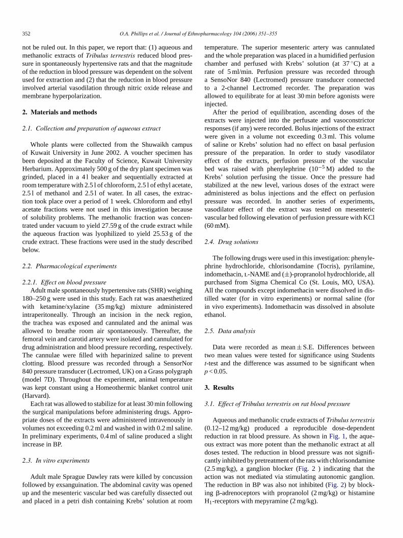
352 O.A. Phillips et al. / Journal of Ethnopharmacology 104 (2006) 351–355
not be ruled out. In this paper, we report that: (1) aqueous andmethanolic extracts of Tribulus terrestris reduced blood pres-sure in spontaneously hypertensive rats and that the magnitudeof the reduction in blood pressure was dependent on the solventused for extraction and (2) that the reduction in blood pressureinvolved arterial vasodilation through nitric oxide release andmembrane hyperpolarization.
2. Materials and methods
2.1. Collection and preparation of aqueous extract
Whole plants were collected from the Shuwaikh campusof Kuwait University in June 2002. A voucher specimen hasbeen deposited at the Faculty of Science, Kuwait UniversityHerbarium. Approximately 500 g of the dry plant specimen wasgrinded, placed in a 4 l beaker and sequentially extracted atroom temperature with 2.5 l of chloroform, 2.5 l of ethyl acetate,2.5 l of methanol and 2.5 l of water. In all cases, the extrac-tion took place over a period of 1 week. Chloroform and ethylacetate fractions were not used in this investigation becauseof solubility problems. The methanolic fraction was concen-trated under vacuum to yield 27.59 g of the crude extract whilethe aqueous fraction was lyophilized to yield 25.53 g of thecrude extract. These fractions were used in the study describedbelow.
2.2. Pharmacological experiments
2.2.1. Effect on blood pressureAdult male spontaneously hypertensive rats (SHR) weighing
180–250 g were used in this study. Each rat was anaesthetizedwith ketamine/xylazine (35 mg/kg) mixture administeredintraperitoneally. Through an incision in the neck region,the trachea was exposed and cannulated and the animal wasallowed to breathe room air spontaneously. Thereafter, thefemoral vein and carotid artery were isolated and cannulated fordrug administration and blood pressure recording, respectively.The cannulae were filled with heparinized saline to preventclotting. Blood pressure was recorded through a SensorNor840 pressure transducer (Lectromed, UK) on a Grass polygraph(model 7D). Throughout the experiment, animal temperaturewas kept constant using a Homeothermic blanket control unit(Harvard).
Each rat was allowed to stabilize for at least 30 min followingthe surgical manipulations before administering drugs. Appro-priate doses of the extracts were administered intravenously involumes not exceeding 0.2 ml and washed in with 0.2 ml saline.In preliminary experiments, 0.4 ml of saline produced a slightincrease in BP.
2.3. In vitro experiments
Adult male Sprague Dawley rats were killed by concussionfollowed by exsanguination. The abdominal cavity was openedup and the mesenteric vascular bed was carefully dissected outand placed in a petri dish containing Krebs’ solution at room
temperature. The superior mesenteric artery was cannulatedand the whole preparation was placed in a humidified perfusionchamber and perfused with Krebs’ solution (at 37 ◦C) at arate of 5 ml/min. Perfusion pressure was recorded througha SensoNor 840 (Lectromed) pressure transducer connectedto a 2-channel Lectromed recorder. The preparation wasallowed to equilibrate for at least 30 min before agonists wereinjected.
After the period of equilibration, ascending doses of theextracts were injected into the perfusate and vasoconstrictorresponses (if any) were recorded. Bolus injections of the extractwere given in a volume not exceeding 0.3 ml. This volumeof saline or Krebs’ solution had no effect on basal perfusionpressure of the preparation. In order to study vasodilatoreffect of the extracts, perfusion pressure of the vascularbed was raised with phenylephrine (10−5 M) added to theKrebs’ solution perfusing the tissue. Once the pressure hadstabilized at the new level, various doses of the extract wereadministered as bolus injections and the effect on perfusionpressure was recorded. In another series of experiments,vasodilator effect of the extract was tested on mesentericvascular bed following elevation of perfusion pressure with KCl(60 mM).
2.4. Drug solutions
The following drugs were used in this investigation: phenyle-phrine hydrochloride, chlorisondamine (Tocris), pyrilamine,indomethacin, l-NAME and (±)-propranolol hydrochloride, allpurchased from Sigma Chemical Co (St. Louis, MO, USA).All the compounds except indomethacin were dissolved in dis-tilled water (for in vitro experiments) or normal saline (forin vivo experiments). Indomethacin was dissolved in absoluteethanol.
2.5. Data analysis
Data were recorded as mean ± S.E. Differences betweentwo mean values were tested for significance using Studentst-test and the difference was assumed to be significant whenp < 0.05.
3. Results
3.1. Effect of Tribulus terrestris on rat blood pressure
Aqueous and methanolic crude extracts of Tribulus terrestris(0.12–12 mg/kg) produced a reproducible dose-dependentreduction in rat blood pressure. As shown in Fig. 1, the aque-ous extract was more potent than the methanolic extract at alldoses tested. The reduction in blood pressure was not signifi-cantly inhibited by pretreatment of the rats with chlorisondamine(2.5 mg/kg), a ganglion blocker (Fig. 2 ) indicating that theaction was not mediated via stimulating autonomic ganglion.The reduction in BP was also not inhibited (Fig. 2) by block-ing �-adrenoceptors with propranolol (2 mg/kg) or histamineH1-receptors with mepyramine (2 mg/kg).

O.A. Phillips et al. / Journal of Ethnopharmacology 104 (2006) 351–355 353
Fig. 1. Effects of methanolic and aqueous extracts of Tribulus terrestris onblood pressure in anaesthetized spontaneously hypertensive rats. Each point isthe mean ± S.E. of five experiments.
Fig. 2. Effects of chlorisondamine (2.5 mg/kg), propranolol (2 mg/kg) andmepyramine (2 mg/kg) on the reduction in blood pressure produced by methano-lic and aqueous extracts of Tribulus terrestris. Each point is the mean ± S.E. offive experiments.
3.2. In vitro experiments
3.2.1. Vasoconstrictor responsesAscending doses of the crude extracts were tested on the
perfused rat mesenteric vascular bed. As shown in Fig. 3, themethanolic extract dose-dependently increased basal perfusionpressure of the mesenteric vascular bed while the aqueous frac-tion had no effect at any of the doses tested. The vasoconstrictoreffect of the methanolic extract was not reduced by phento-lamine (10−6 M) indicating that it did not involve an action on�-adrenoceptors.
Fig. 4. Excitatory and inhibitory effects of methanolic and aqueous extracts ofTribulus terrestris on perfusion pressure of the mesenteric vascular bed. Perfu-sion pressure was raised with phenylephrine (10−5 M). Each point on the graphis the mean ± S.E. of five experiments.
Fig. 5. Effect of l-NAME (10−4 M), KCl (60 mM) and l-NAME (10−4 M) +KCl (60 mM) on reduction in perfusion pressure produced by methanolic extractsof Tribulus terrestris in the mesenteric vascular bed. Perfusion pressure wasraised with phenylephrine (10−5 M). Each point is the mean ± S.E. of five exper-iments.
3.2.2. Vasodilator responsesIn preparations where perfusion pressure was raised with
phenylephrine (10−5 M), the aqueous and methanolic extracts(30–1500 �g) produced a dose-dependent reduction of the per-fusion pressure. The aqueous extract produced vasodilation atall doses tested (Fig. 4). However, a low dose of the methano-lic extract (30 �g) produced vasoconstriction while vasodilatorresponses were produced at higher doses (>30 �g). Vasodila-tor responses to both aqueous and methanolic extracts werenot significantly (p > 0.05) reduced by propranolol (10−6 M) orindomethacin (10−5 M) but were significantly (p < 0.05) reducedby l-NAME (10−4 M), a nitric oxide synthase inhibitor. In
Fig. 3. Typical trace showing the effect of methanolic and aqueous extracts of Tribulus terrestris on basal perfusion pressure of the mesenteric vascular bed.

354 O.A. Phillips et al. / Journal of Ethnopharmacology 104 (2006) 351–355
Fig. 6. Effect of l-NAME (10−4 M), KCl (60 mM) and l-NAME (10−4 M) +KCl (60 mM) on reduction in perfusion pressure produced by aqueous extracts ofTribulus terrestris in the mesenteric vascular bed. Perfusion pressure was raisedwith phenylephrine (10−5 M). Each point is the mean ± S.E. of five experiments.
another series of experiments, the concentration of KCl inthe Krebs’ solution perfusing the mesenteric vascular bed wasincreased to 60 mM. Under this condition, vasodilator responsesto low doses of the extracts (aqueous and methanolic) wereabolished while higher doses of the extract reduced perfusionpressure. The reduction in perfusion pressure was much lessthan when perfusion pressure was elevated with phenylephrine(Figs. 5 and 6). A combination of l-NAME (10−4 M) and KCl(60 mM) abolished the vasodilation produced by the extracts(Figs. 5 and 6).
4. Discussion
These results showed that crude aqueous and methanolicextracts of Tribulus terrestris produced dose-dependent reduc-tion in blood pressure of spontaneously hypertensive rats. Thisobservation is in agreement with a previous report by Sharifiet al. (2003) in renin-dependent 2K-1C model of hyperten-sion. The hypotensive effect was reproducible as tachyphylaxiswas not observed in any of the preparations. The BP lower-ing effect of the extracts was not attenuated by blocking theautonomic ganglia or by blocking �-adrenoceptors or histamineH1-receptors with propranolol and mepyramine, respectivelyindicating that these extracts did not act via stimulating the gan-glion, �-adrenoceptors or histamine H1-receptors. The aqueousextract was more potent that the methanolic extract in reducingBP. We cannot, based on the present results offer any expla-nation on why the aqueous extract was more potent than themethanolic extract. It possibly could be due to different activeprinciples present in the different extracts. It is interesting to notethat Somanadhan et al. (1999) have similarly reported that theeffectiveness of extracts of Tribulus terrestris in inhibiting ACEactivity varied with the method of extraction. In their study, theaqueous extract was more effective than the ethanolic extract ininhibiting ACE activity.
The mechanism(s) by which extracts of Tribulus terrestrislower BP has not been elucidated. Sharifi et al. (2003) confirmedthat aqueous extracts of Tribulus terrestris inhibited ACE activ-
ity and suggested that this could explain the antihypertensiveeffect of this plant extract. However, other mechanisms includ-ing a vasodilator effect mediated via a direct effect on the arterialsmooth muscle or interfering with other neuroeffector mecha-nisms such as the adrenergic system could not be ruled out. Inpreliminary experiments to identify the possible mechanism ofblood pressure lowering effect of the extracts, we tested vari-ous doses of the extract on the perfused rat mesenteric vascularbed. The results showed that the methanolic but not aqueousextract dose-dependently and reproducibly increased basal per-fusion pressure of the mesenteric vascular bed indicating that themethanolic but not aqueous extract contained a vasoconstrictorsubstance. When perfusion pressure was raised with PE, bothextracts produced reproducible and dose-dependent reductionin perfusion pressure. However, low doses of the methanolicextract produced only vasoconstriction. This would indicate themethanolic extract contained vasoconstrictor and vasodilatorsubstances while the aqueous extract contained only vasodilatorsubstance(s).
The vasodilatory effect of the extracts was reduced but notabolished by l-NAME, a nitric oxide synthase inhibitor (Rees etal., 1990) indicating that the antihypertensive effect was partlydependent on nitric oxide release from the vascular endothelium.Increasing the KCl concentration in the physiological solutionto 60 mM significantly reduced the vasodilator response to bothextracts. This would suggest that part of the vasodilatory effectsof the extracts could be due to membrane hyperpolarization.Vasoconstriction produced by the low dose of the methanolicextract was not affected. A combination of l-NAME and KClalmost abolished the vasodilation. This would suggest that thevasodilator effect of aqueous and methanolic extracts of Tribulusterrestris involved hyperpolarization of the membrane in addi-tion to releasing NO from the vascular endothelium.
The major chemical constituents of Tribulus terrestris fromaqueous or alcoholic (methanolic) extracts are the steroidalsaponins. Yan et al. (1996) reported the isolation and charac-terization of steroidal saponins including terrestrosin A, B, C,D and E, desgalactotigonin, gitonin, desglucolanatigonin andF-gitonin. More recently, other steroidal saponins including ter-restrinins A and B, Huang et al. (2003); protodioscin and theirrespective sulfates, De Combarieu et al. (2003); and spirostanoltype saponin, tribulosin and beta-sitosterol-d-glucoside Deepaket al. (2002) and Conrad et al. (2004) have been isolated and char-acterized. We presently cannot link the pharmacological activityof the extract to any of the compounds (or groups) listed abovebut we are in the process of using a bio-assay directed frac-tionation procedure to determine the active principle(s) in theextract.
5. Conclusion
It was therefore concluded that extracts of Tribulus ter-restris displayed significant antihypertensive properties in spon-taneously hypertensive rats, which could involve smooth musclerelaxation via NO release and membrane hyperpolarization. Fur-ther studies are in progress to identify the active principle(s) inthe plant extract.

O.A. Phillips et al. / Journal of Ethnopharmacology 104 (2006) 351–355 355
Acknowledgements
We thank Mrs. Sanaa A. Amine, Bindu Chandrasekhar andElizabeth Kadavil for their technical assistance. This projectwas supported by the Kuwait Foundation for the Advancementof Sciences (KFAS 2002-07-07).
References
Al-Ali, M., Wahbi, S., Twaij, H., Al-Badr, A., 2003. Tribulus terrestris: pre-liminary study of its diuretic and contractile effects and comparison withZea mays. Journal of Ethnopharmacology 85, 257–260.
Adaikan, P.G., Gauthaman, K., Prasad, R.N., Ng, S.C., 2000. Proerectilepharmacological effects of Tribulus terrestris extract on the rabbit corpuscavernosum. Annals of the Academy of Medicine Singapore 29, 22–26.
Arcasoy, H.B., Erenmemisoglu, A., Tekol, Y., Kurucu, S., Kartal, M., 1998.Effect of Tribulus terrestris L. saponin mixture on some smooth musclepreparations: a preliminary study. Bollettino Chimico Farmaceutico 137,473–475.
Conrad, J., Dinchev, D., Klaiber, I., Mika, S., Kostova, I., Kraus W, 2004.A novel furostanol saponin from Tribulus terrestris of Bulgarian origin.Fitoterapia 75 (2), 117–122.
Daoud, H.S., Al-Rawi, A., 1985. The Flora of Kuwait: Dicotyledoneae, vol.1. KPI Publishers, London.
De Combarieu, E., Fuzzati, N., Lovati, M., Mercalli, E., 2003. Furostanolsaponins from Tribulus terrestris. Fitoterapia 74 (6), 583–591.
Deepak, M., Dipankar, G., Prashanth, D., Asha, M.K., Amit, A.,Venkataraman BV, 2002. Tribulosin and beta-sitosterol-d-glucoside, theanthelmintic principles of Tribulus terrestris. Phytomedicine 9 (8),753–756.
Huang, J.W., Tan, C.H., Jiang, S.H., Zhu, D.Y., 2003. Terrestrinins A andB, two new steroid saponins from Tribulus terrestris. Journal of AsianNatural Production Research 5, 285–290.
Middleditch, B.S., Amer, M.A., 1991. Studies in Plant Science. 2. KuwaitiPlants. Elsevier Science Publishers B.V.
Rees, D.D., Palmer, R.M.J., Schulz, H.F., Hodson, H.F., Moncada, S., 1990.Characterization of three inhibitors of endothelial nitric oxide synthase invitro and in vivo. British Journal of Pharmacology 101, 747–752.
Sharifi, A.M., Darabi, R., Akbarloo, N., 2003. Study of antihypertensivemechanism of Tribulus terrestris in 2K1C hypertensive rats: role of tissueACE activity. Life Science 73, 2963–2971.
Somanadhan, B., Varughese, G., Palpu, P., Sreedharan, R., Gudiksen, L.,Smitt, U.W., Nyman, U., 1999. An ethnopharmacological survey forpotential angiotensin converting enzyme inhibitors from Indian medic-inal plants. Journal of Ethnopharmacology 65, 103–112.
Yan, W., Ohtani, K., Kasai, R., Yamasaki K, 1996. Steroidal saponins fromfruits of Tribulus terrestris. Phytochemistry 42 (5), 1417–1422.

Journal of Ethnopharmacology 104 (2006) 356–361
Action of Hygrophila auriculata against streptozotocin-inducedoxidative stress
M. Vijayakumar a, R. Govindarajan a, G.M.M. Rao a, Ch.V. Rao a,A. Shirwaikar b, S. Mehrotra a, P. Pushpangadan a,∗
a Pharmacognosy and Ethnopharmacology Division, National Botanical Research Institute, Lucknow 226 001, Uttar Pradesh, Indiab Pharmaceutics Department, College of Pharmaceutical Sciences, MAHE, Manipal 576119, India
Received 1 November 2004; received in revised form 23 August 2005; accepted 24 September 2005Available online 9 November 2005
Abstract
Hygrophila auriculata (K. Schum.) Heine (Family: Acanthaceae) is a wild herb widely used in ‘Ayurveda’ as ‘Rasayana’ drug for treatment ofvarious disorders. Treatment of diabetic rats with aerial parts of Hygrophila auriculata extract (HAEt, 100 and 250 mg/kg body weight) for 3 weeksshowed significant reduction in blood glucose, thiobarbituric acid reactive substances (TBARS) and hydroperoxide in both liver and kidney. Thetreatment with HAEt significantly increased the glutathione (GSH), glutathione peroxidase (GPx), glutathione S-transferase (GST) and catalase(CAT) in the drug-treated group, which is comparable to the control group. HAEt and glibenclamide-treated rats also showed decreased lipidperoxidation that is associated with increased activity of superoxide dismutase (SOD) and catalase. The ability of HAEt on tissue lipid peroxidationand antioxidant status in diabetic animals has not been studied before. The result of this study thus shows that HAEt possesses significant antidiabeticactivity along with potent antioxidant potential in diabetic conditions.© 2005 Elsevier Ireland Ltd. All rights reserved.
Keywords: Hygrophila auriculata; Diabetes; Antioxidant enzymes; Photochemiluminescence
1. Introduction
Oxidative stress, defined as an imbalance between oxidantsand antioxidants leads to many biochemical changes and is animportant causative factor in several human chronic diseases,such as atherosclerosis and cardiovascular diseases, mutagene-sis and cancer, several neurodegenerative disorders and the agingprocess (Frei, 1999). Diabetes mellitus is one such disease andit is estimated that the number of diabetic patients will continueto increase in the future (Furusho et al., 2002). It has been postu-lated that the etiology of the complications of diabetes involvesoxidative stress perhaps as a result of hyperglycemia (Hunt et al.,1990). The elevated levels of blood glucose in diabetes produceoxygen-free radicals (OFR), which cause membrane damage
Abbreviations: CAT, catalase; GPx, glutathione peroxidase; GSH, glu-tathione; GST, glutathione S-transferase; HAEt, Hygrophila auriculata extract;LPO, lipid peroxidation; ROS, reactive oxygen species; SOD, superoxide dis-mutase; STZ, streptozotocin; TBARS, thiobarbituric acid reactive substances
∗ Corresponding author. Tel.: +91 522 2205848; fax: +91 522 2205836.E-mail address: [email protected] (P. Pushpangadan).
due to peroxidation of membrane lipids and protein glycation(Sato et al., 1979). Baynes (1991) reported that plasma thio-barbituric acid reactive substance (TBARS) levels increasedin diabetic patients due to vascular lesions induced by hyper-glycemia. Diabetic patients thus have an increased incidence ofvascular diseases and it has been suggested that free radical activ-ity increased in diabetes (Oberly, 1988). It has also been shownthat glucose under physiological conditions produces oxidantsthat possesses reactivity similar to the hydroxyl-free radicals.Recent years have witnessed a renewed interest in plants aspharmaceuticals because they synthesize a variety of secondarymetabolites with antioxidant potential which can play a majorrole in protection against molecular damage induced by reac-tive oxygen species (ROS) (Cao et al., 1997; Vaya et al., 1997).Many traditional plant treatments for diabetes mellitus are usedthroughout the world. Few of the medicinal plant treatmentsfor diabetes have received scientific scrutiny, for which WorldHealth Organization (WHO) has also recommended attention(WHO, 1980).
Hygrophila auriculata (K. Schum.) Heine Syn. Asteracanthalongifolia Nees. (Acanthaceae), is a wild herb commonly found
0378-8741/$ – see front matter © 2005 Elsevier Ireland Ltd. All rights reserved.doi:10.1016/j.jep.2005.09.030

M. Vijayakumar et al. / Journal of Ethnopharmacology 104 (2006) 356–361 357
in moist places on the banks of tanks, ditches and paddy fieldsthroughout India and is one of the source of Ayurvedic drug ‘kok-ilaksha’. Aerial parts of the plant are used ethnobotanically forthe treatment of body pain, jaundice and malaria, while the seedsare used for treatment of impotence and thus as aphrodisiac (Jain,1991). Aerial parts of Hygrophila auriculata have been reportedto contain lupeol, stigmasterol and butelin while the seeds of theplant are reported to contain mainly fatty acids (Quasim andDutta, 1967). Hygrophila auriculata has been shown to pos-sess hypoglycemic activity in human subjects (Fernando et al.,1989), hepatoprotective activity against paracetamol and thioac-etamide intoxification in rats (Singh and Handa, 1999) andCCl4-induced liver dysfunctions (Shailajan et al., 2005), antitu-mor (Mazmudar et al., 1997), anabolic and adrogenic activities(Jayatilak et al., 1976). Hygrophila auriculata seeds have beenreported to ameliorate the activities of antioxidant enzymesglutathione peroxidase (GPx) and catalase (CAT) in hepato-carcinoma (Ahmed et al., 2001). However, to date no antiox-idant investigations or hypoglycemic activity in experimentalanimals have been reported in the aerial part of this plant. There-fore, the present study was undertaken to investigate the effectof Hygrophila auriculata on the level of antioxidant enzymeslike superoxide dismutase (SOD), CAT, glutathione (GSH),GPx, glutathione S-transferase (GST) along with TBARS andhydroperoxides in streptozotocin (STZ)-induced diabetic ratsand validate the ethnobotanical and clinical claims of theplant.
2. Materials and methods
2.1. Animals
Male Sprague–Drawley rats (160–180 g) were purchasedfrom the animal house of the Central Drug Research Institute,Lucknow, India. These were kept in the departmental animalhouse at 26 ± 2 ◦C and relative humidity 44–55% 10-h light:14-h dark cycles for 1 week before the experiment. Animals wereprovided with rodent diet (Amruth, India) and water ad libitum.All studies were conducted in accordance with the “NationalInstitute of Health Guide for the Care and Use of LaboratoryAnimals”.
2.2. Drugs and chemicals
All the drugs and chemicals were obtained from SigmaChemical Co. (St. Louis, MO, USA). All solvents were of analyt-ical grade and were obtained from Sd. Fine Chemicals, Mumbai,India.
2.3. Plant material
Aerial parts of Hygrophila auriculata were collected fromRewa, Madhya Pradesh (India) during the month of Septem-ber 2002. The plants were authenticated by Dr. Vivek Kumar,National Botanical Research Institute, Lucknow, and a voucherspecimen (LWG, 4609) was lodged in the departmental herbar-ium.
2.4. Preparation of extract
Aerial parts of Hygrophila auriculata were air-dried atroom temperature and coarsely powdered. The powder obtained(250 g) was exhaustively extracted with 50% aqueous ethanol(3× 1 l) for a period of 24 h and filtered. The extract wasthen concentrated on a rotary evaporator under reduced pres-sure and then freeze-dried to yield 1.8% (w/w) of the extract(HAEt).
2.5. Experimental induction of diabetes
A freshly prepared solution of streptozotocin (50 mg/kg,body weight) in 0.1 M citrate buffer (pH 4.5) was injectedintraperitonealy in a volume of 1 ml/kg. STZ-injected animalsexhibited massive glycosuria and hyperglycemia within 2 days(Siddique et al., 1989). Diabetes was confirmed in STZ ratsby measuring the fasting blood glucose concentration 96 hafter the injection of STZ. The rats with blood glucose level>200 mg/dl were considered to be diabetic and were used in theexperiment.
2.6. Experimental procedure
Group I: Control rats received vehicle solution (2% gum aca-cia).
Group II: Diabetic control rats received vehicle solution (2%gum acacia).
Group III: Diabetic rats treated with HAEt 100 mg/kg bodyweight in 2% gum acacia.
Group IV: Diabetic rats treated with HAEt 250 mg/kg bodyweight in 2% gum acacia.
Group V: Diabetic rats treated with glibenclamide 600 �g/kgbody weight in aqueous solution.
The vehicles and the drugs were administered orally usingan intragastric tube daily for 3 weeks. After 3 weeks of treat-ment, the rats were fasted overnight, the blood samples wereanalyzed for blood glucose content by using the O-toluidinemethod (Sasaki et al., 1972) with optical density measured at520 nm. Then the animal was sacrificed by cervical decapi-tation. The liver and kidney were exposed and perfused withcold phosphate buffer saline of pH 7.4. Blood-free liver andkidney were taken out and homogenized in a glass Teflonhomogeniser separately (10%, w/v). Incubations were done at37 ◦C under controlled conditions for biochemical estimations.Fresh blood drawn was centrifuged for 10 min at 2000 rpm.The erythrocyte sediment was resuspended twice in physi-ological NaCl solution (1:10) and centrifuged again in thesame solution. Two hundred and fifty microliters of washederythrocytes were then resuspended in 1000 �l physiologicalNaCl solution and stored at 4 ◦C in the dark until SOD mea-surement. Hemoglobin in erythrocytes was estimated (Henry,1984).

358 M. Vijayakumar et al. / Journal of Ethnopharmacology 104 (2006) 356–361
2.7. Antioxidant assay
Level of lipid peroxides were estimated using the stan-dard method of Okhawa et al. (1979) with minor modification(Govindarajan et al., 2003). TBARS was estimated by reactionwith thiobarbituric acid in the presence of butylated hydroxytoluene and measuring the absorbance at 535 nm of the pink-coloured chromogen formed against reagent blank. Hydroper-oxides were determined by the method of Jiang et al. (1992)which involves ferrous ion oxidation in the presence of XylenolOrange. Total reduced glutathione was measured by the useof DTNB assay using Elman’s method wherein the incuba-tion mixture at 37 ◦C contained 0.08 M sodium phosphate (pH7.0), 0.08 M EDTA, 1.0 mM sodium azide, 0.4 nM GSH and0.25 mM H2O2. GSH was determined at 3-min intervals usingDTNB (Elman, 1959). For glutathione peroxidase, an enzymeunit represents a decrease in GSH concentration of 0.001 log unitper minute after subtraction of non-enzymic mode (Rotruck etal., 1973) and glutathione S-transferase activities were assayedaccording to the method of Habig et al. (1974). SOD was mea-sured by the principle photochemiluminescence assay of theerythrocytes using Photohem® (Analytikjena, Germany) (Popovand Lewin, 1987). Catalase activity was measured by using therate of decomposition of H2O2 by the method of Aebi (1974)and the values are expressed in nmol H2O2 consumed min−1
(mg protein)−1. All these estimations were made in both liverand kidney. Protein content in tissue homogenate was measuredby the method of Lowry et al. (1951).
2.8. Statistical analysis
Values were represented as means ± S.D. for six experimentsand data were analyzed by paired t-test using SPSS version 11(SPSS, Cary, NC, USA).
3. Results
There was a moderate decrease in the blood sugar level of dia-betic rats upon administration of HAEt (250 mg/kg body weight)(Table 1). Table 2 shows the levels of TBARS and hydroperox-ides in liver and kidney of control and experimental animals,while enzyme activities are presented in Table 3. A significantelevation in tissues TBARS and hydroperoxides and significantreduction in GSH, GPx, GST, SOD and catalase was observedin the diabetic control rats as compared to the normal controlrats.
Oral administration of HAEt (250 mg/kg body weight) for3 weeks shows significant reduction in TBARS and hydroper-oxides in both liver (TBARS 0.81 ± 0.21; hydroperoxides75.63 ± 2.97) and kidney (TBARS 1.43 ± 0.71; hydroperox-ides 57.65 ± 1.43). With respect to GST, GPx and CAT therewas a significant increase in the liver (GST 6.59 ± 0.29; GPx9.17 ± 0.88; CAT 70.22 ± 1.24) and kidney. There was a sig-nificant increase in the liver GSH (128.67 ± 2.54), while nosignificant increase was observed in the kidney GSH (Table 3).SOD level (6.22 ± 0.43) in erythrocytes decreased upon induc-tion of diabetes and significantly increased on treatment with
Table 1Effect of various treatments on blood glucose level
Groups Blood glucose (mg/dl)
0 day 7th day 21st day
Group 1 95.45 ± 1.41 96.21 ± 2.33 93.66 ± 2.76Group 2 240.78 ± 2.68 235.98 ± 2.87 228.33 ± 3.03Group 3 242.36 ± 2.97 (NS) 202.37 ± 3.96a 140.98 ± 3.58b
Group 4 240.69 ± 2.14 (NS) 187.99 ± 4.17b 119.77 ± 4.22c
Group 5 239.59 ± 2.71 (NS) 160.29 ± 3.49b 94.33 ± 3.66c
The values represent the means ± S.E.M. for six rats per group. p-Values were calculated based on the paired t-test. NS, not significant.a p < 0.05 compared to diabetic control group.b p < 0.01 compared to diabetic control group.c p < 0.001 compared to diabetic control group.
Table 2Effect of HAEt on the level of TBARS and hydroperoxides in diabetic rat tissues
Groups TBARS (nM/100 g tissue) Hydroperoxide (nM/100 g tissue)
Liver Kidney Liver Kidney
Group 1 0.78 ± 0.51 1.33 ± 0.09 74.64 ± 2.14 58.33 ± 2.19Group 2 1.59 ± 0.11 2.34 ± 1.16 102.57 ± 2.69 77.75 ± 1.11Group 3 1.02 ± 0.08a 1.71 ± 0.97a 86.58 ± 3.17b 68.52 ± 1.61a
Group 4 0.81 ± 0.21c 1.43 ± 0.71c 75.63 ± 2.97c 57.65 ± 1.43c
Group 5 1.26 ± 0.18b 1.87 ± 0.77a 88.29 ± 1.85b 75.63 ± 1.93b
The values represent the means ± S.E.M. for six rats per group. p-Values were calculated based on the paired t-test.a p < 0.01 compared to diabetic control group.b p < 0.05 compared to diabetic control group.c p < 0.001 compared to diabetic control group.
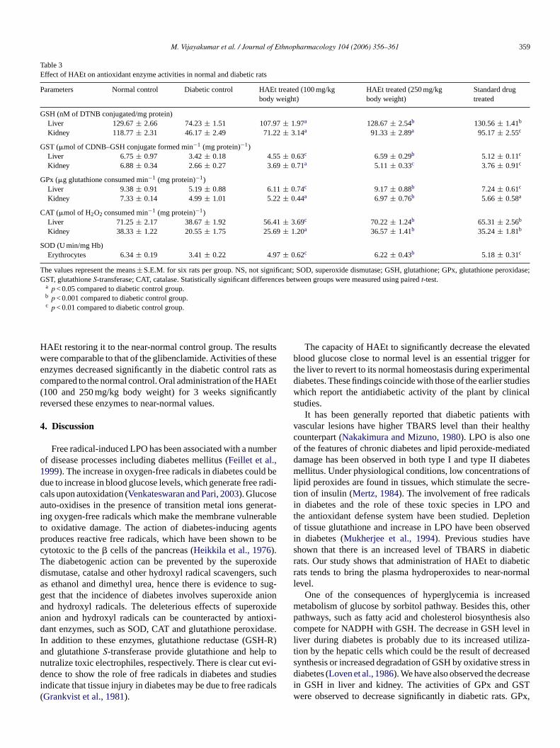
M. Vijayakumar et al. / Journal of Ethnopharmacology 104 (2006) 356–361 359
Table 3Effect of HAEt on antioxidant enzyme activities in normal and diabetic rats
Parameters Normal control Diabetic control HAEt treated (100 mg/kgbody weight)
HAEt treated (250 mg/kgbody weight)
Standard drugtreated
GSH (nM of DTNB conjugated/mg protein)Liver 129.67 ± 2.66 74.23 ± 1.51 107.97 ± 1.97a 128.67 ± 2.54b 130.56 ± 1.41b
Kidney 118.77 ± 2.31 46.17 ± 2.49 71.22 ± 3.14a 91.33 ± 2.89a 95.17 ± 2.55c
GST (�mol of CDNB–GSH conjugate formed min−1 (mg protein)−1)Liver 6.75 ± 0.97 3.42 ± 0.18 4.55 ± 0.63c 6.59 ± 0.29b 5.12 ± 0.11c
Kidney 6.88 ± 0.34 2.66 ± 0.27 3.69 ± 0.71a 5.11 ± 0.33c 3.76 ± 0.91c
GPx (�g glutathione consumed min−1 (mg protein)−1)Liver 9.38 ± 0.91 5.19 ± 0.88 6.11 ± 0.74c 9.17 ± 0.88b 7.24 ± 0.61c
Kidney 7.33 ± 0.14 4.99 ± 1.01 5.22 ± 0.44a 6.97 ± 0.76b 5.66 ± 0.58a
CAT (�mol of H2O2 consumed min−1 (mg protein)−1)Liver 71.25 ± 2.17 38.67 ± 1.92 56.41 ± 3.69c 70.22 ± 1.24b 65.31 ± 2.56b
Kidney 38.33 ± 1.22 20.55 ± 1.75 25.69 ± 1.20a 36.57 ± 1.41b 35.24 ± 1.81b
SOD (U min/mg Hb)Erythrocytes 6.34 ± 0.19 3.41 ± 0.22 4.97 ± 0.62c 6.22 ± 0.43b 5.18 ± 0.31c
The values represent the means ± S.E.M. for six rats per group. NS, not significant; SOD, superoxide dismutase; GSH, glutathione; GPx, glutathione peroxidase;GST, glutathione S-transferase; CAT, catalase. Statistically significant differences between groups were measured using paired t-test.
a p < 0.05 compared to diabetic control group.b p < 0.001 compared to diabetic control group.c p < 0.01 compared to diabetic control group.
HAEt restoring it to the near-normal control group. The resultswere comparable to that of the glibenclamide. Activities of theseenzymes decreased significantly in the diabetic control rats ascompared to the normal control. Oral administration of the HAEt(100 and 250 mg/kg body weight) for 3 weeks significantlyreversed these enzymes to near-normal values.
4. Discussion
Free radical-induced LPO has been associated with a numberof disease processes including diabetes mellitus (Feillet et al.,1999). The increase in oxygen-free radicals in diabetes could bedue to increase in blood glucose levels, which generate free radi-cals upon autoxidation (Venkateswaran and Pari, 2003). Glucoseauto-oxidises in the presence of transition metal ions generat-ing oxygen-free radicals which make the membrane vulnerableto oxidative damage. The action of diabetes-inducing agentsproduces reactive free radicals, which have been shown to becytotoxic to the � cells of the pancreas (Heikkila et al., 1976).The diabetogenic action can be prevented by the superoxidedismutase, catalse and other hydroxyl radical scavengers, suchas ethanol and dimethyl urea, hence there is evidence to sug-gest that the incidence of diabetes involves superoxide anionand hydroxyl radicals. The deleterious effects of superoxideanion and hydroxyl radicals can be counteracted by antioxi-dant enzymes, such as SOD, CAT and glutathione peroxidase.In addition to these enzymes, glutathione reductase (GSH-R)and glutathione S-transferase provide glutathione and help tonutralize toxic electrophiles, respectively. There is clear cut evi-dence to show the role of free radicals in diabetes and studiesindicate that tissue injury in diabetes may be due to free radicals(Grankvist et al., 1981).
The capacity of HAEt to significantly decrease the elevatedblood glucose close to normal level is an essential trigger forthe liver to revert to its normal homeostasis during experimentaldiabetes. These findings coincide with those of the earlier studieswhich report the antidiabetic activity of the plant by clinicalstudies.
It has been generally reported that diabetic patients withvascular lesions have higher TBARS level than their healthycounterpart (Nakakimura and Mizuno, 1980). LPO is also oneof the features of chronic diabetes and lipid peroxide-mediateddamage has been observed in both type I and type II diabetesmellitus. Under physiological conditions, low concentrations oflipid peroxides are found in tissues, which stimulate the secre-tion of insulin (Mertz, 1984). The involvement of free radicalsin diabetes and the role of these toxic species in LPO andthe antioxidant defense system have been studied. Depletionof tissue glutathione and increase in LPO have been observedin diabetes (Mukherjee et al., 1994). Previous studies haveshown that there is an increased level of TBARS in diabeticrats. Our study shows that administration of HAEt to diabeticrats tends to bring the plasma hydroperoxides to near-normallevel.
One of the consequences of hyperglycemia is increasedmetabolism of glucose by sorbitol pathway. Besides this, otherpathways, such as fatty acid and cholesterol biosynthesis alsocompete for NADPH with GSH. The decrease in GSH level inliver during diabetes is probably due to its increased utiliza-tion by the hepatic cells which could be the result of decreasedsynthesis or increased degradation of GSH by oxidative stress indiabetes (Loven et al., 1986). We have also observed the decreasein GSH in liver and kidney. The activities of GPx and GSTwere observed to decrease significantly in diabetic rats. GPx,

360 M. Vijayakumar et al. / Journal of Ethnopharmacology 104 (2006) 356–361
an enzyme with selenium and GST, catalyzes the reduction ofhydrogen peroxide to non-toxic compounds (Illing et al., 1991).Administration of HAEt and glibenclamide increased the activ-ities of GPx and GST in diabetic conditions. SOD and catalaseare two major scavenging enzymes that remove the toxic-freeradical in vivo. Reduced activities of SOD in erythrocytes andcatalase in liver and kidney have been observed during diabetesand this may result in a number of deleterious effects due tothe accumulation of superoxide radicals and hydrogen peroxide(Santhakumari et al., 2003). HAEt and glibenclamide-treatedrats showed decreased LPO that is associated with increasedactivity of SOD and catalase.
The results obtained thus suggest that Hygrophila auriculatapossesses potent antidiabetic and antioxidant activity. It is hopedthat the activity-guided isolation of the extract of this plant mayyield valuable therapeutic compound(s) useful for developingpowerful hypoglycemic or antioxidant drugs. The study alsodemonstrates that pharmacological screening based on the eth-nomedical leads can yield faster hits in search of therapeuticagents from plants.
Acknowledgement
First author (MV) is thankful to Council of Scientific andIndustrial Research, New Delhi, for the Research fellowship.
References
Aebi, H., 1974. Catalase. In: Bergmeyer, H.U. (Ed.), Methods in EnzymaticAnalysis, vol. 3. Academic Press, New York, pp. 276–286.
Ahmed, S., Rahman, A., Mathur, M., Athar, M., Sultana, S., 2001. Anti-tumor promoting activity of Asteracantha longifolia against experimentalhepatocarcinogenesis in rats. Food and Chemical Technology 39, 19–28.
Baynes, J.W., 1991. Perspectives in diabetes, role of oxidative stress on devel-opment of complications in diabetes. Diabetes 40, 405–412.
Cao, G., Sotic, E., Prior, R., 1997. Antioxidant and prooxidant behaviour offlavanoids: structure–activity relationships. Free Radicals in Biology andMedicine 22, 749–760.
Elman, G.L., 1959. Tissue sulphydryl groups. Archives in Biochemistry andBiophysics 82, 70–77.
Feillet, C.C., Rock, E., Coudray, C., Grzelkowska, K., Azais, B.V., Darde-vet, D., 1999. Lipid peroxidation and antioxidant status in experimentaldiabetes. Clinica Chimica Acta 284, 31–43.
Fernando, M.R., Wickramasinghe, S.M.D.N., Thabrew, M.I., Karunanayaka,E.H., 1989. A preliminary investigation of the possible hypoglycemicactivity of Asteracanthus longifolia. Journal of Ethnopharmacology 27,7–14.
Frei, B., 1999. Molecular and biological mechanism of antioxidant action.The FASEB Journal 13, 963–964.
Furusho, T., Kataoka, E., Yasuhara, T., Wada, M., Innami, S., 2002. Adminis-tration of �-carotene suppresses lipid peroxidation in tissues and improvesthe glucose tolerance ability of streptozotocin induced diabetic rats. Inter-national Journal of Vitamins and Nutrition Research 72, 71–76.
Govindarajan, R., Rastogi, S., Vijayakumar, M., Rawat, A.K.S., Shirwaikar,A., Mehrotra, S., Pushpangadan, P., 2003. Studies on the antioxidant activ-ities of Desmodium gangeticum. Biological and Pharmaceutical Bulletin26, 1424–1427.
Grankvist, K., Marklund, S., Taljedal, I.B., 1981. Superoxide dismutase isprophylactic against alloxan diabetes. Nature 294, 158–161.
Habig, W.H., Pabst, M.S., Jekpoly, W.B., 1974. Glutathione transferase: afirst enzymatic step in mercapturic acid formation. Journal of BiologicalChemistry 249, 7130–7139.
Heikkila, R.G., Winston, B., Cohen, G., Barden, H., 1976. Alloxan induceddiabetes: evidence of hydroxyl radical as a cytotoxic intermediate. Bio-chemical Pharmacology 25, 1085–1092.
Henry, J.B., 1984. Clinical Diagnosis and Management by Laboratory Meth-ods, 17th ed. Saunders, Philadelphia, USA, p. 165.
Hunt, J.V., Smith, C.C.T., Wolff, S.P., 1990. Auto-oxidative glycosylation andpossible involvement of peroxides and free radicals in LDL modificationby glucose. Diabetes 39, 1420–1424.
Illing, E.K.B., Gray, C.H., Lawrence, R.D., 1991. Blood glutathione andnon glucose reducing substances in diabetes. Biochemical Journal 48,637.
Jain, S.K., 1991. Dictionary of Indian Folk Medicine and Ethnobotany. DeepPublications, New Delhi, India, p. 105.
Jayatilak, P.G., Pardanani, D.S., Murty, B.D., Seth, A.R., 1976. Effect of anindigenous drug (speman) on accessory reproductive fucntions of mice.Indian Journal of Experimental Biology 14, 170–173.
Jiang, Z.Y., Hunt, J.V., Wolff, S.D., 1992. Ferrous ion oxidation in the pres-ence of Xylenol Orange for detection of lipid hydroperoxide in lowdensity lipoprotein. Analytical Biochemistry 202, 384–389.
Loven, D., Schedl, H., Wilson, H., Daabees, T.T., Stegink, L.D., Diekus,M., 1986. Effect of insulin and oral glutathione on glutathione levelsand superoxide dismutase activities in organs of rats with streptozotocininduced. Diabetes 35, 503–507.
Lowry, O.H., Rosebrough, N.J., Farr, A.L., Randall, R.I., 1951. Protein deter-mination using Folin–Ciocalteau reagent. Journal of Biological Chemistry193, 438–448.
Mazmudar, U.K., Gupta, M., Maiti, S., Mukherji, B., 1997. Antitumoractivity of Hygrophila spinosa on Ehrlich ascites carcinoma and sar-coma 180 induced in mice. Indian Journal of Experimental Biology 35,473–477.
Mertz, S.A., 1984. Oxygenation products of arachidonic acid: third messen-gers for insulin release. Prostaglandins 27, 147–1451.
Mukherjee, B., Mukherjee, J.H., Chatterjee, M., 1994. Lipid peroxidation,glutathione levels and change in glutathione-related enzymes activities instreptozotocin-induced diabetic rats. Immunology and Cell Biology 72,109–111.
Nakakimura, H., Mizuno, K., 1980. Studies on lipid peroxidation in biologicalsystem II. Hyperlipoperoxidemia in mice induced by alloxan. Chemicaland Pharmaceutical Bulletin 28, 2207–2211.
Oberly, L.W., 1988. Free radicals and diabetes. Free Radical in Biology andMedicine 5, 113–116.
Okhawa, H., Ohishi, N., Yagi, K., 1979. Assay for lipid peroxides in ani-mal tissues by thiobarbituric acid reaction. Analytical Biochemistry 95,351–358.
Popov, G., Lewin, B., 1987. Photoluminescent detection of antiradical activ-ity. I: Assay of superoxide dismutase. Biomedica Biochimica Acta 11,775–779.
Quasim, C., Dutta, N.L., 1967. Chemical investigation of Asteracantha longi-folia Nees. Journal of Indian of Chemical Society 44, 82–83.
Rotruck, J.T., Pope, A.L., Ganther, H.E., Swanson, A.B., 1973. Selenium:biochemical roles as a component of glutathione peroxidase. Science 179,588–590.
Santhakumari, P., Prakasam, A., Pugalendi, K.V., 2003. Modulation of oxida-tive stress parmeters by treatment with Piper betel leaf in streptozo-tocin induced diabetic rats. Indian Journal of Pharmacology 35, 373–378.
Sasaki, T., Matzy, S., Sonal, A., 1972. Effect of acetic acid concentrationon the colour reaction in the O-toluidine boric acid method for bloodglucose estimation. Rinsho Kagaku 1, 346–353.
Sato, Y., Hotto, N., Sakamoto, N., Matsuoka, S., Ohishi, N., Yafi, K., 1979.Lipid peroxide level in plasma of diabetic patients. Biochemical Medicine2, 104–107.
Shailajan, S., Chandra, N., Sane, R.T., Menon, S., 2005. Effect of Asteracan-tha longifolia Nees. against CCl4 induced liver dysfunction in rat. IndianJournal of Experimental Biology 43, 68–75.
Siddique, O., Sun, Y., Lin, J.C., Chien, Y.W., 1989. Facilitated transder-mal transport of insulin. Journal of Pharmaceutical Sciences 76, 341–345.

M. Vijayakumar et al. / Journal of Ethnopharmacology 104 (2006) 356–361 361
Singh, A., Handa, S.S., 1999. Hepatoprotective activity of graveolens andHygrophila auriculata against paracetamol and thioacetamide intoxicationin rats. Journal of Ethnopharmacology 49, 119–126.
Vaya, J., Belinky, P., Aviram, M., 1997. Anti oxidant constituents fromlicorice roots: isolation structure elucidation and antioxidative capac-ity toward LDC oxidation. Free Radicals in Biology and Medicine 23,302–313.
Venkateswaran, S., Pari, L., 2003. Effect of Coccinia indica leaves on antiox-idant status in streptozotocin induced diabetic rats. Journal of Ethnophar-macology 84, 163–168.
WHO Expert Committee on Diabetes mellitus, 1980. Technical Report Series646, Second Report. World Health Organization, Geneva.

Journal of Ethnopharmacology 104 (2006) 362–366
Effect of anemonin on NO, ET-1 and ICAM-1 production inrat intestinal microvascular endothelial cells
Huiqin Duan a, Yongdong Zhang b, Jianqin Xu b, Jian Qiao b,Zhanwei Suo a, Ge Hu a, Xiang Mu a,∗
a Department of Animal Science and Technology, Beijing Agricultural College, Beijing 102206, PR Chinab College of Veterinary Medicine, China Agricultural University, Beijing 100094, PR China
Received 14 April 2005; received in revised form 14 September 2005; accepted 29 September 2005Available online 27 October 2005
Abstract
Anemonin (the dilactone of cyclobutane-1, 2-diol-1, 2-diacrylic acid) was isolated from the root of Pulsatilla chinensis Regel. Pulsatilla chinensisRegel has been used in the treatment of enteritis in China for years. However, only little was known about the mechanism underlying its anti-inflammatory effects. We investigated the effect of anemonin on the release of nitric oxide (NO), endothelin-1 (ET-1) and soluble intercellularadhesion molecule-1 (sICAM-1) induced by lipopolysaccharide (LPS) in primary cultures of rat intestinal microvascular endothelial cells (RIMECs).RIMECs were challenged with 1 �g/ml LPS with or without the presence of various concentrations of anemonin (1, 5 and 10 �g/ml). Anemoninsignificantly inhibited the production of NO and ET-1 induced by LPS at a concentration of 5 �g/ml and at 10 �g/ml anemonin down-regulatedLPS-induced sICAM-1 expression. Anemonin itself had no effect on either factor. These findings suggest that anemonin may exert some beneficialtherapeutic action in intestinal inflammation, at least in part by inhibiting the production of NO, ET-1 and ICAM-1 in RIMECs and thus preventingintestinal microvascular dysfunction.© 2005 Elsevier Ireland Ltd. All rights reserved.
Keywords: Anemonin; Microvascular endothelial cell; Nitric oxide; Endothelin-1; Intercellular adhesion molecule-1; Lipopolysaccharide
1. Introduction
Lipopolysaccharide (LPS) is a component of endotoxinreleased from Gram-negative bacteria and is largely responsiblefor the morbidity and mortality associated with infections bythese microorganisms. LPS is also a potent pro-inflammatoryactivator of endothelial cells. Endothelial activation in responseto LPS normally functions as a key regulatory step in the devel-opment and perpetuation of intestinal inflammation.
Endothelial cells (ECs) play an important role in a numberof physiological and pathological processes, such as inflamma-tion through the response to and release of various endogenousvasoactive compounds to modulate vascular relaxation and con-striction (Davies and Hagen, 1993). This critical role of the
Abbreviations: NO, nitric oxide; ET-1, endothelin-1; sICAM-1, solubleintercellular adhesion molecule-1; RIMEC, rat intestinal microvascular endothe-lial cell; LPS, lipopolysaccharide
∗ Corresponding author. Tel.: +86 10 80799480; fax: +86 10 80799515.E-mail address: [email protected] (X. Mu).
endothelial cell has resulted in the development of differentin vitro models that utilize monolayers of cultured cells tosimulate conditions that exist in the intact animal. Endothe-lial activation in response to cytokines and bacterial productsresults in cell adhesion molecule expression and chemokineproduction, both of which mediate increased binding and trans-migration of leukocytes across the vascular wall (Granger andKubes, 1994). The intercellular adhesion molecule (ICAM)-1belongs to the immunoglobulin superfamily and is constitutivelyexpressed by endothelial cells whose expression increases dur-ing inflammation and after endotoxin challenge (Vande-Stolpeand vander-Saag, 1996; Nakae et al., 1996; Watanabe and Fan,1998). One of the hallmarks of inflammation is the activationof inducible nitric oxide (NO) synthase (iNOS), an intracellularenzyme responsible for the oxidation of l-arginine to the highlyreactive gaseous free radical NO (MacMicking et al., 1997;Bogdan, 2001). However, high NO output is associated withvarious forms of gastrointestinal mucosal inflammation underpathologic conditions (Moncada et al., 1991). Several studieshave also shown that the endothelin system becomes activated
0378-8741/$ – see front matter © 2005 Elsevier Ireland Ltd. All rights reserved.doi:10.1016/j.jep.2005.09.034

H. Duan et al. / Journal of Ethnopharmacology 104 (2006) 362–366 363
Fig. 1. Structure of anemonin.
under various inflammatory conditions. ET-1 levels are knownto be elevated in pathological conditions such as inflammation(Kedzierski and Yanagisawa, 2001). Further, the pathophysio-logical importance of the enhanced formation of NO, ET-1 andICAM-1 suggests that inhibitors of this form have therapeuticpotential.
Anemonin (the dilactone of cyclobutane-1, 2-diol-1, 2-diacrylic acid) (Fig. 1) was isolated from root of Pulsatillachinensis Regel, a plant that has been used as a folk remedyin the treatment of Amoebic and Bacillary dysentery inChina. While anemonin is the primary compound responsiblefor the anti-inflammatory activity, the exact mechanismsinvolved are presently unknown. One possible mechanismof the anti-inflammatory effect of anemonin may be viaits affects on the endothelial cells lining the capillariesand venules of the microvasculature. Here we used a ratintestinal microvascular endothelial cell (RIMECs) culturemodel to demonstrate anti-inflammatory activity of anemoninin vitro with a focus on the affect anemonin has on theRIMECs production of NO, ET-1 and sICAM-1 in response toLPS.
2. Materials and methods
2.1. Isolation and culture of rat intestinal microvascularendothelial cells
Rat intestinal microvascular endothelial cell (RIMEC) iso-lation was performed as described previously by Suo et al.(2005) where surgical specimens obtained from normal appear-ing jejunum of Sprague–Dawley (SD) fetal rats were rinsedby Dhank’s and the mucosal strips dissected, washed, andbeaten. Collagenase was not used. Microvascular endothelialcells were extruded by mechanical compression onto platedtissue culture dishes then maintained in Dulbecco’s modifiedEagle’s medium (DMEM) supplemented with of 0.5% Endothe-lial Cell Growth Supplement (ECGS, UPSTATE, Lake Placid,MN, USA), 15% fetal calf serum (FCS, Sigma), 100 U/ml peni-cillin, and 10 �g/ml streptomycin (Gibco, Grand Island, NY).Growth took place at 37 ◦C in a humidified atmosphere of 5%CO2.
After culturing for 7–10 days, the endothelial cell clusterswere physically isolated and a pure culture obtained. Culturemedium was replaced once every 48–72 h. Cells were harvestedusing 0.25% trypsin and seeded into 96-well flat-bottomed tissueculture plates in growth medium and used as soon as they wereconfluent. Cells at confluence were used between passages 6 and8. RIMEC cultures were recognized using microscopic morphol-ogyical features and expression of factor VIII-associated antigen
by fluorescence microscopy. Cell viability and purity was >95%as assessed by Trypan blue exclusion.
The cells were passed into the 96-well plates in complete cul-ture medium. Pure RIMEC monolayers were cultured overnightin media containing 2% fetal bovine serum (FBS) prior to assay.Culture supernatants were collected and centrifuged at 3000 × gfor 15 min at 4 ◦C to clarify and the supernatant saved at 70 ◦Cuntil analysis. Each experimental design was repeated at leasttwo times.
2.2. NO production
RIMEC were cultured to confluence and incubated with freshgrowth medium containing bacterial lipopolysaccharide (LPSfrom Escherichia coli 055:B5; Sigma, 0.1 �g/ml) for 24 h at37 ◦C. Supernatants from cultured EC were harvested at dif-ferent stimulation time points. RIMEC were pretreated withanemonin (Tianjin Institute of Traditional Chinese Medicine,0, 1, 5 and 10 �g/ml) for 3 h at 37 ◦C. These pretreated cellswere then incubated with LPS (0.1 �g/ml) for 12 h. SupernatantNO levels were determined by chemiluminescence using a NOanalyser (R&D Systems, Minneapolis, USA) where the nitrite isdetermined calorimetrically as an azo-product in the Griess reac-tion and the absorbance was measured spectrophotometricallyat 540 nm after the enzymatic conversion of nitrate to nitrite bynitrate reductase.
2.3. ET-1 assays
After LPS (0.1 �g/ml) stimulation with and withoutanemonin (1, 5 and 10 �g/ml) pretreatment, secretion of ET-1 from unstimulated and stimulated RIMECs were assessedusing commercially available ELISA kits (Assay Designs, Inc.)according to the manufacturer’s instructions. The ET-1 concen-tration was measured as absorbance at 450 nm using a microplatereader (Bio-Rad Laboratories, Hercules, CA, USA). Recombi-nant rat ET-1 was used to calculate a standard curve and served asa positive control with an unconditioned growth medium servingas a negative control. Results are shown as mean pg/ml ± S.E.M.
2.4. Determination of sICAM-1
Levels of sICAM-1 in culture supernatants were determinedusing commercially available sandwich ELISA assays in accor-dance with manufacturer instructions (R&D Systems Inc., Min-neapolis, USA). All samples were tested at the recommendeddilution in the appropriate sample diluent. In brief, samplesdiluted with buffered protein were applied to Rat sICAM-1Microplate (Part 892415)—one 96-well polystyrene microplate(12 strips of 8 wells) coated with a monoclonal antibody specificfor rat sICAM-1. After washing away any unbound substances,an enzyme-linked polyclonal antibody specific for rat sICAM-1was added to the wells. Following a wash to remove any unboundantibody–enzyme reagent, a substrate solution was then addedto the wells. The optical density of each well was determinedwithin 30 min using a microplate reader (Bio-Rad Laboratories,Hercules, CA, USA) at 450 nm. The sample values were then

364 H. Duan et al. / Journal of Ethnopharmacology 104 (2006) 362–366
Table 1Effect of LPS (1 �g/ml) on the production of NO, ET-1 and sICAM-1 in RIMECs
3 h 6 h 12 h 24 h
NO (�M)Control 21.67 ± 0.68 23.75 ± 2.04 21.2 ± 1.15 22.16 ± 1.09LPS 29 ± 2.38** 46.5 ± 3.56** 46.75 ± 0.56** 63.39 ± 0.99**
ET-1 (pg/ml)Control 1.57 ± 0.18 1.66 ± 0.11 1.79 ± 0.65 1.15 ± 0.11LPS 4.48 ± 0.64** 6.56 ± 0.99** 7.17 ± 1.83** 10.19 ± 2.76**
sICAM-1 (pg/ml)Control 20.98 ± 0.12 19.41 ± 2.27 21.74 ± 0.1 21.17 ± 0.47LPS 25.1 ± 0.61** 24.08 ± 1.31* 26.25 ± 0.15** 29.95 ± 0.27**
Supernatants were collected at3, 6, 12 and 24 h. Each data represents the mean ± S.E.M. of six experiments.* p < 0.05 (compared to the control).
** p < 0.01 (compared to the control).
read off the standard curve and the results are shown as mean(pg/ml) ± S.E.M.
2.5. Statistical analyses
Statistical analysis was made using a two-tailed Student’st-test at the p < 0.05 level.
3. Results
3.1. Effect of LPS on the release of NO
After 24 h of incubation in a complete medium without LPS,the RIMEC cells produced 21.85 ± 1.39 �M of NO. The pro-duction of NO was increased significantly after a 3 h incubationof endothelial cells with l �g/ml LPS, reaching a maximum(62.8 ± 0.58 �M) 24 h after stimulation (Table 1).
3.2. Inhibits LPS-induced NO production in RIMECs byanemonin
As shown in Table 2, when LPS (1 �g/ml) was used as thestimulus, the production of NO increased to 46.75 ± 0.56 �M.Incubation with anemonin (1, 5 and 10 �g/ml) diminished NOproduction by 23%, 62% and 66%, but did not alter the pro-duction of NO in non-LPS treated control cultures. The highestconcentration of anemonin (10 ng/ml) used in our experiments
did not alter cellular viability as assessed by cell number andcellular morphology.
3.3. Effect of LPS on the release of ET-1
The calculated standard curve demonstrated accurate detec-tion of ET-1 by the ELISA (r = 0.9996). Twenty-four hoursafter incubation in complete medium without LPS, the RIMECcells produced 1.79 ± 1.0 pg/ml of ET-1. When LPS were usedas the stimulus for 24 h, the production of ET-1 increased to10.19 ± 1.69 pg/ml (Table 1).
3.4. Inhibiting effects of LPS-induced ET-1 production inRIMECs by anemonin
The production of ET-1 increased after LPS (1 ng/ml) stim-ulation. When amenonin (1, 5 and 10 ng/ml) was used as apretreatment, the ET-1 production was significantly reduced by55%, 55% and 60% (Table 2).
3.5. Effect of LPS on the expression of sICAM-1
sICAM-1 was constitutively expressed in RIMEC withoutany stimulation and significantly increased at 1 �g/ml of LPS.sICAM-1 expression began to increase at 3 h after stimulation,reached a maximum at 24 h (Table 1).
Table 2Effects of anemoninon LPS (1 �g/ml)-induced ET-1 (pg/ml), NO (�M) and sICAM-1 (pg/ml) production in RIMECs
Group ET-1 (pg/ml) NO (�M) sICAM-1 (pg/ml)
Control 1.79 ± 0.65 21.2 ± 1.15 21.7 ± 40.1LPS (l �g/ml) 7.17 ± 0.83** 46.75 ± 0.56** 26.25 ± 0.15**
Anemonin l �g/ml + LPS 5.52 ± 0.36† 20.93 ± 1.61†† 23.75 ± 8Anemonin 5 �g/ml + LPS 2.72 ± 0.55†† 20.85 ± 4.47†† 21.8 ± 9.9††
Anemonin 10 �g/ml + LPS 2.43 ± 0.28†† 18.85 ± 0.8†† 20.69 ± 4.34††
Anemonin 10 �g/ml 0.53 ± 0.26* 23.7 ± 0.34 20.98 ± 0.12
Supernatants were collected at 12 h. Each data represents the mean ± S.E.M. of six experiments.* p < 0.05 (compared to the control).
** p < 0.01 (compared to the control).† p < 0.05 (compared to †the LPS1).†† p < 0.01 (compared to the LPS).
H. Duan et al. / Journal of Ethnopharmacology 104 (2006) 362–366 365
3.6. Inhibits LPS-induced sICAM-1 expression in RIMECsby anemonin
To investigate whether anemonin could inhibit sICAM-1expression in RIMECs exposed to LPS we administered var-ious concentrations of anemonin (1, 5 and 10 �g/ml) into theculture medium 3 h before LPS stimulation. One microgram permilliliter anemonin did not inhibit sICAM-1 expression eleva-tion after endotoxin stimulation. However anemonin at concen-trations of 5 and 10 �g/ml did inhibit sICAM-1 expression (by17% and 21%). Anemonin alone had no effect on the sICAM-1expression in control cultures (Table 2).
4. Discussion
The results presented here indicate that the constitutive pro-duction of ET-1, NO and ICAM-1 by RIMEC increased afterLPS stimulation. With regard to the effect of the anemonin onLPS-induced damage in primary cultures of RIMECs, our datademonstrate that: (i) LPS increased the production of ET-1 andNO in RIMECs cells, but ET-1 and NO in the RIMECs cellswere reduced significantly by the presence of anemonin in aconcentration-dependent manner; and (ii) anemonin at concen-trations of 5 and 10 �g/ml inhibited sICAM-1 expression. Inendothelial cells, two different NO syntheses can be expressed.In resting endothelial cells, the constitutive enzyme (ecNOS) isnormally expressed, but a cytokine inducible enzyme (iNOS)can also be found after the endothelial cells have been chal-lenged with bacterial endotoxin. In vivo expression of iNOS isnormally correlated with infection and sepsis (Wong and Billiar,1995; Wheeler et al., 1997), or with chronic inflammatory dis-eases such as inflammatory bowel disease or rheumatoid arthritis(Boughton-Smith et al., 1993; McInnes et al., 1996). The role ofNO in intestinal fluid and electrolyte balance varies accordingto the pathophysiological conditions that activate this pathway(Fasano, 2002). Under physiological circumstances, NO exertsa proabsorptive effect that involves the enteric nervous system(Izzo et al., 1998). However, high NO production has been shownin animal models and humans to contribute to diarrhoea by act-ing as a secretagogue (Wilson et al., 1993; Mascolo et al., 1994;Dykhuizen et al., 1996; Turvill et al., 1999). The strong iNOSexpression in endothelial cells and the subsequent increasedrelease of NO may also lead to excessive vasodilatation, hyper-emia and possibly microvascular injury (Whittle, 1995). Thepathophysiological importance of an enhanced formation ofNO suggests that inhibitors of this is form have therapeuticpotential. The experiments presented in this report indicate thatanemonin’s ability to suppress LPS-induced NO production mayprovide the molecular basis for the anti-inflammation propertiesof anemonin. Further, the anemonin-mediated decreases in NOproduction by endothelial cell cultures appear to be due to anactivating effect of the drug leading to expression of inducibleNO synthase.
The endothelin system (first recognized in 1988) hasstrong vasoconstrictor properties, as well as immunomodulat-ing, endocrinological and neurological effects that are exertedthrough at least two types of receptors (Michael et al., 2000).
Endothelins (ETs) are involved in the regulation of vascular tone,in the amplification of the inflammatory response (Kedzierskiand Yanagisawa, 2001). ET-1 can in turn modulate the endothe-lial expression of adhesion molecules and cytokines production(McCarron et al., 1993; Speciale et al., 1998). Local admin-istration of endothelin-1 induces mucosal ulcerations (Lopez-Belmonte and Whittle, 1994) and endothelin antagonism hasbeen demonstrated to protect against mucosal ulceration inducedby HCl and indomethacin. In these experiments we found thatthe LPS-induced increase in production of ET-1 was diminishedin anemonin-treated endothelium cell.
Endothelial activation and leukocyte interaction are thoughtto be critical regulatory steps in the initiation and maintenance ofthe inflammatory response. Expression of adhesion moleculesis a prerequisite for the subsequent adhesion of leukocytes tothe endothelial lining. ICAM-1 has an important role in migra-tion of leukocytes to sites of inflammation, enabling the firmadhesion and diapedesis of leukocytes. Increased expressionof endothelial cell adhesion molecules also leads to endothe-lial dysfunction, capillary leakage and tissue damage (Simonset al., 1996). Anemonin could inhibit LPS induced ICAM-1 expression. Herewith inhibiting ICAM-1 expression mightbe one possible mechanism of the anti-inflammatory effect ofanemonin.
Extracts of the herb Pulsatilla chinensis Rege have beenused for many years in traditional Chinese medicine to treatenteric diseases including Bacillary dysentery. Anemonin wasidentified as one of the major components responsible for theanti-inflammatory effects of this herb. Several clinical trials havebeen conducted in China and several articles have been pub-lished (Linsheng et al., 1992, 1993). The mechanism for thesuppressive effect of anemonin on cytokine production is notclear. Our current data suggest that anemonin may exert a ben-eficial therapeutic action in intestinal inflammation, at least inpart, by inhibiting the expression of sICAM-1 and the productionof NO and ET-1, and thus preventing LPS-induced endothelialcell damage.
In summary, our present study indicates that LPS-inducedRIMEC damage in vitro and anemonin can inhibit LPS-inducedexpression of cell adhesion molecules (CAMs) and secretoryproducts in RIMECs. In this regard, our finding provides arationale for studying the application of anemonin therapy inintestinal inflammation and other related syndromes. Furtherstudy is required to elucidate the details about how anemoninregulates cytokine production after LPS stimulation. The resultssuggest that anemonin may also have applications for variousother diseases, including cardiovascular diseases and arthri-tis where ET-1 and NO activation has been shown to mediatepathogenesis. These possibilities will naturally require furtherinvestigation.
Acknowledgements
This work was supported by Grant (No. 6021001) from Bei-jing Natural Science Foundation of the People’s Republic ofChina. We appreciate all the help from our colleagues and col-laborators.

366 H. Duan et al. / Journal of Ethnopharmacology 104 (2006) 362–366
References
Bogdan, C., 2001. Nitric oxide and the immune response. Nature Immunology2, 907–916.
Boughton-Smith, N.K., Evans, S.M., Hawkey, C.J., et al., 1993. Nitric oxidesynthase activity in ulcerative colitis and Crohn’s disease. Lancet 342,338–340.
Davies, M.G., Hagen, P.O., 1993. The vascular endothelium. A new horizon.Annals of surgery 218 (5), 593–609.
Dykhuizen, R.S., Masson, J., McNight, G., et al., 1996. Plasma nitrate con-centration in infective gastroenteritis and inflammatory bowel diseases.Gut 39, 3–5.
Fasano, A., 2002. Toxins and the gut: role in human disease. Gut 50, 9–14.Granger, D.N., Kubes, P., 1994. The microcirculation and inflammation:
modulation of leukocyte-endothelial cell adhesion. Journal of LeukocyteBiology 55, 662–675.
Izzo, A.A., Mascolo, N., Capasso, F., 1998. Nitric oxide as a modulator ofintestinal water and electrolyte transport. Digestive Diseases and Sciences43, 5–20.
Kedzierski, R.M., Yanagisawa, M., 2001. Endothelin system: the double-edged sword in health and disease. Annual Review of Pharmacology andToxicology 41, 851–876.
Linsheng, Ding, Yuanjun, Tang, Yong, Wang, Yong, Xu, Ruihua, Xu, Zhenjie,Wu, 1992. Check and measure of active ingredient in Baitouweng soup.Chinese Patent Medicine 14, 34–35.
Linsheng, Ding, Ruihua, Xu, Zhenjie, Wu, Yuanjun, Tang, 1993. Study onthe causes leading to the fall of the content of Berberine in BaitouwengSoup. Chinese Patent Medicine 15, 18–19.
Lopez-Belmonte, J., Whittle, B.J.R., 1994. The involvement of endothelialdysfunction, nitric oxide and prostanoids in the rat gastric microcircu-latory responses to endothelin-1. British Journal of Pharmacology 112,267.
MacMicking, J., Xie, Q.W., Nathan, C., 1997. Nitric oxide and macrophagefunction. Annual Review of Immunology 15, 323–350.
Mascolo, N., Izzo, A.A., Autore, G., et al., 1994. Nitric oxide and castoroil-induced diarrhea. Journal of Pharmacology and Experimental Thera-peutics 268, 1–5.
McCarron, R.M., Wang, L., Stanimirovic, D.B., Spatz, M., 1993. Endothelininduction of adhesion molecule expression on human brain microvascularendothelial cells. Neuroscience Letters 156, 31–34.
Mclnnes, I.B., Leung, B.P., Field, M., 1996. Production of nitric oxide in thesynovial membrane of rheumatoid and osteoarthritis patients. Journal ofExperimental Medicine 184, 1519–1524.
Michael, Wanecek, Anders, Rudehill, Anders, Oldner, 2000. The endothelinsystem in septic and endotoxin shock. European Journal of Pharmacology407, 1–15.
Moncada, S., Palmer, R.M.J., Higgs, E.A., 1991. Nitric oxide: physiol-ogy, pathophysiology, and pharmacology. Pharmacological Reviews 43,109–142.
Nakae, H., Endo, S., Inada, K., Takakuwa, T., Kasai, T., 1996. Changes inadhesion molecule levels in sepsis. Research Communications in Molec-ular Pathology and Pharmacology 91, 329–338.
Simons, R.K., Hoyt, D.B., Winchell, R.J., Rose, R.M., Holbrook, T., 1996.Elevated selectin levels after severe trauma: a marker for sepsis and organfailure and a potential target for immunomodulatory therapy. Journal ofTrauma 41, 653–662.
Speciale, L., Roda, K., Saresella, M., Taramelli, D., Ferrante, P., 1998. Differ-ent endothelins stimulate cytokine production by peritoneal macrophageand microglial cell line. Immunology 98, 109–114.
Suo, Zhanwei, Mu, Xiang, Xu, jianqin., et al., 2005. In vitro culture of ratintestinal mucosal microvascular endothelial cells. Acta Anatomica Sinica36, 103–106.
Turvill, J.L., Mourad, F.H., Farthing, M.J.G., 1999. Proabsorptive and pros-ecretory roles of nitric oxide in cholera toxin induced secretion. Gut 44,3–9.
Vande-Stolpe, A., vander-Saag, P.T., 1996. Intercellular adhesion molecule-1.Journal of Molecular Medicine 74, 13–33.
Watanabe, T., Fan, J., 1998. Atherosclerosis and inflammation mononuclearcell recruitment and adhesion molecules with reference to the implicationof ICAM-1/LFA-1 pathway in athero genesis. International Journal ofCardiology 66, 45–53.
Wheeler, M.A., Smith, S.D., Garcia-Gardena, G., Nathan, C.F., Weiss,R.M., Sessa, W.C., 1997. Bacterial infection induces nitric oxide syn-thase in human neutrophils. Journal of Clinical Investigation 99, 10–11.
Whittle, B.J., 1995. Nitric oxide in physiology and pathology. HistochemicalJournal 27, 727–737.
Wilson, K.T., Xie, Y., Musch, M.W., Chang, E.B., 1993. Sodium nitro-prusside stimulates anion secretion and inhibits NaCl absorption in ratcolon. Journal of Pharmacology and Experimental Therapeutics 266, 24–30.
Wong, J.M., Billiar, T.R., 1995. Regulation and function of inducible nitricoxide synthase during sepsis and acute inflammation. Advances in Phar-macology 34, 155–170.

Journal of Ethnopharmacology 104 (2006) 367–373
Antihyperglycemic effect of the fruit-pulp of Eugenia jambolanain experimental diabetes mellitus
Suman Bala Sharma a,∗, Afreena Nasir a, Krishna Madhava Prabhu a,Pothapragada Suryanarayana Murthy b
a Department of Biochemistry, University College of Medical Sciences and GTB Hospital, Delhi 110095, Indiab B-164, Sector 14, Noida, India
Received 24 May 2004; received in revised form 1 September 2005; accepted 27 October 2005Available online 18 January 2006
Abstract
The oral antihyperglycemic effect of the water and ethanolic extracts of the fruit-pulp of Eugenia jambolana (EJ) was investigated in alloxan-induced diabetic with fasting blood glucose between 120 and 250 mg/dl as well as severely diabetic rabbits (fasting blood glucose above 250 mg/dl).Water extract was found to be more effective than the ethanolic extract in reducing fasting blood glucose and improving blood glucose in glucosetolerance test. Chromatographic purification of the water extract yielded not only two hypoglycaemic fractions (F-III more active than F-IV) butindicated the presence of hyperglycemic compounds (F-I and F-II) also in the water extract of Eugenia jambolana fruits. When administered as asingle dose of 25 mg/kg of body weight; F-III could reduce fasting blood glucose from 174.0 ± 4.6 to 137.3 ± 5.4 mg/dl in diabetic (21% fall) andfrom 266.0 ± 5.4 to 202.2 ± 5.2 mg/dl in severely diabetic rabbits (24% fall).
After treatment of diabetic and severely diabetic rabbits daily once with 25 mg/kg, body weight with F-III for 7 and 15 days, respectively, therewas fall in fasting blood glucose (38% diabetic; 48% severely diabetic) and improvement in blood glucose during glucose tolerance test (48%)in diabetic rabbits. Further, there was increase in the plasma insulin levels in both diabetic (24.4%) and severely diabetic rabbits (26.3%). The invitro studies with pancreatic islets showed that the insulin release was nearly two and half times more than that in untreated diabetic rabbits. Themechanism of action of FIII fraction appears to be both pancreatic by stimulating release of insulin and extra pancreatic by directly acting on thetissues.© 2005 Elsevier Ireland Ltd. All rights reserved.
Keywords: Antihyperglycemic; Anti-diabetic; Herbal hypoglycemic compound; Diabetes mellitus; Eugenia jambolana
1. Introduction
The incidence of non-insulin dependent or type 2 diabetesmellitus is much higher than that of the insulin dependent (type1) diabetes mellitus. In view of the side effects associated withthe treatment by insulin and synthetic drugs, which are available
Abbreviations: D, Diabetic; FBG, Fasting blood glucose; GTT, Glucosetolerance test; SD, Severely diabetic; EJ, Eugenia jambolana; DEAE-Cellulose,Diethylamino ethyl cellulose; HBBSS, Hanks’ balanced buffered salt solution;ELISA, Enzyme linked immunosorbent assay; FI–FIV, Partially purified frac-tions I–IV of water extract of Eugenia jambolana; IDDM, Insulin dependentdiabetes mellitus; NIDDM, Non-insulin dependent diabetes mellitus
∗ Corresponding author. Tel.: +91 22582972 74x211(O);fax: +91 11 22590495.
E-mail address: [email protected] (S.B. Sharma).
at present, search for effective and safer antihyperglycemic plantdrugs is going on all over the world.
Out of a large number of herbal drugs stated to possess anti-diabetic activity in the Ayurvedic system of medicine of India,Eugenia jambolana of family Myrtaceae (called black plum inEnglish and Jamun in Hindi in India) is being widely used totreat diabetes by the traditional practitioners over many centuries(Nadkarni, 1954). It is a large tree found in all forests over thegreater part of India from the sub-Himalayan tract to extremesouth. It is also found in Thailand and Philippines. Fruits areoval to elliptical 1.5–3.5 cm long, dark purple or nearly black,luscious, fleshy and edible (Chopra et al., 1958).
The antihyperglycemic activity of seeds of Eugenia jam-bolana is well established (Shrotri et al., 1963; Bansal et al.,1981; Kohli, 1983; Achrekar et al., 1991; Grover et al., 2000;Vikrant et al., 2001; Sharma et al., 2003). However, there are
0378-8741/$ – see front matter © 2005 Elsevier Ireland Ltd. All rights reserved.doi:10.1016/j.jep.2005.10.033

368 S.B. Sharma et al. / Journal of Ethnopharmacology 104 (2006) 367–373
only limited preliminary studies on antihyperglycemic activ-ity of fruit-pulp of Eugenia jambolana. Achrekar et al. (1991)reported that the water extract of fruit pulp of Eugenia jambolanashowed hypoglycemic activity immediately within 30 min afterits administration, while seeds required 24 h for the same effect.Hot water extract of dried fruit pulp was found to be inactive inalloxan-induced hyperglycemia (Shrotri et al., 1963). There wasno further information with fruit pulp of Eugenia jambolana. Inthis paper, we report the partial purification of antihyperglycemicproduct from the water extract of fruit pulp of Eugenia jam-bolana and its effect on the blood glucose and insulin levels ofdiabetic and severely diabetic rabbits and the release in vitro ofinsulin from pancreatic islets.
2. Materials and methods
2.1. Plant material
Fruits of Eugenia jambolana were procured from the AzadpurMandi (herbal market) at Delhi. Identification was made withthe help of Botanist, Dr. Radhey Shyam Sharma, Department ofBotany, University of Delhi, Delhi. The specimen was depositedfor future reference in Indian Botanical Garden, Kolkata, India(Voucher specimen No. P 96/6).
2.2. Preparation of extracts by sequential extraction of thefruit of Eugenia jambolana with water and ethanol
Fruits of Eugenia jambolana (1 kg) were first washed welland pulp was separated from the seeds. The pulp was groundfor 10 min in a mixer along with distilled water (500 ml). It wasallowed to stand overnight and then filtered through several lay-ers of muslin cloth. The whole procedure was carried out in thecold room at 4 ◦C. The filtrate was centrifuged in a refrigeratedcentrifuge at 10,000 rpm. The supernatant was lyophilized toget thick paste of water extract. The yield of lyophilized waterextract was about 10 g from 650 g of fruit-pulp, obtained from1 kg fruits of Eugenia jambolana.
The residue after water extraction was re-extracted threetimes with 100 ml of fresh ethanol each time. Then, the com-bined extract was filtered through several layers of muslin cloth,centrifuged for 10 min in a refrigerated centrifuge at 10,000 rpm.From the supernatant, the ethanol was evaporated off at 4 ◦C.The yield of dry weight was 14.5 g from 650 g of fruit pulp ofEugenia jambolana fruits.
2.3. Experimental animals
Albino rabbits (1.0–1.5 kg) were fed with pellet diet (Hin-dustan Lever Ltd., Mumbai) and water ad libitum. Experimentaldiabetes was induced in rabbits with alloxan (80 mg/kg, bodyweight) as described by Shukla et al. (1994). Fasting blood glu-cose was determined (Braham and Tinder, 1972) at intervals of 5days for a month. The rabbits with stabilized diabetes were arbi-trarily classified by us for convenience as diabetic with fastingblood glucose values of 120–250 mg/dl and severely diabeticwith fasting blood glucose above 250 mg/dl. Rabbits that did
not show any increase in fasting blood glucose levels even afteralloxan injection were considered as resistant and excluded fromthe study.
2.4. Assessment of antihyperglycemic activity of water andethanolic extracts
Water extract of pulp was dissolved in 2 ml of distilled waterand ethanolic extract was suspended in 2 ml of distilled waterwith two to three drops of Tween-80. For the untreated controlrabbits, 2 ml water with two to three drops of Tween-80 wasgiven. All the fractions were administered orally at doses indi-cated with the help of Ryle’s tube No. 8 by force-feeding.
Experimental rabbits were divided into different groups offive animals each. Group I served as normal healthy con-trol, Group II was untreated diabetic control, Group III treatedwith the known drug tolbutamide 250 mg/kg, body weight andthe drug treated groups were given either 50, 100 induced200 mg/kg, body weight of each extract or 25 mg of partiallypurified water extract (fraction F-III). The duration of treatmentwas 7 days for diabetic and 15 days for severely diabetic rab-bits. Antihyperglycemic activity of the extracts as well as thepartially purified fractions from water extract was assessed byfall in fasting blood glucose 90 min after drug administrationand reduction in blood glucose tolerance test in diabetic rabbitsand by fall in fasting blood glucose in severely diabetic rabbits(animals die if glucose tolerance test is performed in severelydiabetic rabbits).
2.4.1. Glucose tolerance testFasting blood was taken from over-night fasted rabbits. Then
the extract or partially purified fraction (F-III) was given slowlyin 1–2 min orally. After 90 min blood was again withdrawn. Thisshows the effect of the drug on fasting (shown as 0 and 90 minblood glucose values in the tables). Then glucose (2 g/kg, bodyweight) was given as aqueous solution. Since peak blood glu-cose was observed at 1 h during glucose tolerance test in healthyanimals, blood was withdrawn after 1 h to know the effect of thedrug on blood glucose during glucose tolerance test.
2.5. Partial purification of antihyperglycemic componentfrom water extract of fruit-pulp
Water extract of pulp showed potent antihyperglycemic activ-ity as compared to ethanolic extract. Therefore, water extractof pulp was subjected to further purification by ion exchangecolumn chromatography using diethyl amino ethyl cellulose.Fractions were then eluted with 0.1 M phosphate buffer (pH7.0). The first colourless fraction (F-I) showed hyperglycemicactivity. Then after elution of some inactive material a colouredfraction (F-II), which also has slight hyperglycemic activity wasobtained followed by hypoglycemic fractions F-III and F-IV.The separation of F-III and F-IV was clear-cut without any over-lapping and elution of some inactive material in between them.F-III was much more active than F-IV. So detailed studies werecarried out with F-III. The dose of the fraction was calculatedby evaporating a known volume to dryness.
S.B. Sharma et al. / Journal of Ethnopharmacology 104 (2006) 367–373 369
2.6. Biological assays
2.6.1. Effect of partially purified fraction (F-III) on therelease of insulin(I) In vivo studies: in order to know whether the partially
purified fraction F-III acts by stimulating the release ofInsulin, blood insulin levels were measured before and aftertreatment for 7 days in diabetic and 15 days in severelydiabetic rabbits. In order to see how F-III would influencethe serum insulin levels after oral glucose administrationduring glucose tolerance test, plasma insulin levels weremeasured at 0 and 1 h during glucose tolerance test in dia-betic rabbits. Glucose tolerance test could not be performedin severely diabetic rabbits since these severely diabetic rab-bits die if glucose was given to them. Plasma insulin levelswere estimated in each sample of blood using enzyme-linked immunosorbant assay Kits (Boehringer Manheim,Germany).
(II) In vitro studies: the effect of F-III on the release of insulinfrom the islets of Langerhans in vitro was also studiedin diabetic rabbits in response to physiological stimuli (3and 10 mM glucose). Twelve to fourteen-week-old malealbino rabbits weighing 2.0–2.5 kg were anaesthetized byintravenous injection of 120–150 mg of pentobarbitonevia an ear vein. After laparotomy and complete exsan-guination, the whole abdominal cavity was cooled withsterile ice. The pancreas was removed and 7–10 ml ofHank’s bicarbonate buffered salt solution, pH 7.4 wasinjected into the central duct to distend the pancreas.Excess of fat and blood vessels, which were now clearlyvisible, were then dissected away and the pancreas waschopped as finely as possible with scissors. The pieceswere transferred to a 15 ml centrifuge tube, any thingthat floated was removed since these consisted predomi-nantly of fragments of adipose tissue. The pancreatic tissuepieces were centrifuged at 15 min at 5000 rpm at 5 ◦C.Supernatant was discarded. The residue was transferredinto a conical flask and incubated with 2.0 ml of colla-genase (8.0 mg/ml Hank’s bicarbonate buffered salt) for30 min at 37 ◦C in a metabolic shaker (Tager et al., 1975).Unwanted acinar debris was further separated by differen-tial layering of dialysed Ficoll (Kemp et al., 1973). Theindividual islets were picked up with a glass loop, usinga dissecting microscope, and were transferred to smallvials. Glucose–Krebs–Ringer bicarbonate buffer contain-ing sodium bicarbonate (0.2%), Hepes (0.38%), insulin-free bovine serum albumin (BSA) (0.1%), Krebs–Ringerbicarbonate buffer 20% (v/v) with either 3 or 10 mM glu-cose was used as incubation buffer. Further, in all the invitro experiments islets were pre-incubated at 37 ◦C with0.5 ml incubating buffer containing glucose (3 or 10 mM)which shows good release of insulin and serves as a stan-dard followed by incubation with 100 �l of partially puri-fied hypoglycemic fraction (F-III) for 1 h. The supernatantobtained after centrifugation at 16,000 × g was used forinsulin estimation by enzyme linked immunosorbant assaymethod.
2.6.2. Toxicological studiesA separate experiment was performed to know whether any
toxic effects are produced by F-III on liver and kidney functionsafter oral administration daily once for a period of 30 days indiabetic animals. In the blood, serum bilirubin (total) was deter-mined by the method of Malloy and Evelyn (1937), creatinineby the method of Cook (1975) and total protein by the method ofLowry et al. (1851). The serum enzymes SGPT and ALP wereestimated by the method of Bergmeyer et al. (1978) and Bowersand McComb (1966).
2.6.3. Statistical analysisInter-group and intra-group comparisons were made by
repeated measure ANOVA and multiple comparisons wereobtained by Tukey’s test at 5%.
3. Results
3.1. Assessment of antihyperglycemic activity of water andethanolic extracts in normal, diabetic and severely diabeticrabbits
In the preliminary experiments, the effect of 50, 100 and200 mg/kg, body weight of water as well as ethanol extract ofEugenia jambolana was studied on the fasting blood glucoseand blood glucose values during glucose tolerance test in normalhealthy, diabetic and severely diabetic rabbits. It was found thata 100 mg/kg, body weight produced optimal effect with both theextracts. The effect with 50 mg/kg, body weight was much less.With 200 mg/kg, body weight, the effect was only slightly butnot proportionately more than that with 100 mg/kg. For example,in normal healthy rabbits the fall in fasting blood glucose with100 mg/kg, body weight and 200 mg/kg, body weight of waterextract was 14.6 and 16.6%, respectively, while with 100 mg/kg,body weight and 200 mg/kg, body weight ethanol extract it was2.7 and 6.4%, respectively.
In diabetic animals also similar results were obtained. So theeffect of only 100 mg/kg, body weight of Eugenia jambolanawater and ethanol extracts on fasting blood glucose and peakblood glucose during glucose tolerance test in normal healthy,diabetic and severely diabetic rabbits is shown in Table 1. It canbe seen that in all the three types of animals, the water extract wasmore effective than the ethanol extract in reducing fasting bloodglucose and peak blood glucose during glucose tolerance test. Asexplained under methods, severely diabetic rabbits die if glucosetolerance test is performed without treatment. The effect of waterextract was more in diabetic animals than in healthy controls.There was only 14.6% fall in fasting blood glucose and 62.8%increase in peak blood glucose during glucose tolerance test innormal healthy animals. But in diabetic animals the same doseof 100 mg/kg, body weight of water extract produced 26.9% fallin fasting blood glucose and 66.8% increase (brought down tonear normal value) in peak blood glucose in diabetic animals.The effect in fasting blood glucose was even more pronouncedin severely diabetic rabbits (37.1% fall). The effect producedby the water extract (100 mg/kg) was comparable to the effectproduced by 250 mg/kg of the standard drug tolbutamide.

370 S.B. Sharma et al. / Journal of Ethnopharmacology 104 (2006) 367–373
Table 1Effect of 100 mg/kg, body weight of water and ethanol extracts of Eugenia jambolana on the fasting blood glucose and on peak blood glucose values during glucosetolerance test in normal healthy, diabetic (treated for 7 days) and severely diabetic (treated for 15 days) rabbits
Group and treatment Fasting blood glucose (mg/dl) (mean ± S.D.) Peak blood glucose during glucosetolerance test (1 h) (mg/dl)
0 h 90 mina
Normal healthyControl (vehicle treated) 84.4 ± 5.3 85.0 ± 3.3 140.2 ± 7.0 (↑64.9)Water extract 82.0 ± 6.5 70.0 ± 3.3* (↓14.6) 114.0 ± 3.3* (↑62.8)Ethanolic extract 79.2 ± 4.4 77.0 ± 3.0 (↓2.7) 127.1 ± 3.8* (↑65.0)
DiabeticUntreated 146.0 ± 2.9 148.0 ± 2.5 (↓1.3) 260.4 ± 6.3 (↑75.9)Tolbutamide (250 mg/kg, body weight) 150.2 ± 11.1 112.3 ± 7.7* (↓25.2) 174.0 ± 5.4* (↑54.9)Water extract 156.0 ± 4.7 114.0 ± 4.4* (↓26.9) 190.2 ± 5.4* (↑66.8)Ethanolic extract 151.0 ± 6.0 140.0 ± 4.1 (↓7.3) 244.5 ± 10.7* (↑74.6)
Severely diabeticUntreated 274.1 ± 3.2 275.3 ± 4.6 (↓0.43)Tolbutamide (250 mg/kg, body weight) 266.0 ± 2.4 185.0 ± 4.1* (↓30.4)Water extract 264.0 ± 1.6 166.0 ± 8.3* (↓37.1)Ethanolic extract 276.0 ± 7.2 235.0 ± 3.1* (↓14.8)
Values within brackets are percentage change from 0 h and 90 min, respectively.a 90 min fasting blood glucose value is 0 h value for glucose tolerance test.* P value < 0.001 vs. untreated control at 90 min and 1 h during glucose tolerance test.
3.2. Effect of partially purified fractions of water-extract ofpulp of Eugenia jambolana on fasting blood glucose andpeak blood glucose during glucose tolerance test
Since water extract of Eugenia jambolana was more effec-tive, purification of active hypoglycemic constituent(s) of thewater extract was tried using diethyl amino ethyl cellulose ionexchange chromatography. As mentioned under methods, elu-tion with 0.1 M phosphate buffer pH 7.0 gave four fractions (onecolourless and three coloured). Their effect was studied on fast-ing blood glucose and on blood glucose during glucose tolerancetest in diabetic rabbits (Table 2) and on fasting blood glucose inseverely diabetic rabbits (Table 3).
In diabetic rabbits (Table 2), the colourless fraction F-I(30 mg/kg, body weight) and coloured fraction F-II (20 mg/kg,body weight) showed hyperglycemic activity as indicated byincrease in fasting blood glucose and rise in blood glucose val-ues. F-III and F-IV are the active antihyperglycemic fractionsand showed at a dose of 25 mg/kg fall in fasting blood glucoseof 21 and 12%, respectively, and also reduced blood glucose
during glucose tolerance test and improve glucose utilization.The fall in fasting blood glucose with F-III is highly significantstatistically (P < 0.0001).
In severely diabetic rabbits also both colourless (F-I) andcoloured (F-II) fractions showed hyperglycemic activity. As indiabetic rabbits, fractions F-III and F-IV were hypoglycemic andshowed fall in fasting blood glucose of 24 and 14% respectively(Table 3).
Since maximum antihyperglycemic activity was foundwith F-III fraction (Tables 2 and 3), its mechanism ofaction was studied. The effect of partially purified F-IIIwas studied on blood glucose and plasma insulin levelsduring glucose tolerance test in diabetic rabbits. In rab-bits orally treated with F-III for 7 days, there was moreincrease in plasma insulin level (59.0 ± 3.4 �U/ml) duringglucose tolerance test than in untreated (44.0 ± 2.7 �U/ml)diabetic rabbits, which explains the improvement in peakblood glucose level at 1 h (229.0 ± 12.6 mg/dl) when com-pared with that of untreated animals (440.0 ± 20.7 mg/dl)(Table 4).
Table 2Effect of partially purified fractions from water extracts of pulp of Eugenia jambolana on fasting blood glucose and on peak blood glucose values during glucosetolerance test in diabetic rabbits
Group and treatment Dose (mg/kg, body weight) Fasting blood glucose (mg/dl) (mean ± S.D.) Peak blood glucose during glucosetolerance test (1 h) (mg/dl)
0 h 90 mina
(a) Untreated diabetic (control) Vehicle treated 146.0 ± 3.1 148.2 ± 3.1 (↓1.3) 260.1 ± 4.8(b) Colourless fraction (F-I) 30 178.3 ± 7.7 200.0 ± 6.7* (↑12) 304.0 ± 5.1*
(c) Coloured fractionF-II 20 152.0 ± 4.4 164.0 ± 5.1 (↑8) 260.1 ± 2.1F-III 25 174.0 ± 4.6 137.3 ± 5.4* (↓21) 184.0 ± 7.0*
F-IV 25 158.2 ± 8.0 139.2 ± 5.7* (↓12) 218.0 ± 4.4*
Values within brackets are percentage change from 0 h.a 90 min fasting blood glucose value is 0 h value for glucose tolerance test.* P < 0.001 vs. untreated control at 90 min and 1 h during glucose tolerance test.
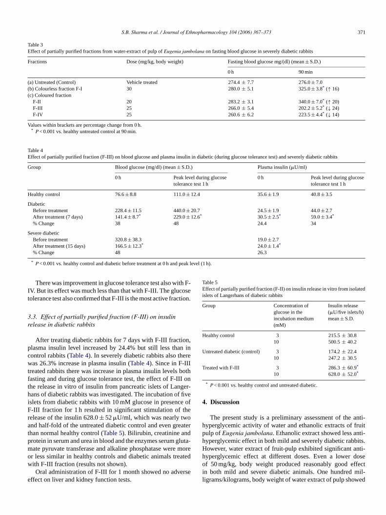
S.B. Sharma et al. / Journal of Ethnopharmacology 104 (2006) 367–373 371
Table 3Effect of partially purified fractions from water-extract of pulp of Eugenia jambolana on fasting blood glucose in severely diabetic rabbits
Fractions Dose (mg/kg, body weight) Fasting blood glucose mg/(dl) (mean ± S.D.)
0 h 90 min
(a) Untreated (Control) Vehicle treated 274.4 ± 7.7 276.0 ± 7.0(b) Colourless fraction F-I 30 280.0 ± 5.1 325.0 ± 3.8* (↑ 16)(c) Coloured fraction
F-II 20 283.2 ± 3.1 340.0 ± 7.0* (↑ 20)F-III 25 266.0 ± 5.4 202.2 ± 5.2* (↓ 24)F-IV 25 260.6 ± 6.2 223.5 ± 4.4* (↓ 14)
Values within brackets are percentage change from 0 h.* P < 0.001 vs. healthy untreated control at 90 min.
Table 4Effect of partially purified fraction (F-III) on blood glucose and plasma insulin in diabetic (during glucose tolerance test) and severely diabetic rabbits
Group Blood glucose (mg/dl) (mean ± S.D.) Plasma insulin (�U/ml)
0 h Peak level during glucosetolerance test 1 h
0 h Peak level during glucosetolerance test 1 h
Healthy control 76.6 ± 8.8 111.0 ± 12.4 35.6 ± 1.9 40.8 ± 3.5
DiabeticBefore treatment 228.4 ± 11.5 440.0 ± 20.7 24.5 ± 1.9 44.0 ± 2.7After treatment (7 days) 141.4 ± 8.7* 229.0 ± 12.6* 30.5 ± 2.5* 59.0 ± 3.4*
% Change 38 48 24.4 34
Severe diabeticBefore treatment 320.8 ± 38.3 19.0 ± 2.7After treatment (15 days) 166.5 ± 12.3* 24.0 ± 1.4*
% Change 48 26.3
* P < 0.001 vs. healthy control and diabetic before treatment at 0 h and peak level (1 h).
There was improvement in glucose tolerance test also with F-IV. But its effect was much less than that with F-III. The glucosetolerance test also confirmed that F-III is the most active fraction.
3.3. Effect of partially purified fraction (F-III) on insulinrelease in diabetic rabbits
After treating diabetic rabbits for 7 days with F-III fraction,plasma insulin level increased by 24.4% but still less than incontrol rabbits (Table 4). In severely diabetic rabbits also therewas 26.3% increase in plasma insulin (Table 4). Since in F-IIItreated rabbits there was increase in plasma insulin levels bothfasting and during glucose tolerance test, the effect of F-III onthe release in vitro of insulin from pancreatic islets of Langer-hans of diabetic rabbits was investigated. The incubation of fiveislets from diabetic rabbits with 10 mM glucose in presence ofF-III fraction for 1 h resulted in significant stimulation of therelease of the insulin 628.0 ± 52 �U/ml, which was nearly twoand half-fold of the untreated diabetic control and even greaterthan normal healthy control (Table 5). Bilirubin, creatinine andprotein in serum and urea in blood and the enzymes serum gluta-mate pyruvate transferase and alkaline phosphatase were moreor less similar in healthy controls and diabetic animals treatedwith F-III fraction (results not shown).
Oral administration of F-III for 1 month showed no adverseeffect on liver and kidney function tests.
Table 5Effect of partially purified fraction (F-II) on insulin release in vitro from isolatedislets of Langerhans of diabetic rabbits
Group Concentration ofglucose in theincubation medium(mM)
Insulin release(�U/five islets/h)mean ± S.D.
Healthy control 3 215.5 ± 30.810 500.5 ± 40.2
Untreated diabetic (control) 3 174.2 ± 22.410 247.2 ± 30.5
Treated with F-III 3 286.3 ± 60.9*
10 628.0 ± 52.0*
* P < 0.001 vs. healthy control and untreated diabetic.
4. Discussion
The present study is a preliminary assessment of the anti-hyperglycemic activity of water and ethanolic extracts of fruitpulp of Eugenia jambolana. Ethanolic extract showed less anti-hyperglycemic effect in both mild and severely diabetic rabbits.However, water extract of fruit-pulp exhibited significant anti-hyperglycemic effect at different doses. Even a lower doseof 50 mg/kg, body weight produced reasonably good effectin both mild and severe diabetic animals. One hundred mil-ligrams/kilograms, body weight of water extract of pulp showed

372 S.B. Sharma et al. / Journal of Ethnopharmacology 104 (2006) 367–373
maximum effect. Higher dose of 200 mg/kg, body weight did notshow any significant increase in activity. Achrekar et al. (1991)reported that water extract of pulp of Eugenia jambolana showshypoglycemic effect 30 min after its administration. However, inthe present study, we observed hypoglycemic activity at 30 min(results not shown) but maximum effect was seen at 90 min afteradministration. This might mean that the drug takes time to reachthe target tissues in the body or it gets metabolized and themetabolite(s) is/are active.
In diabetic rabbits tolbutamide was used as positive con-trol of known drug because this hypoglycemic agent requiresthe presence of functioning �-cells of islets of Langerhans.It is interesting that water extract (100 mg/kg, body weight)of fruit pulp was more effective than tolbutamide (250 mg/kg,body weight). Since water extract of fruit pulp showed activ-ity even in severe diabetic rabbits in which most of the islet� cells are damaged, it is likely that water extract of pulpis also having some direct effect on the tissue utilization ofglucose. Hot water extract of pulp of Eugenia jambolanaadministered at different doses to alloxan-induced diabetic rab-bits was found inactive. This indicates that the active anti-hyperglycemic compound(s) of Eugenia jambolana is (are)heat labile. When water extract of fruit-pulp was further sub-jected to purification by column chromatography, four dif-ferent fractions having effect on fasting blood glucose levelswere obtained. Out of these, two fractions (colourless frac-tion F-I and coloured fraction F-II) showed hyperglycemiceffect while F-III and F-IV (both coloured) showed hypo-glycemic activity. Out of these, F-III fraction showed signif-icant and maximum antihyperglycemic activity in mild andsevere diabetic rabbits. F-IV fraction showed lower activity.A point of interest is that two hyperglycemic compounds (F-Iand F-II) were also present along with hypoglycemic com-pounds. Similar results have been reported earlier by us withsome other plants like Momordica charantia, Trigonella foenumgraecum, Ficus bengalensis, etc. (Shukla et al., 2000). Thepresent results with Eugenia jambolana substantiate our ear-lier view that both hypoglycemic and hyperglycemic com-pounds may be present in some plants. Therefore, unpuri-fied extract of such plants do not give the optimum effect.However, for the desired therapeutic potential of the activeingredient, the separation of hyperglycemic compound isessential.
Results on the plasma insulin release from pancreas directlyindicate that part of the antihyperglycemic activity of F-IIIis through release of insulin from the pancreas i.e. it exertsa direct insulinotropic effect. Achrekar et al. (1991) reportedthat water extract of pulp of Eugenia jambolana stimulatesrelease of insulin both in in vivo and in vitro studies. Ourpresent study showed that even the partially purified waterextract acts by increasing release of insulin many folds prob-ably through �-cells stimulation like some of the sulphony-lureas such as tolbutamide. Bansal et al. (1981) reported thatthe increase in plasma insulin brought by seeds of Eugeniajambolana may be attributed to proinsulin to insulin conver-sions, possibly by pancreatic cathapsin B, and/or its secretion.The diabetics have greater insulinase activity than non-diabetics
(Achrekar et al., 1991). The inhibition of insulinase activ-ity from liver and kidney by extract of Eugenia jambolana,which has been reported (Achrekar et al., 1991) points to anextrapancreatic mechanism of action also. No toxic effect wasobserved after 1-month treatment as evident from normal liverand renal function tests. Further studies, regarding purificationof partially purified compound (F-III) along with metabolicstudies are in progress to understand the exact mechanism ofaction.
Acknowledgments
The authors thank Director-General, Indian Council of Med-ical Research, New Delhi (India), for providing financial assis-tance during study.
References
Achrekar, S., Kaklij, G.S., Pote, M.S., Kelkar, S.M., 1991. Hypoglycemicactivity of Eugenia jambolana and Ficus bengalensis: mechanism ofaction. In vivo 5, 133–148.
Bansal, R., Ahmad, N., Kidwai, J.R., 1981. Effect of oral administration ofEugenia jambolana seeds and chlorpropamide on blood glucose level andpancreatic cathepsin B in rats. Indian J. Biochem. Biophys. 18, 377–381.
Bergmeyer, H.U., Scheibe, P., Wahlefeld, A.W., 1978. Optimization of meth-ods for aspartate amino transferase and alanine amino transferase. Clin.Chem. 24, 58–73.
Bowers, G.N., McComb, R.B., 1966. A Continuous spectrophotometricmethod for measuring the activity of serum alkaline phosphatase. Clin.Chem. 12, 70–89.
Braham, D., Tinder, P., 1972. Estimation of glucose. Glucose oxidase method.Analyst 97, 142–144.
Chopra, R.N., Chopra, I.C., Handa, K.L., Kapur, L.D., 1958. IndigenousDrugs of India, 2nd ed. U.N. Dhur and Sons Pvt. Ltd., Calcutta, pp.686–689.
Cook, J.G.H., 1975. Application of analytical methods of automatic analysers.Annal. Clin. Biochem. 12, 163–168.
Grover, J.K., Vats, V., Rathi, S.S., 2000. Antihyperglycemic effect of Eugeniajambolana and tinospora cordifolia in experimental diabetes and theireffects on key metabolic enzymes involved in carbohydrate metabolism.J. Ethnopharmacol. 73, 461–470.
Kemp, C.B., Knight, M.J., Scharp, D.W., Lacy, P.E., Ballinger, W.F., 1973.Transplantation of isolated pancreatic islets into the portal vein of diabeticrats. Nature 244, 477–481.
Kohli, K.R., 1983. A study on Kriyakala of diabetes mellitus and its treat-ment with Eugenia jambolana MD (Ay). Thesis submitted at Institute ofMedical Sciences, Banaras Hindu University, Varanasi (India).
Lowry, O.H., Rosebrough, N.J., Farr, A.L., Randall, R.J., 1851. Protein mea-surement with Folin phenol reagent. J. Biol. Chem. 193, 265–275.
Malloy, H.T., Evelyn, K.A., 1937. The determination of bilirubin with thephotoelectric colorimeter. J. Biol. Chem. 119, 473–480.
Nadkarni, A.K., 1954. Indian Materia Medica, vol. 1. Popular Prakashan,Bombay, pp. 1331.
Sharma, S.B., Nasir, A., Prabhu, K.M., Dev, G., Murthy, P.S., 2003. Hypo-glycemic and hypolipidemic effect of ethanolic extracts of seeds of E.jambolana in alloxan-induced diabetic model of rabbits. J. Ethnopharma-col. 85, 201–206.
Shrotri, D.S., Kelkar, M., Deshmukh, V.K., Aiman, R., 1963. Investigationsof the hypoglycemic properties of Vinca rosea, Cassia auriculata andEugenia jambolana. Indian J. Med. Res. 51, 464–467.
Shukla, R., Anand, K., Prabhu, K.M., Murthy, P.S., 1994. Hypoglycamiceffect of water extract of Ficus bengalensis in alloxan recovered, mildlydiabetic and severely diabetic rabbits. Int. J. Diab. Dev. Countries 14,78–81.

S.B. Sharma et al. / Journal of Ethnopharmacology 104 (2006) 367–373 373
Shukla, R., Sharma, S.B., Puri, D., Prabhu, K.M., Murthy, P.S., 2000. Medic-inal plants for the treatment of diabetes. Review article. Indian J. Clin.Biochem. 15, 169–177.
Tager, H.S., Rubenstein, A.H., Steiner, D.F., 1975. Methods for assessmentof peptide precursors. Studies on insulin biosynthesis. Meth. Enzymol.37, 326–344.
Vikrant, V., Grover, J.K., Tandon, N., Rathi, S.S., Gupta, N., 2001. Treatmentwith extracts of Momordica charantia and Eugenia jambolana preventshyperglycemia and hyperinsulinemia in fructose fed rats. J. Ethnophar-macol. 76, 139–143.
Journal of Ethnopharmacology 104 (2006) 374–378
Huperzia saururus, activity on synaptic transmission in the hippocampus
M.G. Ortega a, M.G. Vallejo a, J.L. Cabrera a, M.F. Perez b,R.S. Almiron b, O.A. Ramırez b, A.M. Agnese a,∗
a Farmacognosia, Departamento de Farmacia, Facultad de Ciencias Quımicas, Universidad Nacional de Cordoba,Instituto Multidisciplinario de Biologıa Vegetal (IMBIV, UNC-CONICET) Ciudad Universitaria, (5000) Cordoba, Argentina
b Farmacologıa, Departamento de Farmacologıa, Facultad de Ciencias Quımicas, Universidad Nacional de Cordoba,Medina Allende esquina Haya de la Torre, Ciudad Universitaria, (5000) Cordoba, Argentina
Received 22 September 2004; received in revised form 25 June 2005; accepted 1 November 2005Available online 1 December 2005
Abstract
Huperzia saururus (Lam.) Trevis. (Lycopodiaceae) known as cola de quirquincho is used in folk medicine to improve memory. The cholinergicneurons of the basal forebrain, including those in the medial septum, and in the vertical limbs of the diagonal band of Broca and the nucleus basalisof Meynert, provide a major source of cholinergic enervation of the cortex and hippocampus. These neurons have also been shown to play animportant role in learning and memory processes. Thus, the effects of this traditional Argentinean species were studied in relation to its activity onsynaptic transmission in the hippocampus. The alkaloid extract obtained first by decoction of the aerial parts and by subsequent alkaline extraction,was purified by using a Sephadex LH 20 packed column.
Electrophysiological experiments were developed with the purified extract (E2) on rat hippocampus slices, thus eliciting long-term potentiation(LTP). Results show a marked increase in the hippocampal synaptic plasticity. The threshold value for generation of LTP was 22 ± 1.01 Hz onaverage for E2, while for controls it was 86 ± 0.92 Hz. All of these factors could explain the use of Huperzia saururus as a memory improver as isreported in the ethnomedicine.© 2005 Elsevier Ireland Ltd. All rights reserved.
Keywords: Huperzia saururus; Lycopodium alkaloids; Memory and learning; Electrophysiological assays
1. Introduction
Aerial parts of Huperzia saururus (Lam.) Trevis. (syn.Lycopodium saururus Lam., syn. Urostachys saururus (Lam.)Herter) (Lycopodiaceae) have long been used in folk medicinemainly as an aphrodisiac (Amorin, 1974; de la Sota, 1977; Rateraand Ratera, 1980) and moreover it is believed to improve mem-ory (Martinez Crovetto, 1981). It is consumed as an infusion andalso as a decoction. Depending on the concentration, the latter
Abbreviations: E2, purified alkaloid extract; LTP, Long-Term Potentia-tion; AD, Alzheimer’s disease; NaOH, sodium hydroxide; CHCl3, chloroform;MeOH, methanol; H2O, water; E1, crude total alkaloid extract; GLC-MS, gasliquid chromatography-mass spectrum; NaCl, sodium chloride; KCl, potassiumchloride; MgSO4, magnesium sulphate; HKPO4, potassium phosphate monoba-sic; HNaCO3H, sodium bicarbonate; CaCl2, calcium chloride; Ctrol, control;fEPSP, field excitatory postsynaptical potential; GDS, global deterioration scale
∗ Corresponding author. Tel.: +54 351 4334163; fax: +54 351 4334127.E-mail address: [email protected] (A.M. Agnese).
can produce severe adverse effects such as vomiting, diarrhea,convulsions and even death (Amorin, 1974; Ratera and Ratera,1980; Toursarkissian, 1980).
Results of previous studies on Huperzia saururus showedthat its main constituents are alkaloids; sauroine, a novel alka-loid (Ortega et al., 2004a), sauroxine, 6-hydroxylycopodine,N-acetyllycodine, lycopodine, lycodine, N-methyllycodine, andclavolonine were isolated and identified (Ortega et al., 2004b)(Fig. 1). It has also been demonstrated that the alkaloid extracthas a marked anticholinesterase activity with a selectivity ontrue acetylcholinesterase (Ortega et al., 2004b). All the alka-loids that occur in Huperzia saururus belong to Lycopodiumalkaloids. This group forms a special kind of alkaloid foundonly in some restricted genera. For all the Lycopodiumalkaloids, only a few of them have been demonstrated to beacetylcholinesterase inhibitors. Among them, Huperzine A, firstisolated from Huperzia serrata has this effect (Liu et al., 1986)and in addition improves learning and memory (Vincent et al.,1987).
0378-8741/$ – see front matter © 2005 Elsevier Ireland Ltd. All rights reserved.doi:10.1016/j.jep.2005.11.002

M.G. Ortega et al. / Journal of Ethnopharmacology 104 (2006) 374–378 375
Fig. 1. Lycopodium alkaloids present in the Huperzia saururus purified extract(E2).
Alzheimer’s disease (AD) is characterized by loss of mem-ory, functional decline and behavioral disturbance (AmericanPsychiatry Association, 1987). AD is characterized by earlycholinergic neuronal loss in a more consistent way than forother systems (Katzman, 1986). However, many neurotrans-mitter systems also being affected (Perry and Perry, 1985). Asthe cholinergic function is required for short-term memory pro-cesses, it is believed that the cholinergic deficit in AD is alsoresponsible for much of the short-term memory deficit (Galizia,1984; Jarvik et al., 1972).
At the present time, it is known that acetylcholinesteraseinhibitors give temporary cognitive benefit to a percentageof AD patients (McGeer and McGeer, 2003). The fore braincholinergic system is very important in memory functions.Furthermore, there is evidence that the septo-hippocampalpathway is involved in short-term learning and memory pro-cesses. The hippocampal long-term potentiation (LTP) hastherefore been proposed as a potential neuronal mecha-nism for memory storage (Bliss and Lomo, 1973; Blissand Colingridge, 1993). Considering all these antecedents,the aim of the present study was to determine if the puri-fied alkaloid extract of Huperzia saururus elicits and main-tains LTP in hippocampus rat slices measured by electro-physiological assays, and to attempt to correlate the resultsobtained to the supposed effects on memory claimed by eth-nomedicine.
2. Materials and methods
2.1. Plant material
Plant material was collected in Pampa de Achala, Depar-tamento San Alberto, province of Cordoba, in November 2001,and identified by Dr. Gloria Barboza, Instituto Multidisciplinariode Biologıa Vegetal, Universidad Nacional de Cordoba. Avoucher specimen is deposited at CORD as Altamirano No. 684.
2.2. Extraction and purification
In order to obtain a similar extract to that which people con-sume, the following method was used to obtain the extract. Theaerial parts of Huperzia saururus (500 g) were dried, groundand then decocted with boiling water (2 l) for 1 h. This processwas repeated twice. Filtered and combined decoctions were con-centrated under reduced pressure to reduce the volume, and thenalkalinized to pH 12 by the addition of 0.1N NaOH. The alkalineaqueous extract was partitioned with CHCl3 in a liquid-liquidextractor. The chloroformic extract was evaporated until dry inorder to obtain 1.56 g of the crude total alkaloid extract E1 (%w/w, 0.31; % w/w, calculated relative to dry starting material).
E1 was chromatographed over Sephadex LH-20 in a glasscolumn using MeOH/H2O (1:1) as the mobile phase. All the frac-tions resulting positive to Dragendorff’s reagent were combinedand evaporated under reduced pressure to yield the alkaloid puri-fied extract E2 (0.75 g; 0.15 % w/w).
2.3. Chemical composition
In order to separate and identify the components presentin E2 this extract was submitted to GLC-MS (Ortega et al.,2004a). By using a Perkin-Elmer Qmass-910 apparatus, and acapillary column SE 30, 30 m in length, E2 analysis was per-formed. The injection volume was 0.5 �l with He as carrier,with the flow rate being 1 ml/min. Temperature program: 140 ◦C(3 min), 140–250 ◦C at 5 ◦C/min, 250 ◦C (5 min), 250–280 ◦C at5 ◦C/min, 280 ◦C (5 min). The temperatures of injector, interfaceand ion source were 300, 280, and 280 ◦C respectively. The ion-ization energy was 70 eV and individual alkaloid identificationswere made by comparing breakdown patterns with those foundin the literature (MacLean, 1963; Alam et al., 1964; Ayer et al.,1965; Loyola et al., 1979; Sun et al., 1993; Ortega et al., 2004b).
Thus, the presence of sauroine, 6-hydroxylycopodine,lycopodine, and clavolonine (Lycopodane group) and saurox-ine, lycodine, N-methyllycodine, and N-acetyllycodine (Flabel-lidane group), was confirmed, along with other alkaloid com-pounds whose structures have not yet been determined.
2.4. Animals
Male Wistar rats 65–70 days old and weighing 190–260 gwere used. Animals were housed in groups of five in their homeboxes and kept under a 12 h-light/12 h-dark cycle (lights on at 7a.m.) and regular temperature conditions (22 ± 1 ◦C). Food andwater were available ad libitum.
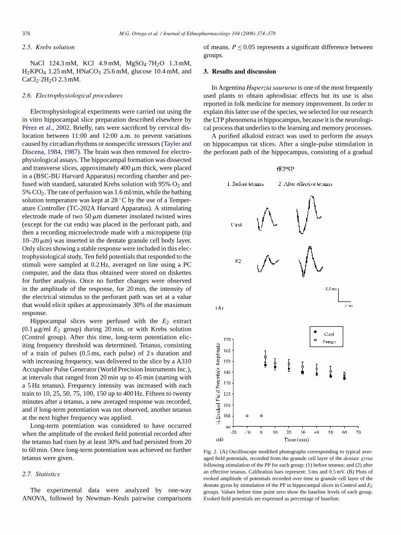
376 M.G. Ortega et al. / Journal of Ethnopharmacology 104 (2006) 374–378
2.5. Krebs solution
NaCl 124.3 mM, KCl 4.9 mM, MgSO4·7H2O 1.3 mM,H2KPO4 1.25 mM, HNaCO3 25.6 mM, glucose 10.4 mM, andCaCl2·2H2O 2.3 mM.
2.6. Electrophysiological procedures
Electrophysiological experiments were carried out using thein vitro hippocampal slice preparation described elsewhere byPerez et al., 2002. Briefly, rats were sacrificed by cervical dis-location between 11:00 and 12:00 a.m. to prevent variationscaused by circadian rhythms or nonspecific stressors (Tayler andDiscena, 1984, 1987). The brain was then removed for electro-physiological assays. The hippocampal formation was dissectedand transverse slices, approximately 400 �m thick, were placedin a (BSC-BU Harvard Apparatus) recording chamber and per-fused with standard, saturated Krebs solution with 95% O2 and5% CO2. The rate of perfusion was 1.6 ml/min, while the bathingsolution temperature was kept at 28 ◦C by the use of a Temper-ature Controller (TC-202A Harvard Apparatus). A stimulatingelectrode made of two 50 �m diameter insolated twisted wires(except for the cut ends) was placed in the perforant path, andthen a recording microelectrode made with a micropipette (tip10–20 �m) was inserted in the dentate granule cell body layer.Only slices showing a stable response were included in this elec-trophysiological study. Ten field potentials that responded to thestimuli were sampled at 0.2 Hz, averaged on line using a PCcomputer, and the data thus obtained were stored on diskettesfor further analysis. Once no further changes were observedin the amplitude of the response, for 20 min, the intensity ofthe electrical stimulus to the perforant path was set at a valuethat would elicit spikes at approximately 30% of the maximumresponse.
Hippocampal slices were perfused with the E2 extract(0.1 �g/ml E2 group) during 20 min, or with Krebs solution(Control group). After this time, long-term potentiation elic-iting frequency threshold was determined. Tetanus, consistingof a train of pulses (0.5 ms, each pulse) of 2 s duration andwith increasing frequency, was delivered to the slice by a A310Accupulser Pulse Generator (World Precision Instruments Inc.),at intervals that ranged from 20 min up to 45 min (starting witha 5 Hz tetanus). Frequency intensity was increased with eachtrain to 10, 25, 50, 75, 100, 150 up to 400 Hz. Fifteen to twentyminutes after a tetanus, a new averaged response was recorded,and if long-term potentiation was not observed, another tetanusat the next higher frequency was applied.
Long-term potentiation was considered to have occurredwhen the amplitude of the evoked field potential recorded afterthe tetanus had risen by at least 30% and had persisted from 20to 60 min. Once long-term potentiation was achieved no furthertetanus were given.
2.7. Statistics
The experimental data were analyzed by one-wayANOVA, followed by Newman–Keuls pairwise comparisons
of means. P ≤ 0.05 represents a significant difference betweengroups.
3. Results and discussion
In Argentina Huperzia saururus is one of the most frequentlyused plants to obtain aphrodisiac effects but its use is alsoreported in folk medicine for memory improvement. In order toexplain this latter use of the species, we selected for our researchthe LTP phenomena in hippocampus, because it is the neurologi-cal process that underlies to the learning and memory processes.
A purified alkaloid extract was used to perform the assayson hippocampus rat slices. After a single-pulse stimulation inthe perforant path of the hippocampus, consisting of a gradual
Fig. 2. (A) Oscilloscope modified photographs corresponding to typical aver-aged field potentials, recorded from the granule cell layer of the dentate gyrusfollowing stimulation of the PP for each group: (1) before tetanus; and (2) afteran effective tetanus. Calibration bars represent: 5 ms and 0.5 mV. (B) Plots ofevoked amplitude of potentials recorded over time in granule cell layer of thedentate gyrus by stimulation of the PP in hippocampal slices in Control and E2
groups. Values before time point zero show the baseline levels of each group.Evoked field potentials are expressed as percentage of baseline.

M.G. Ortega et al. / Journal of Ethnopharmacology 104 (2006) 374–378 377
Fig. 3. Threshold to induce LTP in hippocampal dentate gyrus in Control andE2 groups. Each bar represents the mean and vertical bars ± SME. The numberof animals used in each group is indicated between parentheses. (*) P < 0.0004compared to Control group.
positive-going field excitatory postsynaptic potential (fEPSP),a characteristic evoked field response is produced in the gran-ule cell layer of the dentate gyrus. This can be seen as anincrease in the amplitude after an effective tetanus for the control(Ctrol) as well as for E2 groups, in Fig. 2A. The fEPSP reflectssynaptic currents at perforant path-dentate granule cell synapsesin stratum moleculare. In Fig. 2B the increased amplitude offEPSP over time can be seen, after an effective tetanus for Ctroland E2 groups expressed as a percentage of basal fEPSP. Thepotentiation levels are comparable and there were no statisticaldifferences between groups.
A one-way ANOVA was used to evaluate the differences inthreshold in order to induce LTP for Ctrol and E2 groups. A sig-nificant interaction was revealed (F(1,225) = 45.5.; P < 0.0002)among the effects in E2 perfusion and Ctrol. Newman–Keulspairwise comparisons of means test showed that when sliceswere perfused with E2 an increment in the hippocampal synap-tic plasticity was observed, measured as a diminution in thethreshold necessary to generate LTP, when taking into consid-eration that P < 0.5 represented a significant difference betweengroups. Thus, the E2 perfused slices (n = 5) showed an averagethreshold of 22 ± 1.01 Hz, while for Ctrol (n = 5) the averagewas 86 ± 0.92 Hz (Fig. 3).
It is important to point out that nowadays the treatment ofAD is based on cholinesterase inhibitors, especially for the lightto moderately-serious stage (4–6 of Global Deterioration Scale,GDS) of Reisberg et al. (1982). There are few inhibitors autho-rized to be commercialized for this purpose, so the discovery ofa new source such as Huperzia saururus that besides being anacetilcholinesterase inhibitor (Ortega et al., 2004a) also shows amodification in the threshold for LTP generation, is important.All of this indicates that a new drug of natural origin could be apotential aid in AD treatment as well as in physiological ageing.
4. Conclusions
Long-term potentiation (LTP) of synaptic transmission is arelevant phenomenon and is seemingly linked to neural informa-
tion storage (Douglas and Goddard, 1975). In the hippocampalformation, LTP can be produced by repetitive activation of affer-ent pathways (Harris et al., 1984; Lomo, 1971).
The results obtained so far show that the perfusion of hip-pocampal rat slices with the purified alkaloid extract of Huperziasaururus facilitates the LTP production. Thus, we have demon-strated for the first time that Huperzia saururus has a markedeffect on the hippocampal synaptic plasticity. This effect, shownby an increase in plasticity that it is manifested as a diminu-tion in the threshold, could explain the memory improvementeffect claimed for Huperzia saururus by folk medicine. In viewof the fact that the assayed extract contains Lycopodium alka-loids, and that there exists the antecedent of alkaloids such asHuperzine A improving memory as, it can be said that one ormore of the alkaloids present is/are responsible for the actionhere demonstrated. More behavioral and biochemical studieswill be necessary to clarify, which the active compounds are,and to identify the action mechanism through which they per-form their action.
Acknowledgments
The authors wish to thank Agencia Cordoba Ciencia, SeCyT(UNC), and FONCYT for financial support, Prof. Dr. GloriaBarboza for the identification of the species under study, andDr. Paul Hobson, native speaker, for revision of the manuscript.
References
Alam, S.N., Adams, A.H., MacLean, D.B., 1964. Lycopodium alkaloids XV.Structure and mass spectra of some minor alkaloids of L. flabelliforme.Canadian Journal Chemistry 42, 2456–2466.
American Psychiatry Association, 1987. Diagnostic and Statistical Manual ofMental Disorders, third ed., revised (DSM-III-R). American PsychiatryAssociation, Washington DC.
Amorin, J.L., 1974. Cola de Quirquincho Urostachis saururus (Lam.) Herter(Lycopodiaceas) Una peligrosa planta usada en la medicina popularArgentina. Farmacobotanica 16, 3–6.
Ayer, W., Habgood, T., Deulofeu, V., Juliani, H., 1965. Lycopodium alkaloids.Sauroxine. Tetrahedron 21, 2169–2172.
Bliss, T.V.P., Colingridge, G.L., 1993. A synaptic model of memory: longterm potentiation in the hippocampus. Nature (London) 361, 31–39.
Bliss, T.V.P., Lomo, T., 1973. Long lasting potentiation of synaptic transmis-sion in the dentate area of the anesthetized rabbit following stimulationof the perforant path. The Journal of Physiology (London) 232, 331–356.
de la Sota, E.R., 1977. Pteridophyta. In: Cabrera, A.L., (Ed.), Flora de laProvincia de Jujuy. INTA, Bs. As., pp. 19–27.
Douglas, R.M., Goddard, G.V., 1975. Long-term potentiation of the perforantpath-granule cells synapse in the rat hippocampus. Brain Research 85,205–215.
Galizia, V.J., 1984. Pharmacotherapy of memory loss in the geriatric patient.Drug Intelligence & Clinical Pharmacy 18, 784–791.
Harris, E.W., Ganong, A.H., Cotman, C.W., 1984. Long-term potentiation inthe hippocampus involves activation of N-methyl-d-aspartate receptors.Brain Research 323, 132–137.
Jarvik, M.E., Gritz, E.R., Schneider, N.G., 1972. Drug and memory disordersin human aging. Behavioral Biology 7, 643–668.
Katzman, R., 1986. Alzheimer’s disease. New England Journal of Medicine314, 964–973.
Liu, J.S., Zhu, Y.L., Yu, C.M., Zhou, Y.Z., Han, Y.Y., Wu, F.W., Qi, B.F.,1986. The structures of huperzine A and B, two new alkaloids exhibit-

378 M.G. Ortega et al. / Journal of Ethnopharmacology 104 (2006) 374–378
ing marked anticholinesterase activity. Canadian Journal Chemistry 64,837–839.
Lomo, T., 1971. Potentiation of monosynaptic EPSPs in the perforant path-dentate granule-cells synapse. Experimental Brain Research 12, 46–67.
Loyola, L.A., Morales, G., Castillo, M., 1979. Alkaloids of Lycopodium mag-ellanicum. Phytochemistry 18, 1721–1723.
Martinez Crovetto, R., 1981. Las Plantas utilizadas en medicina popular.Miscelanea 69, 15.
McGeer, E.G., McGeer, P.L., 2003. Clinical tested drugs for Alzheimer’sdisease. Expert Opinion on Investigational Drugs 12, 1143–1151.
MacLean, D.B., 1963. Lycopodium alkaloids XIII. Mass spectra of represen-tative alkaloids. Canadian Journal Chemistry 41, 2654–2670.
Ortega, M.G., Agnese, A.M., Cabrera, J.L., 2004a. Sauroine-a novelLycopodium alkaloid from Huperzia saururus. Tetrahedron Letters,7003–7005.
Ortega, M.G., Agnese, A.M., Cabrera, J.L., 2004b. Anticholinesterase activityin an alkaloid extract of Huperzia saururus. Phytomedicine 11, 539–543.
Perez, M.F., Maglio, L.E., Marchesini, G.R., Molina, J.C., Ramirez, O.A.,2002. Environmental changes modify the expression of diazepam with-drawal. Behavioral Brain Research 136, 75–81.
Perry, E.K., Perry, R.H., 1985. A review of neuropathological and neuro-chemical correlates of Alzheimer’s disease. Danish Medical Bulletin 32S,27–34.
Ratera, E.L., Ratera, M.O., 1980. Plantas de la flora Argentina empleadasen medicina popular. Editorial Hemisferio Sur S.A., Buenos Aires,p. 67.
Reisberg, B., Ferris, S.H., de Leon, M.J., Crook, T., 1982. The Global Deteri-oration Scale for assessment of primary degenerative dementia. AmericanJournal of Psychiatry 139, 1136–1139.
Sun, C.M., Ho, L.K., Sun, M.L., 1993. Revised Stereochemistry of So-called isoselagine and spectroscopic analysis of 6�-hydroxylycopodinefrom Lycopodium serratum var. longipetiolatum. Planta Medica 59, 467–471.
Tayler, T.J., Discena, P., 1984. Long-term potentiation as a candidatemnemonic device. Brain Research Reviews 7, 15–28.
Tayler, J., Di Scenna, P., 1987. Long-term potentiation. Annual Review ofNeuroscience 10, 131–161.
Toursarkissian, M., 1980. Plantas Medicinales de la Argentina. Sus nombresbotanicos, vulgares, usos y distribution geografica. Editorial HemisferioSur S.A., Buenos Aires, p. 80.
Vincent, G.P., Rummennik, L., Cumin, R., Martin, J., Sepinwall, J., 1987. Theeffects of huperzine A, an acetylcholinesterase inhibitor, on the enhance-ment of memory in mice, rats and monkeys. Neuroscience Abstracts 13,844.

Journal of Ethnopharmacology 104 (2006) 379–386
Tissue lipid lowering-effect of a traditional Nigerian anti-diabeticinfusion of Rauwolfia vomitoria foilage and Citrus aurantium fruit
Joan I.A. Campbell a, Alicja Mortensen b, Per Mølgaard a,∗a Department of Medicinal Chemistry, Pharmacognosy Group, The Danish University of Pharmaceutical Sciences,
Universitetsparken 2, DK 2100 Copenhagen East, Denmarkb Danish Institute for Food and Veterinary Research, Mørkhøj Bygade 19, DK-2860 Søborg, Denmark
Received 2 February 2005; received in revised form 30 November 2005; accepted 15 December 2005Available online 7 February 2006
Abstract
The toxicity and anti-diabetic properties of an aqueous plant extract made by boiling Rauwolfia vomitoria foilage and Citrus aurantium fruitswere evaluated in mice. A single dosage corresponding to 70× the human-daily-dose was non-toxic when administered to 6-week-old NMRI leanmice or 6- or 11-week-old C57BL/6J lean mice. Daily treatment of 11-week-old C57BL/KsBom-db (db/db) genetic diabetic mice with a dosecorresponding to 10× human-daily-dose for 6 weeks facilitated a significant weight loss as compared to the untreated controls. During treatment, thedb/db mice were maintained on the carbohydrate-deficient Altromin C1009 diet. Although the food intake in the treated mice was not statisticallysignificant from that in the controls, the treated animals had significantly higher serum triglyceride contents, suggesting that the treatment inducedlipid mobilization from internal stores. Moreover, the fatty acid profile of the eyes from the treated animals showed a significant reduction in totalfatty acid content accompanied by a 33% reduction in estimated Stearoyl-CoA desaturase activity (p = 0.039) as compared with controls. The fattyacid mobilization and a protection of the brittle C57BL/KsBom-db pancreas were observed 5 weeks after cessation of treatment when the treatedanimals were maintained on the poorer Altromin C1009 diet.© 2006 Elsevier Ireland Ltd. All rights reserved.
Keywords: Anti-diabetes; Rauwolfia vomitoria; Citrus aurantium; Type II diabetes; Anti-inflammation; Lipid-lowering
1. Introduction
Diabetes mellitus is primarily a disturbance of the body’scarbohydrate and lipid metabolism. The WHO has estimatedthat 2–10 out of every 100 people will develop the condi-tion during their lifetime and that 90% of these will be ofthe type 2, late onset non-insulin-dependent diabetes melli-tus (NIDDM, Nathan, 1993). In NIDDM, the insulin stimu-lated glucose uptake and utilization in liver, skeletal muscleand adipose tissue is impaired (Zimmet, 1982). The defectscan be improved by caloric restriction, exercise and for somepatients, recourse to a life-long course of hypoglycemic ther-apy. In the later stages of the disease where these mea-sures have failed, the patients are given incremental doses
∗ Corresponding author at: Department of Medicinal Chemistry, Danish Uni-versity of Pharmaceutical Sciences, Universitetsparken 2, DK 2100 Copen-hagen, Denmark. Tel.: +45 35 30 63 35; fax: +45 35 30 60 41.
E-mail address: [email protected] (P. Mølgaard).
of insulin. However, none of the currently available type IIdiabetes management regimes prevent the long term compli-cations such as blindness, kidney failure or cardiac diseasethat are known to be associated with the disease. In addition,problems with the efficacy and the long term usage of thecurrently approved sulphonamides, biguanidines (Turner andClapham, 1998) and thiazolidinediones (Kohlroser et al., 2000)have prompted the interest in the scientific evaluation of tradi-tional herbal medicines in the hope of finding better anti-diabeticdrugs.
The plant extract investigated in this study is made by boil-ing leaves and young stem of Rauwolfia vomitoria Afzel. (fam.Apocynaceae) with cut fruits of Citrus aurantium L. (fam.Rutaceae). In northern Edo state of Nigeria, the resulting infu-sion is claimed to have curative effects on late onset type IIdiabetes when taken in combination with a diet low in car-bohydrate and fat. Thus, during the treatment and afterwards,the patient is expected to adhere to a diet low in carbohy-drate and fat. In addition, abstinence from alcohol is alsoadvised.
0378-8741/$ – see front matter © 2006 Elsevier Ireland Ltd. All rights reserved.doi:10.1016/j.jep.2005.12.029

380 J.I.A. Campbell et al. / Journal of Ethnopharmacology 104 (2006) 379–386
Rauwolfia vomitoria occurs widely in coastal and inland partsof tropical West Africa. The species is a well-known medicinalplant used by different peoples in Africa as the major or minorpart of concoctions for treating various ailments (Sofowora,1982; Burkill, 1985). Rauwolfia vomitoria has been investigatedfor alkaloid content, especially for those with hypotensive andanti-inflammatory properties (Chatterjee and Bandyopadhyay,1979; Amer and Court, 1980; Kweifio-Okai, 1991). Citrusaurantium is reported as having a weight reduction effect whencombined with a strict diet (Preuss et al., 2002). In addition,Citrus aurantium has been shown to be radioprotective becauseit is rich in flavonoids with anti-oxidative activity (Hosseinimehret al., 2003). Our interview with six male ex-patients treated withthe Rauwolfia–Citrus infusion revealed that not only did gluco-suria cease, ulcers were healed, patients felt their body strengthreturn and impotency which they had experienced as a result ofdiabetes was cured. In this report, we present results of testingthe crude Rauwolfia–Citrus decoction as it is used traditionallyon the genetic diabetic inbred C57BL/KsBom-db mice in threedifferent trials assessing (i) the toxicity of the extract; (ii) thebiological activity as measured by the change in serum glucose,triglyceride and cholesterol, and in the fatty acid composition indiabetes relevant body organ; and (iii) the type 2-diabetes-curingeffect of the extract.
2. Materials and methods
2.1. Plant material and preparation of theRauwolfia–Citrus infusion
The leaves attached to young stems of Rauwolfia vomitoriaand fresh fruits of Citrus aurantium were collected in Auchi andIhievbe in northern Endo state of Nigeria (situated north of theequator between latitude 7◦ and longitude 6◦20′E). The foliagewas dried at low temperature in the shade. Voucher specimensof the foliage (herbarium) and fruit specimens stored in ethanolare kept at the Pharmacognosy Group of the Danish Univer-sity of Pharmaceutical Sciences. The washed dried foliage (totalweight, 400 g) was arranged in alternate layers with quarteredwhole citrus fruits (total wet weight 2 kg), in a large aluminiumpot. The plant material was then covered with 8 l of tap water,brought to the boil, and allowed to simmer covered, at low heatfor 1 h. The resulting golden coloured fluid was cooled to roomtemperature and filtered through coarse 1 mm× 1 mm pore fil-ters. The spent plant material was rinsed with 3 l of water, boiledas before and the resulting tea was also filtered and pooled withthe first lot and frozen at −20 ◦C. The total yield was typically7.5 l. The pooled plant extract was then freeze-dried and theyield was typically 12.5 g dried extract from 1 l crude extract.
2.2. Animal models and housing conditions
The experimental animals used for evaluating the toxic-ity of the Rauwolfia–Citrus extract were the lean 6 weeks(mean weight 22.6 ± 1.6 g) and 11 weeks old (mean weight30.3 ± 1.3 g) C57BL/6J inbred mice, as well as lean 6 weeks oldout-bred NMRI mice (mean weight 31.2 ± 2.9 g). Each group
consisted of 12 test and 12 control littermates. For the treatmentexperiments, the model animals used were the male geneticdiabetic C57BL/KsBom-db mice (db/db). At the start of thetreatment regime, the 10 test and 10 control littermates hadan average body weight of 41.9 ± 1.9 g. These mice are char-acterised by obesity, hyperphagia, temporal hyperinsulinaemia,hyperglycemia and degeneration of the pancreatic �-cells withage. Due to the insulin resistance observed in these mice, theyare considered models of type II non-insulin-dependent diabetes.The db gene has been identified as coding for one of the differentmice forms of leptin receptors expressed in the hypothalamus(Lee et al., 1996). Leptin is a hormone secreted by adipocytesthat has pleiotypic effects on a number of body systems includingreproduction and metabolism (Chehab et al., 1996). Specifically,a total deficiency in or resistance to leptin has been shown tocause severe obesity (Ahima et al., 1996).
All the animals were purchased from Bomholtgaard Breed-ing and Research Centre Ltd., Ry, Denmark (now Taconic M &B A/S, Ry, Denmark) and housed six mice per 30 cm × 45 cmmetal cages with wire-mesh tops under controlled environmentalconditions (temperature 21 ± 1 ◦C, relative humidity 55 ± 5%,with a light/dark cycle of 12 h each and air changes of 10 timesper h). The mice were allowed free access to food and water.Housing and caring conditions for the animals and the studyprotocols for the animal experiments were conducted underthe guidelines approved by the Danish Animal ExperimentalInspectorate.
2.3. Experimental design
2.3.1. Toxicity testsThe maximum human dose recommended for the Rauwolfia–
Citrus extract is three average drinking-glassfuls per day. On thebasis of an average volume of 750 ml per 75 kg body weight, wecalculated a 10 ml crude extract per kg body weight dosage forman. Allowing for an equivalency factor of 10 when extrapo-lating from human-daily-dose, the mouse-daily-dose should be100 ml/kg mice body weight. This compensates for the highermetabolic rate in mouse as compared to man. Dried extract(8.75 g) derived from 700 ml crude extract and correspondingto 70 times the human-daily-dose or to 7 times the mouse-daily-dosage was used in the toxicity tests. The concentrated extractwas administered orally as a single dose in a volume of 0.5 mlusing a mouse gavage. The animals were observed continuouslyfor 2 h immediately after the drug administration, allowed accessto water and food (standard Altromin 1314, Altromin GmbH,Lage, Germany) ad libitum and monitored every 2 days for atotal of 4 weeks for body weight changes, as well as for food andwater intake per cage. The controls and the test groups were alsomonitored weekly for unfasted blood glucose contents, using theBoerhinger Mannheim (Reflolux S) sticks.
2.3.2. TreatmentCrude plant extract corresponding to the calculated mouse-
daily-dosage per kg body weight in a total volume of 0.5 ml wasapplied orally daily, to each of the 10 treated diabetic mice usinga mouse gavage for a period of 6 weeks. Each of the 10 control

J.I.A. Campbell et al. / Journal of Ethnopharmacology 104 (2006) 379–386 381
mice received 0.5 ml of water that had been used to dilute theextract. All the animals were observed daily for 1 h after drugor water administration for gross behavioural changes, allowedfree access to water and food, monitored every 2 days for bodyweight changes, food and water intake. Unfasted blood glucosecontent was also measured weekly. During the treatment exper-iment, both the control and test diabetic mice were fed AltrominC1009 (Altromin, Lage, Germany) diet that is poor in carbohy-drate (crude protein 17% (w/w), crude fat 7%, crude fiber 63%,ash 6%, moisture 4%, disaccharide 1%, metabolizable energy1375 kcal/kg) as compared to Altromin 1314 (crude protein 24%(w/w), crude fat 5%, crude fiber 4%, ash 6%, moisture 11%,disaccharide 5%, polysaccharide 33%, metabolizable energy2825 kcal/kg).
2.3.3. Assessing ‘curing effect’ of the treatment and effectof post-treatment diet
In the traditional application of the Rauwolfia–Citrus treat-ment, it is claimed that type II diabetes can be cured if the patientsadhere to a diet low in sugar and fat, and keep alcohol con-sumption to a minimum during and after treatment. The ‘curingeffect’ of the treatment was therefore accessed in two groupsof C57BL/KsBom-db mice treated exactly as described in thetreatment regime above. Each of the two groups consisted of 10test and 10 control mice. After the 6-week treatment period, onegroup was maintained on the poorer Altromin C1009 diet, whilethe second group was fed the richer standard Altromin 1314 dietfor a 5-week period without further treatment.
2.4. Sample collection and general analyses
At the end of the specified duration of the experiments, themice were ether anaesthetized, and blood samples were col-lected by orbital puncture. The animals were then sacrificed andthe organs were dissected out. The blood samples were allowedto clot at room temperature (about an hour) and cleared serumsamples were collected after centrifugation at 4 ◦C, 200 × gfor 30 min. Serum glucose, triglyceride and cholesterol con-tents were determined on the Boehringer Mannheim/Hitachianalytical system. Serum insulin and glucagon content weredetermined using the Rat Insulin RIA Kit (Linco Research, Inc.).The weights of the kidney, liver, pancreas and spleen were deter-mined for the animals and specimens of these tissues, as well asthe eyes were frozen in liquid nitrogen and stored at −70 ◦C.
2.5. Fatty acid analysis
The lipids from the eyes of the treated and control micewere extracted with chloroform and methanol (Folch et al.,1956) followed by BF3 catalyzed methylation. The fatty acidmethyl esters were analysed by gas–liquid chromatography ina Hewlett-Packard 5890 series II Chomatograph with flame-ionisation detection (Hewlett-Packard GmbH, Waldbronn,Germany), essentially as earlier described (Staarup and Høy,2000). Briefly, the apparatus was fitted with a 60 m fused silicacapillary column (SP-2380) and the injector and detector tem-perature were at 270 ◦C. The carrier gas was helium. The initial
oven temperature was 70 ◦C for 0.5 min and the temperature pro-gramming was as follows: 15 ◦C min−1 to 160 ◦C, 1.5 ◦C min−1
to 200 ◦C, maintained for 15 min, and 30 ◦C min−1 to 225 ◦Cmaintained for 5 min.
The amount of each FA in �g mg−1 (% wt. content of FAmultiplied by �g total fat per mg tissue) was quantified for eachsample. The sum of the saturated fatty acid (SFA), monounsat-urated fatty acid (MUFA) and n − 6 and n − 3 polyunsaturatedfatty acids (PUFA) were calculated for each sample. The esti-mated fatty acid-related metabolic process was the delta9 orStearoyl-CoA desaturase activity calculated as the ratio of oleicacid [C18:1(n − 9) + C18:1] to stearic acid (C18:0).
2.6. Statistical analysis
Values are expressed as mean ± S.D. or ±S.E.M., whenappropriate. Statistical differences in data from the test and con-trol groups were tested using ANOVA (non-paired t-tests, andpaired t-tests for the graphs) in Microsoft Excel and StatView.Values of p < 0.05 were considered significant.
3. Results
3.1. Toxicity tests
Upon administration of the single 7× mouse-daily-dose (or70× human-daily-dose) of the Rauwolfia–Citrus extract, theanimals were observed to be lethargic for the first hour. Bythe second hour, the clinical condition of all the test animalsimproved such that they started to exhibit increased sponta-neous motor activity. By the following day, the motor activityand clinical condition of the test mice were similar to that inthe control groups. There were no significant differences in finalbody or organ weights, serum triglyceride and cholesterol con-tents in control compared to the toxicity tested 6 weeks oldC57BL/6J and NMRI mice, 4 weeks after administration of theplant extract. Although the treated C57BL/6J mice had signifi-cantly higher serum glucose content at the end of the 4 weeksobservation period, the serum glucose values of both test andcontrol strains was in the normal range (Table 1).
In the toxicity tested 11 weeks old C57BL/6J mice, there wasa significant difference in the weight gain in the treated groupas compared to the control animals (Fig. 1). The difference wasmore pronounced just after application of the extract. This isreflected in the fact that the treated group ate less and drank lessthan those in control group just within the first 2 days follow-ing application of the concentrated extract (Fig. 2A and B). Incontrast to the 6 weeks old C57BL/6J mice, the toxicity testedolder C57BL/6J mice had statistically significant lower meanliver weight and a higher (albeit normal levels) serum glucosecontent, as compared to their control littermates at the end of the4-week observation period (Table 2).
3.2. Treated diabetic mice lost body weight
Following the shift from the standard rat/mouse diet to thediet low in carbohydrates, a slight decrease in body weight

382 J.I.A. Campbell et al. / Journal of Ethnopharmacology 104 (2006) 379–386
Table 1Toxicity test results: body weight, serum triglyceride, cholesterol, and glucose content in 6-week-old C57 6J and NMRI mice given a single administration of a 70×human-daily-dose
Mice group Body weight (g) Triglyceride Cholesterol Glucose
C57 6J control 29.5 ± 1.6 1.36 ± 0.29 2.43 ± 0.26 5.56 ± 1.3770× human-dose 28.6 ± 2.0 1.25 ± 0.34 2.56 ± 0.29 6.83 ± 0.88*
NMRI control 38.1 ± 2.8 1.80 ± 0.37 3.67 ± 0.41 8.71 ± 1.1370× human-dose 38.0 ± 3.8 1.74 ± 0.39 3.77 ± 0.58 9.37 ± 1.45
The parameters that showed significant differences between the control and tested mice are highlighted.* p < 0.05.
was recorded for both the treated and control db/db mice dur-ing the treatment period. The body weight loss in the treatedgroup was significantly different from that observed in the con-trol group (Fig. 3). Although the water and feed intake in thetreated mice was less than in the control littermates (Fig. 4Aand B), there was only a significant difference in the waterconsumption.
Fig. 1. Toxicity tests: effect of the Rauwolfia–Citrus extract on average bodyweight of twelve (±S.E.M.) 11 weeks old lean C57BL/6J mice in a period of 4weeks following a single administration of a 70× human-daily-dose.
Table 2Toxicity test results: body and organ weights, serum triglyceride, cholesteroland glucose contents in 11 weeks old C57 6J mice given a single administrationof a 70× human-daily-dose
Parameter Control Test
Body weight (g) 32.7 ± 2.0 32.3 ± 2.0Liver weight (g) 1.6 ± 0.1 1.4 ± 0.2*
Kidney weight (g) 0.18 ± 0.02 0.18 ± 0.02Spleen weight (g) 0.10 ± 0.02 0.10 ± 0.05Pancreas weight (g) 0.43 ± 0.06 0.42 ± 0.03Serum triglyceride 1.34 ± 0.15 1.35 ± 0.31Serum cholesterol 2.6 ± 0.2 2.6 ± 0.2Serum glucose 6.9 ± 0.9 7.6 ± 0.9*
The parameters that showed significant differences between the control andtested mice are highlighted.
* p < 0.05.
3.3. Treated mice showed normoglycaemia and an increasein serum triglyceride levels
At the end of the 6-week treatment period, serum glucosein the treated animals was closer to the physiological range ascompared to the values for the control mice. The treated groupalso had significantly higher serum triglyceride levels comparedto the controls, although both groups still had very high seruminsulin and glucagon levels (Table 3). The increase in serumtriglyceride coincided with a 36% reduction in the total fattyacid content in the eyes of the treated mice (Table 4 and Fig. 5A),as compared to the untreated controls. In particular, the reduc-
Fig. 2. Effect of the Rauwolfia–Citrus extract on water (A) and food (B) intakein the 11 weeks old lean C57BL/6J mice in a period of 4 weeks following asingle administration of a 70× human-daily-dose.

J.I.A. Campbell et al. / Journal of Ethnopharmacology 104 (2006) 379–386 383
Fig. 3. Effect of the Rauwolfia–Citrus extract on body weight of 10 (±S.E.M.)genetic diabetic C57BL/KsBom-db/db mice, following treatment with a dailyapplication of 7× human-daily-dose for 6 weeks.
tion in total fatty acid content was reflected in a 25%, 45% and31% reduction in total saturated fatty acids, MUFAs and PUFAs,respectively. The most pronounced reduction amongst the PUFAin the treated mice eyes seemed to be mainly due to a 62%reduction in the linoleic acid content (Table 4). In addition, a33% reduction in the estimated Stearoyl-CoA desaturase activ-
Fig. 4. Effect of the Rauwolfia–Citrus extract on food (A) and water (B) intakein the genetic diabetic C57BL/KsBom-d/db mice, following treatment with adaily application of 7× human-daily-dose for 6 weeks.
Table 3Effect of Rauwolfia–Citrus extract on body and organ weights, serum triglyc-eride cholesterol, glucose, insulin and glucagon in bred diabetic C57BL/KsBom-db mice after 6 weeks of treatment while on the Altromin C1009 diet
Parameter Control Treatment
Body weight (g) 39.4 ± 3.1 38.7 ± 3.7Liver weight (g) 1.08 ± 0.14 1.04 ± 0.12Kidney weight (g) 0.17 ± 0.01 0.17 ± 0.03Spleen weight (g) 0.07 ± 0.03 0.07 ± 0.02Pancreas weight (g) 0.31 ± 0.07 0.30 ± 0.07Serum triglyceride 1.9 ± 0.6 3.6 ± 2.1*
Serum cholesterol 3.0 ± 0.2 3.0 ± 0.2Serum glucose 10.8 ± 2.2 8.4 ± 1.5*
Serum insulin 11.5 ± 1.9 15.3 ± 1.1Serum glucagon 258 ± 39 340 ± 36
Insulin and glucagon values are expressed as �g ml−1 insulin ± S.E.M. andpg m−1 glucagon ± S.E.M., respectively. The parameters that showed signifi-cant differences between the control and tested mice are highlighted.
* p < 0.05.
ity could be calculated from the fatty acid contents in the treatedmice as compared to that in their untreated control littermates(Fig. 5B).
3.4. Effect of diet on the ’cure’ post-treatment
At the end of a 5-week post-treatment observation period,the control mice that were maintained on the carbohydrate lowAltromin C1009 diet had slightly but not significantly lowermean body weight, serum glucose and insulin values when com-
Table 4Effect of Rauwolfia–Citrus extract on fatty acid composition in the eye of treatedand control bred diabetic C57BL/KsBom-db mice after 6 weeks of treatmentregime and the Altromin C1009 diet
Fatty acids Control (n = 9) Treatment (n = 10)
C14:0 0.18 ± 0.05 0.10 ± 0.01C16:0 3.04 ± 0.45 2.06 ± 0.17*
C18:0 1.93 ± 0.10 1.64 ± 0.09*
Total saturated FAs 5.19 ± 0.06 3.81 ± 0.27*
C16:1 n − 9 0.10 ± 0.02 0.050 ± 0.004*
C16:1 n − 7 0.69 ± 0.20 0.29 ± 0.04*
C18:1 n − 9 3.48 ± 0.77 1.85 ± 0.22*
C18:1 n − 7 0.41 ± 0.06 0.27 ± 0.02*
Total MUFAs 4.83 ± 1.06 2.55 ± 0.30*
C18:2 n − 6 2.80 ± 0.85 1.06 ± 0.18*
C20:3 n − 6 0.06 ± 0.006 0.040 ± 0.003*
C20:4 n − 6 0.83 ± 0.03 0.75 ± 0.04*
Total n − 6 PUFAs 3.80 ± 0.83 1.93 ± 0.22*
C18:3 n − 3 0.10 ± 0.04 0.010 ± 0.005*
C22:5 n − 3 0.080 ± 0.003 0.070 ± 0.003*
C22:6 n − 3 1.83 ± 0.06 1.68 ± 0.09Total n − 3 PUFAs 1.97 ± 0.06 1.76 ± 0.09*
Total fatty acids 15.80 ± 2.56 10.05 ± 0.85*
Mean values of the major fatty acids and total amount of the major subtypes(�g fatty acid mg−1 mg tissue) are presented with their standard errors. The val-ues that showed significant differences between the control and tested mice arehighlighted.
* p < 0.05.

384 J.I.A. Campbell et al. / Journal of Ethnopharmacology 104 (2006) 379–386
Table 5Effect of diet on the ‘curing effect’ of the treatment in C57BL/KsBom-db mice maintained for 5 weeks post-treatment on the poor C1009 and richer standard 1314diet without further drug administration
Parameter Control fed C1009 Treatment fed C1009 Control fed Standard 1314 Treatment fed Standard 1314
Body weight (g) 34.9 ± 6.2 37.7 ± 3.2 51.8 ± 2.3 48.3 ± 3.4Liver weight (g) 1.07 ± 0.09 1.05 ± 0.4 1.72 ± 0.23 1.62 ± 0.2Kidney weight (g) 0.17 ± 0.03 0.17 ± 0.02 0.19 ± 0.02 0.21 ± 0.02Pancreas weight (g) 0.17 ± 0.05 0.23 ± 0.04* 0.27 ± 0.04 0.24 ± 0.34Serum triglyceride 1.5 ± 0.3 2.0 ± 0.6* 1.3 ± 0.2 1.4 ± 0.4Serum cholesterol 2.5 ± 0.2 2.6 ± 0.2 3.1 ± 0.4 3.4 ± 0.7Serum glucose 6.6 ± 1.1 7.8 ± 1.7 29.2 ± 10.5 30.3 ± 7Serum insulin 2.7 ± 0.2 3.9 ± 1.0 7.7 ± 1.4 5.2 ± 2.8Serum glucagons 282 ± 23 243 ± 18 248 ± 36 193 ± 16
Insulin and glucagon values are expressed as �g ml−1 insulin ± S.E.M. and pg ml−1 glucagon ± S.E.M., respectively. The parameters that showed significant dif-ferences between the control and tested mice are highlighted. C1009 = Altromin carbohydrate-poor C1009 diet; Standard 1314 = Standard Altromin 1314 rat/micemaintenance diet.
* p < 0.05.
Fig. 5. Effect of the Rauwolfia–Citrus extract treatment on (A) fatty acid content;(B) estimated Stearoyl-CoA desaturase activity in the eye of control (n = 9) andtreated (n = 10) bred diabetic C57BL/KsBom-db mice after the 6-week treatmentperiod. Data are expressed as mean ± S.E.M. values. Significant difference ofdata from treated mice to that in the untreated controls is expressed as *p < 0.05.
pared to the treated mice fed the same diet. The serum triglyc-eride values and mean pancreas weight were significantly higherin the treated mice (Table 5). By contrast, the mice that were fedon the richer standard Altromin 1314 diet post-treatment gainedbody weight and at the end of the 5-week observation period,there were no differences in the serum glucose and triglyceridevalues between the treated and control mice.
4. Discussion
In the present study, the toxic effect of the Rauwolfia–Citrusherbal remedy was investigated in inbred and outbred leanmice. Apart from a transitory depressing effect on motoractivity during the first day of drug application, there were
no significant differences in clinical appearance and in allmonitored parameters in 6 weeks old lean outbred NMRI miceor in the inbred C57BL/6J mice after a single administration ofa 70× human-daily-dose (or 7× mouse-daily-dose). Rauwolfiavomitoria has a strong sedative effect that is used traditionallyto calm psychiatric patients (Sofowora, 1982). The decreasedmotor activity induced with the high single dose may thereforebe due to this sedative effect of the Rauwolfia vomitoria fraction.The application of a similar high dose to 11 weeks old C57BL/6Jmice gave rise to higher serum glucose and lower liver weights.The serum glucose was found to be still in the normal rangeand the lower liver weights observed in the tested group mayindicate that although the high dose of the extract was not toxicfor the older mice, it might affect some metabolic processesthat are perhaps not present or important in younger mice.
At the end of a 6-week treatment period, the treated db/dbmice showed a moderate but not significant body weight loss,as well as a normalization of their blood glucose (Table 3).Aside from death in one of the control mice in the sixth week ofthe treatment regime, the controls and all the test mice showedimprovements in the weekly glucose tolerance tests (data notshown), as compared to their pre-commencement of treatmentstatus. This suggests that the improvement in glucose clear-ance may be as a result of the calorie restriction imposed bythe Altromin C1009 diet. Nevertheless, the final serum non-fastglucose was significantly lower with the treated mice (Table 3).Thus, the treatment must have improved the insulin sensitivityof the treated mice beyond that attributable to calorie restrictionsalone.
As the mice were not being fed a lipid supplement, we inter-preted the significant increase in serum triacylglyceride in thetreated db/db mice observed post-treatment (Table 3) to be dueto an increase in fatty acid mobilization from internal stores.This postulate is supported by the significant reduction in fattyacid deposits in the eye of treated mice (refer Table 4 andFig. 5A). Although the exact cause of insulin resistance is notknown, several studies suggest a causal relationship betweenthe syndrome and dysregulation of fatty acid metabolism. Thisdysregulation has been mainly identified as resulting in an accu-mulation of tissue lipids (Phillips et al., 1996; Perseghin et al.,

J.I.A. Campbell et al. / Journal of Ethnopharmacology 104 (2006) 379–386 385
1999; Petersen et al., 2004). An indication as to the possibil-ity that the Rauwolfia–Citrus treatment may reverse this trendin type II diabetes-lipid homeostasis comes from a report thatCitrus aurantium contains beta agonists that aid weight lossand enhance thermogenesis (Preuss et al., 2002). There is alsoevidence that beta agonists activate peroxisome proliferator-activated receptors (PPARs) with the effect of decreasing plasmalipids and insulinaemia in obese animals (Grimaldi, 2003).PPARs are molecular censors of dietary fatty and serum lipopro-teins and are central to controlling many cellular and metabolicprocesses including development, proliferation, differentiationand lipid homeostasis.
The release of fatty acids from internal deposits is medi-ated by triacylglycerol lipase. Triacylglycerol lipase is activatedvia AMP-stimulated phosphorylation due to protein kinase inresponse to increased metabolic needs in the body. The releasedfatty acids are then transported via the blood stream to tissuesfor disposal by beta-oxidation in mitochondria. Although wedo not know what the active compound(s) in the plant extractare, the dependence of successful treatment on a calorie restric-tion would suggest that this AMP-enhancing pathway could be alikely scenario for the lipid mobilization and hence anti-diabeticeffect of the Rauwolfia–Citrus-based herbal remedy. Thus onthe one hand, the proposed scenario fits with an increased fattyacid mobilization and oxidation proposed for beta agonists ineffecting weight loss and thermogenesis (Preuss et al., 2002).On the other hand, the fatty acid composition of treated andcontrol mice eye reported here indicate a reduction in estimatedStearoyl-CoA desaturase activity upon treatment. Stearoyl-CoAdesaturase catalyses the conversion of dietary saturated lipids tothe more tissue-friendly monounsaturated fatty acids (Kim andNtambi, 1999) and a deficiency in the enzyme has recently beenshown to increase fatty acid oxidation by an activation of AMP-activated protein kinase in liver (Dobrzyn et al., 2004). Takentogether, the Rauwolfia–Citrus remedy might act by enhancingtissue lipid mobilization and oxidation, while reducing fatty acidsynthesis and storage.
Interestingly, the most pronounced reduction amongst thePUFA in eyes of treated mice was due to a 62% reductionin the linoleic acid content. A lipid-based molecule derivedfrom linoleic acid by the action of 15-lipoxygenase-1, 13-S-hydrocyoctadecadienoic acid has been shown to be an apoptoticagent (Shureiqi et al., 2003; Nixon et al., 2004). A drastic reduc-tion in the tissue accumulation of the essential fatty acid upontreatment with the plant extract will render the linoleic acid sub-strate for 15-ipoxygenase-1 scarce and might therefore reducethe incidence of apoptosis in diabetic tissue. Thus, a reductionin tissue content of linoleic acid could be one of the ways theRauwolfia–Citrus remedy enhances wound healing reported bythe former-patients interviewed in Nigeria.
When the animals were kept under further calorie restrictionsfor a 5-week period post-treatment, there was still evidence oflipid mobilization as judged by increased serum triglyceride con-tent. Interestingly, this coincided with significantly higher pan-creas weights in the treated mice as compared to their untreatedcontrol littermate, or a protection of the ‘brittle’ db/db pancreastypical of the db/db mice. In conclusion, our results indicate
that the Rauwolfia–Citrus-based anti-diabetic herbal remedy (i)is non-toxic to young mice; but may have some as yet unex-plained metabolic effects on adult liver, if taken in high doses;(ii) induces lipolysis, reduces tissue lipid accumulation in dia-betic mice placed under calorie restriction and normalizes serumglucose; and (iii) maintains the beneficial anti-diabetic effectsin the treated db/db mice for at least 5 weeks post-treatment ifthe calorie restriction is maintained.
Acknowledgements
We acknowledge the help of all those who have made thisproject possible. Mr. Momodu Osigwe (Ihievbe, Nigeria) forproviding the knowledge about the Rauwolfia–Citrus remedy,Mrs. Kate Campbell (Auchi, Nigeria) for help with collection ofthe plant material, Dr. Sylvia Uzochukwu (Agriculture Univer-sity, Abeokuta, Nigeria) and Prof. Phillip Campbell (Universityof Benin, Nigeria) for helping with the Nigerian passes that arerequired for transport of the plant materials from Nigeria, Dr.Keld Hermansen (retired, Danish University of PharmaceuticalSciences), for help with setting up the animal experiments, andDr. Mu Huiling (Danish Technical University) for the fatty acidanalysis. We acknowledge the invaluable financial support fromHørslev-Fonden, Fondation Idella and Direktør E. Danielsen ogHustrus Fond.
References
Ahima, R.S., Prabakaran, D., Mantzoros, C., Qu, D., Lowell, B., Maratos-Flier, E., Flier, J.S., 1996. Role of leptin in the neuroendocrine responseto fasting. Nature 382, 250–252.
Amer, M.A., Court, W.E., 1980. Leaf alkaloids of Rauwolfia vomitoria. Phy-tochemistry 19, 1833–1836.
Burkill, H.M., 1985. The Useful Plants of Tropical West Africa, vol. 1. RoyalBotanic Gardens, Kew, UK, pp. 174–176.
Chatterjee, A., Bandyopadhyay, S., 1979. Vellosimine, an alkaloid of Rau-wolfia vomitoria. Indian Journal of Chemistry 28B, 87–88.
Chehab, F.F., Lim, M.E., Lu, R., 1996. Correction of the sterility defect inhomozygous obese female mice by treatment with the human recombinantleptin. Nature Genetics 12, 318–320.
Dobrzyn, P., Dobrzyn, A., Miyazaki, M., Cohen, P., Asilmaz, E., Hardie,D.G., Friedman, J.M., Ntambi, J.M., 2004. Stearoyl-CoA desaturase 1deficiency increases fatty acid oxidation by activating AMP-activated pro-tein kinase in liver. Proceedings of the National Academy of Sciences ofthe United States of America 101, 6409–6414.
Folch, J., Lees, M., Sloane, S.G.H., 1956. A simple method for the isolationand purification of total lipids from animal tissues. Journal of BiologicalChemistry 226, 497–509.
Grimaldi, P.A., 2003. Roles of PPAR-delta in the control of muscle devel-opment and metabolism. Biochemical Society Transactions 31, 1130–1132.
Hosseinimehr, S.J., Tavakoli, H., Pourheidari, G., Sobhani, A., Shafiee, A.,2003. Radioprotective effects of citrus extract against gamma-irradiationin mouse bone marrow cells. Journal of Radiation Research (Tokyo) 44,237–241.
Kim, Y., Ntambi, J.M., 1999. Regulation of Stearoyl-CoA desaturase genes:role in cellular metabolism and preadipocyte differentiation. BiochemicalBiophysics Research Communication 266, 1–4.
Kohlroser, J., Mathai, J., Reichheld, J., Banner, B.F., Bonkovsky, H.L., 2000.Hepatotoxicity due to troglitazone: report of two cases and review oradverse events reported to the United States Food and Drug Administra-tion. American Journal Gastroenterology 96, 272–276.
386 J.I.A. Campbell et al. / Journal of Ethnopharmacology 104 (2006) 379–386
Kweifio-Okai, G., 1991. Anti-inflammatory activity of a Ghanaian anti-arthritic herbal preparation, I. Journal of Ethnopharmacology 33, 263–267.
Lee, G.H., Proenca, R., Montez, J.M., Carroll, K.M., Darvishzadesh, J.G.,Lee, J.I., Friedman, J.M., 1996. Abnormal splicing of the leptin receptorin diabetic mice. Nature 379, 632–635.
Nathan, D.M., 1993. Long-term complications of diabetes mellitus. New Eng-land Journal of Medicine 328, 1676–1685.
Nixon, J.B., Kim, K.S., Lamb, P.W., Bottone, F.G., Eling, T.E., 2004.15-Lipoxygenase-1 has anti-tumorigenic effects in colorectal cancer.Prostaglandins Leukotrienes Essential Fatty Acids 70, 7–15.
Perseghin, G., Scifo, P., De Cobelli, F., Pagliato, E., Battezzati, A., Arcelloni,C., Vanzulli, A., Testolin, G., Pozza, G., Del Maschio, A., Luzi, L., 1999.Intramyocellular triglyceride content is a determinant of in vivo insulinresistance in humans. Diabetes 48, 1600–1606.
Petersen, F.K., Dufour, S., Befroy, D., Garcia, R., Shulman, G., 2004.Impaired mitochondrial activity in the insulin-resistant offspring ofpatients with type 2 diabetes. New England Journal of Medicine 350,664–671.
Phillips, D.I., Caddy, S., Ilic, V., Fielding, B.A., Frayn, K.N., Borthwick,A.C., Taylor, R., 1996. Intramuscular triglyceride and muscle insulin sen-
sitivity: evidence for a relationship in nondiabetic subjects. Metabolism45, 947–950.
Preuss, H.G., DiFerdinado, D., Bagchi, M., Bagchi, D., 2002. Citrus auran-tium as a thermogenic weight reduction replacement for ephedra: anoverview. Journal of Medicine 33, 247–264.
Shureiqi, I., Jiang, W., Zuo, X., Wu, Y., Stimmel, J.B., Leesnitzer, L.M.,Morris, J.S., Fan, H.-Z., Fischer, S.M., Lippman, S.M., 2003. The 15-lipoxygenase-1 product 13-S-hydroxyoctadecadienoic acid down-regulatesPPAR-delta to induce apoptosis in colorectal cancer cells. Proceedings ofthe National Academy of Sciences of the United States of America 100,9968–9973.
Sofowora, A., 1982. Medicinal Plants and Traditional Medicine in Africa.Wiley and Sons, Chichester, pp. 75–76.
Staarup, E.M., Høy, C.E., 2000. Structured lipids improve fat adsorption innormal and malabsorbing rats. Journal of Nutrition 130, 2802–2808.
Turner, N.C., Clapham, J.C., 1998. Insulin resistance, impaired glucose tol-erance and NIDDM, pathologic mechanisms and treatment: current statusand therapeutic possibilities. Progress in Drug Research 51, 33–94.
Zimmet, P., 1982. Type II (non-insulin dependent) diabetes: an epidemiolog-ical overview. Diabetologia 22, 399–411.
Journal of Ethnopharmacology 104 (2006) 387–406
The botanical materia medica of the Iatrosophikon—A collection ofprescriptions from a monastery in Cyprus
Andreas Lardos ∗
Received 5 July 2005; received in revised form 21 December 2005; accepted 30 December 2005Available online 3 February 2006
Abstract
This article analyses the botanical material that is contained in the Iatrosophikon, a collection of prescriptions from a monastery in Cyprus writtendown during the island’s Ottoman period (1571–1878). A total of 494 herbal prescriptions were detected in the record and 231 plants belongingto 70 different botanical families, as well as 21 various substances of botanical or mixed origin were identified. The distribution of the plants, theplant part used, the use of the material, and the mode of application are discussed. Parallels with other medical writings of the Greek-speakingOttoman world suggest a local popular as well as a classical Greek and Byzantine influence. The latter is particularly supported by the relationshipof the majority of the plants described to plants mentioned by Dioscorides. Additionally the question of what other sources might have contributedto this herbal knowledge is discussed. The results also show that most of the plants described originated from the island itself, only a minorityof the botanical material presumably had to be imported. All the mentioned plants of local origin are also cited in modern ethnopharmacologicalstudies on Cyprus, the Iatrosophikon demonstrates their use at a time from which no other written source of comparable detail exists.© 2006 Elsevier Ireland Ltd. All rights reserved.
Keywords: Medicinal plants; Monastic medicine; Eastern Mediterranean; Cyprus
1. Introduction
The research of the medicinal plant treasures of humansis not only based on present-day existing but also on his-torical systems of traditional and local medicine. An impor-tant approach to historical sources constitutes the rediscov-ery of the knowledge that has been preserved in monaster-ies. In accordance with this, a research group focusing onmonastic medicine has recently been established in Wuerzburg(Forschergruppe Klostermedizin, 1999). Consequently, there isincreasing research activity in this area, at least as far as herbalmedicinal scriptures from monasteries in Western Europe areconcerned (Mayer and Goehl, 2001; Piendl, 2001; Windhaber,2001). In contrast, so far only minimal knowledge from con-vents of the Byzantine tradition in the Eastern Mediterraneanhas been made accessible. During the spread of Christianity,this area was mainly part of the Byzantium, the Eastern RomanEmpire. Whereas, after the fall of Rome, the knowledge of clas-
∗ Present address: Thurwiesenstrasse 16, 8037 Zurich, Switzerland.Tel.: +41 44 350 63 17.
E-mail address: [email protected].
sical Greek medicine became increasingly lost in the Latin West,it survived in the Byzantium. However, due to the perception ofByzantine Christianity, which considered the ancient medicinalscriptures as dogmas of faith that consequently impeded anyalterations, Byzantine medicine was of a conservative nature(Fronimopoulos and Lascaratos, 1988; Petrucelli, 1994).
During the times of the Ottoman Empire, many importantGreek Orthodox monasteries featured well-organized hospi-tals of the Byzantine tradition. These hospitals were run byphysicians who used pharmacists employed to gather medici-nal plants and prepare remedies, originating from both classicalGreek and popular medicinal practice (Littlewood et al., 2002;Varella, 1999). On the Eastern Mediterranean island of Cyprus,monasteries played an important role in the conservation andtradition of knowledge of medicinal plants. The only extensiverecord of local origin in this respect is the Iatrosophikon (“iatr-”, medicine-/healing-; “sophia”, wisdom), which is a monasticscripture from the Ottoman period on the island (1571–1878).This manuscript contains prescriptions that were compiled andwritten down in 1849 by the monk Mitrophanous (1790–1867)at the Greek Orthodox monastery of Makhairas in Cyprus. Themonastery lies in the eastern Troodos massif at 884 m above sealevel and 37 km south-west of the capital Nicosia and is still in
0378-8741/$ – see front matter © 2006 Elsevier Ireland Ltd. All rights reserved.doi:10.1016/j.jep.2005.12.035
388 A. Lardos / Journal of Ethnopharmacology 104 (2006) 387–406
operation today. According to the Byzantine foundation docu-ments, it was founded by a Palestinian monk from Jordan in themiddle of the 12th century and became a bishop’s residence afew decades later (Constantinides et al., 2000).
Although the Iatrosophikon is known by local specialists andis quoted in the relevant literature about the area, no detailedinvestigation has been published so far. Therefore, it was theaim of the present study to systematically review the botani-cal material contained within the Iatrosophikon to elucidate itsidentity and use. In addition, the historical background and theorigin of this herbal knowledge has been addressed.
2. Methodology
2.1. Approach of the text
The original manuscript of the Iatrosophikon is stored in thearchives of the library at the Makhairas monastery and is usuallynot accessible to the public. Therefore, an identical transcriptionin print, which was written by monk Filaretos of the Makhairasmonastery in 1924, was used as the primary tool (Filaretos,1924–1925). However, to rule out the taking over of transcrip-tion errors, the collected data were verified with the originalmanuscript, which could be studied at the monastery with thepermission of the monastic community.
2.2. Identification of the botanical material mentioned inthe “Iatrosophikon”
Each prescription mentioned in the record was numbered andthose containing botanical material were included in this study.The name of the drug including – if available – the plant partused, as well as the use of the corresponding prescription and themode of application, were recorded in a database. The identifi-cation of a drug was carried out by comparing the correspondingdrug name mentioned in the record with the following literature:
(i) “Explanatory Table to the Iatrosophikon of Makhairas”–anetymological glossary quoting most of the drugs mentionedin the Iatrosophikon (Myrianthopoulos, 1925).
(ii) Old and recent Cypriot–Greek and Greek etymologi-cal encyclopaedias and dictionaries containing data onbotanical drugs (Gennadius, 1914; Hadjiioannou, 2000;Papangellou, 2001).
(iii) Old (Chrysanthis, 1942) and recent (various publications,journal/newspaper articles) popular literature references onlocal herbal medicine (complete literature list availablefrom the author).
(iv) Recent scientific, popular technical and official refer-ences: (a) for drugs derived from local taxa, ethnob-otanical and ethnopharmaceutical references on Cyprusquoting vernacular names (Cypriot–Greek and standardGreek or Cypriot–Turkish and standard Turkish, respec-tively), along with scientific names, were used (Arnold-Apostolides, 1985; Della, 1995; Georgiades, 1987/1992;Kyprianou, 2000, 2001; Savvides, 2000; Tsintides et al.,2002; Viney, 1994; Zanettou-Panteli, 1998, 2000); (b) for
drugs derived from exotic taxa, references quoting ver-nacular names, along with scientific names, were used(Afifi and Abu-Irmaileh, 2000; Blaschek et al., 1998;Ghazanfar, 1994; Greek Food and Drink Codex/KodikasTrofimon Kai Poton, 1988 (edition 1993); Hanlidou et al.,2004; Hansel et al., 1992–1994; Turkish Food Codex/TurkGıda Kodeksi, 1997 (edition 2005)). As complementaryreferences the ancient treatise of Disocorides (Berendes,1988/1992; Mazal, 1999), as well as studies from neigh-bouring regions quoting vernacular plant names in Greekor Turkish, respectively, were consulted (Akcin et al., 2004;Ertug, 2000, 2004; Hanlidou et al., 2004; Sezik et al., 2001).
The name of the drug was first checked with the referencesunder (i) and (ii) and any information relevant for the subsequentassignment to a botanical taxon was collected. The referencesunder (iv) were considered most reliable. Therefore, identifica-tion and verification was primarily done using these references.The references under (iii) were used as a supplementary sourcefor verification in the first line and only in the case of a lack ofother references were they used as a main source for identifica-tion. The reliability of all the used sources was cross-checked.
The distribution status of local plants was checked with theFlora of Cyprus (Meikle, 1977/1985) and the origin of exoticplants with major pharmacognostic references (Blaschek et al.,1998; Hansel et al., 1992–1994; Wichtl, 2002).
2.3. Relationship to the “materia medica” of Dioscorides
The relationship of a plant to the materia medica ofDioscorides was analysed by consulting the following editions:Codex Neapolitanus–Codex medicus Graecus I (Berendes,1988/1992) and Codex Constantinopolitanus (Vienna Diosc-orides)–Codex medicus Graecus I (Mazal, 1999).
2.4. Interviews
Interviews based on the subject of the Iatrosophikon wereconducted with members of the monastic community duringtwo visits to the Makhairas monastery. The purpose of theseinterviews was to gather information on the background and theorigin of the record and on the role of the monastery in herbaldrugs and medicine.
3. Results and discussion
3.1. Content, structure and language of the“Iatrosophikon”
There are a total of 671 prescriptions listed in the record:494 contain botanical material, 177 exclusively mineral and/oranimal material (the investigation of material of non-botanicalorigin lies outside of the scope of this study). Within the recordthe prescriptions are arranged according to the part of the bodyor according to the ailments that they are used for. At the endof the record, there is a chapter containing prescriptions of non-medicinal use, such as cosmetics, colourings and incense. The

A. Lardos / Journal of Ethnopharmacology 104 (2006) 387–406 389
structure of a prescription usually follows the same pattern: theheadline giving the use or the ailment to be cured, followedby a text or a table listing the ingredients with the correspond-ing amounts. The second part of the prescription describes themethod of preparation and the mode of application. The lan-guage used is the Greek popular speech of that time, with astrong influence from the Cypriot–Greek dialect.
3.2. The botanical material in the “Iatrosophikon”
All botanical material traced in the record is presented inAppendices A and B. Appendix A is arranged according to plantsand Appendix B contains miscellaneous drugs. A total of 231taxa (Appendix A: 228; Appendix B: 3) (species, subsp., var.,undefined species) were identified.
In Appendix A, Angiospermae, Gymnospermae and Pteri-dophyta belonging to 70 different families are listed. The best-represented families in relation to the total number of taxa areLabiatae (18 taxa; 7.8%), Compositae (16 taxa; 7.0%), Legumi-nosae (15 taxa; 6.5%), Rosaceae (12 taxa; 5.2%), Liliaceae (11taxa; 4.8%), Graminae (4.3%) and Umbelliferae (9 taxa; 3.9%).
In Appendix B, 21 miscellaneous drugs are listed. Amongthese are products obtained by further processing of certain plantparts (e.g., wines and spirits) but also mixtures, such as the-riac. Theriac was an important multi-ingredient preparation fromantiquity until modern times. Initially designed as an antidote toall known poisons it became a universal panacea containing morethan 50 ingredients including various mineral, herbal and evenanimal drugs (Parojcic et al., 2003). There are also drugs, whichcould not be assigned to one defined taxon as different sourcesare possible (flower water, gum resin, manna, spirits of turpen-tine, tar/pitch) or such that are of unclear origin (amber, soot).
In the record, drug names are usually given in theirCypriot–Greek form. Many of the names are derived fromTurkish or Arabic but also from Romance languages or aredirectly given in one of these languages (Appendices A andB, superscripts “c1, c2, d1, d2”). Cypriot–Greek has alwaysbeen receptive to influences of foreign languages, the culturesof which came in contact with the island (Hadjiioannou, 2000;Papangellou, 2001). Some of the drug names listed in AppendixA are similarly applicable to two or more taxa (same or differ-ent genus). These cases refer to morphologically related taxawithin the same genus (e.g., “trisakida” for Centaurea spp., or“moloha” for Malva spp.) but also between different genus (e.g.,“periplokadin” for Convolvulus spp. and Calystegia sepium).This shows that in certain cases plants of similar habit were notdistinguished, and it can be concluded from this that they wereused as equivalent substitutes for the same indication. This isalso confirmed by the results of an ethnopharmacological studyon local herbs (Arnold-Apostolides, 1985). Moreover, it wasfound that the plant source of a drug can change in the courseof time, whereas the name of the drug remains unchanged (e.g.,Cyprus turpentine, “trimintina”, from Pistacia terebinthus orsugar, “zaharin”, from Saccharum officinarum) (cf. AppendixA, superscripts “n” and “o”). The influence of the time factor onthe botanical identity of the plant source is of particular impor-tance in the analysis of historical data such as the present study.
The greater part of the taxa cited in Appendix A have a rela-tionship to the plants mentioned by Dioscorides; in 154 of thecases (67.5%) the species is identical and in 32 of the cases(14.1%) the genus is identical but the species is different. Onlyin 42 of the cases (18.4%) there is no relationship to any plantmentioned in this reference. This suggests an influence of theancient materia medica on the choice of drugs mentioned inthe Iatrosophikon (cf. Section 3.4). A considerable share ofDioscorides plants in the herbal inventory is also reported fromethnobotanical studies in Greece (Hanlidou et al., 2004).
The majority (200; 86.6%) of all the taxa can be found inCyprus; most of them are wild growing, either as indigenous,adventive or escapes from cultivation (142 taxa; 61.5%) or asendemics (7 taxa; 3.0%), others are found only planted (51 taxa;22.1%). Only 31 taxa (13.4%) are exotic (Appendices A and B).The high percentage of local taxa corresponds to findings of anextensive ethnopharmacological research conducted by Arnold-Apostolides (1985), in which 830 of the approximately 2000 taxaof the local flora were categorized as medicinal plants used inthe 1980s. This wealth of the medicinal herbs may be explainedby the island’s geographical position at the junction of three con-tinents, as well as its topographical diversity that creates variousmicroclimatic zones. The fact that all the mentioned taxa of localorigin are cited in the above-mentioned study or in other modernpublications on local herbal medicine (Georgiades, 1987/1992;Kyprianou, 2000, 2001; Savvides, 2000; Zanettou-Panteli, 1998,2000), allows to draw the conclusion that they have been in con-tinuous use on the island over the period of the last two centuries.
Whole aerial parts or flowers, fruits, leaves and roots are themost frequently recorded plant parts to be used. Specific plantparts (e.g., petals, stigmas) or plant tissues (e.g., fruit skin, rootbark) but also products of plant metabolism (e.g., gums, resinsor balms) or products obtained by processing of certain plantparts (e.g., distillates, essential oils) are recorded for other cases.Nineteen different cases where the part to be used was referredto by a commercial name, most of them gums, resins and balms(e.g., benzoin, ladanum, olibanum), were mentioned (AppendixA). In many prescriptions, however, the part to be used was notspecified. It can be assumed, therefore, that it was clear to theadept, which part had to be used in these cases.
Most of the drugs were applied externally (topical applica-tions or baths) and/or orally. In some cases, the drug’s scent (e.g.,essential oils) or fume (e.g., resins) was applied by inhaling,air-conditioning or exposing the part of the body to be treated(Appendices A and B).
Given the extensive number of different indications men-tioned in the record, some of them had to be summarizedinto categories according to indication range (e.g., rheumaticconditions) or disorder of the corresponding apparatus (e.g.,urinary-tract disorders). Categories comprising different indi-cations were subgrouped. As far as possible, indications arereported as quoted in the record. Most of them are clear or can betranslated into medical terminology, a few, however, appear oddand seem to be connected to diseases or infections difficult todefine (e.g., fever accompanied by fainting and coated tongue,laboured breathing, paralysis, worms in the ear). The indicationsrefer to different organs or body parts, to acute (e.g., common

390 A. Lardos / Journal of Ethnopharmacology 104 (2006) 387–406
cold) or chronic diseases (e.g., asthma) and to bacterial infec-tions (e.g., leprosy, malaria) or parasites (e.g., leeches, worms).Some deal with injuries (e.g., burns, cuts, wounds) or animalbites/stings, others aim at health maintenance, prophylaxis orconvalescence. A few of the prescriptions are not intended foruse in man but describe the manufacturing of incense or colour-ing for painting. As the results in Appendices A and B showmost of the botanical material is used in combination with otherdrugs. Mixtures of different ingredients are common in herbalmedicine and are also reported from other monastic prescrip-tions (Piendl, 2001).
The most cited species of local origin are Ruta chalepen-sis (35×), Vitis vinifera (23×), Allium sativum (21×), Laurusnobilis (20×), Allium cepa (18×), Artemisia arborescens (18×),Ficus carica (16×), Mentha spicata (16×) and Punica grana-tum (16×). Plants that played an important role as a source forthe manufacturing of various products were Rosa damascena(e.g. distillate, essential oil) and Vitis vinifera (e.g. wine, vine-gar) (Appendices A and B). These plants and products thereofare reported as being among the most important herbal drugs intoday’s traditional medicine of the island (Arnold-Apostolides,1985; Kyprianou, 2000; Zanettou-Panteli, 2000). On the otherhand, the most cited clearly defined botanical drug is mas-tic (34×) from Pistacia lentiscus followed by anise seed fromPimpinella anisum, olibanum from Boswellia sacra, cinnamonfrom Cinnamomum zeylanicum and cloves from Syzygium aro-maticum (each 19×), cumin seed from Cuminum cyminum, oliveoil from Olea europaea, resin from Pinus spp., Rhubarb rootfrom Rheum spp. and sugar from Saccharum officinarum (each16×). Mastic, anise and cumin seed as well as olive oil andrhubarb root are widely used in the popular medicine of theneighbouring al-Sham/Levant but are also reported from Greece(Hanlidou et al., 2004; Lev and Amar, 2000, 2002; Said et al.,2002). Olibanum, cinnamon and cloves are exotic substancesthat are well-established in local herbal medicine (Arnold-Apostolides, 1985). This suggests that the island must have beenwell connected to the corresponding trading routes, which enablethem to secure a constant availability of these materials.
The number of plants used for a particular range of indi-cations provides a certain insight into the frequency of therelated diseases; the following therapeutic categories seem to beof particular importance: respiratory tract diseases (RS)—122plants (52.8%); skin diseases (SK)—83 plants (35.9%); gastro-intestinal tract disorders (GI)—70 plants (30.3%); wounds(WO)—49 plants (21.1%). These indications are related to livingconditions, work and climate of the time; some of them includeinfectious diseases and may be linked to poor hygiene. Theyall seem typical for a population group living in a rural envi-ronment. The area around the Makhairas monastery is a remotemountain region, still today the economy of the local villagesis based on agriculture. Many of the people seeking help at themonastery must have been farmers coming from this area.
3.3. Reliability of the found identification
The identification of the drug names mentioned in the recordwas carried out according to the procedure described under Sec-
tion 2.2. The great majority of the names were identified andverified by two or more scientific, popular technical or offi-cial references of recent time (cf. Section 2.2 (iv)). In 12 cases(Appendix A, superscript “a”) the plant name could be identifiedby only one of the latter, not allowing a cross-check verificationwith comparable references. Ten cases (eight plant names andtwo plant part names) (Appendix A, superscript “b”) could beidentified only by etymological and/or popular literature refer-ences (cf. Section 2.2 (i–iii)). They can be assigned to one ormore of the following problems: (1) the name is old and hardlyused today; (2) the name is a regional idiom; (3) the name isforeign. In these cases, therefore, the identity established for arespective name is less sure. Difficulties with the identificationof drug names are also reported from other studies dealing withhistorical data (Lev and Amar, 2000; Moussaieff et al., 2005;Vokou et al., 1993).
3.4. The role of the Makhairas monastery in traditionalmedicine and the cultural-historical background of theknowledge described in the “Iatrosophikon”
Until the first half of the 20th century, the Makhairasmonastery was an important medical centre. It provided medic-inal preparations, advice and care as one of its main features,especially for the inhabitants of the villages in this part of theisland. With the loss of the monastery’s medical role due to theintroduction of a modern healthcare system and the spread ofallopathic medicine, the use of the knowledge that is describedin the record had come to an end.
Today, no other literature on herbal medicine that couldhave influenced the Iatrosophikon can be found in the libraryof the Makhairas monastery. A devastating fire struck in 1892destroying a large part of the monastery’s original buildings(Constantinides et al., 2000). Although it is unknown to whatextent this fire has affected the library, it cannot be excludedthat relevant literature has been lost.
Detailed information, which could shed light on the originof the prescriptions that are contained in the Iatrosophikon, hasnot been handed down. However, to approach the question of thehistorical background of this record, it should be put in relation toother medical literature of the Greek-speaking Ottoman world. Aprofound influence of classical Greek and Byzantine heritage iswell known for numerous manuscripts and medical books of theOttoman period in Greece. The evaluation of these sources hasrevealed that the greatest part of them consisted of iatrosophia.The iatrosophia, initially a product of hospital medicine of theByzantine Empire, were compiled manuals for practical use thatkept being enlarged (Varella, 1999). They contained identical ormodified copies of classical Greek or Byzantine texts, as well asprescriptions of folk medicinal origin and the author’s personalexperience (Karamperopoulos, 2004; Varella, 1993; Vokou etal., 1993). Several parallels to this kind of medical writings canbe found in the Iatrosophikon:
(i) In some prescriptions popular-healers and classical Greekor Byzantine authors, such as Aetios, Dioscorides, Galenand Mithridates are mentioned as sources.

A. Lardos / Journal of Ethnopharmacology 104 (2006) 387–406 391
(ii) The majority of the plants cited have a relationship to theplants mentioned by Dioscorides (cf. Section 3.2).
(iii) The role of the record as a practical tool is emphasized bythe author Mitrophanous, who stated that the Iatrosophikonwas designed for the practitioners within the monastic com-munity to help other monks or any sick person calling atthe monastery.
These findings also support the view of the monks ofthe Makhairas monastery, which believe that different sourceshave provided the basis for the knowledge described in theIatrosophikon: (i) old, handed-down prescriptions once keptin the Makhairas monastery; (ii) practical experience gatheredby the practitioners of the monastic community; (iii) variousexternal sources, such as folk-healers, other monasteries andliterature.
Based on this, it seems reasonable to understand this recordas representative of the iatrosophia. In Greece, these compila-tions maintained their fundamental position during the wholeOttoman period. In some monasteries, they were updated withnew prescriptions until the first half of the 20th century (Varella,1999). Considering this, the late that date the Iatrosophikon waswritten down (1849) becomes more comprehensible. Moreover,it can be concluded that, especially in rural areas, the monaster-ies in the mid-19th century could still retain their position in theisland’s health-care system.
The nature of medical writings of the kinds of the iatrosophiasuggest a popular as well as a classical Greek and Byzantineinfluence. The question remains of what other sources haveinfluenced the Iatrosophikon. Data analysis of the results inAppendix A has revealed that 42 plants are not mentioned byDioscorides (cf. Section 3.2). This suggests that many of themwere not in use in classical Greek medicine. Among these areimportant crops, such as representatives from the Citrus fam-ily or plants that were cultivated earlier such as Gossypiumherbaceum and Saccharum officinarum. Numerous exotics alsobelong to this group: Cinnamomum camphora, Coffea arabica,Commiphora opobalsamum, Liquidambar orientalis, Myristicafragrans, Piper cubeba, Senna alexandrina, Styrax benzoin,Syzygium aromaticum and Tamarindus indica. Most of them areconsidered to have become established in the traditional medic-inal system of the al-Sham/Levant in the course of the spread ofMuslim medicine and still belong to the herbal inventory of Mid-dle Eastern countries (Ghazanfar, 1994; Lev and Amar, 2000;Lev, 2002; Lev and Amar, 2002). Unknown in classical Greekmedicine were also those of the exotic taxa which originatedfrom the New World. Of these, Cinchona pubescens, Crotoneluteria, Pimenta dioica, and Smilax medica are mentioned herefor the first time as being used in traditional medicine of Cyprus.It should be noted that 10 of the above-mentioned exotics arereferred to by a Turkish or Arabic name or by a local namenot related to the Greek standard but directly derived from oneof these languages. Another two are referred to in a foreignname related to a Romance language. The origin of the nameallows us to draw conclusions regarding the trading routes ofthe corresponding material. Therefore, it can be assumed thatthe majority of the above-mentioned exotics must have been
introduced into the local herbal inventory when these languageswere prevailing in the region’s trade. Myrianthopoulos (1925)writes that the exotic substances mentioned in the Iatrosophikonwere mainly imported from Syria and Egypt and that they couldbe bought from the akhtarides (druggists). The akhtarides (Turk.aktarcı) were found in all cities of the Ottoman Empire, they pur-veyed herbal remedies and products but also dispensed adviceof various kinds (Murphey, 1992). The results also suggest, thatnot only material obtained from exotic taxa, but also materi-als that actually could be obtained from the native flora, wereimported from abroad. This can be recognized by the Turkishnames that are used for corresponding materials (e.g. karapashyaghi – lavendar oil from Lavandula stoechas or neft yaghi –spirits of turpentine from Pinus spp.). Presumably these drugswere also provided by the akhtarides, and their Turkish namessuggest that they were imported from Anatolia or other parts ofthe Ottoman Empire.
Even though some of the drugs described in the Iatrosophikonhad to be imported, already Myrianthopoulos (1925) stated, thatmost of them could be obtained from plants growing in Cyprus.This is supported by the analysis of the distribution of the plants,which reveals that the great majority of them can be found inCyprus (cf. Section 3.2). It is notable that many of these localtaxa are also reported to be used medicinally in neighbouringregions, such as south-western and central Anatolia and the al-Sham/Levant (Ali-Shtayeh et al., 2000; Ertug, 2000; Honda etal., 1996; Said et al., 2002; Sezik et al., 2001; Yesilada et al.,1993). This congruence in the herbal inventory can be attributedto comparable climatic and environmental conditions, but mayalso be understood as evidence for the culture linkage betweenCyprus and its neighbouring regions.
4. Conclusion and outlook
The analysis of the herbal prescriptions mentioned in theIatrosophikon of the Makhairas monastery has led to an insightinto the nature of the botanical material contained in this recordand its use in the traditional medicine of Cyprus. The resultsallow an analysis of a large part of the herbal medicine inuse during the Ottoman period on Cyprus (1571–1878). Giventhe fact that the Iatrosophikon is particularly related to theGreek–Cypriot tradition of the island, it would be interestingto compare corresponding data from Turkish–Cypriot sources.A detailed comparison of the content of this record with classicalGreek, Byzantine and local popular medicinal but also Ottomanand European references will allow a deeper insight into theorigin of this medical knowledge.
In a continuation of this work, a critical evaluation of the pre-scriptions with respect to not only their scientific plausibility, butalso regarding the way of preparation, needs to be undertaken. Indoing so, new ranges of indication and synergistic mechanismsof herbal mixtures, which have not been recognized until now,might be detected.
Although the present study considers only the botanicalmaterial contained in the record, the Iatrosophikon consti-tutes a significant document of its time in terms of medicalhistory, ethnopharmaceutical and ethnobotanical aspects–not

392A
.Lardos
/JournalofEthnopharm
acology104
(2006)387–406
onlyfor
Cyprus,but
alsofor
thew
holeE
asternM
editerraneanarea.
Acknow
ledgement
The
authorw
ouldlike
tothank
them
onasticcom
munity
ofthe
Makhairas
monastery
fortheir
helpand
thepossibility
tostudy
theoriginalm
anuscriptofthe
Iatrosophikon.
Appendix A
The botanical materia medica of the Iatrosophikon—the plants
Scientific name Family Common name Dioscorides Distribution Name according toIatrosophikon
Part used Application Indication Citation
Acacia senegal (L.) Willd. Leguminosae Gum Arabic tree + x kommi aravikon Gma (rs) n CO 3Acorus calamus L. Araceae Calamus + x eghric2, kalamos
aromatikosrt o GI 7; HP; HR 1
Adiantum capillus-veneris L. Adiantaceae Venus maidenhair + w skorpidkia (pl.) lf e RS 2; SK 3,9; WO 3Aeonium arboreum (L.) Webb et
Berthel.Crassulaceae Tree-like houseleek + w amarantona lf, ts e EA 1; EY 2; HA; SK 3 4
Alkanna tinctoria Tausch Boraginaceae Alkanet + w havachouac1 rt e CM 4; SK 5 1Allium cepa L. Liliaceae Onion + p krommidin bl e, o CM 3; CV; EA 1; FV; GI
3; LP; MS; PR; RP; SB;SK 3,8,10; VD 2
18
Allium porrum L. Liliaceae Leek + p kouraththesc1 igounprasa (pl.)
ap, fl, lf, rt, sd e, o CV; EA 1; EY 4; HG;HR; IL; IS; MS; PA 4
12
Allium sativum L. Liliaceae Garlic + p skordo bl, lf e, o CV; FV; GI 8; IS; MT 3;PA 4; PR; PS; RD; RP;RS 2; SB; SK 2,6,7,10;TP 4
21
Alnus orientalis Decne. Betulaceae Oriental alder − w sklidros lf, tw e RS 2 2

A.L
ardos/JournalofE
thnopharmacology
104(2006)
387–406393
Aloe vera (L.) Burm.f. Liliaceae Bitter aloe + w aloin Alo (sl) e, o AL; CL; CU; EA 5; EY2,10; FG; GI 1,2,4,5,8; HA;JD; LX; PR; PS; RS 2,3,5,6;SC; SK 4,7,9; VD 3
11
aloin igoun asvain Als (so) e, o EY 5; GI 4; SK 8; WO 6Anchusa azurea Mill., and spp. Boraginaceae Italian alkanet + w voudoglossa rt e CU; WO 1Apium graveolens L. Umbelliferae Celery + w sellino ap, sd o BB; CL; GI 6; PR 3Apium nodiflorum (L.) Lag. Umbelliferae Fool’s water cress − w agriosellino lf e SK 3 1Arbutus andrachne L. Ericaceae East. strawberry tree + w antrouklia fr, lf, tw e, o HG; RS 2 2Aristolochia sempervirens L.,
and spp.Aristolochiaceae Birthwort + w aristolokhia lf, rt e, o EA 5; EY 2,10; HA; MS;
RS 2,5,6; SC; WO3
Artemisia absinthium L. Compositae Common wormwood + x artemisia, avrotanon,(pilinos)
ap, rt e, o HR; RP 2
Artemisia arborescens L. Compositae Tree wormwood + w apsithkia, apsinthion,ghenia, tou gherou
ap, fl, rt, sd, ts e, o AL; DR; EA 5,6; GI 4,7;HA; HR; IL; PS; RP; RS2,3; SK 10; WO
18
Artemisia dracunculus L. Compositae Tarragon − (p) trahourina ap o GI 6 1Arum dioscoridis Sibth. et Sm.,
and spp.Araceae Lords and ladies (+) w drakontia, loufatos,
papoutsa aloupoulf, tb e, o GI 4,5; HR; MB; RP; RS
2,3; SK 3,9,10; VD 3; WO13
Arundo donax L. Graminae Giant reed + w kalamos ap, lf, rt e, o, s EY 3,5; PR; RS 2; SK 2,3;UT 3
7
Asparagus acutifolius L., and Liliaceae Wild Asparagus + w agrellia, sparangia rt, ts e, o CV; TP 4; UT 3 3Asparagus stipularis Forssk. Wild asparagus (+) w igoun agrellia (pl.)Asphodelus aestivus Brot.f Liliaceae Asphodel (+) w agrioskyllokrommida,
skyllokrommida, (asfodelos)rt, tb e, o CM 3; HA; SK 6,9; TP 4 5
tsirishinc1 Arp (rt) o VD 1Asplenium ceterach L. Aspleniaceae Common spleenwort + w asplinon, imionitis,
skolopendrion,skorpidkia (pl.)
lf e, o RS 2; SK 2,3,9; SP; WO 4
Beta vulgaris subsp. cicla (L.)Alef.
Chenopodiaceae Leaf beet + p lahanon lf e SK 1 1
Boswellia sacra Flueck. Burseraceae Frankincense tree + x livanos arsenikos Olb (rs) e, o, f CM 4; FV; GI 3,5,7,8; HP;HR; IN; PA 2,4; RS 3; SK5,8,10; TP 3; VD 1,3; WO
19
Brassica nigra (L.) Koch Cruciferae Black mustard + w sinappin sd e, o HG; MB; PS 3Brassica oleracea var. capitata
(L.) Alef.Cruciferae Cabbage + p kramvin lf, rt, sd e, o, f CM 2; CV; GI 6; PA 2,4;
PS; RD; RS 2; SK 19
Calamintha incana (Sm.) Boiss.ex Benth.
Labiatae Calamint (+) w agriovasilikia fh, lf e RS 2 1
Calamintha nepeta (L.) Savi Labiatae Calamint + p kalaminthia fh, lf e RS 2 1Calycotome villosa (Poir.) Link Leguminosae Spiny broom + w aspalathos igoun
spalathkialf e RS 2; SK 1 2
Calystegia sepium (L.) R. Br. Convolvulaceae Bindweed + w periplokadin fh e RS 2 1Cannabis sativa L. Cannabaceae Hemp + w kannavurin rt e CM 3 1Capparis spinosa L. Capparaceae Caper + w kapparka ap, rt e EA 6; SK 8; TP 4 4Capsicum annuum L. Longum
groupSolanaceae Hot pepper − p piperka kapsera (pl.) fr e RS 3 1
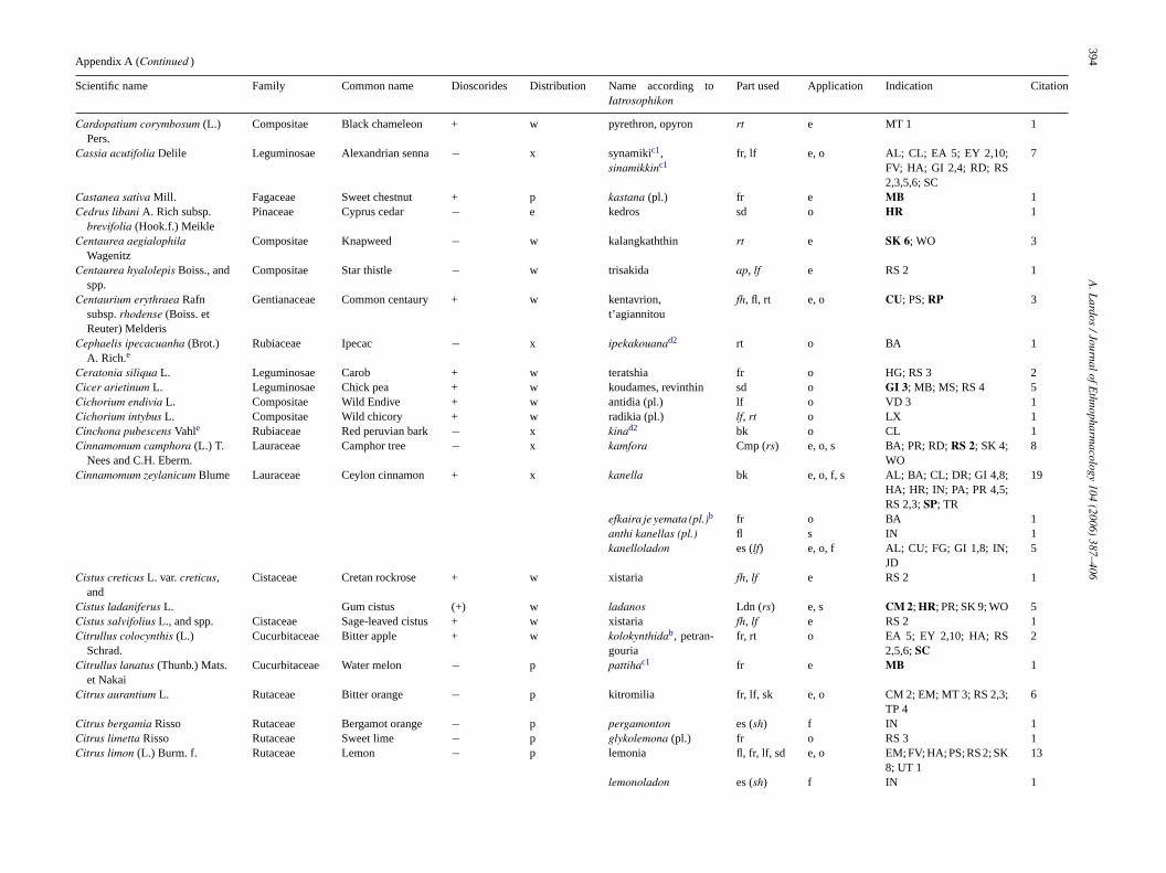
394A
.Lardos
/JournalofEthnopharm
acology104
(2006)387–406
Appendix A (Continued )
Scientific name Family Common name Dioscorides Distribution Name according toIatrosophikon
Part used Application Indication Citation
Cardopatium corymbosum (L.)Pers.
Compositae Black chameleon + w pyrethron, opyron rt e MT 1 1
Cassia acutifolia Delile Leguminosae Alexandrian senna − x synamikic1,sinamikkinc1
fr, lf e, o AL; CL; EA 5; EY 2,10;FV; HA; GI 2,4; RD; RS2,3,5,6; SC
7
Castanea sativa Mill. Fagaceae Sweet chestnut + p kastana (pl.) fr e MB 1Cedrus libani A. Rich subsp.
brevifolia (Hook.f.) MeiklePinaceae Cyprus cedar − e kedros sd o HR 1
Centaurea aegialophilaWagenitz
Compositae Knapweed − w kalangkaththin rt e SK 6; WO 3
Centaurea hyalolepis Boiss., andspp.
Compositae Star thistle − w trisakida ap, lf e RS 2 1
Centaurium erythraea Rafnsubsp. rhodense (Boiss. etReuter) Melderis
Gentianaceae Common centaury + w kentavrion,t’agiannitou
fh, fl, rt e, o CU; PS; RP 3
Cephaelis ipecacuanha (Brot.)A. Rich.e
Rubiaceae Ipecac − x ipekakouanad2 rt o BA 1
Ceratonia siliqua L. Leguminosae Carob + w teratshia fr o HG; RS 3 2Cicer arietinum L. Leguminosae Chick pea + w koudames, revinthin sd o GI 3; MB; MS; RS 4 5Cichorium endivia L. Compositae Wild Endive + w antidia (pl.) lf o VD 3 1Cichorium intybus L. Compositae Wild chicory + w radikia (pl.) lf, rt o LX 1Cinchona pubescens Vahle Rubiaceae Red peruvian bark − x kinad2 bk o CL 1Cinnamomum camphora (L.) T.
Nees and C.H. Eberm.Lauraceae Camphor tree − x kamfora Cmp (rs) e, o, s BA; PR; RD; RS 2; SK 4;
WO8
Cinnamomum zeylanicum Blume Lauraceae Ceylon cinnamon + x kanella bk e, o, f, s AL; BA; CL; DR; GI 4,8;HA; HR; IN; PA; PR 4,5;RS 2,3; SP; TR
19
efkaira je yemata (pl.)b fr o BA 1anthi kanellas (pl.) fl s IN 1kanelloladon es (lf) e, o, f AL; CU; FG; GI 1,8; IN;
JD5
Cistus creticus L. var. creticus,and
Cistaceae Cretan rockrose + w xistaria fh, lf e RS 2 1
Cistus ladaniferus L. Gum cistus (+) w ladanos Ldn (rs) e, s CM 2; HR; PR; SK 9; WO 5Cistus salvifolius L., and spp. Cistaceae Sage-leaved cistus + w xistaria fh, lf e RS 2 1Citrullus colocynthis (L.)
Schrad.Cucurbitaceae Bitter apple + w kolokynthidab, petran-
gouriafr, rt o EA 5; EY 2,10; HA; RS
2,5,6; SC2
Citrullus lanatus (Thunb.) Mats.et Nakai
Cucurbitaceae Water melon − p pattihac1 fr e MB 1
Citrus aurantium L. Rutaceae Bitter orange − p kitromilia fr, lf, sk e, o CM 2; EM; MT 3; RS 2,3;TP 4
6
Citrus bergamia Risso Rutaceae Bergamot orange − p pergamonton es (sh) f IN 1Citrus limetta Risso Rutaceae Sweet lime − p glykolemona (pl.) fr o RS 3 1Citrus limon (L.) Burm. f. Rutaceae Lemon − p lemonia fl, fr, lf, sd e, o EM; FV; HA; PS; RS 2; SK
8; UT 113
lemonoladon es (sh) f IN 1

A.L
ardos/JournalofE
thnopharmacology
104(2006)
387–406395
Citrus medica L. Rutaceae Citron − p kitron sh o GI 4 1Citrus sinensis Osbeck Rutaceae Sweet orange − p portokallin sh o GI 4 1Coffea arabica L. Rubiaceae Coffee − x kafes sd o GI 3; RS 3 2Commiphora myrrha Engl. Burseraceae Myrrh + x myrra Myr (rs) e, o, f, s AL; CU; EA 5; EY 2,10;
FG; GI 1,8; HA; IN; JD;PR; RS 2,3,5,6; SC; SK 5
5
Commiphora opobalsamumEngl.
Burseraceae Balsam tree − x valsamon tis Mekkas Blm (rs) e SK 2 1
Convolvulus althaeoides L., and Convolvulaceae Mallow-lvd. bindweed + w periplokadin ap e RS 2 2Convolvulus arvensis L. Field Bindweed + wConvolvulus scammonia L. Convolvulaceae Scammony + x mahmoudiac1 Scm (sa) o AL; CL; GI 4; PR; RS 2,3 2Corylus avellana L., et al spp. Corylaceae Common Hazel + w fintoukkia,
leptokarydialf, sh, tw e, o RS 2; VD 2
Crocus sativus L. Iridaceae Saffron crocus + (p) krokos igounzafaranc2
st e, o FV; GI 1,4,7,8; HP; HR;MS; SK 5
6
Crocus veneris Tapp. ex Poechand spp.
Iridaceae Aphrodite’s crocus (+) e krokos fl, st e, o, n AL; BA; CL; CO; CU; EA5; EY 2,10; FG; GI 8; HA;JD; PR; RS 2,3,5,6; SC
11
Croton eluteria Benn.e Euphorbiaceae Cascarilla − x amber kabughib,c2,kapnisman, tourkikonb
bk e SK 5 1
Cucurbita maxima Duchesne Cucurbitaceae Giant pumpkin + p kolokin kokkinon fr e WO 1Cucurbita pepo L., et al spp. Cucurbitaceae Marrow, pumpkin + p kolokynthin, kolokin fr e, o HA; SK 10 2Cuminum cyminum L. Umbelliferae Cumin + p artyshia, kyminon sd e, o CL; CV; EA 5; FF; FV; GI
4,5; PA 4; RD; RP; RS 3;SK 10; TP 2; TR; VD 2,3
16
Cupressus sempervirens L. Cupressaceae Italian cypress + w kyparissin fr, tw e, o GI 3; SK 1; RS 2,3; TP 2,4 6Cyclamen persicum Mill., and
spp.Primulaceae Persian violet (+) w kyklaminon ap, tb e CM 3; IL; SC; SK 1,10 7
Cydonia oblonga Mill. Rosaceae Quince + p kydonia frc, lf, tw e TP 4; RS 2 2Cynodon dactylon (L.) Pers. Graminae Bermuda grass + w argasti ap, rt e, o GI 7; HR; RS 2; UT 2,3;
VD 14
Daucus carota L. Umbelliferae Carrot + w dafkin, stafylinos lf, rt e, o SK 9; UT 3; VD 1; WO 3Delphinium peregrinum L. Ranunculaceae Larkspur + w kalogherakib fl e CU; WO 1Ecballium elaterium (L.) A.
Rich.Cucurbitaceae Squirting cucumber + w agriangourka,
agriangouronap, fr, rt e, o, f AL; GI 4; JD; PS; RD; RP;
RS 2,3; SK 97
Elettaria cardamomum Maton Zingiberaceae Chester cardamom + x kakkoullac1, (kardamomon) fr e, o, s BA; EA 4; GI 1,4; PA 4;PR
4
Euphorbia helioscopia L., andspp.
Euphorbiaceae Sun spurge + w galatzida ap, rt o UT 3; VD 1 1
Ferula gummosa Boiss. Umbelliferae Galbanum + x halvani, mavron kapnisman Glb (rs) e, o IA; PA 4 2Ficus carica L. Moraceae Common Fig + w sykia fr, lf, mj, rt e, o CM 1; CV; DR; IS; PA
3; PS; RS 1,2,3,4; SB; SK6,8; WO
16
Foeniculum vulgare Mill. Umbelliferae Fennel + w marathos ap, rt, sd e, o CL; CV; EY 2,7; HR; GI4,8; LP; MB; MS; PR; RD;RS 2,3
14
Fraxinus angustifolia Vahl., andspp.
Oleaceae Narrow-leaved ash (+) p fraxinos lf, rt e, o PS; RP 2
Fumaria officinalis L., and spp. Papaveraceae Common fumitory + w kapnohortin igoun sahteric1 fh o MT 3 1Gentiana lutea L. Gentianaceae Yellow gentian + x gentiani rt o CV 1
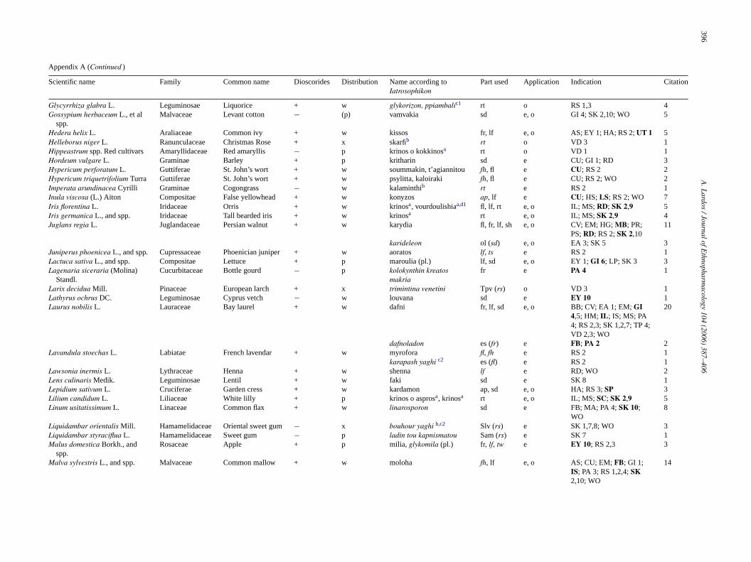
396A
.Lardos
/JournalofEthnopharm
acology104
(2006)387–406
Appendix A (Continued )
Scientific name Family Common name Dioscorides Distribution Name according toIatrosophikon
Part used Application Indication Citation
Glycyrrhiza glabra L. Leguminosae Liquorice + w glykorizon, ppiambalic1 rt o RS 1,3 4Gossypium herbaceum L., et al
spp.Malvaceae Levant cotton − (p) vamvakia sd e, o GI 4; SK 2,10; WO 5
Hedera helix L. Araliaceae Common ivy + w kissos fr, lf e, o AS; EY 1; HA; RS 2; UT 1 5Helleborus niger L. Ranunculaceae Christmas Rose + x skarfib rt o VD 3 1Hippeastrum spp. Red cultivars Amaryllidaceae Red amaryllis − p krinos o kokkinosa rt o VD 1 1Hordeum vulgare L. Graminae Barley + p kritharin sd e CU; GI 1; RD 3Hypericum perforatum L. Guttiferae St. John’s wort + w soummakin, t’agiannitou fh, fl e CU; RS 2 2Hypericum triquetrifolium Turra Guttiferae St. John’s wort + w psylitta, kaloiraki fh, fl e CU; RS 2; WO 2Imperata arundinacea Cyrilli Graminae Cogongrass − w kalaminthib rt e RS 2 1Inula viscosa (L.) Aiton Compositae False yellowhead + w konyzos ap, lf e CU; HS; LS; RS 2; WO 7Iris florentina L. Iridaceae Orris + w krinosa, vourdoulishiaa,d1 fl, lf, rt e, o IL; MS; RD; SK 2,9 5Iris germanica L., and spp. Iridaceae Tall bearded iris + w krinosa rt e, o IL; MS; SK 2,9 4Juglans regia L. Juglandaceae Persian walnut + w karydia fl, fr, lf, sh e, o CV; EM; HG; MB; PR;
PS; RD; RS 2; SK 2,1011
karideleon ol (sd) e, o EA 3; SK 5 3Juniperus phoenicea L., and spp. Cupressaceae Phoenician juniper + w aoratos lf, ts e RS 2 1Lactuca sativa L., and spp. Compositae Lettuce + p maroulia (pl.) lf, sd e, o EY 1; GI 6; LP; SK 3 3Lagenaria siceraria (Molina)
Standl.Cucurbitaceae Bottle gourd − p kolokynthin kreatos
makriafr e PA 4 1
Larix decidua Mill. Pinaceae European larch + x trimintina venetini Tpv (rs) o VD 3 1Lathyrus ochrus DC. Leguminosae Cyprus vetch − w louvana sd e EY 10 1Laurus nobilis L. Lauraceae Bay laurel + w dafni fr, lf, sd e, o BB; CV; EA 1; EM; GI
4,5; HM; IL; IS; MS; PA4; RS 2,3; SK 1,2,7; TP 4;VD 2,3; WO
20
dafnoladon es (fr) e FB; PA 2 2Lavandula stoechas L. Labiatae French lavendar + w myrofora fl, fh e RS 2 1
karapash yaghi c2 es (fl) e RS 2 1Lawsonia inermis L. Lythraceae Henna + w shenna lf e RD; WO 2Lens culinaris Medik. Leguminosae Lentil + w faki sd e SK 8 1Lepidium sativum L. Cruciferae Garden cress + w kardamon ap, sd e, o HA; RS 3; SP 3Lilium candidum L. Liliaceae White lilly + p krinos o asprosa, krinosa rt e, o IL; MS; SC; SK 2,9 5Linum usitatissimum L. Linaceae Common flax + w linarosporon sd e FB; MA; PA 4; SK 10;
WO8
Liquidambar orientalis Mill. Hamamelidaceae Oriental sweet gum − x bouhour yaghi b,c2 Slv (rs) e SK 1,7,8; WO 3Liquidambar styraciflua L. Hamamelidaceae Sweet gum − p ladin tou kapnismatou Sam (rs) e SK 7 1Malus domestica Borkh., and
spp.Rosaceae Apple + p milia, glykomila (pl.) fr, lf, tw e EY 10; RS 2,3 3
Malva sylvestris L., and spp. Malvaceae Common mallow + w moloha fh, lf e, o AS; CU; EM; FB; GI 1;IS; PA 3; RS 1,2,4; SK2,10; WO
14
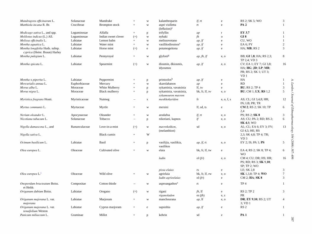
A.L
ardos/JournalofE
thnopharmacology
104(2006)
387–406397
Mandragora officinarum L. Solanaceae Mandrake + w kalanthroparin lf, rt e RS 2; SK 5; WO 3Matthiola incana R. Br. Cruciferae Brompton stock + w aspri violletta
(lefkoıon)art e PA 2 1
Medicago sativa L., and spp. Leguminosae Alfalfa + p trifyllin ap r EY 3,7 1Melilotus indicus (L.) All. Leguminosae Indian sweet clover (+) w nyhaki fh o GI 8 1Melissa officinalis L. Labiatae Lemon balm + w melissovotano ap, lf e CU, WO 1Mentha aquatica L. Labiatae Water mint + w vasilikodiosmosa ap, lf e EA 6; PY 2Mentha longifolia Huds. subsp.
cyprica (Heinr. Braun) HarleyLabiatae Horse mint (+) e potamogeitona ap, lf e HA; MB; RS 2 3
Mentha pulegium L. Labiatae Pennyroyal + w glyfonig ap, fh, lf e, o BB; GI 5,8; HA; RS 2,3;TP 2,4; VD 3
8
Mentha spicata L. Labiatae Spearmint (+) w diosmin, dkiosmis,idyosmos
ap, lf e, o CV; EA 1; EY 7; GI 3,8;HA; HG; JD; LP; MB;PR; RS 2; SK 1; UT 3;VD 1
16
Mentha x piperita L. Labiatae Peppermint + p printzolosh ap, lf e HA 1Mercurialis annua L. Euphorbiaceae Mercury + w skarolahanon ap e RD 1Morus alba L. Moraceae White Mulberry + p sykaminia, vavatsinia lf, tw e BU; RS 2; TP 4 3Morus nigra L. Moraceae Black mulberry + p sykaminia, vavatsinia,
sykamouron mavronbk, fr, lf, tw e, o BU; CM 1; LX; RS 1,2 5
Myristica fragrans Houtt. Myristicaceae Nutmeg − x moskhokaridon fr e, o, f, s AS; CL; GI 3,4,8; HR;IN; LB; PR; TR
12
Myrtus communis L. Myrtaceae Myrtle + w mersini lf, sd, ts e CM 2; RS 2; SK 10; TP2,4
6
Nerium oleander L. Apocynaceae Oleander + w arodafna lf, rt e, o PS; RS 2; SK 8 3Nicotiana tabacum L. Solanaceae Tobacco − p nikotiani, kapnos lf e, o AS; CU; PA 2; RD; RS 2;
SK 4,8; WO6
Nigella damascena L., and Ranunculaceae Love-in-a-mist (+) w mavrokokkon,(melanthion)
sd e, o AL; CL; EA 6; EY 3; FV;GI 4,5; HE; RS
13
Nigella sativa L. Black cumin + W 2,3; SK 4,8; TP 4; TR;VD 3
Ocimum basilicum L. Labiatae Basil + p vasilijia, vasilikia,vasilikos
ap, lf, rt e, o EY 2; IS; PA 1; PS 5
Olea europea L. Oleaceae Cultivated olive + w elaia bk, fr, lf, tw e EA 4; RS 2; SK 8; TP 4;WO
6
ladin ol (fr) e, o CM 4; CU; DR; HS; HR;PS; RD; RS 3; SK 9,10;SP; TP 2; WO
16
pissa elaias rs e LE; SK 2,8 3Olea europea L.i Oleaceae Wild olive + w agrielaia bk, fr, lf, rw e, o SK 1,3,8; TP 4; WO 7
ladin agrioelaias ol (fr) e CM 2; HA; SK 8 3
Onopordum bracteatum Boiss.et Heldr.
Compositae Cotton thistle − w asproangathosa rt e TP 4 1
Origanum dubium Boiss. Labiatae Oregano (+) w rigani fh, lf e RS 2; TP 2 3riganoladon es (fh) e, s PR
Origanum majorana L. var.majorana
Labiatae Marjoram + w manchourana ap, lf e, o DR; EY 9,10; RS 2; UT3; VD 1
4
Origanum majorana L. var.tenuifolium Weston
Labiatae Cyprus marjoram + e sapsishia ap, lf e RS 2 1
Panicum miliaceum L. Graminae Millet + p kehrin sd e PA 1 1

398A
.Lardos
/JournalofEthnopharm
acology104
(2006)387–406
Appendix A (Continued )
Scientific name Family Common name Dioscorides Distribution Name according toIatrosophikon
Part used Application Indication Citation
Papaver rhoeas L., et al spp. Papaveraceae Poppy + w argemonib ap, cp, fl e CU 2Papaver somniferum L. subsp.
setigerumPapaveraceae Opium poppy + w argemoni, haskashinc1 ap, cp, fl e CU 2
DC. Corb.j afhioninc1, afkioninc1 Opm (mj) e, o AL; BA; GI 3,4,7; HA;HP; HR; PA 4; RS 2,3;SK 3,6,8; TP 4
13
Paronychia argentea Lam. Caryophyllaceae Silver nailroot (+) w kalangkaththina ap, rt e SK 5; WO 3Petroselinum crispum (Mill.)
A.W. HillUmbelliferae Parsley + w makidonisin ap o UT 2 1
Phalaris brachystachys Link,and spp.
Graminae Canarygrass + w skanniola sd o PS 1
Phaseolus vulgaris L., and spp. Leguminosae Kidney bean + p fasoulin sd e MB 1Phoenix dactylifera L. Palmae Date Palm + w foinijia (pl.) fr o GI 9 1Phytolacca pruinosa Fenzl. Phytolacaceae Ink cap − w kirmizinc1 ap, fr, rt o BA; CL; GI 4; TR 3Pimenta dioica (L.) Merr.e Myrtaceae Jamaica pepper − x yenibahar c2 fr o PA 1Pimpinella anisum L. Umbelliferae Anise + w glykanissos sd e, o AL, AS, CL; CV; GI 4;
LB, LP; PA 4; PR; RS2,3; SK 10; TP 4; TR; VD2,3
19
ladin tou glykanissou ol (sd) e, o RS 3 2Pimpinella saxifraga L. Umbelliferae Lesser burnet + x pinpinellad2 rt o PR 1
Pinus brutia Ten. Pinaceae Calabrian pine (+) w pefkos, dadin bk, rw, sp e HG; HS; RS 2,3; TP 4;WO
5
pissa toupefkou/retsinin
rs e BU; CU; EY 8; HS; PA 4;RD; SK 1,2,9; TP 2; WO
16
Pinus halepensis Mill.k Pinaceae Aleppo pine + p pefkos, dadin bk, rw, sp e HG; HS; RS 2,3; TP 4;WO
5
Pinus nigra Arnold subsp.pallasiana
Pinaceae Black pine (+) w pefkos, dadin bk, rw, sp e HG; HS; RS 2,3; TP 4;WO
5
(Lamb.) Holmboe pissa tou pefkou/retsinin rs e BU; CU; EY 8; HS; PA 4;RD; SK 1,2,9; TP 2; WO
16
Piper cubeba L. Piperaceae Tailed pepper − x kkepapienc1 fr o GI 7; HP; HR 1Piper nigrum L. Piperaceae Pepper + x piperin fr e, o RS 3,4; TP 4 3
Pistacia atlantica Desf.l Anacardiaceae Terebinth tree + w trimithkia lf e RS 2 1mastihin Mst (rs) o BB; TP 1 1
Pistacia lentiscus L.m Anacardiaceae Lentisk + w shinnos lf, rb e, o GI 3; RS 2; TR 4mastihin Mst (rs) e, o, f AL; BA; BB; CL; CU; EA
5; EY 2,10; GI 2,3,4,7;HA; HP; HR; HS; IN; PA2; PR; RD; RS 2,3,5,6;SC; SK 2,9; TP 1,3,4; WO
34
Pistacia terebinthus L.n Anacardiaceae Terebinth + w trimithkia lf e RS 2 1trimintina Tpc (rs) e, o, f BU; CU; GI 3; HS; MB;
PA 3; RP; SK 5,9; UT 3;VD 1; WO
15
Plantago lanceolata L. Plantaginaceae Ribwort (+) w pentanevron lf, sd e, o CU; EY 9; GI 3; PY; RS2,3; WO
7
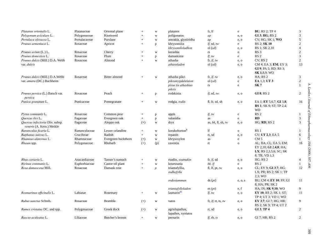
A.L
ardos/JournalofE
thnopharmacology
104(2006)
387–406399
Platanus orientalis L. Platanaceae Oriental plane + w platanos fr, lf e BU; RS 2; TP 4 3Polygonum aviculare L. Polygonaceae Knotweed + w poligonatos ap e, o GI 3; HG; RS 2 3Portulaca oleracea L. Portulacaceae Purslane + w antrakla, glystiridha ap e, o CV; HG; SK 1; WO 5Prunus armeniaca L. Rosaceae Apricot + p khrysomilia lf, sd, tw e RS 2; SK 10 2
khrysomiloladhon ol (sd) e, o RS 1; SK 2,10 4Prunus avium (L.) L. Rosaceae Cherry + w kerashia rs o RS 3 2Prunus domestica L. Rosaceae Plum + p damaskinia lf, tw e RS 2 3Prunus dulcis (Mill.) D.A. Webb Rosaceae Almond + w athasha fr, lf, tw e, o CV; RS 2 2var. dulcis athasoladon ol (sd) e, o CM 4; EA 3; EM; EY 3;
GI 9; PA 5; RD; RS 3;SK 1,8,9; WO
13
Prunus dulcis (Mill.) D.A.Webb Rosaceae Bitter almond + w athasha pikri fr, lf, tw e, o HA; RS 2 3var. amara (DC.) Buchheim pikramygdalelaion ol (sd) e EA 1,3; UT 3 2
pissa tis athashiaspikris
rs e SK 7 1
Prunus persica (L.) Batsch var.persica
Rosaceae Peach + p rodakinia lf, sd, tw e, o GI 8; RS 2 2
Punica granatum L. Punicaceae Pomegranate + w rodgia, rodin fl, fr, sd, sh e, o EA 1; EY 5,6,7; GI 3,8;RS 1; SK 9; ST; TP 2,4;WO
16
Pyrus communis L. Rosaceae Common pear + p appis lf, tw e RS 2 1Quercus ilex L. Fagaceae Evergreen oak + p valanidia as e RD 3Quercus infectoria Oliv. subsp.
veneris (A. Kern.) MeikleFagaceae Aleppo oak (+) w drys as, bk, fl, sh, tw e, o HG; RD; RS 2 3
Ranunculus ficaria L. Ranunculaceae Lesser celandine + w korakohortonb lf o RS 1 1Raphanus sativus L. Cruciferae Radish + w repanin rt, sd e, o CV; UT 2,3; EA 5 6Rhamnus alaternus L. Rhamnaceae Evergreen buckthorn (+) w khrysoxylon as e CM 5 1Rheum spp. Polygonaceae Rhubarb (+) (p) raventin rt o AL; BA; CL; EA 5; EM;
EY 2,10; GI 2,4,8; HA;LX; RS 2,3,5,6; SC; SK8; TR; VD 1,3
16
Rhus coriaria L. Anacardiaceae Tanner’s sumach + w roudin, soumakin fr, lf, sd e, o HG; RS 2 4Ricinus communis L. Euphorbiaceae Castor oil plant + w kourtounia bk, lf e RS 2 1Rosa damascena Mill. Rosaceae Damask rose + p triantafyllia,
rodhofyllafl, lf, pt, tw e, o CL; EY 9; GI 3,7; HG;
LX; PR; RS 2; SK 1; TP2,3; WO
12
rodostamman ds (pt) e, o, s BU; CM 4; EY 10; FF; GI8; HA; PR; SK 2
11
triantafylloladon es (pt) e, f HA; IN; SK 9,10; WO 9Rosmarinus officinalis L. Labiatae Rosemary + w lasmarind1 lf, tw e, o EY 10; RS 2; SK 1; ST;
TP 4; UT 3; VD 1; WO11
Rubus sanctus Schreb. Rosaceae Bramble (+) w vatos fr, lf, rt, ts, tw e, o EY 3,7; GI 7; HG; HR;RS 2; SK 9; TP 4; UT 2
9
Rumex cristatus DC. and spp. Polygonaceae Greek dock (+) w agriolapathos,lapathos, xyniatos
rt, sd e, o GI 3; TP 4 2
Ruscus aculeatus L. Liliaceae Butcher’s broom + w pernarin lf, rb, ts e, o GI 7; HR; RS 2 2
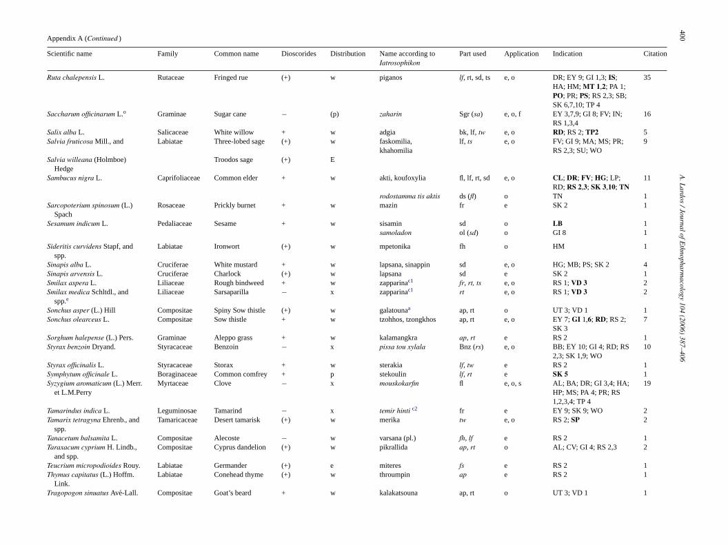
400A
.Lardos
/JournalofEthnopharm
acology104
(2006)387–406
Appendix A (Continued )
Scientific name Family Common name Dioscorides Distribution Name according toIatrosophikon
Part used Application Indication Citation
Ruta chalepensis L. Rutaceae Fringed rue (+) w piganos lf, rt, sd, ts e, o DR; EY 9; GI 1,3; IS;HA; HM; MT 1,2; PA 1;PO; PR; PS; RS 2,3; SB;SK 6,7,10; TP 4
35
Saccharum officinarum L.o Graminae Sugar cane − (p) zaharin Sgr (sa) e, o, f EY 3,7,9; GI 8; FV; IN;RS 1,3,4
16
Salix alba L. Salicaceae White willow + w adgia bk, lf, tw e, o RD; RS 2; TP2 5Salvia fruticosa Mill., and Labiatae Three-lobed sage (+) w faskomilia,
khahomilialf, ts e, o FV; GI 9; MA; MS; PR;
RS 2,3; SU; WO9
Salvia willeana (Holmboe)Hedge
Troodos sage (+) E
Sambucus nigra L. Caprifoliaceae Common elder + w akti, koufoxylia fl, lf, rt, sd e, o CL; DR; FV; HG; LP;RD; RS 2,3; SK 3,10; TN
11
rodostamma tis aktis ds (fl) o TN 1Sarcopoterium spinosum (L.)
SpachRosaceae Prickly burnet + w mazin fr e SK 2 1
Sesamum indicum L. Pedaliaceae Sesame + w sisamin sd o LB 1samoladon ol (sd) o GI 8 1
Sideritis curvidens Stapf, andspp.
Labiatae Ironwort (+) w mpetonika fh o HM 1
Sinapis alba L. Cruciferae White mustard + w lapsana, sinappin sd e, o HG; MB; PS; SK 2 4Sinapis arvensis L. Cruciferae Charlock (+) w lapsana sd e SK 2 1Smilax aspera L. Liliaceae Rough bindweed + w zapparinac1 fr, rt, ts e, o RS 1; VD 3 2Smilax medica Schltdl., and
spp.eLiliaceae Sarsaparilla − x zapparinac1 rt e, o RS 1; VD 3 2
Sonchus asper (L.) Hill Compositae Spiny Sow thistle (+) w galatounaa ap, rt o UT 3; VD 1 1Sonchus olearceus L. Compositae Sow thistle + w tzohhos, tzongkhos ap, rt e, o EY 7; GI 1,6; RD; RS 2;
SK 37
Sorghum halepense (L.) Pers. Graminae Aleppo grass + w kalamangkra ap, rt e RS 2 1Styrax benzoin Dryand. Styracaceae Benzoin − x pissa tou xylala Bnz (rs) e, o BB; EY 10; GI 4; RD; RS
2,3; SK 1,9; WO10
Styrax officinalis L. Styracaceae Storax + w sterakia lf, tw e RS 2 1Symphytum officinale L. Boraginaceae Common comfrey + p stekoulin lf, rt e SK 5 1Syzygium aromaticum (L.) Merr.
et L.M.PerryMyrtaceae Clove − x mouskokarfin fl e, o, s AL; BA; DR; GI 3,4; HA;
HP; MS; PA 4; PR; RS1,2,3,4; TP 4
19
Tamarindus indica L. Leguminosae Tamarind − x temir hinti c2 fr e EY 9; SK 9; WO 2Tamarix tetragyna Ehrenb., and
spp.Tamaricaceae Desert tamarisk (+) w merika tw e, o RS 2; SP 2
Tanacetum balsamita L. Compositae Alecoste − w varsana (pl.) fh, lf e RS 2 1Taraxacum cyprium H. Lindb.,
and spp.Compositae Cyprus dandelion (+) w pikrallida ap, rt o AL; CV; GI 4; RS 2,3 2
Teucrium micropodioides Rouy. Labiatae Germander (+) e miteres fs e RS 2 1Thymus capitatus (L.) Hoffm.
Link.Labiatae Conehead thyme (+) w throumpin ap e RS 2 1
Tragopogon sinuatus Ave-Lall. Compositae Goat’s beard + w kalakatsouna ap, rt o UT 3; VD 1 1
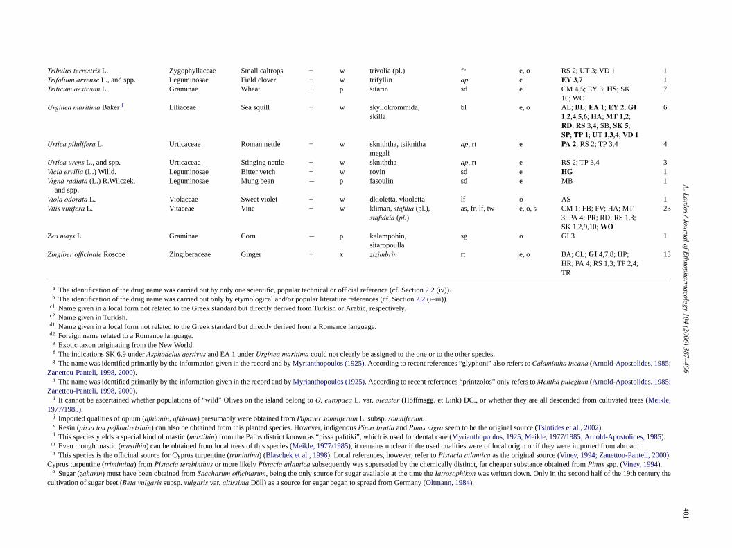
A.L
ardos/JournalofE
thnopharmacology
104(2006)
387–406401
Tribulus terrestris L. Zygophyllaceae Small caltrops + w trivolia (pl.) fr e, o RS 2; UT 3; VD 1 1Trifolium arvense L., and spp. Leguminosae Field clover + w trifyllin ap e EY 3,7 1Triticum aestivum L. Graminae Wheat + p sitarin sd e CM 4,5; EY 3; HS; SK
10; WO7
Urginea maritima Baker f Liliaceae Sea squill + w skyllokrommida,skilla
bl e, o AL; BL; EA 1; EY 2; GI1,2,4,5,6; HA; MT 1,2;RD; RS 3,4; SB; SK 5;SP; TP 1; UT 1,3,4; VD 1
6
Urtica pilulifera L. Urticaceae Roman nettle + w skniththa, tsiknithamegali
ap, rt e PA 2; RS 2; TP 3,4 4
Urtica urens L., and spp. Urticaceae Stinging nettle + w skniththa ap, rt e RS 2; TP 3,4 3Vicia ervilia (L.) Willd. Leguminosae Bitter vetch + w rovin sd e HG 1Vigna radiata (L.) R.Wilczek,
and spp.Leguminosae Mung bean − p fasoulin sd e MB 1
Viola odorata L. Violaceae Sweet violet + w dkioletta, vkioletta lf o AS 1Vitis vinifera L. Vitaceae Vine + w kliman, stafilia (pl.),
stafidkia (pl.)as, fr, lf, tw e, o, s CM 1; FB; FV; HA; MT
3; PA 4; PR; RD; RS 1,3;SK 1,2,9,10; WO
23
Zea mays L. Graminae Corn − p kalampohin,sitaropoulla
sg o GI 3 1
Zingiber officinale Roscoe Zingiberaceae Ginger + x zizimbrin rt e, o BA; CL; GI 4,7,8; HP;HR; PA 4; RS 1,3; TP 2,4;TR
13
a The identification of the drug name was carried out by only one scientific, popular technical or official reference (cf. Section 2.2 (iv)).b The identification of the drug name was carried out only by etymological and/or popular literature references (cf. Section 2.2 (i–iii)).
c1 Name given in a local form not related to the Greek standard but directly derived from Turkish or Arabic, respectively.c2 Name given in Turkish.d1 Name given in a local form not related to the Greek standard but directly derived from a Romance language.d2 Foreign name related to a Romance language.
e Exotic taxon originating from the New World.f The indications SK 6,9 under Asphodelus aestivus and EA 1 under Urginea maritima could not clearly be assigned to the one or to the other species.g The name was identified primarily by the information given in the record and by Myrianthopoulos (1925). According to recent references “glyphoni” also refers to Calamintha incana (Arnold-Apostolides, 1985;
Zanettou-Panteli, 1998, 2000).h The name was identified primarily by the information given in the record and by Myrianthopoulos (1925). According to recent references “printzolos” only refers to Mentha pulegium (Arnold-Apostolides, 1985;
Zanettou-Panteli, 1998, 2000).i It cannot be ascertained whether populations of “wild” Olives on the island belong to O. europaea L. var. oleaster (Hoffmsgg. et Link) DC., or whether they are all descended from cultivated trees (Meikle,
1977/1985).j Imported qualities of opium (afhionin, afkionin) presumably were obtained from Papaver somniferum L. subsp. somniferum.k Resin (pissa tou pefkou/retsinin) can also be obtained from this planted species. However, indigenous Pinus brutia and Pinus nigra seem to be the original source (Tsintides et al., 2002).l This species yields a special kind of mastic (mastihin) from the Pafos district known as “pissa pafitiki”, which is used for dental care (Myrianthopoulos, 1925; Meikle, 1977/1985; Arnold-Apostolides, 1985).
m Even though mastic (mastihin) can be obtained from local trees of this species (Meikle, 1977/1985), it remains unclear if the used qualities were of local origin or if they were imported from abroad.n This species is the officinal source for Cyprus turpentine (trimintina) (Blaschek et al., 1998). Local references, however, refer to Pistacia atlantica as the original source (Viney, 1994; Zanettou-Panteli, 2000).
Cyprus turpentine (trimintina) from Pistacia terebinthus or more likely Pistacia atlantica subsequently was superseded by the chemically distinct, far cheaper substance obtained from Pinus spp. (Viney, 1994).o Sugar (zaharin) must have been obtained from Saccharum officinarum, being the only source for sugar available at the time the Iatrosophikon was written down. Only in the second half of the 19th century the
cultivation of sugar beet (Beta vulgaris subsp. vulgaris var. altissima Doll) as a source for sugar began to spread from Germany (Oltmann, 1984).
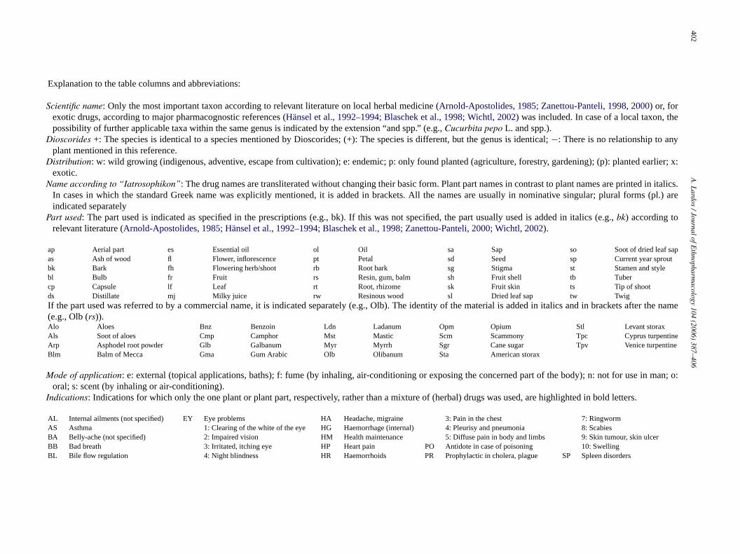
402A
.Lardos
/JournalofEthnopharm
acology104
(2006)387–406
Explanation to the table columns and abbreviations:
Scientific name: Only the most important taxon according to relevant literature on local herbal medicine (Arnold-Apostolides, 1985; Zanettou-Panteli, 1998, 2000) or, forexotic drugs, according to major pharmacognostic references (Hansel et al., 1992–1994; Blaschek et al., 1998; Wichtl, 2002) was included. In case of a local taxon, thepossibility of further applicable taxa within the same genus is indicated by the extension “and spp.” (e.g., Cucurbita pepo L. and spp.).
Dioscorides +: The species is identical to a species mentioned by Dioscorides; (+): The species is different, but the genus is identical; −: There is no relationship to anyplant mentioned in this reference.
Distribution: w: wild growing (indigenous, adventive, escape from cultivation); e: endemic; p: only found planted (agriculture, forestry, gardening); (p): planted earlier; x:exotic.
Name according to “Iatrosophikon”: The drug names are transliterated without changing their basic form. Plant part names in contrast to plant names are printed in italics.In cases in which the standard Greek name was explicitly mentioned, it is added in brackets. All the names are usually in nominative singular; plural forms (pl.) areindicated separately
Part used: The part used is indicated as specified in the prescriptions (e.g., bk). If this was not specified, the part usually used is added in italics (e.g., bk) according torelevant literature (Arnold-Apostolides, 1985; Hansel et al., 1992–1994; Blaschek et al., 1998; Zanettou-Panteli, 2000; Wichtl, 2002).
ap Aerial part es Essential oil ol Oil sa Sap so Soot of dried leaf sapas Ash of wood fl Flower, inflorescence pt Petal sd Seed sp Current year sproutbk Bark fh Flowering herb/shoot rb Root bark sg Stigma st Stamen and stylebl Bulb fr Fruit rs Resin, gum, balm sh Fruit shell tb Tubercp Capsule lf Leaf rt Root, rhizome sk Fruit skin ts Tip of shootds Distillate mj Milky juice rw Resinous wood sl Dried leaf sap tw TwigIf the part used was referred to by a commercial name, it is indicated separately (e.g., Olb). The identity of the material is added in italics and in brackets after the name(e.g., Olb (rs)).Alo Aloes Bnz Benzoin Ldn Ladanum Opm Opium Stl Levant storaxAls Soot of aloes Cmp Camphor Mst Mastic Scm Scammony Tpc Cyprus turpentineArp Asphodel root powder Glb Galbanum Myr Myrrh Sgr Cane sugar Tpv Venice turpentineBlm Balm of Mecca Gma Gum Arabic Olb Olibanum Sta American storax
Mode of application: e: external (topical applications, baths); f: fume (by inhaling, air-conditioning or exposing the concerned part of the body); n: not for use in man; o:oral; s: scent (by inhaling or air-conditioning).
Indications: Indications for which only the one plant or plant part, respectively, rather than a mixture of (herbal) drugs was used, are highlighted in bold letters.
AL Internal ailments (not specified) EY Eye problems HA Headache, migraine 3: Pain in the chest 7: RingwormAS Asthma 1: Clearing of the white of the eye HG Haemorrhage (internal) 4: Pleurisy and pneumonia 8: ScabiesBA Belly-ache (not specified) 2: Impaired vision HM Health maintenance 5: Diffuse pain in body and limbs 9: Skin tumour, skin ulcerBB Bad breath 3: Irritated, itching eye HP Heart pain PO Antidote in case of poisoning 10: SwellingBL Bile flow regulation 4: Night blindness HR Haemorrhoids PR Prophylactic in cholera, plague SP Spleen disorders
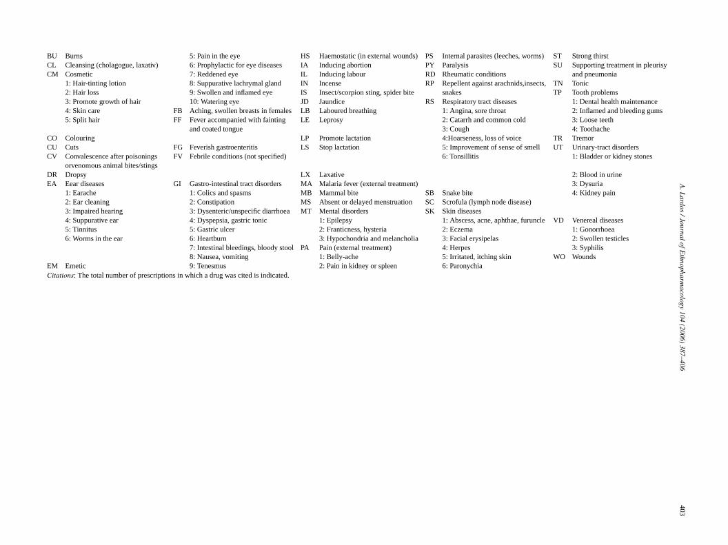
A.L
ardos/JournalofE
thnopharmacology
104(2006)
387–406403
BU Burns 5: Pain in the eye HS Haemostatic (in external wounds) PS Internal parasites (leeches, worms) ST Strong thirstCL Cleansing (cholagogue, laxativ) 6: Prophylactic for eye diseases IA Inducing abortion PY Paralysis SU Supporting treatment in pleurisy
and pneumoniaCM Cosmetic 7: Reddened eye IL Inducing labour RD Rheumatic conditions1: Hair-tinting lotion 8: Suppurative lachrymal gland IN Incense RP Repellent against arachnids,insects,
snakesTN Tonic
2: Hair loss 9: Swollen and inflamed eye IS Insect/scorpion sting, spider bite TP Tooth problems3: Promote growth of hair 10: Watering eye JD Jaundice RS Respiratory tract diseases 1: Dental health maintenance4: Skin care FB Aching, swollen breasts in females LB Laboured breathing 1: Angina, sore throat 2: Inflamed and bleeding gums5: Split hair FF Fever accompanied with fainting
and coated tongueLE Leprosy 2: Catarrh and common cold 3: Loose teeth
3: Cough 4: ToothacheCO Colouring LP Promote lactation 4:Hoarseness, loss of voice TR TremorCU Cuts FG Feverish gastroenteritis LS Stop lactation 5: Improvement of sense of smell UT Urinary-tract disordersCV Convalescence after poisonings
orvenomous animal bites/stingsFV Febrile conditions (not specified) 6: Tonsillitis 1: Bladder or kidney stones
DR Dropsy LX Laxative 2: Blood in urineEA Eear diseases GI Gastro-intestinal tract disorders MA Malaria fever (external treatment) 3: Dysuria
1: Earache 1: Colics and spasms MB Mammal bite SB Snake bite 4: Kidney pain2: Ear cleaning 2: Constipation MS Absent or delayed menstruation SC Scrofula (lymph node disease)3: Impaired hearing 3: Dysenteric/unspecific diarrhoea MT Mental disorders SK Skin diseases4: Suppurative ear 4: Dyspepsia, gastric tonic 1: Epilepsy 1: Abscess, acne, aphthae, furuncle VD Venereal diseases5: Tinnitus 5: Gastric ulcer 2: Franticness, hysteria 2: Eczema 1: Gonorrhoea6: Worms in the ear 6: Heartburn 3: Hypochondria and melancholia 3: Facial erysipelas 2: Swollen testicles
7: Intestinal bleedings, bloody stool PA Pain (external treatment) 4: Herpes 3: Syphilis8: Nausea, vomiting 1: Belly-ache 5: Irritated, itching skin WO Wounds
EM Emetic 9: Tenesmus 2: Pain in kidney or spleen 6: ParonychiaCitations: The total number of prescriptions in which a drug was cited is indicated.

404A
.Lardos
/JournalofEthnopharm
acology104
(2006)387–406
Appendix B
The botanical materia medica of the Iatrosophikon—other plant derived drugs
Common name Name according to Iatrosoph. Applic. Indication Citat. Source of the drug Distrib.
Agaric (White) agarikon aspron o EA 5; EY 2,10; HA; LX; RS 3 Polyporus officinalis Fries, and other related fungi xAmber khehriparon, kehriparoladon o EA 5; EY 2,10; HA; LB; RS 2,5,6; SC 2 Fossilised resin of undefined taxa -Candied Sugar kandion e EY 5 2 Saccharum officinarum L. (p)Carob Molasse teratsomelon e RD; VD 1 2 Ceratonia siliqua L. wCommandaria (Cyprus
Sweet Wine)kommandaria e, o CV; GI 8; PA 2; RS 3 4 Vitis vinifera L. w
Flower Water anthoneron f IN 1 Citrus spp. pPrunus spp. p, w
Grape Molasse petmezinc1 e, o LE; RD; RS 3; SK 7,8; WO 5 Vitis vinifera L. wGum Resin komidin n CO 2 Various tree species, mainly:
Prunus avium (L.) L. wPrunus domestica L. pPrunus dulcis (Mill.) D.A. Webb w
Mannap manna o CL; EA 5; EY 2,10; GI 4; HA; MS; PS;RS 2,5,6; SC; TR
4 Fraxinus ornus L.*, or other sources yieldingdifferent kinds of manna
x
Oak Galls kikkidia (pl.) e, o CO; EY 9; GI 3,6,7; HG; HR; RS 4; SK1; TP 2,4; WO
15 Quercus alnifolia Poech** e
Quercus coccifera L. subsp. calliprinos w(Webb.) Holmboe**
Quercus infectoria Oliv. subsp. veneris w(A. Kern.) Meikle
Soot asvain e EY 1,9; SK 1,2 4 Soot of burnt resin, gum resin, dried sap or othermaterial of various taxa
–
Spiced Candied Sugarq humma sheker c2 o FF 1 Mixture of:“Fever Sugar” Saccharum officinarum L., (p)
Cinnamomum zeylanicum Blume, xMyristica fragrans Houtt., and xtartar (cf.) –
Spirit of Wine oinopnevma e EA 5 1 Vitis vinifera L. wSpirits of turpentine neft yaghi c2 e PA 5; SK 1 2 Pinus spp. p, wRaki (spirit) raki e, o AL; CU; GI 1,8; PA 5; RD; 9 Vitis vinifera L., and w
SK 10; WO Pimpinella anisum L. wRum (spirit) romi e RD 1 Saccharum officinarum L. (p)Tar, Pitch katranin c1 e, o RD; SK 1 2 Cedrus libani A. Rich subsp. brevifolia (Hook.f.)
Meiklee
Pinus spp. p, wTartar kremorion, kremotartaron e, o AL; CL; CO; RS 2,3; WO 5 Vitis vinifera L. wTheriac thiriaki e, o AL; CV; GI 4; MB; PR; RS 2,3 7 Multi-ingredient preparation –Vinegar xydin e, o GI 1; LS; HR; JD; PR; PS; RD; RS;
WO10 Vitis vinifera L. w
Wine (red, white) krasin e, o CV; EA 2; EY 5,9; FB; IS; PS; RP; SK5,9,10; WO
12 Vitis vinifera L. w
Superscripts: For the legend of the superscripts “a-o” refer to Appendix A.p Various botanical as well as insect sources are reported for manna (Bastin, 1895; Lev and Amar, 2002); officinal manna listed in different Pharmacopoeias refers to the sugary exudation of the manna ash Fraxinus
ornus L. (Oleaceae).q The composition could be found only in the references quoted under 2.2.i and ii.* Plant not mentioned in Table 1 belonging to the Oleaceae family.
** Plant not mentioned in Table 1 belonging to the Fagaceae family.For the abbreviations and the explanations to the table columns (Name acc. to “Iatrosophikon”, Mode of Application, Indications, Citations, Distribution) refer to Appendix A.
A. Lardos / Journal of Ethnopharmacology 104 (2006) 387–406 405
References
Akcin, O.E., Kandemir, N., Cansaran, A., 2004. A morphological andanatomical study on endemic Alkanna haussknechtii Bornm. (Boragi-naceae), critically endangered in Turkey. Turkish Journal of Botany 28,591–598.
Afifi, F.U., Abu-Irmaileh, B., 2000. Herbal medicine in Jordan with specialemphasis on less commonly used medicinal herbs. Journal of Ethnophar-macology 72, 101–110.
Ali-Shtayeh, M.S., Yaniv, Z., Mahajna, J., 2000. Ethnobotanical survey inthe Palestinian area: a classification of the healing potential of medicinalplants. Journal of Ethnopharmacology 73, 221–232.
Arnold-Apostolides, N., 1985. Contribution a la connaissance ethnobotaniqueet medicinale de la flore de Chypre. Ph.D. Thesis. Faculte des sciencespharmaceutiques et biologiques, Universite Rene Descartes de Paris, Lux-embourg, 2199 pp.
Bastin, E.S., 1895. Botanical medicinal monographs and sundry. AmericanJournal of Pharmacy 67, 1–36.
Berendes, J. (Ed.) 1988/1992. Dioscorides Pedanius, Codex Neapolitanus(Codex medicus Graecus I, National Library of Naples), in 3 Vols.Akademische Drucks–und Verlagsanstalt, Graz.
Blaschek, W., Hansel, R., Keller, K., Reichling, J., Rimpler, H., Schneider, G.(Eds.), 1998. Hagers Handbuch der Pharmazeutischen Praxis, 5. Auflage,Drogen–Folgeband 2 und 3. Springer, Berlin.
Chrysanthis, K., 1942. Kypriakes pharmakeftikes yles sto “Peri ylis iatrikis”tou Dioskouridi [Cypriot pharmaceutical materials in de materia medicaof Dioskoridis]. Cypriot Studies 6, 19–43 (in Greek).
Constantinides, H.T., Constantinides, H.A., ConsAppendix, G., 2000. Extractfrom: Byzantine Monastic Foundation Documents—A Complete Transla-tion of the Surviving Founders’ Typika and Testaments, Makhairas, Ruleof Neilos, Bishop of Tamasia for the Monastery of Makhairas in Cyprus.Dumbarton Oaks Research Library and Collection, Washington, DC, pp.1107–1108. (Chapter 34).
Della, A., 1995. Cyprus: Country Report to the International Conference andProgramme on Plant Genetic Resource, Leipzig, Germany, 17–23 June1997. Nicosia.
Ertug, F., 2000. An ethnobotanical study in central Anatolia (Turkey). Eco-nomic Botany 54, 155–182.
Ertug, F., 2004. Wild edible plants of the Bodrum area (Mugla, Turkey).Turkish Journal of Botany 28, 161–174.
Filaretos, 1924–1925. Iatrosophikon, Synakhthen ypo tou Skefophylakostis en Kypro Ieras Monis Makhaira Mitrophanous 1790–1867 [TheIatrosophikon of the Holy Monastery of Makhairas]. Ekdosis Ieras MonisMakhaira, Lefkosia/Nicosia (in Greek).
Forschergruppe Klostermedizin, 1999. Forschergruppe Klostermedizingegrundet. Zeitschrift fur Phytotherapie 20 (5), 250.
Fronimopoulos, J., Lascaratos, J., 1988. Eye injuries by the Byzantine writerAetios Amidinos. Documenta Ophthalmologica 68, 121–124.
Gennadius, P.G., 1914. (reprint 1997). Lexikon Phytologikon [PhytologicalDicitonary]. Ekdoseis Ananatyposeis, Trokhalia, Athens (in Greek).
Georgiades, C.C., 1987/1992. Flowers of Cyprus, Plants of Medicine, vols.1 and 2. Private Publication, Nicosia.
Ghazanfar, S.A., 1994. Handbook of Arabian Medicinal Plants. CCR Press,Boca Raton.
Greek Food and Drink Codex/Kodikas Trofimon kai Poton, 1988. (edition1993). Meros A: Trofima kai Pota; Kefalaio 4: Artimatikes Yles kaiAitheria Elaia. Hellenic Republic of Greece, Athens (in Greek).
Hadjiioannou, K., 2000. Etymologiko Lexiko tis Omiloumenis KypriakisDialektou [Etymological Lexicon of the Spoken Cypriot Dialect]. Tama-sos, Lefkosia/Nicosia (in Greek).
Hanlidou, E., Karousou, R., Kleftoyanni, V., Kokkini, S., 2004. The herbalmarket of Thessaloniki (N Greece) and its relation to the ethnobotanicaltradition. Journal of Ethnopharmacology 91, 281–299.
Hansel, R., Keller, K., Rimpler, H., Schneider, G. (Eds.), 1992–1994. HagersHandbuch der Pharmazeutischen Praxis, 5. Auflage, Drogen–Folgeband 4bis 6. Springer, Berlin.
Honda, G., Yesilada, E., Tabata, M., Sezik, E., Fujita, T., Takeda, Y., Takaishi,Y., Tanaka, T., 1996. Traditional medicine in Turkey VI. Folk medicine in
West Anatolia: Afyon, Kuthaya, Denizli, Mugla, Aydın provinces. Journalof Ethnopharmacology 53, 75–87.
Karamperopoulos, D.A., 2004. Paulos Aiginitis [Paul from Aegina]. Istorika227, 38–41 (in Greek).
Kyprianou, T.C., 2000. Apo ti Chlorida tis Kyprou [On the Vegetation ofCyprus]. Christos Livadiotis, Lefkosia/Nicosia (in Greek).
Kyprianou, T.C., 2001. Ta phyta sti laiki iatriki kata ton Kypro Chrysan-thi [Plants of Folk Medicine According to Kypros Chrysanthis].Lefkosia/Nicosia (in Greek).
Lev, E., Amar, Z., 2000. Ethnopharmacological survey of traditional drugssold Israel at the end of the 20th century. Journal of Ethnopharmacology72, 191–205.
Lev, E., 2002. Reconstructed materia medica of the Medieval and Ottomanal-Sham. Journal of Ethnopharmacology 80, 167–179.
Lev, E., Amar, Z., 2002. Ethnopharmacological survey of traditional drugssold in the Kingdom of Jordan. Journal of Ethnopharmacology 82,131–145.
Littlewood, A., Maguire, H., Wolschke-Buhlmahn J., 2002. Extract from:Byzantine Garden Culture. Dumbarton Oaks Research Library and Col-lection, Washington, DC, pp. 54–57.
Mayer, J.G., Goehl, K. (Eds.), 2001. Hohepunkte der Klostermediz: DerMacer Floridus und das Herbarium des Vitus Auslasser. Reprint-VerlagLeipzig, Holzminden.
Mazal, O. (Ed.), 1999. Diosocorides Pedanius, Codex Constantinopoli-tanus (Vienna Dioscorides, Codex medicus Graecus I, Austrian NationalLibrary), vol. 2. Akademische Drucks–und Verlagsanstalt, Graz.
Meikle, R.D., 1977/1985. Flora of Cyprus, vols. 1 and 2. The Bentham-Moxon Trust, Royal Botanic Gardens, Kew.
Moussaieff, A., Fride, E., Amar, Z., Lev, E., Steinberg, D., Gallily, R.,Mechoulam, R., 2005. The Jerusalem balsam: from the FranciscanMonastery in the old city of Jerusalem to Martindale 33. Journal ofEthnopharmacology 101, 16–26.
Murphey, R., 1992. Ottoman medicine and transculturalism from the 16ththrough the 18th century. Bulletin of Historical Medicine 66, 376–403.
Myrianthopoulos, K.I., 1925. Ermineftikos pinax tou Iatrosophikou Makhaira[explanatory table to the Iatrosophikon of Makhairas]. In: Filare-tos, 1924–1925. Iatrosophikon, Synakhthen ypo tou Skefophylakostis en Kypro Ieras Monis Makhaira Mitrophanous 1790–1867 [TheIatrosophikon of the Holy Monastery of Makhairas]. Ekdosis Ieras MonisMakhaira, Lefkosia/Nicosia (in Greek).
Oltmann, W., 1984. Prozesstechnologie und Erfolg der Zuck-errubenkultivierung von der Mitte des 19. Jahrhunderts bis heute,48–65. In: Geschichte der Zuckerrube, Institut fur Zuckerrubenforschung,Gottingen, Dr. Albert Bartens, Berlin.
Papangellou, R., 2001. To kypriako idioma, Lexiko [The Cypriot idiom,Lexikon]. Iolkos Athens. (in Greek).
Parojcic, D., Stupar, D., Mirica, M., 2003. La Theriaque: medicament etAntidote. Vesalius 9, 28–32.
Petrucelli II, R.J., 1994. Monastic incorporation of classical botanic medicinesinto the Renaissance pharmacopoeia. American Journal of Nephrology 14,259–263.
Piendl, F.S., 2001. Arzneipflanzen in der Klostermedizin Karntens. Disserta-tion, Karl-Franzens-Universitat, Graz, 243 pp.
Said, O., Khalil, S., Fulder, H., Azaizeh, H., 2002. Ethnopharmacologicalsurvey of medicinal herbs in Israel, the Golan Heights and the WestBank region. Journal of Ethnopharmacology 83, 251–265.
Savvides, L., 2000. Edible wild plants of the Cyprus flora. Private publication.Nicosia.
Sezik, E., Yesilada, E., Honda, G., Yoshihisa, T., Takeda, Y., Tanaka, T., 2001.Traditional medicine in Turkey X. Folk medicine in Central Anatolia.Journal of Ethnopharmacology 75, 95–115.
Tsintides, T.C., Hadjikyriakou, G.N., Christodoulou, C.S., 2002. Trees andShrubs in Cyprus. Foundation A.G. Leventis, Cyprus Forest Association,Lefkosia/Nicosia.
Turkish Food Codex/Turk Gıda Kodeksi 1997. (edition 2005). Turk GıdaKodeksi Tebligleri, Baharat Tebligli. General Directorate of Protectionand Control. Republic of Turkey, Ankara (in Turkish).

406 A. Lardos / Journal of Ethnopharmacology 104 (2006) 387–406
Varella, E.A., 1993. Pharmazeutische Handbucher im Griechenland desfruhen 19. Jahrhunderts: Dionysus Pyrrhus und Franz Xaver Landerer.Geschichte der Pharmazie 45, 49–54.
Varella, E.A., 1999. La therapeutique Byzantine dans le monde Grec d’epoqueOttomane. Medicina nei Secoli Arte e Scienza 11, 577–584.
Viney, D.E., 1994. An Illustrated Flora of North Cyprus. Koeltz ScientificBooks, Koenigstein.
Vokou, D., Katradi, K., Kokkini, S., 1993. Ethnobotanical survey of Zagori(Epirus, Greece), a renowned centre of folk medicine in the past. Journalof Ethnopharmacology 39, 187–196.
Wichtl, M., 2002. Teedrogen und Phytopharmaka, vol. 4. Auflage. Wis-senschaftliche Verlagsgesellschaft, Stuttgart.
Windhaber, R., 2001. Von der Klostermedizin zur modernen Phytotherapie.Zeitschrift fur Phytotherapie 22, 215.
Yesilada, E., Honda, G., Sezik, E., Tabata, M., Goto, K., Ikeshiro, Y., 1993.Traditional medicine in Turkey IV. Folk medicine in the Mediterraneansubdivision. Journal of Ethnopharmacology 39, 31–38.
Zanettou-Panteli, K., 1998. The Medicinal Plants of Cyprus. Private publica-tion. Larnaca.
Zanettou-Panteli, K., 2000. I Therapeftiki Dinatotita ton Pharmakeftikon Phy-ton tis Kyprou [The Therapeutic Potential of the Medicinal Plants ofCyprus. Private publication]. Larnaca (in Greek).

Journal of Ethnopharmacology 104 (2006) 407–409
Ethnopharmacological communication
Chemoprevention and cytotoxic effect of Bauhinia variegata againstN-nitrosodiethylamine induced liver tumors and human cancer cell lines
B. Rajkapoor a,∗, B. Jayakar a, N. Murugesh b, D. Sakthisekaran c
a Department of Pharmacology, Vinayaka Mission’s College of Pharmacy, Vinayaka Mission’s Research Foundation (Deemed University),Yercaud Road, Salem-636008, Tamilnadu, India
b Institute of Pharmacology, Madurai Medical College, Madurai-625020, Tamilnadu, Indiac Department of Medical Biochemistry, Dr. ALM Post-Graduate Institute of Basic Medical Sciences, University of Madras,
Taramani Campus, Chennai-600113, Tamilnadu, India
Received 13 July 2004; received in revised form 15 July 2005; accepted 5 August 2005Available online 27 October 2005
Abstract
The chemopreventive and cytotoxic effect of ethanol extract of Bauhinia variegata (EBV) was evaluated in N-nitrosodiethylamine (DEN,200 mg/kg) induced experimental liver tumor in rats and human cancer cell lines. Oral administration of ethanol extract of Bauhinia variegata(250 mg/kg) effectively suppressed liver tumor induced by DEN as revealed by decrease in DEN induced elevated levels of serum glutamatepyruvate transaminase (SGPT), serum glutamate oxaloacetate transaminase (SGOT), alkaline phosphatase (ALP), total bilirubin, gamma glutamatetranspeptidase (GGTP), lipid peroxidase (LPO), glutathione peroxidase (GPx) and glutathione S-transferase (GST). The extract produced an increasein enzymatic antioxidant (superoxide dismutase and catalase) levels and total proteins when compared to those in liver tumor bearing rats. Thehistopathological changes of liver samples were compared with respective controls. EBV was found to be cytotoxic against human epithelial larynxcancer (HEp2) and human breast cancer (HBL-100) cells. These results show a significant chemopreventive and cytotoxic effect of ethanol extractof Bauhinia variegata against DEN induced liver tumor and human cancer cell lines.© 2006 Elsevier Ireland Ltd. All rights reserved.
Keywords: Chemoprevention; N-Nitrosodiethylamine; Bauhinia variegata; Biochemical parameters; Antioxidants; Histopathology; Cytotoxicity
Plant: Bauhinia variegata Linn (Caesalpiniaceae) stems col-lected in December 2003 at Salem, India, and identified by Dr.G. Murthy, Botanical Survey of India, Coimbatore, Tamilnadu,India. A voucher specimen (DEC-46) has been kept in our lab-oratory for future reference.
Used in traditional medicine: It is traditionally used inbronchitis, leprosy, tumors and ulcer (Shah and Joshi, 1971;Kirtikar and Basu, 1993) and studies of its extracts have shownantibacterial, antifungal and antiulcer activities (Ali et al., 1999;Rajkapoor et al., 2003a,b).
Previously isolated classes of constituents: 5,7-Dimethoxyand dihydroxy flavonone-4′-O-�-l-rhamnopyronosyl-�-d-glucopyranosides, 5-hydroxyl 7,3′,4′,5′-tetramethoxy flavone-5-O-�-d-xylopyronosyl (1 → 2) �-l-rhamnopyroanoside,lupeol, �-sitosterol, quercertin, flavanone and dihydrodiben-
∗ Corresponding author. Tel.: +91 427 2400174; fax: +91 427 2400174.E-mail address: [email protected] (B. Rajkapoor).
zoxepin (Duret and Paris, 1977; Gupta et al., 1979, 1980;Yadava and Reddy, 2001, 2003; Reddy et al., 2003).
1. Materials and methods
1.1. Plant material and extraction
Stems were dried in shade and pulverized. The powder wastreated with petroleum ether for dewaxing as well as to removechlorophyll and it was later packed into soxhlet apparatus andsubjected to hot continuous percolation using ethanol (95%, v/v)as solvent. The extract was concentrated under vacuum and driedin a vacuum desiccator (yield 3.2%, w/w).
1.2. Animals
Male Wistar rats (100–125 g) were procured from Tamil-nadu Veterinary College, Chennai, India. They were housedin microlon boxes with standard laboratory diet and water ad
0378-8741/$ – see front matter © 2006 Elsevier Ireland Ltd. All rights reserved.doi:10.1016/j.jep.2005.08.074

408 B. Rajkapoor et al. / Journal of Ethnopharmacology 104 (2006) 407–409
libitum. The study was conducted after obtaining InstitutionalAnimal Ethical Committee clearance.
1.3. Chemoprevention studies
The rats were divided into three groups, each group con-sisting of six animals. Animals of group 1 were normal control.Liver tumor was induced in groups 2 and 3 with single intraperi-toneal injection of N-nitrosodiethylamine (DEN) at a dose of200 mg/kg body weight in saline. Two weeks after DEN admin-istration, the carcinogenic effect was promoted by 0.05% pheno-barbital, which was supplemented to the experimental animalsthrough drinking water for up to 16 successive weeks (Yoshijiet al., 1991). Whereas animals of group 2 receive DEN alone,animals of group 3 were given ethanol extract of Bauhinia var-iegata (EBV) (250 mg/kg, p.o.) (Rajkapoor et al., 2003a,b) for16 weeks after the administration of DEN on 5 days per week.
At the end of experiments, animals were fasted overnightand killed by cervical decapitation. Blood was collected andthe serum was separated out. Liver was immediately removedand suspended in ice-cold saline. A small portion of liver wasfixed in 10% formalin for histopathological studies. Serum wasanalysed for the following biochemical parameters: serum glu-tamate oxaloacetate transaminase (SGOT), serum glutamatepyruvate transaminase (SGPT) (Reitman and Frankel, 1957),alkaline phosphatase (ALP) (Kind and King, 1954), total biliru-bin (Mallay and Evelyn, 1937), total protein (Lowry et al., 1951)and gamma glutamate transpeptidase (GGTP) (Szaszi, 1969).A 10% homogenate of the tissue was used for the analysis oflipid peroxidation (LPO) (Devasagayam and Tarachand, 1987),superoxide dismutase (SOD) (Marklund and Marklund, 1974),catalase (Sinha, 1972), glutathione peroxidase (GPx) (Rotrucket al., 1973) and glutathione S-transferase (GST) (Habig et al.,1974).
1.4. Cytotoxicity studies
HEp2 and HBL-100 cell lines used in these experiments wereobtained from National Center for Cell Science, Pune, India.Cytotoxicity experiments were carried out as described previ-ously by using MTT assay (Scudiero et al., 1988).
2. Results
All the animals treated with DEN had a significant incidenceof liver tumor at the end of 16 weeks as evidenced by: (i) increasein liver weight, (ii) increased hepatic enzymes such as SGPT,SGOT, ALP, total bilirubin and decrease in total protein, (iii)increased level of GGTP, GPx, GST and LPO, (iv) decreasein SOD and catalase and (v) morphological changes. All thesechanges were reverted back to normal by the EBV indicatinga strong inhibition of hepatocellular carcinogenesis induced byDEN (Table 1).
The liver weight increased nearly two-folds in those animals,which received DEN, the carcinogen. The liver weight of normalanimals was 4.1 ± 0.10 g/100 g body weight and it increased to7.8 ± 0.12 g in those that received DEN. Administration of the Ta
ble
1E
ffec
tof
EB
Von
seru
mG
PT,G
OT,
AL
P,to
talp
rote
ins,
tota
lbili
rubi
n,G
GT
Pan
dan
tioxi
dant
sle
veli
nD
EN
trea
ted
rats
Des
ign
oftr
eatm
ent
Dos
e(m
g/kg
)SG
PT(U
/L)
SGO
T(U
/L)
AL
P(U
/L)
Tota
lpro
tein
s(m
g%)
Tota
lbili
rubi
n(m
g%)
GG
TP
(U/L
)L
POG
PxG
STSO
DC
atal
ase
Con
trol
–38
.33
±1.
184
.16
±2.
217
9.8
±3.
86.
4±
0.10
0.38
±0.
0348
.3±
1.3
6.9
±0.
086.
58±
0.2
0.18
±0.
005
1.35
±0.
0872
.5±
1.0
DE
N20
021
0±
6.9a
286
±5.
3a39
7±
7.7a
5.1
±0.
4b2.
21±
0.2a
88.2
±3.
6a11
.7±
0.2a
18.3
±0.
8a0.
28±
0.00
8a0.
96±
0.02
a50
.4±
1.20
a
EB
V+
DE
N25
094
±2.
20a
105.
4±
2.7a
210
±3.
2a6.
3±
0.32
c1.
0±
0.02
a58
.8±
1.4a
8.1
±0.
12a
14.5
±0.
4a0.
19±
.002
a1.
20±
0.06
c68
.5±
2.10
a
LPO
:�
mol
MD
A/(
min
mg
prot
ein)
;G
Px:
�m
olG
SHox
idis
ed/(
min
mg
prot
ein)
;G
ST:
�m
olC
DN
Bco
njug
atio
nfo
rmed
/(m
inm
gpr
otei
n);
SOD
:un
its/(
min
mg
prot
ein)
;C
atal
ase:
�m
olH
2O
2co
nsum
ed/
(min
mg
prot
ein)
.N=
6an
imal
sin
each
grou
p.V
alue
sar
eex
pres
sed
asm
ean
±S.
E.M
.a
P<
0.00
1.b
P<
0.05
vs.c
ontr
olan
dD
EN
trea
ted
rats
.c
P<
0.05
vs.D
EN
trea
ted
rats
.

B. Rajkapoor et al. / Journal of Ethnopharmacology 104 (2006) 407–409 409
EBV brought down the weight to 5.8 ± 0.10 g and the reductionis significant (P < 0.05).
DEN treatment increased the levels of liver enzymes SGOT,SGPT, ALP, total bilirubin and decreased the level of total pro-teins. These levels were markedly reversed (P < 0.001) by theadministration of EBV. The level of GGTP, GPx, GST and lipidperoxides was significantly elevated (P < 0.001) by the admin-istration of DEN. These elevated levels were lowered by theadministration of EBV (Table 1).
The levels of SOD and catalase antioxidants were loweredby DEN and were raised to normal by the EBV. DEN inducedhistopathological changes in liver as evidenced by fatty acidinfiltration, variation in mitotic figures and focal necrosis. Thesechanges are indicative of hepatocellular carcinoma. All thesehistopathological changes were reversed by the administrationof EBV. The cytotoxic effect of EBV against human HEp2 andHBL-100 cell lines showed IC50 values of 250 �g/ml for HEp2and >300 �g/ml for HBL-100 cell lines, respectively.
3. Conclusion
All these observations clearly indicate a significant chemo-preventive and cytotoxic effect of the extract of the stem of EBV.Further studies to characterise the active principles and to elu-cidate the mechanism action are in progress.
References
Ali, M.S., Azhar, I., Amtul, Z., Ahmed, V.V., Usmangani, K., 1999. Antimi-crobial screening of Caesalpiniaceae. Fitoterapia 70, 299–304.
Devasagayam, T.P.A., Tarachand, U., 1987. Decreased lipid peroxidation inthe rat kidney during gestation. Biochemical and Biophysical ResearchCommunication 56, 836–842.
Duret, S., Paris, R.R., 1977. The flavonoid of several species of Bauhinia.Plant Medicinal Phytotherapy 11, 213–216.
Gupta, A.K., Vidyapati, T.J., Chauhan, J.S., 1979. 5,7-dihydroxy flavonone–4′-O-�-l-rhamnopyronosyl-�-d-glucopyranosides from the stem ofBauhinia variegata. Indian Journal of Chemistry, Section B 18, 85–86.
Gupta, A.K., Vidyapati, T.J., Chauhan, J.S., 1980. Chemical examination ofstem of Bauhinia variegata. Planta Medica 38, 174–176.
Habig, W.H., Pabst, M.J., Jakoby, W.B., 1974. Glutathione S-transferase, thefirst enzymatic step in mercapturic acid formation. Journal of BiologicalChemistry 249, 7130–7139.
Kind, P.R., King, E.J., 1954. Estimation of plasma phosphatase by determi-nation of hydrolysed phenol with antipyrin. Journal of Clinical Pathology7, 322–326.
Kirtikar, K.R., Basu, B.D., 1993. Indian Medicinal Plants, vol. II. Interna-tional Book Publisher, Dehradun, pp. 898–900.
Lowry, O.H., Rosenbrough, N.T., Farr, A.L., 1951. Protein measurement withfolin–phenol reagent. Journal of Biological Chemistry 173, 265–275.
Mallay, H.T., Evelyn, K.A., 1937. Estimation of serum bilirubin level with thephotoelectric colorimeter. Journal of Biological Chemistry 119, 481–484.
Marklund, S., Marklund, G., 1974. Involvement of superoxide anion radicalin the auto oxidation of pyrogallol and a convenient assay for superoxidedismutase. European Journal of Biochemistry 47, 469–474.
Rajkapoor, B., Jayakar, B., Anandan, R., Kavimani, S., 2003a. Anti-ulcereffect of Bauhinia variegata linn in rats. Journal of Natural Remedies 3,215–216.
Rajkapoor, B., Jayakar, B., Murugesh, N., 2003b. Antitumour activity ofBauhinia variegata on Dalton’s ascitic lymphoma. Journal of Ethnophar-macology 89, 107–109.
Reddy, M.V.B., Reddy, M.K., Gunasekar, D., Caux, C., Bodo, B., 2003. Aflavanone and dihydrodibenzoxepin from Bauhinia variegata. Phytochem-istry 64, 879–882.
Reitman, S., Frankel, S., 1957. A colorimetric method for determination ofserum glutamate oxaloacetate and glutamic pyruvate transaminase. Amer-ican Journal of Clinical Pathology 28, 56–58.
Rotruck, J.T., Pope, A.L., Ganther, H.L., Swanson, A.B., 1973. Selenium:biochemical role as a component of glutathione peroxidase. Science 179,588–590.
Scudiero, D.A., Shoemaker, R.H., Paul, K.D., 1988. Evaluation of solubletetrazolium formazan assay for cell growth and drug sensitivity in culturesusing human and other tumour cell lines. Cancer Research 48, 4827–4833.
Shah, N.C., Joshi, M.C., 1971. An ethnobotanical study of the Kumaon regionof India. Economic Botany 25, 414–422.
Sinha, A.K., 1972. Colorimetric assay of catalase. Analytical Biochemistry47, 389–394.
Szaszi, G., 1969. A kinetic photometric method for serum gamma–glutamyltranspeptidase. Clinical Chemistry 15, 124–126.
Yadava, R.N., Reddy, V.M.S., 2001. A new flavone glycoside 5-hydroxyl7,3′,4′,5′-tetramethoxy flavone 5-O-�-d-xylopyronosyl (1 → 2) �-l-rhamnopyroanoside from Bauhinia variegate Linn. Journal of Asian Nat-ural Product Research 3, 341–346.
Yadava, R.N., Reddy, V.M., 2003. Anti-inflammatory activity of a novelflavonol glycoside from the Bauhinia variegata Linn. Anti-inflammatoryactivity of a novel flavonol glycoside from the Bauhinia variegata Linn.Natural Product Research 17, 165–169.
Yoshiji, H., Nakae, D., Kinugasa, T., Matsuzaki, M., Denda, A., Suiji, T.,1991. Inhibitory effect of the iron deficiency on the induction of putativepreneoplastic foci rat liver initiated with diethylnitrosamine and promotedby phenobarbitol. British Journal of Cancer 64, 939–982.
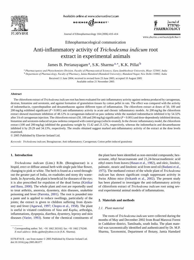
Journal of Ethnopharmacology 104 (2006) 410–414
Ethnopharmacological communication
Anti-inflammatory activity of Trichodesma indicum rootextract in experimental animals
James B. Perianayagam a, S.K. Sharma a,∗, K.K. Pillai b
a Pharmacognosy and Phytochemistry Division, Faculty of Pharmaceutical Sciences, Guru Jambheshwar University, Hisar 125001, Indiab Department of Pharmacology, Faculty of Pharmacy, Jamia Hamdard (Hamdard University), Hamdard Nagar, New Delhi 110062, India
Received 11 June 2004; received in revised form 23 June 2005; accepted 10 August 2005Available online 21 November 2005
Abstract
The chloroform extract of Trichodesma indicum root has been evaluated for anti-inflammatory activity against oedema produced by carrageenan,dextran, histamine and serotonin, and against formation of granulation tissues by cotton pellet in rats. The effect was compared with the activityof indomethacin, cyperoheptadine and dexamethasone against different types of inflammation. The chloroform extract at doses of 50, 100 and200 mg/kg exhibited significant (P < 0.001) anti-inflammatory activity in acute and chronic inflammatory models. At 200 mg/kg the chloroformextract showed maximum inhibition of 48.12% in carrageenan-induced rat paw oedema while the standard indomethacin inhibited it by 54.32%after 3 h of carrageenan injection. The chloroform extract (50, 100 and 200 mg/kg) significantly (P < 0.001) and dose-dependently inhibited dextran,histamine and serotonin-induced rat paw oedema compared with control group (vehicle-treated). In the chronic inflammatory model, the chloroformextract (100 and 200 mg/kg) inhibited the granuloma weight by 15.42 and 21.12%, respectively, whereas the indomethacin and dexamethasoneinhibited it by 29.29 and 34.13%, respectively. The results obtained suggest marked anti-inflammatory activity of the extract at the dose levelsexamined.© 2005 Published by Elsevier Ireland Ltd.
Keywords: Trichodesma indicum; Boraginaceae; Anti-inflammatory; Carrageenan; Cotton pellet-induced granuloma
1. Introduction
Trichodesma indicum (Linn.) R.Br. (Boraginaceae) is ahispid, erect or diffuse annual herb with single pale blue flower,changing to pink or white. The herb is found as a weed through-out the greater part of India, on roadsides and stony dry waste-lands. In Ayurveda, the plant is beneficial for diseases of the eye;it is also prescribed for expulsion of the dead foetus (Kirtikarand Basu, 2000). The whole plant and root are reportedly usedto treat arthritis, anorexia, dysentery, skin diseases, snakebitepoisoning and fever (Parrotta, 2001). The root is pounded intoa paste and is applied to reduce swellings, particularly of thejoints; the extract is given to children suffering from dysen-tery and fever (Agarwal, 1997; Chopra et al., 1958). The plantis useful in vitiated conditions of Vata and Kapha, arthralgia,inflammations, dyspepsia, diarrhea, dysentery, leprosy and skindiseases (Varier, 1993). Some of the chemical constituents of
∗ Corresponding author. Tel.: +91 1662 263162; fax: +91 1662 276240.E-mail address: drsks [email protected] (S.K. Sharma).
the plant have been identified as non-steroidal compounds; hex-acosane, ethyl hexacosanoate and 21,24-hexacosadienoic acidethyl esters from leaves (Hasan et al., 1982), and oleic, linoleic,palmatic, stearic and linolenic acid from seed oil (Badami et al.,1975). The methanol extract of the whole plant of Trichodesmaindicum has shown significant cough suppressant activity inSwiss Albino mice (Srikanth et al., 2002). The present studyhas been planned to investigate the anti-inflammatory activityof chloroform extract of Trichodesma indicum root using sev-eral experimental animal models of inflammations.
2. Materials and methods
2.1. Plant material
The roots of Trichodesma indicum were collected during themonths of May and December 2002 from Road Maruvai Forestin Cuddalore district, Tamilnadu, south India. The plant mate-rial was taxonomically identified and authenticated by Dr. M.P.Sharma, Taxonomist, Department of Botany, Jamia Hamdard
0378-8741/$ – see front matter © 2005 Published by Elsevier Ireland Ltd.doi:10.1016/j.jep.2005.08.077

J.B. Perianayagam et al. / Journal of Ethnopharmacology 104 (2006) 410–414 411
(Hamdard University), Hamdard Nagar, New Delhi, India. Thevoucher specimen (JBT/19) was deposited in the HerbariumSection of the Pharmacognosy and Phytochemistry Division,Faculty of Pharmaceutical Sciences, Guru Jambheshwar Uni-versity, Hisar (India) for future reference. The roots were driedunder shade, sliced into small pieces, pulverised using a mechan-ical grinder and passed through 40-mesh sieve and stored in anair-tight container for further use.
2.2. Preparation of extract
The powdered roots (4.8 kg) were extracted with chloro-form at room temperature (72 h). After exhaustive extraction,the chloroform extract was concentrated under reduced pres-sure at 50–55 ◦C. A reddish brown coloured extract was obtained(yield 0.38%, w/w, with respect to the dry starting material) andkept in desiccator for further use. The chloroform extract (50,100 or 200 mg/kg) was suspended in 1% (w/v) aqueous carboxymethylcellulose for administration to animals.
2.3. Phytochemical study of extract
Phytochemical screening was carried out to ascertain thequalitative chemical composition of Trichodesma indicumusing commonly employed colour reactions and readily per-formed chromatographic technique (TLC) to identify the majorphytoconstituents. The extract tested positive for steroids(Liebermann, 1885), triterpenoids (Noller et al., 1942) and lipids(Trease and Trease, 1972). The extract was examined sepa-rately on silica gel G plates using the upper organic phase ofthe mixture: benzene:ethyl acetate:water (84:16:50). The spotswere revealed by antimony trichloride reagent (Stahl, 1969). Theextract gave the picture revealing the presence of four spots hav-ing the following hRf values and colours: 7 (blue), 15 (pink), 60(violet) and 84 (brown).
2.4. Experimental animals
Swiss Albino mouse (30–45 g) and Albino Wistar rats(150–200 g) of either sex were used for experimental study. Theanimals were housed in colony cages at 25 ± 2 ◦C and relativehumidity (50 ± 5%) with 12-h light:12-h dark cycle. All the ani-mals were acclimatised to laboratory environment for a weekbefore the experiment. They were provided with free accessto food (Lipton India Ltd., Bangalore) and water ad libitum.The Institutional Animal Ethics Committee approved the exper-imental protocols and care of animals was taken according toCPCSEA guidelines.
2.5. Acute toxicity study
Acute toxicity study was performed as per OECD-423 guide-lines (Ecobichon, 1997). Swiss Albino mice of either sex wereused. The animals were fasted for 4 h, but allowed free accessto tap water throughout. The fasted mice were divided into twogroups of six animals each. The extract was administered orallyat a dose of 5 mg/kg. The control group received a similar vol-
ume of 1% (w/v) carboxy methylcellulose solution (5 ml/kg).Mortality in each group was observed for 3 days. If mortalitywas not observed, the procedure was repeated for higher dosessuch as 50, 300 and 2000 mg/kg.
2.6. Carrageenan- and dextran-induced rat paw oedema
Oedema was induced by subplantar injection of 0.1 ml of a1% freshly prepared suspension of �-carrageenan (Sigma, St.Louis, MO, USA) or dextran into the right hind paw of eachrat. The paw volume was measured using a plethysmometerbefore 0 and 3 h after the injection of carrageenan (Gunakunruet al., 2004) or 30 min after dextran challenge (Merlos et al.,1990). The chloroform extract of Trichodesma indicum (50, 100and 200 mg/kg) or indomethacin (10 mg/kg) or cyproheptadine(10 mg/kg) or vehicle control (5 ml/kg) were orally adminis-tered to the different groups of rats. All the treatments weregiven orally 1 h prior to the injection of carrageenan or dextraninjection.
2.7. Histamine- and serotonin-induced rat paw oedema
The paw oedema was produced by subplantar administrationof 0.1 ml of a 0.1% freshly prepared solution of histamine orserotonin into the right hind paw of rats. The paw volume wasrecorded before 0 and 1 h after histamine injection (Parmar andGhosh, 1978) or 30 min after serotonin injection (Ghosh andSingh, 1974). Different groups of animals were pretreated withchloroform extract (50, 100 and 200 mg/kg) or with 5 ml/kg of1% (w/v) carboxy methylcellulose (vehicle control) or 10 mg/kgcyproheptadine (standard drug). The drugs were administeredorally 1 h before eliciting paw oedema.
The percentage inhibition of oedema was calculated for allthe above models as described by Tsai and Lin (1999).
2.8. Cotton pellet-induced granuloma
The rats were divided into five groups, each group consistingof six animals. After shaving of the fur, the animals were anaes-thetised. Sterile pre-weighed cotton pellets (50 ± 1 mg) wereimplanted in the axilla region of each rat through a single needleincision (Winter and Porter, 1957). The chloroform extract (100or 200 mg/kg), positive controls (indomethacin 10 mg/kg anddexamethasone 0.5 mg/kg) or vehicle control (1%, w/v, carboxymethylcellulose, 5 ml/kg) were administered to the respectivegroup of animals for seven consecutive days from the day ofcotton pellet implantation. On the eighth day, the animals wereanaesthetised again; the cotton pellets were removed surgicallyand made free from extraneous tissues. The pellets were incu-bated at 37 ◦C for 24 h and dried at 60 ◦C to constant weight.The increment in the dry weight of the pellets was regarded asmeasure of granuloma formation.
2.9. Statistical analysis
The observations were expressed as mean ± S.E.M. The dif-ference in response to test drugs and control was determined

412 J.B. Perianayagam et al. / Journal of Ethnopharmacology 104 (2006) 410–414
Table 1Effect of chloroform extract of Trichodesma indicum on carrageenan- and dextran-induced rat paw oedema
Treatment Dose (mg/kg) Percentage increase in paw volume Percentage inhibition
Carrageenin control – 62.55 ± 1.27 –Indomethacin (standard) 10 28.59 ± 1.53a 54.32
Chloroform extract of Trichodesma indicum 50 44.86 ± 1.69a 28.90100 37.87 ± 1.84a 39.26200 32.46 ± 2.46a 48.12
Dextran control – 44.67 ± 1.04 –Cyproheptadine (standard) 10 23.16 ± 1.54a 47.92
Chloroform extract of Trichodesma indicum 50 36.45 ± 1.72b 18.43100 32.54 ± 1.21c 27.01200 27.98 ± 1.91a 39.32
Each value represents the mean ± S.E.M., n = 6.a P < 0.001.b P < 0.01 compared with control, Dunnett’s t-test after analysis of variance.c P < 0.05.
by one-way analysis of variance followed by Dunnett’s t-test.P < 0.05 was considered as significant.
3. Results
The chloroform extract did not cause mortality even at doselevel of 2000 mg/kg. Hence, the extract was considered assafe for administration up to 2000 mg/kg (X, unclassified). Thechloroform extract at doses of 50, 100 and 200 mg/kg exhib-ited significant (P < 0.001) anti-inflammatory activity in all theanimal models. The chloroform extract (200 mg/kg) exhibitedmaximum inhibition of 48.12% in carrageenan-induced rat pawoedema whereas indomethacin produced 54.32% of inhibitionafter 3 h of carrageenan injection (Table 1). At 200 mg/kg, thechloroform extract elicited significant (P < 0.001) inhibition of39.32% in dextran-induced rat paw oedema while cyprohepta-dine exhibited 47.92% of inhibition (Table 1). The chloroformextract (50, 100 and 200 mg/kg) significantly (P < 0.001) anddose-dependently inhibited histamine-induced rat paw oedema(13.20, 24.93 and 37.24%, respectively) compared with control
group (Table 2). The chloroform extract (200 mg/kg) also exhib-ited maximum inhibition of 43.09% in serotonin-induced rat pawoedema whereas cyproheptadine produced inhibition of 53.92%after 30 min of the serotonin injection (Table 2). In the model ofchronic inflammation using the cotton pellet-induced granulomain rats, the chloroform extract inhibited significantly (P < 0.001)the formation of granulation tissues in a dose-dependent man-ner (Table 3). In this model, 200 mg/kg extract produced 21.12%inhibition of the granuloma weight while indomethacin and dex-amethasone produced 29.29 and 34.13% of inhibition, respec-tively.
4. Discussion
Inflammation is the response of living tissues to injury. Itinvolves a complex array of enzyme activation, mediator release,extravasations of fluid, cell migration, tissue breakdown andrepair (Vane and Bolting, 1995). It is also known that anti-inflammatory effects can be elicited by a variety of chemicalagents and that there is little correlation between their pharma-
Table 2Effect of chloroform extract of Trichodesma indicum on histamine- and serotonin-induced rat paw oedema
Treatment Dose (mg/kg) Percentage increase in paw volume Percentage inhibition
Histamine control – 57.86 ± 1.88 –Cyproheptadine (standard) 10 30.24 ± 1.63a 48.75
Chloroform extract of Trichodesma indicum 50 50.43 ± 1.49b 13.20100 43.26 ± 1.85a 24.93200 36.23 ± 2.93a 37.24
Serotonin control – 47.27 ± 1.71 –Cyproheptadine (standard) 10 21.66 ± 1.21a 53.92
Chloroform extract of Trichodesma indicum 50 37.47 ± 1.16a 20.35100 31.98 ± 1.94a 31.91200 26.85 ± 1.66a 43.09
Each value represents the mean ± S.E.M., n = 6.a P < 0.001.b P < 0.05 compared with control, Dunnett’s t-test after analysis of variance.

J.B. Perianayagam et al. / Journal of Ethnopharmacology 104 (2006) 410–414 413
Table 3Effect of chloroform extract of Trichodesma indicum on cotton pellet-induced granuloma in rats
Treatment Dose (mg/kg) Weight of granulation (mg) Percentage inhibition
Control – 111.88 ± 3.40 –Dexamethasone (standard) 0.5 73.35 ± 3.18a 34.13Indomethacin (standard) 10 78.85 ± 2.54a 29.29
Chloroform extract of Trichodesma indicum 100 93.63 ± 2.24a 15.42200 88.18 ± 2.62a 21.12
Each value represents the mean ± S.E.M., n = 6.a P < 0.001 compared with control, Dunnett’s t-test after analysis of variance.
cological activity and chemical structure (Sertie et al., 1990).This associated with the complexity of the inflammatory pro-cess, makes the use of different experimental models essentialwhen conducting pharmacological trials.
The present study establishes the anti-inflammatory activityof the chloroform extract of Trichodesma indicum in a num-ber of experimental rat models, representing different phases ofinflammation.
The chloroform extract significantly inhibited thecarrageenan-induced paw oedema after 3 h of carrageenanchallenge. Carrageenin-induced rat paw is a suitable experi-mental animal model for evaluating the anti-oedematous effectof natural products and is believed to be biphasic, the firstphase (1 h) involves the release of serotonin and histamineand the second phase (over 1 h) is mediated by prostaglandin,the cylooxygenase products, and the continuity between thetwo phases is provided by kinins (Vinegar et al., 1969). Theextract effectively suppressed the dextran-induced rat pawoedema, but the effect was less than that of cyproheptadine.The dextran-induced oedema is a well-known experimentalmodel in which the oedema is a consequence of liberationof histamine and serotonin from the mast cell (Rowley andBenditt, 1956). The extract also reduced the oedema producedby histamine and serotonin. The results tend to suggest thatthe anti-inflammatory activity of the extract is possibly backedby its anti-histamine or anti-serotonin activity. The chloroformextract exhibited significant anti-inflammatory activity in thecotton pellet-induced granuloma in rats. This reflected itsefficacy to inhibit the proliferative phase of the inflammationprocess, i.e. increase in the number of fibroblasts and synthesisof collagen and mucopolysaccharides during granuloma tissueformation (Arrigoni-Martellie, 1977).
The present study demonstrates the efficacy of Trichodesmaindicum as an anti-inflammatory agent and also scientificallyjustifies the use of this plant as a non-specific anti-inflammatoryagent in folk medicine (Chopra et al., 1958; Varier, 1993).Further detailed investigation is underway to determine theexact phytoconstituents, which are responsible for the anti-inflammatory activity.
References
Agarwal, V.S., 1997. Drug Plants of India. Kalyani Publishers, New Delhi,p. 694.
Arrigoni-Martellie, E., 1977. Inflammation and Anti-inflammatory. SpectrumPublications, New York, pp. 110–120.
Badami, R.C., Deshpande, G.S., Shanbhag, M.R., 1975. Minor seed oils.VII. Examination of seed oils by gas-liquid chromatography. Journalof the Oil Technologists Association of India (Mumbai, India) 7, 76–77.
Chopra, R.N., Chopra, I.C., Handa, K.L., Kapur, L.D., 1958. IndigenousDrugs of India. U.N. Dhur & Sons Pvt. Ltd., Calcutta, p. 528.
Ecobichon, D.J., 1997. The Basis of Toxicology Testing. CRC Press, NewYork, pp. 43–86.
Ghosh, M.N., Singh, H., 1974. Inhibitory effect of a pyrrolizidine alkaloid,crotalaburnine, on rat paw oedema and cotton pellet granuloma. BritishJournal of Pharmacology 51, 503–508.
Gunakunru, A., Padmanaban, K., Thirumal, P., Vengatesan, N., Gnansekar,N., Raja, S., Rajarajan, A.T., Vijay Kumar, S.G., Britto Perianayagam,J., 2004. Chemical investigations and anti-inflammatory activity offixed oil of Butea monosperma seeds. Natural Product Sciences 10,55–58.
Hasan, M., Ahamad, S., Mohamood, K., 1982. Chemical investigation of Tri-chodesma indicum leaves. I. Nonsteroidol constituents of the petroleumether extract. Journal of the Chemical Society of Pakistan 4, 281–283.
Kirtikar, K.R., Basu, B.D., 2000. Indian Medicinal Plants. Srisatguru Publi-cations, Delhi, pp. 2335–2337.
Liebermann, C., 1885. Uber das Oxychinoferben. Berichte 18, 1803–1809.
Merlos, M., Gomez, L.A., Vericat, L., Garcia-Rafanell, J., Forn, J., 1990.Comparative study of the effect of CV-6909, a specific PAF-antagonist,on rat paw oedema caused by different phlogogen agents. Pharmcology40, 211–217.
Noller, C.R., Smith, R.A., Harris, G.R., Walker, J.M., 1942. Saponins andsapogenins. Some color reactions of triterpenoid sapogonins. Journal ofAmerican Chemical Society 64, 3047.
Parmar, N.S., Ghosh, M.N., 1978. Anti-inflammatory activity of gossypina bioflavonoid isolated from Hibiscus vitifolius Linn. Indian Journal ofPharmacology 10, 277–293.
Parrotta, A.J., 2001. Healing Plants of Peninsular India. CABI Publishing,New York, p. 185.
Rowley, D.A., Benditt, E.P., 1956. 5-Hydoxytryptamine and histamine asmediators of the vascular injury produced by agents which dam-age mast cells in rats. Journal of Experimental Medicine 103, 399–415.
Sertie, J.A., Basile, A.C., Panizza, S., Matida, A.K., Zelink, R., 1990. Anti-inflammatory activity and sub-acute toxicity of artemetin. Planta Medica56, 36–40.
Srikanth, K., Murugesan, T., Kumar, C.A., Suba, V., Das, A.K., Sinha, S.,Arunchalam, G., Manikandan, L., 2002. Effect of Trichodesma indicumextract on cough reflex induced by sulphur dioxide in mice. Phy-tomedicine 9, 75–77.
Stahl, E., 1969. Thin layer chromatography. In: A Laboratory Handbook,second ed. Springer-Verlag, Berlin, New York, p. 857.
Trease, G.E., Trease, N.C., 1972. Pharmacognosy. Crowell-Collier andMacmillan Publishers Ltd., London, pp. 140–147.

414 J.B. Perianayagam et al. / Journal of Ethnopharmacology 104 (2006) 410–414
Tsai, C.C., Lin, C.C., 1999. Anti-inflammatory effects of Taiwan folkmedicine ‘Teng-khia-U’ on carrageenan and adjuvant-induced pawoedema in rats. Journal of Ethnopharmacology 64, 85–89.
Vane, J.R., Bolting, R.M., 1995. New insights into the mode of action ofanti-inflammatory drugs. Inflammation Research 44, 1–10.
Varier, S.P.V., 1993. Indian Medicinal Plants. Orient Longman Ltd., Madras,pp. 316–317.
Vinegar, R., Schreiber, W., Hugo, R., 1969. Biphasic development of car-rageenan oedema in rats. Journal of Pharmacology and ExperimentalTherapeutics 166, 96–103.
Winter, C.A., Porter, C.C., 1957. Effect of alteration in side chainupon anti-inflammatory and liver glycogen activities in hydrocortisoneesters. Journal of the American Pharmaceutical Association 46, 515–519.

Journal of Ethnopharmacology 104 (2006) 415–417
Ethnopharmacological communication
Lepidium meyenii (Maca) does not exert direct androgenic activities
P. Bogani a, F. Simonini b, M. Iriti c, M. Rossoni d, F. Faoro e, A. Poletti b, F. Visioli a,∗a Department of Pharmacological Sciences, University of Milan, Via Balzaretti 9, 20133 Milan, Italy
b Institute of Endocrinology, Centre of Excellence for the Study and Treatment of Neurodegenerative Diseases,University of Milan, Via Balzaretti 9, 20133 Milano, Italy
c Plant Pathology Institute, University of Milan, Italyd Department of Plant Production, University of Milan, Italy
e Plant Virology Institute, National Research Council (CNR), Italy
Received 26 May 2005; received in revised form 14 September 2005; accepted 22 September 2005Available online 18 October 2005
Abstract
Maca is the edible root of the Peruvian plant Lepidum meyenii, traditionally employed for its purported aphrodisiac and fertility-enhancingproperties.
This study aimed at testing the hypothesis that Maca contains testosterone-like compounds, able to bind the human androgen receptor andpromote transcription pathways regulated by steroid hormone signaling.
Maca extracts (obtained with different solvents: methanol, ethanol, hexane and chloroform) are not able to regulate GRE (glucocorticoid responseelement) activation. Further experiments are needed to assess which compound, of the several Maca’s components, is responsible of the observedin vivo effects.© 2005 Elsevier Ireland Ltd. All rights reserved.
Keywords: Maca; Lepidium meyenii; Androgen receptor
1. Introduction
Lepidium meyenii, also known as “Maca”, belongs to the plantfamily of Brassicaceae and was discovered more than 2000 yearsago in the highlands of central Peru (Balick and Lee, 2002).Ethnobotanical surveys demonstrate that Maca was widely usedduring the precolonial and colonial periods of Peru under theSpaniards, who first described of the plant and its properties(Zheng et al., 2000).
Maca roots are edible and rural traditions praise their positiveeffects on human fertility and their high nutritional value. Today,Maca is thought to be useful in the treatment of depression,cancer, menstrual and sexual disorders as well as for mem-ory dysfunction (Balick and Lee, 2002). However, these claimshave rarely been validated by experimental data. Some stud-
Abbreviations: GRE, glucocorticoid response element; RPMI, roswell parkmemorial institute medium; FCS, fetal calf serum; hAR, human androgen recep-tor; LCPS, luminescence counts per minute
∗ Corresponding author. Tel.: +39 0250318280; fax: +39 0250318284.E-mail address: [email protected] (F. Visioli).
ies have already been conducted in vitro, to identify the activecomponents of this plant (Zhao et al., 2005), and in vivo todemonstrate the effects of Maca in rats and humans (Gonzaleset al., 2002; Eddouks et al., 2005). These studies have found thatMaca extracts exert interesting biological activities and containcompounds whose activities are similar to those of testosterone.Actually, serum testosterone levels are directly related to sexualdesire (Gonzales et al., 2004), but data in the literature suggestthat testosterone should not be used to improve sexual desire inmen with normal serum concentrations, because of its effects onthe development of prostate cancer (Nelson and Witte, 2002).Other studies (Zheng et al., 2000; Cicero et al., 2001) indicatedthat oral administration of Maca improves sexual performance,without changing reproductive hormone levels (Gonzales et al.,2005).
Thereby, the hypothesized correlation between Maca intakeand fertility still requires biochemical confirmation. Forthis reason, we have tested the effects of different Macaextracts (obtained with different solvents: methanol, ethanol,hexane, and chloroform) on a gene reporter regulated byandrogens.
0378-8741/$ – see front matter © 2005 Elsevier Ireland Ltd. All rights reserved.doi:10.1016/j.jep.2005.09.028

416 P. Bogani et al. / Journal of Ethnopharmacology 104 (2006) 415–417
2. Materials and methods
2.1. Plant material
A voucher specimen of Maca (Lepidium meyenii Walp.) hasbeen identified, classified, and deposited in the herbarium of theSection of Systematic Botany, Department of Biological Sci-ences, University of Milan, by professor Giuseppe Patrignani.
The powdered tuber (50 g) was extracted with either chlo-roform, methanol, ethanol, or hexane (150 ml), at room tem-perature for 1.5 h. The extracts were brought to dryness undernitrogen. After resuspension in a known volume, the content ofthe extracts was quantified by microgravimetry. Following fur-ther evaporation, the extracts were resuspended in DMSO to afinal concentration of 100 �g/�l.
2.2. Luciferase and β-galactosidase assay
Androgen-independent prostate cancer cells DU145 wereplated into 24-well plates at 300,000 cells/ml, 1 day before trans-fection. The cells were transiently transfected with 0.5 �g/wellof a plasmid containing a glucocorticoid response element(GRE), controlling luciferase gene, 0.19 �g/well of a plas-mid codifying for the human androgen receptor (pCMV-hAR),0.03 �g/well of a plasmid used to evaluate transfection efficiency(pEGFP-N1), and 0.25 �g/well of �-galactosidase expressionvector (pCMV�).
Three hours after transfection, samples were incubated withMaca extracts to a final concentration of 1, 10, and 50 �g/�l.Medium was replaced after 24 h with RPMI 1640 (BiochromKG, Berlin, Germany) supplemented with 5% charcoal-strippedFCS (CS-FCS), to remove any possible effects of the endoge-nous hormones found in calf serum, and the treatments wererepeated.
For the luciferase assay, the medium was replaced with 150 �lRPMI/well and the reaction was stopped by adding 150 �l/wellof luciferase cell culture lysis reagent (Perkin-Elmer, Wellesley,MA, USA). Luciferase activity was determined as luminescencecounts per second (LCPS). �-Galactosidase assay was used tonormalize the results (Dondi et al., 2001; Scaccianoce et al.,2003).
2.3. Statistical analysis
Data are reported as means ± standard error of the mean(S.E.). Statistical analysis was performed using the ANOVA test,to compare control values with all treatments. A p < 0.05 was asconsidered statistically significant.
3. Results and discussion
Lepidium meyenii is the only cruciferous vegetable (Bras-sicaceae family) native to the Americas. The tuber is the onlyplant organ employed in human nutrition (Dini et al., 1994).The effects of Maca consumption on human reproductive poten-tial, i.e. significantly enhanced libido and spermatogenesis, ledmany authors to hypothesize pharmacological actions of Maca
(Zheng et al., 2000; Cicero et al., 2001; Cicero et al., 2002).Indeed, the chemical composition of Maca tuber includes anarray of secondary metabolites arising from different pathways,namely glucosinolates, phenylpropanoids (polyphenols), iso-prenoids (monoterpens and sesquiterpens), and alkaloids (Diniet al., 2002; Piacente et al., 2002; Sandoval et al., 2002; Tellezet al., 2002; Gonzales et al., 2005). Much research has beenpublished, in recent years, to show how Maca interacts with itstarget in vivo and in vitro (Cicero et al., 2001; Gonzales et al.,2003; Zhao et al., 2005). At present, it is not clear whether theeffects of Maca are due to direct activities of its componentson selected cells or to indirect, namely central effects. The aimof this study was to determine if Maca extracts bind the andro-gen receptor and, hence, carry the potential to directly stimulatespermatogenesis and sexual activity. We used various solventsto extract Maca, because of the different biological activitiesreported for its different fractions (Cicero et al., 2002). How-ever, even though activation of the GRE (an enhancer sequenceactivated by AR) was, as expected, greatly stimulated by hARactivated by dihydrotestosterone in prostate DU145 cells, noneof the Maca extracts (1, 10, and 50 �g/�l) were able to activateAR-mediated transcription (Fig. 1). Therefore, our data excludea direct effect of Maca on genes regulated by androgens. It hasalso been postulated that the Maca effects on fertility and sex-ual desire might be modulated by the hypothalamic-pituitaryaxis, through regulation of hormone secretion, but studies per-formed on humans and animals have demonstrated that treatmentwith Maca does not affect serum reproductive hormone levels(Gonzales et al., 2002).
Recently, a metabolite of the aromatic glucosinolates wasdescribed as a specific antagonist of the androgen receptor; there-fore, it is possible that the effects of Maca are due, at least inpart, to interactions between glucosinolates and the androgenreceptor (Le et al., 2003). Also, it is conceivable that Maca con-tains phyto-estrogens: the potential role of phyto-estrogens onhuman male fertility has been attributed to both estrogenic oranti-estrogenic activities (Rochira et al., 2001). Indirect effectsof Maca, on the other hand, can be consequent to its anti-stress
Fig. 1. Regulation by Maca extracts (hexane, chloroform, methanol, andethanol) of the promoter activity of the GRE gene in DU145 cells. Cells weretreated with different concentrations of Maca extracts (1, 10 and 50 �g/�l).Experimental data were obtained in quadruplicate and are presented as the mean(±S.E.) luciferase activity of reporter vectors. All data were compared with pos-itive control (DHT 10−9 M) and vehicle alone (DMSO, 10%).
P. Bogani et al. / Journal of Ethnopharmacology 104 (2006) 415–417 417
and anti-depressive properties, as hypothesized by Gonzales etal. (2003).
In synthesis, our study shows that Maca does not modu-late androgen receptors (Fig. 1). Therefore, although there isevidence associating enhanced sexual libido, fertility, and sper-matogenesis with Maca consumption, a pharmacological andcausal relationship between Maca ingestion and its effects onhuman reproduction is yet to be ascertained, in order to identifythe mechanism(s) of action of Maca components. Further, chem-ical and molecular research is needed to identify which of themany Maca components is responsible for the observed effects.Finally, in vivo experiments are to be undertaken, to confirm thepossibility of using Maca supplementation as a tool to improvefertility (Cicero et al., 2001).
Acknowledgement
We thank Ecoandino (Lima, Peru) for the supply of Maca.
References
Balick, M.J., Lee, R., 2002. Maca: from traditional food crop to energy andlibido stimulant. Alternative Therapies in Health and Medicine 8, 96–98.
Cicero, A.F., Bandieri, E., Arletti, R., 2001. Lepidium meyenii Walp. improvessexual behaviour in male rats independently from its action on sponta-neous locomotor activity. Journal of Ethnopharmacology 75, 225–229.
Cicero, A.F., Piacente, S., Plaza, A., Sala, E., Arletti, R., Pizza, C., 2002.Hexanic Maca extract improves rat sexual performance more effectivelythan methanolic and chloroformic Maca extracts. Andrologia 34, 177–179.
Dini, F., Migliuolo, G., Rastrelli, L., Saturno, P., Schettino, O., 1994. Chim-ical composition of Lepidium meyenii. Food Chemistry 49, 347–349.
Dini, I., Tenore, G., Dini, A., 2002. Glucosinolates from Maca (Lepidiummeyenii). Biochemical Systematics and Ecology 30, 1087–1090.
Dondi, D., Maggi, R., Scaccianoce, E., Martini, L., Motta, M., Poletti, A.,2001. Expression and role of functional glucocorticoid receptors in thehuman androgen-independent prostate cancer cell line, DU145. Journalof Molecular Endocrinology 26, 185–191.
Eddouks, M., Maghrani, M., Zeggwagh, N.A., Michel, J.B., 2005. Study ofthe hypoglycaemic activity of Lepidium sativum L. aqueous extract innormal and diabetic rats. Journal of Ethnopharmacology 97, 391–395.
Gonzales, G.F., Cordova, A., Vega, K., Chung, A., Villena, A., Gonez, C.,2003. Effect of Lepidium meyenii (Maca), a root with aphrodisiac andfertility-enhancing properties, on serum reproductive hormone levels inadult healthy men. Journal of Endocrinology 176, 163–168.
Gonzales, G.F., Cordova, A., Vega, K., Chung, A., Villena, A., Gonez, C.,Castillo, S., 2002. Effect of Lepidium meyenii (Maca) on sexual desireand its absent relationship with serum testosterone levels in adult healthymen. Andrologia 34, 367–372.
Gonzales, G.F., Gasco, M., Cordova, A., Chung, A., Rubio, J., Villegas, L.,2004. Effect of Lepidium meyenii (Maca) on spermatogenesis in malerats acutely exposed to high altitude (4340 m). Journal of Endocrinology180, 87–95.
Gonzales, G.F., Miranda, S., Nieto, J., Fernandez, G., Yucra, S., Rubio, J.,Yi, P., Gasco, M., 2005. Red Maca (Lepidium meyenii) reduced prostatesize in rats. Reproductive Biology and Endocrinology 3, 5.
Le, H.T., Schaldach, C.M., Firestone, G.L., Bjeldanes, L.F., 2003. Plant-derived 3,3′-diindolylmethane is a strong androgen antagonist in humanprostate cancer cells. Journal of Biological Chemistry 278, 21136–21145.
Nelson, K.A., Witte, J.S., 2002. Androgen receptor CAG repeats and prostatecancer. American Journal of Epidemiology 155, 883–890.
Piacente, S., Carbone, V., Plaza, A., Zampelli, A., Pizza, C., 2002. Investiga-tion of the tuber constituents of Maca (Lepidium meyenii Walp.). Journalof Agricultural and Food Chemistry 50, 5621–5625.
Rochira, V., Balestrieri, A., Madeo, B., Baraldi, E., Faustini-Fustini, M.,Granata, A.R., Carani, C., 2001. Congenital estrogen deficiency: in searchof the estrogen role in human male reproduction. Molecular and CellularEndocrinology 178, 107–115.
Sandoval, M., Okuhama, N., Angeles, F., Melchor, V., Condezo, L., Lao, J.,Miller, J., 2002. Antioxidant activity of the cruciferous vegetable Maca(Lepidium meyenii). Food Chemistry 79, 207–213.
Scaccianoce, E., Festuccia, C., Dondi, D., Guerini, V., Bologna, M., Motta,M., Poletti, A., 2003. Characterization of prostate cancer DU145 cellsexpressing the recombinant androgen receptor. Oncology Research 14,101–112.
Tellez, M.R., Khan, I.A., Kobaisy, M., Schrader, K.K., Dayan, F.E., Osbrink,W., 2002. Composition of the essential oil of Lepidium meyenii (Walp.).Phytochemistry 61, 149–155.
Zhao, J., Muhammad, I., Dunbar, D.C., Mustafa, J., Khan, I.A., 2005. Newalkamides from maca (Lepidium meyenii). Journal of Agricultural andFood Chemistry 53, 690–693.
Zheng, B.L., He, K., Kim, C.H., Rogers, L., Shao, Y., Huang, Z.Y., Lu,Y., Yan, S.J., Qien, L.C., Zheng, Q.Y., 2000. Effect of a lipidic extractfrom Lepidium meyenii on sexual behaviour in mice and rats. Urology55, 598–602.
Journal of Ethnopharmacology 104 (2006) 418–422
Ethnopharmacological communication
Screening of plants used in Danish folk medicine to treat memorydysfunction for acetylcholinesterase inhibitory activity
Anne Adsersen ∗, Bente Gauguin, Lene Gudiksen, Anna K. JagerDepartment of Medicinal Chemistry, The Danish University of Pharmaceutical sciences, 2 Universitetsparken, 2100 Copenhagen O, Denmark
Received 24 June 2005; received in revised form 14 September 2005; accepted 22 September 2005Available online 8 November 2005
Abstract
Aqueous and methanolic extracts of 11 plants, used in Danish folk medicine for improvement of memory and cognition, and 3 Corydalis specieswere tested for acetylcholinesterase inhibitory activity using the Ellman colorimetric method. Significant inhibitory activity in dose-dependentmanner was observed for extracts of Corydalis cava, Corydalis intermedia, Corydalis solida ssp. laxa and Corydalis solida ssp. slivenensis. Extractsof Ruta graveolens, Lavandula angustifolia, Rosmarinus officinalis, Petroselinum crispum and Mentha spicata exhibited moderate inhibition ofthe enzyme, defined as more than 15% at 0.1 mg/ml.© 2005 Elsevier Ireland Ltd. All rights reserved.
Keywords: AChE; Acetylcholinesterase inhibition; Danish folk medicine; Memory dysfunction
1. Introduction
Alzheimer’s disease (AD) is the most common form ofdementia among the elderly. In AD patients there is decreasedlevels of acetylcholine in the brain areas related to memory andlearning (Lahiri et al., 2002). Based on the cholinergic hypoth-esis that memory impairments in patients suffering from ADresult from a defect in the cholinergic system, an importantapproach to treat this disease is to enhance the acetylcholine levelin the brain by inhibition of the enzyme acetylcholinesterase(AChE) (Shetty and Woodhouse, 1999).
Plant species, wild growing and cultivated, were selectedbased on the comprehensive work of the Danish ethnobotanistV.J. Brøndegaard (Brøndegaard, 1978). This standard workdescribes the usage of plants in Denmark for different purposes,including medicinal uses, from the Middle Age until now. Plantspecies used as memory enhancers were identified and further-more Corydalis species were selected as several species of thisgenera have been used in treatment of memory dysfunction infolk medicines (Orhan et al., 2004).
Abbreviations: AD, Alzheimer’s disease; AChE, acetylcholinesterase;ATCI, acetylthiocholine iodide; DTNB, 5,5′-dithio-bis(2-nitrobenzoic acid);DRG, Dragendorff reagent
∗ Corresponding author. Tel.: +45 3530 6295; fax: +45 3530 6041.E-mail address: [email protected] (A. Adsersen).
2. Material and methods
2.1. Plant material
Plant materials were collected in 2003 and 2004 at vari-ous locations in Denmark, or bought from an herbal dealer.Voucher specimens and samples are deposited in Depart-ment of Medicinal Chemistry, The Danish University of Phar-maceutical Sciences. The collected material was dried at40 ◦C.
2.2. Preparation of plant extracts
One gram of dried, powdered plant material was extractedwith 2× 10 ml demineralized water or methanol 2× 30 minin an ultrasonic bath. The extracts were filtered and evapo-rated to dryness. The residues were redissolved in methanol ordemineralized water, respectively, to yield a concentration of10 mg/ml.
2.3. Acetylcholinesterase inhibition assay
AChE inhibitory activity was detected by a microtitre plateassay based on Ellman’s method (Rhee et al., 2001) and a thinlayer chromatography (TLC) bioautographic assay (Rhee et al.,2001; Risa et al., 2004) was used to evaluate the plant extractsmost active in the microtitre plate assay.
0378-8741/$ – see front matter © 2005 Elsevier Ireland Ltd. All rights reserved.doi:10.1016/j.jep.2005.09.032

A. Adsersen et al. / Journal of Ethnopharmacology 104 (2006) 418–422 419
2.3.1. Microtitre plate assayAcetylcholinesterase (AChE) from electric eel (type VI-
S), acetylthiocholine iodide (ATCI) and 5,5′-dithio-bis(2-nitrobenzoic acid) (DTNB) was purchased from Sigma. In the96-well plates, 25 �l 15 mM ATCI in Millipore water, 125 �l3 mM DTNB in buffer C (50 mM Tris–HCl, pH 8, 0.1 M NaCl,0.02 M MgCl2·6H2O), 50 �l buffer B (50 mM Tris–HCl, pH 8,0.1% bovine serum albumin), 25 �l plant extract at concentra-tions of 1, 0.5 and 0.25 mg/ml (final concentration in assay: 0.1,0.05 and 0.025 mg/ml) were added and the absorbance was mea-sured five times at 405 nm every 13 s in a Labsystems MultiscanEX type 355 plate reader. Then 25 �l 0.22 U/ml AChE wereadded to the wells and the absorbance was measured again eighttimes at 405 nm every 13 s. The reaction rate was calculated byMultiscan EX software version 1.0 and Microsoft Excel. Anyincrease in absorbance due to the spontaneous hydrolysis of sub-strate was corrected by subtracting the rate of the reaction beforeadding the enzyme. The percentage inhibition was calculated bycomparing the rates for the samples to the blank (10% methanolin buffer A for methanol extracts). The experiment was done intriplicate.
2.3.2. TLC bioautographic assayExtracts were applied to TLC plates and after developing,
the TLC plate was sprayed with 5 mM ATCI and 5 mM DTNBin 50 mM Tris–HCl, pH 8 until the silica was saturated withthe solvent. The plate was then sprayed with 3 U/ml AChE dis-solved in 50 mM Tris–HCl, pH 8 at 37 ◦C. After a few minutes ayellow background appeared, with white spots for AChE inhibit-ing compounds. False-positive reactions were eliminated by themethod of Rhee et al. (2003). A TLC plate was developed andsprayed with 5 mM DTNB in 50 mM Tris–HCl, pH 8. After dry-ing, the plate was sprayed with 5 mM ATCI and 3 U/ml AChEdissolved in 50 mM Tris–HCl, pH 8 at 37 ◦C. After a few min-utes a yellow background appeared; occurrence of white spotsindicated false positive reactions.
2.3.3. TLC system—Corydalis spp.Methanolic extracts, 150 �g, were applied to Merck Silica gel
F254 plates (0.2 mm). Toluene:ethylacetate:methanol (30:8:1)was used as eluent and Dragendorff reagent (DRG) as sprayreagent.
3. Results and discussion
Fifteen plant species were selected for investigation and atotal of 40 extracts were tested for AChE inhibitory activity. Theresults obtained with three concentrations of all plant extractsin the microplate assay are shown in Table 1. All the Cory-dalis extracts tested showed strong inhibitory effect on AChEin a dose-dependent manner in the microtitre assay, methano-lic extracts were the most active and tubers were more activethan herbs. The TLC bioautographic assay demonstrated thatthe activity was due to several compounds, only few false posi-tive compounds were detected (Fig. 1 ). The extracts containedseveral compounds with a blue or yellow-green fluorescence at365 nm, which appeared as white spots in the bioautographic
assay and reacted as brown zones with DRG indicating alka-loids (Fig. 1). The occurrence of alkaloids is well documentedin the genus Corydalis and the alkaloids in tuber and herb forthe same species can be different (Hegnauer, 1969), which couldexplain the observed difference in activity between herbs andtubers. It is also possible that there are different levels of thesame alkaloids in the two plant parts. The chloroform:methanol(1:1) extract of Corydalis solida ssp. solida has previously beenshown to exhibit strong AChE inhibitory activity at 1 mg/ml butno noticeable activity at 10 �g/ml (Orhan et al., 2004). Kim et al.(1999) found that a methanolic extract of the tuber of Corydalisternata showed significant inhibition of AChE, further they iso-lated protopine, determined the IC50 value to 50 �M and showedthat mice treated with protopine exhibited diminished scopo-lamine induced dementia measured in a passive avoidance task.Protopine has as well been isolated from the tubers of Corydaliscava (Preininger et al., 1976) and the aerial parts of Corydalissolida ssp. tauricola (Sener and Temizer, 1990). Hwang et al.(1996) isolated berberine from Corydalis ternata and found itwas a reversible and specific AChE inhibitor with 90% inhibitoryeffect at 2.5 �M. Ulrichova et al. (1983) classified coptisine,berberine and sanguinarine as strong AChE inhibitors. Coptisinehas been isolated from Corydalis cava (Preininger et al., 1976)and berberine and sanguinarine from Corydalis solida ssp. tau-ricola (Sener and Temizer, 1990). The mentioned investigationsindicate that protoberberine- and protopine-type alkaloids, com-mon compounds in Corydalis spp., are potent AChE inhibitors.
The only extracts with moderate activity, defined as more than15% inhibition at the highest concentration tested were, apartfrom the extracts of Corydalis spp., aqueous and methanolicextracts of Ruta graveolens and methanolic extracts of Lavan-dula angustifolia, Rosmarinus officinalis, Petroselinum crispumand Mentha spicata. All five species contains essential oilwith terpenes, a group of compounds reported to have AChEinhibitory Activity (Perry et al., 2000). Essential oil can beextracted with methanol, not with water, and this can explainthe higher activity of the methanolic extracts.
Currently no AChE inhibitory activity has been reported fromRuta graveolens. Ruta graveolens contains in addition to essen-tial oil with terpenes, coumarins and alkaloids (Stashenko et al.,2000), groups of compounds reported to have AChE inhibitoryactivity (Howes et al., 2003; Lee et al., 2004).
In the present study some AChE inhibitory activity wasdetected in extracts of Lavandula angustifolia, in contrast Perryet al. (1996) found no AChE inhibitory activity of lavender oilin a concentration of 0.1 �g/ml. An aqueous extract of Lavan-dula angustifolia flowers was shown to diminish glutamate-induced neurotoxicity in rat pups cerebellar granular cell culture(Buyukokuroglu et al., 2003). Antioxidant and relatively weakAChE inhibition was reported for linalool, one of the main com-ponents in lavender oil (Perry et al., 2000, 2003; Savelev et al.,2003). This indicates that several targets relevant to treatmentof AD, cholinergic, neuroprotective and antioxidant activities,could be found in Lavandula angustifolia.
In the present study Melissa officinalis exhibited no AChEinhibitory activity. Kennedy et al. (2003) tested ethanolicextracts of eight samples of dried Melissa officinalis leaf

420 A. Adsersen et al. / Journal of Ethnopharmacology 104 (2006) 418–422
Table 1Screening of Danish plants for acetylcholinesterase inhibitory activity
Species Family Plant part analyzed Extraction solvent AChE inhibition (%)
0.1 mg/ml 0.05 mg/ml 0.025 mg/ml
Carum carvi L. Apiaceae Radix Water 0 0 0Methanol 11 1 0
Herba Water 0 0 0Methanol 0 0 0
Corydalis cava (L.) Schw. et K. Papaveraceae Herba Water 62 37 37Methanol 85 70 34
Tuber Water 92 78 73Methanol 92 83 77
Corydalis intermedia (L.) Merat Papaveraceae Herba Water 57 39 28Methanol 84 75 54
Tuber Water 78 52 49Methanol 97 86 83
Corydalis solida (L.) Swartz ssp. laxa Papaveraceae Herba Water 78 63 61Methanol 89 71 66
Tuber Water 85 82 58Methanol 96 71 63
Corydalis solida (L.) Swartz ssp. slivenensis Papaveraceae Herba Water 48 26 0Methanol 82 61 53
Tuber Water 87 81 68Methanol 97 82 75
Euphrasia nemorosa (Pers.) Wallr. Scrophulariaceae Herba Water 0 0 0Methanol 0 6 0
Lavandula angustifolia Miller Lamiaceae Herba Water 12 0 0Methanol 34 3 2
Melissa officinalis L. Lamiaceae Herba Water 0 0 0Methanol 0 0 0
Mentha spicata L. Lamiaceae Herba Water 3 10 4Methanol 15 0 0
Origanum vulgare L. Lamiaceae Herba Water 0 3 0Methanol 3 0 0
Petroselinum crispum (Mil.) Nym.ex A.W.Hill. Apiaceae Radix Water 0 0 0Methanol 21 14 0
Pimpinella anisum L. Apiaceae Fructus Water 0 0 0Methanol 3 0 0
Rosmarinus officinalis L. Lamiaceae Herba Water 12 0 0Methanol 17 10 0
Ruta graveolens L. Rutaceae Herba Water 22 9 2Methanol 39 30 7
Thymus vulgaris L. Lamiaceae Herba Water 0 2 0Methanol 0 0 0
and found no AChE inhibitory activity in a concentration of0.25 mg/ml. Perry et al. (1996) found inhibitory activity of twodifferent sources of Melissa oil, 0.1 �g/ml caused 76.3 and 100%inhibition, respectively, activity of an ethanolic extract of freshleaves but no activity in an extract of dried leaves in a con-centration of 2.0 mg/ml. Salah and Jager (2005) found weakactivity in an ethyl acetate extract, and citral, a main compo-nent in the essential oil, is known to be a weak AChE inhibitor(Ryan and Byrne, 1988). It was reported that the ethanolic extractof Melissa officinalis displaced [3H]-(N)-nicotine and [3H]-(N)-scopolamine dose-dependent from human brain cell membranes
bearing acetylcholine receptors with IC50 values less than 1 mgdry plant material/ml, indicating that compounds with acetyl-choline receptor affinities are present in the extract (Wake etal., 2000). The results indicates that compounds with potentialfor use in receptor orientated therapies and enzyme orientatedtherapies for AD and other neurodegenerative diseases associ-ated with aging, could be found in Melissa officinalis, but theactivity depends on the source.
Rosemary oil has previously been shown to exhibit moderateactivity (Perry et al., 1996) but currently no AChE inhibitoryactivity has been reported from Petroselinum crispum, and

A. Adsersen et al. / Journal of Ethnopharmacology 104 (2006) 418–422 421
Fig. 1. TLC plates of extracts from Corydalis sp. (A) Acetylcholinesterase inhibitory activity, white spots indicate inhibition. (B) False positive reactions, where thewhite spots are not due to enzyme inhibition. (C) TLC plate in UV-365 nm. (D) TLC plate sprayed with Dragendorff-R. 1–4 are herbs of Corydalis cava, Corydalisintermedia, Corydalis solida ssp. slivenensis and Corydalis solida ssp. laxa, respectively, 5–8 are tubers of the previously mentioned plant species.
Mentha spicata. All three species contains essential oil andAChE inhibitory activity has been detected for several of themonoterpenoids occurring in the oils (Miyazawa et al., 1997),this could explain the activity proven in the present study. Essen-tial oil, aqueous and ethanolic extracts from Pimpinella anisumwere shown to have bronchodilatory effects on isolated guineapig tracheal chains (Boskabady and Ramazani-Assari, 2001).The effect was, at least in part, due to inhibitory effects onmuscarinic receptors that could counteract any possible AChEinhibitory effects. This, and our negative in vitro results, indi-cates that any effect of Pimpinella anisum on improving memorycould be due to some other course of action.
Acknowledgements
The Danish Medical Research Council is thanked for finan-cial support. The Copenhagen University Botanical Gardens isthanked for the donation of certain plant materials.
References
Boskabady, M.H., Ramazani-Assari, M., 2001. Relaxant effect of Pimpinellaanisum on isolated guinea pig tracheal chains and its possible mecha-nism(s). Journal of Ethnopharmacology 74, 83–88.
Brøndegaard, V.J., 1978. Folk og Flora, Dansk Etnobotanik, vols. I–IV.Rosenkilde og Bagger, Copenhagen, Denmark.
Buyukokuroglu, M.E., Gepdiremen, A., Hacimuftuoglu, A., Oktay, M., 2003.The effects of aqueous extract of Lavandula angustifolia flowers inglutamate-induced neurotoxicity of cerebellar granular cell culture of ratpups. Journal of Ethnopharmacology 84, 91–94.
Hegnauer, R., 1969. Chemotaxonomie der Pflanzen V. Birkhauser Verlag,Basel und Stuttgart, 288.
Howes, M.R., Perry, N.S.L., Houghton, P.J., 2003. Plants with traditionaluses and activities, relevant to the management of Alzheimer’s diseaseand other cognitive disorders. Phytotherapy Research 17, 1–18.
Hwang, S.Y., Chang, Y.P., Byun, S.J., Jeon, M.H., Kim, Y.C., 1996. Anacetylcholinesterase inhibitor isolated from Corydalis tuber and its modeof action. Korean Journal of Pharmacognosy 27, 91–95.
Kennedy, D.O., Wake, G., Savelev, S., Tildesley, N.T.J., Perry, E.K., Wesnes,K.A., Scholey, A.B., 2003. Modulation of mood and cognitive perfor-mance following acute administration of single doses of Melissa officinalis(Lemon balm) with human CNS nicotinic and muscarinic receptor-bindingproperties. Neuropsychopharmacology 28, 1871–1881.
Kim, S.R., Hwang, S.Y., Jang, Y.P., Park, M.J., Markelonis, G.J., Oh, T.H.,Kim, Y.C., 1999. Protopine from Corydalis ternata has anticholinesteraseand antiamnesic activities. Planta Medica 65, 218–221.
Lahiri, D.K., Farlow, M.R., Greig, N.H., Sambamurti, K., 2002. Current drugtargets for Alzheimer’s disease treatment. Drug Development Research 56,267–281.
Lee, J.H., Lee, K.T., Yang, J.H., Baek, N.I., Kim, D.K., 2004. Acetyl-cholinesterase inhibitors from twigs of Vaccinium oldhami Miquel.Archives of Pharmacal Research 27, 53–56.
Miyazawa, M., Watanabe, H., Kameoka, H., 1997. Inhibition of acetyl-cholinesterase activity by monoterpenoids with a p-menthane skeleton.Journal of Agricultural and Food Chemistry 45, 677–679.
Orhan, I., Sener, B., Choudhary, M.I., Khalid, A., 2004. Acetylcholinesteraseand butyrylcholinesterase inhibitory activity of some Turkish medicinalplants. Journal of Ethnopharmacology 91, 57–60.
Perry, N., Court, G., Bidet, N., Court, J., Perry, E., 1996. European herbs withcholinergic activities: potential in dementia therapy. International Journalof Geriatric Psychiatry 11, 1063–1069.
Perry, N.S.L., Houghton, P.J., Theobald, A., Jenner, P., Perry, E.K., 2000.In-vitro inhibition of human erythrocyte acetylcholinesterase by Salvialavandulaefolia essential oil and constituent monoterpenes. Journal ofPharmacy and Pharmacology 52, 895–902.
Perry, N.S.L., Bollen, C., Perry, E.K., Ballard, C., 2003. Salvia for dementiatherapy: review of pharmacological activity and pilot tolerability clinicaltrial. Pharmacology, Biochemistry and Behavior 75, 651–659.
422 A. Adsersen et al. / Journal of Ethnopharmacology 104 (2006) 418–422
Preininger, V., Thakur, R.S., Santavy, F., 1976. Isolation and chemistry ofalkaloids from plants of the family Papaveraceae LXVII: Corydalis cava(L.) Sch. et K. (C. tuberosa DC). Journal of Pharmaceutical Sciences 65,294–296.
Rhee, I.K., van de Meent, M., Ingkaninan, K., Verpoorte, R., 2001. Screen-ing for acetylcholinesterase inhibitors from Amaryllidaceae using silicagel thin-layer chromatography in combination with bioactivity staining.Journal of Chromatography A 915, 217–223.
Rhee, I.K., van Rijn, R.M., Verpoorte, R., 2003. Qualitative determinationof false-positive effects in the acetylcholinesterase assay using thin layerchromatography. Phytochemical Analysis 14, 127–131.
Risa, A., Risa, J., Adsersen, A., Stafford, G.I., van Staden, J., Jager, A.K.,2004. Acetylcholinesterase inhibitory activity of plants used as memory-enhancers in traditional South African medicine. South African Journalof Botany 70, 664–666.
Ryan, M.F., Byrne, O., 1988. Plant–insect coevolution and inhibition ofacetylcholinesterase. Journal of Chemical Ecology 14, 1965–1975.
Salah, S.M., Jager, A.K., 2005. Screening of traditionally used Lebaneseherbs for neurological activities. Journal of Ethnopharmacology 97,145–149.
Savelev, S., Okello, E., Perry, N.S.L., Wilkins, R.M., Perry, E.K., 2003.Synergistic and antagonistic interactions of anticholinesterase terpenoidsin Salvia lavandulaefolia essential oil. Pharmacology, Biochemistry andBehavior 75, 661–668.
Sener, B., Temizer, H., 1990. Chemical studies on the alkaloids from Cory-dalis solida subsp. tauricola. Planta Medica 56, 510.
Shetty, H.G.M., Woodhouse, K., 1999. Geriatrics. In: Walker, R., Edwards,C. (Eds.), Clinical Pharmacy and Therapeutics, 2nd ed. Churchill Living-stone, Edinburgh.
Stashenko, E.E., Acosta, R., Martınez, R., 2000. High-resolution gas-chromatographic analysis of the secondary metabolites obtained bysubcritical-fluid extraction from Colombian rue (Ruta graveolens L.).Journal of Biochemical and Biophysical Methods 43, 379–390.
Ulrichova, J., Walterova, D., Preininger, V., Slavık, J., Lenfeld, J., Cushman,M., Simanek, V., 1983. Inhibition of acetylcholinesterase activity by someisoquinoline alkaloids. Planta Medica 48, 111–115.
Wake, G., Court, J., Pickering, A., Lewis, R., Wilkins, R., Perry, E., 2000.CNS acetylcholine receptor activity in European medicinal plants tradi-tionally used to improve failing memory. Journal of Ethnopharmacology69, 105–114.

Journal of Ethnopharmacology 104 (2006) 423–425
Ethnopharmacological communication
Free radical scavenging potential of Chlorophytum tuberosum baker
Sreevidya Narasimhan ∗, Raghavan Govindarajan, Madhavan Vijayakumar, Shanta MehrotraPharmacognosy and Ethnopharmacology Division, National Botanical Research Institute, Rana Pratap Marg, Lucknow 226 001, India
Received 13 December 2004; received in revised form 19 August 2005; accepted 23 September 2005Available online 4 November 2005
Abstract
Chlorophytum tuberosum Baker commonly referred as ‘Musli’ has been widely used as a potent ‘Rasayana’ drug in ‘Ayurveda’ as a rejuvenatorand tonic. Antioxidant potential of Chlorophytum tuberosum has been investigated for their ability to scavenge 1,1,diphenyl picryl hydrazyl (DPPH),nitric oxide radical along with their capacity to reduce lipid peroxidation in rat liver homogenate, chelation of ferrous ion, radical scavenging potentialusing chemiluminescence and their total antioxidant capacity. Sugar, starch, protein, and Vitamin C content were estimated spectrophotometricallyalong with the percentages of the individual amino acids by HPLC and individual sugars by using HPTLC as standardization tool. The extracthas been found to possess antioxidant activity in all the models tested as evident by IC50 values being 225.31, 888.44, 809.22 and 422.97 �g/mlfor scavenging of DPPH, nitric oxide, lipid peroxidation and ferry bi-pyridyl complex, respectively, along with a integral anitoxidant activity of2.986 nmol ascorbic acid/g equivalents in photochemiluminescence assay.© 2005 Elsevier Ireland Ltd. All rights reserved.
Keywords: Chlorophytum tuberosum; Amino acids; Arginine; Antioxidant; Rasayana drug
1. Introduction
Chlorophytum tuberosum Baker commonly referred as‘Safed Musli’ is been widely used in the Indian traditionalsystems of medicine for rejuvenation and instant energy as a‘Rasayana’ drug (Puri, 2003). Traditionally it is used as generaltonic, in treating rheumatism apart from having immunomodu-lating property. The root powder is fried in ghee and chewed incase of apathae in mouth and throat (Jain, 1991). The tubers ofChlorophytum tuberosum are used as a medicinal expectorantand are used in fever. It is also used in leucorrhoea and also asaphrodisiac (Sreevidya et al., 2003).
2. Materials and methods
2.1. Plant material and extraction
Chlorophytum tuberosum Baker were collected in and aroundPune, India. It was authenticated and a voucher specimen wasdeposited in the departmental herbarium (LWG Voucher Spec-imen No. 221272). Dried powdered material (250 g) was mac-
∗ Corresponding author. Tel.: +91 522 2205831; fax: +91 522 2205836.E-mail address: [email protected] (S. Narasimhan).
erated with petroleum ether to remove the fatty substances; themarc was further extracted with 80% aqueous alcohol (3× 1 L)by cold percolation and filtered. The extract was concentratedunder reduced pressure and lyophilised (Labconco, USA) to getdry residue 30 g (12%).
2.2. Animals
Male Sprague–Drawley rats (160–180 g) were purchasedfrom the animal house of the Central Drug Research Institute,Lucknow, India. These were kept in the departmental animalhouse at 26 ± 2 ◦C and relative humidity 44–55% light and darkcycles of 10 and 14 h, respectively for 1 week before the exper-iment. Animals were provided with rodent diet (Amruth, India)and water ad libitum.
2.3. Phytochemical investigation
Plant material was extracted successively with hexane, chlo-roform, ethylacetate, and 80% aqueous alcohol by using sox-helet extraction. Phytochemical screening of these extractswas carried out according to the methods in Harborne(1973). Sugar, starch, proteins and Vitamin C were esti-mated according to the standard spectrophotometric meth-ods described in official methods of analysis (AOAC, 1984)
0378-8741/$ – see front matter © 2005 Elsevier Ireland Ltd. All rights reserved.doi:10.1016/j.jep.2005.09.029
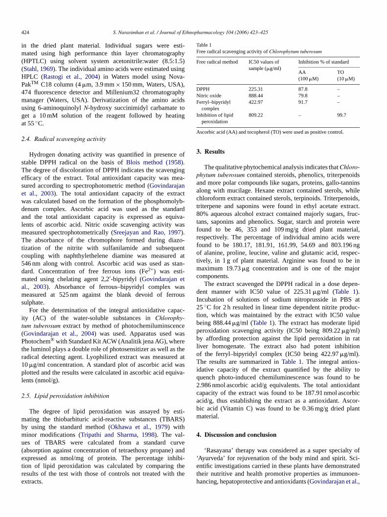
424 S. Narasimhan et al. / Journal of Ethnopharmacology 104 (2006) 423–425
in the dried plant material. Individual sugars were esti-mated using high performance thin layer chromatography(HPTLC) using solvent system acetonitrile:water (8.5:1.5)(Stahl, 1969). The individual amino acids were estimated usingHPLC (Rastogi et al., 2004) in Waters model using Nova-PakTM C18 column (4 �m, 3.9 mm × 150 mm, Waters, USA),474 fluorescence detector and Millenium32 chromatographymanager (Waters, USA). Derivatization of the amino acidsusing 6-aminoquinolyl N-hydroxy succinimidyl carbamate toget a 10 mM solution of the reagent followed by heatingat 55 ◦C.
2.4. Radical scavenging activity
Hydrogen donating activity was quantified in presence ofstable DPPH radical on the basis of Blois method (1958).The degree of discoloration of DPPH indicates the scavengingefficacy of the extract. Total antioxidant capacity was mea-sured according to spectrophotometric method (Govindarajanet al., 2003). The total antioxidant capacity of the extractwas calculated based on the formation of the phosphomolyb-denum complex. Ascorbic acid was used as the standardand the total antioxidant capacity is expressed as equiva-lents of ascorbic acid. Nitric oxide scavenging activity wasmeasured spectrophotometrically (Sreejayan and Rao, 1997).The absorbance of the chromophore formed during diazo-tization of the nitrite with sulfanilamide and subsequentcoupling with naphthylethelene diamine was measured at546 nm along with control. Ascorbic acid was used as stan-dard. Concentration of free ferrous ions (Fe2+) was esti-mated using chelating agent 2,2′-bipyridyl (Govindarajan etal., 2003). Absorbance of ferrous–bipyridyl complex wasmeasured at 525 nm against the blank devoid of ferroussulphate.
For the determination of the integral antioxidative capac-ity (AC) of the water-soluble substances in Chlorophy-tum tuberosum extract by method of photochemiluminscence(Govindarajan et al., 2004) was used. Apparatus used wasPhotochem® with Standard Kit ACW (Analitik jena AG), wherethe luminol plays a double role of photosensitizer as well as theradical detecting agent. Lyophilized extract was measured at10 �g/ml concentration. A standard plot of ascorbic acid wasplotted and the results were calculated in ascorbic acid equiva-lents (nmol/g).
2.5. Lipid peroxidation inhibition
The degree of lipid peroxidation was assayed by esti-mating the thiobarbituric acid-reactive substances (TBARS)by using the standard method (Okhawa et al., 1979) withminor modifications (Tripathi and Sharma, 1998). The val-ues of TBARS were calculated from a standard curve(absorption against concentration of tetraethoxy propane) andexpressed as nmol/mg of protein. The percentage inhibi-tion of lipid peroxidation was calculated by comparing theresults of the test with those of controls not treated with theextracts.
Table 1Free radical scavenging activity of Chlorophytum tuberosum
Free radical method IC50 values ofsample (�g/ml)
Inhibition % of standard
AA(100 �M)
TO(10 �M)
DPPH 225.31 87.8 –Nitric oxide 888.44 79.8 –Ferryl–bipyridyl
complex422.97 91.7 –
Inhibition of lipidperoxidation
809.22 – 99.7
Ascorbic acid (AA) and tocopherol (TO) were used as positive control.
3. Results
The qualitative phytochemical analysis indicates that Chloro-phytum tuberosum contained steroids, phenolics, triterpenoidsand more polar compounds like sugars, proteins, gallo-tanninsalong with mucilage. Hexane extract contained sterols, whilechloroform extract contained sterols, terpinoids. Triterpenoids,triterpene and saponins were found in ethyl acetate extract.80% aqueous alcohol extract contained majorly sugars, fruc-tans, saponins and phenolics. Sugar, starch and protein werefound to be 46, 353 and 109 mg/g dried plant material,respectively. The percentage of individual amino acids werefound to be 180.17, 181.91, 161.99, 54.69 and 803.196 ngof alanine, proline, leucine, valine and glutamic acid, respec-tively, in 1 g of plant material. Arginine was found to be inmaximum 19.73 �g concentration and is one of the majorcomponents.
The extract scavenged the DPPH radical in a dose depen-dent manner with IC50 value of 225.31 �g/ml (Table 1).Incubation of solutions of sodium nitroprusside in PBS at25 ◦C for 2 h resulted in linear time dependent nitrite produc-tion, which was maintained by the extract with IC50 valuebeing 888.44 �g/ml (Table 1). The extract has moderate lipidperoxidation scavenging activity (IC50 being 809.22 �g/ml)by affording protection against the lipid peroxidation in ratliver homogenate. The extract also had potent inhibitionof the ferryl–bipyridyl complex (IC50 being 422.97 �g/ml).The results are summarized in Table 1. The integral antiox-idative capacity of the extract quantified by the ability toquench photo-induced chemiluminescence was found to be2.986 nmol ascorbic acid/g equivalents. The total antioxidantcapacity of the extract was found to be 187.91 nmol ascorbicacid/g, thus establishing the extract as a antioxidant. Ascor-bic acid (Vitamin C) was found to be 0.36 mg/g dried plantmaterial.
4. Discussion and conclusion
‘Rasayana’ therapy was considered as a super specialty of‘Ayurveda’ for rejuvenation of the body mind and spirit. Sci-entific investigations carried in these plants have demonstratedtheir nutritive and health promotive properties as immunoen-hancing, hepatoprotective and antioxidants (Govindarajan et al.,

S. Narasimhan et al. / Journal of Ethnopharmacology 104 (2006) 423–425 425
2005). As carbohydrate plays an important role for the efficacyas immunomodulator, in this study the percentage of free sugarswas calculated. The HPTLC analysis also revealed that Chloro-phytum tuberosum contains sucrose, glucose, fructose in higheramounts and galactose, mannose, xylose in trace amounts. Freeradical scavenging activity of some simple sugars by Fenton-dependent damage has already been reported (Morelli et al.,2003). HPLC profile was used for the quantification of the freeamino acids.
Studies in animal models have suggested a role for NO in thepathogenesis of inflammation and pain and NOS inhibitors havebeen shown to have beneficial effects on some aspects of theinflammation and tissue changes seen in models of inflamma-tory bowel disease (Miller et al., 1992). Photochem® apparatusand method allowed precise as well as time and cost effec-tive determination of the integral antioxidative capacity of theChlorophytum tuberosum extract (Govindarajan et al., 2004).Table 1 shows that extract inhibited FeSO4 induced lipid per-oxidation. The inhibition could be caused by the absence offerryl–perferryl complex or by scavenging of OH radical orsuperoxide radicals or by changing the ratio of Fe3+/Fe2+ orby reducing the rate of conversions of ferrous to ferric or bychelating the iron itself (Braughler et al., 1986). Arginine hasalso been reported to inhibit lipid peroxidation, hence argininemay also in part be responsible for the potent lipid peroxidationof the extract (El-Missiry et al., 2004). In order to test the possi-bility of the change in Fe2+/Fe3+ ratio, a separate experimentwas performed. The extract inhibited the chromogen forma-tion in a dose dependent fashion. Thus there is a possibilitythat the Chlorophytum tuberosum extract chelated the ferrousform and thereby removing the free iron out of the reactionsystem. Since the plant has been found to contain high quanti-ties of the simple sugars, they may also be responsible for theinhibition of the ferryl–bipyridyl complex. The results obtainedsuggest that Chlorophytum tuberosum possesses antioxidant andfree radical scavenging activity suggesting that ethnopharma-cological approach in selecting the plant for study may beuseful.
Acknowledgements
N. Sreevidya and M. Vijayakumar are thankful to CSIR, NewDelhi for providing Senior Research Fellowships.
References
Blois, M.S., 1958. Antioxidant determinations by the use of a stable freeradical. Nature 181, 1199–1200.
Braughler, J.M., Duncan, C.A., Chase, L.R., 1986. The involvement of ironin lipid peroxidation. Importance of ferrous to ferric ratio in initiation.Journal of Biological Chemistry 261, 10282.
El-Missiry, M.A., Othman, A.I., Amer, M.A., 2004. l-Arginine amelioratesoxidative stress in alloxan-induced experimental diabetes mellitus. Journalof Applied Toxicology 24, 93–97.
Govindarajan, R., Rastogi, S., Vijayakumar, M., Rawat, A.K.S., Shirwaikar,A., Mehrotra, S., Pushpangadan, P., 2003. Studies on antioxidant activitiesof Desmodium gangeticum. Biological and Pharmaceutical Bulletin 26,1424–1427.
Govindarajan, R., Vijayakumar, M., Pushpangadan, P., 2005. Antioxidantapproach to disease management and the role of ‘Rasayana’ herbs ofAyurveda. Journal of Ethnopharmacology 99, 165–178.
Govindarajan, R., Vijayakumar, M., Rao, Ch.V., Rawat, A.K.S., Shir-waikar, A., Mehrotra, S., Pushpangadan, P., 2004. Antioxidant poten-tial of Anogeissus latifolia. Biological and Pharmaceutical Bulletin 27,1266–1269.
Harborne, J.B., 1973. Phytohemical Methods. Chapman and Hall InternationalEdition, London.
Jain, S.K., 1991. Dictionary of Indian Folk Medicine and Ethnobotany. DeepPublications, New Delhi.
Miller, M.J., Sadowska, K.H., Chotinaruemol, S., Kakkis, J.L., Clark, D.A.,1992. Amelioration of chronic ileitis by nitric oxide synthase inhibition.Journal of Pharmacology and Experimental Therapeutics 264, 11–16.
Morelli, R., Russo-Volpe, S., Bruno, N., Scalzo, R.L., 2003. Fenton-dependentdamage to carbohydrates: free radical scavenging activity of some simplesugars. Journal of Agriculture and Food Chemistry 51, 7418–7425.
Official methods of Analysis (AOAC), 1984. 14th ed. Williams, S. (Ed.),Association of Official Analytical Chemists Inc., Virginia, USA.
Okhawa, H., Ohishi, N., Yagi, K., 1979. Assay for lipid peroxides in ani-mal tissues by thiobarbituric acid reaction. Analytical Biochemistry 95,351–355.
Puri, H.S., 2003. Rasayana. Taylor and Francis, London.Rastogi, S., Bala, S., Govindarajan, R., Shukla, M., Rawat, A.K.S., Mehrotra,
S., 2004. Quantitative HPLC analysis of amino acids in Chyavanprash:a well known ayurvedic formulation. Indian Journal of PharmaceuticalSciences 66, 753–757.
Sreejayan, N., Rao, M.N.A., 1997. Nitric oxide scavenging by curcuminoids.Journal of Pharmacy and Pharmacology 49, 105.
Sreevidya, N., Kumar, V., Kumar, S., Sikarwar, R.L.S., 2003. Utilization,depletion and conservation of Safed musli (Chlorophytum spp.). Journalof Non-Timber Forest Products 10, 155–157.
Stahl, E., 1969. Thin Layer Chromatography. George Allen & Unwin Ltd.,London.
Tripathi, Y.B., Sharma, M., 1998. Comparison of the antioxidant action ofthe alcoholic extract of Rubia cordifolia with rubiadin. Indian Journal ofBiochemistry and Biophysics 35, 313–316.

Journal of Ethnopharmacology 104 (2006) 426–429
Ethnopharmacological communication
Protective effect of bioactive fraction of Sphaeranthus indicus Linn. againstcyclophosphamide induced suppression of humoral immunity in mice
A.R. Bafna, S.H. Mishra ∗Department of Pharmacy, Faculty of Technology and Engineering, The M.S. University of Baroda, Kalabhavan, Baroda 390 001, Gujarat, India
Received 14 December 2004; received in revised form 27 August 2005; accepted 24 September 2005Available online 10 November 2005
Abstract
The bioactive fraction of Sphaeranthus indicus produced dose dependent increase in humoral immunity and delayed type hypersensitivity (DTH)response as evidenced by increased antibody production and increase in paw edema. The activity at higher doses, however, declines. Humoralantibody (HA) titre lowered by cyclophosphamide (CP) (p < 0.05) was enhanced by bioactive fraction at doses of 200 (p < 0.05) and 400 mg/kg(p < 0.001). There was dose dependent increase in HA titre in normal as well as immunosuppressed animals indicating that drug is effective inhumoral immunity. Animals treated with cyclophosphamide and receiving bioactive fraction showed significant change in DTH response, whichdirectly correlates with cell-mediated immunity, as compared to cyclophosphamide alone. Thus, it can be observed that a bioactive fraction ofSphaeranthus indicus acts as potentiator of DTH. Furthermore, the HPTLC fingerprint profile of the bioactive fraction was established to facilitateits identification and characterization. The results suggest that bioactive fraction influences both humoral and cell-mediated immunity and offersprotection against immunosuppression induced by the cytotoxic agent cyclophosphamide.© 2005 Elsevier Ireland Ltd. All rights reserved.
Keywords: Bioactive fraction; Haemagglutination antibody titre; Delayed type hypersensitivity; Sphaeranthus indicus
1. Introduction
Sphaeranthus indicus Linn. (Compositae) is a herb foundmostly in southern India, bitter stomachic, stimulant, alterative,pectoral and demulcent, and externally emollient (Nadkarni,1976). The preliminary investigation on the methanol extractand its different fractions for immunomodulating activity haveshown promising results and helped in the identification of thebioactive fraction. The methanol fraction showed a dose depen-dent effect with respect to the parameters studied. It was found tooffer protection against cyclophosphamide (CP) induced myelo-suppression (Bafna and Mishra, 2004). Hence, it is designatedas the bioactive fraction of Sphaeranthus indicus. The objec-tive of the present investigation was, therefore, to study theeffect of this bioactive fraction of Sphaeranthus indicus alongwith cyclophosphamide on humoral as well as cellular immu-nity and also to establish its fingerprint profile using HPTLCto facilitate the identification of different components of thefraction.
∗ Corresponding author.E-mail address: [email protected] (S.H. Mishra).
2. Materials and methods
2.1. Animals
Swiss albino mice of either sex, weighing 20–25 g in standardhousehold conditions of temperature, humidity and light wereused. They were fed with standard rodent diet and water adlibitum.
2.2. Plant material and extract preparation
Fresh flower heads of Sphaeranthus indicus were collectedfrom the outfield of city and were authenticated in BotanyDepartment of M.S. University, Baroda. Maceration of air-dried,powdered flower heads of Sphaeranthus indicus afforded 6.65%methanol extract (w/w). The methanol extract was then fraction-ated by maceration into different polarity solvents like petroleumether, benzene, chloroform and methanol. All the respective frac-tions were concentrated under vacuum. The methanol fractionwas found to be biologically effective and hence named ‘bioac-tive fraction’ and utilized in the present study. It gave positivetests for alkaloids, phenolics, flavonoids and carbohydrates onphytochemical screening.
0378-8741/$ – see front matter © 2005 Elsevier Ireland Ltd. All rights reserved.doi:10.1016/j.jep.2005.09.031
A.R. Bafna, S.H. Mishra / Journal of Ethnopharmacology 104 (2006) 426–429 427
2.3. TLC fingerprint profile of bioactive fraction
TLC fingerprint profile was established for the bioactivefraction of Sphaeranthus indicus using HPTLC. Three dif-ferent solvent systems, i.e. toluene:ethyl acetate (7:3), ethylacetate:methanol:water (100:13.5:10) and n-butanol:glacialacetic acid (6:2:2) were used to resolve all the componentspresent in the fraction. The plates were scanned using TLC Scan-ner 3 (CAMAG) at 254 nm (absorbance/reflectance mode) and366 nm (fluorescence/reflectance mode) and Rf values, spectra,λmax and relative percentages were recorded from the scannedarea. Developed chromatograms in different solvent systemswere then sprayed with flavone reagent to detect flavonoid typeof compounds. The compound having Rf 0.07 reacted to thereagent in solvent system 1. Two compounds located at Rf 0.09and 0.36 were reacted in solvent system 2, whereas maximumof five compounds having Rf 0.11, 0.35, 0.38, 0.49 and 0.72reacted to the reagent in solvent system 3.
2.4. Drugs
The bioactive fraction was suspended in 1% sodium carboxymethylcellulose to prepare different doses from 50 to 800 mg/kg.The control animals were given an equivalent volume of sodiumcarboxy methylcellulose vehicle. Cyclophosphamide was usedas a standard immunosuppressant and was administered toone group of animals while all the animals of the other fivedose groups simultaneously received the bioactive fractionalso.
Antigen: Fresh blood was collected from sheep sacrificedin local slaughterhouse. Sheep red blood cells (SRBCs) werewashed three times in normal saline and adjusted to a concen-tration 20% for immunization and 1% for challenge.
2.5. Methods
2.5.1. Humoral antibody (HA) and delayed typehypersensitivity (DTH) response2.5.1.1. Effect of bioactive fraction and cyclophosphamide onHA titre, and DTH response using SRBCs as an antigen inmice—7 days pretreatment. Animals were divided into sevengroups of six animals each. Animals in treatment groupswere given the bioactive fraction (50–800 mg/kg, p.o.) in 1.0%sodium carboxy methyl cellulose daily for 7 days. Cyclophos-phamide was administered on days 4–6 (50 mg/kg).
Animals in control group received equal amount of vehi-cle only. The animals were immunized by injecting 0.1 ml20% fresh sheep red blood cells suspension intraperitonially onday 0. Blood samples, collected in microcentrifuge tubes fromindividual animals by retro-orbital plexus on day 7 were cen-trifuged to obtain the serum. Antibody levels were determinedby haemagglutination technique described by Puri et al. (1994).The reciprocal of the highest dilution of the test serum givingagglutination was taken as the antibody titre. On day 7, the thick-ness of the right hind foot pad was measured using digital verniercalipers and mice were then challenged by injection of 20 �l of1% SRBCs in right hind foot pad. Twenty-four hours after this
challenge foot, thickness was again measured. The differencebetween the pre- and post-challenge foot thickness expressed inmillimetres was taken as a measure of delayed type hypersensi-tivity.
2.5.1.2. Effect of bioactive fraction on HA titre and DTHresponse using SRBCs as an antigen in mice—15 days pretreat-ment. Mice were divided into six groups, each group containingsix mice. Animals in treatment groups were given the bioactivefraction (50–800 mg/kg, p.o.) in 1.0% sodium carboxy methylcellulose. The pretreatment time was 15 days based on themethod described by Sharma et al. (1996). Schedule for drugadministration was 7 days prior to immunization (days −6, −5,−4, −3, −2, −1, 0) and 7 days after immunization (+1, +2, +3,+4, +5, +6, +7). The procedure as described in earlier methodwas followed for the remaining determinations.
2.6. Statistical analysis
Data were expressed as mean ± S.E.M. and differencebetween the groups was statistically determined by analysis ofvariance followed by Tukey–Kramer Multiple Comparisons test,with the level of significance set at p < 0.05.
3. Results
3.1. Humoral antibody and delayed type hypersensitivityresponse
3.1.1. Effect of bioactive fraction and cyclophosphamide onHA titre and DTH response using SRBCs as antigen inmice—7 days pretreatment
Administration of CP resulted in decreased HA titre (Table 1).The reduction was significant (p < 0.05) when compared to con-trol animals. When compared with CP treated animals (groupII), animals receiving bioactive fraction treatment along with CP(groups III–VII) showed dose dependent recovery in HA titre.However, significant increase in HA titre could be obtained at200 (p < 0.05) and 400 mg/kg (p < 0.001) dose. At higher doseit decreased.
A significant increase in paw edema was observed on day 8after challenge on day 7 with SRBCs. Animals treated with CPshowed significant (p < 0.05) DTH response compared to controlanimals. An increase in the DTH response was observed ontreatment of bioactive fraction and cyclophosphamide. However,significant increase in DTH, compared to CP (group II) treatedgroup was noted only at 400 mg/kg (p < 0.001). Thus, it can beobserved that bioactive fraction of Sphaeranthus indicus acts aspotentiator of DTH.
3.1.2. Effect of bioactive fraction on HA titre and DTHresponse using SRBCs as an antigen in mice—15 dayspretreatment
Administration of bioactive fraction produced dose depen-dent increase in the HA titre after challenging with SRBCs(Table 1). Statistically significant increase was observed with200 (p < 0.01) and 400 mg/kg (p < 0.001). Activity declines

428 A.R. Bafna, S.H. Mishra / Journal of Ethnopharmacology 104 (2006) 426–429
Table 1Effect of bioactive fraction of Sphaeranthus indicus on HA titre and DTH response using SRBCs as antigen
Groups Treatment schedules
7 days 15 days
HA titre (mean ± S.E.M.) DTH response (mm) (meanpaw edema ± S.E.M.)
HA titre (mean ± S.E.M.) DTH response (mm) (meanpaw edema ± S.E.M.)
I 64.00 ± 14.31 0.313 ± 0.04 64.00 ± 014.31 0.313 ± 0.04II 2.67 ± 00.42* 0.590 ± 0.05* – –III 3.00 ± 00.44 N.S. 0.576 ± 0.08 N.S. 298.67 ± 71.39 N.S. 0.370 ± 0.03 N.S.IV 56.00 ± 23.18 N.S. 0.670 ± 0.05 N.S. 384.00 ± 57.24 N.S. 0.388 ± 0.04 N.S.V 512.00 ± 114.49* 0.810 ± 0.04 N.S. 640.00 ± 128.00** 0.520 ± 0.09**
VI 1024.00 ± 228.97*** 0.875 ± 0.03* 853.33 ± 107.94*** 0.456 ± 0.07 N.S.VII 192.00 ± 28.62 N.S. 0.546 ± 0.06 N.S. 448.00 ± 131.16 N.S. 0.337 ± 0.05 N.S.F value 15.022 11.726 8.362 6.212P value 0.0001 0.0001 0.0001 0.0005
Values are expressed as mean ± S.E.M. Group I: control group; Group II: cyclophosphamide (CP) treated group. In 7 days treatment schedule: Groups III–VII weretreated with the bioactive fraction at the doses of 50, 100, 200, 400 and 800 mg/kg, respectively, along with cyclophosphamide and were compared with Group II.In 15 days treatment schedule: Groups III–VII were treated with the bioactive fraction alone at the doses of 50, 100, 200, 400 and 800 mg/kg, respectively, and werecompared with Group I. N.S., not significant.
* p < 0.05.** p < 0.01.
*** p < 0.001.
at higher dose. There was dose dependent increase in DTHresponse, but statistically significant increase in paw edemawhen compared to control group was found at 200 mg/kg(p < 0.01) only.
4. Discussion
Immunostimulation in a drug induced immunosuppressionand immunosuppression in an experimental hyper-reactivitymodel by the same preparation can be said to be trueimmunomodulation (Patwardhan et al., 1990). In the presentinvestigation, two schedules of pretreatment period, i.e. 7 and15 days were selected. Results showed dose dependent increasein antibody production and delayed type hypersensitivity. Thestimulation of the humoral response against SRBCs by bioac-tive fraction as evidenced by the increase in the HA titre in micealso indicates the enhanced responsiveness of macrophages andsubsets of T- and B-lymphocytes, involved in antibody synthe-sis (Benacerraf, 1978). Macrophages play an important part incoordinating the processing and presentation of antigen to T- andB-cells, the increase of humoral response to SRBCs reveals thatthe bioactive fraction may enhance the effect by facilitating theprocesses. Cyclophosphamide has a particularly intense effecton short-lived lymphocytes known to include a great proportionof B-cells. And is also one of the most potent inhibitors of anti-body production in most species (Bach, 1976). There was dosedependent increase in HA titre in normal as well as immunosup-pressed animals indicating that the drug is effective in humoralimmunity.
In the similar studies, many natural extracts showed immuno-stimulating activities in immunocompromised animals. Makareet al. (2001) reported reversal of suppression of antibody titreby cyclophosphamide in mice with the treatment of ethanolextract of Mangifera indica containing 2.6% mangiferin in
dose dependent manner. Hafeez et al. (2001) also reportedprotective effect of total aqueous extract of Cassia occiden-talis against cyclophosphamide induced immunosuppression ofhumoral immunity in mice.
Most of the chemotherapeutic agents available today areimmunosuppressants, cytotoxic and exert a variety of side effectsthat are particularly evident in cancer chemotherapy. Botani-cal based immunomodulators are often employed as supportiveor adjuvant therapy to overcome the undesired effects of cyto-toxic chemotherapeutic agents and to restore health to normal.Diwanay et al. (2004) had reported various immunopharma-cological activities of Withania somnifera, Tinospora cordi-folia and Asparagus racemosus in cyclophosphamide treatedmouse ascitic sarcoma. Simultaneous treatment with differ-ent extracts of these plants resulted in protection towards CPinduced immunosuppression as evident by significant increase inwhite cell counts and haemagglutinating and hemolytic antibodytitres.
DTH is antigen specific and causes erythema and inductionat the site of antigen infection in immunized animals. The his-tology of DTH can be different for different species, but thegeneral characteristics are an influx of immune cells at the siteof injection, macrophages and basophills in mice and inductionbecomes apparent within 24–72 h (Poulter et al., 1982). Increasein the DTH response indicates that the drug has a stimulatoryeffect on lymphocytes and necessary cell types required for theexpression reaction (Mitra et al., 1999).
In both the pretreatment schedules, doses of 200 and400 mg/kg were found most effective in inducing immune func-tions. It appears that the range of 200–400 mg/kg is the optimumdose in mice. An increase in dose might have induced down-regulation of immune functions. The response at higher dose waseither identical to control or stimulated as compared to controlanimals. The exertion of an immune response needs a certain

A.R. Bafna, S.H. Mishra / Journal of Ethnopharmacology 104 (2006) 426–429 429
threshold dose, beyond which it does not show any enhance-ment and it has also been reported that immunostimulatingsubstances could exhibit suppressive effects (Wagner and Jurcic,1991).
In the past few years high performance thin layer chro-matography has emerged as a potential tool for rapid and use-ful phytochemical evaluation of herbal drugs (Indian HerbalPharmacopoeia, 1998; Houghton, 1999). TLC fingerprint profilewas established for the bioactive fraction to identify it. Phyto-chemical investigations on Sphaeranthus indicus revealed thepresence of different types of compounds. Two new eudesman-olide, along with a known eudesmanolide and two sesquiter-penoids, cryptomeridiol and 4-epicryptomeridiol have been iso-lated from flower heads (Supada et al., 1992). Three closelyrelated new hydroxy lactone have been isolated from the chlo-roform extract of Sphaeranthus indicus (Gogte et al., 1986).Chughtai et al. (1992) have reported isolation of alkaloids fromflowers. Compounds in the bioactive fraction showed reactionmainly with flavone reagent indicating the flavonoid type ofconstituents. Already, Yadava and Kumar (1998) have reportedisolation of 7-hydroxy-3′,4′,5,6-tetramethoxy-flavone 7-O-�-D-(1 → 4)-di glucoside: a new flavones glycoside from the stem ofSphaeranthus indicus. These types of compounds may also bepresent in flower heads as observed in phytochemical screening.Flavonoids have reported for various pharmacological propertiesincluding antioxidant activity, anticancer and immunomodula-tory effects. A study conducted by Lyu and Park (2005) showedthat flavonoids catechin, epigallocatechin gallate, epicatechin,luteolin, chrysin, quercetin and galangin increased IL-2 secre-tion while apigenin and fisetin inhibited the secretion. Theflavonoid fraction of Tephrosia purpurea was studied for itseffect on cellular and humoral functions and on macrophagephagocytosis in mice (Damre et al., 2003). These observa-tions indicate that flavonoids have the capacity to modulate theimmune response.
On the basis of the results obtained in the present study, itcan be concluded that the bioactive fraction of Sphaeranthusindicus has the potential to stimulate cell-mediated and humoralimmunity.A bioactive fraction also offered protection againstimmunosuppression caused by cyclophosphamide and thereforeit can be supplemented in cancer therapy to deal with adverseeffects of cyclophosphamide. Identification and isolation of phy-toconstituents from bioactive fraction is already taken up andshall follow in our future communications.
Acknowledgement
One of the authors A.R. Bafna wishes to thank CSIR, NewDelhi, for providing financial assistance (SRF) for carrying outthis work.
References
Bach, J.F., 1976. The pharmacological and immunological basis for the useof immunosuppressive drugs. Drugs 11, 1–13.
Bafna, A.R., Mishra, S.H., 2004. Immunomodulatory activity of methanolextract of flower heads of Sphaeranthus indicus Linn. Ars Pharmaceutica45, 281–291.
Benacerraf, B., 1978. A hypothesis to relate the specificity of T lymphocytesand the activity of I region specific Ir genes in macrophages and borrowerlymphocytes. Journal of Immunology 120, 1809–1832.
Chughtai, M.I.D., Khokhar, I., Ahmad, A., 1992. Isolation, purification andstructural determination of alkaloids from the flowers of Sphaeranthusindicus. Science International (Lahore) 4, 151–154.
Damre, A.S., Gokhale, A.B., Phadke, A.S., Kulkarni, K.R., Saraf, M.N.,2003. Studies on the immunomodulatory activity of flavonoidal fractionof Tephrosia purpurea. Fitoterapia 74, 257–261.
Diwanay, S., Chitre, D., Patwardhan, B., 2004. Immunoprotection by botan-ical drugs in cancer chemotherapy. Journal of Ethnopharmacology 90,49–55.
Gogte, M.G., Ananthasubramanian, L., Nargund, K.S., Bhattacharya, S.C.,1986. Some interesting sesquiterpenoids from Sphaeranthus indicus Linn.Indian Journal of Chemistry 25, 233–238.
Hafeez, B.B., Ahmad, I., Haque, R., Raisuddin, S., 2001. Protective effectof Cassia occidentalis L. on cyclophosphamide-induced suppression ofhumoral immunity in mice. Journal of Ethnopharmacology 75, 13–18.
Houghton, P.J., 1999. Pharmacognosy: the basis for quality herbal medicinalproducts. Pharmaceutical News 6, 21–27.
Indian Herbal Pharmacopoeia, vol. I–II. RRL/IDMA, Jammu Tawi/Mumbai,India.
Lyu, S.Y., Park, W.B., 2005. Production of cytokine and NO by RAW 264.7macrophages and PBMC in vitro incubation with flavonoids. Archeivesof Pharmacal Research 28, 573–581.
Makare, N., Bodhankar, S., Rangari, V., 2001. Immunomodulatory activity ofalcoholic extract of Mangifera indica L. in mice. Journal of Ethnophar-macology 78, 133–137.
Mitra, S.K., Gupta, M., Sarma, D.N.K., 1999. Immunomodulatory effect ofIM-133. Phytotherapy Research 13, 341–343.
Nadkarni, K.M., 1976. Indian Mater. Med., vol. 3., third ed. PopularPrakashan, p. 1163.
Patwardhan, B., Kalbagh, D., Patki, P.S., Nagsampagi, B.A., 1990. Search ofimmunomodulatory agents: a review. Indian Drugs 28, 56–63.
Poulter, L.W., Seymour, G.J., Duke, O., Janossy, G., Panayi, G., 1982.Immunohistological analysis of delayed-type hypersensitivity in man. CellImmunology 74, 358–369.
Puri, A., Saxena, R.P., Saxena, K.C., Srivastava, V., Tandon, J.S., 1994.Immunostimulant activity of Nyctanthes arbortristis L. Journal ofEthnopharmacology 42, 31–37.
Sharma, M.L., Kaul, A., Khajuria, A., Singh, S., Singh, G.B., 1996.Immunomodulatory activity of boswellic acids (pentacyclic triterpeneacids) from Boswellia Serrata. Phytotherapy Research 10, 107–112.
Supada, R., Rojatkar, S.R., Bhimsen, A., Nagsampagi, B.A., 1992. 7-Hydroxylated eudesmanolides from Sphaeranthus indicus Linn. Phyto-chemistry 31, 3270–3271.
Wagner, H., Jurcic, K., 1991. Assays for immunomodulation and effectson mediators of inflammation. Methods in Plant Biochemistry, vol. 6.Academic Press, London, p. 207.
Yadava, R.N., Kumar, S., 1998. 7-Hydroxy-3′,4′,5,6-tetramethoxy-flavone 7-O-�-D-(1 → 4)-diglucoside: a new flavone glycoside from the stem ofSphaeranthus indicus Linn. Journal of Chemists (India) 70, 164–166.