next generation sequencing detects premeiotic errors in
TRANSCRIPT
�����������������
Citation: Ghevaria, H.; SenGupta, S.;
Naja, R.; Odia, R.; Exeter, H.; Serhal,
P.; Gonzalez, X.V.; Sun, X.; Delhanty, J.
Next Generation Sequencing Detects
Premeiotic Errors in Human Oocytes.
Int. J. Mol. Sci. 2022, 23, 665. https://
doi.org/10.3390/ijms23020665
Academic Editor: Pilar Coy
Received: 12 November 2021
Accepted: 4 January 2022
Published: 8 January 2022
Publisher’s Note: MDPI stays neutral
with regard to jurisdictional claims in
published maps and institutional affil-
iations.
Copyright: © 2022 by the authors.
Licensee MDPI, Basel, Switzerland.
This article is an open access article
distributed under the terms and
conditions of the Creative Commons
Attribution (CC BY) license (https://
creativecommons.org/licenses/by/
4.0/).
International Journal of
Molecular Sciences
Article
Next Generation Sequencing Detects Premeiotic Errors inHuman OocytesHarita Ghevaria 1 , Sioban SenGupta 1,*, Roy Naja 1, Rabi Odia 2, Holly Exeter 2, Paul Serhal 3,Xavier Viñals Gonzalez 1, Xuhui Sun 1 and Joy Delhanty 1
1 Preimplantation Genetics Group, Institute for Women’s Health, University College London (UCL),London WC1E 6HX, UK; [email protected] (H.G.); [email protected] (R.N.);[email protected] (X.V.G.); [email protected] (X.S.); [email protected] (J.D.)
2 Embryology Department, The Centre for Reproductive and Genetic Health, London W1W 5QS, UK;[email protected] (R.O.); [email protected] (H.E.)
3 Clinical Department, The Centre for Reproductive and Genetic Health, London W1W 5QS, UK;[email protected]
* Correspondence: [email protected]
Abstract: Autosomal aneuploidy is the leading cause of embryonic and foetal death in humans. Thisarises mainly from errors in meiosis I or II of oogenesis. A largely ignored source of error stemsfrom germinal mosaicism, which leads to premeiotic aneuploidy. Molecular cytogenetic studiesemploying metaphase fluorescence in situ hybridization and comparative genomic hybridisationsuggest that premeiotic aneuploidy may affect 10–20% of oocytes overall. Such studies have beencriticised on technical grounds. We report here an independent study carried out on unmanipulatedoocytes that have been analysed using next generation sequencing (NGS). This study confirms thatthe incidence of premeiotic aneuploidy in an unselected series of oocytes exceeds 10%. A totalof 140 oocytes donated by 42 women gave conclusive results; of these, 124 (88.5%) were euploid.Sixteen out of 140 (11.4%) provided evidence of premeiotic aneuploidy. Of the 140, 112 oocytes wereimmature (germinal vesicle or metaphase I), of which 10 were aneuploid (8.93%); the remaining28 were intact metaphase II - first polar body complexes, and six of these were aneuploid (21.4%).Of the 16 aneuploid cells, half contained simple errors (one or two abnormal chromosomes) and halfcontained complex errors. We conclude that germinal mosaicism leading to premeiotic aneuploidyis a consistent finding affecting at least 10% of unselected oocytes from women undergoing eggcollection for a variety of reasons. The importance of premeiotic aneuploidy lies in the fact that, forindividual oocytes, it greatly increases the risk of an aneuploid mature oocyte irrespective of maternalage. As such, this may account for some cases of aneuploid conceptions in very young women.
Keywords: human oocytes; premeiotic aneuploidy; meiosis; metaphase II oocyte and first polar bodycomplex; next generation sequencing
1. Introduction
In humans, reproduction is regarded as a highly inefficient process. A major factorlies with the uniquely high frequency of aneuploidy in human gametes and embryos [1–4].Autosomal aneuploidy in human gametes arises mostly from errors in meiosis I or II ofoogenesis, but mosaicism from errors in early embryogenesis is also a major cause ofembryonic death [5,6].
However, few studies consider the possible contribution of aneuploidy that existspremeiotically in the human female. The origin of this type of aneuploidy occurs either inthe primordial germ cells themselves, a proportion of which may be trisomic or monosomic(gonadal mosaicism) or during the extensive premeiotic mitotic divisions of the oogoniathat occur during early foetal life. The term germinal mosaicism covers both types [7].(Figure 1)
Int. J. Mol. Sci. 2022, 23, 665. https://doi.org/10.3390/ijms23020665 https://www.mdpi.com/journal/ijms
Int. J. Mol. Sci. 2022, 23, 665 2 of 11
Int. J. Mol. Sci. 2022, 23, x FOR PEER REVIEW 2 of 12
mic (gonadal mosaicism) or during the extensive premeiotic mitotic divisions of the oo-
gonia that occur during early foetal life. The term germinal mosaicism covers both types
[7]. (Figure 1)
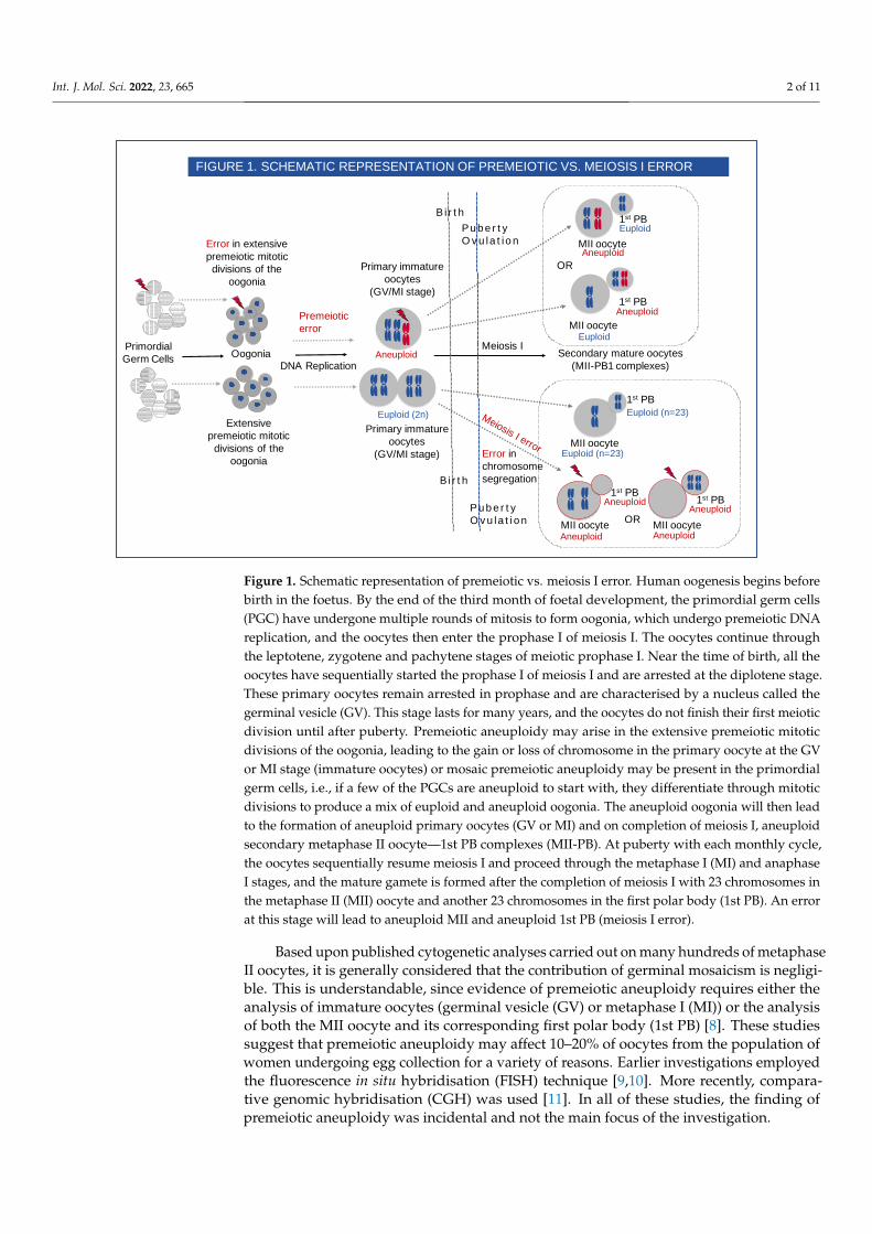
Figure 1. Schematic representation of premeiotic vs. meiosis I error. Human oogenesis begins before birth in the foetus.
By the end of the third month of foetal development, the primordial germ cells (PGC) have undergone multiple rounds of
mitosis to form oogonia, which undergo premeiotic DNA replication, and the oocytes then enter the prophase I of meiosis
I. The oocytes continue through the leptotene, zygotene and pachytene stages of meiotic prophase I. Near the time of birth,
all the oocytes have sequentially started the prophase I of meiosis I and are arrested at the diplotene stage. These primary
oocytes remain arrested in prophase and are characterised by a nucleus called the germinal vesicle (GV). This stage lasts
for many years, and the oocytes do not finish their first meiotic division until after puberty. Premeiotic aneuploidy may
arise in the extensive premeiotic mitotic divisions of the oogonia, leading to the gain or loss of chromosome in the primary
oocyte at the GV or MI stage (immature oocytes) or mosaic premeiotic aneuploidy may be present in the primordial germ
cells, i.e., if a few of the PGCs are aneuploid to start with, they differentiate through mitotic divisions to produce a mix of
euploid and aneuploid oogonia. The aneuploid oogonia will then lead to the formation of aneuploid primary oocytes (GV
or MI) and on completion of meiosis I, aneuploid secondary metaphase II oocyte – 1st PB complexes (MII-PB). At puberty
with each monthly cycle, the oocytes sequentially resume meiosis I and proceed through the metaphase I (MI) and ana-
phase I stages, and the mature gamete is formed after the completion of meiosis I with 23 chromosomes in the metaphase
II (MII) oocyte and another 23 chromosomes in the first polar body (1st PB). An error at this stage will lead to aneuploid
MII and aneuploid 1st PB (meiosis I error).
Based upon published cytogenetic analyses carried out on many hundreds of meta-
phase II oocytes, it is generally considered that the contribution of germinal mosaicism is
negligible. This is understandable, since evidence of premeiotic aneuploidy requires ei-
ther the analysis of immature oocytes (germinal vesicle (GV) or metaphase I (MI)) or the
analysis of both the MII oocyte and its corresponding first polar body (1st PB) [8]. These
studies suggest that premeiotic aneuploidy may affect 10–20% of oocytes from the popu-
lation of women undergoing egg collection for a variety of reasons. Earlier investigations
employed the fluorescence in situ hybridisation (FISH) technique [9,10]. More recently,
comparative genomic hybridisation (CGH) was used [11]. In all of these studies, the find-
ing of premeiotic aneuploidy was incidental and not the main focus of the investigation.
In order to systematically investigate the origins of oocyte aneuploidy, we used array
comparative genomic hybridisation (aCGH) in the analysis of oocytes unexposed to
Primordial
Germ CellsOogonia
DNA Replication
Primary immature
oocytes
(GV/MI stage)
Error in extensive
premeiotic mitotic
divisions of the
oogonia
Premeiotic
error
Aneuploid
Euploid (2n) Meiosis I error
B i r t h
Pu b e r t y
O v u la t i o n
B i r t h
Pu b e r t y
O v u la t i o n
Euploid (n=23)
MII oocyte
1st PB
MII oocyte OR
Aneuploid
Aneuploid
Aneuploid
Aneuploid1st PB
Aneuploid
MII oocyte
1st PB
AneuploidMII oocyte
1st PB
Error in
chromosome
segregation
MII oocyte
1st PB
Euploid (n=23)
Extensive
premeiotic mitotic
divisions of the
oogonia
Euploid
Euploid
OR
FIGURE 1. SCHEMATIC REPRESENTATION OF PREMEIOTIC VS. MEIOSIS I ERROR
Secondary mature oocytes
(MII-PB1 complexes)
Meiosis I
Primary immature
oocytes
(GV/MI stage)
Figure 1. Schematic representation of premeiotic vs. meiosis I error. Human oogenesis begins beforebirth in the foetus. By the end of the third month of foetal development, the primordial germ cells(PGC) have undergone multiple rounds of mitosis to form oogonia, which undergo premeiotic DNAreplication, and the oocytes then enter the prophase I of meiosis I. The oocytes continue throughthe leptotene, zygotene and pachytene stages of meiotic prophase I. Near the time of birth, all theoocytes have sequentially started the prophase I of meiosis I and are arrested at the diplotene stage.These primary oocytes remain arrested in prophase and are characterised by a nucleus called thegerminal vesicle (GV). This stage lasts for many years, and the oocytes do not finish their first meioticdivision until after puberty. Premeiotic aneuploidy may arise in the extensive premeiotic mitoticdivisions of the oogonia, leading to the gain or loss of chromosome in the primary oocyte at the GVor MI stage (immature oocytes) or mosaic premeiotic aneuploidy may be present in the primordialgerm cells, i.e., if a few of the PGCs are aneuploid to start with, they differentiate through mitoticdivisions to produce a mix of euploid and aneuploid oogonia. The aneuploid oogonia will then leadto the formation of aneuploid primary oocytes (GV or MI) and on completion of meiosis I, aneuploidsecondary metaphase II oocyte—1st PB complexes (MII-PB). At puberty with each monthly cycle,the oocytes sequentially resume meiosis I and proceed through the metaphase I (MI) and anaphaseI stages, and the mature gamete is formed after the completion of meiosis I with 23 chromosomes inthe metaphase II (MII) oocyte and another 23 chromosomes in the first polar body (1st PB). An errorat this stage will lead to aneuploid MII and aneuploid 1st PB (meiosis I error).
Based upon published cytogenetic analyses carried out on many hundreds of metaphaseII oocytes, it is generally considered that the contribution of germinal mosaicism is negligi-ble. This is understandable, since evidence of premeiotic aneuploidy requires either theanalysis of immature oocytes (germinal vesicle (GV) or metaphase I (MI)) or the analysisof both the MII oocyte and its corresponding first polar body (1st PB) [8]. These studiessuggest that premeiotic aneuploidy may affect 10–20% of oocytes from the population ofwomen undergoing egg collection for a variety of reasons. Earlier investigations employedthe fluorescence in situ hybridisation (FISH) technique [9,10]. More recently, compara-tive genomic hybridisation (CGH) was used [11]. In all of these studies, the finding ofpremeiotic aneuploidy was incidental and not the main focus of the investigation.
Int. J. Mol. Sci. 2022, 23, 665 3 of 11
In order to systematically investigate the origins of oocyte aneuploidy, we used arraycomparative genomic hybridisation (aCGH) in the analysis of oocytes unexposed to sperm.The GV and MI stage oocytes that have not completed meiosis I provided direct informationon premeiotic errors. Errors seen in the mature metaphase II oocyte and its first PB areexpected to be reciprocal, with a loss in one mirrored by a gain in the other, and can be eitherof chromosome or chromatid origin. Non-reciprocal gains provide evidence of premeioticaneuploidy. As well as giving an estimate of the frequency of germinal mosaicism, ourstudy also compared the incidence of premeiotic aneuploidy with that of errors arising atmeiosis I in the same cohort of women [12] (Figure 1).
Our results provided clear evidence that premeiotic aneuploidy may be detected in10% of informative oocytes (those that provide information on premeiotic errors—GV, MIand MII-PB complexes), affecting 16% of women that are undergoing egg collection, butwho are not necessarily infertile. In cases where the source of the error could be determined,38% were caused by germinal mosaicism compared with 62% that were the outcome of ameiosis I error [12]. The relative frequencies of these two mechanisms will depend uponthe average maternal age of the cohort being investigated.
Following the somewhat surprising findings of our study confirming the relativelycommon occurrence of premeiotic aneuploidy, suggestions were made that they could beartefactual as a result of damage to the DNA caused by the separation of the MII oocytefrom its associated 1st PB and, secondly, that some of those classed as MI may in fact havebeen MIIs that had lost their PBs. [12] We have now carried out this current study usingnext generation sequencing (NGS) on a series of oocytes where there was no manipulationof the oocyte. Oocytes were either immature (GV or MI) or were mature with the MII plusits 1st PB tubed together. More specifically, the GVs and MIs were tubed with their ‘zonapellucida intact’ and the MII oocyte plus its first PB (MII+PB) tubed together in a singlereaction tube, with an intact zona pellucida prior to NGS analysis. This ensured that theentire DNA content of the oocytes at different maturation stages remained intact and thatno DNA was lost during NGS analysis.
2. Results
A total of 149 oocytes were donated for research. Overall, 11% oocytes gave evidenceof premeiotic (PM) aneuploidy. The DNAs from 147 oocytes (immature and mature) (98.6%)were successfully amplified and subjected to NGS analysis using either Veriseq™ PGS orReproSeq™ PGS aneuploidy analysis kits and 140 (95.2%) gave conclusive results. These140 oocytes were donated by 42 women. Seven oocytes gave inconclusive results due tothe degradation of oocyte DNA. The average maternal age of 42 women whose oocytesgave conclusive NGS results was 35.33 years (range 23–44 years). A total of 124 (88.5%)oocytes were found to be euploid with no loss or gain of any chromosome (Figure 2a).Overall, 16/140 (11.4%) oocytes showed PM aneuploidy. The 16 aneuploid oocytes withPM aneuploidy were donated by 10 (23%) women. The average age of women whoseoocytes showed PM aneuploidy was 35.3 years. All 23 chromosomes were observed to beinvolved in a PM aneuploidy event in the oocytes tested in this study.
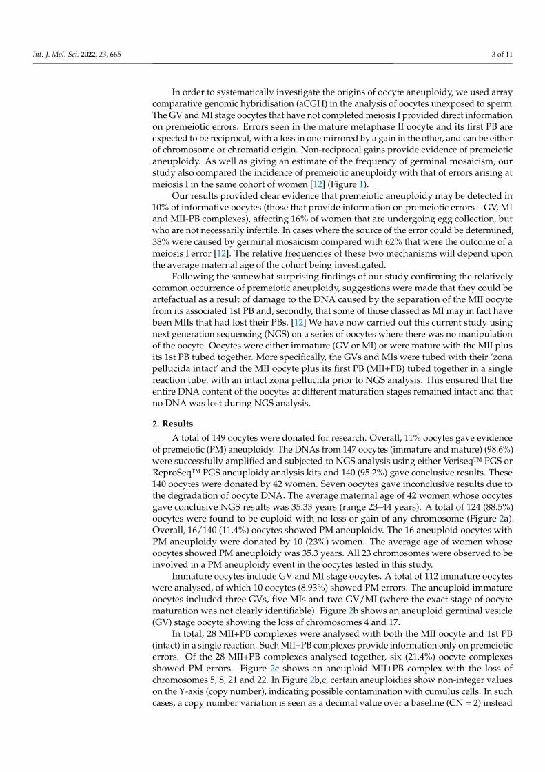
Immature oocytes include GV and MI stage oocytes. A total of 112 immature oocyteswere analysed, of which 10 oocytes (8.93%) showed PM errors. The aneuploid immatureoocytes included three GVs, five MIs and two GV/MI (where the exact stage of oocytematuration was not clearly identifiable). Figure 2b shows an aneuploid germinal vesicle(GV) stage oocyte showing the loss of chromosomes 4 and 17.
In total, 28 MII+PB complexes were analysed with both the MII oocyte and 1st PB(intact) in a single reaction. Such MII+PB complexes provide information only on premeioticerrors. Of the 28 MII+PB complexes analysed together, six (21.4%) oocyte complexesshowed PM errors. Figure 2c shows an aneuploid MII+PB complex with the loss ofchromosomes 5, 8, 21 and 22. In Figure 2b,c, certain aneuploidies show non-integer valueson the Y-axis (copy number), indicating possible contamination with cumulus cells. In suchcases, a copy number variation is seen as a decimal value over a baseline (CN = 2) instead
Int. J. Mol. Sci. 2022, 23, 665 4 of 11
of an integer value. The presence of this contamination tends to mask the true deviation(loss/gain) of the aneuploidy which, in turn, will make the specific gains and losses appearlower (smaller shift) than the expected copy number deviation [13].
Int. J. Mol. Sci. 2022, 23, x FOR PEER REVIEW 4 of 13
Figure 2. Result profiles of euploid and aneuploid oocytes analysed via NGS. The X-axis represents the chromosome number (Chromosome 1–22, X,Y). The Y-axis of the NGS graphs represents the chromosome copy numbers. (a) NGS pro-file of a euploid germinal vesicle (GV) stage oocyte with no loss or gain detected. (b) NGS profile of an aneuploid germinal vesicle (GV) stage oocyte (donor F) with red arrows indicating loss of chromosomes 4 and 17 (simple error). (c) NGS profile of an aneuploid MII+PB complex (donor D) (both cells analysed together in a single reaction) with red arrows indicating loss of chromosomes 5, 8, 21 and 22 (complex errors).
Immature oocytes include GV and MI stage oocytes. A total of 112 immature oocytes were analysed, of which 10 oocytes (8.93%) showed PM errors. The aneuploid immature oocytes included three GVs, five MIs and two GV/MI (where the exact stage of oocyte maturation was not clearly identifiable). Figure 2b shows an aneuploid germinal vesicle (GV) stage oocyte showing the loss of chromosomes 4 and 17.
In total, 28 MII+PB complexes were analysed with both the MII oocyte and 1st PB (intact) in a single reaction. Such MII+PB complexes provide information only on premei-otic errors. Of the 28 MII+PB complexes analysed together, six (21.4%) oocyte complexes showed PM errors. Figure 2c shows an aneuploid MII+PB complex with the loss of chro-mosomes 5, 8, 21 and 22. In Figure 2b,c, certain aneuploidies show non-integer values on the Y-axis (copy number), indicating possible contamination with cumulus cells. In such cases, a copy number variation is seen as a decimal value over a baseline (CN = 2) instead of an integer value. The presence of this contamination tends to mask the true deviation (loss/gain) of the aneuploidy which, in turn, will make the specific gains and losses appear lower (smaller shift) than the expected copy number deviation [13].
Figure 2. Result profiles of euploid and aneuploid oocytes analysed via NGS. The X-axis representsthe chromosome number (Chromosome 1–22, X,Y). The Y-axis of the NGS graphs represents thechromosome copy numbers. (a) NGS profile of a euploid germinal vesicle (GV) stage oocyte withno loss or gain detected. (b) NGS profile of an aneuploid germinal vesicle (GV) stage oocyte (donorF) with red arrows indicating loss of chromosomes 4 and 17 (simple error). (c) NGS profile of ananeuploid MII+PB complex (donor D) (both cells analysed together in a single reaction) with redarrows indicating loss of chromosomes 5, 8, 21 and 22 (complex errors).
The overall summary of the oocytes tested at different maturation stages, along withthe numbers of euploid and aneuploid oocytes with PM errors, are shown in Table 1.
Table 1. Overall summary of NGS results from immature and mature oocyte complexes.
Stage of Oocyte Maturation Total No. of Oocyteswith Conclusive Results
No. of Euploid OocytesEuploid
No. of Oocytes withPremeiotic Errors
Percentage (%) of Oocyteswith Premeiotic Error
Germinal vesicle (GV) 73 70 3 4.11Metaphase I (MI) 35 30 5 14.29
GV/MI 4 2 2 50Metaphase II—first polar
body (MII+PB) 28 22 6 21.43
Totals 140 124 16 11.43
Int. J. Mol. Sci. 2022, 23, 665 5 of 11
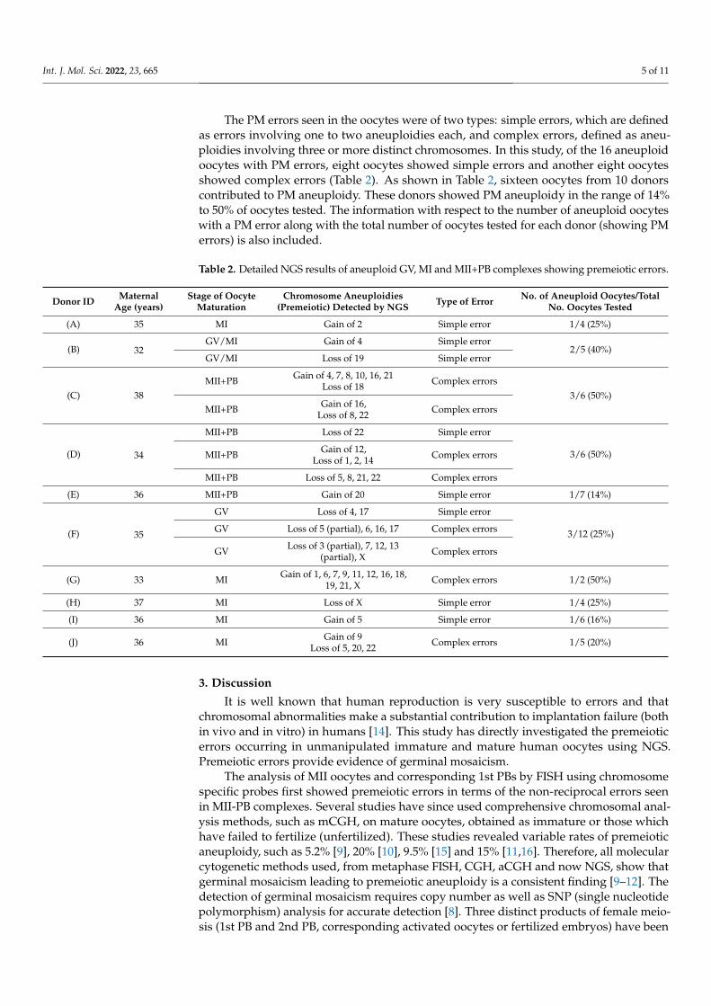
The PM errors seen in the oocytes were of two types: simple errors, which are definedas errors involving one to two aneuploidies each, and complex errors, defined as aneu-ploidies involving three or more distinct chromosomes. In this study, of the 16 aneuploidoocytes with PM errors, eight oocytes showed simple errors and another eight oocytesshowed complex errors (Table 2). As shown in Table 2, sixteen oocytes from 10 donorscontributed to PM aneuploidy. These donors showed PM aneuploidy in the range of 14%to 50% of oocytes tested. The information with respect to the number of aneuploid oocyteswith a PM error along with the total number of oocytes tested for each donor (showing PMerrors) is also included.
Table 2. Detailed NGS results of aneuploid GV, MI and MII+PB complexes showing premeiotic errors.
Donor ID MaternalAge (years)
Stage of OocyteMaturation
Chromosome Aneuploidies(Premeiotic) Detected by NGS Type of Error No. of Aneuploid Oocytes/Total
No. Oocytes Tested
(A) 35 MI Gain of 2 Simple error 1/4 (25%)
(B) 32GV/MI Gain of 4 Simple error
2/5 (40%)GV/MI Loss of 19 Simple error
(C) 38MII+PB Gain of 4, 7, 8, 10, 16, 21
Loss of 18 Complex errors3/6 (50%)
MII+PB Gain of 16,Loss of 8, 22 Complex errors
(D) 34
MII+PB Loss of 22 Simple error
3/6 (50%)MII+PB Gain of 12,Loss of 1, 2, 14 Complex errors
MII+PB Loss of 5, 8, 21, 22 Complex errors
(E) 36 MII+PB Gain of 20 Simple error 1/7 (14%)
(F) 35
GV Loss of 4, 17 Simple error
3/12 (25%)GV Loss of 5 (partial), 6, 16, 17 Complex errors
GV Loss of 3 (partial), 7, 12, 13(partial), X Complex errors
(G) 33 MI Gain of 1, 6, 7, 9, 11, 12, 16, 18,19, 21, X Complex errors 1/2 (50%)
(H) 37 MI Loss of X Simple error 1/4 (25%)
(I) 36 MI Gain of 5 Simple error 1/6 (16%)
(J) 36 MI Gain of 9Loss of 5, 20, 22 Complex errors 1/5 (20%)
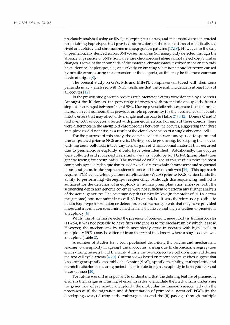
3. Discussion
It is well known that human reproduction is very susceptible to errors and thatchromosomal abnormalities make a substantial contribution to implantation failure (bothin vivo and in vitro) in humans [14]. This study has directly investigated the premeioticerrors occurring in unmanipulated immature and mature human oocytes using NGS.Premeiotic errors provide evidence of germinal mosaicism.
The analysis of MII oocytes and corresponding 1st PBs by FISH using chromosomespecific probes first showed premeiotic errors in terms of the non-reciprocal errors seenin MII-PB complexes. Several studies have since used comprehensive chromosomal anal-ysis methods, such as mCGH, on mature oocytes, obtained as immature or those whichhave failed to fertilize (unfertilized). These studies revealed variable rates of premeioticaneuploidy, such as 5.2% [9], 20% [10], 9.5% [15] and 15% [11,16]. Therefore, all molecularcytogenetic methods used, from metaphase FISH, CGH, aCGH and now NGS, show thatgerminal mosaicism leading to premeiotic aneuploidy is a consistent finding [9–12]. Thedetection of germinal mosaicism requires copy number as well as SNP (single nucleotidepolymorphism) analysis for accurate detection [8]. Three distinct products of female meio-sis (1st PB and 2nd PB, corresponding activated oocytes or fertilized embryos) have been
Int. J. Mol. Sci. 2022, 23, 665 6 of 11
previously analysed using an SNP genotyping bead array, and meiomaps were constructedfor obtaining haplotypes that provide information on the mechanisms of meiotically de-rived aneuploidy and chromosome mis-segregation patterns [17,18]. However, in the caseof premeiotically derived errors, SNP-based analysis (for aneuploidy detected through theabsence or presence of SNPs from an entire chromosome) alone cannot detect copy numberchanges if some of the chromatids of the maternal chromosomes involved in the aneuploidyhave identical haplotypes, i.e., aneuploidy originating via mitotic nondisjunction causedby mitotic errors during the expansion of the oogonia, as this may be the most commonmode of origin [8].
The present study on GVs, MIs and MII+PB complexes (all tubed with their zonapellucida intact), analysed with NGS, reaffirms that the overall incidence is at least 10% ofall oocytes [12].
In the present study, sixteen oocytes with premeiotic errors were donated by 10 donors.Amongst the 10 donors, the percentage of oocytes with premeiotic aneuploidy from asingle donor ranged between 14 and 50%. During premeiotic mitoses, there is an enormousincrease in cell numbers that provides ample opportunity for the occurrence of separatemitotic errors that may affect only a single mature oocyte (Table 2) [8,12]. Donors C and Dhad over 50% of oocytes affected with premeiotic errors. For each of these donors, therewere differences in the aneuploid chromosomes between the oocytes, suggesting that theseaneuploidies did not arise as a result of the clonal expansion of a single abnormal cell.
For the purpose of this study, the oocytes collected were unexposed to sperm andunmanipulated prior to NGS analysis. During oocyte processing, by keeping the oocyteswith the zona pellucida intact, any loss or gain of chromosomal material that occurreddue to premeiotic aneuploidy should have been identified. Additionally, the oocyteswere collected and processed in a similar way as would be for PGT-A (preimplantationgenetic testing for aneuploidy). The method of NGS used in this study is now the mostcommonly applied technique that is used to evaluate the whole chromosome and segmentallosses and gains in the trophectoderm biopsies of human embryos [19]. This approachrequires PCR-based whole genome amplification (WGA) prior to NGS, which limits theability to perform high-throughput sequencing. Although this sequencing method issufficient for the detection of aneuploidy in human preimplantation embryos, both thesequencing depth and genome coverage were not sufficient to perform any further analysisof the actual genotype. The coverage depth is typically low (in the order of 0.01× acrossthe genome) and not suitable to call SNPs or indels. It was therefore not possible toobtain haplotype information or detect structural rearrangements that may have providedimportant information concerning mechanisms that lie behind the generation of premeioticaneuploidy [8].
Whilst this study has detected the presence of premeiotic aneuploidy in human oocytes(11.4%), it was not possible to have firm evidence as to the mechanism by which it arose.However, the mechanisms by which aneuploidy arose in oocytes with high levels ofaneuploidy (50%) may be different from the rest of the donors where a single oocyte wasaneuploid (Table 2).
A number of studies have been published describing the origins and mechanismsleading to aneuploidy in ageing human oocytes, arising due to chromosome segregationerrors during meiosis I and II, mainly during the two consecutive cell divisions and duringthe two cell cycle arrests [4,20]. Current views based on recent oocyte studies suggest thatless stringent spindle assembly checkpoint (SAC), spindle instability, multipolarity andmerotelic attachments during meiosis I contribute to high aneuploidy in both younger andolder women [20].
For future work, it is important to understand that the defining feature of premeioticerrors is their origin and timing of error. In order to elucidate the mechanisms underlyingthe generation of premeiotic aneuploidy, the molecular mechanisms associated with theprocesses of (i) the migration and differentiation of primordial germ cell PGCs (in thedeveloping ovary) during early embryogenesis and the (ii) passage through multiple
Int. J. Mol. Sci. 2022, 23, 665 7 of 11
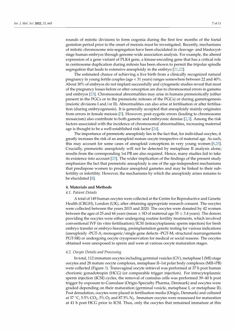
rounds of mitotic divisions to form oogonia during the first few months of the foetalgestation period prior to the onset of meiosis must be investigated. Recently, mechanismsof mitotic chromosome mis-segregation have been elucidated in cleavage- and blastocyst-stage human embryos through genome-wide association analysis. For example, the alteredexpression of a gene variant of PLK4 gene, a kinase-encoding gene that has a critical rolein centrosome duplication during mitosis has been shown to permit the tripolar spindlesegregation that leads to extensive aneuploidy in the embryo [21,22].
The estimated chance of achieving a live birth from a clinically recognized naturalpregnancy in young fertile couples (age < 31 years) ranges somewhere between 22 and 40%.About 30% of embryos do not implant successfully and cytogenetic studies reveal that mostof the pregnancy losses before or after conception are due to chromosomal errors in gametesand embryos [23]. Chromosomal abnormalities may arise in humans premeiotically (eitherpresent in the PGCs or in the premeiotic mitoses of the PGCs) or during gametogenesis(meiotic divisions I and/or II). Abnormalities can also arise at fertilisation or after fertilisa-tion (during embryogenesis). It is generally accepted that aneuploidy mainly originatesfrom errors in female meiosis [5]. However, post-zygotic errors (leading to chromosomemosaicism) also contribute to both gametic and embryonic demise [2,3]. Among the riskfactors associated with the incidence of chromosomal abnormalities, increasing maternalage is thought to be a well-established risk factor [24].
The importance of premeiotic aneuploidy lies in the fact that, for individual oocytes, itgreatly increases the risk of an aneuploid mature oocyte irrespective of maternal age. As such,this may account for some cases of aneuploid conceptions in very young women [8,25].Crucially, premeiotic aneuploidy will not be detected by metaphase II analysis alone;results from the corresponding 1st PB are also required. Hence, many studies fail to takeits existence into account [25]. The wider implication of the findings of the present studyemphasizes the fact that premeiotic aneuploidy is one of the age-independent mechanismsthat predispose women to produce aneuploid gametes and may be linked to their sub-fertility or infertility. However, the mechanisms by which the aneuploidy arises remains tobe elucidated [8].
4. Materials and Methods4.1. Patient Details
A total of 149 human oocytes were collected at the Centre for Reproductive and GeneticHealth (CRGH), London (UK), after obtaining appropriate research consent. The oocyteswere collected between the years 2015 and 2020. The oocytes were donated by 42 womenbetween the ages of 25 and 44 years (mean± SD of maternal age 35± 3.4 years). The donorsproviding the oocytes were either undergoing routine fertility treatments, which involvedconventional IVF (in vitro fertilisation)/ICSI (intracytoplasmic sperm injection) for freshembryo transfer or embryo freezing, preimplantation genetic testing for various indications(aneuploidy -PGT-A; monogenic/single gene defects -PGT-M; structural rearrangementsPGT-SR) or undergoing oocyte cryopreservation for medical or social reasons. The oocytesobtained were unexposed to sperm and were at various oocyte maturation stages.
4.2. Oocyte Details and Processing
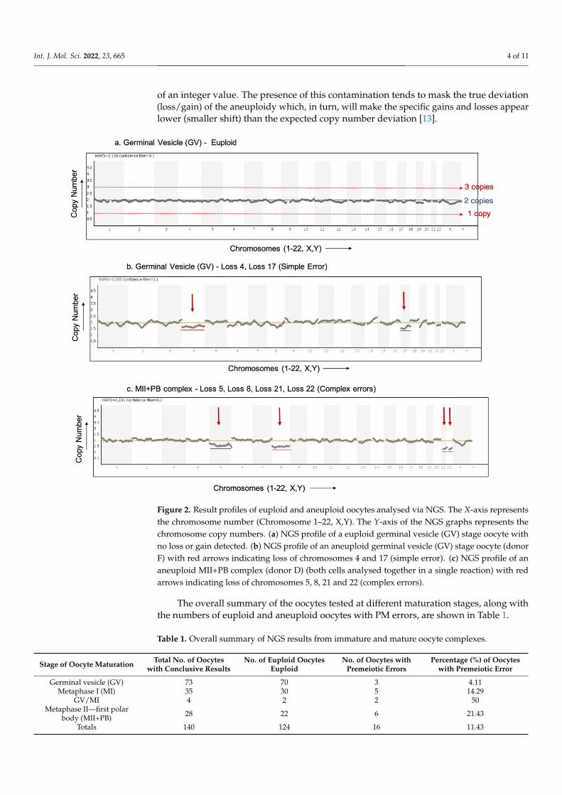
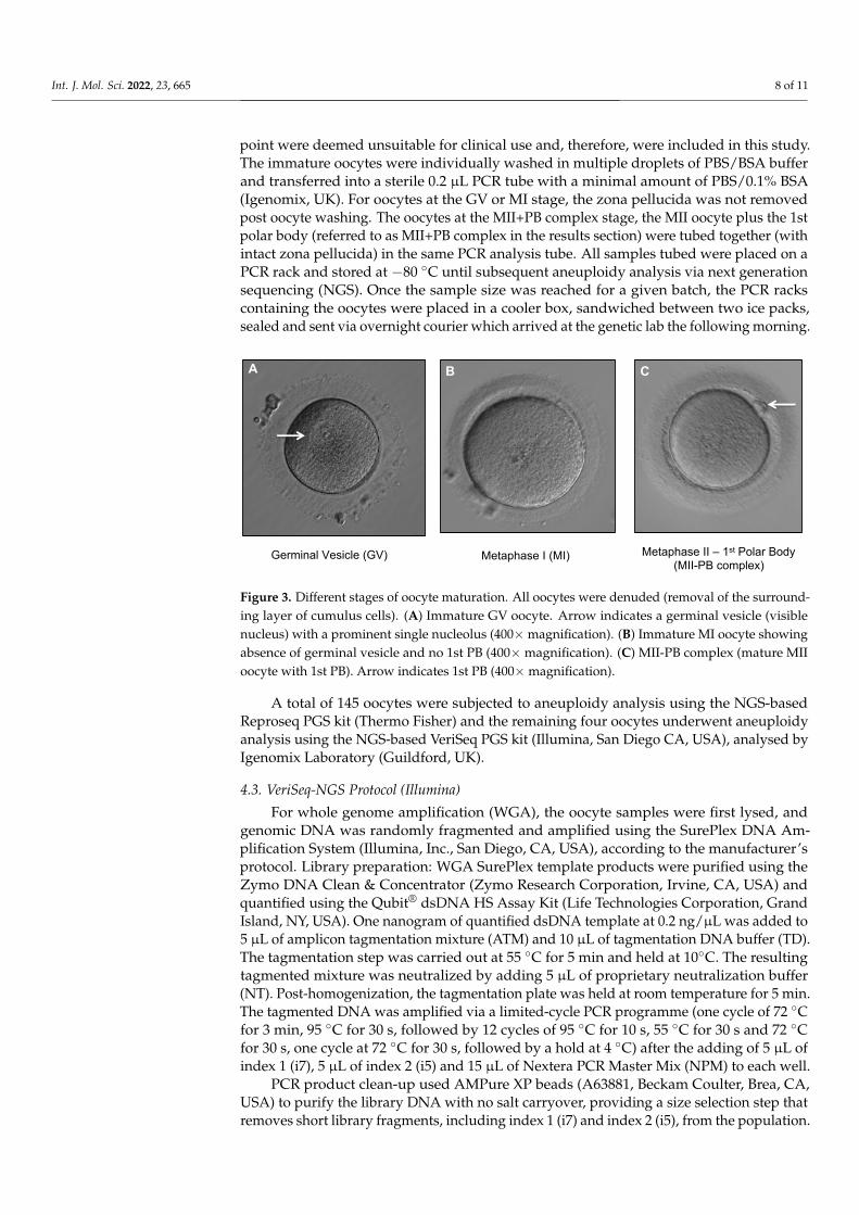
In total, 112 immature oocytes including germinal vesicles (GV), metaphase I (MI) stageoocytes and 28 mature oocyte complexes, metaphase II–1st polar body complexes (MII+PB)were collected (Figure 3). Transvaginal oocyte retrieval was performed at 37 h post humanchorionic gonadotropin (HCG) (or comparable trigger injection). For intracytoplasmicsperm injection (ICSI) cycles, the removal of cumulus cells was performed 39–40 h posttrigger by exposure to Cumulase (Origio Specialty Pharma, Denmark) and oocytes weregraded depending on their maturation (germinal vesicle, metaphase I, or metaphase II).Post denudation, oocytes were placed in fertilisation media (Origio, Denmark) and culturedat 37 ◦C, 5.5% CO2, 5% O2 and 87.5% N2. Immature oocytes were reassessed for maturationat 41 h post HCG prior to ICSI. Thus, only the oocytes that remained immature at this
Int. J. Mol. Sci. 2022, 23, 665 8 of 11
point were deemed unsuitable for clinical use and, therefore, were included in this study.The immature oocytes were individually washed in multiple droplets of PBS/BSA bufferand transferred into a sterile 0.2 µL PCR tube with a minimal amount of PBS/0.1% BSA(Igenomix, UK). For oocytes at the GV or MI stage, the zona pellucida was not removedpost oocyte washing. The oocytes at the MII+PB complex stage, the MII oocyte plus the 1stpolar body (referred to as MII+PB complex in the results section) were tubed together (withintact zona pellucida) in the same PCR analysis tube. All samples tubed were placed on aPCR rack and stored at −80 ◦C until subsequent aneuploidy analysis via next generationsequencing (NGS). Once the sample size was reached for a given batch, the PCR rackscontaining the oocytes were placed in a cooler box, sandwiched between two ice packs,sealed and sent via overnight courier which arrived at the genetic lab the following morning.
Germinal Vesicle (GV) Metaphase I (MI) Metaphase II – 1st Polar Body (MII-PB complex)
A B C
Figure 3. Different stages of oocyte maturation. All oocytes were denuded (removal of the surround-ing layer of cumulus cells). (A) Immature GV oocyte. Arrow indicates a germinal vesicle (visiblenucleus) with a prominent single nucleolus (400×magnification). (B) Immature MI oocyte showingabsence of germinal vesicle and no 1st PB (400× magnification). (C) MII-PB complex (mature MIIoocyte with 1st PB). Arrow indicates 1st PB (400×magnification).
A total of 145 oocytes were subjected to aneuploidy analysis using the NGS-basedReproseq PGS kit (Thermo Fisher) and the remaining four oocytes underwent aneuploidyanalysis using the NGS-based VeriSeq PGS kit (Illumina, San Diego CA, USA), analysed byIgenomix Laboratory (Guildford, UK).
4.3. VeriSeq-NGS Protocol (Illumina)
For whole genome amplification (WGA), the oocyte samples were first lysed, andgenomic DNA was randomly fragmented and amplified using the SurePlex DNA Am-plification System (Illumina, Inc., San Diego, CA, USA), according to the manufacturer’sprotocol. Library preparation: WGA SurePlex template products were purified using theZymo DNA Clean & Concentrator (Zymo Research Corporation, Irvine, CA, USA) andquantified using the Qubit® dsDNA HS Assay Kit (Life Technologies Corporation, GrandIsland, NY, USA). One nanogram of quantified dsDNA template at 0.2 ng/µL was added to5 µL of amplicon tagmentation mixture (ATM) and 10 µL of tagmentation DNA buffer (TD).The tagmentation step was carried out at 55 ◦C for 5 min and held at 10◦C. The resultingtagmented mixture was neutralized by adding 5 µL of proprietary neutralization buffer(NT). Post-homogenization, the tagmentation plate was held at room temperature for 5 min.The tagmented DNA was amplified via a limited-cycle PCR programme (one cycle of 72 ◦Cfor 3 min, 95 ◦C for 30 s, followed by 12 cycles of 95 ◦C for 10 s, 55 ◦C for 30 s and 72 ◦Cfor 30 s, one cycle at 72 ◦C for 30 s, followed by a hold at 4 ◦C) after the adding of 5 µL ofindex 1 (i7), 5 µL of index 2 (i5) and 15 µL of Nextera PCR Master Mix (NPM) to each well.
PCR product clean-up used AMPure XP beads (A63881, Beckam Coulter, Brea, CA,USA) to purify the library DNA with no salt carryover, providing a size selection step thatremoves short library fragments, including index 1 (i7) and index 2 (i5), from the population.
Int. J. Mol. Sci. 2022, 23, 665 9 of 11
Using a multichannel pipette, 45 µL of the PCR product was transferred to 96-well storageplates containing 45 µL of AMPure XP beads. Sealed plates were mixed using a microplateshaker at 1800 rpm for 2 min, then incubated at room temperature without shaking for5 min. Thereafter, the plate was placed on a magnetic stand (AM10027, Life Technology)for 2 min or until the supernatant cleared. While the plates were kept on the magneticstand, the magnetic beads were washed twice with 200 µL of freshly prepared 80% ethanol.Purified libraries were eluted with 50 µL of the Nextera XT Resuspension Buffer.
Single-end, dual index 36 base pair read (1 × 36 donor insemination) sequencing wasperformed following the Illumina v2 chemistry workflow on a MiSeq System (Illumina,Inc.), using the MiSeq Reagent Kit v2 kit (Illumina, Inc.), which contains the ready-to-loadon-board clustering and sequencing by synthesis (SBS) chemistry reagents. The sequencingdata derived from the MiSeq Reporter Software were then uploaded and analysed usingBlueFuse Multi v3.0 for NGS (Illumina, Inc.). The quality control acceptance criteria werethe number of total reads > 700,000 with a number of reads passing the filter > 500,000, andthe DLR (derivative log ratio) at <0.4. The quality metrics for all the four oocytes analysedvia Veriseq-NGS were within the acceptable limits.
4.4. Reproseq-NGS Protocol (Thermo Fisher)
DNA extraction and whole-genome amplification (WGA) was performed using anIon Reproseq PGS kit (Thermo Fisher). The oocyte samples were tubed in 2.5 µL of 1XPBS/0.1%BSA, treated with 5 µL of extraction enzyme master mix and incubated at 75 ◦Cfor 10 min, followed by incubation at 95 ◦C for 4 min. Extracted genomic DNA waspre-amplified with 5 ul of pre-amplification master mix and incubated according to thefollowing program: 1 cycle at 95 ◦C for 2 min and 12 cycles at 95 ◦C for 15 s, 15 ◦C for 50 s,25 ◦C for 40 s, 35 ◦C for 30 s, 65 ◦C for 40 s, 75 ◦C for 40 s, and holding at 4 ◦C. Subsequently,30 µL of Amplification master mix and 5 µL of Ion SingleSeq Barcode Adaptor wereadded to each sample. Library amplification was performed with the following PCRprogram: 1 cycle at 95 ◦C for 3 min, 4 cycles at 95 ◦C for 20 s, 50 ◦C for 25 s, 72 ◦C for40 s, 12 cycles at 95 ◦C for 20 s, 72 ◦C for 55 s, and holding at 4 ◦C. Libraries were thenpooled, purified with AMPure XP beads (Beckman Coulter, Brea, CA, USA), quantifiedusing the Qubit dsDNA High Sensitivity Assay kit (Life Technologies, Carlsbad, CA, USA)and diluted to the final concentration of 80 pM. Template preparation and chip loadingwas performed using the Ion Chef system (Thermo Fisher) according to manufacturer’sinstructions. In each batch, 23 samples combined with one positive control sample weresequenced together in one Ion 520™ Chip. The chip was then loaded and sequenced onan Ion S5 SequencerTM (Thermo Fisher). The sequencing data were then transferred fromthe Ion S5 “Torrent Suite” software v5.10.1 to the Ion ReporterTM software v5.4 (ThermoFisher) for aneuploidy “calling”. This software uses the ‘ReproSeq PGS w1.1′ workflow(low-pass whole-genome aneuploidy workflow, which detects aneuploidies and wholechromosome and sub-chromosome abnormalities (~10 Mb up to a whole chromosome)from a single whole-genome sample with low coverage (0.01× across the genome)). Thenormalization was performed using the informatics baseline Reproseq Low CoverageWhole-Genome Baseline (v5.2) generated from multiple normal samples. The ReproseqPGS w1.1 is designed to detect and call aneuploidy events with integer ploidy values.In addition, the aneuploidy “calls” were verified by an in-house-developed algorithm thatuses machine learning.
The resulting NGS graphs from both sequencing platforms show chromosomal gainsassociated with a copy number ≥ 3 and chromosomal losses with a copy number ≤ 1.Where non-integer ploidy values were detected, this was attributed to cumulus cell contam-ination. Aneuploidy calls were generated by the NGS analyses software (BlueFuse MultiSoftware Version v3.0 (Illumina)/Ion Reporter™ v5.4 software (Thermo Fisher)) and werealso ‘assessed manually’ by ‘two independent scientists’.
The quality control acceptance criteria were the total number of mapped reads of>60,000 and an MAPD (median of the absolute values of all pairwise differences) value of
Int. J. Mol. Sci. 2022, 23, 665 10 of 11
<0.3. The quality metrics for 132/136 oocytes were within the acceptable range. However,for four samples, MAPD results were above the cut-off values. In two oocytes with complexaneuploidies (many step changes), a slightly high MAPD value was detected; however,the results were deemed suitable for manual interpretation by two independent scientists.In the other two oocytes with single aneuploidies, the aneuploidy was called by the softwarealgorithm and interpreted manually.
4.5. Validation
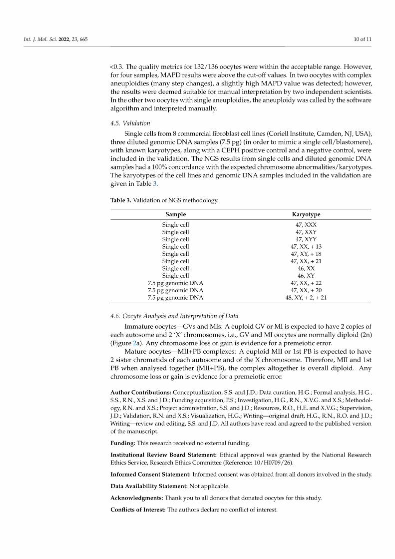
Single cells from 8 commercial fibroblast cell lines (Coriell Institute, Camden, NJ, USA),three diluted genomic DNA samples (7.5 pg) (in order to mimic a single cell/blastomere),with known karyotypes, along with a CEPH positive control and a negative control, wereincluded in the validation. The NGS results from single cells and diluted genomic DNAsamples had a 100% concordance with the expected chromosome abnormalities/karyotypes.The karyotypes of the cell lines and genomic DNA samples included in the validation aregiven in Table 3.
Table 3. Validation of NGS methodology.
Sample Karyotype
Single cell 47, XXXSingle cell 47, XXYSingle cell 47, XYYSingle cell 47, XX, + 13Single cell 47, XY, + 18Single cell 47, XX, + 21Single cell 46, XXSingle cell 46, XY
7.5 pg genomic DNA 47, XX, + 227.5 pg genomic DNA 47, XX, + 207.5 pg genomic DNA 48, XY, + 2, + 21
4.6. Oocyte Analysis and Interpretation of Data
Immature oocytes—GVs and MIs: A euploid GV or MI is expected to have 2 copies ofeach autosome and 2 ‘X’ chromosomes, i.e., GV and MI oocytes are normally diploid (2n)(Figure 2a). Any chromosome loss or gain is evidence for a premeiotic error.
Mature oocytes—MII+PB complexes: A euploid MII or 1st PB is expected to have2 sister chromatids of each autosome and of the X chromosome. Therefore, MII and 1stPB when analysed together (MII+PB), the complex altogether is overall diploid. Anychromosome loss or gain is evidence for a premeiotic error.
Author Contributions: Conceptualization, S.S. and J.D.; Data curation, H.G.; Formal analysis, H.G.,S.S., R.N., X.S. and J.D.; Funding acquisition, P.S.; Investigation, H.G., R.N., X.V.G. and X.S.; Methodol-ogy, R.N. and X.S.; Project administration, S.S. and J.D.; Resources, R.O., H.E. and X.V.G.; Supervision,J.D.; Validation, R.N. and X.S.; Visualization, H.G.; Writing—original draft, H.G., R.N., R.O. and J.D.;Writing—review and editing, S.S. and J.D. All authors have read and agreed to the published versionof the manuscript.
Funding: This research received no external funding.
Institutional Review Board Statement: Ethical approval was granted by the National ResearchEthics Service, Research Ethics Committee (Reference: 10/H0709/26).
Informed Consent Statement: Informed consent was obtained from all donors involved in the study.
Data Availability Statement: Not applicable.
Acknowledgments: Thank you to all donors that donated oocytes for this study.
Conflicts of Interest: The authors declare no conflict of interest.
Int. J. Mol. Sci. 2022, 23, 665 11 of 11
References1. Fragouli, E.; Wells, D. Aneuploidy in the human blastocyst. Cytogenet. Genome Res. 2011, 133, 149–159. [CrossRef] [PubMed]2. Fragouli, E.; Wells, D.; Delhanty, J.D. Chromosome abnormalities in the human oocyte. Cytogenet. Genome Res. 2011, 133, 107–118.
[CrossRef]3. Mantzouratou, A.; Delhanty, J.D. Aneuploidy in the human cleavage stage embryo. Cytogenet. Genome Res. 2011, 133, 141–148.
[CrossRef] [PubMed]4. Wartosch, L.; Schindler, K.; Schuh, M.; Gruhn, J.R.; Hoffmann, E.R.; McCoy, R.C.; Xing, J. Origins and mechanisms leading to
aneuploidy in human eggs. Prenat. Diagn. 2021, 41, 620–630. [CrossRef] [PubMed]5. Hunt, P.A.; Hassold, T.J. Human female meiosis: What makes a good egg go bad? Trends Genet. 2008, 24, 86–93. [CrossRef]6. Fragouli, E.; Alfarawati, S.; Spath, K.; Jaroudi, S.; Sarasa, J.; Enciso, M.; Wells, D. The origin and impact of embryonic aneuploidy.
Hum. Genet. 2013, 132, 1001–1013. [CrossRef]7. Delhanty, J.D. Inherited aneuploidy: Germline mosaicism. Cytogenet. Genome Res. 2011, 133, 136–140. [CrossRef]8. Delhanty, J.D.; SenGupta, S.B.; Ghevaria, H. How common is germinal mosaicism that leads to premeiotic aneuploidy in the
female? J. Assist. Reprod. Genet. 2019, 36, 2403–2418. [CrossRef]9. Mahmood, R.; Brierley, C.H.; Faed, M.J.; Mills, J.A.; Delhanty, J.D. Mechanisms of maternal aneuploidy: FISH analysis of oocytes
and polar bodies in patients undergoing assisted conception. Hum. Genet. 2000, 106, 620–626. [CrossRef]10. Pujol, A.; Boiso, I.; Benet, J.; Veiga, A.; Durban, M.; Campillo, M.; Egozcue, J.; Navarro, J. Analysis of nine chromosome probes in
first polar bodies and metaphase II oocytes for the detection of aneuploidies. Eur. J. Hum. Genet. 2003, 11, 325–336. [CrossRef]11. Obradors, A.; Rius, M.; Cuzzi, J.; Daina, G.; Gutierrez-Mateo, C.; Pujol, A.; Marina, F.; Marquez, C.; Benet, J.; Navarro, J. Errors
at mitotic segregation early in oogenesis and at first meiotic division in oocytes from donor females: Comparative genomichybridization analyses in metaphase II oocytes and their first polar body. Fertil. Steril. 2010, 93, 675–679. [CrossRef] [PubMed]
12. Ghevaria, H.; SenGupta, S.; Sarna, U.; Sargeant, S.; Serhal, P.; Delhanty, J. The contribution of germinal mosaicism to humananeuploidy. Cytogenet. Genome Res. 2014, 144, 264–274. [CrossRef]
13. Geraedts, J.; Montag, M.; Magli, M.C.; Repping, S.; Handyside, A.; Staessen, C.; Harper, J.; Schmutzler, A.; Collins, J.; Goossens, V.;et al. Polar body array CGH for prediction of the status of the corresponding oocyte. Part I: Clinical results. Hum. Reprod. 2011,26, 3173–3180. [CrossRef] [PubMed]
14. Delhanty, J.D. Preimplantation genetics: An explanation for poor human fertility? Ann. Hum. Genet. 2001, 65, 331–338. [CrossRef]15. Gutierrez-Mateo, C.; Benet, J.; Wells, D.; Colls, P.; Bermudez, M.G.; Sanchez-Garcia, J.F.; Egozcue, J.; Navarro, J.; Munne, S.
Aneuploidy study of human oocytes first polar body comparative genomic hybridization and metaphase II fluorescence in situhybridization analysis. Hum. Reprod. 2004, 19, 2859–2868. [CrossRef] [PubMed]
16. Daina, G.; Ramos, L.; Rius, M.; Obradors, A.; Del Rey, J.; Giralt, M.; Campillo, M.; Velilla, E.; Pujol, A.; Martinez-Pasarell, O.; et al.Non-meiotic chromosome instability in human immature oocytes. Eur. J. Hum. Genet. 2014, 22, 202–207. [CrossRef]
17. Handyside, A.H.; Montag, M.; Magli, M.C.; Repping, S.; Harper, J.; Schmutzler, A.; Vesela, K.; Gianaroli, L.; Geraedts, J. Multiplemeiotic errors caused by predivision of chromatids in women of advanced maternal age undergoing in vitro fertilisation. Eur J.Hum. Genet. 2012, 20, 742–747. [CrossRef]
18. Ottolini, C.S.; Newnham, L.; Capalbo, A.; Natesan, S.A.; Joshi, H.A.; Cimadomo, D.; Griffin, D.K.; Sage, K.; Summers, M.C.;Thornhill, A.R.; et al. Genome-wide maps of recombination and chromosome segregation in human oocytes and embryos showselection for maternal recombination rates. Nat. Genet. 2015, 47, 727–735. [CrossRef]
19. Rubio, C.; Rodrigo, L.; Garcia-Pascual, C.; Peinado, V.; Campos-Galindo, I.; Garcia-Herrero, S.; Simón, C. Clinical application ofembryo aneuploidy testing by next-generation sequencing. Biol. Reprod. 2019, 101, 1083–1090. [CrossRef]
20. Thomas, C.; Cavazza, T.; Schuh, M. Aneuploidy in human eggs: Contributions of the meiotic spindle. Biochem. Soc. Trans. 2021,49, 107–118. [CrossRef]
21. McCoy, R.C.; Demko, Z.; Ryan, A.; Banjevic, M.; Hill, M.; Sigurjonsson, S.; Rabinowitz, M.; Fraser, H.B.; Petrov, D.A. Commonvariants spanning PLK4 are associated with mitotic-origin aneuploidy in human embryos. Science 2015, 348, 235–238. [CrossRef]
22. McCoy, R.C.; Newnham, L.J.; Ottolini, C.S.; Hoffmann, E.R.; Chatzimeletiou, K.; Cornejo, O.E.; Zhan, Q.; Zaninovic, N.;Rosenwaks, Z.; Petrov, D.A.; et al. Tripolar chromosome segregation drives the association between maternal genotype at variantsspanning PLK4 and aneuploidy in human preimplantation embryos. Hum. Mol. Genet. 2018, 27, 2573–2585. [CrossRef] [PubMed]
23. Macklon, N.S.; Geraedts, J.P.; Fauser, B.C. Conception to ongoing pregnancy: The ‘black box’ of early pregnancy loss. Hum.Reprod. Update 2002, 8, 333–343. [CrossRef] [PubMed]
24. Hassold, T.; Hunt, P. To err (meiotically) is human: The genesis of human aneuploidy. Nat. Rev. Genet. 2001, 2, 280–291. [CrossRef][PubMed]
25. Gruhn, J.R.; Zielinska, A.P.; Shukla, V.; Blanshard, R.; Capalbo, A.; Cimadomo, D.; Nikiforov, D.; Chan, A.C.; Newnham, L.J.;Vogel, I.; et al. Chromosome errors in human eggs shape natural fertility over reproductive life span. Science 2019, 365, 1466–1469.[CrossRef] [PubMed]