new species and summary of iberian harpellales
TRANSCRIPT
New species and summary of Iberian Harpellales
L.G. ValleUnitat de Botanica, Departament de Biologia Animal,Biologia Vegetal i d’Ecologia, Facultat de Ciencies,Universitat Autonoma de Barcelona,08193-Bellaterra, Spain
Abstract: The occurrence of three new species ofHarpellales in the Iberian Peninsula is reported.Capniomyces celatus was found in Nemouridae (Ple-coptera) nymphs, Stipella latispora in Simuliidae(Diptera) larvae and Legeriomyces dolabrae withBaetidae (Ephemeroptera) nymphs. These speciesare differentiated from others by thalli and/orreproductive structures. Geographic range extensionsfor other species of Harpellales also are reported;these are Graminella bulbosa, Pennella angustispora,Spartiella barbata, Stachylina euthena, St. grandispora,St. pedifer, St. penetralis, St. prolifica, St. robusta andStipella vigilans. All are compared with related taxaand are illustrated with photographs. Finally a sum-mary of the known Harpellales occurring in theIberian territory is provided with data on theirdistribution and ecology.
Key words: gut fungi, insect larva, symbiosis,taxonomy, Zygomycota
INTRODUCTION
The species reported here are part of a long-termstudy to catalogue the fungal diversity in the IberianPeninsula and Balearic Islands (Flora MycologicaIberica-V). With the inclusion of information fromprevious works (Girbal and Santamaria 1998, Santa-maria 1997, Santamaria and Girbal 1997, 1998, Valle2004, 2006, Valle and Santamaria 2002a, b, 2004a, b,2005) the present article encompasses and sum-marizes our knowledge of Harpellales from theIberian Peninsula, a microcontinent with a physio-graphical, bioclimatic and phytogeographical diversi-ty that allows the occurrence of both Mediterraneanand Eurosiberian floristic and vegetation zones(Rivas-Martınez 1987). This area in the westernMediterranean basin, southern Europe, is influencedpredominantly by the Mediterranean Sea, whichborders the peninsula on the east and south, and bythe cooler Atlantic Ocean on the west and north.
Harpellales are endosymbiotic filamentous fungi
that develop within the gut of their immaturearthropod hosts, mostly aquatic species of Diptera,stoneflies (Plecoptera), mayflies (Ephemeroptera),and more infrequently beetles (Coleoptera), caddis-flies (Trichoptera) and Isopoda (Lichtwardt et al1999b, 2001a, White 1999). In terms of insect diversitythe Iberian Peninsula and Balearic Islands have listedca. 155 species of Ephemeroptera (Alba-Tercedor andJımez-Cuellar 2003), ca. 140 Plecoptera (Tierno deFigueroa et al 2003) and ca. 1860 nematoceran Diptera(Carles-Tolra and Hjorth-Andersen 2002). The re-lationship between these gut fungi and their hosts inmost cases is considered to be commensalistic, whereasfor some species of Smittium that have been culturedparasitic or mutualistic effects have been reported(Sweeney 1981a, b, McCreadie et al 2005).
All species reported in this article are in the orderHarpellales, which include species with branched thallicomprising the family Legeriomycetaceae (Capnio-Capniomyces, Graminella, Legeriomyces, Pennella, Spar-tiella and Stipella) and the family containing memberswith unbranched thalli, the Harpellaceae (Stachylina).
MATERIALS AND METHODS
Collections of insect nymphs and larvae were made by hand-picking from rocks, wood or leaves removed from variousIberian streams and with the aid of aquatic dip nets asdescribed in Lichtwardt et al (2001a). The hosts weretransported to the lab in a portable cooler with bagged ice.Insect guts were removed and cleaned of their contentsunder magnification with the aid of a steromicroscope.Trichomycetes attached to gut linings were placed on a slidein water, cover slipped and examined with phase contrast orinterference contrast optics. Fungi on the slides werestained and preserved with lactophenol cotton-blue (LPCB)and deposited in the herbarium (BCB-Mycotheca, at theinstitutional address of the author). All type slides for newspecies are deposited at Farlow Herbarium (FH). Photo-graphs were made from LPCB fixed slides. Insect voucherswere preserved in 75% alcohol. (TABLE I is a summary ofknown Iberian Harpellales and includes distribution ranges,host families and bibliographic source.) Harpellid specieswith a wide distribution and/or geographic domain aremarked as ‘‘Cosmopolitan?’’ to denote the scant informa-tion on Harpellales in many countries.
TAXONOMY
Capniomyces celatus L.G. Valle, sp. nov. FIGS. 1–5Thalli e basali cellula ramificantes. Basalis cellula lata
facta. Trichosporae ovato-ellipsoidales 10.5–13 3 3 mm, sineAccepted for publication 5 March 2007.1 Corresponding author. E-mail: [email protected]
Mycologia, 99(3), 2007, pp. 442–455.# 2007 by The Mycological Society of America, Lawrence, KS 66044-8897
442

collari, cum appendicibus petaloideis, dispositis intracellulam genitalem ante trichosporarum liberationem.Cellulae genitales 4–8 3 2–3.5 mm, seriatim in ramisfertilibus dispositae. Zygosporae biconicae 35–38 3 5–6 mm. Zygosporophorum 16–20 3 3–4.5 mm. Ad cuticulamproctodaei nympharum Nemouridae (Plecoptera) affixi.
Thalli sparsely ramified from the basal cell. Basalcell wider than the upper ones, showing a fine coatingof secreted holdfast material (FIG. 1). Trichospores10.5–13 3 3 mm, ovoid-ellipsoidal, without a collar,carrying an undetermined number of wide petaloidappendages observable inside the corresponding
generative cell (FIGS. 1, 2). Generative cells 4–8 3
2–3.5 mm, with 6–12 cells per fertile branch (FIGS. 1,2). Zygospores 35–38 3 5–6 mm, type II, regularlybiconical and developing directly from the conjuga-tion tube or near it (FIGS. 4, 5). Zygosporophore 16–20 3 3–4.5(–5) mm, released with the zygospore asa collar once mature. In the hindgut of Nemouridae(Plecoptera) nymphs.
Etymology. Latin, celatus 5 hidden, referring to theecology of the species, buried within hyphae ofanother Trichomycete, Lancisporomyces vernalis San-tam. in this case.
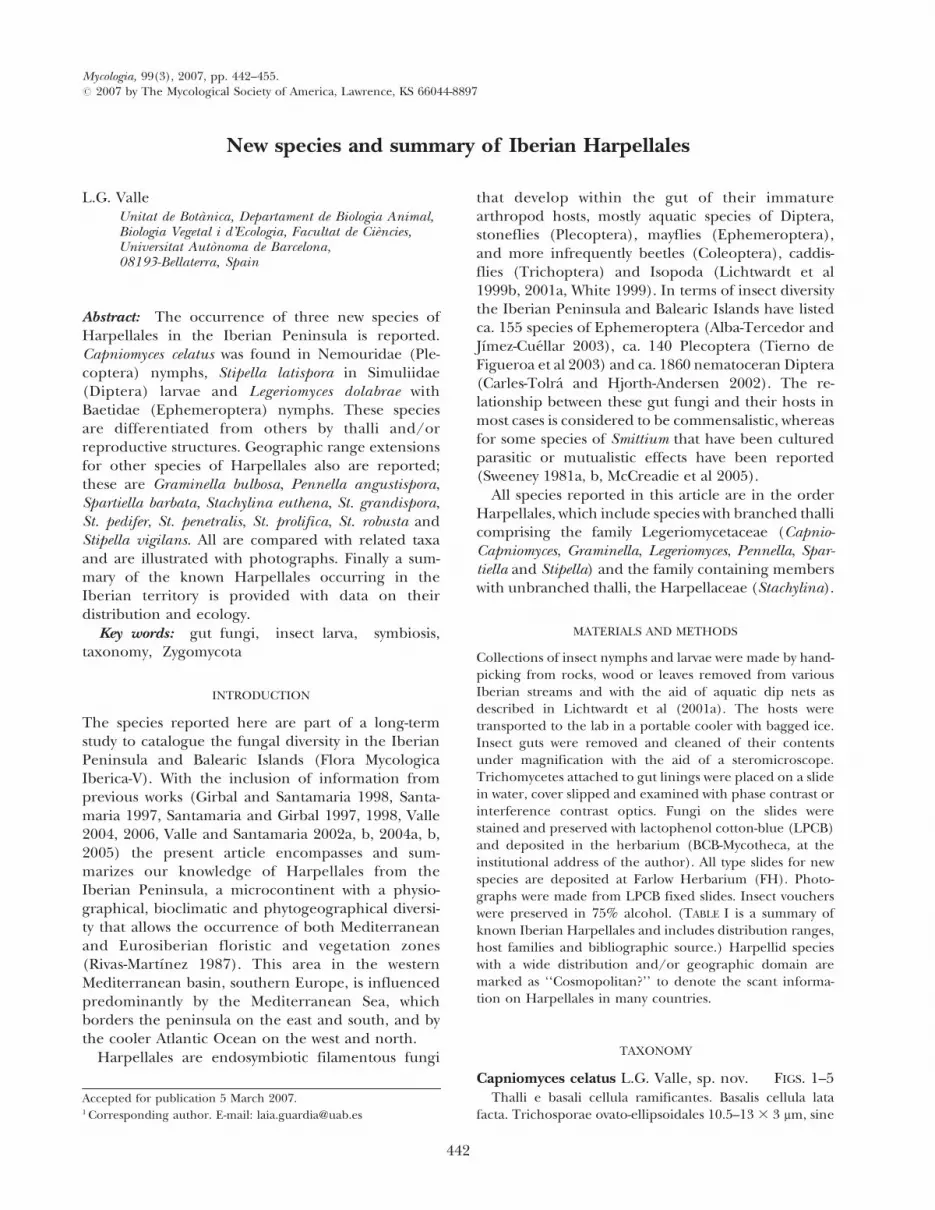
FIGS. 1–5. Capniomyces celatus (with Lancisporomyces vernalis in FIG. 1) from Nemouridae nymphs. 1. Overview ofCapniomyces celatus thallus with holdfast (H), trichospores (tr Cc) and one zygospore (Z Cc), among larger trichospores ofLancisporomyces vernalis (tr Lv). One trichospore on the generative cell is enlarged to show the appendage (a). 2. Fertilebranches with nearly mature (arrowhead) and immature (arrow) trichospores. 3. Thallus with conjugating hypha belowzygosporophore (ZP), zygospore (Z) and trichospores (tr). 4. Heterothallic conjugation tube (arrow) and zygospore just out offocus (arrowhead). 5. Zygospore (arrow) and zygosporophore, apparently homothallic. Bars: 1, 2, 4, 5 5 25 mm; 3 5 50 mm.
VALLE: IBERIAN HARPELLALES 443
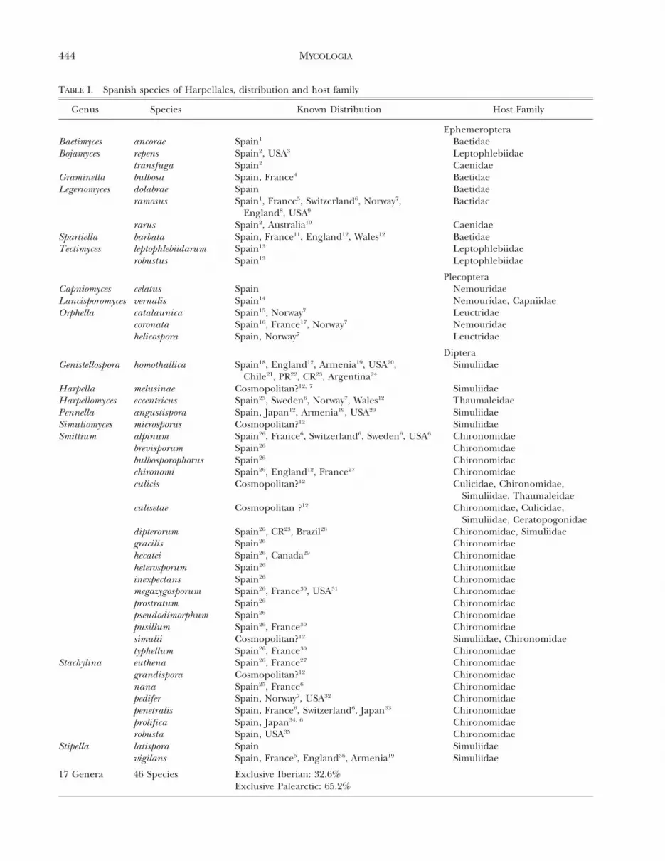
TABLE I. Spanish species of Harpellales, distribution and host family
Genus Species Known Distribution Host Family
EphemeropteraBaetimyces ancorae Spain1 BaetidaeBojamyces repens Spain2, USA3 Leptophlebiidae
transfuga Spain2 CaenidaeGraminella bulbosa Spain, France4 BaetidaeLegeriomyces dolabrae Spain Baetidae
ramosus Spain1, France5, Switzerland6, Norway7,England8, USA9
Baetidae
rarus Spain2, Australia10 CaenidaeSpartiella barbata Spain, France11, England12, Wales12 BaetidaeTectimyces leptophlebiidarum Spain13 Leptophlebiidae
robustus Spain13 Leptophlebiidae
PlecopteraCapniomyces celatus Spain NemouridaeLancisporomyces vernalis Spain14 Nemouridae, CapniidaeOrphella catalaunica Spain15, Norway7 Leuctridae
coronata Spain16, France17, Norway7 Nemouridaehelicospora Spain, Norway7 Leuctridae
DipteraGenistellospora homothallica Spain18, England12, Armenia19, USA20,
Chile21, PR22, CR23, Argentina24
Simuliidae
Harpella melusinae Cosmopolitan?12, 7 SimuliidaeHarpellomyces eccentricus Spain25, Sweden6, Norway7, Wales12 ThaumaleidaePennella angustispora Spain, Japan12, Armenia19, USA20 SimuliidaeSimuliomyces microsporus Cosmopolitan?12 SimuliidaeSmittium alpinum Spain26, France6, Switzerland6, Sweden6, USA6 Chironomidae
brevisporum Spain26 Chironomidaebulbosporophorus Spain26 Chironomidaechironomi Spain26, England12, France27 Chironomidaeculicis Cosmopolitan?12 Culicidae, Chironomidae,
Simuliidae, Thaumaleidaeculisetae Cosmopolitan ?12 Chironomidae, Culicidae,
Simuliidae, Ceratopogonidaedipterorum Spain26, CR23, Brazil28 Chironomidae, Simuliidaegracilis Spain26 Chironomidaehecatei Spain26, Canada29 Chironomidaeheterosporum Spain26 Chironomidaeinexpectans Spain26 Chironomidaemegazygosporum Spain26, France30, USA31 Chironomidaeprostratum Spain26 Chironomidaepseudodimorphum Spain26 Chironomidaepusillum Spain26, France30 Chironomidaesimulii Cosmopolitan?12 Simuliidae, Chironomidaetyphellum Spain26, France30 Chironomidae
Stachylina euthena Spain26, France27 Chironomidaegrandispora Cosmopolitan?12 Chironomidaenana Spain25, France6 Chironomidaepedifer Spain, Norway7, USA32 Chironomidaepenetralis Spain, France6, Switzerland6, Japan33 Chironomidaeprolifica Spain, Japan34, 6 Chironomidaerobusta Spain, USA35 Chironomidae
Stipella latispora Spain Simuliidaevigilans Spain, France5, England36, Armenia19 Simuliidae
17 Genera 46 Species Exclusive Iberian: 32.6%
Exclusive Palearctic: 65.2%
444 MYCOLOGIA

Specimens examined. SPAIN. BARCELONA: Sant Llor-enc Savall, Vall d’Horta stream, 31TDG21, prepared fromNemoura cinerea Retzius nymphs, 1-II-2001, SP-Tr0476(HOLOTYPE: FH), BCB-Tr0477.
Commentary. The type species, C. stellatus S.W.Peterson & Lichtw., was described from hosts of thegenus Allocapnia (Capniidae, Plecoptera), which areendemic in North America (Peterson and Lichtwardt1983). Capniomyces celatus was noticed in a stoneflyhost from a different family, which is widely distributedin Europe. In the type locality the host was abundantand highly infested with Lancisporomyces vernalisSantam. (Santamaria 1997) (FIG. 1) but just two ofthe nymphs were infested with C. celatus. The size ofthis new species, usually obscured by the dense thalli ofLancisporomyces, made detection difficult within thegut contents.
Capniomyces celatus has generative cells arranged inlong series (,12) at the ends of fertile branches(FIG. 2), making it readily distinguishable from C.stellatus with typically only three trichospores perbranch based on examination of the Holotype (FH)MIS-1-104. This characteristic of the new species,together with the smaller trichospores and zygospores(trichospores of [10–]15[–19] 3 4–6 mm and zygos-pores of [42–]52[–64] 3 7–9 mm in C. stellatus[Peterson and Lichtwardt 1983]), makes C. celatusunique, even though it was impossible to determinethe exact number of trichospore appendages (six inC. stellatus, Peterson and Lichtwardt [1983]). Appen-dages in the Spanish specimens were barely visibleinside the generative cells but appeared to be thick,petaloid, and occupying nearly the entire cell.Although most zygospores typically were formed ona zygosporophore from the middle of a heterothallicconjugation tube (FIG. 4) one of the observedzygospores was apparently homothallic (FIG. 5). An-other zygospore (FIG. 6) appeared to have a type IVattachment (see Moss et al 1975 for zygospore types),but the sexual spore was not mature. Apparent ‘‘TypeIV’’ arrangements of the zygospore on the zygospor-ophore occasionally have been observed with otherspecies that typically show different types of attach-ment (e.g. Legeriomyces) (LG Valle unpubl); this
could be caused by a developmental abnormality thatcauses the type II zygospore to appear with a polarrather than an oblique attachment to the zygospor-ophore.
Legeriomyces dolabrae L.G. Valle, sp. nov. FIGS. 6–11Thallus 6–7.5 mm diam cum sparsim ramificanti princi-
pali axe. Basalis cellula cum simplici campanulato tenaculo.Trichosporae elongato-obpyriformes, 36–43 3 8.5–9.5 mm,cum appendicibus duabus, magis tenuibus ad extremum,prima elongata, altera brevi. Appendices helicte dispositaeintra cellulam genitalem. Sine collari. Unaquaeque fertilisrama cum 2–5 genitalibus cellulis. Zygosporae 39–43 3
99.5 mm, asymetricae cum duobus extremis, quorum unumbrevius et arcuatum, 9–12 mm, et alterum, 26–30 mm,longius et rectum. Liberae zygosporae cum collari, 11–13.5 3 5–6 mm. Zygosporophorum 12–13.5 3 5–6 mm. Adcuticulam proctodaei nympharum Baetidae (Ephemerop-tera) affixi.
Thalli sparsely ramified from the base, with an axialfilament diameter of 6–7.5 mm. Basal cell slightlybroadened (FIG. 10), with a conical secreted holdfast.Trichospores 36–43 3 8.5–9.5 mm, elongate-obpyri-form without a collar but with two appendagesdiffering in length (FIG. 8). Appendages wider atthe proximal end, helically coiled inside the genera-tive cell. Generative cells measuring 6–7.5 mm diam,variable in length, occurring in a series of 2–5 cellsper fertile branch. Zygospores 39–43 3 9–9.5 mm,biconical, obliquely attached to the zygosporophoreand markedly asymmetrical. The proximal end (9–12 mm) is curved toward the zygosporophore, whereasthe longer end (26–30 mm) is straight (FIGS. 7, 9, 11).Zygosporophore 12–13.5 3 5–6 mm, with a densecytoplasm that aggregates into a slender conical shapeafter staining with LPCB (FIGS. 6, 7, 11). Thezygospore on release has a collar (11–13.5 3 5–6 mm) at its base (FIG. 7). In the hindgut of Baetidae(Ephemeroptera) larvae.
Etymology. Latin, dolabrae 5 an agriculture diggingtool (a sort of hoe), which resembles the zygospore inmorphology.
Specimens examined. SPAIN. GUADALAJARA: Valdeso-tos, Rio Jarama, 30TWL73, prepared from Baetis rhodani
r
CR: Costa Rica; PR: Puerto Rico; References: (1) Valle and Santamaria 2002a; (2) Valle and Santamaria 2004b; (3) Longcore1989; (4) Leger and Gauthier 1937; (5) Leger and Gauthier, 1932; (6) Lichtwardt 1984; (7) White and Lichtwardt 2004; (8)Moss 1979; (9) Whisler 1963; (10) Williams and Lichtwardt 1993; (11) Tuzet and Manier 1950a; (12) Lichtwardt et al 2001a;(13) Valle and Santamaria 2002b; (14) Santamaria 1997; (15) Santamaria and Girbal 1998; (16) Valle and Santamaria 2005;(17) Leger and Gauthier 1931; (18) Girbal and Santamaria 1998; (19) Nelder et al 2005; (20) Lichtwardt 1972; (21) Lichtwardtand Arenas 1996; (22) White et al 2000; (23) Lichtwardt 1997; (24) Lopez Lastra et al 2005; (25) Santamaria and Girbal 1997;(26) Valle and Santamaria 2004a; (27) Manier 1970; (28) Alencar et al 2003; (29) Strongman Unpublished; (30) Manier andCoste 1971; (31) Beard and Adler 2000; (32) Lichtwardt and Williams 1983; (33) Lichtwardt 1984; (34) Lichtwardt et al 1987;(35) Lichtwardt and Williams 1999; (36) Moss 1970.
VALLE: IBERIAN HARPELLALES 445
(Pictet) larvae, 19-IX-2001, BCB-Tr0985, SP-Tr0986 (HO-LOTYPE: FH), BCB-Tr0987, BCB-Tr0991–Tr0993.
Commentary. The most interesting and characteris-tic feature of this species is the asymmetric zygosporeshape, with the shorter end characteristically curveddownward toward the zygosporophore (FIGS. 6, 7, 9).Also the aggregated cytoplasmic content of the fixedand stained zygosporophore and the larger tricho-spore diameter in L. dolabrae separate it from L.ramosus Pouzar, a species documented from Spain(Valle and Santamaria 2002a). The other species inthe genus, L. rarus Lichtw. & M.C. Williams, alsoreported from Spain (Valle and Santamaria 2004b),has different thallus features, shorter trichospores aswell as more typical, type II biconical zygospores(Williams and Lichtwardt 1993).
Stipella latispora L.G. Valle, sp. nov. FIGS. 12–15Thalli cum principali axe 8–9.5 mm diam, pinnatim
ramificanti. Basalis cellula simplex vel lobata, cum viscosaet amorfa secreta materia ad modum tenaculi. Granuli-formes prominentiae in basali cellularum superficie dis-tributae. Trichosporae ovato-ellipsoidales, 60–70 3 8.5–9 mm, cum 6(–7) appendicibus petaloideis praeditae. Sinecollari. Unaquaeque fertilis rama 3.5–4 mm diam, cum 1–4genitalibus cellulis. Zygosporae biconicae 89–92 3 13 mm,cum collari et zygosporophoro 30 3 11 mm. Ad cuticulamproctodaei nympharum Simuliidarum (Diptera) affixi.
Thallus pinnately ramified, with a central axis 8–9.5 mm wide. Basal cell lobulate (FIG. 12) or simple(FIG. 14), coated with amorphous material. Small peg-like projections irregularly dispersed along the basalcell surface (FIG. 14 arrow). Trichospores 60–70 3
8.5–9 mm, ovoid-ellipsoidal and slightly curved, eccen-
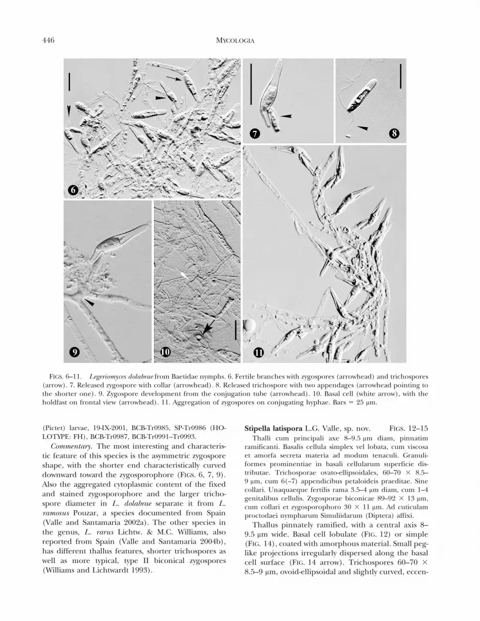
FIGS. 6–11. Legeriomyces dolabrae from Baetidae nymphs. 6. Fertile branches with zygospores (arrowhead) and trichospores(arrow). 7. Released zygospore with collar (arrowhead). 8. Released trichospore with two appendages (arrowhead pointing tothe shorter one). 9. Zygospore development from the conjugation tube (arrowhead). 10. Basal cell (white arrow), with theholdfast on frontal view (arrowhead). 11. Aggregation of zygospores on conjugating hyphae. Bars 5 25 mm.
446 MYCOLOGIA

trically attached to the generative cell, without a collar,6(–7) petaloid appendages present (FIG. 13 arrow).Generative cells 3.5–4 mm diam, arranged in serieswith 1–4 cells per fertile branch (FIG. 12). Zygospores89–92 3 13 mm, biconical, type I, formed homothalli-cally from cells near conjugation tubes (FIG. 12, 15).
Zygosporophore 30 3 11 mm, partially carried asa collar by the zygospore once released. In thehindgut of Simuliidae (Diptera) larvae.
Etymology. Latin, lati 5 wide, spora 5 spore re-ferring to the wide trichospores in the species.
Specimens examined. SPAIN. MURCIA: Moratalla, Rio
FIGS. 12–15. Stipella latispora from Simuliidae. 12. Overview of a thallus with a mature zygospore (arrow) and trichospores;basal cell surrounded by mucilage (arrowhead). 13. Released trichospore with appendages (arrow). 14. Basal cell withmucilage and minute peg-like projections (arrowhead). 15. Thallus with developing immature zygospores (arrows), the lowerone laterally folded. Bars: 12, 14, 15 5 50 mm; 13 5 25 mm. FIGS. 16–18. Graminella bulbosa from Baetidae. 16–17. Delicatethalli germinated from vegetative propagules, with small trichospores at distal branches. 18. Mature thallus with trichosporesand a bulbous cell, empty of its content. Bars 5 50 mm.
VALLE: IBERIAN HARPELLALES 447
Beamor, 30SWH92, prepared from Simuliidae larvae, 13-V-2003, SP-Tr1761 (HOLOTYPE: FH), BCB-Tr1763.HUELVA: Arroyomolinos de Leon, Rio Montemayor, Sierrade Aracena, 29SQC21, prepared from Simuliidae larvae, 6-XI-2003, BCB-Tr1869–1870.
Commentary. This is the second species in thegenus. The key characteristic is its large trichospores,consistently wider than those of the type species S.vigilans Leger & Gauthier (Leger and Gauthier 1932).General features of the thallus are similar for bothspecies. The zygospores of S. latispora (FIG 12 arrow)are also larger and straighter than those of S. vigilans(FIG. 32). One of the more intriguing features is thepresence of six (rarely seven) appendages on thetrichospores (FIG. 13 arrow), whereas only 3–4 weredescribed previously for S. vigilans (FIG. 33). Themorphology of the basal cell is variable (FIGS. 12, 14)as in the type species (FIG. 34). The variability ofappendage number and holdfast characteristics withinS. vigilans had been reported by Manier (1963) andMoss (1970), but trichospores consistently are narrowerin S. vigilans compared to S. latispora (FIGS. 12, 13, 15).
PREVIOUSLY KNOWN SPECIES
Graminella bulbosa Leger & Gauthier ex Manier, 1962.FIGS. 16–18
Specimens examined. SPAIN. BARCELONA: Guardiolade Bergueda, Rio Greixer, 31-V-2001. Cardona, Rio Card-ener, 4-II-2002. GIRONA: Meranges, Foguerade stream,, 9-VIII-2000. Cantallops, Collpregon stream, 14-VI-2001. GUA-DALAJARA: Valdesotos, Rio Jarama, 19-IX-2001. HUESCA:Salinas de Sin, Rio Cinqueta, 10-X-2000. LLEIDA: La Coma,Fonts del Cardener, 30-VIII-2000. Alins, Areu, Aixeus stream(Noguera Rio Vallferrera), 23-VI-2001. SEGOVIA: Vegas deMatute, Rio Moros, 24-IX-2001. TERUEL: Beseit, ElParrissal, Rio Matarranya, 24-III-2001. VIZCAYA: Guriezo,Rio Aguera, 29-V-2002. Mendata, Rio Golazo, 7-X-2002. Allspecimens prepared from nymphs of B. alpinus Pictet, B.rhodani and Baetis sp.
This species was described from France in thehindguts of Baetis sp. (Leger and Gauthier 1937) andwas found in the same country by Manier (1962) inBaetis rhodani (Pictet), a common and widespreadBaetidae. This paper represents the first record of thisspecies in the Iberian Peninsula, where it shows thesame ecology as Legeriomyces ramosus. Both speciesoften were found in the same hindgut. The species ischaracterized by the numerous small trichospores(FIG. 18), the formation of bulbous basal cells (FIGS. 16,17) and vegetative propagules (Leger and Gauthier1937), observed in collections from Iberia.
Pennella angustispora Lichtw., 1972. FIGS. 19–21Specimens examined. SPAIN. GIRONA: El Brull, la
Castanya stream, prepared from Simuliidae larvae, 3-X-2000.
This species is rare in the study area, recorded froma single locality, although the hosts are common. Thetypical cylindrical-clavate trichospores (FIGS. 19–21)are distinctive, and the Iberian specimens (50–73 3
2.5–3.5 mm) share the same characteristics as speci-mens from USA (Lichtwardt 1972) and Japan (Dang1979, Sato 2001). Nelder et al (2005) reported thespecies in endemic Simuliidae from Armenia (Asia).The species has not been reported before fromEurope.
Spartiella barbata Tuzet & Manier ex Manier, 1968FIGS. 22–30
Specimens examined. SPAIN. BARCELONA: Vacarisses,Senana stream, 6-VI-2001. BILBAO: Guriezo, Rio Aguera,29-V-2002. GIRONA: Santa Pau, Can Patxet, Bosquetstream/Sant Martı stream, 5-IX-2000. Sant Jaume deLlierca, Rio Llierca /Castella stream, 5-IX-2000. Vall deBianya, Rio Vall d’en Bac, 5-IX-2000. HUESCA: Loporzano,Rio Isuela, larvae, 19-IX-2000. Sesa, Rio Guatizalema, 19-IX-2000. Adahuesca, Rıo Vero, 20-IX-2000. Santa Eulalia laMenor, Rio Flumen, 20-IX-2000. Salinas del Sin, RioCinqueta, 10-X-2000. LLEIDA: Guixers, La Casa Nova deValls, Rio Aigua de Vall, 30-VIII-2000; Rio Aigua d’Ora, 30-VIII-2000. MADRID: Valdesotos, Rio Jarama, 19-IX-2001.Pinilla del Valle, Rio Lozoya, 3-X-2001. El Paular, Umbrıastream, 3-X-2001. TERUEL: Beseit, Rio Matarranya, 24-III-2001. All specimens prepared from nymphs of B. alpinus, B.rhodani and Baetis sp.
This species was described from France (Tuzet andManier 1950) and is similar to its American congenerS. animae, differing only in trichospore size (Licht-wardt 1997). Spartiella barbata is easily identifiable byits lobulate basal cell (FIG. 22) and typical obpyriform
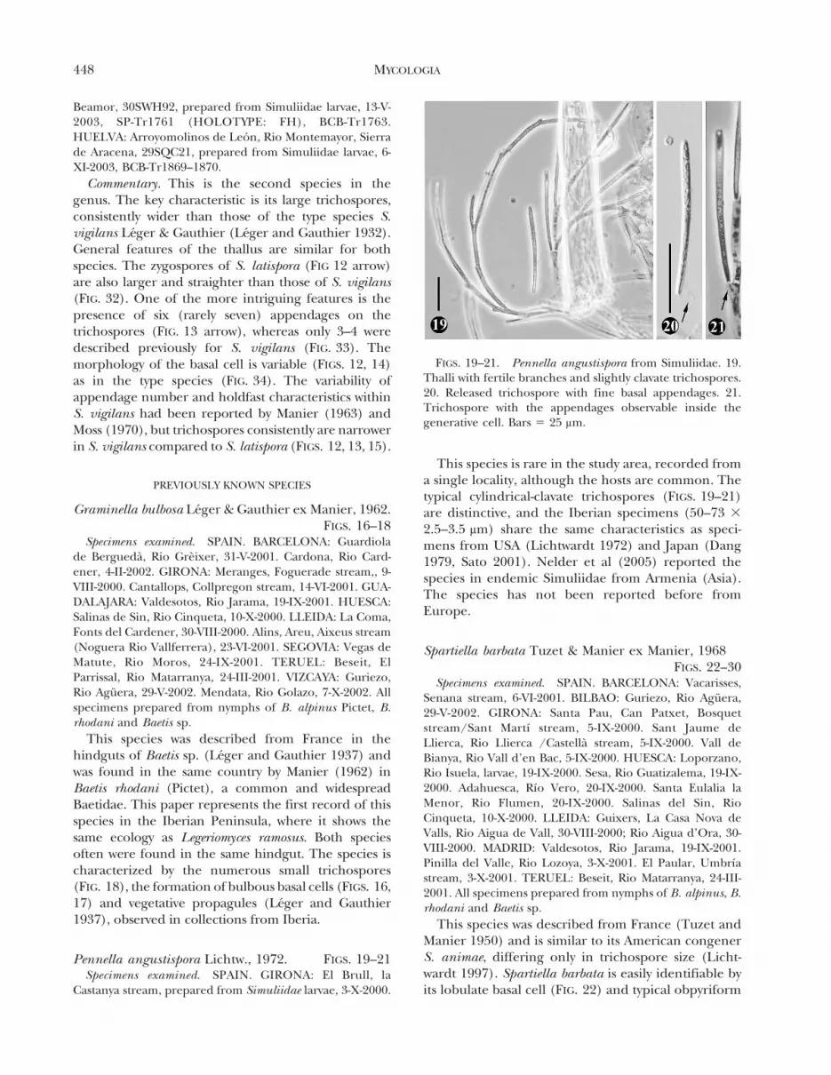
FIGS. 19–21. Pennella angustispora from Simuliidae. 19.Thalli with fertile branches and slightly clavate trichospores.20. Released trichospore with fine basal appendages. 21.Trichospore with the appendages observable inside thegenerative cell. Bars 5 25 mm.
448 MYCOLOGIA

trichospores (20–26 3 6.5–8.5 mm) with one append-age that remains tightly coiled just after release(FIG. 26) but eventually uncoils into a long, fineappendage (FIG. 25). Zygospores (25.5–34 3 6–
7.5 mm) were present (FIGS. 23, 24). The speciesfrequently appeared together with Legeriomyces ramo-sus in Baetidae hindguts. Morphological charactersare congruent with the original description, except
FIGS. 22–30. Spartiella barbata from Baetidae. 22. Lobulate basal cell (arrow). 23. Free zygospore with intact collar (arrow).24. Mass of conjugating thalli and biconical zygospores. 25. Free trichospore with the unfolding long appendage (arrowheads).26. Recently released trichospore with the appendage still folded (arrow). 27–30. Phases of sporangiospore extrusion. 27.Trichospore with apical cell wall elongating (arrowhead) before developing extrusion tube. 28–29. Trichospore with extendedextrusion tube (arrowhead) and extruded content (arrow) at its end. 30. Sporangiospore has been dispersed and the hyalinetube (arrowhead) persists on the empty sporangium (arrow). FIGS. 31–34. Stipella vigilans from Simuliidae. 31. Fertilebranches with cylindrical trichospores. 32. Conjugations and zygospores on a single thallus. 33. Released trichospore with fineappendages (arrowhead). 34. Basal cell with peg-like projections. Bars: 22, 23, 25–30, 33–34 5 25 mm; 24, 31, 32 5 50 mm.
VALLE: IBERIAN HARPELLALES 449
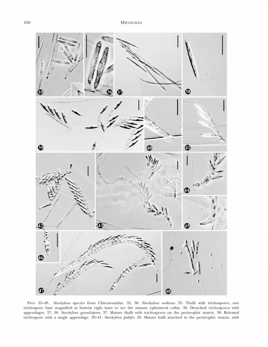
FIGS. 35–48. Stachylina species from Chironomidae. 35, 36. Stachylina euthena. 35. Thalli with trichospores, onetrichospore base magnified at bottom right inset to see the minute ephemeral collar. 36. Detached trichospores withappendages. 37, 38. Stachylina grandispora. 37. Mature thalli with trichospores on the peritrophic matrix. 38. Releasedtrichospore with a single appendage. 39–41. Stachylina pedifer. 39. Mature halli attached to the peritrophic matrix, with
450 MYCOLOGIA
for presence of smaller trichospores in some locali-ties. An interesting feature concerning the method oftrichospore extrusion is described for the first timefrom specimens on microscope slide BCB-Tr0315(from Huesca). The sporangium content is releasedthrough a hyaline tube formed at the apex of thetrichospore (FIGS. 27–30). This channel is formed bya progressive elongation of the apical internal wall,which can be detected initially by an anomalousextension of the trichospore at its apex (FIG. 27). Theextruded sporangiospore is surrounded by a thinhyaline wall. A similar trichospore extrusion methodwas reported from cultured Smittium culisetae Lichtw.,described as a sleeve-like membrane between thetrichospore wall and sporangiospore (Horn 1989). Inthe case of S. barbata the structure is more likea cylindrical tube with a uniform diameter. Anotherobservation of an extrusion process is that ofEjectosporus spica (S.W. Peterson & Lichtw.) Strong-man, where the sporangiospore content of thevegetative cells has a lunate shape (Lichtwardt et al1991, Strongman 2005).
Stipella vigilans Leger & Gauthier, 1932 FIGS. 31–34Specimens examined. SPAIN. BALEARIC ISLANDS (MA-
JORCA): Santa Maria del Camı, Coanegre stream, 27-V-2003.Soller, Biniaraix, l’Ofre stream, 29-V-2003. Valldemossa,Valldemossa stream, 30-V-2003. BARCELONA: Mura, lesNespres stream, 6-II-2001. Sant Llorenc Savall, Vall d’Hortastream, 1-II-2001. Santa Maria de Corco, Paganes stream, 19-II-2001; El Freu stream, 19-II-2001; Sant Julia de Cabrera,Sant Julia stream, 19-II-2001. CUENCA: Tragacete, Rio Jucar,1-X-2001. GIRONA: Espinelves, Mas Joan, Major stream, 10-VII-2000. Setcases, Obaga de Carboners, 12-IX-2000. JAEN:Vadillo-Castril, Rio Guadalquivir, 12-VII-2001. Tranco deBeas, Rio Guadalquivir, 12-VII-2001. LLEIDA: Espot, Pla deFontinals, 21-VI-2001. MADRID: Pinilla del Valle, Rio Lozoya,3-X-2001. VIZCAYA: Trebueso, Rio Aguera, 29-V-2002. Allspecies prepared from Simuliidae larvae.
This species was found in diverse localities on theIberian Peninsula and the Balearic Islands (Majorca),where it frequently was collected and grew denselywithin its hosts. The specimens examined sharethallial and spore characteristics with the French typespecimens. The cylindrical trichospores (FIGS. 31,33), (41–58.5 3 2.5–4 mm), with 2–4 fine appendages,are typical for the species. The zygospores observed
generally were smaller (62–82 3 12.5–16 mm)(FIG. 32) than those reported for the type (80–1053 15–18 mm).
Stachylina euthena Manier & F. Coste, 1971FIGS. 35–36
Specimens examined. SPAIN. BARCELONA: Lluca, SantaEulalia de Puigoriol, Gavarresa stream, prepared fromChironomidae larvae, 27-VII-2001. VIZCAYA: Gorozika,Rio Golako, prepared from Chironomidae Orthocladiinaelarvae, 7-X-2002.
Harpellaceae species have been described fromFrance, in diverse genera of Chironomidae (Manierand Coste 1971). After the initial description S.euthena was not reported again until this study inanother southern European locality, the IberianPeninsula. Stachylina euthena is similar to S. grand-ispora Lichtw. in its general features, the latter havinglarger trichospores and a shorter collar (Lichtwardt1984). The Iberian specimens have trichosporesmeasuring 27–39 3 6.5–8 mm with a collar 2 mm long,matching the original description well. Nonethelessthere is some variability in trichospore shape in theIberian specimens, which ranges from the typicalfusiform with a medial swelling to clearly ellipsoidalones (FIGS. 35, 36). The basal cell of the thallus hasa small discoid holdfast (FIG. 35). However overall thespecimens from the two localities in Spain do notshow significant morphological variation.
Stachylina grandispora Lichtw., 1972 FIGS. 37, 38Specimens examined. SPAIN. BARCELONA: L’Estany,
L’Estany stream, prepared from Chironomidae Diamesiinaelarvae, 19-XII-2000. Navas, Pala de Torroella, Rio Cardener,prepared from Chironomidae Diamesiinae larvae. GIR-ONA: Cruılles, Monells i Sant Sadurnı de l’Heura,Cantagalls stream, prepared from Chironomidae larvae,13-XII-2000.
This is one of the more common species within thegenus, with a worldwide distribution (United King-dom, Australia, Hawaii, New Zealand, Sweden andUSA). Some of the Iberian specimens had trichos-pores (FIGS. 37, 38) slightly narrower ([36–]40–68 3
[5.5–]6–8.5 mm) than those of the original descrip-tion (Lichtwardt 1972), but variability in this charac-ter has been reported before (Lichtwardt et al 2001a).
r
trichospores and appendages observable inside generative cells. 40. Foot-like basal cell penetrating the peritrophic matrix. 41.Thallus with penetrating basal cell and trichospores with appendages observable inside the corresponding generative cells. 42.Stachylina penetralis. Thalli with trichospores and a penetrating basal cell (arrowhead). 43–45. Stachylina prolifica. 43. Thalliwith numerous trichospores. 44, 45. Details of basal cell. 46–48. Stachylina robusta. 46. Detail of basal cell. 47. Mature thalli withtrichospores. 48. Two young thalli conjugating (arrow). Bars 5 25 mm.
VALLE: IBERIAN HARPELLALES 451
Stachylina pedifer Lichtw. & M.C. Williams, 1983FIGS. 39–41
Specimens examined. SPAIN. BARCELONA: Fıgols, Pe-guera, Fontana Coix, prepared from Chironomidae, 31-V-2002. GIRONA: Viladrau, Riera Major stream, FontanaFerro, prepared from Eukierffiella sp. larvae, 23-X-2002.
This species shares characteristics with S. penetralisLichtw., such as the basal cell penetrating theperitrophic matrix of the host (FIGS. 39–41) andvariability in trichospore shape (FIG. 39), which also isvariable for S. penetralis (Lichtwardt 1984, Lichtwardtet al 2001a) and can make identification difficult. TheIberian specimens show the typical foot-like basal cellof S. pedifer, which slightly penetrates the peritrophicmatrix of the midgut (FIG. 40). The number ofgenerative cells per thallus is 2–8, according toLichtwardt and Williams (1983), but up to 10trichospores per thallus were observed, althoughnormally there were 4–8 spores. Trichospores mea-sured 25–33 3 7–9 mm and fit the species descriptionwell.
Stachylina penetralis Lichtw., 1984 FIG. 42Specimens examined. SPAIN. BARCELONA: Moia, la
Fabrega stream, prepared from Chironomidae Diamesinilarvae, 13-V-2002. Avia, Clara stream, 2-IV-2001. Cercs, RioLlobregat, 2-IV-2002. GIRONA: Queralbs, Nuria, Comad’Eina stream, 25-VIII-2000. Boadella d’Emporda, RioMuga, 4-VI-2001. Viladrau, Riera Major stream, FontanaFerro, 23-I-2002. All specimens prepared from Chironomi-dae larvae, mostly Diamesinae.
Stachylina penetralis has been described frominsects collected in France, Sweden and Japan(Lichtwardt 1984) in diverse Chironomidae species.This species has a globose basal cell that penetratesthe peritrophic matrix (FIG. 42). Occasionally, if theview of the basal cell is not lateral but frontal ordorsal, and the specimen is not mature, it is possibleto misidentify this species as S. minuta Gauthier exLichtw. or S. pedifer. The Iberian specimens showedno remarkable differences from the published de-scription of this species, with trichospores measuring33–42(–46) 3 (7–)8–11 mm.
Stachylina prolifica Lichtw., Kobayasi & Indo, 1987FIGS. 43–45
Specimens examined. SPAIN. BARCELONA: Fogars deMontclus; Sta. Fe del Montseny stream, near FontanaPassavets, 25-X-2001. Gualba de dalt, Gualba stream, 7-IX-2001. GIRONA: Agullana, la Guilla torrent, 14-IV-2001.VIZCAYA: Turcıos, Rio Aguera, 29-V-2002. All specimensprepared from Chironomidae Diamesiinae and Orthocla-diinae larvae.
This represents the first report of this speciesoutside the type locality in Japan where it wasdescribed from bloodworm (Chironomus sp.) larvae
(Lichtwardt et al 1987). The thalli of this species showgreat variability in length, and can produce 4–50generative cells each. Similar variability was observedwith the Iberian specimens but most of the thalli hadabout 20 generative cells (range 8–45). Trichosporeswere long and narrow (23.5–38 3 4–6 mm) and wereproduced profusely on the thallus (FIG. 43), borne onnumerous, small generative cells (7–14 3 7–12 mm).Also the specimens had a narrow peg-like basal cell(FIGS. 43–45) which is diagnostic for the species.
Stachylina robusta Lichtw. & M.C. Williams, 1999FIGS. 46–48
Specimens examined. SPAIN. BARCELONA: Gualba deDalt, Gualba stream, prepared from Chironomidae Ortho-cladiinae larvae, 7-XI-2001. VIZCAYA: Trucıos (Turtzioz),Rio Aguera, prepared from Chironomidae Orthocladinaelarvae, 29-V-2002.
These collections are the first for this speciesoutside North America, where it was reported fromthe USA by Lichtwardt and Williams (1999) andCanada by Lichtwardt et al (2001b). This is a specieseasily identified by the relatively thick thallus, thelarge number of generative cells and collarlesstrichospores (FIGS. 46–48). In Spanish collections itwas observed occasionally that the generative cellremained attached as an ephemeral collar to thetrichospore after release. The Iberian specimens hada thallus diameter of 12–14 mm and elongate-ellipsoidtrichospores, 29–35 3 7.5–8.5 mm. Another specieswith similar features is S. jujuyensis Mazzuchelli,Lopez-Lastra & Lichtw. described from Argentina,which has thalli 10 mm thick and trichospores in thesame size range (Lichtwardt et al 2000).
DISCUSSION
The taxa here documented along with those pre-viously published for the Iberian territory total 47species among 17 genera of Harpellales (TABLE I).Most diversity is recorded within the genera Smittiumand Stachylina, in accordance to most previous dataworldwide (Lichtwardt et al 2001a). Within theIberian taxa and according to current information,the proportion of dipteran-associated Harpellalesconfined in the Palearctic regions is 58%. The Iberianharpellid species living within Plecopterans are 100%
Palearctic, while Ephemeropteran-associated speciesare 80% Palearctic. The remaining Harpellales forthese data correspond to widely distributed orHolarctic species (see TABLE I, also for bibliographicinformation). These results are mainly in agreementwith other studies, where a high percentage ofDipteran Harpellales appear not to be restrictedgeographically in spite of their host endemicity
452 MYCOLOGIA
(Nelder et al 2004, 2005) or isolation (Lichtwardt et al2001a, Valle and Santamaria 2004b). In Spain none ofthe species reported among Stachylina, Harpella,Harpellomyces or Genistellospora were new, and 60%
of the Smittium species currently are known fromother places (TABLE I). The capacity of adult femalesof Simuliidae and Chironomidae to carry harpelliddiaspores called ovarian cysts or chlamydospores(Labeyrie et al 1996, Moss and Descals 1986, Yeboahet al 1984) might aid the dispersion of thosecosmopolitan Smittium species, which also have a highadaptive plasticity among diverse hosts (Nelder et al2004). In the case of Harpellales living in the guts ofPlecopteran and Ephemeropteran hosts, the higherpercentage of endemicity (Peninsular or Palearctic) isin agreement with the possible incapacity of adulthosts to transport fungal diaspores inside theirbodies. This could explain why species such asCapniomyces celatus or Legeriomyces dolabrae havea restricted distribution. The locus classicus ofCapniomyces celatus is in a Mediterranean forest ofevergreen pine and oak that was destroyed by fireafter the collections were obtained. In subsequentsurveys stonefly hosts were not found, as a result ofthe ecosystem devastation. This event presents theopportunity to study the resilience of trichomycetespecies subjected to random, severe disturbances,which are frequent in Mediterranean zones, andmight have caused on more than one occasion thefragmentation and destruction of ecosystems andarthropod populations.
The new species Legeriomyces dolabrae shares somecharacteristics with L. ramosus, suggesting a closerelationship. In fact variation in L. ramosus zygo-spores, including abnormalities in the position andshape of these sexual structures, mixed with normalzygospores, also have been noted (unpubl). Even sothe asymmetric zygospores of L. dolabrae are a consis-tent feature in these collections and none of the moretypically biconical zygospores were observed.
Stipella latispora was found in southern localities ofthe Iberian Peninsula, at the middle course ofpermanent streams. This new species might be morewidely dispersed in southern Spain, although it likelyis more rare than S. vigilans. Variability in trichosporefeatures are well know in this genus; neverthelessdiagnostic characters are consistent in all specimensexamined and allow for easy separation of S. latisporafrom the more common S. vigilans. The number ofappendages present on trichospores has been usedwidely to separate genera within the Harpellales, butStipella with the new species S. latispora includespecies with different numbers of appendages. Othergenera with species showing variable number ofappendages are Bojamyces Longcore (Valle and
Santamaria 2004b), Harpella Leger & Duboscq(1929), Pennella Manier (1968), Simuliomyces Lichtw.(1972) and Plecopteromyces Lichtw., Ferrington &Lopez Lastra (Lichtwardt et al 1999a). Stipella vigilansis restricted to Europe and western Asia (see Table I,also for bibliographic information). It is not clearwhether this more restricted distribution is caused bylower dispersive ability compared to other speciesoccupying the same niche in the gut of Simuliidae(i.e. Smittium, Genistellospora). The data concerningMajorcan Harpellales provide information for furtherstudies on insular populations to help elucidatewhether their presence on this island is the result ofrelictual populations, colonization or both. Concern-ing this and other aspects of the understudiedTrichomycetes, the more we come to understandtheir biology, the more they challenge our under-standing of them.
AKNOWLEDGMENTS
The author expresses her gratitude to Robert Lichtwardtand Merlin White for the kind reception in their lab(University of Kansas, Lawrence), where some specimenswere examined. Thanks again to Merlin White and to DougStrongman for their helpful suggestions on the text, and toJose Fortes for the Latin translation. Also to the curatorialstaff of FH (Farlow Herbarium, Harvard University, Cam-bridge, USA) for the loan of diverse specimens. Thanks tothe staff of the Botanic Department at the University of theBasque Country (UPV-EHU, Leioa), particularly to IsabelSalcedo and fellows, Arturo Elosegui, as well as to the RoyalBotanic Garden (Real Jardın Botanico) of Madrid, and theUniversity of Murcia (Andres Millan and colleagues), all ofwhom kindly provided support and shelter to the authorin their laboratories also, to S. Santamaria for his help withphotographs. This research was financed by MCYT andFEDER (REN2002-04068-C02-02 Flora Micologica Iberica V).
LITERATURE CITED
Alba-Tercedor J, Jımez-Cuellar P. 2003. Checklist andhistorical evolution of the knowledge of Ephemerop-tera in the Iberian Peninsula, Balearic and CanaryIslands. In: Gaino E, ed. Research Update on Ephe-meroptera and Plecoptera. Perugia, Italy: University ofPerugia. p 91–97.
Alencar YB, Rıos-Velasquez CM, Lichtwardt RW, Hamada N.2003. Trichomycetes (Zygomycota) in the digestivetract of arthropods in Amazonas, Brazil. Mem InstOswaldo Cruz 98:799–810.
Carles-Tolra M, Hjorth-Andersen, eds. 2002. Catalogo de losDiptera de Espana, Portugal y Andorra (Insecta). Vol. 8.Spain: Monografıas S.E.A. ISBN: 84-932807-0-4. 323 p.
Dang S. 1979. Electron-microscope studies on the holdfaststructure of some Trichomycetes (Master’s thesis).Lawrence: University of Kansas. 78 p.
Girbal J, Santamaria S. 1998. Trichomycetes (Fungi,
VALLE: IBERIAN HARPELLALES 453
Zygomycotina) comensals de larves de Simuliidae(Diptera) a la Penınsula Iberica. Acta Bot Barcinonen45:49–56.
Horn BW. 1989. Requirement for potassium and pH shift inhost-mediated sporangiospore extrusion from tricho-spores of Smittium culisetae and other Smittium species.Mycol Res 93:303–313.
Labeyrie ES, Molloy DP, Lichtwardt RW. 1996. An in-vestigation of Harpellales (Trichomycetes) in New Yorkstate blackflies (Diptera: Simuliidae). J Invert Pathol 68:293–298.
Leger L, Gauthier M. 1931. Orphella coronata n. g., n. sp.Entophyte parasite des larves de Nemurides. Trav LabHydr Pisc Univ Grenoble 23:67–72.
———, ———. 1932. Endomycetes nouveaux des larvesaquatiques d’Insectes. Compt Rend Acad Sci Paris 194:2262–2265.
———, ———. 1937. Graminella bulbosa nouveau genred’Entophyte parasite des larves d’Ephemerides dugenre Baetis. Compt Rend Acad Sci Paris 202:27–29.
Lichtwardt RW. 1972. Undescribed genera and species ofHarpellales (Trichomycetes) from the guts of aquaticinsects. Mycologia 64:167–197.
———. 1984. Species of Harpellales living within the guts ofaquatic Diptera larvae. Mycotaxon 19:529–550.
———. 1997. Costa Rican gut fungi (Trichomycetes)infecting lotic insect larvae. Rev Biol Tropic 45:1339–1383.
———, Arenas J. 1996. Trichomycetes in aquatic insectsfrom southern Chile. Mycologia 88:844–857.
———, Cafaro MJ, White MM. 2001a. The trichomycetes,fungal associates of arthropods. Revised ed. Universityof Kansas: Publ on the Internet at www.nhm.ku.edu/,fungi
———, Ferrington LC, Lopez Lastra C. 1999a. Trichomy-cetes in Argentinean aquatic insect larvae. Mycologia91:1060–1082.
———, Kobayasi Y, Indoh H. 1987. Trichomycetes of Japan.Trans Mycol Japan 28:359–412.
———, Lopez Lastra C, Mazzucchelli MG. 2000. Fungiliving in the guts of larval aquatic insects in northwest-ern Argentina. Mycologia 92:332–340.
———, Peterson SW, Williams MC. 1991. Ejectosporus, anunusual new genus of Harpellales in winter-emergingstonefly nymphs (Capniidae) and a new species ofParamoebidium (Amoebidiales). Mycologia 83:389–396.
———, White MM, Cafaro MJ, Misra JK. 1999b. Fungiassociated with passalid beetles and their mites.Mycologia 91:694–702.
———, ———, Colbo MH. 2001b. Harpellales in New-foundland aquatic insect larvae. Mycologia 93:764–7873.
———, Williams MC. 1983. Two unusual Trichomycetes inan aquatic midge larva. Mycologia 75:728–734.
———, ———. 1999. Three Harpellales that live in onespecies of aquatic chironomid larva. Mycologia 91:396–399.
Longcore JE. 1989. Bojamyces repens: a new genus andspecies of Harpellales (Trichomycetes) from a lenticmayfly. Mycologia 81:482–486.
Lopez Lastra CC, Scorsetti AC, Marti GA, Coscaron S. 2005.Trichomycetes living in the guts of aquatic insects ofMissiones and Tierra del Fuego, Argentina. Mycologia97:320–328.
Manier J-F. 1962. Revision du genre Spartiella Tuzet etManier 1950 (sa place dans la classe des Trichomy-cetes). Ann Sci Nat Zoo Paris 4:517–525.
———. 1963. Trichomycetes de larves de Simulies (Harpel-lales du proctodeum). Ann Sci Nat Bot Paris 4:737–750.
———. 1968. Validation de Trichomycetes par leur di-agnose latine. Ann Sci Nat Bot Paris 9:93–108.
———. 1970. Trichomycetes de France. Ann Sci Nat BotParis 10:565–672.
———, Coste F. 1971. Trichomycetes Harpellales de larvesde Dipteres Chironomidae; creation de cinq nouvellesespeces. Bull Soc Mycol France 87:91–99.
McCreadie JW, Beard CE, Adler PH. 2005. Context-dependent symbiosis between black flies (Diptera:Simuliidae) and trichomycete fungi (Harpellales: Le-geriomycetaceae). OIKOS 108:362–370.
Moss ST. 1970. Trichomycetes inhabiting the digestive tract ofSimulium equinum larvae. Trans Brit Mycol Soc 54:1–13.
———. 1979. Commensalism of the Trichomycetes. In:Batra LR, ed. Insect-fungus Symbiosis: nutrition,mutualism, and commensalism. Montclair: Allanheld,Osmun & Co. p 175–227.
———, Descals E. 1986. A previously undescribed stage inthe life cycle of Harpellales (Trichomycetes). Mycolo-gia 78:213–222.
———, Lichtwardt RW, Manier J-F. 1975. Zygopolaris, a newgenus of Trichomycetes producing zygospores withpolar attachment. Mycologia 67:120–127.
Nelder MP, McCreadie JW, Coscaron C, Brockhouse CL.2004. First report of a trichomycete fungus (Zygomy-cota: Trichomycetes) inhabiting larvae of Simuliumochraceum sensu lato Walker (Diptera: Simuliidae) fromthe Galapagos Islands. J Invert Pathol 87:39–44.
———, Adler PH, Kachvoryan EA. 2005. Do gut symbiotesreflect the endemism of their host black flies (Diptera:Simuliidae) in the Caucasus of Armenia?. J Biogeo 32:1333–1341.
Peterson SW, Lichtwardt RW. 1983. Capniomyces stellatusand Simuliomyces spica: new taxa of Harpellales(Trichomycetes) from winter-emerging stoneflies. My-cologia 75:242–250.
Rivas-Martınez S. 1987. Nociones de fitosociologıa, biogeo-grafıa y bioclimatologıa. In: Peinado M, Rivas-MartınezS, eds. La vegetacion de Espana. Madrid: Serv. Publ.Univ. Alcala de Henares. p 19–46.
Santamaria S. 1997. Lancisporomyces, a new genus ofTrichomycetes with lance-shaped zygospores. Mycolo-gia 89:639–642.
———, Girbal J. 1997. Contribucion al conocimiento de losTrichomycetes (Fungi, Zygomycotina) Ibericos. AnalJardın Bot Madrid 55:219–223.
———, ———. 1998. Two new species of Orphella fromSpain. Mycol Res 102:174–178.
Sato H. 2001. Two ultrastructural aspects of the trichosporeof Pennella angustispora (Harpellales): canals in the
454 MYCOLOGIA

sporangiospore cell wall and appendage formation.Mycoscience 43:33–36.
Strongman DB. 2005. Synonymy of Ejectosporus magnus andSimuliomyces spica, and a new species, Ejectosporustrisporus, from winter-emerging stoneflies. Mycologia97:552–561.
Sweeney AW. 1981a. An undescribed species of Smittium(Trichomycetes) pathogenic to mosquito larvae inAustralia. Trans Brit Mycol Soc 77:55–60.
———. 1981b. Fungal pathogens of mosquito larvae. In:Davidson EW, ed. Pathogenesis of Invertebrate Micro-bial Diseases. Totowa, New Jersey: Allanheld, Osmun &Co. p 403–424.
Tierno de Figueroa JM, Sanchez Ortega A, Membiela IglesiaP, Luzon Ortega JM. 2003. Fauna Iberica. Vol. 22.Plecoptera. Series: Fauna Iberica. Madrid: MuseoNacional de Ciencias Naturales, Consejo Superior deInvestigaciones Cientıficas. 404 p.
Tuzet O, Manier J-F. 1950. Les Trichomycetes. Revision deleur diagnose. Raisons qui nous font y joindre lesAsellariees. Ann Sci Nat Zoo Paris 12:15–23.
Valle LG. 2004. Tricomicets Iberics [Doctoral dissertation].Barcelona: Dept. Biol. Animal, Biol. Vegetal i Ecologia,Unitat de Botanica, Universitat Autonoma de Barce-lona. 324 p.
———. 2006. Asellariales (Trichomycetes) from the IberianPeninsula. Fungal Divers 21:167–179.
———, Santamaria S. 2002a. Baetimyces, a new genus ofHarpellales, and first report of Legeriomyces ramosusfrom the northeastern Iberian Peninsula. Mycologia 94:321–326.
———, ———. 2002b. Tectimyces, a new genus of Harpel-
lales on mayfly nymphs (Leptophlebiidae) in Spain.Mycol Res 106:841–847.
———, ———. 2004a. The genus Smittium (Trichomycetes,Harpellales) in the Iberian Peninsula. Mycologia 96:682–701.
———, ———. 2004b. Bojamyces transfuga sp. nov. and newrecords of Trichomycetes from mayfly larvae in Spain.Mycologia 96:1386–1392.
———, ———. 2005. Zygospores as evidence of sexualreproduction in the genus Orphella. Mycologia 97:1335–1347.
White MM. 1999. Legerioides, a new genus of Harpellales inisopods and other Trichomycetes from New England,USA. Mycologia 91:1021–1030.
———, Cafaro MJ, Lichtwardt RW. 2000. Arthropodgut fungi from Puerto Rico and summary of trop-ical Trichomycetes worldwide. Carib J Sci 36:210–220.
———, Lichtwardt RW. 2004. Fungal symbionts (Harpel-lales) in Norwegian aquatic insect larvae. Mycologia 96:891–910.
Williams MC, Lichtwardt RW. 1993. A new monotypicfungal genus, Allantomyces, and a new species ofLegeriomyces (Trichomycetes, Harpellales) in the hind-gut of a Western Australian mayfly nymph (Tasmano-Tasmanocoenis sp.). Can J Bot 71:1109–1113.
Whisler HC. 1963. Observations on some new andunusual enterophilous Phycomycetes. Can J Bot 41:887–900.
Yeboah DO, Undeen AH, Colbo MH. 1984. Phycomycetesparasitizing the ovaries of blackflies (Simuliidae). JInvert Pathol 43:363–373.
VALLE: IBERIAN HARPELLALES 455