new mid-cretaceous (latest albian) dinosaurs from winton, queensland, australia
TRANSCRIPT
New Mid-Cretaceous (Latest Albian) Dinosaurs fromWinton, Queensland, AustraliaScott A. Hocknull1*, Matt A. White2, Travis R. Tischler2, Alex G. Cook1, Naomi D. Calleja2, Trish Sloan2,
David A. Elliott2
1 Geosciences, Queensland Museum, Hendra, Queensland, Australia, 2 Australian Age of Dinosaurs Museum of Natural History, The Jump-up, Winton, Queensland,
Australia
Abstract
Background: Australia’s dinosaurian fossil record is exceptionally poor compared to that of other similar-sized continents.Most taxa are known from fragmentary isolated remains with uncertain taxonomic and phylogenetic placement. A betterunderstanding of the Australian dinosaurian record is crucial to understanding the global palaeobiogeography ofdinosaurian groups, including groups previously considered to have had Gondwanan origins, such as the titanosaurs andcarcharodontosaurids.
Methodology/Principal Findings: We describe three new dinosaurs from the late Early Cretaceous (latest Albian) WintonFormation of eastern Australia, including; Wintonotitan wattsi gen. et sp. nov., a basal titanosauriform; Diamantinasaurusmatildae gen. et sp. nov., a derived lithostrotian titanosaur; and Australovenator wintonensis gen. et sp. nov., an allosauroid.We compare an isolated astragalus from the Early Cretaceous of southern Australia; formerly identified as Allosaurus sp., andconclude that it most-likely represents Australovenator sp.
Conclusion/Significance: The occurrence of Australovenator from the Aptian to latest Albian confirms the presence inAustralia of allosauroids basal to the Carcharodontosauridae. These new taxa, along with the fragmentary remains of othertaxa, indicate a diverse Early Cretaceous sauropod and theropod fauna in Australia, including plesiomorphic forms (e.g.Wintonotitan and Australovenator) and more derived forms (e.g. Diamantinasaurus).
Citation: Hocknull SA, White MA, Tischler TR, Cook AG, Calleja ND, et al. (2009) New Mid-Cretaceous (Latest Albian) Dinosaurs from Winton, Queensland,Australia. PLoS ONE 4(7): e6190. doi:10.1371/journal.pone.0006190
Editor: Paul Sereno, University of Chicago, United States of America
Received May 15, 2009; Accepted June 20, 2009; Published July 3, 2009
Copyright: � 2009 Hocknull et al. This is an open-access article distributed under the terms of the Creative Commons Attribution License, which permitsunrestricted use, distribution, and reproduction in any medium, provided the original author and source are credited.
Funding: Field and Labwork was funded by the Australian Age of Dinosaurs (www.australianageofdinosaurs.com), Queensland Museum (www.qm.qld.gov.au),Australian Geographic (www.australiangeographic.com.au) and philanthropy. Staff of the Australian Age of Dinosaurs and Queensland Museum assisted in studydesign, data collection and analysis, and preparation of the manuscript. Other funders had no role in study design, data collection and analysis, decision topublish, or preparation of the manuscript.
Competing Interests: The authors have declared that no competing interests exist.
* E-mail: [email protected]
Introduction
Australia’s dinosaur fossil record is extremely poor relative to
faunas recovered from similar-sized land-masses (e.g. North
America, South America and Africa). This poor record is in spite
of extensive Mesozoic-aged sedimentary basins being available
across the continent and dinosaur remains having been found in most
of these; in particular those of Early Cretaceous age [1–3]. Recent
exploration and discoveries in central Queensland’s mid-Cretaceous
Winton Formation have yielded numerous new fossil sites with
enormous potential for the discovery of new dinosaurian taxa [4,5]
(Figure 1). Previous excavations have yielded many specimens,
however, very few elements are considered to be associated to a single
individual or taxon, thus making identification and description of
taxa difficult. Intensive excavations between 2006 and 2009 in
central Queensland by the Australian Age of Dinosaurs Museum of
Natural History and the Queensland Museum has yielded large
quantities of well-preserved dinosaur fossils along with the remains of
other contemporaneous fauna (Table 1) and flora [6–8].
We report on two sites in particular that have yielded the
remains of three individual dinosaur skeletons representing three
distinct taxa; two new sauropods and a new theropod – the most
complete theropod skeleton so far found in Australia.
Geological SettingsThe fossil remains described here are derived from the Winton
Formation, which is the upper-most formation of the Mesozoic-
aged Eromanga Basin [9] (Figure 1). The Winton Formation is
approximately 1100 m thick and is ascribed a late Albian-
Cenomanian age by Burger [10]. This was based on the extensive
formation spanning two palynomorph zones; the lower section
occurring in the upper Phimopollenites pannosus palynomorph Zone
(Albian) and the upper section occurring in the lower Appendicis-
porites distocarinatus Zone (Cenomanian).
The dinosaur remains described here were excavated from the
basal portion of the Winton Formation and are close by (,3 km)
the type locality for Lovellea wintonensis [8], a silicified angiosperm
flora associated with pollens. These pollens indicate a Phimopolle-
nites pannosus palynomorph Zone sequence and have been
suggested as latest Albian in age [8]. Therefore, we consider the
age of the dinosaur remains to be latest Albian in age. A recent
PLoS ONE | www.plosone.org 1 July 2009 | Volume 4 | Issue 7 | e6190
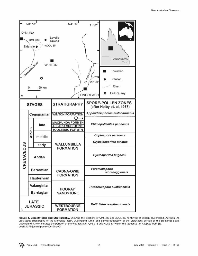
Figure 1. Locality Map and Stratigraphy. Showing the locations of QML 313 and AODL 85, northwest of Winton, Queensland, Australia (A).Cretaceous Stratigraphy of the Eromanga Basin, Queensland. Litho- and palynostratigraphy of the Cretaceous portion of the Eromanga Basin,Queensland. Arrow indicates the position of the type localities QML 313 and AODL 85 within the sequence (B). Adapted from [8].doi:10.1371/journal.pone.0006190.g001
New Australian Dinosaurs
PLoS ONE | www.plosone.org 2 July 2009 | Volume 4 | Issue 7 | e6190
appraisal of the age and interpreted geological settings of the
Winton Formation is available [7,8,11].
Winton FormationTerrestrial and aquatic vertebrate fossil remains were recovered
from fine-grained siltstones, labile sandstones and claystones
derived from within the Winton Formation and interpreted here
to have been formed in a distal fluvial depositional environment.
Two localities were initially discovered by the presence of bone
fragments on the surface of the soil, and with excavation, revealed
the presence of Winton Formation at a depth of less than 1 m
below the ground surface. Within this Winton Formation the
preserved remains of three associated dinosaur skeletons were
recovered. The two sites are approximately 3 kms from one
another and are only two of at least five sites so far known to reveal
dinosaur remains from this one location. Other localities in the
district have also yielded faunal remains typical of the Winton
Formation (Table 1).
Depositional EnvironmentDetailed sedimentologic and taphonomic information collected
from both sites will form part of a more extensive study and will be
published elsewhere. Preliminary investigations indicate that the
AODL 85 ‘Matilda Site’ deposit accumulated as part of a low
energy, silt-rich, abandoned channel fill deposit, most likely as part
of an ox-bow lake, with distinctive ‘billabong’ morphology. The
depositional environment at QML 313 ‘‘Triangle Paddock Site’’
was a higher energy deposit and is considered to be a point-bar
sequence. Excavation at both type localities revealed the presence
of disarticulated, semi-articulated and associated skeletal elements
of the three dinosaurs. Faunal remains recovered during
excavations at AODL 85 include two partial dinosaur skeletons,
a sauropod and theropod, along with the fossil remains of fish,
crocodiliforms, turtles and hyriid bivalves. Fossil remains from
AODL 85 are well preserved within a fine-grained clay sediment
bounded by upper and lower labile sandstone horizons. Macro-
floral remains have been recovered from within the lower clay
sequence and within the sequence preserving the faunal remains.
The macroflora assemblage includes angiosperms, auracarian
gymnosperms, ginkgoes and ferns.
Faunal remains recovered from QML 313 include the semi-
articulated and associated skeleton of a sauropod, along with
fragmentary remains of fish and an isolated theropod tooth. Most
of the sauropod bone elements have been abraded prior to
deposition; however, all are clearly associated with a single
individual. Fossil remains from QML 313 are preserved in
coarsely bedded sandstone overlying fine-grained clay. Macroflo-
ral remains include mostly woody stems and branch impressions;
however, auracarian cones, cone scales and pinnae are recogni-
sable.
Methods
Fossil PreparationThe holotype specimens were prepared using pneumatic air
scribes, pneumatic chisels, high speed diamond tipped rotary tools
and high speed diamond wheel cutters. The majority of the
sauropod elements were encased in an iron-oxide crust which
preserve evidence of pyritic pseudomorphs; some iron-oxide crusts
were up to 10 cm thick. Several elements, including many of the
theropod bones were encased in a concretionary phosphatic crust.
TerminologyThe terms ‘anterior’ and ‘posterior’ are used with respect to the
placement of postcranial elements (e.g. anterior caudal vertebra;
posterior caudal vertebra; anterior dorsal rib; posterior dorsal rib)
and when describing the aspect of the view (e.g. ‘‘In anterior
view’’).
For individual postcranial elements, the terms ‘cranial, caudal,
lateral, medial, proximal and distal’ are used to identify the region
of the element being described. For sauropod metacarpals the
terms ‘external’ and ‘internal’ are used to differentiate the surface
of the metacarpal exposed externally of the manus and those
surfaces which are enclosed (internal) within the manus. We use
previously developed terminology for teeth [12] and vertebral
laminae [13].
Phylogenetic AnalysisAll phylogenetic analyses were conducted using parsimony
analysis software, PAUP 4.0 [14]. Each analysis was conducted
with the following settings: 1. All characters unordered; 2.
Outgroup taxa monophyletic with respect to ingroup taxa; 3.
Heuristic Search using the tree bisection and reconnection
algorithm (TBR), random seed generator; 4. 1000 TBR replicates
to find the most- parsimonious tree; 5. Most parsimonious-trees
(MPT) saved and a strict consensus tree generated of these MPTs;
6. 1000 bootstrap replicates of this analysis with nodes returning
values .50% recorded.
The electronic version of this document does not represent a
published work according to the International Code of Zoological
Nomenclature (ICZN), and hence the nomenclatural acts
contained herein are not available under that Code from the
electronic edition. A separate edition of this document was
produced by a method that assures numerous identical and
durable copies, and those copies were simultaneously obtainable
(from the publication date listed on page 1 of this paper) for the
Table 1. Latest Albian (mid-Cretaceous) fauna currentlyknown from the Winton Formation.
Type Taxon Interpretation Reference
Fauna
Ichnofossil Tyrannosauropus sp. Theropod [41]
Ichnofossil Skartopus australis Theropod [41]
Ichnofossil Wintonopus latomorum ornithopod [41]
Ichnofossil Large ornithopod Large ornithopod [41]
Hyriidae Megalovirgus wintonensis Freshwater mollusc [42,43]
Hyriidae Hyridella goodiwindiensis Freshwater mollusc [42,43]
Hyriidae Prohyria macmichaeli Freshwater mollusc [42,43]
Gastropod Freshwater snail [44]
Insect Cicadid [45]
Lungfish Metaceratodus ellioti Lungfish [46]
Squamate cf. Coniasaurus Dolichosaur lizard [47]
Eusuchian Isisfordia duncani crocodilian [48]
Chelonian Indet. ?chelid Turtle Hocknullpers. obs.
Pterodactyloid Indet. pterosaur Pterosaur Hocknullpers. obs.
Sauropoda ‘‘Austrosaurus sp.’’ Titanosauriform [49]
Ornithopoda hypsilophodontid Small ornithopod [50]
Ankylosauria Small ankylosaur Hocknullpers. obs.
doi:10.1371/journal.pone.0006190.t001
New Australian Dinosaurs
PLoS ONE | www.plosone.org 3 July 2009 | Volume 4 | Issue 7 | e6190
purpose of providing a public and permanent scientific record, in
accordance with Article 8.1 of the Code. The separate print-only
edition is available on request from PLoS by sending a request to
PLoS ONE, 185 Berry Street, Suite 3100, San Francisco, CA
94107, USA along with a check for $10 (to cover printing and
postage) payable to ‘‘Public Library of Science.
The online version of the article is archived and available from
the following digital repositories: PubMedCentral (www.pubmed-
central.nih.gov/), LOCKSS (http://www.lockss.org/lockss/),
Queensland Museum Library (www.qm.qld.gov.au) and Austra-
lian Age of Dinosaurs Library (www.australianageofdinosaurs.
com). In addition, this published work and the nomenclatural acts
it contains have been registered in ZooBank (http://www.
zoobank.org/), the proposed online registration system for the
ICZN. The ZooBank LSIDs (Life Science Identifiers) can be
resolved and the associated information viewed through any
standard web browser by appending the LSID to the prefix
http://zoobank.org/. The LSID Number at Zoobank, for this
publication is: urn:lsid:zoobank.org:pub:E02E6156-CB22-4952-
9E9A-8C6ED7377B40
Institutional Abbreviations
QMF Queensland Museum Fossil
QML Queensland Museum Locality
AODF Australian Age of Dinosaurs Fossil
AODL Australian Age of Dinosaurs Locality
NMVP Museum of Victoria Palaeontological Collec-
tion
FPMN Fukui Prefectural Museum
Results
Systematic PalaeontologySystematic Hierarchy:
Dinosauria Owen, 1842
Saurischia Seeley, 1887
Sauropodomorpha, Huene, 1932
Titanosauriformes, Salgado et al., 1997
Titanosauria, Bonaparte & Coria, 1993
Lithostrotia, Upchurch et al., 2004
Incertae sedis
Diamantinasaurus gen. nov.
urn:lsid:zoobank.org:act:7A72845C-7E28-4C1E-B536-F37D8-
F270F5D
Etymology. Diamantina, in reference to the Diamantina River
which runs near the type locality. sauros, Greek for lizard.
Type species. Diamantinasaurus matildae
Diamantinasaurus matildae sp. nov.
urn:lsid:zoobank.org:act:6BE2173E-5438-4F9A-8978-745C33-
79F90D
Etymology. For Matilda, in reference to ‘‘Waltzing Matilda’’,
one of Australia’s National songs, written by Banjo Patterson in
Winton (‘‘Matilda Country’’) in 1895.
Holotype. AODF 603: Right scapula, right and left humeri,
right ulna, near complete right metacarpus including metacarpals
II–V, phalanges and a manus ungual. Left metacarpal I. Dorsal
ribs and fragmentary gastralia. Left sternal plate. Left ilium and
isolated sacral processes. Right and left pubes and ischia. Right
femur, tibia, fibula and astragalus (Figure 2A–B).
Type Locality. AODL 85, ‘‘Matilda Site’’, Elderslie Station,
approximately 60 km north-west of Winton, western central
Queensland, Australia.
Horizon & Age. Winton Formation, latest Albian
(Cretaceous).
Diagnosis. Diamantinasaurus matildae gen. et sp. nov. is
characterised by the following unique association of features.
Dorsal ribs plank-like with camellate pneumatic cavities in the
proximal-most expansion. Transverse processes of the sacral
vertebrae with camellate pneumatic cavities. Scapular glenoid
bevelled medially and expanded medio-laterally; scapular blade
flat and rectangular in cross-section; acromial blade broad and dish-
shaped with poorly developed acromial ridge. Crescentic-shaped
sternal plate. Humerus stout; intermediate robusticity
(autapomorphic); deltopectoral crest prominent, extending to mid-
shaft; proximal border shallowly sigmoidal; proximo-lateral corner
square; distal condyles merged and flat extending only just onto
anterior face of shaft; proximal breadth 50% of total length. Ulna
stout; cranio-medial process massive and concave; distal condyle
ovo-triangular. Metacarpals massive with undivided condyles and
marked distal rugosities; Mc III longest followed by Mc II, Mc I, Mc
IV and Mc V; phalangeal articular facets on external face of shaft on
Mc I–IV. Phalanges present on digits I–IV (phalangeal formula 2-1-
1-1-0); spike-like ungual present on digit I. Phalanges on digits II–IV
rounded and broadest transversely; Mc III phalange heavily reduced
(autapomorphic). Ilium massive with broad rounded preacetabular
lobe directed perpendicular to sacral axis; pneumatic cavities present
within iliac blade. Pubes massive and long, fused to ischia via a
shallowly sigmoidal pubic apron. Ischia with distinct iliac peduncle
on a narrow shaft leading to a broad ischial blade, which is fused to
the opposite ischium along the mid-line of the pelvic girdle. Femur
robust with lateral bulge; intermediate robusticity (autapomorphic);
distal condyles bevelled dorso-medially; shaft elliptical in cross-
section and deflected medially. Tibia robust; proximal articular
condyle sub-equally expanded; cnemial crest projects cranially then
laterally (autapomorphic); ovoid distal articular surface; distal
breadth more than twice that of mid-shaft. Fibula expanded
proximally; intermediate robusticity (autapomorphic); distal
articular surface bevelled cranio-medially. Astragalus
quadrangular; posterior fossa undivided.
DescriptionAxial Skeleton. The axial skeleton of the holotype (AODF
603) is known from isolated dorsal ribs, isolated gastralia and two
isolated sacral processes.
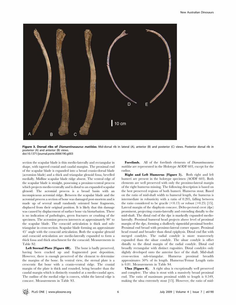
Dorsal Ribs (Figure 3). Ten dorsal ribs have been recovered,
all with damaged or broken proximal and distal ends. A mid-dorsal
rib and posterior dorsal rib preserve the expanded region of the
proximal end and well preserved distal ends. The proximal
expansion possesses an excavated region and pneumatic cavities.
Both ribs possess camellate internal bone structure in the proximal
expansion only. The cross section of the mid-dorsal rib is
rectangular, plank-like and slightly bowed caudally. It is broad
along its length with an expanded distal end, which is rounded and
rugose in distal aspect. The posterior dorsal rib is sub-triangular in
cross-section, bowed along its length, and narrow. Measurements in
Table S1.
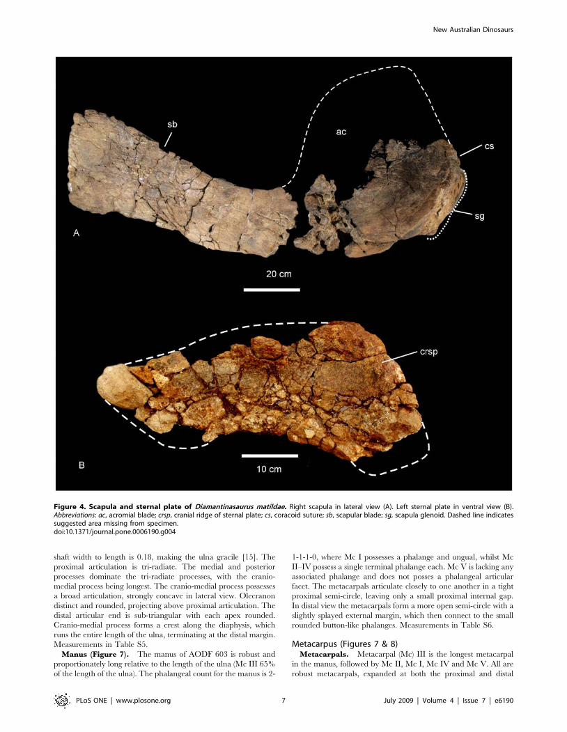
Appendicular SkeletonPectoral Girdle. The pectoral girdle of Diamantinasaurus
matildae is represented in the Holotype AODF 603 by a right
scapula and left sternal plate.
Right Scapula (Figure 4A). The scapular blade has a flat
lateral surface and a shallowly concave medial surface. In cross
New Australian Dinosaurs
PLoS ONE | www.plosone.org 4 July 2009 | Volume 4 | Issue 7 | e6190
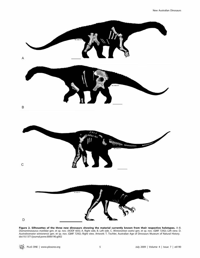
Figure 2. Silhouettes of the three new dinosaurs showing the material currently known from their respective holotypes. A–B.Diamantinasaurus matildae gen. et sp. nov. (AODF 603); A. Right side, B. Left side. C. Wintonotitan wattsi gen. et sp. nov. (QMF 7292); Left view. D.Australovenator wintonensis gen. et sp. nov. (QMF 7292); Right view. Artwork: T. Tischler, Australian Age of Dinosaurs Museum of Natural History.doi:10.1371/journal.pone.0006190.g002
New Australian Dinosaurs
PLoS ONE | www.plosone.org 5 July 2009 | Volume 4 | Issue 7 | e6190
section the scapular blade is thin medio-laterally and rectangular in
shape, with tapered cranial and caudal margins. The proximal end
of the scapular blade is expanded into a broad cranio-dorsal blade
(acromion blade) and a thick and triangular glenoid fossa, bevelled
medially. Midline scapular blade ridge absent. The ventral edge of
the scapular blade is straight, possessing a proximo-ventral process
which projects medio-ventrally and is distal to an expanded scapular
glenoid. The acromial process is a broad basin with an
inconspicuous acromial ridge. Between the scapular blade and the
acromial process a section of bone was damaged post-mortem and is
made up of several small randomly oriented bone fragments,
displaced from their original position. It is likely that this damage
was caused by displacement of surface bone via bioturbation. There
is no indication of pathologies, green fractures or crushing of the
specimen. The acromion process intersects at approximately 90u to
the scapular blade. The coracoid articulation is thick and sub-
triangular in cross-section. Scapular blade forming an approximate
45u angle with the coracoid articulation. Both the scapular glenoid
and coracoid articulation are medio-laterally expanded to form a
thick fossa and thick attachment for the coracoid. Measurements in
Table S2.
Left Sternal Plate (Figure 4B). The bone is badly preserved,
having been cracked up and fragmented post deposition.
However, there is enough preserved of the element to determine
the margins of the bone. In ventral view, the sternal plate is a
crescentic flat bone with a cranio-ventral ridge. The cranial
margin of the plate is thick and rounded, being broader than the
caudal margin which is distinctly rounded at a medio-caudal apex.
The outline of the medial edge is convex, whilst the lateral edge is
concave. Measurements in Table S3.
Forelimb. All of the forelimb elements of Diamantinasaurus
matildae are represented in the Holotype AODF 603, except for the
radius.
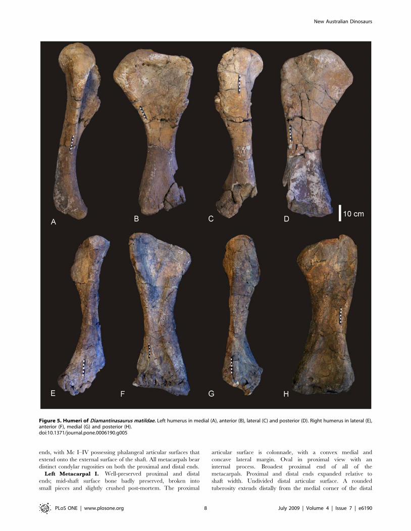
Right and Left Humerus (Figure 5). Both right and left
humeri are present in the holotype specimen (AODF 603). Both
elements are well preserved with only the proximo-lateral margin
of the right humerus missing. The following description is based on
the best preserved regions of both humeri. Humerus stout. Based
on the ratio of mid-shaft width to humeral length, the humerus is
intermediate in robusticity with a ratio of 0.205, falling between
the ratio considered to be gracile (,0.15) or robust (.0.25) [15].
Lateral margin of the diaphysis concave. Delto-pectoral crest (dpc)
prominent, projecting cranio-laterally and extending distally to the
mid-shaft. The distal end of the dpc is markedly expanded medio-
laterally. Proximal humeral head projects above level of proximal
margin of the dpc, forming a shallowly sigmoidal proximal border.
Proximal end broad with proximo-lateral corner square. Proximal
head round and broader than distal epiphysis. Distal end flat with
merged condyles. The radial condyle is more transversely
expanded than the ulnar condyle. The ulnar condyle is offset
distally to the distal margin of the radial condyle. Distal end
broadly rectangular with distinct rugosities. Distal condyles only
slightly developed onto the anterior face of the shaft. Mid-shaft
cross-section sub-triangular. Humerus proximal breadth
approximates 50% of its length. Humerus/Femur Length ratio
is 0.81. Measurements in Table S4.
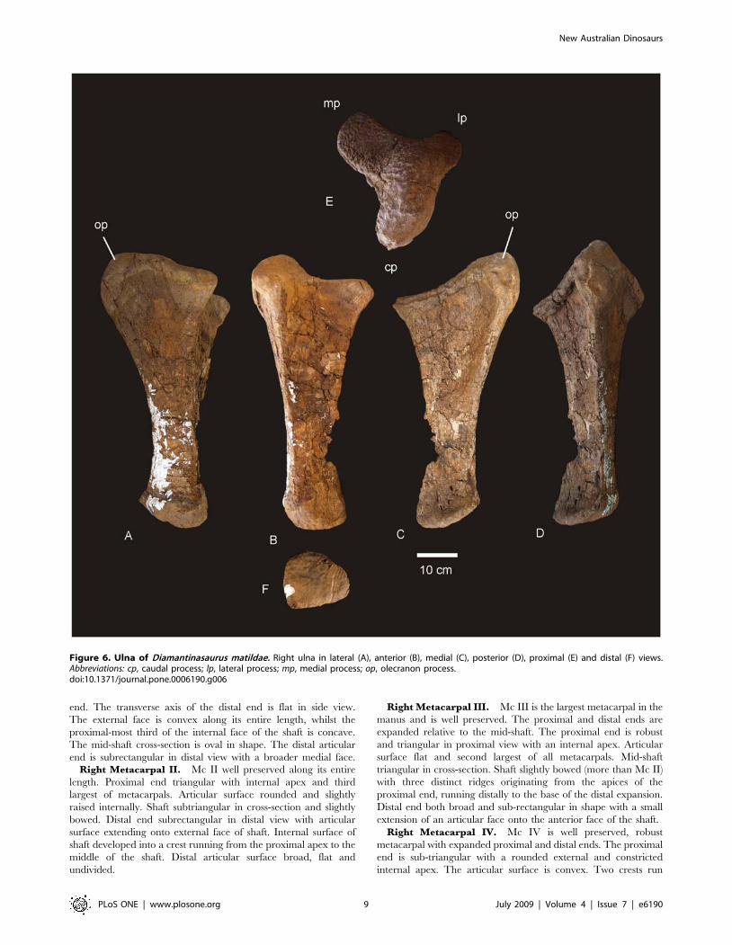
Ulna (Figure 6). A right ulna is exceptionally well preserved
and complete. The ulna is stout with a massively broad proximal
end. The ratio of maximum proximal width to length is 0.549,
making the ulna extremely stout [15]. However, the ratio of mid-
Figure 3. Dorsal ribs of Diamantinasaurus matildae. Mid-dorsal rib in lateral (A), anterior (B) and posterior (C) views. Posterior dorsal rib inposterior (A) and anterior (B) views.doi:10.1371/journal.pone.0006190.g003
New Australian Dinosaurs
PLoS ONE | www.plosone.org 6 July 2009 | Volume 4 | Issue 7 | e6190
shaft width to length is 0.18, making the ulna gracile [15]. The
proximal articulation is tri-radiate. The medial and posterior
processes dominate the tri-radiate processes, with the cranio-
medial process being longest. The cranio-medial process possesses
a broad articulation, strongly concave in lateral view. Olecranon
distinct and rounded, projecting above proximal articulation. The
distal articular end is sub-triangular with each apex rounded.
Cranio-medial process forms a crest along the diaphysis, which
runs the entire length of the ulna, terminating at the distal margin.
Measurements in Table S5.
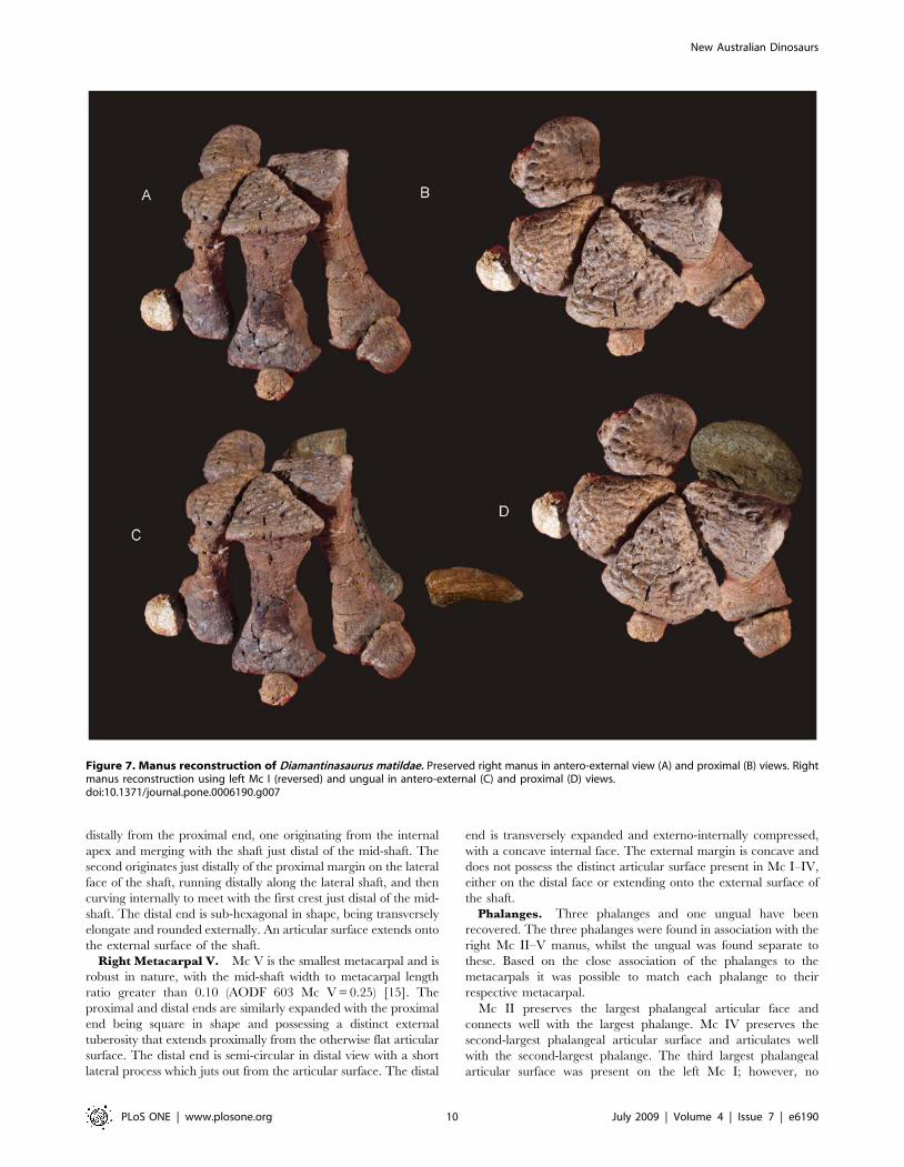
Manus (Figure 7). The manus of AODF 603 is robust and
proportionately long relative to the length of the ulna (Mc III 65%
of the length of the ulna). The phalangeal count for the manus is 2-
1-1-1-0, where Mc I possesses a phalange and ungual, whilst Mc
II–IV possess a single terminal phalange each. Mc V is lacking any
associated phalange and does not posses a phalangeal articular
facet. The metacarpals articulate closely to one another in a tight
proximal semi-circle, leaving only a small proximal internal gap.
In distal view the metacarpals form a more open semi-circle with a
slightly splayed external margin, which then connect to the small
rounded button-like phalanges. Measurements in Table S6.
Metacarpus (Figures 7 & 8)Metacarpals. Metacarpal (Mc) III is the longest metacarpal
in the manus, followed by Mc II, Mc I, Mc IV and Mc V. All are
robust metacarpals, expanded at both the proximal and distal
Figure 4. Scapula and sternal plate of Diamantinasaurus matildae. Right scapula in lateral view (A). Left sternal plate in ventral view (B).Abbreviations: ac, acromial blade; crsp, cranial ridge of sternal plate; cs, coracoid suture; sb, scapular blade; sg, scapula glenoid. Dashed line indicatessuggested area missing from specimen.doi:10.1371/journal.pone.0006190.g004
New Australian Dinosaurs
PLoS ONE | www.plosone.org 7 July 2009 | Volume 4 | Issue 7 | e6190
ends, with Mc I–IV possessing phalangeal articular surfaces that
extend onto the external surface of the shaft. All metacarpals bear
distinct condylar rugosities on both the proximal and distal ends.
Left Metacarpal I. Well-preserved proximal and distal
ends; mid-shaft surface bone badly preserved, broken into
small pieces and slightly crushed post-mortem. The proximal
articular surface is colonnade, with a convex medial and
concave lateral margin. Oval in proximal view with an
internal process. Broadest proximal end of all of the
metacarpals. Proximal and distal ends expanded relative to
shaft width. Undivided distal articular surface. A rounded
tuberosity extends distally from the medial corner of the distal
Figure 5. Humeri of Diamantinasaurus matildae. Left humerus in medial (A), anterior (B), lateral (C) and posterior (D). Right humerus in lateral (E),anterior (F), medial (G) and posterior (H).doi:10.1371/journal.pone.0006190.g005
New Australian Dinosaurs
PLoS ONE | www.plosone.org 8 July 2009 | Volume 4 | Issue 7 | e6190
end. The transverse axis of the distal end is flat in side view.
The external face is convex along its entire length, whilst the
proximal-most third of the internal face of the shaft is concave.
The mid-shaft cross-section is oval in shape. The distal articular
end is subrectangular in distal view with a broader medial face.
Right Metacarpal II. Mc II well preserved along its entire
length. Proximal end triangular with internal apex and third
largest of metacarpals. Articular surface rounded and slightly
raised internally. Shaft subtriangular in cross-section and slightly
bowed. Distal end subrectangular in distal view with articular
surface extending onto external face of shaft. Internal surface of
shaft developed into a crest running from the proximal apex to the
middle of the shaft. Distal articular surface broad, flat and
undivided.
Right Metacarpal III. Mc III is the largest metacarpal in the
manus and is well preserved. The proximal and distal ends are
expanded relative to the mid-shaft. The proximal end is robust
and triangular in proximal view with an internal apex. Articular
surface flat and second largest of all metacarpals. Mid-shaft
triangular in cross-section. Shaft slightly bowed (more than Mc II)
with three distinct ridges originating from the apices of the
proximal end, running distally to the base of the distal expansion.
Distal end both broad and sub-rectangular in shape with a small
extension of an articular face onto the anterior face of the shaft.
Right Metacarpal IV. Mc IV is well preserved, robust
metacarpal with expanded proximal and distal ends. The proximal
end is sub-triangular with a rounded external and constricted
internal apex. The articular surface is convex. Two crests run
Figure 6. Ulna of Diamantinasaurus matildae. Right ulna in lateral (A), anterior (B), medial (C), posterior (D), proximal (E) and distal (F) views.Abbreviations: cp, caudal process; lp, lateral process; mp, medial process; op, olecranon process.doi:10.1371/journal.pone.0006190.g006
New Australian Dinosaurs
PLoS ONE | www.plosone.org 9 July 2009 | Volume 4 | Issue 7 | e6190
distally from the proximal end, one originating from the internal
apex and merging with the shaft just distal of the mid-shaft. The
second originates just distally of the proximal margin on the lateral
face of the shaft, running distally along the lateral shaft, and then
curving internally to meet with the first crest just distal of the mid-
shaft. The distal end is sub-hexagonal in shape, being transversely
elongate and rounded externally. An articular surface extends onto
the external surface of the shaft.
Right Metacarpal V. Mc V is the smallest metacarpal and is
robust in nature, with the mid-shaft width to metacarpal length
ratio greater than 0.10 (AODF 603 Mc V = 0.25) [15]. The
proximal and distal ends are similarly expanded with the proximal
end being square in shape and possessing a distinct external
tuberosity that extends proximally from the otherwise flat articular
surface. The distal end is semi-circular in distal view with a short
lateral process which juts out from the articular surface. The distal
end is transversely expanded and externo-internally compressed,
with a concave internal face. The external margin is concave and
does not possess the distinct articular surface present in Mc I–IV,
either on the distal face or extending onto the external surface of
the shaft.
Phalanges. Three phalanges and one ungual have been
recovered. The three phalanges were found in association with the
right Mc II–V manus, whilst the ungual was found separate to
these. Based on the close association of the phalanges to the
metacarpals it was possible to match each phalange to their
respective metacarpal.
Mc II preserves the largest phalangeal articular face and
connects well with the largest phalange. Mc IV preserves the
second-largest phalangeal articular surface and articulates well
with the second-largest phalange. The third largest phalangeal
articular surface was present on the left Mc I; however, no
Figure 7. Manus reconstruction of Diamantinasaurus matildae. Preserved right manus in antero-external view (A) and proximal (B) views. Rightmanus reconstruction using left Mc I (reversed) and ungual in antero-external (C) and proximal (D) views.doi:10.1371/journal.pone.0006190.g007
New Australian Dinosaurs
PLoS ONE | www.plosone.org 10 July 2009 | Volume 4 | Issue 7 | e6190
corresponding phalange was recovered for this position. The
smallest articular surface was present on Mc III and articulated
well with the heavily reduced and smallest phalange. Each of the
phalanges is rounded cranially and possesses distinctly concave
articular facets on their caudal margins.
Ungual (Figure 8). A single manus ungual has been
recovered. It is considered here to represent a manus ungual
because of its hypertrophic nature, lack of curvature and lack of
distinctly weight-bearing features. The ungual is a relatively
straight spike possessing a slight curvature along its long axis and a
very prominent distal point. The ungual is relatively symmetrical
along the long axis. In cross-section the ungual is tapered both
dorsally and ventrally, whilst also being medio-laterally
compressed. A narrow proximal articular surface suggests its
connection to a relatively small and basic phalange. The lack of a
defined proximal facet indicates that this ungual was not tightly
articulated to the preceding phalange.
These features are not usually seen in sauropod pedal unguals,
instead they are generally broader and more robust in all
dimensions; much more recurved and asymmetrical; possess
broad and distinct proximal articular facets; and do not taper at
both the dorsal and ventral margins. In addition, the presence of
an articular surface on the left Mc I suggests that the first digit had
at least one phalange, which may have connected to a manus
ungual. As yet, no metatarsals, pedal phalanges or pedal unguals
have been recovered from the type specimen locality.
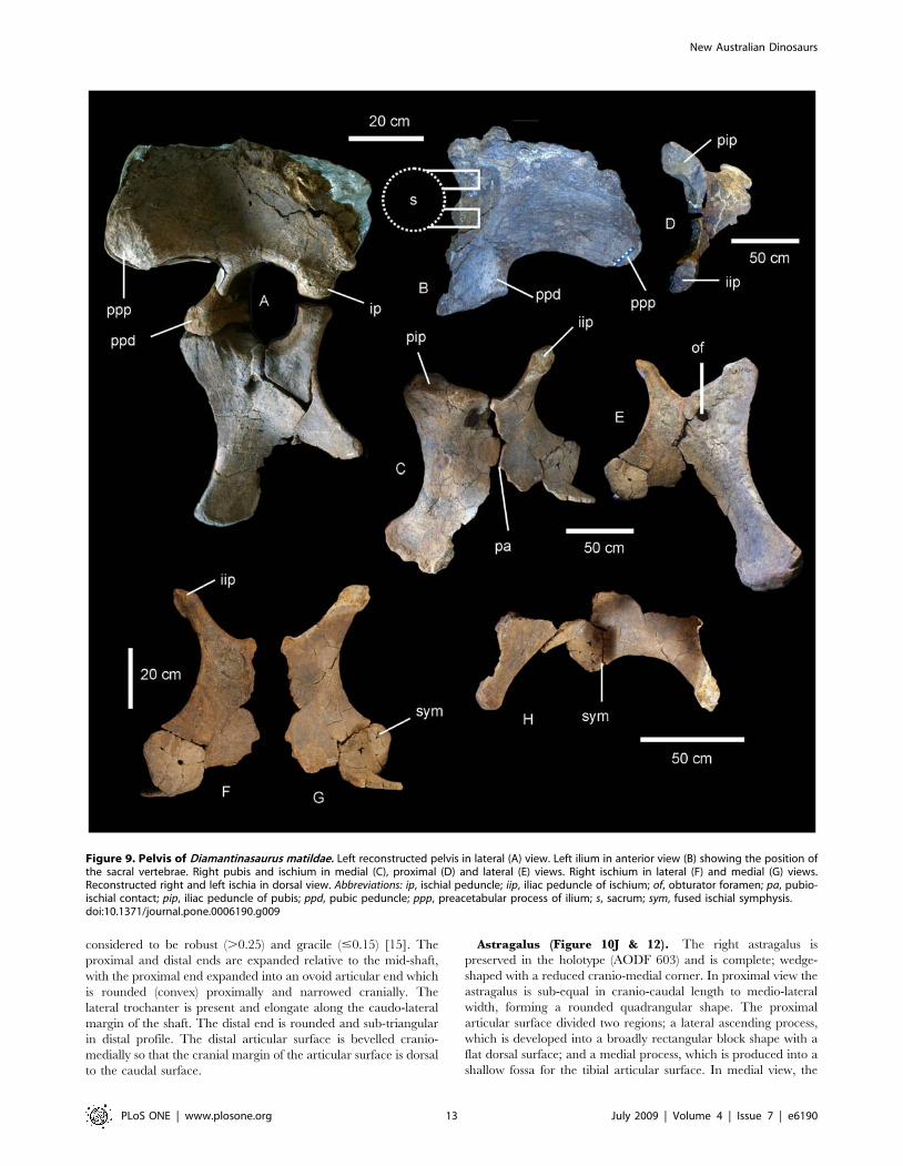
Pelvic Girdle (Figure 9). The pelvis of AODF 603 preserves
the left ilium, right and left pubes, right and left ischia and two
lateral processes from the sacrum. The left ilium is an isolated
element and has broken away from the sacrum and right ilium
post-mortem. The articular section of the pubic peduncle broke
away from the main body of the ilium post-mortem and was found
close by in the deposit. The dorsal rim, internal connection to the
sacrum and the caudal margin of the ilium have lost surface bone,
however, the main body of the ilium is well preserved, including
the articular tuberosities present on the peduncles and the
preacetabular lobe. The internal structure of the iliac blade can
be seen from the medial and caudal faces, illustrating that the
internal bone structure was made up of large camerate pneumatic
vacuities. This internal structure is also found in the isolated
transverse processes of the sacrum.
The ilium is very broad, with the width between the
preacetabular lobes being much broader than the maximum
length of the ilium. In lateral view the preacetabular process is
board and rounded, somewhat truncated as it swings toward a 90uangle from the main axis of the ilium and sacrum. The highest
point of the iliac blade is centred dorsal to the pubic peduncle. The
pubic peduncle is massive and contributes over half of the
acetabular rim. The articular surface of the pubic peduncle
projects perpendicular from the main axis of the ilium and sacrum.
The ischial peduncle is low and rounded, broadening medio-
laterally to accommodate the iliac peduncle of the ischium.
The pubes are long and robust with a thick dorsally oriented
iliac peduncle. Pubic acetabular rim and ischial articular edge
meet at an obtuse angle, where these two elements begin to fuse
together. The obturator foramen is positioned distal to this. The
ischial articular surface of the pubis is much longer than the
contribution of the pubis to the acetabular rim. The pubis is
expanded both proximally and distally relative to the mid-shaft
width. The pubio-ischial contact represents approximately half of
the total pubic length, forming a large apron, which is shallowly S-
shaped. The ratio of ischium to pubis length is 0.62.
The ischia are robust elements; however, during post-mortem
both elements suffered breakages. The two elements were fused
together during life and this complex was fused to the pubic apron
of their respective pubes. The ischial complex broke into four
sections; the two iliac peduncles preserved sections of the ischial
blade, the right preserving the most intact. A third section
represents the remaining piece of the left ischium, which connects
to the pubic apron, whilst the fourth piece connects the right
ischial blade to the left ischial blade and preserves the fused
midline symphysis.
The ischial blade is much shorter than the pubic blade, possessing
a similar medial and lateral depth along its length. The blade
contacts the other ischium via a central fused region, which forms a
shallow basin between the two ischia. The ratio of the width across
the ischial blade at its mid-length to the total length of the ischium is
greater than 0.2. The pubic peduncle of the ischium is dorso-
ventrally extended to encompass the entire length of the ischial
blade. Iliac peduncle of ischium distinctive and well separated from
the body of the ischium. Measurements in Table S7.
Hind limbFemur (Figure 10A–C). A complete right femur is preserved
in the holotype (AODF 603). The only post-depositional
deformation to occur to this heavy element was a crack that has
formed from the proximo-lateral corner across the caudal face of
the shaft toward the distal condyles. Associated with this crack, the
lateral margin has rotated caudally so that the lateral bulge faces
caudally rather than laterally. The femur is intermediate in
robustness ratio ( = 0.196) being close to the minimum ratio for
robust (.0.20) and above the ratio considered gracile (#0.10)
[15]. In caudal view the femoral head is massive and rounded,
located dorsal to the greater trochanter, which is also massive and
rounded, however, constricted cranio-caudally. The fourth
trochanter is reduced to a low ridge, approximately 150 mm
long. Distal to the greater trochanter, a lateral bulge is well
developed. In original position, this bulge would have projected
laterally, forming a greater medio-lateral width across the femur at
this point than what is preserved in the specimen. The transverse
(medio-lateral) length of the femur mid-shaft is approximately
twice that of the cranio-caudal length of the mid-shaft
(262 mm:120 mm), forming an elliptical cross-section. When
rested on the distal condyles, the shaft is deflected medially. The
distal condyles are robust and cranio-caudally extended being sub-
equal in transverse width and bevelled dorso-medially and extend
just on to the disto-cranial margin of the shaft. The epicondyle is
well developed laterally and oriented caudally. The medial
condyle is cranio-caudally longer than the lateral condyle.
Measurements in Table S8.
Tibia (Figure 10D–J). The right tibia is known from the
holotype (AODF 603) and is the best preserved element of the
skeleton. The element is robust with the proximal end expanded
equally cranio-caudally and medio-laterally; the distal end is
expanded medio-laterally and compressed cranio-caudally. The
proximal condyle is sub-circular in shape with a flat articular
surface. The cnemial crest is thick and robust, projecting cranially
at its proximal margin, then scooping distally so that the distal
margin of the crest projects laterally and encloses a deep fossa. The
shaft of the tibia is relatively straight and possesses a twist in the
distal margin. The distal end is broader medio-laterally than
cranio-caudally creating an ovoid distal profile with a distinct
lateral notch. The breadth of the distal end is more than 200% of
the mid-shaft breadth. Measurements in Table S9.
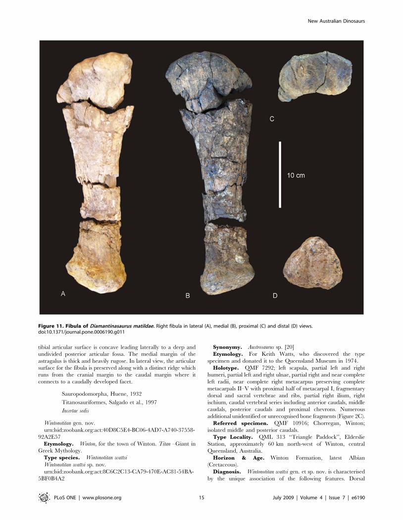
Fibula (Figure 11). The right fibula is known from the
holotype (AODF 603) and is broken in three places. The
robustness of the fibula, as determined by the mid-shaft width:
total length of the fibula, is intermediate ( = 0.187) between that
New Australian Dinosaurs
PLoS ONE | www.plosone.org 11 July 2009 | Volume 4 | Issue 7 | e6190
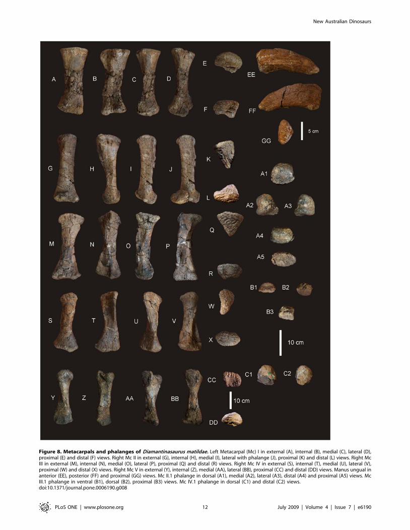
Figure 8. Metacarpals and phalanges of Diamantinasaurus matildae. Left Metacarpal (Mc) I in external (A), internal (B), medial (C), lateral (D),proximal (E) and distal (F) views. Right Mc II in external (G), internal (H), medial (I), lateral with phalange (J), proximal (K) and distal (L) views. Right McIII in external (M), internal (N), medial (O), lateral (P), proximal (Q) and distal (R) views. Right Mc IV in external (S), internal (T), medial (U), lateral (V),proximal (W) and distal (X) views. Right Mc V in external (Y), internal (Z), medial (AA), lateral (BB), proximal (CC) and distal (DD) views. Manus ungual inanterior (EE), posterior (FF) and proximal (GG) views. Mc II.1 phalange in dorsal (A1), medial (A2), lateral (A3), distal (A4) and proximal (A5) views. McIII.1 phalange in ventral (B1), dorsal (B2), proximal (B3) views. Mc IV.1 phalange in dorsal (C1) and distal (C2) views.doi:10.1371/journal.pone.0006190.g008
New Australian Dinosaurs
PLoS ONE | www.plosone.org 12 July 2009 | Volume 4 | Issue 7 | e6190
considered to be robust (.0.25) and gracile (#0.15) [15]. The
proximal and distal ends are expanded relative to the mid-shaft,
with the proximal end expanded into an ovoid articular end which
is rounded (convex) proximally and narrowed cranially. The
lateral trochanter is present and elongate along the caudo-lateral
margin of the shaft. The distal end is rounded and sub-triangular
in distal profile. The distal articular surface is bevelled cranio-
medially so that the cranial margin of the articular surface is dorsal
to the caudal surface.
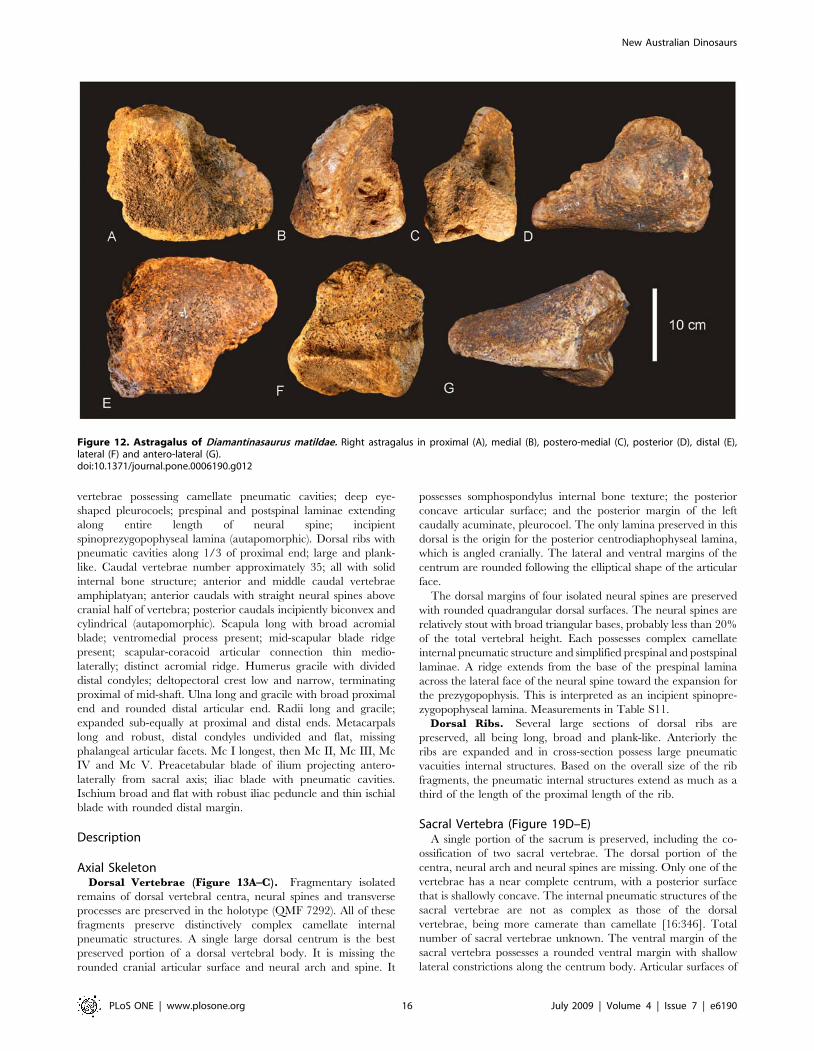
Astragalus (Figure 10J & 12). The right astragalus is
preserved in the holotype (AODF 603) and is complete; wedge-
shaped with a reduced cranio-medial corner. In proximal view the
astragalus is sub-equal in cranio-caudal length to medio-lateral
width, forming a rounded quadrangular shape. The proximal
articular surface divided two regions; a lateral ascending process,
which is developed into a broadly rectangular block shape with a
flat dorsal surface; and a medial process, which is produced into a
shallow fossa for the tibial articular surface. In medial view, the
Figure 9. Pelvis of Diamantinasaurus matildae. Left reconstructed pelvis in lateral (A) view. Left ilium in anterior view (B) showing the position ofthe sacral vertebrae. Right pubis and ischium in medial (C), proximal (D) and lateral (E) views. Right ischium in lateral (F) and medial (G) views.Reconstructed right and left ischia in dorsal view. Abbreviations: ip, ischial peduncle; iip, iliac peduncle of ischium; of, obturator foramen; pa, pubio-ischial contact; pip, iliac peduncle of pubis; ppd, pubic peduncle; ppp, preacetabular process of ilium; s, sacrum; sym, fused ischial symphysis.doi:10.1371/journal.pone.0006190.g009
New Australian Dinosaurs
PLoS ONE | www.plosone.org 13 July 2009 | Volume 4 | Issue 7 | e6190
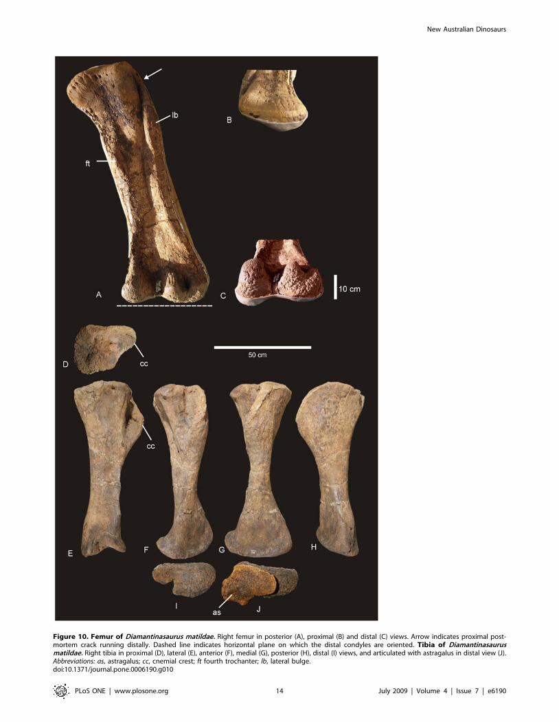
Figure 10. Femur of Diamantinasaurus matildae. Right femur in posterior (A), proximal (B) and distal (C) views. Arrow indicates proximal post-mortem crack running distally. Dashed line indicates horizontal plane on which the distal condyles are oriented. Tibia of Diamantinasaurusmatildae. Right tibia in proximal (D), lateral (E), anterior (F), medial (G), posterior (H), distal (I) views, and articulated with astragalus in distal view (J).Abbreviations: as, astragalus; cc, cnemial crest; ft fourth trochanter; lb, lateral bulge.doi:10.1371/journal.pone.0006190.g010
New Australian Dinosaurs
PLoS ONE | www.plosone.org 14 July 2009 | Volume 4 | Issue 7 | e6190
tibial articular surface is concave leading laterally to a deep and
undivided posterior articular fossa. The medial margin of the
astragalus is thick and heavily rugose. In lateral view, the articular
surface for the fibula is preserved along with a distinct ridge which
runs from the cranial margin to the caudal margin where it
connects to a caudally developed facet.
Sauropodomorpha, Huene, 1932
Titanosauriformes, Salgado et al., 1997
Incertae sedis
Wintonotitan gen. nov.
urn:lsid:zoobank.org:act:40D8C5E4-BC06-4AD7-A740-37558-
92A2E57
Etymology. Winton, for the town of Winton. Titan –Giant in
Greek Mythology.
Type species. Wintonotitan wattsi
Wintonotitan wattsi sp. nov.
urn:lsid:zoobank.org:act:8C6C2C13-CA79-470E-AC81-54BA-
5BF0B4A2
Synonymy. Austrosaurus sp. [20]
Etymology. For Keith Watts, who discovered the type
specimen and donated it to the Queensland Museum in 1974.
Holotype. QMF 7292; left scapula, partial left and right
humeri, partial left and right ulnae, partial right and near complete
left radii, near complete right metacarpus preserving complete
metacarpals II–V with proximal half of metacarpal I, fragmentary
dorsal and sacral vertebrae and ribs, partial right ilium, right
ischium, caudal vertebral series including anterior caudals, middle
caudals, posterior caudals and proximal chevrons. Numerous
additional unidentified or unrecognised bone fragments (Figure 2C).
Referred specimen. QMF 10916; Chorregan, Winton;
isolated middle and posterior caudals.
Type Locality. QML 313 ‘‘Triangle Paddock’’, Elderslie
Station, approximately 60 km north-west of Winton, central
Queensland, Australia.
Horizon & Age. Winton Formation, latest Albian
(Cretaceous).
Diagnosis. Wintonotitan wattsi gen. et sp. nov. is characterised
by the unique association of the following features. Dorsal
Figure 11. Fibula of Diamantinasaurus matildae. Right fibula in lateral (A), medial (B), proximal (C) and distal (D) views.doi:10.1371/journal.pone.0006190.g011
New Australian Dinosaurs
PLoS ONE | www.plosone.org 15 July 2009 | Volume 4 | Issue 7 | e6190
vertebrae possessing camellate pneumatic cavities; deep eye-
shaped pleurocoels; prespinal and postspinal laminae extending
along entire length of neural spine; incipient
spinoprezygopophyseal lamina (autapomorphic). Dorsal ribs with
pneumatic cavities along 1/3 of proximal end; large and plank-
like. Caudal vertebrae number approximately 35; all with solid
internal bone structure; anterior and middle caudal vertebrae
amphiplatyan; anterior caudals with straight neural spines above
cranial half of vertebra; posterior caudals incipiently biconvex and
cylindrical (autapomorphic). Scapula long with broad acromial
blade; ventromedial process present; mid-scapular blade ridge
present; scapular-coracoid articular connection thin medio-
laterally; distinct acromial ridge. Humerus gracile with divided
distal condyles; deltopectoral crest low and narrow, terminating
proximal of mid-shaft. Ulna long and gracile with broad proximal
end and rounded distal articular end. Radii long and gracile;
expanded sub-equally at proximal and distal ends. Metacarpals
long and robust, distal condyles undivided and flat, missing
phalangeal articular facets. Mc I longest, then Mc II, Mc III, Mc
IV and Mc V. Preacetabular blade of ilium projecting antero-
laterally from sacral axis; iliac blade with pneumatic cavities.
Ischium broad and flat with robust iliac peduncle and thin ischial
blade with rounded distal margin.
Description
Axial SkeletonDorsal Vertebrae (Figure 13A–C). Fragmentary isolated
remains of dorsal vertebral centra, neural spines and transverse
processes are preserved in the holotype (QMF 7292). All of these
fragments preserve distinctively complex camellate internal
pneumatic structures. A single large dorsal centrum is the best
preserved portion of a dorsal vertebral body. It is missing the
rounded cranial articular surface and neural arch and spine. It
possesses somphospondylus internal bone texture; the posterior
concave articular surface; and the posterior margin of the left
caudally acuminate, pleurocoel. The only lamina preserved in this
dorsal is the origin for the posterior centrodiaphophyseal lamina,
which is angled cranially. The lateral and ventral margins of the
centrum are rounded following the elliptical shape of the articular
face.
The dorsal margins of four isolated neural spines are preserved
with rounded quadrangular dorsal surfaces. The neural spines are
relatively stout with broad triangular bases, probably less than 20%
of the total vertebral height. Each possesses complex camellate
internal pneumatic structure and simplified prespinal and postspinal
laminae. A ridge extends from the base of the prespinal lamina
across the lateral face of the neural spine toward the expansion for
the prezygopophysis. This is interpreted as an incipient spinopre-
zygopophyseal lamina. Measurements in Table S11.
Dorsal Ribs. Several large sections of dorsal ribs are
preserved, all being long, broad and plank-like. Anteriorly the
ribs are expanded and in cross-section possess large pneumatic
vacuities internal structures. Based on the overall size of the rib
fragments, the pneumatic internal structures extend as much as a
third of the length of the proximal length of the rib.
Sacral Vertebra (Figure 19D–E)A single portion of the sacrum is preserved, including the co-
ossification of two sacral vertebrae. The dorsal portion of the
centra, neural arch and neural spines are missing. Only one of the
vertebrae has a near complete centrum, with a posterior surface
that is shallowly concave. The internal pneumatic structures of the
sacral vertebrae are not as complex as those of the dorsal
vertebrae, being more camerate than camellate [16:346]. Total
number of sacral vertebrae unknown. The ventral margin of the
sacral vertebra possesses a rounded ventral margin with shallow
lateral constrictions along the centrum body. Articular surfaces of
Figure 12. Astragalus of Diamantinasaurus matildae. Right astragalus in proximal (A), medial (B), postero-medial (C), posterior (D), distal (E),lateral (F) and antero-lateral (G).doi:10.1371/journal.pone.0006190.g012
New Australian Dinosaurs
PLoS ONE | www.plosone.org 16 July 2009 | Volume 4 | Issue 7 | e6190
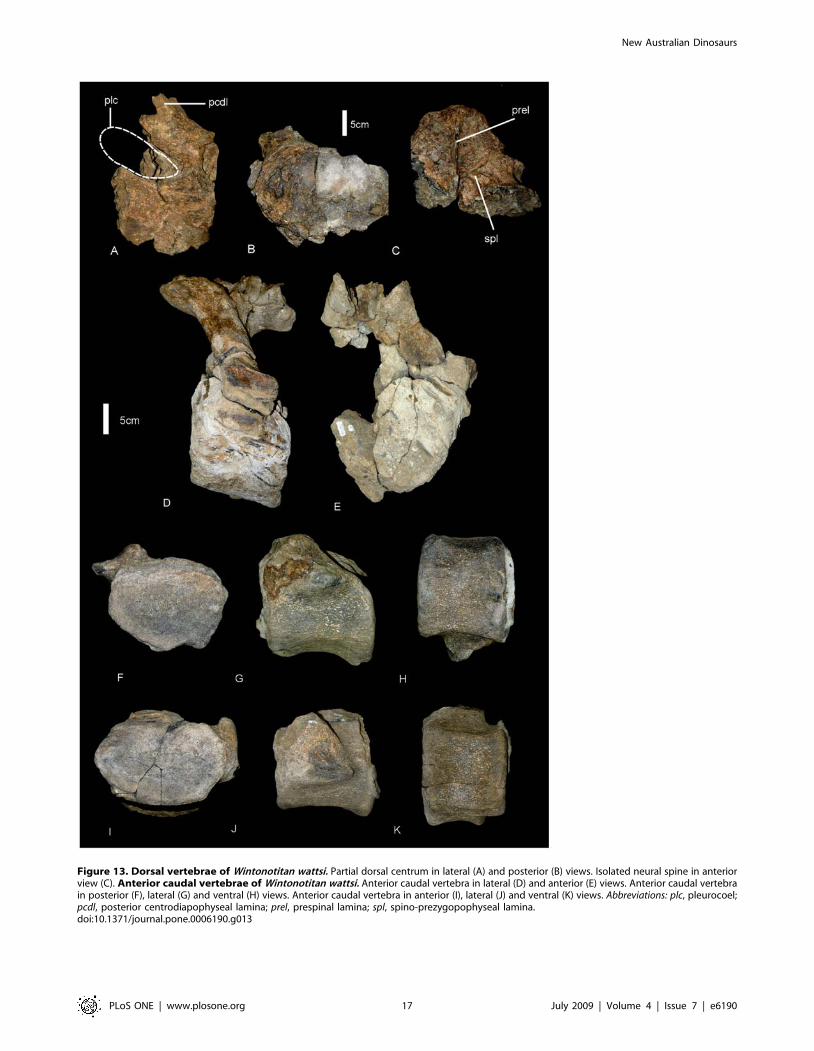
Figure 13. Dorsal vertebrae of Wintonotitan wattsi. Partial dorsal centrum in lateral (A) and posterior (B) views. Isolated neural spine in anteriorview (C). Anterior caudal vertebrae of Wintonotitan wattsi. Anterior caudal vertebra in lateral (D) and anterior (E) views. Anterior caudal vertebrain posterior (F), lateral (G) and ventral (H) views. Anterior caudal vertebra in anterior (I), lateral (J) and ventral (K) views. Abbreviations: plc, pleurocoel;pcdl, posterior centrodiapophyseal lamina; prel, prespinal lamina; spl, spino-prezygopophyseal lamina.doi:10.1371/journal.pone.0006190.g013
New Australian Dinosaurs
PLoS ONE | www.plosone.org 17 July 2009 | Volume 4 | Issue 7 | e6190
sacral vertebrae are shallowly concave (platycoelous). Deep lateral
pneumatic fossae are absent from the sides of the centra. The
location of pneumatic pore that leads into central pneumatic
cavities is unknown. Measurements in Table S11.
Caudal Vertebrae (Figure 13D–K, 14). Twenty-nine
caudal vertebrae are recognised, including nine anterior, seven
middle and thirteen posterior caudals. Several posterior caudals
are missing and we estimate the total number of caudals to be
approximately thirty-five. All caudals possess solid internal
structure, with no pneumaticity. The caudal series maintains its
relative centrum length over the first twenty or so caudals, not
doubling in length. The anterior-most caudal centra are as tall as
long, or slightly taller than long. Middle caudals are slightly longer
than tall, whilst posterior caudals are longer than tall, as much as
twice as long as tall. Anterior caudal centra are approximately as
tall as wide, middle caudals slightly wider than tall and posterior
caudals are taller than wide. Anterior caudal cranial articular faces
are shallowly concave and circular to ‘heart’ shaped in anterior
view. The caudal articular face is flatter than the cranial face and
circular in posterior view. Middle caudals are shallowly concave
on both the cranial and caudal articular faces with circular to
quadrangular articular faces in anterior and posterior views.
Posterior caudals possess unique cranial and caudal articular
surfaces, being convex on the outer margin, then concave within
the inner margin of the central articular surface (Table 2).
Although in lateral view the caudal vertebrae possess distinctly
biconvex articular ends, the presence of an inner concavity
suggests that Wintonotitan wattsi possesses the unique characteristic
of incipient biconvexity, an intermediate condition between
platyan centra to completely biconvex centra. Posterior caudal
articular faces are circular to subtriangular in anterior and
posterior views. The presence of this trait in isolated posterior
caudals from other localities, such as QMF10916, excludes the
holotype (QMF 7292) from representing an abnormal individual.
Neural arches of all caudals are placed cranially over the cranial
half of the caudal centrum. One anterior caudal preserves the
neural arch, pre- and postzygopophysis, transverse processes and
the base of the neural spine. The neural arch slopes cranially of the
cranial face of the centrum, with prezygopophyses placed cranially
of the centrum. The neural spine is straight. The ventral margin of
the anterior caudals is shallowly concave as a groove bordered by
two lateral ridges. The lateral and ventral margins combined form
a quadrangular profile.
Nine anterior caudals preserve transverse processes, which are
all triangular in lateral view, robust and short, projecting laterally.
The transverse processes do not project caudally of the centrum.
We estimate that there are 2–4 anterior caudals that are missing,
therefore, the transverse processes on anterior caudals most likely
disappear by caudal fifteen. The first caudal has not been
recognised however, based on the caudal portion of the sacral
vertebra; the anterior-most caudal vertebra would have had a flat
caudal articular face.
The neural arch and spine preserves a spinoprezygopophyseal
lamina which connects to the lateral surface of the neural spine.
There is no contact between the spinoprezygopophyseal and
spinopostzygopophyseal laminae. Prespinal and postspinal lami-
nae are present. The prezygopophyses face dorsally.
Middle and distal caudals are cylindrical with a longitudinal
shallow groove along the ventral face. Measurements in Table
S11.
Chevrons (Figure 15). Five chevrons are preserved; four
near complete with one missing the majority of the distal shaft. All
possess singular distal spines which are angled caudally. Proximally
the chevrons are divided and V-shaped, each process with a
rounded proximal articular surface. The depth of the haemal
canal is less than 20% the total length of the chevron.
Measurements in Table S12.
Appendicular SkeletonScapula (Figure 16G,H). A near complete scapula preserves
most of the central portion of the scapular blade, acromial ridge,
caudal fossa and the cranio-ventral expansion toward the glenoid
fossa. The scapula does not preserve the distal margin of the
scapular blade; however, this portion is preserved from the only
known section of the right scapula. The scapular blade is slightly
bowed laterally. The cranial fossa, coracoid suture and scapular
portion of the glenoid fossa are also missing. A distinctive mid-line
ridge (scapular ridge) runs the entire length of the preserved
scapula, its origin at the glenoid expansion and its termination
close to the distal margin of the scapular blade. This ridge is
expressed in cross-section along its length as a low ‘D’ shape with
shallowly concave medial face. The acromion ridge originates
dorsally of the scapular ridge, intersecting it at about a 50–60u,and ventral of the cranial fossa. It forms a semi-circular ridge
projecting dorsally and ending in a small dorsal expansion. A fossa
is caudal of the acromion ridge and is only slightly developed,
being shallow and connecting at a near perpendicular angle to the
scapular blade. The glenoid fossa is not preserved; however, the
cranial expansion of the scapula toward the glenoid region
indicates that the glenoid was a broad semi-circular fossa which
was bevelled medially. The coracoid suture is not preserved,
however, the overall shape of the preserved cranial fossa indicates
that the coracoid suture was most likely straight or slightly concave
in shape and compressed medio-laterally.
The scapular blade would have been oriented at approximately
45u to the coracoid. The cranio-dorsal margin of the coracoid was
most-likely very thin as the corresponding region of the cranial
fossa is only 1–2 cm thick. The distal end of the scapular blade is
expanded to a similar degree to that of the proximal end. Caudal
of the scapular glenoid expansion, the scapular blade expands into
a small tuberosity, a ventro-medial process below the scapular
ridge. Measurements in Table S13.
Humeri (Figure 16A–F). Two partial humeri are preserved.
The left humerus preserves the proximal portion, whilst the right
humerus preserves the distal portion. Both humeri are missing
significant portions, including the proximal and distal articular
faces. The left humerus preserves the distal origin of the
deltopectoral crest, which is narrow and low. The crest is
broken approximately 2/3 of its length. The right humerus
preserves the origin of the distal epiphyses. In posterior aspect, the
intercondylar fossa is preserved, dividing the condyles into two
distinct regions, with the lateral condyle being larger than the
medial condyle.
The combined proportions of both humeri indicate a long
gracile element. The preserved mid-shaft width can be seen on the
right humerus, which measures 195 mm wide. This measurement
is taken from preserved surfaces on the medial and lateral sides of
the mid-shaft. By combining the two elements together the total
humerus length is estimated at 1300 mm long and within 1250–
1450 mm. Using these dimensions, the humerus mid-shaft width/
length ratio falls between 0.15 and 0.12, indicating that the
humeral proportions are gracile compared to other sauropods.
Proximal cross-section (based on the right humerus) is semi-
circular with a slightly raised lateral margin which extends into the
deltopectoral crest. Cranially, the proximal end is shallowly
concave. The humeral diaphysis is long and ovoid in cross-
section, concave in lateral profile. The deltopectoral crest extends
less than half the length of the element, with the distal origin
New Australian Dinosaurs
PLoS ONE | www.plosone.org 18 July 2009 | Volume 4 | Issue 7 | e6190
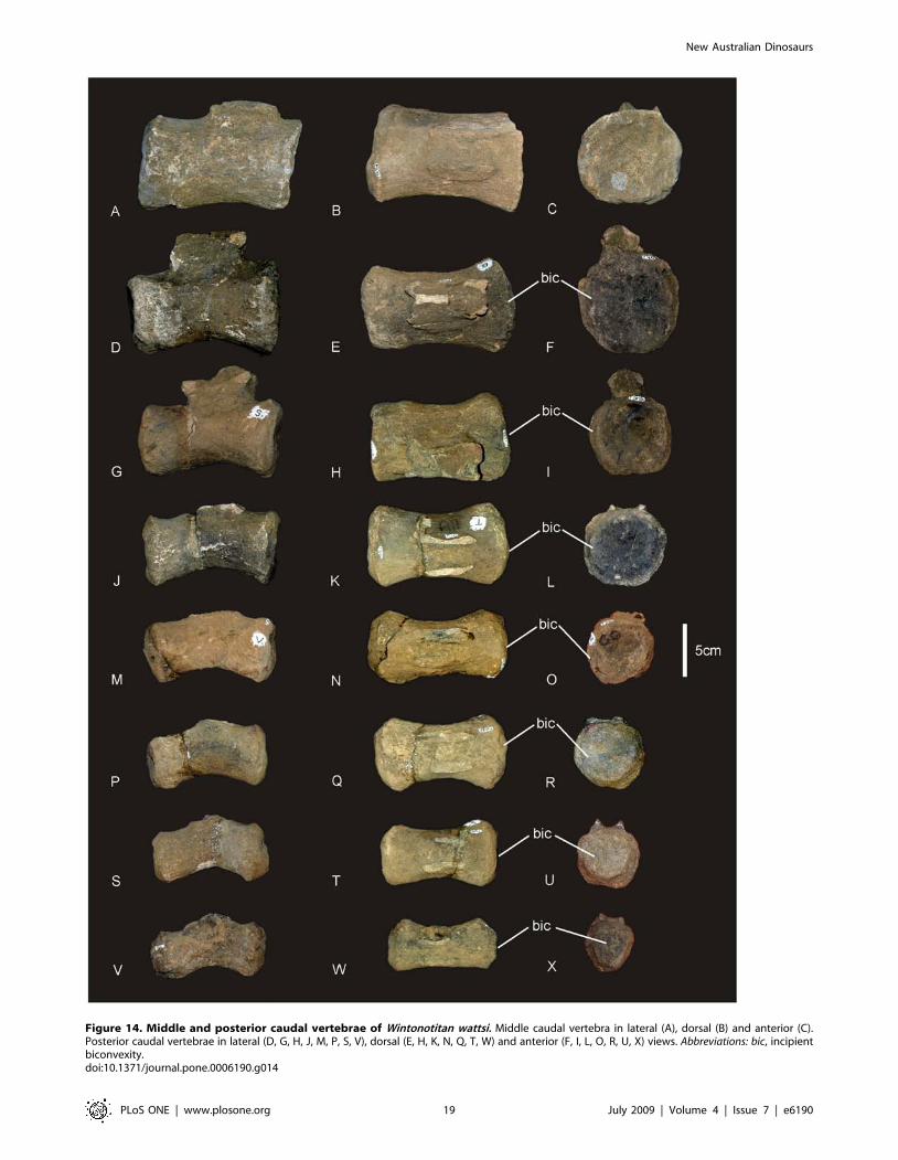
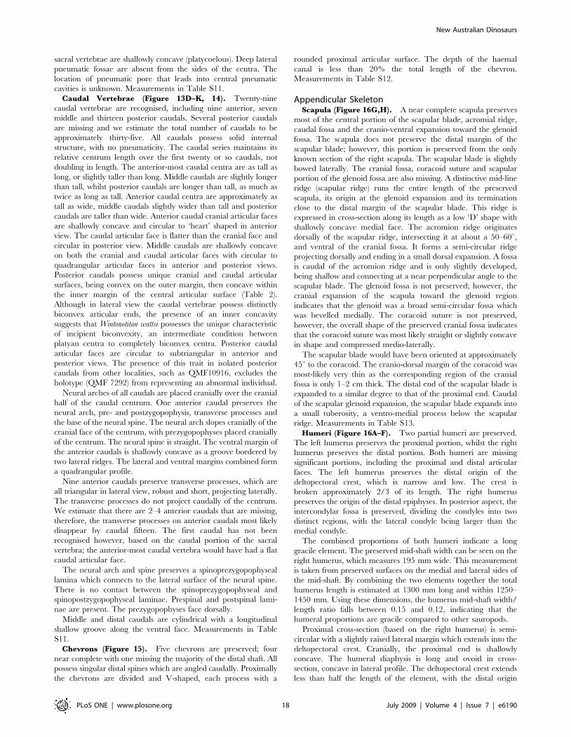
Figure 14. Middle and posterior caudal vertebrae of Wintonotitan wattsi. Middle caudal vertebra in lateral (A), dorsal (B) and anterior (C).Posterior caudal vertebrae in lateral (D, G, H, J, M, P, S, V), dorsal (E, H, K, N, Q, T, W) and anterior (F, I, L, O, R, U, X) views. Abbreviations: bic, incipientbiconvexity.doi:10.1371/journal.pone.0006190.g014
New Australian Dinosaurs
PLoS ONE | www.plosone.org 19 July 2009 | Volume 4 | Issue 7 | e6190
located proximal to the middle of the shaft. Although much of the
deltopectoral crest is not preserved the base of it indicates that the
crest was low and narrow and not prominently projecting from the
main surface of shaft. The crest itself is narrow along its length and
not markedly expanded proximally or distally.
The preserved distal end of the right humerus is massive and
possesses the origin of both the medial and lateral condyles,
divided by the intercondylar fossa. In cross-section the lateral
(ulnar) condyle is larger then the medial (radial) condyle. The
distal end of the left humerus bears a post-mortem break which is
covered by sediment and plant material. Measurements in Table
S13.
Ulnae (Figure 17G–I). The right and left ulnae are preserved
in the holotype (QMF 7292), with the right ulna being the most
complete element. The right ulna preserves the proximal and
distal epiphyses with the proximal end preserving the proximal-
most articular surface, which comprises the lateral and posterior
processes. The articular surface of the medial process is missing.
The left ulna is missing both the proximal and distal epiphyses,
preserving the mid-shaft section and the proximal flange of the
medial process.
The ratio of mid-shaft width to total humerus length is 0.16,
which is considered to be a gracile element (,0.20 [15]), however,
the proximal breadth to total length ratio equals 0.37, which is
considered to represent a stout ulna (.33% [15]). These ratios
reflect the narrow and gracile diaphysis of the ulnae, compared to
the massively expanded proximal epiphyses, where the lateral and
medial processes are extended into narrow crests from the main
shaft. The medial process is longer than the other two processes,
forming a distinctly tri-radiate cross-sectional shape. Each face is
markedly concave with the surface between the medial and lateral
processes being deeply excavated, to form the cranial fossa. The
surface between medial and caudal processes is similarly
excavated, but not to the same degree, whilst the remaining
surface between the caudal and lateral process is even shallower.
The olecranon process is prominent and extends proximally above
the articular surfaces of both the medial and caudal processes. The
cranial margin of the medial process is directed medially from the
main axis of the shaft. The medial crest is constricted along its
length and runs the entire length of the shaft terminating proximal
of the distal epiphysis. The caudal process is thick and rounded.
Measurements in Table S13.
Radii (Figure 17A–F). A right and left radius are preserved;
both are missing the proximal articular ends. The left radius is
better preserved than the right, including the distal epiphysis, main
shaft and proximal epiphysis. The radius is markedly expanded
proximally and distally to a similar degree, with the distal width
approximately twice that of the mid-shaft width. The diaphysis is
relatively straight only slightly bowed forming a concave posterior
face.
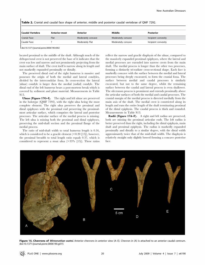
Figure 15. Chevrons of Wintonotitan wattsi. Anterior chevrons in anterior view (A–E). Chevron in (A) is attached to an anterior caudal centrum.doi:10.1371/journal.pone.0006190.g015
Table 2. Cranial and caudal face shape of anterior, middle and posterior caudal vertebrae of QMF 7292.
Caudal Vertebra Anterior-most Anterior Middle Posterior
Cranial Face Flat Moderately concave Moderately concave Incipient convexity
Caudal Face ? Moderately Flat Moderately concave Incipient convexity
doi:10.1371/journal.pone.0006190.t002
New Australian Dinosaurs
PLoS ONE | www.plosone.org 20 July 2009 | Volume 4 | Issue 7 | e6190

Figure 16. Scapula and Humeri of Wintonotitan wattsi. Partial left humerus in posterior (A) and anterior (B) views. Partial right humerus inposterior (C) and anterior (D) views. E. Reconstructed left humerus in anterior view by reversing (D) and aligning shaft curvature with (B). F.Reconstructed right humerus in posterior view by reversing (A) and aligning shaft curvature with (C). Left scapula in lateral (G) and medial (H) views.Dashed lines represent suggested missing areas.doi:10.1371/journal.pone.0006190.g016
New Australian Dinosaurs
PLoS ONE | www.plosone.org 21 July 2009 | Volume 4 | Issue 7 | e6190

The ratio of mid-shaft width to total radius length equals 0.16,
which falls only just outside the ratio considered to be gracile (0.15)
and below the ratio considered for robust radii (.0.25) [15],
therefore the radii are considered here to be gracile, which is
reflected in their long and relatively narrow diaphysis. This is in
contrast to the markedly expanded epiphyses, similar to the
condition in the ulnae.
The distal end is rounded in cranial profile and sub-rectangular
in distal cross-section; compressed cranio-caudally, which reflects a
more expanded transverse margin; transversely expanded more
than the proximal end, which is only expanded medially. The
ulnar ligament scar is well developed and extends proximally
almost a third of the shaft. A prominent oblique ridge runs from
the proximo-caudal face to the medio-distal end. The articular
surface of the distal end extends across the entire distal end and up
onto the lateral face. A well defined interosseous ridge runs from
the medio-caudal margin of the proximal epiphysis to the medial
margin of the distal condyle. The proximal end of the radius is
Figure 17. Radii and ulnae of Wintonotitan wattsi. Right radius in anterior (A) view. Left radius in medial (B), posterior (C), anterior (D), proximal(E) and distal (F) views. Left ulna in antero-lateral view (G). Right ulna in antero-lateral view (H). Reconstructed left humerus, ulna (reversed H andcombined with G) and radius (B) in antero-lateral view. Dashed lines represent suggested missing areas.doi:10.1371/journal.pone.0006190.g017
New Australian Dinosaurs
PLoS ONE | www.plosone.org 22 July 2009 | Volume 4 | Issue 7 | e6190
slender, possessing a proximal width between 25–26% of the to
radius length [15]. Measurements in Table S13.
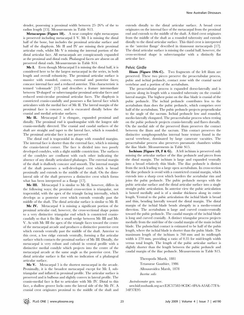
Metacarpus (Figure 18). A near complete right metacarpus
is preserved including metacarpal I–V. Mc I is missing the distal
half of the bone, but includes the proximal articular surface and
half of the diaphysis. Mc II and IV are missing their proximal
articular ends, whilst Mc V is missing the internal portion of the
distal articular face. All metacarpals are conspicuously expanded
at the proximal and distal ends. Phalangeal facets are absent on all
preserved distal ends. Measurements in Table S14.
Mc I. Even though Metacarpal I is missing the distal half, it is
considered here to be the largest metacarpal in the series, in both
length and overall robusticity. The proximal articular surface is
massive with rounded, convex, external and posterior faces;
concave internal face and a reduced anterior. This characteristic is
termed ‘colonnade’ [17] and describes a feature intermediate
between ‘D-shaped’ or subrectangular proximal articular faces and
reduced semi-circular proximal articular faces. The diaphysis is
constricted cranio-caudally and possesses a flat lateral face which
articulates with the medial face of Mc II. The lateral margin of the
proximal face is concave, and is excavated distally below the
cranial and medial articular surfaces.
Mc II. Metacarpal 2 is elongate, expanded proximal and
distally. The proximal end is quadrangular with the longest side
cranio-medially directed. The cranial and caudal surfaces of the
shaft are straight and taper to the lateral face, which is rounded.
The proximal articular face is not preserved.
The distal end is trapezoidal in shape with rounded margins.
The internal face is shorter than the external face, which is missing
the cranio-lateral corner. The face is divided into two poorly
developed condyles, with a slight middle constriction. The surfaces
of the condyles are confluent with each other indicating the
absence of any distally articulated phalanges. The external margin
of the shaft is shallowly concave and smooth. The internal margin
of the shaft possesses a well-developed crest which is broad
proximally and extends to the middle of the shaft. On the disto-
lateral side of the shaft preserves a distinctive crest which forms
what has been interpreted as a flange [17].
Mc III. Metacarpal 3 is similar to Mc II, however, differs in
the following ways; the proximal cross-section is triangular, not
trapezoidal, with the apex of the triangle facing internally, which
develops as a posterior crest which extends ventrally past the
middle of the shaft. The distal articular surface is similar to Mc II.
Mc IV. Metacarpal 4 is missing a significant portion of the
proximal articular end, however, the cross-sectional shape points
to a very distinctive triangular end which is constricted cranio-
caudally so that it fits like a small wedge between Mc III and Mc
V. As with Mc III the apex of the triangle faces toward the centre
of the metacarpal arcade and produces a distinctive posterior crest
which extends ventrally past the middle of the shaft. Anterior to
this crest, a low ridge extends ventrally, forming a flat articular
surface which contacts the proximal surface of Mc III. Distally, the
metacarpal is very robust and cuboid in ventral profile with a
distinctive medial condyle which projects into the centre of the
metacarpal arcade at the same angle as the posterior crest. The
distal articular surface is flat with no indication of a phalangeal
articular surface.
Mc V. Metacarpal 5 is the shortest metacarpal in the arcade.
Proximally, it is the broadest metacarpal except for Mc I, sub-
triangular and inflated in proximal profile. The articular surface is
preserved and is bulbous and slightly convex in lateral profile. The
cranio-medial face is flat to articulate with Mc IV. Distal to this
face, a shallow groove locks onto the lateral side of the Mc IV. A
cranial crest originates proximal to the middle of the shaft and
extends distally to the distal articular surface. A broad crest
originates on the internal face of the metacarpal from the proximal
end and extends to the middle of the shaft. A third crest originates
from the middle of the shaft as a rounded tuberosity and extends
distally to the distal articular surface. This third crest is interpreted
as the ‘anterior flange’ described in titanosaur metacarpals [17].
The distal articular surface is missing the caudal half; however, the
cross-sectional shape is subrectangular with a distinctly flat
articular face.
Pelvic GirdleIlium (Figure 19A–C). Two fragments of the left ilium are
preserved. These two pieces preserve the preacetabular process,
pubic and ischial peduncle, contact zone with the cranial sacral
vertebrae and a portion of the acetabulum.
The preacetabular process is expanded dorso-laterally and is
narrow along its length with a rounded tuberosity on the cranial-
lateral margin. The highest point on the iliac blade is cranial of the
pubic peduncle. The ischial peduncle contributes less to the
acetabulum than does the pubic peduncle, which comprises over
half of the acetabulum. The pubic peduncle projects perpendicular
to the angle of the sacrum. Ischial peduncle low and rounded,
medio-laterally elongated. The preacetabular process when resting
on the pubic peduncle projects cranio-laterally and flares dorsally.
On the medial side of the preserved ilium preserves the contact
between the ilium and the sacrum. This contact preserves the
distinctive somphospondylus internal bone texture found in the
sacral vertebrae, dominated by large camerate vacuities. The
preacetabular process also preserves pneumatic chambers within
the iliac blade. Measurements in Table S15.
Ischium (Figure 19, F & G). A left ischium is preserved only
missing the articular surface of the iliac peduncle and pieces from
the distal margin. The ischium is large and expanded ventrally
into a broad relatively thin blade. The iliac peduncle is distinct
from the neck leading to a large open acetabulum. In cross-section
the iliac peduncle is ovoid with a constricted cranial margin, which
extends into a sharp crest which borders the acetabular rim and
joins the pubic peduncle. The pubic peduncle merges with the
pubic articular surface and the distal articular surface into a single
straight pubic articulation. In anterior view the pubic articulation
is bowed medially and is of a similar thickness along its entire
length. Ventral to the pubic articulation the ischial blade is broad
and thin, bending laterally toward the distal margin. The distal
margin of the ischial blade bends abruptly in a medio-ventral
direction. The acetabulum is large and curved cranio-ventrally
toward the pubic peduncle. The caudal margin of the ischial blade
is long and curved cranially. A distinct triangular process projects
medially from the mid-line of the caudal margin of the main ischial
blade. The puboischial contact is estimated to be half of the pubis
length, where the ischial blade is shorter than the pubic blade. The
maximum length of the ischium is 760 mm and its midlength
width is 270 mm, providing a ratio of 0.35 for mid-length width
versus total length. The length of the pubic articular surface is
slightly shorter than the length between the pubic peduncle and
caudal margin of the iliac peduncle. Measurements in Table S15.
Theropoda Marsh, 1881
Tetanurae Gauthier, 1986
Allosauroidea Marsh, 1878
Incertae sedis
Australovenator gen. nov.
urn:lsid:zoobank.org:act:E2C57582-9CDC-4F6A-A3AE-77FA-
54F37E95
New Australian Dinosaurs
PLoS ONE | www.plosone.org 23 July 2009 | Volume 4 | Issue 7 | e6190
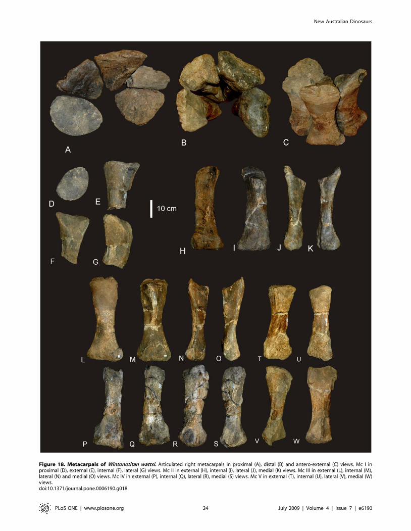
Figure 18. Metacarpals of Wintonotitan wattsi. Articulated right metacarpals in proximal (A), distal (B) and antero-external (C) views. Mc I inproximal (D), external (E), internal (F), lateral (G) views. Mc II in external (H), internal (I), lateral (J), medial (K) views. Mc III in external (L), internal (M),lateral (N) and medial (O) views. Mc IV in external (P), internal (Q), lateral (R), medial (S) views. Mc V in external (T), internal (U), lateral (V), medial (W)views.doi:10.1371/journal.pone.0006190.g018
New Australian Dinosaurs
PLoS ONE | www.plosone.org 24 July 2009 | Volume 4 | Issue 7 | e6190
Etymology. Austral, australis – Latin, meaning southern in
reference to the locality being in the Southern Hemisphere,
Australia. Venator – Latin for hunter. In reference to its carnivorous diet.
Type species. Australovenator wintonensis
Australovenator wintonensis sp. nov.
urn:lsid:zoobank.org:act:37AA3C1B-B498-406B-A51C-C3CF-
8C88A59F
Etymology. From the township of Winton.
Holotype. AODF 604: Nine isolated teeth; left dentary; right
and left dorsal ribs and rib fragments; right and left gastralial ribs
and fragments; partial right ilium; both ulnae; right radius; manus
metacarpals, phalanges and unguals; right femur; both tibiae; right
fibula; right astragalus; metatarsals, phalanges and unguals
(Figure 2D).
Type Locality. AODF 85, ‘‘Matilda Site’’, Elderslie Station,
approximately 60 km north-west of Winton, central Queensland,
Australia.
Horizon & Age. Winton Formation, latest Albian
(Cretaceous)
Diagnosis. A medium-sized allosauroid, with the following
unique association of features. Dentary gracile with sub-parallel
margins; rounded dentary symphysis; articular brace (or ‘chin’)
absent. 18 tooth loci; alveolus 1 quadrangular; alveoli 2–6 and 12–
15 circular; alveoli 7–11 and 16–18 labio-lingually compressed.
Interdental plates fused together along the entire length of the
dentary. Primary neurovascular foramina row parallel to dorsal
margin of dentary, not deflected ventrally. Dorsal ribs with
pneumatic cavities. Gastralia unfused with tapered distal ends.
Figure 19. Pelvis of Wintonotitan wattsi. Preacetabular lobe of ilium in dorsal (A), ventral (B) and lateral (C) views. Arrow indicates the axis of thesacrum. Partial posterior sacral vertebra in lateral (D) and dorsal (E) views. Left ischium in lateral (F) and medial (G) views. Abbreviations: ip, iliacpeduncle; ppd, pubic peduncle; ppp, preacetabular process of ilium.doi:10.1371/journal.pone.0006190.g019
New Australian Dinosaurs
PLoS ONE | www.plosone.org 25 July 2009 | Volume 4 | Issue 7 | e6190
Ulna with straight caudal margin, inflated olecranon process;
lateral groove along diaphysis (autapomorphic); round and
discontinuous lateral tuberosity.
Femur with head directed dorsally in a cranio-medially
direction; lesser trochanter developed proximally to the level of
the greater trochanter (autapomorphic); distal extensor groove
deep and narrow lacking cruciate ridge. Cranio-lateral process of
tibia with latero-ventral process; lateral malleolus extends distal of
medial malleolus. Proximal articular surface of fibula bevelled
cranially (autapomorphic). Astragalus with tall ascending process;
medially expanded medial condyle; superior and inferior grooves
present on cranial face; cranio-lateral process projects from
proximo-cranial margin of lateral condyle. Metatarsals elongate
and gracile.
Description
Cranial SkeletonTeeth (Figure 20 & 21). Nine isolated teeth were recovered
from the type locality and are all considered to be from the
holotype specimen of Australovenator wintonensis gen. et sp. nov. All
of the teeth have broken away from their bases so that only the
tooth crown is preserved. All are recurved and bear fine serrations
along mesial and distal carinae. Two teeth have circular to
quadrangular shaped bases with two series of serrations; one is a
disto-lingual carina and the other is a distal carina. These teeth are
considered to be anterior-most dentary or premaxillary teeth. The
remaining teeth are all labio-lingually compressed with serrations
along the mesial apical edge and along the entire distal carina. On
comparison to similar teeth from Fukuiraptor and Neovenator all of
the remaining teeth are considered to be dentary teeth, with
rounded mesial and tapered distal margins.
Tooth measurements of Fukuiraptor have been compared to
carcharodontosaurids, tyrannosaurids and dromaeosaurids using
the FABL (Fore-Aft Crown Base Length) vs BW (Tooth Crown
Base Width) measurements as an indicator of labio-lingual
compression [19]. It was found that Fukuiraptor had more labio-
lingually compressed dentition than carcharodontosaurids [19].
Comparing our measurements, we found that the teeth of
Australovenator were similarly as compressed as Fukuiraptor. The
teeth do not possess the typical basal crown wrinkles found in
carcharodontosaurids, or Neovenator [28,29].
Wear features are noticeable on four of the teeth, including
mesial wear across the tip of the tooth crown and mesio-labial
wear across the leading edge of the crowns. All preserve weak
proximo-distal striae. Measurements in Table S18.
Dentary (Figure 22). A near complete left dentary is known
from the holotype (AODF 604). In dorsal view the dentary is
slightly curved medially along the posterior half. Eighteen tooth
alveoli are present with four alveoli preserving fragmented bases of
individual teeth (preserved in alveoli 3, 4, 7 & 11). Each alveolus is
a rounded pit bordered lingually by an interdental plate, which is
fused on both the anterior and posterior margin to the preceding
and succeeding interdental plate, forming an interdental ridge
which extends along the entire length of the tooth row. Lingual to
this ridge is a shallow dental sulcus.
In dorsal view the first tooth alveolus is small and quadrangular
in shape with the antero-labial face longest. Alveoli 2–6 are
circular in shape with subequal labio-lingual and antero-posterior
diameters. Alveoli 7–11 are labio-lingually compressed, with the
long axis oriented antero-posteriorly. Alveoli 12–15 return to a
subcircular shape as in alveoli 2–6. Alveoli 16–18 are small,
narrowed labio-lingually with the long axis oriented antero-
posteriorly.
In lingual view the dorsal and ventral margins of the dentary
parallel each other until the position of alveolus 15 where the
margins diverge; the ventral margin gently curves ventrally, whilst
the dorsal margin remains relatively straight, gently curving
dorsally posterior of the last tooth loci. The lingual face of the
dentary preserves an anterior dentary symphysis which originates
ventrally of the first alveolus; is narrow along its length; follows the
anterior margin of the dentary, curving onto the antero-ventral
margin and terminating just antero-ventrally of the Meckelian
foramen. The Meckelian foramen is exposed and sits immediately
anterior of the anterior margin of the Meckelian groove. The
Meckelian groove is long, straight, open and dorsal-ventrally
narrow, extending posteriorly to a position ventral of tooth loci 14,
where it meets a large singular foramen for the inferior alveolar
nerve. Posterior of the foramen, the dentary is thin-walled,
forming a shallow fossa, which expands posteriorly to the broken
posterior margin of the dentary. This expansion is the anterior
remnants of the ventral dentary process which connects to the
surangular.
The posterior margin of the dentary is made up of a dorsal
intramandibular process which extends postero-dorsally. Ventral
to this and following the posterior margin of the preserved element
is the contact for the prearticular. Ventral to this contact the
dentary is broken, missing the posterior process of the surangular
contact. In labial view the anterior surface of the dentary is slightly
rugose, whilst the posterior margin is relatively smooth. A primary
and secondary series of neurovascular foramina are present on the
antero-labial face of the dentary. The primary neurovascular
foramina are present ventral to the dorsal margin of the dentary,
paralleling this margin and not deflected ventrally. Two small
secondary neurovascular foramina are located ventral to the
anterior-most primary neurovascular foramina. Measurements in
Table S17.
Axial SkeletonDorsal Ribs (Figure 23A–F). Several dorsal ribs are
preserved including the first right dorsal rib, second or third
right dorsal rib and seventh or eighth left dorsal rib. Several
isolated rib shaft pieces are also known. All rib elements are
missing the distal ends. Comparisons were made to rib sequences
in Allosaurus [20], Sinraptor [21] and Acrocanthosaurus [22] to
determine the position of the ribs preserving proximal ends.
Measurements in Table S18.
First right dorsal rib. Gracile dorsal rib expanded
proximally into a broad capitulo-tubercular web. Proximal-
most tubercular margin extends above the proximal-most
margin of the capitulum. Tuberculum projects dorsally,
broadens medio-laterally and is narrow cranio-caudally.
Anterior face of tuberculum developed into a shallow fossa,
which connects distally to the costal groove. Fossa bordered by a
cranio-caudally expanded intercostal ridge. Proximal articular
end of the tuberculum is missing. Capitulum projects medially,
almost perpendicular to the dorso-ventral axis of the
tuberculum. Capitulum broadest dorso-ventrally and cranio-
caudally narrow. Proximal articular end of capitulum flattened
cranially and convex caudally with rounded articular facet.
Capitulo-tubercular web thin with a concave proximal margin in
anterior view. Neck of capitulum narrower than proximal-most
end. Costal groove extends from the tubercular fossa distally
along the entire length of the preserved rib shaft. Groove
narrows along its length. Costal groove bordered laterally by a
distally tapering intercostal ridge and medially by a similar
proximally thick medial ridge. Both these ridges coalesce distally,
only divided by a narrow costal groove. In posterior view there
New Australian Dinosaurs
PLoS ONE | www.plosone.org 26 July 2009 | Volume 4 | Issue 7 | e6190
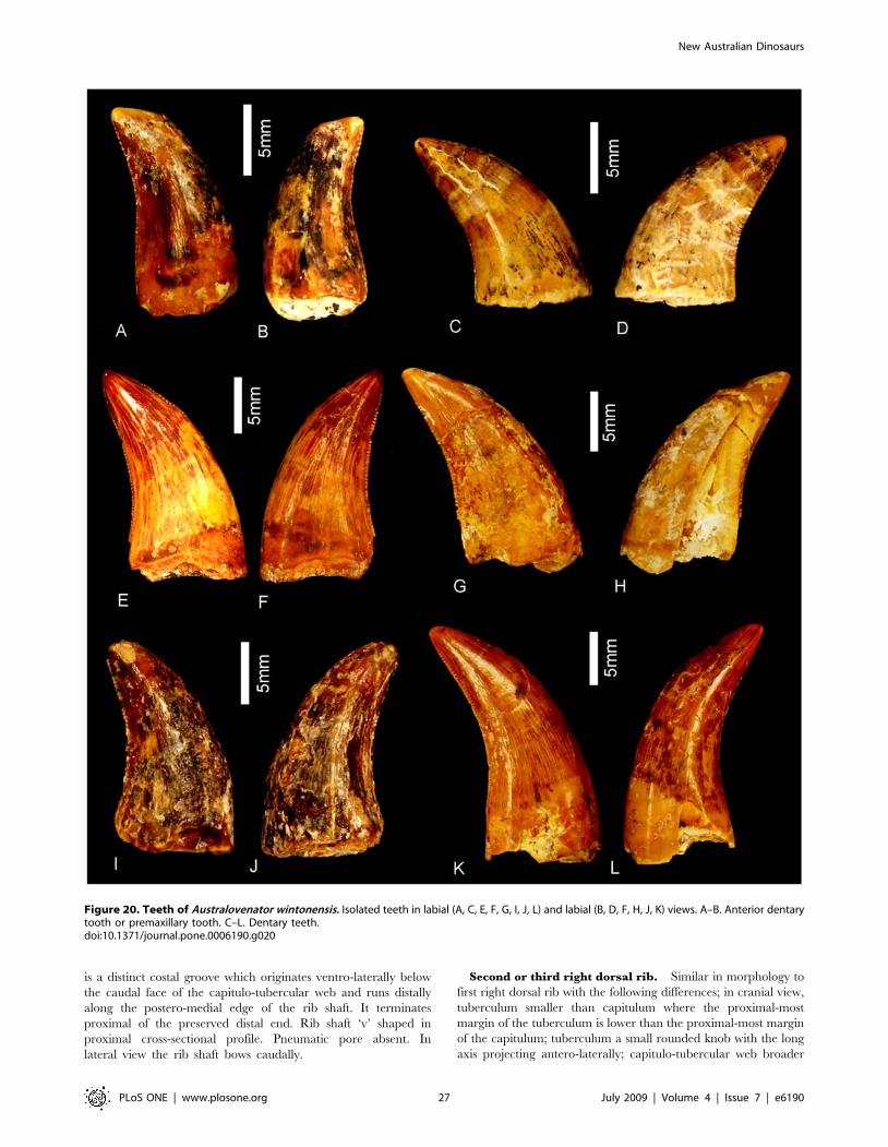
is a distinct costal groove which originates ventro-laterally below
the caudal face of the capitulo-tubercular web and runs distally
along the postero-medial edge of the rib shaft. It terminates
proximal of the preserved distal end. Rib shaft ‘v’ shaped in
proximal cross-sectional profile. Pneumatic pore absent. In
lateral view the rib shaft bows caudally.
Second or third right dorsal rib. Similar in morphology to
first right dorsal rib with the following differences; in cranial view,
tuberculum smaller than capitulum where the proximal-most
margin of the tuberculum is lower than the proximal-most margin
of the capitulum; tuberculum a small rounded knob with the long
axis projecting antero-laterally; capitulo-tubercular web broader
Figure 20. Teeth of Australovenator wintonensis. Isolated teeth in labial (A, C, E, F, G, I, J, L) and labial (B, D, F, H, J, K) views. A–B. Anterior dentarytooth or premaxillary tooth. C–L. Dentary teeth.doi:10.1371/journal.pone.0006190.g020
New Australian Dinosaurs
PLoS ONE | www.plosone.org 27 July 2009 | Volume 4 | Issue 7 | e6190
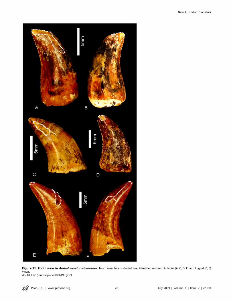
Figure 21. Tooth wear in Australovenator wintonensis. Tooth wear facets (dotted line) identified on teeth in labial (A, C, D, F) and lingual (B, E)views.doi:10.1371/journal.pone.0006190.g021
New Australian Dinosaurs
PLoS ONE | www.plosone.org 28 July 2009 | Volume 4 | Issue 7 | e6190
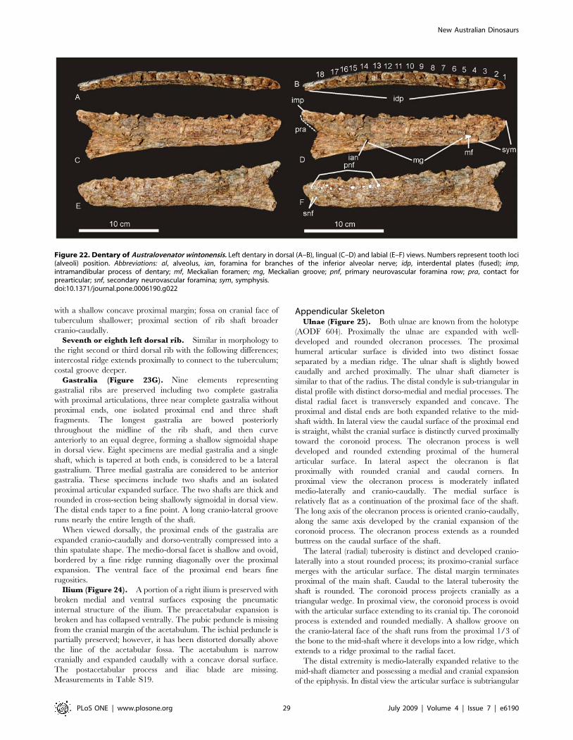
with a shallow concave proximal margin; fossa on cranial face of
tuberculum shallower; proximal section of rib shaft broader
cranio-caudally.
Seventh or eighth left dorsal rib. Similar in morphology to
the right second or third dorsal rib with the following differences;
intercostal ridge extends proximally to connect to the tuberculum;
costal groove deeper.
Gastralia (Figure 23G). Nine elements representing
gastralial ribs are preserved including two complete gastralia
with proximal articulations, three near complete gastralia without
proximal ends, one isolated proximal end and three shaft
fragments. The longest gastralia are bowed posteriorly
throughout the midline of the rib shaft, and then curve
anteriorly to an equal degree, forming a shallow sigmoidal shape
in dorsal view. Eight specimens are medial gastralia and a single
shaft, which is tapered at both ends, is considered to be a lateral
gastralium. Three medial gastralia are considered to be anterior
gastralia. These specimens include two shafts and an isolated
proximal articular expanded surface. The two shafts are thick and
rounded in cross-section being shallowly sigmoidal in dorsal view.
The distal ends taper to a fine point. A long cranio-lateral groove
runs nearly the entire length of the shaft.
When viewed dorsally, the proximal ends of the gastralia are
expanded cranio-caudally and dorso-ventrally compressed into a
thin spatulate shape. The medio-dorsal facet is shallow and ovoid,
bordered by a fine ridge running diagonally over the proximal
expansion. The ventral face of the proximal end bears fine
rugosities.
Ilium (Figure 24). A portion of a right ilium is preserved with
broken medial and ventral surfaces exposing the pneumatic
internal structure of the ilium. The preacetabular expansion is
broken and has collapsed ventrally. The pubic peduncle is missing
from the cranial margin of the acetabulum. The ischial peduncle is
partially preserved; however, it has been distorted dorsally above
the line of the acetabular fossa. The acetabulum is narrow
cranially and expanded caudally with a concave dorsal surface.
The postacetabular process and iliac blade are missing.
Measurements in Table S19.
Appendicular SkeletonUlnae (Figure 25). Both ulnae are known from the holotype
(AODF 604). Proximally the ulnae are expanded with well-
developed and rounded olecranon processes. The proximal
humeral articular surface is divided into two distinct fossae
separated by a median ridge. The ulnar shaft is slightly bowed
caudally and arched proximally. The ulnar shaft diameter is
similar to that of the radius. The distal condyle is sub-triangular in
distal profile with distinct dorso-medial and medial processes. The
distal radial facet is transversely expanded and concave. The
proximal and distal ends are both expanded relative to the mid-
shaft width. In lateral view the caudal surface of the proximal end
is straight, whilst the cranial surface is distinctly curved proximally
toward the coronoid process. The olecranon process is well
developed and rounded extending proximal of the humeral
articular surface. In lateral aspect the olecranon is flat
proximally with rounded cranial and caudal corners. In
proximal view the olecranon process is moderately inflated
medio-laterally and cranio-caudally. The medial surface is
relatively flat as a continuation of the proximal face of the shaft.
The long axis of the olecranon process is oriented cranio-caudally,
along the same axis developed by the cranial expansion of the
coronoid process. The olecranon process extends as a rounded
buttress on the caudal surface of the shaft.
The lateral (radial) tuberosity is distinct and developed cranio-
laterally into a stout rounded process; its proximo-cranial surface
merges with the articular surface. The distal margin terminates
proximal of the main shaft. Caudal to the lateral tuberosity the
shaft is rounded. The coronoid process projects cranially as a
triangular wedge. In proximal view, the coronoid process is ovoid
with the articular surface extending to its cranial tip. The coronoid
process is extended and rounded medially. A shallow groove on
the cranio-lateral face of the shaft runs from the proximal 1/3 of
the bone to the mid-shaft where it develops into a low ridge, which
extends to a ridge proximal to the radial facet.
The distal extremity is medio-laterally expanded relative to the
mid-shaft diameter and possessing a medial and cranial expansion
of the epiphysis. In distal view the articular surface is subtriangular
Figure 22. Dentary of Australovenator wintonensis. Left dentary in dorsal (A–B), lingual (C–D) and labial (E–F) views. Numbers represent tooth loci(alveoli) position. Abbreviations: al, alveolus, ian, foramina for branches of the inferior alveolar nerve; idp, interdental plates (fused); imp,intramandibular process of dentary; mf, Meckalian foramen; mg, Meckalian groove; pnf, primary neurovascular foramina row; pra, contact forprearticular; snf, secondary neurovascular foramina; sym, symphysis.doi:10.1371/journal.pone.0006190.g022
New Australian Dinosaurs
PLoS ONE | www.plosone.org 29 July 2009 | Volume 4 | Issue 7 | e6190
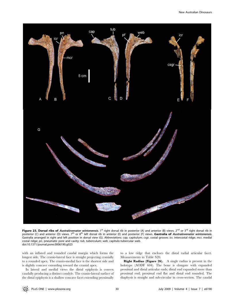
with an inflated and rounded caudal margin which forms the
longest side. The cranio-lateral face is straight projecting cranially
to a rounded apex. The cranio-medial face is the shortest side and
is slightly concave extending toward the cranial apex.
In lateral and medial views the distal epiphysis is convex
caudally producing a distinct condyle. The cranio-lateral surface of
the distal epiphysis is a shallow concave facet extending proximally
to a low ridge that encloses the distal radial articular facet.
Measurements in Table S20.
Right Radius (Figure 26). A single radius is present in the
holotype (AODF 604). The bone is elongate with expanded
proximal and distal articular ends; distal end expanded more than
proximal end; proximal end flat and distal end rounded. The
diaphysis is straight and sub-circular in cross-section. The caudal
Figure 23. Dorsal ribs of Australovenator wintonensis. 1st right dorsal rib in posterior (A) and anterior (B) views. 2nd or 3rd right dorsal rib inposterior (C) and anterior (D) views. 7th or 8th left dorsal rib in anterior (E) and posterior (F) views. Gastralia of Australovenator wintonensis.Gastralia arranged in right and left position in dorsal view (G). Abbreviations: cap, capitulum; csgr, costal groove; icr, intercostal ridge; mcr, medialcostal ridge; pn, pneumatic pore and cavity; tub, tuberculum; web, capitulo-tubercular web.doi:10.1371/journal.pone.0006190.g023
New Australian Dinosaurs
PLoS ONE | www.plosone.org 30 July 2009 | Volume 4 | Issue 7 | e6190
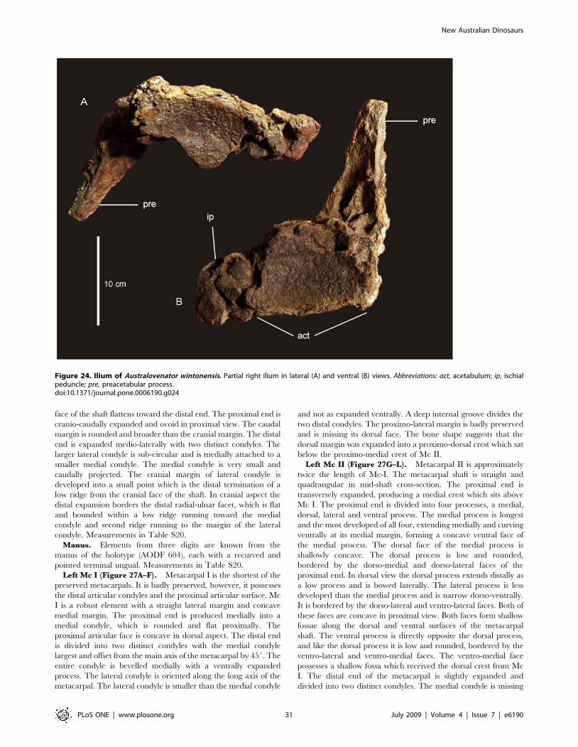
face of the shaft flattens toward the distal end. The proximal end is
cranio-caudally expanded and ovoid in proximal view. The caudal
margin is rounded and broader than the cranial margin. The distal
end is expanded medio-laterally with two distinct condyles. The
larger lateral condyle is sub-circular and is medially attached to a
smaller medial condyle. The medial condyle is very small and
caudally projected. The cranial margin of lateral condyle is
developed into a small point which is the distal termination of a
low ridge from the cranial face of the shaft. In cranial aspect the
distal expansion borders the distal radial-ulnar facet, which is flat
and bounded within a low ridge running toward the medial
condyle and second ridge running to the margin of the lateral
condyle. Measurements in Table S20.
Manus. Elements from three digits are known from the
manus of the holotype (AODF 604), each with a recurved and
pointed terminal ungual. Measurements in Table S20.
Left Mc I (Figure 27A–F). Metacarpal I is the shortest of the
preserved metacarpals. It is badly preserved, however, it possesses
the distal articular condyles and the proximal articular surface. Mc
I is a robust element with a straight lateral margin and concave
medial margin. The proximal end is produced medially into a
medial condyle, which is rounded and flat proximally. The
proximal articular face is concave in dorsal aspect. The distal end
is divided into two distinct condyles with the medial condyle
largest and offset from the main axis of the metacarpal by 45u. The
entire condyle is bevelled medially with a ventrally expanded
process. The lateral condyle is oriented along the long axis of the
metacarpal. The lateral condyle is smaller than the medial condyle
and not as expanded ventrally. A deep internal groove divides the
two distal condyles. The proximo-lateral margin is badly preserved
and is missing its dorsal face. The bone shape suggests that the
dorsal margin was expanded into a proximo-dorsal crest which sat
below the proximo-medial crest of Mc II.
Left Mc II (Figure 27G–L). Metacarpal II is approximately
twice the length of Mc-I. The metacarpal shaft is straight and
quadrangular in mid-shaft cross-section. The proximal end is
transversely expanded, producing a medial crest which sits above
Mc I. The proximal end is divided into four processes, a medial,
dorsal, lateral and ventral process. The medial process is longest
and the most developed of all four, extending medially and curving
ventrally at its medial margin, forming a concave ventral face of
the medial process. The dorsal face of the medial process is
shallowly concave. The dorsal process is low and rounded,
bordered by the dorso-medial and dorso-lateral faces of the
proximal end. In dorsal view the dorsal process extends distally as
a low process and is bowed laterally. The lateral process is less
developed than the medial process and is narrow dorso-ventrally.
It is bordered by the dorso-lateral and ventro-lateral faces. Both of
these faces are concave in proximal view. Both faces form shallow
fossae along the dorsal and ventral surfaces of the metacarpal
shaft. The ventral process is directly opposite the dorsal process,
and like the dorsal process it is low and rounded, bordered by the
ventro-lateral and ventro-medial faces. The ventro-medial face
possesses a shallow fossa which received the dorsal crest from Mc
I. The distal end of the metacarpal is slightly expanded and
divided into two distinct condyles. The medial condyle is missing
Figure 24. Ilium of Australovenator wintonensis. Partial right ilium in lateral (A) and ventral (B) views. Abbreviations: act, acetabulum; ip, ischialpeduncle; pre, preacetabular process.doi:10.1371/journal.pone.0006190.g024
New Australian Dinosaurs
PLoS ONE | www.plosone.org 31 July 2009 | Volume 4 | Issue 7 | e6190
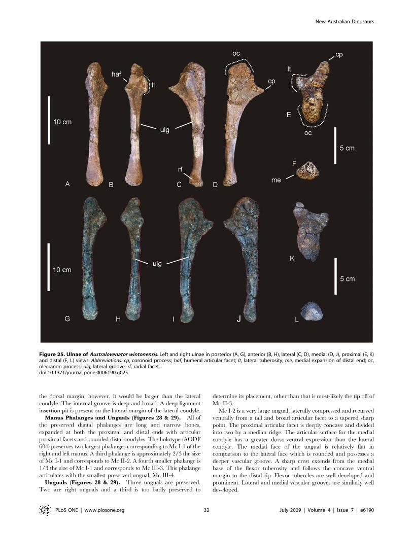
the dorsal margin; however, it would be larger than the lateral
condyle. The internal groove is deep and broad. A deep ligament
insertion pit is present on the lateral margin of the lateral condyle.
Manus Phalanges and Unguals (Figures 28 & 29). All of
the preserved digital phalanges are long and narrow bones,
expanded at both the proximal and distal ends with articular
proximal facets and rounded distal condyles. The holotype (AODF
604) preserves two largest phalanges corresponding to Mc I-1 of the
right and left manus. A third phalange is approximately 2/3 the size
of Mc I-1 and corresponds to Mc II-2. A fourth smaller phalange is
1/3 the size of Mc I-1 and corresponds to Mc III-3. This phalange
articulates with the smallest preserved ungual, Mc III-4.
Unguals (Figures 28 & 29). Three unguals are preserved.
Two are right unguals and a third is too badly preserved to
determine its placement, other than that is most-likely the tip off of
Mc II-3.
Mc I-2 is a very large ungual, laterally compressed and recurved
ventrally from a tall and broad articular facet to a tapered sharp
point. The proximal articular facet is deeply concave and divided
into two by a median ridge. The articular surface for the medial
condyle has a greater dorso-ventral expression than the lateral
condyle. The medial face of the ungual is relatively flat in
comparison to the lateral face which is rounded and possesses a
deeper vascular groove. A sharp crest extends from the medial
base of the flexor tuberosity and follows the concave ventral
margin to the distal tip. Flexor tubercles are well developed and
prominent. Lateral and medial vascular grooves are similarly well
developed.
Figure 25. Ulnae of Australovenator wintonensis. Left and right ulnae in posterior (A, G), anterior (B, H), lateral (C, D), medial (D, J), proximal (E, K)and distal (F, L) views. Abbreviations: cp, coronoid process; haf, humeral articular facet; lt, lateral tuberosity; me, medial expansion of distal end; oc,olecranon process; ulg, lateral groove; rf, radial facet.doi:10.1371/journal.pone.0006190.g025
New Australian Dinosaurs
PLoS ONE | www.plosone.org 32 July 2009 | Volume 4 | Issue 7 | e6190
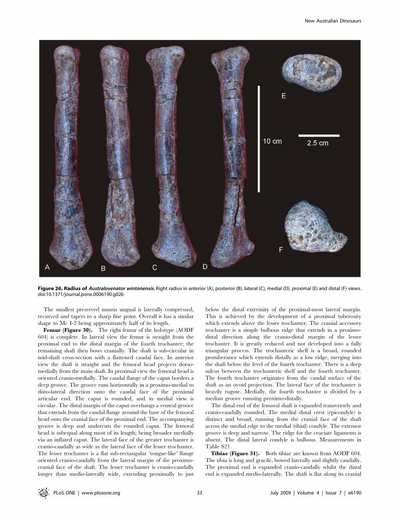
The smallest preserved manus ungual is laterally compressed,
recurved and tapers to a sharp fine point. Overall it has a similar
shape to Mc I-2 being approximately half of its length.
Femur (Figure 30). The right femur of the holotype (AODF
604) is complete. In lateral view the femur is straight from the
proximal end to the distal margin of the fourth trochanter; the
remaining shaft then bows cranially. The shaft is sub-circular in
mid-shaft cross-section with a flattened caudal face. In anterior
view the shaft is straight and the femoral head projects dorso-
medially from the main shaft. In proximal view the femoral head is
oriented cranio-medially. The caudal flange of the caput borders a
deep groove. The groove runs horizontally in a proximo-medial to
disto-lateral direction onto the caudal face of the proximal
articular end. The caput is rounded, and in medial view is
circular. The distal margin of the caput overhangs a ventral groove
that extends from the caudal flange around the base of the femoral
head onto the cranial face of the proximal end. The accompanying
groove is deep and undercuts the rounded caput. The femoral
head is subequal along most of its length; being broader medially
via an inflated caput. The lateral face of the greater trochanter is
cranio-caudally as wide as the lateral face of the lesser trochanter.
The lesser trochanter is a flat sub-rectangular ‘tongue-like’ flange
oriented cranio-caudally from the lateral margin of the proximo-
cranial face of the shaft. The lesser trochanter is cranio-caudally
longer than medio-laterally wide, extending proximally to just
below the distal extremity of the proximal-most lateral margin.
This is achieved by the development of a proximal tuberosity
which extends above the lesser trochanter. The cranial accessory
trochanter is a simple bulbous ridge that extends in a proximo-
distal direction along the cranio-distal margin of the lesser
trochanter. It is greatly reduced and not developed into a fully
triangular process. The trochanteric shelf is a broad, rounded
protuberance which extends distally as a low ridge, merging into
the shaft below the level of the fourth trochanter. There is a deep
sulcus between the trochanteric shelf and the fourth trochanter.
The fourth trochanter originates from the caudal surface of the
shaft as an ovoid projection. The lateral face of the trochanter is
heavily rugose. Medially, the fourth trochanter is divided by a
median groove running proximo-distally.
The distal end of the femoral shaft is expanded transversely and
cranio-caudally rounded. The medial distal crest (epicondyle) is
distinct and broad, running from the cranial face of the shaft
across the medial edge to the medial (tibial) condyle. The extensor
groove is deep and narrow. The ridge for the cruciate ligaments is
absent. The distal lateral condyle is bulbous. Measurements in
Table S21.
Tibiae (Figure 31). Both tibiae are known from AODF 604.
The tibia is long and gracile, bowed laterally and slightly caudally.
The proximal end is expanded cranio-caudally whilst the distal
end is expanded medio-laterally. The shaft is flat along its cranial
Figure 26. Radius of Australovenator wintonensis. Right radius in anterior (A), posterior (B), lateral (C), medial (D), proximal (E) and distal (F) views.doi:10.1371/journal.pone.0006190.g026
New Australian Dinosaurs
PLoS ONE | www.plosone.org 33 July 2009 | Volume 4 | Issue 7 | e6190
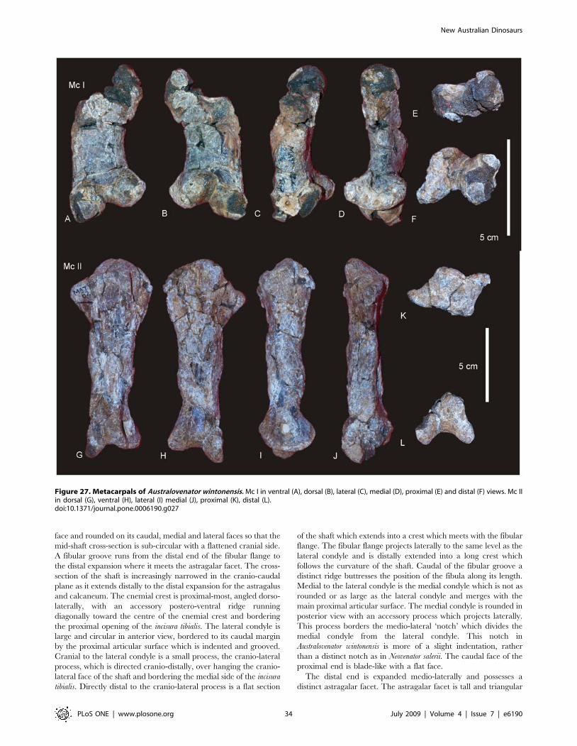
face and rounded on its caudal, medial and lateral faces so that the
mid-shaft cross-section is sub-circular with a flattened cranial side.
A fibular groove runs from the distal end of the fibular flange to
the distal expansion where it meets the astragalar facet. The cross-
section of the shaft is increasingly narrowed in the cranio-caudal
plane as it extends distally to the distal expansion for the astragalus
and calcaneum. The cnemial crest is proximal-most, angled dorso-
laterally, with an accessory postero-ventral ridge running
diagonally toward the centre of the cnemial crest and bordering
the proximal opening of the incisura tibialis. The lateral condyle is
large and circular in anterior view, bordered to its caudal margin
by the proximal articular surface which is indented and grooved.
Cranial to the lateral condyle is a small process, the cranio-lateral
process, which is directed cranio-distally, over hanging the cranio-
lateral face of the shaft and bordering the medial side of the incisura
tibialis. Directly distal to the cranio-lateral process is a flat section
of the shaft which extends into a crest which meets with the fibular
flange. The fibular flange projects laterally to the same level as the
lateral condyle and is distally extended into a long crest which
follows the curvature of the shaft. Caudal of the fibular groove a
distinct ridge buttresses the position of the fibula along its length.
Medial to the lateral condyle is the medial condyle which is not as
rounded or as large as the lateral condyle and merges with the
main proximal articular surface. The medial condyle is rounded in
posterior view with an accessory process which projects laterally.
This process borders the medio-lateral ‘notch’ which divides the
medial condyle from the lateral condyle. This notch in
Australovenator wintonensis is more of a slight indentation, rather
than a distinct notch as in Neovenator salerii. The caudal face of the
proximal end is blade-like with a flat face.
The distal end is expanded medio-laterally and possesses a
distinct astragalar facet. The astragalar facet is tall and triangular
Figure 27. Metacarpals of Australovenator wintonensis. Mc I in ventral (A), dorsal (B), lateral (C), medial (D), proximal (E) and distal (F) views. Mc IIin dorsal (G), ventral (H), lateral (I) medial (J), proximal (K), distal (L).doi:10.1371/journal.pone.0006190.g027
New Australian Dinosaurs
PLoS ONE | www.plosone.org 34 July 2009 | Volume 4 | Issue 7 | e6190
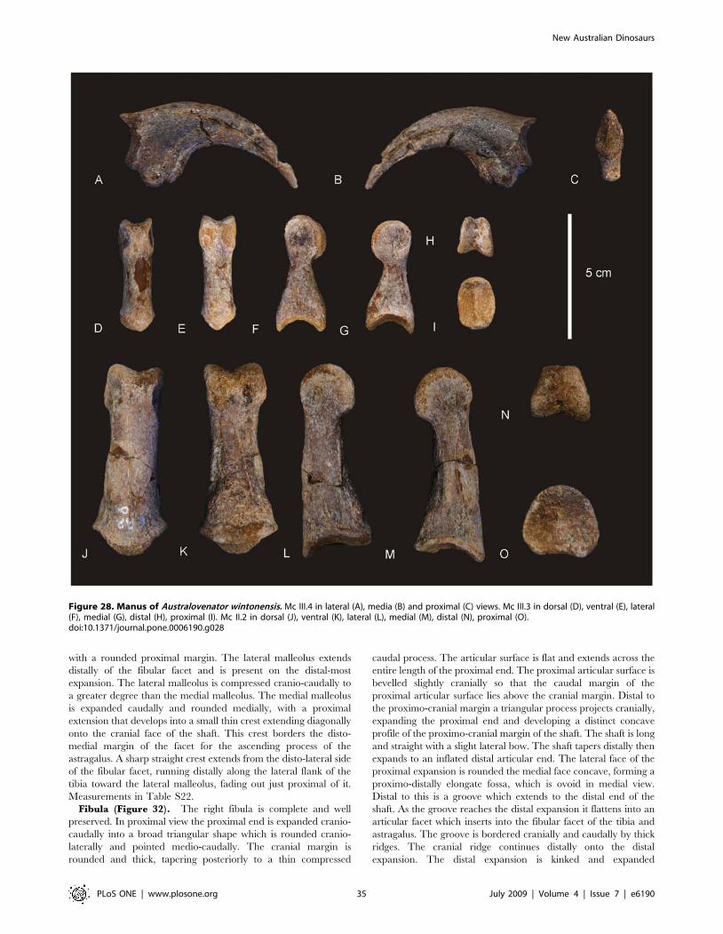
with a rounded proximal margin. The lateral malleolus extends
distally of the fibular facet and is present on the distal-most
expansion. The lateral malleolus is compressed cranio-caudally to
a greater degree than the medial malleolus. The medial malleolus
is expanded caudally and rounded medially, with a proximal
extension that develops into a small thin crest extending diagonally
onto the cranial face of the shaft. This crest borders the disto-
medial margin of the facet for the ascending process of the
astragalus. A sharp straight crest extends from the disto-lateral side
of the fibular facet, running distally along the lateral flank of the
tibia toward the lateral malleolus, fading out just proximal of it.
Measurements in Table S22.
Fibula (Figure 32). The right fibula is complete and well
preserved. In proximal view the proximal end is expanded cranio-
caudally into a broad triangular shape which is rounded cranio-
laterally and pointed medio-caudally. The cranial margin is
rounded and thick, tapering posteriorly to a thin compressed
caudal process. The articular surface is flat and extends across the
entire length of the proximal end. The proximal articular surface is
bevelled slightly cranially so that the caudal margin of the
proximal articular surface lies above the cranial margin. Distal to
the proximo-cranial margin a triangular process projects cranially,
expanding the proximal end and developing a distinct concave
profile of the proximo-cranial margin of the shaft. The shaft is long
and straight with a slight lateral bow. The shaft tapers distally then
expands to an inflated distal articular end. The lateral face of the
proximal expansion is rounded the medial face concave, forming a
proximo-distally elongate fossa, which is ovoid in medial view.
Distal to this is a groove which extends to the distal end of the
shaft. As the groove reaches the distal expansion it flattens into an
articular facet which inserts into the fibular facet of the tibia and
astragalus. The groove is bordered cranially and caudally by thick
ridges. The cranial ridge continues distally onto the distal
expansion. The distal expansion is kinked and expanded
Figure 28. Manus of Australovenator wintonensis. Mc III.4 in lateral (A), media (B) and proximal (C) views. Mc III.3 in dorsal (D), ventral (E), lateral(F), medial (G), distal (H), proximal (I). Mc II.2 in dorsal (J), ventral (K), lateral (L), medial (M), distal (N), proximal (O).doi:10.1371/journal.pone.0006190.g028
New Australian Dinosaurs
PLoS ONE | www.plosone.org 35 July 2009 | Volume 4 | Issue 7 | e6190
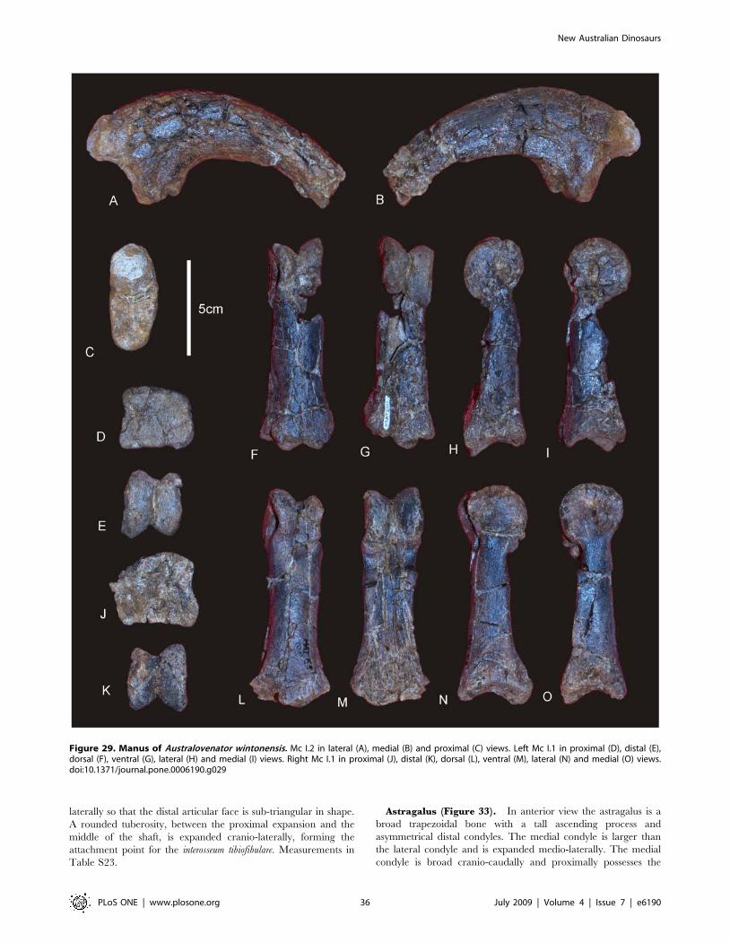
laterally so that the distal articular face is sub-triangular in shape.
A rounded tuberosity, between the proximal expansion and the
middle of the shaft, is expanded cranio-laterally, forming the
attachment point for the interosseum tibiofibulare. Measurements in
Table S23.
Astragalus (Figure 33). In anterior view the astragalus is a
broad trapezoidal bone with a tall ascending process and
asymmetrical distal condyles. The medial condyle is larger than
the lateral condyle and is expanded medio-laterally. The medial
condyle is broad cranio-caudally and proximally possesses the
Figure 29. Manus of Australovenator wintonensis. Mc I.2 in lateral (A), medial (B) and proximal (C) views. Left Mc I.1 in proximal (D), distal (E),dorsal (F), ventral (G), lateral (H) and medial (I) views. Right Mc I.1 in proximal (J), distal (K), dorsal (L), ventral (M), lateral (N) and medial (O) views.doi:10.1371/journal.pone.0006190.g029
New Australian Dinosaurs
PLoS ONE | www.plosone.org 36 July 2009 | Volume 4 | Issue 7 | e6190

medial expansion for the tibial facet, which is rounded to the same
degree as the distal margin of the condyle. The lateral condyle is
medio-laterally and cranio-caudally constricted so that it is only
half the condylar surface of the medial condyle. This constriction
reflects the lateral constriction preserved on the distal end of the
tibia and tibial facet. A cranio-proximal extension of the lateral
condyle projects from the cranial articular face as a rounded
triangular process. This process forms the cranial boarder of the
fibular facet of the astragalus. The fibular facet is shallow and
bordered caudally by the lateral margin of the ascending process.
In anterior view two shallow cranial grooves are present on the
condylar surface. The superior cranial groove runs along the
proximal margin of the condyles. The inferior cranial groove is
situated in the midline of the cranial surface of the condyles. This
groove is crescentic in shape and runs from the middle of the
lateral condyle to middle of the medial condyle. In distal view the
tibial facet is smooth along its length, being broadest medially and
tapering to the lateral side. On the caudal face of the ascending
process is a crescentic shallow groove which runs superior to the
base of the tibial facet. Measurements in Table S24.
PesMetatarsals (Figures 34 & 35). Three metatarsals are
preserved from the right pes, Mt I – III. The right Mt I
(Figure 34A–D) is the smallest of the metatarsals and is well
preserved. In flexor aspect it is tear-drop shaped. The proximal end
tapers to a proximal-most point. The shaft is cranio-caudally
compressed so that the medial and lateral margins form two
opposing crests. Half way along the flexor surface a raised muscle scar
extends to the distally expanded end. The distal condyles are
asymmetrical with the cranial condyle offset ventrally to the caudal
condyle which is bulbous and possesses a deep ligament pit. No deep
pit occurs on the cranial side. A sharp triangular process juts out along
the distal portion of the extensor crest. Measurements in Table S25.
Mt II (Figure 34E–J) is a long and gracile element preserving the
distal articular condyles, and a damaged proximal epiphyseal
expansion. It is missing the proximal articular surface. Proximally,
the bone is subtriangular in cross-section. The cranial surface is
expanded dorsally and rounded medially. The medial surface is
concave and expanded proximally. The shaft is crushed and
distorted so that the distal end is bent slightly medially. The distal
Figure 30. Femur of Australovenator wintonensis. Right femur in anterior (A), posterior (B), lateral (C), medial (D), proximal (E) and distal (F) views.Abbreviations: at, accessory trochanter; ctf, crista tibiofibularis; eg, extensor groove; fc, fibular condyle, fg, flexor groove; fh, femoral head (caput); ft,forth trochanter; gt, greater trochanter; lst; lessor trochanter; mdc, medial-distal (epicondyle) crest; pfc, posterior flange of caput; rg, ridge superior tolesser trochanter; tc, tibial condyle.doi:10.1371/journal.pone.0006190.g030
New Australian Dinosaurs
PLoS ONE | www.plosone.org 37 July 2009 | Volume 4 | Issue 7 | e6190

end is expanded to accommodate a large disto-lateral condyle and
a small postero-medially projecting condyle. The lateral condyle is
rounded in lateral view with a deep collateral ligament pit,
bordered by a distinct and thick rim. In distal view the condyle is
constricted to the lateral side and expands medially as it meets the
distal edge of the medial condyle. The medial and lateral condyles
connect at the distal face via a bridge, which forms part of the
distal articular surface. In posterior view, the lateral and medial
condyles are divided by a deep fossa. In medial view the medial
condyle is restricted to the lower edge of the articular surface. A
deep collateral ligament pit is bordered by a thick rim. A medial
muscle scar extends from the distal quarter of the caudal margin of
Figure 31. Tibiae of Australovenator wintonensis. Left tibia in antero-medial (A), postero-lateral (B), lateral (C), postero-medial (D), proximal (E)and distal (F) views. Right tibia in proximal (G), distal (H), medial (J), medial (K), posterior (L), anterior (M) views and close-up of proximal end inpostero-lateral view (I). Abbreviations: cn, cnemial crest; clp, cranio-lateral process; cast, cranial astragalar facet; dast, astragalar facet (distal); ff, fibularflange; it, incisura tibialis; lc, lateral condyle; lm, lateral malleolus; mc, medial condyle; mm, medial malleolus; pvr, postero-ventral ridge; vp, ventralprocess of lateral condyle.doi:10.1371/journal.pone.0006190.g031
New Australian Dinosaurs
PLoS ONE | www.plosone.org 38 July 2009 | Volume 4 | Issue 7 | e6190
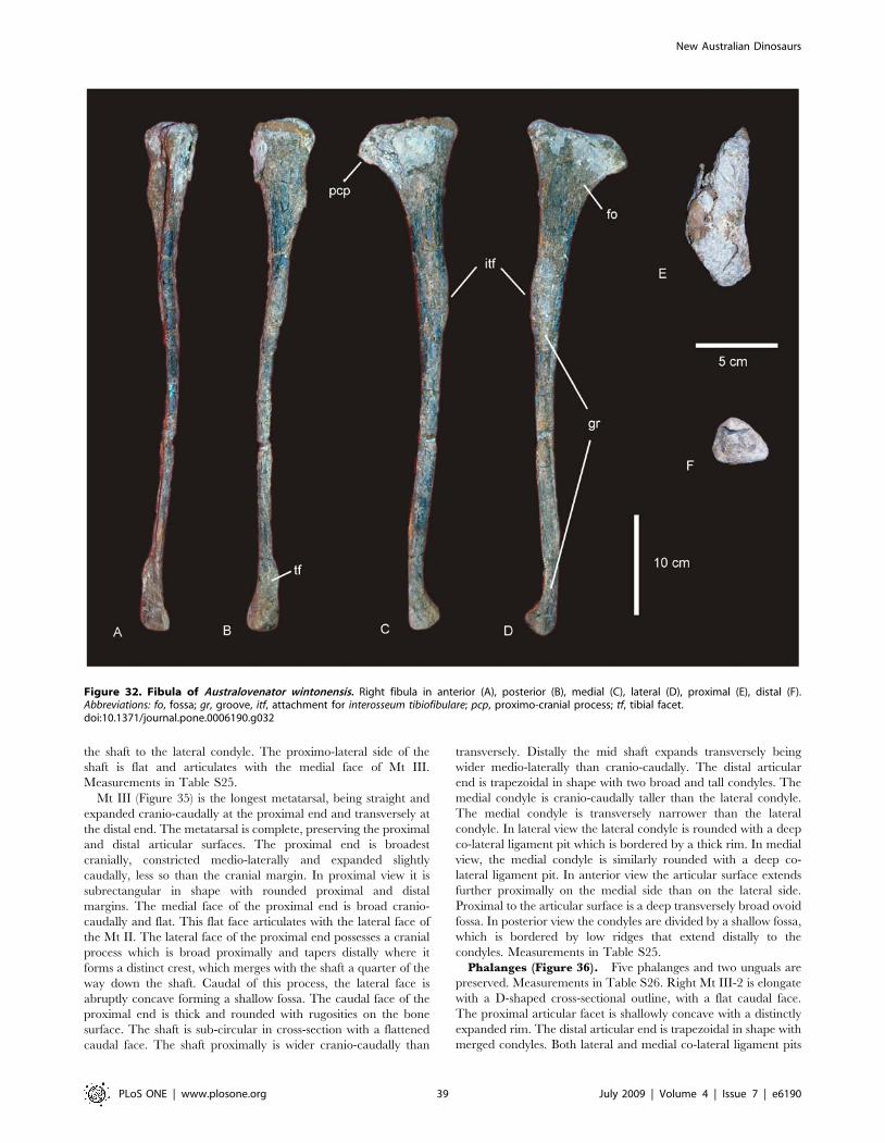
the shaft to the lateral condyle. The proximo-lateral side of the
shaft is flat and articulates with the medial face of Mt III.
Measurements in Table S25.
Mt III (Figure 35) is the longest metatarsal, being straight and
expanded cranio-caudally at the proximal end and transversely at
the distal end. The metatarsal is complete, preserving the proximal
and distal articular surfaces. The proximal end is broadest
cranially, constricted medio-laterally and expanded slightly
caudally, less so than the cranial margin. In proximal view it is
subrectangular in shape with rounded proximal and distal
margins. The medial face of the proximal end is broad cranio-
caudally and flat. This flat face articulates with the lateral face of
the Mt II. The lateral face of the proximal end possesses a cranial
process which is broad proximally and tapers distally where it
forms a distinct crest, which merges with the shaft a quarter of the
way down the shaft. Caudal of this process, the lateral face is
abruptly concave forming a shallow fossa. The caudal face of the
proximal end is thick and rounded with rugosities on the bone
surface. The shaft is sub-circular in cross-section with a flattened
caudal face. The shaft proximally is wider cranio-caudally than
transversely. Distally the mid shaft expands transversely being
wider medio-laterally than cranio-caudally. The distal articular
end is trapezoidal in shape with two broad and tall condyles. The
medial condyle is cranio-caudally taller than the lateral condyle.
The medial condyle is transversely narrower than the lateral
condyle. In lateral view the lateral condyle is rounded with a deep
co-lateral ligament pit which is bordered by a thick rim. In medial
view, the medial condyle is similarly rounded with a deep co-
lateral ligament pit. In anterior view the articular surface extends
further proximally on the medial side than on the lateral side.
Proximal to the articular surface is a deep transversely broad ovoid
fossa. In posterior view the condyles are divided by a shallow fossa,
which is bordered by low ridges that extend distally to the
condyles. Measurements in Table S25.
Phalanges (Figure 36). Five phalanges and two unguals are
preserved. Measurements in Table S26. Right Mt III-2 is elongate
with a D-shaped cross-sectional outline, with a flat caudal face.
The proximal articular facet is shallowly concave with a distinctly
expanded rim. The distal articular end is trapezoidal in shape with
merged condyles. Both lateral and medial co-lateral ligament pits
Figure 32. Fibula of Australovenator wintonensis. Right fibula in anterior (A), posterior (B), medial (C), lateral (D), proximal (E), distal (F).Abbreviations: fo, fossa; gr, groove, itf, attachment for interosseum tibiofibulare; pcp, proximo-cranial process; tf, tibial facet.doi:10.1371/journal.pone.0006190.g032
New Australian Dinosaurs
PLoS ONE | www.plosone.org 39 July 2009 | Volume 4 | Issue 7 | e6190
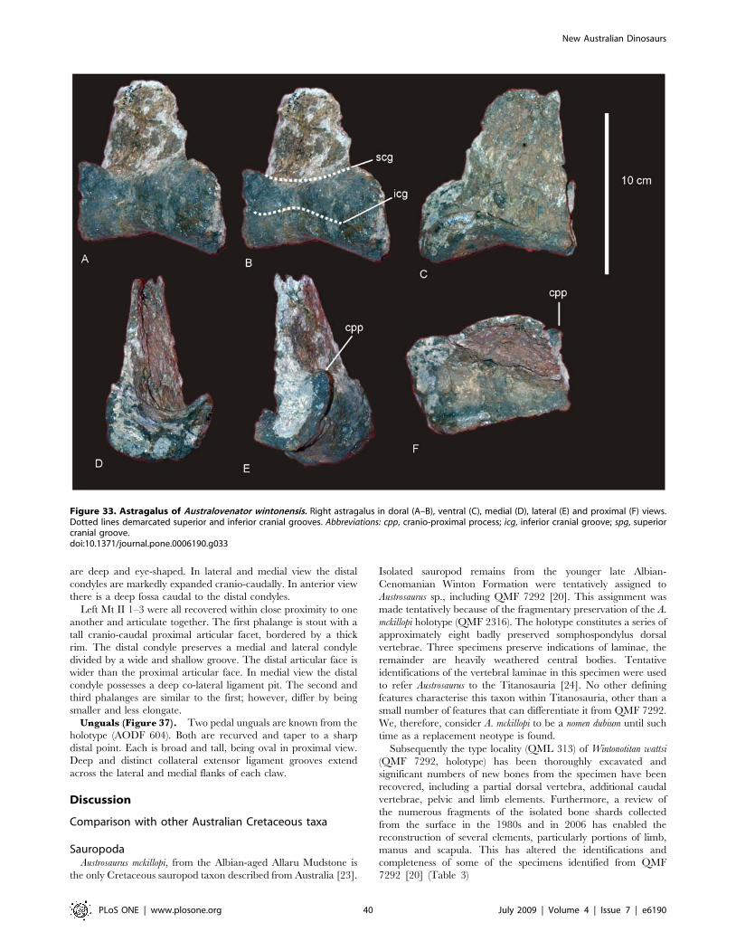
are deep and eye-shaped. In lateral and medial view the distal
condyles are markedly expanded cranio-caudally. In anterior view
there is a deep fossa caudal to the distal condyles.
Left Mt II 1–3 were all recovered within close proximity to one
another and articulate together. The first phalange is stout with a
tall cranio-caudal proximal articular facet, bordered by a thick
rim. The distal condyle preserves a medial and lateral condyle
divided by a wide and shallow groove. The distal articular face is
wider than the proximal articular face. In medial view the distal
condyle possesses a deep co-lateral ligament pit. The second and
third phalanges are similar to the first; however, differ by being
smaller and less elongate.
Unguals (Figure 37). Two pedal unguals are known from the
holotype (AODF 604). Both are recurved and taper to a sharp
distal point. Each is broad and tall, being oval in proximal view.
Deep and distinct collateral extensor ligament grooves extend
across the lateral and medial flanks of each claw.
Discussion
Comparison with other Australian Cretaceous taxa
SauropodaAustrosaurus mckillopi, from the Albian-aged Allaru Mudstone is
the only Cretaceous sauropod taxon described from Australia [23].
Isolated sauropod remains from the younger late Albian-
Cenomanian Winton Formation were tentatively assigned to
Austrosaurus sp., including QMF 7292 [20]. This assignment was
made tentatively because of the fragmentary preservation of the A.
mckillopi holotype (QMF 2316). The holotype constitutes a series of
approximately eight badly preserved somphospondylus dorsal
vertebrae. Three specimens preserve indications of laminae, the
remainder are heavily weathered central bodies. Tentative
identifications of the vertebral laminae in this specimen were used
to refer Austrosaurus to the Titanosauria [24]. No other defining
features characterise this taxon within Titanosauria, other than a
small number of features that can differentiate it from QMF 7292.
We, therefore, consider A. mckillopi to be a nomen dubium until such
time as a replacement neotype is found.
Subsequently the type locality (QML 313) of Wintonotitan wattsi
(QMF 7292, holotype) has been thoroughly excavated and
significant numbers of new bones from the specimen have been
recovered, including a partial dorsal vertebra, additional caudal
vertebrae, pelvic and limb elements. Furthermore, a review of
the numerous fragments of the isolated bone shards collected
from the surface in the 1980s and in 2006 has enabled the
reconstruction of several elements, particularly portions of limb,
manus and scapula. This has altered the identifications and
completeness of some of the specimens identified from QMF
7292 [20] (Table 3)
Figure 33. Astragalus of Australovenator wintonensis. Right astragalus in doral (A–B), ventral (C), medial (D), lateral (E) and proximal (F) views.Dotted lines demarcated superior and inferior cranial grooves. Abbreviations: cpp, cranio-proximal process; icg, inferior cranial groove; spg, superiorcranial groove.doi:10.1371/journal.pone.0006190.g033
New Australian Dinosaurs
PLoS ONE | www.plosone.org 40 July 2009 | Volume 4 | Issue 7 | e6190

Figure 34. Metatarsals of Australovenator wintonensis. Right Mt I in dorsal (A), ventral (B), flexor (C) and distal (D) views. Right Mt III in anterior(E), posterior (F), lateral (G), medial (H), proximal (I) and distal (J) views. Abbreviations: etp, extensor triangular process.doi:10.1371/journal.pone.0006190.g034
New Australian Dinosaurs
PLoS ONE | www.plosone.org 41 July 2009 | Volume 4 | Issue 7 | e6190
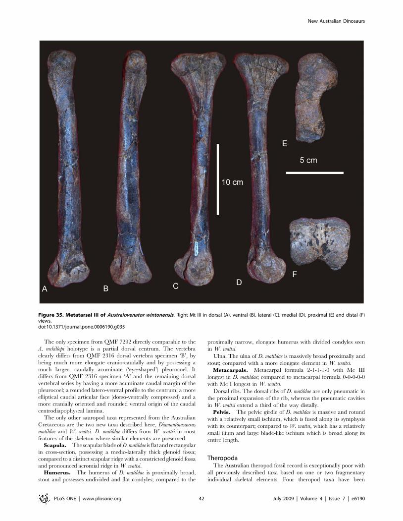
The only specimen from QMF 7292 directly comparable to the
A. mckillopi holotype is a partial dorsal centrum. The vertebra
clearly differs from QMF 2316 dorsal vertebra specimen ‘B’, by
being much more elongate cranio-caudally and by possessing a
much larger, caudally acuminate (‘eye-shaped’) pleurocoel. It
differs from QMF 2316 specimen ‘A’ and the remaining dorsal
vertebral series by having a more acuminate caudal margin of the
pleurocoel; a rounded latero-ventral profile to the centrum; a more
elliptical caudal articular face (dorso-ventrally compressed) and a
more cranially oriented and rounded ventral origin of the caudal
centrodiapophyseal lamina.
The only other sauropod taxa represented from the Australian
Cretaceous are the two new taxa described here, Diamantinasaurus
matildae and W. wattsi. D. matildae differs from W. wattsi in most
features of the skeleton where similar elements are preserved.Scapula. The scapular blade of D. matildae is flat and rectangular
in cross-section, possessing a medio-laterally thick glenoid fossa;
compared to a distinct scapular ridge with a constricted glenoid fossa
and pronounced acromial ridge in W. wattsi.Humerus. The humerus of D. matildae is proximally broad,
stout and possesses undivided and flat condyles; compared to the
proximally narrow, elongate humerus with divided condyles seen
in W. wattsi.
Ulna. The ulna of D. matildae is massively broad proximally and
stout; compared with a more elongate element in W. wattsi.
Metacarpals. Metacarpal formula 2-1-1-1-0 with Mc III
longest in D. matildae; compared to metacarpal formula 0-0-0-0-0
with Mc I longest in W. wattsi.
Dorsal ribs. The dorsal ribs of D. matildae are only pneumatic in
the proximal expansion of the rib, whereas the pneumatic cavities
in W. wattsi extend a third of the way distally.
Pelvis. The pelvic girdle of D. matildae is massive and rotund
with a relatively small ischium, which is fused along its symphysis
with its counterpart; compared to W. wattsi, which has a relatively
small ilium and large blade-like ischium which is broad along its
entire length.
TheropodaThe Australian theropod fossil record is exceptionally poor with
all previously described taxa based on one or two fragmentary
individual skeletal elements. Four theropod taxa have been
Figure 35. Metatarsal III of Australovenator wintonensis. Right Mt III in dorsal (A), ventral (B), lateral (C), medial (D), proximal (E) and distal (F)views.doi:10.1371/journal.pone.0006190.g035
New Australian Dinosaurs
PLoS ONE | www.plosone.org 42 July 2009 | Volume 4 | Issue 7 | e6190
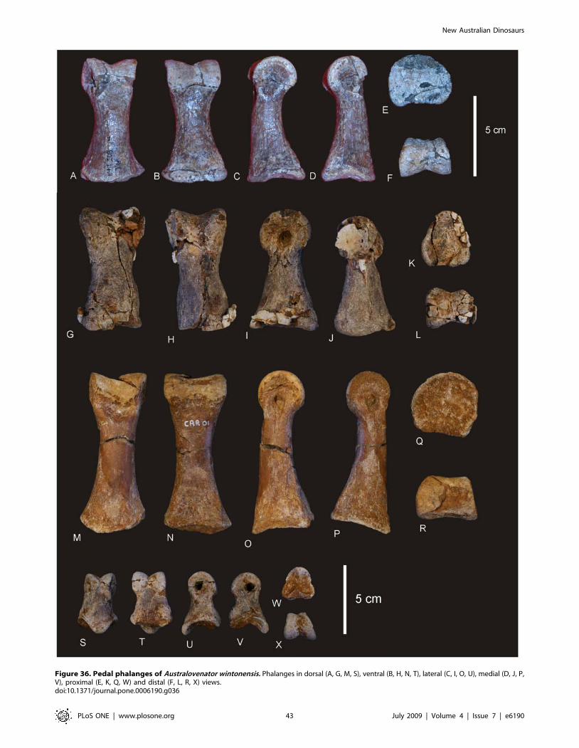
Figure 36. Pedal phalanges of Australovenator wintonensis. Phalanges in dorsal (A, G, M, S), ventral (B, H, N, T), lateral (C, I, O, U), medial (D, J, P,V), proximal (E, K, Q, W) and distal (F, L, R, X) views.doi:10.1371/journal.pone.0006190.g036
New Australian Dinosaurs
PLoS ONE | www.plosone.org 43 July 2009 | Volume 4 | Issue 7 | e6190

formally described from Australia with two additional taxa
referred to taxa from elsewhere; Walgettosuchus woodwardi Huene;
Rapator ornitholestoides Huene; Kakuru kujani Molnar & Pledge;
Timimus hermani Rich & Vickers Rich; Allosaurus sp. Molnar et al.;
and cf. Megaraptor Smith et al.. Both W. woodwardi [25] and T.
hermani [26] are considered to be nomen dubia.
The Mc I of Australovenator wintonensis is larger than to that of the
Rapator ornitholestoides holotype; is missing the prominent caudo-
medial process projecting proximo-dorsally; possesses more
subequal distal condyles; a flat proximal articular surface
compared to the concave surface in R. ornitholestoides; and a
straight distal lateral condyle compared to a disto-laterally
projecting distal lateral condyle in R. ornitholestoides. Rapator
ornitholestoides has been considered to represent an alvarezsaurid
coelurosaur [25], however it’s identification remains equivocal.
An ulna from the Early Cretaceous of southern Australia has
been compared to Megaraptor [27]. The ulnae of A. wintonensis differ
from NMVP186076 (cf. Megaraptor) in several features: 1. It lacks a
proximo-caudally expanded blade-like olecranon process that
extends distally as a caudal olecranon crest. 2. It lacks a
pronounced lateral tuberosity that is continuous distally with a
distinct lateral crest. 3. The caudal margin of the ulna is relatively
Figure 37. Pedal unguals of Australovenator wintonensis. Unguals in dorsal (A, F), ventral (B, G), lateral (C, H), medial (D, I) and proximal (E, J)views.doi:10.1371/journal.pone.0006190.g037
New Australian Dinosaurs
PLoS ONE | www.plosone.org 44 July 2009 | Volume 4 | Issue 7 | e6190
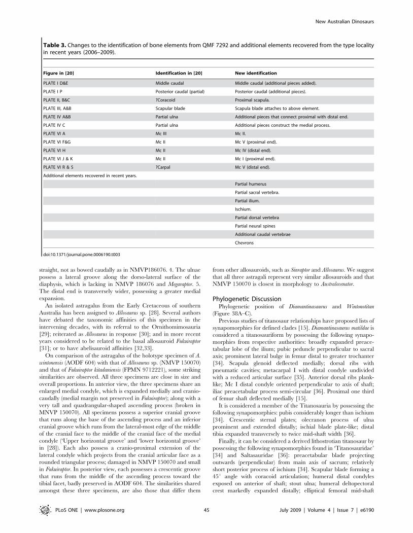
straight, not as bowed caudally as in NMVP186076. 4. The ulnae
possess a lateral groove along the dorso-lateral surface of the
diaphysis, which is lacking in NMVP 186076 and Megaraptor. 5.
The distal end is transversely wider, possessing a greater medial
expansion.
An isolated astragalus from the Early Cretaceous of southern
Australia has been assigned to Allosaurus sp. [28]. Several authors
have debated the taxonomic affinities of this specimen in the
intervening decades, with its referral to the Ornithomimosauria
[29]; reiterated as Allosaurus in response [30]; and in more recent
years considered to be related to the basal allosauroid Fukuiraptor
[31]; or to have abelisauroid affinities [32,33].
On comparison of the astragalus of the holotype specimen of A.
wintonensis (AODF 604) with that of Allosaurus sp. (NMVP 150070)
and that of Fukuiraptor kitadaniensis (FPMN 9712221), some striking
similarities are observed. All three specimens are close in size and
overall proportions. In anterior view, the three specimens share an
enlarged medial condyle, which is expanded medially and cranio-
caudally (medial margin not preserved in Fukuiraptor); along with a
very tall and quadrangular-shaped ascending process (broken in
MNVP 150070). All specimens possess a superior cranial groove
that runs along the base of the ascending process and an inferior
cranial groove which runs from the lateral-most edge of the middle
of the cranial face to the middle of the cranial face of the medial
condyle (‘Upper horizontal groove’ and ‘lower horizontal groove’
in [28]). Each also possess a cranio-proximal extension of the
lateral condyle which projects from the cranial articular face as a
rounded triangular process; damaged in NMVP 150070 and small
in Fukuiraptor. In posterior view, each possesses a crescentic groove
that runs from the middle of the ascending process toward the
tibial facet, badly preserved in AODF 604. The similarities shared
amongst these three specimens, are also those that differ them
from other allosauroids, such as Sinraptor and Allosaurus. We suggest
that all three astragali represent very similar allosauroids and that
NMVP 150070 is closest in morphology to Australovenator.
Phylogenetic DiscussionPhylogenetic position of Diamantinasaurus and Wintonotitan
(Figure 38A–C).
Previous studies of titanosaur relationships have proposed lists of
synapomorphies for defined clades [15]. Diamantinasaurus matildae is
considered a titanosauriform by possessing the following synapo-
morphies from respective authorities: broadly expanded preace-
tabular lobe of the ilium; pubic peduncle perpendicular to sacral
axis; prominent lateral bulge in femur distal to greater trochanter
[34]. Scapula glenoid deflected medially; dorsal ribs with
pneumatic cavities; metacarpal I with distal condyle undivided
with a reduced articular surface [35]. Anterior dorsal ribs plank-
like; Mc I distal condyle oriented perpendicular to axis of shaft;
iliac preacetabular process semi-circular [36]. Proximal one third
of femur shaft deflected medially [15].
It is considered a member of the Titanosauria by possessing the
following synapomorphies: pubis considerably longer than ischium
[34]. Crescentic sternal plates; olecranon process of ulna
prominent and extended distally; ischial blade plate-like; distal
tibia expanded transversely to twice mid-shaft width [36].
Finally, it can be considered a derived lithostrotian titanosaur by
possessing the following synapomorphies found in ‘Titanosauridae’
[34] and Saltasauridae [36]: preacetabular blade projecting
outwards (perpendicular) from main axis of sacrum; relatively
short posterior process of ischium [34]. Scapular blade forming a
45u angle with coracoid articulation; humeral distal condyles
exposed on anterior of shaft; stout ulna; humeral deltopectoral
crest markedly expanded distally; elliptical femoral mid-shaft
Table 3. Changes to the identification of bone elements from QMF 7292 and additional elements recovered from the type localityin recent years (2006–2009).
Figure in [20] Identification in [20] New identification
PLATE I D&E Middle caudal Middle caudal (additional pieces added).
PLATE I P Posterior caudal (partial) Posterior caudal (additional pieces).
PLATE II, B&C ?Coracoid Proximal scapula.
PLATE III, A&B Scapular blade Scapula blade attaches to above element.
PLATE IV A&B Partial ulna Additional pieces that connect proximal with distal end.
PLATE IV C Partial ulna Additional pieces construct the medial process.
PLATE VI A Mc III Mc II.
PLATE VI F&G Mc II Mc V (proximal end).
PLATE VI H Mc II Mc IV (distal end).
PLATE VI J & K Mc II Mc I (proximal end).
PLATE VI R & S ?Carpal Mc V (distal end).
Additional elements recovered in recent years.
Partial humerus
Partial sacral vertebra.
Partial ilium.
Ischium.
Partial dorsal vertebra
Partial neural spines
Additional caudal vertebrae
Chevrons
doi:10.1371/journal.pone.0006190.t003
New Australian Dinosaurs
PLoS ONE | www.plosone.org 45 July 2009 | Volume 4 | Issue 7 | e6190
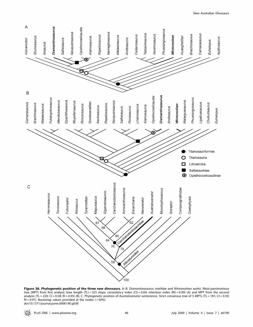
Figure 38. Phylogenetic position of the three new dinosaurs. A–B. Diamantinasaurus matildae and Wintonotitan wattsi. Most-parsimonioustree (MPT) from first analysis (tree length (TL) = 325 steps; consistency index (CI) = 0.64; retention index (RI) = 0.99) (A) and MPT from the secondanalysis (TL = 220; CI = 0.58; RI = 0.93) (B). C. Phylogenetic position of Australovenator wintonensis. Strict consensus tree of 5 MPTs (TL = 181; CI = 0.59;RI = 0.91). Bootstrap values provided at the nodes (.50%).doi:10.1371/journal.pone.0006190.g038
New Australian Dinosaurs
PLoS ONE | www.plosone.org 46 July 2009 | Volume 4 | Issue 7 | e6190
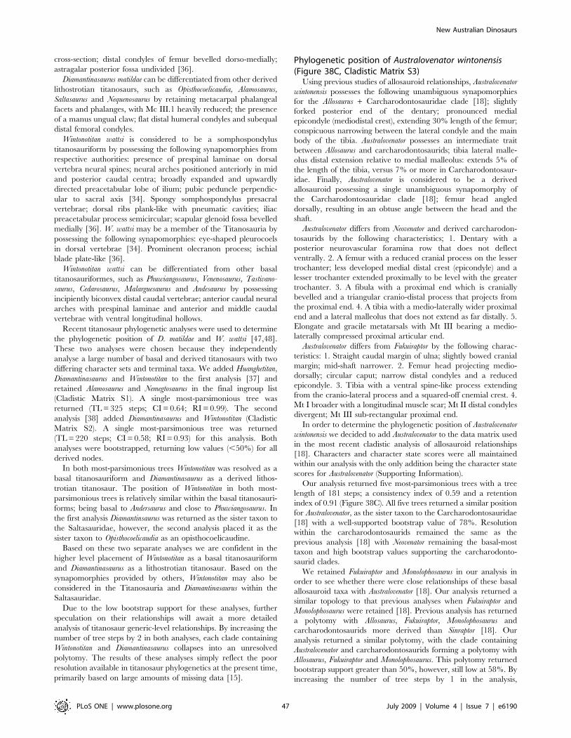
cross-section; distal condyles of femur bevelled dorso-medially;
astragalar posterior fossa undivided [36].
Diamantinasaurus matildae can be differentiated from other derived
lithostrotian titanosaurs, such as Opisthocoelicaudia, Alamosaurus,
Saltasaurus and Nequenosaurus by retaining metacarpal phalangeal
facets and phalanges, with Mc III.1 heavily reduced; the presence
of a manus ungual claw; flat distal humeral condyles and subequal
distal femoral condyles.
Wintonotitan wattsi is considered to be a somphospondylus
titanosauriform by possessing the following synapomorphies from
respective authorities: presence of prespinal laminae on dorsal
vertebra neural spines; neural arches positioned anteriorly in mid
and posterior caudal centra; broadly expanded and upwardly
directed preacetabular lobe of ilium; pubic peduncle perpendic-
ular to sacral axis [34]. Spongy somphospondylus presacral
vertebrae; dorsal ribs plank-like with pneumatic cavities; iliac
preacetabular process semicircular; scapular glenoid fossa bevelled
medially [36]. W. wattsi may be a member of the Titanosauria by
possessing the following synapomorphies: eye-shaped pleurocoels
in dorsal vertebrae [34]. Prominent olecranon process; ischial
blade plate-like [36].
Wintonotitan wattsi can be differentiated from other basal
titanosauriformes, such as Phuwiangosaurus, Venenosaurus, Tastivano-
saurus, Cedarosaurus, Malarguesaurus and Andesaurus by possessing
incipiently biconvex distal caudal vertebrae; anterior caudal neural
arches with prespinal laminae and anterior and middle caudal
vertebrae with ventral longitudinal hollows.
Recent titanosaur phylogenetic analyses were used to determine
the phylogenetic position of D. matildae and W. wattsi [47,48].
These two analyses were chosen because they independently
analyse a large number of basal and derived titanosaurs with two
differing character sets and terminal taxa. We added Huanghetitan,
Diamantinasaurus and Wintonotitan to the first analysis [37] and
retained Alamosaurus and Nemegtosaurus in the final ingroup list
(Cladistic Matrix S1). A single most-parsimonious tree was
returned (TL = 325 steps; CI = 0.64; RI = 0.99). The second
analysis [38] added Diamantinasaurus and Wintonotitan (Cladistic
Matrix S2). A single most-parsimonious tree was returned
(TL = 220 steps; CI = 0.58; RI = 0.93) for this analysis. Both
analyses were bootstrapped, returning low values (,50%) for all
derived nodes.
In both most-parsimonious trees Wintonotitan was resolved as a
basal titanosauriform and Diamantinasaurus as a derived lithos-
trotian titanosaur. The position of Wintonotitan in both most-
parsimonious trees is relatively similar within the basal titanosauri-
forms; being basal to Andersaurus and close to Phuwiangosaurus. In
the first analysis Diamantinsaurus was returned as the sister taxon to
the Saltasauridae, however, the second analysis placed it as the
sister taxon to Opisthocoelicaudia as an opisthocoelicaudine.
Based on these two separate analyses we are confident in the
higher level placement of Wintonotitan as a basal titanosauriform
and Diamantinasaurus as a lithostrotian titanosaur. Based on the
synapomorphies provided by others, Wintonotitan may also be
considered in the Titanosauria and Diamantinasaurus within the
Saltasauridae.
Due to the low bootstrap support for these analyses, further
speculation on their relationships will await a more detailed
analysis of titanosaur generic-level relationships. By increasing the
number of tree steps by 2 in both analyses, each clade containing
Wintonotitan and Diamantinasaurus collapses into an unresolved
polytomy. The results of these analyses simply reflect the poor
resolution available in titanosaur phylogenetics at the present time,
primarily based on large amounts of missing data [15].
Phylogenetic position of Australovenator wintonensis(Figure 38C, Cladistic Matrix S3)
Using previous studies of allosauroid relationships, Australovenator
wintonensis possesses the following unambiguous synapomorphies
for the Allosaurus + Carcharodontosauridae clade [18]; slightly
forked posterior end of the dentary; pronounced medial
epicondyle (mediodistal crest), extending 30% length of the femur;
conspicuous narrowing between the lateral condyle and the main
body of the tibia. Australovenator possesses an intermediate trait
between Allosaurus and carcharodontosaurids; tibia lateral malle-
olus distal extension relative to medial malleolus: extends 5% of
the length of the tibia, versus 7% or more in Carcharodontosaur-
idae. Finally, Australovenator is considered to be a derived
allosauroid possessing a single unambiguous synapomorphy of
the Carcharodontosauridae clade [18]; femur head angled
dorsally, resulting in an obtuse angle between the head and the
shaft.
Australovenator differs from Neovenator and derived carcharodon-
tosaurids by the following characteristics; 1. Dentary with a
posterior neurovascular foramina row that does not deflect
ventrally. 2. A femur with a reduced cranial process on the lesser
trochanter; less developed medial distal crest (epicondyle) and a
lesser trochanter extended proximally to be level with the greater
trochanter. 3. A fibula with a proximal end which is cranially
bevelled and a triangular cranio-distal process that projects from
the proximal end. 4. A tibia with a medio-laterally wider proximal
end and a lateral malleolus that does not extend as far distally. 5.
Elongate and gracile metatarsals with Mt III bearing a medio-
laterally compressed proximal articular end.
Australovenator differs from Fukuiraptor by the following charac-
teristics: 1. Straight caudal margin of ulna; slightly bowed cranial
margin; mid-shaft narrower. 2. Femur head projecting medio-
dorsally; circular caput; narrow distal condyles and a reduced
epicondyle. 3. Tibia with a ventral spine-like process extending
from the cranio-lateral process and a squared-off cnemial crest. 4.
Mt I broader with a longitudinal muscle scar; Mt II distal condyles
divergent; Mt III sub-rectangular proximal end.
In order to determine the phylogenetic position of Australovenator
wintonensis we decided to add Australovenator to the data matrix used
in the most recent cladistic analysis of allosauroid relationships
[18]. Characters and character state scores were all maintained
within our analysis with the only addition being the character state
scores for Australovenator (Supporting Information).
Our analysis returned five most-parsimonious trees with a tree
length of 181 steps; a consistency index of 0.59 and a retention
index of 0.91 (Figure 38C). All five trees returned a similar position
for Australovenator, as the sister taxon to the Carcharodontosauridae
[18] with a well-supported bootstrap value of 78%. Resolution
within the carcharodontosaurids remained the same as the
previous analysis [18] with Neovenator remaining the basal-most
taxon and high bootstrap values supporting the carcharodonto-
saurid clades.
We retained Fukuiraptor and Monolophosaurus in our analysis in
order to see whether there were close relationships of these basal
allosauroid taxa with Australovenator [18]. Our analysis returned a
similar topology to that previous analyses when Fukuiraptor and
Monolophosaurus were retained [18]. Previous analysis has returned
a polytomy with Allosaurus, Fukuiraptor, Monolophosaurus and
carcharodontosaurids more derived than Sinraptor [18]. Our
analysis returned a similar polytomy, with the clade containing
Australovenator and carcharodontosaurids forming a polytomy with
Allosaurus, Fukuiraptor and Monolophosaurus. This polytomy returned
bootstrap support greater than 50%, however, still low at 58%. By
increasing the number of tree steps by 1 in the analysis,
New Australian Dinosaurs
PLoS ONE | www.plosone.org 47 July 2009 | Volume 4 | Issue 7 | e6190

Figure 39. Palaeobiogeographic map and strato-phylogeny of Australovenator wintonensis. A. Palaeogeographic globe for the Early andmiddle Cretaceous [51]. White-lettered taxa represent allosauroids basal to the Carcharodontosauridae. Yellow-lettered taxa represent members ofthe Carcharodontosauridae. B. Stratigraphically calibrated phylogeny based on the phylogeny of the current analysis and adapted from [18].doi:10.1371/journal.pone.0006190.g039
New Australian Dinosaurs
PLoS ONE | www.plosone.org 48 July 2009 | Volume 4 | Issue 7 | e6190
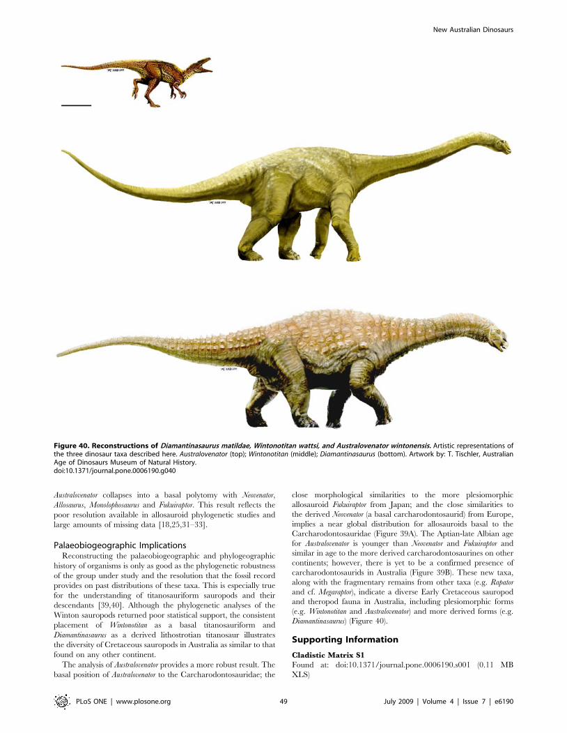
Australovenator collapses into a basal polytomy with Neovenator,
Allosaurus, Monolophosaurus and Fukuiraptor. This result reflects the
poor resolution available in allosauroid phylogenetic studies and
large amounts of missing data [18,25,31–33].
Palaeobiogeographic ImplicationsReconstructing the palaeobiogeographic and phylogeographic
history of organisms is only as good as the phylogenetic robustness
of the group under study and the resolution that the fossil record
provides on past distributions of these taxa. This is especially true
for the understanding of titanosauriform sauropods and their
descendants [39,40]. Although the phylogenetic analyses of the
Winton sauropods returned poor statistical support, the consistent
placement of Wintonotitan as a basal titanosauriform and
Diamantinasaurus as a derived lithostrotian titanosaur illustrates
the diversity of Cretaceous sauropods in Australia as similar to that
found on any other continent.
The analysis of Australovenator provides a more robust result. The
basal position of Australovenator to the Carcharodontosauridae; the
close morphological similarities to the more plesiomorphic
allosauroid Fukuiraptor from Japan; and the close similarities to
the derived Neovenator (a basal carcharodontosaurid) from Europe,
implies a near global distribution for allosauroids basal to the
Carcharodontosauridae (Figure 39A). The Aptian-late Albian age
for Australovenator is younger than Neovenator and Fukuiraptor and
similar in age to the more derived carcharodontosaurines on other
continents; however, there is yet to be a confirmed presence of
carcharodontosaurids in Australia (Figure 39B). These new taxa,
along with the fragmentary remains from other taxa (e.g. Rapator
and cf. Megaraptor), indicate a diverse Early Cretaceous sauropod
and theropod fauna in Australia, including plesiomorphic forms
(e.g. Wintonotitan and Australovenator) and more derived forms (e.g.
Diamantinasaurus) (Figure 40).
Supporting Information
Cladistic Matrix S1
Found at: doi:10.1371/journal.pone.0006190.s001 (0.11 MB
XLS)
Figure 40. Reconstructions of Diamantinasaurus matildae, Wintonotitan wattsi, and Australovenator wintonensis. Artistic representations ofthe three dinosaur taxa described here. Australovenator (top); Wintonotitan (middle); Diamantinasaurus (bottom). Artwork by: T. Tischler, AustralianAge of Dinosaurs Museum of Natural History.doi:10.1371/journal.pone.0006190.g040
New Australian Dinosaurs
PLoS ONE | www.plosone.org 49 July 2009 | Volume 4 | Issue 7 | e6190

Cladistic Matrix S2
Found at: doi:10.1371/journal.pone.0006190.s002 (0.05 MB
XLS)
Cladistic Matrix S3
Found at: doi:10.1371/journal.pone.0006190.s003 (0.03 MB
XLS)
Table S1 Diamantinasaurus matildae - Dorsal rib measurements
(mm)
Found at: doi:10.1371/journal.pone.0006190.s004 (0.03 MB
DOC)
Table S2 Diamantinasaurus matildae - Scapula measurements (mm)
Found at: doi:10.1371/journal.pone.0006190.s005 (0.02 MB
DOC)
Table S3 Diamantinasaurus matildae - Sternal plate measurements
(mm)
Found at: doi:10.1371/journal.pone.0006190.s006 (0.03 MB
DOC)
Table S4 Diamantinasaurus matildae - Humerus measurements
(mm)
Found at: doi:10.1371/journal.pone.0006190.s007 (0.03 MB
DOC)
Table S5 Diamantinasaurus matildae - Ulna measurements (mm)
Found at: doi:10.1371/journal.pone.0006190.s008 (0.04 MB
DOC)
Table S6 Diamantinasaurus matildae - Manus measurements (mm)
Found at: doi:10.1371/journal.pone.0006190.s009 (0.03 MB
DOC)
Table S7 Diamantinasaurus matildae - Pelvic girdle measurements
(mm)
Found at: doi:10.1371/journal.pone.0006190.s010 (0.03 MB
DOC)
Table S8 Diamantinasaurus matildae - Femur measurements (mm)
Found at: doi:10.1371/journal.pone.0006190.s011 (0.03 MB
DOC)
Table S9 Diamantinasaurus matildae - Tibia measurements (mm)
Found at: doi:10.1371/journal.pone.0006190.s012 (0.03 MB
DOC)
Table S10 Diamantinasaurus matildae - Fibula measurements (mm)
Found at: doi:10.1371/journal.pone.0006190.s013 (0.03 MB
DOC)
Table S11 Wintonotitan wattsi - Vertebral measurements (mm)
Found at: doi:10.1371/journal.pone.0006190.s014 (0.05 MB
DOC)
Table S12 Wintonotitan wattsi - Chevron measurements (mm)
Found at: doi:10.1371/journal.pone.0006190.s015 (0.03 MB
DOC)
Table S13 Wintonotitan wattsi - Forearm measurements (mm)
Found at: doi:10.1371/journal.pone.0006190.s016 (0.03 MB
DOC)
Table S14 Wintonotitan wattsi - Metacarpal measurements (mm)
Found at: doi:10.1371/journal.pone.0006190.s017 (0.03 MB
DOC)
Table S15 Wintonotitan wattsi - Ilium and Ischium measurements
(mm)
Found at: doi:10.1371/journal.pone.0006190.s018 (0.03 MB
DOC)
Table S16 Australovenator wintonensis - Tooth measurements (mm)
Found at: doi:10.1371/journal.pone.0006190.s019 (0.31 MB
DOC)
Table S17 Australovenator wintonensis - Dentary measurements
(mm)
Found at: doi:10.1371/journal.pone.0006190.s020 (0.03 MB
DOC)
Table S18 Australovenator wintonensis - Dorsal rib measurements
(mm)
Found at: doi:10.1371/journal.pone.0006190.s021 (0.03 MB
DOC)
Table S19 Australovenator wintonensis - Ilium measurements (mm)
Found at: doi:10.1371/journal.pone.0006190.s022 (0.03 MB
DOC)
Table S20 Australovenator wintonensis - Forearm measurements
(mm)
Found at: doi:10.1371/journal.pone.0006190.s023 (0.04 MB
DOC)
Table S21 Australovenator wintonensis - Femur measurements
Found at: doi:10.1371/journal.pone.0006190.s024 (0.03 MB
DOC)
Table S22 Australovenator wintonensis - Tibia measurements
Found at: doi:10.1371/journal.pone.0006190.s025 (0.03 MB
DOC)
Table S23 Australovenator wintonensis - Fibula measurements (mm)
Found at: doi:10.1371/journal.pone.0006190.s026 (0.03 MB
DOC)
Table S24 Australovenator wintonensis - Astragalus measurements
(mm)
Found at: doi:10.1371/journal.pone.0006190.s027 (0.03 MB
DOC)
Table S25 Australovenator wintonensis - Metatarsal measurements
(mm)
Found at: doi:10.1371/journal.pone.0006190.s028 (0.03 MB
DOC)
Table S26 Australovenator wintonensis - Pes phalange and ungual
measurements (mm)
Found at: doi:10.1371/journal.pone.0006190.s029 (0.03 MB
DOC)
Acknowledgments
We thank the following people for their help, encouragement and support; L.
Beirne, K. & H. Bishop, T. & C. Bonnice, N. Bourguignon, D. Brow, H. & A
Calvey, B. Cannon, L. Cannon, B. Collins, C. Cook, J. Corrigan, J. Cronin,
V. & M. Dharmarajah, H. Elliott, I. Elliott, J. Elliott, K. Elliott, R. Elliott, I.
Galloway, D. & M. Gandini, M. Gibbs, P. Giles, S. Grimmer, W. Hanley, J.
& E. Hannah, D. Hatch, J. & M. Hocknull, S. Hocknull, J. Hooper, P. & S.
Jamieson, T. Keating, J. & R. Lake, A. Leacy, R. & N. Leven, D. Lewis, J. &
M. Macmillan, P. & W. Marks, I & S Muir, D. O’Boyle, M. Pflaum, K.
Piper, R. Proefke, J. Rasmussen, J. Richardson, G. & N. Sankowsky, L.
Sendon, M. Sloan, K. Spring, D. & P. Strang, J. Tapiolas, P. Tierney, W.
Wavish, C. Wawryk, J. Westman, Lill P. Wheeler, J. Wilkinson, T. Williams,
A. Wilson, G. Willson. We thank the following organisations for the help and
support; Queensland Museum, Queensland Museum Foundation, Austra-
lian Age of Dinosaurs Museum of Natural History, Winton Shire Council,
Port of Brisbane Corporation, Qld. Gov. Gambling Community Benefit
Fund and the Australian Geographic Society.
New Australian Dinosaurs
PLoS ONE | www.plosone.org 50 July 2009 | Volume 4 | Issue 7 | e6190
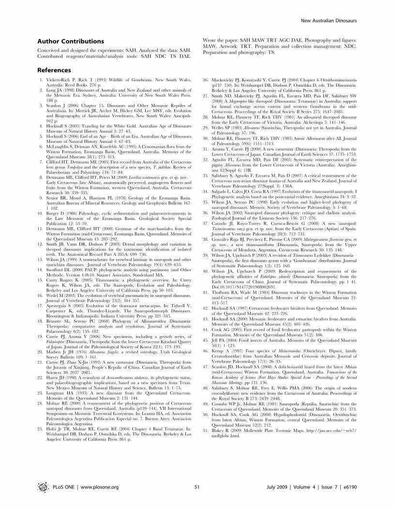
Author Contributions
Conceived and designed the experiments: SAH. Analyzed the data: SAH.
Contributed reagents/materials/analysis tools: SAH NDC TS DAE.
Wrote the paper: SAH MAW TRT AGC DAE. Photography and figures:
MAW. Artwork: TRT. Preparation and collection management: NDC.
Preparation and photography: TS.
References
1. Vickers-Rich P, Rich T (1993) Wildlife of Gondwana. New South Wales,
Australia: Reed Books. 276 p.
2. Long JA (1998) Dinosaurs of Australia and New Zealand and other animals ofthe Mesozoic Era. Sydney, Australia: University of New South Wales Press.
188 p.3. Scanlon J (2006) Chapter 15. Dinosaurs and Other Mesozoic Reptiles of
Australasia. In: Merrick JR, Archer M, Hickey GM, Lee MSY, eds. Evolutionand Biogeography of Australasian Vertebrates. New South Wales: Auscipub.
942 p.
4. Hocknull S (2005) Trawling for the White Gold. Australian Age of DinosaursMuseum of Natural History Annual 3: 27–43.
5. Hocknull S (2006) End of an Age – Birth of an Era. Australian Age of DinosaursMuseum of Natural History Annual 4: 67–83.
6. McLoughlin S, Drinnan AN, Rozefelds AC (1995) A Cenomanian flora from the
Winton Formation, Eromanga Basin, Queensland, Australia. Memoirs of theQueensland Museum 38(1): 273–313.
7. Clifford HT, Dettmann ME (2005) First record from Australia of the Cretaceousfern genus Tempskya and the description of a new species, T. judithae. Review of
Palaeobotany and Palynology 134: 71–84.8. Dettmann ME, Clifford HT, Peters M (2009) Lovellea wintonensis gen. et sp. nov. –
Early Cretaceous (late Albian), anatomically preserved, angiosperm flowers and
fruits from the Winton Formation, western Queensland, Australia. CretaceousResearch 30: 339–335.
9. Senior BR, Mond A, Harrison PL (1978) Geology of the Eromanga Basin.Australian Bureau of Mineral Resources, Geology and Geophysics Bulletin 167:
1–102.
10. Burger D (1986) Palynology, cyclic sedimentation and palaeoenvironments inthe Late Mesozoic of the Eromanga Basin. Geological Society Special
Publication 12: 53–70.11. Dettmann ME, Clifford HT (2000) Gemmae of the marchantiales from the
Winton Formation (mid-Cretaceous), Eromanga Basin, Queensland. Memoirs ofthe Queensland Museum 45: 285–292.
12. Smith JB, Vann DR, Dodson P (2005) Dental morphology and variation in
theopod dinosaurs: implications for the taxonomic identification of isolatedteeth. The Anatomical Record Part A 285A: 699–736.
13. Wilson JA (1999) A nomenclature for vertebral laminae in sauropods and othersaurichian dinosaurs. Journal of Vertebrate Paleontology 19(4): 639–653.
14. Swofford DL (2000) PAUP: phylogenetic analysis using parsimony (and Other
Methods). Version 4.0b10. Sinauer Associates, Sunderland MA.15. Curry Rogers K (2005) Titanosauria: a phylogenetic overview. In: Curry
Rogers K, Wilson JA, eds. The Sauropods, Evolution and Paleobiology.Berkeley and Los Angeles: University of California Press. pp 50–103.
16. Wedel M (2003) The evolution of vertebral pneumaticity in sauropod dinosaurs.Journal of Vertebrate Paleontology 23(2): 344–357.
17. Apesteguia S (2005) Evolution of the titanosaur metacarpus. In: Tidwell V,
Carpenter K, eds. Thunder-Lizards, The Sauropodomorph Dinosaurs.Bloomington & Indianapolis: Indiana University Press. pp 321–345.
18. Brusatte SL, Sereno PC (2008) Phylogeny of Allosauroidea (Dinosauria:Theropoda): comparative analysis and resolution. Journal of Systematic
Palaeontology 6(2): 155–182.
19. Currie PJ, Azuma Y (2006) New specimens, including a growth series, ofFukuiraptor (Dinosauria, Theropoda) from the lower Cretaceous Kitadani Quarry
of Japan. Journal of the Paleontological Society of Korea 22(1): 173–193.20. Madsen Jr JH (1976) Allosaurus fragilis: a revised osteology. Utah Geological
Survey Bulletin 109: 1–161.
21. Currie PJ, Zhao X-Jin (1993) A new carnosaur (Dinosauria, Theropoda) fromthe Jurassic of Xinjiang, People’s Repulic of China. Canadian Journal of Earth
Sciences 30: 2037–2081.22. Harris JD (1998) A reanalysis of Acrocanthosaurus atokensis, its phylogenetic status,
and palaeobiogeographic implications, based on a new specimen from Texas.New Mexico Museum of Natural History and Science, Bulletin 13: 1–75.
23. Longman HA (1933) A new dinosaur from the Queensland Cretaceous.
Memoirs of the Queensland Museum 2: 131–144.24. Molnar RE (2000) A reassessment of the phylogenetic position of Cretaceous
sauropod dinosaurs from Queensland, Australia (p139–144), VII InternationalSymposium on Mesozoic Terrestrial Ecosystems. In: Leanza HA, ed. Asociacion
Paleontologica Argentina Publicacion Especial no. 7. Buenos Aires: Asociacion
Paleontologica Argentina.25. Holtz Jr TR, Molnar RE, Currie RE (2004) Chapter 4 Basal Tetanurae. In:
Weishampel DB, Dodson P, Osmolska D, eds. The Dinosauria. Berkeley & LosAngeles: University of California Press. 861 p.
26. Mackovicky PJ, Konayashi Y, Currie PJ (2004) Chapter 6 Ornithomimosauria
(p137–150). In: Weishampel DB, Dodson P, Osmolska D, eds. The Dinosauria.
Berkeley & Los Angeles: University of California Press. 861 p.
27. Smith ND, Makovicky PJ, Agnolin FL, Ezcurra MD, Pais DF, Salisbury SW
(2008) A Megaraptor-like theropod (Dinosauria: Tetanurae) in Australia: support
for faunal exchange across eastern and western Gondwana in the mid-
Cretaceous. Proceedings of the Royal Society B Series 275: 1647–2085.
28. Molnar RE, Flannery TF, Rich THV (1981) An allosaurid theropod dinosaur
from the Early Cretaceous of Victoria, Australia. Alcheringa 5: 141–146.
29. Welles SP (1983) Allosaurus (Saurischia, Theropoda) not yet in Australia. Journal
of Paleontology 57: 196.
30. Molnar RE, Flannery TF, Rich THV (1985) Aussie Allosaurus after All. Journal
of Paleontology 59(6): 1511–1513.
31. Azuma Y, Currie PJ (2000) A new carnosaur (Dinosauria: Theropoda) from the
Lower Cretaceous of Japan. Canadian Journal of Earth Sciences 37: 1735–1753.
32. Agnolin FL, Ezcurra MD, Pais DF (2005) Systematic reinterpretation of the
pigmy Allosaurus from the Lower Cretaceous of Victoria (Australia). Ameghini-
ana 42(Suppl 4): 13R.
33. Salisbury S, Agnolin F, Ezcurra M, Pais D (2007) A critical reassessment of the
Cretaceous non-avian dinosaur faunas of Australia and New Zealand. Journal of
Vertebrate Paleontology 27(Suppl. 3): 138A.
34. Salgado L, Calvo JO, Coria RA (1997) Evolution of the titanosaurid sauropods. I
Phylogenetic analysis based on the postcranial evidence. Ameghiniana 34: 3–32.
35. Wilson JA, Sereno PC (1998) Early evolution and higher-level phylogeny of
sauropod dinosaurs. Memoir, Society of Vertebrate Paleontology 5: 1–68.
36. Wilson JA (2002) Sauropod dinosaur phylogeny: critique and cladistic analysis.
Zoological Journal of the Linnean Society 136: 217–276.
37. Canudo JI, Royo-Torres R, Cuenca-Bescos G (2008) A new sauropod:
Tastavinsaurus sanzi gen. et sp. nov. from the Early Cretaceous (Aptian) of Spain.
Journal of Vertebrate Paleontology 28(3): 712–731.
38. Gonzalez Riga BJ, Previtera E, Pirrone CA (2009) Malarguesaurus florenciae gen. et
sp. nov., a new titanosauriform (Dinosauria, Sauropoda) from the Upper
Cretaceous of Mendoza, Argentina. Cretaceous Research 30: 135–148.
39. Wilson JA, Upchurch P (2003) A revision of Titanosaurus Lydekker (Dinosauria –
Sauropoda), the first dinosaurs genus with a ‘Gondwanan’ distributions. Journal
of Systematic Palaeontology 1(3): 125–160.
40. Wilson JA, Upchurch P (2009) Redescription and reassessment of the
phylogenetic affinities of Euhelopus zdanski (Dinosauria: Sauropoda) from the
Early Cretaceous of China. Journal of Systematic Palaeontology. pp 1–41.
Doi:10.1017/S1477201908002691.
41. Thulborn RA, Wade M (1984) Dinosaur trackways in the Winton Formation
(mid-Cretaceous) of Queensland. Memoirs of the Queensland Museum 21:
413–517.
42. Hocknull SA (1997) Cretaceous freshwater bivalves from Queensland. Memoirs
of the Queensland Museum 42: 223–226.
43. Hocknull SA (2000) Mesozoic freshwater and estuarine bivalves from Australia.
Memoirs of the Queensland Museum 45(2): 405–426.
44. Cook AG (2005) First record of fossil freshwater gastropods within the Winton
Formation. Memoirs of the Queensland Museum 51(2): 406.
45. Jell PA (2004) Fossil insects of Australia. Memoirs of the Queensland Museum
50(1): 1–123.
46. Kemp A (1997) Four species of Metaceratodus (Osteichtyes: Dipnoi, family
Ceratodontidae) from Australian Mesozoic and Cenozoic deposits. Journal of
Vertebrate Paleontology 17(1): 26–33.
47. Scanlon JD, Hocknull SA (2008) A dolichosaurid lizard from the latest Albian
(mid-Cretaceous) Winton Formation, Queensland, Australia. Transactions of the
Kansas Academy of Science (Fort Hays Studies Special Issue - Proceedings of the Second
Mosasaur Meeting). pp 131–136.
48. Salisbury S, Molnar RE, Frey E, Willis PMA (2006) The origin of modern
crocodyliforms: new evidence from the Cretaceous of Australia. Proceedings of
the Royal Society B 273: 2439–2448.
49. Coombs WP Jr, Molnar RE (1981) Sauropoda (Reptilia, Saurischia) from the
Cretaceous of Queensland. Memoirs of the Queensland Museum 20: 351–373.
50. Hocknull SA, Cook AG (2008) Hypsilophodontid (Dinosauria, Ornithischia)
from latest Albian, Winton Formation, central Queensland. Memoirs of the
Queensland Museum 52(2): 212.
51. Blakey R (2009) Mollewide Plate Tectonic Maps. http://jan.ucc.edu/,rcb7/
mollglobe.html.
New Australian Dinosaurs
PLoS ONE | www.plosone.org 51 July 2009 | Volume 4 | Issue 7 | e6190