neurofibrillary tangles in senile dementia of the alzheimer type share an antigenic determinant with...
TRANSCRIPT
Neurofibrillary Tangles in Senile Dementia of theAlzheimer Type Share an Antigenic Determinant WithIntermediate Filaments of the Vimentin Class
SHU-HUI YEN, PhD, FEUICIA GASKIN, PhD, andSHU MAN FU, MD, PhD
A monoclonal antibody produced by a hybridoma be-tween a plasmacytoma cell and a spleen celi from amouse immunized with human brain microtubule frac-tion was demonstrated to stain neurofibrillary tangles ofsenile dementia of the Alzheimer type (SDAT). Theantibody recognized at least 50% of the tangles inneuronal perikarya isolated from SDAT brains andstained a filamentous network in Hela cells, fibroblasts,and astrocytes. It did not stain skin epithelial cells orneurons isolated from normal brains but reacted with Zbands in skeletal muscle. The monoclonal antibody
IN ALZHEIMER'S DISEASE and senile dementiaof the Alzheimer type (SDAT), one of the most strik-ing histopathologic lesions is the presence of intra-neuronal argentophilic fibrillary tangles composedprimarily of paired helical filaments' as well as 15-nmstraight filaments.2' Immunologic methods havebeen used to study the molecular nature of neurofib-rillary tangles. Antibodies raised against normalhuman microtubule fractions have been shown tobind to neurofibrillary tangles in SDAT brain5'6 and totangles found in postencephalitic Parkinsonism andprogressive supranuclear palsy.7 These results suggestthat abnormal neurofibrous elements found in thesepathologic conditions are immunologically related toproteins present in normal brain. Microtubule frac-tions used in these studies were prepared by two cyclesof assembly and disassembly; and they containedtubulin, ferritin, microtubule-associated proteins,neurofilament triplet proteins, and other minor poly-peptides.6 Previous studies showed that polyclonalantibodies against this microtubule preparation hadmultiple specificities and that the anti-tangle activitieswere not removed by adsorption with purified tubu-lin, ferritin, or neurofilament triplet proteins. In orderto determine which minor component in the micro-
From the Department of Pathology, Albert Einstein College ofMedicine, Bronx, New York, and the Rockefeller University,New York, New York
stained coils in colchicine or colcemid-treated culturedcelis in a pattern characteristic of 10-nm intermediate-sized filaments. Immunoblotting of Triton-insolublecytoskeletal proteins ofcultured celis electrophoresed inSDS polyacrylamide gels showed that the antigenic de-terminant is present in proteins of molecular weight58,000 which comigrates with vimentin. Thus, it ap-pears that the neurofibriliary tangles in SDAT share anantigenic determinant with vimentin. (Am J Pathol1983, 113:373-381)
tubule fraction might be responsible for eliciting theanti-tangle antibodies, we used hybridoma technol-ogy to make monoclonal antibodies to a normalhuman brain microtubule preparation. One hybrid-oma clone recognized tangles of SDAT. It also stainedintermediate filaments in cultured cells.
Materials and Methods
Preparation of Monoclonal Antibodies
The microtubule fraction was prepared from aneurologically normal human brain obtained in au-topsy of a 79-year-old individual by two cycles ofassembly and disassembly according to the method ofShelanski et a18 with minor modifications.6 A femaleBALB/c mouse was immunized intraperitoneally
Supported by NIH Grants AG-01 136, AG-00028, NS-12418, and CA-24338.
Accepted for publication June 30, 1983.Dr. Gaskin's present address is Oklahoma Medical Re-
search Foundation, 825 Northeast 13th Street, OklahomaCity, OK 73103.Address reprint requests to Dr. Shu-Hui Yen, Department
of Pathology, F538, Albert Einstein College of Medicine,1300 Morris Park Avenue, Bronx, NY 10461.
373

374 YEN ET AL
with 150 Mg of microtubule fraction in phosphate- hybridomas were clone(buffered saline (PBS) and 4 mg alum. The second clones were recloned aimmunization was performed in the same manner 3 feeder layer. The hybriweeks later without alum. Six further injections BALB/c mice pretreatewere carried out at biweekly intervals. Cell fusions of ascitic fluid.were done according to the method described by ImmunocytochemistryKennett.9 Indirect immunofluorescence was used forscreening for positive clones whose supernatants Neurons were isolatestained neurofibrillary tangles or HeLa cells. Positive sieving through meshe~
cd in soft agar, and the selectedgain with rat fibroblasts as aidoma cells were injected intod with pristane for generation
d from SDAT brains by serials of decreasing pore size and
c f
7
_J
t I. ft. -..
f.
'*0I.II11
j.
;_ -I 1.i S -A
F'
. P.,
.., ..Figure 1- Double labeling of neuronal perikarya isolated from SDAT brains with the monoclonal antibody and thioflavin-S reagent. The binding of(a and d) immunoglobulins was with rhodamine-conjugated goat anti-mouse immunoglobulin and (b and e) thioflavin with green fluorescence.c and f- Phase-contrast image of the same field as a and d, respectively. Some thioflavin-positive tangles were not stained by the monoclonalantibody as shown in d. (x 420) (With a photographic reduction of 6%)
AJP * December 1983
:
0 ,. :
NEUROFIBRILLARY TANGLES 375
fractionation in density gradients.6 The isolatedneurons, suspended in hexose phosphate buffer, weresmeared on microscopic glass slides coated with eggalbumin. The location of the smear was marked by adiamond pencil. HeLa cells, initially a gift from Dr.Lola Reid, were maintained in culture, split, andplated on coverslips in DME medium containing 10%fetal calf serum for 1 or 2 days. Some coverslips werethen treated overnight with colchicine or colcemid(10 mg/ml).The isolated neurons or HeLa cells were fixed with
anhydrous methanol for 7 minutes at -20 C. Afterwashing with PBS, the cells were incubated with un-diluted, 1:10 diluted culture medium from each cloneor 1:100 to 1:200 diluted ascitic fluid from each clonefor 45 minutes at room temperature, and the excessmedium was washed off. The bound immunoglobulinwas detected with rhodamine-conjugated goat anti-mouse immunoglobulin or rabbit anti-mouse immu-noglobulin. The isolated neurons were further treatedwith 0.0001% thioflavin S in 10% formalin. Theperoxidase-antiperoxidase (PAP) method was alsoused for the study of monoclonal antibody binding inneurons isolated from SDAT and normal brain tissueand HeLa cells.The tangle-positive antibody was further charac-
terized on cryostat sections of rat spinal cord, cerebel-lum, sciatic nerve, skeletal muscle, skin, and on cul-tured rat cerebrum cells and human fibroblasts.Details for growing cerebral cells and fibroblasts havebeen described previously.10 The indirect immuno-fluorescence method again was used for detecting thebound immunoglobulin.
Immunologic Detection of Antigens byProtein Blotting
Four types of preparations were used in proteinblotting experiments. They included human brainmicrotubule fraction, rat brain filament fraction, andcytoskeletal proteins of HeLa cells and of culturedcerebral cells. The cytoskeleton of cultured astrocytescontained three major polypeptides, one of whichcomigrated with vimentin and had peptide mapsidentical to those of vimentin."1 The brain filamentfraction was prepared by the axonal flotation method,and cytoskeletal proteins were prepared according tothe procedure of Chiu and Norton.12 The polypep-tides in each preparation were separated on a 7.5010SDS polyacrylamide gel and transferred to nitrocellu-lose paper.13 The transfer was conducted in 2.5 mMTris, 19.2 mM glycine buffer, pH 8.3, and 20% metha-nol at room temperature for 3 hours with a constantcurrent of 125 mM. Following the transfer, the nitro-cellulose paper was incubated with 5% bovine serum
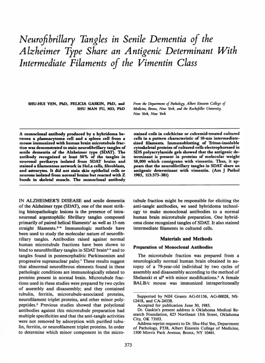
Figure 2-Double labeling of SDAT brain section with the monoclonalantibody (a),and thioflavin-S reagent (b). (x 400) Out of four tangles,two (indicated by arrows) were positive with immunofluorescence.The sma.ll granular fluorescent immages were due to the autofluores-cence-of lipofuscin.
albumin (BSA) in PBS for 90 minutes. The paperwere then incubated with 1:200 diluted ascitic fluid in5% BSA with PBS for 2 hours at room temperature,washed with PBS, incubated with biotinated labeledhorse anti-mouse immunoglobulin followed by wash-ing and incubation with avidin-biotin-peroxidasecomplex (Vector). The substrate for peroxidase en-zymes contained 3 mg DAB and 2 ,l 30% H202 in 10ml 0.1 M Tris buffer, pH 7.6.
Adsorption Studies and Immunodiffusion
Fibroblasts (5 x 10⁵) were homogenized in phos-phate buffer and centrifuged at 12,000 g for 10 min-utes. The pellet was resuspended in 500 Ml of asciticfluid diluted 10 times, incubated at room temperaturefor 1 hour, and centrifuged. The supernatant wasdiluted 20 times before use. The antibody was alsoincubated with two cycles of purified human brain
Vol. 113 * No. 3
376 YEN ET AL
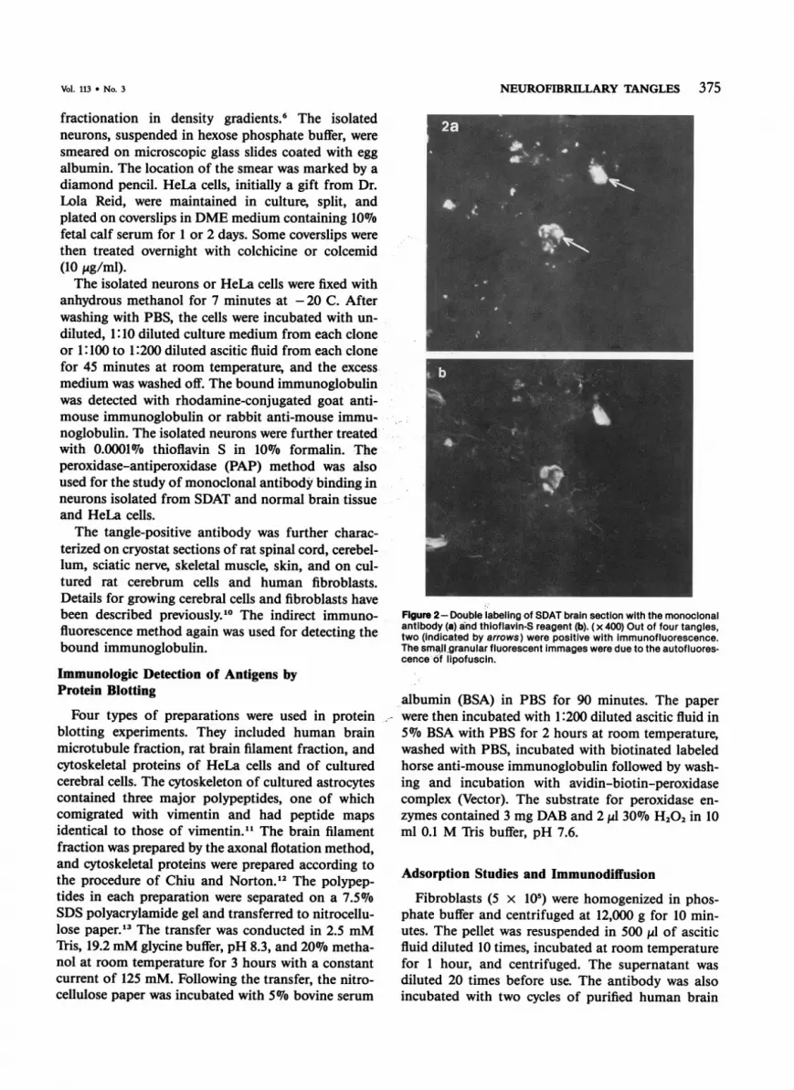
Figure 3-Immunofluores-cent staining of HeLa cells(a), fibroblasts (b) and cerebralcells (c) in cultures with amonoclonal antibody whichbinds SDAT tangles. (x 700)
microtubule fraction (14,ug proteins/,l antibodies) orvimentin (10 Mg/Ml antibodies) eluted electrophore-tically from SDS polyacrylamide gels. After 2 hoursat room temperature the mixtures were centrifuged,and the supernatants were used for immunostaining.The hybridoma medium and ascitic fluid were
tested on an Ouchterlony plate against rabbit anti-mouse immunoglobulins. After overnight incubationat room temperature, the agarose plate was pressed,dried, dialyzed against PBS, and stained with Coo-massie brilliant blue.
ResultsA mouse was immunized repeatedly with a human
microtubule fraction, and its serum was found to
stain neurofibrillary tangles of SDAT neurons. Itsspleen cells were fused with SP2/0 plasmacytomacells. The supernatants of 310 hybridomas werescreened by indirect immunofluorescence. One ofthese stained normal neurons but not HeLa cells orneurofibrillary tangles. Twenty-three stained HeLacells. One of these 23 stained fibrous elements in someneuronal perikarya isolated from SDAT brains. Thishybridoma was of special interest and was clonedtwice on soft agar. One of the selected clones withsimilar specificity was used in the subsequent studies.The monoclonal antibody, by immunodiffusion test,was found to be of the IgM class.
In a previous study,14 it has been shown that thio-flavin bound neurofibrillary tangles of SDAT andemitted green fluorescence under ultraviolet illumina-
AJP * December 1983

Vol. 113 * No. 3 NEUROFIBRILLARY TANGLES
Figure 4- Immunofluorescentstaining of coichicine-treated (a)HeLa cells and fibroblasts (b)with the monoclonal antibody.Coiled fibers were visible in allcells. (x 700) Flgure 5-nniihla bqhplinn nf crrAhralcelwhich had been pretreated withcolchicine with (a) rabbit anti-GFantibodies and (b) monoclonalantibody. Both GF-positive andGF-negative cells were stainedby the monoclonal antibody.(x 420)
tion. This reaction did not alter the binding sites bya polyclonal antiserum to a human microtubule frac-tion.6 These sites could be detected with a rhodamine-labeled antiserum. We used this double fluorochromeimmunofluorescence method to determine whethertangle-containing neurons were also stained by themonoclonal antibody. Over fifty percent of tangle-containing neurons in isolated preparations or cryo-stat sections of SDAT brain were stained by themonoclonal antibody (see Figures 1 and 2 for double-
labeling experiments). For simplicity the antigen de-tected by the monclonal antibody will be referred toas tangle-related antigen.The antibody also bound fibroblasts and newborn
rat cerebrum dissociated cells (Figure 3b and 3c). Thestaining patterns of these cultured cells as well asHeLa cells (Figure 3a) appeared to be filamentous.Treatment of the cells with colchicine or colcemidprior to the incubation with monoclonal antibodyresulted in collapsing or coiling of the filamentous
377
4
1
I
I
I
uuuuilu ittucii[ly VI %miculallii. .. %
i
I.i
378 YEN ET AL
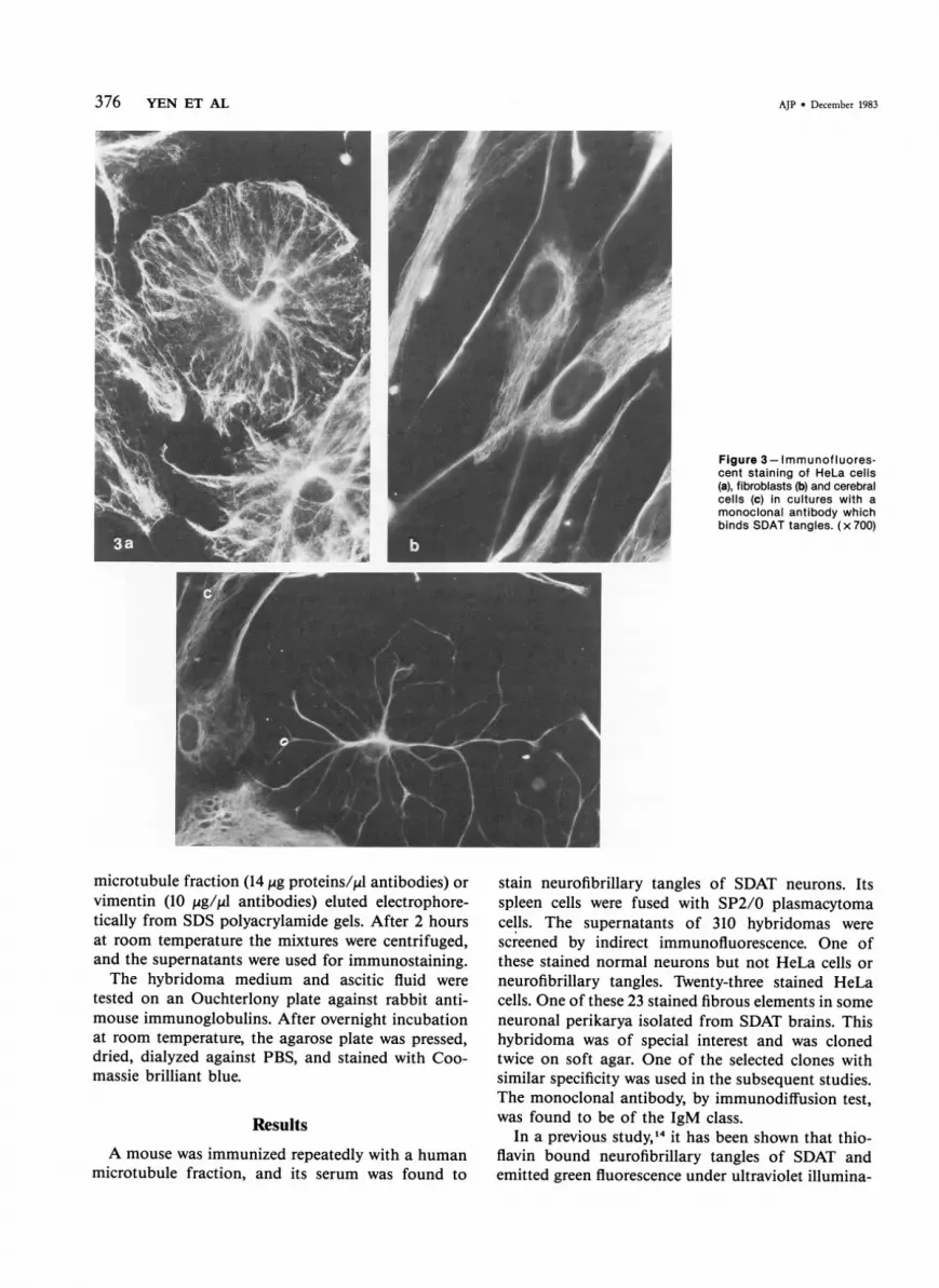
Figure 6-Immunofluorescent staining of newborn rat skin with the monoclonal antibody. a-Epidermis (indicated by arrows) was notstained, whereas the dermis was labeled. (x 420) b-In a deeper layer of dermis the immunofluorescence-positive elements often wererunning parallel to each other. The staining pattern resembles that of anti-vimentin antibodies (not shown).
network (Figure 4).- Also, it appears that all of thecultured cells reacted with the monoclonal antibody.Newborn -rat, cerebrum dissociated cell cultures
contain astrocytes and fibroblasts. I In order to studythe relationship between tangle-related antigen andglial filaments (GF), we double-labeled the cerebrumcells with rabbit- antibodies to human GF proteinsand the monoclonal antibody that stains neurofibrilklary tangles. The results showed that cells containin-GF antigens (presumably astrocytes) and cells nega-tive for anti-GF were both stained by the monoclonalantibody (Figure 5). The staining pattern with GFantibody in astrocytes was similar to that with themonoclonal antibody.
Incubation of the monoclonal antibody with fibro-blasts, cytoskeleton, or purified vimentin removed theanti-tangle activity as well as the ability to stain fila-mentous networks in cultured cells. Repetitive ad-sorptions of the antibody with human brain micro-tubule fractions diminished its staining intensity.Complete removal of the anti-tangle activity was notachieved. The incomplete adsorption might be due toan insufficient amount of tangle-related antigen in themicrotubule fraction.
Immunostaining of cryostat sections of newbornrat skin showed that epidermis, enriched with tono-,filaments, was not stained, whereas the dermis thatcontains vimentin filaments was labeled (Figure 6).The fluorescence-positive elements were long, thinfibers alone or in bundles. In some areas of the sec-tion they were associated with cell bodies. The stain-ing pattern was similar to that of rabbit anti-vimentinantibody. In rat skeletal muscle, the antigens were dis-tributed in a pattern similar to that of Z bands(Figure 7) which have been reported to have inter-mediate filament proteins of vimentin and desmin."5Axons in sciatic nerve or spinal cord sections, knownto contain neurofilaments, did not bind the anti-bodies, but Schwann cells surrounding the myelinatedaxon in sciatic nerves were stained.The monoclonal antibody was further analyzed by
the immunoblotting technique. The antibody reactedwith proteins of molecular weight of 58,000 daltonsin HeLa cell or cerebrum cell preparations (Figure 8)and stained weakly a 56,000-dalton protein band ofthe HeLa cell samples. It did not recognize proteinsin both human microtubule fractions and humanbrain filament fractions which contain GF protein
AJP - December 1983

NEUROFIBRILLARY TANGLES 379
t
Figure 7a - Immunofluorescentstaining of rat skeletal muscle.The antigens were located in Zbands, as shown in (b) the phase-contrast image of the same field.(x 700)
V'..jL.
- . 4
.
4 4.
and neurofilament triplet proteins. GF proteins in thelatter preparation were easily recognized by anti-GFantibodies with the biotin-avidin-peroxidase method.
Discussion
The present study demonstrates that neurofibrillarytangles of SDAT react with a monoclonal antibodyraised against an antigen from the brain of an ap-parently normal elderly person. The antibody, in ad-
dition, recognizes filamentous networks in a varietyof cultured cells. These filaments after incubationwith colchicine or colcemid form coils. This responseis characteristic of 10-nm intermediate-sized filamentsof the vimentin class. In tissue sections, the antigensare located in cell types which are enriched with vi-mentin filaments but not in cells enriched with neuro-filaments or tonofilaments. Immunoblotting studiesshow that the monoclonal antibody stained a 58,000-dalton protein band, which is similar to vimentin in
Figure 8-The gel electrophoretic pattern of rat brain filament fraction (a), molecular weight standards (b), cerebral cells (c), two-cycle purifiedhuman brain microtubule fraction preparation (d), and Hela cells (e). lmmunoblotts showing the antigenic specificities of monoclonal antibodieson triton insoluble cytoskeletal proteins of HeLa cells (f), cerebral cells (g), rat brain filaments (h), and two-cycles purified human brain micro-tubule fraction (i). Molecular weight standards: 1) phosphorylase (94 kd), 2) bovine serum albumin (68 kd), 3) ovalbumin (43 kd), 4) carbonicanhydrase (30 kd), 5) trypsin inhibitor (20 kd).
Vol. 113 * No. 3
380 YEN ET AL AJP * December 1983
molecular weight and peptide maps.11 These resultssuggest that neurofibrillary tangles contain a proteinsharing an antigenic determinant with vimentin. Theantibody gave a weak staining of a 56,000-dalton pro-tein of HeLa cells as well. We do not know the rela-tionship between vimentin and the 56,000-dalton pro-tein. The smaller protein may be a variant or adegraded vimentin. We have tested a polyclonal anti-body against vimentin16 and a monoclonal antibodyagainst intermediate filament"7 for their binding totangles. Although these antibodies recognize vimen-tin of rat origin,10'17 they do not bind well to vimentinof normal human cells, nor do they react with tangles.Only slightly over 5007o of the tangles stained with
the monoclonal antibody. This may be due to thelability of the binding sites secondary to the isolationand fixation procedures. Tangles found in routineautopsy tissue sections fixed in formalin and em-bedded in paraffin did not stain with our monoclonalantibody. Whether this can account for the inabilityof a number of antibodies to vimentin and neurofila-ments to stain the tangles remains to be determined.The affinity of the antibody may also play a role. Al-ternatively, not all of the neurofibrillary tangles con-tain filaments with the same antigenic determinants.
Currently, five major classes of intermediate fila-ments are known, including the cytokeratin of thetrue desmosome-expressing epithelia, the desminfibers of certain muscle types, the glial filaments ofastrocytes, the neurofilaments of neurons and thevimentin filaments in cells of mesenchymal origin. ",18The main protein subunit of each class of filamentshas a distinct molecular weight and isoelectric point.Monospecific antibodies to each major subunit pro-tein have been made which are reported to be specificto only one class of filament. Although matureneurons have been shown not to contain vimentin,'0neuroblastic cells or embryonic neuron precursor cellsare positive with the anti-vimentin antibodies by im-munofluorescence.19 These cells contain numerousfilaments with diameters about 10 nm and of mole-cular weight, resembling vimentin. If the staining oftangles by the monoclonal antibody can be taken asevidence of the presence of vimentin in neurofibril-lary tangles, it suggests that some neurons in SDATare differentiated to reexpress a type of filamentcharacteristic of embryonic neurons. However, thepossibility that in certain Alzheimer cases vimentincontinues to be synthesized in adult brain has notbeen completely ruled out. This possibility can be ex-amined with suitable clinical material. At any rate,the role of vimentin in the pathogenesis of tangles re-mains to be elucidated.
Although different classes of intermediate filamentscontain different proteins, recent studies on the aminoacid sequences of intermediate filament proteins showthat some filament proteins are structuraly related.2021Moreover, several monoclonal antibodies have beenshown to recognize antigenic determinants commonto several classes of intermediate filament proteins.17'22Thus, caution should be taken in applying the im-munologic data to identify proteins in the neurofibril-lary tangle. It remains a possibility that the tangle-related antigen is not due to the presence of vimentinbut rather due to the modification of neurofilaments.Because of the difficulty in solubilizing the tangles,23'24this possibility cannot be ruled out at the presenttime.
Recently a small number of monoclonal antibodiesagainst neurofilament proteins have been reported tobind to tangles.25 The interpretation of this data issubject to constraints similar to those in our studies.At any rate, our monoclonal antibody did not reactwith neurofilaments. These results indicate the pos-sibility that both vimentin and neurofilaments arepresent in the tangle. The exact chemical compositionand the role of intermediate filaments in the patho-genesis of tangles will not be resolved until they canbe solubilized.
References
1. Wisniewski HM, Terry RD: Neuropathology of theaging brain, Neurobiology of Aging. Edited by RDTerry, S Gershon. New York, Raven Press, 1976, pp265-280
2. Oyanagi S: Electron-microscopic observations on thebrains of patients with senile dementia: Conversion ofneurofilaments to twisted tubules and interrelationsbetween Alzheimer's neurofibrillary tangles and Pick'sbodies. Adv Neurol Sci Jpn 1974, 18:77-88
3. Shibayama H, Kitoch J: Electron-microscopic structureof the Alzheimer's neurofibrillary changes in case of,atypical senile dementia. Acta Neuropathol (Berl) 1978,41:229-234
4. Yagishita S, Itoh Y, Amano N: Ultrastructure of neuro-fibrillary tangles in progressive supranuclear palsy. ActaNeuropathol (Berl) 1979, 48:27-30
5. Grundke-Iqbal I, Johnson AB, Wisniewski HM, TerryRD, Iqbal K: Evidence that Alzheimer neurofibrillarytangles originate from neurotubules. Lancet 1979, 1:578-580
6. Yen SH, Gaskin F, Terry RD: Immunocytochemicalstudies of neurofibrillary tangles. Am J Pathol 1981,104:77-89
7. Yen SH, Horoupian DS, Terry RD: Immunnocyto-chemical comparison of neurofibrillary tangles in seniledementia of Alzheimer type, progressive supranuclearpalsy, and postencephalitic Parkinsonism. Ann Neurol1983, 13:172-175
8. Shelanski ML, Gaskin F, Cantor CR: Microtubule as-sembly in the absence of added nucleotides. Proc NatlAcad Sci USA 1973, 70:765-768
9. Kennett R: Cell fusion, Methods in Enzymology.

Vol. 113 * No. 3 381
Edited by W Jakoby, J Pastur. New York, AcademicPress, 1979, pp 345-349
10. Yen S-H, Fields KL: Antibodies to neurofilament, glialfilament, and fibroblast intermediate filament proteinsbind to different cell types of the nervous system. J CellBiol 1981, 88:115-126
11. Chiu FC, Norton WT, Fields KL: The cytoskeleton ofprimary astrocytes in culture contains actin, glial fibril-lary acidic protein and the fibroblast-type filament pro-tein, vimentin. J Neurochem 1981, 37:147-155
12. Chiu FC, Norton WT: Bulk preparation of CNS cyto-skelton and the separation of individual neurofilamentproteins by gel filtration: Dye binding characteristicsand amino acid composition. J Neurochem 1982, 39:1252-1260
13. Towbin H, Strachelin T, Gordon J: Electrophoretictransfer of proteins from polyacrylamide gels to nitro-cellulose sheets: Procedure and some applications.Proc Natl Acad Sci USA 1979, 76:4350-4356
14. Schwartz P: Amyloid degeneration and tuberculosis inthe aged. Gerontologica 1972, 18:321-326
15. Lazarides E: Intermediate filaments as mechanicalintegrators of cellular space. Nature 1980, 283:249-256
16. Hynes R, Destree A: 10 nm filaments in normal andtransformed cells. Cell 1978, 13:151-163.
17. Pruss RM, Mirsky R, Raff MC: All classes of inter-mediate filaments share a common antigenic deter-minant defined by a monoclonal antibody. Cell 1981,27:419-428
18. Franke WW, Schmid E, Winter S, Osborn M, Weber K:Widespread occurrence of intermediate sized filaments
of the vimentin-type in cultured cells from diversevertebrates. Experimental Cell Res 1979, 123:25-46
19. Tapscott SJ, Bennett GS, Toyama Y, Kleinbart F,Holtzer H: Intermediate filament proteins in the de-veloping chick spinal cord. Dev Biol 1981, 86:40-54
20. Geisler N, Weber K: Comparison of the proteins of twoimmunologically distinct intermediate-size filaments byamino acid sequence analysis: desmin and vimentin.Proc Natl Acad Sci USA 1981, 78:4120-4123
21. Geisler N, Plessmann U, Weber K: Related amino acidsequences in neurofilaments and non-neuronal inter-mediate filaments. Nature 1982, 296:448-450
22. Franko MC, Masters CL, Gibbs CJ Jr, Gajdusek DC:Monoclonal antibodies to central nervous system anti-gens. J Neuroimmunol 1981, 1:391-411
23. Selkoe DJ, Ihara Y, Salazar FJ: Alzheimer's diseaseInsolubility or partially purified paired helical filamentsin sodium docecyl sulfate and urea. Science 1982, 215:1243-1245
24. Yen SH, Kress Y: The effect of chemical reagents orproteases on the ultrastructure of paired helical fila-ments, Biological Aspects of Alzheimer's Disease.Edited by R Katzman. Cold Spring Press 1983, pp155-165
25. Anderton BH, Breinburg D, Downes MJ, Green PJ,Tomlinson BE, Ulrich J, Wood JN, Kahn J: Mono-clonal antibodies show that neurofibrillary tangles andneurofilaments share antigenic determinants. Nature1982, 298:84-86