neural correlates of exposure to traumatic pictures and sound in vietnam combat veterans with and...
TRANSCRIPT
ORIGINAL ARTICLES
Neural Correlates of Exposure to Traumatic Picturesand Sound in Vietnam Combat Veterans with andwithout Posttraumatic Stress Disorder: A PositronEmission Tomography Study
J. Douglas Bremner, Lawrence H. Staib, Danny Kaloupek, Steven M. Southwick,Robert Soufer, and Dennis S. Charney
Background: Patients with posttraumatic stress disorder(PTSD) show a reliable increase in PTSD symptoms andphysiological reactivity following exposure to traumaticpictures and sounds. In this study neural correlates ofexposure to traumatic pictures and sounds were measuredin PTSD.
Methods: Positron emission tomography and H2[15O]were used to measure cerebral blood flow during exposureto combat-related and neutral pictures and sounds inVietnam combat veterans with and without PTSD.
Results:Exposure to traumatic material in PTSD (but notnon-PTSD) subjects resulted in a decrease in blood flow inmedial prefrontal cortex (area 25), an area postulated toplay a role in emotion through inhibition of amygdalaresponsiveness. Non-PTSD subjects activated anteriorcingulate (area 24) to a greater degree than PTSDpatients. There were also differences in cerebral bloodflow response in areas involved in memory and visuospa-tial processing (and by extension response to threat),including posterior cingulate (area 23), precentral (mo-tor) and inferior parietal cortex, and lingual gyrus. Therewas a pattern of increases in PTSD and decreases innon-PTSD subjects in these areas.
Conclusions: The findings suggest that functional alter-ations in specific cortical and subcortical brain areasinvolved in memory, visuospatial processing, and emotionunderlie the symptoms of patients with PTSD.Biol Psy-chiatry 1999;45:806–816 ©1999 Society of BiologicalPsychiatry
Key Words: Positron emission tomography, memory,postttraumatic stress disorder, frontal cortex, cingulate,function
Introduction
Posttraumatic stress disorder (PTSD) is characterizedby recurrent trauma-related memories, and increased
fear responding and physiological reactivity to remindersof the trauma, coupled with sleep disturbances, night-mares, avoidance, increased startle, and other symptomsthat can persist for many years after the original traumaticevent (Pitman 1989; Bremner et al 1995a). The failure ofextinction of fear responsiveness to “cues” related to theoriginal trauma can be modeled in laboratory animals andis an important part of the clinical presentation of PTSDpatients. Abnormal traumatic recall and fear responding,which can be reliably provoked with trauma-related pic-tures and sounds or traumatic scripts, are associated withan increase in physiological reactivity, including increasedheart rate and blood pressure, in PTSD patients (Blanchardet al 1982; Malloy et al 1983; Pitman et al 1987; reviewedin Prins et al 1995).
Regulation of these peripheral markers of physiologicalresponsiveness by cingulate and amygdala raises thepossibility that alterations in function in these brain areasmay be associated with PTSD symptoms. Early studiesshowing an increase in fear-related behaviors followingremoval of cerebral cortex in cats also led to the hypoth-esis that these areas, in addition to thalamus, hypothala-mus, hippocampus, and adjacent cortex, regulate emotionand the stress response (MacLean 1949). These brain areasare functionally interrelated, and via pathways through thehypothalamus and medial prefrontal cortex (area 25) effectthe peripheral stress response in increased heart rate, bloodpressure, peripheral catecholamines, and cortisol (Vogt etal 1992; LeDoux 1993; Devinsky et al 1995).
Recently there has been an increased appreciation forthe role that abnormalities in memory play in the presen-
From the Department of Psychiatry (JDB, SMS, DSC) and Department ofDiagnostic Radiology (JDB, LHS, RS), Yale University School of Medicine,New Haven, Connecticut; National Center for Posttraumatic Stress Disorder,New Haven, Connecticut (JDB, SMS, DSC); Yale/VA PET Center, NewHaven, Connecticut (JDB, RS); Yale Psychiatric Institute, New Haven,Connecticut (JDB); VA Connecticut Healthcare Center, West Haven, Connect-icut (JDB, SMS, RS, DSC); Boston VA Medical Center, Boston, Massachusetts(DK); and Department of Psychiatry, Boston University, Boston, Massachu-setts (DK).
Address reprint requests to J. Douglas Bremner, MD, Yale Psychiatric Institute,POB 208038, Yale Station, New Haven, CT 06520.
Received December 12, 1997; revised August 4, 1998; accepted September 8, 1998.
© 1999 Society of Biological Psychiatry 0006-3223/99/$19.00PII S0006-3223(98)00297-2

tation of patients with PTSD. PTSD patients demonstratedeficits in verbal declarative memory, which are associ-ated with a reduction in volume of the hippocampus, abrain area involved in learning and memory (Squire andZola-Morgan 1991) (reviewed in Bremner et al 1995a). Onthe other hand, PTSD is characterized by intensification ofnonverbal aspects of memory, including “hypermne-monic” visual memory traces related to traumatic events(Pitman et al 1993; Bremner et al 1996a). The excessivevigilance seen in PTSD may be associated with increaseddemands on brain areas involved in visuospatial aspects ofmemory function and in integration with evaluation ofstimuli for potential threat and planning of response tostimuli. Based on this we have hypothesized that increasedactivity in cortical brain areas involved in memory andvisuospatial processing, including prefrontal and parietalcortex (Bremner et al 1995a) (in addition to limbic areas),underlies the symptoms of PTSD. Medial prefrontal cor-tical areas also modulate fear responding through inhibi-tory connections with the amygdala that are involved inthe fear response as well as through effecting peripheralsympathetic and hormonal responses to stress (Vogt et al1992; LeDoux 1993; Devinsky et al 1995). We havehypothesized that dysfunction of these areas plays a role infailure of extinction to fear in PTSD (Bremner et al1996a). A complete understanding of functional connec-tions of cortical and subcortical brain areas involved inemotion and memory is important in constructing a modelfor neural correlates of PTSD.
Recent advances in neuroimaging technology havemade it possible to study central brain correlates of PTSD.Reduced volume of the hippocampus measured withmagnetic resonance imaging (MRI) was found in severalpopulations of PTSD patients, possibly secondary tostress-induced atrophy (Bremner et al 1995b, 1997b;Gurvits et al 1996; Stein et al 1997). Decreased metabo-lism measured with positron emission tomography (PET)was found at baseline in temporal and prefrontal cortex incombat-related PTSD (Bremner et al 1997a) and in pari-etal cortex in patients with PTSD and comorbid substancedependence (Semple et al 1996).
Exposure of PTSD patients to traumatic scripts resultedin increased blood flow in limbic regions (right amygdala,insula, orbitofrontal cortex, and anterior cingulate), anddecreased blood flow in middle temporal and left inferiorfrontal cortex, as measured with PET (Rauch et al 1996).This study did not involve a control group, however, so itis not possible to determine whether the changes arespecific to PTSD. A second PET study that did utilizecontrol subjects found increased blood flow in rightamygdala and anterior cingulate, and decreased blood flowin middle temporal and left inferior frontal cortex in PTSDpatients relative to control subjects during trauma-related
mental imagery (Shin et al 1997). Provocation of PTSDsymptoms with the pharmacologic agent yohimbine(which stimulates brain norepinephrine release) resulted ina relative failure of orbitofrontal cortex activation in PTSDpatients relative to control subjects, as well as differentialfunctional responses to challenge in temporal, parietal, andprefrontal cortex. These findings were consistent withincreased noradrenergic responsiveness to yohimbine inPTSD (Bremner et al 1997a).
The purpose of the present study was to use PET in theexamination of neural correlates of exposure to traumaticpictures and sounds in Vietnam combat veterans with andwithout PTSD. Traumatic pictures and sounds represent astandardized and well-established paradigm for the prov-ocation of PTSD symptoms that have several advantagesover other techniques for induction of PTSD symptoms(Kaloupek and Bremner 1996; Prins et al 1995). Theyhave ecological validity, resembling “traumatic cues” thatpatients encounter in their environment, complete with thecomplex interplay of visual and auditory stimuli. Inaddition, traumatic pictures and sounds (unlike individu-alized scripts) can be presented in an identical fashion toall subjects in a research study. We hypothesized thatexposure to traumatic pictures and sounds would result ingreater activation in cortical and subcortical brain areasimplicated in memory, emotion, and the fear response,including amygdala, hippocampus and adjacent cortex,cingulate, and prefrontal and parietal cortex, in combatveterans with PTSD relative to those without PTSD. Wefurther hypothesized a relative failure of activation inmedial prefrontal cortical areas involved in modulation offear responsiveness through inhibition of amygdala func-tion.
Methods and MaterialsSubjects in the study were Vietnam combat veterans with PTSD(n 5 10) and comparison subjects, who were Vietnam combatveterans without PTSD (n 5 10). Subjects were recruitedthrough a VA Medical Center PTSD program and newspaperadvertisement. To identify the subgroup of PTSD patients whoare reactive to the types of traumatic stimuli used in this study(Blanchard et al 1982; Kaloupek and Bremner 1996; Malloy et al1983; Pitman et al 1987; Prins et al 1995), subjects underwent ascreening to determine emotional and psychophysiological reac-tivity. Subjects were determined to be reactive with a 5 beats perminute increase in heart rate and a 50 points increase inSubjective Units of Distress (SUDs) scale score (describedbelow) for the combat slide presentation relative to neutral slidepresentation. Out of 13 PTSD patients screened, 10 met criteria,while all of the non-PTSD veterans met these criteria.
Vietnam veterans were included in the PTSD group who: 1)were demonstrated to be reactive to combat slides and soundsusing the psychophysiology screening procedure outlined above;and 2) met criteria for current combat-related PTSD as assessed
Neural Correlates of PTSD 807BIOL PSYCHIATRY1999;45:806–816

by the Structured Clinical Interview for DSM-IV (SCID) (Spitzeret al 1987). Vietnam veterans were included in the comparisongroup who did not meet these three criteria and did not have anAxis I psychiatric disorder based on the SCID. All subjects hadcombat exposure, which involved being in firefights (gun battles)with the enemy, being wounded, or seeing others killed orwounded. All subjects gave written informed consent for partic-ipation, were free of major medical illness on the basis of historyand physical examination, lab testing, and electrocardiogram,were not actively abusing substances or alcohol (past 6 months),and were free of all medications for at least 4 weeks prior to thestudy. Subjects were not taken off of medication for the purposesof participating in the study.
Subjects were excluded with a serious medical or neurologicalillness, organic mental disorders or comorbid psychotic disor-ders, current alcohol and/or substance abuse or dependence (past6 months), retained metal, a history of head trauma, loss ofconsciousness greater than 10 min, cerebral infectious disease, ordyslexia. There was no difference in age between the PTSDpatients (mean5 47 years, SD5 3) and comparison subjects(mean5 50 years, SD5 3). Nine out of 10 of the PTSD patientswere white, 1/10 black; 9/10 of the comparison subjects werewhite, 1/10 black. All of the subjects, both PTSD and comparisonsubjects, were male and right-handed.
Seven out of 10 PTSD patients (70%) fulfilled criteria for alifetime history of major depression and 3 (30%) for currentmajor depression based on the SCID interview. One patient(10%) met criteria for current dysthymia, and 1 (10%) forlifetime bipolar disorder. Two out of 10 (20%) patients fulfilledcriteria for lifetime history of panic disorder with agoraphobia,and 1 (10%) for current panic disorder with agoraphobia; 1(10%) met criteria for lifetime and current history of panicdisorder without agoraphobia. One out of 10 (10%) fulfilledcriteria for current agoraphobia without panic disorder, 2 (20%)for current social phobia, 2 (20%) for current simple phobia, 1(10%) for current obsessive–compulsive disorder, and 1 (10%)for lifetime generalized anxiety disorder. Six patients (60%)fulfilled criteria for a lifetime history of alcohol dependence, and1 (10%) for lifetime alcohol abuse. There were no patients withsubstance abuse or dependence other than alcohol.
Each subject underwent four scans on a single day. The subjectwas placed in the scanner with his head held in a holder tominimize motion and positioned with the canthomeatal lineparallel to an external laser light. Electrocardiogram leads wereattached to the chest in standard positions for measurement ofheart rate and cardiovascular function, and a dinemap cuff wasattached to the left arm for measurement of blood pressure. Anintravenous line was inserted for administration of [150]H20.Following positioning within the camera gantry, a transmissionscan of the head was obtained using an external67Ga/68Ge rodsource, to correct emission data for attenuation due to overlyingbone and soft tissue. SUDs scale ratings (a visual analogue scalescored from 0 to 100 for the assessment of current subjectivelevel of distress) were performed every 5 min until threesuccessive ratings were unchanged, indicating that the subjecthad adapted to the study setting. Baseline subjective ratings werethen collected, including a 17-item PTSD symptom scale (South-wick et al 1993), the Panic Attack Symptom Scale (PASS)
(Southwick et al 1993), Clinician Administered DissociativeStates Scale (CADSS), a reliable and valid 27-item scale for themeasurement of current dissociative states (Bremner et al 1998),another SUDs scale, and a visual analogue scale (scored from 0to 100) for the assessment of fear (Southwick et al 1993).Subjects also underwent baseline measurement of heart rate andblood pressure.
Subjects then underwent scanning under neutral and traumatic(combat slide) conditions. No “resting” scans (i.e., scans in theabsence of any stimulation) were performed, as there is evidencefor an increase in frontal lobe activity in the absence ofstimulation, which could interfere with the study results. A fixedorder (two presentations of neutral slides and sounds followed bytwo presentations of combat slides and sounds) was used for allsubjects, to prevent anxiety elicited by the combat slides frompersisting into neutral slide presentations. Combat and neutralslides and sounds from an ongoing multisite VA CooperativeStudy on the psychophysiology of PTSD were used for thepresentation materials. Neutral slides were of winter scenes, withnonverbal music. Combat slides were actual photographs fromVietnam, and included scenes such as a male soldier throwing ahand grenade, a sniper in the bushes, and evacuation of woundedsoldiers, accompanied by appropriate sounds, such as machinegun fire, the sound of the jungle, and of helicopters, respectively.Each scan condition involved four slides presented over a total ofa 2-min period. One minute into the presentation subjectsreceived a bolus injection of 30 mCi of [150]H20 immediatelyfollowed by a PET scan acquisition, which was 1 min in length.PET imaging was performed on a Posicam PET camera (PositronCorp) (in plane resolution after filtering, 6 mm full width at halfmaximum). Subjects then underwent measurement of heart rateand blood pressure, as well as behavioral ratings for the time ofthe presentation, including PASS, CADSS, SUDs, fear analogue,and PTSD Symptom Scale.
Images were reconstructed and analyzed on a SunSparcWorkstation using statistical parametric mapping (spm95). Im-ages for each patient set were realigned to the first scan of thestudy session. The mean concentration of radioactivity in eachscan was obtained as an area-weighted sum of the concentrationof each slice and adjusted to a nominal value of 50 mL/min/100g. The data underwent transformation into a common anatomicalspace and was smoothed with a three-dimensional gaussian filterto 16 mm full width at half maximum. The study design involveda comparison of regional blood flow during traumatic vs. neutralconditions in PTSD and comparison subject groups examinedseparately, and the interaction between group (PTSD vs. com-parison subjects) and condition (combat vs. neutral presenta-tions) with global blood flow considered as a confoundingcovariate. Statistical analyses yielded image data sets in whichthe values assigned to individual voxels correspond tot statistic(Friston et al 1991). Statistical images were displayed withvalues ofZ score units. A thresholdZ score of 3.00 (p , .001)was used to examine areas of activation within hypothesizedareas. Location of areas of activation were identified as thedistance from the anterior commissure in mm, with x-, y-, andz-coordinates, using a standard stereotaxic atlas (Talairach andTournoux 1988).
Behavioral (PASS, PTSD, CADSS, SUDs, and analog rating
808 J.D. Bremner et alBIOL PSYCHIATRY1999;45:806–816
scores) and psychophysiological (heart rate and blood pressure)measures were compared between PTSD and comparison subjectgroups using repeated-measures analysis of variance (ANOVA)with time as the repeated measure. When there was a significantmain effect for time, Duncan’s multiple range test was performedto determine what time points showed significant differencesfrom the baseline measures.
Results
Repeated-measures ANOVA for diastolic blood pressure(DBP) demonstrated a significant main effect for diagno-sis, with higher DBP in PTSD relative to comparisonsubjects (Table 1), but not a significant main effect fortime (F 5 1.63; p 5 .17). PTSD patients had relativelygreater increases with traumatic slides than comparisonsubjects (significant time by diagnosis effect), for PTSD,anxiety, and dissociative symptoms, as well as subjectivedistress and fear (Table 1).
Combat veterans with PTSD had increased blood flowwith traumatic pictures and sounds in cerebellum, rightinferior frontal gyrus, and midbrain. Combat veteranswithout PTSD had increased blood flow in cerebellum,
right anterior cingulate, left visual association cortex, leftmiddle frontal gyrus, and right middle temporal gyrus (Zscore. 3.00;p , .001) (Table 2). Exposure to traumaticpictures and sound in combat veterans with PTSD resultedin decreased blood flow in bilateral medial prefrontalcortex (subcallosal gyrus, or Brodmann’s area 25), adja-cent areas of left anterior cingulate, left thalamus, leftvisual association cortex, and superior temporal and leftmiddle temporal cortex. In combat veterans withoutPTSD, traumatic pictures and sounds were associated withdecreased blood flow in left superior temporal cortex, leftprecentral (motor) cortex, left inferior parietal lobule,midcingulate, cerebellum, and right lingual gyrus (Zscore. 3.00;p , .001) (Table 3).
There was a significant difference in pattern of cerebralblood flow response to traumatic pictures and soundsbetween veterans with and without PTSD (i.e., significantinteraction between condition and PTSD diagnosis) in leftinferior parietal lobule, posterior cingulate (area 23), leftmotor cortex (precentral gyrus), right lingual gyrus, and anarea that included dorsal pons and lateral cerebellum, butalso portions of parahippocampal gyrus (Z score. 3.00;
Tabel 1. Psychophysiological and Behavioral Ratings in PTSD Patients and Comparison Subjects
PTSD patients (n 5 10) Comparison subjects (n 5 10)
Time 3diagnosis
int.-F
Baseline Neutral-1 Neutral-2 Combat-1 Combat-2 Baseline Neutral-1 Neutral-2 Combat-1 Combat-2
Mean SD Mean SD Mean SD Mean SD Mean SD Mean SD Mean SD Mean SD Mean SD Mean SD
Systolic BP 130.9 9.5 135.4 17.1 130.2 9.1 133.5 7.3 137.1 11.8 128.5 12.1 133.1 16.2 131.4 16.3 129.0 11.5 131.3 14.1 0.2Diastolic BP 78.8 5.6 80.3 7.4 80.6 6.3 84.6 8.0 86.0 6.8 77.5 5.1 79.0 4.9 80.3 6.0 80.1 6.8 79.2 7.9 0.86a
Pulse 69.0 15.6 68.6 13.2 65.4 12.0 66.3 11.3 66.6 12.0 67.9 11.8 67.1 10.0 66.0 10.0 65.2 8.1 66.2 9.3 0.0CADSS 1.9 2.6 3.6 4.3 7.5 7.7 7.1 8.8 12.3 12.1 1.8 4.1 3.9 6.6 1.4 2.8 1.9 3.2 1.0 1.9 2.94a,b
PASS 28.8 2.1 29.4 2.2 32.7 5.9 35.5 8.3 41.8 13.7 27.4 1.0 28.6 3.4 27.4 0.7 27.2 0.6 27.6 1.6 4.78b,c
PTSD Scale 15.3 1.1 16.5 2.5 19.5 7.3 25.5 6.5 32.5 7.7 14.0 0.0 14.1 0.3 14.1 0.3 14.5 1.3 14.2 0.6 15.1Fear analogue 0.05 0.11 0.05 0.10 0.03 0.08 0.15 0.17 0.26 0.24 0.00 0.00 0.00 0.00 0.00 0.00 0.00 0.00 0.00 0.0 4.33SUDs 13.0 12.0 11.0 11.0 15.0 17.0 33.0 16.0 42.0 28.0 1.0 3.0 4.0 13.0 3.0 8.0 3.0 8.0 0.0 0.0 5.58
CADSS, Clinician Administered Dissociative States Scale; PASS, Panic Attack Symptom Scale; SUDs, Subjective Units of Distress scale.aSignificant main effect for diagnosis (p , .05).bSignificant time by diagnosis interaction (p , .05).cDuncan’s multiple range test showed significant increase during combat slides relative to neutral slides and baseline in the PTSD patients (p , .05).
Table 2. Areas of Increased Blood Flow with Combat-Related Slides and Sounds Relative to Neutral Slides and Sounds in PTSDPatients and Comparison Subjects
PTSD (n 5 10) Comparison subjects (n 5 10)
Z score
Talairach coordinates
Z score
Talairach coordinates
x y z Brian region x y z Brain region
4.89 24 282 228 R. cerebellum 5.25 234 278 224 L. cerebellum4.53 12 292 220 4.23 4 282 216 R. cerebellum4.24 2 280 212 4.11 210 286 28 L. visual association cortex (18)3.62 50 28 0 R. interior frontal (47) 3.75 230 12 28 L. middle frontal (9)3.58 52 20 4 3.67 12 38 20 R. anterior cingulate (32)3.27 12 230 28 R. midbrain 3.27 44 266 12 R. middle temporal gyrus (39)
Z score. 3.00,p , .001.
Neural Correlates of PTSD 809BIOL PSYCHIATRY1999;45:806–816

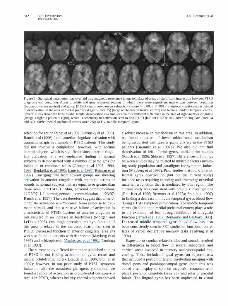
p , .001) (Table 4, Figures 1 and 2). The significantinteraction was related to a pattern of increase in PTSDand decrease in non-PTSD, or the combination of the two.In general the PTSD patients showed a tendency toincrease blood flow, whereas control subjects showed asignificant decrease in blood flow (Table 3), in these areas.The exact percentage of change in these areas in PTSDand non-PTSD groups is shown in Figure 1. In the area ofdorsal pons/cerebellum/parahippocampal gyrus PTSD pa-tients showed an increase in blood flow, whereas innon-PTSD veterans there was a decrease in blood flow.
There were also significant differences in pattern ofcerebral blood flow response to traumatic pictures andsounds between veterans with and without PTSD (i.e.,significant interaction between condition and PTSD diag-nosis) in bilateral medial prefrontal cortex (subcallosalgyrus; Brodmann’s area 25), and bilateral middle temporalgyrus, primarily due to deactivation in the PTSD patients
(Z score. 3.00;p , .001) (Table 4, Figures 1 and 3). Thearea of significant difference in medial prefrontal cortexwas immediately adjacent to and in fact merged intoanterior cingulate (area 24 and 32). In this area differencesbetween groups were due to activation in non-PTSD (butnot PTSD) veterans.
Discussion
Vietnam veterans with combat-related PTSD compared tocombat veterans without PTSD showed significant differ-ences in cerebral blood flow response to traumatic picturesand sounds in cortical and subcortical regions involved inmemory, visuospatial processing, and emotion that arehypothesized to play a role in the generation of symptomsof PTSD. PTSD patients (but not non-PTSD veterans)demonstrated a decrease in blood flow in medial prefrontalcortex (subcallosal gyrus; Brodmann’s area 25) and mid-
Table 3. Areas of Decreased Blood Flow with Combat-Related Slides and Sounds Relative to Neutral Slides and Sounds in PTSDPatients and Comparison Subjects
PTSD patients (n 5 10) Comparison subjects (n 5 10)
Z score
Talairach coordinates
Z score
Talairach coordinates
x y z Brain region x y z Brain region
5.04 250 210 0 L. superior temporal pole (22) 5.80 258 232 20 L. superior temporal gyrus (41)4.88 210 222 12 L. Thalamus 5.66 252 26 8 L. precentral (6)4.42 236 232 12 L. superior temporal (41) 5.09 250 0 244.59 8 20 212 R. Mesofrontal (25) 4.77 246 24 244.36 28 18 212 L. Mesofrontal (25) 4.14 240 228 28 L. inferior pariental lobule (40)4.23 234 286 16 L. visual area (19) 4.13 56 26 8 R. superior tempral pole (22)3.60 222 290 20 3.06 62 234 83.98 28 52 0 L. anterior cingulate (32) 3.61 2 212 32 Posterior cingulate (23)3.87 50 24 0 R. superior temporal gyrus (21) 3.53 12254 24 R. cerebellum3.66 256 232 24 L. middle temporal gyrus (21) 3.09 18 250 212
3.05 8 256 0 R. posterior parahippocampus (lingual) (19)
Z score. 3.00,p , .001.
Table 4. Areas of Significant Interaction between Condition (Combat Slides and Sounds versus Neutral Slides and Sounds) andPTSD Diagnosis in Vietnam Combat Veterans with and without PTSD
Areas with Patterns of Increased Blood Flow in PTSDand Decreased Blood Flow in non-PTSDa
Areas with Patterns of Increased Blood Flow in non-PTSDand Decreased Blood Flow in PTSDb
Z score
Talairach coordinates
Z score
Talairach coordinates
x y z Brain region x y z Brain region
3.36 240 228 28 L. inferior parietal Louule (40) 4.12 8 20212 Medial prefrontal (subcallosal gyrus) (25)3.22 8 256 0 R. lingual gyrus (parahippocampus) (19) 3.32 28 18 2123.17 8 240 220 R. cerebellum/pons/parahippocampus 3.24 44266 12 R. middle temporal gyrus (39)3.17 2 212 32 Mid cingulate (23) 3.23 256 232 24 L. middle temporal gyrus (21)3.00 246 24 24 L. precentral gyrus (motor cortex) (6) 2.99 212 222 12 L. thalamus
Z score. 3.00;p , .001.aSignificance of interaction term could be related to either increases in PTSD or decreases in non-PTSD, or a combination of the two.bSignificance of interaction term could be related to either increases in non-PTSD or decreases in PTSD, or a combination of the two (see Figure 1).Other areas of interaction significant at thep , .01 level included: (greater increase in PTSD) cerebellum (Z score5 2.64; x5 220, y 5 266, z5 228), (greater
decrease in non-PTSD) right superior temporal gyrus (Z score5 2.54; x5 60, y 5 234, z5 12), (greater increase in non-PTSD) right anterior cingulate (Z score 2.83;x 5 12, y 5 44, z5 12), (greater increase in non-PTSD) left visual association cortex (19) (Z score 2.37; x5 230, y 5 288, z5 16).
810 J.D. Bremner et alBIOL PSYCHIATRY1999;45:806–816

dle temporal gyrus. The area of deactivation in medialprefrontal cortex was immediately adjacent to an area ofright anterior cingulate (areas 24 and 32) in which therewas greater activation in the non-PTSD control subjects.There were also significant differences in posterior cingu-late (area 23), inferior parietal cortex, lingual gyrus, andleft precentral gyrus (motor cortex). Differences in patternof activation in these areas were primarily related tosignificant decreases in non-PTSD subjects, although therewas a tendency to increase in PTSD patients.
The area of medial prefrontal cortex (subcallosal gyrus,or Brodmann’s area 25) implicated in this study is adjacent
and inferior to anterior cingulate (Brodmann’s areas 24and 32) and superior to posterior orbitofrontal cortex. Thisarea plays an important role in emotion, social behavior,and endocrine and autonomic responses to stress (Sesacket al 1989; Vogt et al 1992; Morgan et al 1993; Carmichaeland Price 1994, 1995; Damasio et al 1994; Morgan andLeDoux 1995; Devinsky et al 1995; George et al 1995).Recent studies have also shown decreased function in thispart of the medial prefrontal cortex in patients withdepression (Drevets et al 1997), suggesting correlates withpathological emotional states in other psychiatric disor-ders. Lesions in this area in animals resulted in a failure ofextinction to fear responding (Morgan et al 1993) (al-though see Gewirtz et al 1997). This finding may besecondary to a release of inhibitory inputs to the amygdala,which plays a critical role in conditioned fear responding(Davis 1992; LeDoux 1993). A failure of extinction to fearis an important component of the clinical presentation ofpatients with PTSD, which we have hypothesized to besecondary to a failure of medial frontal cortex inhibition ofthe amygdala (Bremner et al 1995a); however, it should benoted that according to our model, one might predictgreater activation of amygdala in PTSD, which unlikeprior PTSD studies (Rauch et al 1996; Shin et al 1997) wasnot found in the current study.
This area merged into another portion of anteriorcingulate (area 24 and 32) in which differences in bloodflow were primarily related to greater activation in non-PTSD subjects compared to PTSD. This portion of ante-rior cingulate is functionally connected with area 25 and isinvolved in emotion, evaluation of stimulus/response, and
Figure 1. Percent change in globally normalized regional cere-bral blood flow with exposure to traumatic cues (combat slidesand sounds) relative to the neutral condition (neutral slides andsounds) in Vietnam combat veterans with PTSD and comparisonsubjects, showing regions in which there were significant groupby condition interactions (p , .001).
Figure 2. Statistical parametric map of areas of significant interaction between PTSD diagnosis and condition. Areas of white and grayrepresent regions in which there were significant interactions between condition (traumatic versus neutral) and group (PTSD versuscomparison subjects) (Z score. 3.00; p , .001). Statistical significance is related to patterns of increased blood flow in PTSD,decreased blood flow in non-PTSD, or some combination of the two. Cer, cerebellum; PH, parahippocampal gyrus; Li, lingual gyrus;PrC, precentral gyrus; IPL, inferior parietal lobule; Ci, posterior cingulate.
Neural Correlates of PTSD 811BIOL PSYCHIATRY1999;45:806–816
selection for action (Vogt et al 1992; Devinsky et al 1995).Rauch et al (1996) found anterior cingulate activation withtraumatic scripts in a sample of PTSD patients. This studydid not involve a comparison, however, with normalcontrol subjects, which is significant since anterior cingu-late activation is a well-replicated finding in normalsubjects as demonstrated with a number of paradigms forinduction of emotional states (George et al 1993, 1994,1995; Benkelfat et al 1995; Lane et al 1997; Reiman et al1997). Emerging data from several groups are showingactivation in anterior cingulate with traumatic scripts orsounds in normal subjects that are equal to or greater thanthose seen in PTSD (L. Shin, personal communication,11/23/97; I. Liberzon, personal communication, 11/23/97;Rauch et al 1997). The data therefore suggest that anteriorcingulate activation is a “normal” brain response to trau-matic stimuli, and that a relative failure of activation ischaracteristic of PTSD. Lesions of anterior cingulate inrats resulted in an increase in fearfulness (Morgan andLeDoux 1995). One might speculate that hypofunction ofthis area is related to the increased fearfulness seen inPTSD. Decreased function in anterior cingulate (area 24)was also found in patients with depression (Mayberg et al1997) and schizophrenia (Andreasen et al 1992; Tamingaet al 1992).
The current study differed from other published studiesof PTSD in not finding activation of gyrus rectus andmedial orbitofrontal cortex (Rauch et al 1996; Shin et al1997); however, in our prior study of PTSD symptominduction with the noradrenergic agent, yohimbine, wefound a failure of activation in orbitofrontal cortex/gyrusrectus in PTSD, whereas healthy control subjects showed
a robust increase in metabolism in this area. In addition,we found a pattern of lower orbitofrontal metabolismbeing associated with greater panic anxiety in the PTSDpatients (Bremner et al 1997a). We also did not finddeactivation of left inferior gyrus, unlike prior studies(Rauch et al 1996; Shin et al 1997). Differences in findingsbetween studies may be related to multiple factors includ-ing study population and paradigms for symptom induc-tion (Mayberg et al 1997). Prior studies that found inferiorfrontal gyrus deactivation (but not the current study)included tasks requiring encoding and processing of verbalmaterial, a function that is mediated by this region. Thecurrent study was consistent with previous investigations(Rauch et al 1996; Bremner et al 1997a; Shin et al 1997)in finding a decrease in middle temporal gyrus blood flowduring PTSD symptom provocation. The middle temporalcortex (in addition to medial prefrontal cortex) plays a rolein the extinction of fear through inhibition of amygdalafunction (Jarrell et al 1987; Romanski and LeDoux 1993).Decreased middle temporal gyrus blood flow has alsobeen consistently seen in PET studies of functional corre-lates of verbal declarative memory tasks (Tulving et al1994).
Exposure to combat-related slides and sounds resultedin differences in blood flow in several subcortical andcortical areas involved in memory and visuospatial pro-cessing. These included lingual gyrus, an adjacent areathat included a portion of lateral cerebellum merging withdorsal pons and parahippocampal gyrus (note this wasadded after display of spm on magnetic resonance tem-plate), posterior cingulate (area 23), and inferior parietallobule. The lingual gyrus has been implicated in visual
Figure 3. Statistical parametric map overlaid on a magnetic resonance image template of areas of significant interaction between PTSDdiagnosis and condition. Areas of white and gray represent regions in which there were significant interactions between condition(traumatic versus neutral) and group (PTSD versus comparison subjects) (Z score. 3.00;p , .001). Statistical significance is relatedto deactivation in the area of medial prefrontal gyrus (area 25) (large white area in frontal cortex) and bilateral middle temporal cortex.Several slices above the large medial frontal deactivation is a smaller area of significant difference in the area of right anterior cingulate(image’s right is patient’s right), which is secondary to activation seen in non-PTSD (but not PTSD). AC, anterior cingulate (area 24and 32); MPfc, medial prefrontal cortex (area 25); MTG, middle temporal gyrus.
812 J.D. Bremner et alBIOL PSYCHIATRY1999;45:806–816

memory and memory for faces (Kapur et al 1995).Parahippocampal gyrus is adjacent to lingual gyrus andhas also been implicated in emotion and anxiety (Nordahlet al 1990; Reiman et al 1984, 1986) as well as symptomsof PTSD (Rauch et al 1996; Shin et al 1997). Visuospatialprocessing [mediated by the inferior parietal cortex(Jonides et al 1993; Pardo et al 1991; Posner et al 1988;Petersen et al 1988) and posterior cingulate (area 23)(Vogt et al 1992; Devinsky et al 1995)] is an importantcomponent of preparation for coping with a physicalthreat. We have hypothesized the involvement of theseareas in the neuroanatomical network mediating symp-toms of PTSD (Bremner et al 1995a). Consistent with thisformulation, a recent PET study found increased bloodflow in posterior cingulate during exposure to films of abank robbery in bank tellers who were victims of a bankrobbery (Fischer et al 1996).
Areas not hypothesized to show a difference betweenPTSD and control subjects included motor cortex, al-though this area was previously implicated in PTSD byRauch et al (1996) and has been consistently shown toactivate in studies of declarative memory. This area maycarry motor aspects of memory necessary for the organ-ism’s preparation for action during the stress response(Squire and Zola-Morgan 1991; Lang et al 1983). PTSDpatients showed greater activation in a portion of lateralcerebellum, although a strong cerebellar activation wasseen in both veterans with and those without PTSD.Recent PET studies have established a role for the cere-bellum in attention and memory, probably mediated by itsprojections through the thalamus to prefrontal cortex(Ashkoomoff and Courchesne 1992; Leiner 1989). Thearea of greater activation in PTSD in lateral cerebellummerged with a portion of the dorsal pons, a major site ofnoradrenergic neurons in the brain that have projectionsthroughout cortical and several subcortical regions. Thissystem plays a critical role in stress and coping with threat(Abercrombie and Jacobs 1987; Aston-Jones et al 1991;Rasmussen et al 1986; reviewed in Bremner et al 1996b,1996c). Our own PET findings showing differences inmetabolic response to yohimbine challenge in PTSD areconsistent with increased central noradrenergic responsiv-ity in PTSD (Bremner et al 1997a).
Findings in the current study are best interpreted assecondary to disturbances of a functional network ofinterrelated brain areas that mediate memory, visuospatialprocessing, and the emotional valence of stimuli. Motorcortex (area 6), anterior (area 24) and posterior cingulate(area 23), lingual gyrus, visual association cortex (19),superior temporal cortex, and thalamus all have projec-tions to both lateral prefrontal and parietal cortex(Selemon and Goldman-Rakic 1988). The area of posteriorcingulate (area 23) implicated in this study has functional
connections with hippocampus and adjacent cortex (para-hippocampal gyrus) [which led to its original classificationas part of the “limbic brain” (Gray 1982)] as well asparietal cortex. These areas (posterior cingulate, parahip-pocampal gyrus, and parietal cortex, all of which areimplicated in the current study) may act in concert tomediate cognitive functions of visuospatial processing andmemory that are necessary for coping with threat (Vogt etal 1992). PTSD may represent a dysfunction in the brain’sresponse to coping with stress and potential threat, whichinvolves excessive recruitment in brain areas responsiblefor visuospatial processing, attention, and memory, inaddition to attaching an affective valence to stimuli.According to this model the symptoms of PTSD arerelated to an abnormality in a functional network involv-ing multiple cortical and subcortical regions, rather thanone or two areas that specifically are responsible for themediation of “emotion.”
In the current study we included only combat veteranswho met specific inclusion criteria based on reactivity totraumatic stimuli, therefore the results are not generaliz-able to PTSD patients without psychophysiological re-sponsivity. Although PTSD patients were included with aheart rate response to traumatic pictures and sounds ofgreater than 5 bpm, there was not a robust measuredresponse during the actual scan. Heart rate and bloodpressure were measured at the end of the stimulus presen-tation period. This may have limited our ability to detectsignificant increases. Although there was a robust behav-ioral effect, changes in SUDs ratings (as well as physio-logical responses) were not as great as those previouslyreported with traumatic scripts. We used a fixed order ofpresentation of stimuli, to avoid contamination of thecontrol condition by traumatic slides; however, this pro-tocol could lead to order effects. Other possible limitationsare related to the pictures and sounds selected for thisstudy. Brain activation may be related to factors related tothe control condition (e.g., feelings of pleasantness duringthe neutral condition). This is avoided, however, bystatistical analyses examining the interaction betweendiagnosis and condition, on which the primary conclusionsof the study are based. The use of pictures and sounds aspresentation materials also has inherent strengths, in thatthey are similar to stimuli encountered in daily life, andthey can be standardized and validated as provocativematerial, and presented in an identical fashion to allsubjects involved in a study protocol. Measurements ofrespiration or pCO2 were not performed to examine thepossibility that PTSD patients hyperventilated to a greaterdegree than non-PTSD patients, resulting in changes inpCO2 and blood flow; however, it was not our observationthat PTSD patients hyperventilated during the study (as,for instance, patients with panic disorder might be ex-
Neural Correlates of PTSD 813BIOL PSYCHIATRY1999;45:806–816
pected to do). In any case pCO2 would be expected to havea global effect on blood flow, and the data analysisperformed involved a correction for variations in globalblood flow.
In summary, the current study showed that exposure totraumatic slides and sounds in PTSD subjects (but notnon-PTSD subjects) resulted in a decrease in blood flow inmedial prefrontal cortex (subcallosal gyrus; Brodmann’sarea 25) and middle temporal gyrus, and a failure inactivation in immediately adjacent right anterior cingulate(areas 24 and 32). There were also differences (primarilyrelated to significant decreases in non-PTSD subjects) inposterior cingulate (area 23), inferior parietal cortex,lingual gyrus, and left precentral gyrus (motor cortex).
Several functional neuroimaging studies have now beenperformed in patients with PTSD. Studies to date areconsistent in implicating medial prefrontal cortex, includ-ing anterior cingulate, in the pathophysiology of PTSD. Inspite of the clear role for the amygdala in conditioned fearresponses in animals, there is some question whether theamygdala is involved in PTSD symptoms; however, dif-ferences in study populations and study paradigms make itdifficult to interpret the findings from different studies.The most important next step in the field would be toestablish similar study populations and a standard para-digm for symptom induction study across sites, to estab-lish with some degree of confidence neuroanatomicalcorrelates for the symptomatology of PTSD.
This study was supported by a Veterans Administration Career Devel-opment Award to Dr. Bremner, and a National Center for PosttraumaticStress Disorder grant.
We would like to thank Eric Anderson for expert assistance in imageprocessing and analysis, Susan Insall, RNMSW, for assistance in datamanagement, Chin Ng, PhD, and Holley Dey, MD, for assistance withPET instrumentation and methodology, and Dayton Rich, CNMT, forassistance in PET image acquisition.
ReferencesAbercrombie ED, Jacobs BL (1987): Single-unit response of
noradrenergic neurons in the locus coeruleus of freely movingcats. I. Acutely presented stressful and nonstressful stimuli.J Neurosci7:2837–2843.
Andreasen NC, Rezai K, Alliger R, Swayze VW II, Flaum M,Kirchner P, et al (1992): Hypofrontality in neuroleptic naı¨vepatients and in patients with chronic schizophrenia. Assess-ment with Xenon 133 single photon emission computedtomography and the Tower of London.Arch Gen Psychiatry49:943–958.
Ashkoomoff NA, Courchesne E (1992): A new role for thecerebellum in cognitive operations.Behav Neurosci106:731–738.
Aston-Jones G, Chiang C, Alexinsky T (1991): Discharge ofnoradrenergic locus coeruleus neurons in behaving rats and
monkeys suggests a role in vigilance. In:Barnes CD, Pom-peiano, editors.Progress in Brain Research.New York:Elsevier Science Publications, pp. 501–519.
Benkelfat C, Bradwejn J, Meyer E, Ellenbogen M, Milot S,Gjedde A, et al (1995): Functional neuroanatomy of CCK sub4-induced anxiety in normal healthy volunteers.Am J Psy-chiatry 152:1180–1184.
Blanchard EB, Kiolb LC, Pallmeyer TP, Gerardi RJ (1982): Apsychophysiological study of posttraumatic stress disorder inVietnam combat veterans.Psychiatr Q54:220–229.
Bremner JD, Krystal JH, Southwick SM, Charney DS (1995a):Functional neuroanatomical correlates of the effects of stresson memory.J Trauma Stress8:527–554.
Bremner JD, Randall PR, Scott TM, Bronen RA, Delaney RC,Seibyl JP, et al (1995b): MRI-based measurement of hip-pocampal volume in posttraumatic stress disorder.Am JPsychiatry152:973–981.
Bremner JD, Krystal JH, Charney DS, Southwick SM (1996a):Neural mechanisms in dissociative amnesia for childhoodabuse: Relevance to the current controversy surrounding theFalse Memory Syndrome.Am J Psychiatry153:FS71–FS82.
Bremner JD, Krystal JH, Southwick SM, Charney DS (1996b):Noradrenergic mechanisms in stress and anxiety: I. Preclini-cal studies.Synapse23:28–38.
Bremner JD, Krystal JH, Southwick SM, Charney DS (1996c):Noradrenergic mechanisms in stress and anxiety: II. Clinicalstudies.Synapse23:39–51.
Bremner JD, Innis RB, Ng CK, Staib L, Duncan J, Bronen R, etal (1997a): PET measurement of central metabolic correlatesof yohimbine administration in posttraumatic stress disorder.Arch Gen Psychiatry54:246–256.
Bremner JD, Randall P, Vermetten E, Staib L, Bronen RA,Mazure CM, et al (1997b): MRI-based measurement ofhippocampal volume in posttraumatic stress disorder relatedto childhood physical and sexual abuse: A preliminary report.Biol Psychiatry41:23–32.
Bremner JD, Krystal JH, Putnam F, Southwick SM, Marmar C,Charney DS, et al (1998): Measurement of dissociative stateswith the Clinician Administered Dissociative States Scale(CADSS).J Trauma Stress11:125–136.
Carmichael ST, Price JL (1994): Architectonic subdivision of theorbital and medial prefrontal cortex in the macaque monkey.J Comp Neurol346:366–402.
Carmichael ST, Price JL (1995): Limbic connections of theorbital and medial prefrontal cortex in macaque monkeys.J Comp Neurol363:615–641.
Damasio H, Grabowski T, Frank R, Galaburda AM, Damasio AR(1994): The return of Phineas Gage: Clues about the brainfrom the skull of a famous patient.Science264:1102–1105.
Davis M (1992): The role of the amygdala in fear and anxiety.Annu Rev Neurosci15:353–375.
Devinsky O, Morrell MJ, Vogt BA (1995): Contributions ofanterior cingulate to behavior.Brain 118:279–306.
Drevets WC, Price JL, Simpson JR Jr, Todd RD, Reich T,Vannier M, et al (1997): Subgenual prefrontal cortex abnor-malities in mood disorders.Nature386:824–827.
Fischer H, Wik G, Fredrikson M (1996): Functional neuroanat-omy of robbery re-experience: Affective memories studiedwith PET.Neuroreport7:2081–2086.
814 J.D. Bremner et alBIOL PSYCHIATRY1999;45:806–816
Friston K, Frith C, Liddle P, Frackowiak R (1991): Comparingfunctional (PET) images: The assessment of significantchange.J Cereb Blood Flow Metab11:690–699.
George MS, Ketter TA, Gill DS, Haxby JV, Ungerleider LG,Herscovitch P, et al (1993): Brain regions involved inrecognizing facial emotion or identity: An oxygen-15 PETstudy.J Neuropsychiatry Clin Neurosci5:384–394.
George MS, Ketter TA, Parekh PI, Rosinsky N, Ring H, CaseyBJ, et al (1994): Regional brain activity when selecting aresponse despite interference: An H215 O PET study of thestroop and an emotional stroop.Hum Brain Mapping1:94–209.
George MS, Ketter TA, Parekh PI, Horwitz B, Herscovitch P,Post RM (1995): Brain activity during transient sadness andhappiness in healthy women.Am J Psychiatry152:341–351.
Gewirtz JC, Falls WA, Davis M (1997): Normal conditionedinhibition and extinction of freezing and fear-potentiatedstartle following electrolytic lesions of medial prefrontalcortex.Behav Neurosci111:712–726.
Goldensohn E (1992): Structural lesions of the frontal lobe:Manifestations, classification, and prognosis. In: Chauvel P,Delgado- Escueta AV, et al, editors.Advances in Neurology.New York: Raven Press.
Gray JA (1982):The Neuropsychology of Anxiety. New York:Oxford University Press.
Gurvits TG, Shenton MR, Hokama H, Ohta H, Lasko NB,Gilberson MW, et al (1996): Magnetic resonance imagingstudy of hippocampal volume in chronic combat-relatedposttraumatic stress disorder.Biol Psychiatry40:192–199.
Jarrell TW, Gentile CG, Romanski LM, McCabe PM, Schnei-derman N (1987): Involvement of cortical and thalamicauditory regions in retention of differential bradycardiacconditioning to acoustic conditioned stimuli in rabbits.BrainRes412:285–294.
Jonides J, Smith EE, Koeppe RA, Awh E, Minoshima S, MintunMA (1993): Spatial working memory in humans as revealedby PET.Nature363:623–625.
Kaloupek DG, Bremner JD (1996): Psychophysiological mea-sures and methods in trauma research. In: Carlson EB, editor.Trauma Research Methodology. Lutherville, MD: SidranPress, pp. 82–104.
Kapur N, Friston KJ, Young A, Frith CD, Frackowiak RSJ(1995): Activation of human hippocampal formation duringmemory for faces: A PET study.Cortex31:99–108.
Lane RD, Reiman EM, Ahern GL, Schwartz GE, Davidson RJ(1997): Neuroanatomical correlates of happiness, sadness,and disgust.Am J Psychiatry154:926–933.
Lang PJ (1983): Cognition in emotion: Concept and action. In:Izard CE, Kagan J, Zajonc R, editors.Emotions, Cognitionand Behavior. New York: Cambridge University Press.
Lang PJ, Levin DN, Miller GA, Kozak MJ (1983): Fearbehavior, fear imagery, and the psychophysiology of emo-tion: The problem of affective response integration.J AbnormPsychol92:276–306.
LeDoux JL (1993): Emotional memory: In search of systems andsynapses.Ann NY Acad Sci149–157.
Leiner HC (1989): Reappraising the cerebellum: What does thehindbrain contribute to the forebrain?Behav Neurosci103:998–1008.
MacLean PD (1949): Psychosomatic disease and the visceralbrain. Recent developments bearing on the Papez Theory ofEmotion.Psychosom Med11:338–353.
Malloy PF, Fairbank JA, Keane TM (1983): Validation of amultimethod assessment of posttraumatic stress disorders inVietnam veterans.J Consult Clin Psychol51:488–494.
Mayberg HS, Brannan SK, Mahurin RK, Jerabek PA, BrickmanJS, Tekell JL, et al (1997): Cingulate function in depression:A potential predictor of treatment response.Neuroreport8:1057–1061.
McEwen BS, Gould EA, Sakai RR (1992): The vulnerability ofthe hippocampus to protective and destructive effects ofglucocorticoids in relation to stress.Br J Psychiatry160:18–24.
Morgan MA, LeDoux JE (1995): Differential contribution ofdorsal and ventral medial prefrontal cortex to the acquisitionand extinction of conditioned fear in rats.Behav Neurosci109:681–688.
Morgan MA, Romanski LM, LeDoux JE (1993): Extinction ofemotional learning: Contribution of medial prefrontal cortex.Neurosci Letters163:109–113.
Nordahl TE, Semple WE, Gross M, Mellman TA, Stein MB,Goyer P, et al (1990): Cerebral glucose metabolic differencesin patients with panic disorder.Neuropsychopharmacology3:261–271.
Pardo JV, Pardo PJ, Janer KW, Raichle ME (1990): The anteriorcingulate cortex mediates processing selection in the Stroopattentional conflict paradigm.Proc Natl Acad Sci USA87:256–259.
Pardo JV, Fox PT, Raichle ME (1991): Localization of a humansystem for sustained attention by positron emission tomogra-phy. Nature349:61–64.
Petersen SE, Fox PT, Posner MI, Mintun M, Raichle ME (1988):Positron emission tomographic studies of the cortical anat-omy of single-word processing.Nature331:585–589.
Pitman RK (1989): Posttraumatic stress disorder, hormones, andmemory [editorial].Biol Psychiatry26:221–223.
Pitman RK, Orr SP, Forgue DF, de Jong JB, Claiborn JM (1987):Psychophysiologic assessment of posttraumatic stress disor-der imagery in Vietnam combat veterans.Arch Gen Psychi-atry 44:970–975.
Posner MI, Petersen SE, Fox PT, Raichle ME (1988): Localiza-tion of cognitive operations in the human brain.Science240:1627–1631.
Prins A, Kaloupek DG, Keane TM (1995): Psychophysiologicalevidence for autonomic arousal and startle in traumatizedadult populations. In: Friedman MJ, Charney DS, Deutch AY,editors. Neurobiological and Clinical Consequences ofStress: From Normal Adaptation to PTSD. New York: RavenPress, pp. 291–314.
Rasmussen K, Morilak DA, Jacobs BL (1986): Single-unitactivity of locus coeruleus neurons in the freely moving cat.I. During naturalistic behaviors and in response to simple andcomplex stimuli.Brain Res371:324–330.
Rauch SL, van der Kolk BA, Fisler RE, Alpert NM, Orr SP,Savage CR, et al (1996): A symptom provocation study ofposttraumatic stress disorder using positron emission tomog-raphy and script driven imagery.Arch Gen Psychiatry53:380–387.
Neural Correlates of PTSD 815BIOL PSYCHIATRY1999;45:806–816

Rauch SL, Savage CR, Alpert NM, Fischman AJ, Jenike MA(1997): The functional neuroanatomy of anxiety: A study ofthree disorders using positron emission tomography andsymptom provocation.Biol Psychiatry42:446–452.
Reiman EM, Raichle ME, Butler FK, Herscovitch P, Robins E(1984): A focal brain abnormality in panic disorder: A severeform of anxiety.Nature310:683–685.
Reiman EM, Raichle ME, Robins E, Mintun MA, Fusselman MJ,Fox PT, et al (1986): The application of positron emissiontomography to the study of panic disorder.Am J Psychiatry143:469–477.
Reiman EM, Lane RD, Ahern GL, Schwartz GE, Davidson RJ,Friston KJ, et al (1997): Neuroanatomical correlates ofexternally and internally generated human emotion.Am JPsychiatry154:918–925.
Romanski LM, LeDoux JE (1993): Information cascade fromprimary auditory cortex to the amygdala: Corticocortical andcorticoamygdaloid projections of temporal cortex in the rat.Cereb Cortex3:515–532.
Semple WE, Goyer PF, McCormick R, Compton-Toth B, MorrisE, Donovan B, et al (1996): Attention and regional cerebralblood flow in posttraumatic stress disorder patients withsubstance abuse histories.Psychiatry Res67:17–28.
Sesack SR, Deutch AY, Roth RH, Bunney BS (1989): Topo-graphic organization of the efferent projections of the medialprefrontal cortex in the rat: An anterograde tract-tracing studyusing Phaseolus vulgarisleucoagglutinin.J Comp Neurol290:213–242.
Shin LM, Kosslyn SM, McNally RJ, Alpert NM, Thompson WL,Rauch SL, et al (1997): Visual imagery and perception in
posttraumatic stress disorder: A positron emission tomo-graphic investigation.Arch Gen Psychiatry54:233–237.
Southwick SM, Krystal JH, Morgan CA, Johnson D, Nagy LM,Nicolaou A, et al (1993): Abnormal noradrenergic function inposttraumatic stress disorder.Arch Gen Psychiatry50:266–274.
Spitzer RL, Williams JBW, Gibbon M (1987):Structured Clin-ical Interview for DSM-IV. New York: New York StatePsychiatric Institute.
Squire LR, Zola-Morgan S (1991): The medial temporal lobememory system.Science253:1380–1386.
Stein MB, Koverola C, Hanna C, Torchia MG, McClarty B(1997): Hippocampal volume in women victimized by child-hood sexual abuse.Psychol Med27:951–959.
Talairach J, Tournoux P (1988):Co-Planar Sterotaxic Atlas ofthe Human Brain: 3-Dimensional Proportional System: AnApproach to Cerebral Imaging. Stuttgart-New York: Thieme-Verlag.
Taminga CA, Thaker GK, Buchanan R, Kirkpatrick B, AlphsLD, Chase TN, et al (1992): Limbic system abnormalitiesidentified in schizophrenia using positron emission tomogra-phy with fluorodeoxyglucose and neocortical alterations withdeficit syndrome.Arch Gen Psychiatry49:522–530.
Tulving E, Kapur S, Craik FIM, Moscovitch M, Houle S (1994):Hemispheric encoding/retrieval asymmetry in episodic mem-ory: Positron emission tomography findings.Proc Natl AcadSci USA91:2016–2020.
Vogt BA, Finch DM, Olson CR (1992): Functional heterogeneityin cingulate cortex: The anterior executive and posteriorevaluative regions.Cereb Cortex2:435–443.
816 J.D. Bremner et alBIOL PSYCHIATRY1999;45:806–816