necator americanus infection: a possible cause of altered dendritic cell differentiation and...
TRANSCRIPT
Necator americanus Infection: A Possible Cause ofAltered Dendritic Cell Differentiation and EosinophilProfile in Chronically Infected IndividualsRicardo T. Fujiwara1,2,3, Guilherme G. L. Cancado1, Paula A. Freitas1, Helton C. Santiago2, Cristiano Lara
Massara4, Omar dos Santos Carvalho4, Rodrigo Correa-Oliveira1, Stefan M. Geiger1, Jeffrey Bethony2*
1 Laboratory of Cellular and Molecular Immunology, Instituto Rene Rachou, Oswaldo Cruz Foundation, Belo Horizonte, Minas Gerais, Brazil, 2 Department of Microbiology,
Immunology and Tropical Medicine, The George Washington University, Washington, D.C., United States of America, 3 Department of Parasitology, Federal University of
Minas Gerais, Belo Horizonte, Minas Gerais, Brazil, 4 Laboratory of Helminthology and Medical Malacology, Instituto Rene Rachou, Oswaldo Cruz Foundation, Belo
Horizonte, Minas Gerais, Brazil
Abstract
Background: Hookworms survive for several years (5 to 7 years) in the host lumen, inducing a robust but largely ineffectiveimmune response. Among the most striking aspects of the immune response to hookworm (as with many other helminths)is the ablation of parasite-specific T cell proliferative response (hyporesponsiveness). While the role of the adaptive immuneresponse in human helminth infection has been well investigated, the role of the innate immune responses (e.g., dendriticcells and eosinophils) has received less attention and remains to be clearly elucidated.
Methodology/Principal Findings: We report on the differentiation/maturation of host dendritic cells in vitro and theeosinophil activation/function associated with human hookworm infection. Mature DCs (mDCs) from Necator americanus(Necator)–infected individuals showed an impaired differentiation process compared to the mDCs of non-infectedindividuals, as evidenced by the differential expression of CD11c and CD14. These same hookworm-infected individuals alsopresented significantly down-regulated expression of CD86, CD1a, HLA-ABC, and HLA-DR. The lower expression of co-stimulatory and antigen presentation molecules by hookworm-infected–derived mDCs was further evidenced by theirreduced ability to induce cell proliferation. We also showed that this alternative DC differentiation is partially induced byexcreted-secreted hookworm products. Conversely, eosinophils from the same individuals showed a highly activated status,with an upregulation of major cell surface markers. Antigen-pulsed eosinophils from N. americanus–infected individualsinduced significant cell proliferation of autologous PBMCs, when compared to non-infected individuals.
Conclusion: Chronic N. americanus infection alters the host’s innate immune response, resulting in a possible modulation ofthe maturation process of DCs, a functional change that may diminish their ability for antigen presentation and thuscontribute to the ablation of the parasite-specific T cell proliferative response. Interestingly, a concomitant upregulation ofthe major cell surface markers of eosinophils was observed in hookworm-infected individuals, indicative of antigen-specificimmune responses, especially antigen presentation. We showed that in addition to the postulated role of the eosinophils aseffector cells against helminth infection, activated cells may also be recruited to sites of inflammation and contribute to theimmune response acting as antigen presenting cells.
Citation: Fujiwara RT, Cancado GGL, Freitas PA, Santiago HC, Massara CL, et al. (2009) Necator americanus Infection: A Possible Cause of Altered Dendritic CellDifferentiation and Eosinophil Profile in Chronically Infected Individuals. PLoS Negl Trop Dis 3(3): e399. doi:10.1371/journal.pntd.0000399
Editor: Maria Yazdanbakhsh, Leiden University Medical Center, The Netherlands
Received July 2, 2008; Accepted February 26, 2009; Published March 24, 2009
Copyright: � 2009 Fujiwara et al. This is an open-access article distributed under the terms of the Creative Commons Attribution License, which permitsunrestricted use, distribution, and reproduction in any medium, provided the original author and source are credited.
Funding: This work was supported by Fundacao Oswaldo Cruz (FIOCRUZ). The funder had no role in study design, data collection and analysis, decision topublish, or preparation of the manuscript.
Competing Interests: The authors have declared that no competing interests exist.
* E-mail: [email protected]
Introduction
Human hookworm infection is caused by the blood-feeding
nematodes Ancylostoma duodenale and Necator americanus, which infects
nearly 740 million people, mostly in rural areas of the tropics [1],
resulting in an estimated annual loss of 22 million disability-
adjusted life years [2]. These DALYs are the result of a well-
established relationship between the intensity of hookworm
infection, intestinal blood loss, and anemia [3–5]. While treatment
with the benzimidazole class of anthelmintic drugs is highly
effective against established hookworm infection, sustained
chemotherapy has proven difficult to implement, especially in
developing countries, where there is rapid reinfection (often within
12 months) [6].
Hookworms survive for several years (5 to 7 years) in the face of
a robust but largely ineffective immune response. The fact that the
immune system is capable of reacting vigorously to hookworm
infection and yet does little to prevent primary infection or re-
infection is a strong indication that the immune response to
hookworms is highly down-regulated. Among the most striking
aspects of this downregulation is the ablation of parasite specific T
cell proliferative responses (‘‘hyporesponsiveness’’) [7–11]. The
www.plosntds.org 1 March 2009 | Volume 3 | Issue 3 | e399
mechanisms underlying T cell hyporesponsiveness during hel-
minth infections vary from organism to organism, and are
associated with such diverse factors such as regulatory cytokines
[12,13], altered function of antigen presenting cells [11,14–17], T
cell apoptosis [18,19], inducible NO synthase [8], modulation by
regulatory T cells [20], and pro- and anti-inflammatory cytokines
[11].
Marked eosinophilia is another striking feature of hookworm
infection [21,22]. As with other helminth infections, eosinophils
are considered end-stage cells involved in host ‘‘protection’’
against hookworms [23–25], based on their ability to mediate
antibody- (or complement-) dependent cytotoxicity in vitro, as
well as the observation that eosinophils aggregate and degran-
ulate in the vicinity of damaged parasites [26]. During helminth-
infection, eosinophils of humans and experimental laboratory
animals exhibit morphological and functional changes associated
with activation in vitro [27]. These include decreased density,
upregulation of surface activation molecules (e.g., CD69, CD25,
CD44, and HLA-DR), enhanced cellular cytotoxicity, and
release of granule proteins, cytokines, leukotrienes, and other
mediators of inflammation (reviewed in [25]). Despite their
potent to kill helminth parasites in vitro, the precise function of
eosinophils during helminth infection remains poorly understood
[25].
While the role of the adaptive immune response in human
helminth infection has been well-described, the influence of the
innate immune response, especially the roles of dendritic cells and
eosinophils remains to be elucidated. In the current study, we
report on the differentiation/maturation of dendritic cells in vitro
and eosinophil activation/function associated with human Necator
infection. We show that along with modulation in the dendritic cell
maturation, possibly mediated by excreted-secreted hookworm
products, there is a concomitant upregulation of the major cell
surface markers on eosinophils, which is indicative of antigen-
specific immune response, especially antigen-presenting cells
(APCs). We suggest that chronic Necator infection alters the host’s
innate immune response, resulting in parasite-impaired dendritic
cells and activated, antigen-presenting eosinophils.
Materials and Methods
Study populationThe study was conducted in areas for endemic N. americanus in
Northeast Minas Gerais State, Brazil. Seventeen volunteers
between ages of 22 and 63, were recruited over the course of
four months (Table 1). These volunteers reside in areas of high N.
americanus transmission and presented with moderate (up to
3,999 epg) to high (.4,000 epg) intensity of Necator infection.
Individuals were selected on the basis of not having any other
helminth infection (mono-infected). The presence of Necator
infection was determined by formalin-ether sedimentation from
2 days of fecal exams. If positive, stool samples were further
examined by the Kato-Katz fecal thick-smear technique, with the
intensity of infection expressed as eggs per gram of feces (epg) [28].
Six hookworm-naive individuals were initially enrolled as non-
infected individuals from Belo Horizonte, Minas Gerais State,
Brazil where no transmission occurs. Later, additional five
hookworm-naive volunteers were included in order to demonstrate
the effects of excreted-secreted hookworm products on DC
differentiation. None of these individuals had a history of Necator
infection and all presented with egg-negative stool and no specific
antibodies to Necator crude antigen extracts. Furthermore, the
nutritional status of non-infected volunteers (controls) was similar
to those presented by hookworm-infected individuals as deter-
mined by anthropometric measurements. The nutritional status of
adults was determined using the absolute body mass index and
classified as eutrophic (18.5–24.9 kg/m2), underweight (,18.5 kg/
m2) or overweight ($25 kg/m2) [29,30]. The study was approved
by the Ethical Committee of Instituto Rene Rachou/FIOCRUZ
(Protocol CEPSH/CPqRR #04/2006). Written informed consent
was obtained from all participants enrolled in this study.
Monocyte isolation, generation, and maturation ofhuman dendritic cells in vitro
Peripheral blood mononuclear cells (PBMCs) were isolated from
heparinized blood by a density gradient (Histopaque 1.077, Sigma
Aldrich Co., USA). Monocytes were sorted using anti-CD14-
labelled magnetic beads (CD14 MicroBeads, Miltenyi Biotech
Inc., USA), according to the manufacturer’s instructions and were
cultured in complete RPMI 1640 medium (Invitrogen Co., USA)
supplemented with 2 mM of L-glutamine (Sigma), 5% heat-
inactivated human AB serum (Sigma) and 6% Antibiotic-
Antimycotic solution (Invitrogen). Recombinant IL-4 and GM-
CSF (both from PeproTech, USA) were added to the culture at
50 ng/mL on days 1, 3 and 5. For DC maturation, cells were
stimulated with 10 mg/mL of Salmonella lipopolysaccharide (LPS,
Sigma) for 48 hours. Matured DC (mDCs) were harvested on day
7 of culture, washed twice with PBS, and used for flow cytometric
analysis and other functional studies.
In order to determine the influence of hookworm antigens on
the expression of mDC surface markers, monocyte-derived
dendritic cells were obtained from five healthy non-exposed
individuals and differentiated in the presence of N. americanus larval
extract (L3), excreted-secreted products from adult worm (ESAw),
and excreted-secreted products from L3 larvae (ESL3), obtained as
previously described [9]. These antigens were added on days 1, 3
and 5 of culture at the concentration of 5 mg/mL. DC maturation
was induced with LPS for 48 hours, as described above. Matured
DC were harvested on day 7 of culture, washed twice with PBS,
and used for flow cytometric analysis.
Author Summary
Hookworms survive for several years in the host lumen,inducing a robust but ineffective immune response. Whilethe role of the adaptive response in human helminthinfection has been well investigated, the role of the innateimmune responses remains to be elucidated. We report onthe development of dendritic cells (DCs) and the role ofeosinophils during human hookworm infection. DCs fromhookworm-infected individuals did not mature in the samemanner as DCs from non-infected volunteers. Additionally,hookworm-infected individuals have lower expression ofcostimulatory (CD86) and antigen presenting molecules(CD1a, HLA-ABC, HLA-DR), which was coincident with areduced ability of the DCs to induce cell proliferation. Wealso showed that this alternative DC differentiation ispartially induced by excreted-secreted hookworm prod-ucts. Conversely, eosinophils from the same individualsshowed a highly activated status, with an upregulation ofmajor cell surface markers. Moreover, eosinophils fromhookworm-infected individuals induced a significant cellproliferation to crude antigen extracts compared to non-infected individuals. We show that, while hookworminfection modulates the development of DCs, the majorcell surface markers of eosinophils are upregulated. Ourdata suggest that hookworm infection may alter the host’sinnate immune response, resulting in parasite-impairedDCs and activated, antigen presenting eosinophils.
Immunological Changes in Hookworm Infection
www.plosntds.org 2 March 2009 | Volume 3 | Issue 3 | e399
Flow cytometric analysis of dendritic cellsMatured dendritic cells were stained using monoclonal
antibodies to determine the expression of antigen-presentation
molecules (HLA-DR, HLA-ABC and CD1a), co-stimulatory
molecules (CD86, CD80 and CD40), and other monocyte markers
(CD14, CD11c and CD16). Monoclonal antibodies to CD14 and
CD11c were used to determine the maturation of monocyte-
derived dendritic cells, with antibodies against CD16 used as a
marker for expression of immunoglobulin receptor (FccRIII). The
following conjugation of monoclonal antibodies (all from BD
Pharmingen, USA) was used: fluorescein isothiocyanate (FITC)-
conjugated mouse anti-human CD80 (clone BB1), phycoerithrin
(PE)-conjugated mouse anti-human HLA-ABC (clone DX17),
CD86 (clone IT2.2), CD40 (clone 5C3), CD16 (clone 3G8),
CD11c (clone B-ly6) and CD14 (clone M5E2), PE-Cy5-conjugated
mouse anti-human CD1a (clone HI149) and HLA-DR (clone
TU36).
Dendritic cells were harvested, washed in PBS, and then stained
with antibodies at room temperature for 20 minutes. Stained cells
were analyzed using a FACScan cytometer (Beckton Dickinson,
USA) and CellQuest software (Becton Dickinson, USA). The
intensity of fluorescence was evaluated by analysis of histograms
generated by 10,000 viable cells.
Mixed leukocyte reactionMatured DCs (5,000) were co-incubated with 56105 heterol-
ogous PBMCs (dilution 1:100) in 96-well flat-bottom microplates
(NUNC, USA). Supernatant from cultures were collected after 5
days of culture to determine cytokine production. Thymidine
incorporation was measured after 5 days of culture at 37uC and
with 5%CO2 in a humidified incubator. After 18 hours, the
cultures were pulsed with 1 mCi of [3H]-thymidine (Amersham
Biosciences, USA). PBMCs were then harvested onto glass fiber
filters, with radioactive incorporation determined by liquid
scintillation spectrometry. Proliferative responses were expressed
as mean counts per minute (cpm) of triplicate cultures.
Flow cytometric analysis of eosinophilsMonoclonal antibodies were used to determine the expression of
antigen-presenting molecules (HLA-DR and HLA-ABC), co-
stimulatory/inhibitory molecules (CD4, CD86, CD80, CD28
and CTLA-4), activation/memory markers (CD69, CD11c,
CD25, CD62LL, CD45RO and CD45RA), immunoglobulin
receptors (CD64 - FccRI, CD16 - FccRIII, CD23 - FceRII and
CD89 - FcaRI) and Eotaxin receptor (CCR3). The monoclonal
antibodies (all from BD Pharmingen, USA) used for flow
cytometric analysis of eosinophils were as follows: FITC-
conjugated anti human CD4 (clone RPA-T4), CD64 (clone
10.1), CD80 (clone BB1), CD28 (clone CD28.2), PE-conjugated
anti-human HLA-ABC (clone DX17), CD89 (clone A59), CD86
(clone IT2.2), CD45RA (clone HI100), CD45RO (clone UCHL1),
CD11c (clone B-ly6), CD62L (clone Dreg 56), CD23 (clone M-
L233), CD16 (clone 3G8) or CCR3 (clone 5E8), and PE-Cy5-
conjugated anti-human HLA-DR (clone TU36), CD25 (clone M-
A251), CD69 (clone FN50), CTLA-4/CD152 (clone BNI3).
Phenotyping of eosinophils was performed using whole blood
samples. In short, 100 mL of whole blood was stained with the
respective antibodies for 30 minutes at room temperature and
then incubated with BD FACS Lysing Solution; unlysed cells were
washed twice with PBS and then fixed. Data on fluorescently
labeled cells were acquired in a FACScan flow cytometer (Becton
Dickinson, USA), by gating on the eosinophil population
according to Carulli et al. [31]. Intensity of fluorescence was
evaluated by analysis of histograms generated by 30,000 viable
cells. Isotype control antibodies (from all three fluorochromes
used) were included in all experiments.
Antigen-presentation assay using eosinophilsEosinophils were purified from a polymorphonuclear cell
(PMNCs) fraction generated after PBMC isolation. Briefly, the
PMNC fraction was lysed with distillated water for 30 seconds,
washed twice in PBS, and separated using a magnetic-based cell
separation kit (Human Eosinophil Enrichment Kit, StemSep
Technologies, USA) according to the manufacturer’s instructions.
Purified eosinophils were counted and cultured with complete
RPMI 1640 medium in polypropylene round-bottom tubes
(Becton Dickinson Labware, USA). Cells were incubated in the
presence or absence of 20 third-stage (L3) Necator americanus larvae
for 48 hours. After incubation, eosinophils were harvested, washed
twice in PBS, and then fixed with fixative solution (10.0 g/L
paraformaldehyde; 10.2 g/L cacodylic acid; 6.65 g/L sodium
chloride; pH 7.2).
Stimulated or unstimulated eosinophils were co-incubated with
autologous PBMCs in complete RPMI 1640 medium for 5 days at
37 uC and 5% CO2 atmosphere. Additional culture controls with
PBMCs, stimulated eosinophils, and unstimulated eosinophils
individually were also included. All tests were done in triplicate in
96-well flat-bottomed culture microplates. Cells were pulsed for
the last 6 hours of incubation, with 1 mCi of [3H]-thymidine
(Amersham Biosciences, USA) and harvested onto glass fiber
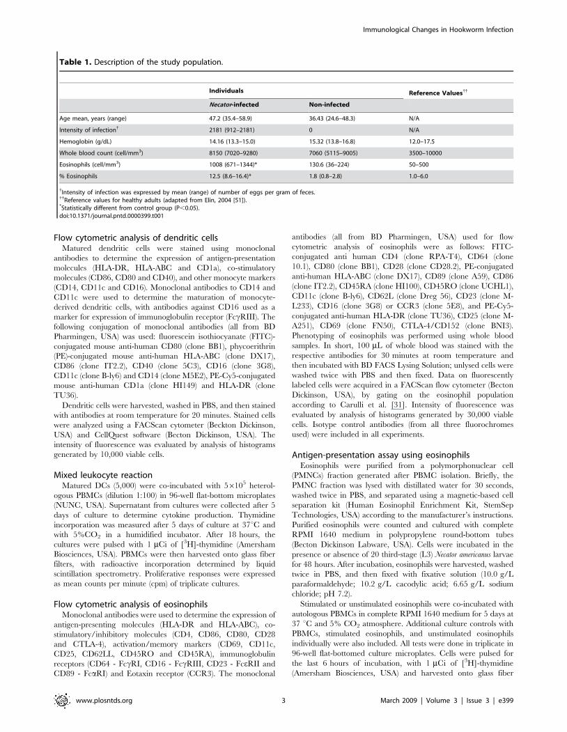
Table 1. Description of the study population.
Individuals Reference Values{{
Necator-infected Non-infected
Age mean, years (range) 47.2 (35.4–58.9) 36.43 (24.6–48.3) N/A
Intensity of infection{ 2181 (912–2181) 0 N/A
Hemoglobin (g/dL) 14.16 (13.3–15.0) 15.32 (13.8–16.8) 12.0–17.5
Whole blood count (cell/mm3) 8150 (7020–9280) 7060 (5115–9005) 3500–10000
Eosinophils (cell/mm3) 1008 (671–1344)* 130.6 (36–224) 50–500
% Eosinophils 12.5 (8.6–16.4)* 1.8 (0.8–2.8) 1.0–6.0
{Intensity of infection was expressed by mean (range) of number of eggs per gram of feces.{{Reference values for healthy adults (adapted from Elin, 2004 [51]).*Statistically different from control group (P,0.05).doi:10.1371/journal.pntd.0000399.t001
Immunological Changes in Hookworm Infection
www.plosntds.org 3 March 2009 | Volume 3 | Issue 3 | e399
filters. Radioactive incorporation was determined by liquid
scintillation spectrometry. Proliferative responses were expressed
as Stimulation index, calculated as follows: mean cpm of
stimulated eosinophils and PBMC co-cultures (triplicates) divided
by mean cpm of unstimulated eosinophils and PBMC co-cultures
(also in triplicates).
Supernatants from eosinophil cultures were collected for
determination of cytokine production after hookworm L3
stimulation and after incubation with PBMCs.
Determination of cytokine and chemokine production byELISA
All cytokines were detected and quantified in culture superna-
tants using cytokine-specific enzyme-linked immunosorbent assays
kits. IFN-c, TNF-a, IL-1b, IL-4, IL-5, IL-6, IL-10, IL-13 kits
(R&D Systems, USA) were used to detect cytokine production in
supernatants from the mixed leukocyte assay. IFN-c, TNF-a, IL-4,
IL-5, IL-10, IL-13, TARC/CCL17 and Eotaxin/CCL11 kits
(R&D Systems, USA) were used to detect cytokine/chemokine
production in cultures from eosinophils. Assays were performed
according to the manufacturer’s instructions. Biotin-conjugated
secondary antibodies were used, followed by streptavidin-HRP
(Amersham Biosciences, USA), and OPD substrate system
(Sigma). The colorimetric reaction was read in an automated
ELISA microplate reader at 492 nm. Calculations of cytokine/
chemokine concentrations from mean optical density values were
determined by interpolating diluted values from 4-parameter
model fitted by SOFTmax Pro 4.8. Results were expressed in pg/
mL, with the detection limits as follows: 7.8 pg/mL for IFN-c,
TNF-a and Eotaxin/CCL11; 3.9 pg/mL for IL-1b and TARC/
CCL17; 15.6 pg/mL for IL-4; 11.7 pg/mL for IL-5; 4.7 pg/mL
for IL-6; 23.4 pg/mL for IL-10; and 40 pg/mL for IL-13.
Statistical analysisThe Mann-Whitney test was used to determine the differences
(P value,0.05) of non-parametric variables (e.g., surface cell
markers, cell proliferation, and antigen presentation) between
Necator-infected individuals and non-infected individuals. All
statistics were performed using Prism 4.0b for Macintosh
(GraphPad Software, Inc.).
Results
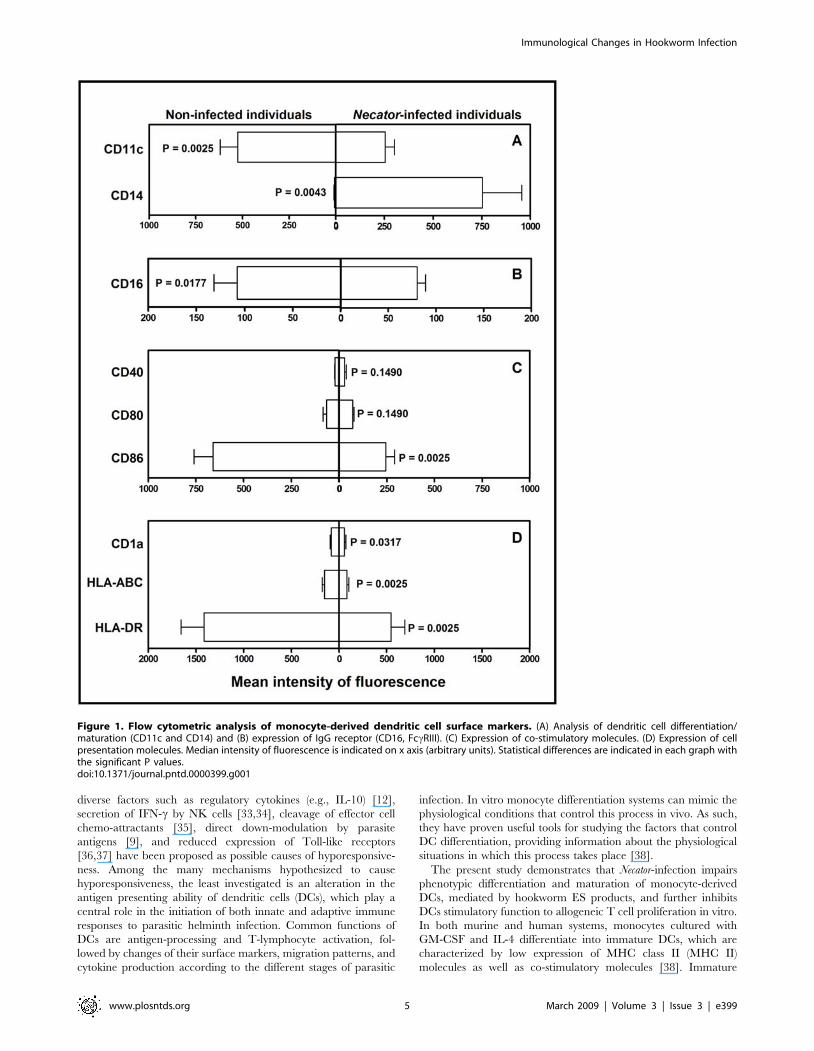
N. americanus infection impairs dendritic celldifferentiation
Analysis of surface cell markers of monocyte-derived dendritic
cells showed that DCs from Necator-infected individuals had an
impaired differentiation process, as evidenced by the differential
expression of CD11c and CD14 on the cell surface (Fig. 1A)
compared to non-infected individuals. Differentiation of the
monocytes into dendritic cells in non-infected individuals occurred
as expected, with a relatively higher expression of CD11c and an
absence lack of CD14 (Fig. 1A). However, dendritic cells from
Necator-infected individuals showed a significantly lower expression
of the immunoglobulin receptor CD16 (FccRIII, P = 0.0177,
Fig. 1B), the co-stimulatory molecule CD86 (P = 0.0025, Fig. 1C),
and cell presentation molecules, such as CD1a (P = 0.0317), HLA-
A, B, C and HLA-DR (P = 0.025 for both, Fig. 1D).
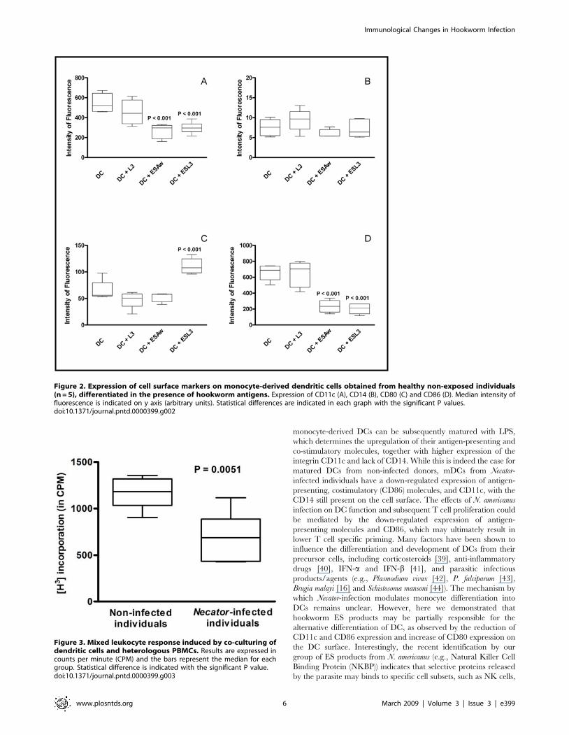
Monocyte-derived dendritic cells differentiated in the presence
of N. americanus excreted-secreted products both from adult worm
(ESAw) and L3 larvae (ESL3) showed a significant decreased
expression of CD11c and CD86 while presented higher expression
of CD80 (Fig 2). Interestingly, levels of CD14 expressed by
dendritic cells differentiated in the presence of hookworm antigens
were similar to those presented by control cells (Fig. 2B).
Decreased cell reactivity by mixed leukocyte responseIn order to assess the effect of the reduced expression of CD86
and antigen presentation molecules on dendritic cells from Necator-
infected patients, co-incubation with heterologous PBMCs was
performed as a mixed leukocyte response. Due to the differences in
the Major Histocompatibility Complex (MHC) marked cellular
proliferation as a rejection response are expected, as observed in
the co-cultures with dendritic cells from the control group (non-
infected individuals) (Fig. 3). Our results showed that co-cultures
with dendritic cells from Necator-infected patients resulted in
markedly ablated cell proliferation (P = 0.0051) compared to non-
infected individuals. This ablation may be the result of an
accompanying lower expression of co-stimulatory and antigen
presentation molecules on the cells of these patients.
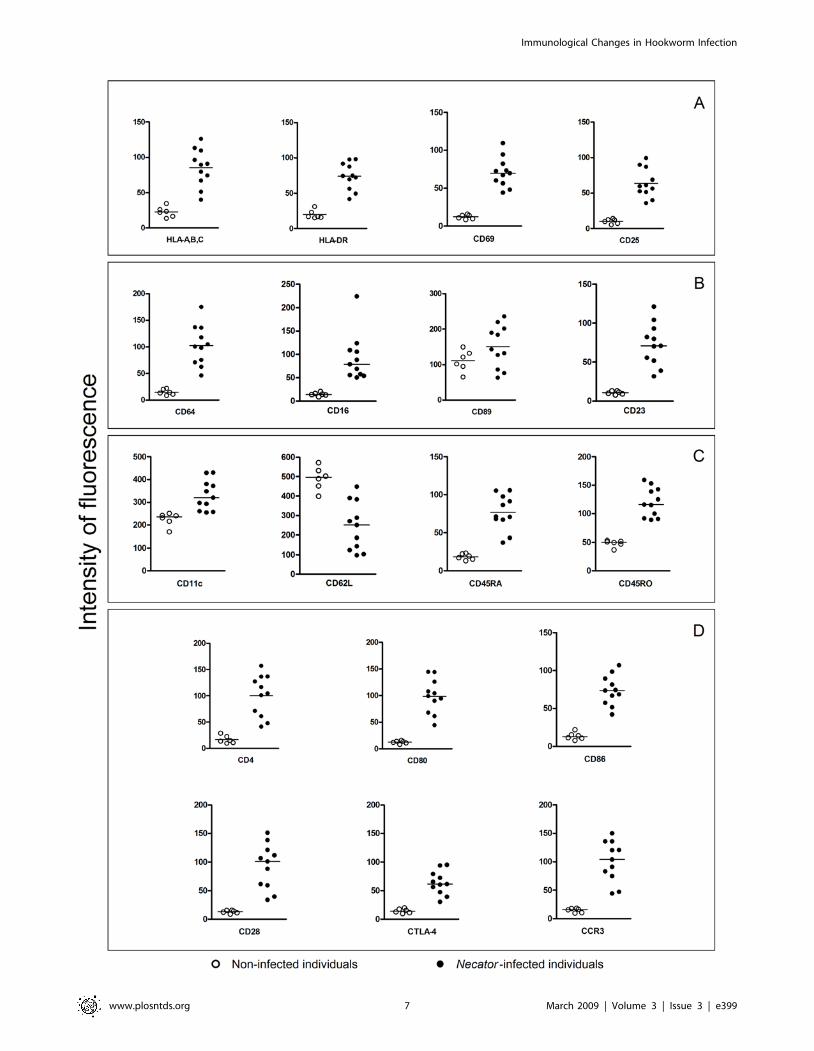
Phenotypic changes in the expression of eosinophilsurface markers on Necator-infected patients
While there was a marked downregulation surface marker
expression on dendritic cells from Necator-infected individuals,
circulating eosinophils from these same subjects showed upregu-
lated expression of major cell surface markers. We observed a
statistically significant increase in expression of cell presentation
molecules HLA-A,B,C and HLA-DR (P = 0.0011 for both),
activation markers CD69 and CD25 (P = 0.0011 and P = 0.0001,
respectively), naive/memory markers CD45RA and CD45RO
(P#0.0001 for both), immunoglobulin receptors (CD64 - FccRI,
CD16 - FccRIII and CD23 - FceRII, P = 0.0011 for all), integrin
CD11c (P#0.0001), accessory molecules (CD4, CD80, CD86,
CD28 and CD152; P = 0.0011 for all), and eotaxin receptor
(CCR3, P = 0.0001) (Fig. 4). On the other hand, the expression of
CD62L, an adhesion molecule, was significantly reduced on
eosinophils from Necator-infected individuals when compared with
the non-infected individuals (P = 0.0016).
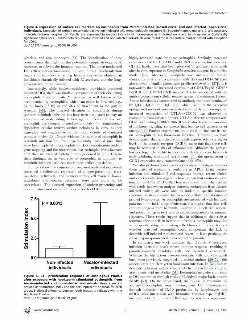
Cell presentation in human Necator-infection byeosinophils
Antigen-pulsed eosinophils from Necator-infected individuals
induced significant cell proliferation in autologous PBMCs
(SI = 4.18362.838), when compared with eosinophils from non-
infected individuals (SI = 1.22660.280, P = 0.0013, Fig. 5). Pro-
liferative responses of control cultures were similar to those
observed in co-cultures of primed eosinophils and PBMCs from
non-infected individuals (data not shown).
Cytokine and chemokine production by dendritic cellsand eosinophils
No statistically significant differences were observed in the
cytokine and chemokine production of dendritic cells and
eosinophils from Necator-infected and non-infected individuals
(data not shown). The cytokine/chemokine production of cultures
from both groups was marginal and reached levels close to the
detection limits of the assay for each analyte.
Discussion
Chronic human N. americanus infection is associated with a
profound ablation of cell proliferation that may even extend to
other parasitic infections and even mitogens (‘‘bystander effect’’)
[12,32]. The mechanisms underlying Necator-induced T cell
hyporesponsiveness have yet to be fully elucidated; however,
Immunological Changes in Hookworm Infection
www.plosntds.org 4 March 2009 | Volume 3 | Issue 3 | e399
diverse factors such as regulatory cytokines (e.g., IL-10) [12],
secretion of IFN-c by NK cells [33,34], cleavage of effector cell
chemo-attractants [35], direct down-modulation by parasite
antigens [9], and reduced expression of Toll-like receptors
[36,37] have been proposed as possible causes of hyporesponsive-
ness. Among the many mechanisms hypothesized to cause
hyporesponsiveness, the least investigated is an alteration in the
antigen presenting ability of dendritic cells (DCs), which play a
central role in the initiation of both innate and adaptive immune
responses to parasitic helminth infection. Common functions of
DCs are antigen-processing and T-lymphocyte activation, fol-
lowed by changes of their surface markers, migration patterns, and
cytokine production according to the different stages of parasitic
infection. In vitro monocyte differentiation systems can mimic the
physiological conditions that control this process in vivo. As such,
they have proven useful tools for studying the factors that control
DC differentiation, providing information about the physiological
situations in which this process takes place [38].
The present study demonstrates that Necator-infection impairs
phenotypic differentiation and maturation of monocyte-derived
DCs, mediated by hookworm ES products, and further inhibits
DCs stimulatory function to allogeneic T cell proliferation in vitro.
In both murine and human systems, monocytes cultured with
GM-CSF and IL-4 differentiate into immature DCs, which are
characterized by low expression of MHC class II (MHC II)
molecules as well as co-stimulatory molecules [38]. Immature
Figure 1. Flow cytometric analysis of monocyte-derived dendritic cell surface markers. (A) Analysis of dendritic cell differentiation/maturation (CD11c and CD14) and (B) expression of IgG receptor (CD16, FccRIII). (C) Expression of co-stimulatory molecules. (D) Expression of cellpresentation molecules. Median intensity of fluorescence is indicated on x axis (arbitrary units). Statistical differences are indicated in each graph withthe significant P values.doi:10.1371/journal.pntd.0000399.g001
Immunological Changes in Hookworm Infection
www.plosntds.org 5 March 2009 | Volume 3 | Issue 3 | e399
monocyte-derived DCs can be subsequently matured with LPS,
which determines the upregulation of their antigen-presenting and
co-stimulatory molecules, together with higher expression of the
integrin CD11c and lack of CD14. While this is indeed the case for
matured DCs from non-infected donors, mDCs from Necator-
infected individuals have a down-regulated expression of antigen-
presenting, costimulatory (CD86) molecules, and CD11c, with the
CD14 still present on the cell surface. The effects of N. americanus
infection on DC function and subsequent T cell proliferation could
be mediated by the down-regulated expression of antigen-
presenting molecules and CD86, which may ultimately result in
lower T cell specific priming. Many factors have been shown to
influence the differentiation and development of DCs from their
precursor cells, including corticosteroids [39], anti-inflammatory
drugs [40], IFN-a and IFN-b [41], and parasitic infectious
products/agents (e.g., Plasmodium vivax [42], P. falciparum [43],
Brugia malayi [16] and Schistosoma mansoni [44]). The mechanism by
which Necator-infection modulates monocyte differentiation into
DCs remains unclear. However, here we demonstrated that
hookworm ES products may be partially responsible for the
alternative differentiation of DC, as observed by the reduction of
CD11c and CD86 expression and increase of CD80 expression on
the DC surface. Interestingly, the recent identification by our
group of ES products from N. americanus (e.g., Natural Killer Cell
Binding Protein (NKBP)) indicates that selective proteins released
by the parasite may binds to specific cell subsets, such as NK cells,
Figure 2. Expression of cell surface markers on monocyte-derived dendritic cells obtained from healthy non-exposed individuals(n = 5), differentiated in the presence of hookworm antigens. Expression of CD11c (A), CD14 (B), CD80 (C) and CD86 (D). Median intensity offluorescence is indicated on y axis (arbitrary units). Statistical differences are indicated in each graph with the significant P values.doi:10.1371/journal.pntd.0000399.g002
Figure 3. Mixed leukocyte response induced by co-culturing ofdendritic cells and heterologous PBMCs. Results are expressed incounts per minute (CPM) and the bars represent the median for eachgroup. Statistical difference is indicated with the significant P value.doi:10.1371/journal.pntd.0000399.g003
Immunological Changes in Hookworm Infection
www.plosntds.org 6 March 2009 | Volume 3 | Issue 3 | e399
Immunological Changes in Hookworm Infection
www.plosntds.org 7 March 2009 | Volume 3 | Issue 3 | e399
platelets, and also monocytes [34]. The identification of these
proteins may shed light on this potentially unique strategy by N.
americanus to subvert the immune response. The down-modulated
DC differentiation/maturation induced during Necator-infection
might contribute to the cellular hyporesponsiveness observed in
individuals chronically infected with N. americanus and the long-
term survival of the parasite.
Interestingly, while hookworm-infected individuals presented
impaired DCs, there was marked upregulation of their circulating
eosinophils. Infection with N. americanus is well known to be
accompanied by eosinophilia, which can either be localized (e.g.,
to the lungs [45,46] or the sites of attachment in the gut) or
systemic [46]. The marked elevation in eosinophils during
parasitic helminth infection has long been postulated to play an
important role in defending the host against infection. In this case,
eosinophils are thought to mediate antibody- (or complement-)
dependent cellular toxicity against helminths in vitro, as they
aggregate and degranulate in the local vicinity of damaged
parasites in vivo [23]. Other evidence for the role of eosinophils in
helminth infection are from experimentally infected mice that
have been depleted of eosinophils by IL-5 neutralization and/or
gene targeting and the observation that eosinophil levels increase
after they are infected with helminths (reviewed in [23]). Despite
these findings, the in vivo role of eosinophils in immunity to
helminth infection has been much more difficult to define.
Our data show that eosinophils from Necator-infected individuals
presented a differential expression of antigen-presenting-, costi-
mulatory-, activation-, and memory-surface cell markers. Immu-
noglobulin and eotaxin receptors were also found to be
upregulated. The elevated expression of antigen-presenting and
co-stimulatory molecules, but reduced levels of CD62L, indicate a
highly activated state for these eosinophils. Similarly, increased
expression of MHC II, CD86, and CD69 molecules, but decreased
CD62L levels, have also been observed in activated eosinophils
after in vitro exposure to Strongyloides stercolaris antigens in a murine
model [47]. Moreover, comprehensive analysis of human
eosinophils after in vitro activation with IL-5 and GM-CSF have
also showed a similar phenotypic profile (reviewed in [27]). It is
noteworthy that the increased expression of CD64/FccRI, CD16/
FccRIII and CD23/FceRII may be directly associated with the
antibody-dependent cellular toxicity against N. americanus. Indeed,
Necator-infection is characterized by antibody responses dominated
by IgG1, IgG4, and IgE [21], which bind to the receptors
upregulated on hookworm-activated eosinophils. Surprisingly, an
increased expression of CTLA-4/CD152 was observed in
eosinophils from infected donors. CTLA-4 directly competes with
CD28 for binding CD80/CD86 (B7) and also directs the assembly
of inhibitory signaling complexes that lead to cell quiescence or
anergy [48]. Further experiments are needed to elucidate its role
on eosinophils during hookworm infection. Moreover, we have
demonstrated that activated eosinophils express relatively higher
levels of the eotaxin receptor (CCR3), suggesting that these cells
may be recruited to sites of inflammation. Although the parasite
has developed the ability to specifically cleave eotaxin, hypothet-
ically inhibiting eosinophil recruitment [35], the upregulation of
CCR3 expression may counterbalance this effect.
We also performed in vitro experiments to investigate whether
these activated eosinophils could act as APCs during Necator-
infection and stimulate T cell responses. Indeed, recent clinical
and experimental investigations have shown that eosinophils can
function as APCs [23,47,49]. Here we showed that, when pulsed
with crude hookworm antigen extracts, eosinophils from Necator-
infected individuals were able to initiate a specific immune
response, as demonstrated by increased cellular proliferation of
primed lymphocytes. As eosinophils are associated with helminth
parasites at the initial stage of infection, it is possible that these cells
capture antigens from helminths, migrate to T cell–rich regions,
and present antigens to T cells to initiate antigen-specific immune
responses. These results suggest that in addition to their role as
terminal effector cells in helminth infections, eosinophils may also
act as specific antigen-presenting cells. However, it is not clear yet
whether activated eosinophils could compensate the lack of
dendritic cell-induced response and revert, at least partially, the
classic hyporesponsiveness induced by the parasite.
In summary, our work indicates that chronic N. americanus
infection alters the host’s innate immune response, resulting in
parasite-impaired dendritic cells and activated eosinophils.
Whereas the interaction between dendritic cells and eosinophils
have been previously suggested by several authors [50–52], this
association is not clear yet in hookworm infection. In fact, human
dendritic cells may induce eosinophil chemotaxis by secreting an
arachidonic acid metabolite [51]. Eosinophils may also contribute
to DC maturation through eosinophil-derived major basic protein
(MBP) [50]. On the other hand, the release of histamine by
activated eosinophils may downregulate DC differentiation
through induction of IL-10 production by lymphocytes and
mDCs, after interaction with histamine receptor type 2 (HR2)
on these cells [53]. Indeed, HR2 agonists acts as a suppressive
Figure 5. Cell proliferation response of autologous PBMCsafter exposure with hookworm stimulated eosinophils fromNecator-infected and non-infected individuals. Results are ex-pressed as stimulation index and the bars represent the mean for eachgroup. Statistical difference between both groups is indicated with thesignificant P value.doi:10.1371/journal.pntd.0000399.g005
Figure 4. Expression of surface cell markers on eosinophils from Necator-infected (closed circle) and non-infected (open circle)individuals. Expression of antigen presentation/activation molecules (A), immunoglobulin receptors (B), integrin/memory markers (C) and accessorymolecules/eotaxin receptor (D). Results are expressed in median intensity of fluorescence as indicated on y axis (arbitrary units). Statisticallysignificant differences (P#0.05) between Necator-infected and non-infected individuals were found for all cell surface markers tested with exceptionfor CD89.doi:10.1371/journal.pntd.0000399.g004
Immunological Changes in Hookworm Infection
www.plosntds.org 8 March 2009 | Volume 3 | Issue 3 | e399
molecule for antigen presentation capacity, while enhancing IL-10
production and IL-10-producing T cells [54,55].
Based on the observation that DCs are required for develop-
ment and maintenance of chronic eosinophilic airway inflamma-
tion [56,57], the study of the eosinophil and DC interaction opens
new strategies for targeting key factors to interfere with an
eosinophil induced/enhanced state of diseases, such as asthma,
atopic dermatitis and helminthic infections [50]. Whether this
profile of host’s innate immune response in N. americanus infection
will provide further benefit to parasite survival through modula-
tion of required adaptive responses remains to be elucidated. In
the present study, we evaluated the profile of cellular components
associated with the innate immune response against hookworm
infection, comparing samples from hookworm mono-infected
individuals living in endemic areas and healthy non-exposed
volunteers. Egg-negative individuals from endemic areas were not
included as controls, since the limited sensitivity of fecal exams and
the long pre-patency period make it difficult to assure that the
volunteers were not infected. Moreover, it has been previously
shown that the immunological status of helminth-infected patients
remains unaltered after anthelmintic treatment for several months
[12,58]. Although all individuals enrolled in our study received
fully medical support and were matched by nutritional status, we
cannot exclude the influence of other factors such as viral and
bacterial infections. Our results should be further validated by
large immunoepidemiological surveys in endemic areas.
Author Contributions
Conceived and designed the experiments: RTF GGLC PAF HdCS CLM
RCO SMG JMB. Performed the experiments: RTF GGLC PAF CLM
RCO SMG JMB. Analyzed the data: RTF GGLC PAF HdCS CLM
OdSC RCO SMG JMB. Contributed reagents/materials/analysis tools:
RTF GGLC PAF HdCS CLM OdSC RCO SMG JMB. Wrote the paper:
RTF GGLC PAF HdCS OdSC RCO SMG JMB.
References
1. de Silva NR, Brooker S, Hotez PJ, Montresor A, Engels D, et al. (2003) Soil-transmitted helminth infections: updating the global picture. Trends Parasitol
19: 547–551.
2. Chan MS (1997) The global burden of intestinal nematode infections–fifty yearson. Parasitol Today 13: 438–443.
3. Brooker S, Peshu N, Warn PA, Mosobo M, Guyatt HL, et al. (1999) The
epidemiology of hookworm infection and its contribution to anaemia amongpre-school children on the Kenyan coast. Trans R Soc Trop Med Hyg 93:
240–246.
4. Lwambo NJ, Bundy DA, Medley GF (1992) A new approach to morbidity risk
assessment in hookworm endemic communities. Epidemiol Infect 108: 469–481.
5. Stoltzfus RJ, Dreyfuss ML, Chwaya HM, Albonico M (1997) Hookworm controlas a strategy to prevent iron deficiency. Nutr Rev 55: 223–232.
6. Albonico M, Crompton DW, Savioli L (1999) Control strategies for human
intestinal nematode infections. Adv Parasitol 42: 277–341.
7. Candolfi E, Hunter CA, Remington JS (1994) Mitogen- and antigen-specificproliferation of T cells in murine toxoplasmosis is inhibited by reactive nitrogen
intermediates. Infect Immun 62: 1995–2001.
8. Dai WJ, Gottstein B (1999) Nitric oxide-mediated immunosuppression followingmurine Echinococcus multilocularis infection. Immunology 97: 107–116.
9. Geiger SM, Caldas IR, Mc Glone BE, Campi-Azevedo AC, De Oliveira LM, et
al. (2007) Stage-specific immune responses in human Necator americanus
infection. Parasite Immunol 29: 347–358.
10. Schleifer KW, Mansfield JM (1993) Suppressor macrophages in Africantrypanosomiasis inhibit T cell proliferative responses by nitric oxide and
prostaglandins. J Immunol 151: 5492–5503.
11. Semnani RT, Liu AY, Sabzevari H, Kubofcik J, Zhou J, et al. (2003) Brugiamalayi microfilariae induce cell death in human dendritic cells, inhibit their
ability to make IL-12 and IL-10, and reduce their capacity to activate CD4+ Tcells. J Immunol 171: 1950–1960.
12. Geiger SM, Massara CL, Bethony J, Soboslay PT, Correa-Oliveira R (2004)
Cellular responses and cytokine production in post-treatment hookworm
patients from an endemic area in Brazil. Clin Exp Immunol 136: 334–340.
13. King CL, Mahanty S, Kumaraswami V, Abrams JS, Regunathan J, et al. (1993)Cytokine control of parasite-specific anergy in human lymphatic filariasis.
Preferential induction of a regulatory T helper type 2 lymphocyte subset. J ClinInvest 92: 1667–1673.
14. Loke P, MacDonald AS, Robb A, Maizels RM, Allen JE (2000) Alternatively
activated macrophages induced by nematode infection inhibit proliferation via
cell-to-cell contact. Eur J Immunol 30: 2669–2678.
15. Semnani RT, Law M, Kubofcik J, Nutman TB (2004) Filaria-induced immuneevasion: suppression by the infective stage of Brugia malayi at the earliest host-
parasite interface. J Immunol 172: 6229–6238.
16. Semnani RT, Sabzevari H, Iyer R, Nutman TB (2001) Filarial antigens impairthe function of human dendritic cells during differentiation. Infect Immun 69:
5813–5822.
17. Whelan M, Harnett MM, Houston KM, Patel V, Harnett W, et al. (2000) Afilarial nematode-secreted product signals dendritic cells to acquire a phenotype
that drives development of Th2 cells. J Immunol 164: 6453–6460.
18. Chow SC, Brown A, Pritchard D (2000) The human hookworm pathogen
Necator americanus induces apoptosis in T lymphocytes. Parasite Immunol 22:21–29.
19. Jenson JS, O’Connor R, Osborne J, Devaney E (2002) Infection with Brugia
microfilariae induces apoptosis of CD4(+) T lymphocytes: a mechanism ofimmune unresponsiveness in filariasis. Eur J Immunol 32: 858–867.
20. Taylor MD, LeGoff L, Harris A, Malone E, Allen JE, et al. (2005) Removal of
regulatory T cell activity reverses hyporesponsiveness and leads to filarialparasite clearance in vivo. J Immunol 174: 4924–4933.
21. Loukas A, Prociv P (2001) Immune responses in hookworm infections. Clin
Microbiol Rev 14: 689–703, table of contents.
22. Ovington KS, Behm CA (1997) The enigmatic eosinophil: investigation of the
biological role of eosinophils in parasitic helminth infection. Mem Inst Oswaldo
Cruz 92 Suppl 2: 93–104.
23. Rothenberg ME, Hogan SP (2006) The eosinophil. Annu Rev Immunol 24:
147–174.
24. Taliaferro WR, Sarles MP (1939) The cellular reactions in the skin, lungs, and
intestine of normal and immune rats after infection with Nippostrongylus brasiliensis.
J Infect Dis 64: 157–192.
25. Klion AD, Nutman TB (2004) The role of eosinophils in host defense against
helminth parasites. J Allergy Clin Immunol 113: 30–37.
26. Desakorn V, Suntharasamai P, Pukrittayakamee S, Migasena S, Bunnag D
(1987) Adherence of human eosinophils to infective filariform larvae of Necator
americanus in vitro. Southeast Asian J Trop Med Public Health 18: 66–72.
27. Bochner BS (2000) Systemic activation of basophils and eosinophils: markers and
consequences. J Allergy Clin Immunol 106: S292–302.
28. Katz N, Chaves A, Pellegrino J (1972) A simple device for quantitative stool
thick-smear technique in Schistosomiasis mansoni. Rev Inst Med Trop Sao
Paulo 14: 397–400.
29. WHO (1995) Physical status: the use and interpretation of anthropometry.
Geneva: WHO.
30. Jardim-Botelho A, Brooker S, Geiger SM, Fleming F, Lopes ACS, et al. (2008)
Age patterns in undernutrition and helminth infection in a rural area of Brazil:
associations with ascariasis and hookworm. Trop Med Int Health 13: 458–467.
31. Carulli G, Sbrana S, Azzara A, Minnucci S, Angiolini C, et al. (1998) Detection
of eosinophils in whole blood samples by flow cytometry. Cytometry 34:
272–279.
32. Fujiwara RT, Geiger SM, Bethony J, Mendez S (2006) Comparative
immunology of human and animal models of hookworm infection. Parasite
Immunol 28: 285–293.
33. Teixeira-Carvalho A, Fujiwara RT, Stemmy E, Olive D, Damsker JM, et al.
(2008) Binding of excreted/secreted products of adult Necator americanus to
human NK cells in hookworm infected individuals from Brazil. Infect Immun
76: 5810–5816.
34. Hsieh GC, Loukas A, Wahl AM, Bhatia M, Wang Y, et al. (2004) A secreted
protein from the human hookworm necator americanus binds selectively to NK
cells and induces IFN-gamma production. J Immunol 173: 2699–2704.
35. Culley FJ, Brown A, Conroy DM, Sabroe I, Pritchard DI, et al. (2000) Eotaxin is
specifically cleaved by hookworm metalloproteases preventing its action in vitro
and in vivo. J Immunol 165: 6447–6453.
36. Hartgers FC, Obeng BB, Kruize YC, Duijvestein M, de Breij A, et al. (2008)
Lower Expression of TLR2 and SOCS-3 Is Associated with Schistosoma
haematobium Infection and with Lower Risk for Allergic Reactivity in Children
Living in a Rural Area in Ghana. PLoS Negl Trop Dis 2: e227. doi:10.1371/
journal.pntd.0000227.
37. Babu S, Blauvelt CP, Kumaraswami V, Nutman TB (2006) Cutting edge:
diminished T cell TLR expression and function modulates the immune response
in human filarial infection. J Immunol 176: 3885–3889.
38. Leon B, Lopez-Bravo M, Ardavin C (2005) Monocyte-derived dendritic cells.
Semin Immunol 17: 313–318.
39. Matyszak MK, Citterio S, Rescigno M, Ricciardi-Castagnoli P (2000)
Differential effects of corticosteroids during different stages of dendritic cell
maturation. Eur J Immunol 30: 1233–1242.
40. Zhang R, Bharadwaj U, Li M, Chen C, Yao Q (2007) Effects of pentoxifylline
on differentiation, maturation, and function of human CD14+ monocyte-
derived dendritic cells. J Immunother 30: 89–95.
Immunological Changes in Hookworm Infection
www.plosntds.org 9 March 2009 | Volume 3 | Issue 3 | e399
41. McRae BL, Nagai T, Semnani RT, van Seventer JM, van Seventer GA (2000)
Interferon-alpha and -beta inhibit the in vitro differentiation of immunocom-petent human dendritic cells from CD14(+) precursors. Blood 96: 210–217.
42. Bueno LL, Fujiwara RT, Soares IS, Braga EM (2008) Direct effect of
Plasmodium vivax recombinant vaccine candidates AMA-1 and MSP-119 onthe innate immune response. Vaccine 26: 1204–1213.
43. Urban BC, Ferguson DJ, Pain A, Willcox N, Plebanski M, et al. (1999)Plasmodium falciparum-infected erythrocytes modulate the maturation of
dendritic cells. Nature 400: 73–77.
44. MacDonald AS, Straw AD, Bauman B, Pearce EJ (2001) CD8- dendritic cellactivation status plays an integral role in influencing Th2 response development.
J Immunol 167: 1982–1988.45. Maxwell C, Hussain R, Nutman TB, Poindexter RW, Little MD, et al. (1987)
The clinical and immunologic responses of normal human volunteers to lowdose hookworm (Necator americanus) infection. Am J Trop Med Hyg 37:
126–134.
46. White CJ, Maxwell CJ, Gallin JI (1986) Changes in the structural and functionalproperties of human eosinophils during experimental hookworm infection.
J Infect Dis 154: 778–783.47. Padigel UM, Lee JJ, Nolan TJ, Schad GA, Abraham D (2006) Eosinophils can
function as antigen-presenting cells to induce primary and secondary immune
responses to Strongyloides stercoralis. Infect Immun 74: 3232–3238.48. Linsley PS, Golstein P (1996) Lymphocyte activation: T-cell regulation by
CTLA-4. Curr Biol 6: 398–400.
49. Shi HZ (2004) Eosinophils function as antigen-presenting cells. J Leukoc Biol 76:
520–527.50. Lotfi R, Lotze MT (2008) Eosinophils induce DC maturation, regulating
immunity. J Leukoc Biol 83: 456–460.
51. Zimpfer U, Dichmann S, Termeer CC, Simon JC, Schroder JM, et al. (2000)Human dendritic cells are a physiological source of the chemotactic arachidonic
acid metabolite 5-oxo-eicosatetraenoic acid. Inflamm Res 49: 633–638.52. Corthay A (2006) A three-cell model for activation of naive T helper cells.
Scand J Immunol 64: 93–96.
53. Jutel M, Blaser K, Akdis CA (2005) Histamine in allergic inflammation andimmune modulation. Int Arch Allergy Immunol 137: 82–92.
54. Caron G, Delneste Y, Roelandts E, Duez C, Herbault N, et al. (2001) Histamineinduces CD86 expression and chemokine production by human immature
dendritic cells. J Immunol 166: 6000–6006.55. Elenkov IJ, Webster E, Papanicolaou DA, Fleisher TA, Chrousos GP, et al.
(1998) Histamine potently suppresses human IL-12 and stimulates IL-10
production via H2 receptors. J Immunol 161: 2586–2593.56. Lambrecht BN, Hammad H (2003) Taking our breath away: dendritic cells in
the pathogenesis of asthma. Nat Rev Immunol 3: 994–1003.57. Lambrecht BN, Salomon B, Klatzmann D, Pauwels RA (1998) Dendritic cells
are required for the development of chronic eosinophilic airway inflammation in
response to inhaled antigen in sensitized mice. J Immunol 160: 4090–4097.58. Loukas A, Constant SL, Bethony JM (2005) Immunobiology of hookworm
infection. FEMS Immunol Med Microbiol 43: 115–124.
Immunological Changes in Hookworm Infection
www.plosntds.org 10 March 2009 | Volume 3 | Issue 3 | e399