natural history of dilated cardiomyopathy due to lamin a/c gene mutations
TRANSCRIPT
doi:10.1016/S0735-1097(02)02954-6 2003;41;771-780 J. Am. Coll. Cardiol.
Luisa Mestroni, and Familial Dilated Cardiomyopathy Registry Research Group Moss, Wai-Lun P. Li, Gary L. Stetler, Francesco Muntoni, Michael R. Bristow,
Debra A. Ferguson, Gary L. Brodsky, Mark M. Boucek, Jean Lascor, Andrew C.Alastair D. Robertson, Elisa Carniel, Andrea Di Lenarda, Teresa J. Bohlmeyer, Matthew R. G. Taylor, Pamela R. Fain, Gianfranco Sinagra, Misi L. Robinson,
Natural history of dilated cardiomyopathy due to lamin A/C gene mutations
This information is current as of July 22, 2011
http://content.onlinejacc.org/cgi/content/full/41/5/771located on the World Wide Web at:
The online version of this article, along with updated information and services, is
by on July 22, 2011 content.onlinejacc.orgDownloaded from

Cardiomyopathy
Natural History of DilatedCardiomyopathy Due to Lamin A/C Gene MutationsMatthew R. G. Taylor, MD,* Pamela R. Fain, PHD,*†‡ Gianfranco Sinagra, MD, FESC,§Misi L. Robinson,� Alastair D. Robertson, PHD,* Elisa Carniel, MD,§ Andrea Di Lenarda, MD, FESC,§Teresa J. Bohlmeyer, MD,* Debra A. Ferguson, MS,* Gary L. Brodsky, PHD,* Mark M. Boucek, MD,*¶Jean Lascor, MS,¶ Andrew C. Moss, BA,* Wai-Lun P. Li, BS,*† Gary L. Stetler, PHD,†Francesco Muntoni, MD, FRCPCH,# Michael R. Bristow, MD, PHD, FACC,*Luisa Mestroni, MD, FACC, FESC,* Familial Dilated Cardiomyopathy Registry Research GroupDenver, Colorado; Trieste, Italy; Omaha, Nebraska; and London, United Kingdom
OBJECTIVES We examined the prevalence, genotype-phenotype correlation, and natural history of laminA/C gene (LMNA) mutations in subjects with dilated cardiomyopathy (DCM).
BACKGROUND Mutations in LMNA have been found in patients with DCM with familial conduction defectsand muscular dystrophy, but the clinical spectrum, prognosis, and clinical relevance oflaminopathies in DCM are unknown.
METHODS A cohort of 49 nuclear families, 40 with familial DCM and 9 with sporadic DCM (269subjects, 105 affected), was screened for mutations in LMNA using denaturing high-performance liquid chromatography and sequence analysis. Bivariate analysis of clinicalpredictors of LMNA mutation carrier status and Kaplan-Meier survival analysis wereperformed.
RESULTS Mutations in LMNA were detected in four families (8%), three with familial (R89L, 959delT,R377H) and one with sporadic DCM (S573L). There was significant phenotypic variability,but the presence of skeletal muscle involvement (p � 0.001), supraventricular arrhythmia(p � 0.003), conduction defects (p � 0.01), and “mildly” DCM (p � 0.006) were predictorsof LMNA mutations. The LMNA mutation carriers had a significantly poorer cumulativesurvival compared with non-carrier DCM patients: event-free survival at the age of 45 yearswas 31% versus 75% in non-carriers.
CONCLUSIONS Mutations in LMNA cause a severe and progressive DCM in a relevant proportion ofpatients. Mutation screening should be considered in patients with DCM, in particular whenclinical predictors of LMNA mutation are present, regardless of family history. (J Am CollCardiol 2003;41:771–80) © 2003 by the American College of Cardiology Foundation
Dilated cardiomyopathy (DCM) is a severe disease of heartmuscle characterized by progressive ventricular dilation andimpaired systolic function. Population surveys estimated aprevalence of 1 DCM case in 2,500 individuals (1). Inrecent years the contribution of genetic mutations to theetiology of DCM has been appreciated. Familial dilatedcardiomyopathy (FDC) is now estimated to account for asmany as 50% of all cases of “idiopathic” DCM (2,3).Mutations in 10 cytoskeletal/sarcomeric protein-encodinggenes have been identified in FDC, including dystrophin(4,5), desmin (6), lamin A/C (7–9), cardiac actin (10),tafazzin (11), cardiac beta-myosin heavy chain and cardiac
troponin T (12), alpha-tropomyosin (13), delta-sarcoglycan(14), and titin (15).
The lamin A/C gene (LMNA) maps on the long arm ofchromosome 1 (1q21.2-q21.3) and encodes two main iso-forms by alternative splicing (16), lamin A and C. Laminproteins are type V intermediate filaments, major compo-nents of the nuclear lamina (a filamentous structure thatsupports the inner nuclear membrane), and are classified asA-type or B-type (17,18). A-type lamins (lamin A and C)are expressed exclusively in differentiated non-proliferatingcells (18,19). Mutations in LMNA cause DCM, frequentlycomplicated by conduction system defects and variableskeletal muscle involvement (7,9,20–22). The LMNA mu-tations have also been reported in autosomal dominant (23)and recessive (24) Emery-Dreifuss muscular dystrophy(EDMD), autosomal dominant limb-girdle muscular dys-trophy (LGMD) (8), sensory and motor axonal neuropathyCharcot-Marie-Tooth type 2 (25), and familial partiallipodystrophy, characterized by the development of abnor-mal patterns of adipose tissue distribution (26,27).
The prevalence of LMNA mutations in patients withDCM, the genotype-phenotype correlations, and the im-
From the *University of Colorado Cardiovascular Institute, †Human MedicalGenetics Program, ‡Department of Internal Medicine, University of ColoradoHealth Sciences Center, Denver, Colorado; §Division of Cardiology, OspedaleMaggiore and University of Trieste, Trieste, Italy; �Transgenomic, Inc., Omaha,Nebraska; ¶Division of Cardiology, The Children’s Hospital, Denver, Colorado; andthe #Neuromuscular Unit, Department of Pediatrics and Neonatal Medicine, Impe-rial College School of Medicine, Hammersmith Hospital, London, United Kingdom.Supported by the National Institutes of Health/National Heart, Lung, and BloodInstitute (1RO1 HL69071-01), the Muscular Dystrophy Association U.S.A.(PN0007056), the American Heart Association (0250271N), and the EU Myo-Cluster grant QLG1 CT 1999 00870.
Manuscript received May 16, 2002; revised manuscript received August 23, 2002,accepted October 4, 2002.
Journal of the American College of Cardiology Vol. 41, No. 5, 2003© 2003 by the American College of Cardiology Foundation ISSN 0735-1097/03/$30.00Published by Elsevier Science Inc. doi:10.1016/S0735-1097(02)02954-6
by on July 22, 2011 content.onlinejacc.orgDownloaded from

pact on survival are still unknown. Over the past twodecades, we have studied families with both FDC andsporadic DCM (28,29). Here we report the results of ourcomprehensive genetic screening of LMNA in 49 unrelatedfamilies comprised of 40 familial and 9 sporadic cases ofDCM.
METHODS
Patient population. The 49 families of patients withDCM (269 subjects, 105 affected) were evaluated by theinvestigators in Europe and the U.S. Subjects (probands andfamily members) with DCM were recruited from generalcardiology clinics at the University of Colorado HealthSciences Center and Children’s Hospital (Denver, Colo-rado) and the University of Trieste (Trieste, Italy) from1991 to 2001 (28,29). Subjects were identified solely on thebasis of having DCM and were not prescreened or prese-lected for any neuromuscular, cardiac conduction, or anyother non-cardiac phenotype. Detailed clinical informationwas obtained from each subject, including family history;age of presentation; initial symptoms of heart failure;New York Heart Association classification; physical exam-ination; serum creatine kinase (MM isoform); electrocar-diograms, echocardiograms, and, when appropriate, Holtermonitoring; treadmill testing; invasive examination (rightand left heart catheterization, ventriculography, coronaryangiogram, and endomyocardial biopsy); and neuromuscu-lar investigations (electromyography and skeletal musclebiopsy).
The diagnostic criteria for FDC followed the Guidelinesfor the Study of Familial Dilated Cardiomyopathies (30). Inbrief, individuals were classified as affected on the basis ofmajor and minor criteria. The major criteria were: 1) leftventricular ejection fraction �45% or frameshift �25%, and2) left ventricular end-diastolic dimension �117% of thepredicted value corrected for age and body surface area(31). Individuals were classified as healthy when found tobe normal or affected by known diseases and unknownwhen isolated minor cardiac or skeletal muscle abnormal-ities were observed, as described in detail previously (30).The final study population included 40 unrelated familieswith FDC, and 9 with non-familial sporadic DCM(Table 1). Families with X-linked DCM due to dystro-phin gene mutations (32,33) were excluded from themutation analysis. Written informed consent was ob-tained from all subjects.Genetic analysis. Genomic deoxyribonucelic acid (DNA)was extracted from blood leukocytes according to standardtechniques. Polymerase chain reaction (PCR) primers (LifeTechnologies, Carlsbad, California) were designed to am-plify all protein coding exons of LMNA, and denaturinghigh-performance liquid chromatography (DHPLC) anal-ysis was carried out on a WAVE DNA fragment analysissystem (Transgenomic, San Jose, California). Elution pro-files that differed from wild-type patterns were selected forsequence analysis, purified using QIAquick PCR Purifica-tion Kits (Qiagen, Valencia, California), and then se-quenced bidirectionally using an ABI 377 DNA sequencer(Applied Biosystem, Foster City, California). Putativedisease-causing mutations were evaluated for segregationwith DCM phenotype within a family and were analyzedfor change in predicted amino acid sequence, predictedprotein secondary and tertiary structure, and for alterationsin ribonucelic acid (RNA) splicing. In the cases of predictedchanges in amino acid sequence, the altered residues wereanalyzed for conservation across different species. A geneticchange was considered to be a putative disease-causingmutation when it altered the predicted amino acid sequence,cosegregated with the disease within the family, was absentin over 150 normal, ethnically matched controls (300chromosomes, p � 0.05) (34) (data not shown), was altering
Abbreviations and AcronymsDCM � dilated cardiomyopathyDHPLC � denaturing high-performance liquid
chromatographyDNA � deoxyribonucelic acidEDMD � Emery-Dreifuss muscular dystrophyFDC � familial dilated cardiomyopathyLGMD � limb-girdle muscular dystrophyLMNA � lamin A/C geneMDDC � dilated cardiomyopathy with muscle diseasePCR � polymerase chain reactionRNA � ribonucelic acidSNP � single-nucleotide polymorphism
Table 1. Phenotypic Characterization (29) and Molecular Epidemiology of the Study Population
PhenotypeNumber of
FamiliesSubjects
ExaminedAffectedSubjects
Families WithLMNA Mutation
Familial DCM, total 40 236 95 3Autosomal dominant 25 163 62 1Autosomal recessive 4 15 7 —With conduction defect 3 18 8 —With muscle disease 4 27 11 2X-linked 2 5 3 —Unclassifiable 2 8 4 —
Sporadic DCM 9 33 10 1Total 49 269 105 4
DCM � dilated cardiomyopathy; LMNA � lamin A/C gene.
772 Taylor et al. JACC Vol. 41, No. 5, 2003LMNA Mutations in DCM March 5, 2003:771–80
by on July 22, 2011 content.onlinejacc.orgDownloaded from

Figure 1. The denaturing high performance liquid chromatography elution profiles for lamin A/C gene (LMNA) mutations detected in three families with dilated cardiomyopathy. (Top) Pedigree of thefamilies: individuals are indicated by generation and pedigree number. Affected status is indicated by filled symbols, unaffected by clear symbols. The (�) and (�) symbols indicate the presence of the mutantallele and the wild type, respectively. (Middle) Elution profiles for the wild-type and patients carrying the LMNA mutations (G266T: III-2; 959delT: II-1; G1130A: II-2; C1718T: II-2). (Bottom) The directsequencing results illustrate the corresponding nucleotide substitutions (reverse sequence shown for G1130A mutation).
773JACC
Vol.41,No.5,2003Taylor
etal.
March
5,2003:771–80LM
NAM
utationsin
DCM
by on July 22, 2011 content.onlinejacc.org
Dow
nloaded from

a highly conserved residue throughout evolution, and waspredicted to alter any combination of protein structure,charge, or function.
Statistical analysis. Comparison of predictor variables andcarrier status was done by Fisher exact test for binary predictorsand by the Wilcoxon rank-sum test for continuous predictors.
Table 2. Phenotype and Genotype of 12 Patients with DCM Heterozygous for LMNA Mutations
Family Mutation Codon Exon ID Age Gender NYHA CK-MM Arrhythmia
TSFDC13 G266T R89L 1 II-2 39 M 4 N Atrial fibrillationII-2 23† F 3 N Sinus rhythmII-1 22 M 4 Upper N Ventricular tachycardia
Atrial ectopyMDDC1§ 959delT 320 6 II-1 22 M 2 N-3X Ventricular and supraventricular
tachycardia
II-5 30 M 3 N Atrial fibrillationVentricular tachycardia
III-1 4 F 1 N Atrial ectopyIII-3 16 M 1 3X Supraventricular tachycardia
Ventricular ectopyIII-4 18 F 1 4X Supraventricular tachycardia
DNFDC33 G1130T R377H 6 I-2 40 F 2II-2 31 F 3 Upper N Atrial fibrillation
Ventricular tachycardiaVentricular fibrillation
SDCM8 C1718T S573L 11 II-2 50 F 3 N Ventricular tachycardiaVentricular ectopy
II-3 60 F 1 N Sinus rhythm
*At autopsy. †Peri-partum. ‡At heart transplant. §Reported by Brodsky et al. (9).CK-MM � creatine kinase, MM isoform; DCM � dilated cardiomyopathy; EDMD � Emery-Dreifuss muscular dystrophy; LVEF � ejection fraction (%); FS � fractional
shortening; ID � pedigree identification number; LGMD � limb-girdle muscular dystrophy; LMNA � lamin A/C gene; LVEDD � left ventricular end-diastolic dimension(31); N � normal; NYHA � New York Heart Association (class).
Figure 2. Structure of lamin A/C gene (LMNA) and of the lamin A/C protein. LMNA encodes lamin A (664 amino acids) and lamin C (572 amino acids)by alternative splicing in exon 10. The region common to both lamins includes the globular head, the �-helical domain (coils 1a, 1b, and 2, separated bylinkers depicted as diagonal stripes), and part of the tail (566 amino acids). The mutations identified in patients with DCM, the corresponding nucleotide,and their position on the protein are indicated (star � splice site mutation).
774 Taylor et al. JACC Vol. 41, No. 5, 2003LMNA Mutations in DCM March 5, 2003:771–80
by on July 22, 2011 content.onlinejacc.orgDownloaded from

Event-free survival analyses used 2 � 2 contingencytables for occurrence of event and the Cox proportionalhazards model for time from birth to event, comparing bycarrier status. Three survival events were used: major cardiacevent, transplant, and cardiovascular death. A major cardiacevent was defined as: 1) hospitalization for worsening heartfailure, 2) hospitalization for major arrhythmia (hypokineticor hyperkinetic), or 3) a thromboembolic event. As thesesurvival events constituted competing risks, analyses usedonly the following outcomes: composite of major cardiacevent, transplant, and death; composite of transplant anddeath; and death alone. For composite outcomes, survivaltime was time to first occurring event.
The primary survival method used was the Cox modelutilizing the SAS procedure PHREG, with the family-specific variable “carrier status” as the predictor of interest.The 105 affected subjects are not independent, but nestedin 49 families. As several families had data for only asingle affected individual, it was not possible to controlfor the nesting in the final statistical analysis. Theprimary analysis compared LMNA mutation “carrier”versus “non-carrier” status for the entire group of affectedindividuals; analyses restricted to the probands onlyprovided secondary results.
Statistical software used was SAS Version 6.12 (SASInstitute, Cary, North Carolina). Unless otherwise stated,data are given as means � SEM. Two-sided statisticalsignificance was taken as p � 0.05. Expected left ventricularend-diastolic dimension was calculated by correcting forbody surface area and age (31). Survival curves were esti-mated by the Kaplan-Meier method.
RESULTS
Genotype analysis. Mutation screening found putativemutations in 4 of 49 families (8%) (Fig. 1, Table 2). In exon1, a G266T missense mutation was found in one Italianfamily with autosomal dominant FDC. The change predictsan arginine (basic) to leucine (hydrophobic) substitution(R89L) in a conserved residue located (16) in the coil 1b ofthe alpha-helical rod domain (Fig. 2). In exon 6, a singlebase deletion (959delT) was identified in another Italianfamily with autosomal dominant FDC with muscle disease(MDDC). As previously reported (9), this mutation pre-dicts a frameshift that is expected to completely disrupt thelamin A and C sequences starting from codon 320. Also inexon 6, a LMNA mutation was found in an American familyof British descent with MDDC. This G1130A missensemutation predicts an arginine to histidine substitution(R377H) at a highly conserved residue. All mutationsinvolved conserved residues (Fig. 3), cosegregated with thedisease within the families, and were absent in 300 controlchromosomes.
A missense mutation (C1718T) was found in one of thenine sporadic cases of DCM, in a family of Italian origin. Aserine (hydrophilic) to a leucine (hydrophobic) (S573L)substitution is predicted to affect the carboxyl tail of thelamin A isoform (Fig. 2). This residue is also highlyconserved (Fig. 3), and the mutation was absent in 300control chromosomes and in a total of 450 chromosomes.The mutation was absent in the proband’s two unaffectedoffspring, whereas it was found in one clinically unaffectedsister.
Table 2 Continued
Conduction LVEDD FS LVEF Muscular Disease Outcome Age
N Dilation* Muscular atrophy* Congestive heart failure, death 41N Dilation‡ no Transplant 251° Atrioventricular block 6.3 10 27 no Congestive heart failure 23
Sudden death2°–3° Atrioventricular block 5.9 12 30 Mild LGMD Transplant 33
Left bundle-branch block Restrictive pattern2°–3° Atrioventricular block 6 24 48 no Congestive heart failure 32
Sudden deathLeft bundle-branch blockN 2.8 36 Minimal weakness Left ventricular dysfunction 6N 4.8 33 49 Mild LGMD Transplant 20
Left anterior hemiblock 4.5 37 63 Mild EDMD Stable 21Mild LGMD Congestive heart failure, death 62
Cardiac defibrillator 4.18 30 Mild EDMD Cardiac arrest 56Pacemaker Restrictive pattern Congestive heart failure, death
N 7 17 20 no Stable 60
N N N N no Stable
775JACC Vol. 41, No. 5, 2003 Taylor et al.March 5, 2003:771–80 LMNA Mutations in DCM
by on July 22, 2011 content.onlinejacc.orgDownloaded from

A total of five exonic (silent) mutations, six intronicsingle-nucleotide polymorphisms (SNPs) substitutions, andone SNP in the 3� untranslated region were each found ina proportion of the 76 study subjects and are reported in theNational Center for Biotechnology Information Single Nu-cleotide Polymorphism database. None of these nucleotidechanges predicted a change in amino acid sequence, coseg-regated with the disease within the family pedigrees, orpredicted altered RNA splicing.Phenotype analysis. The mutations were found in familieswith autosomal dominant isolated FDC, autosomal domi-nant FDC with variable muscle disease (MDDC), and insporadic DCM (Table 1). The expressed phenotype in the12 LMNA mutation carriers was highly variable (Table 2).The age of onset of DCM ranged from 4 to 59 years. Serumcreatine kinase was elevated or “high-normal” in five cases.Seven patients had clinical skeletal muscle involvement,ranging from modest proximal weakness in a 4-year-oldsubject, to mild signs compatible with an EDMD diagnosis(Fig. 4), to disabling LGMD in a 56-year-old subject.Conduction defects were present in 5 of 12 subjects.Genotype-phenotype correlation. Clinical variables andoutcome events were compared between LMNA mutationcarriers (n � 12) and DCM patients without LMNAmutations (n � 93). In carriers, there was a trend toward ayounger age of disease onset (27 � 5 years vs. 37 � 2 years,p � 0.08) and a greater prevalence of females (58% vs. 34%,p � 0.12). Bivariate clinical predictors of carrier status werethe presence of supraventricular arrhythmia (73% vs. 26%,p � 0.003), conduction disease (including atrioventricularblocks and need of a pacemaker; 50% vs. 11%, p � 0.01),and skeletal muscle involvement (including clinical or his-tological signs of muscular dystrophy, or increased serumcreatine kinase; 67% vs. 7%, p � 0.002), where therespective proportions are for carriers and non-carriers.
Figure 3. Amino acid sequences alignment for lamin proteins from various species. The conservation of the residues corresponding to the R89L, R377H,and S573L mutations is shown. The corresponding GenBank accession numbers are listed in the column at far right.
Figure 4. Skeletal muscle biopsy in dilated cardiomyopathy due to lamin-opathy. (A) Family MDDC1, Patient III-4 (hematoxylin-eosin, 40�), and(B) family DNFDC33, Patient II-2 (trichrome, 40�). Vastus lateralismuscle biopsies showing variability in fiber size, some angulated fibers, andseveral internal nuclei.
776 Taylor et al. JACC Vol. 41, No. 5, 2003LMNA Mutations in DCM March 5, 2003:771–80
by on July 22, 2011 content.onlinejacc.orgDownloaded from

Indexes of myocardial function, including ejection fractionand hemodynamic variables, were not different in the twogroups. However, carriers had a lesser degree of dilation ofthe left ventricle than expected, also after correction forbody surface area and age (p � 0.006) (31) (Table 3).Survival analysis. Carriers were characterized by a signif-icantly worse prognosis compared with non-carriers. Byanalysis of contingency tables, the relative risk was 2.6 (5/12vs. 15/93, p � 0.05) for cardiovascular death, 3.4 (8/12 vs.18/93, p � 0.001) for cardiovascular death or transplant,and 2.2 (9/12 vs. 32/93, p � 0.01) for cardiovascular death,transplant, or major event. Kaplan-Meier survival curves fortime to each of three outcomes are shown in Figure 5. Byage 45, event-free survival in carriers compared with non-carriers was 62% versus 90%, 45% versus 89%, and 31%versus 75% (panels A, B, and C, respectively). Using theCox proportional hazards models, for the three outcomesconsidered, the hazard ratios for experiencing an event forLMNA mutation carriers compared with non-carriers were4.3 (p � 0.007), 5.6 (p � 0.0001), and 3.7 (p � 0.0008),respectively. These results were in agreement with resultsfrom a secondary analysis restricted only to probands: 5.9(p � 0.04), 6.1 (p � 0.01), and 4.2 (p � 0.01), respectively.
DISCUSSION
Prevalence of LMNA mutations in DCM. We haveextensively characterized the LMNA gene in a series ofDCM families using a sensitive and efficient method ofscreening based on DHPLC and sequence analysis. Aclinically significant frequency of disease-causing mutationswas found in the overall population (8% of the families),both in the familial (7%) and in the sporadic (11%) forms.Mutations of LMNA were associated with a wide range ofphenotypes, and the LMNA mutation carriers had a severeprognosis and had higher event-rates compared with thenon-carriers. Our findings suggest that LMNA mutations
Table 3. Analysis of Clinical Variables as Predictors of LMNAMutations in Patients With DCM
VariableNon-Carriers
(n � 93)Carriers(n � 12) p Value
Male gender (%) 66 42 0.12Age of onset (yrs) 38 (2)* 28 (5) 0.08NYHA functional class 1.76 (0.10) 2.23 (0.37) 0.20Skeletal muscle involvement (%) 5 58 0.00001Supraventricular arrhythmia (%) 26 73 0.003Conduction defects (%) 10 62 0.01LVEF (EF unit) 32 (1.5) 38 (6) 0.23LVEDD (cm) 6.41 (0.12) 5.02 (0.49) 0.008LVEDD, % of expected† 140 (2) 112 (9) 0.006CI (L/m/m2) 4.3 (0.4) 2.9 (0.3) 0.07PCWP (mm Hg) 12 (1) 13 (1) 0.46CK-MM (U/ml) 85 (11) 288 (88) 0.003
*Standard error in parentheses. †Expected based on predicted value for age and bodysurface area.
CI � cardiac index; LVEF � left ventricular ejection fraction; PCWP �pulmonary capillary wedge pressure. Other abbreviations as in Table 2.
Figure 5. Kaplan-Meier cumulative survival curves for (A) cardio-vascular death, (B) cardiovascular death or transplant, and (C) cardio-vascular death, transplant, or major cardiac events in 105 patients withdilated cardiomyopathy, carriers of lamin A/C gene mutations (dashedlines), and non-carriers (solid lines). P values are derived by Coxregression.
777JACC Vol. 41, No. 5, 2003 Taylor et al.March 5, 2003:771–80 LMNA Mutations in DCM
by on July 22, 2011 content.onlinejacc.orgDownloaded from
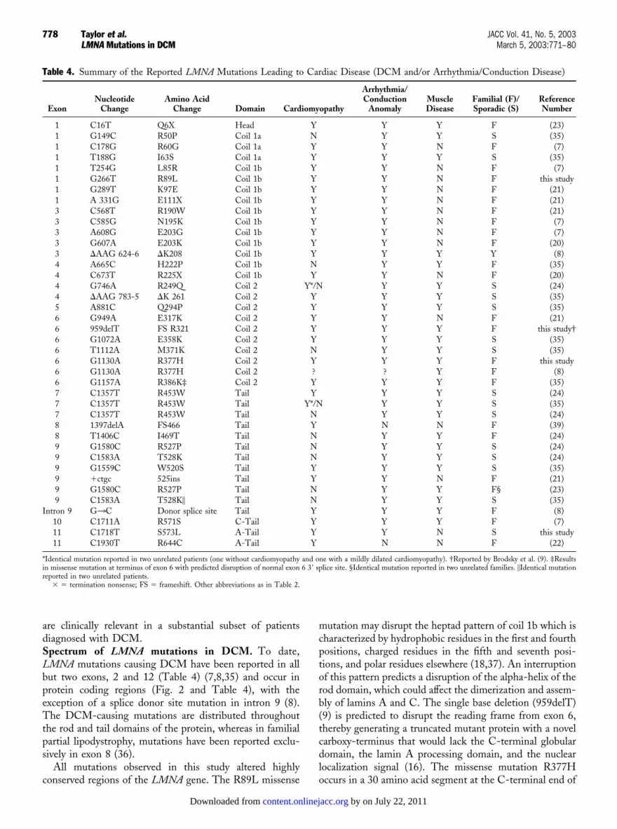
are clinically relevant in a substantial subset of patientsdiagnosed with DCM.Spectrum of LMNA mutations in DCM. To date,LMNA mutations causing DCM have been reported in allbut two exons, 2 and 12 (Table 4) (7,8,35) and occur inprotein coding regions (Fig. 2 and Table 4), with theexception of a splice donor site mutation in intron 9 (8).The DCM-causing mutations are distributed throughoutthe rod and tail domains of the protein, whereas in familialpartial lipodystrophy, mutations have been reported exclu-sively in exon 8 (36).
All mutations observed in this study altered highlyconserved regions of the LMNA gene. The R89L missense
mutation may disrupt the heptad pattern of coil 1b which ischaracterized by hydrophobic residues in the first and fourthpositions, charged residues in the fifth and seventh posi-tions, and polar residues elsewhere (18,37). An interruptionof this pattern predicts a disruption of the alpha-helix of therod domain, which could affect the dimerization and assem-bly of lamins A and C. The single base deletion (959delT)(9) is predicted to disrupt the reading frame from exon 6,thereby generating a truncated mutant protein with a novelcarboxy-terminus that would lack the C-terminal globulardomain, the lamin A processing domain, and the nuclearlocalization signal (16). The missense mutation R377Hoccurs in a 30 amino acid segment at the C-terminal end of
Table 4. Summary of the Reported LMNA Mutations Leading to Cardiac Disease (DCM and/or Arrhythmia/Conduction Disease)
ExonNucleotide
ChangeAmino Acid
Change Domain Cardiomyopathy
Arrhythmia/Conduction
AnomalyMuscleDisease
Familial (F)/Sporadic (S)
ReferenceNumber
1 C16T Q6X Head Y Y Y F (23)1 G149C R50P Coil 1a N Y Y S (35)1 C178G R60G Coil 1a Y Y N F (7)1 T188G I63S Coil 1a Y Y Y S (35)1 T254G L85R Coil 1b Y Y N F (7)1 G266T R89L Coil 1b Y Y N F this study1 G289T K97E Coil 1b Y Y N F (21)1 A 331G E111X Coil 1b Y Y N F (21)3 C568T R190W Coil 1b Y Y N F (21)3 C585G N195K Coil 1b Y Y N F (7)3 A608G E203G Coil 1b Y Y N F (7)3 G607A E203K Coil 1b Y Y N F (20)3 �AAG 624-6 �K208 Coil 1b Y Y Y Y (8)4 A665C H222P Coil 1b N Y Y F (35)4 C673T R225X Coil 1b Y Y N F (20)4 G746A R249Q Coil 2 Y*/N Y Y S (24)4 �AAG 783-5 �K 261 Coil 2 Y Y Y S (35)5 A881C Q294P Coil 2 Y Y Y S (35)6 G949A E317K Coil 2 Y Y N F (21)6 959delT FS R321 Coil 2 Y Y Y F this study†6 G1072A E358K Coil 2 Y Y Y S (35)6 T1112A M371K Coil 2 N Y Y S (35)6 G1130A R377H Coil 2 Y Y Y F this study6 G1130A R377H Coil 2 ? ? Y F (8)6 G1157A R386K‡ Coil 2 Y Y Y F (35)7 C1357T R453W Tail Y Y Y S (24)7 C1357T R453W Tail Y*/N Y Y S (35)7 C1357T R453W Tail N Y Y S (24)8 1397delA FS466 Tail Y N N F (39)8 T1406C I469T Tail N Y Y F (24)9 G1580C R527P Tail N Y Y S (24)9 C1583A T528K Tail N Y Y S (24)9 G1559C W520S Tail Y Y Y S (35)9 �ctgc 525ins Tail Y Y N F (21)9 G1580C R527P Tail N Y Y F§ (23)9 C1583A T528K� Tail N Y Y S (35)
Intron 9 G3C Donor splice site Tail Y Y Y F (8)10 C1711A R571S C-Tail Y Y Y F (7)11 C1718T S573L A-Tail Y Y N S this study11 C1930T R644C A-Tail Y N N F (22)
*Identical mutation reported in two unrelated patients (one without cardiomyopathy and one with a mildly dilated cardiomyopathy). †Reported by Brodsky et al. (9). ‡Resultsin missense mutation at terminus of exon 6 with predicted disruption of normal exon 6 3� splice site. §Identical mutation reported in two unrelated families. �Identical mutationreported in two unrelated patients.
� � termination nonsense; FS � frameshift. Other abbreviations as in Table 2.
778 Taylor et al. JACC Vol. 41, No. 5, 2003LMNA Mutations in DCM March 5, 2003:771–80
by on July 22, 2011 content.onlinejacc.orgDownloaded from

the rod domain, which is conserved among lamins andcytoplasmic intermediate filaments and is critical for thehigher order assembly of lamin polymers (18). The missensemutation S573L predicts a change of the highly conservedsequence and structure of the carboxyl terminal of lamin A,leaving lamin C intact. This region of the protein controlsthe lateral assembly of protofilaments and mediates thelamin network formation (18).
The majority of gene mutations causing DCM appear tobe private mutations. However, the mutations G1130A,G746A, and C1357T have been recurrent in apparentlyunrelated individuals and may suggest possible hotspots or adistant founder effect (24). The R377H mutation (G1130A)found in one of our families has been reported in a LGMD1Bfamily living in the Caribbean (8). Although a remote foundereffect cannot be excluded, the ethnic backgrounds of the twofamilies may suggest an independent mutational event. Inkeeping with this interpretation is the high rate of occurrenceof LMNA de novo mutations that has been estimated in somestudies to be higher than 50% (35).
The mechanism by which LMNA mutations causeDCM, conduction disease, and variable degrees of musculardystrophy is still not well understood. The missense muta-tions observed in our population and by other authors(7,20,21) in a protein known to dimerize and form higherorder assembly structures suggest a dominant negativemechanism. However, haploinsufficiency appears to be an-other potential mechanism in a family with EDMD due toa nonsense mutation in codon 6 (23). Lamin A and C arebelieved to have different complex functions: a currenthypothesis is that LMNA mutations result in cellular andtissue fragility (intermediate filament-fragility syndrome)(17), particularly in tissues subjected to mechanical stress(heart and muscle). However, A-lamins have other impor-tant functions that could cause tissue damage, such aschromatin organization during cell division, signal trans-duction, differentiation maintenance, repair, and finallyanchoring of other lamin-binding proteins, such as emerin,LAP, BAF, and LBR (19). Any of these functions maypotentially lead to cell damage and apoptosis, and eventuallyalter myocardial and muscular function.Genotype-phenotype correlation. Patients with DCMdue to LMNA mutations frequently present with a “mildly”dilated form with severe dysfunction (38). The presence ofany sign of skeletal muscle abnormality, such as increasedserum creatine kinase, mild signs suggestive of an underly-ing muscular dystrophy, or family history suggestive ofmuscle disease in an affected relative, was a strong predictorfor carrying a LMNA mutation in our study. The presenceof atrioventricular blocks and atrial arrhythmia were alsosignificantly associated with the carrier status. Contrary toother autosomal forms of familial DCM (29), we observeda trend toward an excess of females, perhaps suggesting theinvolvement of different “modifier” genes. The exon 11subject’s sister, who is ostensibly clinically healthy at age 60years, illustrates an example of a nonpenetrant mutation
carrier which has been reported previously (7,20). Theoccurrence of clinically unaffected carriers and the remark-able inter- and intra-familial variability in the expression ofLMNA mutations (39) suggest the existence of additionalgenetic and/or environmental factors contributing to phe-notype.Prognosis of LMNA mutations. In our study we foundthat subjects with LMNA mutations had a significantlyworse prognosis compared with the other DCM subjects.Eight of 12 carriers of LMNA mutations were deceased,required heart transplantation, or had severe worsening ofmyocardial function between the third and fifth decade. Thecardiovascular mortality due to sudden death or heart failurewas significantly higher in the carrier group compared withthe DCM patients without LMNA mutations. In each ofthe three sets of curves shown in Figure 5, survival wassignificantly worse in LMNA mutation carriers comparedwith non-carriers with only 31% of event-free survival(panel C) at age 45 versus 75%.
A relatively milder phenotype with isolated DCM wasobserved in the sporadic family where the mutation involvesonly the lamin A isoform. Other authors have reportedsimilar clinical features with mutations restricted to thelamin C isoform (7). These data suggest that the selectiveinvolvement of lamin C may have a more favorable out-come.Study limitations. A potential source of ascertainment biasin the patient selection was that subjects were recruited intertiary care referral centers. Statistical limitations includedthe fact that some variables had missing values distributedsporadically: this pattern precluded multivariate analyses.Many variables highly correlated with carrier status werethemselves highly correlated: therefore, bivariate resultsshould not be taken as additive.Clinical implications. Results of this study provide clinicalinsights for genetic counseling and risk stratification ofpatients with DCM. Mutations of LMNA are frequentamong patients with DCM, particularly when the diseaseis associated with skeletal muscle abnormalities, atrial ar-rhythmia, or conduction defects. Furthermore, a historyof sporadic DCM does not necessarily exclude a LMNAgene defect. Taking into account the severity of prognosisin these patients and the possibility of early interventionsto prevent the progression of heart failure, remodeling, andsudden death, our findings should prompt screening forLMNA in familial and sporadic cases of DCM withclinical indicators suggestive of laminopathy. The avail-ability of DHPLC with targeted sequence analysis makesmutation screening efficient and feasible (40). Future studiesshould investigate the role of the nuclear envelope in thepathogenesis of DCM and the existence of modifiergenes, which could alter the expressivity and severity ofthe disease, and could represent targets for novel thera-peutic strategies.
779JACC Vol. 41, No. 5, 2003 Taylor et al.March 5, 2003:771–80 LMNA Mutations in DCM
by on July 22, 2011 content.onlinejacc.orgDownloaded from

AcknowledgmentsWe thank the family members for their participation inthese studies. Laurel Hunter and Raj Rajkumar are grate-fully acknowledged for their help in manuscript preparation.Dr. Richard Spritz is also acknowledged.
Reprint requests and correspondence: Dr. Luisa Mestroni, Uni-versity of Colorado Cardiovascular Institute, Bioscience ParkCenter, #150, 12635 East Montview Boulevard, Aurora, Colorado80010-7116. E-mail: [email protected].
REFERENCES1. Codd MB, Sugrue DD, Gersh BJ, Melton LJ. Epidemiology of
idiopathic dilated and hypertrophic cardiomyopathy: a population-based study in Olmsted County, Minnesota, 1975–1984. Circulation1989;80:564–72.
2. Baig MK, Goldman JH, Caforio ALP, Coonar AS, Keeling PJ,McKenna WJ. Familial dilated cardiomyopathy: cardiac abnormalitiesare common in asymptomatic relatives and may represent early disease.J Am Coll Cardiol 1998;31:195–201.
3. Gregori D, Rocco C, Miocic S, Mestroni L. Estimating the frequencyof familial dilated cardiomyopathy in the presence of misclassificationerrors. J Appl Stat 2001;28:53–62.
4. Muntoni F, Cau M, Ganau A, et al. Deletion of the dystrophinmuscle-promoter region associated with X-linked dilated cardiomyop-athy. N Engl J Med 1993;329:921–5.
5. Towbin JA, Hejtmancik F, Brink P, et al. X-linked cardiomyopathy(XLCM): molecular genetic evidence of linkage to the Duchennemuscular dystrophy (dystrophin) gene at the Xp21 locus. Circulation1993;87:1854–65.
6. Li D, Tapscoft T, Gonzalez O, et al. Desmin mutation responsible foridiopathic dilated cardiomyopathy. Circulation 1999;100:461–4.
7. Fatkin D, MacRae C, Sasaki T, et al. Missense mutations in the roddomain of the lamin A/C gene as causes of dilated cardiomyopathyand conduction-system disease. N Engl J Med 1999;341:1715–24.
8. Muchir A, Bonne G, van der Kooi AJ, et al. Identification ofmutations in the gene encoding lamins A/C in autosomal dominantlimb girdle muscular dystrophy with atrioventricular conduction dis-turbances (LGMD1B). Hum Mol Genet 2000;9:1453–9.
9. Brodsky GL, Muntoni F, Miocic S, Sinagra G, Sewry C, Mestroni L.A lamin A/C gene mutation associated with dilated cardiomyopathy withvariable skeletal muscle involvement. Circulation 2000;101:473–6.
10. Olson TM, Michels VV, Thibodeau SN, Tai Y, Keating MT. Actinmutation in dilated cardiomyopathy, a heritable form of heart failure.Science 1998;280:750–2.
11. D’ Adamo P, Fassone L, Gedeon A, et al. The X-linked gene G4.5 isresponsible for different infantile dilated cardiomyopathies. Am J HumGenet 1997;61:862–7.
12. Kamisago M, Sharma SD, DePalma SR, et al. Mutations in sarcomereprotein genes as a cause of dilated cardiomyopathy. N Engl J Med2000;343:1688–96.
13. Olson T, Kishimoto NY, Withby FG, Michels V. Mutations that alterthe surface charge of alpha-tropomyosin are associated with dilatedcardiomyopathy. J Mol Cell Cardiol 2001;33:723–32.
14. Tsubata S, Bowles KR, Vatta M, et al. Mutations in the humandelta-sarcoglycan gene in familial and sporadic dilated cardiomyopa-thy. J Clin Invest 2000;106:655–62.
15. Gerull B, Gramlich M, Atherton J, et al. Mutations of TTN, encodingthe giant muscle filament titin, cause familial dilated cardiomyopathy.Nat Genet 2002;30:201–4.
16. Lin F, Worman HJ. Structural organization of the human geneencoding nuclear lamin A and nuclear lamin C. J Biol Chem1993;268:16321–6.
17. Hutchison CJ, Alvarez-Reyes M, Vaughan OA. Lamins in disease:why do ubiquitously expressed nuclear envelope proteins give rise totissue-specific disease phenotypes? J Cell Sci 2001;114:9–19.
18. Stuurman N, Heins S, Aebi U. Nuclear lamins: their structure,assembly and interactions. J Struct Biol 1998;122:42–66.
19. Wilson KL. The nuclear envelope, muscular dystrophy and geneexpression. Trends Cell Biol 2000;10:125–9.
20. Jacobs PM, Hanson EL, Crispell KA, et al. Novel lamin A/C genemutations in two families with dilated cardiomyopathy and conductionsystem disease. J Card Fail 2001;7:249–56.
21. Arbustini E, Pilotto A, Repetto A, et al. Autosomal dominant dilatedcardiomyopathy with atrioventricular block: a lamin A/C defect-related disease. J Am Coll Cardiol 2002;39:981–90.
22. Genschel J, Bochow B, Kuepferling S, et al. An R644C mutationwithin lamin A extends the mutations causing dilated cardiomyopathy.Hum Mutat 2001;17:154.
23. Bonne G, Di Barletta MR, Varnous S, et al. Mutations in the geneencoding lamin A/C cause autosomal dominant Emery-Dreifussmuscular dystrophy. Nat Genet 1999;21:285–88.
24. Raffaele Di Barletta M, Ricci E, Galluzzi G, et al. Different mutations inthe LMNA gene cause autosomal dominant and autosomal recessiveEmery-Dreifuss muscular dystrophy. Am J Hum Genet 2000;66:1407–12.
25. De Sandre-Giovannoli A, Chaouchm M, Kozlov S, et al. Homozy-gous defects in LMNA, encoding lamin A/C nuclear-envelope pro-teins, cause autosomal recessive axonal neuropathy in human(Charcot-Marie-Tooth disorder type 2) and mouse. Am J Hum Genet2002;70:726–36.
26. Cao H, Hegele RA. Nuclear lamin A/C R482Q mutation in Canadiankindreds with Dunningan-type familial partial lipodystrophy. HumMol Genet 2000;9:109–12.
27. Shackleton S, Lloyd DJ, Jackson SN, et al. LMNA, encoding laminA/C, is mutated in partial lipodystrophy. Nat Genet 2000;24:103–4.
28. Mestroni L, Miani D, Di Lenarda A, et al. Clinical and pathologic studyof familial dilated cardiomyopathy. Am J Cardiol 1990;65:1449–53.
29. Mestroni L, Rocco C, Gregori D, et al. Familial dilated cardiomyop-athy: evidence for genetic and phenotypic heterogeneity. J Am CollCardiol 1999;34:181–90.
30. Mestroni L, Maisch B, McKenna WJ, et al. Guidelines for the studyof familial dilated cardiomyopathies. Eur Heart J 1999;20:93–102.
31. Henry WL, Gardin JM, Ware JH. Echocardiographic measurements innormal subjects from infancy to old age. Circulation 1980;62:1054–61.
32. Milasin J, Muntoni F, Severini GM, et al. A point mutation in the 5�splice site of the dystrophin gene first intron responsible for X-linkeddilated cardiomyopathy. Hum Mol Genet 1996;5:73–9.
33. Muntoni F, Di Lenarda A, Porcu M, et al. Dystrophin geneabnormalities in two patients with idiopathic dilated cardiomyopathy.Heart 1997;78:608–12.
34. Haines J, Pericak-Vance M. Gene Mapping in Complex HumanDiseases. New York, NY: Wiley-Liss, 1998.
35. Bonne G, Mercuri E, Muchir A, et al. Clinical and molecular geneticspectrum of autosomal dominant Emery-Dreifuss muscular dystrophydue to mutations of the lamin A/C gene. Ann Neurol 2000;48:170–80.
36. Speckman RA, Garg A, Du F, et al. Mutational and haplotypeanalyses of families with familial partial lipodystrophy (Dunniganvariety) reveal recurrent missense mutations in the globular C-terminaldomain of lamin A/C. Am J Hum Genet 2000;66:1192–8.
37. McLachlan AD. Coiled coil formation and sequence regularities in thehelical regions of alpha-keratin. J Mol Biol 1978;124:297–304.
38. Keren A, Gottlieb S, Tzivoni D, et al. Mildly dilated congestivecardiomyopathy: use of prospective diagnostic criteria and descriptionof the clinical course without heart transplantation. Circulation 1990;81:506–17.
39. Genschel J, Baier P, Kuepferling S, et al. A new frameshift mutation at codon466 (1397delA) within the LMNA gene. Hum Mutat 2000;16:278.
40. Buyse IM, Fang P, Hoon KT, Amir RE, Zoghbi HY, Roa BB.Diagnostic testing for Rett syndrome by DHPLC and direct sequenc-ing analysis of the MECP2 gene: identification of several novelmutations and polymorphisms. Am J Hum Genet 2000;67:1428–36.
APPENDIXFamilial Dilated Cardiomyopathy Registry ResearchGroup: University of Colorado Cardiovascular Institute(U.S.): Dmi Dao, BS, Sharon L. Graw, PhD, Lisa Ku, MS,Brian D. Lowes, MD, Xiao Zhu, BS; Human MedicalGenetics Program: Katherine Gowan, MS, William M.Old, PhD; Hospital and University of Trieste (Italy):Mauro Driussi, MD, Giulio Scherl.
780 Taylor et al. JACC Vol. 41, No. 5, 2003LMNA Mutations in DCM March 5, 2003:771–80
by on July 22, 2011 content.onlinejacc.orgDownloaded from

doi:10.1016/S0735-1097(02)02954-6 2003;41;771-780 J. Am. Coll. Cardiol.
Luisa Mestroni, and Familial Dilated Cardiomyopathy Registry Research Group Moss, Wai-Lun P. Li, Gary L. Stetler, Francesco Muntoni, Michael R. Bristow,
Debra A. Ferguson, Gary L. Brodsky, Mark M. Boucek, Jean Lascor, Andrew C.Alastair D. Robertson, Elisa Carniel, Andrea Di Lenarda, Teresa J. Bohlmeyer, Matthew R. G. Taylor, Pamela R. Fain, Gianfranco Sinagra, Misi L. Robinson,
Natural history of dilated cardiomyopathy due to lamin A/C gene mutations
This information is current as of July 22, 2011
& ServicesUpdated Information
http://content.onlinejacc.org/cgi/content/full/41/5/771including high-resolution figures, can be found at:
References
http://content.onlinejacc.org/cgi/content/full/41/5/771#BIBLfree at: This article cites 39 articles, 17 of which you can access for
Citations
icleshttp://content.onlinejacc.org/cgi/content/full/41/5/771#otherartThis article has been cited by 38 HighWire-hosted articles:
Rights & Permissions
http://content.onlinejacc.org/misc/permissions.dtltables) or in its entirety can be found online at: Information about reproducing this article in parts (figures,
Reprints http://content.onlinejacc.org/misc/reprints.dtl
Information about ordering reprints can be found online:
by on July 22, 2011 content.onlinejacc.orgDownloaded from

CORRECTIONS
Rademaker MT, Charles CJ, Espiner EA, Fisher S, Frampton CM, Kirkpatrick CMJ, Lainchbury JG, Nicholls MG, Richards AM,Vale WW. Beneficial Hemodynamic, Endocrine, and Renal Effects of Urocortin in Experimental Heart Failure: Comparison WithNormal Sheep. J Am Coll Cardiol 2002;40:1495–505. Per the authors, the doses of urocortin were printed incorrectly in the Methodssection of the Abstract (page 1495), the Study Protocol section under the Methods heading (page 1496), and the column headings ofTables 1 and 2 (pages 1502 and 1503). The correct doses are 10, 50, and 100 �g.
doi:10.1016/S0735-1097(03)00886-6
Taylor MRG, Fain PR, Sinagra G, Robinson ML, Robertson AD, Carniel E, Di Lenarda A, Bohlmeyer TJ, Ferguson DA, BrodskyGL, Boucek MM, Lascor J, Moss AC, Li W-LP, Stetler GL, Muntoni F, Bristow MR, Mestroni L, Familiar DilatedCardiomyopathy Registry Research Group. Natural History of Dilated Cardiomyopathy Due to Lamin A/C Gene Mutations. J AmColl Cardiol 2003;41:771– 80. The authors have detected some inconsistencies and inaccuracies within the aforementioned paper. Pleasesee below for list of corrections:
TEXTThe informed consent statement (page 772, 2nd column, 1st paragraph) should have read:
Deoxyribonucleic acid samples from subjects and controls were obtained according to the contemporaneous consent guidelines in place inItaly and the U.S.
In the Abstract (page 771), Methods (page 772), and Tables 1 (page 772) and 3 (page 777), 105 subjects (104 with DCM and 1 withasymptomatic carrier) were included in the statistical analysis.
FIGURESFigure 1 (page 773): The light gray circles indicate individuals with “unknown” status as defined in the Methods section.
Figure 2 (page 774): The word nucleotide should have been amino acid.
Figure 3 (page 776): The light gray boxes indicate divergent amino acids.
TABLES
Table 2 (page 774–5): In the Mutation column, the third mutation G1130T should have been G1130A. In the ID column for FamilyTSFDC13, the ID numbers should read, from top to bottom, II-1, III-2, III-1. For Family MDDC1, individual II-5 should have been II-3.For this family, the screening of controls utilized normal controls and samples from explanted DCM hearts. There was an insertion of an extraspace in the Conduction column, both individuals II-1 and II-3 had 2°–3° AV block and left bundle-branch block. The intent of this Table isto provide a description of the phenotypes of the 12 carriers; it reflects the most up-do-date information collected on these subjects.
Table 3 (page 777): The Age of onset values should have been 37 for non-carriers and 27 for carriers. Skeletal muscle involvement should havebeen Muscular disease. Conduction defects should have been 11% for non-carriers and 50% for carriers. All of the percentages in this Tablecannot be derived from Table 2. This Table represents a comparison of data collected at enrollment between the carriers and non-carriers.
The authors apologize for the inconvenience generated by these corrections.
None of these errors/changes alters the results or conclusions of the manuscript.
doi:10.1016/S0735-1097(03)00887-8
Journal of the American College of Cardiology Vol. 42, No. 3, 2003© 2003 by the American College of Cardiology Foundation ISSN 0735-1097/03/$30.00Published by Elsevier Inc.
by on July 22, 2011 content.onlinejacc.orgDownloaded from

Barst RJ, McGoon M, McLaughlin V, Tapson V, Oudiz R, Shapiro S, Robbins IM, Channick R, Badesch D, Rayburn BK,Flinchbaugh R, Sigman J, Arneson C, Jeffs R, for the Beraprost Study Group. Beraprost Therapy for Pulmonary ArterialHypertension. J Am Coll Cardiol 2003;41:2119 –25. Drs. Rich, Rubin, and Wasserman were inadvertently omitted from the author list.The full author list and their affiliations are printed below. We apologize for this error.
Robyn J. Barst, MD, FACC,* Michael McGoon, MD,† Vallerie McLaughlin, MD, FACC,‡ Victor Tapson, MD, FCCP,§ Stuart Rich,MD,‡ Lewis Rubin, MD,** Karlman Wasserman, MD, PhD,� Ronald Oudiz, MD, FACC,� Shelley Shapiro, MD,¶ Ivan M. Robbins,MD,# Richard Channick, MD,** David Badcsch, MD,†† Barry K. Rayburn, MD, FACC,†† Robin Flinchbaugh, BS,§§ Jeff Sigman,BA,§§ Carl Arneson, MStat,§§ Roger Jeffs, PhD,§§ for the Beraprost Study Group
From the *Columbia University College of Physicians and Surgeons, New York, New York; †Mayo Clinic, Rochester, Minnesota; ‡RushPresbyterian-St. Luke’s Medical Center, Chicago, Illinois; §Duke University Medical Center, Durham, North Carolina; �Research andEducation Institute at Harbor-UCLA Medical Center, Torrance, California; ¶USC Medical Center, Los Angeles, California; #VanderbiltUniversity Medical Center, Nashville, Tennessee; **University of California-San Diego Medical Center, La Jolla, California; ††Universityof Colorado Health Sciences Center, Denver, Colorado; ‡‡University of Alabama at Birmingham, Birmingham, Alabama; and §§UnitedTherapeutics Corporation, Research Triangle Park, North Carolina.
doi:10.1016/S0735-1097(03)00937-9
591JACC Vol. 42, No. 3, 2003 CorrectionsAugust 6, 2003:590–1
by on July 22, 2011 content.onlinejacc.orgDownloaded from
Lack of evidence for a pathogenic role of proteasome-directed autoimmunity in dilated cardiomyopathy
Defects in nuclear structure and function promote dilated cardiomyopathy in lamin A/C–deficient mice
Mutations in Cypher/ZASP in patients with dilated cardiomyopathy and left ventricular non-compaction