natalie boström pyometra thesis.pdf - lsmu
TRANSCRIPT

LITHUANIAN UNIVERSITY OF HEALTH SCIENCES
VETERINARY ACADEMY
Faculty of Veterinary Medicine
Natalie Boström
A retrospective study of prognostic markers in bitches
diagnosed with pyometra
Kalėms, kurioms diagnozuota piometra, retrospektyvi
klinikinių atvejų analizė
MASTER THESIS
of Integrated Studies of Veterinary Medicine
Supervisor: Prof. Dr. Gintaras Zamokas
KAUNAS 2018

2
THE WORK WAS DONE IN THE DEPARTMENT OF SMALL ANIMAL CLINIC
CONFIRMATION OF THE INDEPENDENCE OF DONE WORK
I confirm that the presented Master Thesis “A retrospective study of prognostic markers in bitches
diagnosed with pyometra”.
1. has been done by me;
2. has not been used in any other Lithuanian or foreign university;
3. I have not used any other sources not indicated in the work and I present the complete list of the
used literature.
(date) (author’s name, surname) (signature)
CONFIRMATION ABOUT RESPONSIBILITY FOR CORRECTNESS OF
THE ENGLISH LANGUAGE IN THE DONE WORK
I confirm the correctness of the English language in the done work.
(date) (author’s name, surname) (signature)
CONCLUSION OF THE SUPERVISOR REGARDING DEFENCE OF
THE MASTER THESIS
(date) (supervisor’s name, surname) (signature)
THE MASTER THESIS HAVE BEEN APPROVED IN
THE DEPARTMENT/CLINIC/INSTITUTE
(date of approval) (name, surname of the head of
department/clinic/institute) (signature)
Reviewer of the Master Thesis
(name, surname) (signatures)
Evaluation of defence commission of the Master Thesis:
(date) (name, surname of the secretary of the defence
commission)
(signature)

3
TABLE OF CONTENT SUMMARY .......................................................................................................................................................... 5
SANTRAUKA ........................................................................................................................................................ 6
ABBREVATIONS .................................................................................................................................................. 7
INTRODUCTION .................................................................................................................................................. 8
LITERATURE REVIEW .......................................................................................................................................... 9
Aetiology and pathogenesis ......................................................................................................................... 9
Diagnostic methods of pyometra ............................................................................................................... 10
Clinical signs ........................................................................................................................................... 10
Haematology and biochemical findings ................................................................................................ 10
Imaging .................................................................................................................................................... 11
Treatment method ...................................................................................................................................... 12
Surgical treatment methods .................................................................................................................. 12
Uterine drainage and lavage .................................................................................................................. 12
Medical treatment .................................................................................................................................. 13
MATERIAL AND METHODS ............................................................................................................................... 16
Investigated parameters ............................................................................................................................ 16
Clinical observations .................................................................................................................................. 16
Diagnostic tests ........................................................................................................................................... 17
Haematology and biochemical data ...................................................................................................... 17
Imaging .................................................................................................................................................... 18
Statistical analyses ...................................................................................................................................... 18
Literature review ........................................................................................................................................ 18
RESULTS ............................................................................................................................................................ 19
Number, breed, age and type of treatment .............................................................................................. 19
Breed prevalence .................................................................................................................................... 19
Age prevalence ........................................................................................................................................ 20
Type of treatment ................................................................................................................................... 21
Time of clinical signs after oestrus ............................................................................................................ 22
Anamnestic and clinical findings .............................................................................................................. 23
Haematological and Biochemical findings ............................................................................................... 25
Prevalence of changes in temperature of bitches with pyometra and relationship with peritonitis
.................................................................................................................................................................. 29
Prevalence of increased CRP in bitches with pyometra ..................................................................... 30
Relationship between C-reactive protein and peritonitis ................................................................... 31
Mortality and complications ..................................................................................................................... 31
DISCUSSION ...................................................................................................................................................... 33

4
CONCLUSION .................................................................................................................................................... 36
REFERENCES ..................................................................................................................................................... 37

5
A RETROSPECTIVE STUDY OF PROGNOSTIC MARKERS IN BITCHES DIAGNOSED
WITH PYOMETRA
Natalie Boström
Master Theses
SUMMARY
Pyometra is one of the most common disease in intact bitches with a variety of clinical and
laboratory findings. If the disease is not detected early, it can be fatal and may lead to complications
such as peritonitis or systemic inflammatory response syndrome. The aim of this study was to
investigate etiology and manifestation of pyometra and to identify what parameters of clinical signs
and blood parameters that are most common in pyometra, what treatment methods that are most
common and if there are other options which can be used to keep the bitch intact. Also, to see what
diagnostic methods that can be used and which that are most accurate in early diagnosis of
pyometra.
A retrospective study was done by using data obtained from Albano animal hospital in
Sweden. In the study 119 bitches diagnosed with pyometra during the year 2017 was used. 114
bitches were surgically treated by ovariohysterectomy, 1 were medically treated and 3 were
euthanized without treatment. The most common complication observed in bitches treated
surgically was peritonitis (n=27).
Vaginal discharges, depression, increased CRP and leucocytosis was most common findings
in bitches diagnosed with pyometra. Uterus enlargement was seen in 105 out of 111 bitches and
the size varied between 7 to 80 mm. Peritonitis were found in 27 bitches before treatment and in
25 bitches after treatment.
Keywords used: pyometra, surgical treatment, medical treatment, peritonitis.

6
KALĖMS, KURIOMS DIAGNOZUOTA PIOMETRA, RETROSPEKTYVI KLINIKINIŲ
ATVEJŲ ANALIZĖ
Natalie Boström
Master Theses
SANTRAUKA
Piometra yra viena iš dažniausiai diagnozuojamų nekastruotų kalių ligų, kuriai būdinga
klinikinių ir laboratorinių tyrimų rezultatų įvairovė. Jei liga nėra diagnozuota anksti, ji gali būti
mirtina ir gali sukelti komplikacijas, tokias kaip peritonitas ar sisteminis uždegiminis atsako
sindromas. Šio tyrimo tikslas buvo ištirti piometros etiologiją ir pasireiškimo ypatumus bei
nustatyti, kokie klinikiniai požymiai ir kraujo parametrų pokyčiai yra būdingi sergant piometra,
kokie gydymo metodai dažniausiai taikomi ir ar yra kitų galimybių, kurios gali būti naudojamos,
kad kalės nesirgtų. Be to, tirti diagnostiniai metodai, kurie gali būti naudojami ir kurie yra tiksliausi
ankstyvoje piometros stadijoje.
Duomenys buvo rinkti Albano gyvūnų ligoninėje, Švedijoje. Tyrimui buvo atrinkta 119 kalių,
kurioms per 2017 metus buvo diagnozuota piometra . 114 kalių buvo gydytos chirurginiu būdu -
atlikta ovariohisterektomija, 1 buvo gydyta skiriant medikamentus ir 3 buvo eutanazuotos be
gydymo. Dažniausia komplikacija pasireiškusi kalėms po chirurginio gydymo buvo peritonitas (n
= 27).
Išskyros iš makšties, depresija, padidėjęs CRB kiekis ir leukocitozė buvo dažniausi pokyčiai
nustatyti kalėms sirgusioms piometra. Gimdos padidėjimas buvo nustatytas 105 iš 111 kalių ir
dydis svyravo nuo 7 iki 80 mm. Peritonitas buvo diagnozuotas 27 kalėms prieš gydymą ir 25 kalėms
po chirurginio gydymo.
Raktažodžiai: piometra, chirurginis gydymas, medikamentinis gydymas, peritonitas.

7
ABBREVATIONS
SIRS – systemic inflammatory response syndrome
E.coli – Escherichia coli
CRP – Capillary refill time
SAA – Serum amyloid A
TNFα – Tumour necrosis factor alpha
IL1-IL15 – Interleukin 1-15
MODS – Multiple organ failure
X-ray – Radiographic imaging
OHE – Ovariohysterectomy
PGF2α – Prostaglandin F2 alpha
PR A – Progesterone receptor A
PR B – Progesterone receptor B
TIDA – Tuberoinfundibulum
PAD – Pathological anatomical diagnosis
CRT – Capillary refill time
EDTA – Ethylenediaminetetraacetic acid
ALP – Alkaline phosphatase
ALT - Alanine aminotransferase
Na – Sodium
K – Potassium
Ca – Calcium
Cl – Chloride
GDPR – General data protective regulation
SPSS – Statistical package for the social sciences
CEH – Cystic endometrial hyperplasia
HCT – Hematocrit
Hb – haemoglobin

8
INTRODUCTION
Pyometra is one of the most common diseases among bitches that are not neutered (3). The
disease normally occurs before the age of 10 in 25 per cent of bitches with an average age of 8
years (1,2).
The cause of pyometra is not really known, but some authors have suggested that the cause
might be of hormonal influence of estrogen and progesterone on the uterus and ascending bacteria
from the bitch normal flora. Most common bacteria found are Escherichia coli. Some pathologies
of the uterus, such as cystic endometrial hyperplasia (CEH) and mucometra may lead to infection
of the uterus and then eventually pyometra (18). Some breeds are more predisposed than others
which may indicate that there might be a genetic predisposition in some breeds (3,4).
Different treatment methods can be used to treat pyometra. Most common is
ovariohysterectomy, which is used in most cases when a bitch is diagnosed with pyometra (40).
Medical treatment is used in some cases and the bitch is then treated with prostaglandins,
progesterone receptor antagonists and dopamine agonists, and antibiotics (41).
If the disease is not treated or if diagnosis is done when the disease has progressed to peritonitis
or systemic involvement, the prognosis is often worse, and the bitch chance of survival is lowered.
Some complications are associated with increased mortality such as systemic inflammatory
response syndrome and peritonitis (6). Many dogs are euthanized when diagnosed with pyometra
and normally it is due to high age, or if the owner has limited amount of money or if the bitch has
other diseases that may increase risk of anesthesia or surgery and decreased chance of recovery.
Some studies have reported a mortality rate of 3 to 10 per cent (5). It is important to diagnose the
bitch early in the disease to prevent complications and to have a good outcome in the disease. Some
biological markers can be investigated to predict pyometra and its outcome.
The aim is to learn more about the clinical signs of pyometra and to study the prevalence in
bitches diagnosed in pyometra and try to find what is the cause of pyometra in and peritonitis, and
to study what treatment method mostly used and complications of pyometra and treatment.
The goal with this study is:
1. To investigate breed prevalence, what age pyometra occur at and most common treatment
methods.
2. To analyse what clinical signs that are most common and when during the oestrus cycle
pyometra most commonly is diagnosed.
3. To investigate blood parameters and their relationship with peritonitis and temperature.
4. To investigate what complications that may occur.

9
LITERATURE REVIEW
Aetiology and pathogenesis
Pyometra is a disease where pus accumulates in the uterus. The disease occurs in intact females
and about 25 per cent of bitches get the diagnose of pyometra before the age of 10, with an average
of 8 years (1,2). Pyometra normally occur 4-8 weeks after oestrus, during dioestrus when the uterus
is affected by progesterone, produced by corpus lutea. Oestrogen increases the stimulatory effects
of progesterone by increasing the number of progesterone receptors and the vascularization in the
uterine wall (7). Progesterone starts to stimulate endometrial gland development which leads to
increased glandular secretion, it also decreases myometrial contraction which prevents discharges
from leaving the uterus via the vagina, this leads to a favourable environment for bacteria (8).
The development of cystic endometrial hyperplasia has been discussed to be one factor that
predisposes the uterus to pyometra. But it seems that both CEH and pyometra can occur without
the other and that pyometra is mainly caused by a bacterial infection that render the uterus (9). If
pyometra is not treated, it may lead to systemic inflammatory syndrome (SIRS) and dysfunction of
many organs, that can lead to death if the animal is not treated (4). There might be fluid in the
lumen of uterus even without a bacterial infection and then it is called hydrometra, mucometra or
hemometra and depends on what type of fluid that can be found in the lumen. Normally there are
no clinical signs if no bacterial infection (10).
Gram negative bacteria is the most common bacteria isolated from uterus, these bacteria
releases endotoxins when they grow and dies. These endotoxins may be released into the
bloodstream and cause systemic inflammatory syndrome (SIRS) and multiple organ dysfunction
(MOD). Escherichia coli is the most common pathogen isolated from animals with pyometra and
it is believed to arise from the normal bacterial flora of the animal (11). The bacteria adhere to the
hormonally influenced endometrium in the uterus and leads to the disease (12). Other bacteria that
occur in a less extent is, Klebsiella spp., Citrobacter spp., Proteus spp., Salmonella spp.,
Pseudomonas spp., Streptococcus spp., Staphylococcus spp. and Arcanobacterium pyogenes. Most
of these bacteria are commensals of the bitch normal vaginal flora and reaches the uterus by
ascending through cervix from the urogenital tract or spread via other routes of infection such as
via the blood (13).
According to some studies, some breeds are more likely to be diagnosed with Pyometra than
others, such as Rottweilers, Golden Retriever, Bernese Mountain dog, cavalier king Charles and
Collies that suggests that genetic indicators may be involved (3,4).

10
Diagnostic methods of pyometra
Clinical signs
Pyometra is almost always present during dioestrus and includes a variety of signs that depends
on what type of bacteria present in the uterus, if there is an open or closed pyometra and the severity
of the disease. The most common clinical signs are polydipsia, polyuria, lethargy, vomiting and/or
diarrhea; anorexia, abdominal distension and pain on palpation and vaginal discharges (13). If there
is an open pyometra, vaginal discharges are normally seen, and their common features depends on
the type of bacteria involved; if mucopurulent or purulent discharges, Strepptococcus spp. is
usually present; if containing blood, it is often associated with mucoid or haemolytic E.coli (14).
Other clinical signs that might be present include: dehydration, hyperthermia or hypothermia;
hyperaemic mucus membranes or pale mucus membranes; urinary tract infection or lameness (10).
Systemic inflammatory response
When certain bacteria in the uterus are growing or when they die, they release endotoxins.
These endotoxins when entering the bloodstream will cause systemic inflammation which causes a
cascade of different immunological reactions in the animal, and this will lead to production of
acute phase proteins (C-reactive proteins (CRP), Serum amyloid A (SAA)) by the hepatic cells
(15). Pyometra develops into systemic inflammatory response syndrome (SIRS) in almost 6 of 10
cases. (16). To be able to diagnose SIRS, at least 2 or more criteria must be present of the clinical
signs; Heart rate >120 bpm, respiratory rate >40 bpm or PaCO2 <30 mm Hg, temperature <38 oC or
>40,1oC, leukocytes concentration in the blood >18000 white blood cells/µL or <5000white blood
cells /µL and band neutrophils percentage.
SIRS can occur with injury or when microbes enter the blood stream (17). CRP and serum
amyloid A (SAA) , Tumour necrosis factor α (TNF- α) , Interleukin 1 (IL-1), interleukin 6 (IL-6),
Interleukin7 (IL-7) Interleukin 8 (IL8), Interleukin 10 (IL-10) and Interleukin 15 (IL15) are some
biomarkers that have been analysed, to find prognostic markers for SIRS and multiple organ failure
(MODS) which often occur in pyometra cases. It is important to recognize SIRS in the patient fast
to be able to treat the animal and to have a good outcome and also to prevent the misuse of
antibiotics (15,16).
Haematology and biochemical findings
In a study performed in Sweden of 356 bitches diagnosed with pyometra, blood tests showed
an elevation of total white blood cells, increased number of neutrophils (increased number of
immature neutrophils) or decreased neutrophils, if neutrophils pooled in the uterus which occurs

11
often in closed pyometra, monocytosis, a mild normochromic, normocytic non-regenerative
anaemia (5,18). Leukopenia may be present if the bitch has septicemia or if an uncontrollable
infection, and the outcome is considered less positive (7,18). Anaemia of chronic disease occur in
inflammatory disorders, such as in chronic infection and is believed to be the cause of toxic effects
on the bone marrow and by the increase of erythrocytes in the uterus (13). Thrombocytopenia occur
if the animals bone marrow is affected by the toxic events from the infection (7).
An increase in CRP can be seen in animals with infections, neoplasia, immune mediated
disorders or tissue injury which causes inflammation. CRP is an acute phase protein and it is not
affected by age or gender, steroids, NSAIDs or opioids (19). It is synthesized in the liver by the
hepatocytes in response to the ongoing inflammation triggered by cytokines released from
macrophages (20). Closed-cervix pyometra has shown to give a higher increase in CRP than open
pyometra (21).
Biochemical abnormalities that may occur are: hyperproteinaemia, hyperglobulinemia (due to
the inflammation and hypoalbulinemia that are a part of the acute phase reaction); increased ALP,
bilirubin and cholesterol occur due to intrahepatic cholestasis and not due to hepatic damage, which
would be the result if increased ALT; decreased glucose may indicate SIRS; azotemia due to
chronic inflammation that leads to pooling of red blood cells in uterus, supressed production of
erythrocytes. Decreased sodium and increased potassium can be seen if the animal vomits or has
diarrhea (7,18).
When doing a urinalysis, proteins and urine specific gravity may be increased and bacteria may
be found (7). Cytology taken with swabs from the vagina can be done, but should not be the primary
diagnosis of pyometra, it is a tool used to identify the bacteria invading the uterus to decide
appropriate antibiotics to be used (22).
Imaging
Radiographic imaging
Radiographic-imaging is used to diagnose pyometra in the bitch but can sometimes mislead us
the information needed. If the uterus is enlarged and fluid-filled it is normally seen on radiographs,
but if not, it is harder to use this method for this cause, and ultrasound is more accurate. Closed-
pyometra leads to a more enlarged uterus than an open-pyometra, this increases the chance of
diagnosis with radiographs. On the X-ray picture, a large tubular structure can be seen in the caudo-
ventral abdomen, pushing the intestines more cranio-dorsally in abdomen. The animal should be
placed in lateral recumbency for the best visibility of the uterus on the X-ray picture (23,7,10).

12
Ultrasound
The most common way to detect pyometra is by doing an ultrasound. With an ultrasound, it is
possible to determine thickness of uterine wall, the size and if fluid is present in the uterus even if
the diameter of uterus is normal. It is hard to differentiate pyometra from mucometra, hydrometra,
and hemometra with an ultrasound, but sometimes the fluid can be said to be serous or viscid (10,8).
In the case where clinical signs are not present but there is anechoic luminal content in the uterus,
mucometra and hydrometra can be suspected. In pyometra, the luminal contents normally are
homogenous or echo-dense and move in a swirling pattern (23). The enlarged uterus can be found
in the caudo-ventral abdomen, pushing the intestines more cranial-dorsally (7).
Treatment method
Surgical treatment methods
The best treatment of a confirmed pyometra is ovariohysterectomy (OHE) for bitches that are
not being used for breeding, is affected systemically or if cervix is closed. Ovariohysterectomy
removes the source of infection and prevents of pyometra to come back. Sometimes the bitch
general condition is needed to be stabilized before the surgery, this is done by administrating
intravenous fluids that adjusts the electrolyte and acid-base balance, and a broad-spectrum
antibiotic against the most common pathogen in pyometra, if the animal is systemically affected
the antibiotic therapy is continued after surgery (24,25). If the animal has azotaemia that cannot be
resolved before surgery, the prognosis for the bitch less positive (8). The surgical techniques of
pyometra in the bitch is well described in the literature. Surgical treatment of pyometra have been
proven to lead to fast recovery of the bitch and normalisation of haematological and biochemical
abnormalities.
Uterine drainage and lavage
Some studies have been made to try to keep the fertility at the bitch by trying to make the
treatment duration shorter and to improve the time of recovery. The procedure is done by making
a flushing medium made of a 50:50 mixture of povidoneiodine 100 mg/ml and normal saline, which
made a 5 per cent betadine-saline mixture. The bitch is injected with atropine sulphate 0.5 per cent
and acetylpromazine maleate 2 per cent. The uterus is entered via a linea alba incision and the
uterus is taken out from abdomen. A protoscope was put in the vagina cranially and then the
introducer was removed from the protoscope. An insemination pipette with syringe attachment,
used for artificial insemination in bovines is introduced via the protoscope and moved cranially
through the cervix into the uterus and then adding the medium of 5 per cent betadine-saline mixture

13
and flush the uterus, the amount of the medium depends on the size of uterus. The uterus and uterus
horns may have to be massaged gently to be able to make the pus in uterus more liquid to be able
to be aspirated via the syringe. The procedure is repeated until both horns are flushed and until the
aspirated liquid looks more like the medium itself. Uterus is emptied form liquid by massaging the
uterus and uterus horns caudally and then pipette is removed from cervix. Vagina is flushed with
the medium as well to remove possible pus that collected during the aspiration and emptying of the
vagina. Abdominal wall is closed by using same method used for ovariohysterectomy. Antibiotics
should be used based on sensitivity testing (26).
Medical treatment
Medical treatment with prostaglandins (PGF2α), progesterone receptor antagonist
(aglepristone) and dopamine agonists (cabergoline, bromocriptine) are used to treat bitches with
pyometra without using an invasive procedure (7). Progesterone stops the contractions of the
myometrium of uterus and causes cervix to close, which prevents the evacuation of pus from the
uterus (27). When pyometra develops in the female, it is often a serious situation that needs to be
solved as soon as possible and therefore medical treatment of pyometra is often contraindicated
and ovariohysterectomy most commonly recommended. When surgery is not an alternative,
medical treatment is an option. Surgical treatment is contraindicated if the bitch is old or if surgery
and anesthesia might lead to complications or death (36,5).
Pyometra can be open-cervix or closed-cervix pyometra. Open pyometra with vaginal
discharge can be treated medically and closed pyometra is recommended to be treated by surgery,
because if treated medically it might rupture and pus can leak out in abdomen and cause peritonitis.
It takes 48 hours for some of the used drugs to be effective and during this time, the risk of a
ruptured pyometra increases (27). A severely ill patient with peritonitis, sepsis, or a patient that has
liver, kidney or cardiac disease, hypothermia/hyperthermia is also not good candidate for medical
treatment (27,36,5). Medical treatment is often the choice when the bitch is younger than 5 years
of age, clinically healthy, metabolically stable, if cervix is open and no ovarian cysts (22,36).
Prostaglandins
The use of low dose prostaglandin elicits relaxation of the cervix and induce contraction of the
myometrium immediately, which then allows emptying of the uterus via the vagina. Repeated doses
are necessary to fulfil the desired effect to cause luteal regression which normally occur after 5
days and uterine emptying which may occur after 7-14 days. Prostaglandins has luteolytic and
uterotonic effect and is the most commonly used drug to treat pyometra in bitches. There are natural

14
prostaglandins (Dinoprost tromethamine) and synthetic prostaglandins (Cloprostenol sodium) that
can be used for treatment of pyometra in bitches. The natural prostaglandin is more effective than
synthetic prostaglandins but also has more side effects. If natural prostaglandins are to be used for
treatment of the bitch, lower doses should be started with and it can later be increased with each
injection. Prostaglandins should not be used in animals with closed cervix pyometra due to the risk
of uterine leakage of pus into the abdominal cavity also if the bitch has sepsis, peritonitis, if kidney
or liver disease or if the bitch is pregnant. If the bitch gets sicker and the vaginal discharges do not
improve, ovariohysterectomy should be performed (28,7,34,29,37, 27).
Side effects of prostaglandins
Reactions that may occur with the use of natural or synthetic prostaglandins normally starts
within 5 min and lasts up to 1 to 1,5 hour depending on the dose given subcutaneously. Dinoprost
tromethamine, which is a natural prostaglandin cause side effects such as salivation, restlessness,
vomiting, diarrhea, urination, panting. Synthetic prostaglandins do not cause such severe side
effects as natural prostaglandins, but these side effects can be hypersalivation, vomiting and
diarrhea. These reactions are caused due to the action of prostaglandin on the smooth muscles. Side
effects can be prevented by not feeding the bitch before the injection of prostaglandins and walking
the bitch for 15 min after administration (38,28,29,30,18,5).
Progesterone receptor antagonists
Progesterone antagonists binds to progesterone receptors (PR-A and PR-B) found in the
endometrium and myometrium which prevents binding of progesterone to the uterus and it also
removes progesterone that has already bound to the receptors. The concentration of plasma
progesterone remains the same, only their effects are blocked. These events lead to supressed
biological action of progesterone and causes cervix to open and start parturition in a pregnant bitch.
These effects are necessary to treat pyometra with the use of medical management. There are some
different drugs used to elicit the anti-progesterone effect: mifepristone, onapristone and
aglepristone. Aglepristone is the one approved for veterinary use in most countries to induce of
abortion and Mifepristone is only approved in few countries. The effect of antiprogestins are to
elicit contractions of uterus and cervical opening, for emptying of uterus, these effects started
within 4-48 h in a study done by Fieni (2006) in bitches with closed-cervix pyometra. Aglepristone
is also used to induce abortion and to treat vaginal fibroma in the bitch. (Mechanism of action and
clinical effects of antiprogestins on the nonpregnant uterus) (32,33, 34).

15
Side effects of progesterone receptor antagonists
Side effects are not common in bitches treated with Aglepristone, some studies show vomiting,
but is mostly due to the combination with the severity of the disease. Aglepristone should not be
used in animals that have renal or hepatic dysfunction, the bitch should be in a good condition if
medical treatment should be applicable to prevent the risk of side effects. If the bitch even thou
having hepatic disease, kidney disease or peritonitis, she should be controlled during the whole
treatment. Systemic disease seen in the bitch during medical treatment with Aglepristone may lead
to severe side effects such as hypothermia, which can be seen in the bitch before parturition when
the plasma progesterone concentration is elevated, speak for that Aglepristone has effect on the
hypothalamus, restlessness, vomiting, diarrhea, anorexia, depression and local inflammatory
reaction after injection (27, 33).
Dopamine agonists (cabergoline, bromocriptine)
Dopamine agonists act on the D2-dopamine receptors of lactotrophs in the pituitary gland to
inhibit prolactin. Pituitary prolactin secretion is regulated by endocrine neurons in the
hypothalamus. The most important of these are the neurosecretory tuberoinfundibulum (TIDA)
neurons of the arcuate nucleus that secrete dopamine (aka Prolactin Inhibitory Hormone) to act on
the D2 receptors of lactotrophs, causing inhibition of prolactin secretion. Thyrotropin-releasing
hormone has a stimulatory effect on prolactin release, however prolactin is the only
adenohypophyseal hormone whose principal control is inhibitory (42,43).

16
MATERIAL AND METHODS
A retrospective study was done based on patient journals from the journal system “Trofast” at
Anicura Albano Veterinary hospital in Sweden. Bitches diagnosed with pyometra receives a
diagnose code KA4121 used to search after cases with pyometra from the year 2017. A number of
274 journals was received and a total number of 200 bitches was used in the study. The journals
were studied, and the data was transferred to an excel table for later analysis. When going through
the journals, a number of cases were decided to not to be used in the study: cases from another
clinic, when the animal only came to the clinic for surgery of pyometra, or if blood samples were
not taken due to young bitch and no severe clinical signs, cost restrictive from the owner, and if the
clinical signs were not described, if the animal was diagnosed with mucometra verified on
pathological anatomical diagnosis (PAD) or if uterus was not filled with liquid/pus at surgery. This
led to a study of 119 cases of pyometra.
Ethical permission was not necessary since this study used journals that already existed in the
journal system.
Investigated parameters
Anamnesis was taken at Albano animal hospital by veterinarians and the nurses. Anamnestic
data that was used for analyses included breed, age, vaginal discharges, anorexia, depression,
polyuria, polydipsia, vomiting, diarrhea, increased/decreased weight and lameness, size of uterus,
complications, type of treatment (surgery, medical, euthanasia). Most of the anamnesis was based
on the owner’s experience of the dog at home.
Clinical observations
A physical examination was done in each animal to receive as much information about the
animal and its clinical status as possible. The anamnestic data that was noted was general condition,
mucus membranes, capillary refill time (CRT), temperature, hydration status, abdominal pain,
tension, palpable uterus; vaginal discharges and lameness. Also, the size of uterus based on
ultrasound and the surgeon’s description was described. Data was only used from the first
observations when arriving to the clinic. In the case if the animal was sent home and the came back
a few days later, the clinical signs observed when coming back to the clinic was included in the
data.
The diagnose peritonitis and ascites was done during ultrasound or during surgery. If during
surgery, rupture of uterus was observed or leakage of pus from ovaries, and if the diagnose code

17
for peritonitis was not mentioned in the patient journal, it was still counted in this analysis as
peritonitis.
Diagnostic tests
Haematology and biochemical data
The data received for haematology and biochemistry was done by withdrawing blood from the
animal and then the blood was analysed at the clinic’s own laboratory. The nurses take a blood
sample from the cephalic vein in the front leg of the dog. In some cases, the blood might have been
taken from the saphenous vein if not able to withdraw blood from front legs. Jugular vein may also
have been used, if there was hard to take blood from the legs (i.e. if the animal had bad blood
pressure in the veins or if the animal was dehydrated). Sterile material is always used when
withdrawing blood. When the animal comes to the clinic and is suspected to have pyometra based
on clinical signs, a venous catheter is normally used to withdraw blood and left for further
treatment. The site of sample collection is shaved, cleaned and disinfected before putting the
venous catheter and collecting the blood. The tubes normally used are serum (biochemistry) and
EDTA (haematology) tubes, or if laboratory personal is not present in the clinic or if the
veterinarian required a quick analysis, heparin is used instead of serum. EDTA do not need to be
centrifuged before testing but should be put on a machine that turns the tube all the time to mix it
properly with the EDTA. Serum needs to be centrifuged but should only be done so after 30
minutes. Heparin can be centrifuged after collection and then the plasma is withdrawn into a small
container that is used in the machine analysing the biochemical parameters. The data analysed from
blood sample was the C-reactive protein (CRP), erythrocytes, haematocrit, haemoglobin, white
blood cells, platelets, alkaline phosphatase (ALP), Alanine aminotransferase (ALT), urea,
creatinine, albumin, phosphate, sodium (Na), Potassium (K), Calcium (Ca), Chloride (Cl), bile
acids, glucose and lactate. Blood tests taken before surgery or medical treatment was chooses for
the data analysis used for this thesis. If data were added after the first testing of the blood and before
surgery, this data were also analysed.
The blood tests were analysed at the laboratory at Albano animal hospital where the patient
journals were obtained. For haematological analyses, Sysmex XN-1000 haematology analyser
were used and for biochemical analyses, Hitachi-Roche-Cobas c 311 analyser were used. For the
biochemical analyses that was done when the laboratory personal were not present, the
haematology was analysed in IDEXX procyte dxTM and the biochemical analyses was done on
IDEXX catalyst dxTM. Lactate was either tested by the nurse using the “The edge lactate analyser”
or by laboratory personal using the Gem premier 3500.

18
Several analysis equipment was used during the analysis of blood and most of the values were
in the same reference value in both machines. If the reference value were not the same, blood tested
in each machine is evaluated and mentioned in two different analyses. The number of tests for each
machine is mentioned in the table nr 3 and 4.
When the machines have finished the analysing of the blood, the blood values is inserted into
the patient journal in the clinics journal system “Trofast”. These data are saved until the clinic
decides to delete these data, the owner information is only saved for up to 5 years after the last visit
at the clinic due to the general data protective regulation (GDPR) law.
Imaging
Ultrasound
An ultrasound machine was used to perform abdominal ultrasound in bitches coming to the
clinic and if the veterinarian suspected pyometra. Pictures were taken of the uterus and saved in
the clinics imaging system. The number of bitches analysed with ultrasound were 119. The size of
uterus was estimated during the ultrasound or during surgery.
Statistical analyses
The statistical analysis was performed in Excel and Statistical Package for the Social Sciences
(SPSS). The limit for statistical significance was set at p = < 0,05 to evaluate the significance
between groups in different age, what clinical signs and blood parameters that occurred with
peritonitis and increased CRP and temperature. Data was collected and analysed by the statistical
systems and calculated for average, median, min, max values, percentage and number of animals
tested included for each parameter. Diagrams and tables were created in excel or in the SPSS
program.
Literature review
Databases as GoogleSchoolar, PubMed, National centre of Biotechnology information was
used to find articles relevant in the area of Pyometra. Search words as pyometra, open/closed
pyometra, CEH (cystic endometrial hyperplasia), mucometra, bitch, dog, canine, surgical
treatment, medical treatment, diagnosis, CRP, peritonitis, sepsis, SIRS, Aglepristone,
antiprogestins, progesterone receptor blocker, bromotocropine, cloprostenol, prostaglandin F2α,
Dopaminergic drugs, treatment and treatment methods were used. Books in veterinary medicine
subject was used.

19
RESULTS
Number, breed, age and type of treatment
Breed prevalence
The total number of bitches included in this study were 119. When analysing the frequency of
breed distribution among bitches diagnosed with pyometra in the study group it was noticed that a
wide range of different breeds were represented (n=54). Different size of Poodle and Dachshund
was estimated as the same breed. Different size of Schnauzer was treated as a separate breed. The
most common breed with the highest percentage was Labrador Retriever with 9.2 per cent, the
breed with the second highest was Mixed breed with 8.4 per cent, followed by Miniature schnauzer
6.7 per cent, Poodle and Golden Retriever received 5.9 per cent each, French bulldog, Stafford
shire bullterrier, Chihuahua received 4.3 per cent and Dachshund, English springer spaniel and
French bulldog received 3.4 per cent. Other dogs of different breeds received a lower percentage
than two and was counted as others in the diagram. Some of these breeds included (Rottweiler,
English pointer, Old English sheepdog, Field spaniel, English bulldog, Kromfohrländer, Samoyed,
German spaniel, Havaneese, Dalmatian, Japanese spitz, Small Brabant G., Cane corse, German
short haired pointing dog, Polish low land dog, Eurasian, Swedish Elk hound, Irish soft coated
wheat terrier etc.) had a percentage that ranged between 0,5 and 2. Figure 1 below shows most
common breeds in this group that developed pyometra.
Fig. 1. Demonstrating the most common breeds diagnosed with pyometra.
20
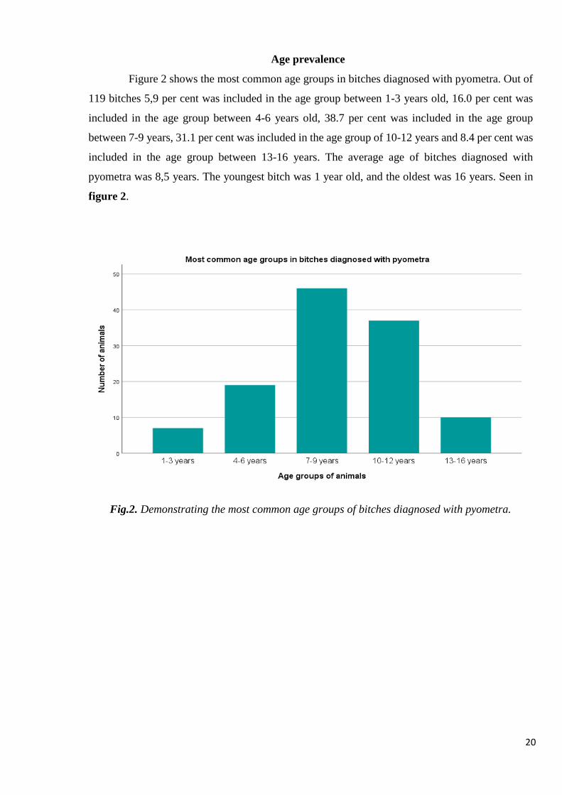
Age prevalence
Figure 2 shows the most common age groups in bitches diagnosed with pyometra. Out of
119 bitches 5,9 per cent was included in the age group between 1-3 years old, 16.0 per cent was
included in the age group between 4-6 years old, 38.7 per cent was included in the age group
between 7-9 years, 31.1 per cent was included in the age group of 10-12 years and 8.4 per cent was
included in the age group between 13-16 years. The average age of bitches diagnosed with
pyometra was 8,5 years. The youngest bitch was 1 year old, and the oldest was 16 years. Seen in
figure 2.
Fig.2. Demonstrating the most common age groups of bitches diagnosed with pyometra.

21
Type of treatment
Out of the 119 bitches diagnosed with pyometra, 1 (0.8 per cent) bitch was medically treated
on the owners request, 114 (95.7 per cent) bitches were surgical treated at the hospital the same
day or the day after, 3 (2.5 per cent) bitches were euthanized before surgery without treatment due
to severe disease, age and on the owners request, 1 (0.8 per cent) bitch were sent to another hospital
for surgical treatment. The 6 (5.2 per cent) bitches that were euthanized after surgery, were
euthanized due to complications (peritonitis, anaemia) and serious illness not improving after
surgery. Seen in figure 3.
Fig. 3. The diagram shows the type of treatment chosen for the bitch after clinical examination and
how many that were euthanised after treatment.

22
Time of clinical signs after oestrus
Clinical signs of pyometra was normally noticed by the owner that brought the dog to the clinic.
sometimes the owner noticed some symptoms of pyometra a few days earlier and in some cases
even weeks earlier. The signs the owner normally noticed at home were polyuria/polydipsia,
anorexia, vaginal discharges, diarrhea, vomiting and depression. These signs normally occurred
during oestrus in 18.8 per cent, in <3 days after oestrus in 11.5 per cent, and in 1-8 weeks after
oestrus in 61 per cent, in 9-12 weeks after oestrus in 8,7 per cent of bitches. This information was
received from 69 bitches. All cases of pyometra in this study was not included, for the reasons that
the owner didn’t know when the bitch was in oestrus, she was breed recently (within 2-6 weeks),
pseudo pregnant or in oestrus more than 6 months ago. Seen in figure 4.
Fig.4. Time when clinical signs appeared from last oestrus to day of diagnosis
0 5 10 15 20 25 30 35 40 45
NUMBER OF ANIMALS
TIME OF CLINICAL SIGNS AFTER OESTRUS
9-12 weeks after oestrus 1-8 weeks after oestrus <3 days Oestrus

23
Anamnestic and clinical findings
Table 1. Prevalence of anamnestic and clinical findings in 119 dogs with pyometra. Data was not
available for all parameters.
CRT= Capillary refill time
Variable Number of bitches with
abnormal findings/total
number of bitches with
available data
Number of bitches %
Anamnesis
Vaginal discharges 88/112 78.6 %
Anorexia 78/102 76.5 %
Depression 100/112 89.3 %
Polyuria 38/63 60.3 %
Polydipsia 66/91 72.5 %
Vomiting 39/116 33.6 %
Diarrhea 22/116 19.0 %
Lameness/stiff 8/119 6,7 %
Clinical signs
Temp
Increased
Decreased
Normal
35/101
6/101
60/101
34.7 %
5.9 %
59.4%
CRT 2/108 1.9 %
Mucus membranes
Pale
Hyperaemic
Icteric
13/119
25/119
1/119
10.9 %
21.0 %
0.8 %
Abdominal
pain/tension/structure
74/119 62.2 %
Enlarged uterus US 105/112 93.8 %
General condition
Mild, moderate, severe
97/119
81.5 %
Ascites 17/119 14.4 %
Peritonitis 27/119 22.7 %
Closed Pyometra
Open Pyometra
23/111
88/111
20.7 %
79.3 %

24
Anamnestic data was obtained by the veterinarian asking the owner questions, some was
obtained by the veterinarian. Clinical signs were obtained by the veterinarian while doing a clinical
examination of the animal.
The most common clinical signs present in bitches with pyometra was collected from the
patient journals and analysed by using SPSS and excel. In table 1 “Prevalence of anamnestic and
clinical findings” the data includes the number of bitches with abnormal clinical findings and total
number of animals included for the analysis and the percentage. As shown, for the anamnestic
parameters, vaginal discharges can be seen in 78.6 per cent, anorexia 76.5 per cent, depression 89.3
per cent, polyuria 60.3 per cent, polydipsia 72.5 per cent, vomiting 33.6 per cent and diarrhea in
19.0 per cent. 6.7 per cent of the dogs showed lameness. In the parameters for physical examination
Increased temperature was present in 34.7, decreased in 5.9 per cent and normal in 59.4 per cent.
Pale mucus membranes were present in 10.9 per cent bitches, hyperaemic in 21.0 per cent and 0,8
per cent of bitches had icteric mucus membranes. When palpating the abdomen, many dogs reacted
by tension of abdomen and showing discomfort that was interpreted as pain in the journal. Also,
during palpation, in many cases, the veterinarian could feel a structure within the abdomen.
Abdominal tension, pain and abdominal structure were calculated within the same group since
these variables occurred together in most cases and it showed an occurrence of 82.2 per cent. The
animal’s general condition was evaluated as mild, moderate and severe and showed that the general
condition was mildly changed in 52.1 per cent, moderate changed in 26.1 per cent and severely
changed in the bitch in 3.4 per cent. Ascites were present in 14.3 per cent of bitches and peritonitis
occurred in 22.7 bitches before treatment. Uterus was enlarged in 93.8 per cent of bitches, evaluated
during ultrasound before treatment or during surgery. Closed pyometra occurred in 20.7 per cent
of bitches and open pyometra occurred in 79.3 per cent.

25
Haematological and Biochemical findings
The presence of haematological and biochemical clinical findings is shown in table 2.
Table 2. Haematological and biochemical clinical findings in 119 bitches with pyometra.
Haematology was analysed with Sysmex XN-1000 Haematology Analyser and IDEXX procyte dxTM
and biochemistry was analysed with Hitachi-Roche-Cobas c 311and IDEXX catalyst dxTM
analyser.
CRP= C-reactive protein, Hct= haematocrit, Hb= Haemoglobin, ALP=Alkaline phosphate,
ALT=Alanine aminotransferase, Na=Sodium, K=Potassium, Ca=Calcium, Cl=Chloride
Variable
Number of
bitches with
abnormal
findings /
total number of
bitches with
available data
Number
of bitches
with
respective
findings
%
Normal
references
for Sysmex
XN-1000
and Cobas c
311
Normal
references
for
IDEXX
procyte
dxTM and
IDEXX
dx TM
Unit
Haematology
CRP 67/76 88,2 % <20 mg/L
Erythrocytes 36/115 31.3 % 5,7-8,9 5,7-8,9 x10¹²/L
Hct 44/115 38.2 % 37-62 37-62 %
Hb 39/115 33.9 % 131-205 131-205 g/L
Leukocytes
76/115 66.1 % 5-16,8 5-16,8 x10⁹/L
Thrombocytes 36/115 31.3 % 148-484 148-484 x109/L
Biochemistry
Glucose 16/115 13.9 % 3,7-6,6 4,1-7,9 mmol/L
Urea 27/108 25.0 % 3,0-9,0 2,5-9,6 mmol/L
ALP 49/108 45.3 % <1,4 23-212 IU/L
ALT 20/115 17.4 % <1,2 10-100 IU/L
Creatinine 3/114 2.6 % <135 44-159 µmol/L
Albumin 34/108 31.4 % 30-45 23-40 g/L
Phosphate 4/59 6.8 % 0,7-1,9 mg/dL
Total Protein 33/114 28.9 % 49-71 52-82 g/L
Bile acids 7/62 11.3 % <20 µmol/L
Na 6/108 5.5 % 138-149 144-160 mmol/L
K 5/107 4.6 % 3,4-4,8 3,5-5,8 mmol/L
Ca 5/62 8.0 % 2,4-3
mmol/L
Cl 2/46 4.3 % 109-122 mmol/L
Lactate 1/5 20 % <2 mmol/L

26
Table 2 shows the haematological and biochemical clinical findings with the number of
abnormal findings and the total number of dogs analysed. Haematology (Erythrocytes, haematocrit,
haemoglobin, leukocytes, thrombocytes) were analysed with Sysmex XN-1000 Haematology
Analyser and IDEXX procyte dxTM and biochemical values (Glucose, urea, alkaline phosphate,
alanine aminotransferase, creatinine, phosphate, total proteins, bile acids, sodium, potassium,
calcium and chlorine) were analysed with Hitachi-Roche-Cobas c 311and IDEXX catalyst dxTM
analyser. CRP was increased with 88.2 per cent of the controlled bitches, erythrocytes was
abnormal in 36 animals (increased in 4 bitches and decreased in 32), haematocrit was abnormal in
44 bitches (2 increased and 42 decreased), 39 bitches had abnormal haemoglobin (3 had increased,
36 decreased), leukocytes were increased in 64.3 per cent and decreased in 1.2 per cent of bitches,
urea was increased in 11 (10.2 per cent) bitches and decreased in 16 (14.8) bitches, thrombocytes
was decreased in 17 (14.8 per cent) bitches and decreased in 19 (16.5 per cent), alkaline phosphate
were increased in 48 (44.4 per cent) and decreased in 1 (0,9 per cent), alanine aminotransferase
was increased in 18 (15.7 per cent) and decreased in 2 (1.7 per cent), total protein was increased in
33 (28.9) bitches, albumin was decreased in 31 (28.7 per cent) of bitches and increased in 3 (2.8),
7 (1.8 per cent) had increased bile acids. Sodium, potassium, calcium and chloride were increased
between 4 to 8 per cent. Lactate were only tested in 5 bitches and 1 bitch had increased lactate and
was euthanized after surgery. 3 bitches were tested for lactate after surgery and 1 bitch showed an
increase in lactate, they were not tested before surgery and not included in the study.

27
Table 3. Haematology and Clinical biochemistry findings in 76 bitches with pyometra. average,
median, normal reference range. Haematology parameters were analysed with “Sysmex XN-1000
Haematology Analyser” and biochemical parameters were analysed with “Hitachi-Roche-Cobas
c 311 analyser”. Lactate was analysed with “The edge lactate analyser” or “Gem premier 3500”.
CRP= C-reactive protein, Hct= haematocrit, Hb= Haemoglobin, ALP=Alkaline phosphate,
ALT=Alanine aminotransferase, Na=Sodium, K=Potassium, Ca=Calcium, Cl=Chloride.
Variable
Number
of tests Average Median
Min-
Max Unit
Normal
reference
range
Haematology
CRP 68 146 143 3-350 mg/L <20
Erythrocytes 72 6,2 6,2 3,9-10,1 x10¹²/L 5,7-8,9
Hct 72 37,6 38 4,1-63 % 37-62
Hb 72 138,5 142,5 22,3-230 g/L 131-205
Leukocytes 72 21,1 18,1 1,3-75,6 x10⁹/L 5-16,8
Thrombocytes 72 299,3 270,5 15-859 x109/L 148-484
Biochemistry
Glucose 72 5,5 5,5 2,7-8,1 mmol/L 3,7-6,6
Urea 67 5,3 4 2,0-19,0 mmol/L 3,0-9,0
ALP 67 15,9 2,1 0,3-278 IU/L <1,4
ALT 72 14,8 0,55 0,1-466 IU/L <1,2
Creatinine 72 72 66 30-270 µmol/L <135
Albumin 67 29,8 31 15-42 g/L 30-45
Phosphate 62 1,2 1,2 0,6-2,2 mg/dL 0,7-1,9
Proteins 71 68,7 68 45-92 g/L 49-71
Bile acids 61 8,8 4 0-69 µmol/L <20
Na 67 145 146 136-153 mmol/L 138-149
K 67 4,2 4,1 3,3-5,9 mmol/L 3,4-4,8
Ca 62 2,6 2,55 2,1-3,6 mmol/L 2,4-3
Cl 5 114 114 113-116 mmol/L 109-122
Lactate 5 1,7 1,4 1,1-3,5 mmol/L <2

28
Table 4. Haematology and Clinical biochemistry findings in 41 bitches with pyometra. Average,
Median, Normal reference range. Haematology were analysed with IDEXX procyte dxTM and
biochemical parameters were analysed with IDEXX catalyst dxTM. Lactate was analysed with The
Edge Lactate Analyser or the Gem premier 3500.
Variable
Number
of tests Average Median Min-Max Unit
Normal
reference
range
Haematology
Erythrocytes 41 6,4 6,48 3,6-9,31 x10¹²/L 5,7-8,9
Hct 41 39,5 39,8 3,7-56,3 % 37-62
Hb 41 14,7 140 79-206 g/L 131-205
Leukocytes 41 20,5 19,4 7,58-64,95 x10⁹/L 5-16,8
Thrombocytes 41 301,2 286 79-766 x109/L 148-484
Biochemistry
Glucose 41 5,8 5,66 3,99-10 mmol/L 4,1-7,9
Urea 41 4,2 3,5 1,1-12,9 mmol/L 2,5-9,6
ALP 41 199 136 10-2000 IU/L 23-212
ALT 39 54,7 41 10-206 IU/L 10-100
Creatinine 41 83 75 29-187 µmol/L 44-159
Albumin 41 30,3 29 24-79 g/L 23-40
Proteins 41 74,6 75 59-93 g/L 52-82
Bile acids 1 200 200 200-200 µmol/L
Na 41 157,7 151 144-157 mmol/L 144-160
K 41 4,1 4,2 3,2-5,1 mmol/L 3,5-5,8
Cl 41 113,3 113 106-119 mmol/L 109-122
CRP= C-reactive protein, Hct= haematocrit, Hb= Haemoglobin, ALP=Alkaline phosphate,
ALT=Alanine aminotransferase, Na=Sodium, Ca=Calcium, Cl=Chloride.

29
Haematology and clinical biochemistry findings from bitches with pyometra are presented in
Table 3 and table 4 above. Table 3 covers blood analysis from Sysmer XN-1000 and Hitachi-
Roche Cobas c311 and table 4 covers blood analysis from IDEXX procyte dx TM and IDEXX
Catalyst dxTM. Lactate in table 3 were analysed with The Edge Lactate analyser or Gem premier
3500. Different blood analysis machines were used because the laboratory personal were not
present in the evening and the personal then used other machines to analyse the blood tests. In table
number 3, a number of 72 bitches were analysed, but not all were tested for all parameters. In table
4, 41 bitches were analysed for all parameters except bile acids and ALT.
Prevalence of changes in temperature of bitches with pyometra and relationship with
peritonitis
Of the 119 bitches diagnosed with pyometra, 101 bitches were checked for temperature. 59.4
per cent had normal temperature. 34.7 per cent had increased temperature. 5.9 per cent had
decreased temperature. Seen in figure 5 below.
Out of the 27 bitches with peritonitis 7 (26.0 per cent) had increased temperature, 0 bitches had
decreased temperature and 12 (44.0 per cent) had normal temperature. 8 (30.0 per cent) animals
with peritonitis had unknown temperature, figure 6.
Fig. 5. Shows the occurrence of changes in temperature in bitches with pyometra before
treatment.
35
6
60
0
10
20
30
40
50
60
70
Increased Decreased Normal
NU
MB
ER O
F A
NIM
ALS
CHANGES IN TEMPERATURE IN BITCHES WITH PYOMETRA

30
Fig. 6. Shows the relationship between peritonitis and temperature
Prevalence of increased CRP in bitches with pyometra
Table 5. Data of C-reactive protein in bitches with pyometra.
Of the 76 bitches where C-reactive protein (CRP) where analysed, 67 (88,2 per cent) had
increased CRP and 9 (11,8 per cent) had normal CRP at the time of pyometra.
CRP Number of animals/total
number of animals Number of animals per cent
Normal 9/76 11.8
Increased 67/76 88.2
26%
44%
0%
30%
Relationship between Peritonitis and Temperature
Increased temperaturePeritonitis
Normal temperaturePeritonitis
Decreased temperaturePeritonitis
Unknown temperaturePeritonitis

31
Relationship between C-reactive protein and peritonitis
Increased CRP was found in 20 bitches with peritonitis and it gave a p-value of 0.276 which is
not significant. CRP was also increased in 48 bitches with no peritonitis. CRP was unknown in 6
bitches with peritonitis. In 8 of the bitches analysed, CRP was normal, and they had no peritonitis.
In 1 bitch with peritonitis, CRP was normal. The relationship can be seen in figure 7 below.
Figure 7. The relationship between peritonitis and CRP.
Mortality and complications
Among those 114 bitches treated surgically with ovariohysterectomy 1 (0.9 per cent) was
euthanized during surgery because of tumours in liver and spleen and the conclusion were that it
was a bad prognosis for the dog and the owner was informed about the situation and agreed on
euthanasia, 5 (4.4 per cent) was euthanized after surgery because they didn’t get healthy from
pyometra/peritonitis and their health status worsened. 3 (2.5 per cent) of the bitches was euthanized
without treatment, because of severe disease and marked general status. 16 (13.5 per cent) bitches
hade other diseases such as splenic tumour, heart disease, corp al or liver tumour, glaucoma, uveitis,
urinary tract infection and was operated.
Complications that occurred after surgery could be peritonitis, abdominal tension and pain,
respiratory distress, inappetence, vomiting, diarrhea, anaemia, abdominal drainage of fluid and
20
48
6 10
10
20
30
40
50
60
Increased CRP +Peritonitis
Increased CRP + Noperitonitis
Unknown CRP+Peritonitis
Normal CRP+Peritonitis
NU
MB
ER O
F A
NIM
AL
Relationship between CRP and Peritonitis

32
blood, hypothermia, bleeding from ligatures, these complications occurred in 28 (25.6 per cent)
bitches and diarrhea alone occurred in 28 bitches (24,5 per cent) after surgical treatment.
Antibiotics was used in 27/27 bitches with peritonitis (p=0.001).
Table 6. Total number, mortality, number of bitches diagnosed with peritonitis before surgery and
after surgery or that had prolonged hospitalisation, treated surgically or medically.
Peritonitis occurred in 27 (22.7 per cent) before surgery in the 119 bitches diagnosed with
pyometra. 25 (21.9 per cent) bitches had peritonitis after surgery and 1 (0.9 per cent) bitch was
euthanized after surgery due to peritonitis. Prolonged hospitalization (>3 days) was observed in 28
bitches after surgery and one bitch was euthanized during the hospitalization.
Variables N Mortality
n (%)
Peritonitis before
surgery n (%)
Peritoni
tis n
after
surgery
Prolonged
hospitalisation
n
Total number 119 6 27 (22,7) 25 (21.9) 28
Medically
treated 1 0 (0) 0 (0) 0 (0.0) 0
Surgically
treated 114 6 (5,2) 25 (21.9) 25 (21.9) 28
Euthanized
with no
treatment 3 3 (2,5) 2 (1.7) 1 (0.9) 1

33
DISCUSSION
This study is based on 119 bitches diagnosed with pyometra. The study material used is taken
from one clinic in Sweden and includes bitches diagnosed with pyometra during the year 2017.
To be able to determine why pyometra develops, it is important to look at all factors possible.
Bree, Age, Time of clinical signs
Many studies done before, have evaluated breed occurrence and genetically predisposition
among certain breeds in cases with pyometra. One study done by Ragnevi Hagman (2004), showed
that Bernese Mountain Dog, Rottweiler, Collie (rough haired), Cavalier King Charles Spaniel,
Golden Retriever and English Cocker Spaniel had increased risk of developing pyometra. Breeds
with decreased risk were Swedish hound, normal size Dachshund and miniature Dachshund,
German Shepherd dog and Drever. A reduced risk for mixed-breed dogs have also been shown by
Niskanen and Thrushfield (1998). A study done by Francis. O.Smith (2006) also showed an
increased risk for: Golden Retriever, Miniature Schnauzer, Irish terrier, Bernease Mountain Dog
and Airdale terrier. In present study, a number of 54 breeds were included and the most common
breeds diagnosed with pyometra were: Labrador Retriever, Mixed breeds, Miniature Schnauzer,
different size of Poodle, Golden Retriever, French Bulldog, Stafford shire Bull Terrier and
chihuahua. These results are similar to those of previous researchers. The result might be affected
by the different types of breeds that might be more common in certain regions than others for
example in the city where chihuahua probably is more common than on the country side. Mixed
breed was the second most common breed in this study, while Niskanen and Thrushfield (1998)
found in their study that pyometra was not that common in Mixed breeds.
The most common age group in this study was obtained in bitches between 7-9 years old (38.7
per cent). The average age was 8.5 years and the youngest bitch was 1 year old and the oldest 16
years old. Previous studies showed a similar result to the result in this study. In one study done by
Fransson et al. (2004), it was showed that the average age of pyometra was 8.4 years, and Egenvall
et al. (2001), wrote that the range of age was between 4 months and 18 years. Why bitches develop
pyometra when they get older is not really known. But one theory is that the hormonal stimulation
on uterus that is repeated with each cycle is increasing the risk of pyometra with age.
The administration of steroid hormones (progestins and oestrogen) used to induce abortion
might increase the risk of pyometra, and this might be one of the causes why young individuals
develop pyometra at young age (8).
Clinical signs most commonly were observed 1-8 weeks (61 per cent) after oestrus, and in 18.8
per cent of cases pyometra occurred during oestrus. 69 bitches were included in this study, in many

34
cases, the owner didn’t know when the female was in oestrus last time and was not included in the
data. The signs the owner normally noticed before seeking medical care for the dog were
polyuria/polydipsia, anorexia, vaginal discharges, diarrhea, vomiting and depression and
sometimes the owner noticed some signs weeks before coming to the clinic, which might lead to
misinformation for the received data. Most studies that have been done, show that the most
common time of pyometra is during the dioestrus in the bitch reproductive cycle (23).
The increase in progesterone during end of oestrus and beginning of dioestrus (with peak at 15
to 30 days after ovulation), results in endometrial proliferation and uterine glandular secretion,
decreased myometrial contraction and induces closure of cervix, this increases the risk of bacterial
growth in the uterus which makes it a favourable place for bacterial growth (7,36). Due to repeated
stimulation of hormones on the uterus, the risk of developing pyometra increases with age (8, 23).
In this study many cases of pyometra occurred during the end of oestrus (in 18.8 per cent of cases)
which may indicate bacterial ascendance from the normal flora of vagina or urinary tract during
proestrus or oestrus (24).
The most common clinical signs present in >50 per cent of bitches included vaginal discharges,
anorexia, depression, polydipsia, and polyuria (Table 1), which reflects systemic involvement, in
most bitches and supports previous studies by Jitpean et al. (2014). Vaginal discharges were
present in 88 (78.6 per cent) of bitches and was absent in 24 bitches (21.4 per cent). Closed
pyometra, when vaginal discharges are absent, normally indicates more severe disease (39).
Laboratory findings
Laboratory findings in this study showed that 88.2 per cent had increased CRP which is
supported by studies done by Jitpean (2015), B.A. Fransson (2004) and many others. R.Dabrowski
et al. (2013) did a study where it was showed that CRP was more increased in bitches with closed-
cervix pyometra. The results obtained in this study is supported by the data from the previous study
where closed-pyometra had an average of 181 in CRP and open-pyometra had an average of 143
in CRP. Decreased erythrocytes, haematocrit and haemoglobin was found in this study in 32-36
bitches. Anemia is thought to be the caused by the long duration of the disease, erythrocytes are
decreased due to the toxic effects on the bone marrow that occur or lack of iron in the blood or and
also due to the loss of erythrocytes to the uterine lumen (18). Dehydration can interfere with the
evaluation of anemia, if the animal is dehydrated, a misleading value can be received, and a higher
haematocrit is obtained. Increased leukocytes and ALP were also common findings in this study.
Increased leukocytes was found in 66.1 per cent and is a common finding in bitches according to
R.Hagman (2012) and many other authors. Leucocytosis is an indicative of sepsis and can be found
elevated early in the disease. When the disease worsens, the presence of band and toxic neutrophils

35
can be seen (17). Leukopenia was found in 2 bitches which have been found to be an indicative of
poorer prognosis (18), in this study both bitches (Labrador Retriever and Golden Retriever) with
leukopenia had hyperaemic mucus membranes, closed-cervix pyometra and one of them also had
circulatory shock and ruptured uterus. Increased ALT and proteins were seen in 16 to 29 per cent
and albumin was decreased in 28.7 per cent.
Treatment Methods
Most animals are treated surgically, and some are treated medically. Many authors discuss
treatment methods used for animals that are young and has an open-cervix pyometra and a normal
organ function where the owner wants to preserve the bitch reproductive tract. These bitches can
be treated medically with prostaglandins, progesterone receptor antagonists and dopamine agonists
together with antibiotics. Bitches that received medical treatment and that are not breed and
conceive may have a higher chance of getting pyometra again at the next cycle according to
Frances O. Smith (2006). F.Fieni (2006) studied the efficacy of Aglepristone with or without
cloprostenol in bitches with pyometra. The author followed up on those that were 23 bitches that
were successfully treated after 24 months, 3 bitches developed pyometra after 7 to 12 months and
1 bitch after 19 months. Two bitches out of the 23 that received Aglepristone together with
cloprostenol did not show occurrence of pyometra at the next oestrus cycle. After 6 years, 3 bitches
still didn’t have reoccurrence of pyometra and had normal oestrus.
Ovariohysterectomy is done in most animals and is recommended if the bitch is older or
affected systemically, or if increased risk of anesthesia or surgery due to other diseases. In this
study 114 bitches were surgically treated by ovariohysterectomy and 6 bitches were euthanized
after surgery du to peritonitis or because their health status did not improve after surgery. In one
study done by Jitpean et al. (2014) complications was observed in 19 per cent of bitches and
prolonged hospitalization (>3 days) in 25 per cent. Peritonitis was observed in 13 per cent of
bitches, which is a bit lower than received in this study where peritonitis occurred in 21.9 per cent.
Prolonged hospitalization on the other hand was observed in 25 per cent in present study, which
supports these of previous study by Jitepean et al (2006).

36
CONCLUSION
1. Labrador retriever (9.2 per cent), mixed breed (8.4 per cent) and Miniature Schnauzer (6.7
per cent) was the most common breed diagnosed with pyometra in this study. Most common
age group was 7-9 years (38.7 per cent). The most common treatment method used was
ovariohysterectomy (95.7 per cent).
2. Vaginal discharges, anorexia, depression, polyuria, and polydipsia could be seen in more
than 50 per cent of bitches diagnosed with pyometra. Pyometra was most commonly
diagnosed (in 61 per cent of bitches) 1-8 weeks after oestrus.
3. Increased CRP (88.2 per cent), leucocytosis (64.3 per cent), increased ALP (44.4 per cent),
decreased erythrocytes (27.8 per cent), decreased haematocrit (36.5) and decreased
haemoglobin (31.3), was the most common parameters found in bitches diagnosed with
pyometra. The prevalence of increased temperature in bitches with peritonitis was 26 per
cent and 5.9 per cent had decreased temperature. Increased CRP was seen in 20 bitches with
peritonitis (p=0.276) which is not significant.
4. The most common complication that occurred after ovariohysterectomy was peritonitis
(21.9 per cent) and diarrhea occurred in 24.5 per cent after surgery.

37
REFERENCES
1. Hagman, R., Reezigt, B. J., Bergström Ledin, H., & Karlstam, E. “Blood lactate levels in 31
female dogs with pyometra”; Acta Veterinaria Scandinavica.2009;51(1):2. doi:10.1186/1751-
0147-51-2
2. Tobias K.M., Johnston S.A., Ovaries and uterus. Veterinary surgery small animal.2012;Elsevier
sunders. 1875-1885
3. Egenvall, A., Hagman, R., Bonnett, B. N., Hedhammar, A., Olson, P., & Lagerstedt, A.-S.
Breed Risk of Pyometra in Insured Dogs in Sweden. Journal of veterinary internal
medicine.2001;15(6):530–538. doi:10.1111/j.1939-1676.2001.tb01587.x
4. Hagman, R. Canine pyometra: What is new? 2016;Reproduction in Domestic Animals.
2016;52:288–292. doi:10.1111/rda.12843
5. Jitpean, S. et al. Outcome of pyometra in female dogs and predictors of peritonitis and
prolonged postoperative hospitalization in surgically treated cases.2014; BMC Veterinary
Research, 10(1), 6. doi:10.1186/1746-6148-10-6
6. Jitpean, S. et al. Serum insulin-like growth factor-I, iron, C-reactive protein, and serum amyloid
A for prediction of outcome in dogs with pyometra.2014; Theriogenology, 82(1), 43–48.
doi:10.1016/j.theriogenology.2014.02.01
7. Fossum T.W. Small animal surgery. 4th ed. St. Louis Missouri: Elsevier Mosby; 2014. p. 780-
855.
8. Nelson WR, Couto CG. Small Animal Internal Medicine. 4th ed.2009; Mosby Elsevier. p. 885-
982.
9. Gibson, A., Dean, R., Yates, D., & Stavisky, J. A retrospective study of pyometra at five
RSPCA hospitals in the UK: 1728 cases from 2006 to 2011. 2013;Veterinary Record, 173(16),
396–396. doi:10.1136/vr.101514
10. Hagman, R. Pyometra in Small Animals. 2018;Veterinary Clinics of North America: Small
Animal Practice, 48(4), 639–661. doi:10.1016/j.cvsm.2018.03.001
11. Jitpean, S., Pettersson, A., Höglund, O. V., Holst, B. S., Olsson, U., & Hagman, R. Increased
concentrations of Serum amyloid A in dogs with sepsis caused by pyometra. 2014;BMC
Veterinary Research, 10(1). doi:10.1186/s12917-014-0273-9
12. Nelson WR, Couto CG. Disorders of hemostasis. Small animal internal medicine. 5th ed.
2014.St. Louis Missouri: Elsevier Mosby. p.1245-1263
13. Fransson.B.A.Systemic inflammatory response.2004.Reserach gate.
14. Ettinger S.J., Feldman C.E., Textbook of veterinary internal medicine 7th edition, volume 2
Chapter 320, p.1602

38
15. Fransson, B.A. et al., C-reactive protein, tumor necrosis factor α, and interleukin-6 in dogs with
pyometra and SIRS.2007; Journal of Veterinary Emergency and Critical Care, 17(4), 373–381.
doi:10.1111/j.1476-4431.2006.00203.x
16. Karlsson, I., Hagman, R., Johannisson, A., Wang, L., Karlstam, E., & Wernersson, S. Cytokines
as Immunological Markers for Systemic Inflammation in Dogs with Pyometra.2012;
Reproduction in Domestic Animals, 47, 337–341. doi:10.1111/rda.12034
17. Brady, C. A., & Otto, C. M. Systemic Inflammatory Response Syndrome, Sepsis, and Multiple
Organ Dysfunction.2001; Veterinary Clinics of North America: Small Animal Practice, 31(6),
1147–1162. doi:10.1016/s0195-5616(01)50097-2
18. Hagman, R. Clinical and Molecular Characteristics of Pyometra in Female Dogs.2012;
Reproduction in Domestic Animals, 47, 323–325. doi:10.1111/rda.12031
19. Enginler, S.,Ateş, A.,Diren Sığırcı, B.,Sontaş, B.,Sönmez, K.,Karaçam, E.,Gürel, A.
Measurement of C-reactive protein and Prostaglandin F2α Metabolite Concentrations in
Differentiation of Canine Pyometra and Cystic Endometrial Hyperplasia/Mucometra2014;
Reproduction in Domestic Animals. 49(4), 641–647. doi:10.1111/rda.12340
20. Pepys, M. B., & Hirschfield, G. M. C-reactive protein: a critical update.2003; Journal of
Clinical Investigation, 111(12), 1805–1812. doi:10.1172/jci18921)
21. Dąbrowski, R., Kostro, K., & Szczubiał, M. Concentrations of C-reactive protein, serum
amyloid A, and haptoglobin in uterine arterial and peripheral blood in bitches with
pyometra.2013;Theriogenology, 80(5), p.494–497. doi:10.1016/j.theriogenology.2013.05.01
22. Nelson WR, Couto CG. Small Animal Internal Medicine. 5th ed.2014;Mosby Elsevier. 915-950.
23. Pretzer, S. D. Clinical presentation of canine pyometra and mucometra: A review.
2008;Theriogenology, 70(3), 359–363. doi:10.1016/j.theriogenology.2008.04.028
24. Hagmen. R. New aspects of canine pyometra. 2004; Swedish University of Agricultural
Sciences Uppsala.
25. Fransson.B., Ragle C. Canine pyometra: An update on pathogenesis and
treatment.2003;Compendium on continuing education for the practising veterinarian-North
American edition, 25(8):602-612
26. De Cramer K. Surgical uterine drainage and lavage as treatment for canine pyometra.
2010;African Veterinary Ass. p.172-177.
27. Fieni F, Topie E, Gogny A. Medical treatment for pyometra in dogs. Reproduction in domestic
animals. 2014: p.28-32.
28. Jiena, B., Rao, K., Reddy, K., & Raghavender, K. Therapeutic efficacy of natural prostaglandin
in the treatment of pyometra in bitches. 2013;Veterinary World, 6(6), 295.
doi:10.5455/vetworld.2013.295-299

39
29. Jiena.B., Sadasiva Rao K., Das D., Reddy K.C., Therapeutic effects of synthetic prostaglandin
in the treatment of pyometra in bitches. 2014; The bioscan. 9(3):1019-1021
30. Baird, D. T. Luteotrophic control of the corpus luteum.1992;Animal Reproduction Science.
28(1-4), 95–102. doi:10.1016/0378-4320(92)90096-v
31. Brady C.A, Otto C.M. Systemic inflammatory response syndrome, sepsis, and multiple organ
dysfunction. 2001; Critical care. P.1147-1162
32. F.Fieni. Clinical evaluation of the use of aglepristone, with or without cloprostenol, to treat
cystic endometrial hyperplasia-pyometra complex in bitches.2006; Theriogenology 66 (2006)
1550–1556
33. Jurka, P., Max A., Hawryńska, K., & Snochowski, M. Age-Related Pregnancy Results and
Further Examination of Bitches after Aglepristone Treatment of Pyometra. Reproduction in
Domestic Animals, 2008;45(3), 525–529. doi:10.1111/j.1439-0531.2008.01288.x
34. S.Metcalf, C.Vischer. Medical treatment of pyometra and the use of Aglepristone. 2006;
Applecross veterinary hospital.Aust Vet Practit 36(4.
35. Corrada, Y., Arias, D., Rodríguez, R., Tortora, M., & Gobello, C., Combination dopamine
agonist and prostaglandin agonist treatment of cystic endometrial hyperplasia–pyometra
complex in the bitch. 2006;Theriogenology, 66(6-7), 1557–1559.
doi:10.1016/j.theriogenology.2006.01.00
36. Jitpean S. Predictive markers and risk factors in canine pyometra. 2015.
37. Annika Bergström. Textbook of veterinary internal medicine diseases of the dog and cat, 8th
edition. 2017; Elsevier, St.Louise, Missouri .p1878.
38. Sant’ anna, M. C., Trautwein, L. G. C., Giordano, L. G. P., Justino, R. C., Flaiban, K. K. M. da
C., & Martins, M. I. M. The importance of Gram positive bacteria as the cause of canine
pyometra. 2017.Semina: Ciências Agrárias, 38(2), 1077. doi:10.5433/1679-
0359.2017v38n2p1077
39. Jitpean et al. Closed cervix is associated with more severe illness in dogs with pyometra. 2017;
BMC Veterinary Research 13:11. Doi 10.1186/s12917-016-0924-0.
40. Ros, L., Holst, B. S., & Hagman, R. A retrospective study of bitches with pyometra, medically
treated with aglepristone.2014;Theriogenology, 82(9), 1281–1286.
doi:10.1016/j.theriogenology.2014.08.01
41. Gobello, C. Dopamine agonists, anti-progestins, anti-androgens, long-term-release GnRH
agonists and anti-estrogens in canine reproduction:2006;A review. Theriogenology, 66(6-7),
1560–1567. doi:10.1016/j.theriogenology.2006.02.00

40
42. Corrada, Y., Arias, D., Rodríguez, R., Tortora, M., & Gobello, C. Combination dopamine agonist
and prostaglandin agonist treatment of cystic endometrial hyperplasia–pyometra complex in the
bitch. 2006;Theriogenology, 66(6-7), 1557–1559. doi:10.1016/j.theriogenology.2006.01.00
43. Onclin, K., & Verstegen, J. P. In vivo investigation of luteal function in dogs: Effects of
cabergoline, a dopamine agonist, and prolactin on progesterone secretion during mid-pregnancy
and -diestrus. 1997;Domestic Animal Endocrinology, 14(1), 25–38. doi:10.1016/s0739-
7240(96)00093-8
44. Niskanen M, Thrustfield M.V. Association between age, parity, hormonal therapy and breed, and
pyometrs in Finnish dogs. 1998;Veterinary record, 143:493-498