nanotechnology applications in medicine
TRANSCRIPT
Key words: nanomedicine, geno-
mics, nanotechnology.
Correspondence to: Ugo Valbusa,
NanoMed Labs, Centro di Biotecnolo-
gie Avanzate CBA, Largo Rosanna
Benzi, Genova, Italy.
Tel +39-010-5737561;
fax +39-010-5737382;
website http://www.nanomed.unige.it
Received ..........................;
accepted ..........................
Nanotechnology applications in medicine
Elena Angeli, Renato Buzio, Giuseppe Firpo, Raffaella Magrassi, Valentina Mussi, Luca Repetto, and Ugo Valbusa
NanoMed Labs, Centro di Biotecnologie Avanzate CBA and Dipartimento di Fisica dell’Università di Genova, Genova, Italy
ABSTRACT
In recent years there has been a rapid increase in nanotechnology applications tomedicine in order to prevent and treat diseases in the human body. The establishedand future applications have the potential to dramatically change medical science.The present paper will give a few examples that could transform common medicalprocedures.
Introduction
It is well known that nanoscale objects have different physical, chemical and bio-logical properties from those of the corresponding mass materials. These properties,together with their nano-size, the same of the naturally occurring biomolecules,promise revolutionary potential applications in clinical practice that could con-tribute to improve health care in the 21st century. This emerging area of research,nanomedicine, is the application of nanotechnology to medicine. Key issues ofnanomedicine include miniaturization of devices, novel nanosized materials, chip-based technologies, imaging techniques, drug delivery, new analytical tools thatcould quickly lead to a better understanding of initiation and progression of disease.This paper wishes to review and highlight some of these research fields. The first partis devoted to methods based on atomic force microscopy (AFM) to image DNA mol-ecules for single nucleotide polymorphism detection and molecular haplotyping,section 3 describes the state of the art of drug delivery based on carbon nanotubes,section 4 gives examples of DNA sequencing by means of the nanopore technology,section 5 illustrates the advancement in the technology of planar patch clamping,and section 6 focuses on micro- and nanofluidics for lab-on-a-chip applications.
Imaging methods based on AFM to image DNA molecules for SNPsdetection and molecular haplotyping (si può accorciare questo titolo?)
Nanoscale characterization of DNA by atomic force microscopy
Atomic force microscopy (AFM) measures interaction forces acting between asharp probe and the specimen surface scanned beneath the probe, to provide a 3Dreconstruction of surface morphology with nanoscale accuracy1. The rapid growth ofthe AFM technique in the years following its invention in 19862 revealed an outstand-ing capability to image surfaces under ambient air, liquid and vacuum environmentsand suggested the potential to image biological systems in real time, under naturalconditions and with molecular resolution. A number of important studies soon ap-peared showing AFM imaging of isolated macromolecules (mostly nucleic acids,polysaccharides and proteins3) as well as cells, tissues and biomaterials4.
AFM investigation of DNA requires the deposition of molecules from a liquid solu-tion (~1nM concentration) onto a solid support: muscovite mica () is an invaluablecandidate, being an hydrophilic and layered material that can be cleaved by adhesive
Tumori, 94: 206-215, 2008
NANOTECHNOLOGY APPLICATIONS IN MEDICINE 207
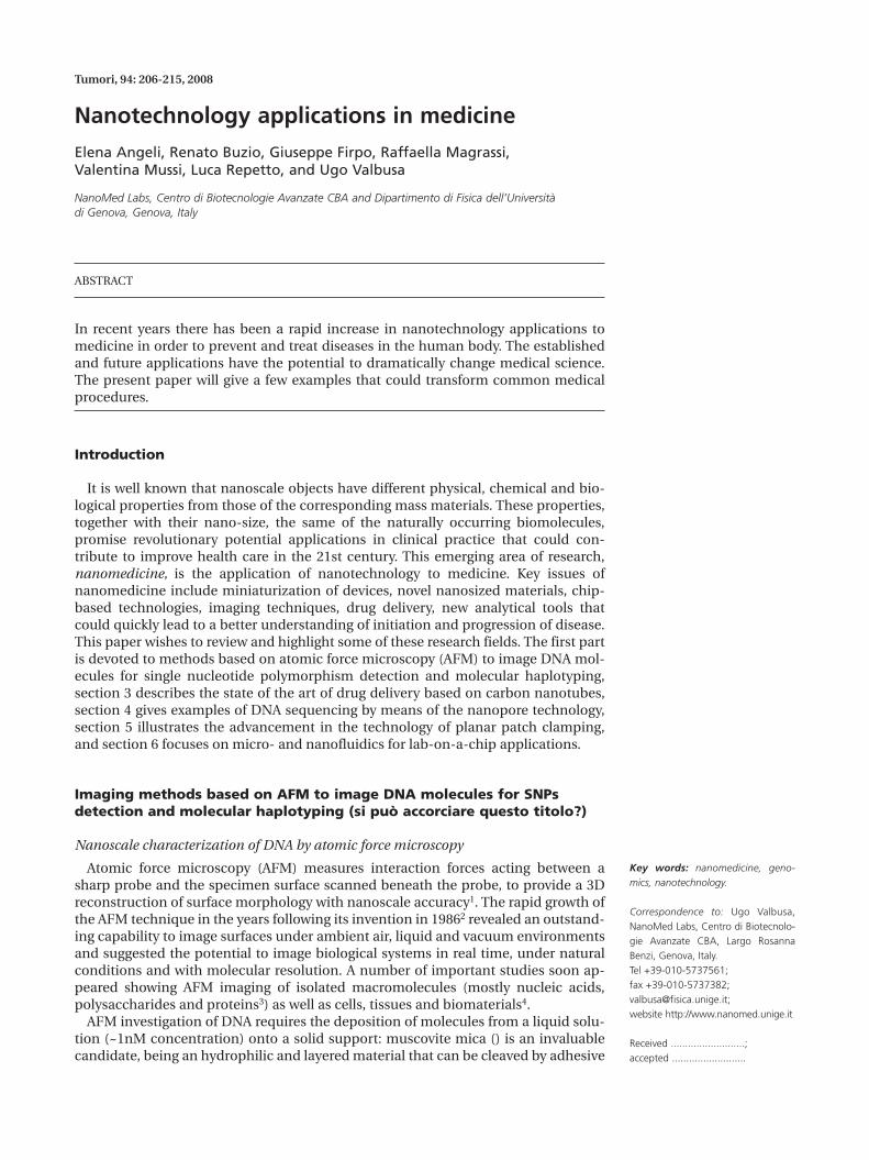
tape to expose atomically flat planes extending severalsquare millimeters. Glass, silicon wafers and molecular-ly-functionalized graphite can also be used for this pur-pose. Standard protocols consist of the deposition of asmall drop of solution on the chosen substrate, followedby drying with a gentle stream of nitrogen and imagingunder laboratory air5. AFM resolves the conformation ofindividual duplexes with a spatial resolution of 1 nm to10 nm and allows to quantify their intrinsic geometricalproperties (like persistence length, local intrinsic curva-ture and flexibility), which might then be efficiently cor-related with nucleotide sequence6. A representativeAFM topography of 1336-bp fragments is shown in Fig-ure 1; whereas the expected DNA lateral should be 2 nm,the molecule width, as measured on AFM topographies,is 5 to 9 nm and the molecule height is 0.6 to 1 nm. Suchdiscrepancy, and the related difficulty to resolve thedouble-helix structure, arises from the finite size of theAFM probe, the limits in force sensitivity, and moleculerelaxation at adsorption.
Direct DNA sizing represents the most simple and di-rect application of AFM imaging7,8. Advanced studiesshedding light on the nanoscale geometrical and me-chanical properties of DNA are based on a proper char-acterization of the conformations displayed by individ-ual molecules on AFM images9,10. The above figure al-ready reveals the presence of “configurational noise”arising from the Brownian motion of molecules in a liq-uid environment and at adsorption; starting from this
fact, Zuccheri et al. demonstrated that the local intrinsiccurvature estimated from a population of ~100-1000DNA molecules can be efficiently related to the specificnucleotide sequence11. This goal, which is prone tohigh-throughput automation12, is accomplishedthrough a software extraction of molecule profiles fromAFM topographies, and the subsequent analysis of thechains of pixels with numerical algorithms. It is well known that the local DNA curvature can be es-timated as the angular deviation of neighboring seg-ments located along the chain approximating the mo-lecular profile13. Such curvature depends of course onnucleotide sequence and thermal motion; ensemble av-erages conducted over several molecule conformationsprovide the following expression:
where represents the DNA curvature along the n dinu-cleotide step, is the intrinsic curvature, represents thelocal curvature fluctuation at the finite temperature Tand <….> indicates ensemble averaging. The last termon the right of the above equation (corresponding to thedynamic contribution to the curvature) is zero for rea-sons of symmetry, with the result that the local intrinsiccurvature can be estimated from AFM images. More-over, it can be shown that the standard deviation of thecurvature can be related to the nanoscale DNA localflexibility.
The above studies demonstrate practical methods toinvestigate important properties of individual DNAmolecules, like their conformation and local stiffness, atthe nanoscale.
AFM experiments addressing functional DNA proper-ties at the subnanometer scale often require hybridiza-tion of target DNA with specific probes, which are thenco-located along the DNA profile to highlight the pres-ence of specific sites; a powerful application of thisstrategy is the use of AFM for molecular haplotyping14,which involves detection and correlation of single nu-cleotide polymorphisms (SNPs) along individual DNAmolecules.
SNP detection and molecular haplotyping by AFM
Comparison of genomic DNA sequences in differentindividuals reveals positions at which sequence varia-tions involving individual base substitutions may occur.These SNPs are highly abundant, being estimated to ap-pear on average at 1 every 500-700 bases along the 3-bil-lion-base human genome (http://www.hapmap.org).Depending on their position, SNPs may have differentconsequences at the phenotypic level: if located withinregulatory and coding regions of genes, they are likely toinfluence the risk of common disorders, or even tocause most of the known recessively or dominantly in-herited monogenic common disorders by altering thestructure or function of coded proteins; they can also af-
),()(C)()(C)(C 00 Tnnnnn χχ +=+=
Figure 1 - AFM topography of 1336-bp DNA molecules, amplifiedby PCR and adsorbed on mica. Sample was imaged in air.
fect drug responsiveness through an alteration of theprimary structure of proteins involved in the drug me-tabolism. This type of SNP is therefore routinely ana-lyzed for diagnostic purposes, human genetic studies,epidemiology and pharmacogenomics. Most SNPs,however, are located in non-coding regions of thegenome, and have no direct known impact on the phe-notype of an individual: these SNPs are useful as mark-ers to identify genes that predispose individuals to com-mon human disorders, thus playing a crucial role forlinkage disequilibrium mapping using a case-controlpopulation association study design15. The great inter-est in SNPs is documented by the intensive efforts madeto identify them in humans and the availability of pub-lic databases containing data for more than 2 millionSNPs (the SNP Consortium and the InternationalHapMap Project). Many methods exist for genotypingSNPs, i.e., establishing which alleles an individual car-ries at different gene loci. A harder problem is haplotyp-ing, i.e., determining which alleles lie on each of the 2homologous chromosomes in a diploid individual. Infact, it is increasingly appreciated that haplotypes,rather than genotypes alone, carry the richest data onhuman variation, and that association studies utilizinghaplotypes formed from SNPs may be more powerful tofind genes affecting health, disease and responses todrugs and environmental factors16.
Unfortunately, no method of high-throughput haplo-typing of multiple SNPs currently exists. Most difficul-ties are due to the fact that humans are diploid andgenotyping methods use genomic DNA as target with-out separating the parental chromosomes. Classically,haplotypes are inferred by genotyping family membersor they are predicted by statistical arguments: the for-mer are, however, not always readily available whereasdifferent methodologies often produce conflicting datafor the latter method. A molecular approach to haplo-typing, capable of physically resolving the diploid chro-mosomes of an individual into their haplotypes, wouldtherefore be highly desirable and of tremendous impor-tance and applicability.
As anticipated above, an AFM approach to molecularhaplotyping can be implemented by the analysis of in-dividual DNA molecules: this was pioneered in 2001 byWoolley et al.17, using single-walled carbon nanotubeprobes to identify polymorphic sites on 10-kilobase-sized DNA fragments. Labeled oligonucleotides werehybridized specifically to complementary target se-quences in template DNA, and the positions of thetagged sequences were detected by direct AFM imaging.In detail, oligonucleotides were labeled with strepta-vidin or the fluorophore IRD800, which can be consis-tently distinguished from one another on the basis oftheir size, and the nanotube tips allowed to locate themwith respect to a restriction site with 3 nm spatial reso-lution, corresponding to a ∼10-bp uncertainty. Haplo-type determination was therefore successfully recon-
208 E ANGELI, R BUZIO, G FIRPO ET AL
ducted to visual inspection of AFM images of individualDNA molecules immobilized on a solid surface. Woolleyet al. justly recognized that direct haplotyping usingAFM might represent a significant advance over con-ventional approaches and could facilitate the use ofSNPs for association and linkage studies, with virtuallyno limitations on the size of fragments to be examinedand use of very small amounts of DNA sample.
Despite such promising results and subsequent ex-pectations for AFM-based haplotyping platforms18, nosignificant developments have been reported so far. Wemust point out that crucial intrinsic limitations of theabove method again arise from the poor spatial resolu-tion characterizing standard AFM when imaging elasti-cally soft biomolecules. This ultimately impedes theachievement of robustness and reliability for the SNPdetection scheme, since label discrimination based on-ly on their physical size is prone to errors due to un-avoidable deformations and damage of DNA fragmentsand the labels themselves.
NanoMed Labs is currently exploring novel methodsto move forward into AFM molecular haplotyping,mainly through conceptual and technical develop-ments concerning the introduction of a novel SNP de-tection scheme and the use of an advanced, custom-de-signed AFM platform.
Carbon nanotubes for targeted drug delivery
Starting from their discovery in 199119, carbon nan-otubes have found application in a growing number ofdifferent fields. Their peculiar properties and their spe-cific geometrical structure make them a versatile andcomplex tool on the two fronts of basic characterizationand technological application.
More recently their possible use in the field of bio-medicine was suggested as a result of the proven bio-compatibility and reduced cytotoxicity of these carbonnanometric tubular structures20. Methods were devel-oped for attaching molecules to the inside and outsideof the hollow nanotubes, opening the way to their use asbiosensors and molecular transporters21. In particular,single-walled carbon nanotubes (SWNT) have beenshown to be able to shuttle various molecular cargos in-side living cells including proteins, short peptides, andnucleic acids22-24. This gives one the ability to target anddestroy individual cells that may be cancerous or infect-ed by a virus.
Their role as biocarriers could be made even morepowerful by exploiting their intrinsic physical proper-ties, such as their optical absorbance in the near-in-frared (NIR) spectral region25,26. In this context, Kamand coworkers recently succeeded in demonstrating thepossible usefulness of SWCNTs as multifunctional bio-logical transporters and NIR agents for selective cancercell destruction27. As they experimentally showed, while
tude of the current dip is directly correlated with thevolume of the particle, this analytical technique provesto be very effective in determining the concentrationand size distribution of the dispersed particles. The re-sults presented in 31 demonstrates the possibility toscale this behavior to the nanometer regime, by meas-uring the length of single-stranded DNA or RNA passingthrough a biological pore formed by alfa-hemolysin, aprotein toxin produced by Staphylococcus aureus whichself-assembles into a lipid bilayer featuring a trans-membrane channel with a width of 1.4 nm at its narrow-est point.
The same group also showed that the current dropscaused by homopolymers of polycytidylic, polyadenylicand polyuridylic acid are readily distinguished from oneanother based on current blockade amplitude or block-ade kinetics, thereby opening the way to the possibilityof developing a nanopore-based ultrafast device to readoff DNA sequences33.
A different approach has been proposed to sense indi-vidual DNA strands with single-base resolution bymeans of “engineered nanopores”34. This was achievedby covalently tethering a DNA oligonucleotide (probe)near the entrance to the pore’s lumen to partially ob-struct the nanopore and reduce its ion conductivity.When a short complementary polynucleotide (target) isdrawn into the lumen of the nanopore by voltage bias, itis likely to form a DNA duplex with the tetheredoligomer, producing a characteristic current reduction.On the other hand, a polynucleotide that does not com-plement the tethered oligomer is rapidly drawn by thevoltage bias into the constriction of the transmembraneregion, producing a marked current reduction that sig-nals molecular translocation without duplex formation.In this way engineered biological nanopore deviceshave been used to sequence a codon in a single mole-cule of DNA34, or to detect both single-base-pair andsingle-nucleotide differences between molecules35.
In this context Li et al.36 reported that “low-energyion-beam sculpting”, i.e., lateral mass transport inducedby ion irradiation, can be used to fabricate solid statenanopores in a thin Si3N4 membrane capable of regis-tering single DNA molecules in aqueous solution. Al-though biological pores have proved to be very usefulfor a range of interesting translocation experiments,fabrication of nanopores from solid-state materialspresents obvious advantages, such as very high stability,control of diameter and channel length, adjustable sur-face properties, and the potential for integration intodevices and arrays.
To manufacture nanopores in a relatively simple andefficient manner, the group of Lo in 37 devised ananofabrication technique that used a focused ionbeam (FIB) system and a transmission electron micro-scope (TEM). An initial pore with various sizes and ir-regular shape is first milled by using the FIB. The ionbeam sculpting is then directly performed by scanning
biological systems are transparent to 700-1100 nm NIRlight, the strong absorbance of SWCNTs in this windowcan be used for various functions: once oligonu-cleotides attached to SWCNTs are transported insidecells, they can translocate into the cell nucleus upon en-dosomal rupture triggered by exciting NIR laser pulses,while continuous NIR radiation can cause hosting can-cer cell death (in vitro) because of excessive local heat-ing of the SWCNTs.
To obtain selective cancer cell destruction in this way,correct labeling and targeting functionalization of nan-otubes is obviously necessary, together with accuratecontrol of their translocation mechanism. Even morechallenging is the goal of applying these techniques in vi-vo, also following tissue biodistribution and blood clear-ance rates of intravenously administered carbon nan-otube radiotracers28. In this case the problem of residualtoxicity must be addressed. As discussed by Sayes andcoworkers29, the SWCNT cytotoxicity in vitro seems todepend on functionalization density. In particular, as thedegree of sidewall functionalization increases, the SWC-NT sample becomes less cytotoxic. Furthermore, side-wall-functionalized SWCNT samples are substantiallyless cytotoxic than surfactant-stabilized SWNTs.
Finally, some preliminary (and very partial) attemptshave been made recently to experimentally insert sin-gle-stranded DNA into SWCNTs by means of direct cur-rent and radio-frequency electric fields30.
Nanopore devices for DNA sequencing
The Human Genome project allowed the identifica-tion of new genes and expressed sequence tags (EST)that will complete the decoding of the human genome(http://www.ornl.gov/hgmis/). In the meantime, newtechnologies for genome analysis have been developed,including microarray technology, which enables re-searchers to analyze tens of thousands genes in a singleexperiment. Microarray technology is based on theprinciple that single-strand DNA molecules attached tosolid supports can hybridize to complementary se-quences. Microarray devices are utilized to characterizegene expression profiles, to detect point mutations andSNPs, and to identify gains or losses in chromosome re-gions.
It was recently suggested that it might be possible touse biological nanopores as sensors for DNA31. The useof nanopores for the detection and analysis of singlemolecules is directly inspired by the Coulter counterworking principle32. A small channel separates 2 elec-trolyte-filled reservoirs in which target particles are dis-persed. By applying a voltage over the channel the par-ticles are drawn through it, and their passage is detect-ed as a current drop due to the displacement of theirown volume solution, which results in an increase in theelectrical impedance of the channel. Since the magni-
NANOTECHNOLOGY APPLICATIONS IN MEDICINE 209
on the insulating membrane the ion beam itself, to sym-metrically shrink the pore, whose diameter is finallyfinely tuned by means of a TEM apparatus. In this way itis possible to both obtain highly symmetrical in shapenanopores and array of multiple pores.
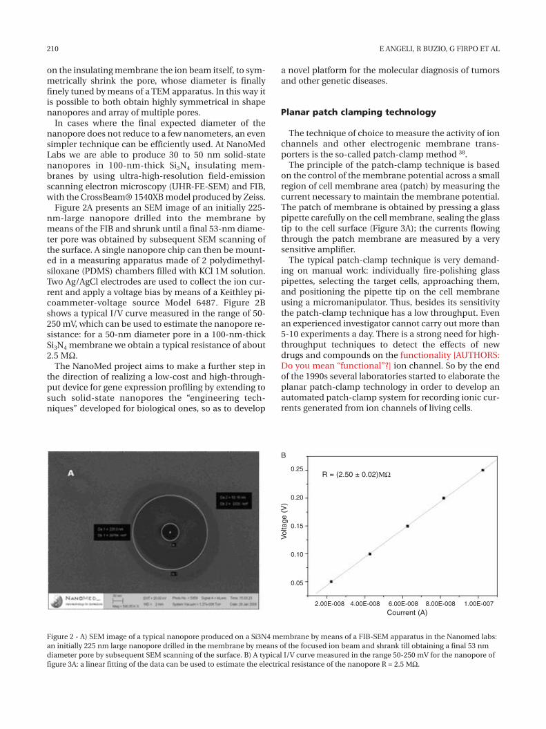
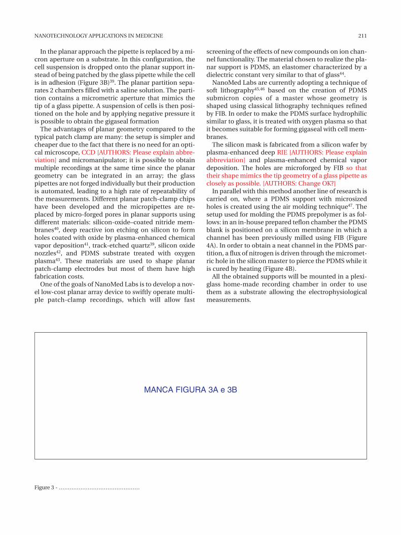
In cases where the final expected diameter of thenanopore does not reduce to a few nanometers, an evensimpler technique can be efficiently used. At NanoMedLabs we are able to produce 30 to 50 nm solid-statenanopores in 100-nm-thick Si3N4 insulating mem-branes by using ultra-high-resolution field-emissionscanning electron microscopy (UHR-FE-SEM) and FIB,with the CrossBeam® 1540XB model produced by Zeiss.
Figure 2A presents an SEM image of an initially 225-nm-large nanopore drilled into the membrane bymeans of the FIB and shrunk until a final 53-nm diame-ter pore was obtained by subsequent SEM scanning ofthe surface. A single nanopore chip can then be mount-ed in a measuring apparatus made of 2 polydimethyl-siloxane (PDMS) chambers filled with KCl 1M solution.Two Ag/AgCl electrodes are used to collect the ion cur-rent and apply a voltage bias by means of a Keithley pi-coammeter-voltage source Model 6487. Figure 2Bshows a typical I/V curve measured in the range of 50-250 mV, which can be used to estimate the nanopore re-sistance: for a 50-nm diameter pore in a 100-nm-thickSi3N4 membrane we obtain a typical resistance of about2.5 MΩ.
The NanoMed project aims to make a further step inthe direction of realizing a low-cost and high-through-put device for gene expression profiling by extending tosuch solid-state nanopores the “engineering tech-niques” developed for biological ones, so as to develop
210 E ANGELI, R BUZIO, G FIRPO ET AL
a novel platform for the molecular diagnosis of tumorsand other genetic diseases.
Planar patch clamping technology
The technique of choice to measure the activity of ionchannels and other electrogenic membrane trans-porters is the so-called patch-clamp method 38.
The principle of the patch-clamp technique is basedon the control of the membrane potential across a smallregion of cell membrane area (patch) by measuring thecurrent necessary to maintain the membrane potential.The patch of membrane is obtained by pressing a glasspipette carefully on the cell membrane, sealing the glasstip to the cell surface (Figure 3A); the currents flowingthrough the patch membrane are measured by a verysensitive amplifier.
The typical patch-clamp technique is very demand-ing on manual work: individually fire-polishing glasspipettes, selecting the target cells, approaching them,and positioning the pipette tip on the cell membraneusing a micromanipulator. Thus, besides its sensitivitythe patch-clamp technique has a low throughput. Evenan experienced investigator cannot carry out more than5-10 experiments a day. There is a strong need for high-throughput techniques to detect the effects of newdrugs and compounds on the functionality [AUTHORS:Do you mean “functional”?] ion channel. So by the endof the 1990s several laboratories started to elaborate theplanar patch-clamp technology in order to develop anautomated patch-clamp system for recording ionic cur-rents generated from ion channels of living cells.
Figure 2 - A) SEM image of a typical nanopore produced on a Si3N4 membrane by means of a FIB-SEM apparatus in the Nanomed labs:an initially 225 nm large nanopore drilled in the membrane by means of the focused ion beam and shrank till obtaining a final 53 nmdiameter pore by subsequent SEM scanning of the surface. B) A typical I/V curve measured in the range 50-250 mV for the nanopore offigure 3A: a linear fitting of the data can be used to estimate the electrical resistance of the nanopore R = 2.5 ΜΩ.
B
R = (2.50 ± 0.02)ΜΩ
Vol
tage
(V)
0.25
0.20
0.15
0.10
0.05
2.00E-008 4.00E-008 6.00E-008 8.00E-008 1.00E-007Courrent (A)
screening of the effects of new compounds on ion chan-nel functionality. The material chosen to realize the pla-nar support is PDMS, an elastomer characterized by adielectric constant very similar to that of glass44.
NanoMed Labs are currently adopting a technique ofsoft lithography45,46 based on the creation of PDMSsubmicron copies of a master whose geometry isshaped using classical lithography techniques refinedby FIB. In order to make the PDMS surface hydrophilicsimilar to glass, it is treated with oxygen plasma so thatit becomes suitable for forming gigaseal with cell mem-branes.
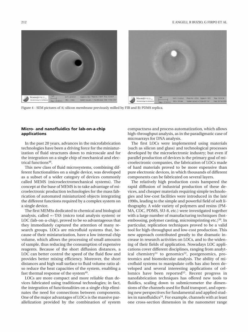
The silicon mask is fabricated from a silicon wafer byplasma-enhanced deep RIE [AUTHORS: Please explainabbreviation] and plasma-enhanced chemical vapordeposition. The holes are microforged by FIB so thattheir shape mimics the tip geometry of a glass pipette asclosely as possible. [AUTHORS: Change OK?]
In parallel with this method another line of research iscarried on, where a PDMS support with microsizedholes is created using the air molding technique47. Thesetup used for molding the PDMS prepolymer is as fol-lows: in an in-house prepared teflon chamber the PDMSblank is positioned on a silicon membrane in which achannel has been previously milled using FIB (Figure4A). In order to obtain a neat channel in the PDMS par-tition, a flux of nitrogen is driven through the micromet-ric hole in the silicon master to pierce the PDMS while itis cured by heating (Figure 4B).
All the obtained supports will be mounted in a plexi-glass home-made recording chamber in order to usethem as a substrate allowing the electrophysiologicalmeasurements.
In the planar approach the pipette is replaced by a mi-cron aperture on a substrate. In this configuration, thecell suspension is dropped onto the planar support in-stead of being patched by the glass pipette while the cellis in adhesion (Figure 3B)39. The planar partition sepa-rates 2 chambers filled with a saline solution. The parti-tion contains a micrometric aperture that mimics thetip of a glass pipette. A suspension of cells is then posi-tioned on the hole and by applying negative pressure itis possible to obtain the gigaseal formation
The advantages of planar geometry compared to thetypical patch clamp are many: the setup is simpler andcheaper due to the fact that there is no need for an opti-cal microscope, CCD [AUTHORS: Please explain abbre-viation] and micromanipulator; it is possible to obtainmultiple recordings at the same time since the planargeometry can be integrated in an array; the glasspipettes are not forged individually but their productionis automated, leading to a high rate of repeatability ofthe measurements. Different planar patch-clamp chipshave been developed and the micropipettes are re-placed by micro-forged pores in planar supports usingdifferent materials: silicon-oxide–coated nitride mem-branes40, deep reactive ion etching on silicon to formholes coated with oxide by plasma-enhanced chemicalvapor deposition41, track-etched quartz39, silicon oxidenozzles42, and PDMS substrate treated with oxygenplasma43. These materials are used to shape planarpatch-clamp electrodes but most of them have highfabrication costs.
One of the goals of NanoMed Labs is to develop a nov-el low-cost planar array device to swiftly operate multi-ple patch-clamp recordings, which will allow fast
NANOTECHNOLOGY APPLICATIONS IN MEDICINE 211
MANCA FIGURA 3A e 3B
Figure 3 - ………………………………………
Micro- and nanofluidics for lab-on-a-chipapplications
In the past 20 years, advances in the microfabricationtechnologies have been a driving force for the miniatur-ization of fluid structures down to microscale and forthe integration on a single chip of mechanical and elec-trical functions48.
This new class of fluid microsystems, combining dif-ferent functionalities on a single device, was developedas a subset of a wider category of devices commonlycalled MEMS (microelectromechanical systems). Theconcept at the base of MEMS is to take advantage of mi-croelectronic production technologies for the mass fab-rication of automated miniaturized objects integratingthe different functions required by a complex system ona single device.
The first MEMSs dedicated to chemical and biologicalanalysis, called ∝-TAS (micro total analysis system) orLOC (lab-on-a-chip), proved to be so advantageous thatthey immediately captured the attention of many re-search groups. LOCs are microfluid systems that, be-cause of their miniaturization, have a low internal chipvolume, which allows the processing of small amountsof sample, thus reducing the consumption of expensivereagents. Because of the short diffusion distances, aLOC can better control the speed of the fluid flow andprovides better mixing efficiency. Moreover, the shortdistances and high wall surface to fluid volume ratio al-so reduce the heat capacities of the system, enabling afast thermal response of the system49.
LOCs are more compact and more reliable than de-vices fabricated using traditional technologies; in fact,the integration of functionalities on a single chip elimi-nates the need for connections between components.One of the major advantages of LOCs is the massive par-allelization provided by the combination of system
212 E ANGELI, R BUZIO, G FIRPO ET AL
compactness and process automatization, which allowshigh-throughput analysis, as in the paradigmatic case ofmicroarrays for DNA analysis.
The first LOCs were implemented using materials(such as silicon and glass) and technological processesdeveloped by the microelectronic industry; but even ifparallel production of devices is the primary goal of mi-croelectronic companies, the fabrication of LOCs madeof hard materials proved to be more expensive thanpure electronic devices, in which thousands of differentcomponents can be fabricated on several layers.
The relatively high production costs hampered therapid diffusion of industrial production of these de-vices, and cheaper materials requiring simple technolo-gies and low-cost facilities were introduced in the late1990s, leading to the simple and powerful field of soft li-thography. A wide variety of polymers and resins (PM-MA, COC, PDMS, SU-8, etc.) were investigated togetherwith a large number of manufacturing techniques (hot-embossing, polymer casting, microimprinting etc.)50. Inparticular, replication techniques proved to be a validtool for high-throughput and low-cost production. Thisnew approach contributed greatly to the dramatic in-crease in research activities on LOCs, and to the widen-ing of their fields of application. Nowadays LOC appli-cations cover different disciplines, ranging from analyt-ical chemistry51 to genomics52, postgenomics, pro-teomics and biomolecular analysis. The ability of mi-crofluid systems to manipulate cells has also been de-veloped and several interesting applications of cel-lomics have been reported53. Recent progress innanofabrication techniques has offered new tools tofluidics, scaling down to submicrometer the dimen-sions of the channels used for fluid transport, and open-ing new perspectives for fundamental and applied stud-ies in nanofluidics54. For example, channels with at leastone cross-section dimension in the nanometer range
Figure 4 - SEM pictures of A) silicon membrane previously milled by FIB and B) PDMS replica.
tions are generally used, but in some devices also hy-drodynamic phenomena are exploited.
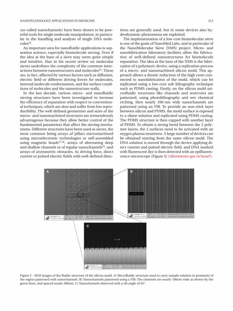
The implementation of a low-cost biomolecular sieveis one of the goals of NanoMed Labs, and in particular ofthe NanoMolecular Sieve (NMS) project. Micro- andnanofabrication laboratory facilities allow the fabrica-tion of well-defined nanostructures for biomoleculeseparation. The idea at the base of the NMS is the fabri-cation of a polymeric device, using a replication processof a micro- and nanomachined silicon mold. This ap-proach allows a drastic reduction of the high costs con-nected to nanofabrication of the mold, which can bereplicated using a low-cost soft lithography techniquesuch as PDMS casting. Firstly, on the silicon mold mi-crofluidic structures like channels and reservoirs arepatterned, using photolithography and wet chemicaletching, then nearly 100-nm wide nanochannels arepatterned using an FIB. To provide an non-stick layerbetween silicon and PDMS, the mold surface is exposedto a silane solution and replicated using PDMS casting.The PDMS structure is then capped with another layerof PDMS. To obtain a strong bond between the 2 poly-mer layers, the 2 surfaces need to be activated with anoxygen plasma treatment. A large number of devices canbe obtained starting from the same silicon mold. TheDNA solution is moved through the device applying di-rect current and pulsed electric field, and DNA markedwith fluorescent dye is then detected with an epifluores-cence microscope (Figure 5) (riferimento qui va bene?).
(so-called nanochannels) have been shown to be pow-erful tools for single molecule manipulation, in particu-lar in the handling and analysis of single DNA mole-cules55.
An important area for nanofluidic applications is sep-aration science, especially biomolecule sieving. Even ifthe idea at the base of a sieve may seem quite simpleand intuitive, Han in his recent review on molecularsieves underlines the complexity of the common inter-actions between nanostructures and molecules56. Theseare, in fact, affected by various factors such as diffusion,electric field or different driving forces for molecules,internal molecule conformation, and the surface condi-tions of molecules and the nanostructure walls.
In the last decade, various micro- and nanofluidicsieving structures have been investigated to increasethe efficiency of separation with respect to convention-al techniques, which are slow and suffer from low repro-ducibility. The well-defined geometries and sizes of themicro- and nanomachined structures are tremendouslyadvantageous because they allow better control of thefundamental parameters that affect the sieving mecha-nisms. Different structures have been used as sieves, themost common being arrays of pillars micromachinedusing microelectronic technologies or self-assembledusing magnetic beads57,58, arrays of alternating deepand shallow channels or of regular nanochannels59, andarrays of asymmetric obstacles. As driving force, directcurrent or pulsed electric fields with well-defined direc-
NANOTECHNOLOGY APPLICATIONS IN MEDICINE 213
Figure 5 - SEM images of the fluidic structure of the silicon mold. A) Microfluidic structure used to carry sample solution in proximity ofthe region patterned with nanochannels. B) Nanochannels patterned using a FIB. The channels are nearly 100nm wide as shown by thegreen lines, and spaced nearly 300nm. C) Nanochannels observed with a tilt angle of 45°.
A B
C
The sieve implemented at NanoMed Labs could beeasily integrated into a more complex polymeric LOC,and the low production costs should attract the interestof biomedical industries and promote the mass diffu-sion of labs-on-a-chip.
References
1. Morris VJ, Kirby AR, Gunning AP: Atomic force microscopyfor biologists. Imperial College Press, London, 1999.
2. Binning G, Quate CF, Gerber CH: Atomic force microscope.Phys Rev Lett, 56: 930-933, 1986.
3. Hansma HG, Kim KJ, Laney DE, Garcia RA, Argaman M,Allen MJ, Parsons SM: Properties of biomolecules meas-ured from atomic force microscope images: a review. JStruct Biol, 119: 99-108, 1997.
4. Henderson E: Atomic force microscopy of living cells. ProgSurface Sci, 46: 39-60, 1994.
5. Rivetti C, Guthold M, Bustamante C: Scanning force mi-croscopy of DNA deposited onto mica: equilibration ver-sus kinetic trapping studied by statistical polymer chainanalysis. J Mol Biol, 264: 919-932, 1996.
6. Zuccheri G, Scipioni A, Cavaliere V, Gargiulo G, DeSantis P,Samorì B: Mapping the intrinsic curvature and flexibilityalong the DNA chain. Proc Natl Acad Sci, 98: 3074-3079,2001.
7. Spisz TS, Fang Y, Reeves RH, Seymour CK, Bankman IN,Hoh JH: Automated sizing of DNA fragments in atomicforce microscope images. Med Biol Eng Comput, 36: 667-672, 1998.
8. Rivetti C, Codeluppi S: Accurate length determination ofDNA molecules visualized by atomic force microscopy: ev-idence for a partial B- to A-form transition on mica. Ultra-microscopy, 87: 55-66, 2001.
9. Rivetti C, Walker C, Bustamante C: Polymer chain statisticsand conformational analysis of DNA molecules with bendsand sections of different flexibility. J Mol Biol, 280: 41-59,1998.
10. Wiggins PA, van der Heijden T, Moreno-Herrero F,Spakowitz A, Phillips R, Widom J, Dekker C, Nelson PC:High flexibility of DNA on short length scales probed byatomic force microscopy. Nature Nanotech, 1: 137-141,2006.
11. Sampaolese B, Bercia A, Scipioni A, Zuccheri G, Savino M,Samorì B, De Santis P: Recognition of the DNA sequence byan inorganic crystal surface. Proc Natl Acad Sci, 99: 13566-13570, 2002.
12. Ficarra E, Masotti D, Macii E, Benini L, Zuccheri G, SamorìB: Automatic intrinsic DNA curvature computation fromAFM images. IEEE Trans Biomed Engineering, 52: 2074-2086, 2005.
13. Anselmi C, DeSantis P, Scipioni A: Nanoscale mechanicaland dynamical properties of DNA single molecules. Bio-phys Chem, 113: 209-221, 2005.
14. Xiao M, Gordon MP, Phong A, Ha C, Chan TF, Cai D, SelvinPR, Kwok PY: Determination of haplotypes from singleDNA molecules: a method for single molecule barcoding.Hum Mut, 28: 913-921, 2007.
15. Syvanen A: Accessing genetic variation: genotyping singlenucleotide polymorphisms. Nat Rev Gen, 2: 930-942, 2001.
16. The International HAPMAP Consortium: The Internation-al HAPMAP Project. Nature, 426: 789-796, 2003.
17. Woolley AT, Guillemette C, Li Cheung C, Housman DE,Lieber CM: Direct haplotyping of kilobase-size DNA usingcarbon nanotube probes. Nat Biotechnol, 18: 760-763,2000.
18. Kwok P, Xiao M: Single-molecule analysis for molecularhaplotyping. Hum Mutat, 23: 442-446, 2004.
19. Iijima S: Helical microtubules of graphitic carbon. Nature,354: 56, 1991.
20. Smart SK, Cassady AI, Lu GQ, Martin DJ: The biocompati-bility of carbon nanotubes. Carbon, 44: 1034-1047, 2006.
21. Jeng ES, Moll AE, Roy AC, Gastala JB, Strano MS: Detectionof DNA hybridization using the near-infrared band-gapfluorescence of single-walled carbon nanotubes. NanoLett, 6: 371-375, 2006.
22. Wu W, Wieckowski S, Pastorin G, Benincasa M, Klumpp C,Briand JP, Gennaro R, Prato M, Bianco A: Targeted deliveryof amphotericin B to cells by using functionalized carbonnanotubes. Angew Chem Int Ed Engl, 44: 6358-6362, 2005.
23. Kam WS, Liu Z, Dai H: Carbon nanotubes as intracellulartransporters for proteins and DNA: an investigation of theuptake mechanism and pathway. Angew Chem Int Ed En-gl, 45: 577-581, 2006.
24. Pantarotto D, Briand JP, Prato M, Bianco A: Translocation ofbioactive peptides across cell membranes by carbon nan-otubes. Chem Comm, 1: 16-17, 2004.
25. O’Connell MJ, Bachilo SM, Huffman CB, Moore VC, StranoMS, Haroz EH, Rialon KL, Boul PJ, Noon WH, Kittrell C, MaJ, Hauge RH, Weisman RB, Smalley RE: Band gap fluores-cence from individual single-walled carbon nanotubes.Science, 297: 593-596, 2002.
26. Bachilo SM, Strano MS, Kittrell C, Hauge RH, Smalley RE,Weisman RB: Structure-assigned optical spectra of single-walled carbon nanotubes. Science, 298: 2361-2366, 2002.
27. Kam WS, O’Connell M, Wisdom JA, Dai H: Carbon nan-otubes as multifunctional biological transporters andnear-infrared agents for selective cancer cell destruction.Proc Natl Acad Sci U S A, 102: 11600-11605, 2005.
28. Singh R, Pantarotto D, Lacerda L, Pastorin G, Klumpp C,Prato M, Bianco A, Kostarelos K: Tissue biodistribution andblood clearance rates of intravenously administered car-bon nanotube radiotracers. Proc Natl Acad Sci U S A, 103:3357-3362, 2006.
29. Sayes CM, Liang F, Hudson JL, Mendez J, Guo W, Beach JM,Moore VC, Doyle CD, West JL, Billups WE, Ausman KD,Colvin VL: Functionalization density dependence of sin-gle-walled carbon nanotubes cytotoxicity in vitro. ToxicolLett, 161: 135-142, 2006.
30. Hirai K, Okada T, Kaneko T, Hatakeyama R, Yoshiki H: In-vestigation on the generation of atmospheric-pressureglow discharge plasmas in contact with solution. XXVIIthICPIG, Eindhoven, the Netherlands, 18-22 July, 2005.
31. Kasianowicz JJ, Brandin E, Branton D, Deamer DW: Char-acterization of individual polynucleotide molecules usinga membrane channel. Proc Natl Acad Sci USA, 93: 13770-13773, 1996.
32. Coulter WH: High speed automatic blood cell counter andcell size analyzer. Proceedings of the National ElectronicsConference, pp 1034-1042, 1957.
33. Akeson M, Branton D, Kasianowicz JJ, Brandin E, DeamerDW: Microsecond time-scale discrimination among poly-cytidylic acid, polyadenylic acid, and polyuridylic acid ashomopolymers or as segments within single RNA mole-cules. Biophys J, 77: 3227-3233, 1999.
34. Howorka S, Cheley S, Bayley H: Sequence-specific detec-tion of individual DNA strands using engineerednanopores. Nature Biotechnology, 19: 636-639, 2001.
35. Winters-Hilt S, Landry M, Akeson M, Tanase M, Amin I,Coombs A, Morales E, Millet J, Baribault C, SendamangalamS: Cheminformatics methods for novel nanopore analysis ofHIV DNA termini. BMC Bioinformatics, 7 (Suppl 2): S22, 2006.
36. Li J, Stein D, McMullan C, Branton D, Aziz MJ,Golovchenko JA: Ion-beam sculpting at nanometre lengthscales. Nature, 412: 166-169, 2001.
214 E ANGELI, R BUZIO, G FIRPO ET AL
37. Lo CJ, Aref T, Bezryadin A: Fabrication of symmetric sub-5nm nanopores using focused ion and electron beams.Nanotechnology, 17: 3264-3267, 2006.
38. Hamill OP, Marty A, Neher E, Sakmann B: Improved patch-clamp techniques for high-resolution current recordingfrom cells and cell-free membrane patches. Pflugers Arch,391: 85-100, 1981.
39. Fertig N, Blick RH, Behrends JC: Whole cell patch clamprecording performed on a planar glass chip. Biophys J, 82:3056-3062, 2002.
40. Fertig N, George M, Klau M, Meyer C, Tilke A, Sobotta C,Blick RH, Behrends JC: Microstructured apertures in pla-nar glass substrates for ion channel research. ReceptorsChannels, 9: 29-40, 2003.
41. Pantoja R, Nagarah JM, Starace DM, Melosh NA, Blunck R,Bezanilla F, Heath JR: Silicon chip-based patch-clamp elec-trodes integrated with PDMS microfluidics. Biosens Bio-electron, 20: 509-517, 2004.
42. Lehnert T, Gijs MAM, Netzer R, Bischoff U: Realization ofhollow SiO2 micronozzles for electrical measurements onliving cells. Appl Phys Lett, 81: 5063-5065, 2002.
43. Klemic KG, Klemic JF, Reed MA, Sigworth FJ: MicromoldedPDMS planar electrode allows patch clamp electricalrecordings from cells. Biosens Bioelectron, 17: 597-604,2002.
44. Langowski BA, Uhrich KE: Microscale plasma initiated pat-terning (mPIP). Langmuir, 21: 6366-6372, 2005.
45. Younan X, Whitesides GM: Soft lithography. Annu RevMater Sci, 28: 153-184, 1998.
46. Duffy DC, McDonald JC, Schueller OJA, Whitesides GM:Rapid prototyping of microfluidic systems in poly(di-methylsiloxane). Anal Chem 70: 4974-4984, 1998.
47. Li X, Klemic KG, Reed MA, Sigworth FJ: Microfluidic system
for planar patch clamp electrode arrays. Nanoletters, 6:815-819, 2006.
48. Abgrall P, Gué AM: Lab-on-chip technologies: making a mi-crofluidic network and coupling it into a complete microsys-tem-a review. J Micromech Microeng, 17: R15-R49, 2007.
49. Chang CC, Yang R: Electrokinetic mixing in microfluidicsystems. Microfluid Nanofluid, 3: 501-525, 2007.
50. Guo LJ: Recent progress in nanoimprint technology and itsapplications. J Phys D Appl Phys, 37: R123-R141, 2004.
51. Khandurina J, Guttman A: Bioanalysis in microfluidic de-vices. J Chromatogr A, 943: 159-183, 2002.
52. Andersson H, van den Berg A: Microfluidic devices for cel-lomics: a review. Sensors Actuators, B 92: 315-325, 2003.
53. DeMello A: Control and detection of chemical reactions inmicrofluidic systems. Nature, 442: 394-402, 2006.
54. Tegenfeldt JO, Prinz C, Cao H, Huang RL, Austin R, ChouSY, Cox EC, Sturm JC: Micro- and nanofluidics for DNAanalysis. Anal Bioanal Chem, 378: 1678-1692, 2004.
55. Craighead H: Future lab-on-a-chip technologies for inter-rogating individual molecules. Nature, 442: 387, 2006.
56. Han J, Fu J, Schoch RB: Molecular sieving using nanofilters:Past, present and future. Lab Chip, 8: 23-33, 2008.
57. Kaji N, Tezuka Y, Takamura Y,Ueda M, Nishimoto T, Nakan-ishi H, Horiike Y, Baba Y: Separation of long DNA moleculesby quartz nanopillar chips under a direct current electricfield. Anal Chem, 76: 15-22, 2004.
58. Doyle P, Bibette J, Bancaud A, Viovy JL: Self-assembledmagnetic matrices for DNA separation chips. Science, 295:2237, 2002.
59. Fu J, Schoch R, Stevens A, Tannenbaum S, Han J: A pat-terned anisotropic nanofluidic sieving structure for con-tinuous-flow separation of DNA and proteins. Nature Nan-otech, 2: 121-128, 2007.
NANOTECHNOLOGY APPLICATIONS IN MEDICINE 215