nanocelluloses: sources, pretreatment, isolations ... - mdpi
TRANSCRIPT
polymers
Review
Nanocelluloses: Sources, Pretreatment, Isolations, Modification,and Its Application as the Drug Carriers
Valentino Bervia Lunardi 1 , Felycia Edi Soetaredjo 1,2, Jindrayani Nyoo Putro 1, Shella Permatasari Santoso 1,2,Maria Yuliana 1, Jaka Sunarso 3, Yi-Hsu Ju 4,5 and Suryadi Ismadji 1,*
�����������������
Citation: Lunardi, V.B.; Soetaredjo,
F.E.; Putro, J.N.; Santoso, S.P.; Yuliana,
M.; Sunarso, J.; Ju, Y.-H.; Ismadji, S.
Nanocelluloses: Sources,
Pretreatment, Isolations,
Modification, and Its Application as
the Drug Carriers. Polymers 2021, 13,
2052. https://doi.org/10.3390/
polym13132052
Academic Editors: Denis
M. Panaitescu and Adriana
Nicoleta Frone
Received: 26 May 2021
Accepted: 21 June 2021
Published: 23 June 2021
Publisher’s Note: MDPI stays neutral
with regard to jurisdictional claims in
published maps and institutional affil-
iations.
Copyright: © 2021 by the authors.
Licensee MDPI, Basel, Switzerland.
This article is an open access article
distributed under the terms and
conditions of the Creative Commons
Attribution (CC BY) license (https://
creativecommons.org/licenses/by/
4.0/).
1 Department of Chemical Engineering, Widya Mandala Surabaya Catholic University, Kalijudan 37,Surabaya 60114, Indonesia; [email protected] (V.B.L.); [email protected] (F.E.S.);[email protected] (J.N.P.); [email protected] (S.P.S.); [email protected] (M.Y.)
2 Department of Chemical Engineering, National Taiwan University of Science and Technology, No. 43,Section 4, Keelung Rd, Da’an District, Taipei City 10607, Taiwan
3 Research Centre for Sustainable Technologies, Faculty of Engineering, Computing and Science, SwinburneUniversity of Technology, Kuching 93350, Sarawak, Malaysia; [email protected]
4 Graduate Institute of Applied Science, National Taiwan University of Science and Technology, No. 43,Section 4, Keelung Rd, Da’an District, Taipei City 10607, Taiwan; [email protected]
5 Taiwan Building Technology Center, National Taiwan University of Science and Technology, No. 43, Section 4,Keelung Rd, Da’an District, Taipei City 10607, Taiwan
* Correspondence: [email protected]; Tel.: +62-31-389-1264
Abstract: The ‘Back-to-nature’ concept has currently been adopted intensively in various industries,especially the pharmaceutical industry. In the past few decades, the overuse of synthetic chemicalshas caused severe damage to the environment and ecosystem. One class of natural materialsdeveloped to substitute artificial chemicals in the pharmaceutical industries is the natural polymers,including cellulose and its derivatives. The development of nanocelluloses as nanocarriers indrug delivery systems has reached an advanced stage. Cellulose nanofiber (CNF), nanocrystalcellulose (NCC), and bacterial nanocellulose (BC) are the most common nanocellulose used asnanocarriers in drug delivery systems. Modification and functionalization using various processesand chemicals have been carried out to increase the adsorption and drug delivery performanceof nanocellulose. Nanocellulose may be attached to the drug by physical interaction or chemicalfunctionalization for covalent drug binding. Current development of nanocarrier formulationssuch as surfactant nanocellulose, ultra-lightweight porous materials, hydrogel, polyelectrolytes,and inorganic hybridizations has advanced to enable the construction of stimuli-responsive andspecific recognition characteristics. Thus, an opportunity has emerged to develop a new generation ofnanocellulose-based carriers that can modulate the drug conveyance for diverse drug characteristics.This review provides insights into selecting appropriate nanocellulose-based hybrid materials andthe available modification routes to achieve satisfactory carrier performance and briefly discusses theessential criteria to achieve high-quality nanocellulose.
Keywords: drug delivery; drug release; functionalization; nanocellulose
1. Introduction
Drug delivery technology (DDT) is a cutting-edge applied science for delivering drugsto specific targets. This technology regulates the absorption and release of therapeuticdrugs via various drug carriers to the desired organs, including subcellular organs, tis-sues, and cells, to improve human health [1]. DDT has advanced rapidly in the pastfew decades, enabled by various discoveries in various fields, including pharmaceutical,materials, and biomedical sciences. DDT development aims to improve therapeutic drugs’pharmacological activity and overcome various disadvantages of conventional therapeuticdrugs such as drug agglomeration, biodistribution deficiency, low bioavailability, limitedsolubility, and insufficient selectivity to prevent the concurrent effects of therapeutic drugs.
Polymers 2021, 13, 2052. https://doi.org/10.3390/polym13132052 https://www.mdpi.com/journal/polymers
Polymers 2021, 13, 2052 2 of 47
The majority of research studies on drug delivery technology revolve around de-veloping materials suitable for drug delivery with desirable characteristics such as highdrug adsorption capacity, targeted drug administration, controlled release, biocompati-bility, and non-immunogenic and non-toxic effects that optimize therapeutic efficacy andeliminates side effects [2]. Many engineered nanomaterials have been studied for drugdelivery applications [3]. Some nanomaterials have recently been undergoing developmentand clinical investigation; however, each nanomaterial has its various characteristics andlimitations, challenging the researcher in creating a suitable drug delivery system.
Natural-based polymers have drawn considerable attention as suitable biomateri-als for numerous applications in drug delivery systems. Various nature-based polymerssuch as polysaccharides (cellulose, chitosan, hyaluronic acid, pectins, alginate, celluloseethers), proteins (silk fibroin and collagen), and peptides have been identified as promis-ing biomaterials for drug delivery systems given their biocompatibility, processability,and characteristics (e.g., nanoparticles, hydrogels, aerogels, tablets, and so on) that can beregulated by modifying various polymer functional groups such as amino groups, carboxylgroups, and hydroxyl groups [4]. The current development of these mentioned variouspolysaccharides, proteins, and peptides for drug delivery systems have been well-reviewedelsewhere [4–7].
Several natural polymers have been shown to have a higher affinity for cell re-ceptors and modulate cellular processes such as adhesion, migration, and proliferation.These advantages make these natural polymers attractive for effective and high-efficiencydrug delivery systems [8]. They can also be degraded in the presence of in vivo en-zymes, which ensures their ability to create responsive local delivery systems. However,only polysaccharides and proteins have been extensively studied in drug delivery systems(DDS). These natural polymers have unique characteristics in each tissue and have identicalcharacteristics in the extracellular skeleton. These characteristics support these naturalpolymers’ utilization as drug carriers with insignificant invasive features [9–11].
Cellulose is the most abundant and commonly found natural polymer [12]. Its an-nual production is estimated at more than 7.5·1010 tons [13]. As a promising fuel andchemical precursor, cellulose has been widely utilized in various industries such as textile,pulp, paper, composite, and pharmaceutical excipients [2]. However, the development ofcellulose-based materials as a direct molecule controller for drug adsorption and releasehad not been evaluated until the discovery of nanocellulose, which became a turning pointfor using carbohydrate-based nanomaterials in the field of drug delivery [14,15].
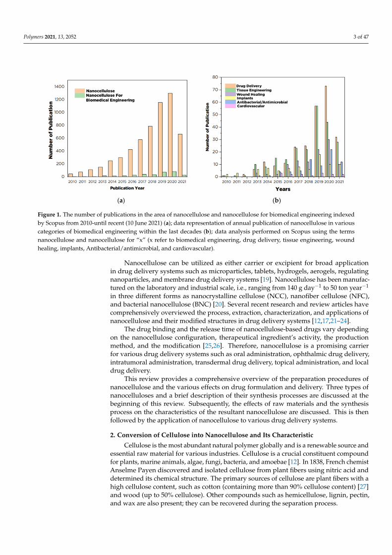
As illustrated in Figure 1, the publication on nanocellulose for biomedical engineeringapplications increases every year, especially for drug delivery applications. The increase inthe number of publications on the utilization of nanocellulose for drug delivery systemsis a strong indication of the potential application of this material in the future. The rapiddevelopment of nanotechnology and materials science has brought about nanocelluloseas a potential drug carrier because of its extraordinary physicochemical and biologicalcharacteristics. Nanocellulose has a large surface-area-to-volume ratio, thus enablingmore significant adsorption and therapeutic drug-binding capacity than other materials.With these properties, nanocellulose can facilitate drug release mechanisms and allocatedrug delivery precisely to the target to drastically reduce drug consumption, leading toimproved drug delivery system effectiveness [16,17]. Nanocellulose additionally exhibitsother attractive characteristics such as stiffness, high mechanical strength, biocompatibility,low toxicity, lightweight, tunable surface chemistry, and renewability [11,18], which aredesirable for the design of advanced drug delivery system.
Polymers 2021, 13, 2052 3 of 47
Polymers 2021, 13, x 3 of 49
(a) (b)
Figure 1. The number of publications in the area of nanocellulose and nanocellulose for biomedical engineering indexed by Scopus from 2010-until recent (10 June 2021) (a); data representation of annual publication of nanocellulose in various categories of biomedical engineering within the last decades (b); data analysis performed on Scopus using the terms nano-cellulose and nanocellulose for “x” (x refer to biomedical engineering, drug delivery, tissue engineering, wound healing, implants, Antibacterial/antimicrobial, and cardiovascular).
Nanocellulose can be utilized as either carrier or excipient for broad application in drug delivery systems such as microparticles, tablets, hydrogels, aerogels, regulating na-noparticles, and membrane drug delivery systems [19]. Nanocellulose has been manufac-tured on the laboratory and industrial scale, i.e., ranging from 140 g day−1 to 50 ton year−1 in three different forms as nanocrystalline cellulose (NCC), nanofiber cellulose (NFC), and bacterial nanocellulose (BNC) [20]. Several recent research and review articles have com-prehensively overviewed the process, extraction, characterization, and applications of nanocellulose and their modified structures in drug delivery systems [12,17,21–24].
The drug binding and the release time of nanocellulose-based drugs vary depending on the nanocellulose configuration, therapeutical ingredient’s activity, the production method, and the modification [25,26]. Therefore, nanocellulose is a promising carrier for various drug delivery systems such as oral administration, ophthalmic drug delivery, in-tratumoral administration, transdermal drug delivery, topical administration, and local drug delivery.
This review provides a comprehensive overview of the preparation procedures of nanocellulose and the various effects on drug formulation and delivery. Three types of nanocelluloses and a brief description of their synthesis processes are discussed at the beginning of this review. Subsequently, the effects of raw materials and the synthesis pro-cess on the characteristics of the resultant nanocellulose are discussed. This is then fol-lowed by the application of nanocellulose to various drug delivery systems.
2. Conversion of Cellulose into Nanocellulose and Its Characteristic Cellulose is the most abundant natural polymer globally and is a renewable source
and essential raw material for various industries. Cellulose is a crucial constituent com-pound for plants, marine animals, algae, fungi, bacteria, and amoebae [12]. In 1838, French chemist Anselme Payen discovered and isolated cellulose from plant fibers using nitric acid and determined its chemical structure. The primary sources of cellulose are plant fibers with a high cellulose content, such as cotton (containing more than 90% cellulose content) [27] and wood (up to 50% cellulose). Other compounds such as hemicellulose, lignin, pectin, and wax are also present; they can be recovered during the separation pro-cess.
Figure 1. The number of publications in the area of nanocellulose and nanocellulose for biomedical engineering indexedby Scopus from 2010-until recent (10 June 2021) (a); data representation of annual publication of nanocellulose in variouscategories of biomedical engineering within the last decades (b); data analysis performed on Scopus using the termsnanocellulose and nanocellulose for “x” (x refer to biomedical engineering, drug delivery, tissue engineering, woundhealing, implants, Antibacterial/antimicrobial, and cardiovascular).
Nanocellulose can be utilized as either carrier or excipient for broad applicationin drug delivery systems such as microparticles, tablets, hydrogels, aerogels, regulatingnanoparticles, and membrane drug delivery systems [19]. Nanocellulose has been manufac-tured on the laboratory and industrial scale, i.e., ranging from 140 g day−1 to 50 ton year−1
in three different forms as nanocrystalline cellulose (NCC), nanofiber cellulose (NFC),and bacterial nanocellulose (BNC) [20]. Several recent research and review articles havecomprehensively overviewed the process, extraction, characterization, and applications ofnanocellulose and their modified structures in drug delivery systems [12,17,21–24].
The drug binding and the release time of nanocellulose-based drugs vary dependingon the nanocellulose configuration, therapeutical ingredient’s activity, the productionmethod, and the modification [25,26]. Therefore, nanocellulose is a promising carrierfor various drug delivery systems such as oral administration, ophthalmic drug delivery,intratumoral administration, transdermal drug delivery, topical administration, and localdrug delivery.
This review provides a comprehensive overview of the preparation procedures ofnanocellulose and the various effects on drug formulation and delivery. Three types ofnanocelluloses and a brief description of their synthesis processes are discussed at thebeginning of this review. Subsequently, the effects of raw materials and the synthesisprocess on the characteristics of the resultant nanocellulose are discussed. This is thenfollowed by the application of nanocellulose to various drug delivery systems.
2. Conversion of Cellulose into Nanocellulose and Its Characteristic
Cellulose is the most abundant natural polymer globally and is a renewable source andessential raw material for various industries. Cellulose is a crucial constituent compoundfor plants, marine animals, algae, fungi, bacteria, and amoebae [12]. In 1838, French chemistAnselme Payen discovered and isolated cellulose from plant fibers using nitric acid anddetermined its chemical structure. The primary sources of cellulose are plant fibers with ahigh cellulose content, such as cotton (containing more than 90% cellulose content) [27]and wood (up to 50% cellulose). Other compounds such as hemicellulose, lignin, pectin,and wax are also present; they can be recovered during the separation process.
Polymers 2021, 13, 2052 4 of 47
Recently, various agricultural wastes with high cellulose content were explored as asource of cellulose, such as oil palm empty fruit bunches (OPEFB) [28], palm and bananafronds, passionfruit peel waste [29], bagasse, wheat straw, rice straw, bamboo stalks, hempbark, potato tubers, mulberry bark, hemp avicel, and sugar beets [30]. Cellulose derivedfrom these non-plant precursors can have a molecular structure similar to that of plantcellulose. However, the main difference is that much less hemicellulose or lignin is presentin these non-plant-based precursors; higher cellulose content with much lower impuritiescan be obtained from these precursors.
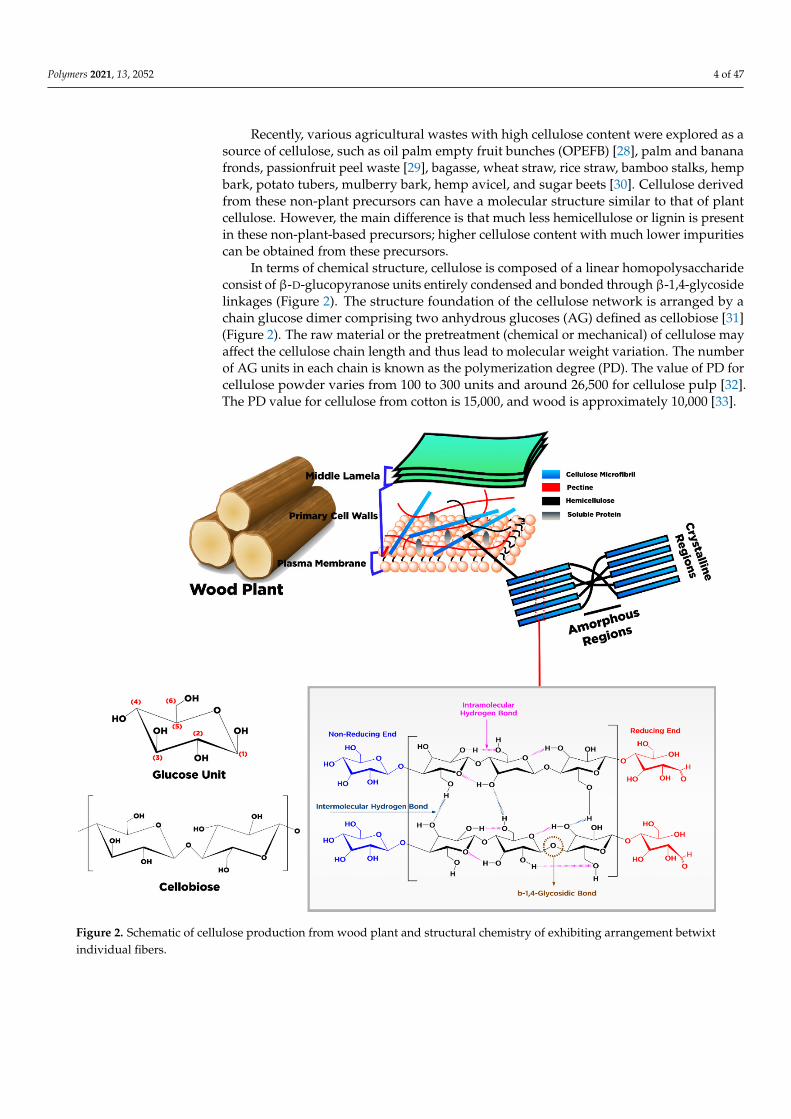
In terms of chemical structure, cellulose is composed of a linear homopolysaccharideconsist of β-D-glucopyranose units entirely condensed and bonded through β-1,4-glycosidelinkages (Figure 2). The structure foundation of the cellulose network is arranged by achain glucose dimer comprising two anhydrous glucoses (AG) defined as cellobiose [31](Figure 2). The raw material or the pretreatment (chemical or mechanical) of cellulose mayaffect the cellulose chain length and thus lead to molecular weight variation. The numberof AG units in each chain is known as the polymerization degree (PD). The value of PD forcellulose powder varies from 100 to 300 units and around 26,500 for cellulose pulp [32].The PD value for cellulose from cotton is 15,000, and wood is approximately 10,000 [33].
Polymers 2021, 13, x 4 of 49
Recently, various agricultural wastes with high cellulose content were explored as a source of cellulose, such as oil palm empty fruit bunches (OPEFB) [28], palm and banana fronds, passionfruit peel waste [29], bagasse, wheat straw, rice straw, bamboo stalks, hemp bark, potato tubers, mulberry bark, hemp avicel, and sugar beets [30]. Cellulose derived from these non-plant precursors can have a molecular structure similar to that of plant cellulose. However, the main difference is that much less hemicellulose or lignin is present in these non-plant-based precursors; higher cellulose content with much lower impurities can be obtained from these precursors.
In terms of chemical structure, cellulose is composed of a linear homopolysaccharide consist of β-D-glucopyranose units entirely condensed and bonded through β-1,4-glyco-side linkages (Figure 2). The structure foundation of the cellulose network is arranged by a chain glucose dimer comprising two anhydrous glucoses (AG) defined as cellobiose [31] (Figure 2). The raw material or the pretreatment (chemical or mechanical) of cellulose may affect the cellulose chain length and thus lead to molecular weight variation. The number of AG units in each chain is known as the polymerization degree (PD). The value of PD for cellulose powder varies from 100 to 300 units and around 26,500 for cellulose pulp [32]. The PD value for cellulose from cotton is 15,000, and wood is approximately 10,000 [33].
Figure 2. Schematic of cellulose production from wood plant and structural chemistry of exhibiting arrangement betwixt individual fibers.
Each cellulose monomer contains three reactive hydroxyl groups in the repeating chemical structure of the β-D-glucopyranose unit. In the same chain, these hydroxyl groups can make hydrogen bonds with the adjacent β-D-glucopyranose units. At different
Figure 2. Schematic of cellulose production from wood plant and structural chemistry of exhibiting arrangement betwixtindividual fibers.
Polymers 2021, 13, 2052 5 of 47
Each cellulose monomer contains three reactive hydroxyl groups in the repeatingchemical structure of the β-D-glucopyranose unit. In the same chain, these hydroxylgroups can make hydrogen bonds with the adjacent β-D-glucopyranose units. At differ-ent chain locations, the bonds present are intramolecular and intermolecular hydrogenbonds responsible for the crystal arrangement, determining the cellulose’s physical char-acteristics. Based on molecular orientation and hydrogen network between moleculesand intramolecular, cellulose is classified into different types, i.e., I, II, III, IIII, IIIII, IVI,and IVII. For details about the classification of cellulose, the reader can refer to the work ofMoon et al. [34]. Some of the cellulose characteristics are mainly represented by hydrogenlinkage coordination [35,36].
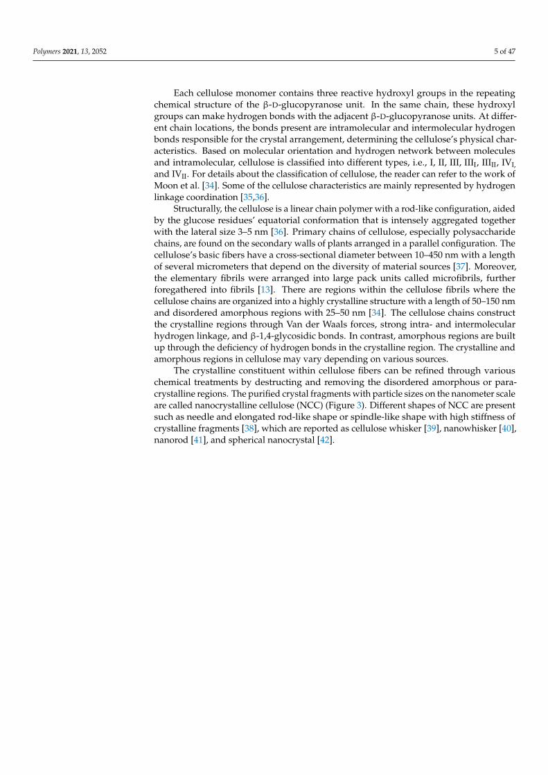
Structurally, the cellulose is a linear chain polymer with a rod-like configuration, aidedby the glucose residues’ equatorial conformation that is intensely aggregated togetherwith the lateral size 3–5 nm [36]. Primary chains of cellulose, especially polysaccharidechains, are found on the secondary walls of plants arranged in a parallel configuration. Thecellulose’s basic fibers have a cross-sectional diameter between 10–450 nm with a lengthof several micrometers that depend on the diversity of material sources [37]. Moreover,the elementary fibrils were arranged into large pack units called microfibrils, furtherforegathered into fibrils [13]. There are regions within the cellulose fibrils where thecellulose chains are organized into a highly crystalline structure with a length of 50–150 nmand disordered amorphous regions with 25–50 nm [34]. The cellulose chains constructthe crystalline regions through Van der Waals forces, strong intra- and intermolecularhydrogen linkage, and β-1,4-glycosidic bonds. In contrast, amorphous regions are builtup through the deficiency of hydrogen bonds in the crystalline region. The crystalline andamorphous regions in cellulose may vary depending on various sources.
The crystalline constituent within cellulose fibers can be refined through variouschemical treatments by destructing and removing the disordered amorphous or para-crystalline regions. The purified crystal fragments with particle sizes on the nanometer scaleare called nanocrystalline cellulose (NCC) (Figure 3). Different shapes of NCC are presentsuch as needle and elongated rod-like shape or spindle-like shape with high stiffness ofcrystalline fragments [38], which are reported as cellulose whisker [39], nanowhisker [40],nanorod [41], and spherical nanocrystal [42].
Polymers 2021, 13, 2052 6 of 47Polymers 2021, 13, x 6 of 49
Figure 3. Schematic representation nanocrystalline cellulose fabrication by chemical treatment ((a) transmission electron microscopy (TEM) images of rod-like cellulose nanocrystals [38], reprinted with permission; transmission electron micros-copy (TEM) images of cellulose nano whisker reprinted with permission from [25]. Copyright © 2019 Elsevier B.V.; (b) transmission electron microscopy (TEM) images of spherical cellulose nanocrystal reprinted with permission from [43]. Copyright © 2018 Elsevier B.V.).
A top-down process has been applied for NCC production in which a large unit of cellulose fibers (cm) is disintegrated through chemical or mechanical treatment into small units of nanocellulose (nm) [44]. NCC’s chemical structure is constructed by intra- and intermolecular hydrogen linkage of cellulose macromolecules with a high crystallinity value varying from 54 to 88% [45]. NCC’s particle size depends on the origin of the cellu-lose sources, with the diameter and length typically varying between 5 and 30 nm and between 100 and 500 nm, respectively [46]. Thus, NCCs have become an attractive candi-date as drug carriers, given their outstanding physical and chemical properties [21,47,48].
Cellulose nanofiber (CNF), also known as cellulose nanofibril, micro-fibrillated cel-lulose, nano-fibrillar cellulose, nano-fibrillated cellulose, or cellulose microfibril, has a similar molecule structure to NCCs with nano-size particles. Similar to NCC, CNF can
Figure 3. Schematic representation nanocrystalline cellulose fabrication by chemical treatment ((a) transmission electronmicroscopy (TEM) images of rod-like cellulose nanocrystals [38], reprinted with permission; transmission electron mi-croscopy (TEM) images of cellulose nano whisker reprinted with permission from [25]. Copyright © 2019 Elsevier B.V.; (b)transmission electron microscopy (TEM) images of spherical cellulose nanocrystal reprinted with permission from [43].Copyright © 2018 Elsevier B.V.).
A top-down process has been applied for NCC production in which a large unit ofcellulose fibers (cm) is disintegrated through chemical or mechanical treatment into smallunits of nanocellulose (nm) [44]. NCC’s chemical structure is constructed by intra- andintermolecular hydrogen linkage of cellulose macromolecules with a high crystallinityvalue varying from 54 to 88% [45]. NCC’s particle size depends on the origin of the cellulosesources, with the diameter and length typically varying between 5 and 30 nm and between100 and 500 nm, respectively [46]. Thus, NCCs have become an attractive candidate asdrug carriers, given their outstanding physical and chemical properties [21,47,48].
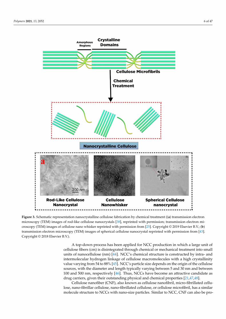
Cellulose nanofiber (CNF), also known as cellulose nanofibril, micro-fibrillated cellu-lose, nano-fibrillar cellulose, nano-fibrillated cellulose, or cellulose microfibril, has a similarmolecule structure to NCCs with nano-size particles. Similar to NCC, CNF can also be pro-
Polymers 2021, 13, 2052 7 of 47
duced from various cellulose sources. However, the morphology and crystallinity of NCCand CNF are the unique features that differentiate these two cellulose-based compounds.CNFs have long, flexible cellulose chains of amorphous and crystalline regions isolatedfrom cellulose fibrils through mechanical treatment (Figure 4) [46]. The diameter of CNFsvaries from 1 to 100 nm, while their length varies between 500 and 2000 nm. The dimensionof CNFs molecules is strongly influenced by mechanical treatment and defibrillation [49].
Polymers 2021, 13, x 7 of 49
also be produced from various cellulose sources. However, the morphology and crystal-linity of NCC and CNF are the unique features that differentiate these two cellulose-based compounds. CNFs have long, flexible cellulose chains of amorphous and crystalline re-gions isolated from cellulose fibrils through mechanical treatment (Figure 4) [46]. The di-ameter of CNFs varies from 1 to 100 nm, while their length varies between 500 and 2000 nm. The dimension of CNFs molecules is strongly influenced by mechanical treatment and defibrillation [49].
Figure 4. Schematic representation of cellulose nanofibers fabrication by mechanical treatment (scanning electron microscopy (SEM) images of micro fibrillated cellulose reprinted with permis-sion from ref. [50]; Copyright © 2007 Elsevier Ltd.; scanning electron microscopy (SEM) images of cellulose nanofibers reprinted with permission from ref. [51]. Copyright © 2006 Elsevier Ltd.).
NCC has high crystalline cellulose purity, resulting in a rigid structure, whereas the CNF structure consists of irregular amorphous parts, with some parts exhibiting a high degree of crystallinity. The amorphous regions in CNF control the structure flexibility of
Figure 4. Schematic representation of cellulose nanofibers fabrication by mechanical treatment(scanning electron microscopy (SEM) images of micro fibrillated cellulose reprinted with permissionfrom ref. [50]; Copyright © 2007 Elsevier Ltd.; scanning electron microscopy (SEM) images of cellulosenanofibers reprinted with permission from ref. [51]. Copyright © 2006 Elsevier Ltd.).
NCC has high crystalline cellulose purity, resulting in a rigid structure, whereas theCNF structure consists of irregular amorphous parts, with some parts exhibiting a highdegree of crystallinity. The amorphous regions in CNF control the structure flexibilityof nanocellulose [52]. Figure 4 presents an illustration of CNF extracted from cellulosefragments via mechanical defibrillation. The exerted force fractures the cellulose fibrils
Polymers 2021, 13, 2052 8 of 47
along its longitudinal axis [34]. Compared with NCC, CNF exhibits unique propertiessuch as extended length with excellent aspect proportion (length to diameter), superlativesurface area, hydrophilicity, biocompatibility, and adjustable characteristic through surfacemodification [53].
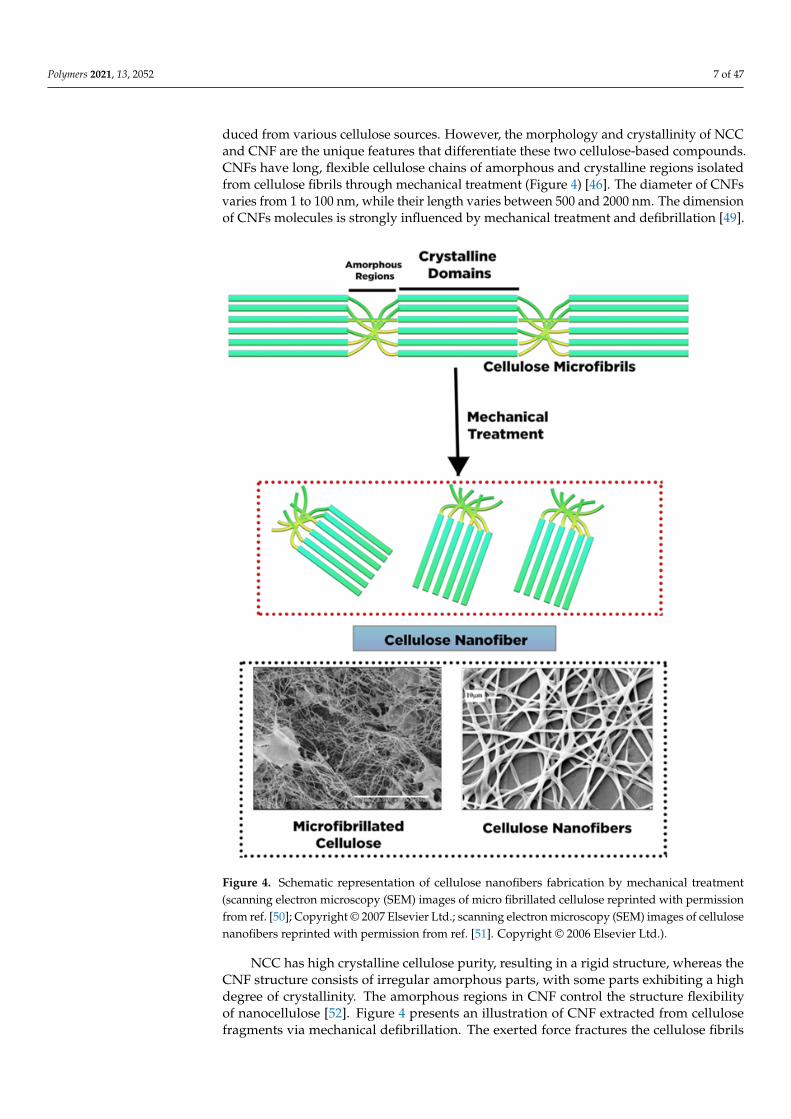
Microbial cellulose (MC), bacterial nanocellulose (BC), and bio-cellulose (BC) havebeen used as the other terms for bacteria cellulose (BC). In contrast to NCC and CNF,BC’s structure comprises sugars with low molecular weight. Many bacteria strains havebeen used to generate BC as an extracellular metabolic product, such as Gluconaceto-bacter, Sarcina, Aerobacteria, Escheria, Achromobacter, Rhizobium, Rhodobacter, Azotobacter,and Agrobacterium [54,55]. However, only Gluconacetobacter xylinus has been commerciallyutilized to produce BC on an industrial scale [27]. The bacteria strains are commonlyincubated in nutrient-rich aqueous media and produce BC on the upper layer (interfacewith air) as an exopolysaccharide. In this case, the β-D-glucopyranose units are initiallypresent during the growth of cellulose molecules within the bacterial cell. The elementaryfibril is released across the pores of the cellulose surface, which was further arranged andcrystallized into microfibrils with twisting ribbons shape followed by pellicle formation(Figure 5) [56]. The fabricated BC comprises a nanofibers framework with a diameter of20–100 nm with a length of several micrometers and a large surface area composed mainlyof water (99%) [57].
Polymers 2021, 13, x 8 of 49
nanocellulose [52]. Figure 4 presents an illustration of CNF extracted from cellulose frag-ments via mechanical defibrillation. The exerted force fractures the cellulose fibrils along its longitudinal axis [34]. Compared with NCC, CNF exhibits unique properties such as extended length with excellent aspect proportion (length to diameter), superlative surface area, hydrophilicity, biocompatibility, and adjustable characteristic through surface mod-ification [53].
Microbial cellulose (MC), bacterial nanocellulose (BC), and bio-cellulose (BC) have been used as the other terms for bacteria cellulose (BC). In contrast to NCC and CNF, BC’s structure comprises sugars with low molecular weight. Many bacteria strains have been used to generate BC as an extracellular metabolic product, such as Gluconacetobacter, Sarcina, Aerobacteria, Escheria, Achromobacter, Rhizobium, Rhodobacter, Azotobacter, and Ag-robacterium [54,55]. However, only Gluconacetobacter xylinus has been commercially uti-lized to produce BC on an industrial scale [27]. The bacteria strains are commonly incu-bated in nutrient-rich aqueous media and produce BC on the upper layer (interface with air) as an exopolysaccharide. In this case, the β-D-glucopyranose units are initially present during the growth of cellulose molecules within the bacterial cell. The elementary fibril is released across the pores of the cellulose surface, which was further arranged and crystal-lized into microfibrils with twisting ribbons shape followed by pellicle formation (Figure 5) [56]. The fabricated BC comprises a nanofibers framework with a diameter of 20–100 nm with a length of several micrometers and a large surface area composed mainly of water (99%) [57].
Figure 5. Schematic production of bacteria cellulose through extracellular secretion (scanning electron microscopy (SEM) images of 3-D dimensional network of bacteria cellulose [58], reprinted with permission; (c) scanning electron microscopy (TEM) images bacteria cellulose pellicle, reprinted with permission from ref. [59]. Copyright © 2019 Elsevier Ltd.)
In terms of chemical composition, BC is indistinguishable from plant-based nanocel-lulose (e.g., NCC and CNF). However, BC has higher crystallinity (up to 84–89%) with
Figure 5. Schematic production of bacteria cellulose through extracellular secretion (scanning electron microscopy (SEM)images of 3-D dimensional network of bacteria cellulose [58], reprinted with permission; (c) scanning electron microscopy(TEM) images bacteria cellulose pellicle, reprinted with permission from ref. [59]. Copyright © 2019 Elsevier Ltd.)
In terms of chemical composition, BC is indistinguishable from plant-based nanocel-lulose (e.g., NCC and CNF). However, BC has higher crystallinity (up to 84–89%) withfewer amorphous regions than NCC and CNF. Moreover, BC contains fewer impuritiesand contaminants such as hemicellulose, lignin, and pectin, mainly found in plant-based
Polymers 2021, 13, 2052 9 of 47
nanocellulose. BC is a biocompatible material with non-cytotoxicity and non-genotoxicityfor biomedical applications, especially drug delivery [60]. BC synthesis does not involve acomplicated process such as mechanical and chemical treatment to cleave the hemicelluloseor lignin within the lignocellulosic biomass, thereby allowing high cellulose purity.
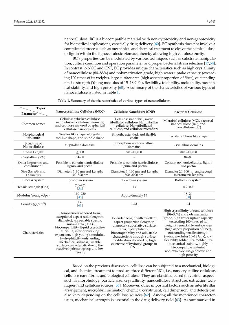
BC’s properties can be modulated by various techniques such as substrate manipula-tion, culture condition and operation parameter, and proper bacterial strain selection [17,54].In contrast to NCC and CNF, BC provides unique characteristics such as high crystallinityof nanocellulose (84–88%) and polymerization grade, high water uptake capacity (exceed-ing 100 times of its weight), large surface area (high aspect proportion of fiber), outstandingtensile strength (Young modulus of 15–18 GPa), flexibility, foldability, moldability, mechan-ical stability, and high porosity [60]. A summary of the characteristics of various types ofnanocellulose is listed in Table 1.
Table 1. Summary of the characteristics of various types of nanocelluloses.
Parameter
TypesNanocrystalline Cellulose (NCC) Cellulose Nanofibers (CNF) Bacterial Cellulose
Common names
Cellulose whisker, cellulosenanowhisker, cellulose nanowire,
and cellulose nanorod or sphericalcellulose nanocrystals
Cellulose nanofibril, microfibrillated cellulose, Nanofibrillar
cellulose, Nanofibrillatedcellulose, and cellulose microfibril
Microbial cellulose (MC), bacterialnanocellulose (BC), and
bio-cellulose (BC)
Morphologicalstructure
Needles like shape, elongatedrod-like shape, and spindle shape
Smooth, extended, and flexiblechain Twisted ribbons like shape
Structure ofNanocellulose Crystalline domains amorphous and crystalline
domains Crystalline domains
Chain Length ≥500 500–15,000 4000–10,000
Crystallinity (%) 54–88 - 84–88
Other Impurities andcontaminant
Possible to contain hemicellulose,lignin, and pectin
Possible to contain hemicellulose,lignin, and pectin
Contain no hemicellulose, lignin,and pectin
Size (Length andDiameter)
Diameter: 5–30 nm and Length:100–500 nm
Diameter: 1–100 nm and Length:500–2000 nm
Diameter 20–100 nm and severalmicrometric lengths
Process System Top-down system Top-down system Bottom-up system
Tensile strength (Gpa) 7.5–7.7[34] 13 0.2–0.3
Modulus Young (Gpa) 110–220[45] Approximately 15 18–20
[60]
Density (gr/cm3) 1.6[61] 1.42 1.1
Characteristics
Homogenous nanorod form,exceptional aspect ratio (length to
diameter), appreciable specificsurface area (SSA),
biocompatibility, liquid crystallineattribute, inferior breaking
expansion, high young’s modulus,hydrophilicity, outstanding
mechanical stiffness, tunablesurface characteristic due to the
reactive hydroxyl group and lowdensity
Extended length with excellentaspect proportion (length to
diameter), superlative surfacearea, hydrophilicity,
biocompatibility and adjustablecharacteristic through surfacemodification afforded by high
extensive of hydroxyl groups inCNF.
High crystallinity of nanocellulose(84–88%) and polymerization
grade, high water uptake capacity(exceeding 100 times of its
weight), remarkable surface area(high aspect proportion of fiber),
outstanding tensile strength(young modulus 15–18 Gpa), andflexibility, foldability, moldability,
mechanical stability, highlybiocompatible material,
non-cytotoxic, un-genotoxic andhigh porosity
Based on the previous discussion, cellulose can be subjected to a mechanical, biologi-cal, and chemical treatment to produce three different NCs, i.e., nanocrystalline cellulose,cellulose nanofibrils, and biological cellulose. They are classified based on various aspectssuch as morphology, particle size, crystallinity, nanocellulose structure, extraction tech-niques, and cellulose sources [56]. Moreover, other important factors such as interfibrillararrangement, microfibril inclination, chemical constituent, cell dimension, and defects canalso vary depending on the cellulose sources [62]. Among all the mentioned character-istics, mechanical strength is essential in the drug delivery field [63]. As summarized in
Polymers 2021, 13, 2052 10 of 47
Table 1, NCC possesses a high modulus young, up to 220 GPa, which is higher than glass(86 GPa) [61] and kevlar KM2 fiber (88 GPa) [45]. Furthermore, the mechanical stiffness ofNCC can reach up to 7.7 GPa, which is higher than 302 stainless steel (3.88 GPa) [45] andkevlar KM2 fiber (1.28 Gpa) [45].
3. Sources and Pretreatment of Raw Materials for Nanocellulose Productions
In general, the production of nanocellulose (NC) consists of three steps: (1) Finding thesuitable sources, (2) raw material pretreatment, and (3) NC extraction. The raw material’ssource and type influence the physical and chemical properties and the NC product’syield. Currently, most nanocellulose sources utilize high-quality biomass such as cotton,wood pulp, and dissolving pulp, which comprises the high cellulose content. However,in response to recent essential issues, such as the depletion of non-renewable energy andincreasing global temperature, the researchers realized the development of waste-basedbiomass as a feedstock for the production of nanocellulose. Various types of biomasswaste, including forest residues, algae, agricultural, and industrial by-products, appear aspotential raw materials for nanocellulose production. In terms of chemical composition,each category of biomass waste is primarily composed of cellulose, lignin, hemicellulose,pectin, and other minor substances with different physical and chemical characteristics [64].Agricultural and forest residues have similarities in their chemical composition, but lignincomposition in agricultural waste is significantly high, while the cellulose content in forestresidues is higher than in agricultural waste [64,65].
Among all of the waste-based cellulose sources, the nanocellulose extraction fromindustrial waste seems more complex since the chemical and structural composition offeedstock is variable and crucially depends on the residue types. The various impurities(e.g., hemicellulose, lignin, wax, and pectin) act as a structural barrier that hinders theaccessibility to the cellulose material for the extraction process [22]. Therefore, pretreatmentis necessary to remove the cellulose framework’s impurities, permitting the aperture ofthe material framework to expedite cellulose microstructure access. Moreover, removingimpurities is also beneficial to reduce the consumption energy of mechanical treatment forcellulose disintegration [66]. Another objective of raw material pretreatment is to regulatethe biomass structure and size and overcome the plant cell wall recalcitrance.
The pretreatment is generally divided into four categories such as physical (milling,grinding, microwave, ultrasound, etc.), chemical (dilute acid, mild alkali, TEMPO medi-ated oxidation, organosolv, and ionic liquid), biological (fungi, bacterial, and archaeal),and physicochemical (steam explosion, liquid hot water, wet oxidation, etc.) [67]. The effec-tiveness of the biomass pretreatment process depends on pH, temperature, type of catalyst,and pretreatment time. Selecting the appropriate pretreatment would allow avoiding thestructure disintegration or loss of cellulose, ensuring low cost, and minimizing energy useto reduce toxic and hazardous waste [68].
The chemical pretreatment process is considered the most efficient and economicallyfeasible for the disintegration of biomass with low pretreatment severity. However, chem-ical pretreatment is non-environmentally friendly and requires a wastewater treatmentprocess [69]. Physical pretreatment is environmentally friendly and scarcely generateshazardous or toxic substances, but the major disadvantage lies in its high energy con-sumption, which is generally higher than chemical treatment [70]. Biological treatmentis widely known as an eco-friendly process, operates under mild conditions, and con-sumes a lower energy amount. However, long pretreatment duration, low conversion,and carbohydrate loss tendency throughout pretreatment remain the main challenges ofbiological pretreatment by the microorganism [71]. Physicochemical pretreatment using acombination of chemicals and high temperature or pressure in extreme conditions can ef-fectively escalate biomass degradation. Nevertheless, high energy input is required, whichtranslates to high operation costs for this method. Proper pretreatment of cellulosic fiberscan improve the hydroxyl group’s accessibility, inner surface enhancement, crystallinityalteration, and fracture of the intra and inter hydrogen bonds of cellulose, leading to the
Polymers 2021, 13, 2052 11 of 47
increased fibers reactivity [72]. Detailed pretreatment of cellulose-based raw materials hasbeen thoroughly discussed elsewhere [73].
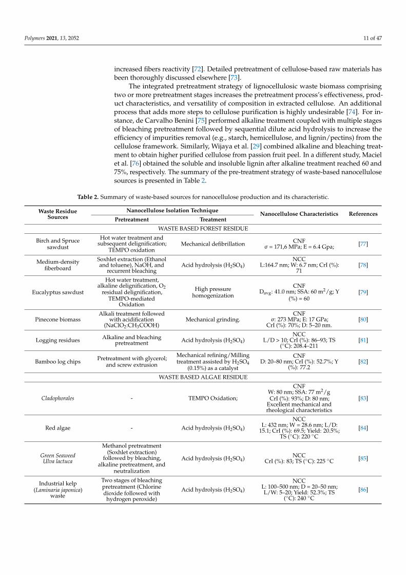
The integrated pretreatment strategy of lignocellulosic waste biomass comprisingtwo or more pretreatment stages increases the pretreatment process’s effectiveness, prod-uct characteristics, and versatility of composition in extracted cellulose. An additionalprocess that adds more steps to cellulose purification is highly undesirable [74]. For in-stance, de Carvalho Benini [75] performed alkaline treatment coupled with multiple stagesof bleaching pretreatment followed by sequential dilute acid hydrolysis to increase theefficiency of impurities removal (e.g., starch, hemicellulose, and lignin/pectins) from thecellulose framework. Similarly, Wijaya et al. [29] combined alkaline and bleaching treat-ment to obtain higher purified cellulose from passion fruit peel. In a different study, Macielet al. [76] obtained the soluble and insoluble lignin after alkaline treatment reached 60 and75%, respectively. The summary of the pre-treatment strategy of waste-based nanocellulosesources is presented in Table 2.
Table 2. Summary of waste-based sources for nanocellulose production and its characteristic.
Waste ResidueSources
Nanocellulose Isolation Technique Nanocellulose Characteristics ReferencesPretreatment Treatment
WASTE BASED FOREST RESIDUE
Birch and Sprucesawdust
Hot water treatment andsubsequent delignification;
TEMPO oxidationMechanical defibrillation CNF
σ = 171,6 MPa; E = 6.4 Gpa; [77]
Medium-densityfiberboard
Soxhlet extraction (Ethanoland toluene), NaOH, and
recurrent bleachingAcid hydrolysis (H2SO4)
NCCL:164.7 nm; W: 6.7 nm; CrI (%):
71[78]
Eucalyptus sawdust
Hot water treatment,alkaline delignification, O2
residual delignification,TEMPO-mediated
Oxidation
High pressurehomogenization
CNFDavg: 41.0 nm; SSA: 60 m2/g; Y
(%) = 60[79]
Pinecone biomassAlkali treatment followed
with acidification(NaClO2:CH3COOH)
Mechanical grinding.CNF
σ: 273 MPa; E: 17 GPa;CrI (%): 70%; D: 5–20 nm.
[80]
Logging residues Alkaline and bleachingpretreatment Acid hydrolysis (H2SO4)
NCCL/D > 10; CrI (%): 86–93; TS
(◦C): 208.4–211[81]
Bamboo log chips Pretreatment with glycerol;and screw extrusion
Mechanical refining/Millingtreatment assisted by H2SO4
(0.15%) as a catalyst
CNFD: 20–80 nm; CrI (%): 52.7%; Y
(%): 77.2[82]
WASTE BASED ALGAE RESIDUE
Cladophorales - TEMPO Oxidation;
CNFW: 80 nm; SSA: 77 m2/gCrI (%): 93%; D: 80 nm;
Excellent mechanical andrheological characteristics
[83]
Red algae - Acid hydrolysis (H2SO4)NCC
L: 432 nm; W = 28.6 nm; L/D:15.1; CrI (%): 69.5; Yield: 20.5%;
TS (◦C): 220 ◦C[84]
Green SeaweedUlva lactuca
Methanol pretreatment(Soxhlet extraction)
followed by bleaching,alkaline pretreatment, and
neutralization
Acid hydrolysis (H2SO4) NCCCrI (%): 83; TS (◦C): 225 ◦C [85]
Industrial kelp(Laminaria japonica)
waste
Two stages of bleachingpretreatment (Chlorinedioxide followed with
hydrogen peroxide)
Acid hydrolysis (H2SO4)NCC
L: 100–500 nm; D = 20–50 nm;L/W: 5–20; Yield: 52.3%; TS
(◦C): 240 ◦C[86]
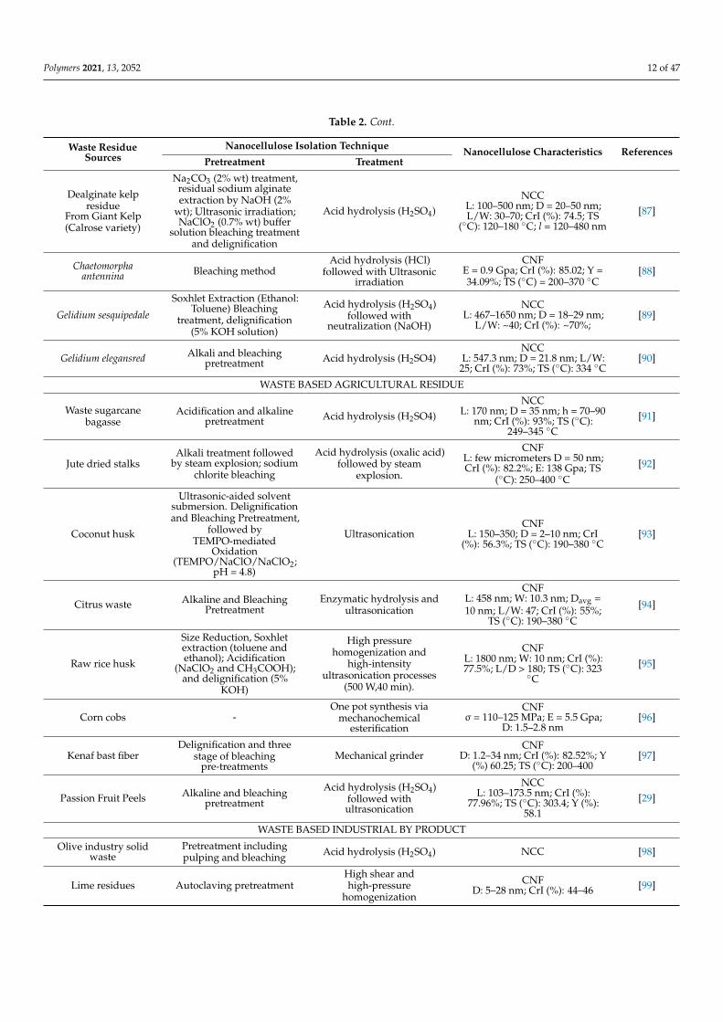
Polymers 2021, 13, 2052 12 of 47
Table 2. Cont.
Waste ResidueSources
Nanocellulose Isolation Technique Nanocellulose Characteristics ReferencesPretreatment Treatment
Dealginate kelpresidue
From Giant Kelp(Calrose variety)
Na2CO3 (2% wt) treatment,residual sodium alginateextraction by NaOH (2%
wt); Ultrasonic irradiation;NaClO2 (0.7% wt) buffer
solution bleaching treatmentand delignification
Acid hydrolysis (H2SO4)NCC
L: 100–500 nm; D = 20–50 nm;L/W: 30–70; CrI (%): 74.5; TS
(◦C): 120–180 ◦C; l = 120–480 nm[87]
Chaetomorphaantennina Bleaching method
Acid hydrolysis (HCl)followed with Ultrasonic
irradiation
CNFE = 0.9 Gpa; CrI (%): 85.02; Y =34.09%; TS (◦C) = 200–370 ◦C
[88]
Gelidium sesquipedale
Soxhlet Extraction (Ethanol:Toluene) Bleaching
treatment, delignification(5% KOH solution)
Acid hydrolysis (H2SO4)followed with
neutralization (NaOH)
NCCL: 467–1650 nm; D = 18–29 nm;
L/W: ~40; CrI (%): ~70%;[89]
Gelidium elegansred Alkali and bleachingpretreatment Acid hydrolysis (H2SO4)
NCCL: 547.3 nm; D = 21.8 nm; L/W:
25; CrI (%): 73%; TS (◦C): 334 ◦C[90]
WASTE BASED AGRICULTURAL RESIDUE
Waste sugarcanebagasse
Acidification and alkalinepretreatment Acid hydrolysis (H2SO4)
NCCL: 170 nm; D = 35 nm; h = 70–90
nm; CrI (%): 93%; TS (◦C):249–345 ◦C
[91]
Jute dried stalksAlkali treatment followed
by steam explosion; sodiumchlorite bleaching
Acid hydrolysis (oxalic acid)followed by steam
explosion.
CNFL: few micrometers D = 50 nm;CrI (%): 82.2%; E: 138 Gpa; TS
(◦C): 250–400 ◦C
[92]
Coconut husk
Ultrasonic-aided solventsubmersion. Delignificationand Bleaching Pretreatment,
followed byTEMPO-mediated
Oxidation(TEMPO/NaClO/NaClO2;
pH = 4.8)
UltrasonicationCNF
L: 150–350; D = 2–10 nm; CrI(%): 56.3%; TS (◦C): 190–380 ◦C
[93]
Citrus waste Alkaline and BleachingPretreatment
Enzymatic hydrolysis andultrasonication
CNFL: 458 nm; W: 10.3 nm; Davg =10 nm; L/W: 47; CrI (%): 55%;
TS (◦C): 190–380 ◦C
[94]
Raw rice husk
Size Reduction, Soxhletextraction (toluene andethanol); Acidification
(NaClO2 and CH3COOH);and delignification (5%
KOH)
High pressurehomogenization and
high-intensityultrasonication processes
(500 W,40 min).
CNFL: 1800 nm; W: 10 nm; CrI (%):77.5%; L/D > 180; TS (◦C): 323
◦C[95]
Corn cobs -One pot synthesis via
mechanochemicalesterification
CNFσ = 110–125 MPa; E = 5.5 Gpa;
D: 1.5–2.8 nm[96]
Kenaf bast fiberDelignification and three
stage of bleachingpre-treatments
Mechanical grinderCNF
D: 1.2–34 nm; CrI (%): 82.52%; Y(%) 60.25; TS (◦C): 200–400
[97]
Passion Fruit Peels Alkaline and bleachingpretreatment
Acid hydrolysis (H2SO4)followed withultrasonication
NCCL: 103–173.5 nm; CrI (%):
77.96%; TS (◦C): 303.4; Y (%):58.1
[29]
WASTE BASED INDUSTRIAL BY PRODUCT
Olive industry solidwaste
Pretreatment includingpulping and bleaching Acid hydrolysis (H2SO4) NCC [98]
Lime residues Autoclaving pretreatmentHigh shear andhigh-pressure
homogenization
CNFD: 5–28 nm; CrI (%): 44–46 [99]
Polymers 2021, 13, 2052 13 of 47
Table 2. Cont.
Waste ResidueSources
Nanocellulose Isolation Technique Nanocellulose Characteristics ReferencesPretreatment Treatment
Recycled Tetra PakFood Packaging
Wastes
Delignification andbleaching pretreatment
Acid hydrolysis (H2SO4)followed withultrasonication
NCCL: 127–258 nm; D: 11.4–14 nm;
L/D: 10; CrI (%): 94.8%; TS (◦C):204
[100]
Waste paper Deinking method andalkaline pretreatment
Acid hydrolysis (H2SO4)followed withultrasonication
NCCL: 271 nm [101]
Discarded cigarettefilters
Ethanol extraction, alkalinepretreatment, and bleaching
pretreatment,
Acid hydrolysis (H2SO4)followed withultrasonication
NCCL: 143 nm; W: 8 nm; CrI (%):
96.77%; Y (%): 29.4[102]
Recycled Paper MillSludge Ozonation pretreatment Acid hydrolysis (Maleic
acid)
NCCL: 2431 nm; W: 165 nm; L/D:
16.7CrI (%): 77%; Y (%): 0.8
[103]
Citrus Pulp of Floater(CPF)
Alkaline and bleachingpretreatment with autoclave Enzymatic hydrolysis n.d
CrI (%):60 [104]
Sweet lime pulp waste Blending and acidhydrolysis (H2SO4)
Komagataeibacter europaeusSGP37 incubated in static
intermittent fed-batchcultivation
BNCY(g/L): CrI (%):89.6; TS (◦C):
348[105]
Abbreviation: D: Diameter; L: Length; W: Width; TS: Thermal Stability; Y: Yield; L/D: Aspect Ratio; CrI: Crystallinity Index; l: Lateral size;σ: Tensile strength; E: Young Modulus.
4. Isolation of Nanocellulose4.1. Isolation of Nano-Fibrillated Cellulose (NFC)
Regardless of its cellulose sources, NFC is mainly fabricated from cellulose pulpthrough mechanical treatment by breaking down the linkage of interfibrillar hydrogen [106].The exerted mechanical force triggers the cracking phenomenon to form a critical tensioncenter in fibrous substances. The development of NFC from fibrous material requiresintense mechanical treatment with or without pretreatment. However, fibrous material’smechanical disintegration may cause pulp clogging, causing the fiber to agglomerate andrequire high energy to break it down. Thus, another pretreatment is required to overcomethis problem.
Several pretreatments have been introduced before the primary mechanical treatmentto diminish the polymerization degree and debilitate the hydrogen linkage. These pre-treatments include mechanical refining, alkaline hydrolysis, solvent-assisted pretreatment,organic acid hydrolysis, 2,6,6-tetramethylpiperidine-1-oxyl (TEMPO)-mediated oxidation,enzymatic disintegration, periodate-chlorite oxidation, oxidative sulfonation, cationization,ionic liquid, carboxymethylation, deep eutectic solvents, and acetylation [17].
The earliest production of NFC was reported by Turbak et al. [107] and Herricket al. [108]. They isolated NFC from wood via high-pressure homogenization (HPH). HPHexerted a mechanical force on cellulose fibrils driven by crushing, shear, and cavitationalforces in which cellulose pulp is transferred into the chamber through a small nozzleto enable particle size reduction to the nanoscale of the cellulose fibrils [72]. Currently,the HPH is the most commonly utilized method for NFC production on an industrialand laboratory scale, given its simplicity, high efficiency, and lack of organic solventrequirements [109]. Furthermore, HPH enables high conversion of cellulose materialtoward CNF. High energy, high pressure, and long duration of the HPH process may alsoescalate the fibrillation degree. However, the difficulty of cleaning the equipment due tothe blockage in the homogenizer valve is the major drawback of the HPH method [110].Different processes have also been developed to produce CNF, such as micro-fluidization,micro-grinding, cryo-crushing, ultrasonication, mechanical refining, radiation, ball milling,blending, extrusion, steam explosion, and aqueous counter collision [111].
Polymers 2021, 13, 2052 14 of 47
4.2. Isolation of Cellulose Nanocrystal (NCC)
According to the previous discussion, the main difference between NCC and CNFlies in their structure, in which CNF comprises amorphous and crystalline regions whileNCC has high crystalline purity in cellulose regions. Therefore, the primary step inisolating NCC is to break down the disordered amorphous or paracrystalline regions thatintegrate the crystalline regions within cellulose fibrils. Initially, an NCC suspension wasproduced in 1949 from lignocellulosic biomass through an integrated alkaline and bleachingpretreatment and acid hydrolysis [13]. Acid hydrolysis remains the paramount process forNCC extraction. The crystalline part in cellulose fibers is not hydrolyzed because it has ahigh resistance to acids, although acids can easily hydrolyze the amorphous regions [112].In this method, sulfuric acid (H2SO4), hydrochloric acid (HCl), hydrobromic acid (HBr),and phosphoric acid (H3PO4) have been extensively employed as the acid component tobreakdown the amorphous region of cellulose.
Following acid hydrolysis, the remaining free acid molecules and other impuritiesshould be removed by diluting and washing with water using centrifugation and dialysisprocesses. Moreover, specific mechanical treatment like sonication may be needed to stabi-lize the NCC particles in uniform suspensions. However, the high tendency of corrosion,low recuperation rate, and high acid wastewater produced due to the high amount of waterfor the washing process for nanocellulose suspension neutralization become the significantdrawbacks of the acid hydrolysis process [46]. To avoid excessive equipment corrosionand environmental issue, various nanocellulose isolation processes have been developed,such as extraction using ionic liquids, TEMPO oxidation, enzymatic, and others. Variousresearchers have carried out the combination and integration of various isolation processesto increase the isolation process’s efficiency, such as enzymatic hydrolysis with TEMPOoxidation and enzymatic hydrolysis with ultrasonication [113]. Chemical treatment iscrucial for NCC isolation, while mechanical treatment is the vital stage for CNF production.
4.3. Isolation of Bacteria Cellulose (BC)
The selection of strains of microorganisms is a very crucial factor in the synthesis of BC.There are currently two main methods that have been used for BC production, i.e., staticfermentation and submerged fermentation [54]. Static fermentation has been widelyemployed as an extracellular-based production route. In the static fermentation, a 3Dnetwork of gelatinous pellicles with high water content formed during the interspersingand intertwining of the ribbons structure form of BC, reaching a particular thicknesscorresponding to longer incubation time and causing the entrapment of bacteria cellsand its further inactivity. The static fermentation produces BC with excellent crystallinityand mechanical strength, although prolonged cultivation and low productivity limit theirindustrial utilization.
Furthermore, the BC layer’s uneven thickness is produced due to the exposure ofbacteria to uncertain conditions (nutrient, oxygen level, and cell distribution) throughoutthe growth cycle. Fed-batch strategies and submerged fermentation involving aerationand agitation fermentation have been introduced to overcome static fermentation’s sig-nificant drawbacks. Submerged fermentation leads to higher BC productivity than staticfermentation, which has been extensively utilized commercially. The cultivated bacteriaare adequately exposed to oxygen, thereby generating a high yield of BC in the shape ofsmall granules or pellets during aerated fermentation [114]. Moreover, agitation in thefermentation would result in a more homogeneous BC and oxygen evenly distributed tobacterial cells. However, the produced BC has lower crystallinity and mechanical strengththan static fermentation [115].
Several submerged fermentation issues such as the advancement of cellulose non-production strains [116], irregular shapes of BC granules or pellets, and physical character-istic modification of BC remain challenging for the researcher to overcome. In addition,excessive-high rotation speed and hydrostatic stresses may promote gluconic acid pro-duction by bacteria due to the accumulation of self-protection metabolism [117]. Several
Polymers 2021, 13, 2052 15 of 47
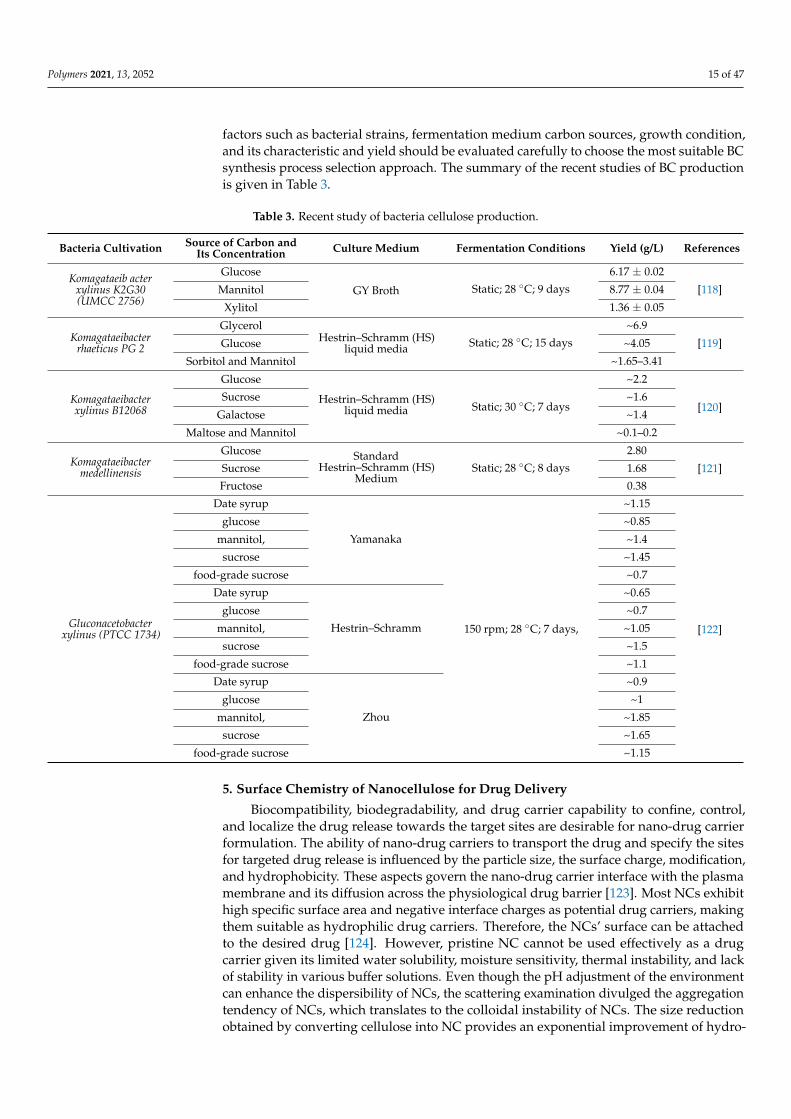
factors such as bacterial strains, fermentation medium carbon sources, growth condition,and its characteristic and yield should be evaluated carefully to choose the most suitable BCsynthesis process selection approach. The summary of the recent studies of BC productionis given in Table 3.
Table 3. Recent study of bacteria cellulose production.
Bacteria Cultivation Source of Carbon andIts Concentration Culture Medium Fermentation Conditions Yield (g/L) References
Komagataeib acterxylinus K2G30(UMCC 2756)
Glucose
GY Broth Static; 28 ◦C; 9 days
6.17 ± 0.02
[118]Mannitol 8.77 ± 0.04
Xylitol 1.36 ± 0.05
Komagataeibacterrhaeticus PG 2
GlycerolHestrin–Schramm (HS)
liquid media Static; 28 ◦C; 15 days
~6.9
[119]Glucose ~4.05
Sorbitol and Mannitol ~1.65–3.41
Komagataeibacterxylinus B12068
Glucose
Hestrin–Schramm (HS)liquid media Static; 30 ◦C; 7 days
~2.2
[120]Sucrose ~1.6
Galactose ~1.4
Maltose and Mannitol ~0.1–0.2
Komagataeibactermedellinensis
Glucose StandardHestrin–Schramm (HS)
MediumStatic; 28 ◦C; 8 days
2.80
[121]Sucrose 1.68
Fructose 0.38
Gluconacetobacterxylinus (PTCC 1734)
Date syrup
Yamanaka
150 rpm; 28 ◦C; 7 days,
~1.15
[122]
glucose ~0.85
mannitol, ~1.4
sucrose ~1.45
food-grade sucrose ~0.7
Date syrup
Hestrin–Schramm
~0.65
glucose ~0.7
mannitol, ~1.05
sucrose ~1.5
food-grade sucrose ~1.1
Date syrup
Zhou
~0.9
glucose ~1
mannitol, ~1.85
sucrose ~1.65
food-grade sucrose ~1.15
5. Surface Chemistry of Nanocellulose for Drug Delivery
Biocompatibility, biodegradability, and drug carrier capability to confine, control,and localize the drug release towards the target sites are desirable for nano-drug carrierformulation. The ability of nano-drug carriers to transport the drug and specify the sitesfor targeted drug release is influenced by the particle size, the surface charge, modification,and hydrophobicity. These aspects govern the nano-drug carrier interface with the plasmamembrane and its diffusion across the physiological drug barrier [123]. Most NCs exhibithigh specific surface area and negative interface charges as potential drug carriers, makingthem suitable as hydrophilic drug carriers. Therefore, the NCs’ surface can be attachedto the desired drug [124]. However, pristine NC cannot be used effectively as a drugcarrier given its limited water solubility, moisture sensitivity, thermal instability, and lackof stability in various buffer solutions. Even though the pH adjustment of the environmentcan enhance the dispersibility of NCs, the scattering examination divulged the aggregationtendency of NCs, which translates to the colloidal instability of NCs. The size reductionobtained by converting cellulose into NC provides an exponential improvement of hydro-
Polymers 2021, 13, 2052 16 of 47
gen bonding that triggers the NC aggregation. This limitation can be made worse by thedrug coordination, which is exposed on the NC exterior, consequently altering the dis-persibility and solubility [125]. Therefore, various surface modification and pretreatmentfiber methodologies have been developed to overcome limitations and advance specificcharacteristics [126].
From a structural perspective, the three hydroxyl groups in each cellulose monomerare the most prominent characteristic that makes the NC surface reactive. The reactivityof hydroxyl groups influences the surface modification of anhydroglucose units. It wasreported that in the molecular framework of cellulose, the hydroxyl group at the sixthposition behaves as primary alcohol with a reactivity ten times larger than the otherhydroxyl groups, while the hydroxyl group at the second position has two-fold higherreactivity than that in the third position, both of which serve as secondary alcohols. Thisphenomenon manifests from the steric hindrance of each hydroxyl group, in which thehydroxyl group at the sixth position attached to the carbon atom that is connected to onlyone alkyl groups while the carbon atom that carries the hydroxyl groups in the secondand third positions bonded to two alkyl groups [127]. Regarding the surface receptivenessof NC’s hydroxyl groups, the addition of solvent and reactant may alter the group’sreceptiveness in diverse positions. De la Motte et al. [128] modified NCC through cationicepoxide 2,3-epoxypropyltrimethyl ammonium chloride (EPTMAC) by spray technique.It was revealed that the hydroxyl bunch receptiveness of cationic modified NC followsthe order of OH-C6 = OH-C2 > OH-C3, which was validated through nuclear magneticresonance (NMR).
Nanocellulose surface modification for drug delivery was developed by modulatingthe NC hydroxyl groups. In general, the main objective of nanocellulose surface modifica-tion is to incorporate new functional groups or drug components into the nanocelluloseframework to escalate the degree of substitution and the efficacy of material grafting with-out altering the structure, morphology, and crystallinity of nanocellulose [129]. Severalprocesses have been developed for the surface modification of NC, either by physical orchemical processes, presented in more detail in the following sections.
5.1. Functionalization of Nanocellulose through Physical Technique
Several physical techniques such as surface defibrillation, irradiation, electric current,and electric discharge have been developed to modify and functionalize nanocellulosesurfaces for diverse applications [130]. Surface defibrillation disintegrates cellulose intoelementary fibrils by exerting mechanical force using various devices such as ultra-refining,a high-pressure homogenizer, a grinder, a microfluidizer, and spray-drying. In nanocellu-lose functionalization, the combination of nanocellulose and drug entities can be subjectedto surface defibrillation to modify the morphology of nanocellulose and construct a newmatrix system with a tight fiber network.
Microparticles from BC with fibrillar structure morphology have been prepared byspray-drying technique. An ultra-refining-assisted method was also conducted to constructbacteria cellulose nanofiber (BCNF) with various sizes and shapes. The coating of BCNFwith mannitol (MN), maltodextrin (MF), and hydroxypropylmethylcellulose (HPCM) werealso carried out at various ratios to study the drug release characteristics. The addition ofsuch coating matrices exhibits benefits towards the spray-drying process and drug carrierability, i.e., superior protection of drug confinement, decreased droplet adhesion on thedrying chamber, and improved powder performance. As a result, the BC-microparticles cansuccessfully enhance the drug uptake capacity and sustain the drug release of diclofenacsodium (hydrophilic) and caffeine (lipophilic) [131].
As a recent advanced method, irradiation exerted high energy, which modifies thecellulose exterior. For example, the radiated gamma energy can generate reactive in-termediates comprising ions and free radicals that provoke reaction pathways such ascross-linking, scission degradation, oxidation, and polymer and molecule grafting. Thepresence of irradiation beams, such as microwave and electron, accelerates the polymer
Polymers 2021, 13, 2052 17 of 47
growth. UV-irradiation has also been developed to improve the reaction rate to allowpre-synthesized grafted polymer formation on the nanocellulose surface [132]. Recently,this method has been developed to induce polymer grafting and polymer growth onnanocellulose surfaces.
Plasma treatment is considered an environmentally friendly method to achievenanocellulose surface functionalization by utilizing plasma ionized gas without altering itscharacteristics. Researchers have widely applied this method for various modifications,such as increasing material–cell interaction, introducing the surface of NC with hydropho-bicity or hydrophilicity characteristics, and incorporating chitosan towards cellulose sub-strates. For instance, Kusano et al. [133] modified CNF by utilizing dielectric-based plasmadischarge treatment, leading to the formation of many carboxyl groups, carbonyl groups,and oxygen-containing groups on the surface of nanocellulose [133]. Moreover, assistedultrasonic irradiation combined with plasma discharge treatment can refine the wetting andoxidation of the nanofibers coating. Plasma treatment is an attractive route for surface func-tionalization of nanocellulose given its benefits such as non-polluting, fast-modification,and simple chemical treatments compared to the conventional modification method.
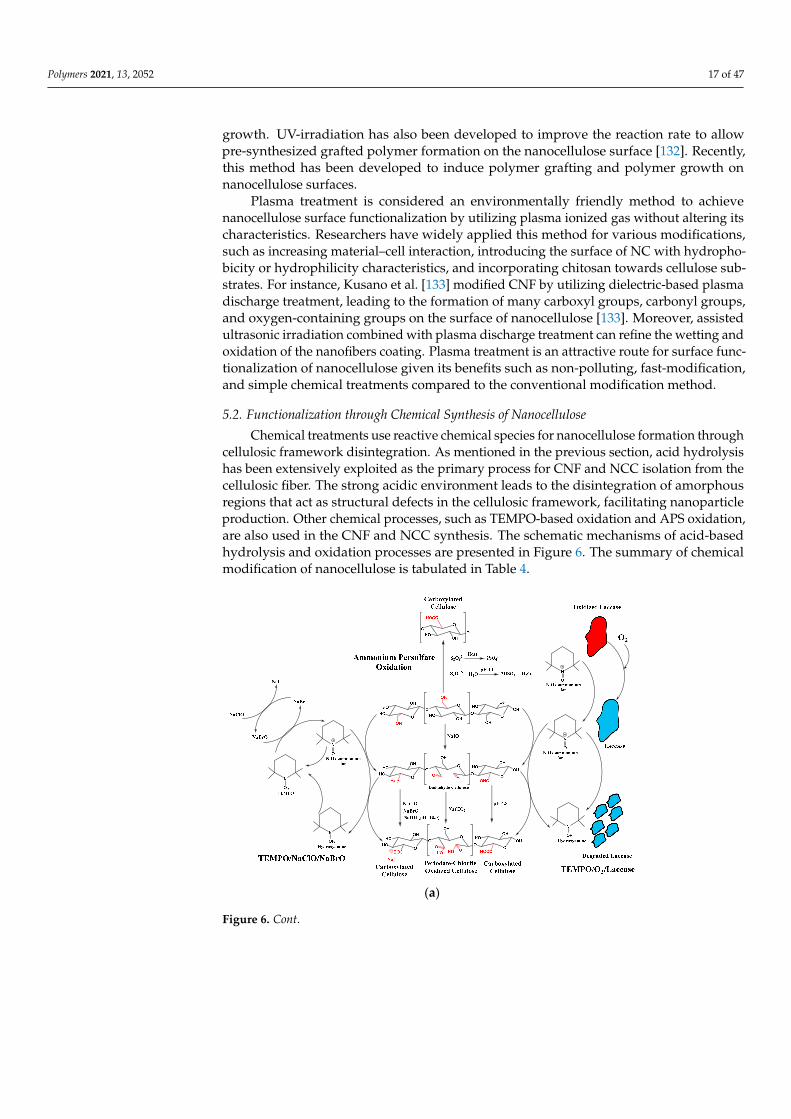
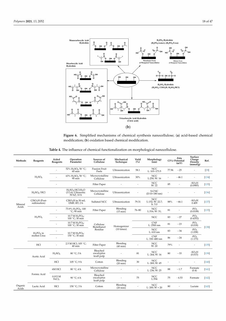
5.2. Functionalization through Chemical Synthesis of Nanocellulose
Chemical treatments use reactive chemical species for nanocellulose formation throughcellulosic framework disintegration. As mentioned in the previous section, acid hydrolysishas been extensively exploited as the primary process for CNF and NCC isolation from thecellulosic fiber. The strong acidic environment leads to the disintegration of amorphousregions that act as structural defects in the cellulosic framework, facilitating nanoparticleproduction. Other chemical processes, such as TEMPO-based oxidation and APS oxidation,are also used in the CNF and NCC synthesis. The schematic mechanisms of acid-basedhydrolysis and oxidation processes are presented in Figure 6. The summary of chemicalmodification of nanocellulose is tabulated in Table 4.
Polymers 2021, 13, x 17 of 49
As a recent advanced method, irradiation exerted high energy, which modifies the cellulose exterior. For example, the radiated gamma energy can generate reactive inter-mediates comprising ions and free radicals that provoke reaction pathways such as cross-linking, scission degradation, oxidation, and polymer and molecule grafting. The pres-ence of irradiation beams, such as microwave and electron, accelerates the polymer growth. UV-irradiation has also been developed to improve the reaction rate to allow pre-synthesized grafted polymer formation on the nanocellulose surface [132]. Recently, this method has been developed to induce polymer grafting and polymer growth on nanocel-lulose surfaces.
Plasma treatment is considered an environmentally friendly method to achieve nano-cellulose surface functionalization by utilizing plasma ionized gas without altering its characteristics. Researchers have widely applied this method for various modifications, such as increasing material–cell interaction, introducing the surface of NC with hydro-phobicity or hydrophilicity characteristics, and incorporating chitosan towards cellulose substrates. For instance, Kusano et al. [133] modified CNF by utilizing dielectric-based plasma discharge treatment, leading to the formation of many carboxyl groups, carbonyl groups, and oxygen-containing groups on the surface of nanocellulose [133]. Moreover, assisted ultrasonic irradiation combined with plasma discharge treatment can refine the wetting and oxidation of the nanofibers coating. Plasma treatment is an attractive route for surface functionalization of nanocellulose given its benefits such as non-polluting, fast-modification, and simple chemical treatments compared to the conventional modification method.
5.2. Functionalization through Chemical Synthesis of Nanocellulose Chemical treatments use reactive chemical species for nanocellulose formation
through cellulosic framework disintegration. As mentioned in the previous section, acid hydrolysis has been extensively exploited as the primary process for CNF and NCC iso-lation from the cellulosic fiber. The strong acidic environment leads to the disintegration of amorphous regions that act as structural defects in the cellulosic framework, facilitating nanoparticle production. Other chemical processes, such as TEMPO-based oxidation and APS oxidation, are also used in the CNF and NCC synthesis. The schematic mechanisms of acid-based hydrolysis and oxidation processes are presented in Figure 6. The summary of chemical modification of nanocellulose is tabulated in Table 4.
(a)
Figure 6. Cont.
Polymers 2021, 13, 2052 18 of 47Polymers 2021, 13, x 18 of 49
(b)
Figure 6. Simplified mechanisms of chemical synthesis nanocellulose; (a) acid-based chemical mod-ification; (b) oxidation based chemical modification.
Table 4. The influence of chemical functionalization on morphological nanocellulose.
Methods Reagents Aided
Reagents Operation Parameter
Sources of Cellulose
Mechanical Technique Yield (%)
Morphology (nm) CI%)
Zeta Potential (mV)
Surface Charge Density
(mmol/g)
Ref.
Mineral Acids
H2SO4
- 52% H2SO4
50 °C; 60 min Passion Fruit
Peels Ultrasonication 58.1 NCC
L: 103–173.5 77.96 −25 - [29]
- 63% H2SO4
50 °C; 90 min Microcrystal-line Cellulose
Ultrasonication 30% NCC
L:250; W: 16 - −46.1 - [134]
- - Filter Paper - - NCC W: 22
85 - -SO3H
(0.0985) [135]
H2SO4/HCl -
H2SO4:HCl:H2O (3:1:6); Ul-
trasonic 50 hZ; 10 h
Microcrystal-line Cellulose
Ultrasonication - S-CNC
(D:10–180 nm)
- - - [136]
ClSO3H (Post-sul-fonation)
- ClSO3H in 50 mL DMF; RT;
2 h Sulfated NCC Ultrasonication 79.31
NCC L:152; W: 22.7; h: 5.0
88% −66.1 -SO3H 0.409
[137]
H3PO4
- 73.9% H3PO4;
100 °C; 90 min
Filter Paper Blending (15 min)
76–80 NCC
L:316; W: 31; 81 -
-PO3 (0.0108)
[135]
-
10.7 M H3PO4;
100 °C; 30 min
Cellulose Bio-tethanol Resi-
due
Homogenizer (10 times)
NCC 83 −27 -PO3
(0.4352)
[138] -
10.7 M H3PO4;
100 °C; 30 min
CNF
L: 2500 nm 81 −23
-PO3 (0.018)
H3PO4 in molten Urea
- 10.7 M H3PO4;
150 °C; 30 min
NCC
L: 610 nm 83 −34
-PO3 (1.038)
- CNF
L: 330–480 nm
86 −24 -PO3
(1.173)
OH OH OHS OH SO OO
OOO
O OH O O OH OPO
OHO
PO
O OH
Monobasic Formof Phospated Nanocellulose
Dibasic Formof Phospated Nanocellulose
POH
OHOPOH
O OH
O OH O O OH O
O OH O
1
O O OO
12
OH
3O
1 2
OH
3O
O OH O2 1
O
HOO
21
O
OHO
43
OH
21
O
O
HOO OH O
4 3
OH
21
O
O
OH
O OH O43
2
1 OOHO
43
2
1OO OH
32
1
OO
HO O OH O 32
1
O O
OH
2
1
2
OH
1
34
OO OH
O
HO O OH O 1
2
OH
1
34
OOHO
O
OH
H3PO4 Hydrolisis(H3PO4/water); (H3PO4/Urea)
H2SO4 Hydrolisis(H2SO4; ClSO3H; H2SO4/HCl)
Tricarboxylic Acid Hydrolisis(Citric acid)
Dicarboxylic AcidHydrolisis
Monocarboxylic AcidHydrolisis
Figure 6. Simplified mechanisms of chemical synthesis nanocellulose; (a) acid-based chemicalmodification; (b) oxidation based chemical modification.
Table 4. The influence of chemical functionalization on morphological nanocellulose.
Methods Reagents AidedReagents
OperationParameter
Sources ofCellulose
MechanicalTechnique
Yield(%)
Morphology(nm) CI%)
ZetaPotential
(mV)
SurfaceChargeDensity(mmol/g)
Ref.
MineralAcids
H2SO4
- 52% H2SO4 50 ◦C;60 min
Passion FruitPeels Ultrasonication 58.1 NCC
L: 103–173.5 77.96 −25 - [29]
- 63% H2SO4 50 ◦C;90 min
MicrocrystallineCellulose Ultrasonication 30% NCC
L:250; W: 16 - −46.1 - [134]
- - Filter Paper - - NCCW: 22 85 - -SO3H
(0.0985) [135]
H2SO4/HCl -H2SO4 :HCl:H2O(3:1:6); Ultrasonic
50 hZ; 10 h
MicrocrystallineCellulose Ultrasonication - S-CNC
(D:10–180 nm) - - - [136]
ClSO3H (Post-sulfonation) - ClSO3H in 50 mL
DMF; RT; 2 h Sulfated NCC Ultrasonication 79.31NCC
L:152; W: 22.7;h: 5.0
88% −66.1 -SO3H0.409 [137]
H3PO4
- 73.9% H3PO4 ; 100◦C; 90 min Filter Paper Blending
(15 min) 76–80 NCCL:316; W: 31; 81 - -PO3
(0.0108) [135]
- 10.7 M H3PO4 ;100 ◦C; 30 min
CelluloseBiotethanol
ResidueHomogenizer
(10 times)
NCC 83 −27 -PO3(0.4352)
[138]- 10.7 M H3PO4 ;
100 ◦C; 30 minCNF
L: 2500 nm 81 −23 -PO3(0.018)
H3PO4 inmolten Urea
-10.7 M H3PO4 ;150 ◦C; 30 min
NCCL: 610 nm 83 −34 -PO3
(1.038)
- CNFL: 330–480 nm 86 −24 -PO3
(1.173)
HCl - 2.5 M HCl; 105 ◦C;40 min Filter Paper Blending
(40 min) - NCCW: 20 79% - - [135]
OrganicAcids
Acetic AcidH2SO4 80 ◦C; 3 h
Bleachedeucalyptuskraft pulp
- 81 NCCL: 264; W: 16 80 −33 -SO3H
(0.015) [139]
HCl 105 ◦C; 9 h Cotton Blending(20 min) 30 NCC
L: 269; W: 45 - - - [140]
Formic Acid
6M HCl 80 ◦C; 4 h MicrocrystallineCellulose - - NCC
L: 236; W: 25 88 −1.7 Formate(0.4) [141]
0.015 MFeCl3
90 ◦C; 6 hBleached
eucalyptuskraft pulp
- 75 NCCL:594 75 −6.53 Formate [142]
Lactic Acid HCl 150 ◦C; 3 h Cotton Blending(20 min) - NCC
L: 200; W = 20 80 - Lactate [143]
Polymers 2021, 13, 2052 19 of 47
Table 4. Cont.
Methods Reagents AidedReagents
OperationParameter
Sources ofCellulose
MechanicalTechnique
Yield(%)
Morphology(nm) CI%)
ZetaPotential
(mV)
SurfaceChargeDensity(mmol/g)
Ref.
Butyric Acid 0.027 MHCl 105 ◦C; 9 h Cotton Blending
(20 min) 20 NCCL: 226; W = 34 - - Butyrate [140]
Maleic Acid(MA)
- 70% MA; 100 ◦C;45 min
Bleachedeucalyptuskraft pulp
- 12% NCC - -33 -COOH(0.29) [144]
-60% MA; 120 ◦C;
2 h
Bleachedeucalyptuskraft pulp
Microfluidizer(120 mPa; 5
passes)
3% L: 329.9; h =15.9 - −46.9 -COOH
(0.368)[145]
84% CNFh: 13.4 - −45.2 -COOH
(0.059)
Oxalic Acid(OA)
- 8.75% OA; 110 ◦C;15 min Filter paper Sonication
(60 min) 93.77NCC
L: 150–200;W: 5–20
- −36 -COOH,0.29 [146]
- 70% OA; 100 ◦C; 1h
Bleachedeucalyptuskraft pulp
- 24.7 NCC 80 −42.5 -COOH [144]
- 30% OA; 100◦ C;30 min Celery Sonication
(18 min) 76.8 CNFh: 5.5 49 −32.9 -COOH [147]
Malonic Acid- 80% wt of
Malonic Acid; 140◦C; 3 h
RamieCellulose
Blending(5 min)
5%
NCCL: ~220; W: ~12
- - -COOH
[148]
0.025 MHCl 19.8% 75 - -COOH
Malic acid-
80% wt of MalicAcid; 140 ◦C; 3 h
3.4% - - -COOH,(1.617)
0.05 MHCl 20% 78 - -COOH
Citric Acid
-80% wt of CitricAcid; 140 ◦C; 3 h
5.1 - - -COOH
0.05 MHCl 20.5 78 - -COOH,
(1.884)
-80% wt of CitricAcid; 100 ◦C; 4 h
BleachedBaggase Pulp Ultrasonication
32 NCC,L: 251; W: 21 78 −122.9 -COOH,
0.65[149]
- - CNF,L: 654; W: 32 69 190.3 -COOH,
0.3
OxidationTreat-ment
TEMPO/NaCl/NaBr
-
TEMPO (0.094mmol)-NaBr (1.57
mmol)- NaClO(1.24 M); 10 ◦C; 45
min
NanocrystallineCellulose Ultrasonication - NCC,
L: 100; W: 5–20 80% - - [150]
-
TEMPO (0.1mmol
mmol)-NaBr (1mmol)- NaClO (5
mmol/gcellulose);Ambient
condition; 1.5 h
HBKP Ultrasonication - CNF 85% --COOH;-CHO(1.191)
[151]
TEMPO/O2/Laccase50 mM TEMPO, 5U mL–1 laccase;
96 hHBKP Ultrasonication - CNF,
L: > 100; W: 4–8 - --COOH;-CHO(0.837)
SequentialPeriodate-Chlorite
Oxidation
1 MAcetic
Acid (2)
(1). 46 mmolNaIO4 ; 50 ◦C;4.5h followed by (2).12 g NaClO2l 50
◦C; 40 h
HardwoodPulp
Homogenizer(5 passes; 80
MPa)- CNF,
L: 95.8; W: 2.72 - −128 -COOH(2.0) [152]
APS Oxidation - 1 M APS; 75 ◦C;16 h Cotton Linters - 34.4 CNF,
L: 95.8; W: 2.72 63.8 --COOH(0.16);-SO3(0.98)
[153]
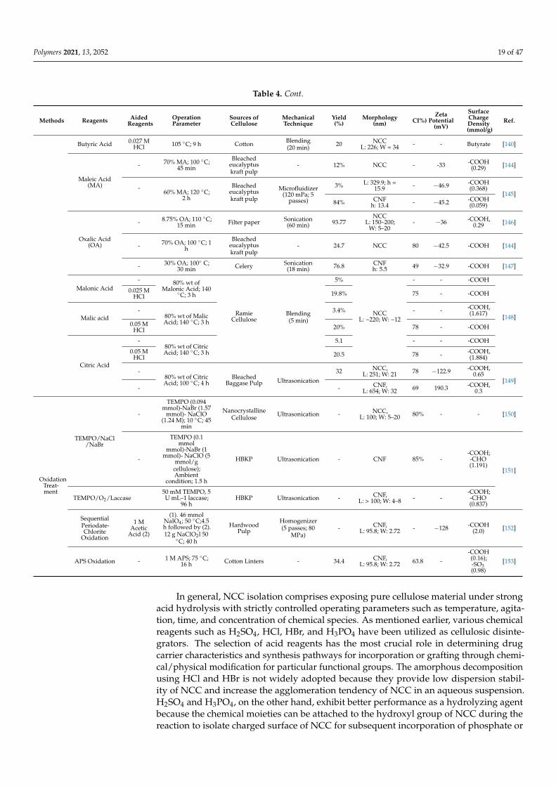
In general, NCC isolation comprises exposing pure cellulose material under strongacid hydrolysis with strictly controlled operating parameters such as temperature, agita-tion, time, and concentration of chemical species. As mentioned earlier, various chemicalreagents such as H2SO4, HCl, HBr, and H3PO4 have been utilized as cellulosic disinte-grators. The selection of acid reagents has the most crucial role in determining drugcarrier characteristics and synthesis pathways for incorporation or grafting through chemi-cal/physical modification for particular functional groups. The amorphous decompositionusing HCl and HBr is not widely adopted because they provide low dispersion stabil-ity of NCC and increase the agglomeration tendency of NCC in an aqueous suspension.H2SO4 and H3PO4, on the other hand, exhibit better performance as a hydrolyzing agentbecause the chemical moieties can be attached to the hydroxyl group of NCC during thereaction to isolate charged surface of NCC for subsequent incorporation of phosphate or
Polymers 2021, 13, 2052 20 of 47
sulfate functional groups. The new functional group incorporation causes the spontaneousdispersibility of NCCs in an aqueous environment due to the colloidal stability restora-tion through electrostatic repulsion refinement, which is the preferred characteristic ofdrug carriers.
A subsequent treatment of H2SO4 followed by HCl synthesis has been utilized tocontrol the sulfate moieties on the NC surfaces. The as-synthesized particle had a simi-lar particle size to those particles directly acquired from acid hydrolysis. Nevertheless,the surface charge density can be adjusted on the hydroxyl groups exploited by sulfategroups [49]. Lin and Dufresne [137] proposed a strategy of inaugurating progressivesulfate group content on NCCs surface through the modulation ratio of reactants andpost-sulfonation (chlorosulfonic acid) and desulfonation conditions. They also evaluatedthe impact of sulfonation degree on the morphology, dimension, physical characteristic,and surface chemistry of modified NCCs. Diverse zeta potential ranged from −7 mV to−66 mV and approximately 0.0563 mmol/g–1.554 mol/g of sulfonation degree was ac-quired. Therefore, it is indicated that the zeta potential of nanocellulose is mainly controlledby the sulfonation degree of nanocellulose itself [137].
Wijaya et al. [29] successfully isolated NCC through sulfuric acid hydrolysis ofbleached passionfruit peels waste fiber by adjusting the acid concentration, hydrolysistime, and reaction temperature. The NCC was used for tetracycline hydrochloride ad-sorption through electrostatic and Van der Waals interaction. The adsorption isothermwas correlated using Langmuir and Freundlich isotherm models. With pH environmentadjustment, the adsorption affinity of the drug can be altered to control the uptake andsustained release of drugs [29].
(2,2,6,6-tetramethylpiperidine-1-oxyl)-mediated (or TEMPO-mediated) oxidation ofnanocellulose has arisen as an alternative NC isolation route to replace the conventionalacid hydrolysis method due to its environmentally friendly and facile synthesis nature.The synthesis starts by using TEMPO/NaBr/NaClO or TEMPO/NaClO2/NaClO as areagent. TEMPO (stable nitroxyl radical) forms as the catalyst for NC synthesis, whichfurther transforms into N-oxoammonium salt (R1R2N+=O) under certain conditions whilethe NaClO acts as a primary oxidant [46]. Both NaClO and NaBr can reversibly transformthe N-oxoammonium salt into TEMPO form. The hydroxymethyl groups of NC (primaryhydroxyl groups located on C6) are selectively transformed into carboxylated groups whilethe secondary hydroxyl groups remain unchanged (secondary hydroxy groups located onC2 and C3) [66]. The incorporated carboxyl groups imparted negative surface charges fromthe change in the environment pH, which leads to improved colloidal stability.
As reported by Montanari et al. [154], TEMPO-mediated oxidation with the degreeof oxidation 0.24 has imparted negative charges on the crystalline regions of nanocellu-lose, which provide dispersibility and individualization improvement time decreasingthe crystallite size [154]. Meanwhile, Habibi et al. [150] underlined that the TEMPO-mediated oxidation did not affect the morphological and crystallinity of NCCs. Further-more, they highlighted that the ratio of primary oxidizing agents affected the negativecharge of NCCs [150].
A novel oxidation system of TEMPO/laccase/O2 has been utilized to modify NC.The TEMPO/laccase/O2 system with sufficient catalytic amounts of laccase and TEMPOreagent produced reactive TEMPO+, which subsequently transformed primary hydroxylmoieties into aldehyde moieties through oxidation. After the oxidation, the reactiveTEMPO+ was reduced into N-hydroxyl TEMPO. However, no-cycle regeneration occurredbetween TEMPO+ and N-hydroxyl TEMPO due to the breakdown of the primary hydroxylgroups of polysaccharides and laccase molecules. Furthermore, the N-hydroxyl-TEMPOwas accumulated in the reaction environment due to the absence of active laccase inthe system. Therefore, a large amount of TEMPO and laccase and prolonged reactiontime are required to oxidize the primary hydroxyl groups, which are considered majordisadvantages of this process [151].
Polymers 2021, 13, 2052 21 of 47
TEMPO-mediated oxidation was mainly used to modify NFCs before mechanicaldefibrillation to promote the fiber’s individualization. TEMPO-mediated oxidation leads tothe breakage of the strong intra-fiber hydrogen coordination to facilitate the softening andimpairing of its rigid structure, which is beneficial in converting TEMPO-oxidized cellulosefiber into highly crystalline individual nanofibers through mechanical treatment. TheNaClO concentration and mechanical treatment strength were considered crucial factors indetermining the polymerization degree, carboxylate group numbers, and CNFs yield.
Carlsson et al. [155] emphasized the influence of surface charges in nanocelluloseformulation as a drug carrier by introducing TEMPO-mediated oxidation in mesoporousclaodophora cellulose for aspirin degradation. The surface charge negativity (carboxylatecontent 0.44 ± 0.01 mmol g−1) significantly accelerated the degradation of aspirin comparedto the native source of CNFs, which had a deficient surface charge (0.06 ± 0.01 mmol g−1).This phenomenon is caused by the strong interaction of opposite charge entities betweenaspirin and TEMPO-oxidized cellulose nanofibers (TOCNFs), leading to increased partialamorphization ability inside the mesoporous TOCNFs [155].
Without a chlorine-containing oxidant, 1.1 mmol g−1 of carboxyl groups were incor-porated onto wood cellulose. High in carboxylate content, wood cellulose underwenttremendous depolymerization during oxidation. In addition, a long reaction duration of upto 15 h was required to achieve 0.6 mmol g−1 carboxylate content, while 1.1 mmol g−1 wasachieved by increasing the reaction time up to 20 h. Prolonged reaction time is consideredthe major disadvantage of this process. This method has been utilized for nanocellu-lose modification in drug delivery applications [156]. The sequential periodate-chlorineoxidation selectively and simultaneously incorporates two carboxyl groups through theoxidative transformation of two vicinal secondary hydroxyl groups (located in C2 and C3instead of C6 position), enabling higher surface charge density introduction. The increaseof surface charge density is essential in retaining the colloidal stability of drug carrierand improving the electrostatic interaction between drug and carrier, which increase theloading uptake of drugs.
Plappert et al. [152] investigated the pretreatment effect of sequential chlorite pe-riodic oxidation on open-porous anisotropic CNF hydrogel membrane assembly. Hy-drogel membranes were used for transdermal drug delivery systems for nonsteroidalanti-inflammatory drugs (NSAIDs) and piroxicam (PRX). By tuning the surface charge den-sity and the amount of carboxylated groups (0.74–2.00 mmol g−1) by varying the reagentconcentration, the drug carrier uptake capacity can be increased to within the range of30–60 mg g−1 with the surface charge −66 mV to −128 mV. The electrostatic interactionbetween the cationic drug (PCX) and the anionic characterized surface of CNF membranesis the main driving factor behind the loading of drugs in the membrane [152].
5.3. Functionalization through Post Chemical Modification via Covalent and Physical Bonding Strategy
Maintaining the structural integrity of nanocellulose to prevent the polymorphic trans-formation and maintaining the crystalline area while modifying its surface are consideredthe main challenges. Therefore, several post-chemical modifications have been studiedfor surface modification and functionalization of nanocellulose surfaces before the drugupload. Sulfonation treatment is the most common strategy to introduce sulfate groups intohydroxyl moieties of nanocellulose, which produces a highly negatively charged surface.Nevertheless, the degree of sulfonation was highly determined by several factors such astemperature, acid concentration, and hydrolysis time. Treatment of NC with sulfuric acidor sulfonation followed by acid hydrolysis [137,157] can improve the characteristics of NCs.However, these improvements may lead to lowering the colloidal stability of NC due tothe reduction in the sulfonate degree. Since the primary goal of the drug delivery system isto achieve higher colloidal stability and strong electronegativity for further electrostaticdrug adsorption or modification, straight H2SO4 hydrolysis remains the primary treatmentfor NC modification.
Polymers 2021, 13, 2052 22 of 47
On account of the simple and straightforward treatment, modification of hydroxylgroups at the NC surface by Fischer esterification is another common approach. Severalreactants have been used to acetylate the surface of nanocellulose, such as acetic, citric,malonic, and malic acid with the combination of HCl or H2SO4. The utilization of H3PO4provides NC modification with higher thermal stability than sulfonated NC. CamareroEspinosa et al. [135] suggested that only one hydroxyl group was incorporated by one esterbond of phosphoric groups. Another study by Kokol et al. [138] revealed the possibility ofphosphate-modified nanocellulose (P-NC) originating from two structural isomers, eitherof which can behave as monobasic acid or dibasic groups. Acetylation of hydroxyl groupsof NC can also be performed using enzymatic modification. In an environmentally friendlyapproach, enzymatic modification serves as a favorable modification route without the needfor any addition of chemicals and has low energy requirements, improving biocompatibilityand lowering the cytotoxicity of NC for drug delivery.
The acid hydrolysis and oxidation treatments are mainly considered as a primarysynthesis of nanocellulose. Indeed, during acid-based hydrolysis or TEMPO-oxidation,hydroxyl groups of nanocellulose grafted by anionic sulfate ester groups (-OSO3
−) andcarboxylate groups (-COOH) produce the negative electrostatic layer of nanocellulose.Consequently, high stability of nanocellulose occurs in the aqueous solution resulting inelectrostatic repulsion between individual particles. Maintaining the structural integrity ofnanocellulose to prevent the polymorphic transformation and maintaining the crystallinearea while modifying its surface are considered the main challenges. Several post-chemicalmodifications have been studied for surface modification and functionalization of nanocel-lulose surfaces before the drug upload.
Silylation is another approach to modify the surface nanocellulose by conjugatingsmall molecules. A series of alkyl dimethyl-dimethylchlorosilane (alkyl-DMSiCl) withvarious alkyl groups such as isopropyl, N-butyl, N-octyl, and N-dodecyl can be grafted onthe surface of NCC in the presence of toluene. However, the high price and high toxicityof the reagents limit the progress of silylation modification in the drug delivery field.Recently, Li et al. [158] developed an NC template for mesoporous hollow silica material(R-nCHMSNs) for ibuprofen and lysozyme drug delivery. The presence of NC as a templateincreases the content of geminal silanols on the R-nCHMSNs surface. Nanoparticles withhigh content of geminal silanols present outstanding delivery characteristics for variousdrugs [158].
The amine derivatives can covalently bond the surface of NC through a carbodiimideamidation reaction. The majority of amidation-mediated couplings were incorporated onthe carboxylic groups of pre-oxidized NC without re-molding the morphology and crys-talline native structure. N-ethyl-N-(3-dimethylaminopropyl) carbodiimide hydrochloride(EDAC) has been widely used for the amidation among carbodiimide derivatives. Theaddition of n-hydroxysuccinimide (NHS) is required to avoid unstable intermediate O-acylurea formation and to achieve the direct formation of the stable N-acyl urea. The amidationapproach was presented by Akhlagi et al. [159] to create a drug delivery system based onchitosan oligosaccharides (CSOS) and TEMPO-oxidized NCC. The carboxylic moieties onthe oxidized NCC were coordinated into the primary alcohol and amino moieties of CSOS.Several limiting factors such as medium reaction, time reaction, pH, and the molar ratioof reagent and cross-linker reaction can be altered, translating to the modified graftingbehavior and degree of substitution of CSOS into oxidized NCC. Electrostatic interactionswere performed to achieve 21.5% of binding efficiency loading and 14% w/w of procainehydrochloride (PrHy) loading. The rapid release profile observed in this study is suitablefor local drug delivery by the oral system [159].
Direct covalent drug attachment towards the NC crystal backbone via a novel spacerarm through amine-mediated couplings is another potential strategy [160]. Tortorellaet al. [160] modified NCC via periodate-oxidation-generated NCC-DAC (dialdehyde cellu-lose) and inserted them into molecules of g-aminobutyric acid (GABA) via the Schiff basecondensation reaction. Subsequently, the nucleophilic substitution of 4-hydroxy benzyl al-
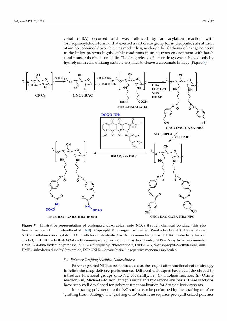
Polymers 2021, 13, 2052 23 of 47
cohol (HBA) occurred and was followed by an acylation reaction with4-nitrophenylchloroformiat that exerted a carbonate group for nucleophilic substitutionof amino contained doxorubicin as model drug nucleophilic. Carbamate linkage adjacentto the linker presents highly stable conditions in an aqueous environment with harshconditions, either basic or acidic. The drug release of active drugs was achieved only byhydrolysis in cells utilizing suitable enzymes to cleave a carbamate linkage (Figure 7).
Polymers 2021, 13, x 24 of 49
Figure 7. Illustrative representation of conjugated doxorubicin onto NCCs through chemical bonding (this picture is re-drawn from Tortorella et al. [160]. Copyright © Springer Fachmedien Wiesbaden GmbH). Abbreviations: NCCs = cellulose nanocrystals, DAC = cellulose dialdehyde, GABA = c-amino butyric acid, HBA = 4-hydroxy benzyl alcohol, EDC HCl = 1-ethyl-3-(3-dimethylaminopropyl) carbodiimide hydrochloride, NHS = N-hydroxy succinimide, DMAP = 4-dimethylamino pyridine, NPC = 4-nitrophenyl chloroformate, DIPEA = N,N-diisopropyl-N-ethylamine, anh. DMF = anhydrous dimethyl-formamide, DOXONH2 = doxorubicin, * is repetitive monomer molecules.
5.4. Polymer Grafting Modified Nanocellulose Polymer-grafted NC has been introduced as the sought-after functionalization strat-
egy to refine the drug delivery performance. Different techniques have been developed to introduce functional groups onto NC covalently, i.e., (i) Thiolene reaction; (ii) Oxime re-action; (iii) Michael addition; and (iv) imine and hydrazone synthesis. These reactions have been well-developed for polymer functionalization for drug delivery systems.
Integrating polymer onto the NC surface can be performed by the ‘grafting onto’ or ‘grafting from’ strategy. The ‘grafting onto’ technique requires pre-synthesized polymer attachment that can bear the reactive end groups onto either modified or non-modified hydroxyl groups of the NC surface. The adherence of polymer onto the NC surface’s spe-cific moieties can be performed via physical or chemical attachment. The ‘grafting onto’ approach offers the possibility of characterizing polymer before grafting and modulating the resultant carriers’ characteristics.
Strong electrostatic interaction can be used to initiate the polymer grafting onto NC. There is a possibility of incorporating polydopamine (PDA) into the NCC surface to fortify the PDA material and develop NC’s colloidal stability. The presence of functional groups
Figure 7. Illustrative representation of conjugated doxorubicin onto NCCs through chemical bonding (this pic-ture is re-drawn from Tortorella et al. [160]. Copyright © Springer Fachmedien Wiesbaden GmbH). Abbreviations:NCCs = cellulose nanocrystals, DAC = cellulose dialdehyde, GABA = c-amino butyric acid, HBA = 4-hydroxy benzylalcohol, EDC HCl = 1-ethyl-3-(3-dimethylaminopropyl) carbodiimide hydrochloride, NHS = N-hydroxy succinimide,DMAP = 4-dimethylamino pyridine, NPC = 4-nitrophenyl chloroformate, DIPEA = N,N-diisopropyl-N-ethylamine, anh.DMF = anhydrous dimethylformamide, DOXONH2 = doxorubicin, * is repetitive monomer molecules.
5.4. Polymer Grafting Modified Nanocellulose
Polymer-grafted NC has been introduced as the sought-after functionalization strategyto refine the drug delivery performance. Different techniques have been developed tointroduce functional groups onto NC covalently, i.e., (i) Thiolene reaction; (ii) Oximereaction; (iii) Michael addition; and (iv) imine and hydrazone synthesis. These reactionshave been well-developed for polymer functionalization for drug delivery systems.
Integrating polymer onto the NC surface can be performed by the ‘grafting onto’ or‘grafting from’ strategy. The ‘grafting onto’ technique requires pre-synthesized polymer
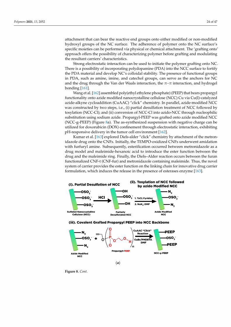
Polymers 2021, 13, 2052 24 of 47
attachment that can bear the reactive end groups onto either modified or non-modifiedhydroxyl groups of the NC surface. The adherence of polymer onto the NC surface’sspecific moieties can be performed via physical or chemical attachment. The ‘grafting onto’approach offers the possibility of characterizing polymer before grafting and modulatingthe resultant carriers’ characteristics.
Strong electrostatic interaction can be used to initiate the polymer grafting onto NC.There is a possibility of incorporating polydopamine (PDA) into the NCC surface to fortifythe PDA material and develop NC’s colloidal stability. The presence of functional groupsin PDA, such as amine, imine, and catechol groups, can serve as the anchors for NCand the drug through the Van der Waals interaction, the π–π interaction, and hydrogelbonding [161].
Wang et al. [162] assembled poly(ethyl ethylene phosphate) (PEEP) that bears propargylfunctionality onto azide modified nanocrystalline cellulose (NCC) Cu via Cu(I)-catalyzedazide-alkyne cycloaddition (CuAAC) “click” chemistry. In parallel, azide-modified NCCwas constructed by two steps, i.e., (i) partial desulfation treatment of NCC followed bytosylation (NCC-Cl); and (ii) conversion of NCC-Cl into azido-NCC through nucleophilicsubstitution using sodium azide. Propargyl-PEEP was grafted onto azide modified NCC(NCC-g-PEEP) (Figure 8a). The as-synthesized suspension with negative charge can beutilized for doxorubicin (DOX) confinement through electrostatic interaction, exhibitingpH-responsive delivery in the tumor cell environment [162].
Kumar et al. [163] explored Diels-alder “click” chemistry by attachment of the metron-idazole drug onto the CNFs. Initially, the TEMPO-oxidized CNFs underwent amidationwith furfuryl amine. Subsequently, esterification occurred between metronidazole as adrug model and maleimide-hexanoic acid to introduce the ester function between thedrug and the maleimide ring. Finally, the Diels–Alder reaction occurs between the furanfunctionalized CNF-t (CNF-fur) and metronidazole containing maleimide. Thus, the novelsystem of carrier provides the ester function on the linking chain for innovative drug carrierformulation, which induces the release in the presence of esterases enzyme [163].
Polymers 2021, 13, x 25 of 49
in PDA, such as amine, imine, and catechol groups, can serve as the anchors for NC and the drug through the Van der Waals interaction, the π–π interaction, and hydrogel bond-ing [161].
Wang et al. [162] assembled poly(ethyl ethylene phosphate) (PEEP) that bears pro-pargyl functionality onto azide modified nanocrystalline cellulose (NCC) Cu via Cu(I)-catalyzed azide-alkyne cycloaddition (CuAAC) “click” chemistry. In parallel, azide-mod-ified NCC was constructed by two steps, i.e., (i) partial desulfation treatment of NCC fol-lowed by tosylation (NCC-Cl); and (ii) conversion of NCC-Cl into azido-NCC through nucleophilic substitution using sodium azide. Propargyl-PEEP was grafted onto azide modified NCC (NCC-g-PEEP) (Figure 8a). The as-synthesized suspension with negative charge can be utilized for doxorubicin (DOX) confinement through electrostatic interac-tion, exhibiting pH-responsive delivery in the tumor cell environment [162].
Kumar et al. [163] explored Diels-alder “click” chemistry by attachment of the met-ronidazole drug onto the CNFs. Initially, the TEMPO-oxidized CNFs underwent ami-dation with furfuryl amine. Subsequently, esterification occurred between metronidazole as a drug model and maleimide-hexanoic acid to introduce the ester function between the drug and the maleimide ring. Finally, the Diels–Alder reaction occurs between the furan functionalized CNF-t (CNF-fur) and metronidazole containing maleimide. Thus, the novel system of carrier provides the ester function on the linking chain for innovative drug carrier formulation, which induces the release in the presence of esterases enzyme [163].
(a)
Figure 8. Cont.
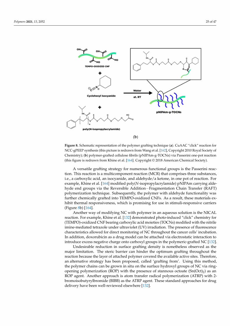
Polymers 2021, 13, 2052 25 of 47
Polymers 2021, 13, x 26 of 49
(b)
Figure 8. Schematic representation of the polymer grafting technique (a). CuAAC “click” reaction for NCC-gPEEP synthesis (this picture is redrawn from Wang et al. [162], Copyright 2010 Royal Society of Chemistry); (b) polymer-grafted cellulose fibrils (pNIPAm-g-TOCNs) via Passerini one-pot reaction (this figure is redrawn from Khine et al. [164]. Copyright © 2018 American Chemical Society).
A versatile grafting strategy for numerous functional groups is the Passerini reaction. This reaction is a multicomponent reaction (MCR) that comprises three substances, i.e., a carboxylic acid, an isocyanide, and aldehyde/a ketone, in one pot of reaction. For example, Khine et al. [164] modified poly(N-isopropylacrylamide) pNIPAm carrying aldehyde end groups via the Reversible Addition−Fragmentation Chain Transfer (RAFT) polymeriza-tion technique. Subsequently, the polymer with aldehyde functionality was further chem-ically grafted into TEMPO-oxidized CNFs. As a result, these materials exhibit thermal re-sponsiveness, which is promising for use in stimuli-responsive carriers (Figure 8b) [164].
Another way of modifying NC with polymer in an aqueous solution is the NICAL reaction. For example, Khine et al. [132] demonstrated photo-induced “click” chemistry for (TEMPO)-oxidized CNF bearing carboxylic acid moieties (TOCNs) modified with the nitrile imine-mediated tetrazole under ultraviolet (UV) irradiation. The presence of fluo-rescence characteristics allowed for direct monitoring of NC throughout the cancer cells’ incubation. In addition, doxorubicin as a drug model can be attached via electrostatic in-teraction to introduce excess negative charge onto carboxyl groups in the polymeric-grafted NC [132].
Undesirable reduction in surface grafting density is nonetheless observed as the ma-jor limitation. The steric barrier can hinder the optimum grafting throughout the reaction because the layer of attached polymer covered the available active sites. Therefore, an al-ternative strategy has been proposed, called ‘grafting from’. Using this method, the poly-mer chains can be grown in situ on the surface hydroxyl groups of NC via ring-opening polymerization (ROP) with the presence of stannous octoate (Sn(Oct)2) as an ROP agent. Another approach is atom transfer radical polymerization (ATRP) with 2-bromoisobutyr-ylbromide (BIBB) as the ATRP agent. These standard approaches for drug delivery have been well-reviewed elsewhere [132].
5.5. Surfactant Modified Nanocellulose
Figure 8. Schematic representation of the polymer grafting technique (a). CuAAC “click” reaction forNCC-gPEEP synthesis (this picture is redrawn from Wang et al. [162], Copyright 2010 Royal Society ofChemistry); (b) polymer-grafted cellulose fibrils (pNIPAm-g-TOCNs) via Passerini one-pot reaction(this figure is redrawn from Khine et al. [164]. Copyright © 2018 American Chemical Society).
A versatile grafting strategy for numerous functional groups is the Passerini reac-tion. This reaction is a multicomponent reaction (MCR) that comprises three substances,i.e., a carboxylic acid, an isocyanide, and aldehyde/a ketone, in one pot of reaction. Forexample, Khine et al. [164] modified poly(N-isopropylacrylamide) pNIPAm carrying alde-hyde end groups via the Reversible Addition−Fragmentation Chain Transfer (RAFT)polymerization technique. Subsequently, the polymer with aldehyde functionality wasfurther chemically grafted into TEMPO-oxidized CNFs. As a result, these materials ex-hibit thermal responsiveness, which is promising for use in stimuli-responsive carriers(Figure 8b) [164].
Another way of modifying NC with polymer in an aqueous solution is the NICALreaction. For example, Khine et al. [132] demonstrated photo-induced “click” chemistry for(TEMPO)-oxidized CNF bearing carboxylic acid moieties (TOCNs) modified with the nitrileimine-mediated tetrazole under ultraviolet (UV) irradiation. The presence of fluorescencecharacteristics allowed for direct monitoring of NC throughout the cancer cells’ incubation.In addition, doxorubicin as a drug model can be attached via electrostatic interaction tointroduce excess negative charge onto carboxyl groups in the polymeric-grafted NC [132].
Undesirable reduction in surface grafting density is nonetheless observed as themajor limitation. The steric barrier can hinder the optimum grafting throughout thereaction because the layer of attached polymer covered the available active sites. Therefore,an alternative strategy has been proposed, called ‘grafting from’. Using this method,the polymer chains can be grown in situ on the surface hydroxyl groups of NC via ring-opening polymerization (ROP) with the presence of stannous octoate (Sn(Oct)2) as anROP agent. Another approach is atom transfer radical polymerization (ATRP) with 2-bromoisobutyrylbromide (BIBB) as the ATRP agent. These standard approaches for drugdelivery have been well-reviewed elsewhere [132].
Polymers 2021, 13, 2052 26 of 47
5.5. Surfactant Modified Nanocellulose
The adsorption of surfactants represents a promising alternative for the chemicalmodification of NC. Surfactants are classified into cationic, anionic, zwitterionic, and non-ionic. The distinct properties of the surfactant manifest through its micelle formulationin the aqueous solution, which is highly beneficial in the drug delivery system. Thelack of a strong covalent bond is considered the significant drawback towards enablingmolecule release. Therefore, it is necessary to study several factors affecting the interactionof surfactant and NC and their impact on drug uptake and release. Tardy et al. [165]reviewed several factors that influence the affinity of NC and the surfactant. This studyprovides some additional information on the affinity of NC and the surfactant on the drugdelivery system.
The opposite charge between the NCC surface and CTAB drove the electrostaticinteraction and physical adsorption for the NCC surface modified with the surfactant.NCC’s negative charge creates a non-covalent interaction towards the cationic charge ofCTAB, resulting in a strong electrostatic interaction. Zainuddin et al. [166] pointed outseveral factors that mainly involve the interaction between NCC and the surfactant, i.e., pHand ionic strength, the CTAB concentration, and the ratio of CTAB to NCC. They high-lighted that the CTAB concentration and mass ratio of NCC: CTAB affects the interactionof surfactant-modified NCC with curcumin as a hydrophobic drug model. Increasing theCTAB concentration intensifies the hydrophobic character of the carrier, which is intenselycoordinated with curcumin. However, at a high concentration of CTAB, the amount ofcurcumin attached tends to decrease [166].
Low surfactant concentration favors the electrostatic interaction between the monomerCTAB head with the negative charge of NCC surface, giving hydrophobic properties. Whilethe CTAB concentration increases progressively, the adsorbed monomer of the surfactanttends to restructure and initiate surfactant cluster formation induced by hydrophobiccoordination between surfactant alkyl chains. The CTAB cluster molecules can be absorbedthrough the NCC surface by hydrophobic interaction. However, the hydrophobic coordina-tion of the surfactant and NCC manifested as a weak electrostatic interaction, which easilyreleases CTAB from NCC surfaces through the washing. Moreover, an excessive amountof CTAB concentration over the boundary of the surfactant critical micelles concentration(0.93 mM CTAB) might provoke the surfactant micelles formation on the NCC surface,which degrades the hydrophobic characters. Only ionic interaction between the cationichead of CTAB and anionic sulfate ester groups remains unaffected, which acts as availableactive sites for hydrophobic drug loading (Figure 9).
Raghav and Sharma [167] reported the coordination of the hydrophobic tail of CTAB inphosphate NCC. They also observed that the surfactant types (CTAB and TBAB) influencethe capability of modified NCC to bind and release the drug. By observing the structureconfiguration, stearic near the central nitrogen in TBAB-NCC causes the insufficiency ofdrug binding, which exacerbates the coordination and controlled release of the carrier [168].
Putro et al. [25] modified the NCC with various types of surfactants such as cationic(CTAB), anionic (sodium dodecyl sulfate), and non-ionic surfactant (Tween 20). Differ-ent types of surfactants exhibit distinct interactions towards NCC, which influenced theelectronegativity of modified NCC itself and the drug adsorption–desorption behavior.The presence of salt in the system had a significant influence on the uptake of paclitaxel.Different behavior of surfactants due to the salt effect significantly influences the interactionof NCC and drugs. They concluded that (1) electrostatic and Van der Waals interactions arethe primary mechanism of paclitaxel adsorption towards surfactant-modified NCC, whichcan be enhanced through salt addition; and (2) pH played a significant role in the drugadsorption and release of paclitaxel by altering the surface charge of surfactant-modifiedNCC and the electrostatic interaction of hydroxyl ions and paclitaxel in solution.
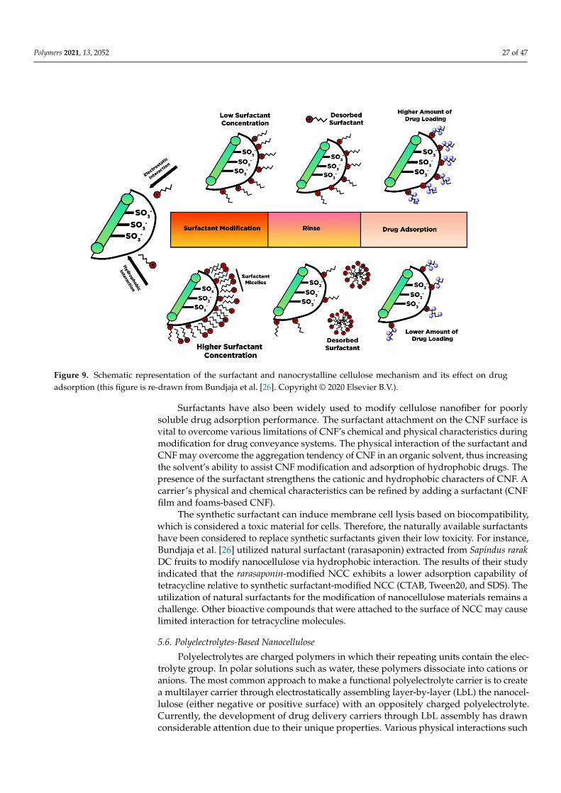
Polymers 2021, 13, 2052 27 of 47
Polymers 2021, 13, x 28 of 49
Figure 9. Schematic representation of the surfactant and nanocrystalline cellulose mechanism and its effect on drug ad-sorption (this figure is re-drawn from Bundjaja et al. [26]. Copyright © 2020 Elsevier B.V.).
Raghav and Sharma [167] reported the coordination of the hydrophobic tail of CTAB in phosphate NCC. They also observed that the surfactant types (CTAB and TBAB) influ-ence the capability of modified NCC to bind and release the drug. By observing the struc-ture configuration, stearic near the central nitrogen in TBAB-NCC causes the insufficiency of drug binding, which exacerbates the coordination and controlled release of the carrier [168].
Putro et al. [25] modified the NCC with various types of surfactants such as cationic (CTAB), anionic (sodium dodecyl sulfate), and non-ionic surfactant (Tween 20). Different types of surfactants exhibit distinct interactions towards NCC, which influenced the elec-tronegativity of modified NCC itself and the drug adsorption–desorption behavior. The presence of salt in the system had a significant influence on the uptake of paclitaxel. Dif-ferent behavior of surfactants due to the salt effect significantly influences the interaction of NCC and drugs. They concluded that (1) electrostatic and Van der Waals interactions are the primary mechanism of paclitaxel adsorption towards surfactant-modified NCC, which can be enhanced through salt addition; and (2) pH played a significant role in the drug adsorption and release of paclitaxel by altering the surface charge of surfactant-mod-ified NCC and the electrostatic interaction of hydroxyl ions and paclitaxel in solution.
Surfactants have also been widely used to modify cellulose nanofiber for poorly sol-uble drug adsorption performance. The surfactant attachment on the CNF surface is vital to overcome various limitations of CNF’s chemical and physical characteristics during modification for drug conveyance systems. The physical interaction of the surfactant and CNF may overcome the aggregation tendency of CNF in an organic solvent, thus increas-ing the solvent’s ability to assist CNF modification and adsorption of hydrophobic drugs. The presence of the surfactant strengthens the cationic and hydrophobic characters of CNF. A carrier’s physical and chemical characteristics can be refined by adding a surfac-tant (CNF film and foams-based CNF).
The synthetic surfactant can induce membrane cell lysis based on biocompatibility, which is considered a toxic material for cells. Therefore, the naturally available surfactants
Figure 9. Schematic representation of the surfactant and nanocrystalline cellulose mechanism and its effect on drugadsorption (this figure is re-drawn from Bundjaja et al. [26]. Copyright © 2020 Elsevier B.V.).
Surfactants have also been widely used to modify cellulose nanofiber for poorlysoluble drug adsorption performance. The surfactant attachment on the CNF surface isvital to overcome various limitations of CNF’s chemical and physical characteristics duringmodification for drug conveyance systems. The physical interaction of the surfactant andCNF may overcome the aggregation tendency of CNF in an organic solvent, thus increasingthe solvent’s ability to assist CNF modification and adsorption of hydrophobic drugs. Thepresence of the surfactant strengthens the cationic and hydrophobic characters of CNF. Acarrier’s physical and chemical characteristics can be refined by adding a surfactant (CNFfilm and foams-based CNF).
The synthetic surfactant can induce membrane cell lysis based on biocompatibility,which is considered a toxic material for cells. Therefore, the naturally available surfactantshave been considered to replace synthetic surfactants given their low toxicity. For instance,Bundjaja et al. [26] utilized natural surfactant (rarasaponin) extracted from Sapindus rarakDC fruits to modify nanocellulose via hydrophobic interaction. The results of their studyindicated that the rarasaponin-modified NCC exhibits a lower adsorption capability oftetracycline relative to synthetic surfactant-modified NCC (CTAB, Tween20, and SDS). Theutilization of natural surfactants for the modification of nanocellulose materials remains achallenge. Other bioactive compounds that were attached to the surface of NCC may causelimited interaction for tetracycline molecules.
5.6. Polyelectrolytes-Based Nanocellulose
Polyelectrolytes are charged polymers in which their repeating units contain the elec-trolyte group. In polar solutions such as water, these polymers dissociate into cations oranions. The most common approach to make a functional polyelectrolyte carrier is to createa multilayer carrier through electrostatically assembling layer-by-layer (LbL) the nanocel-lulose (either negative or positive surface) with an oppositely charged polyelectrolyte.Currently, the development of drug delivery carriers through LbL assembly has drawnconsiderable attention due to their unique properties. Various physical interactions such
Polymers 2021, 13, 2052 28 of 47
as hydrogen bonding, hydrophobic interaction, and Van der Waals interaction are presentin functional polyelectrolyte carriers. Those interactions act as the driving force in drugbinding and maintain the stability of the multilayer [169]. LbL hybridization assembly ofnanocellulose with other organic and inorganic materials usually instigates an outstandingperformance improvement for the entire LbL system to stimuli-responsive and localizeddrug delivery. Early development of the LbL approaches was demonstrated on the flatsubstrates and is currently extended to spherical particles.
Coating LbL film on a spherical sacrificial template becomes another layer-by-layerassembly approach for hollow polyelectrolytes capsule formation to encapsulate and re-lease the drug. Melamine formaldehyde (MF) is a popular template for microcapsulespreparation via LbL assembly due to narrow-sized distribution and optimized disso-lution conditions [170]. The physicochemical characteristics of templates such as size,shape, porosity, colloidal stability, and template solubility modulate the characteristic ofas-synthesized hollow capsules. For instance, the capsule size can be adjusted dependingon the size of the template, which is common in the range of 150 nm to a few microme-ters [171]. Nanocellulose has been used to construct the interior of multilayer thin film andhollow microcapsules for various types of therapeutic molecules loading such as DNA,RNA, protein, and drugs.
Several aspects should be considered to assemble suitable polyelectrolyte complexesthrough the LbL system, i.e., charge stoichiometry, charge density, molecular weight, poly-electrolytes concentration, pH, ionic strength, order of addition, mixing ratio, and modeof mixing the polyelectrolyte solution. These factors greatly influence the drug carrierthickness, the surface charge, and the morphological structure, such as the size, shape,and porous structure of the drug delivery system. Reviews on some crucial aspects thatinfluence the stability of polyelectrolyte complexes for drug delivery systems are availableelsewhere [172].
Mohanta et al. [173] produced an NCC multilayer thin film with counterionic poly-electrolytes (chitosan) on a quartz crystal microbalance (QCM) plate through LbL growthassembly. They also developed hollow microcapsules using MF as a template. By varyingthe concentration of the polyelectrolyte (either NCC and chitosan) and the number ofdepositions, a homogeneous multilayer thickness with a porous structure can be obtained.The thin film and microcapsule were utilized as carriers for hydrophilic drugs (doxorubicin)and hydrophobic drugs (curcumin). The protonation of amine groups in acidic conditionsbecomes the driving force for doxorubicin release, while the concentration difference be-tween the medium and carrier is considered the primary factor affecting curcumin release.The stimulus-responsive pH in LbL system-based nanocellulose may apply to local drugtransport and tumor therapy [173].
Other types of layer-by-layer assembly approaches were also used to constructPEC-based nanocellulose by incorporating various types of polyelectrolytes. For in-stance, Li et al. [174] proposed the buildup technique of LbL for opposite-charge buildingblocks (e.g., cellulose nanocrystal (NCC), polyethyleneimine (PEI), cis-aconityl-doxorubicin(CAD), and building blocks of folate (FA)). The highly negative charge of NCC serves as ananchor to carry the positive-charge PEI through electrostatic interaction as an intermediarylayer. The coordination of NCC-PEI resulted in positive-charge material for electrostaticadsorption of the negative charge of FA and CAD to construct the outermost layer, whichtook place sequentially (denoted as FA/CAD@PEI@NCC). The presence of FA on thesurface carrier increased the active targeting ability towards folate receptors in the tumorcell. Cis-aconityl amide linkage in doxorubicin (CAD) can specifically release DOX atthe lysosomal pH due to the pH labile characteristic and hydrolysis cis-aconityl amidelinkage by β-carboxylic acid under low pH. The integration of each layer can increasethe uptake to 20 times larger than its counterpart due to the strong electrostatic charge.Besides the surface chemistry carrier, the carrier’s morphological structure also helps thecarrier delivery reach tumor cells [174]. Another potential form of polyelectrolytes, in-
Polymers 2021, 13, 2052 29 of 47
cluding hydrogel, aerogel, lightweight porous materials, and integrated inorganic–organiccomposites, are thoroughly discussed in the following sections.
6. Hydrogel Based Nanocellulose for Drug Delivery
Hydrogels are three-dimensional (3D) cross-linked polymeric networks that carryabsorbed water and store a large quantity of water in the swelling state. The hydrogelscan be cross-linked through physical (non-covalent interaction), chemical (covalent co-ordination), or an integration of both physical and chemical cross-links [175]. Given itsbiocompatibility and stimulus-responsive swelling behavior, the hydrogel has gained atten-tion for drug delivery application. As a drug carrier the physically cross-linked hydrogel ispreferable to the chemically cross-linked hydrogel. The covalently cross-linked hydrogelgenerates a permanent structure that limits the swelling ability, and therefore, most chemi-cally cross-linked hydrogels are used as implantables. Furthermore, the incorporation ofthe drug via adsorption towards chemically cross-linked hydrogel restrains the loadingefficacy. Although the cross-linked reaction may perform drug conjugation on the hydrogel,it sacrifices the chemical integrity of the drugs. Therefore, it is more desirable to constructa hydrogel delivery system where simultaneous gel formation and drug adsorption canoccur in an aqueous environment without covalent cross-linking.
Due to the presence of sol-gel transition characteristics (such as swelling behavior, me-chanical strength, and network structure), which are affected by the external stimulus suchas pH, thermal, light wavelength, ultrasonic waves, pressure, magnetic field, and electricalfield; the smart hydrogel-based nanocellulose has been well-developed for various drugdelivery formulation. Diverse types of polyelectrolytes can modify the substantial chargeof nanocellulose (either positive and negative) to form a variety of intelligent hydrogelssuch as injectable hydrogel [161], stimuli-responsive hydrogel [176], double-membranehydrogel [177], supramolecular hydrogel [178], microsphere hydrogels, bacteria cellulosehydrogel [179], shape memory-based bacteria cellulose [180], and aerogel/cryogel [174].All those hydrogels have desirable physical and chemical characteristics to be adaptedto various drug delivery systems. Liu et al. [161] reviewed the current developmentof nanocellulose-based hydrogel and its modification for drug delivery systems. How-ever, double-membrane hydrogel and supramolecular hydrogel are excluded from theirreview [161].
Different types of hydrogels have diverse morphological structures, network coor-dination, and functional groups, affecting the drug’s diffusional path during adsorptionand release. Double-membrane hydrogel was developed by Lin et al. [177], consisting ofan external membrane composed of alginate and consolidation of cationic NCC (CNCC).Two different drugs were introduced on different layers of the membrane with contrastingtypes of release behavior. The outer hydrogel releases the drug rapidly, while sustaineddrug release occurs in the inner membrane hydrogel. This phenomenon occurred dueto the ‘nano-obstruction effect’ and ‘nano-locking effect’ induced by CNCC componentsin the hydrogels. The ‘nano-obstruction effect’ offers sustained drug release throughoutfragmentary disintegration, and the ‘Nano-locking effect’ is responsible for restricting theburst of drug release through progressive hydrogel disintegration (Figure 10). The differentcompositions and properties of external and internal hydrogels affect the drug’s behaviorand diffusional path [177].

Polymers 2021, 13, 2052 30 of 47
Polymers 2021, 13, x 31 of 49
sustained drug release occurs in the inner membrane hydrogel. This phenomenon oc-curred due to the ‘nano-obstruction effect’ and ‘nano-locking effect’ induced by CNCC components in the hydrogels. The ‘nano-obstruction effect’ offers sustained drug release throughout fragmentary disintegration, and the ‘Nano-locking effect’ is responsible for restricting the burst of drug release through progressive hydrogel disintegration (Figure 10). The different compositions and properties of external and internal hydrogels affect the drug’s behavior and diffusional path [177].
(A)
(B)
Figure 10. (A) The route fabrication of single membrane and double-membrane microsphere hydro-gel with its optical microscope of single membrane SA/CNCC microsphere hydrogel and SA/CNCC double-membrane microsphere hydrogel (this figure is reprinted with permission from ref. [177]. Copyright © 2016 American Chemical Society); (B) schematic illustration of dual drug release mech-anism from a double-layer membrane hydrogel constructed from Cationic NCC and Alginate (this figure is redrawn from Lin et al. [177]. Copyright © 2016 American Chemical Society).
Supramolecular hydrogels have been characterized as the 3-D solid network hydro-gel organized by non-covalent interactions such as hydrogen linkage, hydrophobic coor-dination, and cation-π and π-π interactions. In contrast to the chemically cross-linked hy-drogels, gel morphology is equilibrated through covalent coordination; the supramolecu-lar hydrogel morphology is stabilized by a non-covalent interaction. Supramolecular hy-drogel has been synthesized through extensive, diverse supramolecular configurations, including host–guest complexation, biomimetic interaction, hydrogen bonding, stereo-
Figure 10. (A) The route fabrication of single membrane and double-membrane microsphere hydrogel with its opticalmicroscope of single membrane SA/CNCC microsphere hydrogel and SA/CNCC double-membrane microsphere hydrogel(this figure is reprinted with permission from ref. [177]. Copyright © 2016 American Chemical Society); (B) schematicillustration of dual drug release mechanism from a double-layer membrane hydrogel constructed from Cationic NCC andAlginate (this figure is redrawn from Lin et al. [177]. Copyright © 2016 American Chemical Society).
Supramolecular hydrogels have been characterized as the 3-D solid network hydrogelorganized by non-covalent interactions such as hydrogen linkage, hydrophobic coordina-tion, and cation-π and π-π interactions. In contrast to the chemically cross-linked hydrogels,gel morphology is equilibrated through covalent coordination; the supramolecular hy-drogel morphology is stabilized by a non-covalent interaction. Supramolecular hydrogelhas been synthesized through extensive, diverse supramolecular configurations, includ-ing host–guest complexation, biomimetic interaction, hydrogen bonding, stereo-complexformation, and ionic and metal-ligand. Hydrogel-based supramolecular self-assemblythrough host–guest complexation is the most widely explored method for supramolecularhydrogel formation. Specifically, supramolecular hydrogels constructed by host–guestinclusion between polymer and cyclodextrin demonstrated the thixotropic reversibility,which is advantageous for syringe drug delivery.
Lin and Dufresne et al. [178] produced supramolecular hydrogel DDS by self-assemblyof a covalently grafted α-cyclodextrin (α-CD) NCC surface with epichlorohydrin as a cou-pling agent through a one-step process. Furthermore, pluronic composed of triblock copoly-mers with different molecular weights (Pluronic F68 or F108), both bearing hydrophobic
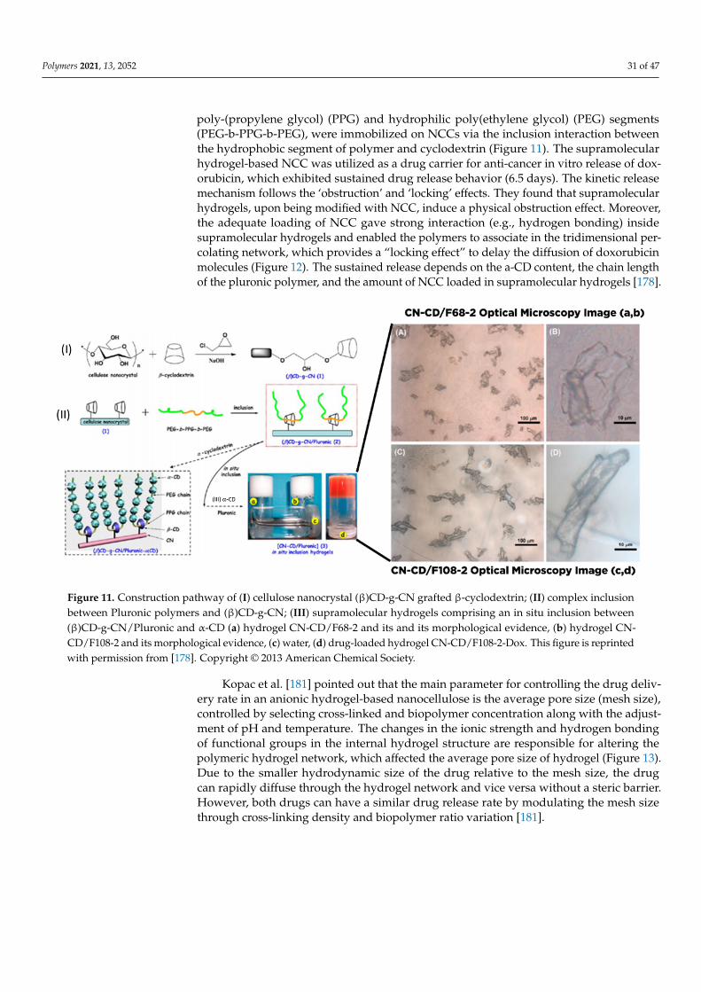
Polymers 2021, 13, 2052 31 of 47
poly-(propylene glycol) (PPG) and hydrophilic poly(ethylene glycol) (PEG) segments(PEG-b-PPG-b-PEG), were immobilized on NCCs via the inclusion interaction betweenthe hydrophobic segment of polymer and cyclodextrin (Figure 11). The supramolecularhydrogel-based NCC was utilized as a drug carrier for anti-cancer in vitro release of dox-orubicin, which exhibited sustained drug release behavior (6.5 days). The kinetic releasemechanism follows the ‘obstruction’ and ‘locking’ effects. They found that supramolecularhydrogels, upon being modified with NCC, induce a physical obstruction effect. Moreover,the adequate loading of NCC gave strong interaction (e.g., hydrogen bonding) insidesupramolecular hydrogels and enabled the polymers to associate in the tridimensional per-colating network, which provides a “locking effect” to delay the diffusion of doxorubicinmolecules (Figure 12). The sustained release depends on the a-CD content, the chain lengthof the pluronic polymer, and the amount of NCC loaded in supramolecular hydrogels [178].
Polymers 2021, 13, x 32 of 49
complex formation, and ionic and metal-ligand. Hydrogel-based supramolecular self-as-sembly through host–guest complexation is the most widely explored method for supra-molecular hydrogel formation. Specifically, supramolecular hydrogels constructed by host–guest inclusion between polymer and cyclodextrin demonstrated the thixotropic re-versibility, which is advantageous for syringe drug delivery.
Lin and Dufresne et al. [178] produced supramolecular hydrogel DDS by self-assem-bly of a covalently grafted α-cyclodextrin (α-CD) NCC surface with epichlorohydrin as a coupling agent through a one-step process. Furthermore, pluronic composed of triblock copolymers with different molecular weights (Pluronic F68 or F108), both bearing hydro-phobic poly-(propylene glycol) (PPG) and hydrophilic poly(ethylene glycol) (PEG) seg-ments (PEG-b-PPG-b-PEG), were immobilized on NCCs via the inclusion interaction be-tween the hydrophobic segment of polymer and cyclodextrin (Figure 11). The supramo-lecular hydrogel-based NCC was utilized as a drug carrier for anti-cancer in vitro release of doxorubicin, which exhibited sustained drug release behavior (6.5 days). The kinetic release mechanism follows the ‘obstruction’ and ‘locking’ effects. They found that supra-molecular hydrogels, upon being modified with NCC, induce a physical obstruction ef-fect. Moreover, the adequate loading of NCC gave strong interaction (e.g., hydrogen bonding) inside supramolecular hydrogels and enabled the polymers to associate in the tridimensional percolating network, which provides a “locking effect” to delay the diffu-sion of doxorubicin molecules (Figure 12). The sustained release depends on the a-CD content, the chain length of the pluronic polymer, and the amount of NCC loaded in su-pramolecular hydrogels [178].
Figure 11. Construction pathway of (I) cellulose nanocrystal (β)CD-g-CN grafted β-cyclodextrin; (II) complex inclusion between Pluronic polymers and (β)CD-g-CN; (III) supramolecular hydrogels comprising an in situ inclusion between (β)CD-g-CN/Pluronic and α-CD (a) hydrogel CN-CD/F68-2 and its and its morphological evidence, (b) hydrogel CN-CD/F108-2 and its morphological evidence, (c) water, (d) drug-loaded hydrogel CN-CD/F108-2-Dox. This figure is re-printed with permission from [178]. Copyright © 2013 American Chemical Society.
Figure 11. Construction pathway of (I) cellulose nanocrystal (β)CD-g-CN grafted β-cyclodextrin; (II) complex inclusionbetween Pluronic polymers and (β)CD-g-CN; (III) supramolecular hydrogels comprising an in situ inclusion between(β)CD-g-CN/Pluronic and α-CD (a) hydrogel CN-CD/F68-2 and its and its morphological evidence, (b) hydrogel CN-CD/F108-2 and its morphological evidence, (c) water, (d) drug-loaded hydrogel CN-CD/F108-2-Dox. This figure is reprintedwith permission from [178]. Copyright © 2013 American Chemical Society.
Kopac et al. [181] pointed out that the main parameter for controlling the drug deliv-ery rate in an anionic hydrogel-based nanocellulose is the average pore size (mesh size),controlled by selecting cross-linked and biopolymer concentration along with the adjust-ment of pH and temperature. The changes in the ionic strength and hydrogen bondingof functional groups in the internal hydrogel structure are responsible for altering thepolymeric hydrogel network, which affected the average pore size of hydrogel (Figure 13).Due to the smaller hydrodynamic size of the drug relative to the mesh size, the drugcan rapidly diffuse through the hydrogel network and vice versa without a steric barrier.However, both drugs can have a similar drug release rate by modulating the mesh sizethrough cross-linking density and biopolymer ratio variation [181].

Polymers 2021, 13, 2052 32 of 47Polymers 2021, 13, x 33 of 49
Figure 12. Schematic illustration of possible locking effect of the drug via host–guest inclusion in supramolecular hydrogel constructed from cyclodextrin and chemically modified nanocrystalline cellulose (this picture is redrawn from [178]. Copyright © 2013 American Chemical Society.)
Kopac et al. [181] pointed out that the main parameter for controlling the drug deliv-ery rate in an anionic hydrogel-based nanocellulose is the average pore size (mesh size), controlled by selecting cross-linked and biopolymer concentration along with the adjust-ment of pH and temperature. The changes in the ionic strength and hydrogen bonding of functional groups in the internal hydrogel structure are responsible for altering the poly-meric hydrogel network, which affected the average pore size of hydrogel (Figure 13). Due to the smaller hydrodynamic size of the drug relative to the mesh size, the drug can rapidly diffuse through the hydrogel network and vice versa without a steric barrier. However, both drugs can have a similar drug release rate by modulating the mesh size through cross-linking density and biopolymer ratio variation [181].
Figure 12. Schematic illustration of possible locking effect of the drug via host–guest inclusion in supramolecular hydrogelconstructed from cyclodextrin and chemically modified nanocrystalline cellulose (this picture is redrawn from [178].Copyright © 2013 American Chemical Society.)
Polymers 2021, 13, x 34 of 49
Figure 13. Schematic illustration of the swelling mechanism of hydrogel fabricated from TEMPO-mediated CNFs and alginate towards drug release (this figure is redrawn from [181]. Copyright © 2020 Elsevier B.V.).
7. Lightweight Porous Based Nanocellulose for Drug Delivery Lightweight porous materials have been classified as a 3-D solid class of material
with several features such as high specific surface area, very low density (<50%), and di-verse pore structure with various pore sizes ranging from nanometer to micron. Sponge, foam, and aerogels are the three major categories of lightweight porous materials. The sponge is constructed by gas dispersion in the solid matrix, commonly present as an open cell structure of low density porous elastic polymer. The sponge has a macroporous struc-ture full of gaps and channels, permitting easy access to water or molecules flow [182]. Similarly, foam can be made through the steady gas dispersion into a hydrogel or solid matrix and even liquid. Foam is commonly characterized as having a bubble diameter (pore diameter) greater than 50 nm [183]. Aerogel is a three-dimensional (3D) porous ma-terial constructed by self-assembly of the colloidal component or polymeric chains, creat-ing nano-porous networks that can be filled up with a gaseous dispersion medium. Aer-ogel is prepared through the wet-gel drying process by removing the liquid component in the hydrogel, which is replaced by a gas constituent while still preserving the gel net-work [184]. The specific surface area of aerogel can reach up to 1000 m2 g−1 with a porosity range between 80 and 99.8%. On the other hand, other aerogels, namely xerogel and cry-ogel, have been prepared by evaporation and freeze-drying. Detailed preparation of light-weight porous material-based nanocellulose has been reviewed elsewhere [185].
For the drug delivery field, carrier morphology, especially the porosity structure, controls the drug adsorption and release since the drug will pass through the internal pore to be retained inside and release outside regardless of the chemistry interaction. Sun et al. [186] underlined that the critical factor in controlling and modulating the pore structure of ultralightweight porous materials is selecting a drying method [186]. Initially, freeze-drying, supercritical drying, and evaporation drying have been utilized in fabricating ul-tralightweight porous materials. Evaporation drying has emerged as a conventional tech-nique of synthesizing nanocellulose-based porous materials. However, there are several major drawbacks, such as internal network structure collapse due to the capillary forces
Figure 13. Schematic illustration of the swelling mechanism of hydrogel fabricated from TEMPO-mediated CNFs andalginate towards drug release (this figure is redrawn from [181]. Copyright © 2020 Elsevier B.V.).
Polymers 2021, 13, 2052 33 of 47
7. Lightweight Porous Based Nanocellulose for Drug Delivery
Lightweight porous materials have been classified as a 3-D solid class of material withseveral features such as high specific surface area, very low density (<50%), and diversepore structure with various pore sizes ranging from nanometer to micron. Sponge, foam,and aerogels are the three major categories of lightweight porous materials. The spongeis constructed by gas dispersion in the solid matrix, commonly present as an open cellstructure of low density porous elastic polymer. The sponge has a macroporous structurefull of gaps and channels, permitting easy access to water or molecules flow [182]. Similarly,foam can be made through the steady gas dispersion into a hydrogel or solid matrix andeven liquid. Foam is commonly characterized as having a bubble diameter (pore diameter)greater than 50 nm [183]. Aerogel is a three-dimensional (3D) porous material constructedby self-assembly of the colloidal component or polymeric chains, creating nano-porousnetworks that can be filled up with a gaseous dispersion medium. Aerogel is preparedthrough the wet-gel drying process by removing the liquid component in the hydrogel,which is replaced by a gas constituent while still preserving the gel network [184]. Thespecific surface area of aerogel can reach up to 1000 m2 g−1 with a porosity range between80 and 99.8%. On the other hand, other aerogels, namely xerogel and cryogel, have beenprepared by evaporation and freeze-drying. Detailed preparation of light-weight porousmaterial-based nanocellulose has been reviewed elsewhere [185].
For the drug delivery field, carrier morphology, especially the porosity structure,controls the drug adsorption and release since the drug will pass through the internalpore to be retained inside and release outside regardless of the chemistry interaction.Sun et al. [186] underlined that the critical factor in controlling and modulating the porestructure of ultralightweight porous materials is selecting a drying method [186]. Initially,freeze-drying, supercritical drying, and evaporation drying have been utilized in fabricat-ing ultralightweight porous materials. Evaporation drying has emerged as a conventionaltechnique of synthesizing nanocellulose-based porous materials. However, there are sev-eral major drawbacks, such as internal network structure collapse due to the capillaryforces of the solid matrix and the difficulty to prevent the shrinkage. Therefore, freeze-drying and supercritical drying have been used as drying methods to overcome thesedrawbacks. Freeze drying can retain the porous structure through the sublimation of liquidinto gas. It is also possible to cross the solid–gas interface bypassing the liquid criticalpoint through adjusting the temperature and pressure (supercritical drying). Both methodseffectively retain the pore structure and refine the porosity and specific surface area ofnanocellulose-based porous materials.
Aerogel-, xerogel-, and cryogel-based nanocelluloses are promising materials as thevehicle for a drug delivery system. Before the drying process, physical and chemical cross-linking are vital in controlling the 3D network formation and porous material performance.Physical cross-linking is commonly established by weaker interactions such as Van derWaals, hydrogen bonding, and electrostatic interaction. In contrast, covalent cross-linkingcan create a 3-D robust mechanical framework through the action of covalent coordina-tion and polymerization. Chemical cross-linking exhibits better mechanical stiffness andstructural stability compared to physical cross-linking.
Muller et al. [180] synthesized water-responsive xerogel to retain its original shape bysubmerging it in water through moisture utilization as the stimulus. The post-modificationof BNC with the different supplementary hydrophilic substances was performed to achievethe re-swelling behavior. Rapid re-swelling behavior can be acquired by supplementarymagnesium chloride, glucose, sucrose, and sorbitol with up to 88% maximum rehydration.Their findings of re-swelling modified BNC showed the possibility of developing a carrierwith controlled release properties for hydrophilic drug model azorubine in the drugdelivery system.
Li et al. [174] synthesized two types of nanocellulose/gelatin composite cryogelsthrough hydrogen bonding and chemical cross-linking with dialdehyde starch (DAS) forcontrolled drug delivery of 5-fluorouracil (5-FU). DAS subsequently reacted with both
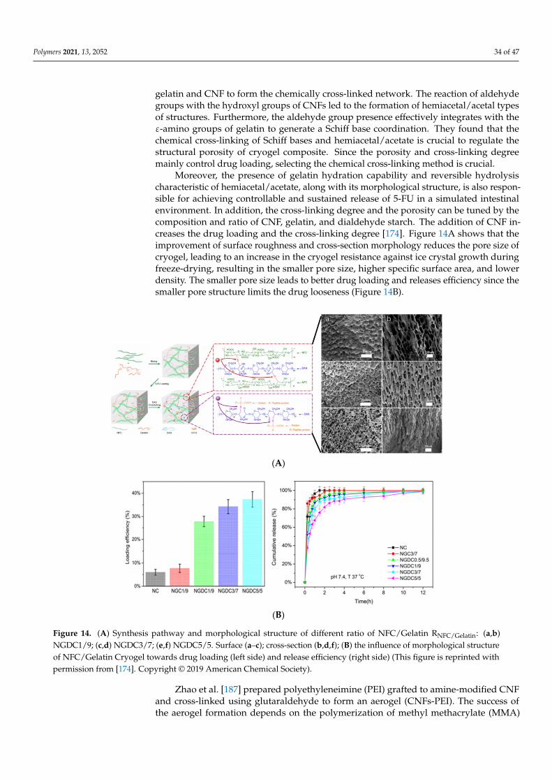
Polymers 2021, 13, 2052 34 of 47
gelatin and CNF to form the chemically cross-linked network. The reaction of aldehydegroups with the hydroxyl groups of CNFs led to the formation of hemiacetal/acetal typesof structures. Furthermore, the aldehyde group presence effectively integrates with theε-amino groups of gelatin to generate a Schiff base coordination. They found that thechemical cross-linking of Schiff bases and hemiacetal/acetate is crucial to regulate thestructural porosity of cryogel composite. Since the porosity and cross-linking degreemainly control drug loading, selecting the chemical cross-linking method is crucial.
Moreover, the presence of gelatin hydration capability and reversible hydrolysischaracteristic of hemiacetal/acetate, along with its morphological structure, is also respon-sible for achieving controllable and sustained release of 5-FU in a simulated intestinalenvironment. In addition, the cross-linking degree and the porosity can be tuned by thecomposition and ratio of CNF, gelatin, and dialdehyde starch. The addition of CNF in-creases the drug loading and the cross-linking degree [174]. Figure 14A shows that theimprovement of surface roughness and cross-section morphology reduces the pore size ofcryogel, leading to an increase in the cryogel resistance against ice crystal growth duringfreeze-drying, resulting in the smaller pore size, higher specific surface area, and lowerdensity. The smaller pore size leads to better drug loading and releases efficiency since thesmaller pore structure limits the drug looseness (Figure 14B).
Polymers 2021, 13, x 36 of 49
(A)
(B)
Figure 14. (A) Synthesis pathway and morphological structure of different ratio of NFC/Gelatin RNFC/Gelatin: (a,b) NGDC1/9; (c,d) NGDC3/7; (e,f) NGDC5/5. Surface (a–c); cross-section (b,d,f); (B) the influence of morphological structure of NFC/Gelatin Cryogel towards drug loading (left side) and release efficiency (right side) (This figure is reprinted with permission from [174]. Copyright © 2019 American Chemical Society).
Zhao et al. [187] prepared polyethyleneimine (PEI) grafted to amine-modified CNF and cross-linked using glutaraldehyde to form an aerogel (CNFs-PEI). The success of the aerogel formation depends on the polymerization of methyl methacrylate (MMA) on the surface of CNFs, which induced the formation of the network between PEI and CNF. Pol-yethyleneimine (PEI) carries some primary and secondary amine functional groups, which increase the loading of sodium salicylate (NaSA) to 20 times higher than its coun-terpart (CNFs-based aerogel). The sustained and controlled release was achieved by the CNFs-PEI aerogel, which is highly responsive to pH because of the protonation and deprotonation of amine groups in PEI [187].
Chemically cross-linked PEI with TEMPO-mediated BC CNF (abbreviated as PEI-BC) for aspirin, gentamicin, and bovine serum albumin (BSA) carrier has been studied by Chen et al. [156]. The PEI cross-linking induced the morphological changes of BC by in-creasing the density of interconnected structures and thickening the pore walls, which provide the CNF interpenetrated network with improved mechanical strength [156]. Liang et al. [188] proposed a well-balanced dual responsive polymer (temperature and pH) by modifying branched PEI with N-isopropyl acrylamide (NIPAM), which was fur-ther grafted onto CNF through the condensation reaction (abbreviated as CNF-PEI-NIPAM). Remarkably, the pH and temperature of the carrier can alter the hydrophobic and hydrophilic characteristics of CNF-PEI-NIPAM [188].
CNF has been combined with the non-edible surfactant to make air bubble confine-ment by the Pickering technique, generating stable air bubbles encapsulated in wet-stable foams. Using the unique drying technique, the dry-foams with closed holes (cellular solid
Figure 14. (A) Synthesis pathway and morphological structure of different ratio of NFC/Gelatin RNFC/Gelatin: (a,b)NGDC1/9; (c,d) NGDC3/7; (e,f) NGDC5/5. Surface (a–c); cross-section (b,d,f); (B) the influence of morphological structureof NFC/Gelatin Cryogel towards drug loading (left side) and release efficiency (right side) (This figure is reprinted withpermission from [174]. Copyright © 2019 American Chemical Society).
Zhao et al. [187] prepared polyethyleneimine (PEI) grafted to amine-modified CNFand cross-linked using glutaraldehyde to form an aerogel (CNFs-PEI). The success ofthe aerogel formation depends on the polymerization of methyl methacrylate (MMA)
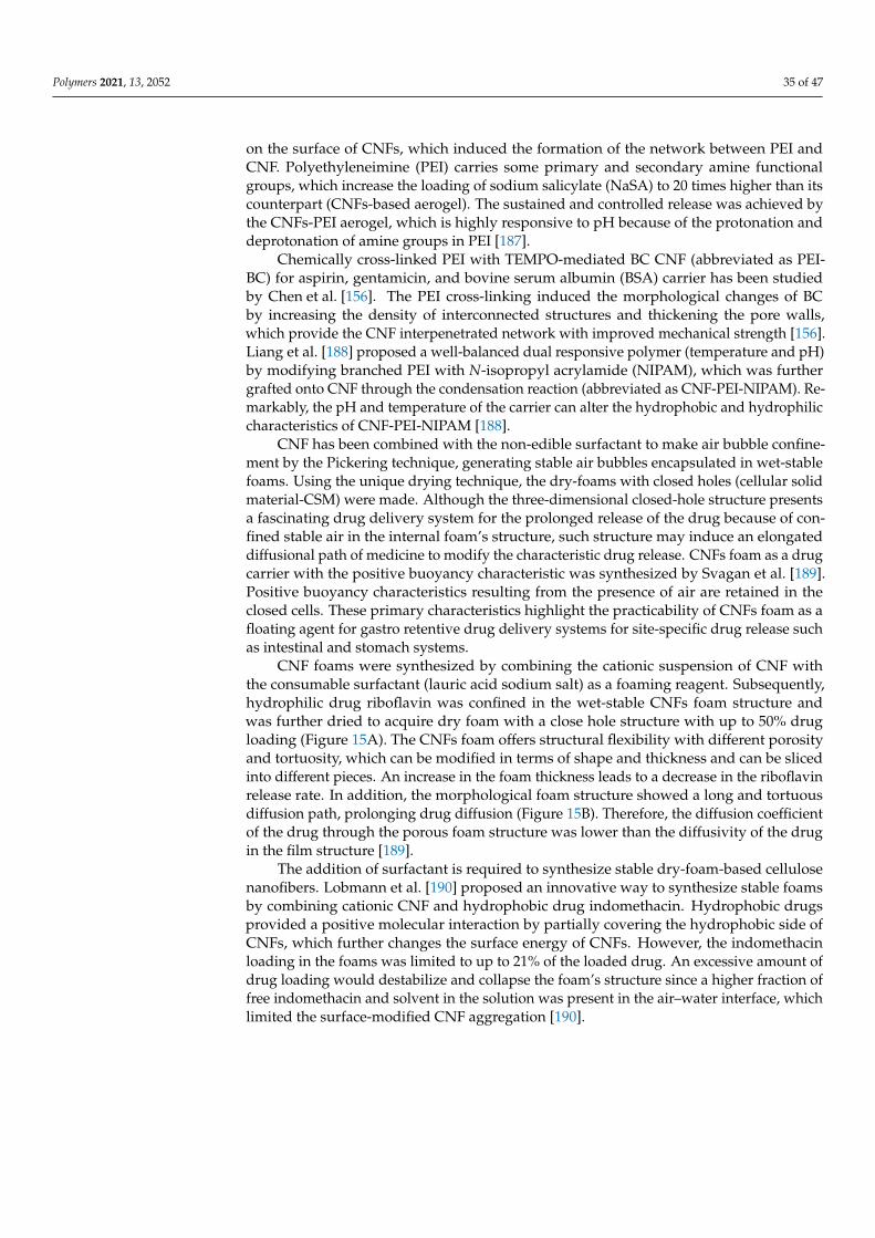
Polymers 2021, 13, 2052 35 of 47
on the surface of CNFs, which induced the formation of the network between PEI andCNF. Polyethyleneimine (PEI) carries some primary and secondary amine functionalgroups, which increase the loading of sodium salicylate (NaSA) to 20 times higher than itscounterpart (CNFs-based aerogel). The sustained and controlled release was achieved bythe CNFs-PEI aerogel, which is highly responsive to pH because of the protonation anddeprotonation of amine groups in PEI [187].
Chemically cross-linked PEI with TEMPO-mediated BC CNF (abbreviated as PEI-BC) for aspirin, gentamicin, and bovine serum albumin (BSA) carrier has been studiedby Chen et al. [156]. The PEI cross-linking induced the morphological changes of BCby increasing the density of interconnected structures and thickening the pore walls,which provide the CNF interpenetrated network with improved mechanical strength [156].Liang et al. [188] proposed a well-balanced dual responsive polymer (temperature and pH)by modifying branched PEI with N-isopropyl acrylamide (NIPAM), which was furthergrafted onto CNF through the condensation reaction (abbreviated as CNF-PEI-NIPAM). Re-markably, the pH and temperature of the carrier can alter the hydrophobic and hydrophiliccharacteristics of CNF-PEI-NIPAM [188].
CNF has been combined with the non-edible surfactant to make air bubble confine-ment by the Pickering technique, generating stable air bubbles encapsulated in wet-stablefoams. Using the unique drying technique, the dry-foams with closed holes (cellular solidmaterial-CSM) were made. Although the three-dimensional closed-hole structure presentsa fascinating drug delivery system for the prolonged release of the drug because of con-fined stable air in the internal foam’s structure, such structure may induce an elongateddiffusional path of medicine to modify the characteristic drug release. CNFs foam as a drugcarrier with the positive buoyancy characteristic was synthesized by Svagan et al. [189].Positive buoyancy characteristics resulting from the presence of air are retained in theclosed cells. These primary characteristics highlight the practicability of CNFs foam as afloating agent for gastro retentive drug delivery systems for site-specific drug release suchas intestinal and stomach systems.
CNF foams were synthesized by combining the cationic suspension of CNF withthe consumable surfactant (lauric acid sodium salt) as a foaming reagent. Subsequently,hydrophilic drug riboflavin was confined in the wet-stable CNFs foam structure andwas further dried to acquire dry foam with a close hole structure with up to 50% drugloading (Figure 15A). The CNFs foam offers structural flexibility with different porosityand tortuosity, which can be modified in terms of shape and thickness and can be slicedinto different pieces. An increase in the foam thickness leads to a decrease in the riboflavinrelease rate. In addition, the morphological foam structure showed a long and tortuousdiffusion path, prolonging drug diffusion (Figure 15B). Therefore, the diffusion coefficientof the drug through the porous foam structure was lower than the diffusivity of the drugin the film structure [189].
The addition of surfactant is required to synthesize stable dry-foam-based cellulosenanofibers. Lobmann et al. [190] proposed an innovative way to synthesize stable foamsby combining cationic CNF and hydrophobic drug indomethacin. Hydrophobic drugsprovided a positive molecular interaction by partially covering the hydrophobic side ofCNFs, which further changes the surface energy of CNFs. However, the indomethacinloading in the foams was limited to up to 21% of the loaded drug. An excessive amount ofdrug loading would destabilize and collapse the foam’s structure since a higher fraction offree indomethacin and solvent in the solution was present in the air–water interface, whichlimited the surface-modified CNF aggregation [190].
Polymers 2021, 13, 2052 36 of 47
Polymers 2021, 13, x 37 of 49
material-CSM) were made. Although the three-dimensional closed-hole structure pre-sents a fascinating drug delivery system for the prolonged release of the drug because of confined stable air in the internal foam’s structure, such structure may induce an elon-gated diffusional path of medicine to modify the characteristic drug release. CNFs foam as a drug carrier with the positive buoyancy characteristic was synthesized by Svagan et al. [189]. Positive buoyancy characteristics resulting from the presence of air are retained in the closed cells. These primary characteristics highlight the practicability of CNFs foam as a floating agent for gastro retentive drug delivery systems for site-specific drug release such as intestinal and stomach systems.
CNF foams were synthesized by combining the cationic suspension of CNF with the consumable surfactant (lauric acid sodium salt) as a foaming reagent. Subsequently, hy-drophilic drug riboflavin was confined in the wet-stable CNFs foam structure and was further dried to acquire dry foam with a close hole structure with up to 50% drug loading (Figure 15A). The CNFs foam offers structural flexibility with different porosity and tor-tuosity, which can be modified in terms of shape and thickness and can be sliced into different pieces. An increase in the foam thickness leads to a decrease in the riboflavin release rate. In addition, the morphological foam structure showed a long and tortuous diffusion path, prolonging drug diffusion (Figure 15B). Therefore, the diffusion coefficient of the drug through the porous foam structure was lower than the diffusivity of the drug in the film structure [189].
(A)
(B)
Figure 15. (A). Preparation route of CNF-based foams and its morphological, structural characteristic: (a): CNF-basedfoams cross-section morphological image; (b) cell structure image of CNF/LA loaded with riboflavin (the arrow pointsto riboflavin). (B) elongated diffusional pathways of the drug in foam-based CNFs (this figure is redrawn from Svaganet al. [189]. Copyright © 2016 Elsevier B.V.).
Svagan et al. [191] performed similar assembling of controlled-release CNFs foamwith buoyance characteristics utilizing the poorly soluble drug furosemide as a foamingagent. They highlighted several factors such as the amount of drug loading, the foampiece dimension, and the solid-state of the incorporated drug that influenced the kineticrelease of the drugs. Regarding the solid-state of the drug within the closed cell of foam,at 21% furosemide loading in foam, furosemide mainly exists in an amorphous state offurosemide salt, which leads to rapid release with the increase of the drug loading. Inaddition, the mass of incorporated drugs inside the foam structure can provide differentfoam dimensions, which alter the drug release kinetics. Bannow et al. [192] investigated theinfluence of processing parameters on the foaming characteristic and structure of nanofoamCNF/indomethacin. They found that the nanofoam density and the number of entrappedair bubbles depend on the pH, the mass of confined drugs, and the preparation route (pre-or post-adjustment of pH) [192].
The development of sponge-based nanocellulose for the drug delivery system byadding citric acid (CA) as a co-cross-linker between branched polyethyleneimine (bPEI)and TOCNFs was conducted by Fiorati and coworkers [193]. CA was added as an auxiliarycarboxyl moieties source to improve the cross-linking process to bPEI. They investigatedthe as-synthesized sponge capability as a drug vehicle for amoxicillin and ibuprofen. Theconfined drug in the sponge structure with non-contained citric acid moieties exhibiteda higher drug release percentage than that with the cross-linker. The presence of citric
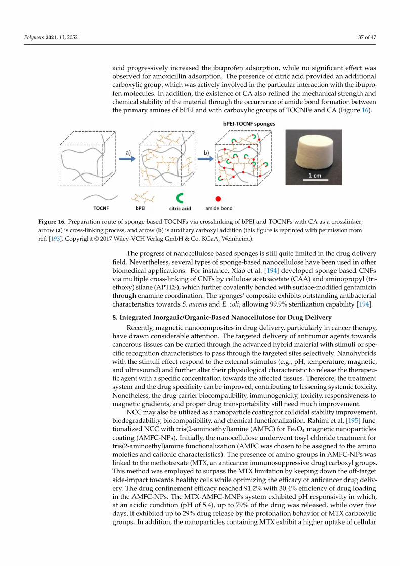
Polymers 2021, 13, 2052 37 of 47
acid progressively increased the ibuprofen adsorption, while no significant effect wasobserved for amoxicillin adsorption. The presence of citric acid provided an additionalcarboxylic group, which was actively involved in the particular interaction with the ibupro-fen molecules. In addition, the existence of CA also refined the mechanical strength andchemical stability of the material through the occurrence of amide bond formation betweenthe primary amines of bPEI and with carboxylic groups of TOCNFs and CA (Figure 16).
Polymers 2021, 13, x 39 of 49
Figure 16. Preparation route of sponge-based TOCNFs via crosslinking of bPEI and TOCNFs with CA as a crosslinker; arrow (a) is cross-linking process, and arrow (b) is auxiliary carboxyl addition (this figure is reprinted with permission from ref. [193]. Copyright © 2017 Wiley-VCH Verlag GmbH & Co. KGaA, Weinheim.).
8. Integrated Inorganic/Organic-Based Nanocellulose for Drug Delivery Recently, magnetic nanocomposites in drug delivery, particularly in cancer therapy,
have drawn considerable attention. The targeted delivery of antitumor agents towards cancerous tissues can be carried through the advanced hybrid material with stimuli or specific recognition characteristics to pass through the targeted sites selectively. Nanohy-brids with the stimuli effect respond to the external stimulus (e.g., pH, temperature, mag-netic, and ultrasound) and further alter their physiological characteristic to release the therapeutic agent with a specific concentration towards the affected tissues. Therefore, the treatment system and the drug specificity can be improved, contributing to lessening sys-temic toxicity. Nonetheless, the drug carrier biocompatibility, immunogenicity, toxicity, responsiveness to magnetic gradients, and proper drug transportability still need much improvement.
NCC may also be utilized as a nanoparticle coating for colloidal stability improve-ment, biodegradability, biocompatibility, and chemical functionalization. Rahimi et al. [195] functionalized NCC with tris(2-aminoethyl)amine (AMFC) for Fe3O4 magnetic na-noparticles coating (AMFC-NPs). Initially, the nanocellulose underwent tosyl chloride treatment for tris(2-aminoethyl)amine functionalization (AMFC was chosen to be as-signed to the amino moieties and cationic characteristics). The presence of amino groups in AMFC-NPs was linked to the methotrexate (MTX, an anticancer immunosuppressive drug) carboxyl groups. This method was employed to surpass the MTX limitation by keeping down the off-target side-impact towards healthy cells while optimizing the effi-cacy of anticancer drug delivery. The drug confinement efficacy reached 91.2% with 30.4% efficiency of drug loading in the AMFC-NPs. The MTX-AMFC-MNPs system exhibited pH responsivity in which, at an acidic condition (pH of 5.4), up to 79% of the drug was released, while over five days, it exhibited up to 29% drug release by the protonation be-havior of MTX carboxylic groups. In addition, the nanoparticles containing MTX exhibit a higher uptake of cellular compared to the AMFC-MNPs, which were contributed to by the chemical similarity of MTX with folic acid (FA), which assists the internalization of receptor-mediated cellulose. This enhances the potential of MTX for cancer cell targeting (Figure 17) [195].
Figure 16. Preparation route of sponge-based TOCNFs via crosslinking of bPEI and TOCNFs with CA as a crosslinker;arrow (a) is cross-linking process, and arrow (b) is auxiliary carboxyl addition (this figure is reprinted with permission fromref. [193]. Copyright © 2017 Wiley-VCH Verlag GmbH & Co. KGaA, Weinheim.).
The progress of nanocellulose based sponges is still quite limited in the drug deliveryfield. Nevertheless, several types of sponge-based nanocellulose have been used in otherbiomedical applications. For instance, Xiao et al. [194] developed sponge-based CNFsvia multiple cross-linking of CNFs by cellulose acetoacetate (CAA) and aminopropyl (tri-ethoxy) silane (APTES), which further covalently bonded with surface-modified gentamicinthrough enamine coordination. The sponges’ composite exhibits outstanding antibacterialcharacteristics towards S. aureus and E. coli, allowing 99.9% sterilization capability [194].
8. Integrated Inorganic/Organic-Based Nanocellulose for Drug Delivery
Recently, magnetic nanocomposites in drug delivery, particularly in cancer therapy,have drawn considerable attention. The targeted delivery of antitumor agents towardscancerous tissues can be carried through the advanced hybrid material with stimuli or spe-cific recognition characteristics to pass through the targeted sites selectively. Nanohybridswith the stimuli effect respond to the external stimulus (e.g., pH, temperature, magnetic,and ultrasound) and further alter their physiological characteristic to release the therapeu-tic agent with a specific concentration towards the affected tissues. Therefore, the treatmentsystem and the drug specificity can be improved, contributing to lessening systemic toxicity.Nonetheless, the drug carrier biocompatibility, immunogenicity, toxicity, responsiveness tomagnetic gradients, and proper drug transportability still need much improvement.
NCC may also be utilized as a nanoparticle coating for colloidal stability improvement,biodegradability, biocompatibility, and chemical functionalization. Rahimi et al. [195] func-tionalized NCC with tris(2-aminoethyl)amine (AMFC) for Fe3O4 magnetic nanoparticlescoating (AMFC-NPs). Initially, the nanocellulose underwent tosyl chloride treatment fortris(2-aminoethyl)amine functionalization (AMFC was chosen to be assigned to the aminomoieties and cationic characteristics). The presence of amino groups in AMFC-NPs waslinked to the methotrexate (MTX, an anticancer immunosuppressive drug) carboxyl groups.This method was employed to surpass the MTX limitation by keeping down the off-targetside-impact towards healthy cells while optimizing the efficacy of anticancer drug deliv-ery. The drug confinement efficacy reached 91.2% with 30.4% efficiency of drug loadingin the AMFC-NPs. The MTX-AMFC-MNPs system exhibited pH responsivity in which,at an acidic condition (pH of 5.4), up to 79% of the drug was released, while over fivedays, it exhibited up to 29% drug release by the protonation behavior of MTX carboxylicgroups. In addition, the nanoparticles containing MTX exhibit a higher uptake of cellular
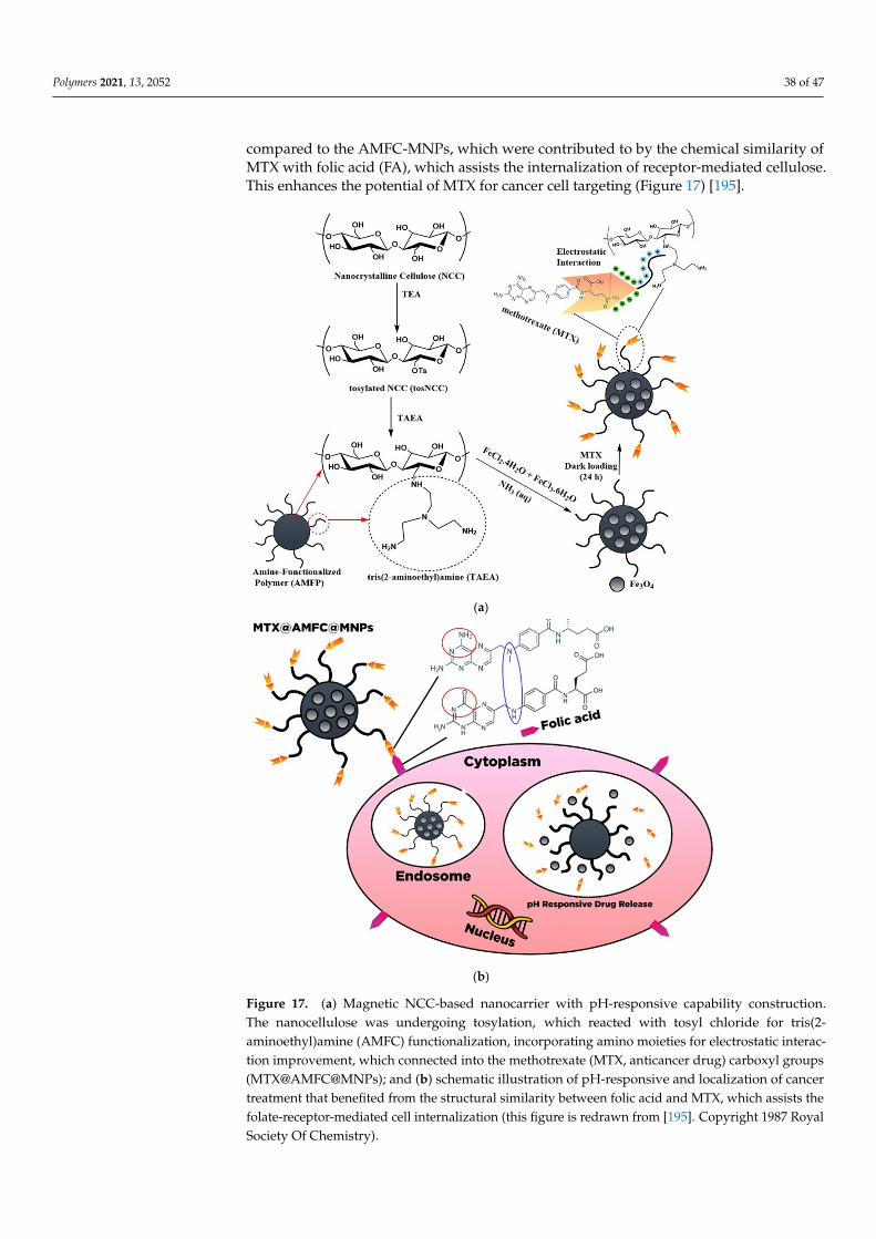
Polymers 2021, 13, 2052 38 of 47
compared to the AMFC-MNPs, which were contributed to by the chemical similarity ofMTX with folic acid (FA), which assists the internalization of receptor-mediated cellulose.This enhances the potential of MTX for cancer cell targeting (Figure 17) [195].
Polymers 2021, 13, x 40 of 49
(a)
(b)
Figure 17. (a) Magnetic NCC-based nanocarrier with pH-responsive capability construction. The nanocellulose was undergoing tosylation, which reacted with tosyl chloride for tris(2-ami-noethyl)amine (AMFC) functionalization, incorporating amino moieties for electrostatic interaction improvement, which connected into the methotrexate (MTX, anticancer drug) carboxyl groups (MTX@AMFC@MNPs); and (b) schematic illustration of pH-responsive and localization of cancer treatment that benefited from the structural similarity between folic acid and MTX, which assists
Figure 17. (a) Magnetic NCC-based nanocarrier with pH-responsive capability construction.The nanocellulose was undergoing tosylation, which reacted with tosyl chloride for tris(2-aminoethyl)amine (AMFC) functionalization, incorporating amino moieties for electrostatic interac-tion improvement, which connected into the methotrexate (MTX, anticancer drug) carboxyl groups(MTX@AMFC@MNPs); and (b) schematic illustration of pH-responsive and localization of cancertreatment that benefited from the structural similarity between folic acid and MTX, which assists thefolate-receptor-mediated cell internalization (this figure is redrawn from [195]. Copyright 1987 RoyalSociety Of Chemistry).
Polymers 2021, 13, 2052 39 of 47
Recently, Supramaniam et al. [196] introduced magnetic characteristics towardsnanocellulos-based hydrogels, which were further utilized for controlling drug deliv-ery. The co-precipitation with Fe (II) and Fe (III) ions was incorporated into the NCCs,followed by the insertion of the magnetic characteristic, and subsequently, morphologicallymodified into beads with sodium alginate. It was observed that the magnetic nanocellu-lose existence refines the physical and mechanical characteristics of hydrogel beads andswelling degree improvement and limits the drug release due to the formation of physicalentanglement inside the hydrogel. Jeddi and Mahkam [197] developed magnetic hydro-gel beads composite-based carboxymethyl nanocellulose to deliver dexamethasone. Thecomposite can control the dexamethasone delivery up to 12 h.
Carbon nanotubes have outstanding characteristics such as high thermal stability,homogenous pore arrangement, high specific area, and excellent electrical features. Thisadvanced material has also been employed as a vehicle in the drug delivery systemin recent years. The combination of this material with the NC material provides someadvantages. The incorporation of nanocellulose in the composite increased biocompatibilityand biodegradability while the CNTs provided good stability, magnetic and electromagneticbehavior, and high cellular uptake [198]. Although the cytotoxicity of material still becamean issue, CNTs were widely exploited in drug delivery systems, particularly cancer therapyapplications [199].
Integration of nanocellulose into graphene-based materials through the layer-by-layer assembly as a drug carrier was carried out by Anirudhan et al. [200]. Chemicallymodified GO was used as a template for the layer-by-layer assembly of aminated nano-dextran (AND) and carboxylic acid functionalized nanocellulose (NCCs) to form a MGO-AND/NCCs nanocomposite. Curcumin can be loaded into the carrier through π–π stackingand hydrogen bonding interactions due to the phenolic and aromatic rings of curcumin.Based on the release study, the acidic environment promotes COO- groups’ protonationand amino in aminated in nano-dextran to form NH3
+. This phenomenon decreased thestatic interaction between MGO-AND/NCC, resulting in the electrostatic repulsion of eachcomponent, consequently provoking the drug release. In addition, a cytotoxicity assay onHCT116 cells exhibited high efficacy of curcumin-loaded MGO-AND/NCC.
The electrochemical activity of the carbon nanotube was utilized to modulate thedrug release. The release of ibuprofen from a novel hybrid hydrogel composed of sodiumalginate (SA), bacterial cellulose (BC), and multi-walled carbon nanotubes (MWCNTs) wasstudied by Shi et al. [176]. The release of ibuprofen can be provoked by electrostatic repul-sion. Thereby, the on–off release mechanism can be attained by introducing electrochemicalpotential [176].
9. Conclusions
Modified and functionalized nanocelluloses with low toxicity and high biocompatibil-ity render them promising materials as advanced drug carriers. Various hydroxyl groupson the surface of the nanocellulose serve as attachment sites of drugs through covalentand/or physical interactions. In addition, nanocellulose modification results in a differentmorphological structure for the carrier, which contributes to an increase in the diffusionpathway of the drug within the carrier. Therefore, surface chemistry is a crucial factorthat should be considered in the design of nanocellulose as a drug carrier for effectivedrug delivery. High-purity nanocelluloses are also required to obtain drug carriers withthe well-constructed framework, thus facilitating drug adsorption and release control.Considering all these factors, carrier-based nanocellulose is a promising candidate fordeveloping novel sustained drug delivery systems.
Author Contributions: Conceptualization V.B.L., F.E.S. and S.I.; redraw the figures V.B.L. and M.Y.;drafting the manuscript V.B.L., J.N.P., S.P.S. and S.I.; Supervision S.I. and F.E.S.; review and editing,J.S, and Y.H.J.; proof reading J.S. and Y.H.J. All authors have read and agreed to the published versionof the manuscript.
Polymers 2021, 13, 2052 40 of 47
Funding: This study was supported by the Directorate of Research and Community Service, Deputyfor Strengthening Research and Development, Ministry of Research and Technology/NationalResearch and Innovation Agency, Number: 150J/WM01.5/N/2021.
Institutional Review Board Statement: Not applicable.
Informed Consent Statement: Not applicable.
Conflicts of Interest: The authors declare no conflict of interest.
References1. Jain, K.K. An overview of drug delivery systems. In Drug Delivery Systems; Springer: New York, NY, USA, 2020.2. Sunasee, R.; Hemraz, U.D.; Ckless, K. Cellulose nanocrystals: A versatile nanoplatform for emerging biomedical applications.
Expert Opin. Drug Deliv. 2016, 13, 1243–1256. [CrossRef] [PubMed]3. Bamrungsap, S.; Zhao, Z.; Chen, T.; Wand, L.; Li, C.; Fu, T.; Tan, W. Nanotechnology in therapeutics: A focus on nanoparticles as
a drug delivery system. Nanomedicine 2012, 7, 1253–1271. [CrossRef]4. Tong, X.; Pan, W.; Su, T.; Zhang, M.; Dong, W.; Qi, X. Recent advances in natural polymer-based drug delivery systems. React.
Funct. Polym. 2020, 148, 104501. [CrossRef]5. Cavallaro, G.; Micciulla, S.; Chiappisi, L.; Lazzara, G. Chitosan-based smart hybrid materials: A physico-chemical perspective. J.
Mater. Chem. B 2021, 9, 594–611. [CrossRef]6. Bertolino, V.; Cavallaro, G.; Milioto, S.; Lazzara, G. Polysaccharides/Halloysite nanotubes for smart bionanocomposite materials.
Carbohydr. Polym. 2020, 245, 116502. [CrossRef]7. Ahmad, A.; Mubarak, N.; Jannat, F.T.; Ashfaq, T.; Santulli, C.; Rizwan, M.; Najda, A.; Bin-Jumah, M.; Abdel-Daim, M.M.; Hussain,
S. A Critical Review on the Synthesis of Natural Sodium Alginate Based Composite Materials: An Innovative Biological Polymerfor Biomedical Delivery Applications. Processes 2021, 9, 137. [CrossRef]
8. Chen, J.; Ouyang, J.; Chen, Q.; Deng, C.; Meng, F.; Zhang, J.; Cheng, R.; Lan, Q.; Zhong, Z. EGFR and CD44 dual-targetedmultifunctional hyaluronic acid nanogels boost protein delivery to ovarian and breast cancers in vitro and in vivo. ACS Appl.Mater. Interfaces 2017, 9, 24140–24147. [CrossRef] [PubMed]
9. Qi, X.; Wei, W.; Shen, J.; Dong, W. Salecan polysaccharide-based hydrogels and their applications: A review. J. Mater. Chem. B2019, 7, 2577–2587. [CrossRef] [PubMed]
10. Dragan, E.S.; Dinu, M.V. Polysaccharides constructed hydrogels as vehicles for proteins and peptides. A review. Carbohydr. Polym.2019, 225, 115210. [CrossRef] [PubMed]
11. Jorfi, M.; Foster, E.J. Recent advances in nanocellulose for biomedical applications. J. Appl. Polym. Sci. 2015, 132, 41719. [CrossRef]12. Xue, Y.; Mou, Z.; Xiao, H. Nanocellulose as a sustainable biomass material: Structure, properties, present status and future
prospects in biomedical applications. Nanoscale 2017, 9, 14758–14781. [CrossRef]13. Habibi, Y.; Lucia, L.A.; Rojas, O.J. Cellulose nanocrystals: Chemistry, self-assembly, and applications. Chem. Rev. 2010, 110,
3479–3500. [CrossRef]14. Lin, N.; Dufresne, A. Nanocellulose in biomedicine: Current status and future prospect. Eur. Polym. J. 2014, 59, 302–325.
[CrossRef]15. Plackett, D.; Letchford, K.; Jackson, J.; Burt, H. A review of nanocellulose as a novel vehicle for drug delivery. Nord. Pulp Pap. Res.
J. 2014, 29, 105–118. [CrossRef]16. Jawaid, M.; Mohammad, F. Nanocellulose and Nanohydrogel Matrices: Biotechnological and Biomedical Applications; John Wiley & Sons:
Hoboken, NJ, USA, 2017.17. Hasan, N.; Rahman, L.; Kim, S.-H.; Cao, J.; Arjuna, A.; Lallo, S.; Jhun, B.H.; Yoo, J.-W. Recent advances of nanocellulose in drug
delivery systems. J. Pharm. Investig. 2020, 50, 553–572. [CrossRef]18. Grishkewich, N.; Mohammed, N.; Tang, J.; Tam, K.C. Recent advances in the application of cellulose nanocrystals. Curr. Opin.
Colloid Interface Sci. 2017, 29, 32–45. [CrossRef]19. Yan, G.; Chen, B.; Zeng, X.; Sun, Y.; Tan, X.; Lin, L. Recent advances on sustainable cellulosic materials for pharmaceutical carrier
applications. Carbohydr. Polym. 2020, 244, 116492. [CrossRef] [PubMed]20. Rajinipriya, M.; Nagalakshmaiah, M.; Robert, M.; Elkoun, S. Importance of agricultural and industrial waste in the field of
nanocellulose and recent industrial developments of wood based nanocellulose: A review. ACS Sustain. Chem. Eng. 2018, 6,2807–2828. [CrossRef]
21. Karimian, A.; Parsian, H.; Majidinia, M.; Rahimi, M.; Mir, S.M.; Kafil, H.S.; Shafiei-Irannejad, V.; Kheyrollah, M.; Ostadi, H.;Yousefi, B. Nanocrystalline cellulose: Preparation, physicochemical properties, and applications in drug delivery systems. Int. J.Biol. Macromol. 2019, 133, 850–859. [CrossRef] [PubMed]
22. Salimi, S.; Sotudeh-Gharebagh, R.; Zarghami, R.; Chan, S.Y.; Yuen, K.H. Production of nanocellulose and its applications in drugdelivery: A critical review. ACS Sustain. Chem. Eng. 2019, 7, 15800–15827. [CrossRef]
23. Seabra, A.B.; Bernandes, J.S.; Favaro, W.J.; Paula, A.J.; Duran, N. Cellulose nanocrystals as carriers in medicine and their toxicities:A review. Carbohydr. Polym. 2018, 181, 514–527. [CrossRef] [PubMed]
Polymers 2021, 13, 2052 41 of 47
24. Sheikhi, A.; Hayashi, J.; Eichenbaum, J.; Gutin, M.; Kuntjoro, N.; Khorsandi, D.; Khademhosseini, A. Recent advances innanoengineering cellulose for cargo delivery. J. Control. Release 2019, 294, 53–76. [CrossRef] [PubMed]
25. Putro, J.N.; Ismadji, S.; Gunarto, C.; Yuliana, M.; Santoso, S.P.; Soetaredjo, F.E.; Ju, Y.H. The effect of surfactants modification onnanocrystalline cellulose for paclitaxel loading and release study. J. Mol. Liq. 2019, 282, 407–414. [CrossRef]
26. Bundjaja, V.; Sari, T.M.; Soetaredjo, F.E.; Yuliana, M.; Angkawijaya, A.E.; Ismadji, S.; Cheng, K.-C.; Santoso, S.P. Aqueous sorptionof tetracycline using rarasaponin-modified nanocrystalline cellulose. J. Mol. Liq. 2020, 301, 112433. [CrossRef]
27. Huang, Y.; Zhu, C.; Yang, J.; Nie, Y.; Chen, C.; Sun, D. Recent advances in bacterial cellulose. Cellulose 2014, 21, 1–30. [CrossRef]28. Foo, M.L.; Tan, C.R.; Lim, P.D.; Ooi, C.W.; Tan, K.W.; Chew, I.M.L. Surface-modified nanocrystalline cellulose from oil palm empty
fruit bunch for effective binding of curcumin. Int. J. Biol. Macromol. 2019, 138, 1064–1071. [CrossRef]29. Wijaya, C.J.; Saputra, S.N.; Soetaredjo, F.E.; Putro, J.N.; Lin, C.X.; Kurniawan, A.; Ju, Y.-H.; Ismadji, S. Cellulose nanocrystals from
passion fruit peels waste as antibiotic drug carrier. Carbohydr. Polym. 2017, 175, 370–376. [CrossRef] [PubMed]30. Dufresne, A. Cellulose nanomaterial reinforced polymer nanocomposites. Curr. Opin. Colloid Interface Sci. 2017, 29, 1–8. [CrossRef]31. Gopinath, V.; Saravanan, S.; Al-Maleki, A.; Ramesh, M.; Vadivelu, J. A review of natural polysaccharides for drug delivery
applications: Special focus on cellulose, starch and glycogen. Biomed. Pharmacother. 2018, 107, 96–108. [CrossRef]32. Wertz, J.-L.; Bedue, O.; Mercier, J.P. Cellulose Science and Technology; EPFL Press: Lausanne, Switzerland, 2010.33. George, J.; Sabapathi, S. Cellulose nanocrystals: Synthesis, functional properties, and applications. Nanotechnol. Sci. Appl. 2015, 8,
45. [CrossRef]34. Moon, R.J.; Martini, A.; Nairn, J.; Simonsen, J.; Youngblood, J. Cellulose nanomaterials review: Structure, properties and
nanocomposites. Chem. Soc. Rev. 2011, 40, 3941–3994. [CrossRef]35. Medronho, B.; Lindman, B. Brief overview on cellulose dissolution/regeneration interactions and mechanisms. Adv. Colloid
Interface Sci. 2015, 222, 502–508. [CrossRef]36. Kamel, R.; El-Wakil, N.A.; Dufresne, A.; Elkasabgy, N.A. Nanocellulose: From an agricultural waste to a valuable pharmaceutical
ingredient. Int. J. Biol. Macromol. 2020, 163, 1579–1590. [CrossRef] [PubMed]37. Miao, C.; Hamad, W.Y. Cellulose reinforced polymer composites and nanocomposites: A critical review. Cellulose 2013, 20,
2221–2262. [CrossRef]38. Li, N.; Lu, W.; Yu, J.; Xiao, Y.; Liu, S.; Gan, L.; Huang, J. Rod-like cellulose nanocrystal/cis-aconityl-doxorubicin prodrug: A
fluorescence-visible drug delivery system with enhanced cellular uptake and intracellular drug controlled release. Mater. Sci. Eng.C 2018, 91, 179–189. [CrossRef] [PubMed]
39. Jia, B.; Li, Y.; Yang, B.; Xiao, D.; Zhang, S.; Rajulu, A.V.; Kondo, T.; Zhang, L.; Zhou, J. Effect of microcrystal cellulose and cellulosewhisker on biocompatibility of cellulose-based electrospun scaffolds. Cellulose 2013, 20, 1911–1923. [CrossRef]
40. Dash, R.; Ragauskas, A.J. Synthesis of a novel cellulose nanowhisker-based drug delivery system. RSC Adv. 2012, 2, 3403–3409.[CrossRef]
41. Biswas, S.K.; Sano, H.; Yang, X.; Tanphichai, S.; Shams, M.I.; Yano, H. Highly thermal-resilient AgNW transparent electrodeand optical device on thermomechanically superstable cellulose nanorod-reinforced nanocomposites. Adv. Opt. Mater. 2019, 7,1900532. [CrossRef]
42. Wang, N.; Ding, E.; Cheng, R. Thermal degradation behaviors of spherical cellulose nanocrystals with sulfate groups. Polymer2007, 48, 3486–3493. [CrossRef]
43. Ram, B.; Chauhan, G.S.; Mehta, A.; Gupta, R.; Chauhan, K. Spherical nanocellulose-based highly efficient and rapid multifunc-tional naked-eye Cr (VI) ion chemosensor and adsorbent with mild antimicrobial properties. Chem. Eng. J. 2018, 349, 146–155.[CrossRef]
44. Mariano, M.; El Kissi, N.; Dufresne, A. Cellulose nanocrystals and related nanocomposites: Review of some properties andchallenges. J. Polym. Sci. B Polym. Phys. 2014, 52, 791–806. [CrossRef]
45. Brinchi, L.; Cotana, F.; Fortunati, E.; Kenny, J. Production of nanocrystalline cellulose from lignocellulosic biomass: Technologyand applications. Carbohydr. Polym. 2013, 94, 154–169. [CrossRef]
46. Phanthong, P.; Reybroycharoen, P.; Hao, X.; Xu, G.; Abudula, A.; Guan, G. Nanocellulose: Extraction and application. CarbonResour. Convers. 2018, 1, 32–43. [CrossRef]
47. Lam, E.; Male, K.B.; Chong, J.H.; Leung, A.C.; Luong, J.H. Applications of functionalized and nanoparticle-modified nanocrys-talline cellulose. Trends Biotechnol. 2012, 30, 283–290. [CrossRef] [PubMed]
48. Prathapan, R.; Tabor, R.F.; Garnier, G.; Hu, J. Recent progress in cellulose nanocrystal alignment and its applications. ACS Appl.Bio Mater. 2020, 3, 1828–1844. [CrossRef]
49. Abitbol, T.; Rivkin, A.; Cao, Y.; Nevo, Y.; Abraham, E.; Ben-Shalom, T.; Lapidot, S.; Shoseyov, O. Nanocellulose, a tiny fiber withhuge applications. Curr. Opin. Biotechnol. 2016, 39, 76–88. [CrossRef] [PubMed]
50. Henriksson, M.; Henriksson, G.; Berglund, L.; Lindström, T. An environmentally friendly method for enzyme-assisted preparationof microfibrillated cellulose (MFC) nanofibers. Eur. Polym. J. 2007, 43, 3434–3441. [CrossRef]
51. Kim, C.-W.; Kim, D.-S.; Kang, S.-Y.; Marquez, M.; Joo, Y.L. Structural studies of electrospun cellulose nanofibers. Polymer 2006, 47,5097–5107. [CrossRef]
52. Dufresne, A. Nanocellulose: A new ageless bionanomaterial. Mater. Today 2013, 16, 220–227. [CrossRef]53. Lavoine, N.; Desloges, I.; Dufresne, A.; Bras, J. Microfibrillated cellulose–Its barrier properties and applications in cellulosic
materials: A review. Carbohydr. Polym. 2012, 90, 735–764. [CrossRef]
Polymers 2021, 13, 2052 42 of 47
54. Blanco Parte, F.G.; Santoso, S.P.; Chou, C.-C.; Verma, V.; Wang, H.-T.; Ismadji, S.; Cheng, K.-C. Current progress on the production,modification, and applications of bacterial cellulose. Crit. Rev. Biotechnol. 2020, 40, 397–414. [CrossRef] [PubMed]
55. Siro, I.; Plackett, D. Microfibrillated cellulose and new nanocomposite materials: A review. Cellulose 2010, 17, 459–494. [CrossRef]56. Huang, J.; Dufresne, A.; Lin, N. Nanocellulose: From Fundamentals to Advanced Materials; John Wiley & Sons: Hoboken, NJ, USA,
2019.57. Jozala, A.F.; De Lencastre-Novaes, L.C.; Lopes, A.M.; De Carvalho Santos-Ebinuma, V.; Mazzola, P.G.; Pessoa-JR, A.; Grotto, D.;
Gerenutti, M.; Chaud, M.V. Bacterial nanocellulose production and application: A 10-year overview. Appl. Microbiol. Biotechnol.2016, 100, 2063–2072. [CrossRef] [PubMed]
58. Wei, B.; Yang, G.; Hong, F. Preparation and evaluation of a kind of bacterial cellulose dry films with antibacterial properties.Carbohydr. Polym. 2011, 84, 533–538. [CrossRef]
59. Wan, Y.; Wang, J.; Gama, M.; Guo, R.; Zhang, Q.; Zhang, P.; Yao, F.; Luo, H. Biofabrication of a novel bacteria/bacterial cellulosecomposite for improved adsorption capacity. Compos. Part A Appl. Sci. Manuf. 2019, 125, 105560. [CrossRef]
60. Gorgieva, S.; Trcek, J. Bacterial cellulose: Production, modification and perspectives in biomedical applications. Nanomaterials2019, 9, 1352. [CrossRef] [PubMed]
61. Kim, J.-H.; Shim, B.S.; Kim, H.S.; Lee, Y.-J.; Min, S.-K.; Jang, D.; Abas, Z.; Kim, J. Review of nanocellulose for sustainable futurematerials. Int. J. Precis. Eng. Manuf. Green Technol. 2015, 2, 197–213. [CrossRef]
62. Dufresne, A. Cellulose-based composites and nanocomposites. In Monomers, Polymers, and Composites from Renewable Resources;Elsevier: Amsterdam, The Netherlands, 2008.
63. Song, F.; Li, X.; Wang, Q.; Liao, L.; Zhang, C. Nanocomposite hydrogels and their applications in drug delivery and tissueengineering. J. Biomed. Nanotechnol. 2015, 11, 40–52. [CrossRef]
64. Ibrahim, M.M.; Fahmy, T.Y.; Salaheldin, E.I.; Mobarak, F.; Youssef, M.A.; Mabrook, M.R. Synthesis of tosylated and trimethylsily-lated methyl cellulose as pH-sensitive carrier matrix. Life Sci. J. 2015, 1, 29–37.
65. Ximenes, F.A.; Gardner, W.D.; Kathuria, A. Proportion of above-ground biomass in commercial logs and residues following theharvest of five commercial forest species in Australia. For. Ecol. Manag. 2008, 256, 335–346. [CrossRef]
66. Kargarzadeh, H.; Ioelovich, M.; Ahmad, I.; Thomas, S.; Dufresne, A. Methods for extraction of nanocellulose from various sources.Handb. Nanocellulose Cellul. Nanocomposites 2017, 2, 1–49.
67. Kumar, A.K.; Sharma, S. Recent updates on different methods of pretreatment of lignocellulosic feedstocks: A review. Bioresour.Bioproces. 2017, 4, 1–19. [CrossRef]
68. Ravindran, R.; Jaiswal, S.; Abu-Ghannam, N.; Jaiswal, A.K. A comparative analysis of pretreatment strategies on the propertiesand hydrolysis of brewers’ spent grain. Bioresour. Technol. 2018, 248, 272–279. [CrossRef] [PubMed]
69. Pires, J.R.; De Souza, V.G.L.; Fernando, A.L. Production of nanocellulose from lignocellulosic biomass wastes: Prospects andlimitations. In Proceedings of the International Conference on Innovation, Engineering and Entrepreneurship, Guimaraes,Portugal, 27–29 June 2018; pp. 719–725.
70. Shirkavand, E.; Baroutian, S.; Gapes, D.J.; Young, B.R. Combination of fungal and physicochemical processes for lignocellulosicbiomass pretreatment–A review. Renew. Sust. Energy Rev. 2016, 54, 217–234. [CrossRef]
71. Baramee, S.; Siriatcharanon, A.-K.; Ketbot, P.; Teeravivattanakit, T.; Waeonukul, R.; Pason, P.; Tachaapaikoon, C.;Ratanakhanokchai, K.; Phitsuwan, P. Biological pretreatment of rice straw with cellulase-free xylanolytic enzyme-producingBacillus firmus K-1: Structural modification and biomass digestibility. Renew. Energy 2020, 160, 555–563. [CrossRef]
72. Khalil, H.A.; Davoudpour, Y.; Islam, M.N.; Mustapha, A.; Sudesh, K.; Dungani, R.; Jawaid, M. Production and modification ofnanofibrillated cellulose using various mechanical processes: A review. Carbohydr. Polym. 2014, 99, 649–665. [CrossRef]
73. Mood, S.H.; Golfeshan, A.H.; Tabatabaei, M.; Jouzani, G.S.; Najafi, G.H.; Gholami, M.; Ardjmand, M. Lignocellulosic biomass tobioethanol, a comprehensive review with a focus on pretreatment. Renew. Sust. Energy Rev. 2013, 27, 77–93. [CrossRef]
74. Hassan, S.S.; Williams, G.A.; Jaiswal, A.K. Emerging technologies for the pretreatment of lignocellulosic biomass. Bioresour.Technol. 2018, 262, 310–318. [CrossRef]
75. De Carvalho Benini, K.C.C.; Voorwald, H.J.C.; Cioffi, M.O.H.; Rezende, M.C.; Arantes, V. Preparation of nanocellulose fromImperata brasiliensis grass using Taguchi method. Carbohydr. Polym. 2018, 192, 337–346. [CrossRef]
76. Maciel, M.M.Á.D.; De Carvalho Benini, K.C.C.; Voorwald, H.J.C.; Cioffi, M.O.H. Obtainment and characterization of nanocellulosefrom an unwoven industrial textile cotton waste: Effect of acid hydrolysis conditions. Int. J. Biol. Macromol. 2019, 126, 496–506.[CrossRef]
77. Liu, J.; Korpinen, R.; Mikkonen, K.S.; Willför, S.; Xu, C. Nanofibrillated cellulose originated from birch sawdust after sequentialextractions: A promising polymeric material from waste to films. Cellulose 2014, 21, 2587–2598. [CrossRef]
78. Couret, L.; Irle, M.; Belloncle, C.; Cathala, B. Extraction and characterization of cellulose nanocrystals from post-consumer woodfiberboard waste. Cellulose 2017, 24, 2125–2137. [CrossRef]
79. Vallejos, M.E.; Felissia, F.E.; Area, M.C.; Ehman, N.V.; Tarrés, Q.; Mutjé, P. Nanofibrillated cellulose (CNF) from eucalyptussawdust as a dry strength agent of unrefined eucalyptus handsheets. Carbohydr. Polym. 2016, 139, 99–105. [CrossRef] [PubMed]
80. Rambabu, N.; Panthapulakkal, S.; Sain, M.; Dalai, A. 2016. Production of nanocellulose fibers from pinecone biomass: Evaluationand optimization of chemical and mechanical treatment conditions on mechanical properties of nanocellulose films. Ind. CropsProd. 2016, 83, 746–754. [CrossRef]
Polymers 2021, 13, 2052 43 of 47
81. Moriana, R.; Vilaplana, F.; Ek, M. Cellulose nanocrystals from forest residues as reinforcing agents for composites: A study frommacro-to nano-dimensions. Carbohydr. Polym. 2016, 139, 139–149. [CrossRef]
82. Lu, H.; Zhang, L.; Liu, C.; He, Z.; Zhou, X.; Ni, Y. A novel method to prepare lignocellulose nanofibrils directly from bamboochips. Cellulose 2018, 25, 7043–7051. [CrossRef]
83. Hua, K.; Strømme, M.; Mihranyan, A.; Ferraz, N. Nanocellulose from green algae modulates the in vitro inflammatory responseof monocytes/macrophages. Cellulose 2015, 22, 3673–3688. [CrossRef]
84. Son, H.N.; Seo, Y.B. Physical and bio-composite properties of nanocrystalline cellulose from wood, cotton liners, cattail, and redalgae. Cellulose 2015, 22, 1789–1798.
85. Rathod, M.; Haldar, S.; Basha, S. Nanocrystalline cellulose for removal of tetracycline hydrochloride from water via biosorption:Equilibrium, kinetic and thermodynamic studies. Ecol. Eng. 2015, 84, 240–249. [CrossRef]
86. Liu, Z.; Li, X.; Xie, W.; Deng, H. Extraction, isolation and characterization of nanocrystalline cellulose from industrial kelp(Laminaria japonica) waste. Carbohydr. Polym. 2017, 173, 353–359. [CrossRef] [PubMed]
87. Feng, X.; Meng, X.; Zhao, J.; Miao, M.; Shi, L.; Zhang, S.; Fang, J. Extraction and preparation of cellulose nanocrystals fromdealginate kelp residue: Structures and morphological characterization. Cellulose 2015, 22, 1763–1772. [CrossRef]
88. Bhutiya, P.L.; Misra, N.; Rasheed, M.A.; Hasan, S.Z. Nested seaweed cellulose fiber deposited with cuprous oxide nanorods forantimicrobial activity. Int. J. Biol. Macromol. 2018, 117, 435–444. [CrossRef]
89. De Oliveira, J.P.; Bruni, G.P.; Fabra, M.J.; Da Rosa Zavareze, E.; López-Rubio, A.; Martínez-Sanz, M. Development of foodpackaging bioactive aerogels through the valorization of Gelidium sesquipedale seaweed. Food Hydrocoll. 2019, 89, 337–350.[CrossRef]
90. Chen, Y.W.; Lee, H.V.; Juan, J.C.; Phang, S.-M. Production of new cellulose nanomaterial from red algae marine biomass Gelidiumelegans. Carbohydr. Polym. 2016, 151, 1210–1219. [CrossRef] [PubMed]
91. Mandal, A.; Chakrabarty, D. Isolation of nanocellulose from waste sugarcane bagasse (SCB) and its characterization. Carbohydr.Polym. 2011, 86, 1291–1299. [CrossRef]
92. Thomas, M.G.; Abraham, E.; Jyotishkumar, P.; Maria, H.J.; Pothen, L.A.; Thomas, S. Nanocelluloses from jute fibers and theirnanocomposites with natural rubber: Preparation and characterization. Int. J. Biol. Macromol. 2015, 81, 768–777. [CrossRef][PubMed]
93. Wu, J.; Du, X.; Yin, Z.; Xu, S.; Xu, S.; Zhang, Y. Preparation and characterization of cellulose nanofibrils from coconut coir fibersand their reinforcements in biodegradable composite films. Carbohydr. Polym. 2019, 211, 49–56. [CrossRef] [PubMed]
94. Mariño, M.; Lopes Da Silva, L.; Durán, N.; Tasic, L. Enhanced materials from nature: Nanocellulose from citrus waste. Molecules2015, 20, 5908–5923. [CrossRef]
95. Dilamian, M.; Noroozi, B. A combined homogenization-high intensity ultrasonication process for individualizaion of cellulosemicro-nano fibers from rice straw. Cellulose 2019, 26, 5831–5849. [CrossRef]
96. Kang, X.; Sun, P.; Kuga, S.; Wang, C.; Zhao, Y.; Wu, M.; Huang, Y. Thin cellulose nanofiber from corncob cellulose and itsperformance in transparent nanopaper. ACS Sust. Chem. Eng. 2017, 5, 2529–2534. [CrossRef]
97. Karimi, S.; Tahir, P.M.; Karimi, A.; Dufresne, A.; Abdulkhani, A. Kenaf bast cellulosic fibers hierarchy: A comprehensive approachfrom micro to nano. Carbohydr. Polym. 2014, 101, 878–885. [CrossRef]
98. Jodeh, S.; Hamed, O.; Melhem, A.; Salghi, R.; Jodeh, D.; Azzaoui, K.; Benmassaoud, Y.; Murtada, K. Magnetic nanocellulosefrom olive industry solid waste for the effective removal of methylene blue from wastewater. Environ. Sci. Poll. Res. 2018, 25,22060–22074. [CrossRef]
99. Jongaroontaprangsee, S.; Chiewchan, N.; Devahastin, S. Production of nanofibrillated cellulose with superior water redispersibilityfrom lime residues via a chemical-free process. Carbohydr. Polym. 2018, 193, 249–258. [CrossRef] [PubMed]
100. Diop, C.I.K.; Lavoie, J.-M. Isolation of nanocrystalline cellulose: A technological route for valorizing recycled tetra pak asepticmultilayered food packaging wastes. Waste Biomass Valor. 2017, 8, 41–56. [CrossRef]
101. Putro, J.N.; Santoso, S.P.; Soetaredjo, F.E.; Ismadji, S.; Ju, Y.-H. Nanocrystalline cellulose from waste paper: Adsorbent for azodyes removal. Environ. Nanotech. Monit. Manag. 2019, 12, 100260. [CrossRef]
102. Ogundare, S.A.; Moodley, V.; Van Zyl, W.E. Nanocrystalline cellulose isolated from discarded cigarette filters. Carbohydr. Polym.2017, 175, 273–281. [CrossRef] [PubMed]
103. Peretz, R.; Sterenzon, E.; Gerchman, Y.; Vadivel, V.K.; Luxbacher, T.; Mamane, H. Nanocellulose production from recycled papermill sludge using ozonation pretreatment followed by recyclable maleic acid hydrolysis. Carbohydr. Polym. 2019, 216, 343–351.[CrossRef] [PubMed]
104. Cypriano, D.Z.; Da Silva, L.L.; Tasic, L. High value-added products from the orange juice industry waste. Waste Manag. 2018, 79,71–78. [CrossRef] [PubMed]
105. Dubey, S.; Singh, J.; Singh, R. Biotransformation of sweet lime pulp waste into high-quality nanocellulose with an excellentproductivity using Komagataeibacter europaeus SGP37 under static intermittent fed-batch cultivation. Bioresour. Technol. 2018,247, 73–80. [CrossRef] [PubMed]
106. Mondal, S. Preparation, properties and applications of nanocellulosic materials. Carbohydr. Polym. 2017, 163, 301–316. [CrossRef]107. Turbak, A.F.; Snyder, F.W.; Sandberg, K.R. Microfibrillated cellulose, a new cellulose product: Properties, uses, and commercial
potential. J. Appl. Polym. Sci. Appl. Polym. Sym. 1983, 37, 815–827.
Polymers 2021, 13, 2052 44 of 47
108. Herrick, F.W.; Casebier, R.L.; Hamilton, J.K.; Sandberg, K.R. Microfibrillated cellulose: Morphology and accessibility. J. Appl.Polym. Sci. Appl. Polym. Sym. 1983, 37, 797–813.
109. Kawee, N.; Lam, N.T.; Sukyai, P. Homogenous isolation of individualized bacterial nanofibrillated cellulose by high pressurehomogenization. Carbohydr. Polym. 2018, 179, 394–401. [CrossRef]
110. Yusra, A.I.; Juahir, H.; Firdaus, N.N.A.; Bhat, A.; Endut, A.; Khalil, H.A.; Adiana, G. Controlling of green nanocellulose fiberproperties produced by chemo-mechanical treatment process via SEM, TEM, AFM and image analyzer characterization. J. Fund.Appl. Sci. 2018, 10, 1–17.
111. Kalia, S.; Boufi, S.; Celli, A.; Kango, S. Nanofibrillated cellulose: Surface modification and potential applications. Colloid Polym.Sci. 2014, 292, 5–31. [CrossRef]
112. Niu, F.; Li, M.; Huang, Q.; Zhang, X.; Pan, W.; Yang, J.; Li, J. The characteristic and dispersion stability of nanocellulose producedby mixed acid hydrolysis and ultrasonic assistance. Carbohydr. Polym. 2017, 165, 197–204. [CrossRef] [PubMed]
113. Trache, D.; Hussin, M.H.; Haafiz, M.M.; Thakur, V.K. Recent progress in cellulose nanocrystals: Sources and production. Nanoscale2017, 9, 1763–1786. [CrossRef]
114. Wang, J.; Tavakoli, J.; Tang, Y. Bacterial cellulose production, properties and applications with different culture methods–A review.Carbohydr. Polym. 2019, 219, 63–76. [CrossRef]
115. Czaja, W.; Romanovicz, D.; Malcolm, B.R. Structural investigations of microbial cellulose produced in stationary and agitatedculture. Cellulose 2004, 11, 403–411. [CrossRef]
116. Matsutani, M.; Ito, K.; Azuma, Y.; Ogino, H.; Shirai, M.; Yakushi, T.; Matsushita, K. Adaptive mutation related to celluloseproducibility in Komagataeibacter medellinensis (Gluconacetobacter xylinus) NBRC 3288. Appl. Microbiol. Biotechnol. 2015, 99,7229–7240. [CrossRef] [PubMed]
117. Liu, M.; Zhong, C.; Wu, X.-Y.; Wei, Y.-Q.; Bo, T.; Han, P.-P.; Jia, S.-R. Metabolomic profiling coupled with metabolic networkreveals differences in Gluconacetobacter xylinus from static and agitated cultures. Biochem. Eng. J. 2015, 101, 85–98. [CrossRef]
118. Gullo, M.; La China, S.; Petroni, G.; Di Gregorio, S.; Giudici, P. Exploring K2G30 genome: A high bacterial cellulose producingstrain in glucose and mannitol based media. Front. Microbiol. 2019, 10, 58. [CrossRef] [PubMed]
119. Thorat, M.N.; Dastager, S.G. High yield production of cellulose by a Komagataeibacter rhaeticus PG2 strain isolated frompomegranate as a new host. RSC Adv. 2018, 8, 29797–29805. [CrossRef]
120. Volova, T.G.; Prudnikova, S.V.; Sukovatyi, A.G.; Shishatskaya, E.I. Production and properties of bacterial cellulose by the strainKomagataeibacter xylinus B-12068. App. Microbiol. Biotechnol. 2018, 102, 7417–7428. [CrossRef] [PubMed]
121. Molina-Ramírez, C.; Castro, M.; Osorio, M.; Torres-Taborda, M.; Gómez, B.; Zuluaga, R.; Gómez, C.; Gañán, P.; Rojas, O.J.;Castro, C. Effect of different carbon sources on bacterial nanocellulose production and structure using the low pH resistant strainKomagataeibacter medellinensis. Materials 2017, 10, 639. [CrossRef]
122. Mohammadkazemi, F.; Azin, M.; Ashori, A. Production of bacterial cellulose using different carbon sources and culture media.Carbohydr. Polym. 2015, 117, 518–523. [CrossRef] [PubMed]
123. Mitchell, M.J.; Billingsley, M.M.; Haley, R.M.; Wechsler, M.E.; Peppas, N.A.; Langer, R. Engineering precision nanoparticles fordrug delivery. Nat. Rev. Drug Discov. 2020, 20, 101–124. [CrossRef] [PubMed]
124. Hare, J.I.; Lammers, T.; Ashford, M.B.; Puri, S.; Strom, G.; Barry, S.T. Challenges and strategies in anti-cancer nanomedicinedevelopment: An industry perspective. Adv. Drug Deliv. Rev. 2017, 108, 25–38. [CrossRef] [PubMed]
125. Araki, J. Electrostatic or steric?–preparations and characterizations of well-dispersed systems containing rod-like nanowhiskersof crystalline polysaccharides. Soft Mater. 2013, 9, 4125–4141. [CrossRef]
126. Kargarzadeh, H.; Mariano, M.; Gopakumar, D.; Ahmad, I.; Thomas, S.; Dufresne, A.; Huang, J.; Lin, N. Advances in cellulosenanomaterials. Cellulose 2018, 25, 2151–2189. [CrossRef]
127. Hebeish, A. , Guthrie, T. The Chemistry and Technology of Cellulosic Copolymers; Springer Science & Business Media: Belin, Germany,2012.
128. De La Motte, H.; Hasani, M.; Brelid, H.; Westman, G. Molecular characterization of hydrolyzed cationized nanocrystallinecellulose, cotton cellulose and softwood kraft pulp using high resolution 1D and 2D NMR. Carbohydr. Polym. 2011, 85, 738–746.[CrossRef]
129. Habibi, Y. Key advances in the chemical modification of nanocelluloses. Chem. Soc. Rev. 2014, 43, 1519–1542. [CrossRef]130. Pakharenko, V.; Pervaiz, M.; Pande, H.; Sain, M.; Sain, M. Chemical and physical techniques for surface modification of
nanocellulose reinforcements. In Interface/Interphase in Polymer Nanocomposites; John Wiley & Sons: Hoboken, NJ, USA, 2016; pp.283–310.
131. Meneguin, A.B.; da Silva Barud, H.; Sabio, R.M.; de Sousa, P.Z.; Manieri, K.F.; de Freitas, L.A.P.; Pacheco, G.; Alonso, J.D.; Chorilli,M. Spray-dried bacterial cellulose nanofibers: A new generation of pharmaceutical excipient intended for intestinal drug delivery.Carbohydr. Polym. 2020, 249, 116838. [CrossRef]
132. Khine, Y.Y.; Stenzel, M.H. Surface modified cellulose nanomaterials: A source of non-spherical nanoparticles for drug delivery.Mater. Horiz. 2020, 7, 1727–1758. [CrossRef]
133. Kusano, Y.; Madsen, B.; Berglund, L.; Oksman, K. Modification of cellulose nanofibre surfaces by He/NH 3 plasma at atmosphericpressure. Cellulose 2019, 26, 7185–7194. [CrossRef]
134. Taheri, A.; Mohammadi, M. The use of cellulose nanocrystals for potential application in topical delivery of hydroquinone. Chem.Biol. Drug Design 2015, 86, 102–106. [CrossRef] [PubMed]
Polymers 2021, 13, 2052 45 of 47
135. Camarero Espinosa, S.; Kuhnt, T.; Foster, E.J.; Weder, C. Isolation of thermally stable cellulose nanocrystals by phosphoric acidhydrolysis. Biomacromol. 2013, 14, 1223–1230. [CrossRef]
136. Wang, N.; Ding, E.; Cheng, R. Preparation and liquid crystalline properties of spherical cellulose nanocrystals. Langmuir 2008, 24,5–8. [CrossRef] [PubMed]
137. Lin, N.; Dufresne, A. Surface chemistry, morphological analysis and properties of cellulose nanocrystals with gradiented sulfationdegrees. Nanoscale 2014, 6, 5384–5393. [CrossRef]
138. Kokol, V.; Božic, M.; Vogrincic, R.; Mathew, A.P. Characterisation and properties of homo- and heterogeneously phosphorylatednanocellulose. Carbohydr. Polym. 2015, 125, 301–313. [CrossRef]
139. Wang, H.; Xie, H.; Du, H.; Wang, X.; Liu, W.; Duan, Y.; Zhang, X.; Sun, L.; Zhang, X.; Si, C. Highly efficient preparation offunctional and thermostable cellulose nanocrystals via H2SO4 intensified acetic acid hydrolysis. Carbohydr. Polym. 2020, 239,116233. [CrossRef]
140. Braun, B.; Dorgan, J.R. Single-step method for the isolation and surface functionalization of cellulosic nanowhiskers. Biomacro-molecules 2009, 10, 334–341. [CrossRef] [PubMed]
141. Chen, G.-Y.; Yu, H.-Y.; Zhang, C.-H.; Zhou, Y.; Yao, J.-M. A universal route for the simultaneous extraction and functionalizationof cellulose nanocrystals from industrial and agricultural celluloses. J. Nanopart. Res. 2016, 18, 48. [CrossRef]
142. Du, H.; Liu, C.; Mu, X.; Gong, W.; Lv, D.; Hong, Y.; Si, C.; Li, B. Preparation and characterization of thermally stable cellulosenanocrystals via a sustainable approach of FeCl 3-catalyzed formic acid hydrolysis. Cellulose 2016, 23, 2389–2407. [CrossRef]
143. Spinella, S.; Re, G.L.; Liu, B.; Dorgan, J.; Habibi, Y.; Leclere, P.; Raquez, J.-M.; Dubois, P.; Gross, R.A. Polylactide/cellulosenanocrystal nanocomposites: Efficient routes for nanofiber modification and effects of nanofiber chemistry on PLA reinforcement.Polymer 2015, 65, 9–17. [CrossRef]
144. Chen, L.; Zhu, J.Y.; Baez, C.; Kitin, P.; Elder, T. Highly thermal-stable and functional cellulose nanocrystals and nanofibrilsproduced using fully recyclable organic acids. Green Chem. 2016, 18, 3835–3843. [CrossRef]
145. Bian, H.; Chen, L.; Dai, H.; Zhu, J. Integrated production of lignin containing cellulose nanocrystals (LCNC) and nanofibrils(LCNF) using an easily recyclable di-carboxylic acid. Carbohydr. Polym. 2017, 167, 167–176. [CrossRef]
146. Jia, W.; Liu, Y. Two characteristic cellulose nanocrystals (CNCs) obtained from oxalic acid and sulfuric acid processing. Cellulose2019, 26, 8351–8365. [CrossRef]
147. Luo, J.; Huang, K.; Xu, Y.; Fan, Y. A comparative study of lignocellulosic nanofibrils isolated from celery using oxalic acidhydrolysis followed by sonication and mechanical fibrillation. Cellulose 2019, 26, 5237–5246. [CrossRef]
148. Spinella, S.; Maiorana, A.; Qian, Q.; Dawson, N.J.; Hepworth, V.; Mccallum, S.A.; Ganesh, M.; Singer, K.D.; Gross, R.A. 2016.Concurrent cellulose hydrolysis and esterification to prepare a surface-modified cellulose nanocrystal decorated with carboxylicacid moieties. ACS Sust. Chem. Eng. 2016, 4, 1538–1550. [CrossRef]
149. Ji, H.; Xiang, Z.; Qi, H.; Han, T.; Pranovich, A.; Song, T. Strategy towards one-step preparation of carboxylic cellulose nanocrystalsand nanofibrils with high yield, carboxylation and highly stable dispersibility using innocuous citric acid. Green Chem. 2019, 21,1956–1964. [CrossRef]
150. Habibi, Y.; Chanzy, H.; Vignon, M.R. TEMPO-mediated surface oxidation of cellulose whiskers. Cellulose 2006, 13, 679–687.[CrossRef]
151. Jiang, J.; Ye, W.; Liu, L.; Wang, Z.; Fan, Y.; Saito, T.; Isogai, A. Cellulose nanofibers prepared using the TEMPO/laccase/O2 system.Biomacromolecules 2017, 18, 288–294. [CrossRef] [PubMed]
152. Plappert, S.F.; Liebner, F.W.; Konnerth, J.; Nedelec, J.-M. Anisotropic nanocellulose gel–membranes for drug delivery: Tailoringstructure and interface by sequential periodate–chlorite oxidation. Carbohydr. Polym. 2019, 226, 115306. [CrossRef] [PubMed]
153. Mascheroni, E.; Rampazzo, R.; Ortenzi, M.A.; Piva, G.; Bonetti, S.; Piergiovanni, L. Comparison of cellulose nanocrystals obtainedby sulfuric acid hydrolysis and ammonium persulfate, to be used as coating on flexible food-packaging materials. Cellulose 2016,23, 779–793. [CrossRef]
154. Montanari, S.; Roumani, M.; Heux, L.; Vignon, M.R. Topochemistry of carboxylated cellulose nanocrystals resulting fromTEMPO-mediated oxidation. Macromolecules 2005, 38, 1665–1671. [CrossRef]
155. Carlsson, D.O.; Hua, K.; Forsgren, J.; Mihranyan, A. Aspirin degradation in surface-charged TEMPO-oxidized mesoporouscrystalline nanocellulose. Int. J. Pharm. 2014, 461, 74–81. [CrossRef]
156. Chen, X.; Xu, X.; Li, W.; Sun, B.; Yan, J.; Chen, C.; Liu, J.; Qian, J.; Sun, D. Effective drug carrier based on polyethylenimine-functionalized bacterial cellulose with controllable release properties. ACS Appl. Bio Mater. 2018, 1, 42–50. [CrossRef]
157. Singhsa, P.; Narain, R.; Manuspiya, H. Bacterial cellulose nanocrystals (BNCC) preparation and characterization from threebacterial cellulose sources and development of functionalized BNCCs as nucleic acid delivery systems. ACS Appl. Nano Mater.2017, 1, 209–221. [CrossRef]
158. Li, L.; Yu, C.; Yu, C.; Chen, Q.; Yu, S. Nanocellulose as template to prepare rough-hydroxy rich hollow silicon mesoporousnanospheres (R-nCHMSNs) for drug delivery. Int. J. Biol. Macromol. 2021, 180, 432–438. [CrossRef]
159. Akhlaghi, S.P.; Berry, R.C.; Tam, K.C. Surface modification of cellulose nanocrystal with chitosan oligosaccharide for drug deliveryapplications. Cellulose 2013, 20, 1747–1764. [CrossRef]
160. Tortorella, S.; Maturi, M.; Dapporto, F.; Spanu, C.; Sambri, L.; Franchini, M.C.; Chiariello, M.; Locatelli, E. Surface modification ofnanocellulose through carbamate link for a selective release of chemotherapeutics. Cellulose 2020, 27, 8503–8511. [CrossRef]
Polymers 2021, 13, 2052 46 of 47
161. Liu, Y.; Sui, Y.; Liu, C.; Liu, C.; Wu, M.; Li, B.; Li, Y. A physically crosslinked polydopamine/nanocellulose hydrogel as potentialversatile vehicles for drug delivery and wound healing. Carbohydr. Polym. 2018, 188, 27–36. [CrossRef] [PubMed]
162. Wang, H.; He, J.; Zhang, M.; Tam, K.C.; Ni, P. A new pathway towards polymer modified cellulose nanocrystals via a “graftingonto” process for drug delivery. Polym. Chem. 2015, 6, 4206–4209. [CrossRef]
163. Kumar, A.; Durand, H.; Zeno, E.; Balsollier, C.; Watbled, B.; Sillard, C.; Fort, S.; Baussanne, I.; Belgacem, N.; Lee, D. The surfacechemistry of a nanocellulose drug carrier unraveled by MAS-DNP. Chem. Sci. 2020, 11, 3868–3877. [CrossRef] [PubMed]
164. Khine, Y.Y.; Ganda, S.; Stenzel, M.H. Covalent tethering of temperature responsive pNIPAm onto TEMPO-oxidized cellulosenanofibrils via three-component passerini reaction. ACS Macro Lett. 2018, 7, 412–418. [CrossRef]
165. Tardy, B.L.; Yokota, S.; Ago, M.; Xiang, W.; Kondo, T.; Bordes, R.; Rojas, O.J. Nanocellulose–surfactant interactions. Curr. Opi.Colloid Interface Sci. 2017, 29, 57–67. [CrossRef]
166. Zainuddin, N.; Ahmad, I.; Kargarzadeh, H.; Ramli, S. Hydrophobic kenaf nanocrystalline cellulose for the binding of curcumin.Carbohydr. Polym. 2017, 163, 261–269. [CrossRef]
167. Raghav, N.; Sharma, M.R. Usage of nanocrystalline cellulose phosphate as novel sustained release system for anti-inflammatorydrugs. J. Mol. Struct. 2021, 1233, 130108. [CrossRef]
168. Gupta, R.D.; Raghav, N. Differential effect of surfactants tetra-n-butyl ammonium bromide and N-Cetyl-N,N,N-trimethylammonium bromide bound to nano-cellulose on binding and sustained release of some non-steroidal anti-inflammatory drugs.Int. J. Biol. Macromol. 2020, 164, 2745–2752. [CrossRef]
169. Liu, X.Q.; Picart, C. Layer-by-layer assemblies for cancer treatment and diagnosis. Adv. Mater. 2016, 28, 1295–1301. [CrossRef][PubMed]
170. De Koker, S.; Hoogenboom, R.; De Geest, B.G. Polymeric multilayer capsules for drug delivery. Chem. Soc. Rev. 2012, 41,2867–2884. [CrossRef]
171. Eivazi, A.; Medronho, B.; Lindman, B.; Norgren, M. On the development of all-cellulose capsules by vesicle-templated layer-by-layer assembly. Polymers 2021, 13, 589. [CrossRef]
172. Kulkarni, A.D.; Vanjari, Y.H.; Sancheti, K.H.; Patel, H.M.; Belgamwar, V.S.; Surana, S.J.; Pardeshi, C.V. Polyelectrolyte complexes:Mechanisms, critical experimental aspects, and applications. Artif. Cells Nanomed. Biotechnol. 2016, 44, 1615–1625. [CrossRef]
173. Mohanta, V.; Madras, G.; Patil, S. Layer-by-layer assembled thin films and microcapsules of nanocrystalline cellulose forhydrophobic drug delivery. ACS Appl. Mater. Interfaces 2014, 6, 20093–20101. [CrossRef] [PubMed]
174. Li, J.; Wang, Y.; Zhang, L.; Xu, Z.; Dai, H.; Wu, W. Nanocellulose/gelatin composite cryogels for controlled drug release. ACSSust. Chem. Eng. 2019, 7, 6381–6389. [CrossRef]
175. Hennink, W.E.; Van Nostrum, C.F. Novel crosslinking methods to design hydrogels. Adv. Drug Deliv. Rev. 2012, 64, 223–236.[CrossRef]
176. Shi, X.; Zheng, Y.; Wang, G.; Lin, Q.; Fan, J. pH-and electro-response characteristics of bacterial cellulose nanofiber/sodiumalginate hybrid hydrogels for dual controlled drug delivery. RSC Adv. 2014, 4, 47056–47065. [CrossRef]
177. Lin, N.; Geze, A.; Wouessidjewe, D.; Huang, J.; Dufresne, A. Biocompatible double-membrane hydrogels from cationic cellulosenanocrystals and anionic alginate as complexing drugs codelivery. ACS Appl. Mater. Interfaces 2016, 8, 6880–6889. [CrossRef]
178. Lin, N.; Dufresne, A. Supramolecular hydrogels from in situ host–guest inclusion between chemically modified cellulosenanocrystals and cyclodextrin. Biomacromolecules 2013, 14, 871–880. [CrossRef]
179. Muller, A.; Ni, Z.; Hessler, N.; Wesarg, F.; Muller, F.A.; Kralisch, D.; Fischer, D. The biopolymer bacterial nanocellulose as drugdelivery system: Investigation of drug loading and release using the model protein albumin. J. Pharma. Sci. 2013, 102, 579–592.[CrossRef]
180. Muller, A.; Zink, M.; Hessler, N.; Wesarg, F.; Muller, F.A.; Kralisch, D.; Fischer, D. Bacterial nanocellulose with a shape-memoryeffect as potential drug delivery system. RSC Adv. 2014, 4, 57173–57184. [CrossRef]
181. Kopac, T.; Krajnc, M.; Rucigaj, A. A mathematical model for pH-responsive ionically crosslinked TEMPO nanocellulose hydrogeldesign in drug delivery systems. Int. J. Biol. Macromol. 2021, 168, 695–707. [CrossRef] [PubMed]
182. Jiang, S.; Agarwal, S.; Greiner, A. Low-density open cellular sponges as functional materials. Angew. Chem. Int. Ed. 2017, 56,15520–15538. [CrossRef] [PubMed]
183. Lavoine, N.; Bergstrom, L. Nanocellulose-based foams and aerogels: Processing, properties, and applications. J. Mater. Chem. A2017, 5, 16105–16117. [CrossRef]
184. Pierre, A.C.; Pajonk, G.M. Chemistry of aerogels and their applications. Chem. Rev. 2002, 102, 4243–4266. [CrossRef]185. Kargarzadeh, H.; Huang, J.; Lin, N.; Ahmad, I.; Mariano, M.; Dufresne, A.; Thomas, S.; Galeski, A. Recent developments in
nanocellulose-based biodegradable polymers, thermoplastic polymers, and porous nanocomposites. Prog. Polym. Sci. 2018, 87,197–227. [CrossRef]
186. Sun, Y.; Chu, Y.; Wu, W.; Xiao, H. Nanocellulose-based Lightweight Porous Materials: A Review. Carbohydr. Polym. 2021, 255,117489. [CrossRef]
187. Zhao, J.; Lu, C.; He, X.; Zhang, X.; Zhang, W.; Zhang, X. Polyethylenimine-grafted cellulose nanofibril aerogels as versatilevehicles for drug delivery. ACS Appl. Mater. Interfaces 2015, 7, 2607–2615. [CrossRef] [PubMed]
188. Liang, Y.; Zhu, H.; Wang, L.; He, H.; Wang, S. Biocompatible smart cellulose nanofibres for sustained drug release via pH andtemperature dual-responsive mechanism. Carbohydr. Polym. 2020, 249, 116876. [CrossRef] [PubMed]
Polymers 2021, 13, 2052 47 of 47
189. Svagan, A.J.; Benjamins, J.-W.; Al-Ansari, Z.; Shalom, D.B.; Mullertz, A.; Wagberg, L.; Lobmann, K. Solid cellulose nanofiberbased foams–towards facile design of sustained drug delivery systems. J. Controlled Release 2016, 244, 74–82. [CrossRef] [PubMed]
190. Lobmann, K.; Svagan, A.J. Cellulose nanofibers as excipient for the delivery of poorly soluble drugs. Int. J. Pharm. 2017, 533,285–297. [CrossRef] [PubMed]
191. Svagan, A.J.; Mullertz, A.; Lobmann, K. Floating solid cellulose nanofibre nanofoams for sustained release of the poorly solublemodel drug furosemide. J. Pharm. Pharmacol. 2017, 69, 1477–1484. [CrossRef]
192. Bannow, J.; Benjamins, J.-W.; Wohlert, J.; Lobmann, K.; Svagan, A.J. Solid nanofoams based on cellulose nanofibers andindomethacin—The effect of processing parameters and drug content on material structure. Int. J. Pharm. 2017, 526, 291–299.[CrossRef] [PubMed]
193. Fiorati, A.; Turco, G.; Travan, A.; Caneva, E.; Pastori, N.; Cametti, M.; Punta, C.; Melone, L. Mechanical and drug release propertiesof sponges from cross-linked cellulose nanofibers. ChemPlusChem 2017, 82, 848–858. [CrossRef] [PubMed]
194. Xiao, Y.; Rong, L.; Wang, B.; Mao, Z.; Xu, H.; Zhong, Y.; Zhang, L.; Sui, X. A light-weight and high-efficacy antibacterialnanocellulose-based sponge via covalent immobilization of gentamicin. Carbohydr. Polym. 2018, 200, 595–601. [CrossRef][PubMed]
195. Rahimi, M.; Shojaei, S.; Safa, K.D.; Ghasemi, Z.; Salehi, R.; Yousefi, B.; Shafiei-Irannejad, V. Biocompatible magnetic tris (2-aminoethyl) amine functionalized nanocrystalline cellulose as a novel nanocarrier for anticancer drug delivery of methotrexate.New J. Chem. 2017, 41, 2160–2168. [CrossRef]
196. Supramaniam, J.; Adnan, R.; Kaus, N.H.M.; Bushra, R. Magnetic nanocellulose alginate hydrogel beads as potential drug deliverysystem. Int. J. Biol. Macromol. 2018, 118, 640–648. [CrossRef]
197. Jeddi, M.K.; Mahkam, M. Magnetic nano carboxymethyl cellulose-alginate/chitosan hydrogel beads as biodegradable devices forcontrolled drug delivery. Int. J. Biol. Macromol. 2019, 135, 829–838. [CrossRef]
198. Cirillo, G.; Hampel, S.; Spizzirri, U.G.; Parisi, O.I.; Picci, N.; Iemma, F. Carbon nanotubes hybrid hydrogels in drug delivery: Aperspective review. BioMed Res. Int. 2014, 825017. [CrossRef]
199. Zhang, W.; Zhang, Z.; Zhang, Y. The application of carbon nanotubes in target drug delivery systems for cancer therapies.Nanoscale Res. Lett. 2011, 6, 1–22. [CrossRef] [PubMed]
200. Anirudhan, T.; Shainy, F.; Thomas, J.P. Effect of dual stimuli-responsive dextran/nanocellulose polyelectrolyte complexes forchemo photothermal synergistic cancer therapy. Int. J. Biol. Macromol. 2019, 135, 776–789.