my revision notes: aqa applied science - hodder education
TRANSCRIPT


4
My
Rev
isio
n P
lan
ner
Check your understanding and progress at www.hoddereducation.co.uk/myrevisionnotes
My Revision Planner
8 Exam breakdown
Unit 1 Key concepts in the application of science
10 Key concepts in the application of biology10 Introduction
10 Cell structure
15 Transport mechanisms
17 The heart
19 Homeostasis
23 Breathing and cellular respiration
26 Photosynthesis and food chain productivity
29 Summary
30 Exam practice
33 Key concepts in the application of chemistry33 Introduction
33 Atomic structure
38 The Periodic Table
42 Amount of substance
51 Bonding and structure
56 Enthalpy changes
64 Summary
64 Exam practice
66 Key concepts in the application of physics66 Introduction
66 Useful energy and efficiency
73 Electricity and circuits
80 Dynamics
87 Summary
88 Exam practice
Unit 2 Applied experimental techniques
92 Introduction93 Risk assessments
93 Applied experimental techniques in biology93 Rate of respiration
96 Light-dependent reaction in photosynthesis (the Hill reaction)
98 Applied experimental techniques in chemistry98 Volumetric analysis
103 Colorimetric analysis
9781398317628.indb 49781398317628.indb 4 13/04/21 11:58 PM13/04/21 11:58 PMProgress Geo KS3_8072.indb 4 8/24/18 5:23 PM
Copyright: Sample material

5
My
Revi
sion
Pla
nner
My Revision Notes: AQA Applied Science Suitable for Level 3 and Level 3 Extended Certificates
108 Applied experimental techniques in physics108 Resistivity
111 Specific heat capacity
Unit 3 Science in the modern world
116 Introduction116 Dealing with the pre-release document
117 Assessment objective 1: Topical scientific issues obtained from a variety of media sources
118 Assessment objective 2: The public perception of science and the influence that the media have
121 Assessment objective 3: The ethical, moral, commercial, environmental, political and social issues involved in scientific advances, and how these are represented in the media
122 Assessment objective 4: The roles and responsibilities that science personnel carry out in the science industry
123 Summary
124 Exam practice
Unit 4 The human body
129 The digestive system and diet129 The digestive system – structure and functions
131 Absorption in the small intestine
133 The musculoskeletal system and movement133 Structure and functions of the skeleton
134 Bone formation and resorption
139 Oxygen transport and physiological measurements139 Haemoglobin and oxygen transport
140 Oxygen transport and high-altitude training
140 Oxygen saturation
140 Blood pressure and its measurement
141 The structure and function of the nervous system and brain141 The nervous system
141 The brain
142 Nerve impulses142 Nerve structure
145 The importance of the myelin sheath
145 Synapses
146 Synapses and disorders
147 Summary
148 Exam practice
9781398317628.indb 59781398317628.indb 5 13/04/21 11:58 PM13/04/21 11:58 PMProgress Geo KS3_8072.indb 4 8/24/18 5:23 PM
Copyright: Sample material

6
My
Rev
isio
n P
lan
ner
Check your understanding and progress at www.hoddereducation.co.uk/myrevisionnotes
Unit 5 Investigating science
150 Introduction
151 Prepare for a scientific investigation
152 Carry out the investigation and record results
154 Analyse results, draw conclusions and evaluate the investigation
156 Present findings of the investigation to a suitable audience
Unit 6
158 6a Microbiology158 The main groups of microorganisms in terms of their
structure and function
160 Using aseptic techniques to safely cultivate microorganisms
162 Using practical techniques to investigate factors that affect the growth of microorganisms
166 The use of microorganisms in biotechnological industries
167 6b Medical physics168 Imaging methods
169 Radiotherapy techniques and the use of radioactive tracers
171 Working with radioisotopes in the laboratory
174 The medical uses of optical fibres and lasers
177 6c Organic chemistry177 Molecular structure, functional groups and isomerism
180 Reactions of functional groups
183 Preparing organic compounds
188 Glossary
191 Answers to Now test yourself and Maths skills practice questions
200 Index
9781398317628.indb 69781398317628.indb 6 13/04/21 11:58 PM13/04/21 11:58 PMProgress Geo KS3_8072.indb 4 8/24/18 5:23 PM
Copyright: Sample material

7
My Revision Notes: AQA Applied Science Suitable for Level 3 and Level 3 Extended Certificates
Cou
ntdo
wn
to m
y ex
ams
Countdown to my exams
6–8 weeks to go
✚ Start by looking at the specification — make sure you know exactly what material you need to revise and the style of the examination. Use the revision planner on pages 4–6 to familiarise yourself with the topics.
✚ Organise your notes, making sure you have covered everything on the specification. The revision planner will help you to group your notes into topics.
✚ Work out a realistic revision plan including time for relaxation. Set aside days and times for all the subjects that you need to study, and stick to your timetable.
✚ Set yourself sensible targets. Break down your revision into focused sessions of around 40 minutes, divided by breaks. These Revision Notes organise the basic facts into short, memorable sections to make revising easier.
2–6 weeks to go
✚ Read through the relevant sections of this book and refer to the exam tips, summaries and key terms. Tick off the topics as you feel confident about them. Highlight those topics you find difficult and look at them again in detail.
✚ Test your understanding of each topic by working through the ‘Now test yourself’ questions in the book. Look up the answers at the back of the book.
✚ Make a note of any problem areas as you revise, and ask your teacher to go over these in class.
✚ Look at past papers. They are one of the best ways to revise and practise your exam skills. Write or prepare planned answers to the exam practice questions provided in this book. Check your answers online and try out the extra quick quizzes at www.hoddereducation.co.uk/myrevisionnotesdownloads
✚ Try out different revision methods. For example, you can make notes using mind maps, spider diagrams or flash cards.
✚ Track your progress using the revision planner and give yourself a reward when you have achieved your target.
One week to go
✚ Try to fit in at least one more timed practice of an entire past paper and seek feedback from your teacher, comparing your work closely with the mark scheme.
✚ Check the revision planner to make sure you haven’t missed out any topics. Brush up on any areas you find difficult by talking them over with a friend or getting help from your teacher.
✚ Attend any revision classes put on by your teacher. Remember, he or she is an expert at preparing people for examinations.
The day before the examination
✚ Flick through these Revision Notes for useful reminders, for example, examiners’ tips, examiners’ summaries and key terms.
✚ Check the time and place of your examination.
✚ Make sure you have everything you need — extra pens and pencils, highlighter pen, tissues, a watch, bottled water, sweets.
✚ Allow some time to relax and have an early night to ensure you are fresh and alert for the examinations.
My exams – when and where
................................................................................................
................................................................................................
................................................................................................
................................................................................................
................................................................................................
................................................................................................
................................................................................................
................................................................................................
................................................................................................
9781398317628.indb 79781398317628.indb 7 13/04/21 11:58 PM13/04/21 11:58 PMProgress Geo KS3_8072.indb 4 8/24/18 5:23 PM
Copyright: Sample material

8
Check your understanding and progress at www.hoddereducation.co.uk/myrevisionnotes
Exam
bre
akd
own
Exam breakdownThese pages outline how AQA Level 3 Applied Science will be examined. There are two qualifications available following this specification – Certificate and Extended Certificate. This book covers both, but you should check which qualification you are taking with your teacher, so that you only learn the material relevant to you.
The qualifications break down as follows.
Level 3 Certificate in Applied Science consists of three mandatory units:
Unit/Chapter Unit title Assessment type Unit weighting (%)
1 Key concepts in science Written examination 33.3
2 Applied experimental techniques Portfolio 33.3
3 Science in the modern world Written examination with pre-release material 33.3
Level 3 Extended Certificate in Applied Science consists of five mandatory units and one optional unit from a choice of three. Again, it is worth checking which optional unit you are studying:
Unit/Chapter Unit title Assessment type Unit weighting (%)
Mandatory
1 Key concepts in science Written examination 16.6
2 Applied experimental techniques Portfolio 16.6
3 Science in the modern world Written examination with pre-release material 16.6
4 The human body Written examination 16.6
5 Investigating science Portfolio 16.6
Optional
6a Microbiology Portfolio 16.6
6b Medical physics Portfolio 16.6
6c Organic chemistry Portfolio 16.6
Assessment detailsEach unit may be assessed in January and June of each year – check when you are likely to be assessed so you can ensure you’re prepared well in advance. Below is a quick summary, but you can find more information including past papers, mark schemes and Examiner’s Reports on the AQA website.
✚ Unit 1 Key concepts in science. This is a written examination that takes place over 1 hour and 30 minutes. It has three sections:✚ A Applications of biology✚ B Applications of chemistry✚ C Applications of physics
Each section will be allocated 20 marks and will consist of short-answer questions set in applied contexts.
You will be given a Formulae Sheet containing all the mathematical formulae required for Unit 1 plus a copy of the Periodic Table.
9781398317628.indb 89781398317628.indb 8 13/04/21 11:58 PM13/04/21 11:58 PMProgress Geo KS3_8072.indb 4 8/24/18 5:23 PM
Copyright: Sample material

9
My Revision Notes: AQA Applied Science Suitable for Level 3 and Level 3 Extended Certificates
Exam
bre
akdo
wn
✚ Unit 2 Applied experimental techniques. This is a portfolio of six experimental reports based on the following experiments:✚ 1(a) Rate of respiration✚ 1(b) Light-dependent reaction in photosynthesis (the Hill reaction)✚ 2(a) Volumetric analysis✚ 2(b) Colorimetric analysis✚ 3(a) Resistivity✚ 3(b) Specific heat capacity
plus written risk assessments for one applied experimental technique from each of biology, chemistry and physics (three in total).
✚ Unit 3 Science in the modern world. This is a written examination (with pre-released materials) that takes place over 1 hour and 30 minutes and has an allocation of 60 marks. It is in two parts:✚ Section A – short and extended answer questions (usually) based on
four ‘sources’ published in the pre-release material, provided to centres approximately 2 months before the examination
✚ Section B – short and extended answer questions not based on the pre-release material, although they may or may not be based on the same topical scientific issue. Questions will generally include some form of analysis of data presented in the examination paper.
✚ Unit 4 The human body. This is a written examination, taking place over 1 hour and 30 minutes. It has an allocation of 60 marks, and comprises short-answer questions.
✚ Unit 5 Investigating science. This is a portfolio of reports (which can be in a variety of formats) based on one practical investigation approved by AQA – either from their pre-approved list or approved through your centre.
✚ Unit 6 is in three parts:✚ 6a Microbiology is a portfolio of reports (which can be in a variety of
formats):– identifying the main groups of microorganisms in terms of their
structure– using aseptic techniques to safely culture microorganisms– using practical techniques to investigate factors that affect the
growth of microorganisms– identifying the use of microorganisms in biotechnological
industries.✚ 6b Medical physics is a portfolio of reports (which can be in a variety of
formats):– understanding imaging methods– understanding radiotherapy techniques and the use of radioactive
tracers– demonstrating the ability to work with radioisotopes in the
laboratory– understanding the medical uses of optical fibres and lasers.
✚ 6c Organic chemistry is a portfolio of reports (which can be in a variety of formats):– identifying molecular structure, functional groups and isomerism– understanding reactions of functional groups– preparing organic compounds.
9781398317628.indb 99781398317628.indb 9 13/04/21 11:58 PM13/04/21 11:58 PMProgress Geo KS3_8072.indb 4 8/24/18 5:23 PM
Copyright: Sample material

10
Check your understanding and progress at www.hoddereducation.co.uk/myrevisionnotes
Key concepts in the application of biologyIntroductionIn this section you will learn that cells are the basic units of life, and that a knowledge of their structure and function is vital for the understanding of diseases, fertility, growth and development as well as the effect of a variety of environmental chemicals, including drugs.
You will also learn about the heart. Heart and circulatory system disease cause more than a quarter of the deaths in the UK.
Then you will learn that life consists of a series of co-ordinated chemical reactions. For the chemical systems to work efficiently, certain factors must be kept at a constant or near constant level. The maintenance of this steady state is called homeostasis. Factors controlled include: body temperature; the concentration of body fluids; and blood sugar and pH levels.
You will also discover that respiration is the process which provides all cells with the energy they need for living processes. The energy comes from food materials, but its efficient extraction requires oxygen and, in many animals, including humans, oxygen enters the body by the process of breathing.
Finally, you will learn that the world’s food supply depends upon the process of photosynthesis, by which plants make food. Animals rely on plants for their food supply, either directly or indirectly.
Making links
You can learn more about the roles and responsibilities of biomedical scientists, pharmacologists, biochemists, environmental scientists, research scientists and sport and exercise scientists in Unit 3. See page 122.
Cell structure
Cell types have differences in ultrastructureThe development of high-magnification electron microscopes has led to discoveries about the ultrastructure of cells.
Cells come in two types:1 prokaryotic2 eukaryotic.
Prokaryotic cells are those found in bacteria and blue-green algae, while eukaryotic cells make up animals, plants and fungi. Both types of cell have cytoplasm and a cell membrane. The differences between the two types of cell are shown in Figure 1.1 and Table 1.1.
Eukaryotic Cells which contain membrane-bound organelles and a nucleus.
Prokaryotic Prokaryotic cells are simple cells that do not have a true nucleus or other membrane-bound organelles.
Ultrastructure The fine structure of cells which is only visible with electron microscopes.
Unit 1 Key concepts in the application of science
9781398317628.indb 109781398317628.indb 10 13/04/21 11:58 PM13/04/21 11:58 PMProgress Geo KS3_8072.indb 4 8/24/18 5:23 PM
Copyright: Sample material

Uni
t 1
Key
conc
epts
in t
he a
pplic
atio
n of
sci
ence
11
My Revision Notes: AQA Applied Science Suitable for Level 3 and Level 3 Extended Certificates
Table 1.1 Differences between prokaryotic and eukaryotic cells
Feature Prokaryotic cells Eukaryotic cells
Nucleus Absent Present
DNA Free in the cytoplasm Inside the nucleus
Plasmids (small rings of DNA) Present Absent
Membrane-bound organelles Absent Present
Capsule Present Absent
Mesosomes Present Absent
Ribosomes Smaller (called ‘70S’) Larger (called ‘80S’)
Pili Sometimes present Never present
Size 0.1–5.0 micrometres (µm) 10–100 µm
Maths skills
Cell structures are usually measured in micrometres (µm). 1 mm is 1000 µm, e.g. 12.4 mm = 12 400 µm, 8 µm = 0.008 mm.
Cell membraneCell wallCapsuleCytoplasm
Flagellum(plural: flagella)
Pilus (plural: pili)
Plasmid
RibosomesMesosomeDNA free
in cytoplasm
Mitochondrion
Free ribosomes
Lysosome
Cell membrane
Vesicle
Smooth endoplasmicreticulum
Golgi body
Centrioles
Cytoplasm
Nuclear membraneNucleolus
Nuclear pore
Rough endoplasmicreticulum
Figure 1.1 Structure of a typical prokaryotic and eukaryotic cell. The eukaryotic cell show here is an animal cell
Note that the eukaryotic cell shown in Figure 1.1 is an animal cell. Plant cells have a cell wall and sometimes chloroplasts, and no centrioles.
Ultrastructure and functions of cell organellesBoth prokaryotic and eukaryotic cells have specialised organelles inside them, each of which carries out a particular function. The functions of each organelle are shown in Table 1.2.
Making links
You can learn more about the characteristic structural features of prokaryotes and eukaryotes in Unit 6, page 158.
Exam tip
If asked for a set number of differences between prokaryotic cells and eukaryotic cells, do not include both the absence of a nucleus and the absence of membrane-bound organelles in prokaryotic cells as separate differences. The nucleus is a membrane-bound organelle and there will be a mark for either difference, but not both.
Organelle Structure found inside a cell which has a specific function.
9781398317628.indb 119781398317628.indb 11 13/04/21 11:58 PM13/04/21 11:58 PMProgress Geo KS3_8072.indb 4 8/24/18 5:23 PM
Copyright: Sample material

Un
it 1
Key
co
nce
pts
in t
he
app
licat
ion
of
scie
nce
12
Check your understanding and progress at www.hoddereducation.co.uk/myrevisionnotes
Table 1.2 Functions of organelles
Organelle Function Notes
Nucleus Contains the DNA which controls the manufacture of proteins by the cell.
Only in eukaryotes.
Smooth endoplasmic reticulum (SER)
A variety of functions, but mainly concerned with the manufacture and transport of lipids.
The endoplasmic reticulum is a system of membrane channels running through the cytoplasm, connected to both the nuclear and plasma membranes. The RER has ribosomes attached to it. Only in eukaryotes.
Rough endoplasmic reticulum (RER)
The manufacture and transport of proteins for secretion out of the cell.
Mitochondrion (pl: mitochondria)
Carries out aerobic respiration. Only in eukaryotes.
Vesicles Transport substances to the cell membrane for secretion.
A vesicle is a membrane ‘bag’. Only in eukaryotes.
Lysosome A special type of vesicle which contains protein-digesting enzymes, which can digest its own or invading cells.
Found in old cells near death and white blood cells (to kill bacteria). Only in eukaryotes.
Golgi apparatus Combines and ‘packages’ chemicals from the SER and RER.
A stack of membrane sacs. Only in eukaryotes.
Chloroplast Carries out photosynthesis. Only in eukaryotes, and only ever found in plant cells.
Vacuole Storage of sugars. Large central membrane-bound space in the centre of plant cells.
Cell wall Provides support. Made of cellulose in plant cells, peptidoglycan in bacterial cells.
Ribosome Manufacture of proteins. Larger in eukaryotic cells, where they are both free in the cytoplasm and attached to the RER.
Flagellum (pl: flagella)
Moves the cell. More often found in prokaryotic cells, but occasionally in eukaryotic cells.
Nucleoid The region of prokaryotic cells where most of the DNA is found.
The nucleoid is not a structure, but an ‘area’. Cannot really be classed as an organelle.
Plasmid Controls the manufacture of proteins.
Circular strand of DNA. Only in prokaryotes.
Mesosome Carries out respiration. Infolding of a bacterial cell membrane.
Pilus (pl: pili) Helps some bacterial cells attach to other cells.
Capsule Protection of bacterial cells from desiccation and chemicals.
Only in prokaryotes. Polysaccharide layer outside the cell wall. There may also be a ‘slime’ layer on its surface.
Making links
You can learn more about the characteristic structural features of prokaryotes and eukaryotes in Unit 6, page 158.
Secretion The release of a substance from the inside of a cell to the outside.
Exam tip
You should familiarise yourself with the appearance of these organelles in electron micrographs (photos taken using an electron microscope) as well as in diagrams.
9781398317628.indb 129781398317628.indb 12 13/04/21 11:58 PM13/04/21 11:58 PMProgress Geo KS3_8072.indb 4 8/24/18 5:23 PM
Copyright: Sample material
Uni
t 1
Key
conc
epts
in t
he a
pplic
atio
n of
sci
ence
13
My Revision Notes: AQA Applied Science Suitable for Level 3 and Level 3 Extended Certificates
Now test yourself
1 Which of these features would NOT be found in eukaryotic cells? Mesosome, chloroplast, ribosome, mitochondrion, capsule, endoplasmic reticulum.
2 Both bacteria and plant cells have a cell wall. What features of these structures suggest they may have had a different origin?
3 State one similarity and one difference between ribosomes in prokaryotic and eukaryotic cells.
Nucleic acids: DNA and RNA are central to lifeDeoxyribonucleic acid (DNA) is a chemical which is central to life. It forms a chemical code which instructs ribosomes how to make specific proteins. Many of these proteins are enzymes, which control all the chemical reactions in the body. As DNA is in the nucleus, but the ribosomes are in the cytoplasm, DNA is partially copied as another nucleic acid, ribonucleic acid (RNA), which can pass through the nuclear membrane and into the cytoplasm. You do not need to know the mechanism for the manufacture of proteins, but you do need to be aware of the structures of DNA and RNA.
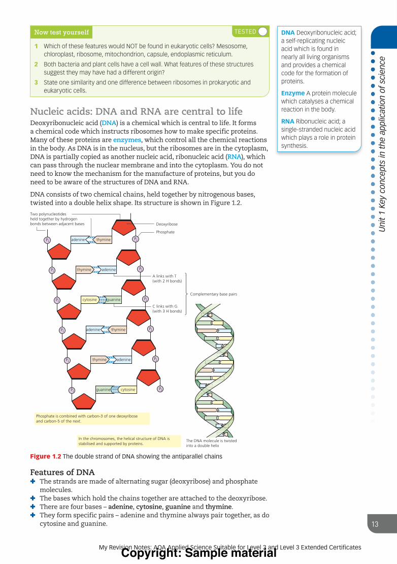
DNA consists of two chemical chains, held together by nitrogenous bases, twisted into a double helix shape. Its structure is shown in Figure 1.2.
adenine
Two polynucleotidesheld together by hydrogenbonds between adjacent bases
adenine
Deoxyribose
Phosphate
A links with T(with 2 H bonds)
C links with G(with 3 H bonds)
Complementary base pairs
The DNA molecule is twistedinto a double helix
guanine
Pi
Pi
Pi
Pi
Pi
Pi
cytosine
thymine
cytosine
thymine
Pi
Pi
Pi
Pi
Pi
Pi
thymine
adenine
guanine
thymine
adenine
Phosphate is combined with carbon-3 of one deoxyriboseand carbon-5 of the next.
In the chromosomes, the helical structure of DNA isstabilised and supported by proteins.
Figure 1.2 The double strand of DNA showing the antiparallel chains
Features of DNA✚ The strands are made of alternating sugar (deoxyribose) and phosphate
molecules.✚ The bases which hold the chains together are attached to the deoxyribose.✚ There are four bases – adenine, cytosine, guanine and thymine.✚ They form specific pairs – adenine and thymine always pair together, as do
cytosine and guanine.
DNA Deoxyribonucleic acid; a self-replicating nucleic acid which is found in nearly all living organisms and provides a chemical code for the formation of proteins.
Enzyme A protein molecule which catalyses a chemical reaction in the body.
RNA Ribonucleic acid; a single-stranded nucleic acid which plays a role in protein synthesis.
9781398317628.indb 139781398317628.indb 13 13/04/21 11:58 PM13/04/21 11:58 PMProgress Geo KS3_8072.indb 4 8/24/18 5:23 PM
Copyright: Sample material

Un
it 1
Key
co
nce
pts
in t
he
app
licat
ion
of
scie
nce
14
Check your understanding and progress at www.hoddereducation.co.uk/myrevisionnotes
✚ The basic ‘unit’ of DNA is a nucleotide. A nucleotide consists of a phosphate group, a pentose sugar and its attached base.
✚ DNA is a very long molecule (in humans, between 50 million and 260 million nucleotides in length).
Features of RNARNA has the same structure as a single strand of DNA, but with the following differences:✚ RNA is not coiled into a helix.✚ The sugar is ribose instead of deoxyribose.✚ Thymine is not found in RNA. Another base, uracil, takes its place.✚ The RNA molecule is much shorter (only a few thousand nucleotides in
length).
Now test yourself
4 Name the chemical molecules that make up a nucleotide.
5 State two chemical differences between DNA and RNA.
You can calculate magnification as observed sizeactual size
If we know the magnification used in a light or electron microscope, it is easy to calculate the actual size of the object using the equation:
actual size = observed sizemagnification
Maths skills
The magnification equation allows us to calculate the magnification of a diagram/photo (if we know the actual size of the structure) or the actual size of a structure (if we know the magnification).
Worked example
An object’s image down a light microscope (magnification ×40) is measured as 0.5 mm. Calculate the actual size of the object.
actual size = observed sizemagnification
= 0.540
0.0125 mm=
The same equation can be rearranged to calculate the magnification if we know the actual size of an object (this is done to calculate the magnification of a drawing or electron micrograph, as when using a microscope, we know the magnification).
Worked example
Red blood cells have an average diameter of around 7.5 µm (0.0075 mm). In a photograph of red blood cells down a microscope, the average cell diameter is 3 mm. What is the magnification of the photo?
magnification = = = ×observed sizeactual size
3.00.0075
400
Practice questions
1 A structure in an electron micrograph is 2 cm (20 000 µm) long. The magnification is given as ×3000. What is the real length of the structure?
2 An electron micrograph of a cell is printed in a textbook. The microscope magnification used is ×20 000. Explain why the magnification given for the photograph in the textbook is ×40 000.
Exam tip
Note that the magnification of a drawing or photograph is not necessarily the same as the magnification used by the microscope. Drawings and photographs are rarely ‘life size’.
9781398317628.indb 149781398317628.indb 14 13/04/21 11:58 PM13/04/21 11:58 PMProgress Geo KS3_8072.indb 4 8/24/18 5:23 PM
Copyright: Sample material

Uni
t 1
Key
conc
epts
in t
he a
pplic
atio
n of
sci
ence
15
My Revision Notes: AQA Applied Science Suitable for Level 3 and Level 3 Extended Certificates
Transport mechanisms
The cell membrane surrounds the cellAll living cells have a cell (plasma) membrane. In eukaryotic cells it consists of a lipid bilayer with protein molecules embedded in it. The structure (known as the fluid mosaic model) is shown in Figure 1.3.
Sugar chain
Lipid
Glycolipid
ProteinSugar chain Glycoprotein
Lipidbilayer
Channel proteinwith pore
Protein on oneside of themembrane
Cholesterol
Transmembrane proteinthat spans the membraneand is exposed at bothsurfaces
Intrinsic protein – Embedded in the lipid bilayer
Extrinsic proteins –Attached to surface of lipid bilayer
Outside of cell
Inside of cell
Phospholipid
Figure 1.3 Structure of a eukaryotic plasma membrane
Most of the membrane consists of a double layer of phospholipids, known as a bilayer. Phospholipid molecules have a hydrophilic head which readily mixes with water, and a hydrophobic tail which is repelled by water. They naturally form a bilayer in water as it is the most stable structure. The heads are towards the outside in the aqueous medium and the tails are as far away from it as possible; see Figure 1.4). Any substance which is soluble in lipids can easily get through this bilayer, but water-soluble substances cannot.
Phospholipidmolecule
Aqueous fluidoutside cell
HydrophilicheadAqueouscytoplasm
Hydrophobictail
Figure 1.4 Structure of the phospholipid bilayer
Now test yourself
6 Which two classes of chemical make up the bulk of the cell membrane?
7 The cell membrane is more permeable to lipid-soluble chemicals than to water-soluble chemicals. Suggest a reason for this.
8 Explain how the hydrophilic heads and hydrophobic tails of lipid molecules lead to the bilayer structure seen in cell membranes.
Aqueous medium A liquid which contains water, i.e. water or an aqueous solution.
Concentration gradient The difference in the concentration of a solute between two areas. The bigger the difference, the ‘steeper’ the concentration gradient.
Intrinsic protein A protein embedded in the lipid bilayer of the cell membrane, sometimes completely penetrating it.
9781398317628.indb 159781398317628.indb 15 13/04/21 11:58 PM13/04/21 11:58 PMProgress Geo KS3_8072.indb 4 8/24/18 5:23 PM
Copyright: Sample material

Un
it 1
Key
co
nce
pts
in t
he
app
licat
ion
of
scie
nce
16
Check your understanding and progress at www.hoddereducation.co.uk/myrevisionnotes
Transport by intrinsic proteins allows water-soluble substances to pass through the cell membraneThe presence of intrinsic proteins spanning the width of the membrane allows water-soluble substances to pass through. The intrinsic proteins are of two types, channel proteins and carrier proteins. The two types transport substances in slightly different ways.
Substances naturally move from an area of higher concentration to an area of lower concentration if there is no barrier in the way, by diffusion. Lipid molecules can diffuse through the membrane at any point in the lipid bilayer, but water-soluble (polar) molecules can only diffuse through where there is an intrinsic protein. This conditional form of diffusion is called facilitated diffusion. Channel proteins have a pore which allows water-soluble substances to pass through the membrane (Figure 1.5). The channel proteins mainly transport ions and small polar molecules.
Carrier proteins can transport substances by facilitated diffusion (including larger molecules) but can also transport molecules against a concentration gradient (i.e. from a lower concentration to a higher concentration) by active transport. Active transport requires energy in the form of adenosine triphosphate (ATP) (Figure 1.6).
Extracellular fluid
Channel protein
Ion or polar moleculeCytoplasm
Extracellular fluid Carrier protein
changes shape
Carrier proteinIon or polarmolecule
Cytoplasm
Figure 1.5 Facilitated diffusion
Extracellular fluid
Carrier protein Transported particle
CytoplasmATP
Figure 1.6 Active transport by carrier proteins
Channel proteins can transport any small water-soluble substance, but carrier proteins are specific and only transport a specific substance or group of substances.
Extrinsic proteins have many functions, including acting as antigens and receptorsExtrinsic proteins can be pure proteins or glycoproteins. Glycoproteins are proteins with a short carbohydrate chain attached. Extrinsic proteins have a variety of functions, but the main ones are as follows:✚ Acting as antigens (molecules which allow the cell to be recognised by the
immune system).✚ Acting as receptors. There are many instances where a chemical (e.g.
hormones) needs to act on some cells but not on others. Receptors allow the chemical to detect the cells which it needs to affect or not affect.
Exam tip
When referring to the movement of substances by diffusion or facilitated diffusion, it is always best to use the term net movement. This is because the movement is not one-way.
Substances move in both directions, but more move down the concentration gradient than up it.
Making links
You can learn more about intrinsic proteins and their role in absorption of digested products in the small intestine in Unit 4, page 129.
Extrinsic protein A protein attached to the outside or inside of the lipid bilayer in a cell membrane.
9781398317628.indb 169781398317628.indb 16 13/04/21 11:58 PM13/04/21 11:58 PMProgress Geo KS3_8072.indb 4 8/24/18 5:23 PM
Copyright: Sample material

Uni
t 1
Key
conc
epts
in t
he a
pplic
atio
n of
sci
ence
17
My Revision Notes: AQA Applied Science Suitable for Level 3 and Level 3 Extended Certificates
Now test yourself
9 Explain the difference between simple diffusion and facilitated diffusion.
10 State two differences between the processes of active transport and facilitated diffusion.
11 What is an antigen?
The heart
The structure of the heart ensure blood flows through it in one directionThe structure of the mamallian heart is shown in Figure 1.7.
Superiorvena cava
Semilunarvalves
Right atrium
Atrioventricular valves
Right ventricle
Inferiorvena cava
Aorta
Pulmonary arteryto left lung
Pulmonary veinsfrom left lungLeft atrium
Valvetendons
Left ventricle
Septum
Figure 1.7 Internal structure of the mammalian heart
The valves in the heart ensure that blood always flows through it in the right direction. The bicuspid and tricuspid valves prevent back-flow from the ventricles into the atria, and the semilunar valves stop blood flowing back into the heart from the aorta and pulmonary artery. Some diseases of the heart are caused by faults in these valves, and artificial valves sometimes need to be fitted.
Control of the heart rate is essentialIn an adult the average resting heart rate is about 70 beats per minute. This regular rhythm is controlled entirely from within the heart, but sometimes the rate needs to be adjusted. For example, during exercise the muscles have a high demand for oxygen carried in the blood so the heart rate is increased to supply this. This adjustment is carried out using external stimulation by the nervous system.
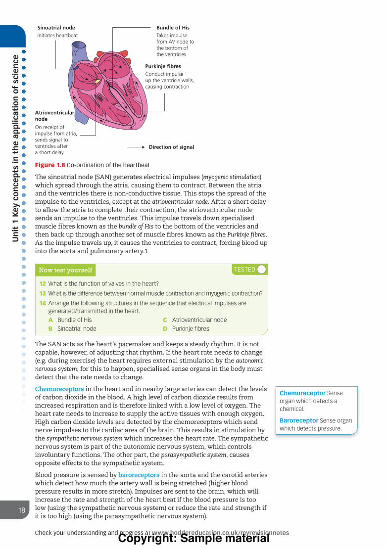
The cardiac muscle of the heart is unusual in that it can carry out myogenic contraction. This is muscle contraction without any external nervous stimulation. The contraction is initiated by the sinoatrial node (SAN), a patch of specialised muscle tissue in the upper right atrium (see Figure 1.8).
Exam tip
Always use the term (electrical) impulse when describing how the heart contracts. Vague terms like ‘message’ or ‘signal’ are unlikely to be acceptable.
9781398317628.indb 179781398317628.indb 17 13/04/21 11:58 PM13/04/21 11:58 PMProgress Geo KS3_8072.indb 4 8/24/18 5:23 PM
Copyright: Sample material
Un
it 1
Key
co
nce
pts
in t
he
app
licat
ion
of
scie
nce
18
Check your understanding and progress at www.hoddereducation.co.uk/myrevisionnotes
Sinoatrial nodeInitiates heartbeat
Bundle of HisTakes impulsefrom AV node tothe bottom ofthe ventricles
Purkinje fibresConduct impulseup the ventricle walls,causing contraction
Atrioventricularnode
On receipt of impulse from atria, sends signal to ventricles after a short delay
Direction of signal
Figure 1.8 Co-ordination of the heartbeat
The sinoatrial node (SAN) generates electrical impulses (myogenic stimulation) which spread through the atria, causing them to contract. Between the atria and the ventricles there is non-conductive tissue. This stops the spread of the impulse to the ventricles, except at the atrioventricular node. After a short delay to allow the atria to complete their contraction, the atrioventricular node sends an impulse to the ventricles. This impulse travels down specialised muscle fibres known as the bundle of His to the bottom of the ventricles and then back up through another set of muscle fibres known as the Purkinje fibres. As the impulse travels up, it causes the ventricles to contract, forcing blood up into the aorta and pulmonary artery.1
Now test yourself
12 What is the function of valves in the heart?
13 What is the difference between normal muscle contraction and myogenic contraction?
14 Arrange the following structures in the sequence that electrical impulses are generated/transmitted in the heart.
A Bundle of His
B Sinoatrial node
C Atrioventricular node
D Purkinje fibres
The SAN acts as the heart’s pacemaker and keeps a steady rhythm. It is not capable, however, of adjusting that rhythm. If the heart rate needs to change (e.g. during exercise) the heart requires external stimulation by the autonomic nervous system; for this to happen, specialised sense organs in the body must detect that the rate needs to change.
Chemoreceptors in the heart and in nearby large arteries can detect the levels of carbon dioxide in the blood. A high level of carbon dioxide results from increased respiration and is therefore linked with a low level of oxygen. The heart rate needs to increase to supply the active tissues with enough oxygen. High carbon dioxide levels are detected by the chemoreceptors which send nerve impulses to the cardiac area of the brain. This results in stimulation by the sympathetic nervous system which increases the heart rate. The sympathetic nervous system is part of the autonomic nervous system, which controls involuntary functions. The other part, the parasympathetic system, causes opposite effects to the sympathetic system.
Blood pressure is sensed by baroreceptors in the aorta and the carotid arteries which detect how much the artery wall is being stretched (higher blood pressure results in more stretch). Impulses are sent to the brain, which will increase the rate and strength of the heart beat if the blood pressure is too low (using the sympathetic nervous system) or reduce the rate and strength if it is too high (using the parasympathetic nervous system).
Chemoreceptor Sense organ which detects a chemical.
Baroreceptor Sense organ which detects pressure.
9781398317628.indb 189781398317628.indb 18 13/04/21 11:58 PM13/04/21 11:58 PMProgress Geo KS3_8072.indb 4 8/24/18 5:23 PM
Copyright: Sample material

Uni
t 1
Key
conc
epts
in t
he a
pplic
atio
n of
sci
ence
19
My Revision Notes: AQA Applied Science Suitable for Level 3 and Level 3 Extended Certificates
Artificial pacemakers can correct irregular heartbeatsSometimes a fault in the sinoatrial node or associated structures leads to an irregular heartbeat, a condition known as arrhythmia. The patient must be fitted with an electronic artificial pacemaker to correct the condition. Pacemakers are made up of a long-lasting battery and a tiny computer (a pulse generator) in a metal case that is fitted underneath the skin. The pacemaker is connected to the heart muscle by leads which can detect heart contractions and send electrical impulses to the muscle. If the heart rate drops below the desired rate, the pacemaker generates an electrical impulse that causes the heart muscle to contract in the correct rhythm.
There are three types of pacemaker:1 Single chamber pacemakers have one lead going to either the right atrium or
the right ventricle.2 Dual chamber pacemakers have two leads, one each going to the right
atrium and the right ventricle.3 Biventricular pacemakers have three leads, one each going to the right
atrium, the right ventricle, and the left ventricle.
The type of pacemaker fitted depends on the specific heart problem the patient has. As a rule, the more serious the condition, the more leads are used. More leads allow for greater control and synchronisation of the heartbeat, but the operation to fit them becomes more complicated.
Leadless micropacemakers are now being developed. These are small devices fixed to the heart which can be fitted by a much simpler (and therefore safer) surgical procedure. However, there is some risk of damaging the wall of the heart and complications can occur if the pacemaker becomes dislodged, which is more likely than with the older types. The first micropacemaker was developed as recently as 2014 and research and development are ongoing.
Now test yourself
15 Which part of the autonomic nervous system causes an increase in the heart rate?
16 Which structure in the heart is likely to be faulty if a single chamber pacemaker is fitted?
Homeostasis
Homeostatic control mechanisms control such things as body temperature and blood pHThe body has physiological control systems that maintain the internal environment within restricted limits. Examples of this include:✚ body temperature range (35.8–37.5 °C)✚ blood glucose range (82–110 mg/dL)✚ blood pH range (7.35–7.45).
To achieve homeostasis, the body has means to detect the current values, and mechanisms to adjust them if they go outside the safe range. These mechanisms often involve hormones (see below).
Many homeostatic control systems are carried out by a mechanism known as negative feedback. An example is the control of water retention (osmoregulation) in the body involving anti-diuretic hormone (ADH), the kidney, and the hypothalamus and pituitary gland in the brain. The kidney controls the water content of the blood by reabsorbing more or less water from the urine. The water is reabsorbed as the urine passes through a structure called the collecting duct, and the permeability of the collecting duct wall can be altered by ADH. The mechanism is shown in Figure 1.9.
Making links
You can learn more about the role of science in medicine in Unit 3 The role of biomedical scientists.
Homeostasis The maintenance of a constant internal state within the body.
Hypothalamus An area in the floor of the brain that maintains the body’s internal balance, often by stimulating the release of hormones from the pituitary gland.
Negative feedback A process where a change causes a series of events which reverse that change.
Pituitary gland A gland hanging from the floor of the brain which produces hormones which control the activity of endocrine (hormone-producing) glands around the body.
9781398317628.indb 199781398317628.indb 19 13/04/21 11:58 PM13/04/21 11:58 PMProgress Geo KS3_8072.indb 4 8/24/18 5:23 PM
Copyright: Sample material

Un
it 1
Key
co
nce
pts
in t
he
app
licat
ion
of
scie
nce
20
Check your understanding and progress at www.hoddereducation.co.uk/myrevisionnotes
Exam tip
It is safer to use the term anti-diuretic hormone in exam answers rather than the abbreviation ADH (at least the first time you use it). This hormone is also known as vasopressin, which is acceptable in answers.
Detectedby osmoreceptors
in the hypothalamus
Increase inblood concentration Collecting duct
walls become lesspermeable (less water is
absorbed)
Increased secretionof ADH by pituitary
gland
Collecting duct wallsbecome more permeable(more water is absorbed)
Decreasein blood
concentration
Decreased secretionof ADH by
pituitary gland
Detectedby osmoreceptors
in the hypothalamus
Figure 1.9 Negative feedback for release of ADH
Hormones control many body functionsHormones are chemicals produced by specialised glands (endocrine glands) which have an effect elsewhere in the body. They travel around the body in the blood. Hormones are important in homeostasis, and also in other aspects of body function. We have already seen how ADH controls water retention in the body. Some other examples of hormones and their functions are given below.
Insulin is produced in the pancreas and lowers blood sugar levels if they get too high. Excess glucose is stored as glycogen in the liver and, when needed, the glycogen is broken down to glucose once again. Insulin lowers blood sugar by activating the enzymes which convert glucose into glycogen.
Glucagon is also produced in the pancreas and has the opposite effect to insulin, raising blood sugar levels if they fall too low. It activates the enzymes which convert glycogen to glucose in the liver.
Adrenaline also affects blood sugar levels. It activates enzymes in the same way as glucagon to boost blood glucose levels in stressful situations and during exercise. It is not involved in the routine maintenance of blood glucose levels. Adrenaline is produced in the inner region of the adrenal glands – the medulla.
Aldosterone is a hormone produced in the cortex (the outer region) of the adrenal glands. (These are paired endocrine glands located on top of the kidneys. Each consists of two regions, an inner cortex and an outer medulla.) Aldosterone helps to regulate blood pressure mainly by increasing the amount of sodium reabsorbed into the bloodstream by the kidney and the colon and boosting the amount of potassium in the urine. As water is reabsorbed along with the sodium, this increases blood volume and causes an increase in blood pressure.
Exam tip
Learn the normal ranges given here. Questions in past exams have tested them.
9781398317628.indb 209781398317628.indb 20 13/04/21 11:58 PM13/04/21 11:58 PMProgress Geo KS3_8072.indb 4 8/24/18 5:23 PM
Copyright: Sample material

Uni
t 1
Key
conc
epts
in t
he a
pplic
atio
n of
sci
ence
21
My Revision Notes: AQA Applied Science Suitable for Level 3 and Level 3 Extended Certificates
Now test yourself
17 How do hormones travel from place to place in the body?
18 How does ADH determine the concentration of the urine?
19 Name the hormone that will be produced if blood glucose levels fall too low.
The treatment of diabetes differs for type 1 and type 2 diabetesThere are two types of diabetes, known as type 1 and type 2. Both result in an inability of the body to control blood sugar levels, but the causes and treatments of the two types are different.
Type 1 diabetes:✚ Causes are uncertain. Type 1 diabetes is an autoimmune disease, which
means that the body’s immune system attacks its own cells, in this case specialised cells called beta cells in the Islets of Langerhans in the pancreas, which produce insulin.
✚ Patients with type 1 diabetes produce little or no insulin.✚ Onset of type 1 diabetes is usually in childhood or young adulthood.✚ Type 1 diabetes is treated by regular insulin injections and control of
carbohydrate intake.
Type 2 diabetes:✚ Causes are uncertain but are known to be linked with obesity and lack of
physical activity.✚ Some patients with type 2 diabetes may have reduced levels of insulin, but
the main problem is that the body has become insensitive to insulin, so it does not work effectively.
✚ Onset of type 2 diabetes is usually in middle age, although recently there has been a rise in younger people with the condition.
✚ Type 2 diabetes is usually treated by a strict diet to restrict carbohydrate intake and by tablets, although insulin injections may also be used in some cases.
The key clinical symptom of diabetes is sugar in the urine. This can quickly be detected by the use of urine dipsticks, which change colour according to the level of sugar in the urine and are then compared with a standard colour chart. The diagnosis can be confirmed and refined by use of blood glucose ‘pinprick’ tests, which provide a digital reading of blood glucose levels. In diagnosis, fasting glucose levels are used, where the patient eats nothing for 8–12 hours before the test. Eating carbohydrate influences blood glucose levels and a fasting glucose test eliminates that variable.
Urine dip sticks used to be used by patients to monitor their glucose levels but these have largely been replaced by blood pinprick tests which give a direct reading of blood glucose.
Now test yourself
20 Which type of diabetes is most likely to develop in middle age?
21 Suggest a reason why people with diabetes control their intake of carbohydrates in general, rather than sugar specifically.
9781398317628.indb 219781398317628.indb 21 13/04/21 11:58 PM13/04/21 11:58 PMProgress Geo KS3_8072.indb 4 8/24/18 5:23 PM
Copyright: Sample material

Un
it 1
Key
co
nce
pts
in t
he
app
licat
ion
of
scie
nce
22
Check your understanding and progress at www.hoddereducation.co.uk/myrevisionnotes
The kidney filters the blood to excrete waste and control salt levelsThe kidney has two functions, excretion of wastes and osmoregulation. Osmoregulation involves both the control of water content of the blood and the salt concentration.
The kidney consists of millions of microscopic tubules called nephrons.
Renal artery
Renalpelvis
Renalmedulla
Renalcortex
Renal vein
Ureter
Glomerulus
Proximalconvolutedtubule
Distal convolutedtubule
Renal corpuscle
Bowman'scapsule
Renalartery
Renalvein
Loop ofHenle
Collectingduct
To ureter
Figure 1.10 Nephron structure and location in the kidney
The kidney functions by filtering the blood (a process known as ultrafiltration) and then reabsorbing useful small molecules that pass through the filter. Essential molecules like glucose are all reabsorbed in the proximal convoluted tubule, but salts such as sodium are only partly reabsorbed, just enough to maintain a suitable level in the blood.
UltrafiltrationHigh blood pressurein the glomerulus forcesfluid into Bowman'scapsule through abasement membranewhich acts as a filter
Small molecules enteringBowman's capsule includeglucose, amino acids andsalts (e.g. sodium)
In the proximal convoluted tubule,there is selective reabsorption.Glucose, amino acids, some waterand some of the sodium are reabsorbed byfacilitated diffusion and active transport
In the distal convoluted tubule andcollecting duct, water is reabsorbed.The amount is controlled by thehormone ADH, according tothe body's needs
Figure 1.11 Processes occurring in the nephron
As outlined earlier in this chapter, the amount of sodium absorbed in the kidneys is controlled by the hormone aldosterone, which is produced in the adrenal cortex. Increased aldosterone secretion increases the amount of sodium absorbed.
The control of salt is important to healthIf the concentration of salt in the blood falls below or rises above the normal range, health problems result.
Ultrafiltration Filtration of small molecules.
9781398317628.indb 229781398317628.indb 22 13/04/21 11:58 PM13/04/21 11:58 PMProgress Geo KS3_8072.indb 4 8/24/18 5:23 PM
Copyright: Sample material

Uni
t 1
Key
conc
epts
in t
he a
pplic
atio
n of
sci
ence
23
My Revision Notes: AQA Applied Science Suitable for Level 3 and Level 3 Extended Certificates
Sodium chloride (salt) deficiency can result from drinking too much water, chronic and severe vomiting or diarrhoea, problems with the heart, liver or kidneys, a deficiency of aldosterone and as a side-effect of certain medications. It is referred to as hyponatremia and can result in the following:✚ nausea and vomiting✚ headache✚ confusion✚ loss of energy, drowsiness and fatigue✚ spasms or cramps.
In severe cases, if there is a continuous period of salt deficiency, rapid brain swelling can cause coma and death.
Excess salt in the blood is most likely to result from taking in too much salt in the diet. The main effect is an increase in blood pressure, which increases the risk of cardiovascular disease. The increased concentration of salt in the blood causes water to enter by osmosis, increasing the blood pressure.
Now test yourself
22 State the two functions of the kidney.
23 Explain why glucose passes from the blood into the nephron, but proteins do not.
24 State the part of the body that produces the hormone which controls sodium levels in the blood.
Breathing and cellular respiration
Breathing and respiration are not the sameIn everyday language, breathing and respiration are sometimes used interchangeably, but to a biologist they are distinctly different processes. Breathing is a physical, external process which draws air containing oxygen into the body where it is exchanged for carbon dioxide, whereas respiration is a chemical, internal process which extracts energy from food in a usable form.
A number of methods are used by scientists to monitor the respiratory system. Breathing rate can be easily counted. An instrument known as a spirometer can measure tidal volume (the volume of air breathed in and out during normal breathing) and vital capacity (the maximum amount of air that can be breathed in and out during deep breathing). A peak flow meter measures how fast a person can breathe out after taking a full breath in.
These tests are used to diagnose and/or monitor respiratory conditions and diseases, including asthma.
Cellular respiration breaks down foodIn the process of cellular respiration, food materials (mainly glucose) are gradually broken down in multiple steps, and the energy within them is used to form adenosine triphosphate (ATP) from adenosine diphosphate (ADP). This addition of phosphate is called a phosphorylation reaction. Every cellular process which requires energy uses ATP as its source.
Cellular respiration can be broken down into three stages: glycolysis, which occurs in the cytoplasm; the Krebs cycle, which occurs in the mitochondria; and the electron transfer chain, which also occurs in the mitochondria.
Exam tip
In answers, make sure you do not confuse breathing with respiration. Note, however, that the system involved with breathing is called the respiratory system, not the breathing system!
Making links
You learned about mitochondria earlier in this unit. See pages 11 and 12.
9781398317628.indb 239781398317628.indb 23 13/04/21 11:58 PM13/04/21 11:58 PMProgress Geo KS3_8072.indb 4 8/24/18 5:23 PM
Copyright: Sample material

Un
it 1
Key
co
nce
pts
in t
he
app
licat
ion
of
scie
nce
24
Check your understanding and progress at www.hoddereducation.co.uk/myrevisionnotes
The whole process of cellular respiration is shown in Figure 1.12.
2ADP + 2Pi NAD
Reduced NAD
NAD
Reduced NAD
2ATP
CO2
CO2
CO2
Hexose bisphosphate (6C)
Triose phosphate (3C) Triose phosphate (3C)
Pyruvate
Acetyl coenzyme A
Krebs cycle
Coenzyme A
Coenzyme A
Citrate (6C)Oxaloacetate (4C)
Reduced hydrogen carriers (reduced NAD and FAD are oxidised)
Electron transport chain
Glucose pyruvate = glycolysis
Pyruvate acetyl coA = link reaction
NAD
NAD
ADP + Pi
FAD
Oxygen
Water
NADReduced
NAD
Reduced NAD
ATP
ADP + PiATP
Reduced FAD
Reduced NAD
Figure 1.12 An overview of all stages of respiration
Now test yourself
25 Explain the difference between vital capacity and tidal volume.
26 Which stage of cellular respiration does NOT take place in the mitochondria?
27 What type of chemical reaction is involved in the conversion of ADP to ATP?
Key facts about glycolysis✚ Glycolysis takes place in the cytoplasm.✚ The purpose of glycolysis is to convert glucose into pyruvate, which can
enter the Krebs cycle.✚ Each of the first two steps (the conversion of glucose to glucose phosphate
and then to hexose biphosphate) uses a molecule of ATP.✚ In glycolysis four molecules of ATP are produced, a net gain of two.✚ The production of ATP in this stage is by substrate-linked phosphorylation.✚ Two molecules of NAD are converted into reduced NAD during glycolysis.✚ If oxygen is present, the reduced NAD is fed into the electron transfer
chain.✚ The conversion of triose phosphate into pyruvate is an oxidation reaction.
NAD is an abbreviation of nicotinamide adenine dinucleotide. Reduced NAD is sometimes abbreviated to NADH.
Exam tip
Use the full term adenosine triphosphate when mentioning it for the first time, not the abbreviation ATP.
Substrate-linked phosphorylation The formation of ATP which occurs when a reaction in the cell produces enough energy to convert ADP to ATP, without the involvement of the electron transfer chain.
Oxidation A reaction involving the addition of oxygen, the removal of hydrogen or the loss of electrons. The opposite of oxidation is reduction.
9781398317628.indb 249781398317628.indb 24 13/04/21 11:58 PM13/04/21 11:58 PMProgress Geo KS3_8072.indb 4 8/24/18 5:23 PM
Copyright: Sample material

Uni
t 1
Key
conc
epts
in t
he a
pplic
atio
n of
sci
ence
25
My Revision Notes: AQA Applied Science Suitable for Level 3 and Level 3 Extended Certificates
Exam tip
Hexose bisphosphate is converted into TWO molecules of triose phosphate, so when calculating ATP and reduced NAD production from one glucose molecule in the Krebs cycle and electron transfer chain, the number needs to be doubled.
Key facts about the Krebs cycle✚ The pyruvate formed in glycolysis is converted to acetyl coenzyme a,
which enters the Krebs cycle. This reaction is known as the link reaction.✚ Acetyl CoA reacts with a 4-carbon molecule to form a 6-carbon molecule.✚ A series of oxidation–reduction reactions release hydrogen, which will
provide electrons for the electron transfer chain.✚ The hydrogen attaches to NAD (see above) or flavine adenine dinucleotide
(FAD). NAD and FAD belong to a group of chemicals called coenzymes.✚ For each turn of the Krebs cycle, one molecule of ATP is formed by
substrate-linked phosphorylation (so two in total for every glucose molecule).
✚ Carbon dioxide is released during the Krebs cycle.
Key facts about the electron transfer chain✚ Electrons are passed along a series of electron carriers in the inner
membrane of the mitochondrion.✚ The electrons enter the chain when hydrogen, bought to the membrane
by NAD or FAD, is released and broken down into electrons and hydrogen ions (H+).
✚ As the electrons pass from carrier to carrier, energy is released and used to phosphorylate ADP to ATP. This is oxidative phosphorylation.
✚ The final electron acceptor which takes the electrons out of the chain is oxygen, which combines with the electrons and hydrogen ions to form water.
✚ For each reduced NAD molecule brought to the electron transfer chain, three ATP molecules are formed, and two for each reduced FAD.
Now test yourself
28 Name the end product of glycolysis.
29 How is the hydrogen generated in the Krebs cycle used in the electron transfer chain?
30 What is the precise location of the electron transfer chain in the mitochondrion?
Anaerobic respiration is used when oxygen is unavailableIf oxygen is unavailable or in short supply, certain cells (e.g. muscle cells) can switch to anaerobic respiration. In the anaerobic pathway, the Krebs cycle and electron transfer chain do not function, but glycolysis continues. The pyruvate formed is converted into lactic acid in animals and into ethanol and carbon dioxide in plant cells. No ATP is formed in this stage, and so the total net production from a molecule of glucose is two ATP molecules. In aerobic respiration, approximately 30 molecules of ATP are produced per glucose molecule. If anaerobic respiration continues for some time, lactic acid build-up can result in muscle pain, cramps and muscular fatigue.
Basal metabolic rate is the rate at which the body uses energyBasal metabolic rate (BMR) is the rate at which the body uses energy at rest to maintain basic life functions. It can be measured in kcal/day or kJ/day and varies among individuals. Its measurement is useful in weight control programmes, to help determine a suitable daily calorie intake for the patient.
Exam tip
You are not expected to name the 4-carbon and 6-carbon molecules in the Krebs cycle.
Exam tip
Do not use the abbreviation BMR in answers unless you (or the question) have already defined its meaning.
9781398317628.indb 259781398317628.indb 25 13/04/21 11:58 PM13/04/21 11:58 PMProgress Geo KS3_8072.indb 4 8/24/18 5:23 PM
Copyright: Sample material

Un
it 1
Key
co
nce
pts
in t
he
app
licat
ion
of
scie
nce
26
Check your understanding and progress at www.hoddereducation.co.uk/myrevisionnotes
BMR can be determined by direct or indirect methods. BMR may be measured by gas analysis through direct calorimetry. This is the measurement of the heat production of an individual, when placed in an insulated chamber where the heat is transferred to surrounding water. This is the most accurate method of measuring BMR. Indirect calorimetry can also be used. This calculates heat that living organisms produce by measuring either their production of carbon dioxide and nitrogen waste or their consumption of oxygen. Heart rate at rest can be used to estimate energy expenditure because there is a correlation between heart rate and oxygen consumption.
BMR measurements vary among individuals but BMR is generally higher in males than in females, and tends to decrease with age. It is possible that obesity might increase BMR.
Now test yourself
31 Suggest a reason why the electron transfer chain cannot function without oxygen.
32 Suggest why the measurement of carbon dioxide production to establish BMR is referred to as indirect calorimetry.
Photosynthesis and food chain productivity
The process of photosynthesis uses carbon dioxide and water to make glucose and oxygenPhotosynthesis involves a complex series of chemical reactions (see below), but can be summarised by the following equation:
carbon dioxide + water → glucose + oxygen
Carbon dioxide and water are the essential raw materials need for photosynthesis, along with light as an energy source. Carbon dioxide is obtained from the air (although the carbon dioxide produced by the plant in respiration can also be used) and water comes from the soil. Photosynthesis takes place in the chloroplasts in leaves. Carbon dioxide can enter the leaves directly through pores called stomata, and water enters the plant via the roots and is carried up the stem to the leaves.
The biochemistry of photosynthesis has a light-dependent stage and a light-independent stageThe biochemistry of photosynthesis can be broken down into two stages:1 The light-dependent stage, in which light energy is used to hydrolyse water
into hydrogen and oxygen. The hydrogen provides electrons which are essential for the manufacture of ATP, and eventually joins with nicotinamide adenine dinucleotide phosphate (NADP) to form reduced NADP. The oxygen is released as a waste product. In the light-dependent stage, ATP is formed in an electron transfer chain similar to that in respiration.
2 The light-independent stage, in which the ATP and reduced NADP formed in the light-dependent stage are used to produce carbohydrate (mainly glucose). This process also requires carbon dioxide. The glucose formed in the light-independent stage can be processed in the plant to produce the other essential foodstuffs, proteins (with the addition of nitrogen) and lipids.
Exam tip
You may need to analyse secondary data in relation to BMR. When comparing BMR, it is necessary to control certain variables – gender, age, level of activity, level of obesity (whichever of these is not being investigated).
Making links
You can learn more about respiration in the previous section, page 23.
9781398317628.indb 269781398317628.indb 26 13/04/21 11:58 PM13/04/21 11:58 PMProgress Geo KS3_8072.indb 4 8/24/18 5:23 PM
Copyright: Sample material
