muscle proteomics of the indian major carp catla (catla catla, hamilton)
TRANSCRIPT
ISSN: 0974-276X
The International Open AccessJournal of Proteomics and Bioinformatics Studies
Executive Editors
Helmut E. MeyerRuhr-University of Bochum, Germany
Alan J TackettUniversity of Arkansas for Medical Sciences, USA
Fuchu HeThe State Key Laboratory of Proteomics, China
Sandra OrchardWellcome Trust Genome Campus, UK
Richard J. SimpsonUniversity of Melbourne, Australia
This article was originally published in a journal by OMICS Publishing Group, and the attached copy is provided by OMICS Publishing Group
for the author’s benefit and for the benefit of the author’s institution, for commercial/research/educational use including without limitation use in instruction at your institution, sending it to specific colleagues that you know, and providing a copy to your institution’s administrator.
All other uses, reproduction and distribution, including without limitation commercial reprints, selling or licensing copies or access, or posting on open internet sites, your personal or institution’s website or repository, are requested to cite properly.
Available online at: OMICS Publishing Group (www.omicsonline.org)
Digital Object Identifier: http://dx.doi.org/10.4172/jpb.1000288
Research Article Open Access
Mohanty et al., J Proteomics Bioinform 2013, 6:11http://dx.doi.org/10.4172/jpb.1000288
Research Article Open Access
Proteomics & Bioinformatics
Volume 6(11) 252-263 (2013) - 252 J Proteomics BioinformISSN: 0974-276X JPB, an open access journal
Muscle Proteomics of the Indian Major Carp Catla (Catla catla, Hamilton)Bimal Prasanna Mohanty1*, Sudeshna Banerjee1, Soma Bhattacharjee1, Tandrima Mitra1, Gopal Krishna Purohit2, Anil Prakash Sharma1, Dhanasekar Karunakaran1 and Sasmita Mohanty2
1ICAR- Central Inland Fisheries Research Institute, Biochemistry Laboratory, Barrackpore, Kolkata-700 120, India2KIIT School of Biotechnology, KIIT University, Bhubaneswar- 7520241, India
*Corresponding author: Bimal Prasanna Mohanty, ICAR- Central Inland Fisheries Research Institute, Biochemistry Laboratory, FREM Division, Barrackpore, Kolkata-700 120, India, Tel: +91-33-25921190; Fax: +91-33-2592 0388; E-mail: [email protected]
Received September 06, 2013; Accepted October 30, 2013; Published November 01, 2013
Citation: Mohanty BP, Banerjee S, Bhattacharjee S, Mitra T, Purohit GK, et al. (2013) Muscle Proteomics of the Indian Major Carp Catla (Catla catla, Hamilton). J Proteomics Bioinform 6: 252-263. doi:10.4172/jpb.1000288
Copyright: © 2013 Mohanty BP, et al. This is an open-access article distributed under the terms of the Creative Commons Attribution License, which permits unrestricted use, distribution, and reproduction in any medium, provided the original author and source are credited.
Keywords: Catla catla; 2-D electrophoresis; Proteogenomics; MALDI-TOF-MS; Reference muscle proteome; Indian major carp
IntroductionProteomics is an unbiased, technology driven approach for
the comprehensive cataloguing of entire protein complements and represent an ideal analytical tool for the high throughput discovery of protein alterations in health and disease [1]. Mass spectrometry-based proteomics is concerned with the global analysis of protein composition, posttranslational modifications and the dynamic nature of expression levels. The generation of large data sets on protein expression levels makes proteomics a preeminent hypothesis generating approach in modern biology [2,3]. Organisms with no available genome sequence data can be studied using comparative proteogenomic approach [3]. Aiming to better understand proteome alterations, it is vital to have a reference proteome map for a specific tissue and species. In this respect, proteogenomics is a thorough approach for the detailed biochemical analysis of heterogeneous and plastic types of tissue, such as muscles.
Muscle plays a central role in whole-body protein metabolism by serving as the principal reservoir for amino acids to maintain protein synthesis in vital tissues and organs [4]. Skeletal muscle fibers represent one of the most abundant cell types in the vertebrates [5] and contractile fibers of skeletal muscle tissues provide coordinated excitation-contraction-relaxation cycles for voluntary movements and postural control [6], besides playing a central physiological role in heat homeostasis and presenting itself as a crucial metabolic tissue that integrates various biochemical pathways [7]. Skeletal muscle proteomics aims at the global identification, detailed cataloguing and biochemical characterization of the entire protein complement of voluntary contractile tissues in normal and pathological specimens [8]. Muscle proteomics has been applied to the comprehensive biochemical profiling of developing, maturity and ageing muscle, as well as the analysis of contractile tissues undergoing physiological adaptations
seen in disuse atrophy, physical exercise and chronic muscular transformations [2].
Among the higher vertebrates muscle proteomic information is available on human [9], rat [10], rabbit [11] and chicken [12] and in lower vertebrates such information has been generated on the piscines zebrafish [13], common carp [14], seabream [15], cod [16] and the snakehead [17]; however, little is known about the patterns, mechanisms of muscle growth and muscle proteome of the Indian major carps rohu (Labeo rohita), catla (Catla catla) and mrigal (Cirrhinus mrigala), the major aquacultured species in the Indian subcontinent. In fish, skeletal muscle is the largest organ system and represents the edible part. It constitutes 34-48% of the total body weight of fish [15]. Muscle composition contributes strongly to quality; in fact texture, elasticity, and water holding capacity, all features highly related to quality, are dependent on number and integrity of muscular fibers [18]. The number of muscle fiber recruited during growth is subjected to variation depending on several factors, such as the fish strain, diet, exercise training and temperature [15].
AbstractBackground: Skeletal muscle proteomics aims at global identification, cataloging and biochemical
characterization of the entire protein complement of voluntary contractile tissues. In the lower vertebrates like fish, skeletal muscle contributes 34-48% of the total body weight and the muscle composition contributes strongly to the quality. Characterization of the muscle proteome is a key to many aspects of aquaculture, encompassing physiology, growth, food safety, seafood authentication and quality, traceability and shelf-life. Catla catla is a commercially important carp species contributing a major share to be freshwater aquaculture production in the Indian subcontinent; however, little omics information is available on this species.
Methods: In the present study, protein spots excised from 2-D gels of catla muscle proteome were identified through MALDI-TOF-MS. Based on the muscle proteomic information, transcript information on the identified proteins was generated.
Results: A reference muscle proteome map for Catla catla was generated and 70 protein spots from 2-D gels, representing 22 proteins, have been identified by MALDI-TOF MS. We have also generated the partial gene sequence information on the identified proteins which have been submitted at GenBank.
Conclusion: This is the first study on the muscle proteogenomics of the commercially important carp Catla catla. Besides adding to the existing knowledgebase on comparative muscle proteomics, the information generated would also serve as the baseline proteogenomic information on this Indian major carp.
Citation: Mohanty BP, Banerjee S, Bhattacharjee S, Mitra T, Purohit GK, et al. (2013) Muscle Proteomics of the Indian Major Carp Catla (Catla catla, Hamilton). J Proteomics Bioinform 6: 252-263. doi:10.4172/jpb.1000288
Volume 6(11) 252-263 (2013) - 253 J Proteomics BioinformISSN: 0974-276X JPB, an open access journal
Catla (Catla catla) is a commercially important carp species and contributes a major share to the freshwater aquaculture production in the Indian subcontinent. In the present study, we have generated reference muscle proteome map of Catla catla, identified 70 spots on 2-D gel and have compared these proteins with the muscle proteins identified across species. As the genome sequence data are not available for this organism, a proteogenomic approach is adopted to generate the partial gene sequence information on the identified muscle proteins.
Materials and MethodsFish
Apparently healthy major carp Catla catla (n=12), weighing 800-1000 g (length 35-40 cm), were procured from a reputed fish farm and hatchery in Kolkata. The species status of the specimens was confirmed by analyzing for species-specific RAPD markers for catla [19].
Preparation of muscle extracts and protein quantification
Axial white skeletal muscle from midway down the body, under the dorsal fin and above the lateral line, was swiftly dissected out from fishes euthanized with MS 222 (>100-200 mgL-1). For muscle protein extraction, white muscle tissues were pooled and mechanically homogenized in ice-cold PBS (137 mM NaCl, 2.7 mM KCl, 4.3 mM Na2HPO4.7H2O, 1.4 mM KH2PO4), pH 7.3 containing protease inhibitor cocktail (Sigma) [20]. To minimize protein modification or degradation, all dissection and sample processing was performed on ice. The homogenates were centrifuged in a high speed refrigerated centrifuge (Biofuge FRESCO, Heraeus) at 10,000 rpm at 4ºC for 10 min and supernatants (representing the soluble protein extracts) were aspirated out. Protein concentration of the extracts was determined using Bradford method [21] using BSA (Sigma) as the standard. The samples were stored as aliquots at -40ºC, until further use.
Gel electrophoresis
Prior to 2-D GE analysis, the proteins were analyzed by sodium dodecyl sulfate polyacrylamide gel electrophoresis (SDS-PAGE) [22] to check the protein quality and also to ensure equal loading. For 2-D GE analysis, the first dimension run (isoelectric focusing) was performed using a Bio-Rad Protean IEF Cell with 11 cm immobilized pH gradient (IPG) strips (pH 5-8, Sigma) following standard protocol [23] and have been described earlier [20]. The protein sample (~150 μg) was premixed with rehydration buffer (8 M urea, 2 M thiourea 2% CHAPS, 50 mM DTT, 0.2% Biolyte, 5/8 ampholyte, and 0.001% bromophenol blue) and rehydration of the IPG strips was carried out for 12 h. The rehydrated strips were isoelectrofocussed at a current of 50 μA/strip at the stated voltage gradient: 250 V for 20 min, 500 V for 30 min, 1000 V for 15 min, 2000 V for 15 min, 4000 V for 15 min, 8000 V for 2 h 30 min, 8000 V for 20000 V-h with an end voltage of 30,000 V-h After the IEF run, the focused strips were equilibrated with the equilibration buffers (EB) - I and II [EB 1 (reducing buffer): 0.375 M Tris-HCl at pH 8.8, 6 M urea, 20% v/v Glycerol, 2% SDS, 130 mM DTT and EB II (alkylating buffer): 0.375 M Tris-HCl at pH 8.8, 6 M urea, 20% v/v glycerol, 2% SDS, 135 mM iodoacetamide] and then placed on SDS polyacrylamide slab gels for second dimension run. The second dimension (SDS-PAGE) run was performed using 12% separating gels with 5% (w/v) stacking gel on a PROTEAN XI cell (Bio-Rad). The gels were stained with Coomassie Brilliant blue or Coomassie-silver double stained and the 2-D gel images were acquired by ImageScanner III LabScan 6.0 (GE Healthcare Biosciences).
MALDI-TOF-TOF-MS
Protein spots of interest were cut from the 2-D polyacrylamide gel, destained in methanol and ammonium bicarbonate buffer, and digested overnight with trypsin. The resulting peptides were extracted following standard techniques [24] by two 20-min incubations with 10-20 µL ACN containing 1% TFA, depending on the size of the gel piece. The resulting tryptic peptide extract was dried by rotary evaporation and stored at -20°C for further analysis by MS. The peptides were analysed by MALDI-TOF-TOF mass spectrometer using a 5800 Proteomics Analyzer (AB Sciex).
The dry peptide samples were reconstituted in 10 µL standard diluent (30:70; ACN:water). The resulting solution was diluted 1:10 with matrix solution (CHCA, 10 mg/mL) and spotted on a 384-well Opti-TOF stainless steel plate. The spotted samples were analyzed using a first run of standard TOF-MS. The system was set to perform a second run of MS/MS focused on the 20 most intensive peaks of the first MS (excluding peaks known to be of trypsin). The laser was set to fire 400 times per spot in MS and 2000 times per spot in MS/MS mode. Laser intensity was 2800 J and 3900 J for MS and MS/MS, respectively. A mass range of 800-4000 amu with a focus mass of 2100 amu was used.
For protein identification, peptide masses from trypsin digests derived using the MALDI-TOF- MS were used to search against Ludwig NR Database using the MASCOT program (www.matrixscience.com) [25]. The MASCOT search parameters were as follows: peptide mass accuracy was 100 ppm, protein modifications: cysteine as S-carbamidomethyl-derivative and oxidation of methionine allowed. The default search parameters used were: enzyme, trypsin; maximum missed cleavages, 1; fixed modifications, carbamidomethyl (C); variable modifications, oxidation (M); peptide tolerance+0.4 Da; Fragment mass tolerance+0.4 Da; Protein mass, unrestricted; instrument=Default.
Data and network pathway
Proteins were identified by MS analyses and protein database (Mascot) searches, basic descriptions of identified protein spots including protein names, accession numbers, theoretical molecular mass and pI values were provided. In addition to these descriptions other fundamental information relevant to identified proteins were acquired through further search of databases available in the public domain. Database accession numbers for identified proteins were provided as inputs to the Uniprot database (http://www.uniprot.org/uniprot/) to obtain gene ontology annotation and sequence length. The consensus lists of proteins obtained were then analyzed for their respective molecular function, biological processes involved protein class and associated metabolic pathway by using Panther classification system software (www.pantherdb.org/pathway/).
Tissue collection and RNA extraction for transcript analysis
White muscle tissues were collected from anaesthetized fishes and stored at (-80°C) in RNA later (Sigma) to avoid RNA degradation. Subsequently, in total RNA isolation was completed by using RNA Express Reagent (Himedia). Briefly, samples (~1 gm) were homogenized in 1 ml RNA Express reagent; chloroform was added to separate the mixture into protein, DNA and RNA. The RNA supernatant was precipitated with 2- propanol (Himedia). The pellets were resuspended in 25 μl of DEPC (Himedia) treated water and stored at -80°C until further use.
Citation: Mohanty BP, Banerjee S, Bhattacharjee S, Mitra T, Purohit GK, et al. (2013) Muscle Proteomics of the Indian Major Carp Catla (Catla catla, Hamilton). J Proteomics Bioinform 6: 252-263. doi:10.4172/jpb.1000288
Volume 6(11) 252-263 (2013) - 254 J Proteomics BioinformISSN: 0974-276X JPB, an open access journal
cDNA preparation by Reverse Transcriptase
Following isolation of RNA, the concentration of each RNA sample was measured and DNase (Fermentas) treatment was done to remove genomic DNA carryover. RNA (1μg) was reversed transcribed using M-MuLV reverse transcriptase. RT reactions were conducted in 20 μl reactions containing 5 μg DNase I-treated RNA, 1 μl oligo dT primers (Fermentas), 4μl dNTPs (Himedia), 0.5 μl RNase inhibitor (Fermentas), 5×RT reaction buffer, and 1 μl of RevertAid RT (Fermentas). Detailed procedures were followed from the manufacturer’s instructions.
Polymerase chain reaction
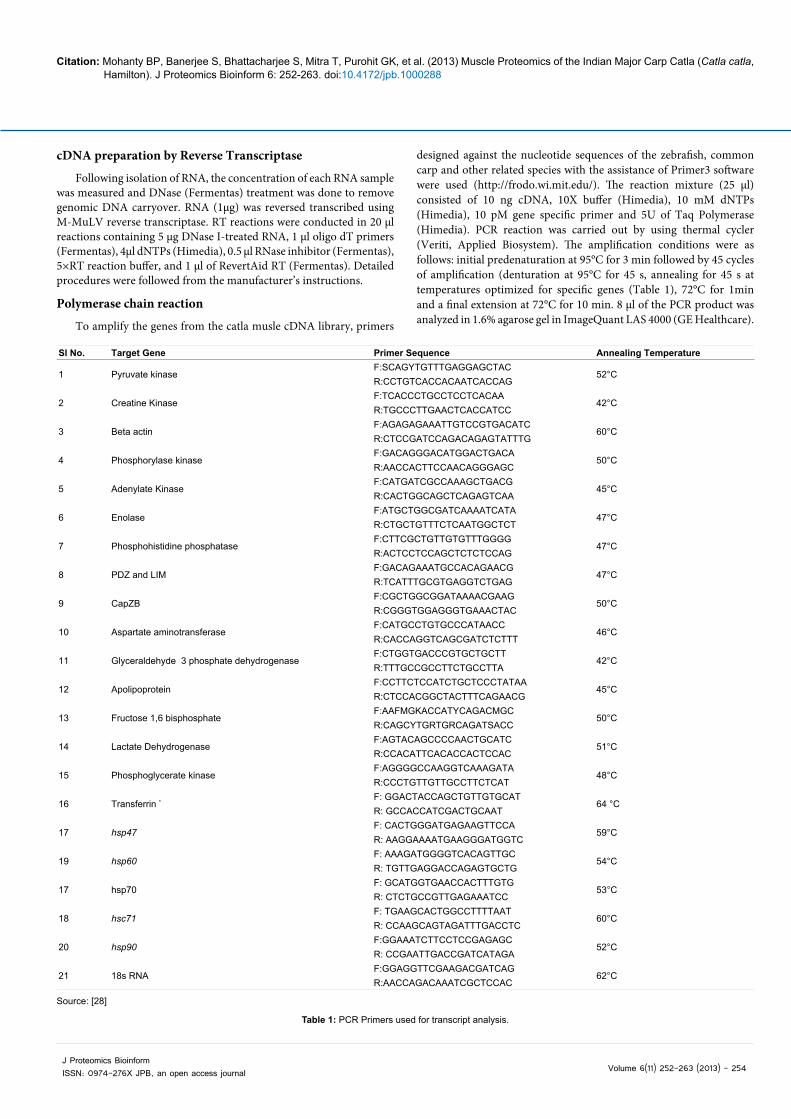
To amplify the genes from the catla musle cDNA library, primers
designed against the nucleotide sequences of the zebrafish, common carp and other related species with the assistance of Primer3 software were used (http://frodo.wi.mit.edu/). The reaction mixture (25 μl) consisted of 10 ng cDNA, 10X buffer (Himedia), 10 mM dNTPs (Himedia), 10 pM gene specific primer and 5U of Taq Polymerase (Himedia). PCR reaction was carried out by using thermal cycler (Veriti, Applied Biosystem). The amplification conditions were as follows: initial predenaturation at 95°C for 3 min followed by 45 cycles of amplification (denturation at 95°C for 45 s, annealing for 45 s at temperatures optimized for specific genes (Table 1), 72°C for 1min and a final extension at 72°C for 10 min. 8 μl of the PCR product was analyzed in 1.6% agarose gel in ImageQuant LAS 4000 (GE Healthcare).
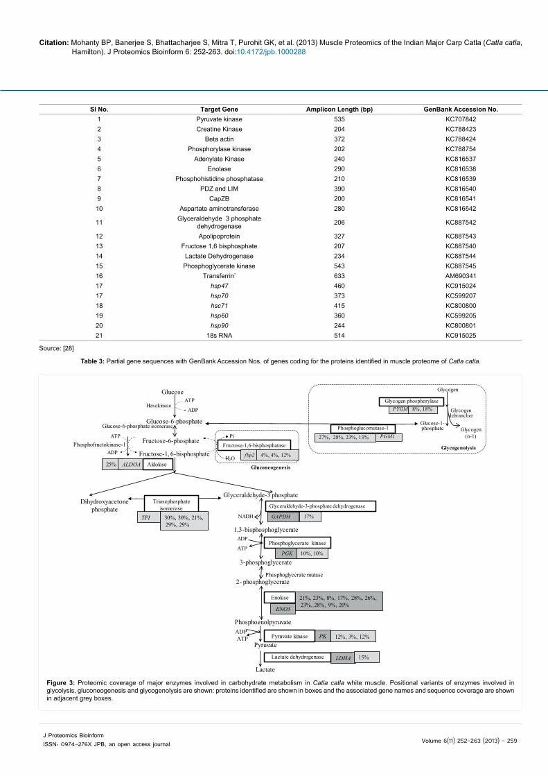
Source: [28]
Table 1: PCR Primers used for transcript analysis.
Sl No. Target Gene Primer Sequence Annealing Temperature
1 Pyruvate kinase F:SCAGYTGTTTGAGGAGCTAC
52°CR:CCTGTCACCACAATCACCAG
2 Creatine Kinase F:TCACCCTGCCTCCTCACAA
42°CR:TGCCCTTGAACTCACCATCC
3 Beta actin F:AGAGAGAAATTGTCCGTGACATC
60°CR:CTCCGATCCAGACAGAGTATTTG
4 Phosphorylase kinase F:GACAGGGACATGGACTGACA
50°CR:AACCACTTCCAACAGGGAGC
5 Adenylate Kinase F:CATGATCGCCAAAGCTGACG
45°CR:CACTGGCAGCTCAGAGTCAA
6 Enolase F:ATGCTGGCGATCAAAATCATA
47°CR:CTGCTGTTTCTCAATGGCTCT
7 Phosphohistidine phosphatase F:CTTCGCTGTTGTGTTTGGGG
47°CR:ACTCCTCCAGCTCTCTCCAG
8 PDZ and LIM F:GACAGAAATGCCACAGAACG
47°CR:TCATTTGCGTGAGGTCTGAG
9 CapZB F:CGCTGGCGGATAAAACGAAG
50°CR:CGGGTGGAGGGTGAAACTAC
10 Aspartate aminotransferase F:CATGCCTGTGCCCATAACC
46°CR:CACCAGGTCAGCGATCTCTTT
11 Glyceraldehyde 3 phosphate dehydrogenase F:CTGGTGACCCGTGCTGCTT
42°CR:TTTGCCGCCTTCTGCCTTA
12 Apolipoprotein F:CCTTCTCCATCTGCTCCCTATAA
45°CR:CTCCACGGCTACTTTCAGAACG
13 Fructose 1,6 bisphosphate F:AAFMGKACCATYCAGACMGC
50°CR:CAGCYTGRTGRCAGATSACC
14 Lactate Dehydrogenase F:AGTACAGCCCCAACTGCATC
51°CR:CCACATTCACACCACTCCAC
15 Phosphoglycerate kinase F:AGGGGCCAAGGTCAAAGATA
48°CR:CCCTGTTGTTGCCTTCTCAT
16 Transferrin *F: GGACTACCAGCTGTTGTGCAT
64 °CR: GCCACCATCGACTGCAAT
17 hsp47F: CACTGGGATGAGAAGTTCCA
59°CR: AAGGAAAATGAAGGGATGGTC
19 hsp60F: AAAGATGGGGTCACAGTTGC
54°CR: TGTTGAGGACCAGAGTGCTG
17 hsp70F: GCATGGTGAACCACTTTGTG
53°CR: CTCTGCCGTTGAGAAATCC
18 hsc71F: TGAAGCACTGGCCTTTTAAT
60°CR: CCAAGCAGTAGATTTGACCTC
20 hsp90F:GGAAATCTTCCTCCGAGAGC
52°CR: CCGAATTGACCGATCATAGA
21 18s RNAF:GGAGGTTCGAAGACGATCAG
62°CR:AACCAGACAAATCGCTCCAC
Citation: Mohanty BP, Banerjee S, Bhattacharjee S, Mitra T, Purohit GK, et al. (2013) Muscle Proteomics of the Indian Major Carp Catla (Catla catla, Hamilton). J Proteomics Bioinform 6: 252-263. doi:10.4172/jpb.1000288
Volume 6(11) 252-263 (2013) - 255 J Proteomics BioinformISSN: 0974-276X JPB, an open access journal
C-62
C-75
C-95
CM-142
C-78C-2
C-25C-24C-23
C-20
CM-141
CM-143CM-138
CM-137
C-15C-14C-18
C-13C-109C-8
C-117
C-58
C-57
C-59
C-38
C-39
C-41
C-46
C-66
C-68C-69C-70C-72C-76C-110
C-73C-77
C-80
C-82C-84
C-86
C-88
C-92C-91
C-98
C-101
C-97C-99
C-67
CM-108
CM-90
C-162
CM-160
C-126
CM-124
CM-123CM-74
C-122C-121
C-114
C-111C-113
C-112
C-116C-115
C-104C-120
C-119
C-44
5 8MW (in kDa)
20
14
29
36
45
55
66
97
116
AB
pI
C-65
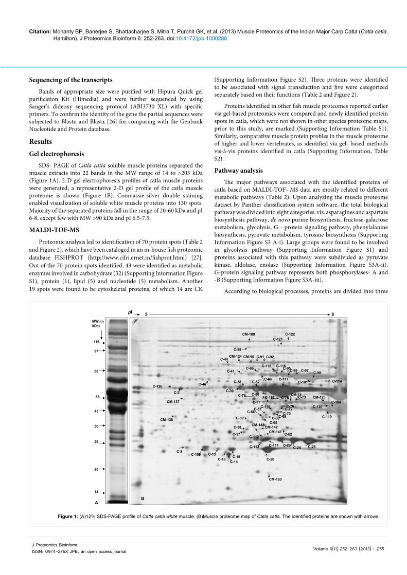
Figure 1: (A)12% SDS-PAGE profile of Catla catla white muscle. (B)Muscle proteome map of Catla catla. The identified proteins are shown with arrows.
Sequencing of the transcripts
Bands of appropriate size were purified with Hipura Quick gel purification Kit (Himedia) and were further sequenced by using Sanger’s dideoxy sequencing protocol (ABI3730 XL) with specific primers. To confirm the identity of the gene the partial sequences were subjected to Blastn and Blastx [26] for comparing with the Genbank Nucleotide and Protein database.
ResultsGel electrophoresis
SDS- PAGE of Catla catla soluble muscle proteins separated the muscle extracts into 22 bands in the MW range of 14 to >205 kDa (Figure 1A). 2-D gel electrophoresis profiles of catla muscle proteins were generated; a representative 2-D gel profile of the catla muscle proteome is shown (Figure 1B). Coomassie-silver double staining enabled visualization of soluble white muscle proteins into 130 spots. Majority of the separated proteins fall in the range of 20-60 kDa and pI 6-8, except few with MW >90 kDa and pI 6.5-7.5.
MALDI-TOF-MS
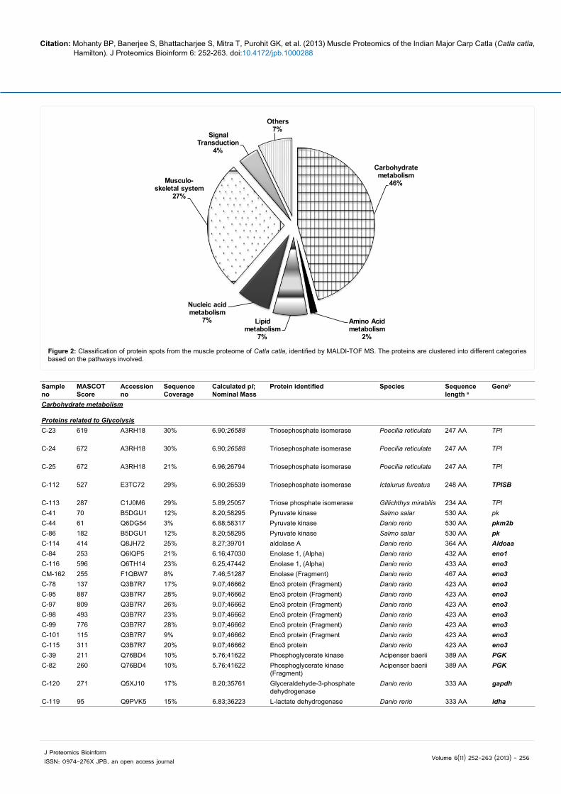
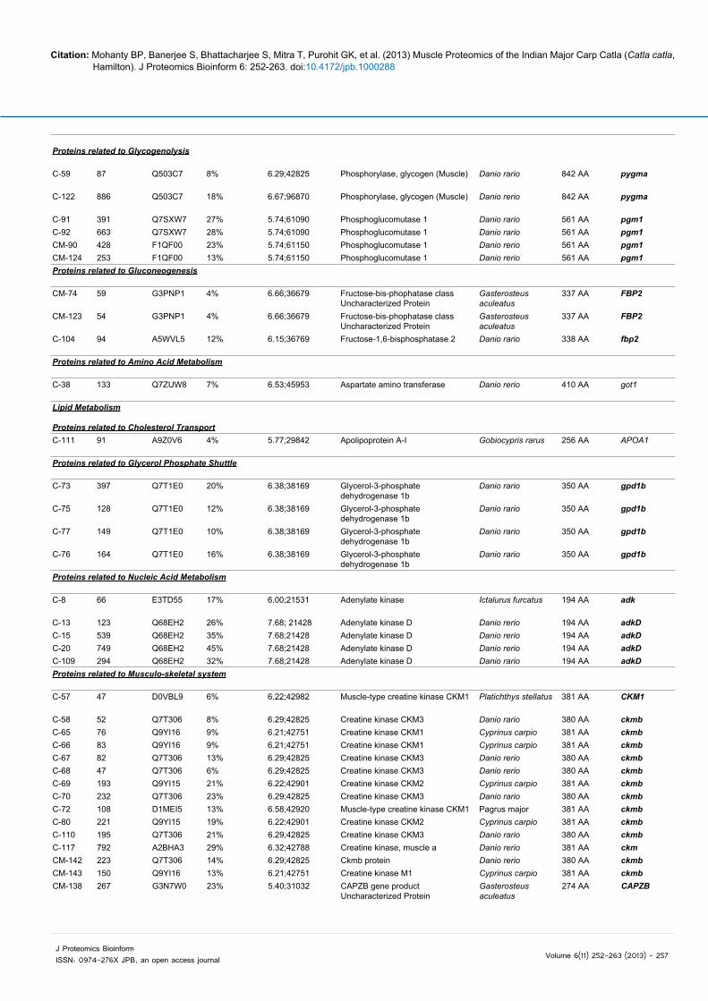
Proteomic analysis led to identification of 70 protein spots (Table 2 and Figure 2), which have been cataloged in an in-house fish proteomic database FISHPROT (http://www.cifri.ernet.in/fishprot.html) [27]. Out of the 70 protein spots identified, 43 were identified as metabolic enzymes involved in carbohydrate (32) (Supporting Information Figure S1), protein (1), lipid (5) and nucleotide (5) metabolism. Another 19 spots were found to be cytoskeletal proteins, of which 14 are CK
(Supporting Information Figure S2). Three proteins were identified to be associated with signal transduction and five were categorized separately based on their functions (Table 2 and Figure 2).
Proteins identified in other fish muscle proteomes reported earlier via gel-based proteomics were compared and newly identified protein spots in catla, which were not shown in other species proteome maps, prior to this study, are marked (Supporting Information Table S1). Similarly, comparative muscle protein profiles in the muscle proteome of higher and lower vertebrates, as identified via gel- based methods vis-à-vis proteins identified in catla (Supporting Information, Table S2).
Pathway analysis
The major pathways associated with the identified proteins of catla based on MALDI-TOF- MS data are mostly related to different metabolic pathways (Table 2). Upon analyzing the muscle proteome dataset by Panther classification system software, the total biological pathway was divided into eight categories: viz. asparagines and aspartate biosynthesis pathway, de novo purine biosynthesis, fructose-galactose metabolism, glycolysis, G - protein signaling pathway, phenylalanine biosynthesis, pyruvate metabolism, tyrosine biosynthesis (Supporting Information Figure S3 A-i). Large groups were found to be involved in glycolysis pathway (Supporting Information Figure S1) and proteins associated with this pathway were subdivided as pyruvate kinase, aldolase, enolase (Supporting Information Figure S3A-ii). G-protein signaling pathway represents both phosphorylases- A and -B (Supporting Information Figure S3A-iii).
According to biological processes, proteins are divided into three
Citation: Mohanty BP, Banerjee S, Bhattacharjee S, Mitra T, Purohit GK, et al. (2013) Muscle Proteomics of the Indian Major Carp Catla (Catla catla, Hamilton). J Proteomics Bioinform 6: 252-263. doi:10.4172/jpb.1000288
Volume 6(11) 252-263 (2013) - 256 J Proteomics BioinformISSN: 0974-276X JPB, an open access journal
Carbohydrate metabolism
46%
Amino Acid metabolism
2%
Lipid metabolism
7%
Nucleic acid metabolism
7%
Musculo-skeletal system
27%
Signal Transduction
4%
Others7%
Figure 2: Classification of protein spots from the muscle proteome of Catla catla, identified by MALDI-TOF MS. The proteins are clustered into different categories based on the pathways involved.
Sample no
MASCOT Score
Accession no
Sequence Coverage
Calculated pI; Nominal Mass
Protein identified Species Sequence length a
Geneb
Carbohydrate metabolism
Proteins related to GlycolysisC-23 619 A3RH18 30% 6.90;26588 Triosephosphate isomerase Poecilia reticulate 247 AA TPI
C-24 672 A3RH18 30% 6.90;26588 Triosephosphate isomerase Poecilia reticulate 247 AA TPI
C-25 672 A3RH18 21% 6.96;26794 Triosephosphate isomerase Poecilia reticulate 247 AA TPI
C-112 527 E3TC72 29% 6.90;26539 Triosephosphate isomerase Ictalurus furcatus 248 AA TPISB
C-113 287 C1J0M6 29% 5.89;25057 Triose phosphate isomerase Gillichthys mirabilis 234 AA TPIC-41 70 B5DGU1 12% 8.20;58295 Pyruvate kinase Salmo salar 530 AA pkC-44 61 Q6DG54 3% 6.88;58317 Pyruvate kinase Danio rerio 530 AA pkm2bC-86 182 B5DGU1 12% 8.20;58295 Pyruvate kinase Salmo salar 530 AA pkC-114 414 Q8JH72 25% 8.27;39701 aldolase A Danio rerio 364 AA AldoaaC-84 253 Q6IQP5 21% 6.16;47030 Enolase 1, (Alpha) Danio rario 432 AA eno1C-116 596 Q6TH14 23% 6.25;47442 Enolase 1, (Alpha) Danio rerio 433 AA eno3CM-162 255 F1QBW7 8% 7.46;51287 Enolase (Fragment) Danio rerio 467 AA eno3C-78 137 Q3B7R7 17% 9.07;46662 Eno3 protein (Fragment) Danio rario 423 AA eno3C-95 887 Q3B7R7 28% 9.07;46662 Eno3 protein (Fragment) Danio rario 423 AA eno3C-97 809 Q3B7R7 26% 9.07;46662 Eno3 protein (Fragment) Danio rario 423 AA eno3C-98 493 Q3B7R7 23% 9.07;46662 Eno3 protein (Fragment) Danio rario 423 AA eno3C-99 776 Q3B7R7 28% 9.07;46662 Eno3 protein (Fragment) Danio rario 423 AA eno3C-101 115 Q3B7R7 9% 9.07;46662 Eno3 protein (Fragment Danio rario 423 AA eno3C-115 311 Q3B7R7 20% 9.07;46662 Eno3 protein Danio rerio 423 AA eno3C-39 211 Q76BD4 10% 5.76;41622 Phosphoglycerate kinase Acipenser baerii 389 AA PGKC-82 260 Q76BD4 10% 5.76;41622 Phosphoglycerate kinase
(Fragment)Acipenser baerii 389 AA PGK
C-120 271 Q5XJ10 17% 8.20;35761 Glyceraldehyde-3-phosphate dehydrogenase
Danio rerio 333 AA gapdh
C-119 95 Q9PVK5 15% 6.83;36223 L-lactate dehydrogenase Danio rerio 333 AA ldha
Citation: Mohanty BP, Banerjee S, Bhattacharjee S, Mitra T, Purohit GK, et al. (2013) Muscle Proteomics of the Indian Major Carp Catla (Catla catla, Hamilton). J Proteomics Bioinform 6: 252-263. doi:10.4172/jpb.1000288
Volume 6(11) 252-263 (2013) - 257 J Proteomics BioinformISSN: 0974-276X JPB, an open access journal
Proteins related to Glycogenolysis
C-59 87 Q503C7 8% 6.29;42825 Phosphorylase, glycogen (Muscle) Danio rario 842 AA pygma
C-122 886 Q503C7 18% 6.67;96870 Phosphorylase, glycogen (Muscle) Danio rerio 842 AA pygma
C-91 391 Q7SXW7 27% 5.74;61090 Phosphoglucomutase 1 Danio rario 561 AA pgm1C-92 663 Q7SXW7 28% 5.74;61090 Phosphoglucomutase 1 Danio rario 561 AA pgm1CM-90 428 F1QF00 23% 5.74;61150 Phosphoglucomutase 1 Danio rerio 561 AA pgm1CM-124 253 F1QF00 13% 5.74;61150 Phosphoglucomutase 1 Danio rerio 561 AA pgm1Proteins related to Gluconeogenesis
CM-74 59 G3PNP1 4% 6.66;36679 Fructose-bis-phophatase class Uncharacterized Protein
Gasterosteus aculeatus
337 AA FBP2
CM-123 54 G3PNP1 4% 6.66;36679 Fructose-bis-phophatase class Uncharacterized Protein
Gasterosteus aculeatus
337 AA FBP2
C-104 94 A5WVL5 12% 6.15;36769 Fructose-1,6-bisphosphatase 2 Danio rario 338 AA fbp2
Proteins related to Amino Acid Metabolism
C-38 133 Q7ZUW8 7% 6.53;45953 Aspartate amino transferase Danio rerio 410 AA got1
Lipid Metabolism
Proteins related to Cholesterol TransportC-111 91 A9Z0V6 4% 5.77;29842 Apolipoprotein A-I Gobiocypris rarus 256 AA APOA1
Proteins related to Glycerol Phosphate Shuttle
C-73 397 Q7T1E0 20% 6.38;38169 Glycerol-3-phosphate dehydrogenase 1b
Danio rario 350 AA gpd1b
C-75 128 Q7T1E0 12% 6.38;38169 Glycerol-3-phosphate dehydrogenase 1b
Danio rario 350 AA gpd1b
C-77 149 Q7T1E0 10% 6.38;38169 Glycerol-3-phosphate dehydrogenase 1b
Danio rario 350 AA gpd1b
C-76 164 Q7T1E0 16% 6.38;38169 Glycerol-3-phosphate dehydrogenase 1b
Danio rario 350 AA gpd1b
Proteins related to Nucleic Acid Metabolism
C-8 66 E3TD55 17% 6.00;21531 Adenylate kinase Ictalurus furcatus 194 AA adk
C-13 123 Q68EH2 26% 7.68; 21428 Adenylate kinase D Danio rerio 194 AA adkDC-15 539 Q68EH2 35% 7.68;21428 Adenylate kinase D Danio rerio 194 AA adkDC-20 749 Q68EH2 45% 7.68;21428 Adenylate kinase D Danio rerio 194 AA adkDC-109 294 Q68EH2 32% 7.68;21428 Adenylate kinase D Danio rario 194 AA adkDProteins related to Musculo-skeletal system
C-57 47 D0VBL9 6% 6.22;42982 Muscle-type creatine kinase CKM1 Platichthys stellatus 381 AA CKM1
C-58 52 Q7T306 8% 6.29;42825 Creatine kinase CKM3 Danio rario 380 AA ckmbC-65 76 Q9YI16 9% 6.21;42751 Creatine kinase CKM1 Cyprinus carpio 381 AA ckmbC-66 83 Q9YI16 9% 6.21;42751 Creatine kinase CKM1 Cyprinus carpio 381 AA ckmbC-67 82 Q7T306 13% 6.29;42825 Creatine kinase CKM3 Danio rerio 380 AA ckmbC-68 47 Q7T306 6% 6.29;42825 Creatine kinase CKM3 Danio rerio 380 AA ckmbC-69 193 Q9YI15 21% 6.22;42901 Creatine kinase CKM2 Cyprinus carpio 381 AA ckmbC-70 232 Q7T306 23% 6.29;42825 Creatine kinase CKM3 Danio rario 380 AA ckmbC-72 108 D1MEI5 13% 6.58;42920 Muscle-type creatine kinase CKM1 Pagrus major 381 AA ckmbC-80 221 Q9YI15 19% 6.22;42901 Creatine kinase CKM2 Cyprinus carpio 381 AA ckmbC-110 195 Q7T306 21% 6.29;42825 Creatine kinase CKM3 Danio rario 380 AA ckmbC-117 792 A2BHA3 29% 6.32;42788 Creatine kinase, muscle a Danio rerio 381 AA ckmCM-142 223 Q7T306 14% 6.29;42825 Ckmb protein Danio rerio 380 AA ckmbCM-143 150 Q9YI16 13% 6.21;42751 Creatine kinase M1 Cyprinus carpio 381 AA ckmbCM-138 267 G3N7W0 23% 5.40;31032 CAPZB gene product
Uncharacterized ProteinGasterosteus aculeatus
274 AA CAPZB
Citation: Mohanty BP, Banerjee S, Bhattacharjee S, Mitra T, Purohit GK, et al. (2013) Muscle Proteomics of the Indian Major Carp Catla (Catla catla, Hamilton). J Proteomics Bioinform 6: 252-263. doi:10.4172/jpb.1000288
Volume 6(11) 252-263 (2013) - 258 J Proteomics BioinformISSN: 0974-276X JPB, an open access journal
C-2 129 A6MWU8 13% 5.17;34634 Beta actin Atherina boyeri 309 AA ACTBCM-126 648 Q7ZZL6 29% 5.23;41851 Skeletal alpha actin-type2b Coryphaenoides
armatus377 AA alpha-actin-
2b
CM-137 274 C0LJU8 17% 5.23;41946 Skeletal alpha actin Hemibarbus mylodonj
377 AA acta1
CM-141 64 F1QII4 9% 9.37;23627 PDZ and LIM domain 7 Danio rerio 207 AA pdlim7Proteins related to Signal TransductionC-14 84 A3FKF8 24% 6.34;15485 DJ-1 (Fragment) Carassius auratus 148 AA DG1C-18 384 Q6DGJ6 38% 5.93;21837 Peroxiredoxin Danio rario 197 AA. prdx1CM-160 53 Q5TYP3 9% 6.20;12757 Novel protein similar to
phosphohistidine phosphatase 1 (PHPT1)
Danio rerio 115 AA si:dkey-51e6.1
OthersC-46 43 P32759 4% 5.91;41873 Alpha-1-antitrypsin homolog Cyprinus carpio 372 AA SERPIN
C-62 40 C1BH79 4% 6.01;27343 Proteosome subunit alpha type 6 Oncorhynchus mykiss
246 AA PSA6
C-88 231 A6H8Q3 6% 5.39;57112 Zgc:165344 Danio rario 529 AA mybphbCM-108 204 A6H8Q3 4% 5.39;57112 Zgc:165344 Danio rerio 529 AA mybphbC-121 156 B3GPN3 5% 5.91;73041 Transferrin variant F Cyprinus carpio 666 AA TF
a, b The corresponding gene name and amino acid sequence length of the identified proteins according to Uniprot database (http://www.uniprot.org/uniprot/)
Table 2: Proteins identified from Catla catla muscle proteome by MALDI-TOF-MS.
categories, viz. immune system, system process and metabolic process (Supporting Information, Figure S3B-i) of which metabolic process represented the largest group, which is subdivided as primary and ROS metabolic process (Supporting Information, Figure S3B-ii). Primary metabolic process is further divided in to carbohydrate, cellular amino acid, lipid and nucleotide metabolism (Supporting Information Figure S3B-iii).
On the basis of molecular function, proteins are divided into two classes: viz. catalytic activity and antioxidant activity (Supporting Information Figure S4A-i) where catalytic activities of the identified proteins are further divided into transferase, isomerase, lyase and oxidoreductase catalysis (Supporting Information, Figure S4A-ii). Transferase activity of the enzyme proteins are further divided into glycosyltransferase, transaminase and a large group associated with kinase activity (Supporting Information Figure S4A-iii), which are further divided into nucleotide, amino acid, carbohydrate kinase (Supporting Information Figure S4A-iv).
Dataset proteins were grouped into five protein classes’ viz. transferase, isomerase, kinase, lyase and oxidoreductase (Supporting Information Figure S4B-i). Proteins of transferase class are further sub grouped into transaminase, kinase and phosphorylase (Supporting Information Figure S4B-ii), whereas kinases (Supporting Information Figure S4B-iii) are divided as described in Supporting Information Figure S4A-iv and oxidoreductase activity is further divided as peroxidase and dehydrogenase (Supporting Information Figure S4B-iv).
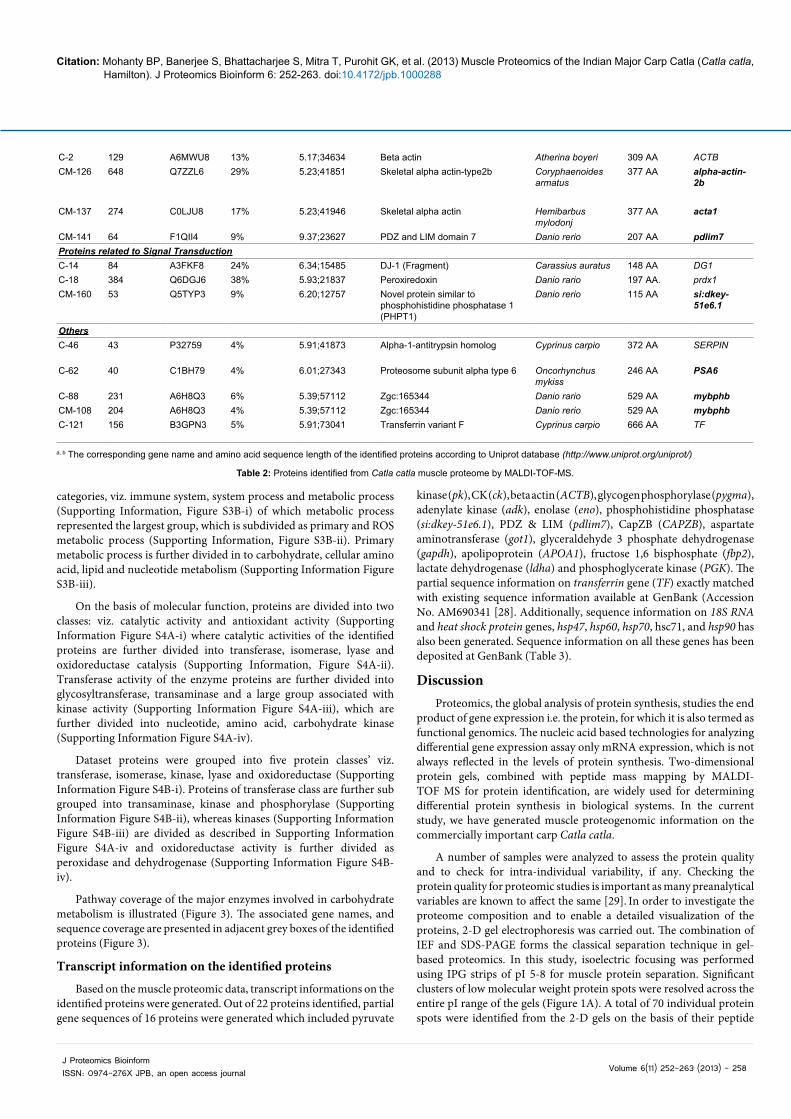
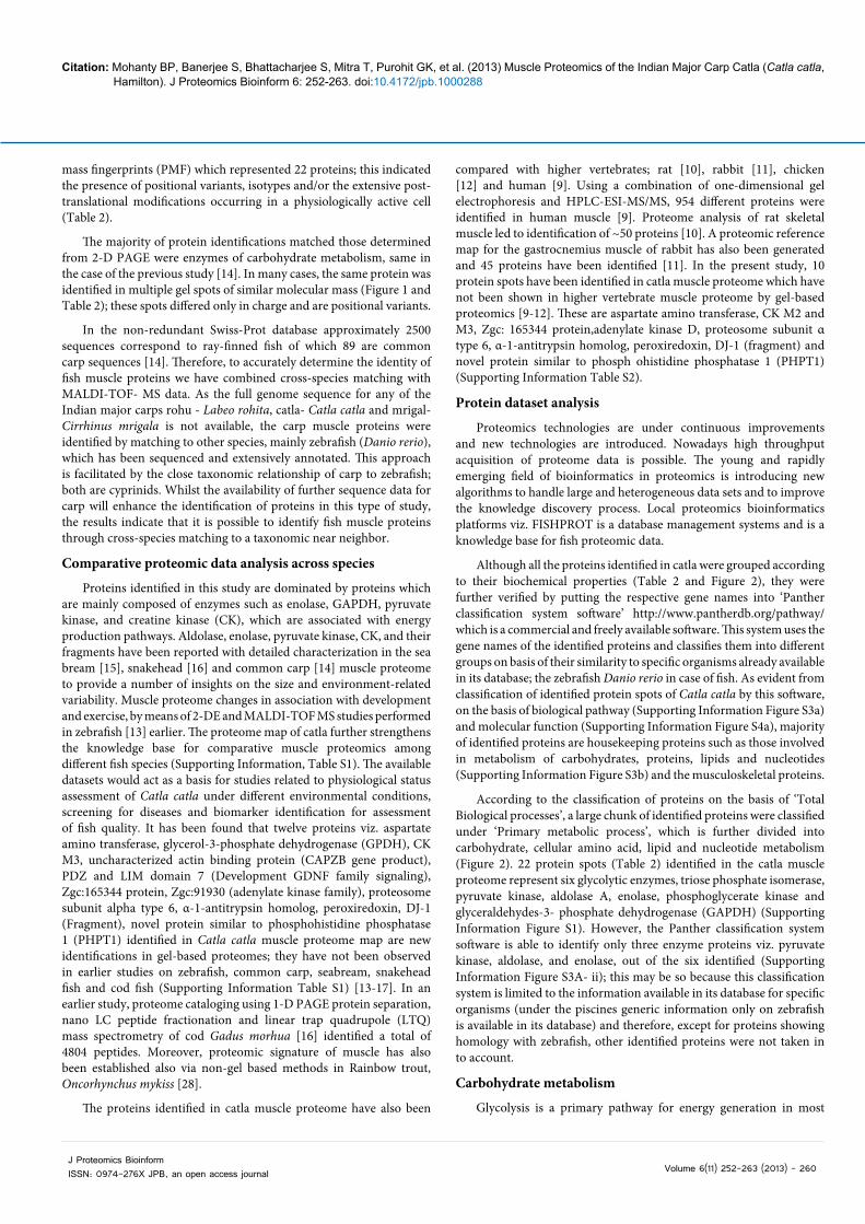
Pathway coverage of the major enzymes involved in carbohydrate metabolism is illustrated (Figure 3). The associated gene names, and sequence coverage are presented in adjacent grey boxes of the identified proteins (Figure 3).
Transcript information on the identified proteins
Based on the muscle proteomic data, transcript informations on the identified proteins were generated. Out of 22 proteins identified, partial gene sequences of 16 proteins were generated which included pyruvate
kinase (pk), CK (ck), beta actin (ACTB), glycogen phosphorylase (pygma), adenylate kinase (adk), enolase (eno), phosphohistidine phosphatase (si:dkey-51e6.1), PDZ & LIM (pdlim7), CapZB (CAPZB), aspartate aminotransferase (got1), glyceraldehyde 3 phosphate dehydrogenase (gapdh), apolipoprotein (APOA1), fructose 1,6 bisphosphate (fbp2), lactate dehydrogenase (ldha) and phosphoglycerate kinase (PGK). The partial sequence information on transferrin gene (TF) exactly matched with existing sequence information available at GenBank (Accession No. AM690341 [28]. Additionally, sequence information on 18S RNA and heat shock protein genes, hsp47, hsp60, hsp70, hsc71, and hsp90 has also been generated. Sequence information on all these genes has been deposited at GenBank (Table 3).
DiscussionProteomics, the global analysis of protein synthesis, studies the end
product of gene expression i.e. the protein, for which it is also termed as functional genomics. The nucleic acid based technologies for analyzing differential gene expression assay only mRNA expression, which is not always reflected in the levels of protein synthesis. Two-dimensional protein gels, combined with peptide mass mapping by MALDI-TOF MS for protein identification, are widely used for determining differential protein synthesis in biological systems. In the current study, we have generated muscle proteogenomic information on the commercially important carp Catla catla.
A number of samples were analyzed to assess the protein quality and to check for intra-individual variability, if any. Checking the protein quality for proteomic studies is important as many preanalytical variables are known to affect the same [29]. In order to investigate the proteome composition and to enable a detailed visualization of the proteins, 2-D gel electrophoresis was carried out. The combination of IEF and SDS-PAGE forms the classical separation technique in gel-based proteomics. In this study, isoelectric focusing was performed using IPG strips of pI 5-8 for muscle protein separation. Significant clusters of low molecular weight protein spots were resolved across the entire pI range of the gels (Figure 1A). A total of 70 individual protein spots were identified from the 2-D gels on the basis of their peptide
Citation: Mohanty BP, Banerjee S, Bhattacharjee S, Mitra T, Purohit GK, et al. (2013) Muscle Proteomics of the Indian Major Carp Catla (Catla catla, Hamilton). J Proteomics Bioinform 6: 252-263. doi:10.4172/jpb.1000288
Volume 6(11) 252-263 (2013) - 259 J Proteomics BioinformISSN: 0974-276X JPB, an open access journal
Source: [28]
Table 3: Partial gene sequences with GenBank Accession Nos. of genes coding for the proteins identified in muscle proteome of Catla catla.
Sl No. Target Gene Amplicon Length (bp) GenBank Accession No.1 Pyruvate kinase 535 KC7078422 Creatine Kinase 204 KC7884233 Beta actin 372 KC7884244 Phosphorylase kinase 202 KC7887545 Adenylate Kinase 240 KC8165376 Enolase 290 KC8165387 Phosphohistidine phosphatase 210 KC8165398 PDZ and LIM 390 KC8165409 CapZB 200 KC81654110 Aspartate aminotransferase 280 KC816542
11 Glyceraldehyde 3 phosphate dehydrogenase 206 KC887542
12 Apolipoprotein 327 KC88754313 Fructose 1,6 bisphosphate 207 KC88754014 Lactate Dehydrogenase 234 KC88754415 Phosphoglycerate kinase 543 KC88754516 Transferrin* 633 AM690341 17 hsp47 460 KC91502417 hsp70 373 KC59920718 hsc71 415 KC80080019 hsp60 360 KC59920520 hsp90 244 KC80080121 18s RNA 514 KC915025
Gluconeogenesis
Glyceraldehyde-3 phosphate
GAPDH 17%
Glyceraldehyde-3-phosphate dehydrogenase
1,3-bisphosphoglycerate
Phosphoglycerate kinase
PGK 10%, 10%
3-phosphoglycerate
2- phosphoglycerate
Phosphoenolpyruvate
Phosphoglycerate mutase
Enolase
ENO3
21%, 23%, 8%, 17%, 28%, 26%,23%, 28%, 9%, 20%
NADH
ADP
ATP
LDHA
PKPyruvate kinase 12%, 3%, 12%
Lactate dehydrogenase
Pyruvate
Lactate
15%
ADPATP
Glycogen
PYGM 8%, 18% Glycogen debrancher
Glucose-1-phosphate
Glycogen phosphorylase
Glycogen (n-1)
Phosphoglucomutase-1PGM127%, 28%, 23%, 13%
Glycogenolysis
ATP
ADPH2O
Pi
ATP
ADP
25% ALDOA
Glucose
Glucose-6-phosphate
Fructose-6-phosphate
Fructose-1, 6-bisphosphate
Dihydroxyacetone phosphate
Hexokinase
Glucose-6-phosphate isomerase
Phosphofructokinase-1
Aldolase
Fructose-1,6-bisphosphatase
fbp2 4%, 4%, 12%
Triosephosphateisomerase
30%, 30%, 21%,29%, 29%
TPI
Figure 3: Proteomic coverage of major enzymes involved in carbohydrate metabolism in Catla catla white muscle. Positional variants of enzymes involved in glycolysis, gluconeogenesis and glycogenolysis are shown: proteins identified are shown in boxes and the associated gene names and sequence coverage are shown in adjacent grey boxes.
Citation: Mohanty BP, Banerjee S, Bhattacharjee S, Mitra T, Purohit GK, et al. (2013) Muscle Proteomics of the Indian Major Carp Catla (Catla catla, Hamilton). J Proteomics Bioinform 6: 252-263. doi:10.4172/jpb.1000288
Volume 6(11) 252-263 (2013) - 260 J Proteomics BioinformISSN: 0974-276X JPB, an open access journal
mass fingerprints (PMF) which represented 22 proteins; this indicated the presence of positional variants, isotypes and/or the extensive post-translational modifications occurring in a physiologically active cell (Table 2).
The majority of protein identifications matched those determined from 2-D PAGE were enzymes of carbohydrate metabolism, same in the case of the previous study [14]. In many cases, the same protein was identified in multiple gel spots of similar molecular mass (Figure 1 and Table 2); these spots differed only in charge and are positional variants.
In the non-redundant Swiss-Prot database approximately 2500 sequences correspond to ray-finned fish of which 89 are common carp sequences [14]. Therefore, to accurately determine the identity of fish muscle proteins we have combined cross-species matching with MALDI-TOF- MS data. As the full genome sequence for any of the Indian major carps rohu - Labeo rohita, catla- Catla catla and mrigal- Cirrhinus mrigala is not available, the carp muscle proteins were identified by matching to other species, mainly zebrafish (Danio rerio), which has been sequenced and extensively annotated. This approach is facilitated by the close taxonomic relationship of carp to zebrafish; both are cyprinids. Whilst the availability of further sequence data for carp will enhance the identification of proteins in this type of study, the results indicate that it is possible to identify fish muscle proteins through cross-species matching to a taxonomic near neighbor.
Comparative proteomic data analysis across species
Proteins identified in this study are dominated by proteins which are mainly composed of enzymes such as enolase, GAPDH, pyruvate kinase, and creatine kinase (CK), which are associated with energy production pathways. Aldolase, enolase, pyruvate kinase, CK, and their fragments have been reported with detailed characterization in the sea bream [15], snakehead [16] and common carp [14] muscle proteome to provide a number of insights on the size and environment-related variability. Muscle proteome changes in association with development and exercise, by means of 2-DE and MALDI-TOF MS studies performed in zebrafish [13] earlier. The proteome map of catla further strengthens the knowledge base for comparative muscle proteomics among different fish species (Supporting Information, Table S1). The available datasets would act as a basis for studies related to physiological status assessment of Catla catla under different environmental conditions, screening for diseases and biomarker identification for assessment of fish quality. It has been found that twelve proteins viz. aspartate amino transferase, glycerol-3-phosphate dehydrogenase (GPDH), CK M3, uncharacterized actin binding protein (CAPZB gene product), PDZ and LIM domain 7 (Development GDNF family signaling), Zgc:165344 protein, Zgc:91930 (adenylate kinase family), proteosome subunit alpha type 6, α-1-antitrypsin homolog, peroxiredoxin, DJ-1 (Fragment), novel protein similar to phosphohistidine phosphatase 1 (PHPT1) identified in Catla catla muscle proteome map are new identifications in gel-based proteomes; they have not been observed in earlier studies on zebrafish, common carp, seabream, snakehead fish and cod fish (Supporting Information Table S1) [13-17]. In an earlier study, proteome cataloging using 1-D PAGE protein separation, nano LC peptide fractionation and linear trap quadrupole (LTQ) mass spectrometry of cod Gadus morhua [16] identified a total of 4804 peptides. Moreover, proteomic signature of muscle has also been established also via non-gel based methods in Rainbow trout, Oncorhynchus mykiss [28].
The proteins identified in catla muscle proteome have also been
compared with higher vertebrates; rat [10], rabbit [11], chicken [12] and human [9]. Using a combination of one-dimensional gel electrophoresis and HPLC-ESI-MS/MS, 954 different proteins were identified in human muscle [9]. Proteome analysis of rat skeletal muscle led to identification of ~50 proteins [10]. A proteomic reference map for the gastrocnemius muscle of rabbit has also been generated and 45 proteins have been identified [11]. In the present study, 10 protein spots have been identified in catla muscle proteome which have not been shown in higher vertebrate muscle proteome by gel-based proteomics [9-12]. These are aspartate amino transferase, CK M2 and M3, Zgc: 165344 protein,adenylate kinase D, proteosome subunit α type 6, α-1-antitrypsin homolog, peroxiredoxin, DJ-1 (fragment) and novel protein similar to phosph ohistidine phosphatase 1 (PHPT1) (Supporting Information Table S2).
Protein dataset analysis
Proteomics technologies are under continuous improvements and new technologies are introduced. Nowadays high throughput acquisition of proteome data is possible. The young and rapidly emerging field of bioinformatics in proteomics is introducing new algorithms to handle large and heterogeneous data sets and to improve the knowledge discovery process. Local proteomics bioinformatics platforms viz. FISHPROT is a database management systems and is a knowledge base for fish proteomic data.
Although all the proteins identified in catla were grouped according to their biochemical properties (Table 2 and Figure 2), they were further verified by putting the respective gene names into ‘Panther classification system software’ http://www.pantherdb.org/pathway/ which is a commercial and freely available software. This system uses the gene names of the identified proteins and classifies them into different groups on basis of their similarity to specific organisms already available in its database; the zebrafish Danio rerio in case of fish. As evident from classification of identified protein spots of Catla catla by this software, on the basis of biological pathway (Supporting Information Figure S3a) and molecular function (Supporting Information Figure S4a), majority of identified proteins are housekeeping proteins such as those involved in metabolism of carbohydrates, proteins, lipids and nucleotides (Supporting Information Figure S3b) and the musculoskeletal proteins.
According to the classification of proteins on the basis of ‘Total Biological processes’, a large chunk of identified proteins were classified under ‘Primary metabolic process’, which is further divided into carbohydrate, cellular amino acid, lipid and nucleotide metabolism (Figure 2). 22 protein spots (Table 2) identified in the catla muscle proteome represent six glycolytic enzymes, triose phosphate isomerase, pyruvate kinase, aldolase A, enolase, phosphoglycerate kinase and glyceraldehydes-3- phosphate dehydrogenase (GAPDH) (Supporting Information Figure S1). However, the Panther classification system software is able to identify only three enzyme proteins viz. pyruvate kinase, aldolase, and enolase, out of the six identified (Supporting Information Figure S3A- ii); this may be so because this classification system is limited to the information available in its database for specific organisms (under the piscines generic information only on zebrafish is available in its database) and therefore, except for proteins showing homology with zebrafish, other identified proteins were not taken in to account.
Carbohydrate metabolism
Glycolysis is a primary pathway for energy generation in most
Citation: Mohanty BP, Banerjee S, Bhattacharjee S, Mitra T, Purohit GK, et al. (2013) Muscle Proteomics of the Indian Major Carp Catla (Catla catla, Hamilton). J Proteomics Bioinform 6: 252-263. doi:10.4172/jpb.1000288
Volume 6(11) 252-263 (2013) - 261 J Proteomics BioinformISSN: 0974-276X JPB, an open access journal
organisms. In the present study, out of the 10 glycolytic enzymes (6/10), namely, aldolase (C-114), triose phosphate isomerase (C-23, 24, 25, 112, 113), GAPDH (C-120), phosphoglycerate kinase (C-39, 82), enolase (C-78, 84, 95, 97, 98, 99, 101, 115, 116, CM-162) and pyruvate kinase (C-41, 44, 86) have been identified and they represent 22 number of spots on the catla muscle proteome (Table 2 and Figure 3; Supporting Information Figure S1). Lactate dehydrogenase (C-119), the enzyme involved in anaerobic glycolysis, converting pyruvate to lactate, has also been identified.
Two spots (C-59, 122) were identified as glycogen phosphorylase. Glycogen phosphorylase catalyzes and regulates the entry of glucose residues into glycolysis from glycogen via glycogenolysis pathway. This is a regulatory enzyme present in both liver and muscle. In skeletal muscles the enzyme occurs in two forms, a catalytically active phosphorylated form (phosphorylase a) and a much less active dephosphorylated form (phosphorylase b). In muscle, the rate of conversion of glycogen units into glucose 1-phosphate is regulated by the ratio of the active phosphorylase a to the less active phosphorylase b. Glycogen synthase, the rate-limiting enzyme in glycogen biosynthesis is also regulated by glycogen phosphorylase (Figure 3).
Four spots (C-91, 92; CM-90, 124) were identified as phosphoglucomutase. Phosphoglucomutase is a key enzyme in the metabolism of glycogen and protein glycosylation. It is responsible for the reversible inter conversion of glucose 1-phosphate to glucose 6-phosphate, both of which are key intermediates in the synthesis and breakdown of glycogen and galactose metabolism. It is also important for the formation of UDP-glucose which is an essential intermediary metabolite in protein glycosylation. Inhibition of phosphoglucomutase has drastic effects on carbohydrate metabolism which reduces the steady-state levels of UDP-glucose, resulting in a defect of glycogen and trehalose biosynthesis, while galactose metabolism is inhibited, leading to galactosemia, accumulation of galactose 1-phosphate and Glucose 1-phosphate i.e., poor glycogen turnover.
Three spots (C-104, CM-74, CM-123) were identified as Fructose-bis-phosphatase, which catalyzes the conversion of Fructose-1,6-bisphosphatase to Fructose-6-phosphate, an important reaction step in gluconeogenesis.
Lipid metabolism
Four spots (C-73, 75, 76, 77) were identified as Glycerol 3-phophate dehydrogenase 1b. Glycerol 3-phosphate and fatty acyl-CoAs are the common precursors for triacylglycerols and glycerol phosphatides. Glycerol phosphate is formed in two ways; it is formed from the dihydroxy acetone phosphate generated during glycolysis by the action of cytosolic NAD-linked GPDH. It is also be formed from glycerol by the action of glycerol kinase.
Spot number C-111 has been identified as apolipoprotein A1 (Apo A-I) (Supporting Information Table S1). This protein has earlier been identified in Cyprinus carpio [14]. Apo A-I is the major protein component of high density lipoprotein (HDL) in plasma, which confers water solubility to the lipoprotein complex thus facilitating lipid transport and metabolism. It promotes cholesterol efflux from tissues to the liver for excretion. It is a cofactor for lecithin cholesterolacyltransferase (LCAT) which is responsible for the formation of most plasma cholesteryl esters. In lower vertebrate species Apo A-I is also synthesized in a number of peripheral tissues, e.g., rainbow trout gill [29] and liver [30], carp optic nerve [31] and skin
[32]. In the cod (Gadus morhua) Apo A-I is closely associated with the C3 component of the complement system [33]. Apo A-I has also been reported to have a restorative or protective role and plays significant role in maintaining epithelial integrity [29].
Nucleotide metabolism
Five spots have been grouped as proteins related to nucleic acid metabolism; out of these, one spot (C-8) has been identified as adenylate kinase and the other 4 spots (C-13, 15, 20, 109) have been identified as adenylate kinase D (Table 2). The adenylate kinase D has not been reported in the vertebrates by gel-based proteomics.
CK and other contractile proteins
CK catalyzes the transphosphorylation between phosphocreatine and ADP and is central to the regulation of muscle bioenergetics. Large number of protein spots (13) across a broad range of MW and pI were identified as positional variants of muscle CK in the present study (Supporting Information Figure S2). It has been reported earlier that creatine/phosphocreatine interconversion played an important role in ATP regeneration since depletion of glycolytic enzymes in carp resulted in anoxia. High tissue CK activity, whether constitutive, induced, or both, may rather directly enhance contractile responses by enhancing cellular energy and contractile reserve. This high CK activity may alter local ADP levels at the contractile proteins and contribute to the enhanced contractility and myosin ATPase activity.
Complex patterns of CK isoforms exist in the skeletal muscle of fish [34-36] and three isoforms of CK have previously been identified in carp skeletal muscle [37]. These are referred to as M1, M2 and M3 and have predicted masses of 43 kDa and pI 6.22-6.32 [37]. In the muscle proteome of catla, 14 spots have been identified as CK; out of these, six (C-57, 65, 66, 72, 117, CM-143) are CK muscle type - I (CK M-1), three are CK M-2 (C-69, 80, 142) and five are CK M-3 (C-58, 67, 68, 70, 110) (Table 2; Supporting Information Figure S2). This observation is consistent with the earlier reports on presence of multiple forms of this enzyme in muscle tissue of other fish [35,37]. CK M-3 has not been reported earlier in any vertebrates by gel-based proteomics.
Four spots (C-2, CM-126, 137, 138) were identified as actin (Table 2). Actin, a 42-kDa globular protein found in all eukaryotic cells, participates in many important cellular processes including muscle contraction, cell motility, cell division and cytokinesis, vesicle and organelle movement, cell signaling, and the establishment and maintenance of cell junctions and cell shape.
CM-141, identified as PDZ and LIM domain -7 protein, is a muscle-specific protein [9]. It is representative of a family of proteins composed of conserved PDZ and LIM domains. PDZ is an acronym combining the first letters of three proteins - Post synaptic density protein (PSD95), Drosophila disc large tumor suppressor (Dlg1), and Zonula occludens-1 protein (zo-1) - which were first discovered to share the domain [38]. The PDZ domain is a common structural domain of 80-90 amino-acids found in the signaling proteins of bacteria, yeast, plants, viruses and animals. LIM domains are protein structural domains, composed of two contiguous zinc finger domains, separated by a two-amino acid residue hydrophobic linker; they are named after their initial discovery in the proteins Lin11, Isl-1 and Mec-3. LIM-domain containing proteins have been shown to play roles in cytoskeletal organization, organ development and oncogenesis. LIM-domains mediate protein: protein interactions that are critical to cellular processes [39]. LIM domains are proposed to function in
Citation: Mohanty BP, Banerjee S, Bhattacharjee S, Mitra T, Purohit GK, et al. (2013) Muscle Proteomics of the Indian Major Carp Catla (Catla catla, Hamilton). J Proteomics Bioinform 6: 252-263. doi:10.4172/jpb.1000288
Volume 6(11) 252-263 (2013) - 262 J Proteomics BioinformISSN: 0974-276X JPB, an open access journal
proteinprotein recognition in a variety of contexts including gene transcription, development and in cytoskeletal interaction. The LIM domains of this protein bind to protein kinases, whereas the PDZ domain binds to actin filaments. The gene product is involved in the assembly of an actin filament-associated complex essential for transmission of ret/ptc2 mitogenic signaling. The biological function is likely to be that of an adapter, with the PDZ domain localizing the LIM-binding proteins to actin filaments of both skeletal muscle and non muscle tissues [9]. This protein has not been reported earlier in any fish species, by gel-based proteomics.
Signal transduction
Protein phosphorylation is a key regulatory mechanism for signal transduction in both prokaryote and eukaryotic cells. Most of our understanding regarding the signaling events in eukaryotes comes from tyrosine, serine/threonine kinases, and phosphatases. Histidine phosphorylation- dependent signaling in eukaryotes is less well characterized. The first vertebrate protein histidine phosphatase, PHPT1 was identified in 2002 [40] and it was found that PHPT1 is ubiquitously expressed in eukaryotes, from C. elegans to Homo sapiens. In the present study, one protein spot, CM160, has been identified as PHPT (Table 2); this protein has not been reported in other vertebrates by gel-based proteomics earlier (Supporting Information Table S1).
Two spots, C14 and C 18, were identified as peroxiredoxin and DJ-1, respectively (Table 1, Supporting Information Table S1 and S2). Peroxiredoxin controls cytokine induced peroxide levels and thereby mediating signal transduction in mammals. DJ-1, a chaperone protein having antioxidant properties, is involved in the cellular response to stress and have been described to play a role in apoptosis.
Transcript analysis
The partial gene sequence for the identified proteins (16/22) and for some additional proteins that include hsp47, hsp60, hsp70, hsc71, hsp90 and 18S RNA have been generated. Thus, this is the first study on this commercially important species with a proteogenomics approach. Proteogenomics aims mainly to use proteomics data and technologies for identifying novel genomic features, such as novel un-annotated genes, and for improving, correcting or confirming the structural and functional genome annotation. This approach is ideal to harness the wealth of information available at the proteome level and apply it to the available genomic information of organisms [3,41]. This would help to focus on the functional genome, rather than the whole genome and could possibly help to identify protein variants that could cause diseases, to identify protein biomarkers, to study genome variation and to identify QTLs associated with production traits. Multi-prolonged approaches such as transcriptomics and proteomics in addition to genomics should be included in future studies, as proteogenomic analyses provide a more accurate catalog of protein-coding genes [3,41].
In fish, flesh quality is dependent on environmental factors, mainly water and food quality for product safety and food composition for flesh nutritional quality. Nevertheless, amongst sensory quality, flesh texture is mainly determined by biological factors such as muscle organization, protein content, and composition. In fish, the best quality is firm and cohesive flesh with good water holding capacity. These traits are mainly determined by proteins’ nature and properties, so proteomic tools appear especially of interest to study fish flesh quality. However, very few studies were undertaken to identify flesh quality biomarkers [42-45] and is a research gap needing attention of researchers.
The primary objective of the study was to establish a reference muscle proteome map for Catla catla and to identify the muscle proteins, which has been achieved to a great extent. The identified proteins in this study have been cataloged in the database http://www.cifri.ernet.in/fishprot.html and would act as baseline information on proteogenomics of Catla catla and other aquacultured species. The information generated could also be useful for biotechnological interventions in fish health and disease management; besides adding to the existing knowledge base on comparative muscle proteomics.
Acknowledgements
This work was supported by the CIFRI core-Project No. FHE/ER/07/06/005 (to BPM) and ICAR National Fund for Basic, Strategic and Frontier Application Research in Agriculture (NFBSFARA) project #AS-2001 (to BPM and SM). Sudeshna Banerjee and Tandrima Mitra are thankful to ICAR for the Research Fellowship. Soma Bhattacharjee was DBT Post Doctoral Fellow and GKP is a ICAR/NFBSFARA Senior Research Fellow. The authors are thankful to the unknown reviewers for the valuable suggestions for improving the manuscript.
Conflict of Interest Statement
The authors declare that there are no conflicts of interest.
References
1. Hochstrasser DF, Sanchez JC, Appel RD (2002) Proteomics and its trends facing nature’s complexity. Proteomics 2: 807-812.
2. Cravatt BF, Simon GM, Yates JR 3rd (2007) The biological impact of mass-spectrometry-based proteomics. Nature 450: 991-1000.
3. Renuse S, Chaerkady R, Pandey A (2011) Proteogenomics. Proteomics 11: 620-630.
4. Wolfe RR (2006) The underappreciated role of muscle in health and disease. Am J ClinNutr 84: 475-482.
5. Ohlendieck K (2011) Proteomic Profiling of Fast-To-Slow Muscle Transitions during Aging. Front Physiol 2: 105.
6. Gordon AM, Homsher E, Regnier M (2000) Regulation of contraction in striated muscle. Physiol Rev 80: 853-924.
7. Abdul-Ghani MA, DeFronzo RA (2010) Pathogenesis of insulin resistance in skeletal muscle. J Biomed Biotech 2010: 476279.
8. Doran P, Donoghue P, O’Connell K, Gannon J, Ohlendieck K (2007) Proteomic profiling of pathological and aged skeletal muscle fibres by peptide mass fingerprinting (Review). Int J Mol Med 19: 547-564.
9. Højlund K, Yi Z, Hwang H, Bowen B, Lefort N, et al. (2008) Characterization of the human skeletal muscle proteome by one-dimensional gel electrophoresis and HPLC-ESI-MS/MS. Mol Cell Proteomics 7: 257-267.
10. Piec I, Listrat A, Alliot J, Chambon C, Taylor RG, et al. (2005) Differential proteome analysis of aging in rat skeletal muscle. FASEB J 19: 1143-1145.
11. Almeida AM, Campos A, van Harten S, Cardoso LA, Coelho AV (2009) Establishment of a proteomic reference map for the gastrocnemius muscle in the rabbit (Oryctolaguscuniculus). Res Vet Sci 87: 196-199.
12. Doherty MK, McLean L, Hayter JR, Pratt JM, Robertson DHL, et al. (2004) The proteome of chicken skeletal muscle: Changes in Soluble protein expression during growth in a layer strain. Proteomics 4: 2082-2089.
13. Bosworth CA 4th, Chou CW, Cole RB, Rees BB (2005) Protein expression patterns in zebrafish skeletal muscle: initial characterization and the effects of hypoxic exposure. Proteomics 5: 1362-1371.
14. McLean L, Young IS, Doherty MK, Robertson DH, Cossins AR, et al. (2007) Global cooling: cold acclimation and the expression of soluble proteins in carp skeletal muscle. Proteomics 7: 2667-2681.
15. Addis MF, Cappuccinelli R, Tedde V, Pagnozzi D, Porcu MC, et al. (2010) Proteomic analysis of muscle tissue from gilthead sea bream (Sparus aurata, L.) farmed in offshore floating cages. Aquaculture 309: 245-252.
16. Gebriel M, Uleberg KE, Larssen E, HjelleBjørnstad A, Sivertsvik M, et al. (2010) Cod (Gadusmorhua) muscle proteome cataloging using 1D-PAGE protein separation, nano-liquid chromatography peptide fractionation, and Linear Trap Quadrupole (LTQ) Mass Spectrometry. J Agric Food Chem 58: 12307-12312.
Citation: Mohanty BP, Banerjee S, Bhattacharjee S, Mitra T, Purohit GK, et al. (2013) Muscle Proteomics of the Indian Major Carp Catla (Catla catla, Hamilton). J Proteomics Bioinform 6: 252-263. doi:10.4172/jpb.1000288
Volume 6(11) 252-263 (2013) - 263 J Proteomics BioinformISSN: 0974-276X JPB, an open access journal
Submit your next manuscript and get advantages of OMICS Group submissionsUnique features:
• User friendly/feasible website-translation of your paper to 50 world’s leading languages• Audio Version of published paper• Digital articles to share and explore
Special features:
• 300 Open Access Journals• 25,000 editorial team• 21 days rapid review process• Quality and quick editorial, review and publication processing• Indexing at PubMed (partial), Scopus, EBSCO, Index Copernicus and Google Scholar etc• Sharing Option: Social Networking Enabled• Authors, Reviewers and Editors rewarded with online Scientific Credits• Better discount for your subsequent articles
Submit your manuscript at: http://www.editorialmanager.com/proteomics
Citation: Mohanty BP, Banerjee S, Bhattacharjee S, Mitra T, Purohit GK, et al. (2013) Muscle Proteomics of the Indian Major Carp Catla (Catla catla, Hamilton). J Proteomics Bioinform 6: 245-251. doi:10.4172/jpb.1000288
17. Gam LH, Leow CY, Baie S (2006) Proteomic analysis of Snakehead fish (Channastriata) muscle tissue. Malaysian J BiochemMolBiol 14: 25-32.
18. Johnston IA (1999) Muscle development and growth: potential implications for flesh quality in fish. Aquaculture 177: 99-115.
19. Islam MS, Ahmed ASI, Azam MS, Alam MS (2005) Genetic analysis of three river populations ofCatlacatla(Hamilton) using randomly amplified polymorphic DNA markers. Asian-Aust J AnimSci 18: 453-457.
20. Barik SK, Banerjee S, Bhattacharjee S, Das Gupta SK, Mohanty S, et al. (2013) Proteomic analysis of sarcoplasmic peptides of two related fish species for food authentication. ApplBiochemBiotechnol 171: 1011-1021.
21. Bradford MM (1976) A rapid and sensitive method for the quantitation of microgram quantities of protein utilizing the principle of protein-dye binding. Anal Biochem 72: 248-254.
22. Laemmli UK (1970) Cleavage of structural proteins during the assembly of the head of bacteriophage T4. Nature 227: 680-685.
23. O’Farrell PH (1975) High resolution two-dimensional electrophoresis of proteins. J BiolChem 250: 4007-4021.
24. Bringans S, Eriksen S, Kendrick T, Gopalakrishnakone P, Livk A, et al. (2008) Proteomic analysis of the venom of Heterometruslongimanus (Asian black scorpion). Proteomics 8: 1081-1096.
25. Perkins DN, Pappin DJ, Creasy DM, Cottrell JS (1999) Probability-based protein identification by searching sequence databases using mass spectrometry data. Electrophoresis 20: 3551-3567.
26. Altschul SF, Gish W, Miller W, Myers EW, Lipman DJ (1990) Basic local allingment search tool. J MolBiol 215: 403-410
27. http://www.cifri.ernet.in/fishprot.html
28. Sahoo PK, Mohanty BR, Kumari J, Barat A, Sarangi N (2009) Cloning, nucleotide sequence and phylogenetic analyses and tissue-specific expression of the transferrin gene in Cirrhinusmrigala infected with Aeromonashydrophila. Comp ImmunolMicrobiol Infect Dis 6: 527-537
29. Bhattacharjee S, Mohanty S, Sharma AP, Mohanty BP (2011) Effect of storage temperature as a preanalytical variable on the lens crystallins protein quality for proteomic studies. Proteomics ClinAppl 5: 504-512.
30. Salem M, Kenney PB, Rexroad CE 3rd, Yao J (2010) Proteomic signature of muscle atrophy in rainbow trout. J Proteomics 73: 778-789.
31. Smith RW, Wood CM, Cash P, Diao L, Pärt P (2005) Apolipoprotein AI could be a significant determinant of epithelial integrity in rainbow trout gill cell cultures: a study in functional proteomics. BiochimBiophysActa 1749: 81-93.
32. Martin SA, Vilhelmsson O, Médale F, Watt P, Kaushik S, et al. (2003) Proteomic sensitivity to dietary manipulations in rainbow trout. BiochimBiophysActa 1651: 17-29.
33. Harel A, Fainaru M, Shafer Z, Hernandez M, Cohen A, et al. (1989) Optic nerve regeneration in adult fish and apolipoprotein A-I. J Neurochem 52: 1218-1228.
34. Concha MI, Molina S, Oyarzún C, Villanueva J, Amthauer R (2003) Local expression of apolipoprotein A-I gene and a possible role for HDL in primary defence in the carp skin. Fish Shellfish Immunol 14: 259-273.
35. Magnadóttir B, Lange S (2004) Is ApolipoproteinA-I a regulating protein for the complement system of cod (Gadusmorhua L)? Fish Shellfish Immunol 16: 265-269.
36. Scholl A, Eppenberger HM (1972) Patterns of the isoenzymes of creatine kinase in teleostean fish. CompBiochemPhysiol B 42: 221-226.
37. Winnard P, Cashon RE, Sidell BD, Vayda ME (2003) Isolation, characterization and nucleotide sequence of the muscle isoforms of creatine kinase from the Antarctic teleost Chaenocephalus aceratus. Comp Biochem Physiol B Biochem Mol Biol 134: 651-667.
38. Grzyb K, Skorkowski EF (2005) Characterization of creatine kinase isoforms in herring (Clupeaharengus) skeletal muscle. Comp BiochemPhysiol B BiochemMolBiol 140: 629-634.
39. Sun HW, Hui CF, Wu JL (1998) Cloning, characterization, and expression in Escherichia coli of three creatine kinase muscle isoenzymecDNAs from carp (Cyprinuscarpio) striated muscle. J BiolChem 273: 33774-33780.
40. Kennedy MB (1995) Origin of PDZ (DHR, GLGF) domains. Trends BiochemSci 20: 350.
41. Bach I (2000) The LIM domain: regulation by association. MechDev 91: 5-17.
42. Busam RD, Thorsell AG, Flores A, Hammarström M, Persson C, et al. (2006) First structure of a eukaryotic phosphohistidine phosphatase. J BiolChem 281: 33830-33834.
43. Ansong C, Purvine SO, Adkins JN, Lipton MS, Smith RD (2008) Proteogenomics: needs and roles to be filled by proteomics in genome annotation. Brief Funct Genomic Proteomic 7: 50-62.
44. Picard B, Lefèvre F, Lebret B (2010) Meat and fish flesh quality improvement with proteomic applications. Animal Front 2: 18-25
45. Kjaersgård IV, Jessen F (2003) Proteome analysis elucidating post-mortem changes in cod (Gadus morhua) muscle proteins. J Agric Food Chem 51: 3985-3991.