muscarinic receptors mediate cold stress-induced detrusor overactivity in type 2 diabetes mellitus...
TRANSCRIPT

Original Article: Laboratory Investigation
Muscarinic receptors mediate cold stress-induced detrusoroveractivity in type 2 diabetes mellitus ratsTetsuya Imamura,1 Osamu Ishizuka,1,2 Teruyuki Ogawa,2 Takahiro Yamagishi,2 Hitoshi Yokoyama,2
Tomonori Minagawa,2 Masaki Nakazawa,2 Sudha Silwal Gautam1 and Osamu Nishizawa1,2
Departments of 1Lower Urinary Tract Medicine and 2Urology, Shinshu University School of Medicine, Matsumoto, Japan
Abbreviations & AcronymscDNA = complementarydeoxyribonucleic acidGK = Goto-KakizakiLT = low temperatureLUTS = lower urinary tractsymptomsmRNA = messengerribonucleic acidOAB = overactive bladderRT = room temperatureRT–PCR = reversetranscription polymerasechain reactionSMA = smooth muscleactin antibodyWKY = Wister Kyoto
Correspondence: TetsuyaImamura Ph.D., Department ofLower Urinary Tract Medicine,Shinshu University School ofMedicine, 3-1-1 Asahi,Matsumoto, Nagano 390-8621,Japan. Email:[email protected]
Received 14 January 2014;accepted 3 April 2014.Online publication 8 May 2014
Objectives: This study determined if muscarinic receptors could mediate the cold stress-induced detrusor overactivity induced in type 2 diabetes mellitus rats.Methods: Ten-week-old female Goto-Kakizaki diabetic rats (n = 12) and Wister Kyoto non-diabetic rats (n = 12) were maintained on a high-fat diet for 4 weeks. Cystometric investiga-tions of the unanesthetized rats were carried out at room temperature (27 ± 2°C) for 20 min.They were intravenously administered imidafenacin (0.3 mg/kg, n = 6) or vehicle (n = 6). After5 min, the rats were transferred to a low temperature (4 ± 2°C) for 40 min where thecystometry was continued. The rats were then returned to room temperature for the finalcystometric measurements. Afterwards, expressions of bladder muscarinic receptor M3 andM2 messenger ribonucleic acids and proteins were assessed by reverse transcription poly-merase chain reaction and immunohistochemistry.Results: In non-diabetic Wister Kyoto rats, imidafenacin did not reduce cold stress-induceddetrusor overactivity. In diabetic Goto-Kakizaki rats, just after transfer to a low temperature,the cold stress-induced detrusor overactivity in imidafenacin-treated rats was reduced com-pared with vehicle-treated rats. Within the urinary bladders, the ratio of M3 to M2 receptormessenger ribonucleic acid in the diabetic Goto-Kakizaki rats was significantly higher thanthat of the non-diabetic Wister Kyoto rats. The proportion of muscarinic M3 receptor-positivearea within the detrusor in diabetic Goto-Kakizaki rats was also significantly higher than thatin non-diabetic Wister Kyoto rats.Conclusions: Imidafenacin partially inhibits cold stress-induced detrusor overactivity indiabetic Goto-Kakizaki rats. In this animal model, muscarinic M3 receptors partially mediatecold stress-induced detrusor overactivity.
Key words: cold stress, detrusor overactivity, diabetic mellitus rat, imidafenacin,muscarinic receptors.
Introduction
Cold stress induced by a sudden drop to low environmental temperature elicits urinary sensationand frequent urination in healthy individuals, even in the absence of LUTS.1 For patients withLUTS, seasonal or continuous low environmental temperature seriously exacerbates urinaryurgency, urinary frequency and/or nocturia.2 To investigate mechanisms, treatments and preven-tion for cold stress-exacerbated LUTS, we established an animal model with detrusoroveractivity induced by cold stress.1,3 Cold stress-induced detrusor overactivity, characterized bydecreased voiding interval, micturition volume and bladder capacity, occurs just after exposureto LT.1 In normal rats, the detrusor overactivity slowly diminishes over time during LT periods,and completely disappears after returning to RT.1,3
Type 2 diabetes mellitus is characterized by high blood glucose that results from insulinresistance and can lead to diabetic cystopathy in later stages.4,5 In patients with diabetes mellitus,approximately 20% have OAB syndrome, defined as urgency, with or without incontinence,usually with urinary frequency and nocturia.6–8 Although there are no large-scale study reports,some diabetes patients tend to complain about exacerbated OAB syndrome in cold weather. Wehave reported that cold stress is one of the risk factors in exacerbated LUTS including OABsyndrome.1,3,9
In the current study, we used type 2 diabetes mellitus rats as a model of cold stress-exacerbatedLUTS in diabetes patients. Muscarinic receptor antagonists are considered as the first-line drugto treat OAB syndrome.10,11 We determined if the muscarinic receptor antagonist, imidafenacin,could inhibit the cold stress-induced detrusor overactivity in non-diabetic and diabetic rats.
bs_bs_banner
International Journal of Urology (2014) 21, 1051–1058 doi: 10.1111/iju.12475
© 2014 The Japanese Urological Association 1051

Based on the cold stress responses, we investigated the func-tional roles of urinary bladder muscarinic receptors in coldstress-induced detrusor overactivity.
Methods
Animals
Ten-week-old female GK rats (n = 12; Japan SLC, Shizuoka,Japan) were used for the experiments. These rats show geneti-cally reduced insulin secretion and diabetic peripheral neuropa-thy, and are widely accepted as a rodent model for diabetes.12,13
Age-matched WKY rats (n = 12; Japan SLC) were used asnon-diabetic controls. The animals were treated in accordancewith the National Institutes of Health Animal Care Guidelinesand guidelines approved by the Animal Ethics Committee ofShinshu University School of Medicine.
The animals were provided with freely available water and ahigh-fat diet that was developed for diabetes and obesityresearch (Rodent Diet Quick Fat; CLEA Japan, Tokyo, Japan).They were maintained under a 12-h alternating light–dark cyclefor 4 weeks. Before and after the study period, bodyweight andglucose levels in tail vein blood were measured at 17.00–17:30hours (Glucose Pilot Kit, Syntron Bioresearch, Carlsbad, CA,USA).
Drug
Imidafenacin (4-[2-Methyl 1-H-imidazpl-1-yl]-2,2,diphenylbutanamide) was kindly provided by Kyorin Pharmaceutical(Tokyo, Japan). It was dissolved in 0.9% saline with 1 mol/Lhydrochloric acid, and then neutralized with 0.01 mol/L sodiumhydroxide. The dissolved solution was diluted to the desiredconcentration with 0.9% saline.
Cystometric investigations
After 4 weeks of the high-fat diet, the animals were cannulatedfor cystometric investigations according to our previousreports.1,3 Briefly, the animals were anesthetized with pentobar-bital sodium solution (40 mg/kg bodyweight; Kyoritsu Seiyaku,Tokyo, Japan), and then a polyethylene catheter (PE50; BectonDickinson and Company, Sparks, MD, USA) was inserted intothe urinary bladder through the dome.
Two days after the cannulation, a polyethylene catheter(PE10; Becton Dickinson and Company) was inserted intothe jugular vein of rats anesthetized by inhalation of 3%sevoflurane (Abbot Japan, Tokyo, Japan). Each rat was allowedto recover from the anesthesia in a metabolic cage for 2 h. Afterrecovery, cystometric measurements were made as previouslydescribed.1,3 Throughout the experiments, saline kept at RT waspumped into the bladder at a rate of 10 mL/h. The bladderpressure and micturition volume were recorded continuouslyto measure basal pressure (cmH2O), micturition pressure(cmH2O), voiding interval (min), micturition volume (mL),residual volume (mL) and bladder capacity (mL).
Cystometric measurements of the unanesthetized, unre-stricted rats were taken under the following environmental tem-perature conditions. The rats were placed singly in metaboliccages at RT (27 ± 2°C) for 20 min during which the firstcystometric measurements were made to estimate baselinevalues. They were then intravenously administered 0.2 mL
imidafenacin (0.3 mg/kg, n = 6 rats) or vehicle (n = 6 rats).Then, 5 min later, they were gently and quickly transferred tometabolic cages in a cold room for LT exposure (4 ± 2°C) fortwo 20-min periods, phases I and II, during which cystometrywas continued. Afterwards, the rats were gently and quicklyreturned to the RT room for the final 20 min of cystometry.
After the cystometric investigations, the rats were re-anesthetized by pentobarbital sodium, and the urinary bladderswere removed, trimmed and processed for real-time RT–PCRand immunohistochemical investigation. After the bladderswere removed, the animals were euthanized by diethyl etherinhalation.
Real-time RT–PCR
Expression levels of muscarinic M3 and muscarinic M2 receptormRNA were semi-quantitatively estimated by real-timeRT–PCR as previously described.3 Briefly, total RNA wasextracted from approximately one-third of the bladder tissue(bladder dome to trigone). cDNA was synthesized from 0.1 μgof total RNA. The synthesized cDNA was mixed with thefollowing gene assay probes: muscarinic M3 cholinergicreceptor (Chrm3, Rn00560986_s1; Life Technology, Carlsbad,CA, USA), muscarinic M2 cholinergic receptor (Chrm2,Rn02532311_s1; Life Technology), or beta-actin (Actb,Rn00667869_m1; Life Technology). Real-time RT–PCR of thecDNA-probe mixed solution was carried out. Gene expressionwas calculated by the delta-delta method as the ratio to thresh-old cycle value of the internal standard gene beta-actin. Inpreliminary experiments, we confirmed that there were no dif-ferences between WKY and GK rats in the bladder tissueexpression of beta-actin mRNA that served as PCR controls.
Immunohistochemistry
Muscarinic M3 and muscarinic M2 receptors were visualized byimmunohistochemistry and semi-quantitatively estimated aspreviously described.1,3 The harvested urinary bladders werefixed and embedded in paraffin. Serial sections (5 μm) werethen incubated with either muscarinic acetylcholine receptorM3 antibody (1:200, rabbit polyclonal; Abcam, Cambridge,UK) or muscarinic acetylcholine receptor M2 antibody (1:200,rat monoclonal; Chemicon International, Temecula, CA, USA)for 12 h at 4°C. The sections were rinsed with 0.01 mol/Lphosphate-buffered saline, and then incubated with donkeyanti-rabbit or anti-rat immunoglobulin G secondary antibodyconjugated with Alexa Fluor 488 (1:250; Life Technology) for1 h at 4°C. After rinsing, triple staining of each section wasachieved by incubation with SMA (1:100, mouse monoclonal;Progen Biotechnik GmbH, Heidelberg, Germany) anduroplakin III antibody (1:100, goat polyclonal; Santa Cruz Bio-technology, Santa Cruz, CA, USA) for 12 h at 4°C. The sectionswere rinsed and incubated with donkey anti-mouse and anti-goat immunoglobulin G secondary antibody conjugated withAlexa Fluor 594 (1:250, Life Technology) for 1 h at 4°C.Finally, after rinsing, cell nuclei were counterstained with5 μg/mL 4′, 6-diamidino-2-phenylindole dihydrochloride (LifeTechnology). The stained samples were observed with a LeicaDAS Microscopethe (Leica Microsystems GmbH, Wetzlar,Germany).
T IMAMURA ET AL.
1052 © 2014 The Japanese Urological Association

Observers, who were not aware of the treatment status, semi-quantitatively evaluated with Image-pro Plus (Media Cybernet-ics, Bethesda, MD, USA) the anti-muscarinic acetylcholinereceptor M3- and M2-positive areas within the SMA-positivedetrusor and the uroplakin III antibody-positive urothelium.Fluorescently labeled areas of the muscarinic M3- or muscarinicM2-receptor antibodies within the detrusor or urothelium wereaveraged from 6–10 observed regions (×630 power lens) ineach sample, and expressed as a proportion of the totalobserved detrusor or urothelium areas.
Statistical analysis
The results were expressed as means ± standard error of themeans. Two-way repeated measures ANOVA and Scheffe’s testwere used within each group. To compare imidafenacin- andvehicle-treated rats, two-way non-repeated ANOVA andScheffe’s test were used. Differences with P < 0.05 were con-sidered significant.
Results
Effect of high-fat diet on bodyweight andblood glucose
At the beginning of the 4-week period during which the ratswere fed a high-fat diet, the bodyweight of the GK rats wassignificantly lower than that of the WKY rats. However, at theend of the high-fat diet, the bodyweight of the GK rats(233.4 ± 3.7 g) was significantly higher than the WKY rats
(224.1 ± 2.2 g, P < 0.01). Both before and after the high-fatdiet, blood glucose of the GK rats (203.0 ± 9.7 and 273.6 ±13.1 mg/dL, respectively) was significantly higher than that ofthe WKY rats (172.7 ± 5.9 and 188.5 ± 6.3 mg/dL, respec-tively, P < 0.05).
Effects of imidafenacin on cold stress-induceddetrusor overactivity in non-diabeticWKY rats
During LT exposure, there were no distinct differences in mic-turition patterns of the non-diabetic WKY treated with vehicle(Fig. 1a) or imidafenacin (Fig. 1b). During the first 20 minof LT exposure, phase I, both the vehicle-treated and theimidafenacin-treated WKY rats showed cold stress-induceddetrusor overactivity patterns with distinctly increased micturi-tion reflex. The cold stress-induced detrusor overactivity slowlylessened during the second 20 min of LT exposure, phase II. Onreturning to RT, the cold stress-induced detrusor overactivitygradually disappeared.
During LT exposure, basal pressure of the vehicle- andimidafenacin-treated WKY rats increased significantly,whereas micturition pressure did not change (Table 1). Afterreturn to RT, the basal pressures recovered to the baseline RTvalues. During phase I, voiding interval (Fig. 2a), micturitionvolume (Fig. 2b) and bladder capacity (Fig. 2c) of the vehicle-treated WKY rats decreased significantly compared with RT(P < 0.05, P < 0.01, P < 0.01, respectively). During the samephase, the voiding interval of the imidafenacin-treated WKY
Bladder pressure
Micturition volume
1ml
RT LT: Phase I LT: Phase II RT: re-RT
Micturition volume
1ml
RT LT: Phase I LT: Phase II RT: re-RT
(a)
Vehicle i.v.5 min
5 min
40 cmH2O
Bladder pressure(b)
Imidafenacin i.v.
40 cmH2O
Fig. 1 Effect of environmental temperature on micturition patterns in non-diabetic WKY rats. In the non-diabetic WKY rats, there were no distinct differences of
micturition patterns between the (a) vehicle- and the (b) imidafenacin-treated rats except for micturition pressure levels. Arrowheads, voiding points during LT: phase I;
short arrows, voiding points during LT: phase II; long arrows, voiding points during RT: re-RT.
Cold stress and muscarinic receptors
© 2014 The Japanese Urological Association 1053

rats also decreased significantly (P < 0.05, Fig. 2a); however,both the micturition volume and bladder capacity tended todecrease compared with RT (Fig. 2b,c, respectively), but thedifferences were not statistically significant. During phase II,voiding interval, micturition volume and bladder capacity of
both groups increased. After the return to RT, voiding interval,micturition volume and bladder capacity of both groups recov-ered to the baseline RT values.
The micturition pressures in the imidafenacin-treated WKYrats were lower than the vehicle-treated rats at both RT and LT
Table 1 Effects of temperature and imidafenacin on basal and micturition pressure
RT LT: Phase I LT: Phase II RT: re-RT
Basal pressure (cmH2O)
Vehicle-treated WKY 9.23 ± 1.60 14.95 ± 2.64** 15.20 ± 2.94 9.43 ± 2.02†
Imidafenacin-treated WKY 7.25 ± 1.43 11.94 ± 2.37* 13.90 ± 3.58 6.85 ± 2.44†
Vehicle-treated GK 8.15 ± 2.45 12.58 ± 3.39 11.24 ± 2.45 7.71 ± 2.88
Imidafenacin-treated GK 7.68 ± 1.44 9.75 ± 1.20 9.87 ± 1.63 4.40 ± 1.04†
Micturition pressure (cmH2O)
Vehicle-treated WKY 36.16 ± 5.77 38.22 ± 5.42 38.69 ± 5.19 33.67 ± 5.81
Imidafenacin-treated WKY 21.48 ± 1.92‡ 21.67 ± 3.94‡ 24.38 ± 4.66‡ 26.13 ± 9.77
Vehicle-treated GK 30.73 ± 3.82 39.10 ± 6.68 37.42 ± 6.71 35.16 ± 6.97
Imidafenacin-treated GK 21.2 ± 3.31‡ 21.87 ± 3.22‡ 20.22 ± 2.27‡ 20.29 ± 3.74‡
*P < 0.05, **P < 0.01 compared with RT in each group; †P < 0.01, compared with phase II in each group; ‡P < 0.05, compared with vehicle-treated rats in each rat.
Vehicle-treated Imidafenacin-treated
6.0
5.0
4.0*
3.0
2.0
1.0
0.0
(a)
Non-diabetic WKY rat
Voi
din
g in
terv
al (m
in)
*
* *
*
Vehicle-treated Imidafenacin-treated
(d)
Diabetic GK rat
Voi
din
g in
terv
al (m
in)
6.0
5.0
4.0**
**
3.0
2.0
1.0
0.0
†
†
Vehicle-treated Imidafenacin-treated
(c)
Non-diabetic WKY rat
Bla
dd
er c
apac
ity (m
L) **
1.0
0.8
0.6
0.4
0.2
0.0
*
*
Vehicle-treated Imidafenacin-treated
(f)
Diabetic GK rat
Bla
dd
er c
apac
ity (m
L)
1.0
0.8
0.6
0.4
0.2
0.0
**
*†
†
Vehicle-treated Imidafenacin-treated
(b)
Non-diabetic WKY rat
Mic
turi
tion
volu
me
(mL) ****
†
1.0
0.8
0.6
0.4
0.2
0.0
Vehicle-treated Imidafenacin-treated
(e)
Diabetic GK rat
Mic
turi
tion
volu
me
(mL)
1.0
0.8
0.6
0.4
0.2
0.0
*
*
†
†
Fig. 2 Changes of (a,d) voiding interval, (b,e) micturition volume and (c,f) bladder capacity. (a–c) In the non-diabetic WKY rats, imidafenacin inhibited the only decreased
micturition volume (b) during phase I. (d–f) In diabetic GK rats, imidafenacin inhibited the (d) decreased voiding interval, (e) micturition volume and (f) bladder capacity
during phase I. The imidafenacin-treated diabetic rats showed significantly increased (d) voiding interval, (e) micturition volume and (f) bladder capacity compared with the
vehicle-treated diabetic rats after return to RT. *P < 0.05 and **P < 0.01, †P < 0.05. , RT; , LT: phase I; , LT: phase II; , RT: re-RT.
T IMAMURA ET AL.
1054 © 2014 The Japanese Urological Association

(Table 1). In the non-diabetic WKY rats, imidafenacin signifi-cantly inhibited the decrease of the micturition volume com-pared to the vehicle-treated rats during Phase I (Fig. 2b).However, the phase I decreases of voiding interval (Fig. 2a) andbladder capacity (Fig. 2c) in the imidafenacin-treated WKYrats were not inhibited compared with the vehicle-treated WKYrats.
Cold stress-induced detrusor overactivity indiabetic GK rats
After transfer from RT to LT, cold stress-induced detrusoroveractivity was elicited in vehicle-treated diabetic GK rats(n = 6; Fig. 3a). In contrast to the non-diabetic WKY rats, at LTthe detrusor overactivity of the vehicle-treated GK rats wasmaintained during phase II (Fig. 3a). After the return to RT, thecold stress-induced detrusor overactivity of the vehicle-treatedGK rats did not recover to baseline levels (Fig. 3a).
During LT exposure, both basal and micturition pressures ofthe vehicle-treated GK rats tended to increase over RT values,but the changes were not significant (Table 1). After return toRT, neither value changed significantly. During phase I, voidinginterval (Fig. 2d), micturition volume (Fig. 2e) and bladdercapacity (Fig. 2f) of the vehicle-treated GK rats significantlydecreased compared with RT (from 3.51 ± 0.59 to 1.84 ±0.24 min, P < 0.01; from 0.54 ± 0.09 to 0.30 ± 0.04 mL,P < 0.05; from 0.60 ± 0.10 to 0.33 ± 0.04 mL, P < 0.01, respec-
tively). During phase II, voiding interval (2.09 ± 0.26 min;Fig. 2d), micturition volume (0.39 ± 0.05 mL; Fig. 2e) andbladder capacity (0.41 ± 0.05 mL; Fig. 2f) did not change. Afterthe return to RT, voiding interval (2.61 ± 0.22 min; Fig. 2d),micturition volume (0.40 ± 0.04 mL; Fig. 2e) and bladdercapacity (0.47 ± 0.04 mL; Fig. 2f) did not increase.
Imidafenacin-treated diabetic GK rats also showed detrusoroveractivity patterns as a result of LT exposure; however, incontrast to vehicle-treated GK rats, the cold stress-induceddetrusor overactivity was partially reduced (n = 6; Fig. 3b). Inaddition, after return to RT, the micturition patterns recoveredto baseline RT patterns (Fig. 3b).
Neither basal nor micturition pressures of the imidafenacin-treated GK rats changed significantly throughout the cysto-metric investigations (Table 1). During LT phase I, voidinginterval (2.82 ± 0.37 min), micturition volume (0.50 ±0.05 mL) and bladder capacity (0.52 ± 0.05 mL) of theimidafenacin-treated GK rats tended to decrease compared withRT, but the differences were not significant (Fig. 2d–f). DuringLT phase II, these values did not change (Fig. 2d–f).After return to RT, voiding interval (4.36 ± 0.83 min, P <0.01; Fig. 2d), micturition volume (0.74 ± 0.13 mL, P < 0.05;Fig. 2e) and bladder capacity (0.78 ± 0.13 mL, P < 0.05;Fig. 2f) of the imidafenacin-treated GK rats increased signifi-cantly compared with phase II, and were similar to the baselinevalues.
(b)
(a)Bladder pressure
Micturition volume
1ml
Micturition volume
1ml
40 cmH2O Vehicle i.v.
Bladder pressure
40 cmH2OImidafenacin i.v.
5 min
5 min
RT LT: Phase I LT: Phase II RT: re-RT
RT LT: Phase I LT: Phase II RT: re-RT
Fig. 3 Effect of environmental temperature on micturition patterns in diabetic GK rats. In each panel, the top tracing is bladder pressure and the bottom tracing is
micturition volume. (a) The vehicle-treated GK rats had cold stress-induced detrusor overactivity during the LT exposure. When reintroduced to RT, the detrusor
overactivity responses did not disappear. (b) The imidafenacin-treated GK rats had reduced cold stress-induced detrusor overactivity compared with the (a) vehicle-treated
GK rats. After the return to RT, the micturition patterns were similar to the baseline of the first RT period. Arrowheads, voiding points during LT: phase I; short arrows,
voiding points during LT: phase II; long arrows, voiding points during RT: re-RT.
Cold stress and muscarinic receptors
© 2014 The Japanese Urological Association 1055
The basal pressure of imidafenacin-treated GK rats wassimilar to vehicle-treated GK rats, except after the return to RT(Table 1). Micturition pressure of imidafenacin-treated GK ratswas significantly lower than that of vehicle-treated GK ratsthroughout the cystometric investigation (Table 1), whereasresidual volume did not change. After transfer to LT, thedecreases of voiding interval (Fig. 2d), micturition volume(Fig. 2e) and bladder capacity (Fig. 2f) in imidafenacin-treatedGK rats were significantly inhibited compared with vehicle-treated GK rats. Furthermore, after return to RT, increases ofvoiding interval, micturition volume and bladder capacity inimidafenacin-treated GK rats were significantly higher than invehicle-treated GK rats.
Expression of muscarinic receptors within theurinary bladders
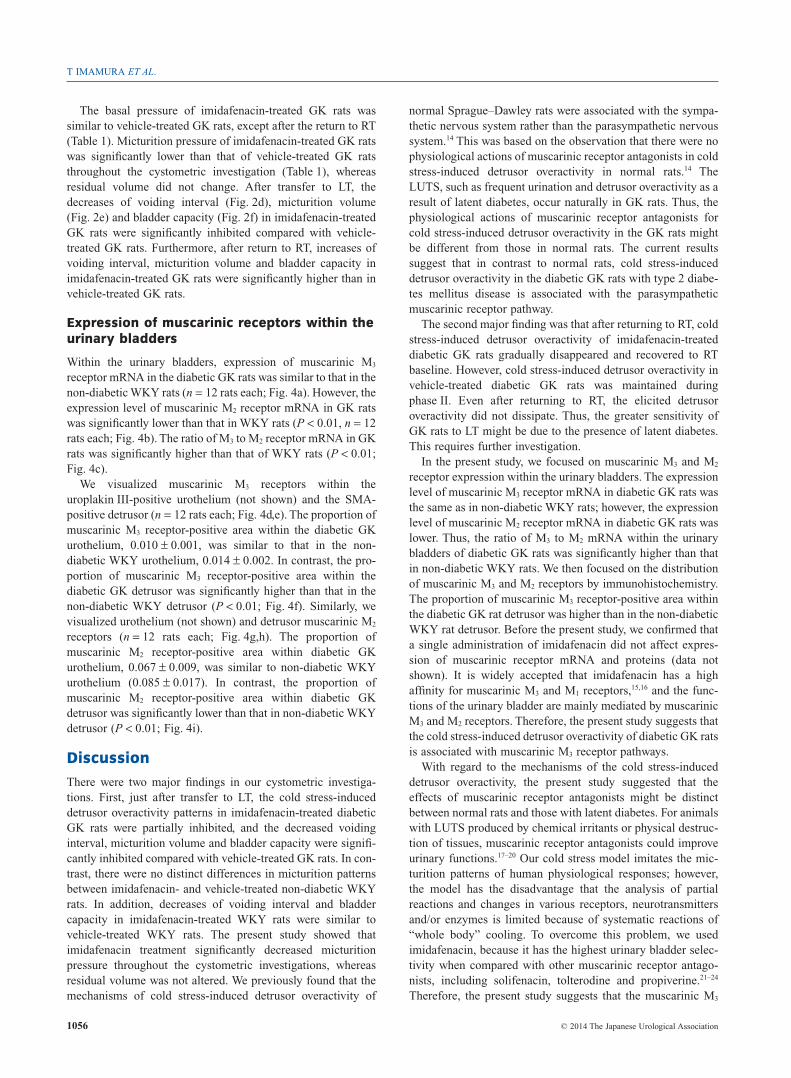
Within the urinary bladders, expression of muscarinic M3
receptor mRNA in the diabetic GK rats was similar to that in thenon-diabetic WKY rats (n = 12 rats each; Fig. 4a). However, theexpression level of muscarinic M2 receptor mRNA in GK ratswas significantly lower than that in WKY rats (P < 0.01, n = 12rats each; Fig. 4b). The ratio of M3 to M2 receptor mRNA in GKrats was significantly higher than that of WKY rats (P < 0.01;Fig. 4c).
We visualized muscarinic M3 receptors within theuroplakin III-positive urothelium (not shown) and the SMA-positive detrusor (n = 12 rats each; Fig. 4d,e). The proportion ofmuscarinic M3 receptor-positive area within the diabetic GKurothelium, 0.010 ± 0.001, was similar to that in the non-diabetic WKY urothelium, 0.014 ± 0.002. In contrast, the pro-portion of muscarinic M3 receptor-positive area within thediabetic GK detrusor was significantly higher than that in thenon-diabetic WKY detrusor (P < 0.01; Fig. 4f). Similarly, wevisualized urothelium (not shown) and detrusor muscarinic M2
receptors (n = 12 rats each; Fig. 4g,h). The proportion ofmuscarinic M2 receptor-positive area within diabetic GKurothelium, 0.067 ± 0.009, was similar to non-diabetic WKYurothelium (0.085 ± 0.017). In contrast, the proportion ofmuscarinic M2 receptor-positive area within diabetic GKdetrusor was significantly lower than that in non-diabetic WKYdetrusor (P < 0.01; Fig. 4i).
Discussion
There were two major findings in our cystometric investiga-tions. First, just after transfer to LT, the cold stress-induceddetrusor overactivity patterns in imidafenacin-treated diabeticGK rats were partially inhibited, and the decreased voidinginterval, micturition volume and bladder capacity were signifi-cantly inhibited compared with vehicle-treated GK rats. In con-trast, there were no distinct differences in micturition patternsbetween imidafenacin- and vehicle-treated non-diabetic WKYrats. In addition, decreases of voiding interval and bladdercapacity in imidafenacin-treated WKY rats were similar tovehicle-treated WKY rats. The present study showed thatimidafenacin treatment significantly decreased micturitionpressure throughout the cystometric investigations, whereasresidual volume was not altered. We previously found that themechanisms of cold stress-induced detrusor overactivity of
normal Sprague–Dawley rats were associated with the sympa-thetic nervous system rather than the parasympathetic nervoussystem.14 This was based on the observation that there were nophysiological actions of muscarinic receptor antagonists in coldstress-induced detrusor overactivity in normal rats.14 TheLUTS, such as frequent urination and detrusor overactivity as aresult of latent diabetes, occur naturally in GK rats. Thus, thephysiological actions of muscarinic receptor antagonists forcold stress-induced detrusor overactivity in the GK rats mightbe different from those in normal rats. The current resultssuggest that in contrast to normal rats, cold stress-induceddetrusor overactivity in the diabetic GK rats with type 2 diabe-tes mellitus disease is associated with the parasympatheticmuscarinic receptor pathway.
The second major finding was that after returning to RT, coldstress-induced detrusor overactivity of imidafenacin-treateddiabetic GK rats gradually disappeared and recovered to RTbaseline. However, cold stress-induced detrusor overactivity invehicle-treated diabetic GK rats was maintained duringphase II. Even after returning to RT, the elicited detrusoroveractivity did not dissipate. Thus, the greater sensitivity ofGK rats to LT might be due to the presence of latent diabetes.This requires further investigation.
In the present study, we focused on muscarinic M3 and M2
receptor expression within the urinary bladders. The expressionlevel of muscarinic M3 receptor mRNA in diabetic GK rats wasthe same as in non-diabetic WKY rats; however, the expressionlevel of muscarinic M2 receptor mRNA in diabetic GK rats waslower. Thus, the ratio of M3 to M2 mRNA within the urinarybladders of diabetic GK rats was significantly higher than thatin non-diabetic WKY rats. We then focused on the distributionof muscarinic M3 and M2 receptors by immunohistochemistry.The proportion of muscarinic M3 receptor-positive area withinthe diabetic GK rat detrusor was higher than in the non-diabeticWKY rat detrusor. Before the present study, we confirmed thata single administration of imidafenacin did not affect expres-sion of muscarinic receptor mRNA and proteins (data notshown). It is widely accepted that imidafenacin has a highaffinity for muscarinic M3 and M1 receptors,15,16 and the func-tions of the urinary bladder are mainly mediated by muscarinicM3 and M2 receptors. Therefore, the present study suggests thatthe cold stress-induced detrusor overactivity of diabetic GK ratsis associated with muscarinic M3 receptor pathways.
With regard to the mechanisms of the cold stress-induceddetrusor overactivity, the present study suggested that theeffects of muscarinic receptor antagonists might be distinctbetween normal rats and those with latent diabetes. For animalswith LUTS produced by chemical irritants or physical destruc-tion of tissues, muscarinic receptor antagonists could improveurinary functions.17–20 Our cold stress model imitates the mic-turition patterns of human physiological responses; however,the model has the disadvantage that the analysis of partialreactions and changes in various receptors, neurotransmittersand/or enzymes is limited because of systematic reactions of“whole body” cooling. To overcome this problem, we usedimidafenacin, because it has the highest urinary bladder selec-tivity when compared with other muscarinic receptor antago-nists, including solifenacin, tolterodine and propiverine.21–24
Therefore, the present study suggests that the muscarinic M3
T IMAMURA ET AL.
1056 © 2014 The Japanese Urological Association

receptors expressed within the detrusors are a key componentof cold stress-induced detrusor overactivity in the rats withinherited diabetes mellitus and latent LUTS.
In conclusion, the muscarinic receptor antagonist, imida-fenacin, reduced cold stress-induced detrusor overactivity indiabetic GK rats, but not in non-diabetic WKY rats. Both theratio of muscarinic M3 receptor to M2 receptor mRNA andproportion of muscarinic M3 receptors within the detrusor of
GK rats were significantly higher than in WKY rats. Therefore,the muscarinic M3 pathway, at least in part, mediates the coldstress-induced detrusor overactivity of GK rats with inheritedtype 2 diabetes mellitus.
Conflict of interest
None declared.
(a) 2.0
1.5
1.0
0.5
0.0
Exp
ress
ion
leve
l of M
3 m
RN
A
Non-diabetic WKY Diabetic GK
2.0
1.5
1.0
0.5
0.0Non-diabetic WKY Diabetic GK
(b)
Exp
ress
ion
leve
l of M
2 m
RN
A
*
Pro
por
tion
of M
3 p
ositi
ve a
rea
(%)
(f) 0.04
0.03
0.02
0.01
0.00Non-diabetic WKY Diabetic GK
*
Pro
por
tion
of M
2 p
ositi
ve a
rea
(%)
(i) 0.04
0.03
0.02
0.01
0.00Non-diabetic WKY Diabetic GK
*
0.3
0.2
0.1
0.0Non-diabetic WKY Diabetic GK
*
(c)
Rat
io o
f M3
to M
2 m
RN
A
(d)
(h)
(e)
(g)
Fig. 4 Muscarinic M3 and muscarinic M2 receptor expressing within the urinary bladders. (a) The expression level of muscarinic M3 receptor mRNA in diabetic GK rats was
similar to that in non-diabetic WKY rats. (b) The expression level of muscarinic M2 receptor mRNA of the diabetic GK rats was significantly lower than that in non-diabetic
WKY rats. (c) The ratio of M3 to M2 mRNA in the diabetic GK rats was significantly higher than that of the non-diabetic WKY rats. (d,e) Muscarinic M3 receptors (green and/or
yellow) within the detrusor (red) in non-diabetic WKY (d) and diabetic GK (e) rats were visualized (arrowheads). (f) The proportion of muscarinic M3 receptor-positive area
within the detrusor in diabetic GK rats was significantly higher than that in the non-diabetic WKY rats. (g,h) Muscarinic M2 receptors (green and/or yellow) within the
detrusor (red) in non-diabetic WKY (g) and diabetic GK (h) rats were also visualized (arrowheads). (f) The proportion of muscarinic M2 receptor-positive area within the
detrusor in diabetic GK rats was significantly lower than that in the non-diabetic WKY rats. *P < 0.01 compared with non-diabetic WKY rats.
Cold stress and muscarinic receptors
© 2014 The Japanese Urological Association 1057

References1 Imamura T, Ishizuka O, Aizawa N et al. Cold environmental stress induces
detrusor overactivity via resiniferatoxin-sensitive nerves in conscious rats.Neurourol. Urodyn. 2008; 27: 348–52.
2 Watanabe T, Maruyama S, Maruyama Y et al. Seasonal changes in symptomscore and uroflowmetry in patients with lower urinary tract symptoms. Scand.
J. Urol. Nephrol. 2007; 41: 521–6.3 Imamura T, Ishizuka O, Sudha GS et al. A galenical produced from
Ba-Wei-Die-Huang-Wan (THC-002) provides resistance to the coldstress-induced detrusor overactivity in conscious rats. Neurourol. Urodyn.
2013; 32: 486–92.4 Kebapci N, Yenilmez A, Efe B, Entok E, Demirustu C. Bladder dysfunction
in type 2 diabetic patients. Neurourol. Urodyn. 2007; 26: 814–19.5 Van Den Eeden SK, Ferrara A, Shan J et al. Impact of type 2 diabetes on
lower urinary tract symptoms in men: a cohort study. BMC Urol. 2013; 13:12.
6 Chiu AF, Huang MH, Wang CC, Kuo HC. Higher glycosylated hemoglobinlevels increase the risk of overactive bladder syndrome in patients with type 2diabetes mellitus. Int. J. Urol. 2012; 19: 995–1001.
7 Saito M. Editorial comment to higher glycosylated hemoglobin levelsincrease the risk of overactive bladder syndrome in patients with type 2diabetes mellitus. Int. J. Urol. 2012; 19: 1001–2.
8 Uzun H, Ogullar S, Sahin SB et al. Increased bladder wall thickness indiabetic and nondiabetic women with overactive bladder. Int. Neurourol. J.
2013; 17: 67–72.9 Inoue H, Ishizuka O, Imamura T et al. Relationship between toe temperature
and lower urinary tract symptoms. LUTS 2012; 4: 144–9.10 Maggiore UL, Scala C, Venturini PL, Ferrero S. Imidafenacin for the
treatment of overactive bladder. Expert Opin. Pharmacother. 2013; 14:1383–97.
11 Takeuchi T, Zaitsu M, Mikami K. Experience with imidafenacin in themanagement of overactive bladder disorder. Ther. Adv. Urol. 2013; 5: 43–58.
12 Goto Y, Kakizaki M, Masaki N. Production of spontaneous diabetic rats byrepetition of selective breeding. Tohoku J. Exp. Med. 1976; 119: 85–90.
13 Portha B, Serradas P, Bailbe D, Suzuki K, Goto Y, Giroix MH. Beta-cellinsensitivity to glucose in the GK rat, a spontaneous nonobese model for typeII diabetes. Diabetes 1991; 40: 486–91.
14 Chen Z, Ishizuka O, Imamura T et al. Role of alpha1-adrenergic receptors indetrusor overactivity induced by cold stress in conscious rats. Neurourol.
Urodyn. 2009; 28: 251–6.
15 Kobayashi F, Yageta Y, Segawa M, Matsuzawa S. Effects of imidafenacin(KRP-197/ONO-8025), a new anti-cholinergic agent, on muscarinicacetylcholine receptors. High affinities for M3 and M1 receptor subtypes andselectivity for urinary bladder over salivary gland. Arzneimittelforschung
2007; 57: 92–100.16 Kobayashi F, Yageta Y, Yamazaki T et al. Pharmacological effects of
imidafenacin (KRP-197/ONO-8025), a new bladder selective anti-cholinergicagent, in rats. Comparison of effects on urinary bladder capacity andcontraction, salivary secretion and performance in the Morris water mazetask. Arzneimittelforschung 2007; 57: 147–54.
17 Nishijima S, Sugaya K, Kadekawa K, Naka H, Miyazato M. Comparison ofthe effect of anti-muscarinic agents on bladder activity, urinary ATP level, andautonomic nervous system in rats. Biomed. Res. 2009; 30: 107–12.
18 Watanabe N, Akino H, Kurokawa T et al. Antidiuretic effect ofantimuscarinic agents in rat model depends on C-fibre afferent nerves in thebladder. BJU Int. 2013; 112: 131–6.
19 Yamamoto S, Maruyama S, Ito Y et al. Effect of oxybutynin andimidafenacin on central muscarinic receptor occupancy and cognitivefunction: a monkey PET study with [(11)C](+)3-MPB. Neuroimage 2011; 58:1–9.
20 Yokoyama O, Tanaka I, Kusukawa N et al. Antimuscarinics suppressadenosine triphosphate and prostaglandin E2 release from urothelium withpotential improvement in detrusor overactivity in rats with cerebral infarction.J. Urol. 2011; 185: 2392–7.
21 Yamada S, Kuraoka S, Osano A, Ito Y. Characterization of bladder selectivityof antimuscarinic agents on the basis of in vivo drug-receptor binding. Int.
Neurourol. J. 2012; 16: 107–15.22 Yamada S, Seki M, Ogoda M, Fukata A, Nakamura M, Ito Y. Selective
binding of bladder muscarinic receptors in relation to the pharmacokinetics ofa novel antimuscarinic agent, imidafenacin, to treat overactive bladder. J.
Pharmacol. Exp. Ther. 2011; 336: 365–71.23 Yamazaki T, Muraki Y, Anraku T. In vivo bladder selectivity of
imidafenacin, a novel antimuscarinic agent, assessed by using an effectivenessindex for bladder capacity in rats. Naunyn. Schmiedebergs Arch. Pharmacol.
2011; 384: 319–29.24 Yoshida A, Maruyama S, Fukumoto D, Tukada H, Ito Y. Noninvasive
evaluation of brain muscarinic receptor occupancy of oxybutynin, darifenacinand imidafenacin in rats by positron emission tomography. Life Sci. 2010; 87:175–80.
T IMAMURA ET AL.
1058 © 2014 The Japanese Urological Association